murine models to evaluate novel and conventional therapeutic strategies for cancer
TRANSCRIPT
ASIP
Journal
CME Program
ReviewMurine Models to Evaluate Novel and ConventionalTherapeutic Strategies for Cancer
James E. Talmadge,* Rakesh K. Singh,*Isaiah J. Fidler,† and Avraham Raz‡
From the University of Nebraska Medical Center,* Omaha,
Nebraska; the University of Texas M.D. Anderson Cancer Center,†
Houston, Texas; and the Karmanos Cancer Institute,‡
Detroit, Michigan
Animal models, by definition, are an approximationof reality, and their use in developing anti-cancerdrugs is controversial. Positive retrospective clinicalcorrelations have been identified with several animalmodels, in addition to limitations and a need forimprovement. Model inadequacies include experi-mental designs that do not incorporate biologicalconcepts, drug pharmacology, or toxicity. Ascitesmodels have been found to identify drugs activeagainst rapidly dividing tumors; however, neither as-citic nor transplantable subcutaneous tumors are pre-dictive of activity for solid tumors. In contrast, pri-mary human tumor xenografts have identifiedresponsive tumor histiotypes if relevant pharmacody-namic and toxicological parameters were considered.Murine toxicology studies are also fundamental be-cause they identify safe starting doses for phase Iprotocols. We recommend that future studies incor-porate orthotopic and spontaneous metastasis mod-els (syngeneic and xenogenic) because they incorpo-rate microenvironmental interactions, in addition toconfirmatory autochthonous models and/or geneti-cally engineered models, for molecular therapeutics.Collectively, murine models are critical in drug devel-opment, but require a rational and hierarchical ap-proach beginning with toxicology and pharmacologystudies, progressing to human primary tumors toidentify therapeutic targets and models of metastaticdisease from resected orthotopic, primary tumors tocompare drugs using rigorous, clinically relevant out-come parameters. (Am J Pathol 2007, 170:793–804; DOI:10.2353/ajpath.2007.060929)
Animal models are critical for the development of noveltherapeutics; however, we have been minimally success-
ful in decreasing the age-adjusted death rate for cancercompared with cardiac disease. In 2003, for the first timesince 1930 when epidemiological records were initiated,fewer people (�85 years old) died of cardiac disease ascompared with cancer.1 This historic change was attrib-utable to a 60, 70, and 0% decrease in mortality by heartdisease, stroke, and cancer, respectively. Thus, it is war-ranted to review the approaches and tumor models usedin the identification and development of new anti-cancertherapeutics. Tumor initiation, progression, and metasta-sis is a complex, multifactorial process that selects tumorvariants from a heterogeneous primary tumor.2,3 Thera-peutic intervention is also a selective pressure that canresult in tumor cell populations refractory to specificdrugs.4 Therefore, to model and study tumor biology anddrug activity, the selection of clinically relevant animaland tumor models is critical.
Originally, drug screens used leukemic cell lines that,when injected intraperitoneally (i.p.) resulted in tumorascites. These tumor models were successful in identify-ing active therapeutics against leukemias and some lym-phomas; however, they were inadequate for the identifi-cation of therapeutics against solid tumors.5–7
Subsequent studies using ectopically implanted synge-neic or human tumor cell lines were found to be ineffec-tive for identifying therapeutically active drugs. In con-trast, primary human tumors can predict responsivetumor histiotypes for targeting in phase I/II studies.8,9
Rodent toxicological studies have reliably identified safeinitial doses for phase I studies; nonetheless, we have notyet identified animal models that can predict the extent ofclinical efficacy. Recent studies have suggested that an-imal models using orthotopically implanted, syngeneictumors are more predictive of responses than ectopictumors.10–13 There has also been interest in geneticallyengineered models (GEMs), in part, because of theirorthotopic primary tumors and relevance to moleculartherapeutics.14 However, GEMs use artificial promotersthat can influence the affected cell type, vary expression
Accepted for publication December 12, 2006.
Address reprint requests to James E. Talmadge, Ph.D., University ofNebraska Medical Center, 987660 Nebraska Medical Center, Omaha,NE, 68198-7660. E-mail: [email protected].
The American Journal of Pathology, Vol. 170, No. 3, March 2007
Copyright © American Society for Investigative Pathology
DOI: 10.2353/ajpath.2007.060929
793
based on the genetic background,15 and decrease cel-lular heterogeneity, which in turn can affect tumor pro-gression and metastasis. Furthermore, GEMs may not beuniversally relevant, ie, only 30% of breast cancer pa-tients with human epidermal growth factor receptor 2(HER2/neu) mutation can be modeled with HER2/neutransgenic mice. Critical to studies of therapeutic inter-vention using animal models is the incorporation of phar-macological and toxicological considerations, as well asthe mechanistic concepts of tumor induction, progres-sion, and metastasis. We suggest that the cycles of en-thusiasm and pessimism for tumor models7 may havebecome overly negative and that an assessment of clin-ically useful correlations provided by animal models andtheir rational utilization is needed.
History of Tumor Models
The origin of our failure to identify drugs that have in-creased clinical activity is multifactorial and includes, butis not limited to, differences in efficacy of drugs in miceversus humans. Toxicity issues are also a commonsource of drug failure and are associated with the use ofmodels selected for ease of modeling and a high inci-dence of positive responses. Many commonly used solidtumor models are biased toward false-positive resultsbecause they are selected based on ease of use, sensi-tivity to therapeutics, rapid growth, and other attributesthat facilitate studies, but not clinical correlations. Thesedeficiencies can be reduced by strict attention to properdesign and conduct of efficacy studies, as well as theincorporation of a rational design based on an under-standing of tumor biology, variant selection, and surro-gate endpoints and the integration of testing strategiesthat reflect clinical tumor biology (Table 1). The history ofanimal models in the development of cancer drugs hasbeen previously discussed5–7,9,16,17; however, few ofthese reviews have incorporated recommendations forfuture approaches.
Ascites Tumors
In 1955, it was suggested that a correlation existed be-tween efficacy against transplanted tumors and clinicalactivity.18 This stimulated the National Cancer Institute(NCI) to launch an anti-cancer drug screening programusing three murine models.19 Over time, the number oftumors studied was reduced, and by 1968, drugs werescreened only against the L1210 leukemia cell line. Theclinical response to chemotherapy by human leukemiasand lymphomas significantly improved during this devel-opmental period,20,21 whereas the treatment of most solidtumors did not. Further, concerns were identified regard-ing reliance on a single leukemia tumor model becausethis could preferentially select for drugs targeting rapidlygrowing tumors.22 Therefore, the B16 melanoma andLewis lung carcinoma mouse models were incorporatedinto the screening program in 1972.23,24
Solid Human and Murine Tumors
In 1976, the NCI’s Division of Cancer Treatment initiatedthe use of a new tumor panel incorporating transplant-able solid human tumors that were representative of themajor histological types of cancer. The panel consisted ofhuman tumors of the breast, colon, and lung, in additionto the murine L1210 leukemia and B16 melanoma syn-geneic models.22 These syngeneic models involved in-oculation of tumor cells by i.p., subcutaneous (s.c.), orintravenous (i.v.) routes, whereas human tumor xeno-grafts were grown under the renal subcapsule.25 Al-though the subrenal capsule assay is labor intensive, itprovides a rapid evaluation of drug activity.26 Subse-quent analysis of this strategy (1976 to 1982) revealedthat the mouse-human tumor panel identified an anti-tumor agent (taxol) that would have been missed by theL1210 model.22 Furthermore, �30% of compounds foundto be active in at least one human tumor xenograft werenot identified by the syngeneic models. Therefore, it wasconcluded that the mouse-human tumor panel may be
Table 1. Animal Model Attributes versus Clinical Situation
Attribute Murine model Clinical situation
Drug administered at the MTD versus LD MTD MTD-LDTumor implant site Ectopic OrthotopicDuration of cell cycle Short cell cycle Long cell cycleAntigenicity High antigenicity Low antigenicitySite of therapeutic target Primary tumor Metastatic tumorsTreatment protocol 1 Single cycle of therapy Multiple cycles of therapyTreatment protocol 2 Monotherapy PolychemotherapyTreatment protocol 3 Single therapeutic approach,
occasionally with surgeryMulti-modality including surgery, hormonal,
immune, and molecular therapyTreatment protocol 4 1 to 3 days of therapy 1 to 2 weeks of therapyTreatment protocol 5 Single cycle of therapy Multiple cycles of therapyPharmacokinetic consideration Push delivery Infusion of drug if it has a short half lifeTumor burden Minimal tumor burden Locally advanced or systemic diseaseDuration of tumor presence before diagnosis Days to weeks Years to decades
Clinical and model attributes that need to be considered in the design of screening and developmental strategies and protocols. MTD, maximumtolerated dose; LD, lethal dose.
794 Talmadge et alAJP March 2007, Vol. 170, No. 3
successful in identifying new drugs for clinical studies,but with a low correlation between preclinical and clinicalefficacy.27 Despite their inherent deficiencies, transplant-able tumors remain valuable because they provide anevaluation within the context of an intact immune systemand host stroma and extracellular matrix.
Sequential Tumor Model
Based on these results, the NCI implemented a sequen-tial screening strategy in 1982, whereby a potential drugwas examined using progressively more rigorous mod-els. Initially, drugs were examined against the P388 leu-kemia as a prescreen, followed by studies with a panel ofmurine tumor models (MX-1, B16, M5076, and L1210),resulting in the identification of a large number of poten-tially active compounds.22 Drugs active in the primarytumor panel were advanced to a secondary screen usingcompound-orientated tumors based on the properties ofeach drug and experience in the primary tumor panel.However, a retrospective analysis of preclinical-clinicalefficacy28 did not demonstrate a correlation based ontumor histiotype. This was perceived to be attributable toan experimental design that limited tumors to one mouseand one human tumor for each of the three major histio-types. Thus, it was concluded that a model system com-posed of several tumors with the same histiotype mightbetter predict a clinical response against a specific tumorhistiotype.28
Human Tumor Stem Cell (HTSC)Assay/Clonogenic Assay
A HTSC assay29,30 was developed in the early 1980s todetermine whether a model system incorporating multipletumors of the same histiotype could predict a clinicalresponse for a specific tumor histiotype.22 The HTSCassay was disease-orientated using soft agar colonygrowth of freshly explanted human tissue with outcomesbased on growth inhibition. Salmon et al29 compared invitro results with the clinical responses of myeloma andovarian cancer patients and found correlations based onsensitivity and resistance. The HTSC assay was sug-gested to be appropriate for drug screening based onfeasibility, validity, and potential to identify new anti-tumoragents.31 Initial studies examined established chemo-therapeutic agents and found that most drugs were ac-tive with the exception of drugs requiring systemic acti-vation. Clinically ineffective agents were also found to benegative with 97% accuracy, an observation confirmedby other reports.8,32–35
However, the HTSC assay8,36 is limited because of thelow plating efficiency of most solid tumors and the pooravailably of tumor tissue. Thus, only breast, colorectal,kidney, lung, melanoma, and ovarian tumors have beenfound to provide sufficient cells for evaluation, althoughstrategies have been suggested to improve the growthrates of primary tumor tissues.8 Furthermore, althoughthese models predict responsive histiotypes, no clinical
analysis of individualized therapy has demonstrated asignificant increase in survival compared with empiricallydetermined standard treatment; therefore, the HTSC as-say has not found a role in the individualization of patienttherapy.8
In Vitro Human Tumor Cell Line Screen
In 1990, a study using human tumor cell lines for large-scale drug screening was initiated.37 This in vitro humantumor cell line screen shifted from being a compound-orientated to a disease-orientated screening strategy.The initial panel incorporated 60 different human tumorcell lines, resulting in the use of multiple tumors with thesame histiotype. Toward the end of the decade, an in vitroprescreen was introduced using three cell lines: MCF-7(breast carcinoma), NCI-H460 (lung carcinoma), and SF-268 (glioma). The rationale for this prescreen was that itcould remove inactive compounds from unnecessaryand costly full-scale evaluation. In a study by the NCI ofCanada Clinical Trial Group,17 an in vitro cell line modelfor non-small cell lung cancer was shown to be predictivefor phase II activity. This observation was also confirmedin a human xenograft model for non-small cell lung can-cer, but not with breast or colon cancer. In addition, amouse allograft model was found not to be predictive forany histiotype.17 Studies using the panel of 60 humantumor cell lines to assess mechanisms of action (MOAs)supported the concept that pharmacokinetic (PK) andpharmacodynamic parameters must be consid-ered.38–41 This is critical because the failure of drugs inthe clinic41,42 is often associated with a poor PK profile ordrug toxicity.40 Indeed, primary human tumor xenograftscan be predictive of clinical cytotoxic therapy for a giventumor histiotype provided that clinically relevant, pharma-cological dosing parameters are used.9,39–42 It is notedthat human tumor cell lines, in contrast to primary humantumor cells, have generally been cultured for years, los-ing much of their heterogeneity. This has resulted inundifferentiated tumors lacking the histology and cellulararchitecture characteristic of the modeled human tumor.Thus, administration of clinically relevant drug doses toanimals with s.c. xenografts results in response patternssimilar to those observed with the human tumor histiotypeand the same drugs.40 These studies emphasize theneed to determine the exposure levels required for anti-tumor activity with the intention that unnecessary toxicityis avoided in phase I clinical trials. Further, they suggestthat primary human tumor xenografts can be used toidentify responsive human histiotypes.
Screening Using Human Tumor Xenografts inImmunodeficient Mice
Many of the initial reports using malignant human tumorsshowed that they did not metastasize in nude mouse,which cast doubts on the validity of this model. It is nowclear that tumor metastasis depends on intrinsic tumorcell properties and host factors, the experimental tech-
Animal Models in Anti-Cancer Drug Development 795AJP March 2007, Vol. 170, No. 3

nique(s) used, as well as the origin, health, and mainte-nance of the immune-deficient animal. Today, we knowthat human neoplasms can be studied in immune-defi-cient mice; however, clinical relevance is obtained only ifcareful attention is paid to the experimental conditions.The neoplasms must be free of mouse pathogens, andthe mice must be kept in specific pathogen-free condi-tions. Careful consideration must be given to the anatom-ical site of implantation because the metastatic potentialof human tumor cells is dependent on both intrinsic prop-erties of the tumor cells and host factors, which can differbetween tissues and organs. These studies require nude(athymic) or severe combined immunodeficient (SCID)mice that are T- and B-cell-deficient, allowing the engraft-ment of human tumor cells. However, innate immunity,particularly natural killer (NK) cells, can limit tumor growthand prevent metastasis in nude mice.43 Mice with thenude mutation, although T-cell-deficient, have a compen-satory increase in innate immunity, most notably in-creased NK activity and tumoricidal macrophages. Thebeige mutation (murine homolog of Chediak-Higashi syn-drome) results in a delay in NK activation, but not the lossof NK cell function.44 Thus, the NK cells lack secondarygranules and have a delayed killing ability. Further, nono-bese diabetic-severe combined immunodeficiency(NOD-SCID) mice can be humanized (rendered chi-meric) with the injection of human peripheral blood (PB)or bone marrow (BM) cells,45 resulting in a somewhatmore relevant microenvironment.
Pharmacological and toxicological parameters havebeen studied using s.c. tumor xenograft in nude mice,and efficacy defined by a delay in tumor growth, withbody weight loss, and mortality as parameters of toxicity.Several reports support the use of this model,46,47 includ-ing a clinical response comparison study that used alarge panel of xenografts derived from patient biop-sies.8,48 This study observed a correlation with clinicaloutcome for both tumor resistance (97%) and sensitivity(90%). It was concluded that if primary tumors are used,xenograft models can predict clinical activity similar tothe clonogenic assay. In contrast, a retrospective NCIstudy evaluated 39 drugs using transplantable humantumor cell lines and compared them to phase II clinicalresults.9 In these studies, in vivo xenograft activity did notcorrelate with activity against the same human tumorhistology. However, drugs that were active against onethird of xenografts correlated with clinical activity.9 Acomparison of these two large studies9,48 suggests thatex vivo studies using primary tumors may be predictive ofclinical activity, whereas studies using human cell linesare not. One may conclude that the s.c. injection ofxenograft cell lines may have value in the development ofcytotoxic drugs8,9,39,49; however, these seem to bepoorly predictive of a specific histological response. It isnoted that a recent review of the athymic nude mousemodel using human xenografts39 suggested that an out-come focused on tumor growth rate or cytostasis may bemore predictive of clinical activity than tumor shrinkage(cytotoxicity). Regardless of these correlations, murinexenograft models are not ideal for cancer drug develop-ment. In addition to the points discussed above, these
models lack human stroma and immune cells, which areimportant to the metastatic process.50
Humanized Mice
The term humanized mice has been used to describenumerous animal models, including immunodeficientmice reconstituted with human stem cells or lympho-cytes.51,52 This approach has also been combined withthe transplantation of human thymi and/or BM beforestem cell injection to provide a human stromal environ-ment. These humanized mice are used to study graft-versus-host disease and solid organ transplantation. Im-munologically humanized mice have also beenimmunized to induce human hybridomas and to studyT-cell responses against tumors and viruses. In thesemodels, humanized mice are ones injected with stemcells and then immunized or immune-deficient mice hu-manized by the injection of T cells from immunized pa-tients/donors. They can then be challenged with humantumor xenografts or viruses to study the effect of immunityon tumor/viral growth.53 Indeed, this approach providesan ethical and cost-effective strategy to test vaccine ef-ficacy. Humanized mice transplanted with fragments ofhuman organs are also used to study the role of interac-tions between xenogenic human stroma and tumors intumor progression and metastasis.
Another definition of humanized mice involves the in-sertion of a human gene into the mouse genome.54 SuchGEMs are used to study species-associated differencesin phenotypes, including responses to drugs or tumorantigens (Ags).55 Models to study drug responses in-clude GEM mice expressing human cytochrome p450genes54 that allow the in vivo analysis of cytochrome P450metabolism of endogenous and exogenous chemicals,including xenobiotics. One example of these studies isthe analysis of cytochrome P450 2E1 (CYP2E1) expres-sion on cisplatin-induced hepatotoxicity, using mice withinduced or steady-state CYP2E1 levels and a compari-son to knockout and CYP2E1-humanized mice.56 Humantumor Ags have also been expressed in GEMs, renderingthem tolerant to human Ags and providing a model tostudy vaccine responses in the presence of immunolog-ical tolerance.57 Traditional murine vaccination modelsmay recognize human tumor Ags as foreign, providingoverly optimistic results. Thus, a humanized model withthe human tumor Ag provides a more relevant toleranthost. Furthermore, wild-type mice do not express class Iand II human leukocyte antigens (HLAs), and as such,relevant HLA Ag processing does not occur. However,GEMs that express HLA-Ags in the mouse thymus can beused to support the selection of T cells and recognizerelevant antigenic epitopes. These GEMs, after humanstem cell engraftment, can potentially allow accuratemodeling of T-cell responses via the expressed HLA-Ag.
In summary, humanized mouse models have madetremendous progress since their development almost 20years ago.56 However, a number of practical limitationsstill limit their use as rigorous paradigms of the humansystem. Additional work and validation remain before
796 Talmadge et alAJP March 2007, Vol. 170, No. 3

they can be routinely and confidentially used in drugdevelopment.
Orthotopic Tumor Models
Clinical observations have suggested that the organ en-vironment can influence the response of tumors to che-motherapy. For example, in women with breast cancer,lymph node and skin metastases are more sensitive tochemotherapeutic intervention than metastases in eitherthe lung or bone.58 Likewise, orthotopic implantation ofhuman tumor cells from surgical specimens into nudemice is mandatory for an accurate analysis of tumorgrowth and metastasis. This has been shown with coloncarcinomas (into the wall of the colon), renal cell cancers(into the kidney), melanomas (into the skin), mammarycarcinomas (into the mammary fat pad), bladder carci-nomas (into the bladder wall), prostate carcinoma (intothe prostate), pancreatic carcinoma (into the pancreas),and lung cancer (into the bronchi). Orthotopic implanta-tion results in rapid growth of local tumors and in severaltumor models, distant metastasis. There is also a striking,site-specific variation in response to chemotherapy. Inone study,59 colon carcinoma cells were implanted intodifferent anatomical locations of nude mice using thehighly metastatic KM12L4a human colon carcinoma cellline. In this study, mice were injected in the subcutis(ectopic site), spleen (leading to experimental liver me-tastasis), or cecum (growth at the orthotopic site). Tumor-bearing mice were treated with doxorubicin and subse-quently evaluated for responses. Tumors grown withinthe s.c. tissue showed an 80% inhibition of growth aftertwo i.v. injections of doxorubicin (10 mg/kg), comparedwith �40% inhibition of the intracecal tumors and lessthan 10% inhibition of lesions in the liver.59
Anti-cancer drugs are commonly screened using pan-els of human tumor xenografts implanted s.c. in nudemice. However, as discussed above, s.c. tumor modelsare not representative of the primary tumor site.60 Inaddition, clinically we treat well-established and fre-quently advanced metastatic disease, whereas conven-tional s.c. xenograft models are of recent origin (1 to 14days) and rarely have metastatic disease.40 Thus, ortho-topic tumor models seem to be a better model to assessthe morphology and the growth characteristics of clinicaldisease10–12,61 and to be more representative of a pri-mary tumor with respect to tumor site and metastasis.13
One of the obvious advantages of orthotopic models isthat targeting processes involved in local invasion (eg,angiogenesis) can be undertaken at a more clinicallyrelevant site.60 Since the early studies showing orthotopictransplantation of colon tumors and metastasis to theliver,62 tumor xenografts have been grown orthotopicallyin mice. Whether preclinical models are representative ofclinical disease (eg, orthotopic/metastatic models) andshould replace traditional s.c. nonmetastatic xeno-grafts12,40 remains an unanswered, yet critical, question.Clearly, the poor predictive power of our current modelssupport the use of alternative models and approaches.7
However, despite the clinical relevance of orthotopic
models, their utilization is hindered by a need for a highlevel of technical skill, time, and cost. Therapeutic effi-cacy is also more difficult to assess with orthotopic mod-els in contrast to the relative ease of s.c. tumor measure-ments.60 Clearly, murine tumors in intact synergicanimals have significant advantage beyond expense asthe model of a more clinically relevant host environment.
GEMs
Throughout the past 20 years, GEMs have contributed toour understanding of the molecular pathways responsi-ble for the initiation, progression, and metastasis of can-cer cells and have extended our knowledge of the mech-anistic role that oncogenes and tumor suppressor geneshave in these processes. In addition, studies with GEMshave improved our understanding of the role genes andtheir mutated counterparts have in tumorigenesis, as wellas the cooperation of individual mutations in tumor de-velopment. The initial GEMs were murine models thatoverexpressed viral and cellular oncogenes.63 Subse-quent studies used genes targeted to mouse embryonicstem cells, providing oncogene-bearing transgenic mice(knockin) or loss of function, ie, gene knockout mice. Inaddition to the use of transgene overexpression models,conditional strategies have been developed that allowcontrolled gene expression in both a tissue- and tempo-ral-specific manner.64 Thus, tet-regulated65 or CRE-in-ducible alleles can regulate the timing, duration, andtissue compartment of gene expression or inactivation.Furthermore, these technologies can be combined, re-sulting in GEMs with specific cancers that overexpress orlack genes of interest in all cells or in a specific tissuecompartment and/or developmental stage. These ap-proaches have significantly contributed to our under-standing of cancer pathogenesis and may ultimately helpin the identification of anti-neoplastic drugs.
Although the use of GEMs in drug development hasnot been validated against drugs with efficacy in thecorresponding human tumor, studies with several havesuggested potential utility. Retinoic acid has shown ac-tivity in GEMs of acute promyelocytic leukemia,66 andImatinib, a BCR-ABL inhibitor that is active againstchronic myelogenous leukemia, has been shown to limitthe development of BCR-ABL mutations in P190BCR-ABL
GEM mice.67 One model used in the development of thecyclooxygenase (COX-) 2 inhibitor, celecoxib, is the mul-tiple intestinal neoplasia (Min) mouse that was created bygerm line mutagenesis. This resulted in a point mutationin the Apc tumor suppressor gene and a high frequencyof intestinal tumors.68 The outcomes from this GEM aresimilar to patients who have familial adenomatous polyp-osis and a high frequency of intestinal tumors. Overall,GEMs have seldom been used to test novel anti-cancertherapeutics with the goal of accurately predicting clini-cal responses.69,70 The few studies that have comparedGEMs using clinically effective agents have not beenencouraging.66,71–73 Thus, despite their mechanisticpromise, transgenic mouse models have not yet demon-strated a role in drug discovery.
Animal Models in Anti-Cancer Drug Development 797AJP March 2007, Vol. 170, No. 3

GEMs have been primarily used to study specific ther-apeutic questions relevant to the affected gene and tostudy interactions between tumor cells and their micro-environment. They are potentially more representative ofspecific human tumor histiotypes than transplanted xeno-grafts because of their in situ and autochthonous origin.70
However, GEMs have limitations, including expense, timecommitment, intellectual property restrictions,74 and spe-cies-specific differences, resulting in different mutantphenotypes in man and mouse.75 Further, no one trans-genic model is representative of all of the different formsof even one tumor histiotype; just as one human tumorcannot represent another human tumor of the same his-tiotype. In GEMs, transgenes are driven by artificial pro-moters, which may influence the cell type affected. Inaddition, the genetic background can affect transgeneexpression. Thus, mice carrying a mutation in the Apcgene express different lesions dependent on the geneticbackground.15 In this case, the predisposing mutation isidentical, but the outcome, including rapidity of lesiondevelopment (months to years), type of lesion (hyperpla-sia to metastatic tumors), and tumor histiotype (mam-mary, colon cancers, and lymphomas), are affected bygenetic background. Furthermore, by their very nature,GEMs do not incorporate the heterogeneity inherent to
tumor initiation, progression, and metastasis,16 and sys-temic disease is rarely observed in GEMs.76,77
Autochthonous Tumor Models
Autochthonous tumors include spontaneously occurringtumors and chemical, viral, or physical carcinogen-in-duced tumors and are believed to model human tumorsmore closely than transplanted tumors. Advantages ofautochthonous tumors include orthotopic growth, tumorhistology devoid of transplantation introduced changes,and metastasis via lymphatic and vascular vessels sur-rounding and within the primary tumor.78 Despite suchpositive properties, autochthonous tumor models havenot been widely used as an animal model for drug de-velopment. Autochthonous tumor models have an inher-ent variability in the time to and frequency of tumor in-duction, number of tumor(s) induced, and thus thenumber of animals required for a study.79 It is noted thatall of these suboptimal attributes are similar to these alsofound with GEMs (Table 2). Thus, time frames of severalmonths to a year for a single experiment, as opposed toweeks with transplanted xenograft models, are re-quired.78–80 Thus, autochthonous tumor models are best
Table 2. Comparative Clinical Relevance of Model
Transgenic tumors Orthotopic tumors Ectopic tumors
Histologically similar to human tumors � Often histologically similar to humantumors
� Often histologically similar to humantumors
Generally low immunogenic � Low to highly immunogenic � Low to highly immunogenicTumors arise in a stochastic manner, but
from a common molecular event� Highly heterogeneous � Highly heterogeneous
The use of a strong promoter results intransgenic overexpression that is notexpected clinically
� Highly heterogeneous � Highly heterogeneous
Metastatic distribution often parallels thatobserved clinically
� Metastatic distribution often parallelshuman distribution
� Metastases occur in the lungs, rarelyat other sites
Metastasis occurs infrequently � Metastasis occurs frequently � Gross metastasis occurs infrequentlyOne can study chemoprevention � One can study prophylaxis � One can study prophylaxisMay need to screen for expression if
homozygous lethality, or administerinducers for conditional expression
� Fully inbred mouse strains � Fully inbred mouse strains
Time for tumor induction anddevelopment is long, often requiring ayear or more
� If one is studying metastasisprotocols, often requires 3 to 4months
� If one is studying metastasis protocols,often requires 3 to 4 months
Transgenic mouse strains are oftenestablished in outbred mice, requiringinbreeding, and can result in straindependant activities
� Syngenic mouse strains, but tumor totumor variation in response occurs
� Syngenic mouse strains, but tumor totumor variation in response occurs
Expensive, based on labor and housing � Labor intensive, but relatively shorthousing duration
� Inexpensive based on labor andhousing duration
Relevant host immune cell infiltration andtumor microenvironment
� Relevant host immune cell infiltration � Irrelevant host infiltration and tumormicroenvironment
Multiple primary tumors precludessurgical resection
� Allows surgical resection of primarytumor
� Allows surgical resection of primarytumor, but in general it is minimallyinvasive
Difficult to vary therapeutic protocolrelative to tumor burden
� One can vary therapeutic schedulerelative to tumor burden
� One can vary therapeutic schedulerelative to tumor burden
Autochthonous tumor models have provenmore predictive then transgenic models
� Little information available regardingprediction of clinical response
� High frequency of false positiveresponses observed relative toclinical response
Various in vivo and ex vivo outcome measures that are used in animal models. Many of these measures are can also be extended.A comparison of attributes of transgenic, orthotopic transplanted, and entopic transplanted tumors and the relevance of these parameters to clinical
reality. The symbols used are greater than (�), lesser than (�), and equal to (�).
798 Talmadge et alAJP March 2007, Vol. 170, No. 3
reserved for confirmation studies,78 although in the post-genome era, autochthonous models have to an extentbeen replaced by GEMs. A recent study that comparedoutcomes from autochthonous models and GEMs with ameta-analysis of clinical outcomes81 found that carcino-gen-induced tumors correlated best with clinical re-sponses. These intervention studies used aspirin, �-car-otene, calcium, and wheat bran to treat recurrent colonadenoma in human volunteers compared with chemopre-vention studies with carcinogen-induced intestinal tu-mors in rats and large intestinal polyp induction in Min(Apc�/�) mice. The final meta-analyses included 6714volunteers, 3911 rats, and 458 mice. These studiesshowed that therapeutic responses in carcinogen-in-duced rat tumors predicted clinical responses for aspirin,calcium, and carotene and were compatible for wheatbran with the Min mouse models. Results from the trans-genic Min model were consistent with human respondersfor aspirin but were discordant for calcium and wheatbran. These results suggest that the carcinogen-inducedtumor models may be more predictive for human activityas compared with GEMs. Regardless of the differencesbetween the rodent models, they both provided correla-tion for chemopreventive activity, clinically.
Outcome Criteria for Animal Tumor Models
Inarguably, our ultimate goal is to cure patients of all oftheir tumors. However, the realistic goal in clinical oncol-
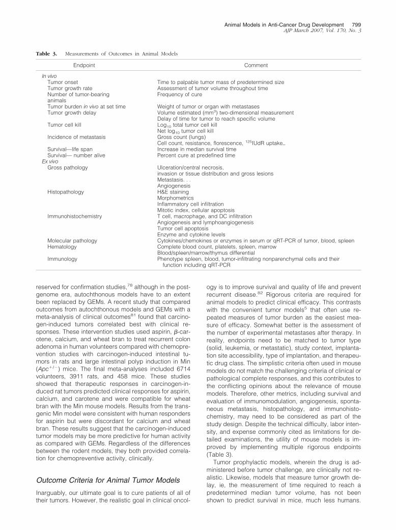
ogy is to improve survival and quality of life and preventrecurrent disease.82 Rigorous criteria are required foranimal models to predict clinical efficacy. This contrastswith the convenient tumor models5 that often use re-peated measures of tumor burden as the easiest mea-sure of efficacy. Somewhat better is the assessment ofthe number of experimental metastases after therapy. Inreality, endpoints need to be matched to tumor type(solid, leukemia, or metastatic), study context, implanta-tion site accessibility, type of implantation, and therapeu-tic drug class. The simplistic criteria often used in mousemodels do not match the challenging criteria of clinical orpathological complete responses, and this contributes tothe conflicting opinions about the relevance of mousemodels. Therefore, other metrics, including survival andevaluation of immunomodulation, angiogenesis, sponta-neous metastasis, histopathology, and immunohisto-chemistry, may need to be considered as part of thestudy design. Despite the technical difficulty, labor inten-sity, and expense commonly cited as limitations for de-tailed examinations, the utility of mouse models is im-proved by implementing multiple rigorous endpoints(Table 3).
Tumor prophylactic models, wherein the drug is ad-ministered before tumor challenge, are clinically not re-alistic. Likewise, models that measure tumor growth de-lay, ie, the measurement of time required to reach apredetermined median tumor volume, has not beenshown to predict survival in mice, much less humans.
Table 3. Measurements of Outcomes in Animal Models
Endpoint Comment
In vivoTumor onset Time to palpable tumor mass of predetermined sizeTumor growth rate Assessment of tumor volume throughout timeNumber of tumor-bearinganimals
Frequency of cure
Tumor burden in vivo at set time Weight of tumor or organ with metastasesTumor growth delay Volume estimated (mm3) two-dimensional measurement
Delay of time for tumor to reach specific volumeTumor cell kill Log10 total tumor cell kill
Net log10 tumor cell killIncidence of metastasis Gross count (lungs)
Cell count, resistance, florescence, 125IUdR uptake�Survival—life span Increase in median survival timeSurvival— number alive Percent cure at predefined time
Ex vivoGross pathology Ulceration/central necrosis,
invasion or tissue distribution and gross lesionsMetastasis. . .Angiogenesis
Histopathology H&E stainingMorphometricsInflammatory cell infiltrationMitotic index, cellular apoptosis
Immunohistochemistry T cell, macrophage, and DC infiltrationAngiogenesis and lymphoangiogenesisTumor cell apoptosisEnzyme and cytokine levels
Molecular pathology Cytokines/chemokines or enzymes in serum or qRT-PCR of tumor, blood, spleenHematology Complete blood count, platelets, spleen, marrow
Blood/spleen/marrow/thymus differentialImmunology Phenotype spleen, blood, tumor-infiltrating nonparenchymal cells and their
function including qRT-PCR
Animal Models in Anti-Cancer Drug Development 799AJP March 2007, Vol. 170, No. 3
Treatment of mice bearing gross tumors typically assessthe therapeutic response based on slowed tumor growthkinetics as opposed to tumor regression. In contrast, theresponse evaluation criteria in solid tumors criteria clas-sically used for evaluating efficacy in human clinical trialsrequires at least 50% shrinkage in tumor size to be con-sidered a response. Thus, common outcome criteria foranimal models and humans are disparate. Clinically, andin rodent studies, survival provides a rigorous and con-sistent endpoint for the evaluation of treatment efficacy.However, preclinical studies that monitor survival at thetermination of therapy are inadequate because of thelack of follow-up after treatment. Survival must be fol-lowed after treatment to assess the complete life expect-ancy to and if tumor regrowth occurs. Despite a delay intumor growth throughout the treatment period, a reboundeffect after treatment can occur and is indicative of loweroverall efficacy. It should be noted that a chronic slow-ness of tumor growth rate, ie, cytostasis, can be consid-ered a relevant outcome as compared with tumor regres-sion if our goal is to delay tumor progression.
Absorption, Distribution, Metabolism, andExcretion (ADME) and Toxicology
The value of a preclinical tumor model depends on itsability to reflect a clinical process or predict a clinicalresponse. However, the clinical relevance of a tumormodel requires that it be studied within the context of amechanistic hypothesis that incorporates clinically rele-vant outcome parameters. In addition to assessing anti-tumor activity, preclinical animal models need to provideinformation on pathology, toxicity, and ADME. Althoughthe ideal tumor model does not yet exist, appropriatedevelopment and implementation can provide insight intocarcinogenesis, angiogenesis, tumor progression, me-tastasis, and therapeutic response. As discussed above,xenograft tumor models can effectively predict respon-sive tumor histiotype(s); however, these models need toincorporate a pharmacological and toxicological founda-tion to be successful. In addition, animal models can beused to resolve a specific experimental question that canbe appropriately translated into clinical trials.
Freireich et al83 showed that rodents, as well as otherspecies, could reliably provide a safe starting dose forphase I studies. They evaluated the results from 18 drugsin human and six different animal species. They con-cluded that on a mg/kg basis, the maximum tolerateddose (MTD) in human is 1/12 the LD10 in mice and 1/7the LD10 in rats. This difference is the same as the factorrequired to convert from mg/kg to mg/m2 skin surfacearea. Using this approach and two different rodent tox-icity studies, 50 new anti-cancer therapies were safelyintroduced into clinical testing.84 Therefore, animal mod-els can successfully predict a safe starting dose forphase I studies, as well as quantitatively and qualitativelypredict human toxicology.85 The MTD that is the lethaldose for 10% of mice (MTD/LD10) has been shown to beassociated with the maximum administered dose andclinical dose-limiting toxicity. Thus, in phase I studies in
which the starting dose was a dose one-tenth the mouseMTD/LD10 (mg/m2), it was found to be safe for all of the25 drugs that were investigated. The one toxicity param-eter that was an exception was nausea and vomiting,which cannot be assessed in rodents. In this study, dose-limiting toxicities were accurately predicted by murinestudies for 7/7 hematological and 3/3 neurological dose-limiting toxicities.
In addition to a quantitative determination of anti-tumoractivity, responsive preclinical tumor models can also beused to assess preliminary ADME information and toxic-ity. Traditionally, toxicity and ADME information is ob-tained as the last step in the development of a drug,frequently resulting in drug loss to development late inthe process. However, if preclinical models are initiallyused to obtain PK and MTD data, valid preclinical phar-macology and efficacy studies can be undertaken facil-itating clinical translation.
Rational Development of Animal Model(s)
Before clinical testing, a new drug or drug formulationshould demonstrate an improved safety and/or efficacyprofile compared with current therapeutics in animalmodels. The comparison should incorporate rigorous an-imal models and not be based on highly responsivemodel(s), such as ones with a rapid outcome that areconvenient or with which the investigator is familiar. Fur-thermore, tumor and animal models should meet specificbiological criteria, including heterogeneity, appropriatehistology, metastatic propensity, and appropriate geneticcriteria depending on the targeted drug mechanism, lim-ited immunogenicity, and potentially etiology (Table 1).Last, the model should have the potential to provide acorrelation between therapeutic model outcome and clin-ical activity, optimally with previous documentation ofrelevance between mice and humans.76,86,87
Before undertaking efficacy studies, base line PK andtoxicity data are needed, including an initial analysis ofcellular/organ toxicity. If during PK studies a half-life �12hours is observed, administration protocols other thendaily injection may be appropriate. Equally, a brief half-life (5 to 20 minutes) would suggest multiple daily injec-tions such that a slow release formulation or administra-tion by continuous infusion might also be required. Itshould be noted that the toxicity and therapeutic profilecan differ significantly between push and continuous in-fusion. Thus, PK studies can help focus the initial dosefinding and toxicological studies. In the initial study(s),five dose escalations should be used to include an ex-pected no effect dose and a 10% lethal dose (LD-10).Weight should also be monitored, and if a cohort loses�30% weight, this dose can be identified as the MTD. Ingeneral, most drugs have an MOA and toxicity profile thatbuilds on prior drugs such that the pharmacology andtoxicity may be predicted. The route of administrationshould also be identified before the assessment of toxic-ity, although most drugs will be initially administered i.v.Weight loss typically parallels toxicity; however, moni-
800 Talmadge et alAJP March 2007, Vol. 170, No. 3
tored leucopenia may prove to be a sensitive measure oftoxicity.
The assessment of organ toxicity should include ani-mal necropsies at multiple time points after drug admin-istration and include, but not necessarily be limited to,two time points, including 24 to 72 hours and 7 to 14 daysafter completion of drug administration. Target organanalysis must include hematopoietic toxicity (PB, BM,and spleen cellularity), major organ toxicity (lungs, liver,gastrointestinal, and renal) and other targets as appro-priate based on the drug profile. Although not a classictoxicity analysis, this preliminary profile will confirm theMTD, organ toxicity targets, and the potential recoverytime from toxicity. The latter is critical for insight into thetiming of multiple therapy cycles.
A target tumor histiotype responsive to the therapeuticunder study can be provisionally identified based onstudies using primary human tumor cells. However, thesestudies require an understanding of the achievable bloodserum levels (Cmax) and the toxicity limitation. Thus, in-formation obtained from the PK analyses and the MTDstudies is needed to design in vitro and in vivo studiesusing primary human tumor cells. The identification of aresponsive human tumor histiotype can then be used to
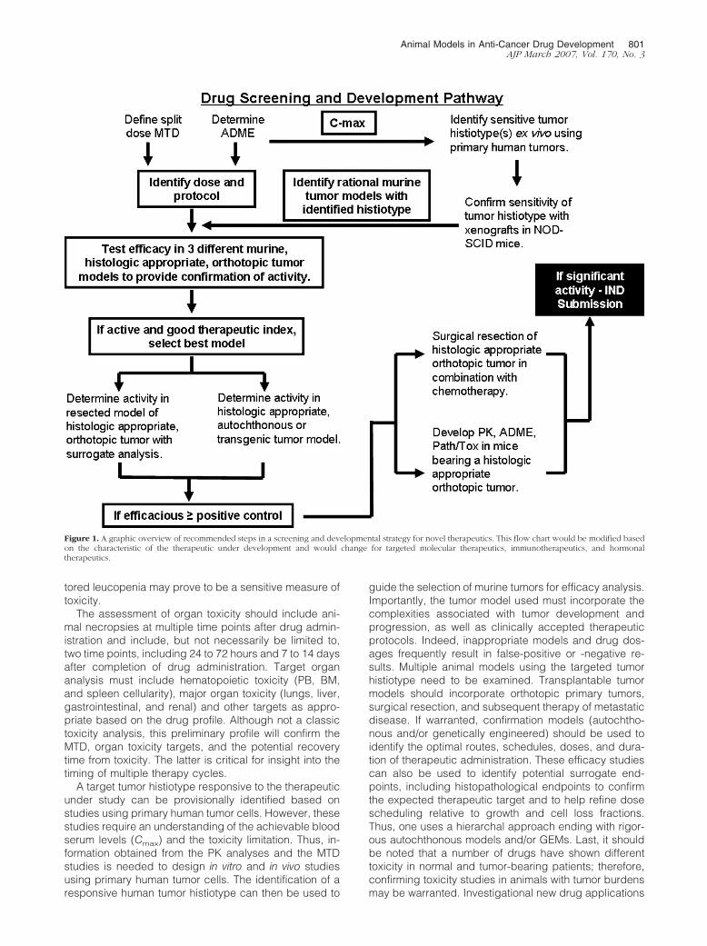
guide the selection of murine tumors for efficacy analysis.Importantly, the tumor model used must incorporate thecomplexities associated with tumor development andprogression, as well as clinically accepted therapeuticprotocols. Indeed, inappropriate models and drug dos-ages frequently result in false-positive or -negative re-sults. Multiple animal models using the targeted tumorhistiotype need to be examined. Transplantable tumormodels should incorporate orthotopic primary tumors,surgical resection, and subsequent therapy of metastaticdisease. If warranted, confirmation models (autochtho-nous and/or genetically engineered) should be used toidentify the optimal routes, schedules, doses, and dura-tion of therapeutic administration. These efficacy studiescan also be used to identify potential surrogate end-points, including histopathological endpoints to confirmthe expected therapeutic target and to help refine dosescheduling relative to growth and cell loss fractions.Thus, one uses a hierarchal approach ending with rigor-ous autochthonous models and/or GEMs. Last, it shouldbe noted that a number of drugs have shown differenttoxicity in normal and tumor-bearing patients; therefore,confirming toxicity studies in animals with tumor burdensmay be warranted. Investigational new drug applications
Figure 1. A graphic overview of recommended steps in a screening and developmental strategy for novel therapeutics. This flow chart would be modified basedon the characteristic of the therapeutic under development and would change for targeted molecular therapeutics, immunotherapeutics, and hormonaltherapeutics.
Animal Models in Anti-Cancer Drug Development 801AJP March 2007, Vol. 170, No. 3
are strengthened by conducting additional safety studiesin tumor-bearing animals.
This approach to the identification and development ofnew drugs is predicated on a rational preclinical cascadeculminating in the demonstration of in vivo proof of prin-ciple efficacy using appropriate animal models (Table 3).The proposed pathway follows the outline identified inFigure 1. Note that dosage translation from mouse tohuman needs to reflect skin surface area rather thanbody weight. In addition, the route of the administrationcan be critical such that i.p. injections, commonly used inrodent studies, are not used clinically outside of rabiesvaccines and the treatment of ovarian cancer. Further-more, clinically many of the anti-tumor drugs inducetissue damage requiring i.v. administration, although inrodents they may be administered by s.c. or i.p.administration.
Inarguably, animal models cannot be used to model allhuman therapeutics because of the dichotomy in murineand human receptors; however, murine models can gen-erally assess their pharmacology and toxicology. In ad-dition, there are differences in the physiology of murinemodels and humans such that different enzymes mayhave varying levels of prominence in human disease.Overall, rodent models have been shown to addressmechanisms of toxicology, pharmacology, and efficacy.However, in the instances when a protein sequence ispoorly conserved, the human homolog may not be ap-propriate for preclinical toxicology and pharmacology.Thus, a murine homolog may be active, but at a differentIC-50 such that a murine homolog may be better used forpharmacological and toxicological studies. Although oflesser concern with traditional chemotherapy drugs ob-tained by medicinal chemistry, biotherapeutics such asinterferon-� and granulocyte macrophage colony stimu-lating factor have used the murine homolog for toxicolog-ical and pharmacological studies.
Summary
Translation of a therapeutic into the clinic requires evi-dence of efficacy and safety as compared with the stan-dard of care. Furthermore, one needs to use animal mod-els that parallel the biological, genetic, etiological,immunological, and therapeutic properties of human can-cer. To assess clinical relevance, initial studies need tobe based on preliminary pathology and toxicity andADME studies to establish drug dose and use a splitdose protocol. In these basic studies, therapeutic targetscan be identified using primary human tumor cell models.The establishment of therapeutic efficacy requires a hi-erarchical approach beginning with orthotopic primarytumors, progressing to the models in which the orthotopicprimary tumor is resected before therapy initiation, andthen finishing with confirmation in autochthonous modelsor GEMs. The orthotopic and autochthonous models orGEMs incorporate homeostatic mechanisms in the organmicroenvironment that regulates tumor cell growth andsurvival. Efficacy needs to be justified based on pre-defined and rigorous outcome criteria and in many in-
stances, surrogate studies are justified. Ideally once ef-ficacy has been demonstrated, additional pathology andtoxicity studies and PK/ADME should be initiated in tu-mor-bearing animals, incorporating histopathology,blood chemistry, and biodistribution analysis. The surro-gate parameters could include tumor histopathology andimmunohistochemistry (angiogenesis, lymphangiogen-esis, tumor cell apoptosis, and infiltrating cellularphenotype).
Although this approach may seem exhaustive, it isrelatively rapid and of minimal expense as compared withthe failure of a candidate drug in phase II or III clinicalstudies. Traditional murine tumor models result in a highfrequency of false positive(s) as rapidly growing murinetumors are cured, whereas slower growing human tumorsprogress. We suggest, therefore, that a hierarchal strat-egy using rigorous outcome measures and continuousimprovements in stringency and consistency provides anapproach with the potential to facilitate the identificationand aid in the development of anti-cancer agents.
Acknowledgment
We thank Ms. Kirsten Stites for critical review and editingof the manuscript.
References
1. Jemal A, Siegel R, Ward E, Murray T, Xu J, Smigal C, Thun MJ: Cancerstatistics, 2006. CA Cancer J Clin 2006, 56:106–130
2. Talmadge JE, Wolman SR, Fidler IJ: Evidence for the clonal origin ofspontaneous metastases. Science 1982, 217:361–363
3. Talmadge JE, Fidler IJ: Cancer metastasis is selective or randomdepending on the parent tumour population. Nature 1982,297:593–594
4. Talmadge JE, Benedict K, Madsen J, Fidler IJ: Development of bio-logical diversity and susceptibility to chemotherapy in murine cancermetastases. Cancer Res 1984, 44:3801–3805
5. Schuh JC: Trials, tribulations, and trends in tumor modeling in mice.Toxicol Pathol 2004, 32(Suppl 1):53–66
6. Suggitt M, Bibby MC: 50 years of preclinical anticancer drugscreening: empirical to target-driven approaches. Clin Cancer Res2005, 11:971–981
7. Schein PS, Scheffler B: Barriers to efficient development of cancertherapeutics. Clin Cancer Res 2006, 12:3243–3248
8. Fiebig HH, Maier A, Burger AM: Clonogenic assay with establishedhuman tumour xenografts: correlation of in vitro to in vivo activity as abasis for anticancer drug discovery. Eur J Cancer 2004, 40:802–820
9. Johnson JI, Decker S, Zaharevitz D, Rubinstein LV, Venditti JM,Schepartz S, Kalyandrug S, Christian M, Arbuck S, Hollingshead M,Sausville EA: Relationships between drug activity in NCI preclinical invitro and in vivo models and early clinical trials. Br J Cancer 2001,84:1424–1431
10. Fidler IJ: Orthotopic implantation of human colon carcinomas intonude mice provides a valuable model for the biology and therapy ofmetastasis. Cancer Metastasis Rev 1991, 10:229–243
11. Hoffman RM: Orthotopic metastatic mouse models for anticancerdrug discovery and evaluation: a bridge to the clinic. Invest NewDrugs 1999, 17:343–359
12. Hoffman R: Fertile seed and rich soil: the development of clinicallyrelevant models of human cancer by surgical orthotopic implantationof intact tissues. Anticancer Drug Development Guide: PreclinicalScreening, Clinical Trials, and Approval. Edited by BA Teicher. To-towa, Humana Press Inc., 1997, pp 127–144
13. Killion JJ, Radinsky R, Fidler IJ: Orthotopic models are necessary to
802 Talmadge et alAJP March 2007, Vol. 170, No. 3
predict therapy of transplantable tumors in mice. Cancer MetastasisRev 1999, 17:279–284
14. Dexter DL, Diamond M, Creveling J, Chen SF: Chemotherapy ofmammary carcinomas arising in ras transgenic mice. Invest NewDrugs 1993, 11:161–168
15. Moser AR, Hegge LF, Cardiff RD: Genetic background affects sus-ceptibility to mammary hyperplasias and carcinomas in Apc(min)/�mice. Cancer Res 2001, 61:3480–3485
16. Stringer JR, Larson JS, Fischer JM, Medvedovic M, Hersh MN, BoivinGP, Stringer SL: Modeling variation in tumors in vivo. Proc Natl AcadSci USA 2005, 102:2408–2413
17. Voskoglou-Nomikos T, Pater JL, Seymour L: Clinical predictive valueof the in vitro cell line, human xenograft, and mouse allograft preclin-ical cancer models. Clin Cancer Res 2003, 9:4227–4239
18. Gellhorn A, Hirschberg E: Investigation of diverse systems for cancerchemotherapy screening. Cancer Res 1955, 15(Suppl 3):1–125
19. Goldin A, Venditti JM, Kline I, Mantel N: Evaluation of anti-leukemicagents employing advanced leukemia L1210 in mice. Cancer Res1959, 19:429–466
20. DeVita VT, Schein PS: The use of drugs in combination for thetreatment of cancer: rationale and results. N Engl J Med 1973,288:998–1006
21. Zubrod C: Chemical control of cancer. Proc Natl Acad Sci USA 1972,69:1042–1047
22. Venditti JM, Wesley RA, Plowman J: Current NCI preclinical antitumorscreening in vivo: results of tumor panel screening, 1976–1982, andfuture directions. Adv Pharmacol Chemother 1984, 20:1–20
23. Geran R, Greenberg N, MacDonald M, Schumacher A: Protocols forscreening chemical agents and natural products against animal tu-mors and other biological systems. Cancer Chemother Rep 1972,3:1–13
24. Ovejera A, Johnson R, Goldin A: Growth characteristics and chemo-therapeutic response of intravenously implanted Lewis lung carci-noma. Cancer Chemother Rep 1975, 5:111–125
25. Bogden A: A rapid screening method for testing chemotherapeuticagents against human tumour xenografts. Proceedings of the Sym-posium of the Use of Athymic (Nude) Mice in Cancer Research.Edited by DOA Houchens. New York, Gustav Fischer, 1978, pp231–250
26. Bogden AE: The subrenal capsule assay: biological properties andtesting capability. Relevance of tumour models for anticancer drugdevelopment. Contributions to Oncology. Edited by HH Fiebig, BABurger. Basel, Karger, 1999, pp 89–99
27. Staquet MJ, Byar DP, Green SB, Rozencweig M: Clinical predictivityof transplantable tumor systems in the selection of new drugs for solidtumors: rationale for a three-stage strategy. Cancer Treat Rep 1983,67:753–765
28. Venditti JM: Preclinical drug development: rationale and methods.Semin Oncol 1981, 8:349–361
29. Salmon SE, Hamburger AW, Soehnlen B, Durie BG, Alberts DS, MoonTE: Quantitation of differential sensitivity of human-tumor stem cells toanticancer drugs. N Engl J Med 1978, 298:1321–1327
30. Von Hoff DD, Harris GJ, Johnson G, Glaubiger D: Initial experiencewith the human tumor stem cell assay system: potential and prob-lems. Prog Clin Biol Res 1980, 48:113–124
31. Shoemaker RH, Wolpert-DeFilippes MK, Kern DH, Lieber MM,Makuch RW, Melnick NR, Miller WT, Salmon SE, Simon RM, VendittiJM: Application of a human tumor colony-forming assay to new drugscreening. Cancer Res 1985, 45:2145–2153
32. Bertelsen CA, Sondak VK, Mann BD, Korn EL, Kern DH: Chemosen-sitivity testing of human solid tumors. A review of 1582 assays with258 clinical correlations. Cancer 1984, 53:1240–1245
33. Salmon SE, Alberts DS, Durie BG, Meyskens FL, Jones SE, SoehnlenB, Chen HS, Moon T: Clinical correlations of drug sensitivity in thehuman tumor stem cell assay. Recent Results Cancer Res 1980,74:300–305
34. Tveit KM, Fodstad O, Lotsberg J, Vaage S, Pihl A: Colony growth andchemosensitivity in vitro of human melanoma biopsies. Relationshipto clinical parameters. Int J Cancer 1982, 29:533–538
35. Von Hoff DD, Casper J, Bradley E, Sandbach J, Jones D, Makuch R:Association between human tumor colony-forming assay results andresponse of an individual patient’s tumor to chemotherapy. Am J Med1981, 70:1027–1041
36. Selby P, Buick RN, Tannock I: A critical appraisal of the “human tumorstem-cell assay.” N Engl J Med 1983, 308:129–134
37. Alley MC, Scudiero DA, Monks A, Hursey ML, Czerwinski MJ, FineDL, Abbott BJ, Mayo JG, Shoemaker RH, Boyd MR: Feasibility of drugscreening with panels of human tumor cell lines using a microculturetetrazolium assay. Cancer Res 1988, 48:589–601
38. Sausville EA, Feigal E: Evolving approaches to cancer drug discoveryand development at the National Cancer Institute, USA. Ann Oncol1999, 10:1287–1291
39. Kelland LR: Of mice and men: values and liabilities of the athymicnude mouse model in anticancer drug development. Eur J Cancer2004, 40:827–836
40. Kerbel RS: Human tumor xenografts as predictive preclinical modelsfor anticancer drug activity in humans: better than commonly per-ceived—but they can be improved. Cancer Biol Ther 2003,2:S134–S139
41. Peterson JK, Houghton PJ: Integrating pharmacology and in vivocancer models in preclinical and clinical drug development. Eur JCancer 2004, 40:837–844
42. Takimoto CH: Why drugs fail: of mice and men revisited. Clin CancerRes 2001, 7:229–230
43. Habu S, Fukui H, Shimamura K, Kasai M, Nagai Y, Okumura K,Tamaoki N: In vivo effects of anti-asialo GM1. I. Reduction of NKactivity and enhancement of transplanted tumor growth in nude mice.J Immunol 1981, 127:34–38
44. Talmadge JE, Meyers KM, Prieur DJ, Starkey JR: Role of NK cells intumour growth and metastasis in beige mice. Nature 1980,284:622–624
45. Cespedes MV, Casanova I, Parreno M, Mangues R: Mouse models inoncogenesis and cancer therapy. Clin Transpl Oncol 2006,8:318–329
46. Becher OJ, Holland EC: Genetically engineered models have advan-tages over xenografts for preclinical studies. Cancer Res 2006,66:3355–3358
47. Sausville EA, Burger AM: Contributions of human tumor xenografts toanticancer drug development. Cancer Res 2006, 66:3351–3354
48. Scholz CC, Berger DP, Winterhalter BR, Henss H, Fiebig HH: Corre-lation of drug response in patients and in the clonogenic assay withsolid human tumour xenografts. Eur J Cancer 1990, 26:901–905
49. Steel GG, Courtenay VD, Peckham MJ: The response to chemother-apy of a variety of human tumour xenografts. Br J Cancer 1983,47:1–13
50. Varney ML, Olsen KJ, Mosley RL, Bucana CD, Talmadge JE, SinghRK: Monocyte/macrophage recruitment, activation and differentiationmodulate interleukin-8 production: a paracrine role of tumor-associ-ated macrophages in tumor angiogenesis. In Vivo 2002, 16:471–477
51. Legrand N, Weijer K, Spits H: Experimental models to study devel-opment and function of the human immune system in vivo. J Immunol2006, 176:2053–2058
52. Chang DH, Liu N, Klimek V, Hassoun H, Mazumder A, Nimer SD,Jagannath S, Dhodapkar MV: Enhancement of ligand-dependentactivation of human natural killer T cells by lenalidomide: therapeuticimplications. Blood 2006, 108:618–621
53. Thomsen M, Yacoub-Youssef H, Marcheix B: Reconstitution of ahuman immune system in immunodeficient mice: models of humanalloreaction in vivo. Tissue Antigens 2005, 66:73–82
54. Gonzalez FJ, Yu AM: Cytochrome P450 and xenobiotic receptorhumanized mice. Annu Rev Pharmacol Toxicol 2006, 46:41–64
55. Bogaards JJ, Bertrand M, Jackson P, Oudshoorn MJ, Weaver RJ, vanBladeren PJ, Walther B: Determining the best animal model for hu-man cytochrome P450 activities: a comparison of mouse, rat, rabbit,dog, micropig, monkey and man. Xenobiotica 2000, 30:1131–1152
56. Macchiarini F, Manz MG, Palucka AK, Shultz LD: Humanized mice:are we there yet? J Exp Med 2005, 202:1307–1311
57. Casares N, Pequignot MO, Tesniere A, Ghiringhelli F, Roux S, ChaputN, Schmitt E, Hamai A, Hervas-Stubbs S, Obeid M, Coutant F, Me-tivier D, Pichard E, Aucouturier P, Pierron G, Garrido C, Zitvogel L,Kroemer G: Caspase-dependent immunogenicity of doxorubicin-in-duced tumor cell death. J Exp Med 2005, 202:1691–1701
58. Donelli MG, Rosso R, Garattini S: Selective chemotherapy in relationto the site of tumor transplantation. Int J Cancer 1967, 2:421–424
59. Wilmanns C, Fan D, O’Brian C, Radinsky R, Bucana C, Tsan R, FidlerI: Modulation of doxorubicin sensitivity and level of P-glycoprotein
Animal Models in Anti-Cancer Drug Development 803AJP March 2007, Vol. 170, No. 3
expression in human colon carcinoma cells by ectopic and orthotopicenvironments in nude mice. Int J Oncol 1993, 3:413–422
60. Bibby MC: Orthotopic models of cancer for preclinical drugevaluation: advantages and disadvantages. Eur J Cancer 2004,40:852–857
61. Fidler IJ: Rationale and methods for the use of nude mice to study thebiology and therapy of human cancer metastasis. Cancer MetastasisRev 1986, 5:29–49
62. Tan MH, Holyoke ED, Goldrosen MH: Murine colon adenocarcinoma:syngeneic orthotopic transplantation and subsequent hepatic metas-tases. J Natl Cancer Inst 1977, 59:1537–1544
63. Brinster RL, Chen HY, Messing A, Van DT, Levine AJ, Palmiter RD:Transgenic mice harboring SV40 T-antigen genes develop charac-teristic brain tumors. Cell 1984, 37:367–379
64. Heineke J, Molkentin JD: Regulation of cardiac hypertrophy by intra-cellular signalling pathways. Nat Rev Mol Cell Biol 2006, 7:589–600
65. Gossen M, Bujard H: Tight control of gene expression in mammaliancells by tetracycline-responsive promoters. Proc Natl Acad Sci USA1992, 89:5547–5551
66. Rego EM, He LZ, Warrell Jr RP, Wang ZG, Pandolfi PP: Retinoic acid(RA) and As2O3 treatment in transgenic models of acute promyelo-cytic leukemia (APL) unravel the distinct nature of the leukemogenicprocess induced by the PML-RARalpha and PLZF-RARalpha onco-proteins. Proc Natl Acad Sci USA 2000, 97:10173–10178
67. Brain J, Saksena A, Laneuville P: The kinase inhibitor STI571 reversesthe Bcr-Abl induced point mutation frequencies observed in pre-leukemic P190(Bcr-Abl) transgenic mice. Leuk Res 2002,26:1011–1016
68. Shoemaker AR, Gould KA, Luongo C, Moser AR, Dove WF: Studies ofneoplasia in the Min mouse. Biochim Biophys Acta 1997,1332:F25–F48
69. Macleod KF, Jacks T: Insights into cancer from transgenic mousemodels. J Pathol 1999, 187:43–60
70. Sharpless NE, DePinho RA: The mighty mouse: genetically engi-neered mouse models in cancer drug development. Nat Rev DrugDiscov 2006, 5:741–754
71. Basu GD, Pathangey LB, Tinder TL, Lagioia M, Gendler SJ, Mukher-jee P: Cyclooxygenase-2 inhibitor induces apoptosis in breast cancercells in an in vivo model of spontaneous metastatic breast cancer.Mol Cancer Res 2004, 2:632–642
72. Lallemand-Breitenbach V, Guillemin MC, Janin A, Daniel MT, DegosL, Kogan SC, Bishop JM, de The H: Retinoic acid and arsenicsynergize to eradicate leukemic cells in a mouse model of acutepromyelocytic leukemia. J Exp Med 1999, 189:1043–1052
73. Bearss DJ, Subler MA, Hundley JE, Troyer DA, Salinas RA, Windle JJ:Genetic determinants of response to chemotherapy in transgenic
mouse mammary and salivary tumors. Oncogene 2000,19:1114–1122
74. Weiss B, Shannon K: Mouse cancer models as a platform for per-forming preclinical therapeutic trials. Curr Opin Genet Dev 2003,13:84–89
75. Jacks T: Tumor suppressor gene mutations in mice. Annu Rev Genet1996, 30:603–636
76. Hann B, Balmain A: Building ‘validated’ mouse models of humancancer. Curr Opin Cell Biol 2001, 13:778–784
77. Van Dyke T, Jacks T: Cancer modeling in the modern era: progressand challenges. Cell 2002, 108:135–144
78. Berger M: Is there a relevance for anticancer drug development.Relevance of Tumour Models for Anticancer Drug Development. Con-tributions to Oncology. Edited by HH Fiebig, BA Burger. Basel,Karger, 1999, pp 15–27
79. Talmadge JE, Lenz BF, Klabansky R, Simon R, Riggs C, Guo S,Oldham RK, Fidler IJ: Therapy of autochthonous skin cancers in micewith intravenously injected liposomes containing muramyltripeptide.Cancer Res 1986, 46:1160–1163
80. Schwartz B, Birk Y, Raz A, Madar Z: Nutritional-pharmacologicalcombinations—a novel approach to reducing colon cancer inci-dence. Eur J Nutr 2004, 43:221–229
81. Corpet DE, Pierre F: How good are rodent models of carcinogenesisin predicting efficacy in humans? A systematic review and meta-analysis of colon chemoprevention in rats, mice and men. Eur JCancer 2005, 41:1911–1922
82. Schipper H, Goh CR, Wang TL: Shifting the cancer paradigm: mustwe kill to cure? J Clin Oncol 1995, 13:801–807
83. Freireich EJ, Gehan EA, Rall DP, Schmidt LH, Skipper HE: Quantita-tive comparison of toxicity of anticancer agents in mouse, rat, ham-ster, dog, monkey, and man. Cancer Chemother Rep 1966,50:219–244
84. Burtles SS, Newell DR, Henrar RE, Connors TA: Revisions of generalguidelines for the preclinical toxicology of new cytotoxic anticanceragents in Europe. The Cancer Research Campaign (CRC) Phase I/IIClinical Trials Committee and the European Organization for Re-search and Treatment of Cancer (EORTC) New Drug DevelopmentOffice. Eur J Cancer 1995, 31A:408–410
85. Newell DR, Burtles SS, Fox BW, Jodrell DI, Connors TA: Evaluation ofrodent-only toxicology for early clinical trials with novel cancer ther-apeutics. Br J Cancer 1999, 81:760–768
86. Balmain A: Cancer as a complex genetic trait: tumor susceptibility inhumans and mouse models. Cell 2002, 108:145–152
87. Siemann DW: Satisfactory and unsatisfactory tumor models factorsinfluencing the selection of a tumor model for experimental evalua-tion. Rodent Tumor Models in Experimental Cancer Therapy. Editedby RF Kallman. Elmsford, Pergamon Books, Inc., 1987
804 Talmadge et alAJP March 2007, Vol. 170, No. 3