multifunctional roles of reticular fibroblastic cells: more than meets the eye?
TRANSCRIPT
Review ArticleMultifunctional Roles of Reticular Fibroblastic Cells:More Than Meets the Eye?
H. G. Alvarenga1,2 and L. Marti1,2
1 Experimental Research, Hospital Israelita Albert Einstein, Avenida Albert Einstein 627, 05152 000 Sao Paulo, SP, Brazil2 Programa de Alergia e Imunopatologia, Faculdade de Medicina da Universidade de Sao Paulo (FMUSP),Av. Dr. Arnaldo, 455, 01418 200 Sao Paulo, SP, Brazil
Correspondence should be addressed to L. Marti; [email protected]
Received 4 February 2014; Revised 25 March 2014; Accepted 25 March 2014; Published 16 April 2014
Academic Editor: Ethan M. Shevach
Copyright © 2014 H. G. Alvarenga and L. Marti.This is an open access article distributed under theCreative CommonsAttributionLicense, which permits unrestricted use, distribution, and reproduction in anymedium, provided the originalwork is properly cited.
Fibroblastic reticular cells (FRCs) are stromal cells found in secondary lymphoid organ. Despite its structural function in the lymphnodes being well established, recent studies indicate that the FRCs also play a key role in immunological processes, associated withcell transit, immune response, and cells activation quality, and contribute to peripheral tolerance. To this end, we focus this reviewon lymph nodes FRC characterization and discuss functional aspects such as production of cytokines and chemokines and theirinvolvement in the immune response, seeking to establish whether certain subsets have a more functional specialization.
1. Introduction
Fibroblastic reticular cells (FRCs) are stromal cells of meso-dermal origin which are found in secondary lymphoidorgans (SLO). The most studied are the FRCs located in thelymph nodes (LN). They comprise distinct subpopulationsdistributed in the cortical and medullar regions. Some FRCsare found in the T cell zones and were therefore named T cellzone fibroblastic reticular cells (TRCs). The FRCs structuralfunction in the lymph nodes is well established. Yet recentstudies indicate that FRCs also participate in the regulationof immune responses and in the maintenance of peripheraltolerance. The cellular traffic in SLO and the activationof specific lymphocyte populations can be influenced byFRCs and consequently also the quality and intensity of theimmune responses [1–3]. Nevertheless, this is a relatively newresearch area and many of the pathways and mechanisms ofcellular interactions between FRCs and neighboring cells arenot yet known and need further investigation.
Themorphological variations among FRCs and the diver-sity of their functions in the immune response have beendescribed [4–6]. However, so far, a clear match between thedescribed FRC phenotypes and their respective functions hasnot been established. A further complication to the issue isthat mouse FRCs exhibit, in addition to classically known cell
surface markers, several other distinct specific markers forFRCs subtypes.
We review herein the studies on phenotypic character-ization of FRCs isolated from murine and human lymphnodes and on the selective synthesis of chemokines andcytokines as well as interactions with other cells. Recentadvances in understanding the functional diversity of FRCsubpopulations are highlighted.
2. Fibroblastic Reticular Cells Morphology
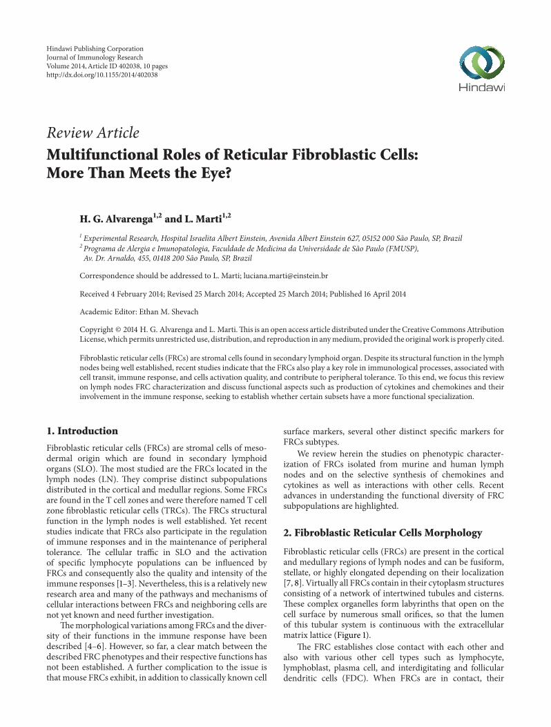
Fibroblastic reticular cells (FRCs) are present in the corticaland medullary regions of lymph nodes and can be fusiform,stellate, or highly elongated depending on their localization[7, 8]. Virtually all FRCs contain in their cytoplasm structuresconsisting of a network of intertwined tubules and cisterns.These complex organelles form labyrinths that open on thecell surface by numerous small orifices, so that the lumenof this tubular system is continuous with the extracellularmatrix lattice (Figure 1).
The FRC establishes close contact with each other andalso with various other cell types such as lymphocyte,lymphoblast, plasma cell, and interdigitating and folliculardendritic cells (FDC). When FRCs are in contact, their
Hindawi Publishing CorporationJournal of Immunology ResearchVolume 2014, Article ID 402038, 10 pageshttp://dx.doi.org/10.1155/2014/402038
2 Journal of Immunology Research
Conduit
Cytoplasmicchannel
Soluble molecules
Antigen
Figure 1: Schematic representation of FRCs. They are spatially arranged so as to delimit a conduit channel that drives soluble molecules.Other structures evidenced are the FRC intracellular cytoplasmic channels, through which antigens are transported from the lymph to thenearby-lying antigen-presenting cells.
plasma membranes remain separated by a space of about20 nm, forming an intercellular channel through which driv-ing soluble molecules are transported [8].
More precisely, FRCs organization generates a conduitsystem between them; these conduits are called reticularfiber network that are responsible for transporting solubleantigens from the afferent lymph to resident dendritic cellsin T cell area of the lymph node. This structure produces theinfrastructure necessary, at least for the first wave of antigenpresentation, which takes place few minutes after solubleantigen injection in a subcutaneous site of an animal modelas described by Sixt et al. article [9].
In the lymph nodes cortical areas, FRCs are situatedclose to the subcapsular sinus, according to their antigen-capturing function. The cortical FRCs are polarized: sol-uble molecules (antigens, cytokines, neuropeptides, lipids,microbial products) are collected at the FRC cell side facingthe subcapsular lumen and released at an area in contactwith lymphocytes and follicular dendritic cells (FDC). Theintracellular channels of FRCs are the organelles that facilitatethe transport of antigen from the afferent lymph to the FDC(Figure 1) [8, 10].
3. Phenotypic Diversity
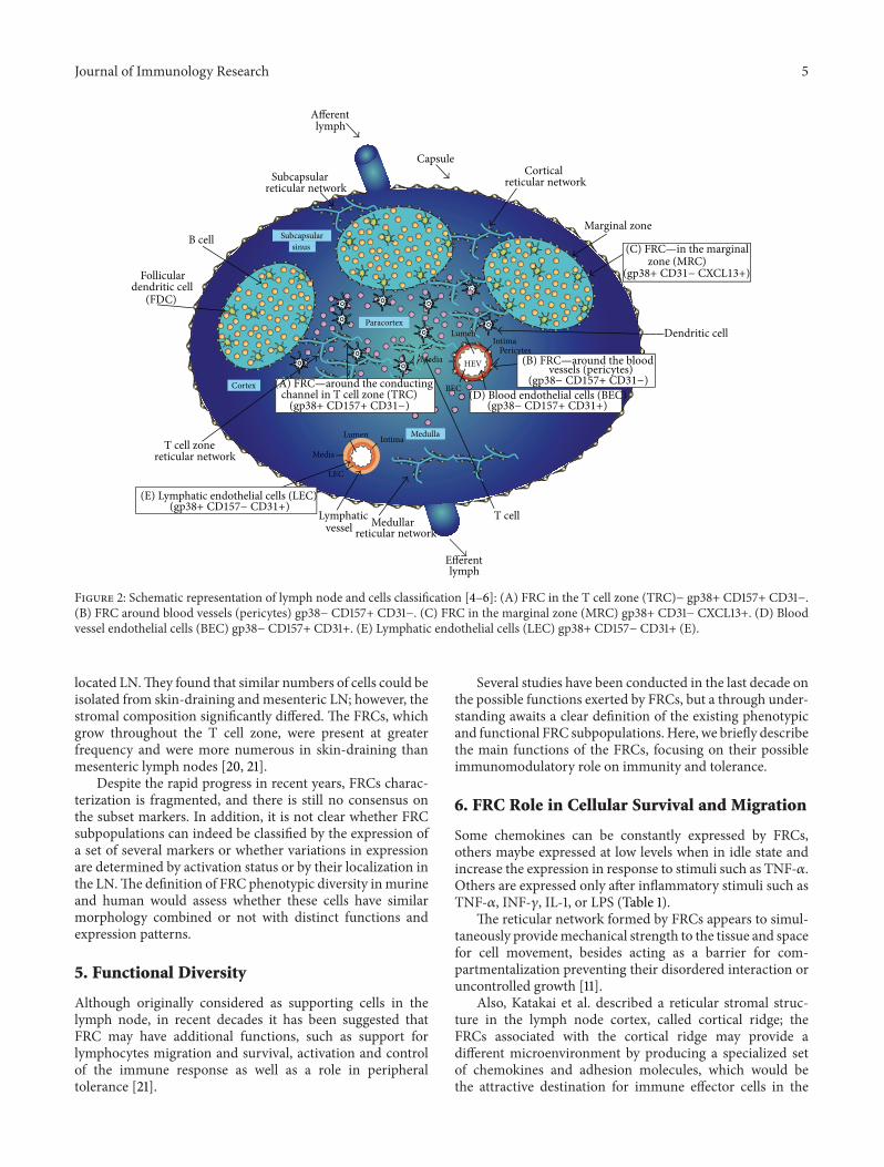
FRC identification is based on a set of markers that areexpressed or exclude other cells types. Murine FRCs arebasically characterized by the expression of gp38 (a knownmarker of lymphatic endothelium), absence of CD31 (anendothelial cell marker), as well as the secretion of thechemokines CCL19 and CCL21 and of the cytokine IL-7.However, in the last decade several studies have identifiedadditional markers for these cells present in lymph nodesof different species (Table 1). Although a variety of newermarkers may assist the identification of FRCs and theirsubsets, there is no consensus on the correspondence amongthe subsets described by several authors.
As an attempt to characterize FRCs in mouse lymphnodes (LN) several fibroblastic cell lines were derived. Thecells characterized as FRCs expressed CD44, CD106, andgp38, intracellular ER-TR7, and did not express CD11b/CD18,CD16, CD31, CD32, CD35, LYVE-1, and MHC-II [11].
However it was soon recognized that FRCs are heteroge-neous with respect to their morphology and location. Subsetsof cells were described in distinct locations of mouse LN,
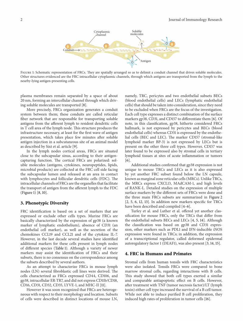
namely, TRC, pericytes and two endothelial subsets BECs(blood endothelial cells) and LECs (lymphatic endothelialcells) that should be taken into consideration, since they needto be excluded when FRCs are the focus of the investigation.Each cell type expresses a distinct combination of the surfacemarkers gp38, CD31, and CD157 to differentiate them [6]. Ofnote, in this classification, gp38, hitherto considered FRCshallmark, is not expressed by pericytes and BECs (bloodendothelial cells) whereas CD31 is expressed by the endothe-lial cells (BEC and LEC). The marker CD157 (stromal-likelymphoid marker BP-3) is not expressed by LECs but ispresent on the other three cell types. However, CD157 waslater found to be expressed also by stromal cells in tertiarylymphoid tissues at sites of acute inflammation or tumors[6].
Additional studies confirmed that gp38 expression is notunique to mouse TRCs and LECs as it is also expressedby yet another FRC subset found below the LN capsule,known asmarginal zone reticular cells (MRCs). Unlike TRCs,the MRCs express CXCL13, MAdCAM-1, and high levelsof RANK-L. Detailed studies on the expression of multiplesurface markers by the different sets of FRCs were done andthe three main FRCs subsets are summarized in Figure 2[2, 5, 6, 12, 13]. In addition new markers specific for TRCshave been described and compiled [4–6].
Turley et al. and Luther et al. offered yet another clas-sification for mouse FRCs, only the TRCs that differ fromthe endothelial subsets BECs and LECs [4, 5, 14]. Althoughthe classification was based on gp38 and CD31 expres-sion, other markers such as PDL1 and IFN-inducible iNOSexpression were found in TRCs; in addition, the expressionof a transcriptional regulator, called deformed epidermalautoregulatory factor 1 (DEAF1), was also present [3, 14, 15].
4. FRC in Humans and Primates
Stromal cells from human tonsils with FRC characteristicswere also isolated. Tonsils FRCs were compared to bonemarrow stromal cells, regarding interactions with B cells.This study showed that both cell types exerted a similarand comparable antiapoptotic effect on B cells. However,after treatment with TNF (tumor necrosis factor)/LT (lymphtoxin) either cell type increased the survival of a B cell tumor.While not able to induce purified B cell proliferation, theyinduced high rates of proliferation in tumor cells [16].
Journal of Immunology Research 3
Table 1: FRC characterizationmarkers. FRCwas classified by species, surface or intracellularmakers, induciblemarkers and ones that excludeother cells presence, and chemokines and cytokines secretion, listed in time order of publishing.
Publication year—author(species)
Markers Chemokines CytokinesSurface Intracellular Inducible Excluding Regular Inducible
2004—Katakai et al. (a) [11](MLN)
CD44, gp38,CD106 ER-TR7 CD54
CD11b/CD18,CD16,CD31,CD32, CD35,
MHCII,LYVE1
CCL2, CXCL12,CX3CL1
CCL4,CCL5,CCL20,CXCL10
IL6, IL7, IL15
2004—Katakai et al. (b) [22](MLN) — ER-TR7 — — — — —
2005—Sixt et al. [9](MLN)
gp38, 𝛼SMA,desmin ER-TR7 — — — — —
2006—Bajenoff et al. [23](MLN)
Desmin, CD106,CD54 ER-TR7 — — CCL19, CCL21,
CXCL12 (SDF-1) — —
2006—Hara et al. [25](MLN) gp38, CD106 ER-TR7 — — — CXCL16 IL7, IL15
2007—Ame-Thomas et al. [16](HT)
CD73, CD90,CD105 — CD54
CD106CD21, CD23,CD35, CD45
CCL5, CXCL9,CXCL10,CXCL12
CCL19 —
2007—Link et al. [4](MLN)
gp38, 𝛼SMA,desmin, CD157,
PDGFR𝛼,PDGFR𝛽, CD54,CD106, LT𝛽R,
TNF-R
ER-TR7 —CD21, CD31,CD35 CD45,
LYVE-1CCL19, CCL21 — IL7
2009—Roozendaal et al. [30](MLN) gp38 ER-TR7 — — — — —
2009—Mueller and Germain[12](review)
gp38, PDL1 VEGF, ER-TR7 —
CD45, CD31,CD21, CD35,C4, CD16,
CD23, CD32,CD157, Mfge8
CCL19, CCL21 — IL7
2009—Steel et al. [17](NHPLN)
gp38, p75NGFR,TTG, CD54,CD106, CD157,LT𝛽R, PDGFR𝛼,PDGFR𝛽, 𝛼SMA,TNFR1, Meca79,
desmin
ER-TR7 — —CCL19, CCL21,
CXCL16,CCL2/MCP1
— IL7, IL6
2010—Turley et al. [14](review) gp38, PDL1 DEAF1 CD31 CCL19, CCL21 — IL7
2011—Khan et al. [15](MLN) gp38, PDL1 — iNOS CD45, CD31 CCL19, CCL21 — IL7
2011—Luther et al. [5](review)
gp38, CD54,CD106, CD157,
PDGFR𝛼𝛽, LT𝛽R,TNFR1, desmin,𝛼SMA
VEGF —CD21, CD31,CD35, CD45,C4, Mfge8
CCL19, CCL21
CCL2,CCL4,CCL5,CXCL12,CXCL16,CX3CL1
IL7, IL6
2011—Link et al. [6](MHLN)
gp38, CD54,CD106, CD157,
PDGFR𝛼𝛽, LT𝛽R,desmin, 𝛼SMA
ERTR7 — CD31, CD35,CD45 CCL19, CCL21 — IL7
2011—Onder et al. [3](MLN) gp38 — — CD31, CD45 CCL19, CCL21 — IL7
2011—Fletcher et al. (a) [20](MHLN)
gp38, PDGF𝛼,CD54, CD106 VEGF — CD45, CD31
CCL19, CCL21CXCL9,CXCL10
— IL7
4 Journal of Immunology Research
Table 1: Continued.
Publication year—author(species)
Markers Chemokines CytokinesSurface Intracellular Inducible Excluding Regular Inducible
2011—Fletcher et al. (b) [21](review)
gp38, PDGF𝛼,CD54, CD106 VEGF — CD45, CD31 CCL19, CCL21,
CXCL12 (SDF1) — IL7
2011—Frontera et al. [37](MLN)
CD54, CD106,PDGF𝛼, CD141 JAMC — CD45, CD31,
LYVE1 CCL21 — —
2011—Siegert et al. [2](MLN) gp38 iNOS —
CD45, CD35,CD31,EpCAM
CCL19, CCL21 — IL7
2011—Lukacs-Kornek [34](MLN)
gp38, PDL1,INFGR1, TNFR1,
TNFR2NOS2, IDO — CD45, CD31 CCL19, CCL21 — —
2012—Zeng et al. [19](NHPLN) Desmin — — CD35, CD21 — — —
2012—Siegert and Luther [33](review) gp38, PDL1 COX2, Aire,
DEAF1, NO
iNOS,MHCII,IDO,CD80
CD21, CD35,CD31 CCL19, CCL21 — IL7
2012—Graw and Regoes [26](MLN) — — — — CCL19, CCL21 — —
2012—Onder et al. [29](MLN) gp38 — — CD31, CD45 CCL19, CCL21 — IL7
2012—Hess et al. [13](MLN)
gp38, CD106,MadCAM1 RankL — CD31, CD45 — — IL7
2012—Malhotra et al. [28](MLN) gp38, CD140a
VEGFA and C,ANGPTL2 and4, HGF, GREM1,
SERPINF1cadherin-11,
IFITM-1, Flt3L
— CD31, CD45
CXCL14, CCL19,CCL21, CXCL13CXCL12, CCL2,
CCL7
IL34
2012—Acton et al. [24](MLN) gp38 — — CD31 CCL19, CCL21 — —
2013—Chai et al. [31](MLN) gp38, 𝛼SMA, ER-TR7, NO — CD31, CD45 CCL19, CCL21 — IL-7
2014—Yang et al. [32](MLN)
gp38, PDGFR𝛼𝛽,LT𝛽R, 𝛼SMA
VEGF, iNOS,VEGF, MyD88 — CD31, CD45,
LYVE-1 CCL19, CCL21 — IL-7
Murine lymph node (MLN); human tonsils (HT); nonhuman primate lymph node (NHPLN); murine and human lymph node (MHLN).
A study on viral pathogenesis in nonhuman primates bySteel et al. identified two other FRC markers besides thosedescribed in the mouse by Link et al. [6, 17]. The authorsobserved that the previously described FRC subsets werepositive for a TNF receptor (TNFR) family member knownas p75 NGFR and for transglutaminase (TTG) in severalspecies of nonhuman primates. They hypothesized that p75NGFR expression could be related to a mechanism wherebythe nervous system regulates immune responses via FRCs. Inaddition, they discuss that TTG has previously been shownby Thomazy et al. to play a role in phenotypic regulation ofhuman lymph node FRC [17, 18].
Thomazy et al. compared functional aspects and TTGexpression between LN obtained from normal individualsand from lymphoma patients. Basically, when the FRCnetwork is open, migration of cells andmolecules around thefollicle occurs at high turnover; this condition is accompaniedby germinal center expansion and by increased expression
of TTG in the FRCs of the subcapsular sinus and cortex.In contrast, when the FRC network is tighter, migrationof cells from the subcapsular sinus is reduced and TTGexpression is limited to the sinus. In the various lymphomas,high TTG levels were found in the LN stromal cell sug-gesting that cell migration is altered in these conditions[18].
Not only tumors but also infections with SIV in nonhu-man primates or HIV in humans determine the loss of thefibroblast and FRCs network integrity in LN [19].
Fletcher et al. are the sole investigators who made usein humans of murine consensus FRC markers such as gp38,CD31, CD45, CD54, CD106, and PDGFR, to compare thestromal composition between mouse and humans. Theyreported that the human lymph node stromal cells weremarked similar to the murine ones. Using the same murinemethods and markers they were able to isolate and cultivatehuman stromal cell subsets of mesenteric and elsewhere
Journal of Immunology Research 5
Paracortex
Afferentlymph
CapsuleSubcapsular
reticular networkCortical
reticular network
Marginal zone
IntimaLumen
Media
LEC
IntimaLumen
Media HEV
BEC
Subcapsularsinus
Medulla
Cortex
T cell zonereticular network
T cell
Dendritic cell
Efferent lymph
Medullarreticular network
Lymphaticvessel
B cell
(FDC)
Pericytes
Follicular
(D) Blood endothelial cells (BEC)(gp38− CD157+ CD31+)
channel in T cell zone (TRC)(gp38+ CD157+ CD31−)
(gp38− CD157+ CD31−)vessels (pericytes)
(gp38+ CD31− CXCL13+)zone (MRC)
(A) FRC—around the conducting
(B) FRC—around the blood
(C) FRC—in the marginal
dendritic cell
(E) Lymphatic endothelial cells (LEC)(gp38+ CD157− CD31+)
Figure 2: Schematic representation of lymph node and cells classification [4–6]: (A) FRC in the T cell zone (TRC)− gp38+ CD157+ CD31−.(B) FRC around blood vessels (pericytes) gp38− CD157+ CD31−. (C) FRC in the marginal zone (MRC) gp38+ CD31− CXCL13+. (D) Bloodvessel endothelial cells (BEC) gp38− CD157+ CD31+. (E) Lymphatic endothelial cells (LEC) gp38+ CD157− CD31+ (E).
located LN.They found that similar numbers of cells could beisolated from skin-draining and mesenteric LN; however, thestromal composition significantly differed. The FRCs, whichgrow throughout the T cell zone, were present at greaterfrequency and were more numerous in skin-draining thanmesenteric lymph nodes [20, 21].
Despite the rapid progress in recent years, FRCs charac-terization is fragmented, and there is still no consensus onthe subset markers. In addition, it is not clear whether FRCsubpopulations can indeed be classified by the expression ofa set of several markers or whether variations in expressionare determined by activation status or by their localization inthe LN.The definition of FRC phenotypic diversity inmurineand human would assess whether these cells have similarmorphology combined or not with distinct functions andexpression patterns.
5. Functional Diversity
Although originally considered as supporting cells in thelymph node, in recent decades it has been suggested thatFRC may have additional functions, such as support forlymphocytes migration and survival, activation and controlof the immune response as well as a role in peripheraltolerance [21].
Several studies have been conducted in the last decade onthe possible functions exerted by FRCs, but a through under-standing awaits a clear definition of the existing phenotypicand functional FRC subpopulations. Here, we briefly describethe main functions of the FRCs, focusing on their possibleimmunomodulatory role on immunity and tolerance.
6. FRC Role in Cellular Survival and Migration
Some chemokines can be constantly expressed by FRCs,others maybe expressed at low levels when in idle state andincrease the expression in response to stimuli such as TNF-𝛼.Others are expressed only after inflammatory stimuli such asTNF-𝛼, INF-𝛾, IL-1, or LPS (Table 1).
The reticular network formed by FRCs appears to simul-taneously providemechanical strength to the tissue and spacefor cell movement, besides acting as a barrier for com-partmentalization preventing their disordered interaction oruncontrolled growth [11].
Also, Katakai et al. described a reticular stromal struc-ture in the lymph node cortex, called cortical ridge; theFRCs associated with the cortical ridge may provide adifferent microenvironment by producing a specialized setof chemokines and adhesion molecules, which would bethe attractive destination for immune effector cells in the
6 Journal of Immunology Research
CCL19CCL19CCL21
CCR7 CXCR5CXCL12
CXCL13
CCR7
(a)
BEC
Pericytes
IntimaLumen
Media
CXCL12
CXCL12
CXCR4
(b)
Media
Lumen
LEC
Intima
CCL20
CCL20
CCR6
(c)
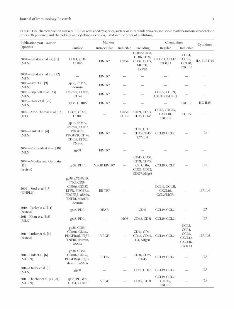
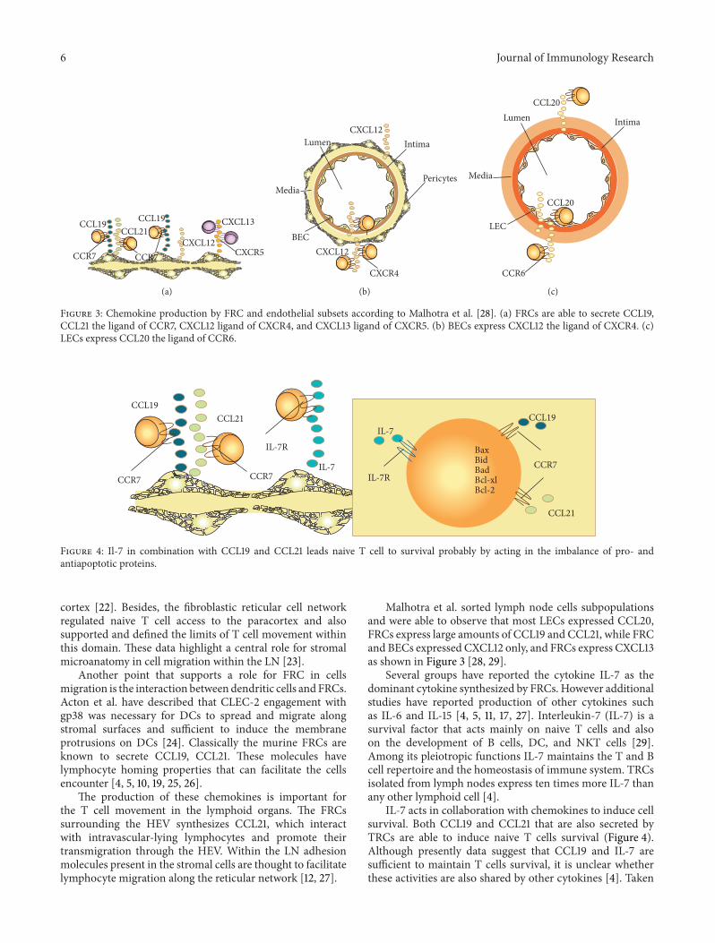
Figure 3: Chemokine production by FRC and endothelial subsets according to Malhotra et al. [28]. (a) FRCs are able to secrete CCL19,CCL21 the ligand of CCR7, CXCL12 ligand of CXCR4, and CXCL13 ligand of CXCR5. (b) BECs express CXCL12 the ligand of CXCR4. (c)LECs express CCL20 the ligand of CCR6.
IL-7
CCL19CCL21
CCR7 CCR7
CCL19
CCL21
IL-7
BaxBidBadBcl-xlBcl-2
CCR7IL-7R
IL-7R
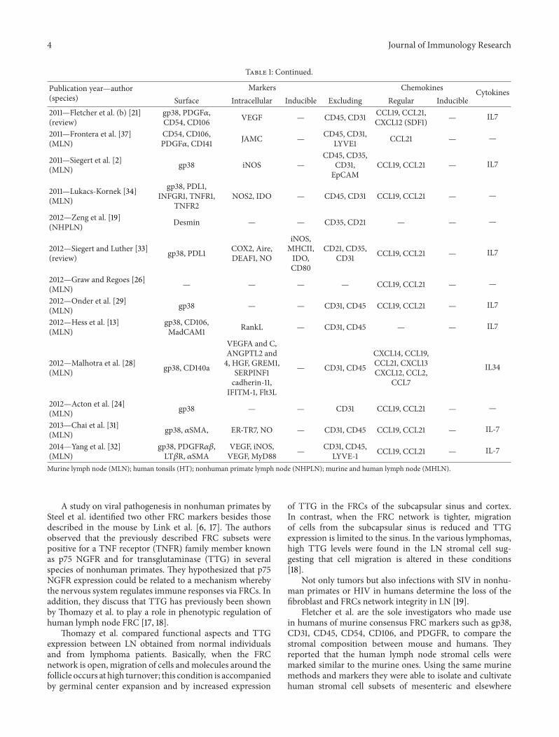
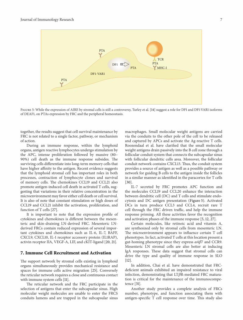
Figure 4: Il-7 in combination with CCL19 and CCL21 leads naive T cell to survival probably by acting in the imbalance of pro- andantiapoptotic proteins.
cortex [22]. Besides, the fibroblastic reticular cell networkregulated naive T cell access to the paracortex and alsosupported and defined the limits of T cell movement withinthis domain. These data highlight a central role for stromalmicroanatomy in cell migration within the LN [23].
Another point that supports a role for FRC in cellsmigration is the interaction between dendritic cells and FRCs.Acton et al. have described that CLEC-2 engagement withgp38 was necessary for DCs to spread and migrate alongstromal surfaces and sufficient to induce the membraneprotrusions on DCs [24]. Classically the murine FRCs areknown to secrete CCL19, CCL21. These molecules havelymphocyte homing properties that can facilitate the cellsencounter [4, 5, 10, 19, 25, 26].
The production of these chemokines is important forthe T cell movement in the lymphoid organs. The FRCssurrounding the HEV synthesizes CCL21, which interactwith intravascular-lying lymphocytes and promote theirtransmigration through the HEV. Within the LN adhesionmolecules present in the stromal cells are thought to facilitatelymphocyte migration along the reticular network [12, 27].
Malhotra et al. sorted lymph node cells subpopulationsand were able to observe that most LECs expressed CCL20,FRCs express large amounts of CCL19 and CCL21, while FRCand BECs expressed CXCL12 only, and FRCs express CXCL13as shown in Figure 3 [28, 29].
Several groups have reported the cytokine IL-7 as thedominant cytokine synthesized by FRCs. However additionalstudies have reported production of other cytokines suchas IL-6 and IL-15 [4, 5, 11, 17, 27]. Interleukin-7 (IL-7) is asurvival factor that acts mainly on naive T cells and alsoon the development of B cells, DC, and NKT cells [29].Among its pleiotropic functions IL-7 maintains the T and Bcell repertoire and the homeostasis of immune system. TRCsisolated from lymph nodes express ten times more IL-7 thanany other lymphoid cell [4].
IL-7 acts in collaboration with chemokines to induce cellsurvival. Both CCL19 and CCL21 that are also secreted byTRCs are able to induce naive T cells survival (Figure 4).Although presently data suggest that CCL19 and IL-7 aresufficient to maintain T cells survival, it is unclear whetherthese activities are also shared by other cytokines [4]. Taken
Journal of Immunology Research 7
DF1PTA
PTA
PTA
PTA
DF1-VAR1
MHC-I
TCR
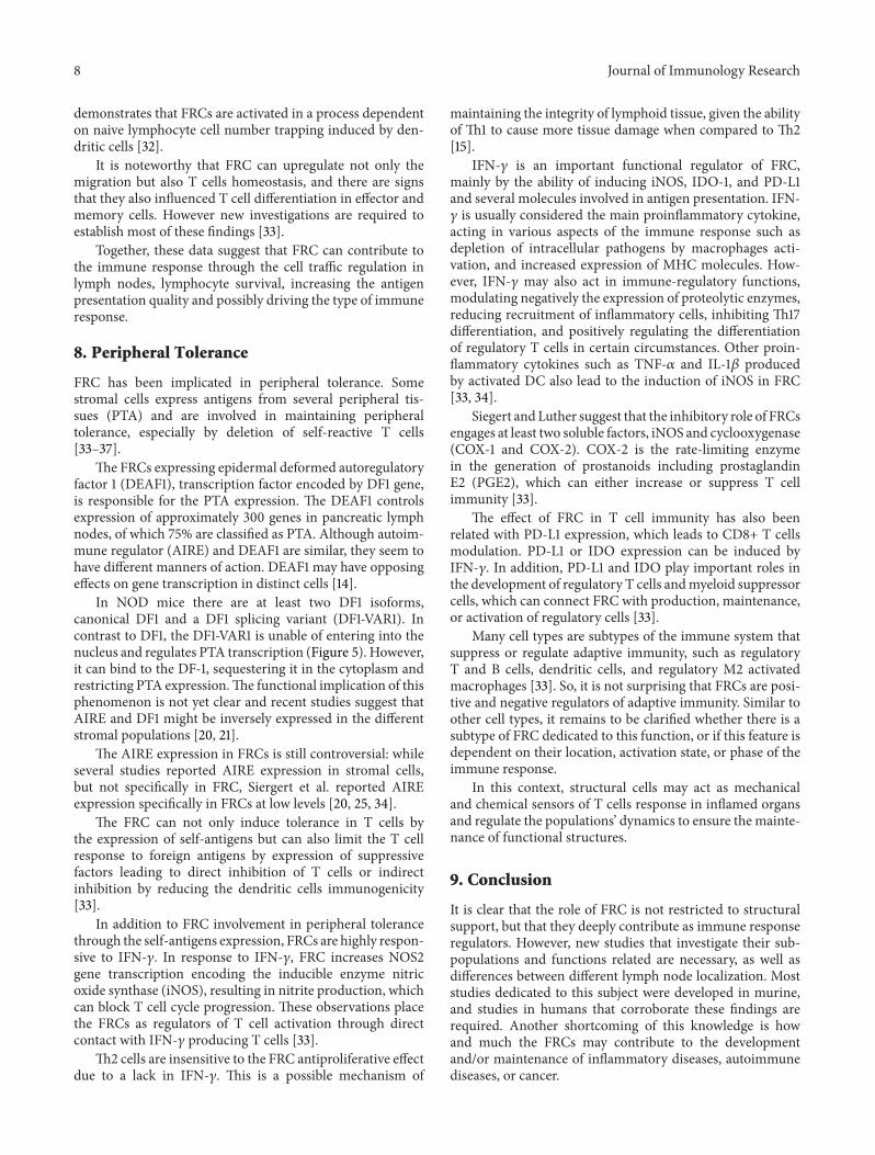
Figure 5: While the expression of AIRE by stromal cells is still a controversy, Turley et al. [14] suggest a role for DF1 and DF1-VAR1 isoformsof DEAF1, on PTAs expression by FRC and the peripheral homeostasis.
together, the results suggest that cell survival maintenance byFRC is not related to a single factor, pathway, or mechanismof action.
During an immune response, within the lymphoidorgans, antigen reactive lymphocytes undergo stimulation bythe APC, intense proliferation followed by massive (80–90%) cell death as the immune response subsides. Thesurviving cells differentiate into long-termmemory cells thathave higher affinity to the antigen. Recent evidence suggeststhat the lymphoid stromal cell has important roles in bothprocesses, contraction of lymphocyte clones and survivalof memory cells. The chemokines CCL19 and CCL21 alsopromote antigen-induced cell death in activated T cells, sug-gesting that variations in their relative concentration in themicroenvironmentmay favor either cell death or cell survival.It is also of note that constant stimulation or high doses ofCCL19 and CCL21 inhibit the activation, proliferation, andfunction of T cells [27].
It is important to note that the expression profile ofcytokines and chemokines is different between the mesen-teric and skin-draining LN-derived FRC. Mesenteric LN-derived FRCs contain reduced expression of several impor-tant cytokines and chemokines such as IL-6, IL-7, BAFF,CXCL9, CXCL10, IL-1 receptor accessory protein (IL1RAP),activin receptor IIA, VEGF-A, LIF, and cKIT-ligand [20, 21].
7. Immune Cell Recruitment and Activation
The support network by stromal cells existing in lymphoidorgans simultaneously provides mechanical resistance andspaces for immune cells active migration [25]. Converselythe reticular network requires a close and continuous contactwith immune system cells [11].
The reticular network and the FRC participate in theselection of antigens that enter the subcapsular sinus. Highmolecular weight molecules are unable to enter the FRCSconduits lumens and are trapped in the subcapsular sinus
macrophages. Small molecular weight antigens are carriedvia the conduits to the other pole of the cell to be releasedand captured by APCs and activate the Ag-reactive T cells.Roozendaal et al. have clarified that the small molecularweight antigens drain passively into the B cell zone through afollicular conduit system that connects the subcapsular sinuswith follicular dendritic cells area. Moreover, the follicularconduit network contains CXCL13. Thus, the conduit systemprovides a source of antigen as well as a possible pathway ornetwork for guiding B cells to the antigen inside the folliclesin a similar manner as identified in the paracortex for T cells[30].
IL-7 secreted by FRC promotes APC function andthe molecules CCL19 and CCL20 enhance the interactionbetween dendritic cell (DC) and T cells and stimulate endo-cytosis and DC antigen presentation (Figure 5). ActivatedDCs in turn produce CCL3 and CCL4, recruit rare Tcell through the FRC driven traffic, and help the immuneresponse priming. All these activities favor the recognitionand activation phases of the immune response [5, 12, 27].
Certain molecules, like retinoic acid and vitamin A,are synthesized only by stromal cells from mesenteric LN.The microenvironment appears to influence certain T cellphenotypes. In fact, activated T cells at this location present agut-homing phenotype since they express 𝛼4𝛽7 and CCR9.Mesenteric LN stromal cells are also better at inducingIgA responses. These data suggest that stromal cells candrive the type and quality of immune response in SLO[12].
In addition, Chai et al. have demonstrated that FRC-deficient animals exhibited an impaired resistance to viralinfection, demonstrating that LT𝛽R-mediated FRC matura-tion is critical for the maintenance of the immunocompe-tence [31].
Another study provides a complete analysis of FRCsnumber, phenotype, and function associating them withantigen-specific T cell response over time. This study also
8 Journal of Immunology Research
demonstrates that FRCs are activated in a process dependenton naive lymphocyte cell number trapping induced by den-dritic cells [32].
It is noteworthy that FRC can upregulate not only themigration but also T cells homeostasis, and there are signsthat they also influenced T cell differentiation in effector andmemory cells. However new investigations are required toestablish most of these findings [33].
Together, these data suggest that FRC can contribute tothe immune response through the cell traffic regulation inlymph nodes, lymphocyte survival, increasing the antigenpresentation quality and possibly driving the type of immuneresponse.
8. Peripheral Tolerance
FRC has been implicated in peripheral tolerance. Somestromal cells express antigens from several peripheral tis-sues (PTA) and are involved in maintaining peripheraltolerance, especially by deletion of self-reactive T cells[33–37].
The FRCs expressing epidermal deformed autoregulatoryfactor 1 (DEAF1), transcription factor encoded by DF1 gene,is responsible for the PTA expression. The DEAF1 controlsexpression of approximately 300 genes in pancreatic lymphnodes, of which 75% are classified as PTA. Although autoim-mune regulator (AIRE) and DEAF1 are similar, they seem tohave different manners of action. DEAF1 may have opposingeffects on gene transcription in distinct cells [14].
In NOD mice there are at least two DF1 isoforms,canonical DF1 and a DF1 splicing variant (DF1-VAR1). Incontrast to DF1, the DF1-VAR1 is unable of entering into thenucleus and regulates PTA transcription (Figure 5). However,it can bind to the DF-1, sequestering it in the cytoplasm andrestricting PTA expression.The functional implication of thisphenomenon is not yet clear and recent studies suggest thatAIRE and DF1 might be inversely expressed in the differentstromal populations [20, 21].
The AIRE expression in FRCs is still controversial: whileseveral studies reported AIRE expression in stromal cells,but not specifically in FRC, Siergert et al. reported AIREexpression specifically in FRCs at low levels [20, 25, 34].
The FRC can not only induce tolerance in T cells bythe expression of self-antigens but can also limit the T cellresponse to foreign antigens by expression of suppressivefactors leading to direct inhibition of T cells or indirectinhibition by reducing the dendritic cells immunogenicity[33].
In addition to FRC involvement in peripheral tolerancethrough the self-antigens expression, FRCs are highly respon-sive to IFN-𝛾. In response to IFN-𝛾, FRC increases NOS2gene transcription encoding the inducible enzyme nitricoxide synthase (iNOS), resulting in nitrite production, whichcan block T cell cycle progression. These observations placethe FRCs as regulators of T cell activation through directcontact with IFN-𝛾 producing T cells [33].
Th2 cells are insensitive to the FRC antiproliferative effectdue to a lack in IFN-𝛾. This is a possible mechanism of
maintaining the integrity of lymphoid tissue, given the abilityof Th1 to cause more tissue damage when compared to Th2[15].
IFN-𝛾 is an important functional regulator of FRC,mainly by the ability of inducing iNOS, IDO-1, and PD-L1and several molecules involved in antigen presentation. IFN-𝛾 is usually considered the main proinflammatory cytokine,acting in various aspects of the immune response such asdepletion of intracellular pathogens by macrophages acti-vation, and increased expression of MHC molecules. How-ever, IFN-𝛾 may also act in immune-regulatory functions,modulating negatively the expression of proteolytic enzymes,reducing recruitment of inflammatory cells, inhibiting Th17differentiation, and positively regulating the differentiationof regulatory T cells in certain circumstances. Other proin-flammatory cytokines such as TNF-𝛼 and IL-1𝛽 producedby activated DC also lead to the induction of iNOS in FRC[33, 34].
Siegert and Luther suggest that the inhibitory role of FRCsengages at least two soluble factors, iNOS and cyclooxygenase(COX-1 and COX-2). COX-2 is the rate-limiting enzymein the generation of prostanoids including prostaglandinE2 (PGE2), which can either increase or suppress T cellimmunity [33].
The effect of FRC in T cell immunity has also beenrelated with PD-L1 expression, which leads to CD8+ T cellsmodulation. PD-L1 or IDO expression can be induced byIFN-𝛾. In addition, PD-L1 and IDO play important roles inthe development of regulatory T cells andmyeloid suppressorcells, which can connect FRC with production, maintenance,or activation of regulatory cells [33].
Many cell types are subtypes of the immune system thatsuppress or regulate adaptive immunity, such as regulatoryT and B cells, dendritic cells, and regulatory M2 activatedmacrophages [33]. So, it is not surprising that FRCs are posi-tive and negative regulators of adaptive immunity. Similar toother cell types, it remains to be clarified whether there is asubtype of FRC dedicated to this function, or if this feature isdependent on their location, activation state, or phase of theimmune response.
In this context, structural cells may act as mechanicaland chemical sensors of T cells response in inflamed organsand regulate the populations’ dynamics to ensure the mainte-nance of functional structures.
9. Conclusion
It is clear that the role of FRC is not restricted to structuralsupport, but that they deeply contribute as immune responseregulators. However, new studies that investigate their sub-populations and functions related are necessary, as well asdifferences between different lymph node localization. Moststudies dedicated to this subject were developed in murine,and studies in humans that corroborate these findings arerequired. Another shortcoming of this knowledge is howand much the FRCs may contribute to the developmentand/or maintenance of inflammatory diseases, autoimmunediseases, or cancer.
Journal of Immunology Research 9
Conflict of Interests
The authors declare that there is no conflict of interestsregarding the publication of this paper.
Acknowledgments
The authors are thankful to Professor Ises de AlmeidaAbrahamsohn that reviewed very carefully this paper. Theyare also grateful to Sociedade Beneficente Israelita BrasileiraHospital Albert Einstein (SBIBHAE) for all the support.
References
[1] G. P. Sobocinski, K. Toy,W. F. Bobrowski, S. Shaw, A. Anderson,and E. P. Kaldjian, “Ultrastructural localization of extracellularmatrix proteins of the lymph node cortex: evidence supportingthe reticular network as a pathway for lymphocyte migration,”BMC Immunology, vol. 17, no. 11, article 42, 2010.
[2] S. Siegert, H.-Y. Huang, C.-Y. Yang et al., “Fibroblastic reticularcells from lymph nodes attenuate T cell expansion by producingnitric oxide,” PLoS ONE, vol. 6, no. 11, Article ID e27618, 2011.
[3] L. Onder, E. Scandella, Q. Chai et al., “A novel bacterial arti-ficial chromosome-transgenic podoplanin-cre mouse targetslymphoid organ stromal cells in vivo,” Frontiers in Immunology,vol. 2, article 50, 2011.
[4] A. Link, T. K. Vogt, S. Favre et al., “Fibroblastic reticular cells inlymph nodes regulate the homeostasis of naive T cells,” NatureImmunology, vol. 8, no. 11, pp. 1255–1265, 2007.
[5] S. A. Luther, T. K.Vogt, and S. Siegert, “Guiding blindT cells anddendritic cells: a closer look at fibroblastic reticular cells foundwithin lymph node T zones,” Immunology Letters, vol. 138, no.1, pp. 9–11, 2011.
[6] A. Link, D. L. Hardie, S. Favre et al., “Association of T-zonereticular networks and conduits with ectopic lymphoid tissuesin mice and humans,” The American Journal of Pathology, vol.178, no. 4, pp. 1662–1675, 2011.
[7] S. L. Clark Jr., “The reticulum of lymph nodes in mice stud-ied with the electron microscope,” The American Journal ofAnatomy, vol. 110, pp. 217–257, 1962.
[8] E. Crivellato and F. Mallardi, “Stromal cell organisation inthe mouse lymph node. A light and electron microscopicinvestigation using the zinc iodide-osmium technique,” Journalof Anatomy, vol. 190, no. 1, pp. 85–92, 1997.
[9] M. Sixt, N. Kanazawa, M. Selg et al., “The conduit systemtransports soluble antigens from the afferent lymph to residentdendritic cells in the T cell area of the lymph node,” Immunity,vol. 22, no. 1, pp. 19–29, 2005.
[10] A. O. Anderson and S. Shaw, “T cell adhesion to endothelium:the FRC conduit system and other anatomic and molecularfeatures which facilitate the adhesion cascade in lymph node,”Seminars in Immunology, vol. 5, no. 4, pp. 271–282, 1993.
[11] T. Katakai, T. Hara, M. Sugai, H. Gonda, and A. Shimizu,“Lymph node fibroblastic reticular cells construct the stromalreticulum via contact with lymphocytes,” Journal of Experimen-tal Medicine, vol. 200, no. 6, pp. 783–795, 2004.
[12] S. N. Mueller and R. N. Germain, “Stromal cell contributionsto the homeostasis and functionality of the immune system,”Nature Reviews Immunology, vol. 9, no. 9, pp. 618–629, 2009.
[13] E. Hess, V. Duheron, M. Decossas et al., “RANKL inducesorganized lymph node growth by stromal cell proliferation,”Journal of Immunology, vol. 188, no. 3, pp. 1245–1254, 2012.
[14] S. J. Turley, A. L. Fletcher, and K. G. Elpek, “The stromaland haematopoietic antigen-presenting cells that reside insecondary lymphoid organs,” Nature Reviews Immunology, vol.10, no. 12, pp. 813–825, 2010.
[15] O. Khan, M. Headley, A. Gerard, W. Wei, L. Liu, and M. F.Krummel, “Regulation of T cell priming by lymphoid stroma,”PLoS ONE, vol. 6, no. 11, Article ID e26138, 2011.
[16] P. Ame-Thomas, H.M.-E. Hajjami, C.Monvoisin et al., “Humanmesenchymal stem cells isolated from bone marrow and lym-phoid organs support tumor B-cell growth: role of stromal cellsin follicular lymphoma pathogenesis,” Blood, vol. 109, no. 2, pp.693–702, 2007.
[17] K. E. Steel, A. O. Anderson, and M. Mohamadzadeh, “Fibrob-lastic reticular cell infection by hemorrhagic fever viruses,”Immunotherapy, vol. 1, no. 2, pp. 187–197, 2009.
[18] V. A. Thomazy, F. Vega, L. J. Medeiros, P. J. Davies, and D.Jones, “Phenotypicmodulation of the stromal reticular networkin normal and neoplastic lymph nodes: tissue transglutaminasereveals coordinate regulation of multiple cell types,”The Amer-ican Journal of Pathology, vol. 163, no. 1, pp. 165–174, 2003.
[19] M. Zeng, M. Paiardini, J. C. Engram et al., “Critical role of CD4T cells in maintaining lymphoid tissue structure for immunecell homeostasis and reconstitution,” Blood, vol. 120, pp. 1856–1867, 2012.
[20] A. L. Fletcher, D. Malhotra, S. E. Acton et al., “Reproducibleisolation of lymphnode stromal cells reveals site-dependent dif-ferences in fibroblastic reticular cells,” Frontiers in Immunology,vol. 2, article 35, 2011.
[21] A. L. Fletcher, D. Malhotra, and S. J. Turley, “Lymph nodestroma broaden the peripheral tolerance paradigm,” Trends inImmunology, vol. 32, no. 1, pp. 12–18, 2011.
[22] T. Katakai, T. Hara, J.-H. Lee, H. Gonda, M. Sugai, and A.Shimizu, “A novel reticular stromal structure in lymph nodecortex: an immuno-platform for interactions among dendriticcells, T cells and B cells,” International Immunology, vol. 16, no.8, pp. 1133–1142, 2004.
[23] M. Bajenoff, J. G. Egen, L. Y. Koo et al., “Stromal cell networksregulate lymphocyte entry, migration, and territoriality inlymph nodes,” Immunity, vol. 25, no. 6, pp. 989–1001, 2006.
[24] S. E. Acton, J. L. Astarita, D. Malhotra et al., “Podoplanin-richstromal networks induce dendritic cell motility via activation ofthe C-type lectin receptor CLEC-2,” Immunity, vol. 37, no. 2, pp.276–289, 2012.
[25] T. Hara, T. Katakai, J.-H. Lee et al., “A transmembranechemokine, CXC chemokine ligand 16, expressed by lymphnode fibroblastic reticular cells has the potential to regulate Tcell migration and adhesion,” International Immunology, vol. 18,no. 2, pp. 301–311, 2006.
[26] F. Graw and R. R. Regoes, “Influence of the fibroblastic reticularnetwork on cell-cell interactions in lymphoid organs,” PLoSComputational Biology, vol. 8, no. 3, Article ID e1002436, 2012.
[27] S.N.Mueller andR.Ahmed, “Lymphoid stroma in the initiationand control of immune responses,” Immunological Reviews, vol.224, no. 1, pp. 284–294, 2008.
[28] D. Malhotra, A. L. Fletcher, J. Astarita et al., “Transcriptionalprofiling of stroma from inflamed and resting lymph nodesdefines immunological hallmarks,” Nature Immunology, vol. 13,no. 5, pp. 499–510, 2012.
10 Journal of Immunology Research
[29] L. Onder, P. Narang, E. Scandella et al., “IL-7-producing stromalcells are critical for lymph node remodeling,” Blood, vol. 120, pp.4675–4683, 2012.
[30] R. Roozendaal, T. R. Mempel, L. A. Pitcher et al., “Conduitsmediate transport of low-molecular-weight antigen to lymphnode follicles,” Immunity, vol. 30, no. 2, pp. 264–276, 2009.
[31] Q. Chai, L. Onder, E. Scandella et al., “Maturation of lymphnode fibroblastic reticular cells frommyofibroblastic precursorsis critical for antiviral immunity,” Immunity, vol. 38, no. 5, pp.1013–1024, 2013.
[32] C. Y. Yang, T. K. Vogt, S. Favre et al., “Trapping of naivelymphocytes triggers rapid growth and remodeling of thefibroblast network in reactivemurine lymphnodes,”Proceedingsof the National Academy of Sciences of the United States ofAmerica, vol. 111, no. 1, pp. E109–E118, 2014.
[33] S. Siegert and S. A. Luther, “Positive and negative regulation of Tcell responses by fibroblastic reticular cells within paracorticalregions of lymph nodes,” Frontiers in Immunology, vol. 3, article285, 2012.
[34] V. Lukacs-Kornek, D. Malhotra, A. L. Fletcher et al., “Regulatedrelease of nitric oxide by nonhematopoietic stroma controlsexpansion of the activated T cell pool in lymph nodes,” NatureImmunology, vol. 12, no. 11, pp. 1096–1104, 2011.
[35] E. P. Kaldjian, J. Elizabeth Gretz, A. O. Anderson, Y. Shi, andS. Shaw, “Spatial and molecular organization of lymph node Tcell cortex: a labyrinthine cavity bounded by an epithelium-likemonolayer of fibroblastic reticular cells anchored to basementmembrane-like extracellular matrix,” International Immunol-ogy, vol. 13, no. 10, pp. 1243–1253, 2001.
[36] F. Vega, K. R. Coombes, V. A. Thomazy, K. Patel, W. Lang, andD. Jones, “Tissue-specific function of lymph node fibroblasticreticulum cells,” Pathobiology, vol. 73, no. 2, pp. 71–81, 2006.
[37] V. Frontera, M.-L. Arcangeli, C. Zimmerli et al., “Cutting edge:JAM-C controls homeostatic chemokine secretion in lymphnode fibroblastic reticular cells expressing thrombomodulin,”Journal of Immunology, vol. 187, no. 2, pp. 603–607, 2011.

Submit your manuscripts athttp://www.hindawi.com
Stem CellsInternational
Hindawi Publishing Corporationhttp://www.hindawi.com Volume 2014
Hindawi Publishing Corporationhttp://www.hindawi.com Volume 2014
MEDIATORSINFLAMMATION
of
Hindawi Publishing Corporationhttp://www.hindawi.com Volume 2014
Behavioural Neurology
EndocrinologyInternational Journal of
Hindawi Publishing Corporationhttp://www.hindawi.com Volume 2014
Hindawi Publishing Corporationhttp://www.hindawi.com Volume 2014
Disease Markers
Hindawi Publishing Corporationhttp://www.hindawi.com Volume 2014
BioMed Research International
OncologyJournal of
Hindawi Publishing Corporationhttp://www.hindawi.com Volume 2014
Hindawi Publishing Corporationhttp://www.hindawi.com Volume 2014
Oxidative Medicine and Cellular Longevity
Hindawi Publishing Corporationhttp://www.hindawi.com Volume 2014
PPAR Research
The Scientific World JournalHindawi Publishing Corporation http://www.hindawi.com Volume 2014
Immunology ResearchHindawi Publishing Corporationhttp://www.hindawi.com Volume 2014
Journal of
ObesityJournal of
Hindawi Publishing Corporationhttp://www.hindawi.com Volume 2014
Hindawi Publishing Corporationhttp://www.hindawi.com Volume 2014
Computational and Mathematical Methods in Medicine
OphthalmologyJournal of
Hindawi Publishing Corporationhttp://www.hindawi.com Volume 2014
Diabetes ResearchJournal of
Hindawi Publishing Corporationhttp://www.hindawi.com Volume 2014
Hindawi Publishing Corporationhttp://www.hindawi.com Volume 2014
Research and TreatmentAIDS
Hindawi Publishing Corporationhttp://www.hindawi.com Volume 2014
Gastroenterology Research and Practice
Hindawi Publishing Corporationhttp://www.hindawi.com Volume 2014
Parkinson’s Disease
Evidence-Based Complementary and Alternative Medicine
Volume 2014Hindawi Publishing Corporationhttp://www.hindawi.com