morphological study of the male genital organs of gracilinanus microtarsus
TRANSCRIPT
447
Braz. J. Vet. Res. Anim. Sci., São Paulo, v. 50, n. 6, p. 447-456, 2013
Abstract
Gracilinanus microtarsus is one of the smallest marsupials on earth. Since it spreads seeds, it has great ecological relevance. However, its reproduction data, especially those related to the anatomy of its reproduction apparatus, are scarce in the literature. Current analysis describes the male genital organs of six adult specimens of G. microtarsus. Macroscopic studies were undertaken on dissected organs, whereas histological studies were performed by inclusion technique in paraffin and by hematoxylin and eosin, and Masson trichrome staining. The male genital organs and accessory glands of G. microtarsus consist of two testicles within a pendular scrotum, placed cranially to the penis, featuring a histology consisting of seminiferous tubules with spermatogenic cells, spermatozoa and Sertoli cells, and a peritubular region with Leydig cells; testicles closely associated with epididymis with head, body and tail, with histological differences between the different regions; deferent ducts; funiculus spermaticus; accessory glands composed of prostate, divided into three distinct segments and bulbourethral glands; pelvic and penile urethra and penis with bifid glans. Results show that the male genital organs and accessory glands of G. Microtarsus are anatomically similar to that found in Didelphis sp. and other marsupials groups, with slight details such as the site of each organ.Keywords: Didelphimorphia. Marsupials. Reproduction. Prostatic segments.
Resumo
Gracilinanus microtarsus é um dos menores marsupiais do mundo. Possui grande potencial dispersor de sementes e por esta razão apresenta grande importância ecológica. No entanto, dados em relação à reprodução desses animais são es-cassos na literatura, principalmente, aqueles relacionados à anatomia dos órgãos do aparelho reprodutor. Por essa razão, objetivamos com este estudo realizar uma descrição dos órgãos genitais masculinos em seis exemplares de G. microtarsus adultos. Para tanto, realizamos estudos macroscópicos por dissecação dos órgãos e estudos histológicos por técnica de inclusão em parafina e coloração em hematoxilina e eosina, e tricromo de Masson. Encontramos, que os órgãos genitais masculinos e glândulas acessórias em G. microtarsus são representados por dois testículos localizados dentro de um es-croto pendular, posicionados cranialmente ao pênis, cuja histologia revelou túbulos seminíferos com espermatogônias, espermatozoides e células de Sertoli e região peritubular com células de Leydig; testículos em íntima associação com os epidídimos, divididos em cabeça, corpo e cauda, com diferenças histológicas entre as diferentes regiões; ductos deferen-tes; funículo espermático; glândulas anexas compostas pelas glândulas prostáticas divididas em três distintos segmentos e glândulas bulbouretrais; uretra pélvica e peniana e um pênis com glande bífida. Concluímos que os órgãos genitais e as glândulas acessórias masculina de G. microtarsus, anatomicamente são muito semelhantes aos dos Didelphis sp. e outros grupos de marsupiais, diferenciando-se apenas em alguns detalhes, como a localização de cada órgão.Palavras-chave: Didelphimorphia. Marsupiais. Reprodução. Segmentos prostáticos.
Morphological study of the male genital organs of Gracilinanus microtarsus
Estudo morfológico dos órgãos genitais masculinos em Gracilinanus microtarsus
Jussara Marcolino do Nascimento LIMA1; Amilton Cesar dos SANTOS2; Diego Carvalho VIANA2; Bruno Machado BERTASSOLI3; Luis Miguel LOBO2; Vanessa Cristina de OLIVEIRA2;
Denis Cristiano BRIANI4; Gerlane Medeiros COSTA5; Antônio Chaves de Assis NETO2; Carlos Eduardo AMBRÓSIO6; Ana Flávia CARVALHO1; Celina Almeida Furlanetto MANÇANARES1
1 Universitary Center of Education Foundation Octávio Bastos, São João da Boa Vista – SP, Brasil2 School of Veterinary Medicine and Animal Science of the University of São Paulo, São Paulo – SP, Brasil
3 Federal University of Minas Gerais, Belo Horizonte – MG, Brasil4 Claretianas Integrate Faculties, Rio Claro – SP, Brasil
5 University of Mato Grosso State, Alta Floresta – MT, Brasil6 School of Animal Science and Food Engineering of the University of São Paulo, Pirassununga – SP, Brasil
Correspondence to: Amilton Cesar dos SantosAv. Prof. Dr. Orlando Marques de Paiva, 87, Cidade Universitária CEP 05508-270, São Paulo – SP, Brasile-mail: [email protected]
Received: 22/10/2013Approved: 18/12/2013
448
Braz. J. Vet. Res. Anim. Sci., São Paulo, v. 50, n. 6, p. 447-4565, 2013
Introduction
Marsupials are mammals commonly recognized by a pouch, where the females carry their neonate. The pouch is highly relevant since the neonates are relatively undeveloped and their development mostly occurs in the post-partum period, precisely within the pouch. All marsupials in Brazil belong to the order Didelphimorphia, native to the Americas. In fact, fifty-five out of the 92 species of opossum occur in Brazil (COSTA; LEITE; PATTON, 2003; ARAGONA; MARINHO FILHO, 2009).
The Gracilinanus microtarsus, popularly called catita or guaiquica in Brazil, is one of the smallest marsupials on earth, measuring between 20 and 25 cm long, with the tail comprising more than half of its length. The G. microtarsus is a highly relevant animal since it is a spreader of seeds of several plants (CAMPOS et al., 2012). Its weight in adulthood varies between 15 and 30 g even though some specimens may weigh 40 g (CÂMARA; MURTA, 2003). Their pelage is short and smooth and of a somewhat dark brown color. Its dorsal pelage ranges from gray-brown to red-brown and its thorax color varies between pale orange and cream. Their eyes are round, black and highly conspicuous with a strain of black hair surrounding them. The feet and paws are small and whitish and the ears are bare and mobile. Besides the prehensile tail, its feet have an opposing thumb to grip tree branches (ARAGONA; MARINHO FILHO, 2009).
The start of the G. microtarsus reproductive period is closely linked to the rainy season, characterized by abundant food. Females may have heat twice a year, the first of which begins approximately after one year of age. Pregnancy period is short and does not exceed 15 days. The neonates are directed to the teats by the mother until their complete development. Afterwards, they grip the back of the mother until they become totally independent. Mothers normally have seven or eight pups each birth, even though the birth of 13 young is known (NOGUEIRA et al., 2004).
Due to the ecological relevance of the G. microtarsus and the lack of studies on the male reproduction morphology of the species, current investigation provided data that may complement others, mainly on animal reproduction, in order to preserve the species and to its rational and controlled exploitation. The morphological and histological aspects of the male genital organs and accessory glands were described.
Materials and Methods
Morphological studies on six male G. microtarsus provided by Morphological Sciences Laboratory from Centro Universitário Fundação de Ensino Octávio Bastos in São João da Boa Vista SP Brazil (IBAMA-02001.007176/03-69) were performed. This research was licensed by the Bioethics Committee of the Animals from the same institution above described. The animals had undergone other former assays and were already fixed in formaldehyde solution 10%.
Genital organs were dissected for macroscopic description and histological analysis. Fragments of the genital organs and accessory glands were collected, dehydrated at increasing percentages of alcohol solution (70% to 100%) and diaphanized in xylol to be later included in paraffin blocks from where slices with 5 µm were obtained. These slices were placed on histology slides for staining with Hematoxylin-Eosin (HE) and Masson trichrome.
All procedures were photodocumented with digital Sony Mavica 3.2 Mp, magnifying glass Lambda LEE-3 S/Na 005252 and Photomicroscope Olympus BX61VS. Photomicrographs and nomenclature were based on Nomina Anatomica Veterinaria (ICVGAN, 2012). Assays were performed at the Laboratories of Histology and Embryology of the Faculty of Veterinary Medicine and Animal Science of the University of Sao Paulo, São Paulo, SP, Brazil and Morphological Sciences Laboratory of the Centro Universitário Fundação de Ensino Octávio Bastos, São João da Boa Vista, SP Brasil.
449
Braz. J. Vet. Res. Anim. Sci., São Paulo, v. 50, n. 6, p. 447-456, 2013
Results
Morphological aspects of the male genital organs and accessory glands of G. microtarsus
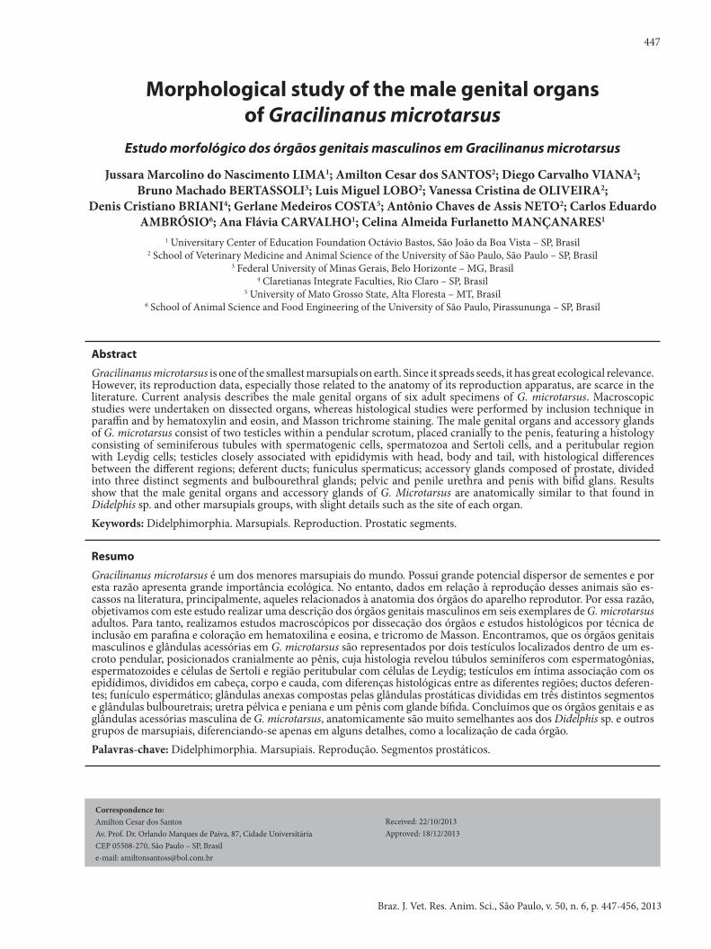
Th e external male genital organs comprise the testicles surrounded by the scrotum and the prepuce with an orifi ce, preputial ostium, through which the urogenital apparatus is linked to the outside when the penis is exposed. Th e scrotum and testicles are pendular and are localized in the inguinal region at the ventral portion of the abdomen, cranially positioned to the penis. Th e scrotum adheres to the ventral surface of the body by the skin. It is lined on the outside by a thin skin covered by short and delicate hairs (Figure 1A). Th e testicle-headed extremity coupled to the epididymis head lies in a ventral-cranial position, whereas the testicle tail extremity together with the epididymis end is ventrally arranged to the body of animal (Figure 1B).
Th e testicles have an elliptic shape with a long horizontal axis and are linked to the epididymis by the eff erent duct system, both of which are lined by the vaginal tunic. Th e epididymides are divided into head, body and tail. Th e tail is round and slightly projects itself on the surface of the caudal scrotum.
Figure 1 - Topographic photo of G. microtarsus scrotum and penis in situ and the epididymis ex situ. A: scrotum (s) and prepuce (arrow). B: epididymis with head (eh), body (eb), tail (et) and deferent duct (arrow). C: penis with bifi d glans (arrows). Bar: 0.1 cm.
Source: (LIMA et al., 2013)
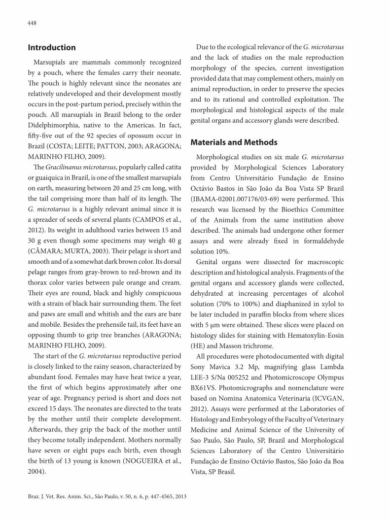
Th e deferent duct ends at the cranial section of the prostatic urethra without crossing the ureter. Th e long funiculus spermaticus consists of the deferent duct, artery and testicular vein, lymphatic vases, nerves and cremaster muscles.
Testicles, enveloped in a fi brous spermatic fascia, have an oval shape, with two margins and two extremities, fl attened lateral-laterally. Th e testicles loose margin is folded laterally, whereas the epididymis margin is medially linked to the epididymis. Th e testicles also have two extremities, or rather, at the head where the epididymis starts, and at the tail where the epididymis gives rise to the deferent duct.
G. microtarsus also has the accessory glands, represented by prostate and bulbourethral glands. Ampoule and vesicular glands were not reported in current analysis. Prostate has three segments: the fi rst is small and close to the urinary vesicle neck; the second is windy and large; the third is long and narrow and extends up to the membranous urethra, in the pelvic urethra. Th e bulbourethral glands of the G. microtarsus are lobule-shaped and divided into three lobules on the right and three on the left ; the lateral pair is pear-shaped and bigger; the medial pair is smaller and placed ventrally to the pelvic urethra. Th e above gland apparatus releases secretions into the section of the penile urethra.
Th e funiculus spermaticus in the testicular entrance consisted of the testicular artery, pampiniform plexus, cremaster muscle, lymphatic vessels and nerves, deferent duct and vaginal tunic (Figures 2A and B).
Th e penis of the G. microtarsus is positioned caudally to the scrotum and reveals a bifi d penis glans (Figure 1C).
Histological aspects of the male genital organs and accessory glands of G. microtarsus
Th e histology of the testicular parenchyma of G. microtarsus comprises a tubular compartment, in which lie the seminiferous tubules, the intertubular
450
Braz. J. Vet. Res. Anim. Sci., São Paulo, v. 50, n. 6, p. 447-4565, 2013
Figure 2 – Th e urogenital apparatus of G. microtarsus. A: kidneys (k), ureters (u), urinary vesicle (uv), bulbourethral gland (bg), deferent ducts (dd), right testicle (rt) left testicle (lt). B: funiculus spermaticus (fe), testicular artery (ta) pampini-form plexus (pp). Bar: 0.1cm.
Source: (LIMA et al., 2013)
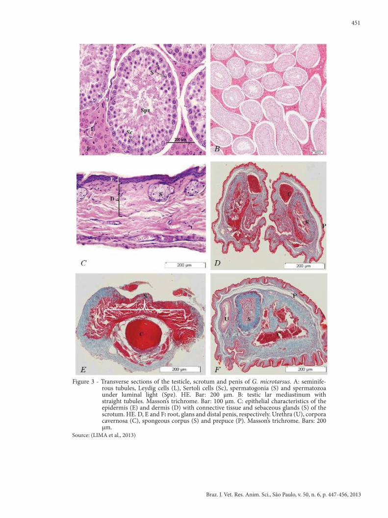
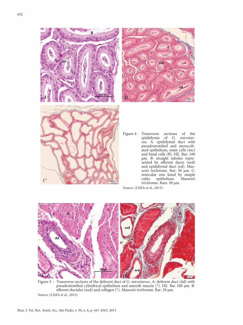
compartment, in which Leydig cells may be found, connective tissue, and blood vessels. Th e seminiferous tubules contain a connective tissue tunic, a basal lamina and an internal layer formed by the germinal or seminiferous epithelium which becomes less twisted as they come close to the testicular mediastinum. Th ey have an almost straight line direction and exit in the rectum tubules which anastomize within the testicular network. Th e rectum tubules are lined with a simple cubic epithelium with myoid cells around it and in the midst of loose connective tissue. Th e testicular network is linked to the epididymis by eff erent ducts placed at the testicle´s head extremities (Figures 3A and B).
Spermatogonia in the germinal epithelium are easily perceived due to their big cytoplasm and nucleus. Spermatids have smaller, more condensed and less granulated nuclei, whereas the spermatozoa are long, with a condensed nucleus and placed within the light of the seminiferous tubule. Leydig cells may be easily distinguished from the other cells in the loose connective tissue of the intertubular region, because of their normal triangular or polyhedral shape and because of their great round nucleus with not dense chromatin. Th e lining tunic in the scrotum comprises the dartos tunica, which is closely related to the skin for the retraction of scrotum (Figures 3A, B and C).
Th e epididymis is divided into rete testis, eff erent duct and epididymal duct. Th e latter has a stereociliated and pseudostratifi ed epithelium composed of long main cells and fl attened basal cells in contact with the basal membrane in great numbers. Cilia may be found in great numbers so that the spermatozoa could move towards the deferent ducts (Figures 4A, B and C).
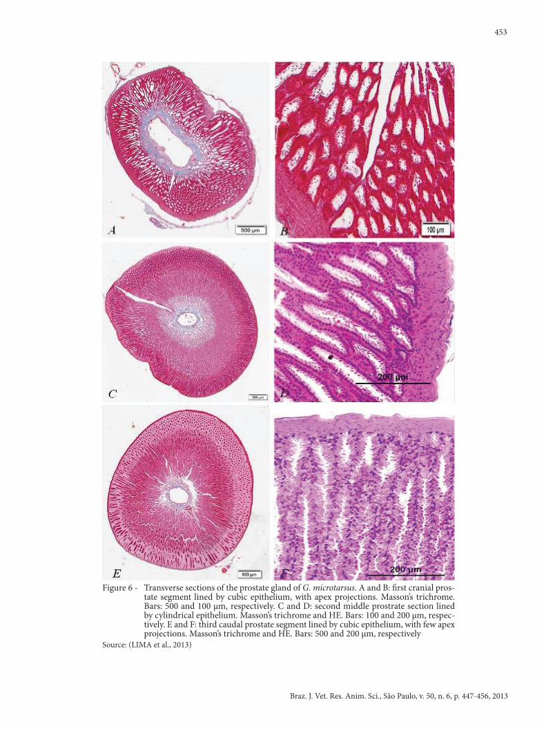
Deferent ducts are continuous to the epididymides and they have a small tube-like structure, laterally following the ureters, widening as a small ampoule. Th e deferent duct is lined with a pseudostratifi ed cylindrical epithelium (Figures 5A and B).
Th e prostate among the annexed glands comprises several round or polygonal tubules-alveolar units, surrounded by few fi bers and abundant dense connective tissue and smooth muscles. Th e secreting epithelium is composed of cubic cells in a single layer around oval nuclei, which lie in the central position. Beneath the epithelium there is a delicate fi brous sheet composed of tubular secreting units branched by vesicular distension. Th e secretory epithelium of glands is classifi ed as pseudo-stratifi ed (Figures 6A, B, C, D, E and F).
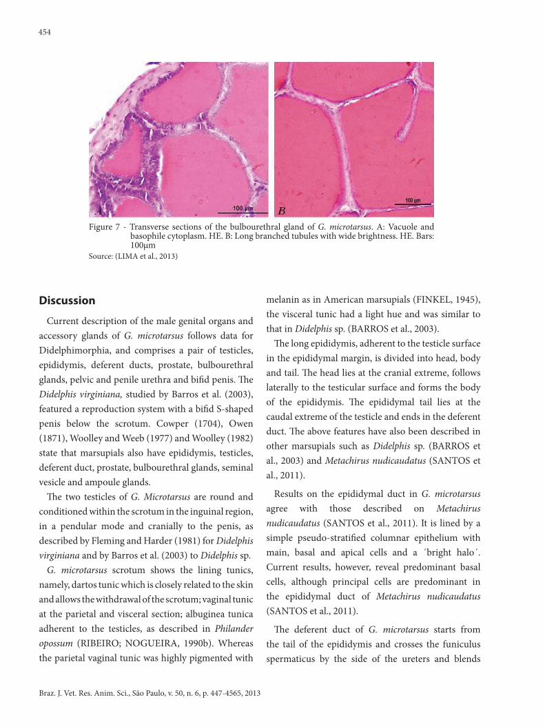
Th e bulbourethral glands are round and made of three pairs of glands (lateral, intermediate and medial) conditioned in the perineal region and exiting at the start of the penile urethra. Th ese pairs of glands, enveloped by a connective tissue and skeletal muscle, reveal a weak stained cytoplasm and vacuoles, which suggest mucoprotein secretion (Figures 7A, and B).
Pelvic and penile urethra of G. microtarsus is lined by transitional epithelium in all extension. Th e penis contains a cavernous body made of an erectile tissue with heavy areola lined by a thick layer of dense connective tissue which makes up the albuginea tunica of the cavernous corpus. Th e spongeous corpus of penis is a thin erectile tissue around the urethra lined by the endothelium. Th e tissue is enveloped by a layer of dense connective tissue which makes up the albuginea tunica of the spongeous corpus (Figures 3D, E and F).
451
Braz. J. Vet. Res. Anim. Sci., São Paulo, v. 50, n. 6, p. 447-456, 2013
Figure 3 - Transverse sections of the testicle, scrotum and penis of G. microtarsus. A: seminiferous
tubules, Leydig cells (L), Sertoli cells (Sc), spermatogonia (S) and spermatozoa under luminal light (Spz). HE. Bar: 200 µm. B: testicular mediastinum with straight tubules. Masson’s trichrome. Bar: 100 µm. C: epithelial characteristics of the epidermis (E) and dermis (D) with connective tissue and sebaceous glands (S) of the scrotum. HE. D, E and F: root, glans and distal penis, respectively. Urethra (U), corpora cavernosa (C), spongeous corpus (S) and prepuce (P). Masson’s trichrome. Bars: 200 µm
Source: (LIMA et al., 2013)
Figure 3 - Transverse sections of the testicle, scrotum and penis of G. microtarsus. A: seminife-rous tubules, Leydig cells (L), Sertoli cells (Sc), spermatogonia (S) and spermatozoa under luminal light (Spz). HE. Bar: 200 µm. B: testic lar mediastinum with straight tubules. Masson’s trichrome. Bar: 100 µm. C: epithelial characteristics of the epidermis (E) and dermis (D) with connective tissue and sebaceous glands (S) of the scrotum. HE. D, E and F: root, glans and distal penis, respectively. Urethra (U), corpora cavernosa (C), spongeous corpus (S) and prepuce (P). Masson’s trichrome. Bars: 200 µm.
Source: (LIMA et al., 2013)
452
Braz. J. Vet. Res. Anim. Sci., São Paulo, v. 50, n. 6, p. 447-4565, 2013
Figure 4 - Transverse sections of the epididymis of G. microtar-sus. A: epididymal duct with pseudostratifi ed and stereocili-ated epithelium, main cells (mc) and basal cells (B). HE. Bar: 100 µm. B: straight tubules repre-sented by eff erent ducts (eed) and epididymal duct (ed). Mas-son’s trichrome. Bar: 50 µm. C: testicular rete lined by simple cubic epithelium. Masson’s trichrome. Bars: 50 µm.
Source: (LIMA et al., 2013)
Figure 5 – Transverse sections of the deferent duct of G. microtarsus. A: deferent duct (dd) with pseudostratifi ed cylindrical epithelium and smooth muscle (*). HE. Bar 100 µm. B: eff erent ductules (eed) and collagen (*). Masson’s trichrome. Bar: 20 µm.
Source: (LIMA et al., 2013)
453
Braz. J. Vet. Res. Anim. Sci., São Paulo, v. 50, n. 6, p. 447-456, 2013
Figure 6 - Transverse sections of the prostate gland of G. microtarsus. A and B: fi rst cranial pros-tate segment lined by cubic epithelium, with apex projections. Masson’s trichrome. Bars: 500 and 100 µm, respectively. C and D: second middle prostrate section lined by cylindrical epithelium. Masson’s trichrome and HE. Bars: 100 and 200 µm, respec-tively. E and F: third caudal prostate segment lined by cubic epithelium, with few apex projections. Masson’s trichrome and HE. Bars: 500 and 200 µm, respectively
Source: (LIMA et al., 2013)
454
Braz. J. Vet. Res. Anim. Sci., São Paulo, v. 50, n. 6, p. 447-4565, 2013
Figure 7 - Transverse sections of the bulbourethral gland of G. microtarsus. A: Vacuole and basophile cytoplasm. HE. B: Long branched tubules with wide brightness. HE. Bars: 100µm
Source: (LIMA et al., 2013)
Discussion
Current description of the male genital organs and accessory glands of G. microtarsus follows data for Didelphimorphia, and comprises a pair of testicles, epididymis, deferent ducts, prostate, bulbourethral glands, pelvic and penile urethra and bifi d penis. Th e Didelphis virginiana, studied by Barros et al. (2003), featured a reproduction system with a bifi d S-shaped penis below the scrotum. Cowper (1704), Owen (1871), Woolley and Weeb (1977) and Woolley (1982) state that marsupials also have epididymis, testicles, deferent duct, prostate, bulbourethral glands, seminal vesicle and ampoule glands.
Th e two testicles of G. Microtarsus are round and conditioned within the scrotum in the inguinal region, in a pendular mode and cranially to the penis, as described by Fleming and Harder (1981) for Didelphis virginiana and by Barros et al. (2003) to Didelphis sp.
G. microtarsus scrotum shows the lining tunics, namely, dartos tunic which is closely related to the skin and allows the withdrawal of the scrotum; vaginal tunic at the parietal and visceral section; albuginea tunica adherent to the testicles, as described in Philander opossum (RIBEIRO; NOGUEIRA, 1990b). Whereas the parietal vaginal tunic was highly pigmented with
melanin as in American marsupials (FINKEL, 1945), the visceral tunic had a light hue and was similar to that in Didelphis sp. (BARROS et al., 2003).
Th e long epididymis, adherent to the testicle surface in the epididymal margin, is divided into head, body and tail. Th e head lies at the cranial extreme, follows laterally to the testicular surface and forms the body of the epididymis. Th e epididymal tail lies at the caudal extreme of the testicle and ends in the deferent duct. Th e above features have also been described in other marsupials such as Didelphis sp. (BARROS et al., 2003) and Metachirus nudicaudatus (SANTOS et al., 2011).
Results on the epididymal duct in G. microtarsus agree with those described on Metachirus nudicaudatus (SANTOS et al., 2011). It is lined by a simple pseudo-stratifi ed columnar epithelium with main, basal and apical cells and a ´bright halo´. Current results, however, reveal predominant basal cells, although principal cells are predominant in the epididymal duct of Metachirus nudicaudatus (SANTOS et al., 2011).
Th e deferent duct of G. microtarsus starts from the tail of the epididymis and crosses the funiculus spermaticus by the side of the ureters and blends
455
Braz. J. Vet. Res. Anim. Sci., São Paulo, v. 50, n. 6, p. 447-456, 2013
within the dorsal wall of the urethra in the urinary vesicle, as in Didelphis sp. (BARROS et al., 2003). In the latter species, they start at the end of the epididymal tail, cross the funiculus spermaticus and muscular wall and penetrate the abdominal cavity.
The deferent ducts in Metachirus nudicaudatus (SANTOS et al., 2011) are also divided into the juxta-epididymal, funicular and abdominal sections, without ampoule and without crossing the ureter prior to exiting into the urethra.
Prostate of G. microtarsus is disseminated in three distinct segments, also described in other Didelphidae (CHASE, 1939; GARCIA; GONÇALVES, 1984). Cranial, medium and caudal segments of the prostate agree with terminology by Nogueira, Ribeiro and Campos (1995). In Didelphis sp., the gland has a tubular form (BARROS et al., 2003) and lies around the urethra which distinguishes it from the prostate of G. microtarsus.
Prostate histology of G. microtarsus resembles that of other marsupials (CHASE, 1939; RODGER; HUGHES, 1973; TEMPLE-SMITH, 1984; TYNDALE- BISCOE; RENFREE, 1987), divided by secretory tubules into zones, as other Didelphidae species. The epithelial characteristics of the different zones of the prostate segments of the species, which include G. microtarsus, are different with regard to the development of apex projections and cell pigment (HRUBAN et al., 1965; GARCIA; GONÇALVES, 1984; NOGUEIRA; RIBEIRO; CAMPOS, 1995).
Nogueira et al. (2004) report that Chironectes minimus has two pairs of bulbourethral glands, similar to those registered in marsupials of the genera Caluromys, Gracilinanus, Monodelphis, Thylamis and Glironia.
On the other hand, the bulbourethral glands of G. microtarsus, also reported by Martinelli, Nogueira and Campos (1991) in Marmosa cinerea are composed of reticular fibers that involve adenomeres and ducts with elastic and collagen fibers. Its parenchyma comprises tubules, with simple epithelium, characteristic for each gland pair. These glands in Didelphis sp. have morphological traits, which are similar to those described for G. microtarsus. The authors also registered that ampoule and vesicular glands were missing (BARROS et al., 2003).
Similar to other marsupials, such as Philander opossum studied by Ribeiro and Nogueira (1990a), the penis of G. microtarsus has a bifid glans. Further, Martinelli and Nogueira (1997) investigated the urethral glands and drip to establish taxonomic identification between opossum and other marsupials. Matheus et al. (2011) reported that the spongeous corpus is formed by gaps and the corpora cavernosa is separated by trabeculas of connective tissues and smooth muscular fibers, corroborating reports by current study.
The methodology employed reveals that the male genital organs and accessory glands of the Gracilinanus microtarsus are anatomically similar to that of Didelphis sp. and other marsupial groups, with slight details such as the site of each organ.
Acknowledgements
We would like to thank Ronaldo Agostinho Silva for the technical support provided for the analyses of histological material. Thanks are also due to the Fundação de Amparo à Pesquisa do Estado de São Paulo for the scholarship awarded to the first author.
456
Braz. J. Vet. Res. Anim. Sci., São Paulo, v. 50, n. 6, p. 447-4565, 2013
ARAGONA, M.; MARINHO-FILHO, J. História natural e biologia reprodutiva de marsupiais no Pantanal, Mato Grosso, Brasil. Zoologia, v. 26, n. 2, p. 220-230, 2009.
BARROS, M. A.; MIGLINO, M. A.; MARTINS, J. F. P.; AMBRÓSIO, C. E.; VERECHIA, F. T.; ROSA, R. A.; CARVALHO, A. F. Morfologia do sistema reprodutor masculino do gambá (Didelphis sp.). Revista Brasileira de Reprodução Animal, v. 27, n. 2. p. 225-226, 2003.
CÂMARA, T.; MURTA, R. Mamíferos da Serra do Cipó. Belo Horizonte: PUC-Minas: Museu de Ciências Naturais, 2003. 129 p.
CAMPOS, W. H.; NETO, A. M.; PEIXOTO, H. J. C.; GODINHO, L. B.; SILVA, E. Contribuição da fauna silvestre em projetos de restauração ecológica no Brasil. Pesquisa Florestal Brasileira, v. 32, n. 72, p. 429-440, 2012.
CHASE, G. B. The reproductive system of the male opossum Didelphis virginiana Kerr and its experimental modification. Journal of Morphology, v. 65, n. 2, p. 215-39, 1939.
COSTA, L. P.; LEITE, Y. R.; PATTON, J. L. Phylogeography and systematic notes on two species of gracile mouse opossums, genus Gracilinanus (Marsupialia: Didelphidae) from Brazil. Proceedings of the Biological Society of Washington, v. 116, n. 2, p. 275-292, 2003.
COWPER, W. Carigueya, seu marsupiale americanum masculum or, the anatomy of a male opossum: in a letter to Dr. Edward Tyson from Mr. William Cowper. Philosophical Transactions of the Royal Society, v. 24, p. 1565-1590, 1704.
FINKEL, M. P. The relation of sex hormones to pigmentation and to testis in the opossum and ground squirrel. American Journal of Anatomy, v. 76, n. 1, p. 93-152, 1945.
FLEMING, M. W.; HARDER, J. D. Uterine histology and reproductive cycles in pregnant and non-pregnant opossums, Didelphis virginiana. Journal of Reproduction and Fertility, v. 63, n. 1, p. 21-24, 1981.
GARCIA, P. J.; GONÇALVES, R. P. Observações morfológicas da próstata do gambá (Didelphis azarae). Revista Brasileira de Ciências Morfológicas, v. 1, n. 1, p. 17-23, 1984.
HRUBAN, Z.; MARTAN, J.; SLESERS, A.; STEINER, D. F.; LUBRAN, M.; RECHCIGI, M. Fine structure of the prostatic epithelium of the opossum (Didelphis virginiana Kerr). Journal of Experimental Zoology, v. 160, n. 1, p. 81-105, 1965.
INTERNATIONAL COMMITTEE ON VETERINARY GROSS ANATOMICAL NOMENCLATURE - ICVGAN. Nomina Anatomica Veterinaria, Hannover: Editorial Committee, 2012. 177 p.
MARTINELLI, P. M.; NOGUEIRA, J. C.; CAMPOS, P. A. Morfologia e histoquímica de glicogênio e mucossubstância na próstata e glândulas bulbouretrais de Marmosa cinerea Temminck, 1824 (MARSUPIALIA – DEDELPHIDAE). Revista Brasileira de Ciências Morfológicas, v. 7/8, n. 1, p. 3-12, 1991.
ReferencesMARTINELLI, P. M.; NOGUEIRA, J. C. Penis morphology as a distinctive character of the murine opossum group (Marsupialia Didelphidae): a preliminary report. Mammalia, v. 61, n. 2, p. 161-166, 1997.MATHEUS, S. M. M.; GUAZZELLI-FILHO, J.; PINTO E SILVA, J. R. C.; CESÁRIO, M. D. Aspectos morfológicos do pênis do gambá sul americano (Didelphis albiventris). Revista Científica Eletrônica de Medicina Veterinária, v. 9, n. 17, p. 1-11, 2011.NOGUEIRA, J. C.; CASTRO, A. C. S.; CÂMARA, E. V. C.; CÂMARA, B. G. O. Morphology of the male genital system of Chironectes minimus and comparison to other didelphid marsupials. Journal of Mammalogy, v. 85, n. 5, p. 834-841, 2004.NOGUEIRA, J. C.; RIBEIRO, M. G.; CAMPOS, P. A. Histology and carbohydrate histochemistry of the prostate gland of Brazilian four-eyed opossum (Philander opossum Linnaeus, 1758). Anatomischer Anzeiger, v. 159, n. 1, p. 241-52, 1995.OWEN, R. On the fossil mammals of Australia 4. Dentition and mandible of Thylacoleo carnifex with remarks on the arguments for herbivory. Philosophical Transactions of the Royal Society of London, v. 161, p. 213-266, 1871.RIBEIRO, M. G.; NOGUEIRA, J. C. The penis morphology of the four-eyed opossum (Philander opossum). Anatomischer Anzeiger, v. 171, n. 2, p. 65-72, 1990a.RIBEIRO, M. G.; NOGUEIRA, J. C. Histologia da pele escrotal e túnica vaginal da cuíca Philander opossum. Revista Brasileira de Zoologia, v. 7, n. 3, p. 245-250, 1990b.RODGER, J. C.; HUGHES, R. L. Studies of the accessory glands of males marsupials. Australian Journal of Zoology, v. 21, n. 3, p. 303-20, 1973.SANTOS, R. C.; BARBOSA, F. O.; CRUZ, M. S.; COSTA, S. F.; TEÓFILO, T. S.; NOGUEIRA, J. C. Aspectos morfológicos do epidídimo e do ducto deferente do Jupati (Metachirus nudicaudatus). In: CONGRESSO DE INICIAÇÃO CIENTÍFICA DA UFLA, 24., 2011, Lavras. Anais... Lavras: Universidade Federal de Lavras, 2011. p. 462.TEMPLE-SMITH, P. H. Reproductive structures and strategies in male possums and gliders. In: SMITH, A. P.; HUME, I. D. Opossums and gliders. Sydney: Surrey Beaty & Sons, 1984. p. 89-106.TYNDALE-BISCOE, H.; RENFREE, M. Male anatomy and spermatogenesis. In: TYNDALE-BISCOE, H.; RENFREE, M. Reproductive physiology of marsupials. Cambridge: Cambridge University Press, 1987. p. 124-171.WOOLLEY, P. A. Phallic morphology of the Australian species of Antechinus (Dasyuridae, Marsupialia): a new taxonomic tool? In: ARCHER, M. Carnivorous marsupials. Sydney: Royal Zoological Society of New South Wales, 1982. p. 767-781.WOOLLEY, P. A.; WEBB, S. J. The penis of dasyurid marsupials. In: STONEHOUSE, B.; GILMORE, D. P. The Biology of Marsupials. London: Macmillan, 1977. p. 306-326.