molar crown formation in the late miocene asian hominoids, sivapithecus parvada and sivapithecus...
TRANSCRIPT
Journal of Human Evolution 53 (2007) 61e68
Molar crown formation in the Late Miocene Asian hominoids,Sivapithecus parvada and Sivapithecus indicus
Patrick Mahoney a,*, Tanya M. Smith b, Gary T. Schwartz c,Christopher Dean d, Jay Kelley e
a Department of Archaeology, University of Sheffield, Northgate House, Sheffield S1 4ET, Englandb Department of Human Evolution, Max Planck Institute for Evolutionary Anthropology, Deutscher Platz 6, D-04103 Leipzig, Germany
c School of Human Evolution and Social Change and Institute of Human Origins, Arizona State University, Tempe, AZ 85287, USAd Department of Anatomy and Developmental Biology, University College London, Gower Street, London WC1E 6BT, Englande Department of Oral Biology, College of Dentistry, University of Illinois at Chicago, 801 S. Paulina, Chicago, IL 60612, USA
Received 2 June 2006; accepted 29 January 2007
Abstract
During the past decade, studies of enamel development have provided a broad temporal and geographic perspective on evolutionary devel-opmental biology in Miocene hominoids. Here we report some of the first data for molar crown development in one hominoid genus, Sivapi-thecus. The data are compared to a range of extant and extinct hominoids.
Crown formation times (CFTs), daily rates of enamel secretion (DSR), Retzius line number and periodicity, and relative enamel thickness(RET) were calculated in a mandibular first molar of Sivapithecus parvada and a maxillary first molar of Sivapithecus indicus from the Siwaliksequence of Pakistan. A CFT of 2.40 years for the protoconid of S. parvada and 2.25 years for the protocone of S. indicus lie within the range offirst molar (M1) formation times for the majority of Miocene hominoids (1.96e2.40 years, excluding Proconsul heseloni), and are similar to anM1 from Gorilla (2.31 years) and M1s from Pan (2.22e2.39 years). This is unlike the longer CFTs in modern humans, which appear to be linkedwith their extended growth period. In contrast to extant great apes and humans, daily rates of enamel secretion are rapid in the Sivapithecus M1sduring the early stages of growth, which seems to be a common pattern for most Miocene apes. The rapid accumulation of cuspal enamel in theSivapithecus molars produced thicker enamel than either Pan or Gorilla in a comparable period of time. Future studies on larger samples ofliving and fossil hominoids are needed to clarify trends in crown development, which may be better understood in the context of life historystrategies coupled with good data on body mass and brain size.� 2007 Elsevier Ltd. All rights reserved.
Keywords: Enamel development; Primate evolution; Enamel thickness; Cross-striation; Retzius lines
Introduction
Fossil remains assigned to the extinct hominoid genus Siva-pithecus include three species that date between 12.7 and8.5 Ma: Sivapithecus indicus, Sivapithecus parvada, and
* Corresponding author. Tel.: þ44 0 114 222 2946; fax: þ44 0 114 27 22
563.
E-mail addresses: [email protected] (P. Mahoney), tsmith@eva.
mpg.de (T.M. Smith), [email protected] (G.T. Schwartz), [email protected].
uk (C. Dean), [email protected] (J. Kelley).
0047-2484/$ - see front matter � 2007 Elsevier Ltd. All rights reserved.
doi:10.1016/j.jhevol.2007.01.007
Sivapithecus sivalensis (Kelley, 2005). The largest of these,S. parvada (ca. 10 Ma), has an estimated body mass rangeof roughly 50e90 kg based on postcranial remains, with pre-sumed female remains comparable in size to chimpanzeesand presumed male remains comparable to female gorillas(Rose, 1986; Kelley, 1988; Pilbeam et al., 1991; Spooret al., 1991). The other two species are smaller, and estimatesof their body mass are less certain because of limited postcra-nial remains. What remains there are range in size betweenthose of large macaques and baboons on the one hand andsmall chimpanzees on the other (Pilbeam et al., 1980; Rose,

62 P. Mahoney et al. / Journal of Human Evolution 53 (2007) 61e68
1984; Madar et al., 2002), suggesting that S. indicus and S. si-valensis were roughly half the size of S. parvada.
While there have been numerous studies of both the cranio-dental and postcranial morphology of Sivapithecus, little isknown about dental development in any of these species. Mar-tin (1983) carried out the only examination of molar enamelhistology in Sivapithecus (S. indicus) and provided informa-tion on relative enamel thickness as well as preliminary resultson rates of enamel secretion. Using incisor perikymata to de-termine growth duration, Kelley (1997, 2002) provided an es-timate of the age at first molar emergence in S. parvada thatwas similar to that for chimpanzees. Aspects of molar crownformation, as well as measures of enamel thickness, havealso been reported for a range of other Miocene hominoids(Beynon et al., 1998; Kelley et al., 2001; Dean and Schrenk,2003; Schwartz et al., 2003; Smith et al., 2003a, 2003b, 2004).
The aim of this study is to provide data on dental develop-ment in two species of Sivapithecus, and to consider this in thecontext of dental development for Miocene and extant homi-noids. Crown formation times, rates of enamel secretion, Re-tzius line numbers and periodicities, and relative enamelthickness are reported based on histological sections fromtwo Sivapithecus molars.
Materials and methods
One mandibular first molar (M1) attributed to S. parvada(GSP 47585) and one maxillary first molar (M1) attributed toS. indicus (NHM M13365) were examined. Standard histolog-ical procedures were followed as described in Schwartz et al.(2003) and Smith et al. (2004) to produce a single groundsection from each tooth. Each molar was molded prior tosectioning and an epoxy cast was prepared. The molars wereembedded in polyester resin or methyl methacrylate to reducethe risk of splintering while sectioning. A single coronal sectionmeasuring between 180e500 mm in thickness was takenthrough the mesial cusp tips and dentine horns of each tooth us-ing a diamond-wafering blade saw (Buehler� Isomet 5000) orannular saw (Logitech). The sections were mounted on micro-scope slides, lapped to 100e120 mm thickness using a gradedseries of grinding pads (Buehler� EcoMet 4000), polishedwith a 0.3 mm aluminum-oxide powder, placed in an ultrasonicbath to remove surface debris, dehydrated through a series ofalcohol baths, cleared (using Histoclear�), and mounted withcover slips using a xylene-based mounting medium (DPX�).
The sections were examined under polarized light micros-copy to record the presence of both short-period and long-period incremental growth lines in enamel. Short-period lines,or cross-striations, represent daily increments of enamel depo-sition. Long-period lines, referred to as Retzius lines or striaeof Retzius, represent periodic disruptions in enamel depositionthat occur every six to twelve days in extant great apes and hu-mans, although the periodicity is constant within any given in-dividual (see Dean, 1987, 1998, 2000; FitzGerald, 1998; forfuller descriptions of enamel incremental growth lines andtheir use in reconstructing dental ontogeny). The presence ofa neonatal growth line formed in response to stress during
and immediately after birth was also recorded. Lastly, mea-surements were made to calculate an index of relative enamelthickness (Martin, 1983, 1985). These approaches are de-scribed in detail in the following sections.
Enamel secretion rates, Retzius line number and periodicity,crown formation times, and pre- and postnatal crownformation times
The enamel cap is divided into two portions for the purposeof calculating crown formation time: cuspal enamel, in whichsuccessive increments of enamel are buried under subse-quently formed enamel; and lateral enamel, in which Retziuslines reach the tooth surface and outcrop as perikymata. Thefirst line to reach the surface of the tooth divides the crowninto cuspal and lateral regions (see Ramirez Rozzi, 1994).Total enamel formation time is calculated differently in thesetwo crown portions.
The cuspal enamel was divided into three regions of equalthickness (inner, middle, and outer). For each region, averagedaily enamel secretion rates (DSR) were calculated by mea-suring the distance corresponding to five days of secretionalong the long axis of an enamel prism. This procedure wasrepeated a minimum of six times in different areas in each re-gion to produce a mean value and standard deviation. Themeans for each region were then averaged to produce a grandmean DSR for cuspal enamel. The cuspal enamel thicknesswas measured from the tip of the dentine horn to the positionof the first Retzius line at the tooth surface, reduced by 50 mmfor each cusp due to slight obliquity, and was then divided bythe overall mean cuspal DSR to yield the time taken to formthe cuspal enamel.
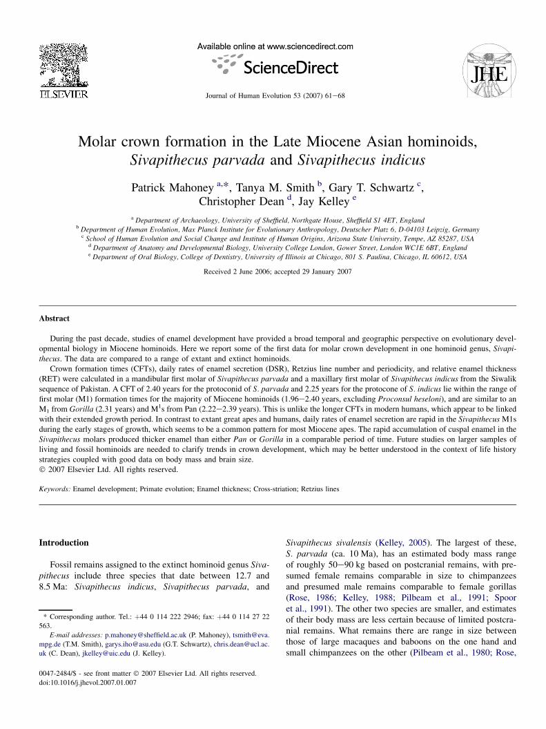
Lateral (buccal or lingual) enamel formation times werecalculated by multiplying the number of Retzius lines by theirperiodicity. Periodicity was determined by counting the num-ber of cross-striations between adjacent long-period lines.Where long-period lines were indistinct in the S. parvada mo-lar, enamel prism lengths were divided by secretion rates to es-timate formation time in the affected area (Fig. 1; Boyde,1963). In this procedure, the last clearly visible long-periodline at the surface of the enamel was followed cervically tothe enamel-dentine junction (EDJ). Prisms originating at theboundary between the EDJ and this long-period line weretraced toward the enamel surface, thereby by passing thearea of indistinct striae. The time taken to form these prismswas included in the estimate of buccal or lingual lateralenamel formation time.
Prenatal crown formation time was calculated by locatingthe position of the neonatal linedthe first accentuated growthline over the dentine horn. The thickness of the enamel be-tween this line and the dentine horn was measured and dividedby the local DSR to yield a formation time in days.
Enamel thickness
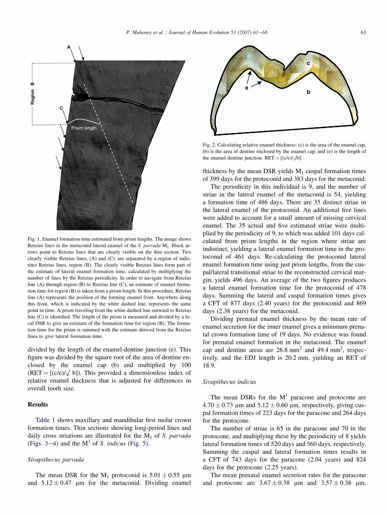
Relative enamel thickness was calculated following Martin(1985; also see Fig. 2). The area of the enamel cap (c) was
63P. Mahoney et al. / Journal of Human Evolution 53 (2007) 61e68
divided by the length of the enamel-dentine junction (e). Thisfigure was divided by the square root of the area of dentine en-closed by the enamel cap (b) and multiplied by 100(RET¼ [(c/e)/O b]). This provided a dimensionless index ofrelative enamel thickness that is adjusted for differences inoverall tooth size.
Results
Table 1 shows maxillary and mandibular first molar crownformation times. Thin sections showing long-period lines anddaily cross striations are illustrated for the M1 of S. parvada(Figs. 3e4) and the M1 of S. indicus (Fig. 5).
Sivapithecus parvada
The mean DSR for the M1 protoconid is 5.01� 0.55 mmand 5.12� 0.47 mm for the metaconid. Dividing enamel
Fig. 1. Enamel formation time estimated from prism lengths. The image shows
Retzius lines in the metaconid lateral enamel of the S. parvada M1. Black ar-
rows point to Retzius lines that are clearly visible on the thin section. Two
clearly visible Retzius lines, (A) and (C), are separated by a region of indis-
tinct Retzius lines, region (B). The clearly visible Retzius lines form part of
the estimate of lateral enamel formation time, calculated by multiplying the
number of lines by the Retzius periodicity. In order to navigate from Retzius
line (A) through region (B) to Retzius line (C), an estimate of enamel forma-
tion time for region (B) is taken from a prism length. In this procedure, Retzius
line (A) represents the position of the forming enamel front. Anywhere along
this front, which is indicated by the white dashed line, represents the same
point in time. A prism traveling from the white dashed line outward to Retzius
line (C) is identified. The length of the prism is measured and divided by a lo-
cal DSR to give an estimate of the formation time for region (B). The forma-
tion time for the prism is summed with the estimate derived from the Retzius
lines to give lateral formation time.
thickness by the mean DSR yields M1 cuspal formation timesof 399 days for the protoconid and 383 days for the metaconid.
The periodicity in this individual is 9, and the number ofstriae in the lateral enamel of the metaconid is 54, yieldinga formation time of 486 days. There are 35 distinct striae inthe lateral enamel of the protoconid. An additional five lineswere added to account for a small amount of missing cervicalenamel. The 35 actual and five estimated striae were multi-plied by the periodicity of 9, to which was added 101 days cal-culated from prism lengths in the region where striae areindistinct, yielding a lateral enamel formation time in the pro-toconid of 461 days. Re-calculating the protoconid lateralenamel formation time using just prism lengths, from the cus-pal/lateral transitional striae to the reconstructed cervical mar-gin, yields 496 days. An average of the two figures producesa lateral enamel formation time for the protoconid of 478days. Summing the lateral and cuspal formation times givesa CFT of 877 days (2.40 years) for the protoconid and 869days (2.38 years) for the metaconid.
Dividing prenatal enamel thickness by the mean rate ofenamel secretion for the inner enamel gives a minimum prena-tal crown formation time of 19 days. No evidence was foundfor prenatal enamel formation in the metaconid. The enamelcap and dentine areas are 26.8 mm2 and 49.4 mm2, respec-tively, and the EDJ length is 20.2 mm, yielding an RET of18.9.
Sivapithecus indicus
The mean DSRs for the M1 paracone and protocone are4.70� 0.73 mm and 5.12� 0.60 mm, respectively, giving cus-pal formation times of 223 days for the paracone and 264 daysfor the protocone.
The number of striae is 65 in the paracone and 70 in theprotocone, and multiplying these by the periodicity of 8 yieldslateral formation times of 520 days and 560 days, respectively.Summing the cuspal and lateral formation times results ina CFT of 743 days for the paracone (2.04 years) and 824days for the protocone (2.25 years).
The mean prenatal enamel secretion rates for the paraconeand protocone are 3.67� 0.38 mm and 3.57� 0.38 mm.
Fig. 2. Calculating relative enamel thickness: (c) is the area of the enamel cap,
(b) is the area of dentine enclosed by the enamel cap, and (e) is the length of
the enamel dentine junction. RET¼ [(c/e)/Ob].
64 P. Mahoney et al. / Journal of Human Evolution 53 (2007) 61e68
Table 1
Mesial crown formation time for two species of Sivapithecus
Sivapithecus parvada Sivapithecus indicus
M1 M1
Protoconid Metaconid Paracone Protocone
Cuspal
Daily Secretion Rates (mm/day): Inner 4.48� 0.41 4.48� 0.411 4.03� 0.37 4.73� 0.21
Middle 5.16� 0.59 5.30� 0.49 4.97� 0.54 5.44� 0.54
Outer 5.40� 0.66 5.60� 0.51 5.11� 0.75 5.20� 0.73
Mean 5.01� 0.55 5.12� 0.47 4.70� 0.73 5.12� 0.60
Cuspal Enamel Thickness (mm) 2048 2014 1100 1400
Adjustment for obliquity2 1998 1964 1050 1350
Cuspal Enamel Formation Time (days) 3993 383 223 264
Prenatal Daily Secretion Rates (mm/day) e e 3.67� 0.38 3.57� 0.38
Prenatal Enamel Thickness (mm) 86 e 135 110
Prenatal Enamel Formation Time (days) 19.14 e 37 31
Lateral
No. striae 405 54 65 70
Periodicity (days) 9 9 8 8
Formation time from striae (days) 360 486 520 560
Prism lengths (days) 101 (496)6 e e e
Imbricational formation time (days) 4787 486 520 560
Crown formation time (days) 877 869 743 824
Maximum mesial crown formation time 877 days (2.40 years) 824 days (2.25 years)
1 An inner enamel rate was not available for the metaconid. Given the similarity in DSRs between cusps of the same tooth type (Smith et al., 2007), inner DSR
for the protoconid was used for the metaconid.2 Minus an estimate of 50 mm due to the obliquity of the protconid, metaconid, paracone, and protocone dentine horn. The sections were oblique because they
were taken slightly mesial or distal to the apex of the dentine horn, thus producing slightly thicker enamel, which could lead to an overestimation of appositional
formation time.3 A count of cross striations along continuous prism tracks gave an estimate of 380 days, similar to the above estimate.4 The prenatal enamel formation time is calculated from the inner enamel DSR (4.48� 0.41) because of indistinct prenatal cross-striations, which may slightly
underestimate the actual value due to the higher rate of secretion.5 Includes estimate for missing protoconid cervical region enamel.6 The first figure was calculated from prism lengths in a region where striae were indistinct (see Materials and methods). The second figure in brackets, was
derived using only prism lengths, from the cuspal/lateral transitional striae to the reconstructed cervical margin.7 The lateral formation time for the protoconid is an average of 461 days (prism lengths and Retzius lines combined) and 496 days (prism lengths only).
Dividing prenatal enamel thickness by these secretion ratesgives a prenatal crown formation time of 37 days for the para-cone and 31 days for the protocone. The areas of the enamelcap and dentine are 19.1 mm2 and 37.9 mm2, respectively,while the length of the EDJ is 18.8 mm, yielding an RET of16.5.
Discussion
Daily rates of enamel secretion
There were no marked differences in the rates of enamelsecretion between the two Sivapithecus molars. In both, thereis a progressive increase in the secretion rate from the innerenamel to the outer, a phenomenon that characterizes extinctand extant hominoids (e.g., Beynon et al., 1991, 1998; Reidet al., 1998; Kelley et al., 2001; Schwartz et al., 2001, 2003;Dean and Schrenk, 2003; Smith et al., 2003a; Smith et al.,2007).
Secretion rates during the early stages of enamel growth inthe Sivapithecus molars are higher than in some other species
(Table 2; Fig. 6). The DSRs in the inner enamel of both Siva-pithecus species are above the known ranges for M1 in Gorilla,M2 in Pongo and humans, and overlap only the upper end ofthe range for M1 in Pan. The Sivapithecus DSRs do, however,completely or substantially overlap the ranges recorded forP. nyanzae and A. turkanensis M2s, as well as G. freybergiand G. blacki M3s. The rates of inner enamel secretion forSivapithecus seem to support the idea that some fossil homi-noids shared a common pattern of higher initial average cuspalDSRs than extant apes and humans (Beynon et al., 1998;Dean, 2000; Smith et al., 2003a).
Crown formation
Like extant hominoids, the sequence of enamel initiationdiffered between the mesial molar cusps of S. parvada. Theprotoconid is generally the first cusp to mineralize prior tobirth in humans (at e0.05 years), gorillas (at e0.23 years),and chimpanzees (at e0.13 years) (Reid et al., 1998; Schwartzet al., 2006; Smith et al., 2007). Calcification in the S. parvadaM1 commenced with the protoconid, also prenatally, suggesting
65P. Mahoney et al. / Journal of Human Evolution 53 (2007) 61e68
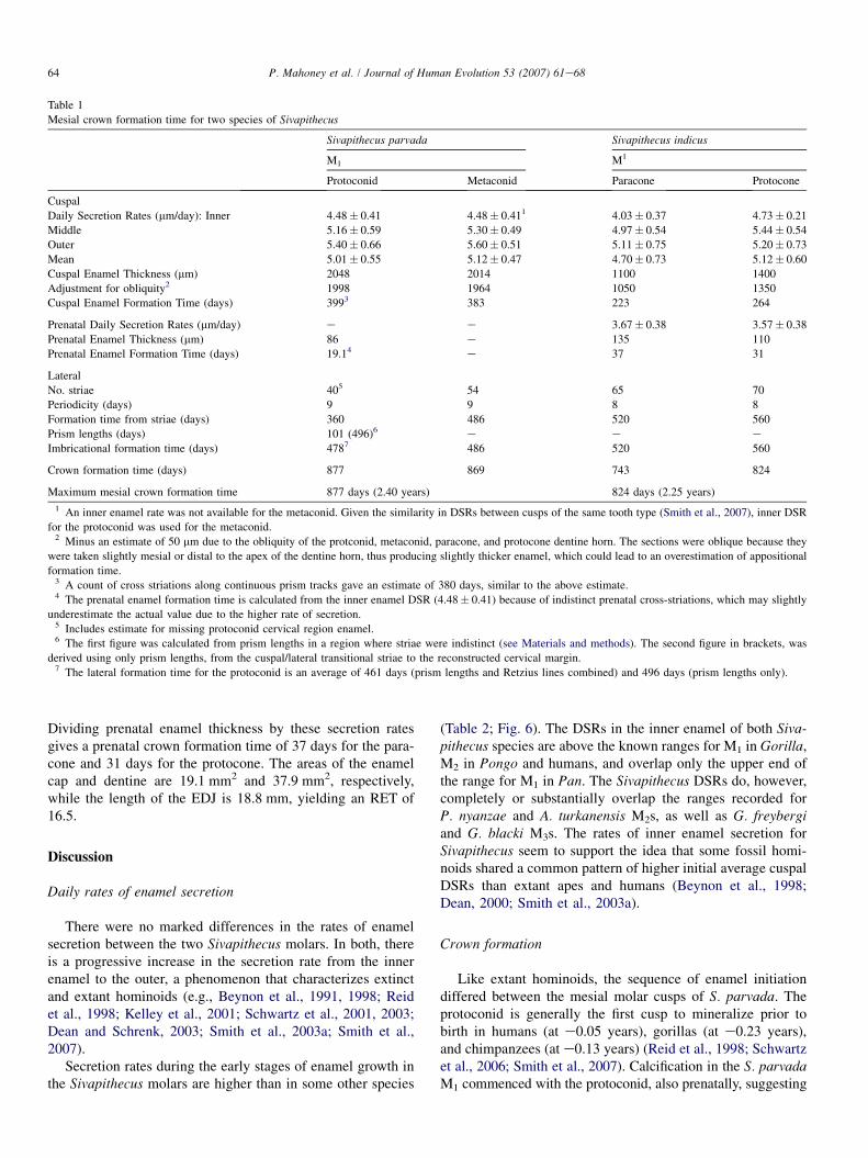
Fig. 3. Thin section of S. parvada M1. (A) Thin section through the metaconid
(left) and protoconid (right), imaged using polarized microscopy at a magnifi-
cation of �10. (B) Close-up of inset, �20. White arrows point to Retzius lines.
(C) Close-up of inset, �60. White arrows point to cross-striations.
Fig. 4. Calculating periodicity. (A) Retzius lines (white arrows) in the S. par-
vada protoconid, �40. Black arrows show the direction of enamel prisms,
which run towards the outer enamel surface. (B) Cross striations, �60. White
arrows point to cross striations appearing along enamel prisms between two
adjacent Retzius lines. Periodicity is determined by counting the number of
cross-striations between adjacent Retzius lines.
an enamel initiation sequence in the mesial cusps similar tothe extant hominoids, which is further supported by a lackof evidence of prenatal mineralization in the metaconid. Theorder of initiation of the mesial cusps of M1s can vary withinextant apes (Smith et al., 2007), which makes it difficult tocompare prenatal calcification between S. indicus and the ex-tant species.
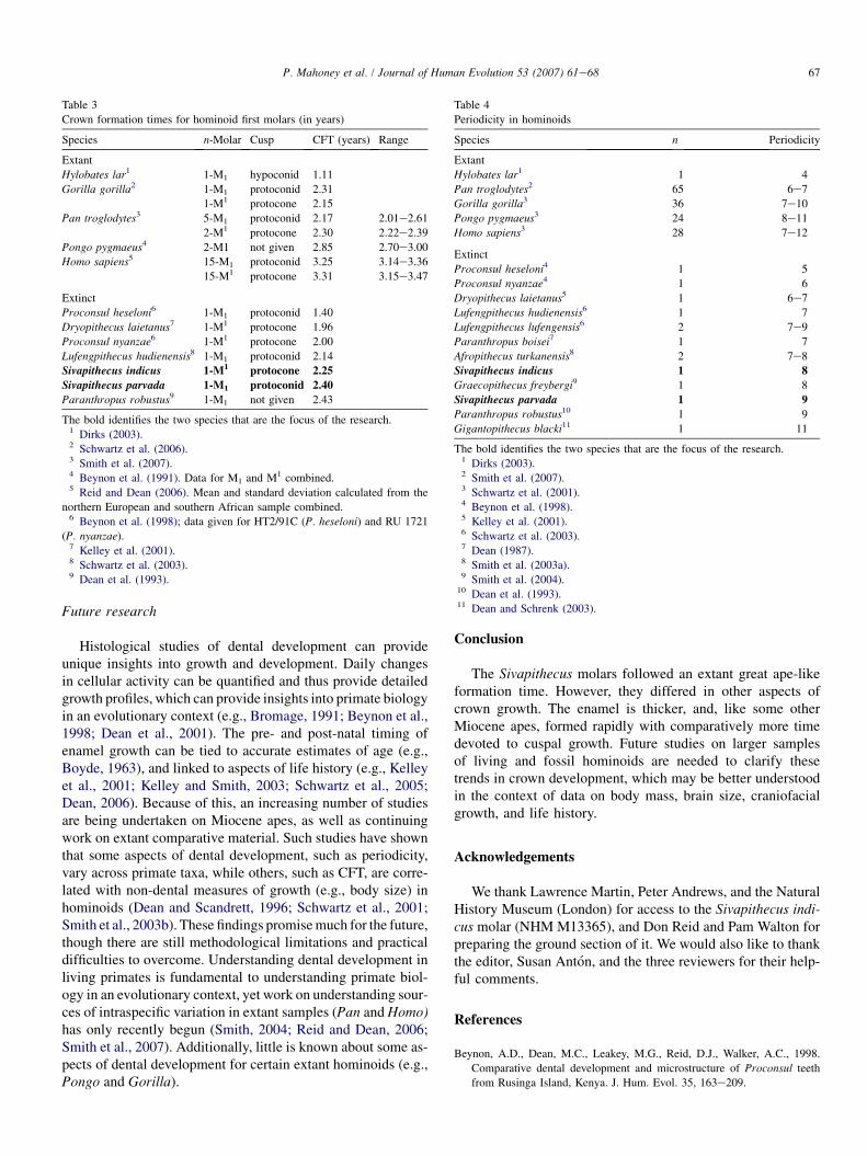
First molar CFTs for Miocene hominoids range betweenapproximately 1.96 years and 2.43 years, with only P. heselonishowing a substantially shorter formation time (Table 3). Themesial crown formation times for Sivapithecus fall within therange of CFTs for the Miocene hominoids. The values forSivapithecus also follow an extant great ape-like formationtime, similar to an M1 from Gorilla and M1s from Pan, ratherthan M1s in modern humans, where longer CFTs may beassociated with an extended growth period (Table 3; Reidand Dean, 2006).
The development of thick enamel in some of the fossil hom-inoids corresponds to a longer period of cuspal enamel growth(Smith et al., 2003a). The data for Sivapithecus supports thisidea. Cuspal formation in the thick-enameled S. parvada molar(discussed below) required 45% of the CFT; cuspal enamel onthe slightly thinner-enameled S. indicus molar formed in 32%of the cuspal formation period (Table 1). This trend contrastswith cuspal formation in first molars from extant great apes,which requires between 14% (Pongo) and 21% (Pan) of the totalcusp formation time (Smith et al., 2003a: Table 7).
Periodicity
The known range of long-period stria periodicities in extantlarge- and small-bodied hominoids is four to twelve days
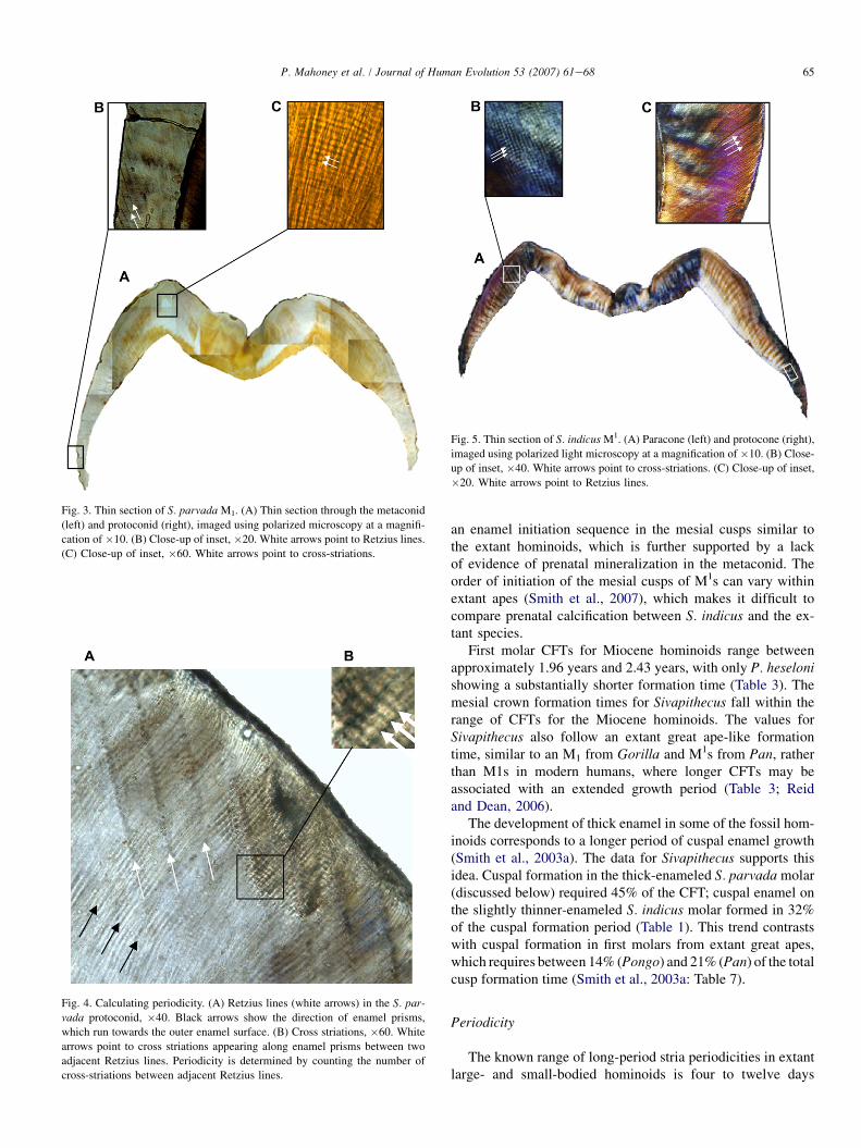
Fig. 5. Thin section of S. indicus M1. (A) Paracone (left) and protocone (right),
imaged using polarized light microscopy at a magnification of �10. (B) Close-
up of inset, �40. White arrows point to cross-striations. (C) Close-up of inset,
�20. White arrows point to Retzius lines.

66 P. Mahoney et al. / Journal of Human Evolution 53 (2007) 61e68
Table 2
Molar cuspal daily enamel secretion rates in hominoids (in microns/day)
Species n-Cusp Inner Mid Outer Mean
Extant
Homo sapiens1 1 2.66� 0.15 3.44� 0.25 5.50� 0.85 4.00� 0.42
Pongo pygmaeus1 1 3.27� 0.50 4.65� 0.45 5.20� 0.15 4.37� 0.36
Gorilla gorilla2 1 3.37� 0.49 5.37� 0.08 5.47� 0.08 4.74� 0.69
Pan troglodytes3 73 3.62� 0.42 4.24� 0.50 4.62� 0.49 4.16� 0.47
Extinct
Lufengpithecus hudienensis4 1 3.00� 0.60 5.60� 0.55 4.30� 0.57
Dryopithecus laietanus5 1 3.25� 0.50 5.05� 0.30 5.50� 0.20 4.60� 0.33
Afropithecus turkanensis6 2 3.97� 0.62 4.34� 0.71 4.85� 1.08 4.38� 0.80
Gigantopithecus blacki7 1 4.10� 0.30 4.50 6.00 4.86� 0.30
Graecopithecus freybergi8 2 4.15� 0.59 4.46� 0.44 4.30� 0.45 4.31� 0.49
Sivapithecus indicus9 2 4.38 ± 0.29 5.20 ± 0.54 5.15 ± 0.74 4.91 ± 0.66
Proconsul nyanzae10 1 4.40� 0.15 4.80� 0.20 5.40� 0.60 4.87� 0.31
Sivapithecus parvada9 2 4.48 ± 0.41 5.23 ± 0.54 5.50 ± 0.58 5.07 ± 0.52
The bold identifies the two species that are the focus of the research.1 Dean (1998: Table 1); data for M2 divided into inner, mid and outer regions, with an average and standard deviation calculated for each region. Overall mean
from Dean (1998).2 Schwartz et al. (2006).3 Smith et al. (2007). Data for M1eM3 combined.4 Schwartz et al. (2003); data for M1. Outer region value is for outer- and mid-cuspal enamel combined.5 Kelley et al. (2001); data for M1 with the mean calculated from the three regions and SD calculated from average cross-striation spacings by monthly zones.6 Smith et al. (2003a); data for M2.7 Dean and Schrenk (2003); data for M3.8 Smith et al. (2004); data for M3.9 Average secretion rate calculated from the combined rates for the protoconid and metaconid, and the paracone and protocone.
10 Beynon et al. (1998); data for M2 with standard deviations calculated by Kelley et al. (2001).
(Table 4). The 8-day periodicity in the S. indicus molar and 9-day periodicity in the S. parvada molar both lie in the middleof this range.
With the exception of Gorilla and Homo, periodicity differssignificantly among extant hominoids (Schwartz et al., 2001),and is correlated with body size in extant and fossil hominoids(Smith et al., 2003b), though this relationship is more complexin non-anthropoid primates (Schwartz et al., 2002, 2005). Thedata presented here show that another aspect of molar
2 2.5 3 3.5 4 4.5 5 5.5
S.parvada †
P.nyanzae †
S.indicus †
G.freybergi †
G.blacki †
A.turkanensis †
P.troglodytes
G.gorilla
P.pygmaeus
D.laietanus †
L.hudienensis †
H.sapiens
Daily enamel secretion (µm/day)
† Extinct taxa.
Fig. 6. Daily secretion rates in the inner enamel of selected hominoids. Mean
value (A) and range ( ). Data taken from Table 2.
development, M1 CFT, is positively correlated with periodicityin extant and fossil hominoids (Pearson’s R¼ 0.927, P< 0.001;data for M1 taken from Table 3, periodicity taken from Table 4).Given these correlations, and others (e.g., CFT and brain size;Schwartz et al., 2005), it seems likely that more comprehensiveanalyses may reveal more detailed information on the relation-ship among dental development, brain size, body size, life his-tory strategies, and craniofacial growth across primates.
Enamel thickness
The calculated values of relative enamel thickness placeS. parvada into a category classified as thick-enamel, and S.indicus into a category classified intermediate-thick enamel(Fig. 2; Martin, 1985).
Comparing just M1s, thus removing any possible variationin enamel thickness due to differences in molar type (Machoand Berner, 1993, 1994; Smith et al., 2005, 2006), Sivapithe-cus and P. nyanzae have thicker enamel than Pan or Gorilla,yet all of these species have broadly similar M1 cusp forma-tion times (Beynon et al., 1998; Smith et al., 2005). Enamelthickness is governed by the rate and duration of enamel secre-tion, and the number of active cells producing enamel (Grineand Martin, 1988). Sivapithecus and P. nyanzae have higherdaily inner enamel cuspal secretion rates than either Pan orGorilla. A higher rate of secretion during the early stages ofenamel formation might have contributed towards the thick-enameled molars of Sivapithecus and P. nyanzae growing inthe same time as thinner-enameled Pan and Gorilla.
67P. Mahoney et al. / Journal of Human Evolution 53 (2007) 61e68
Future research
Histological studies of dental development can provideunique insights into growth and development. Daily changesin cellular activity can be quantified and thus provide detailedgrowth profiles, which can provide insights into primate biologyin an evolutionary context (e.g., Bromage, 1991; Beynon et al.,1998; Dean et al., 2001). The pre- and post-natal timing ofenamel growth can be tied to accurate estimates of age (e.g.,Boyde, 1963), and linked to aspects of life history (e.g., Kelleyet al., 2001; Kelley and Smith, 2003; Schwartz et al., 2005;Dean, 2006). Because of this, an increasing number of studiesare being undertaken on Miocene apes, as well as continuingwork on extant comparative material. Such studies have shownthat some aspects of dental development, such as periodicity,vary across primate taxa, while others, such as CFT, are corre-lated with non-dental measures of growth (e.g., body size) inhominoids (Dean and Scandrett, 1996; Schwartz et al., 2001;Smith et al., 2003b). These findings promise much for the future,though there are still methodological limitations and practicaldifficulties to overcome. Understanding dental development inliving primates is fundamental to understanding primate biol-ogy in an evolutionary context, yet work on understanding sour-ces of intraspecific variation in extant samples (Pan and Homo)has only recently begun (Smith, 2004; Reid and Dean, 2006;Smith et al., 2007). Additionally, little is known about some as-pects of dental development for certain extant hominoids (e.g.,Pongo and Gorilla).
Table 3
Crown formation times for hominoid first molars (in years)
Species n-Molar Cusp CFT (years) Range
Extant
Hylobates lar1 1-M1 hypoconid 1.11
Gorilla gorilla2 1-M1 protoconid 2.31
1-M1 protocone 2.15
Pan troglodytes3 5-M1 protoconid 2.17 2.01e2.61
2-M1 protocone 2.30 2.22e2.39
Pongo pygmaeus4 2-M1 not given 2.85 2.70e3.00
Homo sapiens5 15-M1 protoconid 3.25 3.14e3.36
15-M1 protocone 3.31 3.15e3.47
Extinct
Proconsul heseloni6 1-M1 protoconid 1.40
Dryopithecus laietanus7 1-M1 protocone 1.96
Proconsul nyanzae6 1-M1 protocone 2.00
Lufengpithecus hudienensis8 1-M1 protoconid 2.14
Sivapithecus indicus 1-M1 protocone 2.25
Sivapithecus parvada 1-M1 protoconid 2.40
Paranthropus robustus9 1-M1 not given 2.43
The bold identifies the two species that are the focus of the research.1 Dirks (2003).2 Schwartz et al. (2006).3 Smith et al. (2007).4 Beynon et al. (1991). Data for M1 and M1 combined.5 Reid and Dean (2006). Mean and standard deviation calculated from the
northern European and southern African sample combined.6 Beynon et al. (1998); data given for HT2/91C (P. heseloni) and RU 1721
(P. nyanzae).7 Kelley et al. (2001).8 Schwartz et al. (2003).9 Dean et al. (1993).
Conclusion
The Sivapithecus molars followed an extant great ape-likeformation time. However, they differed in other aspects ofcrown growth. The enamel is thicker, and, like some otherMiocene apes, formed rapidly with comparatively more timedevoted to cuspal growth. Future studies on larger samplesof living and fossil hominoids are needed to clarify thesetrends in crown development, which may be better understoodin the context of data on body mass, brain size, craniofacialgrowth, and life history.
Acknowledgements
We thank Lawrence Martin, Peter Andrews, and the NaturalHistory Museum (London) for access to the Sivapithecus indi-cus molar (NHM M13365), and Don Reid and Pam Walton forpreparing the ground section of it. We would also like to thankthe editor, Susan Anton, and the three reviewers for their help-ful comments.
References
Beynon, A.D., Dean, M.C., Leakey, M.G., Reid, D.J., Walker, A.C., 1998.
Comparative dental development and microstructure of Proconsul teeth
from Rusinga Island, Kenya. J. Hum. Evol. 35, 163e209.
Table 4
Periodicity in hominoids
Species n Periodicity
Extant
Hylobates lar1 1 4
Pan troglodytes2 65 6e7
Gorilla gorilla3 36 7e10
Pongo pygmaeus3 24 8e11
Homo sapiens3 28 7e12
Extinct
Proconsul heseloni4 1 5
Proconsul nyanzae4 1 6
Dryopithecus laietanus5 1 6e7
Lufengpithecus hudienensis6 1 7
Lufengpithecus lufengensis6 2 7e9
Paranthropus boisei7 1 7
Afropithecus turkanensis8 2 7e8
Sivapithecus indicus 1 8
Graecopithecus freybergi9 1 8
Sivapithecus parvada 1 9
Paranthropus robustus10 1 9
Gigantopithecus blacki11 1 11
The bold identifies the two species that are the focus of the research.1 Dirks (2003).2 Smith et al. (2007).3 Schwartz et al. (2001).4 Beynon et al. (1998).5 Kelley et al. (2001).6 Schwartz et al. (2003).7 Dean (1987).8 Smith et al. (2003a).9 Smith et al. (2004).
10 Dean et al. (1993).11 Dean and Schrenk (2003).

68 P. Mahoney et al. / Journal of Human Evolution 53 (2007) 61e68
Beynon, A.D., Dean, M.C., Reid, D.J., 1991. Histological study on the chro-
nology of the developing dentition in gorilla and orangutan. Am. J.
Phys. Anthropol. 86, 189e203.
Boyde, A., 1963. Estimation of age at death of young human skeletal remains
from incremental lines in dental enamel. Third International Meeting in
Forensic Immunology, Medicine, Pathology, and Toxicology, Plenary Ses-
sion 11A, London.
Bromage, T., 1991. Enamel incremental periodicity in the pig-tailed macaque:
a polychrome fluorescent labeling study of dental hard tissues. Am. J.
Phys. Anthropol. 86, 205e214.
Dean, M.C., 1987. Growth layers and incremental markings in hard tissues:
a review of the literature and some preliminary observations about enamel
structure in Paranthropus boisei. J. Hum. Evol. 16, 157e172.
Dean, M.C., 1998. A comparative study of cross striation spacings in cuspal
enamel and of four methods of estimating the time taken to grow molar
cuspal enamel in Pan, Pongo and Homo. J. Hum. Evol. 35, 449e462.
Dean, M.C., 2000. Progress in understanding hominoid dental development.
J. Anat. 197, 77e101.
Dean, M.C., 2006. Tooth microstructure tracks the pace of human life-history
evolution. Proc. R. Soc. Ser. B, Biol. Sci. 273, 2799e2808.
Dean, M.C., Beynon, A.D., Thackeray, J.F., Macho, G.A., 1993. Histological
reconstruction of dental development and age at death of a juvenile Para-
nthropus robustus specimen, SK 63, from Swartkrans, South Africa. Am.
J. Phys. Anthropol. 91, 401e419.
Dean, M.C., Leakey, M.G., Reid, D.J., Schrenk, F., Schwartz, G.T., Stringer, C.,
Walker, A.C., 2001. Growth processes in teeth distinguish modern humans
from Homo erectus and earlier hominins. Nature 414, 628e631.
Dean, M.C., Scandrett, A.E., 1996. The relation between long-period incre-
mental markings in dentine and daily cross-striations in enamel in human
teeth. Arch. Oral. Biol. 41, 233e241.
Dean, M.C., Schrenk, F., 2003. Enamel thickness and development in
a third permanent molar of Gigantopithecus blacki. J. Hum. Evol. 45,
381e387.
Dirks, W., 2003. Effect of diet on dental development in four species of catar-
rhine primates. Am. J. Primatol. 61, 29e40.
FitzGerald, C.M., 1998. Do enamel microstructures have regular time depen-
dency? Conclusions from the literature and a large scale study. J. Hum.
Evol. 35, 371e386.
Grine, F.E., Martin, L.B., 1988. Enamel thickness and development in Aus-
tralopithecus and Paranthropus. In: Grine, F.E. (Ed.), Evolutionary His-
tory of the ‘‘Robust’’ Australopithecines. Aldine de Gruyter, New York,
pp. 3e42.
Kelley, J., 1988. A new large species of Sivapithecus from the Siwaliks of
Pakistan. J. Hum. Evol. 17, 305e324.
Kelley, J., 1997. Paleobiological and phylogenetic significance of life history
in Miocene Hominoids. In: Begun, D.R., Ward, C.V., Rose, M.D. (Eds.),
Function, Phylogeny and Fossils. Miocene Hominoid Evolution and Adap-
tations. Plenum Press, New York, pp. 173e208.
Kelley, J., 2002. The hominoid radiation in Asia. In: Hartwig, W.C. (Ed.), The
Primate Fossil Record. Cambridge University Press, Cambridge, England,
pp. 369e384.
Kelley, J., 2005. Twenty-five years contemplating Sivapithecus taxonomy.
In: Lieberman, D.L., Smith, R.J., Kelley, J. (Eds.), Interpreting the
Past: Essays on Human, Primate and Mammal Evolution in Honor of
David Pilbeam. Brill Academic Publishers, Boston, pp. 123e143.
Kelley, J., Dean, M.C., Reid, D.J., 2001. Molar growth in the late Miocene
hominoid, Dryopithecus laietanus. In: Mayhall, J.T., Heikkinen, T.
(Eds.), Dental Morphology 1998. Proceedings of the Eleventh Interna-
tional Symposium on Dental Morphology, Oulu, Finland. Oulu University
Press, Oulu, pp. 123e134.
Kelley, J., Smith, T.M., 2003. Age at first molar emergence in early Miocene
Afropithecus turkanensis and life-history evolution in the Hominoidea.
J. Hum. Evol. 44, 307e329.
Macho, G.A., Berner, M.E., 1993. Enamel thickness of human maxillary
molars reconsidered. Am. J. Phys. Anthropol. 92, 189e200.
Macho, G.A., Berner, M.E., 1994. Enamel thickness and the helicoidal occlu-
sal plane. Am. J. Phys. Anthropol. 94, 327e337.
Madar, S.I., Rose, M.D., Kelley, J., Maclatchy, L., Pilbeam, D., 2002. New
Sivapithecus postcranial specimens from the Siwaliks of Pakistan. J.
Hum. Evol. 43, 771e772.
Martin, L.B., 1983. The relationships of the later Miocene Hominoidea. Ph.D.
Dissertation, University College London.
Martin, L., 1985. Significance of enamel thickness in hominoid evolution.
Nature 314, 260e263.
Pilbeam, D., Rose, M.D., Badgley, C., Lipschutz, B., 1980. Miocene homi-
noids from Pakistan. Postilla 181, 1e94.
Pilbeam, D.R., Rose, M.D., Barry, J.C., Shah, S.M.I., 1991. New Sivapithecus
humeri from Pakistan and the relationship of Sivapithecus and Pongo.
Nature 348, 237e239.
Ramirez Rozzi, F.V., 1994. Enamel growth markers in hominid dentition. Mic.
Anal. July, 21e23.
Reid, D.J., Beynon, A.D., Ramirez Rozzi, F.V., 1998. Histological reconstruc-
tion of dental development in four individuals from a medieval site in
Picardie, France. J. Hum. Evol. 35, 463e477.
Reid, D.J., Dean, M.C., 2006. Variation in modern human enamel formation
times. J. Hum. Evol. 50, 329e346.
Rose, M.D., 1984. Hominoid postcranial specimens from the Middle Miocene
Chinji Formation, Pakistan. J. Hum. Evol. 13, 503e516.
Rose, M.D., 1986. Further hominoid postcranial specimens from the Late Mio-
cene Nagri formation of Pakistan. J. Hum. Evol. 15, 333e367.
Schwartz, G.T., Liu, W., Zheng, L., 2003. Preliminary investigation of dental
microstructure in the Yuanmou hominoid (Lufengpithecus hudienensis),
Yunnan Province, China. J. Hum. Evol. 44, 189e202.
Schwartz, G.T., Mahoney, P., Godfrey, L.R., Cuozzo, F.P., Jungers, W.L.,
Randria, G.F.N., 2005. Dental development in Megaladapis edwardsi (Pri-
mates, Lemuriformes): implications for understanding life history variation
in sub-fossil lemurs. J. Hum. Evol. 49, 702e721.
Schwartz, G.T., Reid, D., Dean, C., 2001. Developmental aspects of sexual
dimorphism in hominoid canines. Int. J. Primatol. 22, 837e860.
Schwartz, G.T., Reid, D.J., Dean, C.M., Zihlman, A.L., 2006. A faithful record
of stressful life events preserved in the dental developmental record of
a juvenile gorilla. Int. J. Primatol. 27, 1201e1222.
Schwartz, G.T., Samonds, K.E., Godfrey, L.R., Jungers, W.L., Simons, E.L.,
2002. Dental microstructure and life history in subfossil Malagasy lemurs.
Proc. Natl. Acad. Sci. U.S.A. 99, 6124e6129.
Smith, T.M., 2004. Incremental development of primate dental enamel. Ph.D.
Dissertation, Stony Brook University.
Smith, T.M., Dean, M.C., Kelley, J., Martin, L.B., Reid, D.J., Schwartz, G.T.,
2003b. Molar crown formation in Miocene hominoids: a preliminary syn-
thesis. Am. J. Phys. Anthropol. Suppl. 36, 196.
Smith, T.M., Martin, L., Leakey, M.G., 2003a. Enamel thickness and micro-
structure in Afropithecus turkanensis. J. Hum. Evol. 44, 283e306.
Smith, T.M., Martin, L.B., Reid, D.J., de Bonis, L., Koufos, G.D., 2004.
An examination of dental development in Graecopithecus freybergi
(¼Ouranopithecus macedoniensis). J. Hum. Evol. 46, 551e577.
Smith, T.M., Olejniczak, A.J., Martin, L.B., Reid, D.J., 2005. Variation in
hominoid molar enamel thickness. J. Hum. Evol. 48, 575e592.
Smith, T.M., Olejniczak, A.J., Reid, D.J., Ferrell, R.J., Hublin, J.J., 2006.
Modern human molar enamel thickness and enamel-dentine junction
shape. Arch. Oral Biol. 51, 974e995.
Smith, T.M., Reid, D.J., Olejniczak, A.J., Martin, L.B., 2007. Molar crown
development in common chimpanzees (Pan troglodytes). J. Hum. Evol.
52, 201e216.
Spoor, C.F., Sondaar, P.Y., Hussain, S.T., 1991. A new hominoid hamate
and first metacarpal from the late Miocene Nagri Formation of Pakistan.
J. Hum. Evol. 21, 413e424.