modality-specific perceptual expectations selectively modulate baseline activity in auditory,...
TRANSCRIPT

Cerebral Cortex
doi:10.1093/cercor/bhr083
Modality-Specific Perceptual Expectations Selectively Modulate Baseline Activityin Auditory, Somatosensory, and Visual Cortices
Robert Langner1,2,3, Thilo Kellermann1,4, Frank Boers3, Walter Sturm2, Klaus Willmes2 and Simon B. Eickhoff1,3,4
1Department of Psychiatry, Psychotherapy and Psychosomatics, and 2Neuropsychology Section, Department of Neurology, Medical
School, RWTH Aachen University, 52074 Aachen, Germany, 3Institute of Neuroscience and Medicine (INM-2, INM-4), Research
Centre Julich, 52425 Julich, Germany and 4JARA - Translational Brain Medicine, Germany
Address correspondence to Dr Robert Langner, Klinik fuer Psychiatrie, Psychotherapie und Psychosomatik, Universitaetsklinikum Aachen,
Pauwelsstrasse 30, 52074 Aachen, Germany. Email: [email protected].
Valid expectations are known to improve target detection, but thepreparatory attentional mechanisms underlying this perceptualfacilitation remain an open issue. Using functional magneticresonance imaging, we show here that expecting auditory, tactile,or visual targets, in the absence of stimulation, selectivelyincreased baseline activity in corresponding sensory cortices anddecreased activity in irrelevant ones. Regardless of sensorymodality, expectancy activated bilateral premotor and posteriorparietal areas, supplementary motor area as well as right anteriorinsula and right middle frontal gyrus. The bilateral putamenwas sensitive to the modality specificity of expectations duringthe unexpected omission of targets. Thus, across modalities,detection improvement arising from selectively directing attentionto a sensory modality appears mediated through transient changesin pretarget activity. This flexible advance modulation of baselineactivity in sensory cortices resolves ambiguities among previousstudies unable to discriminate modality-specific preparatory activityfrom attentional modulation of stimulus processing. Our resultsagree with predictive-coding models, which suggest that theseexpectancy-related changes reflect top-down biases—presumablyoriginating from the observed supramodal frontoparietal net-work—that modulate signal-detection sensitivity by differentiallymodifying background activity (i.e., noise level) in different inputchannels. The putamen appears to code omission-related Bayesian‘‘surprise’’ that depends on the specificity of predictions.
Keywords: fMRI, intermodal attention, multimodal stimulus anticipation,predictive coding, surprise
Introduction
Valid expectations improve performance in speeded reaction
tasks, presumably by top-down modulation of perceptual,
central, and motor processes starting before the target event
occurs (Brunia and van Boxtel 2001; Hackley 2009; Rolke and
Ulrich 2010). The preparatory facilitation of perceptual
processing (Correa et al. 2005; Bausenhart et al. 2007) is
hypothesized to result from supramodal attentional control
signals that establish a bias in the competition between
concurrent sensory inputs in favor of expected information
(Desimone and Duncan 1995), either voluntarily (endoge-
nously, ‘‘top-down’’) or automatically (exogenously, ‘‘bottom-
up’’) (Beck and Kastner 2009). Apart from well-known
beneficial effects of location-, time-, or feature-based atten-
tional biases, performance gains have also been shown to arise
from advance information on the relevant sensory channel
(e.g., Spence and Driver 1997). Here, we investigated neural
correlates of voluntary attentional biases toward the sensory
modality of an expected target in the absence of stimulation.
Modality-selective attentional modulations of sensory pro-
cessing have previously been investigated in tasks requiring the
selection of one sensory modality versus another during
multimodal stimulation (i.e., intersensory selective attention).
For instance, electrophysiological recordings in monkeys
revealed selective modulation of activity in areas V1, V2, and
V4 while attending to visual stimuli and ignoring auditory
ones (Mehta et al. 2000). In a pioneering positron emission
tomography (PET) study in humans, Roland (1982) showed
that during simultaneous auditory, tactile, and visual stimula-
tion, attention to one modality selectively enhanced activity
in sensory areas that process stimuli of the to-be-attended
modality, with the exception of somatosensory cortex. Later
neuroimaging studies, using bimodal visuoauditory or visuo-
tactile stimulation, corroborated these early findings but
also found attentional modulations in somatosensory areas
(Kawashima et al. 1999; Macaluso et al. 2002; Shomstein and
Yantis 2004). Analogously, studies using electroencephalo-
graphy (EEG) reported modality-selective attentional modula-
tion of sustained steady-state responses (Saupe et al. 2009) and
of early perceptual components of electrocortical activity
(Eimer and Schroger 1998; Teder-Salejarvi et al. 1999; Foxe and
Simpson 2005; Karns and Knight 2009).
Attention-induced modulations of brain activity during
stimulus processing, however, only provide indirect evidence
for the neural basis of attentional biases in perception. In
particular, these modulations (i.e., the difference between
attended and nonattended stimulus processing) may be driven
by unknown overadditive or underadditive interactions be-
tween attention- and stimulation-related signals, making it hard
to isolate purely attentional effects. More direct evidence for
neural correlates of attentional biasing comes from studies
investigating attention-induced increases in baseline activity in
the absence of actual sensory stimulation (cf. Beck and Kastner
2009). For instance, single-cell recordings in monkeys during
visual attention revealed significantly increased spontaneous
(baseline) firing rates for neurons in ventral-stream areas V2v
and V4 when the animal covertly attended to a location within
the neuron’s receptive field before stimulus presentation
(Luck et al. 1997). A similar effect was shown in the lateral
intraparietal sulcus (IPS) as part of the dorsal visual stream
(Colby et al. 1996). Moreover, several studies using functional
magnetic resonance imaging (fMRI) in humans showed that
within-modality allocation of attention to the location or
a specific feature of a visual stimulus induces increased baseline
activity in relevant areas of visual cortex (Chawla et al. 1999;
� The Author 2011. Published by Oxford University Press. All rights reserved.
For permissions, please e-mail: [email protected]
Cerebral Cortex Advance Access published April 28, 2011 at N
ational University of Ireland, G
alway on A
pril 29, 2011cercor.oxfordjournals.org
Dow
nloaded from
Kastner et al. 1999; Giesbrecht et al. 2006; Sylvester et al. 2007;
Smith et al. 2010). Analogous results have been reported for
auditory spatial attention (Wu et al. 2007; Smith et al. 2010).
Findings from the tactile domain are equivocal: an early PET
study on neural correlates of anticipating touch only found
reduced blood flow in somatosensory areas representing
nontarget body zones (Drevets et al. 1995), whereas a later
fMRI study reported both increased activity in task-relevant
somatosensory areas and decreased activity in task-irrelevant
ones (Carlsson et al. 2000).
In contrast to preparatory within-modality attention, there
is a surprising dearth of evidence for the neural correlates
of preparatory between-modality attention, that is, directing
attention to a specific sensory channel in the absence of
stimulation. Using EEG, Brunia and van Boxtel (2004) reported
selectively enhanced preparatory activity (as indicated by an
increased slow cortical negativity prior to stimulus onset) over
frontal or occipital areas when anticipating auditory or visual
feedback stimuli, respectively. Another EEG study (Foxe et al.
2005), using trial-by-trial cueing of the upcoming target
modality, demonstrated a larger negativity over left fronto-
central regions when expecting auditory targets and a larger
positivity over right parietooccipital regions when expecting
visual targets. In a pioneering fMRI study on the endogenous
orienting of visual and tactile spatial attention, Macaluso et al.
(2003) found spatially selective but no modality-selective
transient preparatory increases in baseline activity. According
to the authors’ reasoning, the blocked presentation of stimuli of
a given modality might have prevented the establishment of
transient modality-specific increases in baseline activity over
persistent sustained modulations. A more recent event-related
fMRI study using auditory and visual targets (Mozolic et al.
2008) reported deactivations in irrelevant sensory cortices but
did not find increased baseline activity in relevant cortices
when attention was directed to either modality. This lack of
baseline increases is at odds with the above-mentioned baseline
increases during within-modality expectations in human and
nonhuman primates as well as with the results of human EEG
studies on between-modality preparatory attention.
In light of these sparse and inconsistent findings on the
neural mechanisms underlying modality-specific perceptual
expectations, we aimed to answer the following questions:
1. Does modality-selective preparatory attention transiently
increase baseline activity in task-relevant sensory cortices,
decrease activity in task-irrelevant ones, or both? And, is
there a consistent pattern to be found across different
modalities?
2. Which areas are activated supramodally (i.e., across modal-
ities) during modality-specific expectations, potentially
controlling top-down attention and motor preparation?
3. Are differential neural ‘‘surprise’’ responses induced by the
absence of stimulation under specific versus nonspecific
expectancy conditions? If so, are they represented in the
basal ganglia (cf. den Ouden et al. 2009)?
These questions were tackled using event-related fMRI
to measure changes in baseline activity evoked by cue-induced
expectations of auditory, vibrotactile, or visual targets in
a speeded detection task. To isolate pure expectancy effects,
analysis of brain activity was restricted to those trials
where the modality cue was not followed by any target
(‘‘cue-only trials’’).
Materials and Methods
ParticipantsTwenty-four (10 females) healthy right-handed volunteers (mean age =24.1 years, standard deviation = 3.5) were recruited via advertisements
and paid for their participation in the experiment. The study was
approved by the ethics committee of the RWTH Aachen University
Hospital, and all participants gave written informed consent before
entering the study.
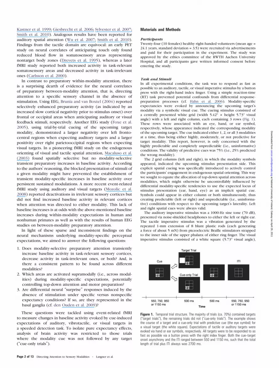
Task and StimuliIn all experimental conditions, the task was to respond as fast as
possible to an auditory, tactile, or visual imperative stimulus by a button
press with the right-hand index finger. Using a simple reaction-time
(RT) task prevented potential confounds from differential response-
preparation processes (cf. Hahn et al. 2006). Modality-specific
expectancies were evoked by announcing the upcoming target’s
modality via a symbolic visual cue. The cueing stimulus consisted of
a centrally presented white grid (width 5.42� 3 height 5.73� visual
angle) with a left and right column, each containing 3 rows (Fig. 1).
These rows were associated with an eye, hand, or ear symbol,
respectively, whose appearance indicated the corresponding modality
of the upcoming target. The cue indicated either 1, 2, or all 3 modalities
at a time, thus being either highly, moderately, or not predictive for
target modality. This report, however, is only concerned with the
highly predictable and completely unpredictable (i.e., uninformative)
conditions. The validity of predictive cues was 75% (i.e., 25% predicted
the wrong modality).
The 2 grid columns (left and right), in which the modality symbols
appeared, indicated the upcoming stimulus presentation side. This
explicit spatial cueing was specifically introduced to actively control
the participants’ engagement in endogenous spatial orienting. This way
we sought to equate the allocation of top-down spatial attention across
modalities, which might otherwise be uncontrollably influenced by
differential modality-specific tendencies to use the expected locus of
stimulus presentation (ear, hand, eye) as an implicit spatial cue.
Symbols could appear in either column or both simultaneously, thus
creating predictable (left or right) and unpredictable (i.e., uninforma-
tive) conditions with respect to the upcoming target’s laterality. Left
and right spatial cues were always valid.
The auditory imperative stimulus was a 1000-Hz sine tone (70 dB),
presented via noise-shielded headphones to either the left or right ear.
The tactile imperative stimulus was a vibration generated by the
repeated 1-mm extension of 8 blunt plastic rods (each generating
a force of about 5 mN) from piezoelectric Braille stimulators strapped
to the inner side of the upper phalanx of either ring finger. The visual
imperative stimulus consisted of a white square (5.73� visual angle),
Figure 1. Temporal trial structure. The majority of trials (ca. 70%) contained targets(‘‘target trials’’), the remaining trials did not (‘‘cue-only trials’’). The example showsthe course of a target and a cue-only trial with predictive cue (the eye symbol) fora visual target (the white square). Expectations of tactile or auditory targets wereevoked via hand or ear symbols, respectively. All targets were to be responded to asfast as possible via a button press with the right index finger. Both the cue--targetonset asynchrony and the ITI ranged between 550 and 1150 ms, such that the totallength of trial plus ITI always was 2700 ms.
Page 2 of 13 Directing Attention to Sensory Modalities d Langner et al.
at National U
niversity of Ireland, Galw
ay on April 29, 2011
cercor.oxfordjournals.orgD
ownloaded from

presented via MRI-compatible goggles to the left or right of the central
cueing stimulus (distance between the cue’s and either square’s center:
7.63� visual angle). Since by their nature, vibrotactile stimuli can only be
presented in a cyclic on/off fashion, all imperative stimuli were
presented for 500 ms with a frequency of 10 Hz (five 50-ms-on/50-ms-
off cycles each) to maximize comparability among modalities.
Procedure and DesignThe experiment was run on a standard PC using Presentation 10.0
(Neurobehavioral Systems, Inc.). The structure and timing of a trial is
shown in Figure 1. Target onset followed cue onset with a variable,
equiprobable delay of 550, 750, 950, or 1150 ms. This temporal
unpredictability required participants to continuously pay attention to
stimulus modality and location while anticipating the target. The cue
remained present during the imperative stimulus and for another 500
ms afterward. It disappeared, with only the empty grid remaining,
during the subsequent intertrial interval (ITI), which varied in length
from 550 to 1150 ms such that the total trial-plus-ITI duration always
was 2700 ms.
The task was presented in 7 separate 4.7-min runs, each containing
72 experimental trials and 36 null-event trials (i.e., baseline periods
with only the grid present). Of the 72 experimental trials per run, 21
(i.e., about 30%) were cue-only trials, in which no target was presented
after the cue. In each run, the modality cue was highly predictive (i.e.,
unimodal) on 33 trials, moderately predictive (i.e., bimodal) on 30 trials,
and uninformative (i.e., trimodal) on 9 trials. Within the first 2
categories, the 3 modalities were cued equally often. Thus, in each
run, there were 3 cue-only trials per modality with highly predictive
cues and 3 cue-only trials with uninformative cues. On invalid or
uninformative target trials, target modality was distributed equally. Left,
right, and uninformative spatial cues were equally distributed among all
experimental trials. The order of trial types was pseudorandomized
within each run.
Participants were instructed to pay attention to the cue and use it to
prepare themselves, even when the cue sometimes was misleading
(i.e., in trials with invalidly cued modality) or not followed by a target
at all (i.e., in cue-only trials). After instruction, participants were
familiarized with the task during a practice block.
fMRI Data AcquisitionBrain imaging data were obtained with a 3-T MRI scanner (Philips
Achieva, Philips Medical Systems) with a SENSE head coil. Participants
lay supine in the scanner, their heads immobilized with cushions to
minimize movements. Blood oxygenation level--dependent signals were
acquired using echo-planar imaging (EPI) covering the whole brain in
28 transverse slices parallel to the AC/PC line (echo time = 32 ms,
repetition time = 2.0 s, flip angle = 80�, SENSE factor = 1.3, matrix size =64 3 74, field of view = 192 3 228 mm2, voxel size = 3 3 3 3 3.6 mm3,
0.8-mm gap between slices, interleaved slice acquisition). During each
run, 133 volumes were acquired, preceded by 7 dummy scans.
fMRI Data AnalysisData were analyzed with SPM5 (Wellcome Department of Imaging
Neuroscience) implemented in Matlab 7.2 (The MathWorks, Inc.). After
discarding the dummy scans, EPI images were corrected for head
movement by affine registration using a 2-pass procedure by which
images were initially realigned to the first image and subsequently to
the mean of the realigned images. Spatial normalization into standard
stereotaxic Montreal Neurological Institute (MNI) space was achieved
by applying the ‘‘unified segmentation’’ procedure (Ashburner and
Friston 2005) to each participant’s mean EPI image. This approach
combines the segmentation of the mean EPI image of each participant
with a nonlinear spatial normalization into the space of the priors used
for this segmentation (i.e., the MNI tissue probability maps). The
resulting parameters of a discrete cosine transformation, which
define the deformation field necessary to move the participant’s data
into the space of the MNI tissue probability maps, were combined with
a second deformation field that describes the optimal transformation
between the MNI tissue probability maps and the MNI single-subject
template (Holmes et al. 1998). The ensuing combined deformation was
subsequently applied to all individual EPI volumes, which were hereby
transformed into the MNI single-subject space and resampled at 2 3 2 3
2-mm3 voxel size. Normalized images were spatially smoothed with
a Gaussian filter of 8mm full-width at half-maximum to accommodate
assumptions of random-field theory as well as residual interindividual
variation.
The expected hemodynamic response for each trial was modeled by
convolving trial onsets and durations with a canonical hemodynamic
response function (HRF; Friston et al. 1998) and its first-order temporal
derivative to create predictors in a general linear model. Trials were
averaged across all cue--target delays and spatial-cue types. The analysis
included the following 10 regressors of interest: 3 regressors
for auditory, tactile, and visual cue-only trials; the same for validly
cued target trials with highly predictive (unimodal) modality cues; the
same for target trials with uninformative (trimodal) modality cues; one
regressor for cue-only trials with uninformative modality cues.
Additionally, we included nuisance regressors for the remaining
conditions (trials with invalid or bimodal modality cues) and for 6
head-motion parameters (translation and rotation movements). Low-
frequency signal drifts were filtered using a cutoff period of 128 s. After
correction of the time series for dependent observations according to
an autoregressive first-order correlation structure, parameter estimates
of the HRF regressors were calculated from the least-mean-squares fit
of the model to the time series.
Group analyses were done by entering parameter estimates of the
regressors of interest into a random-effects repeated-measures analysis
of variance (ANOVA), allowing for unequal variances among conditions
and participants, as implemented in SPM5. By restricting the analysis of
expectancy effects to cue-only trials, we excluded trivial differences
based on processing different sensory input as well as confounding
effects of stimulus-driven orienting and attentional modulations of
target processing. Additionally, we report supplementary analyses that
included both cue-plus-target and cue-only trials. Activity differences
were considered significant when surviving a single-voxel threshold of
P < 0.001 and a cluster-level threshold of P < 0.05, familywise error
(FWE) corrected for multiple comparisons across the whole brain
(Worsley et al. 1996).
Results
Behavioral Data
To examine the effectiveness of our expectancy manipulation,
we analyzed the behavioral effects of cueing stimulus modality
in target trials (i.e., in those trials in which the cue was
actually followed by a target). Because of the saliency of the
imperative stimuli, detection performance was at ceiling:
errors of omission and false alarms were very rare (0.42% and
0.27%, respectively, on average) and not further analyzed.
Cueing effects on individual median RT were tested by a 3 3 3
repeated-measures ANOVA with factors target modality (audi-
tory, tactile, visual) and cue validity (valid, uninformative,
invalid). Whenever necessary, the Greenhouse--Geisser correc-
tion was employed to compensate for violations of sphericity.
Group-averaged RT data for each condition are shown in
Figure 2.
The ANOVA on RT revealed a main effect for both target
modality (F2,46 = 20.66, P < 0.001, gp2 = 0.47) and cue validity
(F2,46 = 58.47, P < 0.001, gp2 = 0.72). Simple contrasts revealed
that responses to tactile stimuli were significantly faster
than responses to auditory (F1,23 = 19.65, P < 0.001, gp2 =
0.46) or visual (F1,23 = 30.94, P < 0.001, gp2 = 0.57) stimuli,
with the latter 2 not being significantly different from each
other (F1,23 = 1.70, P > 0.2). The analysis further showed that,
across modalities, responses to validly cued targets were faster
than responses to uninformatively cued ones (F1,23 = 38.03,
Cerebral Cortex Page 3 of 13
at National U
niversity of Ireland, Galw
ay on April 29, 2011
cercor.oxfordjournals.orgD
ownloaded from

P < 0.001, gp2 = 0.62), which were, in turn, faster than
responses to invalidly cued targets (F1,23 = 36.77, P < 0.001, gp2
= 0.62). There was also a significant target-modality 3 cue-
validity interaction (F4,92 = 3.84, P = 0.014, gp2 = 0.14), which
was driven by a stronger validity effect in the visual modality, as
compared with the tactile or auditory one. Nevertheless, since
this interaction was ordinal, that is, cue validity affected all 3
modalities in the same direction (and vice versa), the validity
main effect can be interpreted globally. Thus, across modalities,
performance depended on the validity of the cue in a similar
manner, which corroborates the effectiveness of our modality-
cueing manipulation.
Imaging Data
Modality-Selective Baseline Increases
Activation driven by top-down attention to a given sensory
modality was analyzed by calculating balanced contrasts
between cue-only trials for one modality and those for the
remaining 2. The outcome was restricted to true task-related
(positive) activations by means of a conjunction analysis across
the difference of interest and its minuend (i.e., the main effect
of the condition of interest), the latter being assessed relative
to resting baseline as provided by the randomly interspersed
null-event trials. That is, we, for example, assessed where
auditory attention significantly modulated baseline activity and
did so more strongly than visual or tactile attention. The
conjunction approach (based on the minimum t-statistic;
Nichols et al. 2005) was preferred over an inclusive masking
procedure, since the former constitutes a more rigorous test. In
particular, only a conjunction combines the t-maps of all
contrasts involved to make a joint statistical inference. The
results of these analyses, including anatomical localization
based on cytoarchitectonic probability maps (Eickhoff et al.
2005), are reported in Table 1 and Figure 3. Over all conditions,
modality-specific cue-driven activity was predominantly found
in areas specialized in processing input of the respective
sensory channel: Comparing auditory attention (AA) with
tactile (TA) and visual (VA) attention [(2 3 AA – (TA + VA)) \AA] revealed stronger bilateral activity in Heschl’s gyrus as well
as in posterior and middle aspects of the superior and middle
temporal gyri (for cytoarchitectonic assignments, see Table 1).
These areas correspond to primary and higher order auditory
cortices. Further bilateral activity was found in the supra-
marginal gyrus and precuneus. Contrasting tactile against
auditory and visual attention [(2 3 TA – (AA + VA)) \ TA]
resulted in stronger bilateral activity in the postcentral and
Figure 2. Group-averaged RT for responses to visual, auditory, and tactile targetsafter valid, uninformative, and invalid cues.
Table 1Modality-specific activations during preparatory attention to a given sensory modality
(cue-only trials)
Cluster/macroanatomical structure x, y, z Histological assignment t-Score
Auditory attentionCluster 1 (k 5 2417, P\ 0.001)
R posterior STG 44, �36, 12 — 6.7R supramarginal gyrus 48, �40, 26 PFm 6.0R posterior MTG 64, �44, 8 — 5.6R middle STG 52, �18, �2 — 4.9R middle STG 46, �24, 2 TE 1.1 4.7R middle STG 66, �20, 10 TE 3 4.4R supramarginal gyrus 50, �36, 22 PFcm 3.9R Heschl’s gyrus 54, �22, 10 TE 1.0 3.6R Rolandic operculum 44, �31, 16 OP 1 3.4
Cluster 2 (k 5 2278, P\ 0.001)L posterior STG �50, �32, 10 — 6.6L middle STG �48, �18, �2 — 6.0L Heschl’s gyrus �48, �24, 6 TE 1.0 5.7L Rolandic operculum �36, �32, 16 TE 1.1 5.6L posterior STG �40, �36, 16 PFcm 5.6L posterior STG �62, �32, 9 TE 3 5.1L Rolandic operculum �43, �26, 16 OP 1 4.3L supramarginal gyrus �54, �46, 30 PFm 4.2L supramarginal gyrus �50, �40, 28 PFcm 4.0L middle STG �53, �10, 0 TE 1.2 4.0
Cluster 3 (k 5 134, P 5 0.047)R precuneus 4, �66, 40 7M 4.2L precuneus �4, �68, 40 7A 4.1L/R precuneus 0, �70, 42 7P 4.0
Tactile attentionCluster 1 (k 5 371, P\ 0.001)
L postcentral gyrus �50, �20, 30 PFt 5.0L supramarginal gyrus �60, �26, 30 PFop 3.8L IPL �52, �30, 44 Area 2 3.3
Cluster 2 (k 5 138, P 5 0.043)R supramarginal gyrus 40, �36, 44 Area 2 4.8R postcentral gyrus 32, �42, 66 PFt 4.2
Visual attentionCluster 1 (k 5 2528, P\ 0.001)
R superior occipital gyrus 28, �82, 22 — 7.2R IPS 26, �56, 44 hIP3 6.3R lingual gyrus 14, �90, �6 Area 18 6.0R fusiform gyrus 28, �78, �8 hOC3v (V3v) 5.8R middle occipital gyrus 30, �70, 34 — 5.3R lingual gyrus 10, �84, �14 Area 17 4.4R/L pericalcarine cortex �2, �94, �4 Area 17 4.3R fusiform gyrus 32, �68, �12 hOC4v (V4) 4.3R SPL 18, �62, 60 7A 3.9R precuneus 12, �62, 62 7A 3.7R middle occipital gyrus 26, �94, 10 Area 18 3.4
Cluster 2 (k 5 1478, P\ 0.001)L middle occipital gyrus �32, �78, 20 — 8.7L SPL �16, �64, 60 7A 4.7L IPS �26, �54, 44 hIP3 4.2L superior occipital gyrus �16, �98, 12 Area 18 3.9L precuneus �13, �66, 56 7A 3.5
Cluster 3 (k 5 277, P 5 0.002)L fusiform gyrus �38, �70, �14 — 4.1L lingual gyrus �14, �86, �14 hOC3v (V3v) 4.0L lingual gyrus �12, �84, �16 Area 18 4.0L fusiform gyrus �26, �76, �12 hOC4v (V4) 3.5
Notes: Coordinates x, y, z of local maxima refer to MNI space; k 5 number of voxels in cluster;
P 5 cluster-level error probability corrected for multiple comparisons. L 5 left; R 5 right; STG 5
superior temporal gyrus; MTG 5 middle temporal gyrus. References for histological assignments:
7A, 7M, 7P: Scheperjans et al. (2008); Area 2: Grefkes et al. (2001); Areas 17, 18: Amunts et al.
(2000); hIP3: Scheperjans et al. (2008); hOC3v, hOC4v: Rottschy et al. (2007); OP 1: Eickhoff
et al. (2006); PFm, PFcm, PFt, PFop: Caspers et al. (2006); TE 1.0, TE 1.1, TE 1.2: Morosan et al.
(2001); TE 3: Morosan et al. (2005).
Page 4 of 13 Directing Attention to Sensory Modalities d Langner et al.
at National U
niversity of Ireland, Galw
ay on April 29, 2011
cercor.oxfordjournals.orgD
ownloaded from

supramarginal gyri and parietal operculum (for cytoarchitec-
tonic assignments, see Table 1). These regions correspond to
primary (S1) and secondary (S2) somatosensory cortices and
somatosensory association areas. Finally, comparing visual with
auditory and tactile attention [(2 3 VA – (AA + TA)) \ VA]
revealed stronger bilateral activity in superior and middle
occipital gyri, pericalcarine cortex, fusiform and lingual gyri,
caudal superior parietal lobule (SPL), and precuneus (for
cytoarchitectonic assignments, see Table 1). These regions
correspond to primary (V1), secondary (V2), and higher order
visual areas of the dorsal and ventral processing streams. Across
modalities, the time course of expectancy-induced baseline
shifts in relevant sensory cortices was similar, with a slightly
less sustained shift in visual areas (see Supplementary Fig. S3).
In a supplementary analysis, we tested for regions commonly
active during both expectation and actual stimulus detection.
To this end, target trials were analyzed in the same way as
was just described for cue-only trials but additionally in
conjunction with modality-specific baseline increases. For
example, we assessed where auditory attention significantly
modulated auditory stimulus detection and did so more
strongly than visual or tactile attention during visual or tactile
stimulus detection, in conjunction with those areas where
auditory attention modulated baseline activity more strongly
than did tactile or visual attention. For all 3 modalities, these
conjunction analyses largely yielded the same clusters as
expectation alone (see Supplementary Fig. S1), showing that
expectancy affects baseline activity in brain regions that also
subserve actual target detection.
Modality-Selective Baseline Decreases
Deactivation driven by top-down attention to a given sensory
modality was analyzed by reversing the balanced contrasts for
activations as described above. Results were restricted to true
deactivations (relative to baseline) via conjunction across the
difference of interest and the negative main effect of its
subtrahend. Again, these contrasts were exclusively based on
cue-only trials. Auditory attention [(–2 3 AA + TA + VA) \ –AA]
was specifically related to activity decreases in right fusiform
and lingual gyri (hOC3v; hOC4v; Area 18 [for references
regarding histologically defined areas, see Table 1]; MNI
coordinates and t-score of local maxima: 24/–72/–10, t = 4.6;
32/–50/–12, t = 4.3; and 20/–78/–8, t = 4.2). Testing our
hypothesis at a more liberal threshold (P < 0.001, uncorrected;
cluster-extent threshold k = 30) yielded 2 additional foci of
deactivation in the left anterior fusiform gyrus (–36/–50/–14, t =4.1) and left anterior middle temporal gyrus (–42/8/–30, t = 4.0)
(Fig. 4). The deactivated areas correspond to higher order
visual areas presumably subserving object recognition.
Figure 3. Modality-selective increases in cortical baseline activity while expecting auditory (red, AA), tactile (green, TA), or visual (blue, VA) targets (for overlap betweenbaseline increases and activations related to actual stimulus detection, see Supplementary Fig. S1). Coordinates refer to MNI space; activations are significant at P\ 0.05(familywise error corrected at cluster level; voxelwise cluster-forming threshold P\ 0.001).
Cerebral Cortex Page 5 of 13
at National U
niversity of Ireland, Galw
ay on April 29, 2011
cercor.oxfordjournals.orgD
ownloaded from
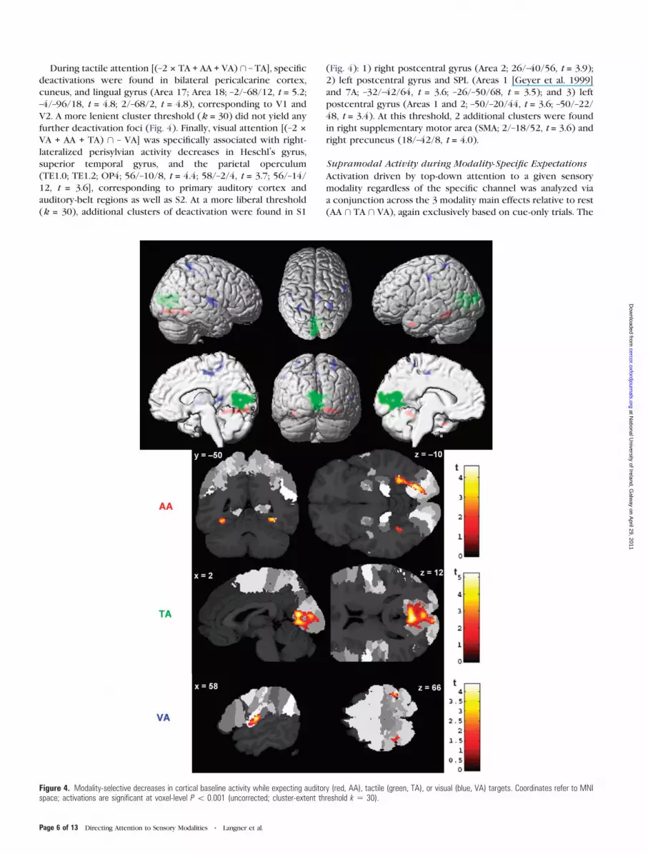
During tactile attention [(–2 3 TA + AA + VA) \ – TA], specific
deactivations were found in bilateral pericalcarine cortex,
cuneus, and lingual gyrus (Area 17; Area 18; –2/–68/12, t = 5.2;
–4/–96/18, t = 4.8; 2/–68/2, t = 4.8), corresponding to V1 and
V2. A more lenient cluster threshold (k = 30) did not yield any
further deactivation foci (Fig. 4). Finally, visual attention [(–2 3
VA + AA + TA) \ – VA] was specifically associated with right-
lateralized perisylvian activity decreases in Heschl’s gyrus,
superior temporal gyrus, and the parietal operculum
(TE1.0; TE1.2; OP4; 56/–10/8, t = 4.4; 58/–2/4, t = 3.7; 56/–14/
12, t = 3.6], corresponding to primary auditory cortex and
auditory-belt regions as well as S2. At a more liberal threshold
(k = 30), additional clusters of deactivation were found in S1
(Fig. 4): 1) right postcentral gyrus (Area 2; 26/–40/56, t = 3.9);
2) left postcentral gyrus and SPL (Areas 1 [Geyer et al. 1999]
and 7A; –32/–42/64, t = 3.6; –26/–50/68, t = 3.5); and 3) left
postcentral gyrus (Areas 1 and 2; –50/–20/44, t = 3.6; –50/–22/
48, t = 3.4). At this threshold, 2 additional clusters were found
in right supplementary motor area (SMA; 2/–18/52, t = 3.6) and
right precuneus (18/–42/8, t = 4.0).
Supramodal Activity during Modality-Specific Expectations
Activation driven by top-down attention to a given sensory
modality regardless of the specific channel was analyzed via
a conjunction across the 3 modality main effects relative to rest
(AA \ TA \ VA), again exclusively based on cue-only trials. The
Figure 4. Modality-selective decreases in cortical baseline activity while expecting auditory (red, AA), tactile (green, TA), or visual (blue, VA) targets. Coordinates refer to MNIspace; activations are significant at voxel-level P\ 0.001 (uncorrected; cluster-extent threshold k 5 30).
Page 6 of 13 Directing Attention to Sensory Modalities d Langner et al.
at National U
niversity of Ireland, Galw
ay on April 29, 2011
cercor.oxfordjournals.orgD
ownloaded from

analysis yielded activity in a widespread frontoparietal network
(Table 2 and Fig. 5) including bilateral IPS and adjacent areas in
superior and inferior parietal lobules (IPL), bilateral dorsal
premotor cortex (dPMC) including the frontal eye fields (FEFs)
as well as bilateral SMA extending into dorsal midcingulate
cortex. Additional right-lateralized activations were found in
pre-SMA, middle frontal gyrus (MFG), and anterior insula.
Furthermore, we observed increased modality-independent
activity in bilateral middle and inferior occipital gyri, pre-
sumably reflecting the processing of the symbolic cueing
stimulus, which was present across conditions (cf. Hopfinger
et al. 2000). Supramodal activity decreases were restricted
to bilateral inferior precuneus (6/–58/18, t = 4.5; –8/–54/12,
t = 4.2).
To control for potential effects of cue-driven spatial
orienting, a supplementary analysis examined supramodal
activations in a model exclusively based on those (cue-only)
trials that contained spatially uninformative cues. Since the
power of this model, relative to the main analyses, was
substantially reduced by small trial numbers, we did not apply
a strict cluster-level correction but used a minimum cluster
extent of k = 30 voxels. At this threshold, all but 2 of the
original nodes of the observed supramodal network (right
middle/inferior frontal gyrus and right anterior insula) showed
significant activation in the conjunction analysis (see Supple-
mentary Fig. S2). The 2 missing areas, however, also showed
activity at lower thresholds (P < 0.05). These results
demonstrate that the supramodal network’s activity does not
substantially depend on any endogenous spatial cue--driven
orienting of attention.
Activity Related to Violations of Specific Versus Nonspecific
Expectations
Our experimental design also lent itself to studying stimulus-
independent brain responses to expectancy violations arising
from the unexpected absence of targets in cue-only trials.
Specifically, we hypothesized that omission-related responses
under more specific expectations should be stronger than
responses under less specific expectations. Therefore, we
compared activity between cue-only trials with predictive
(indicating a specific modality) and with uninformative
(indicating all 3 modalities) cues, in conjunction with the main
effect of predictively cued trials averaged across modalities.
Since we expected differential activity in basal-ganglia struc-
tures (cf. O’Doherty et al. 2004; den Ouden et al. 2009),
we employed a small-volume correction for these regions of
interest, comprising caudate, putamen, and pallidum as defined
Table 2Supramodal brain activity during preparatory attention to a given sensory modality (conjunction
across cue-only trials of all 3 modalities)
Cluster/macroanatomical structure x, y, z Histological assignment t-Score
Cluster 1 (k 5 1645, P\ 0.001)L SPL �22, �64, 56 7A 5.9L IPS �34, �50, 44 hIP3 5.8L IPS �36, �48, 46 hIP1 5.8L SPL �20, �74, 48 7P 4.9L IPL �28, �64, 44 PGa 4.6L IPS �50, �42, 50 hIP2 3.9L SPL �30, �52, 52 7PC 3.6
Cluster 2 (k 5 1195, P\ 0.001)R SPL 22, �66, 54 7P 6.3R SPL 28, �66, 54 7A 4.9R angular gyrus 30, �52, 44 hIP3 4.4R IPS 34, �62, 50 hIP3 4.3R angular gyrus 38, �56, 52 PGa 3.9L SPL 28, �44, 42 7PC 3.5L IPL 32, �72, 48 PGp 3.5L IPS 40, �54, 44 hIP1 3.2L IPS 40, �48, 50 hIP2 3.1
Cluster 3 (k 5 791, P\ 0.001)R precentral gyrus 42, �4, 50 — 6.1R precentral gyrus 38, �4, 48 Area 6 6.1R IFG (pars triangularis) 46, 26, 30 — 4.8R MFG 44, 20, 32 — 4.3
Cluster 4 (k 5 757, P\ 0.001)R middle occipital gyrus 34, �86, 8 — 6.4R middle occipital gyrus 26, �92, 8 hOC3Ad (V3A) 5.2R middle occipital gyrus 26, �94, 4 hOC3v (V3v) 5.1R superior occipital gyrus 20, �98, 6 Area 18 5.1R pericalcarine cortex 18, �100, 2 Area 17 4.6R inferior occipital gyrus 38, �84, �6 hOC4v (V4) 3.6
Cluster 5 (k 5 463, P\ 0.001)L middle occipital gyrus �28, �90, 8 — 4.9L inferior occipital gyrus �40, �80, �12 hOC4v (V4) 4.9L inferior temporal gyrus �46, �60, �24 — 4.7L middle occipital gyrus �28, �92, 12 hOC3Ad (V3A) 3.8L middle occipital gyrus �24, �96, 10 hOC3d (V3d) 3.4L middle occipital gyrus �22, �97, 8 Area 18 3.2
Cluster 6 (k 5 444, P\ 0.001)L precentral gyrus �38, �44, 8 — 6.8L precentral gyrus �36, �8, 46 Area 6 5.2L precentral gyrus �46, �2, 44 Area 6 3.5
Cluster 7 (k 5 274, P 5 0.002)R posterior SFG (SMA) 4, 2, 54 Area 6 4.7L posterior SFG (SMA) �4, 6, 48 Area 6 4.4R posterior SFG (pre-SMA) 6, 14, 50 Area 6 4.0R/L dorsal midcingulate cortex �2, 8, 44 — 3.3
Cluster 8 (k 5 172, P 5 0.018)R anterior insula 34, 24, �2 — 5.0R IFG (pars triangularis) 46, 18, 5 Area 45 3.3
Notes: Coordinates x, y, z of local maxima refer to MNI space; k 5 number of voxels in cluster;
P 5 cluster-level error probability corrected for multiple comparisons. L 5 left; R 5 right; IFG/
SFG 5 inferior/superior frontal gyrus. References for histological assignments: 7A, 7P, 7PC:
Scheperjans et al. (2008); Area 6: Geyer (2004); Areas 17, 18: Amunts et al. (2000); Area 45:
Amunts et al. (1999); hIP1, hIP2: Choi et al. (2006); hIP3: Scheperjans et al. (2008); hOC3Ad,
hOC3d: Kujovic et al. (2007); hOC3v, hOC4v: Rottschy et al. (2007); PGa, PGp: Caspers et al.
(2006).
Figure 5. Supramodal activity while expecting auditory, tactile, or visual targets (conjunction across main effects of all 3 modality-specific expectancy conditions relative toresting baseline). Coordinates refer to MNI space; activations are significant at P\ 0.05 (familywise error corrected at cluster level; voxelwise cluster-forming threshold P\0.001). Abbreviation: IOG/MOG 5 inferior/middle occipital gyrus.
Cerebral Cortex Page 7 of 13
at National U
niversity of Ireland, Galw
ay on April 29, 2011
cercor.oxfordjournals.orgD
ownloaded from
with 50% probability by the Harvard--Oxford anatomical atlas
(distributed with the FSL software package; http://fsl.fmrib.ox.-
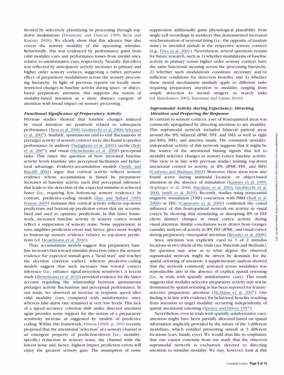
ac.uk/fsl). Averaged across modalities, absence of targets after
predictive modality cues was associated with stronger activa-
tion in bilateral ventral striatum (anterior [–20/14/–4, t = 4.5;
24/14/–4, t = 3.6] and posterior [–28/–8/0, t = 3.8; 30/–14/4,
t = 3.8] putamen) (Fig. 6; for a color version, see Supplementary
Fig. S4).
Discussion
This study examined the neural correlates of voluntarily
directing attention to a given sensory modality in the absence
of stimulation, induced by explicit modality-specific expect-
ations for upcoming auditory, tactile, or visual targets. Since
the main analyses were restricted to trials without target
presentation (cue-only trials), their results should reflect pure
effects of allocating attention (cf. Beck and Kastner 2009).
Modality-specific preparatory attention was associated with
significant increases and decreases in baseline activity of
relevant and irrelevant sensory cortices, respectively. Supple-
mentary analyses revealed that these baseline changes oc-
curred in areas that were also selectively activated during
actual stimulus detection. Faster responses in target trials with
valid versus uninformative or invalid modality cues indicated
that the expectancy-driven preparatory changes in brain
activity most likely reflect a processing bias in favor of the
cued modality. In all cue-only trials, the sensory input was
identical across modalities (except for the slight difference in
the cueing symbols) and should have canceled each other out
in direct comparison. Thus, modality-specific baseline changes
exclusively reflect effects of directed (top-down) attention.
Attention-Related Changes in Baseline Activity of SensoryCortices
The results of our study bridge 2 as-yet isolated literatures on
attentional mechanisms in the human brain: they connect
previous evidence on the modulation of stimulus processing by
intersensory selective attention during multimodal stimulation
(Roland 1982; Kawashima et al. 1999; Mehta et al. 2000;
Macaluso et al. 2002; Shomstein and Yantis 2004; Saupe et al.
2009) with findings of preparatory changes in baseline activity
during attention to some stimulus feature within a given
modality (Colby et al. 1996; Chawla et al. 1999; Kastner et al.
1999; Carlsson et al. 2000; Hopfinger et al. 2000; Giesbrecht
et al. 2006; Wu et al. 2007; Smith et al. 2010). To our
knowledge, we show here for the first time with fMRI that
brain activity can be flexibly tuned in advance, on a trial-by-trial
basis, not only according to space- or object-selective expect-
ations but also according to more basic expectations about
stimulus modality. This resolves ambiguities among previous
studies which either could not discriminate modality-specific
preparatory activity from attentional modulation of stimulus
processing (e.g., Macaluso et al. 2002; Langner et al. 2011) or
could not isolate cue-driven modality-selective increases in
baseline activity, presumably due to prevailing sustained
selection biases (Macaluso et al. 2003).
Despite these differences, our results substantially overlap
with modality-specific attentional modulation of stimulus-
driven activity: We found the same cortical areas selectively
preactivated by attention that 1) are devoted to processing
input of the cued modality and 2) showed activity modulation
by attention in previous studies. This overlap suggests that the
mechanisms underlying attentional modulation of baseline
activity and those underlying attentional facilitation of stimulus
processing are closely related. A recent study (Sylvester et al.
2009) provided direct evidence for this assumption with
respect to visuospatial attention. Similarly, Esterman and Yantis
(2010) found support for a substantial congruence between
regions recruited by visual anticipation and subsequent
perception in a cued category judgment task. Based on our
results, we conjecture that the congruence between expec-
tancy- and processing-related modulations might generalize
across modalities and, moreover, to selective attention to
a particular sensory channel.
Further, we found specific decreases in baseline activity of
sensory areas that were rendered irrelevant by the modality
cue. These deactivations might reflect an inhibitory mechanism
of the brain to reduce the impact of irrelevant input that is
complementary to enhancing activity in relevant areas (Mozolic
et al. 2008). Such anticipatory suppression has also been
reported during unimodal spatial attention with regard to
irrelevant locations (Sylvester et al. 2008) or body parts
(Drevets et al. 1995). Our results are also consistent with
reports of modality-selective decreases in brain activity when
directing attention away from a given sensory modality during
bimodal stimulation (Kawashima et al. 1995; Sokolov et al.
2004). Taken together, these decreases corroborate the
assumption of an active filtering of unwanted information in
the unattended modality, which appears to be implemented
not only during stimulus processing but already during
modality-specific expectations.
Our results are consistent with biased-competition models
of attention, according to which expected sensory input is
Figure 6. Activity in bilateral anterior and posterior putamen related to violatingspecific (cue-only trials with predictive cues) versus nonspecific (cue-only trials withuninformative cues) expectations, presumably reflecting stronger surprise at theunexpected omission of predictively cued sensory input. Coordinates refer to MNIspace; left hemisphere shown on the left; activations are significant at P \ 0.05(small-volume familywise error corrected for the basal ganglia at cluster level;voxelwise cluster-forming threshold P \ 0.001). For a color version, seeSupplementary Figure S4.
Page 8 of 13 Directing Attention to Sensory Modalities d Langner et al.
at National U
niversity of Ireland, Galw
ay on April 29, 2011
cercor.oxfordjournals.orgD
ownloaded from
favored by selectively prioritizing its processing through top-
down modulations (Desimone and Duncan 1995; Beck and
Kastner 2009). We clearly show that this advance bias also
covers the sensory modality of the upcoming stimulus.
Behaviorally, this was evidenced by performance gains from
valid modality cues and performance losses from invalid ones,
relative to uninformative cues, respectively. Neurally, this effect
was reflected by anticipatory activity increases in primary and
higher order sensory cortices, suggesting a rather pervasive
effect of preparatory modulations across the sensory process-
ing hierarchy. In light of previous reports on locally more
restricted changes in baseline activity during space- or object-
based preparatory attention, this supports the notion of
modality-based attention as a more abstract category of
attention with broad impact on sensory processing.
Functional Significance of Preparatory Activity
Previous studies showed that baseline changes induced
by visual attention are positively related to subsequent
performance (Ress et al. 2000; Giesbrecht et al. 2006; Sylvester
et al. 2007). Similarly, spontaneous trial-to-trial fluctuations in
pretarget activity of sensory cortices were also found to predict
performance in auditory (Sadaghiani et al. 2009), tactile (Boly
et al. 2007), and visual (Hesselmann et al. 2008) perceptual
tasks. This raises the question of how increased baseline
activity levels translate into perceptual facilitation and behav-
ioral advantage. Evidence-accumulation models (Smith and
Ratcliff 2004) argue that cortical activity reflects sensory
evidence whose accumulation is biased by preparatory
increases in baseline activity such that perceptual inference
that leads to the detection of the expected stimulus is achieved
faster (i.e., requiring less bottom-up sensory evidence). In
contrast, predictive-coding models (Rao and Ballard 1999;
Friston 2005) maintain that cortical activity reflects top-down
predictions and bottom-up prediction errors that are accumu-
lated and used to optimize predictions. In this latter frame-
work, increased baseline activity in sensory cortex would
reflect a suppression of sensory noise. Noise suppression, in
turn, amplifies prediction errors and, hence, gives more weight
to bottom-up sensory evidence relative to top-down predic-
tions (cf. Hesselmann et al. 2010).
Thus, accumulation models suggest that preparatory base-
line increases bias toward stimulus detection (since the sensory
evidence for expected stimuli gets a ‘‘head start’’ and reaches
the decision criterion earlier), whereas predictive-coding
models suggest that such increases bias toward correct
inference (i.e., enhance signal-detection sensitivity). A recent
study (Hesselmann et al. 2010) provided evidence for the latter
account regarding the relationship between spontaneous
pretarget activity fluctuations and perceptual performance. In
our study, we observed an increase in response speed after
valid modality cues, compared with uninformative ones,
whereas false-alarm rate remained at very low levels. This lack
of a speed--accuracy criterion shift under directed attention
again provides some support for the notion of a preparatory
sensitivity increase as suggested by models of predictive
coding. Within this framework, Friston (2009, p. 299) recently
proposed that the attentional ‘‘selection’’ of a sensory channel is
an emergent property of prediction-driven (i.e., modality-
specific) reduction in sensory noise; the channel with the
lowest noise and, hence, highest impact prediction errors will
enjoy the greatest sensory gain. The assumption of noise
suppression additionally gains physiological plausibility from
single-cell recordings in monkeys that demonstrated increased
synchronization of neuronal firing (i.e., the opposite of random
noise) to attended stimuli in the respective sensory cortices
(e.g., Fries et al. 2001). Nevertheless, several questions remain
for future research, such as 1) whether modulations of baseline
activity in primary versus higher order sensory cortices have
the same functional meaning across the processing hierarchy,
2) whether such modulations constitute necessary and/or
sufficient conditions for detection benefits, and 3) whether
these neural mechanisms similarly apply to different tasks
requiring preparatory attention to modality, ranging from
simple detection to mental imagery in search tasks
(cf. Bartolomeo 2002; Esterman and Yantis 2010).
Supramodal Activity during Expectancy: DirectingAttention and Preparing the Response
In contrast to sensory cortices, a set of frontoparietal areas was
commonly upregulated by directing attention to any modality.
This supramodal network included bilateral parietal areas
around the IPS, bilateral dPMC/FEF, and SMA as well as right
pre-SMA, MFG, and anterior insula. The consistent modality-
independent activity of this network suggests that it might be
the source of the attentional biasing signals that led to
modality-selective changes in sensory-cortex baseline activity.
This view is in line with previous studies relating top-down
attentional control to activity in IPS, dPMC/FEF, and MFG
(Corbetta and Shulman 2002). Moreover, these areas were also
found active during unimodal location- or object-based
attention in the absence of stimulation (Kastner et al. 1999;
Hopfinger et al. 2000; Macaluso et al. 2003; Giesbrecht et al.
2006; Smith et al. 2010). Recently, studies using transcranial
magnetic stimulation (TMS) concurrent with fMRI (Ruff et al.
2008) or EEG (Capotosto et al. 2009) confirmed the causal
influence of this frontoparietal network on activity in visual
cortex by showing that stimulating or disrupting IPS or FEF
elicits distinct changes in visual cortex activity during
visual attention. Similar conclusions were drawn from Granger
causality analyses of activity in IPS, FEF/dPMC, and visual cortex
during preparatory visuospatial attention (Bressler et al. 2008).
Since attention was explicitly cued to 1 of 2 stimulus
locations in two-thirds of the trials (see Materials and Methods),
the question may arise as to what degree the observed
supramodal network might be driven by demands for the
spatial orienting of attention. A supplementary analysis showed
that the network commonly activated across modalities was
reproducible also in the absence of explicit spatial orienting
(i.e., in trials with spatially uninformative cues). This result
suggests that modality-selective preparatory activity may not be
dominated by spatial orienting as has been reported for feature-
selective preparatory attention (McMains et al. 2007). Our
finding is in line with evidence for behavioral benefits resulting
from attention to target modality occurring independently of
spatial attentional orienting (Spence and Driver 1997).
Nevertheless, even in trials with spatially uninformative cues,
attention might have been partially allocated based on spatial
information implicitly provided by the nature of the 3 different
modalities, which entailed presenting stimuli at 3 different
locations (ears, hands, eyes). We would thus like to emphasize
that one cannot conclude from our study that the observed
supramodal network is exclusively devoted to directing
attention to stimulus modality. We may, however, look at this
Cerebral Cortex Page 9 of 13
at National U
niversity of Ireland, Galw
ay on April 29, 2011
cercor.oxfordjournals.orgD
ownloaded from

seeming drawback from the opposite direction: in exchange
for the remaining ambiguity regarding pure attention-to-
modality effects, we gain further evidence for the modality
independence of the network controlling spatial attention
(cf. Eimer and Van Velzen 2002; Krumbholz et al. 2009;
Smith et al. 2010). Moreover, since modality- and location-
based attention are usually interwoven in real life, our finding
of a supramodal core network that may subserve both
attentional functions has higher ecological validity than results
from previous studies on attention toward single stimulus
dimensions.
Apart from preparatory attention to sensory input, our task also
induced motor preparation. Indeed, we found supramodal pre-
paratory activity in dPMC, SMA, and pre-SMA, which have been
previously found involved in preparatory motor processes. For
instance, a TMS study revealed dPMC involvement in using cue
information for the preparatory scaling of grip force (Chouinard
et al. 2005). Dorsal PMC was also found to process information
from spatial cues to direct movements, regardless of the cue’s
sensory modality (Weinrich and Wise 1982). Hoshi and Tanji
(2007) argued that dPMC integrates sensory and memory in-
formation to establish action intentions and develop associated
motor programs (see also Cieslik et al. 2010). Our data are
consistentwith this notionof dPMC function,whichmight include
the preparatory activation of the currently expected stimulus--
responsemapping (Jakobs et al. 2009). SMA andpre-SMAwere also
shown to be involved in movement preparation (Matsuzaka and
Tanji 1996; Hoshi and Tanji 2004; Cunnington et al. 2005). Since in
simple RT paradigms like ours the motor response can be fully
prepared in advance, our findings agree well with the assumption
that dPMC, SMA, pre-SMA, and midcingulate cortex subserve the
establishment of a preparatory set for the expected movement,
independent of the response signal’s modality.
Finally, the anterior insula is known to be involved in
representing bodily states (Craig 2002), including arousal
induced by mental or physical stressors (Critchley et al. 2000;
Pollatos et al. 2007). Thus, cue-induced insula activitymay reflect
the general alerting property of the cue. This way the insula
might code the behavioral relevance of the cue and establish an
appropriate level of alertness to ready body and brain for an
efficient response to the impending imperative signal (Sterzer
and Kleinschmidt 2010; Langner et al. 2011). Additionally, the
anterior insula might contribute to (re)activating the general
task set following the cue (Dosenbach et al. 2006).
Taken together, our results show that in simple RT tasks top-
down control of attention and mechanisms of response
preparation, which in the current design could not be
separated, operate mainly independently of the modality of
expected response signals. This underlines the importance of
using various sensory input channels in studies on ‘‘central’’
processes such as preparation, in order to arrive at generaliz-
able conclusions. Otherwise, input-specific and input-indepen-
dent effects might be hard to disentangle. For instance, our
results suggest that earlier findings of increases in firing rate of
monkey lateral IPS during anticipation (Colby et al. 1996) might
not be specific to visual attention but rather supramodal.
Activity Related to the Unexpected Omission of ResponseSignals
Apart from preparatory activity, expectancy effects should also
manifest themselves when expectations are not met, that is, in
the brain response to the omission of an expected response
signal. As predicted, this response was stronger, across
modalities, in the anterior and posterior putamen after
disconfirming more precise expectations (in cue-only trials
with predictive cues) compared with less precise ones (in cue-
only trials with uninformative cues). This differential omission-
related response provides independent support for the view
that predictive cues were used indeed to develop selective
expectations leading to perceptual facilitation.
We suggest that increased activity in the anterior putamen
reflects enhanced prediction-error responses to target omis-
sions after predictive versus uninformative cues. This assump-
tion is corroborated by other studies that found activity in
this area to covary with prediction errors (McClure et al. 2003;
O’Doherty et al. 2004), including error responses to the
unexpected absence of input (den Ouden et al. 2009).
Importantly, our results indicate that such omission-related
responses may not simply be equated with information-
theoretic (Shannon) surprise, which denotes the improbability
of a given event (here: the target omission in any cue-only trial;
cf. Strange et al. 2005). Rather, we found omission-related
response differences between specific and nonspecific expect-
ations, although the event of interest, that is, the omission of
sensory input, was similarly improbable under both specific and
nonspecific expectancy conditions (27.3% and 33.3% cue-only
trials in conditions with predictive and uninformative cues,
respectively). This similarity argues against a simple depen-
dence of event-bound putaminal surprise responses on the
event’s occurrence probability. Instead, finding substantial
differences despite highly similar occurrence probabilities
agrees more with recent notions that surprise is captured best
in a relative manner related to subjective expectations of the
observer (Itti and Baldi 2009). Based on this premise, event-
bound surprise has been defined in a Bayesian framework as
the degree to which prior expectations are violated and, thus,
changed (‘‘updated’’) by the occurrence of an event (Baldi
2005; see also Maguire et al. 2011). Within this framework,
omission-related responses in the anterior putamen might
index the omission-induced degree of adjustment of the prior
expectation rather than information-theoretic surprise.
Our finding that putaminal responses to omissions of
expected input are stronger when expectations are specific
fits very well with the predictive-coding approach alluded to
above, which argues that modality-specific expectations
selectively reduce noise in relevant sensory cortices (cf. Friston
2009). In a Bayesian perspective, this reduces prior variance,
hereby enhancing the weight of bottom-up input for percep-
tual inference on a given sensory channel. If, as in cue-only
trials, the expected input does not arrive, a prediction error is
generated. When predictions are specific, such as after
predictive cues, the error’s impact and the associated striatal
response are enhanced compared with nonspecific predictions,
such as after uninformative cues. This reasoning is in line with
the view that the perceived level of surprise at a given event be
associated with the difficulty of integrating the event (here: the
stimulus omission) with an existing representation (Maguire
et al. 2011). That is, when the integration even of low-
probability events is made easy, for instance by nonspecific (as
compared with highly specific) expectations, only little
surprise is evoked, since the existing representation is not
changed much by the event’s integration. In sum, this effect
demonstrates that information-theoretic (Shannon) surprise,
which was highly similar for both trial types, and Bayesian
Page 10 of 13 Directing Attention to Sensory Modalities d Langner et al.
at National U
niversity of Ireland, Galw
ay on April 29, 2011
cercor.oxfordjournals.orgD
ownloaded from

surprise, which depends on the specificity of prior beliefs, are
dissociably represented in the human brain.
The response in the posterior putamen might be specifically
related to predictive coding in the motor domain (cf. Kilner
et al. 2007; Jakobs et al. 2009): predictive cues are thought to
elicit enhanced motor preparation (Koski et al. 1999; Bestmann
et al. 2008), contributing to shorter RT. This interpretation
corresponds to previous findings on the role of the posterior
putamen in skeletomotor control and movement initiation
(Alexander and Crutcher 1990; Boussaoud and Kermadi 1997).
The functional differentiation between posterior and anterior
putaminal activity accords with the finding that learning
sequential finger movements versus overlearned responding
activated anterior as opposed to posterior putamen and vice
versa (Jueptner and Weiller 1998).
Conclusions
We have shown that voluntarily directing attention to various
sensory channels in a flexible, trial-by-trial manner leads to
improved detection performance and transient changes
in baseline activity of sensory cortices, with increased activity
in relevant cortices and decreased activity in irrelevant ones.
These expectancy-related changes occur for attention to
auditory, tactile, and visual channels alike, attesting to the
generality of the mechanism. Modulations of sensory cortices
appear to be controlled via a common supramodal frontopar-
ietal network, which may emit biasing top-down signals that
influence competition between sensory inputs in favor of the
most relevant (i.e., expected) information. It remains to be
examined whether attention-induced increases in baseline
activity reflect increased synchrony (i.e., reduced noise) in
sensory cortices, as suggested previously for spontaneous
pretarget fluctuations, or whether these increases (also) reflect
the ‘‘replacement’’ of sensory data with top-down expectations
to facilitate target detection by speeding up evidence
accumulation. Finally, activity in the anterior putamen related
to unexpected input omissions may reflect a prediction-error
signal that is sensitive to the specificity of expectations and
represents (Bayesian) surprise.
Supplementary Material
Supplementary material can be found at: http://www.cercor.
oxfordjournals.org/
Funding
Deutsche Forschungsgemeinschaft (IRTG 1328 to R.L., K.W.,
and S.B.E.); Human Brain Project (R01-MH074457-01A1 to
S.B.E.); Initiative and Networking Fund of the Helmholtz
Association within the Helmholtz Alliance on Systems Biology
(Human Brain Model to S.B.E).
Notes
Conflict of Interest: None declared.
References
Alexander GE, Crutcher MD. 1990. Preparation for movement: neural
representations of intended direction in three motor areas of the
monkey. J Neurophysiol. 64:133--150.
Amunts K, Malikovic A, Mohlberg H, Schormann T, Zilles K. 2000.
Brodmann’s areas 17 and 18 brought into stereotaxic space—where
and how variable? Neuroimage. 11:66--84.
Amunts K, Schleicher A, Burgel U, Mohlberg H, Uylings HB, Zilles K.
1999. Broca’s region revisited: cytoarchitecture and intersubject
variability. J Comp Neurol. 412:319--341.
Ashburner J, Friston KJ. 2005. Unified segmentation. Neuroimage.
26:839--851.
Baldi P. 2005. Surprise: a shortcut for attention? In: Itti L, Rees G,
Tsotsos JK, editors. Neurobiology of attention. Amsterdam: Elsevier
Academic Press. p. 24--28.
Bartolomeo P. 2002. The relationship between visual perception and
visual mental imagery: a reappraisal of the neuropsychological
evidence. Cortex. 38:357--378.
Bausenhart KM, Rolke B, Ulrich R. 2007. Knowing when to hear aids
what to hear. Q J Exp Psychol. 60:1610--1615.
Beck DM, Kastner S. 2009. Top-down and bottom-up mechanisms in
biasing competition in the human brain. Vision Res. 49:1154--1165.
Bestmann S, Harrison LM, Blankenburg F, Mars RB, Haggard P,
Friston KJ, Rothwell JC. 2008. Influence of uncertainty and surprise
on human corticospinal excitability during preparation for action.
Curr Biol. 18:775--780.
Boly M, Balteau E, Schnakers C, Degueldre C, Moonen G, Luxen A,
Phillips C, Peigneux P, Maquet P, Laureys S. 2007. Baseline brain
activity fluctuations predict somatosensory perception in humans.
Proc Natl Acad Sci U S A. 104:12187--12192.
Boussaoud D, Kermadi I. 1997. The primate striatum: neuronal activity
in relation to spatial attention versus motor preparation. Eur J
Neurosci. 9:2152--2168.
Bressler SL, Tang W, Sylvester CM, Shulman GL, Corbetta M. 2008. Top-
down control of human visual cortex by frontal and parietal cortex
in anticipatory visual spatial attention. J Neurosci. 28:10056--10061.
Brunia CH, van Boxtel GJ. 2001. Wait and see. Int J Psychophysiol.
43:59--75.
Brunia CH, van Boxtel GJ. 2004. Anticipatory attention to verbal and
non-verbal stimuli is reflected in a modality-specific SPN. Exp Brain
Res. 156:231--239.
Capotosto P, Babiloni C, Romani GL, Corbetta M. 2009. Frontoparietal
cortex controls spatial attention through modulation of anticipatory
alpha rhythms. J Neurosci. 29:5863--5872.
Carlsson K, Petrovic P, Skare S, Petersson KM, Ingvar M. 2000. Tickling
expectations: neural processing in anticipation of a sensory
stimulus. J Cogn Neurosci. 12:691--703.
Caspers S, Geyer S, Schleicher A, Mohlberg H, Amunts K, Zilles K. 2006.
The human inferior parietal cortex: cytoarchitectonic parcellation
and interindividual variability. Neuroimage. 33:430--448.
Chawla D, Rees G, Friston KJ. 1999. The physiological basis of attentional
modulation in extrastriate visual areas. Nat Neurosci. 2:671--676.
Choi HJ, Zilles K, Mohlberg H, Schleicher A, Fink GR, Armstrong E,
Amunts K. 2006. Cytoarchitectonic identification and probabilistic
mapping of two distinct areas within the anterior ventral bank of
the human intraparietal sulcus. J Comp Neurol. 495:53--69.
Chouinard PA, Leonard G, Paus T. 2005. Role of the primary motor and
dorsal premotor cortices in the anticipation of forces during object
lifting. J Neurosci. 25:2277--2284.
Cieslik EC, Zilles K, Kurth F, Eickhoff SB. 2010. Dissociating bottom-up
and top-down processes in a manual stimulus-response compatibil-
ity task. J Neurophysiol. 104:1472--1483.
Colby CL, Duhamel JR, Goldberg ME. 1996. Visual, presaccadic, and
cognitive activation of single neurons in monkey lateral intraparietal
area. J Neurophysiol. 76:2841--2852.
Corbetta M, Shulman GL. 2002. Control of goal-directed and stimulus-
driven attention in the brain. Nat Rev Neurosci. 3:201--215.
Correa A, Lupianez J, Tudela P. 2005. Attentional preparation based on
temporal expectancy modulates processing at the perceptual level.
Psychon Bull Rev. 12:328--334.
Craig AD. 2002. How do you feel? Interoception: the sense of the
physiological condition of the body. Nat Rev Neurosci. 3:655--666.
Critchley HD, Corfield DR, Chandler MP, Mathias CJ, Dolan RJ. 2000.
Cerebral correlates of autonomic cardiovascular arousal: a functional
neuroimaging investigation in humans. J Physiol. 523(Pt 1):259--270.
Cerebral Cortex Page 11 of 13
at National U
niversity of Ireland, Galw
ay on April 29, 2011
cercor.oxfordjournals.orgD
ownloaded from

Cunnington R, Windischberger C, Moser E. 2005. Premovement activity
of the pre-supplementary motor area and the readiness for action:
studies of time-resolved event-related functional MRI. Hum Mov Sci.
24:644--656.
den Ouden HE, Friston KJ, Daw ND, McIntosh AR, Stephan KE. 2009.
A dual role for prediction error in associative learning. Cereb
Cortex. 19:1175--1185.
Desimone R, Duncan J. 1995. Neural mechanisms of selective visual
attention. Annu Rev Neurosci. 18:193--222.
Dosenbach NU, Visscher KM, Palmer ED, Miezin FM, Wenger KK,
Kang HC, Burgund ED, Grimes AL, Schlaggar BL, Petersen SE. 2006.
A core system for the implementation of task sets. Neuron.
50:799--812.
Drevets WC, Burton H, Videen TO, Snyder AZ, Simpson JR, Jr,
Raichle ME. 1995. Blood flow changes in human somatosensory
cortex during anticipated stimulation. Nature. 373:249--252.
Eickhoff SB, Schleicher A, Zilles K, Amunts K. 2006. The human parietal
operculum. I. Cytoarchitectonic mapping of subdivisions. Cereb
Cortex. 16:254--267.
Eickhoff SB, Stephan KE, Mohlberg H, Grefkes C, Fink GR, Amunts K,
Zilles K. 2005. A new SPM toolbox for combining probabilistic
cytoarchitectonic maps and functional imaging data. Neuroimage.
25:1325--1335.
Eimer M, Schroger E. 1998. ERP effects of intermodal attention and
cross-modal links in spatial attention. Psychophysiology.
35:313--327.
Eimer M, Van Velzen J. 2002. Crossmodal links in spatial attention are
mediated by supramodal control processes: evidence from event-
related potentials. Psychophysiology. 39:437--449.
Esterman M, Yantis S. 2010. Perceptual expectation evokes category-
selective cortical activity. Cereb Cortex. 20:1245--1253.
Foxe JJ, Simpson GV. 2005. Biasing the brain’s attentional set: II. Effects
of selective intersensory attentional deployments on subsequent
sensory processing. Exp Brain Res. 166:393--401.
Foxe JJ, Simpson GV, Ahlfors SP, Saron CD. 2005. Biasing the brain’s
attentional set: I. Cue-driven deployments of intersensory selective
attention. Exp Brain Res. 166:370--392.
Fries P, Reynolds JH, Rorie AE, Desimone R. 2001. Modulation of
oscillatory neuronal synchronization by selective visual attention.
Science. 291:1560--1563.
Friston K. 2005. A theory of cortical responses. Philos Trans R Soc Lond
B Biol Sci. 360:815--836.
Friston K. 2009. The free-energy principle: a rough guide to the brain?
Trends Cogn Sci. 13:293--301.
Friston KJ, Fletcher PC, Josephs O, Holmes A, Rugg MD, Turner R. 1998.
Event-related fMRI: characterizing differential responses. Neuro-
image. 7:30--40.
Geyer S. 2004. The microstructural border between the motor and the
cognitive domain in the human cerebral cortex. Adv Anat Embryol
Cell Biol. 174(I--VIII):1--89.
Geyer S, Schleicher A, Zilles K. 1999. Areas 3a, 3b, and 1 of human
primary somatosensory cortex. Neuroimage. 10:63--83.
Giesbrecht B, Weissman DH, Woldorff MG, Mangun GR. 2006.
Pre-target activity in visual cortex predicts behavioral
performance on spatial and feature attention tasks. Brain Res.
1080:63--72.
Grefkes C, Geyer S, Schormann T, Roland P, Zilles K. 2001. Human
somatosensory area 2: observer-independent cytoarchitectonic
mapping, interindividual variability, and population map. Neuro-
image. 14:617--631.
Hackley SA. 2009. The speeding of voluntary reaction by a warning
signal. Psychophysiology. 46:225--233.
Hahn B, Ross TJ, Stein EA. 2006. Neuroanatomical dissociation between
bottom-up and top-down processes of visuospatial selective
attention. Neuroimage. 32:842--853.
Hesselmann G, Kell CA, Kleinschmidt A. 2008. Ongoing activity
fluctuations in hMT+ bias the perception of coherent visual motion.
J Neurosci. 28:14481--14485.
Hesselmann G, Sadaghiani S, Friston KJ, Kleinschmidt A. 2010.
Predictive coding or evidence accumulation? False inference and
neuronal fluctuations. PLoS One. 5:e9926.
Holmes CJ, Hoge R, Collins L, Woods R, Toga AW, Evans AC. 1998.
Enhancement of MR images using registration for signal averaging. J
Comput Assist Tomogr. 22:324--333.
Hopfinger JB, Buonocore MH, Mangun GR. 2000. The neural mecha-
nisms of top-down attentional control. Nat Neurosci. 3:284--291.
Hoshi E, Tanji J. 2004. Differential roles of neuronal activity in the
supplementary and presupplementary motor areas: from informa-
tion retrieval to motor planning and execution. J Neurophysiol.
92:3482--3499.
Hoshi E, Tanji J. 2007. Distinctions between dorsal and ventral
premotor areas: anatomical connectivity and functional properties.
Curr Opin Neurobiol. 17:234--242.
Itti L, Baldi P. 2009. Bayesian surprise attracts human attention. Vision
Res. 49:1295--1306.
Jakobs O, Wang LE, Dafotakis M, Grefkes C, Zilles K, Eickhoff SB. 2009.
Effects of timing and movement uncertainty implicate the temporo-
parietal junction in the prediction of forthcoming motor actions.
Neuroimage. 47:667--677.
Jueptner M, Weiller C. 1998. A review of differences between basal
ganglia and cerebellar control of movements as revealed by
functional imaging studies. Brain. 121(Pt 8):1437--1449.
Karns CM, Knight RT. 2009. Intermodal auditory, visual, and tactile
attention modulates early stages of neural processing. J Cogn
Neurosci. 21:669--683.
Kastner S, Pinsk MA, De Weerd P, Desimone R, Ungerleider LG. 1999.
Increased activity in human visual cortex during directed attention
in the absence of visual stimulation. Neuron. 22:751--761.
Kawashima R, Imaizumi S, Mori K, Okada K, Goto R, Kiritani S, Ogawa A,
Fukuda H. 1999. Selective visual and auditory attention toward
utterances: a PET study. Neuroimage. 10:209--215.
Kawashima R, O’Sullivan BT, Roland PE. 1995. Positron-emission
tomography studies of cross-modality inhibition in selective
attentional tasks: closing the ‘‘mind’s eye’’. Proc Natl Acad Sci
U S A. 92:5969--5972.
Kilner JM, Friston KJ, Frith CD. 2007. Predictive coding: an account of
the mirror neuron system. Cogn Process. 8:159--166.
Koski L, Paus T, Hofle N, Petrides M. 1999. Increased blood flow in the basal
ganglia when using cues to direct attention. Exp Brain Res. 129:241--246.
Krumbholz K, Nobis EA, Weatheritt RJ, Fink GR. 2009. Executive
control of spatial attention shifts in the auditory compared to the
visual modality. Hum Brain Mapp. 30:1457--1469.
Kujovic M, Malikovic A, Schleicher A, Rottschy C, Eickhoff SB,
Mohlberg H, Homke L, Zilles K, Amunts K. 2007. Observer-
independent cytoarchitectonic mapping of the dorsal extrastriate
human visual cortex. Neuroimage. 36(Suppl 1):S57.
Langner R, Kellermann T, Eickhoff SB, Boers F, Chatterjee A, Willmes K,
Sturm W. 2011. Staying responsive to the world: modality-specific and
-nonspecific contributions to speeded auditory, tactile, and visual
stimulus detection. Hum Brain Mapp. doi:10.1002/hbm.21220.
Luck SJ, Chelazzi L, Hillyard SA, Desimone R. 1997. Neural mechanisms
of spatial selective attention in areas V1, V2, and V4 of macaque
visual cortex. J Neurophysiol. 77:24--42.
Macaluso E, Eimer M, Frith CD, Driver J. 2003. Preparatory states in
crossmodal spatial attention: spatial specificity and possible control
mechanisms. Exp Brain Res. 149:62--74.
Macaluso E, Frith CD, Driver J. 2002. Directing attention to locations
and to sensory modalities: multiple levels of selective processing
revealed with PET. Cereb Cortex. 12:357--368.
Maguire R, Maguire P, Keane MT. 2011. Making sense of surprise: an
investigation of the factors influencing surprise judgments. J Exp
Psychol Learn Mem Cogn. 37:176--186.
Matsuzaka Y, Tanji J. 1996. Changing directions of forthcoming arm
movements: neuronal activity in the presupplementary and supple-
mentary motor area of monkey cerebral cortex. J Neurophysiol.
76:2327--2342.
McClure SM, Berns GS, Montague PR. 2003. Temporal prediction errors
in a passive learning task activate human striatum. Neuron.
38:339--346.
McMains SA, Fehd HM, Emmanouil TA, Kastner S. 2007. Mechanisms of
feature- and space-based attention: response modulation and
baseline increases. J Neurophysiol. 98:2110--2121.
Page 12 of 13 Directing Attention to Sensory Modalities d Langner et al.
at National U
niversity of Ireland, Galw
ay on April 29, 2011
cercor.oxfordjournals.orgD
ownloaded from

Mehta AD, Ulbert I, Schroeder CE. 2000. Intermodal selective attention
in monkeys. I: distribution and timing of effects across visual areas.
Cereb Cortex. 10:343--358.
Morosan P, Rademacher J, Schleicher A, Amunts K, Schormann T,
Zilles K. 2001. Human primary auditory cortex: cytoarchitectonic
subdivisions and mapping into a spatial reference system. Neuro-
image. 13:684--701.
Morosan P, Schleicher A, Amunts K, Zilles K. 2005. Multimodal
architectonic mapping of human superior temporal gyrus. Anat
Embryol (Berl). 210:401--406.
Mozolic JL, Joyner D, Hugenschmidt CE, Peiffer AM, Kraft RA,
Maldjian JA, Laurienti PJ. 2008. Cross-modal deactivations during
modality-specific selective attention. BMC Neurol. 8:35.
Nichols T, Brett M, Andersson J, Wager T, Poline JB. 2005. Valid
conjunction inference with the minimum statistic. Neuroimage.
25:653--660.
O’Doherty J, Dayan P, Schultz J, Deichmann R, Friston K, Dolan RJ. 2004.
Dissociable roles of ventral and dorsal striatum in instrumental
conditioning. Science. 304:452--454.
Pollatos O, Schandry R, Auer DP, Kaufmann C. 2007. Brain structures
mediating cardiovascular arousal and interoceptive awareness. Brain
Res. 1141:178--187.
Rao RP, Ballard DH. 1999. Predictive coding in the visual cortex:
a functional interpretation of some extra-classical receptive-field
effects. Nat Neurosci. 2:79--87.
Ress D, Backus BT, Heeger DJ. 2000. Activity in primary visual cortex
predicts performance in a visual detection task. Nat Neurosci.
3:940--945.
Roland PE. 1982. Cortical regulation of selective attention in man. A
regional cerebral blood flow study. J Neurophysiol. 48:1059--1078.
Rolke B, Ulrich R. 2010. On the locus of temporal preparation:
enhancement of pre-motor processes? In: Nobre AC, Coull JT,
editors. Attention and time. Oxford: Oxford University Press.
p. 227--241.
Rottschy C, Eickhoff SB, Schleicher A, Mohlberg H, Kujovic M, Zilles K,
Amunts K. 2007. Ventral visual cortex in humans: cytoarchitectonic
mapping of two extrastriate areas. Hum Brain Mapp. 28:1045--1059.
Ruff CC, Bestmann S, Blankenburg F, Bjoertomt O, Josephs O,
Weiskopf N, Deichmann R, Driver J. 2008. Distinct causal influences
of parietal versus frontal areas on human visual cortex: evidence
from concurrent TMS-fMRI. Cereb Cortex. 18:817--827.
Sadaghiani S, Hesselmann G, Kleinschmidt A. 2009. Distributed and
antagonistic contributions of ongoing activity fluctuations to
auditory stimulus detection. J Neurosci. 29:13410--13417.
Saupe K, Schroger E, Andersen SK, Muller MM. 2009. Neural
mechanisms of intermodal sustained selective attention with
concurrently presented auditory and visual stimuli. Front Hum
Neurosci. 3:58.
Scheperjans F, Hermann K, Eickhoff SB, Amunts K, Schleicher A,
Zilles K. 2008. Observer-independent cytoarchitectonic mapping
of the human superior parietal cortex. Cereb Cortex. 18:846--867.
Shomstein S, Yantis S. 2004. Control of attention shifts between vision
and audition in human cortex. J Neurosci. 24:10702--10706.
Smith DV, Davis B, Niu K, Healy EW, Bonilha L, Fridriksson J, Morgan PS,
Rorden C. 2010. Spatial attention evokes similar activation patterns
for visual and auditory stimuli. J Cogn Neurosci. 22:347--361.
Smith PL, Ratcliff R. 2004. Psychology and neurobiology of simple
decisions. Trends Neurosci. 27:161--168.
Sokolov A, Pavlova M, Lutzenberger W, Birbaumer N. 2004.
Reciprocal modulation of neuromagnetic induced gamma activity
by attention in the human visual and auditory cortex. Neuroimage.
22:521--529.
Spence C, Driver J. 1997. On measuring selective attention to an
expected sensory modality. Percept Psychophys. 59:389--403.
Sterzer P, Kleinschmidt A. 2010. Anterior insula activations in
perceptual paradigms: often observed but barely understood. Brain
Struct Funct. 214:611--622.
Strange BA, Duggins A, Penny W, Dolan RJ, Friston KJ. 2005. Information
theory, novelty and hippocampal responses: unpredicted or un-
predictable? Neural Netw. 18:225--230.
Sylvester CM, Jack AI, Corbetta M, Shulman GL. 2008. Anticipatory
suppression of nonattended locations in visual cortex marks target
location and predicts perception. J Neurosci. 28:6549--6556.
Sylvester CM, Shulman GL, Jack AI, Corbetta M. 2007. Asymmetry of
anticipatory activity in visual cortex predicts the locus of attention
and perception. J Neurosci. 27:14424--14433.
Sylvester CM, Shulman GL, Jack AI, Corbetta M. 2009. Anticipatory and
stimulus-evoked blood oxygenation level-dependent modulations
related to spatial attention reflect a common additive signal. J
Neurosci. 29:10671--10682.
Teder-Salejarvi WA, Munte TF, Sperlich F, Hillyard SA. 1999. Intra-
modal and cross-modal spatial attention to auditory and visual
stimuli. An event-related brain potential study. Cogn Brain Res.
8:327--343.
Weinrich M, Wise SP. 1982. The premotor cortex of the monkey.
J Neurosci. 2:1329--1345.
Worsley KJ, Marrett S, Neelin P, Vandal AC, Friston KJ, Evans AC. 1996. A
unified statistical approach for determining significant signals in
images of cerebral activation. Hum Brain Mapp. 4:58--73.
Wu CT, Weissman DH, Roberts KC, Woldorff MG. 2007. The neural
circuitry underlying the executive control of auditory spatial
attention. Brain Res. 1134:187--198.
Cerebral Cortex Page 13 of 13
at National U
niversity of Ireland, Galw
ay on April 29, 2011
cercor.oxfordjournals.orgD
ownloaded from