mkp1 and mkp2, two mapkap-kinase homologues in schizosaccharomyces pombe, interact with the map...
TRANSCRIPT
ORIGINAL PAPER
Mkp1 and Mkp2, two MAPKAP-kinase homologuesin Schizosaccharomyces pombe, interactwith the MAP kinase Sty1
Received: 28 July 2002 / Accepted: 8 November 2002 / Published online: 15 January 2003� Springer-Verlag 2003
Abstract Mkp1 (MAPKAP kinase Schizosaccharomycespombe 1) and Mkp2 are two members from fission yeastof the sub-class of putative MAPK-activated proteinkinases in yeasts, the other known members being Rck1and Rck2 from Saccharomyces cerevisiae. The Mkp1protein is readily co-immunoprecipitated with Sty1 fromS. pombe extracts; Mkp2 shows a weaker interactionwith Sty1. In mkp1 mutants, conjugation and meiosisproceed more readily and rapidly than in wild-type cells,in analogy to what was previously found for S. cerevisiaerck1 mutants. Conversely, overexpression of mkp1+
delays meiosis. Mkp1 is phosphorylated in vivo in asty1+-dependent manner; this modification is removedwhen cells are starved for nitrogen, a condition that isconducive to entry into stationary phase and meiosis.Overexpression of mkp1+, like a sty1 mutation, alsocauses vegetative cells to elongate. The level of Mkp1phosphorylation drops as cells enter mitosis. We havelocalised Mkp1 to the cytoplasm, excluded from thenucleus, in vegetative cells. The Mkp1 protein accumu-lates in zygotic asci and is concentrated within spores.The mkp2+ gene has no noticeable impact on meiosis.Mkp2 is excluded from the nucleus in vegetative cells,and is concentrated at the septa of dividing cells. Mkp2does not accumulate in meiotic cells.
Keywords Meiosis Æ Mitosis Æ Proteinphosphorylation Æ Green fluorescent protein (GFP) ÆFission yeast
Introduction
In order to respond appropriately to changes in theenvironment or to intracellular events, the cell needs tointegrate signals coming from inside and outside the cell;this is the task of the many interconnected signallingpathways. Eukaryotic cells, plant, fungal, or animal,build signalling pathways from structurally conservedmodules, one of which is the mitogen-activated proteinkinase (MAPK) cascade. Such a cascade is three-lay-ered, consisting of the MAPK itself, which is activatedby a tyrosine/threonine dual specificity MAPK kinase(MAPKK) that is in turn activated by a serine/threo-nine-specific MAPKK kinase (MAPKKK). These cas-cades are remarkably versatile in that they collectivelyrespond to a wide variety of stimuli such as peptidepheromones, DNA damage, nitrogen limitation etc., toultimately produce responses that include the activationof transcriptional programs, entry into meiosis, changesin growth pattern and so on. Typically, a given organismpossesses several MAPK cascades. The specificity ofsignal transmission within a given cascade is guaranteedin part through physical interactions with a scaffoldprotein. Nevertheless communication between cascadescan also occur, e.g. when one MAPKKK controls morethan one MAPKK. Eukaryotic MAPK cascades havebeen reviewed recently (Millar 1999; Chang and Karin2001; Kyriakis and Avruch 2001).
The Sty1 MAPK cascade in Schizosaccharomycespombe is required for activation of the mating and mei-otic programs. Meiosis is favoured by the same condi-tions as mating; newly formed zygotes normally proceeddirectly to meiosis, and so mating and meiosis are cou-pled processes in S. pombe. sty1 mutants are defective inboth processes (Kato et al. 1996; Shiozaki and Russell1996; Kon et al. 1998). The requirement for Sty1 stems,at least in part, from Sty1-dependent phosphorylation ofthe heterodimeric transcription factor Atf1/Pcr1 (Konet al. 1998), which is responsible for expression of theste11+ gene. The product of ste11+ is a transcription
Mol Gen Genomics (2003) 268: 585–597DOI 10.1007/s00438-002-0786-y
E. Asp Æ P. Sunnerhagen
Communicated by C. P. Hollenberg
E. Asp Æ P. Sunnerhagen (&)Department of Cell and Molecular Biology,Lundberg Laboratory, Goteborg University,P.O. Box 462, 405 30, Goteborg, SwedenE-mail: [email protected].: +46-31-7733830Fax: +46-31-7733801
factor which in turn activates transcription of a numberof genes involved in conjugation, meiosis, and sporula-tion. In addition, low levels of cAMP activate the ex-pression of ste11+ through the action of protein kinaseA and the transcription factor Rst2 (Kunitomo et al.2000). The Byr2-Byr1-Spk1 MAPK cascade, which ishomologous to the Ste11-Ste7-Fus3 relay in Sacchar-omyces cerevisiae, is likewise required for mating(Neiman et al. 1993). Finally, the protein kinase Pat1inhibits meiosis by negatively regulating Ste11 throughphosphorylation (Li and McLeod 1996). Besides its in-volvement in regulating meiosis, Sty1 plays a central rolein defence against a range of cellular stresses includingosmotic shock, oxidative stress, and DNA damage(Degols and Russell 1997; Degols et al. 1996; Millar et al.1995; Shiozaki and Russell 1995b; Stettler et al. 1996;Toone et al. 1998). It is clear that the Sty1 MAPK cas-cade initiates responses at the gene expression level viathe transcription factors Pap1, after oxidative stress(Toone et al. 1998), and Atf1, after osmotic and a varietyof other stresses (Degols and Russell1997; Shiozaki andRussell 1996; Wilkinson et al. 1996). In addition, Sty1has recently been shown to be required for a mitoticentry checkpoint in fission yeast that monitors the in-tegrity of the actin cytoskeleton (Gachet et al. 2001).Finally, the Sty1 pathway somehow influences mitoticcell cycle progression through G2, since sty1 cells areelongated and have a prolonged G2 phase (Millar et al.1995; Shiozaki and Russell 1995a; Kato et al. 1996).
In mammalian cells, many cellular responses ofMAPK cascades have been shown to be mediated byMAP kinase-activated protein kinases (MAPKAPK).For instance, the MAPKAP-K1 (p90rsk) family is acti-vated by the ERK and JNK MAPK classes followingexposure to UV (Zhang et al. 2001). MAPKAP kinase 2is capable of directly phosphorylating transcriptionfactors (Neufeld et al. 2000), and is also required for theexpression of TNFa (Kotlyarov et al. 1999). The MAPKpathway in which a particular MAPKAPK functions islargely dictated by its MAPK binding domain, which islocated at the C-terminus of the protein (Smith et al.1999; 2000).
In S. cerevisiae, two MAPKAPK homologues havebeen found, Rck1 and Rck2. The RCK1 gene was orig-inally isolated as a suppressor of S. pombe checkpointmutants, and RCK2 was found in the genome sequenceas a homologue of RCK1 (Dahlkvist and Sunnerhagen1994; Dahlkvist et al. 1995). The RCK2 gene has inde-pendently been found in a search for sequences related toCa2+/calmodulin-regulated protein kinases (Melcherand Thorner 1996). A role for Rck1 in inhibition or delayof meiosis is evidenced by the finding that rck1/rck1mutants enter meiosis at an enhanced rate. Corre-spondingly, overexpression of RCK1 or RCK2 causes adecrease in the fraction of meiotic cells, but only wheneither of the the checkpoint genes MEC1 and RAD24 ismutated (Ramne et al. 2000). We have previously shownthat Rck2 is a substrate for the MAP kinase Hog1, andthat it is phosphorylated at Ser519 by this protein kinase
following osmotic shock; this phosphorylation results inthe activation of the catalytic activity of Rck2. There isalso genetic evidence to link the two, in that overex-pression of RCK2 restores osmotic resistance in a hog1 ora pbs2 mutant, and an rck2 deletion rescues the lethalityassociated with hyperactivation of the HOG pathway(Bilsland-Marchesan et al. 2000). Recently, a role forRck2 in down-regulating translation in osmoticallyshocked cells via phosphorylation of EF-2 was defined.This global decrease in protein synthesis was shown to bedependent on both RCK2 and HOG1 (Teige et al. 2001).
Thus, Rck2 is a MAPKAPK that acts downstream ofthe stress-activated MAPK Hog1. We now identify twofission yeast genes that code for homologues of Rck1/Rck2, and present evidence to show that they act down-stream of the Sty1, the Hog1 homologue in S. pombe.
Materials and methods
S. pombe genetic methods
Standard methods were used for genetic crosses (Gutz et al. 1974).S. pombe cells were grown on YES (0.5% yeast extract, 3% glu-cose, 225 mg/l each of histidine, adenine, leucine, and uracil)for vegetative growth, or Edinburgh minimal medium (EMM)(Moreno et al. 1991). Meiosis was induced on by growth on maltextract agar plates (ME), or by shifting from growth at 30�C withshaking in regular liquid EMM to growth at 25�C in nitrogen-freeEMM (Kato et al.1996), as indicated.
DNA constructs for chromosomal disruptions and epitopetaggings were made by PCR, and integrated by homologous re-combination into the desired loci using the method outlined byBahler et al. (1998) with minor modifications. When the KanMX6cassette was used as a selectable marker, transformants were grownin liquid YES medium for 16 h to allow for stable integration andexpression of the kanamycin resistance gene, before plating on solidYES containing G418 (100 mg/l).
For constructs encoding Mkp1 or Mkp2 C-terminally taggedwith green fluorescent protein (GFP) or HA(3), transformationswere done with PCR fragments from pFA6a-GFP(S65T)-kanMX6and pFA6a-3HA-kanMX6 cassettes (Bahler et al. 1998) flanked by80 bp corresponding to the sequence immediately upstream of thestop codon and 80 bp corresponding to the sequence 80–200 bpdownstream of the stop codon. To insert the wild-type or attenuatedversion of the nmt1 promoter in place of the endogenous mkp1+ ormkp2+ promoter, pFA6a-kanMX-P3nmt1-3HA constructs flankedby 80 bp corresponding to sequences 90–200 bp upstream of thestart codon of the genes and 80 bp corresponding to the N-terminalpart of the gene from the start codon were used. Such constructs alsocarried the HA(3) epitope fused to the N-terminus of the protein.Gene disruption ofmkp2+ and sty1+ was done by the same methodwith the KanMX6 and ura4+ disruption cassettes flanked by 80 bphomologous to regions 5¢ and 3¢ to the coding sequence. Disruptionof mkp1+ was done by transforming with the PCR amplified 1.8 kbHind III ura4+ genomic fragment plus 437 bp homologous to theregion 15 bp upstream of mkp1+ and 462 bp homologous to theregion 198 bp downstream of mkp1+. In each case, correct inte-gration at the desired chromosomal locus was verified by PCR.
Protein preparation
Cells were grown to mid-log phase and harvested in stop buffer[150 mM NaCl, 50 mM NaF, 1 mM NaN3, 10 mM EDTA(pH 8.0)]. The cells were lysed in buffer A [50 mM TRIS-HCl (pH 8.0), 50 mM NaCl, 0.2% Triton X-100, 1% NP-40]
586
supplemented with Complete protease inhibitor mix (Roche), usinga FastPrep 120 apparatus at a speed setting of 4.5 for 20 s. Cells tobe used for co-immunoprecipitation were lysed in buffer A with0.25% NP-40. Protein concentrations were measured using theBCA protein assay kit (Pierce) to ensure even loading.
Co-immunoprecipitation
Protein extracts (0.5 mg) were first clarified by the addition ofPansorbin (formalin-fixed Staphylococcus aureus cells) and cen-trifugation. For precipitation, 4 lg of the precipitating antibodywas added to the supernatant. After 4 h of incubation on a rotarymixer at 4�C, Pansorbin was added, and incubation was continuedat 4�C. Precipitates were collected by high-speed centrifugation,washed in buffer A containing 1% NP-40, and finally resolved bySDS-PAGE.
Western analysis
Fifty lg of total protein per lane was loaded on 10 % SDS-poly-acrylamide gels, electrophoresed, and blotted onto a Protran ni-trocellulose filter (Schleicher and Schuell) in a semi-dry blottingapparatus (Sigma Aldrich). Tagged proteins were detected with amonoclonal a-c-Myc (Santa Cruz Biotechnology) or a-HA anti-body and an a-mouse Ig-POD Fab fragment as the secondaryantibody (Roche) using the ECL Western blotting analysis system(Amersham Pharmacia).
Northern analysis
Total RNA was prepared using the RNeasy kit (Qiagen). Samples(10 lg) were electrophoresed under denaturing conditions on a1 % agarose gel containing formaldehyde, blotted onto Hybond-N+ membranes (Amersham Pharmacia) and hybridised to a32P-labelled probe and washed at high stringency.
Microscopy
Cells were viewed using a Leica DM RXA microscope equippedwith a Fluotar lens at 100· (fluorescence) or 63· (bright field)magnification, and photographed with a COHU cooled CCDcamera. For visualisation of DNA, cells were stained for 15 min inthe presence of 0.25 lg/ml of Hoechst 33258 (bisbenzimide) dis-solved in growth medium.
Results
The S. pombe genes mkp1+ and mkp2+ encodehomologues of the S. cerevisiae protein kinases Rck1and Rck2
In a sequence similarity search for genes encodingpotential homologues of S. cerevisiae Rck1 and Rck2 inthe fission yeast (S. pombe) genome sequence database atthe Sanger Institute (http://www.sanger.ac.uk/Projects/S_pombe/), we identified two genes encoding proteinsequences closely related to those of the budding yeastkinases. The first, mkp1+, is an open reading frame onchromosome III, which is interrupted by a shortpredicted intron, and has a capacity to encode a proteinof 580 amino acids (cosmid SPCC1322.08; AccessionNo CAA22861.1). The second, mkp2+, is located on
chromosome I, has three predicted introns, and a codingcapacity of 504 residues (cosmid SPAC23A1.06c; Acc.No. CAA16980.1).
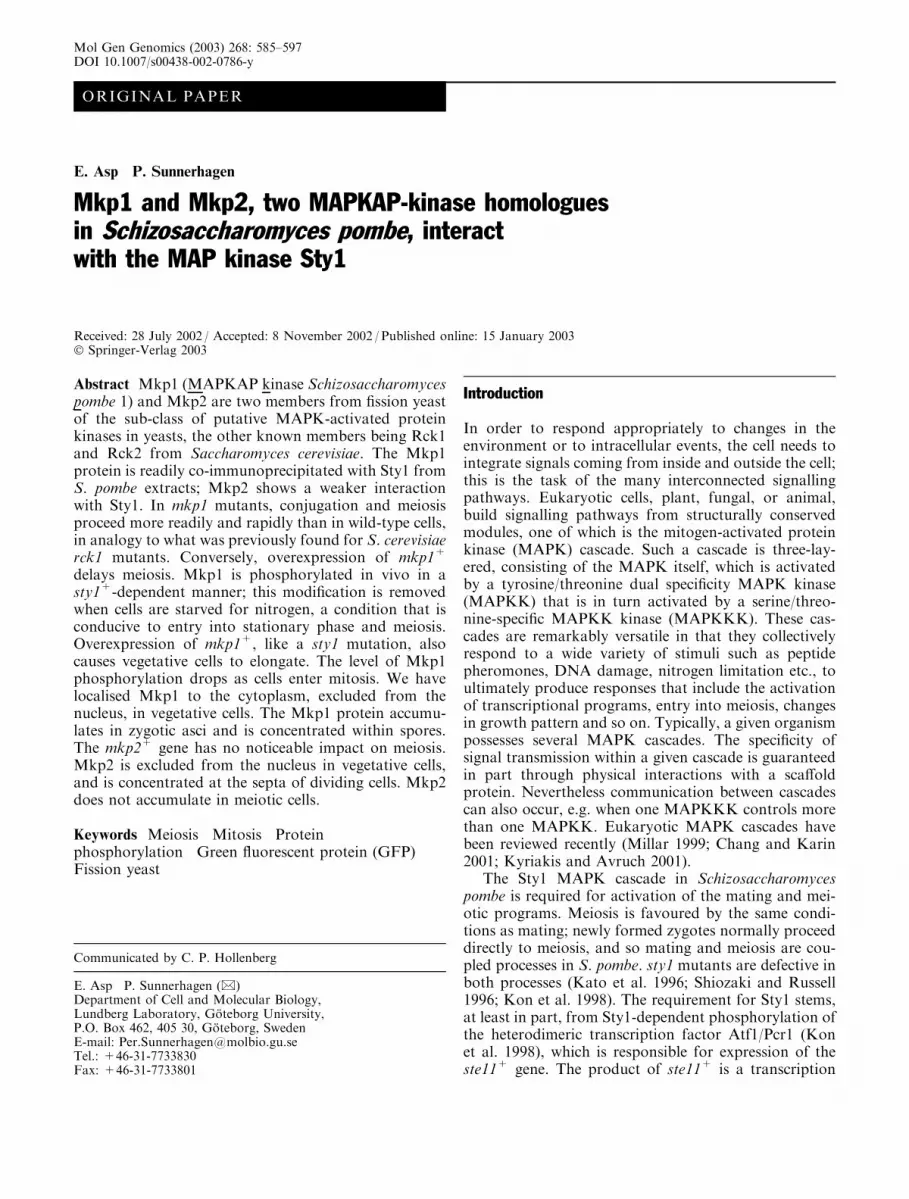
In Fig. 1A, an alignment of the predicted sequencesof the four members of this subfamily of protein kinasesis shown, together with a mammalian MAPKAPK, andan S. pombe Ca2+/calmodulin-regulated protein kinase,Cmk1. The four yeast protein kinases share two prom-inent features. First, within the catalytic domain (upperrow), there is a glycine-rich sequence N-terminal to thehighly conserved DFG-box, which was initially noted inRck1 and Rck2 (Dahlkvist and Sunnerhagen 1994). Thissequence is not found in any other protein kinases as faras we have been able to ascertain. We speculate that itmay protrude from the otherwise conserved structure ofthe catalytic domain, and so we have dubbed it the‘‘glycine loop’’. The second feature (bottom row), whichunlike the first one is shared with mammalian MAP-KAPKs, is the putative MAPK-binding site near theC-terminus of the protein, containing the conservedconsensus sequence RR (Tanoue et al. 2000). Indeed, thebinding of Rck2 to the MAPK Hog1 has been shown tooccur through the C-terminal part of Rck2 (Bilsland-Marchesan et al. 2000). From various measures of se-quence relatedness, it is not obvious if either of the twoS. pombe proteins is more closely related to one of thetwo proteins in S. cerevisiae (Fig. 1B).
Inactivation of mkp1+ facilitates conjugationand entry into meiosis
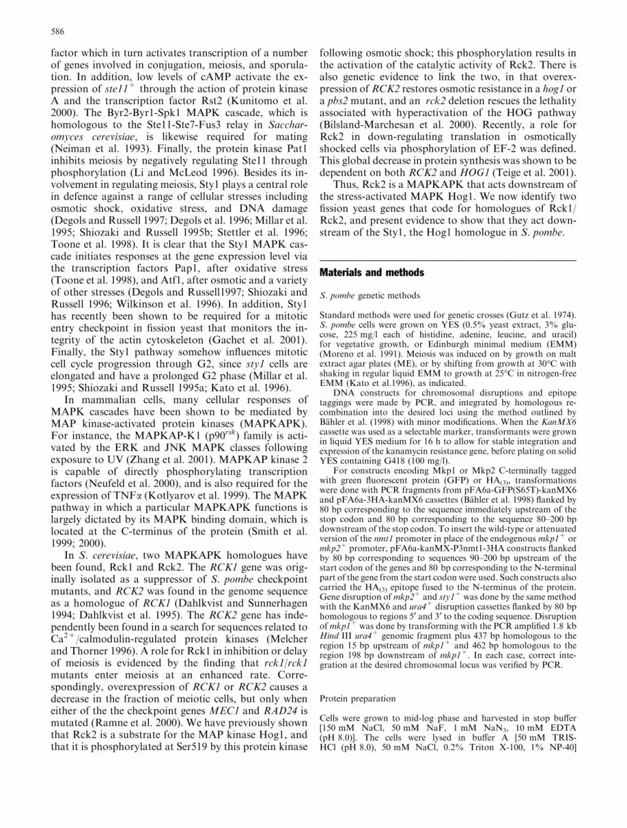
Since the mkp1+/mkp2+ homologues in S. cerevisiae,RCK1 and RCK2, delay or depress meiosis (Ramne et al.2000), and since Sty1, the fission yeast homologue of theHog1 MAPK, is required for efficient meiosis (Katoet al.1996; Shiozaki and Russell 1996; Kon et al. 1998),we wanted to investigate the effects on conjugation andmeiosis of disruption or up-regulation of mkp1+ andmkp2+. In Fig. 2A, spore staining with iodine vapourreveals that h90 mkp1D cells enter meiosis faster thanwild-type h90 cells on a variety of media. After 72 h onstandard meiosis-inducing medium (malt extract agarplates), markedly more staining is seen in the mutantsthan in the h90 wild-type strain (Fig. 2A); a similar dif-ference is seen on EMM, which is semi-permissive formeiosis. On YES medium, containing yeast extract andglucose, meiosis is normally inhibited in S. pombe cells.However, mkp1D mutants are able to sporulate exten-sively on YES plates when the glucose concentration isreduced to 1%; very few wild-type cells undergo meiosisunder these conditions (Fig. 2A). At 2% glucose, how-ever, sporulation was completely suppressed in bothmutant and wild-type strains (Fig. 2A).
We also investigated the effect of up-regulating bothgenes under the control of the regulatable nmt1+ pro-moter. In Fig. 2B, we see that overexpression of mkp1+
leads to considerable depression of sporulation; thiseffect is abolished by lowering the expression level by the
587
addition of thiamine to the medium. It should be notedthat the nmt1-P3 promoter variant used permits suffi-cient transcription even in the presence of thiamine thatno phenotypic effects of down-regulation are seen here.In contrast, neither up-regulation nor disruption of themkp2+ gene had any obvious impact on the kinetics ofspore formation (not shown).
Because of the obvious impact by mkp1+, but not bymkp2+, on the timing of spore production, we wanted toinvestigate the kinetics of appearance of zygotes (prod-
ucts of conjugation), as well as asci and spores (productsof meiosis), in strains in which mkp1+ was disrupted orectopically overexpressed. Zygotes appear in the mkp1
Fig. 1A, B The Rck family of protein kinases: Rck1, S. cerevisiae(Acc. No. S47900); Rck2, S. cerevisiae (Acc. No. S59394); Mkp1,S. pombe (Acc. No. O94547); Mkp2, S. pombe (Acc. No. O42844).For comparison, the following protein kinase sequences are shown:MAPKAP-2, human MAPK-activated protein kinase 2 (Acc. No.P49137); Cmk1Sp, S. pombe calmodulin-activated protein kinase 1(Acc. No. O74235); Cmk1Sc, S. cerevisiae calmodulin-activatedprotein kinase 1 (Acc. No. A40896); Cmk2Sc, S. cerevisiaecalmodulin-activated protein kinase 2 (Acc. No. B40896). ADefining sequence features. The subdomains VIb and VII of theprotein kinase catalytic domain (Hunter 1991) are marked abovethe sequence. The ‘‘glycine loop’’, which is not present in otherprotein kinases, upstream of the highly conserved DFG box in thekinase catalytic domain, and the MAP kinase binding domain nearthe C-terminus are boxed in grey. Conserved positions within thesedomains are marked in bold in the consensus sequence. BPhylogenetic comparison of the yeast MAPKAP kinases and theirrelationship to Ca2+/CaM-regulated protein kinases in yeast.Estimated relative phylogenetic distances are indicated by the tree,which was calculated using Kimura’s correction (Kimura andTakahata 1983)
Fig. 2A–C Conjugation and meiosis in strains with altered expres-sion of mkp1+ or mkp2+. All strains are in the meiosis-proficienth90 genetic background. Cells were grown at 30�C to mid log-phasein liquid EMM with the required supplements prior to spotting onsolid media as indicated. In panels A and B sporulation wasdetected by staining with iodine vapour. A Sporulation in wild type(h90) and mkp1D mutants (EA31) on different solid media. Pictureswere taken at 72 h after plating and incubation at 25�C. BSporulation in wild type (h90) and in a strain with ectopicoverexpression of mkp1+ (EA64) on solid EMM. Expression fromthe nmt1 promoter (nmt1-P3) was induced at maximal levels bycultivation in the absence of thiamine, or repressed by the additionof thiamine (10 lM) to the medium. Strains were initially culturedin liquid medium of the same composition as that on which theywere subsequently plated. Pictures were taken at 72 or 96 h afterplating and incubation at 25�C. C Time course of conjugation andmeiosis in the wild-type strain, and in strains lacking oroverexpressing mkp1+. Cells were first cultivated in liquid standardEMM (without added thiamine) at 30�C. The meiotic pathway wasinduced by transfer to nitrogen-free EMM at 25�C (time 0), andcultivation with shaking was continued. Ectopic expression fromthe nmt1-P3:mkp1+ construct was maximised by cultivation inmedium without thiamine throughout the experiment. Sampleswere taken at the indicated times after shift and examined bymicroscopy for the presence of zygotes, four-spored asci and freespores. Meiotic efficiency was defined as (2Z+2A+0.5S)/(2Z+2A+0.5S+V), where Z is the number of zygotes; A thenumber of asci; S the number of free spores; and V the number ofvegetative cells (Kunitomo et al. 1995)
c
588
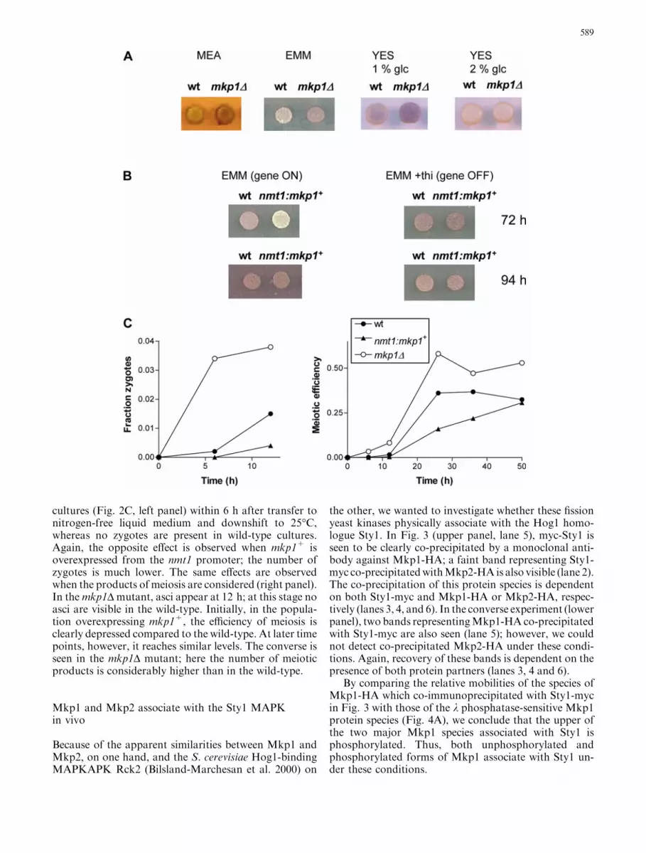
cultures (Fig. 2C, left panel) within 6 h after transfer tonitrogen-free liquid medium and downshift to 25�C,whereas no zygotes are present in wild-type cultures.Again, the opposite effect is observed when mkp1+ isoverexpressed from the nmt1 promoter; the number ofzygotes is much lower. The same effects are observedwhen the products of meiosis are considered (right panel).In themkp1D mutant, asci appear at 12 h; at this stage noasci are visible in the wild-type. Initially, in the popula-tion overexpressing mkp1+, the efficiency of meiosis isclearly depressed compared to the wild-type. At later timepoints, however, it reaches similar levels. The converse isseen in the mkp1D mutant; here the number of meioticproducts is considerably higher than in the wild-type.
Mkp1 and Mkp2 associate with the Sty1 MAPKin vivo
Because of the apparent similarities between Mkp1 andMkp2, on one hand, and the S. cerevisiae Hog1-bindingMAPKAPK Rck2 (Bilsland-Marchesan et al. 2000) on
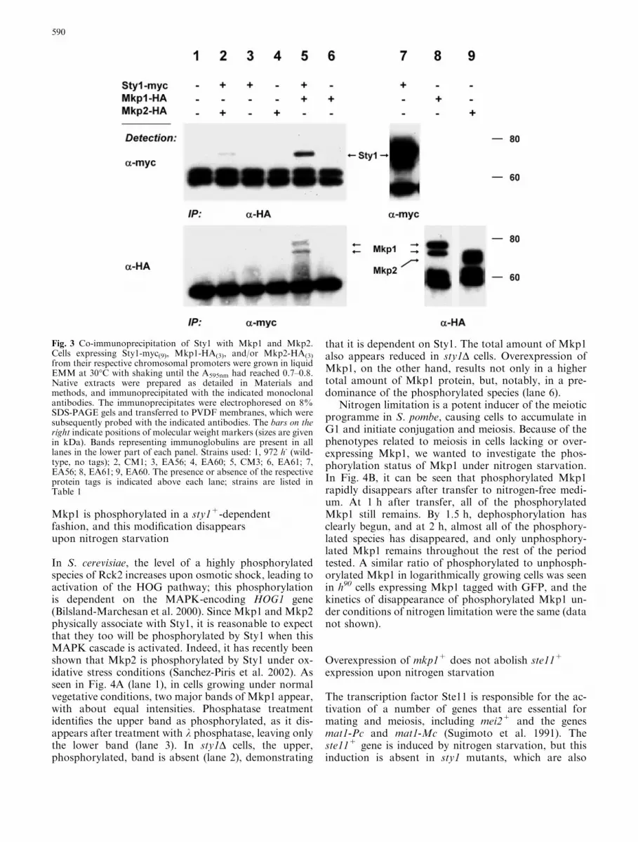
the other, we wanted to investigate whether these fissionyeast kinases physically associate with the Hog1 homo-logue Sty1. In Fig. 3 (upper panel, lane 5), myc-Sty1 isseen to be clearly co-precipitated by a monoclonal anti-body against Mkp1-HA; a faint band representing Sty1-myc co-precipitatedwithMkp2-HA is also visible (lane 2).The co-precipitation of this protein species is dependenton both Sty1-myc and Mkp1-HA or Mkp2-HA, respec-tively (lanes 3, 4, and 6). In the converse experiment (lowerpanel), two bands representingMkp1-HAco-precipitatedwith Sty1-myc are also seen (lane 5); however, we couldnot detect co-precipitated Mkp2-HA under these condi-tions. Again, recovery of these bands is dependent on thepresence of both protein partners (lanes 3, 4 and 6).
By comparing the relative mobilities of the species ofMkp1-HA which co-immunoprecipitated with Sty1-mycin Fig. 3 with those of the k phosphatase-sensitive Mkp1protein species (Fig. 4A), we conclude that the upper ofthe two major Mkp1 species associated with Sty1 isphosphorylated. Thus, both unphosphorylated andphosphorylated forms of Mkp1 associate with Sty1 un-der these conditions.
589
Mkp1 is phosphorylated in a sty1+-dependentfashion, and this modification disappearsupon nitrogen starvation
In S. cerevisiae, the level of a highly phosphorylatedspecies of Rck2 increases upon osmotic shock, leading toactivation of the HOG pathway; this phosphorylationis dependent on the MAPK-encoding HOG1 gene(Bilsland-Marchesan et al. 2000). Since Mkp1 and Mkp2physically associate with Sty1, it is reasonable to expectthat they too will be phosphorylated by Sty1 when thisMAPK cascade is activated. Indeed, it has recently beenshown that Mkp2 is phosphorylated by Sty1 under ox-idative stress conditions (Sanchez-Piris et al. 2002). Asseen in Fig. 4A (lane 1), in cells growing under normalvegetative conditions, two major bands of Mkp1 appear,with about equal intensities. Phosphatase treatmentidentifies the upper band as phosphorylated, as it dis-appears after treatment with k phosphatase, leaving onlythe lower band (lane 3). In sty1D cells, the upper,phosphorylated, band is absent (lane 2), demonstrating
that it is dependent on Sty1. The total amount of Mkp1also appears reduced in sty1D cells. Overexpression ofMkp1, on the other hand, results not only in a highertotal amount of Mkp1 protein, but, notably, in a pre-dominance of the phosphorylated species (lane 6).
Nitrogen limitation is a potent inducer of the meioticprogramme in S. pombe, causing cells to accumulate inG1 and initiate conjugation and meiosis. Because of thephenotypes related to meiosis in cells lacking or over-expressing Mkp1, we wanted to investigate the phos-phorylation status of Mkp1 under nitrogen starvation.In Fig. 4B, it can be seen that phosphorylated Mkp1rapidly disappears after transfer to nitrogen-free medi-um. At 1 h after transfer, all of the phosphorylatedMkp1 still remains. By 1.5 h, dephosphorylation hasclearly begun, and at 2 h, almost all of the phosphory-lated species has disappeared, and only unphosphory-lated Mkp1 remains throughout the rest of the periodtested. A similar ratio of phosphorylated to unphosph-orylated Mkp1 in logarithmically growing cells was seenin h90 cells expressing Mkp1 tagged with GFP, and thekinetics of disappearance of phosphorylated Mkp1 un-der conditions of nitrogen limitation were the same (datanot shown).
Overexpression of mkp1+ does not abolish ste11+
expression upon nitrogen starvation
The transcription factor Ste11 is responsible for the ac-tivation of a number of genes that are essential formating and meiosis, including mei2+ and the genesmat1-Pc and mat1-Mc (Sugimoto et al. 1991). Theste11+ gene is induced by nitrogen starvation, but thisinduction is absent in sty1 mutants, which are also
Fig. 3 Co-immunoprecipitation of Sty1 with Mkp1 and Mkp2.Cells expressing Sty1-myc(9), Mkp1-HA(3), and/or Mkp2-HA(3)
from their respective chromosomal promoters were grown in liquidEMM at 30�C with shaking until the A595nm had reached 0.7–0.8.Native extracts were prepared as detailed in Materials andmethods, and immunoprecipitated with the indicated monoclonalantibodies. The immunoprecipitates were electrophoresed on 8%SDS-PAGE gels and transferred to PVDF membranes, which weresubsequently probed with the indicated antibodies. The bars on theright indicate positions of molecular weight markers (sizes are givenin kDa). Bands representing immunoglobulins are present in alllanes in the lower part of each panel. Strains used: 1, 972 h- (wild-type, no tags); 2, CM1; 3, EA56; 4, EA60; 5, CM3; 6, EA61; 7,EA56; 8, EA61; 9, EA60. The presence or absence of the respectiveprotein tags is indicated above each lane; strains are listed inTable 1
590
defective in meiosis (Shiozaki and Russell 1996). Therequirement for Sty1 for ste11+ expression is mediatedthrough the transcription factor Atf1/Pcr1 (Kon et al.1998). Since overexpression of mkp1+ causes a delay inmeiosis, and since Sty1 is required for entry into meiosis,we wanted to see if the delay could be ascribed to a lackof expression of ste11+. In Fig. 5, it is seen that ste11+
induction is somewhat delayed in cells overexpressingmkp1+, but that the transcript is clearly induced to es-sentially wild-type levels following nitrogen starvation,in contrast to the case in sty1 mutants.
Overexpression of Mkp1 causes elongationof mitotic cells
It has previously been shown that sty1 cells are elon-gated and the G2 phase of the cell cycle is prolonged(Millar et al. 1995; Shiozaki and Russell 1995a; Kato etal. 1996). When Mkp1 is ectopically expressed from thefully induced nmt1-P3 promoter for an extended period,the strain displays a marked increase in average celllength, as well as in length at septation (Fig. 6). Theconverse situation, an mkp1D background, does nothave such an obvious effect on cell length, nor doesup-regulation or deletion of mkp2+ have a noticeableinfluence in this regard (not shown).
Mkp1 phosphorylation decreases as cells enter mitosis
The G2/M transition in the mitotic cell cycle is regulatedthrough multiple phosphorylation events, includingphosphorylation of Cdc2 on Tyr 15. Our observation
that overexpression of Mkp1, like a sty1 mutation,causes cell elongation, and the fact that phosphorylationof Mkp1 is Sty1-dependent, led us to examine the level
Table 1 S. pombe strains
Strain Genotype Source/reference
EA34 h90 Wild typeEA31 h90 mkp1::ura4+ ura4-D18 This workEA99 h90 mkp2::KanMX6 This workEA64 h90 KanMX6:nmt1-P3:HA(3):mkp1+ This workEA45 h90 KanMX6:nmt1-P3:HA(3):mkp2+ This workEA69 h90 mkp1+:GFP:KanMX6 This workEA66 h90 mkp2+:GFP:KanMX6 This work972 h- Wild typeEA1 h-his3 leu1–32 ura4-D18 P. RussellEA18 h-mkp1::ura4+ his3 leu1-32 ura4-D18 This workEA98 h-mkp2::KanMX6 This workEA56 h-sty1+:myc(9):ura4
+ leu132 ura4D-18 ade6-M210 J. Millar (JM1698)CM1 h-sty1 +:myc(9):ura4
+mkp2 +:HA(3):KanMX6 leu132 ura4D-18 ade6-M210 This workCM3 h-sty1+:myc(9):ura4
+ mkp1+:HA(3):KanMX6 leu1-32 ura4D-18 ade6-M210 This workEA61 h-mkp1 +:HA(3):KanMX6 leu1-32 This workEA60 h-mkp2+:HA(3):KanMX6 leu1-32 This workCM9 h-KanMX6:nmt1-P3:HA(3):mkp1+ This workCM10 h-KanMX6:nmt1-P3:HA(3):mkp2+ This workEA79 h-mkp2+:GFP:KanMX6 This workEA82 h-mkp1+:GFP:KanMX6 This workEA36 h-cdc25-22 leu1-32 S. MorenoEA87 h-mkp1+:HA(3):KanMX6 cdc25-22 leu1-32 This workEA95 h-mkp1+:HA(3):KanMX6 leu1-32 ura4-D18 This workEA100 h-mkp1+:HA(3):KanMX6sty1::ura4+ ura4D-18 leu1-32 This work
Fig. 4A, B Phosphorylation of Mkp1 in vivo. Cells expressingHA-tagged Mkp1 were grown in liquid medium as detailed below.Protein samples were processed for analysis by Western blotting,and probed with a-HA antibodies. A Phosphorylated andunphosphorylated species of Mkp1. From left to right: logarith-mically growing wild-type cells (EA61; mkp1+:HA(3)) in normalEMM medium; sty1D (EA100; mkp1+:HA(3) sty1::ura4+); as inlane 1 but treated with 400 U of k phosphatase (New EnglandBiolabs) at 37�C for 15 min.; same as in lane 1; negative controlwithout HA tag (972 h-); Mkp1 overexpression (CM9; nmt1-P3:HA(3):mkp1+), cells grown in the absence of thiamine to ensuremaximal induction of the nmt1 promoter. BMkp1 phosphorylationstatus during nitrogen starvation. Wild-type h- cells (EA61;mkp1+:HA(3)) were grown to mid-logarithmic phase in normalEMM and then transferred to nitrogen-free EMM medium asdescribed in the legend to Fig. 2C. Samples were taken at theindicated times thereafter. The last lane shows a negative controlwithout the HA tag (972 h-)
591
and phosphorylation status of Mkp1 during the mitoticcell cycle. In Fig. 7, the result of a cdc25 block and re-lease experiment is shown. The total amount of Mkp1does not change appreciably during the mitotic cell cy-cle. However, at time points 30 and 180 min after shift-
down, there is a clear decrease in the upper band, rep-resenting phosphorylated Mkp1. These timepoints arecoincident with the two metaphases of the cycles repre-sented in this experiment. The decrease is only transient,as normal phosphorylation levels reappear in the fol-lowing samples, corresponding to anaphase.
Localisation of Mkp1 and Mkp2 in vegetative cells
We constructed gene fusions expressing green fluores-cent protein (GFP) C-terminally linked to Mkp1 orMkp2, integrated at their respective chromosomal locusand under control of their endogenous promoters. Thesub-cellular localisation of Mkp1-GFP and Mkp2-GFPwas examined by fluorescence microscopy (Fig. 8).
Fig. 7 Expression and phosphorylation status of Mkp1 during themitotic cell cycle. mkp1-HA cdc25-22 cells (EA87) grown in liquidEMM were synchronised in G2 by incubation for 3 h at 35�C, andthen released from the block by lowering the temperature to 25�C.Samples were taken at the indicated times thereafter andimmediately placed on ice. From these, denatured protein extractswere prepared and subjected to Western analysis with a-HAantibodies. An aliquot of each sample was stained with DAPI andanalysed by fluorescence microscopy to determine the the cell cyclestage (indicated in the lower panel)
Fig. 5 Expression of ste11+ after nitrogen starvation in wild-typecells and cells overexpressing mkp1+. Cells growing logarithmicallyin liquid EMM at 30�C were washed and transferred to nitrogen-free EMM at 25�C; cultivation was continued under theseconditions for the indicated times. Ectopic overexpression ofmkp1+ from the nmt1-P3 promoter was maximised before andafter nitrogen starvation by omitting thiamine from the culturemedium. RNA was prepared from samples taken at the indicatedtimes after transfer. A probe for ste11+ was made by PCR usingthe primers Ste11-F (5¢-AGCTTTATTAGCTCCCGCTC-3¢) andSte11-R (5¢-ACTTTGCTCACTCACGGTTC-3¢). Lanes 1–4, wild-type cells (972 h-); lanes 5–8, cells overexpressing mkp1+ (CM9,h- nmt1-P3:HA(3):mkp1+). Lower panel Loading controls (28Sribosomal RNA) stained with ethidium bromide
Fig. 6 Elongation of cells overexpressing Mkp1. Cells were grownin liquid EMM in the absence of thiamine (maximal induction ofthe nmt1-P3 promoter) at 30�C for 48 h, and bright-fieldmicrographs were taken. Upper panel CM9 (expressing Mkp1 fromthe strong nmt1-P3 promoter). Lower panel Wild-type (972 h-)
Fig. 8 Localisation of Mkp1-GFP and Mkp2-GFP in vegetativecells. Cells carrying constructs expressing mkp1+:GFP(EA82) ormkp2+:GFP(EA79) from their respective endogenous chromoso-mal loci in a h- background were grown to mid-logarithmic phasein normal EMM at 30�C and examined by fluorescence microscopy
592
For Mkp1, the overall fluorescent signal is somewhatstronger than for Mkp2. Mkp1-GFP is seen in the cy-toplasm, and is clearly excluded from the nucleus(Fig. 8A, B).
In non-septated cells, a weak signal from Mkp2-GFPis seen in the cytoplasm, excluding the nucleus (Fig. 8C,D). This exclusion is less pronounced than for Mkp1,however. In post-mitotic cells, we observe a distinctsignal from Mkp2 at the newly formed septa.
Localisation of Mkp1 and Mkp2 in meiotic cells
Because of the effects of the mkp1+ gene on meiosis, weinvestigated the status of the Mkp1 and Mkp2 proteinsunder conditions conducive to meiosis. The Mkp1-GFPand Mkp2-GFP fusion proteins were also expressed inmeiotic cells by integrating the same DNA constructs asabove into the corresponding loci in the genome of wild-type h90 cells. Cells were induced to enter meiosis bycultivation in liquid nitrogen-free medium, and sampleswere taken at various time-points.
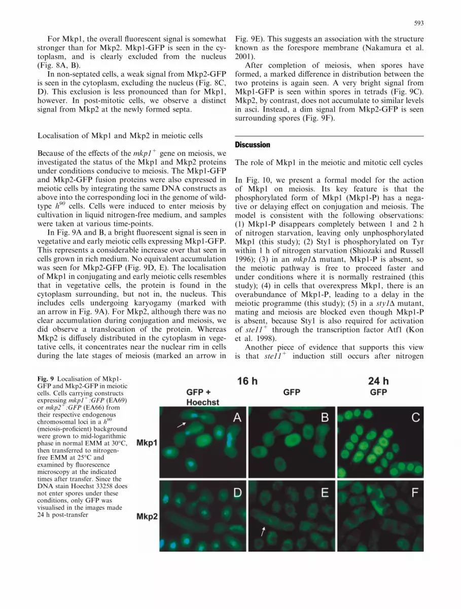
In Fig. 9A and B, a bright fluorescent signal is seen invegetative and early meiotic cells expressing Mkp1-GFP.This represents a considerable increase over that seen incells grown in rich medium. No equivalent accumulationwas seen for Mkp2-GFP (Fig. 9D, E). The localisationof Mkp1 in conjugating and early meiotic cells resemblesthat in vegetative cells, the protein is found in thecytoplasm surrounding, but not in, the nucleus. Thisincludes cells undergoing karyogamy (marked withan arrow in Fig. 9A). For Mkp2, although there was noclear accumulation during conjugation and meiosis, wedid observe a translocation of the protein. WhereasMkp2 is diffusely distributed in the cytoplasm in vege-tative cells, it concentrates near the nuclear rim in cellsduring the late stages of meiosis (marked an arrow in
Fig. 9E). This suggests an association with the structureknown as the forespore membrane (Nakamura et al.2001).
After completion of meiosis, when spores haveformed, a marked difference in distribution between thetwo proteins is again seen. A very bright signal fromMkp1-GFP is seen within spores in tetrads (Fig. 9C).Mkp2, by contrast, does not accumulate to similar levelsin asci. Instead, a dim signal from Mkp2-GFP is seensurrounding spores (Fig. 9F).
Discussion
The role of Mkp1 in the meiotic and mitotic cell cycles
In Fig. 10, we present a formal model for the actionof Mkp1 on meiosis. Its key feature is that thephosphorylated form of Mkp1 (Mkp1-P) has a nega-tive or delaying effect on conjugation and meiosis. Themodel is consistent with the following observations:(1) Mkp1-P disappears completely between 1 and 2 hof nitrogen starvation, leaving only unphosphorylatedMkp1 (this study); (2) Sty1 is phosphorylated on Tyrwithin 1 h of nitrogen starvation (Shiozaki and Russell1996); (3) in an mkp1D mutant, Mkp1-P is absent, sothe meiotic pathway is free to proceed faster andunder conditions where it is normally restrained (thisstudy); (4) in cells that overexpress Mkp1, there is anoverabundance of Mkp1-P, leading to a delay in themeiotic programme (this study); (5) in a sty1D mutant,mating and meiosis are blocked even though Mkp1-Pis absent, because Sty1 is also required for activationof ste11+ through the transcription factor Atf1 (Konet al. 1998).
Another piece of evidence that supports this viewis that ste11+ induction still occurs after nitrogen
Fig. 9 Localisation of Mkp1-GFP and Mkp2-GFP in meioticcells. Cells carrying constructsexpressing mkp1+:GFP (EA69)or mkp2+:GFP (EA66) fromtheir respective endogenouschromosomal loci in a h90
(meiosis-proficient) backgroundwere grown to mid-logarithmicphase in normal EMM at 30�C,then transferred to nitrogen-free EMM at 25�C andexamined by fluorescencemicroscopy at the indicatedtimes after transfer. Since theDNA stain Hoechst 33258 doesnot enter spores under theseconditions, only GFP wasvisualised in the images made24 h post-transfer
593
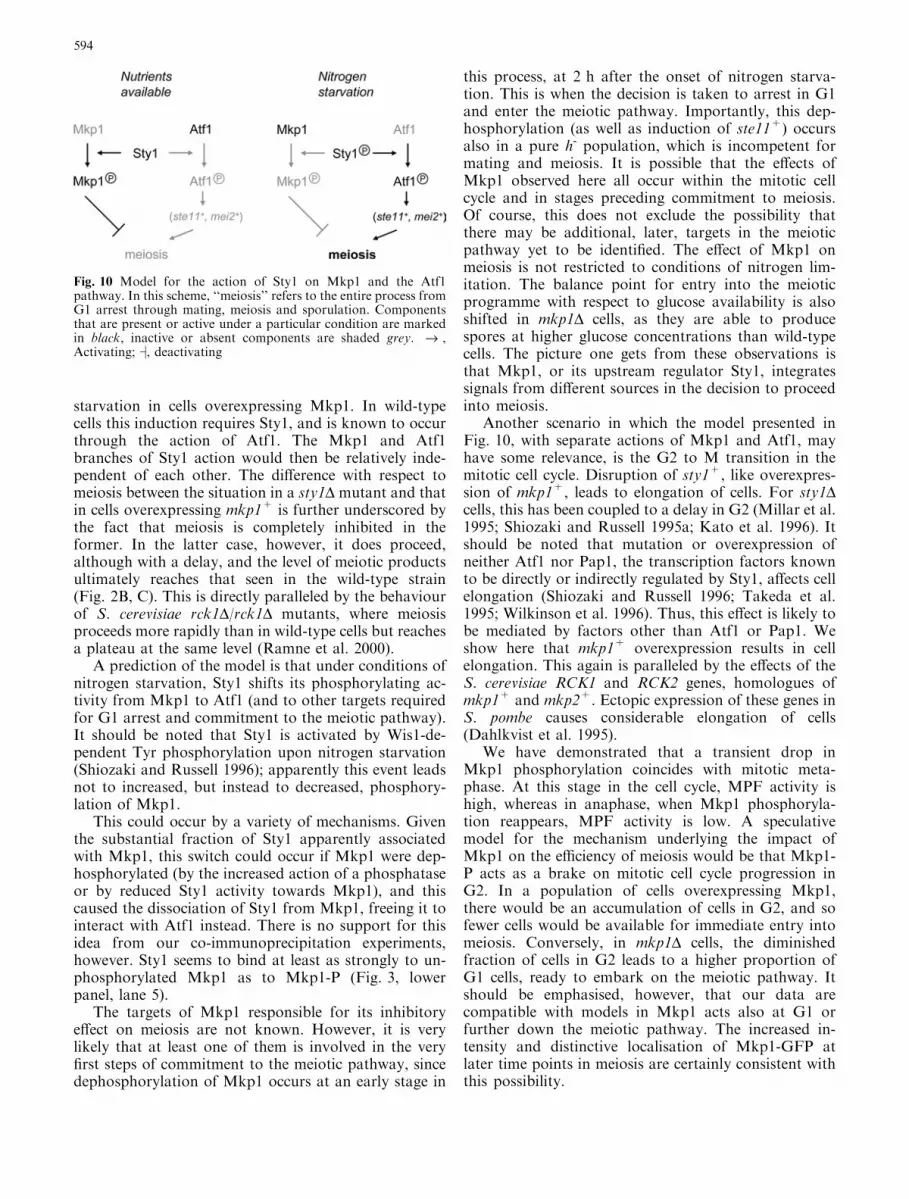
starvation in cells overexpressing Mkp1. In wild-typecells this induction requires Sty1, and is known to occurthrough the action of Atf1. The Mkp1 and Atf1branches of Sty1 action would then be relatively inde-pendent of each other. The difference with respect tomeiosis between the situation in a sty1D mutant and thatin cells overexpressing mkp1+ is further underscored bythe fact that meiosis is completely inhibited in theformer. In the latter case, however, it does proceed,although with a delay, and the level of meiotic productsultimately reaches that seen in the wild-type strain(Fig. 2B, C). This is directly paralleled by the behaviourof S. cerevisiae rck1D/rck1D mutants, where meiosisproceeds more rapidly than in wild-type cells but reachesa plateau at the same level (Ramne et al. 2000).
A prediction of the model is that under conditions ofnitrogen starvation, Sty1 shifts its phosphorylating ac-tivity from Mkp1 to Atf1 (and to other targets requiredfor G1 arrest and commitment to the meiotic pathway).It should be noted that Sty1 is activated by Wis1-de-pendent Tyr phosphorylation upon nitrogen starvation(Shiozaki and Russell 1996); apparently this event leadsnot to increased, but instead to decreased, phosphory-lation of Mkp1.
This could occur by a variety of mechanisms. Giventhe substantial fraction of Sty1 apparently associatedwith Mkp1, this switch could occur if Mkp1 were dep-hosphorylated (by the increased action of a phosphataseor by reduced Sty1 activity towards Mkp1), and thiscaused the dissociation of Sty1 from Mkp1, freeing it tointeract with Atf1 instead. There is no support for thisidea from our co-immunoprecipitation experiments,however. Sty1 seems to bind at least as strongly to un-phosphorylated Mkp1 as to Mkp1-P (Fig. 3, lowerpanel, lane 5).
The targets of Mkp1 responsible for its inhibitoryeffect on meiosis are not known. However, it is verylikely that at least one of them is involved in the veryfirst steps of commitment to the meiotic pathway, sincedephosphorylation of Mkp1 occurs at an early stage in
this process, at 2 h after the onset of nitrogen starva-tion. This is when the decision is taken to arrest in G1and enter the meiotic pathway. Importantly, this dep-hosphorylation (as well as induction of ste11+) occursalso in a pure h- population, which is incompetent formating and meiosis. It is possible that the effects ofMkp1 observed here all occur within the mitotic cellcycle and in stages preceding commitment to meiosis.Of course, this does not exclude the possibility thatthere may be additional, later, targets in the meioticpathway yet to be identified. The effect of Mkp1 onmeiosis is not restricted to conditions of nitrogen lim-itation. The balance point for entry into the meioticprogramme with respect to glucose availability is alsoshifted in mkp1D cells, as they are able to producespores at higher glucose concentrations than wild-typecells. The picture one gets from these observations isthat Mkp1, or its upstream regulator Sty1, integratessignals from different sources in the decision to proceedinto meiosis.
Another scenario in which the model presented inFig. 10, with separate actions of Mkp1 and Atf1, mayhave some relevance, is the G2 to M transition in themitotic cell cycle. Disruption of sty1+, like overexpres-sion of mkp1+, leads to elongation of cells. For sty1Dcells, this has been coupled to a delay in G2 (Millar et al.1995; Shiozaki and Russell 1995a; Kato et al. 1996). Itshould be noted that mutation or overexpression ofneither Atf1 nor Pap1, the transcription factors knownto be directly or indirectly regulated by Sty1, affects cellelongation (Shiozaki and Russell 1996; Takeda et al.1995; Wilkinson et al. 1996). Thus, this effect is likely tobe mediated by factors other than Atf1 or Pap1. Weshow here that mkp1+ overexpression results in cellelongation. This again is paralleled by the effects of theS. cerevisiae RCK1 and RCK2 genes, homologues ofmkp1+ and mkp2+. Ectopic expression of these genes inS. pombe causes considerable elongation of cells(Dahlkvist et al. 1995).
We have demonstrated that a transient drop inMkp1 phosphorylation coincides with mitotic meta-phase. At this stage in the cell cycle, MPF activity ishigh, whereas in anaphase, when Mkp1 phosphoryla-tion reappears, MPF activity is low. A speculativemodel for the mechanism underlying the impact ofMkp1 on the efficiency of meiosis would be that Mkp1-P acts as a brake on mitotic cell cycle progression inG2. In a population of cells overexpressing Mkp1,there would be an accumulation of cells in G2, and sofewer cells would be available for immediate entry intomeiosis. Conversely, in mkp1D cells, the diminishedfraction of cells in G2 leads to a higher proportion ofG1 cells, ready to embark on the meiotic pathway. Itshould be emphasised, however, that our data arecompatible with models in Mkp1 acts also at G1 orfurther down the meiotic pathway. The increased in-tensity and distinctive localisation of Mkp1-GFP atlater time points in meiosis are certainly consistent withthis possibility.
Fig. 10 Model for the action of Sty1 on Mkp1 and the Atf1pathway. In this scheme, ‘‘meiosis’’ refers to the entire process fromG1 arrest through mating, meiosis and sporulation. Componentsthat are present or active under a particular condition are markedin black, inactive or absent components are shaded grey. fi ,Activating; ˛, deactivating
594
The nature of the interaction betweenthe MAPK Sty1 and Mkp1/2
There appears to be quite a tight association betweenSty1 and Mkp1, since a considerable fraction of Sty1 isco-precipitated with Mkp1 and vice versa, when bothproteins are expressed from their endogenous promot-ers. This view is reinforced by the finding that Mkp1 isconstitutively phosphorylated; in our hands about halfof total Mkp1 is in the form of Mkp1-P. By contrast, wefind only a weak interaction between Sty1 and Mkp2.Recently, Sanchez-Piris et al. (2002) demonstrated aninteraction between these two proteins by overexpress-ing both of them in the same cells. We can detect Sty1co-immunoprecipitated with Mkp2 when both proteinsare expressed at normal levels from their endogenouspromoters at their normal chromosomal loci; however,the signal is faint. Since Mkp1 and Mkp2 are expressedat similar levels in wild-type cells under standard con-ditions, it follows that under normal, unstressed, con-ditions, Sty1 binds to Mkp2 with only a small fraction ofthe affinity that it has for Mkp1.
We have shown here that the S. pombe proteinsMkp1 and Mkp2, homologues of Rck1 and Rck2, bindto the MAPK Sty1. Of course, this does not excludepotential additional regulatory mechanisms for Mkp1and Mkp2. Grouping of eukaryotic protein kinase se-quences places the Rck/Mkp subfamily close to Ca2+/calmodulin (CaM)-regulated kinases (Dahlkvist andSunnerhagen 1994; Melcher and Thorner 1996; Teige etal. 2001), yet experimental evidence supporting regula-tion of Rck1 or Rck2 from S. cerevisiae, or of S. pombeMkp2 by Ca2+/CaM is lacking (Melcher and Thorner1996; Ramne et al. 2000; Sanchez-Piris et al.2002). Itshould be noted that a bona fide Ca2+/CaM proteinkinase from S. pombe, Cmk1, has already been de-scribed. There is clear evidence that this protein bothbinds to and is activated by Ca2+/CaM, in contrast tothe Rck/Mkp subfamily (Rasmussen 2000). Mkp2 hasboth sequence features typical of the Rck subfamily,whereas Cmk1 lacks both (Fig. 1A). The contention thatMkp2 is more closely related to the Rck/Mkp subfamilythan to CaM-regulated kinases, on the one hand, andthat S. pombe Cmk1 is more related to the S. cerevisiaeCaM-regulated kinases (Cmk1 and Cmk2) on the other,is also supported by the clustering based on sequencesimilarity (Fig. 1B).
How many MAPKAPKs are there in yeast?
Although a relatively large number of protein kinaseswith putative MAPK-binding domains are found inplant, animal and fungal species, so far we have onlyidentified the ‘‘glycine loop’’ in the four members of theRck subfamily. Do these four then make up the wholeset of MAPKAPKs in S. pombe and S. cerevisiae? Usingless stringent homology criteria, excluding the ‘‘glycineloop’’ but including a protein kinase domain and a
sequence similar to a MAPK binding box at the C-ter-minus, one can find between five and eight candidategenes in each of the yeast genomes. Only experiment cantell if any of these other candidates correspond to actualMAPKAPKs. Until we know the number of MAP-KAPKs in yeast, we cannot tell if all MAPKAPKs arespecific to one MAPK cascade, or if one or a fewMAPKAPKs integrate the responses of several MAPKcascades.
We have found both Mkp1, and, to a lesser degree,Mkp2, to be physically associated with Sty1. It is notunreasonable to suppose that both these putativeMAPKAPKs are linked to the same MAPK, given thefollowing considerations regarding the situation in S.cerevisiae. Rck2 has been shown to interact with Hog1by co-immunoprecipitation as well as two-hybridanalysis (Bilsland-Marchesan et al. 2000). In globalscreens for protein-protein interactions, Hog1 has beenfound to bind to Rck1, using both two-hybrid analysis(Uetz et al. 2000) and affinity tag purification/massspectrometry (Ho et al. 2002). In addition, an interac-tion between Rck1 and Fus3 has been reported (Ho etal. 2002). Thus, Rck1 potentially binds two MAPKs,Hog1 and Fus3. The RCK1 transcript is strongly up-regulated in fus3 mutants and after treatment with apheromone (Roberts et al. 2000), hinting at a role forRck1 in the mating pathway. By analogy, one mayspeculate that Mkp1 could interact with Spk1, the Fus3homologue. In fission yeast, the Sty1 cascade, as well asSpk1, has a major impact on mating and meiosis. Thesame is not true for its budding yeast homologue, theHog1 MAPK cascade. It is conceivable that Mkp2,besides its weak interaction with Sty1, has anotherMAPK interaction partner; at this stage we cannotspeculate about likely candidates.
Additional functions of Mkp1 and Mkp2
Besides its effect on mating and meiosis, Sty1 is alsorequired for the activation of responses to osmotic andoxidative stress conditions. Mutation of the sty1+ genecauses sensitivity to a wide range of oxidative and DNA-damaging agents, as well as to osmotic shock (Millar etal. 1995; Shiozaki and Russell 1995a; Kato et al. 1996;Degols and Russell 1997). Its homologue in buddingyeast, HOG1, is required for survival under osmoticstress (Brewster et al. 1993). Recently, mkp2 mutantswere shown to be moderately sensitive to arsenite(Sanchez-Piris et al.2002). From the similarity of themeiotic phenotypes, one gets the impression that Mkp1is more similar to Rck1 than to Rck2; however, asmentioned above, sequence similarities do not yield aclear-cut confirmation of this. In a limited search forphenotypes related to osmotic and oxidative stressin mkp1 mutants, none were found (not shown). InS. cerevisiae, no phenotypes coupled to osmotic stresssurvival were detected in rck1, rck2, or rck1 rck2 dis-ruption mutants (Dahlkvist and Sunnerhagen 1994;
595
Bilsland-Marchesan et al. 2000). Only the expression ofa dominant-negative kinase-dead RCK2 mutant alleleconfers sensitivity to osmotic stress (Bilsland-Marchesanet al. 2000; E. Bilsland-Marchesan, S. Swaminathan andZ. Akerblom, unpublished data). Thus, such phenotypesmay well be revealed by further investigations of mkp1mutants.
The different localisation patterns of Mkp1 andMkp2 suggest that they have divergent roles in the cell,despite their sequence similarity. The localisation ofMkp1 and Mkp2 in vegetative cells, in the cytoplasmand excluded from the nucleus, is consistent with that ofS. cerevisiae Rck2 (Melcher and Thorner 1996; Ramneet al. 2000). In meiotic cells, their patterns are clearlydissimilar. Mkp1 increases in overall intensity at laterstages of meiosis, and localises to the interior of formingspores. One may speculate that Mkp1 is necessary forspore survival or germination. It is interesting to notethat the proteins Psy1, a fission yeast syntaxin homolog,and Spo3 both localise near septa during vegetativegrowth, but relocate to forespore membranes duringmeiosis (Nakamura et al. 2001). This is the same local-isation pattern as we have observed for Mkp2 in mitoticand meiotic cells, and may reflect a role for this MAP-KAP kinase homologue in the organisation of thesestructures.
Future work with Mkp1 and Mkp2, and their ho-mologues in budding yeast, will aim at identifying crit-ical cellular targets of these protein kinases in thecontext of their roles in the meiotic and mitotic cell cy-cles, as well as in stress responses.
Note added in proof. Independently of our work, the gene mkp1+has recently been described as srk1+ (Smith DA, Toone WM,Chen D, Bahler J, Jones N et al. (2002) The Srk1 protein kinase is atarget for the Sty1 stress-activated MAPK in fission yeast. J BiolChem 277:33411–33421).
Acknowledgements This work was supported by the SwedishCancer Fund (2163-B00-11XAC), the Swedish Natural ScienceResearch Council (2000-5471), and the Swedish Research Councilfor Medicine (K2002-31X-14197-01A). Claes Molin is acknowl-edged for technical assistance during part of this work.
References
Bilsland-Marchesan E, Arino J, Saito H, Sunnerhagen P, Posas F(2000) Rck2 kinase is a substrate for the osmotic-stress acti-vated MAP kinase Hog1. Mol Cell Biol 20:3887–3895
Bahler J, Wu JQ, Longtine MS, Shah NG, McKenzie A, 3rd et al.(1998) Heterologous modules for efficient and versatile PCR-based gene targeting in Schizosaccharomyces pombe. Yeast14:943–951
Brewster JL, de Valoir T, Dwyer ND, Winter E, Gustin MC (1993)An osmosensing signal transduction pathway in yeast. Science259:1760–1763
Chang L, Karin M (2001) Mammalian MAP kinase signallingcascades. Nature 410:37–40
Dahlkvist A, Sunnerhagen P (1994) Two novel deduced serine/threonine protein kinases from Saccharomyces cerevisiae. Gene139:27–33
Dahlkvist A, Kanter-Smoler G, Sunnerhagen P (1995) The RCK1and RCK2 protein kinase genes from Saccharomyces cerevisiaesuppress cell cycle checkpoint mutations in Schizosaccharomy-ces pombe. Mol Gen Genet 246:316–326
Degols G, Russell P (1997) Discrete roles of the Spc1 kinase andthe Atf1 transcription factor in the UV response of Schizosac-charomyces pombe. Mol Cell Biol 17:3356–63
Degols G, Shiozaki K, Russell P (1996) Activation and regulationof the Spc1 stress-activated protein kinase in Schizosacchar-omyces pombe. Mol Cell Biol 16:2870–2877
Gachet Y, Tournier S, Millar JB, Hyams JS (2001) A MAP kinase-dependent actin checkpoint ensures proper spindle orientationin fission yeast. Nature 412:352–355
Gutz H, Heslot H, Leupold U, Loprieno N. 1974. Schizosacchar-omyces pombe. In King RC (ed) Handbook of genetics, Vol. 1:Bacteria, bacteriophages, and fungi. Plenum, New York, pp.395–446.
Ho Y, Gruhler A, Heilbut A, Bader GD, Moore L et al. (2002)Systematic identification of protein complexes in Saccharomy-ces cerevisiae by mass spectrometry. Nature 415:180–183
Hunter T (1991) Protein kinase classification. Meth Enzymol200:3–37
Kato T Jr, Okazaki K, Murakami H, Stettler S, Fantes PA,Okayama H (1996) Stress signal, mediated by a Hog1-like MAPkinase, controls sexual development in fission yeast. FEBS Lett378:207–12
Kimura M, Takahata N (1983) Selective constraint in proteinpolymorphism: study of the effectively neutral mutation modelby using an improved pseudosampling method. Proc Natl AcadSci U S A 80:1048–52
Kon N, Schroeder SC, Krawchuk MD, Wahls WP (1998) Regu-lation of the Mts1-Mts2-dependent ade6-M26 meiotic recom-bination hot spot and developmental decisions by the Spc1mitogen-activated protein kinase of fission yeast. Mol Cell Biol18:7575–7583
Kotlyarov A, Neininger A, Schubert C, Eckert R, Birchmeier C etal. (1999) MAPKAP kinase 2 is essential for LPS-induced TNF-alpha biosynthesis. Nat Cell Biol 1:94–97
Kunitomo H, Sugimoto A, Wilkinson CR, Yamamoto M (1995)Schizosaccharomyces pombe pac2+ controls the onset of sexualdevelopment via a pathway independent of the cAMP cascade.Curr Genet 28:32–38
Kunitomo H, Higuchi T, Iino Y, Yamamoto M (2000) A zinc-finger protein, Rst2p, regulates transcription of the fission yeastste11+ gene, which encodes a pivotal transcription factor forsexual development. Mol Biol Cell 11:3205–3217
Kyriakis JM, Avruch J (2001) Mammalian mitogen-activatedprotein kinase signal transduction pathways activated by stressand inflammation. Physiol Rev 81:807–869
Li P, McLeod M (1996) Molecular mimicry in development:identification of ste11+ as a substrate and mei3+ as apseudosubstrate inhibitor of ran1+ kinase. Cell 87:869–880
Melcher ML, Thorner J (1996) Identification and characterizationof the CLK1 gene product, a novel CaM kinase-like proteinkinase from the yeast Saccharomyces cerevisiae. J Biol Chem271:29958–29968
Millar JB (1999) Stress-activated MAP kinase (mitogen-activatedprotein kinase) pathways of budding and fission yeasts. Bio-chem Soc Symp 64:49–62
Millar JB, Buck V, Wilkinson MG (1995) Pyp1 and Pyp2 PTPasesdephosphorylate an osmosensing MAP kinase controlling cellsize at division in fission yeast. Genes Dev 9:2117–2130
Moreno S, Klar A, Nurse P (1991) Molecular genetic analysis ofthe fission yeast Schizosaccharomyces pombe. Meth Enzymol194:795–823
Nakamura T, Nakamura-Kubo M, Hirata A, Shimoda C (2001)The Schizosaccharomyces pombe spo3+ gene is required forassembly of the forespore membrane and genetically interactswith psy1+ encoding syntaxin-like protein. Mol Biol Cell12:3955–72
596
Neiman AM, Stevenson BJ, Xu HP, Sprague GF, Jr., Herskowitz Iet al. (1993) Functional homology of protein kinases requiredfor sexual differentiation in Schizosaccharomyces pombe andSaccharomyces cerevisiae suggests a conserved signal trans-duction module in eukaryotic organisms. Mol Biol Cell 4:107–120
Neufeld B, Grosse-Wilde A, Hoffmeyer A, Jordan BW, Chen P,Dinev D, Ludwig S, Rapp UR (2000) Serine/Threonine kinases3pK and MAPK-activated protein kinase 2 interact with thebasic helix-loop-helix transcription factor E47 and repress itstranscriptional activity. J Biol Chem 275:20239–20242
Ramne A, Bilsland-Marchesan E, Erickson S, Sunnerhagen P(2000) The protein kinases Rck1 and Rck2 inhibit meiosis inbudding yeast. Mol Gen Genet 263:253–261
Rasmussen CD (2000) Cloning of a calmodulin kinase I homologuefrom Schizosaccharomyces pombe. J Biol Chem 275:685–690
Roberts CJ, Nelson B, Marton MJ, Stoughton R, Meyer MR et al.(2000) Signaling and circuitry of multiple MAPK pathwaysrevealed by a matrix of global gene expression profiles. Science287:873–880
Sanchez-Piris M, Posas F, Alemany V, Winge I, Hidalgo E et al.(2002) The serine/threonine kinase Cmk2 is required for oxi-dative stress response in fission yeast. J Biol Chem 277:17722–17727
Shiozaki K, Russell P (1995a) Cell-cycle control linked to extra-cellular environment by MAP kinase pathway in fission yeast.Nature 378:739–743
Shiozaki K, Russell P (1995b) Counteractive roles of proteinphosphatase 2C (PP2C) and a MAP kinase kinase homolog inthe osmoregulation of fission yeast. EMBO J 14:492–502
Shiozaki K, Russell P (1996) Conjugation, meiosis, and the os-motic stress response are regulated by Spc1 kinase throughAtf1 transcription factor in fission yeast. Genes Dev 10:2276–2288
Smith JA, Poteet-Smith CE, Malarkey K, Sturgill TW (1999)Identification of an extracellular signal-regulated kinase (ERK)docking site in ribosomal S6 kinase, a sequence critical for ac-tivation by ERK in vivo. J Biol Chem 274:2893–2898
Smith JA, Poteet-Smith CE, Lannigan DA, Freed TA, Zoltoski AJ,Sturgill TW (2000) Creation of a stress-activated p90 ribosomalS6 kinase. The carboxyl-terminal tail of the MAPK-activatedprotein kinases dictates the signal transduction pathway inwhich they function. J Biol Chem 275:31588–31593
Stettler S, Warbrick E, Prochnik S, Mackie S, Fantes P (1996) Thewis1 signal transduction pathway is required for expression ofcAMP- repressed genes in fission yeast. J Cell Sci 109:1927–35
Sugimoto A, Iino Y, Maeda T, Watanabe Y, Yamamoto M (1991)Schizosaccharomyces pombe ste11+ encodes a transcriptionfactor with an HMG motif that is a critical regulator of sexualdevelopment. Genes Dev 5:1990–1999
Takeda T, Toda T, Kominami K, Kohnosu A, Yanagida M, JonesN (1995) Schizosaccharomyces pombe atf1+ encodes a tran-scription factor required for sexual development and entry intostationary phase. EMBO J 14:6193–6208
Tanoue T, Adachi M, Moriguchi T, Nishida E (2000) A conserveddocking motif in MAP kinases common to substrates, activa-tors and regulators. Nat Cell Biol 2:110–116
Teige M, Scheikl E, Reiser V, Ruis H, Ammerer G (2001) Rck2, amember of the calmodulin-protein kinase family, links proteinsynthesis to high osmolarity MAP kinase signaling in buddingyeast. Proc Natl Acad Sci USA 98:5625–5630
Toone WM, Kuge S, Samuels M, Morgan BA, Toda T, Jones N(1998) Regulation of the fission yeast transcription factor Pap1by oxidative stress: requirement for the nuclear export factorCrm1 (Exportin) and the stress-activated MAP kinase Sty1/Spc1. Genes Dev 12:1453-63
Uetz P, Giot L, Cagney G, Mansfield TA, Judson RS et al. (2000)A comprehensive analysis of protein-protein interactions inSaccharomyces cerevisiae. Nature 403:623–627
Wilkinson MG, Samuels M, Takeda T, Toone WM, Shieh JC et al.(1996) The Atf1 transcription factor is a target for the Sty1stress-activated MAP kinase pathway in fission yeast. GenesDev 10:2289–2301
Zhang Y, Zhong S, Dong Z, Chen N, Bode AM, Ma W (2001)UVA induces Ser381 phosphorylation of p90RSK/MAPKAP-K1 via ERK and JNK pathways. J Biol Chem 276:14572–14580
597