micrographia today
TRANSCRIPT
Micrographia Today
www.micrographiatoday.org
FROM EDITOR’S DESK
This issue of Micrographia Today talk’sto the ecosystem and to the mankindvarious sections of marine life in a simple yet in a detailed scientific manner.
In this issue we would also like to introduce science communicator and a post dYourWildLife.org lab of Dr. Dunn. She has been working on the research based on the understanding of the arthropod contribution to the microorganism in houses. wellknown as “WASP LADY”being awared as “WOMEN OF THE YEAR 2015
With best wishes,
Micrographia Today
2
FROM EDITOR’S DESKDear Readers,
Micrographia Today’s editorial Boardto read one of the finest and most famousMicrobiology, Biotechnology and other
In the past decade Marine Microbiology has emerged as an important and dynamic discipline.represent the largest ecosystem on Earth and 90% of its biomass is microbial. The diversity of miclife in the oceans is extremely high. However, this diversity is highly under sampled and a thorough understanding of the identity and physiology of marine microbes and their interactions is a major field of research where progress urgently required
This issue of Micrographia Today talk’s about the Marine Microbiology and to the mankind. We have tried our best to explore and present the
various sections of marine life in a simple yet in a detailed scientific manner.
In this issue we would also like to introduce Dr. Anne A. Madden, a scientific explorer, a science communicator and a post doctoral researcher at North Carolina State University in YourWildLife.org lab of Dr. Dunn. She has been working on the research based on the understanding of the arthropod contribution to the microorganism in houses.
“WASP LADY”. Team Micrographia Today Congratulates Dr. Madden for “WOMEN OF THE YEAR 2015”.
Volume.2. Issue 2
FROM EDITOR’S DESK
Board welcomes you all famous magazine of
other allied Sciences.
In the past decade Marine Microbiology has emerged as an important and dynamic discipline. The oceans represent the largest ecosystem on Earth and 90% of its biomass is microbial. The diversity of microbial life in the oceans is extremely high. However, this diversity is highly under sampled and a thorough understanding of the identity and physiology of marine microbes and their interactions is a major field
where progress urgently required.
about the Marine Microbiology and its importance We have tried our best to explore and present the
various sections of marine life in a simple yet in a detailed scientific manner.
a scientific explorer, a octoral researcher at North Carolina State University in
YourWildLife.org lab of Dr. Dunn. She has been working on the research based on the understanding of the arthropod contribution to the microorganism in houses. She is
icrographia Today Congratulates Dr. Madden for
Micrographia Today
3
Volume.2. Issue 2
www.micrographiatoday.org
MAGAZINE BOARDEditor - in - ChiefMr. Swapnil Vichare
Co-Editor in ChiefMr. Saumyadip Sarkar
EditorMr. Golam Moktadir Khan
News CorrespondentMr. Bapi Jha
ReviewersMr. B. Princeteejay TimothyMr. Shrikant SonawaneMs. Neha AilaniMr. Kunlere 'Hi-Dee' IdowuMd. Mehedi Hasan MagnetDr. Azhar Bhatt
www.micrographiatoday.org
Disclaimer: Views and opinion expressed in this magazine are not those of We The Microbiologist, it’s of the authors and writers. We, at We The Microbiologist Micrographia Today do our best to verify the information published but do not take any responsibility of absolute accuracy of the information. We The Microbiologist does not accept any responsibility for any decision taken by readers based on the information provided in the magazine. No part of this magazine can be reproduced without prior written permission.
[Note: Image of the cover page is derived from Dr. Anne A. Madden]
LEADERSHIPSPresident
Ms. Harshada [email protected]
International Outreach Coordinators
Principal SecretaryMr. Bapi Jha
Mr. Golam Moktadir KhanBangladesh
(bd.wethemicrobiologist.in)
Managing DirectorMr. Saumyadip Sarkar
Mr. Bamgbose Princeteejay TimothyNigeria
Organizing SecretaryMr. Trinankur Bhattacharya
Mr. Sajjad AhmadPakistan
(pk.wethemicrobiologist.in)
Editor - in – ChiefMr. Swapnil Vichare
EditorMr. Golam Moktadir Khan
www.wethemicrobiologist.in
Micrographia Today e-magazine We The Microbiologist
4www.micrographiatoday.org
Table of Content
Sr.no TITLE AUTHORPAGE NO.
1. News Updates Bapi Jha 05
2. Cholera Toxin transmission- “Host to Host” Saumyadip Sarkar 08
3.Real Time Mapping Of AIDS Virus Replicating In Entire Body of Ape
Correspondents 12
4.COVER Story: Marine Microbiology
A Field to DesirePrinceteejay 14
5. Do You Know? Correspondents 17
6. Person on Spotlight: Dr. Anne A. Madden Saumyadip Sarkar 19
7. Methylation Strategy of DNA in Bacteria Saumyadip Sarkar 26
8. Innovation Correspondents 29
9. Events Update WTM 30
Micrographia Today e-magazine We The Microbiologist
www.micrographiatoday.org
NEWS UPDATES
Cheaper Way of Producing Bio
Researchers from Indiana University have identified a cheaper and faster way of
traditional producer of ethanol, which even baker’s yeast cannot do.
The bacteria able to convert sugars to ethanol, more than a rate expected by the researchers and much faster when fed with Nability of production, researchers reasoned that the NNow researchers claimed that tuse and also allows lower carbon emissions than conventional ethanol production. Researchers arehow this research can be integra
Research Materials:
http://news.indiana.edu/releases/iu/2015/02/mckinlayimprovement.shtml
Journal Source:
Kremer, T.A., LaSarre, B., Postoeffective fertilizer for bio-ethanolFebruary 2, 2015.
magazine We The Microbiologist
5
UPDATES
Cheaper Way of Producing Bio-Ethanol Discovered
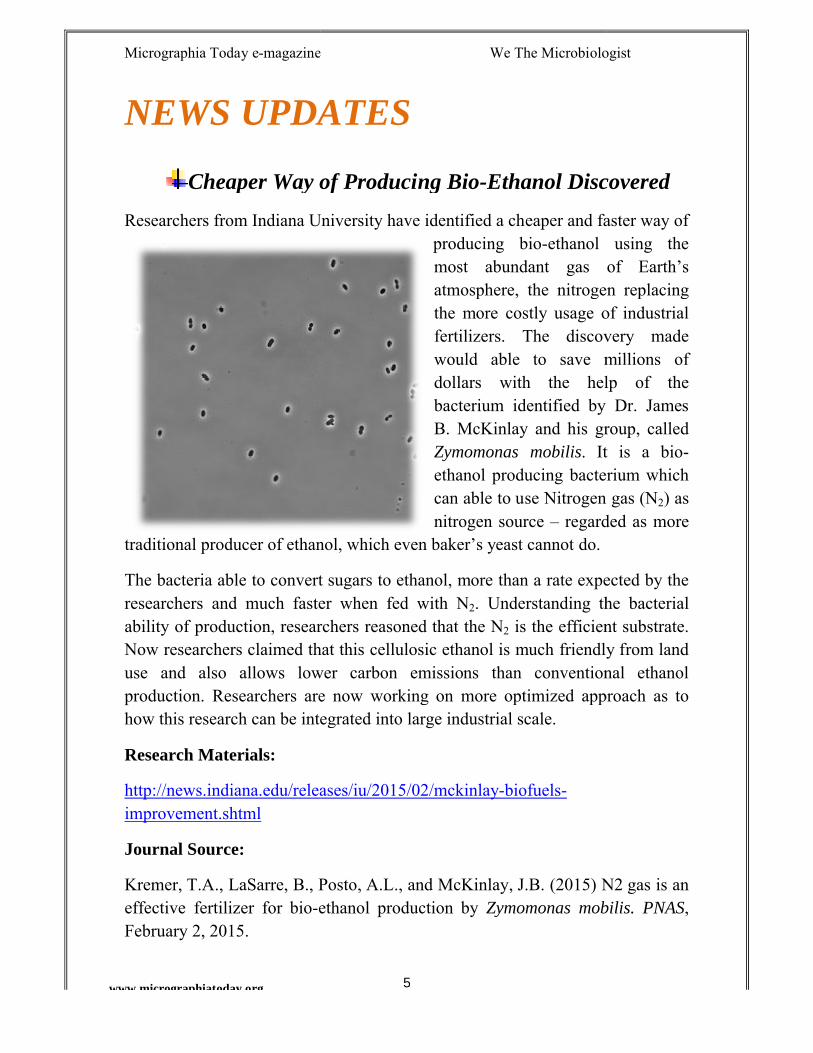
Researchers from Indiana University have identified a cheaper and faster way of producing bio-ethanol using the most abundant gas of Earth’s atmosphere, the nitrogen replacing the more costly usage of industrial fertilizers. The discovery made would able to save millions of dollars with the help of the bacterium identified by Dr. JameB. McKinlay and his group, called Zymomonas mobilis. It is a ethanol producing bacterium which can able to use Nitrogen gas (Nnitrogen source – regarded as more
traditional producer of ethanol, which even baker’s yeast cannot do.
able to convert sugars to ethanol, more than a rate expected by the researchers and much faster when fed with N2. Understanding the bacterial ability of production, researchers reasoned that the N2 is the efficient substrate. Now researchers claimed that this cellulosic ethanol is much friendly from land use and also allows lower carbon emissions than conventional ethanol
are now working on more optimized approach how this research can be integrated into large industrial scale.
http://news.indiana.edu/releases/iu/2015/02/mckinlay-biofuels-
., LaSarre, B., Posto, A.L., and McKinlay, J.B. (2015) N2 gas is an ethanol production by Zymomonas mobilis. PNAS
magazine We The Microbiologist
Ethanol Discovered
Researchers from Indiana University have identified a cheaper and faster way of ethanol using the
most abundant gas of Earth’s atmosphere, the nitrogen replacing the more costly usage of industrial fertilizers. The discovery made would able to save millions of dollars with the help of the bacterium identified by Dr. James B. McKinlay and his group, called
. It is a bio-producing bacterium which
can able to use Nitrogen gas (N2) as regarded as more
able to convert sugars to ethanol, more than a rate expected by the . Understanding the bacterial
is the efficient substrate. his cellulosic ethanol is much friendly from land
use and also allows lower carbon emissions than conventional ethanol now working on more optimized approach as to
., and McKinlay, J.B. (2015) N2 gas is an Zymomonas mobilis. PNAS,
Micrographia Today e-magazine We The Microbiologist
www.micrographiatoday.org
Mushrooms to punch the cells, a new way to punch Cancer Cells.
Edible oyster mushrooms bear a wonderful secret to puncback tidy holes, in a way to eat spiders and roundworms. It’s a trick which our immune system also uses this method to protect from infected cells, bacteria and cancer cells.
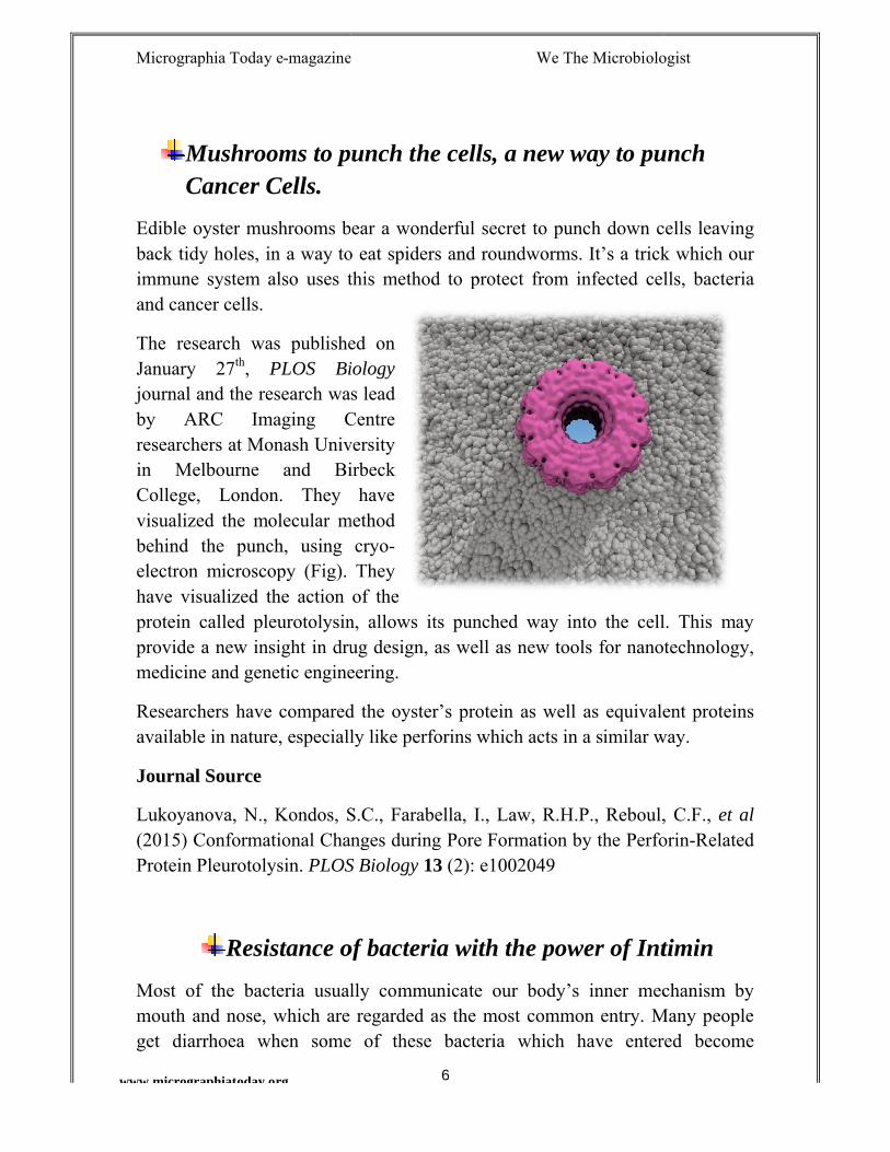
The research was published on January 27th, PLOS Biologyjournal and the research was lead by ARC Imaging Centre researchers at Monash University in Melbourne and Birbeck College, London. They have visualized the molecular method behind the punch, using cryoelectron microscopy (Fig). They have visualized the action of the protein called pleurotolysin, allows its punched way into the cell. This may provide a new insight in drug design, as well as new tools for nanotechnology, medicine and genetic engineering.
Researchers have compared the oyster’s protein as well aavailable in nature, especially like perforins which acts in a similar way.
Journal Source
Lukoyanova, N., Kondos, S.C., Farabella, I., Law, R.H.P., Reboul, C.F., (2015) Conformational Changes during Pore Formation by the PerforProtein Pleurotolysin. PLOS Biology
Resistance of bacteria with the power of Intimin
Most of the bacteria usually communicate our body’s inner mechanism by mouth and nose, which are regarded as the most common entry. Many people get diarrhoea when some of these bacteria which have entered become
magazine We The Microbiologist
6
Mushrooms to punch the cells, a new way to punch
Edible oyster mushrooms bear a wonderful secret to punch down cells leaving back tidy holes, in a way to eat spiders and roundworms. It’s a trick which our immune system also uses this method to protect from infected cells, bacteria
The research was published on PLOS Biology
rnal and the research was lead by ARC Imaging Centre researchers at Monash University in Melbourne and Birbeck College, London. They have visualized the molecular method behind the punch, using cryo-electron microscopy (Fig). They
n of the protein called pleurotolysin, allows its punched way into the cell. This may provide a new insight in drug design, as well as new tools for nanotechnology, medicine and genetic engineering.
Researchers have compared the oyster’s protein as well as equivalent proteins available in nature, especially like perforins which acts in a similar way.
Lukoyanova, N., Kondos, S.C., Farabella, I., Law, R.H.P., Reboul, C.F., (2015) Conformational Changes during Pore Formation by the Perfor
PLOS Biology 13 (2): e1002049
Resistance of bacteria with the power of Intimin
Most of the bacteria usually communicate our body’s inner mechanism by mouth and nose, which are regarded as the most common entry. Many people get diarrhoea when some of these bacteria which have entered become
magazine We The Microbiologist
Mushrooms to punch the cells, a new way to punch
h down cells leaving back tidy holes, in a way to eat spiders and roundworms. It’s a trick which our immune system also uses this method to protect from infected cells, bacteria
protein called pleurotolysin, allows its punched way into the cell. This may provide a new insight in drug design, as well as new tools for nanotechnology,
s equivalent proteins available in nature, especially like perforins which acts in a similar way.
Lukoyanova, N., Kondos, S.C., Farabella, I., Law, R.H.P., Reboul, C.F., et al (2015) Conformational Changes during Pore Formation by the Perforin-Related
Resistance of bacteria with the power of Intimin
Most of the bacteria usually communicate our body’s inner mechanism by mouth and nose, which are regarded as the most common entry. Many people get diarrhoea when some of these bacteria which have entered become
Micrographia Today e-magazine We The Microbiologist
7www.micrographiatoday.org
opportunistic. One of the most common is E. coli. These bacteria can able to attach themselves on the walls of the intestines and inject toxins which allow us to fall sick. But one thing which might be missing in between is that, these bacteria which follow their entry into digestive system should get killed by strong acid present in stomach, which we know as a strong barrier of infection.
A research group from “The Bacterial Cell Envelope” research centre at University of Tübingen with Tübingen University Hospitals investigated the phenomenon how gut bacteria survives the strong acid of stomach to enter into intestines. The results were published in the journal Molecular Microbiology.
Commonly, E. coli and Yersinia are found in small intestines and absorb nutrients. A protein named as intimin (named after intimate adherence) which allows the bacterium to adhere to the intestinal walls. It also forms tiny channels between the bacterial wall and the intestinal cells to allow the toxins (causing diarrhoea) to move into intestines.
Intimins are autotransporters and are important virulence factors of both E. coli and Yersinia spp. These proteins have lysin motif which allows the binding with the peptidoglycan. This binding is possible only in acidic conditions, which clearly displays the reason how these bacteria uses the stomach acid for getting resistance. The intimin gets inserted then into the bacterial envelope thus stabilizing the structure of peptidoglycan.
Scientists conclude that intimin supports infection process in intestine, using the stomach acid boosting the bacterial virulence.
Research Source:
Jack C. Leo, Philipp Oberhettinger, Manish Chaubey, Monika Schütz, Daniel Kühner, Ute Bertsche, Heinz Schwarz, Friedrich Götz, Ingo B. Autenrieth, Murray Coles, Dirk Linke: "The Intimin periplasmic domain mediates dimerisation and binding to peptidoglycan." Molecular Microbiology, DOI:10.1111/mmi.12840
Micrographia Today e-magazine We The Microbiologist
www.micrographiatoday.org
AUTHORS SPEAK
Cholera Toxin “Host
Department of Human Genetics, Institute of Life Sciences, Bhubaneswar
One of the most important pandemic organisms is known to be
It preys on us almost sliding from host to host, when we are at most vulnerable,
almost everywhere on food and
It is a life threatening infection, although there are several measures of
prevention. The pathogenesis is due to the toxin is secretes, which makes the
host to hyper secrete electrolytes and water, causing r
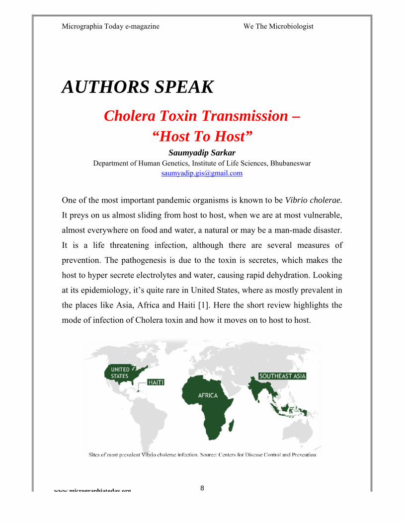
at its epidemiology, it’s quite rare in United States, where as mostly prevalent in
the places like Asia, Africa and Haiti [1]. Here the short review highlights the
mode of infection of Cholera toxin and how it moves on to host to
magazine We The Microbiologist
8
AUTHORS SPEAK
Cholera Toxin Transmission –“Host To Host”
Saumyadip SarkarDepartment of Human Genetics, Institute of Life Sciences, Bhubaneswar
One of the most important pandemic organisms is known to be Vibrio cholerae.
It preys on us almost sliding from host to host, when we are at most vulnerable,
almost everywhere on food and water, a natural or may be a man-made disaster.
It is a life threatening infection, although there are several measures of
prevention. The pathogenesis is due to the toxin is secretes, which makes the
host to hyper secrete electrolytes and water, causing rapid dehydration. Looking
at its epidemiology, it’s quite rare in United States, where as mostly prevalent in
the places like Asia, Africa and Haiti [1]. Here the short review highlights the
mode of infection of Cholera toxin and how it moves on to host to host.
magazine We The Microbiologist
Department of Human Genetics, Institute of Life Sciences, Bhubaneswar
Vibrio cholerae.
It preys on us almost sliding from host to host, when we are at most vulnerable,
made disaster.
It is a life threatening infection, although there are several measures of
prevention. The pathogenesis is due to the toxin is secretes, which makes the
apid dehydration. Looking
at its epidemiology, it’s quite rare in United States, where as mostly prevalent in
the places like Asia, Africa and Haiti [1]. Here the short review highlights the
host.
Micrographia Today e-magazine We The Microbiologist
www.micrographiatoday.org
The Organism
there are 154 serogroups, whose biochemical and morphological characteristics
are much similar, but are agglutinable with their own antisera. These non
vibrios are mostly associated with sporadic cases of diarrhoea and
intestinal infections. [3]
The most important reservoir of these O1 and O139 serotypes are humans (the
host) and the aquatic environment. Humans are considered as asymptomatic
carriers of V. cholerae. It is
targets for another. The mode of transition of its gene transcriptional pattern
allows the bacterium to escape for the survival in the aquatic environment.
cholerae exists in fresh waster li
and there it is known to be endemic for centuries with variety of serotypes
extending from pathogenic to non
forms. The primary mode of transmission is
ingestion of food or water contaminated with t
bacterium and the portal of exit is the anus as
fecal waste. [2]
The Cholera Toxin
The Cholera toxin is abbreviated as CT or CTX,
which is a member of AB5 family of
magazine We The Microbiologist
9
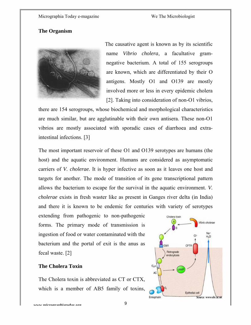
The causative agent is known as by its scientific
name Vibrio cholera, a facultative gram
negative bacterium. A total of 155 serogroups
are known, which are differentiated by their O
antigens. Mostly O1 and O139
involved more or less in every epidemic cholera
[2]. Taking into consideration of non-
there are 154 serogroups, whose biochemical and morphological characteristics
are much similar, but are agglutinable with their own antisera. These non
mostly associated with sporadic cases of diarrhoea and
The most important reservoir of these O1 and O139 serotypes are humans (the
host) and the aquatic environment. Humans are considered as asymptomatic
It is hyper infective as soon as it leaves one host and
targets for another. The mode of transition of its gene transcriptional pattern
allows the bacterium to escape for the survival in the aquatic environment.
exists in fresh waster like as present in Ganges river delta (in India)
and there it is known to be endemic for centuries with variety of serotypes
extending from pathogenic to non-pathogenic
forms. The primary mode of transmission is
ingestion of food or water contaminated with the
bacterium and the portal of exit is the anus as
The Cholera toxin is abbreviated as CT or CTX,
which is a member of AB5 family of toxins,
magazine We The Microbiologist
The causative agent is known as by its scientific
, a facultative gram-
negative bacterium. A total of 155 serogroups
are known, which are differentiated by their O
are mostly
more or less in every epidemic cholera
-O1 vibrios,
there are 154 serogroups, whose biochemical and morphological characteristics
are much similar, but are agglutinable with their own antisera. These non-O1
mostly associated with sporadic cases of diarrhoea and extra-
The most important reservoir of these O1 and O139 serotypes are humans (the
host) and the aquatic environment. Humans are considered as asymptomatic
as soon as it leaves one host and
targets for another. The mode of transition of its gene transcriptional pattern
allows the bacterium to escape for the survival in the aquatic environment. V.
ke as present in Ganges river delta (in India)
and there it is known to be endemic for centuries with variety of serotypes
Micrographia Today e-magazine We The Microbiologist
10www.micrographiatoday.org
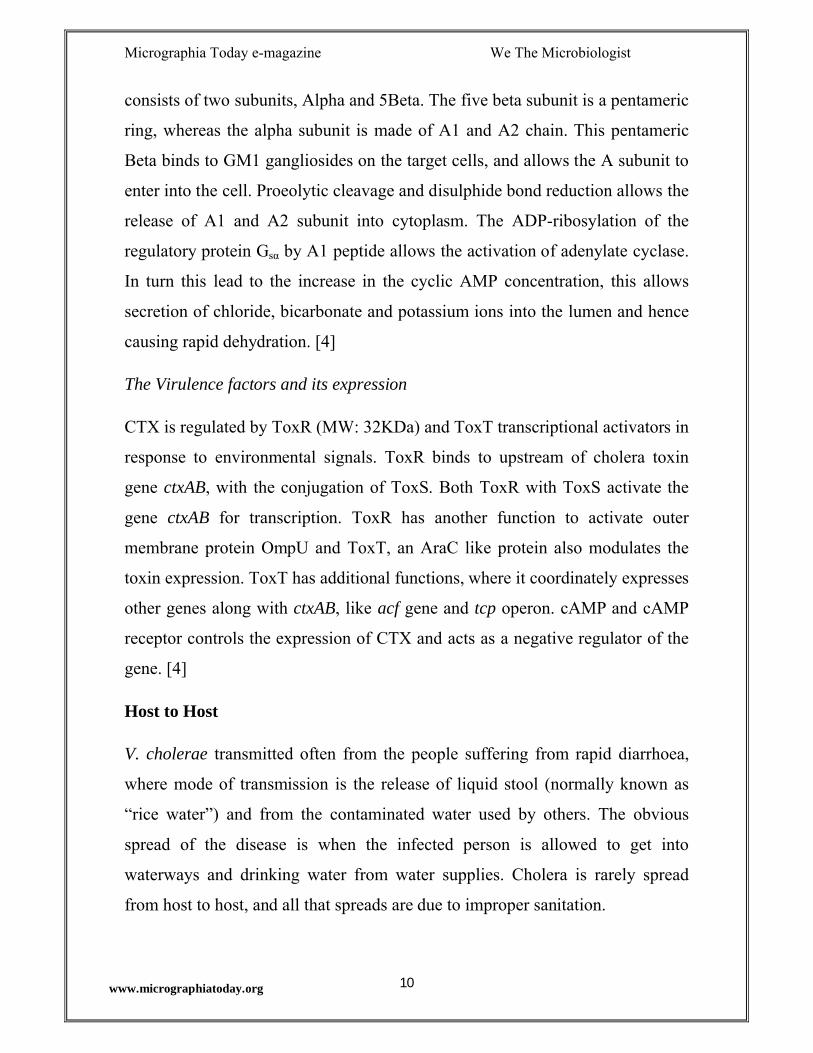
consists of two subunits, Alpha and 5Beta. The five beta subunit is a pentameric
ring, whereas the alpha subunit is made of A1 and A2 chain. This pentameric
Beta binds to GM1 gangliosides on the target cells, and allows the A subunit to
enter into the cell. Proeolytic cleavage and disulphide bond reduction allows the
release of A1 and A2 subunit into cytoplasm. The ADP-ribosylation of the
regulatory protein Gsα by A1 peptide allows the activation of adenylate cyclase.
In turn this lead to the increase in the cyclic AMP concentration, this allows
secretion of chloride, bicarbonate and potassium ions into the lumen and hence
causing rapid dehydration. [4]
The Virulence factors and its expression
CTX is regulated by ToxR (MW: 32KDa) and ToxT transcriptional activators in
response to environmental signals. ToxR binds to upstream of cholera toxin
gene ctxAB, with the conjugation of ToxS. Both ToxR with ToxS activate the
gene ctxAB for transcription. ToxR has another function to activate outer
membrane protein OmpU and ToxT, an AraC like protein also modulates the
toxin expression. ToxT has additional functions, where it coordinately expresses
other genes along with ctxAB, like acf gene and tcp operon. cAMP and cAMP
receptor controls the expression of CTX and acts as a negative regulator of the
gene. [4]
Host to Host
V. cholerae transmitted often from the people suffering from rapid diarrhoea,
where mode of transmission is the release of liquid stool (normally known as
“rice water”) and from the contaminated water used by others. The obvious
spread of the disease is when the infected person is allowed to get into
waterways and drinking water from water supplies. Cholera is rarely spread
from host to host, and all that spreads are due to improper sanitation.
Micrographia Today e-magazine We The Microbiologist
11www.micrographiatoday.org
References
1. Cholera – Vibrio cholerae infection. Centers for Disease Control and
Prevention. http://www.cdc.gov/cholera/index.html
2. Cholera’s chain of infection. Contagions. March 1, 2011.
https://contagions.wordpress.com/2011/03/01/choleras-chain-of-
infection/
3. Faruque, S.M., Albert, M.J., Mekalanos, J.J. (1998). Epidemiology,
genetics and ecology of toxicogenic Vibrio cholerae. Microbiol Mol Biol
Rev 62(4): 1301-1314.
4. Chiang, S.L., Mekalanos, J.J. Horizontal gene transfer in the emergence
of Vibrio cholerae. American Society of Microbiology Press, Washington
D.C. Microbial ecology and infectious disease. 1999.
Micrographia Today e-magazine We The Microbiologist
12www.micrographiatoday.org
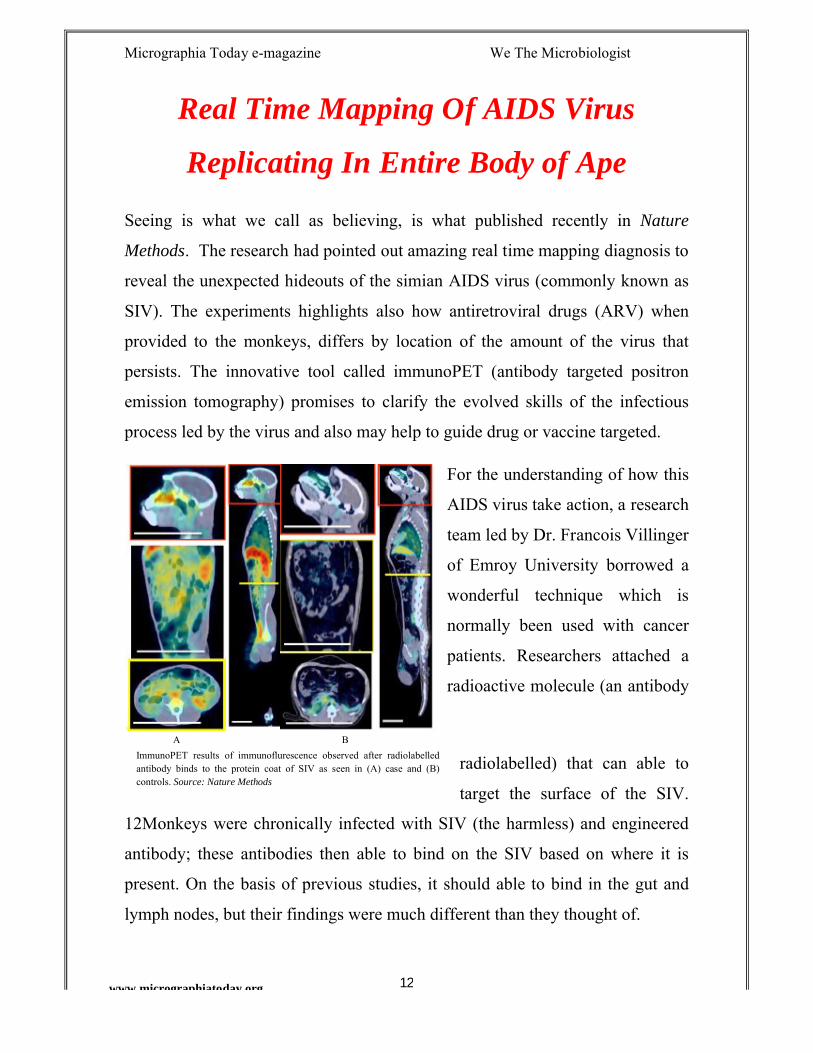
ImmunoPET results of immunoflurescence observed after radiolabelled antibody binds to the protein coat of SIV as seen in (A) case and (B) controls. Source: Nature Methods
A
Real Time Mapping Of AIDS Virus
Replicating In Entire Body of Ape
Seeing is what we call as believing, is what published recently in Nature
Methods. The research had pointed out amazing real time mapping diagnosis to
reveal the unexpected hideouts of the simian AIDS virus (commonly known as
SIV). The experiments highlights also how antiretroviral drugs (ARV) when
provided to the monkeys, differs by location of the amount of the virus that
persists. The innovative tool called immunoPET (antibody targeted positron
emission tomography) promises to clarify the evolved skills of the infectious
process led by the virus and also may help to guide drug or vaccine targeted.
For the understanding of how this
AIDS virus take action, a research
team led by Dr. Francois Villinger
of Emroy University borrowed a
wonderful technique which is
normally been used with cancer
patients. Researchers attached a
radioactive molecule (an antibody
radiolabelled) that can able to
target the surface of the SIV.
12Monkeys were chronically infected with SIV (the harmless) and engineered
antibody; these antibodies then able to bind on the SIV based on where it is
present. On the basis of previous studies, it should able to bind in the gut and
lymph nodes, but their findings were much different than they thought of.
B
Micrographia Today e-magazine We The Microbiologist
13www.micrographiatoday.org
Illumination method of immunoPET provided a surprising result where the
radiolabelled antibody targeting SIV protein coat was found mostly in the nasal
cavity. Researchers does claim that due to the presence of rich lymphatic tissue
in the upper respiratory tract it was highly detectable, which had received least
attention in previous studies. Dr. Villinger was astonished with the result, where
high SIV levels were also present in the genital tract of males, which provides
insight that sexual transmission of this SIV virus does not occur readily.
In another experimental process, researchers gave infected monkeys with ARV
drugs and followed the same kind of scanning process 5weeks later. Although
none of them had detectable SIV in blood, but all have SIV reproducing in
multiple tissues. ImmunoPET technique can be adapted in humans as the
researches have claimed for similar identification to know also how fully
suppressed people can able to reproduce these virus within at lower levels. Dr.
Villenger suggests that, immunoPET can help to stop retroviral treatment, and
then it will allow how these viruses can able to spread on the host body. Finally,
researchers claim that immuPET tracking of AIDS virus need more refinement
and the technical details are much unclear.
Source: Santangelo, P.J., Rogers, K.A., Zurla, C., Blanchard, E.L., Gumber, S.,
Strait, K. Et al. (2015). Whole-body immunoPETET reveals active SISIV
dynamics in viremic and antiretroviral therapy–treated macaques. Nature
Methods doi:10.1038/nmeth.3320.
Micrographia Today e-magazine We The Microbiologist
www.micrographiatoday.org
COVER STORY
Marine Microbiology-A Field to Desire
(International Outreach Coordinator
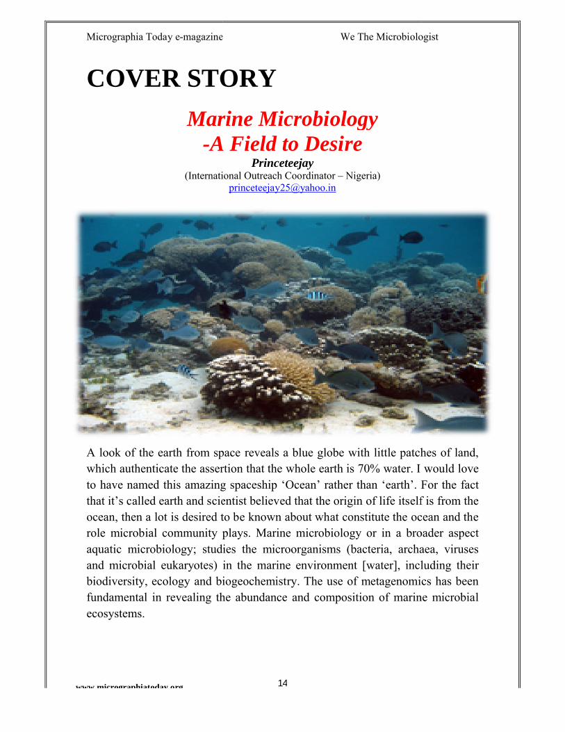
A look of the earth from space reveals a blue globe with little patches of land, which authenticate the assertion that the whole earth is 70% water. I would love to have named this amazing spaceship ‘Ocean’ rather than ‘earth’. For the fact that it’s called earth and scientist believed that the origin of life itself is from the ocean, then a lot is desired to be known about what constitute the ocean and the role microbial community plays. Marine microbiology or in a broader aspect aquatic microbiology; studies the microorganisms (bacteria, archaea, viruses and microbial eukaryotes) in the marine environment [water], including their biodiversity, ecology and biogeochemistry. The ufundamental in revealing the abundance and composition of marine microbial ecosystems.
magazine We The Microbiologist
14
COVER STORY
Marine MicrobiologyA Field to Desire
Princeteejay(International Outreach Coordinator – Nigeria)
A look of the earth from space reveals a blue globe with little patches of land, which authenticate the assertion that the whole earth is 70% water. I would love
this amazing spaceship ‘Ocean’ rather than ‘earth’. For the fact that it’s called earth and scientist believed that the origin of life itself is from the ocean, then a lot is desired to be known about what constitute the ocean and the
ity plays. Marine microbiology or in a broader aspect aquatic microbiology; studies the microorganisms (bacteria, archaea, viruses and microbial eukaryotes) in the marine environment [water], including their biodiversity, ecology and biogeochemistry. The use of metagenomics has been fundamental in revealing the abundance and composition of marine microbial
magazine We The Microbiologist
A look of the earth from space reveals a blue globe with little patches of land, which authenticate the assertion that the whole earth is 70% water. I would love
this amazing spaceship ‘Ocean’ rather than ‘earth’. For the fact that it’s called earth and scientist believed that the origin of life itself is from the ocean, then a lot is desired to be known about what constitute the ocean and the
ity plays. Marine microbiology or in a broader aspect aquatic microbiology; studies the microorganisms (bacteria, archaea, viruses and microbial eukaryotes) in the marine environment [water], including their
se of metagenomics has been fundamental in revealing the abundance and composition of marine microbial
Micrographia Today e-magazine We The Microbiologist
15www.micrographiatoday.org
Why Marine Microbiology!!Marine microorganisms are essential to the maintenance of our biosphere, yet we have only fragmentary understanding of the diversity and function of the microbial life in our oceans. Studying marine microbiology involves research of fundamental issues such as the evolution of life, the functioning of marine food webs, global climate change, the fate of pollutants, and the biodiversity of the ocean.
Marine Microbes Rule the WorldIt is of a known fact that the oceans represent the largest ecosystem on Earth and most of its biomass is microbial (90%). The diversity of microbial life in the oceans is extremely high and spans all known groups of Bacteria, Archaea and microbial Eukarya. Having a good understanding of this microorganism diversity is very important – their identity, physiology and interactions. Cyanobacteria were responsible for the oxygenation of the atmosphere 2.2 billion years ago but this group of organisms may be as old as 3.5 billion years. Almost all life in the oceans is directly or indirectly dependent on photosynthesis. Nitrogen is after carbon the most important component of organisms, but only a few specialized bacteria – especially cyanobacteria – are capable of utilizing the omnipresent atmospheric nitrogen (N2). Fifty percent of the global fixation of carbon and nitrogen occurs in the oceans and is carried out predominantly by phototrophic microorganisms. Microbes are the principal players in marine sediments and a wealth of physiologies and biogeochemical processes are present in these microbial ecosystems.
Ecological Roles of Marine MicrobesPrimary ProductionPrimary production is 'first production' – which is the creation of organic matter, refers to the transformation, or fixation, of inorganic carbon into simple sugars using solar energy through photosynthesis. Among the prokaryotes, the most abundant and important primary producers are species of the genera Synechococcus and Prochlorococcus. This species are capable of reproducing once per day and form most of the 'plant' biomass in many areas of the open sea, especially in the tropics.
Micrographia Today e-magazine We The Microbiologist
16www.micrographiatoday.org
Secondary and Tertiary productionProduction of herbivores is called secondary production, and the consumers of plants or primary producers are known as herbivores. Among marine microbes, consumers of primary producers are those which feed on autotrophic prokaryotes or autotrophic protists. Thus, the viruses which attack the autotrophic prokaryotes Synechococcus, the bacteria which absorb dissolved organic excreted by autotrophic protists such as diatoms and dinoflagellates, and the protists such as ciliates, radiolarians which feed on autotrophic protists are all consumers of primary production. However, these simple relationships exist alongside many others because among marine microbes there is not a food chain but rather a web.Thinking in terms of food chains, more often than not marine microbes are not exactly akin to neither plant (phytoplankton) nor animal (zooplankton). Furthermore, relationships between different microbes are usually neither direct nor exclusive. For example, the dissolved organic matter absorbed by a bacterium is likely a mixture of that excreted by a primary producer and some from the viral lysis of another bacterium as well the excreta of yet another organism that fed on herbivore or a primary producer. While the ecological roles are not often clear cut among marine microbes, the rest of the marine food web ultimately depends on the microbial community. A lot of researches are ongoing, I so much desires to read outcome of deep sea exploration, I really want to know which microbes exist in the deepest of seas and the significance – after all, scientist still believes life originated from sea.
References http://www.marmic.mpg.de/marmic/
http://www.nioz.nl/marine_microbiology http://www.nature.com/subjects/marine-microbiology http://www.eoearth.org/view/article/154477/
Micrographia Today e-magazine We The Microbiologist
www.micrographiatoday.org
DO YOU KNOW?Defining the Ultra structure
The peptidoglycan is one of the most essential and important part of the cell wall of bacteria. It is made up of glycan strands which are crossshort peptides. From species to species, the structure of the peptidoglycan varies with composition. In fine detail, the structure also varies with the growth conditions. Being the most essential component, it functions to preserve the integrity of the cell by withstanding the turgor, maintaining the shape and scaffolds the anchoring of different cellulaassociated proteins.
Chemical structure of the peptidoglycan
NAM and NAG are connected via β
In case of a gram negative bamino acid chain, consisting of Lalanine. Whereas, in gram positive bacterium like attached to L-alanine, D-glutamine, Linterbridge.
Do You Know?
VANCOMYCIN is an antibiotic effective only against gram positive bacterium, by binding to Dala-D-ala residues and hence prevents
magazine We The Microbiologist
17
DO YOU KNOW?Ultra structure of Peptidoglycan in Bacteria
The peptidoglycan is one of the most essential and important part of the cell wall of bacteria. It is made up of glycan strands which are cross-short peptides. From species to species, the structure of the peptidoglycan varies
In fine detail, the structure also varies with the growth conditions. Being the most essential component, it functions to preserve the integrity of the cell by withstanding the turgor, maintaining the shape and scaffolds the anchoring of different cellular components, like teichoic acids and
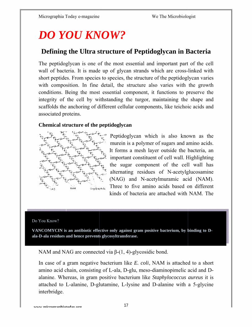
Chemical structure of the peptidoglycan
Peptidoglycan which is also known as the murein is a polymer of sugars and amino acids. It forms a mesh layer outside the bacteria, an important constituent of cell wall. Highlighting the sugar component of the cell wall has alternating residues of N-acetylglucosamine (NAG) and N-acetylmuramic acid (NAM). Three to five amino acids based on different kinds of bacteria are attached with NAM. The
NAM and NAG are connected via β-(1, 4)-glycosidic bond.
In case of a gram negative bacterium like E. coli, NAM is attached to a short amino acid chain, consisting of L-ala, D-glu, meso-diaminopimelic acid and Dalanine. Whereas, in gram positive bacterium like Staphylococcus aureus
glutamine, L-lysine and D-alanine with a 5
VANCOMYCIN is an antibiotic effective only against gram positive bacterium, by binding to Dala residues and hence prevents glycosyltransferase.
magazine We The Microbiologist
of Peptidoglycan in Bacteria
The peptidoglycan is one of the most essential and important part of the cell -linked with
short peptides. From species to species, the structure of the peptidoglycan varies In fine detail, the structure also varies with the growth
conditions. Being the most essential component, it functions to preserve the integrity of the cell by withstanding the turgor, maintaining the shape and
r components, like teichoic acids and
Peptidoglycan which is also known as the murein is a polymer of sugars and amino acids. It forms a mesh layer outside the bacteria, an important constituent of cell wall. Highlighting the sugar component of the cell wall has
acetylglucosamine acetylmuramic acid (NAM).
Three to five amino acids based on different kinds of bacteria are attached with NAM. The
, NAM is attached to a short diaminopimelic acid and D-Staphylococcus aureus it is
alanine with a 5-glycine
VANCOMYCIN is an antibiotic effective only against gram positive bacterium, by binding to D-
Micrographia Today e-magazine We The Microbiologist
18www.micrographiatoday.org
Transpeptidase is an enzyme, which plays an important role here in the making of the cell wall so rigid as a cross linking agent. In case of gram positive bacterium, the peptidoglycan molecule is cross linked by a pentapeptide bridge, but in gram negative bacterium, peptidoglycan molecules are covalently bound.
Teichoic Acids
The Teichoic acids which is an important component of the peptidoglycan is found in the cell wall of gram positive bacteria like in the genera of Bacillus, Streptococcus, Staphylococcus, Clostridium, Corynebacterium, and Listeria. In gram positive bacteria there is an absence of this compound. There are two types of teichoic acids based on with which it anchor to; when it is attached
with lipids, then they are called as lipoteichoic acids, and when covalently bound to peptidoglycan it is known as wall teichoic acids. The major function of this teichoic acid is to provide rigidity to the cell wall as well as to attract cations like Mg2+ and Na+.
Do You Know?
BACTERIOPHAGE (some of them) binds to the bacteria recognizing the lipoteichoic acids, which acts as a receptor molecule.
Micrographia Today e-magazine We The Microbiologist
www.micrographiatoday.org
PERSON IN LIGHTInterview of Dr. Anne A. Madden, “The Wasp Lady”
Dr. Anne A. Madden is a scientific explorer, aresearcher at North Carolina State University with Dr. Rob Dunn and Dr. Noah Fierer at University of Colorado. She has been working on the research based on the understanding of the arthropod contribution to the miconnected with the well known “Wasp Lady” with us to share her journey of scientific research.
The interview was conducted by Research Fellow at Institute of Life Sciences, Bhubaneswar, India.
Micrographia Today (MT): Dr. Anne A. Madden, “The Wasp Lady” postdoctoral researcher and a well known science communicator with leading interests and contribution towards Microbiology. Before we continue to know your scientific journey, we would like to know about your childhood life. How you grown up with science and also how your
magazine We The Microbiologist
19
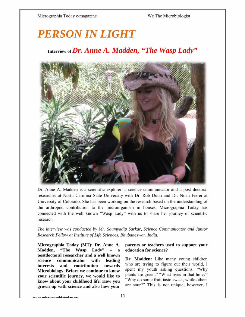
PERSON IN LIGHTDr. Anne A. Madden, “The Wasp Lady”
Dr. Anne A. Madden is a scientific explorer, a science communicator and a post doctoral researcher at North Carolina State University with Dr. Rob Dunn and Dr. Noah Fierer at University of Colorado. She has been working on the research based on the understanding of the arthropod contribution to the microorganism in houses. Micrographia Today has connected with the well known “Wasp Lady” with us to share her journey of scientific
The interview was conducted by Mr. Saumyadip Sarkar, Science Communicator and Junior Research Fellow at Institute of Life Sciences, Bhubaneswar, India.
Dr. Anne A. Madden, “The Wasp Lady” – a postdoctoral researcher and a well known science communicator with leading interests and contribution towards
ontinue to know your scientific journey, we would like to know about your childhood life. How you grown up with science and also how your
parents or teachers used to support your education for science?
Dr. Madden: Like many young children who are trying to figure out their world, I spent my youth asking questions. “Why plants are green,” “What lives in that hole?” “Why do some fruit taste sweet, while others are sour?” This is not unique; however, I
magazine We The Microbiologist
Dr. Anne A. Madden, “The Wasp Lady”
science communicator and a post doctoral researcher at North Carolina State University with Dr. Rob Dunn and Dr. Noah Fierer at University of Colorado. She has been working on the research based on the understanding of
croorganism in houses. Micrographia Today has connected with the well known “Wasp Lady” with us to share her journey of scientific
Sarkar, Science Communicator and Junior
parents or teachers used to support your
Like many young children figure out their world, I
spent my youth asking questions. “Why green,” “What lives in that hole?”
“Why do some fruit taste sweet, while others are sour?” This is not unique; however, I
Micrographia Today e-magazine We The Microbiologist
www.micrographiatoday.org
don’t think I ever grew out of this developmental stage.
Growing up where I did in the United States of America, I had the opportunity to play outside in the forests and on the ocean shore every day. Whether it was exploring the scattered shells of animals long dead at the beach, or investigating the different vpatterns on tree leaves, I was always curious about nature in particular. Life was fascinating, and in school this only became more apparent as I took more and more science classes. The world of science held so many fantastic stories, bizarre relationsand complex processes; unlike fiction stories, they were something I could often witness in front of me. I remember the revelation I had when I learned that body movements were the result of a relatively simple network of inflexible bones, and contracting muscles seemed so elegant and logical. I continued to hunt down these stories, and learn of new characters, from zooplankton to tapirs.
While my family members are not scientists, they have always supported this natural curiosity as it led me from a small child catching frogs near our house, to a professional scientist investigating microbiological questions. With their support, and those of a number of inspiring biology teachers, I continued down a path that eventually led to a career discerning these biological stories at the source.
MT: What was your subject during your graduation and then post graduation? Can you please explain your outlook of
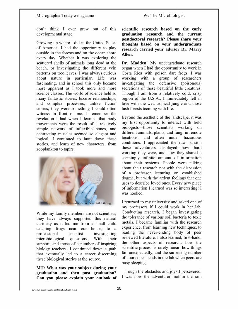
Credit: Lauren Nichols/YourWildLife.org
magazine We The Microbiologist
20
don’t think I ever grew out of this
Growing up where I did in the United States of America, I had the opportunity to play outside in the forests and on the ocean shore every day. Whether it was exploring the scattered shells of animals long dead at the beach, or investigating the different vein patterns on tree leaves, I was always curious about nature in particular. Life was fascinating, and in school this only became more apparent as I took more and more science classes. The world of science held so many fantastic stories, bizarre relationships, and complex processes; unlike fiction stories, they were something I could often witness in front of me. I remember the revelation I had when I learned that body movements were the result of a relatively simple network of inflexible bones, and
cting muscles seemed so elegant and logical. I continued to hunt down these stories, and learn of new characters, from
While my family members are not scientists, they have always supported this natural
a small child catching frogs near our house, to a professional scientist investigating microbiological questions. With their support, and those of a number of inspiring biology teachers, I continued down a path that eventually led to a career discerning
ese biological stories at the source.
What was your subject during your graduation and then post graduation? Can you please explain your outlook of
scientific research based on the early graduation research and the current postdoctoral research? Pleasethoughts based on your undergraduate research carried your advisor Dr. Marry Allen.
Dr. Madden: My undergraduate research began when I had the opportunity to work in Costa Rica with poison dart frogs. I was working with a group of researchers investigating the defensive (poisonous) secretions of these beautiful little creatures. Though I am from a relatively cold, crisp region of the U.S.A., I immediately fell in love with the wet, tropical jungle and those lush forests teeming with life.
Beyond the aesthetic of the landscape, it was my first opportunity to interact with field biologists—those scientists working on different animals, plants, and fungi in remote locations, and often under hazardous conditions. I appreciated the raw passion these adventurers displayed—working they were, and how they shared a seemingly infinite amount of informatiabout their systems. People were talking about their research not with the dispassion of a professor lecturing on established dogma, but with the ardent feelings that one uses to describe loved ones. Every new piece of information I learned was so interwas hooked.
I returned to my university and asked one of my professors if I could work in her lab. Conducting research, I began investigating the tolerance of various soil bacteria to toxic metals. I became familiar with the research experience, from learning new techniques, to reading the never-ending body of peer reviewed literature. I also learned, firstthe other aspects of research: how the scientific process is rarely linear, how things fail unexpectedly, and the surprising number of hours one spends in the lab when peers are busy sleeping.
Through the obstacles and joys I persevered. I was now the adventurer, not in the rain
Credit: Lauren Nichols/YourWildLife.org
magazine We The Microbiologist
scientific research based on the early graduation research and the current postdoctoral research? Please share your thoughts based on your undergraduate research carried your advisor Dr. Marry
undergraduate research began when I had the opportunity to work in Costa Rica with poison dart frogs. I was working with a group of researchers investigating the defensive (poisonous) secretions of these beautiful little creatures.
tively cold, crisp region of the U.S.A., I immediately fell in love with the wet, tropical jungle and those
Beyond the aesthetic of the landscape, it was my first opportunity to interact with field
ts working on different animals, plants, and fungi in remote locations, and often under hazardous conditions. I appreciated the raw passion
—how hard working they were, and how they shared a seemingly infinite amount of information about their systems. People were talking about their research not with the dispassion of a professor lecturing on established dogma, but with the ardent feelings that one uses to describe loved ones. Every new piece of information I learned was so interesting! I
I returned to my university and asked one of my professors if I could work in her lab. Conducting research, I began investigating the tolerance of various soil bacteria to toxic
came familiar with the research experience, from learning new techniques, to
ending body of peer reviewed literature. I also learned, first-hand, the other aspects of research: how the scientific process is rarely linear, how things
unexpectedly, and the surprising number of hours one spends in the lab when peers are
Through the obstacles and joys I persevered. I was now the adventurer, not in the rain
Micrographia Today e-magazine We The Microbiologist
www.micrographiatoday.org
forest, but in the laboratory. I started to speak of my research like those scientists I worked with in the jungle. Though it’s easy to say I was passionate about that early research, I think it is more accurate to say I was obsessed.
Forever after this experience, I found myself fascinated with microbiology. While I lovstudying animals and plants, it was this invisible jungle around us that preoccupied my brain and its endless questions.
After learning about microbiology, I think it’s hard to not share in this obsession. Though invisible to the naked eye, these microbes impact most of what we do, what animals do, what plants do, how nutrients cycle, and even our weather. As with everything in science, another bonus in learning about them was that the more I learned, the more I saw around me. I began to notice the smells of the soil (geosmin) derived from the very bacteria that produce most of our antibiotics. I noticed the metallic sheen on certain stagnant water that is caused by iron-munching microorganisms, not oil spills. Microbes offered a whole new world of fantastic stories, which made me routinely question the importance of these small beings we didn’t even know existed until the 1600s.
MT: Microbiology is revealing the unseen. The vast microbial science has been an important aspect of scientific research. During your journey as a science communicator how you reveal this importance? Please explain your journey for science communication?
Dr Madden: Science communication, in my opinion, is about revealing the excitement, and relevance, of research. It comes down tjustifying why we spend so much of our lives and energy pursuing a field that is objectively very difficult. Making the research relevant to the audience is key to such engagement. To reveal the importance of science to the non-science audience, I think we, as scientists, need to forget a lot of
magazine We The Microbiologist
21
forest, but in the laboratory. I started to ke those scientists I
worked with in the jungle. Though it’s easy to say I was passionate about that early research, I think it is more accurate to say I
Forever after this experience, I found myself fascinated with microbiology. While I loved studying animals and plants, it was this invisible jungle around us that preoccupied my brain and its endless questions.
After learning about microbiology, I think it’s hard to not share in this obsession. Though invisible to the naked eye, these
bes impact most of what we do, what animals do, what plants do, how nutrients cycle, and even our weather. As with everything in science, another bonus in learning about them was that the more I learned, the more I saw around me. I began
ls of the soil (geosmin) derived from the very bacteria that produce most of our antibiotics. I noticed the metallic sheen on certain stagnant water that is caused
munching microorganisms, not oil spills. Microbes offered a whole new world
stic stories, which made me routinely question the importance of these small beings we didn’t even know existed until the
Microbiology is revealing the unseen. The vast microbial science has been an important aspect of scientific research.
ng your journey as a science communicator how you reveal this importance? Please explain your journey
communication, in my opinion, is about revealing the excitement, and relevance, of research. It comes down to justifying why we spend so much of our lives and energy pursuing a field that is objectively very difficult. Making the research relevant to the audience is key to such engagement. To reveal the importance
science audience, I we, as scientists, need to forget a lot of
what we have learned. I work hard at trying to put myself in the position of the audience: an audience who hasn’t devoted their life to this work, who get frustrated by jargon (words that don’t make sense or nonseacronyms). I continue to learn how to better do this with the help of great advisors, editors, and mentors; however, this training began when I was very young, and by a group of people who had nothing to do with science communication, or science in general.
I consider myself lucky to have been born into a family that was kind and supportive, but not from a scientific background. Beginning in early science courses, I was bursting to share the information I learned with my family at the dinner table. I quickly learned I had to develop a better communications toolkit if I was going to have anyone share in the science joys I was exploding with. This continues even today. If I see that my family and friends away from me, or their eyes are glazing over when I discuss my research, I know I have to approach the topic in a different way.
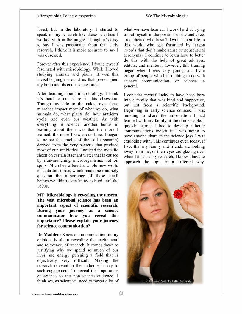
Credit: Alonso Nichols/ Tufts University
magazine We The Microbiologist
what we have learned. I work hard at trying to put myself in the position of the audience: an audience who hasn’t devoted their life to this work, who get frustrated by jargon
or nonsensical acronyms). I continue to learn how to better do this with the help of great advisors, editors, and mentors; however, this training began when I was very young, and by a group of people who had nothing to do with science communication, or science in
I consider myself lucky to have been born into a family that was kind and supportive, but not from a scientific background. Beginning in early science courses, I was bursting to share the information I had learned with my family at the dinner table. I quickly learned I had to develop a better communications toolkit if I was going to have anyone share in the science joys I was exploding with. This continues even today. If I see that my family and friends are looking away from me, or their eyes are glazing over when I discuss my research, I know I have to approach the topic in a different way.
Credit: Alonso Nichols/ Tufts University
Micrographia Today e-magazine We The Microbiologist
22www.micrographiatoday.org
Beyond my informal training in science communication, I learned more concrete tactics to add to my toolkit in graduate school at Tufts University, writing press releases for my published research. As part of an internship with the Society for Integrative and Comparative Biology, I got to work with people who were professional science communicators. I learned the importance of ‘hooking’ the audience—grabbing their attention with your first written sentence, the way a movie trailer teases you about an upcoming film. I also learned about the structure of a science communication. Now, as a postdoctoral researcher, I’ve had the privilege of working with Dr. Rob Dunn, who is a fantastic and utterly inspiring science communicator. I’m continually learning from great writers about what they do successfully, and this education continues every day.
MT: You had been an Associate Microbiologist at Novobiotic Pharmaceuticals LLC, Cambridge. Please explain your work experience and the role of your work. Some experiences of early research help to climb of stairs of achievements. Do feel that the early research experience in Industrial research helped you in gaining experiences?
Dr. Madden: After graduating from my undergraduate university (Wellesley College), I joined the company Novobiotic Pharmaceuticals. This biotechnology company uses patented technology to grow hard to culture bacteria. These bacteria are cultivated to produce novel antibiotics. I learned a vast number of laboratory techniques during my time at this job, from mammalian cell culture, to antimicrobial screening, to culturing various groups of rare bacteria. I contributed to the discovery of novel antibiotics, Neocitreamicins I and II. Beyond the techniques and research though, I learned a great deal about communicating with non-scientists as well, as a company is made up of more than just the scientific groups. Investors, business associates,
potential associates, and scientists can have disparate motives and end-goals.
This revealing experience further honed my communication skills, and gave me experience working with larger teams, rather than the more individual role as a scientist in academia. I am very thankful for the experience. Generally I recommend that scientists work outside of academia for at least a small portion of their life. For me, this experience provided an important, alternate view of an academic science program. Understanding this viewpoint has helped me in both my science communication and in my grant writing.
MT: Currently you are a Postdoctoral Researcher at North Carolina State University and at the University of Colorado. Please explain your current research and your future goals through this research.
Dr. Madden: I currently have a joint postdoctoral position at North Carolina State University in the YourWildLife.org lab of Dr. Rob Dunn, and at the University of Colorado, Boulder in the lab of Dr. Noah Fierer. We are investigating the contribution of arthropods (insects, spiders, mites, etc.) to the microbial communities of homes. This project is funded by the Alfred P. Sloan Foundation, as part of a greater interest in understanding the microbiology of the built environment (those habitats we construct, such as houses, hospitals, and industrial buildings).
No matter the cleanliness of our homes, they contain a myriad of invisible roommates in the form of dust mites, large house spiders, cockroaches, and centipedes. Many of these bugs have lived with us since our ancestors made nests in the trees. They are living in our homes—these places we spend most of our life in. We know they can influence our homes and health in by contributing to allergies, spreading diseases, or by degrading our houses. Despites this, we know very little about what bugs exist in our homes, and
Micrographia Today e-magazine We The Microbiologist
23www.micrographiatoday.org
even less about what microbes that they harbor. Isn’t it shocking that we know more about the microbes in space stations and the bugs in certain jungles than we do about the community in our houses? Our research specifically investigates what microbes and bugs exist within houses and ultimately how they may be contributing to our health.
MT: With Awards and recognition builds big responsibilities towards scientific community. Naming some of your recognitions like SICB Grants-in-aid of Research Grants-in-Aid Research, Theodore Roosevelt Memorial Grant, The Lingos Prize in Life Sciences and Honorable Mention Poster Award, Ecohealth International Conference, 2012. How awards and grants influence you in your field of research?
Dr. Madden: I am sincerely grateful for the science awards I have received in my life. These include the research grants and fellowships from multiple societies including the National Science Foundation, Tufts Institute for the Environment, the American Philosophical Society, and the American Museum of Natural History, as well as the more whimsical awards such as being named the 2014 Woman of the Year from the Luxuriant Flowing Hair Club for Scientists (a subgroup of the organization that hosts the Ig Nobels). Most of these awards have financially supported my research and allowed me to tackle the next research idea, but all have supported me as a developing researcher.
I am incredibly thankful for these people and organizations investing in me and allowing me to invest in the science. Without their financial support, I could not have conducted the research that I did. Beyond their financial support, these helped me, particularly as a young scientist, to know that colleagues in my field found worth in my proposed research. Science can feel isolating and leave a researcher often with feelings of self-doubt. I only hope that every young researcher has
the opportunity to feel supported during their career.
MT: Science Communication is a big field for highlighting the research carried out in four walls lab to the scientific community as well as to all the scientific readers. An article published in the Society of Integrative and Comparative Biology, which was re-highlighted in the ASM’s Blog under “Small Things Considered”. How you feel during your scientific communication that is useful for spreading knowledge?
Dr. Madden: Science writing often entails three things: Finding a topic, writing the piece, and finding a venue to share the writing. For this piece, I was lucky enough to work with a scientist who was very engaging and patiently worked with me to tell the story of his work.
As scientists, I don’t think we often run out of material to share, so the harder part is typically the other two parts (the actual writing and the venue). Writing is hard. I actually think it’s one of the hardest things we do. It requires draft, after draft, after draft. I have learned to do some of my best writing by reading great science writers and understanding strategies they have for writing such engaging prose. Carl Zimmer, Hannah Holmes, and Rob Dunn are some of my favorites. Malcolm Gladwell is also well versed in making research relevant, manageable, and engaging.
My process also involves bouncing ideas off of other people—non-scientists. For the piece for the ASM blog in particular, I went over a lot of it with my sister, who is not in a scientific field. I asked her where she got lost, what words didn’t make sense, and what she was left wanting more from. I then was lucky enough to work with great editors who helped hone my message and clarify my speech. There is a great deal of editing that goes into any one piece. Every word is carefully chosen. The best advice I can give to aspiring science communicators is to read
Micrographia Today e-magazine We The Microbiologist
24www.micrographiatoday.org
the writing of successful science communicators and practice, practice, practice. Writing is hard work, but feeling like you connected with an audience is a magical feeling. The researcher who I worked with for the ASM blog post thanked me for writing the piece that because it finally made his life’s work accessible to his family. I love this role of communication.
MT: Narrowing our discussion, highlighting an issue in front. Microbiology – the science of unseen, is a major field which gets good exposure in developed countries. But still students don’t want to carry forward their research only because of lesser opportunities they receive in developing countries. Some who perceive to this field need to follow hard struggle to go abroad to continue their research. How you feel the microbiological research is important in the developing nations?
Dr. Madden: While separately small, the metabolic potential, abundance, and diversity of microorganisms make them incredibly powerful. In general, microbiology has the potential to change our world, and many of these impacts will have a disproportionally greater impact in developing, than developed countries. Here I am speaking particularly of microbial technologies that relate to waste water treatment, decontamination of toxic waste sites, cures for diseases, greater efficiency of food production, fuel production with lesser environmental impacts, even ‘natural’ batteries and biodegradable packaging.
I have seen first-hand how promising these technologies can be, from working on antibiotic discoveries at Novobiotic Pharmaceuticals, to working with a company—PIARCS, in Boulder, CO—which uses bacteria to reclaim phosphate from waste water. Phosphate being a limited resource that is critical for agriculture yet is currently creating environmental hazardsfirst through its mining, second through its impact on natural water systems. A non-
profit I have worked with in the past, the Urban Beekeeping Laboratory & Bee Sanctuary, conduct microbial vaccine research to increase the health of bees—bugs critical to our food supply. These are only three of many, many organizations working on applying what we have learned about microorganisms to make our work a healthier place.
The field of microbiology is filled with promising new discoveries that allow us to help humans, animals, plants, and our planet. While some research tackles these questions directly, other research does not have such a linear path to such an application. Often it may not be clear how each research project can lead to these downstream effects. It can often feel like the small project you are working on does not matter. I applaud each and every researcher who continues the adventure and arduous journey that is research in spite of such hurdles. I know these hurdles are real. I can’t presume to know what all of these hurdles are. I do know that I’m proud to live in a world where people are facing these challenges and continuing to contribute to our great body of knowledge.
I think we must remember that we are only pieces of a larger scientific community. We might be creating the platform for the next technology that will help the world. It is a noble struggle. Science can unite us across the globe, across cultures, across languages. We are all united in this pursuit of truth—of knowing what we do not yet know.
MT: Ebola – the current outbreak which shook the world to fear. What’s your opinion to halt this exposure of the virus after the recent identification in US?
Dr. Madden: Ebola is a deadly disease, which is affecting many countries right now. I think this particular infection has highlighted the importance of communicating science to make sure people can prevent infections and the spread of the disease. I think it also has highlighted the
Micrographia Today e-magazine We The Microbiologist
www.micrographiatoday.org
importance of a structure for facilitating the distribution of information and supplies. In general, I believe our international community is strong enough to face this enemy. In our shared history, people have stopped small pox in its tracks. By working together, we have decreased incidence of polio. With scientific knowledge, social motivation, and structures of distribution, humans have the power to combat disease, retain humanity, and even move toward creating vaccines—arguably one of thamazing technologies we’ve created as humans. I think we should be proud of our past and continue this tradition of not living in fear, but learning about each new pathogen and how we can smartly fighting back.
MT: Before concluding the big conversation about this scientific journey you wished to walk along with us, please share your life apart from science.
Dr. Madden: When not working on my research, or sharing the research of other scientists with the larger community, I enjoy investigating other novelties in life. This can take the form of traveling to new places,
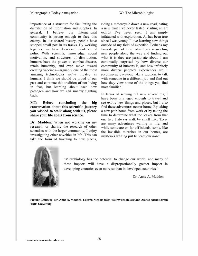
“Microbiology these impacts will have a disproportionally greater impact in developing
Picture Courtesy: Dr. Anne A. Madden, Lauren Nichols Tufts University
magazine We The Microbiologist
25
importance of a structure for facilitating the distribution of information and supplies. In general, I believe our international community is strong enough to face this enemy. In our shared history, people have
ks. By working together, we have decreased incidence of polio. With scientific knowledge, social motivation, and structures of distribution, humans have the power to combat disease, retain humanity, and even move toward
arguably one of the most amazing technologies we’ve created as humans. I think we should be proud of our past and continue this tradition of not living in fear, but learning about each new pathogen and how we can smartly fighting
Before concluding the big tion about this scientific journey
you wished to walk along with us, please share your life apart from science.
When not working on my research, or sharing the research of other scientists with the larger community, I enjoy
novelties in life. This can take the form of traveling to new places,
riding a motorcycle down a new road, eating a new fruit I’ve never tasted, visiting an art exhibit I’ve never seen. I am simply infatuated with exploration. As has been true since I was young, I love learning new things outside of my field of expertise. Perhaps my favorite part of these adventures is meeting new people along the way and finding out what it is they are passionate about. I am continually surprised by how diverse our community of humans is, and how infinitely more diverse people’s experiences are. I recommend everyone take a moment to talk with someone in a different job and find out how they view some of the things you find most familiar.
In terms of seeking out new adventurhave been privileged enough to travel and see exotic new things and places, but I also find these adventures nearer home. By taking a new path home from work or by taking the time to determine what the leaves from that one tree I always walk by smellare many adventures waiting in life, and while some are on far off islands, some, like the invisible microbes in our homes, are mysteries waiting just beneath our nose.
icrobiology has the potential to change our world, and many of these impacts will have a disproportionally greater impact in developing countries even more so than in developed countries.”
– Dr. Anne A. Madden
Dr. Anne A. Madden, Lauren Nichols from YourWildLife.org and Alonso Nichols from
magazine We The Microbiologist
riding a motorcycle down a new road, eating a new fruit I’ve never tasted, visiting an art exhibit I’ve never seen. I am simply infatuated with exploration. As has been true
young, I love learning new things outside of my field of expertise. Perhaps my favorite part of these adventures is meeting new people along the way and finding out what it is they are passionate about. I am continually surprised by how diverse our
ty of humans is, and how infinitely more diverse people’s experiences are. I recommend everyone take a moment to talk with someone in a different job and find out how they view some of the things you find
In terms of seeking out new adventures, I have been privileged enough to travel and see exotic new things and places, but I also find these adventures nearer home. By taking a new path home from work or by taking the time to determine what the leaves from that one tree I always walk by smell like. There are many adventures waiting in life, and while some are on far off islands, some, like the invisible microbes in our homes, are mysteries waiting just beneath our nose.
has the potential to change our world, and many of these impacts will have a disproportionally greater impact in
countries even more so than in developed countries.”
Dr. Anne A. Madden
from YourWildLife.org and Alonso Nichols from
Micrographia Today e-magazine We The Microbiologist
26www.micrographiatoday.org
Methylation Strategy of DNA in BacteriaSaumyadip Sarkar
Managing Director and Science Communicator ([email protected])
DNA methylation is a sort of DNA modification which is present in all bacteria including some eukaryotes also. There are two kinds of methylation present in bacteria N6-methyl-adenine or m6A (also find in lower eukaryotes) and N4-methyl-cytosine or m4C (exclusively bacterial). Another archetypal methylated base present in eukaryotes, C5-methyl-cytosine (m5C) is also found in bacteria. These three of these methyl groups protrudes out of the major groove of the DNA which is typical for the recognition of DNA motifs by DNA binding proteins.
The typical methylation is catalyzed by DNA methyl transferees that recognize specific DNA motifs. Base methylation involves the transfer of the methyl group from the S-adenosyl metheonine to DNA. During the process of replication, nonmethylated DNA strands are incorporated into newly synthesized strand. Hence the daughter stand remains unmethylated or commonly termed as “hemimethylated”, i.e. methylated in only one strand.
Restriction Modification System (RMS)
Each and every RMS is made up of a restriction endonuclease with a DNA methyltransferase (adenine or cysteine). The base methylation helps to prevent the DNA cleavage by cognate endonuclease. This is how RMS works. Although the primitive idea has been how bacteria protects or identifies self and non self genome like of invading bacteria, but the additional roles has been broadened. One of the most common parts has been is the gene knockout of some RMS systems alters the gene expression pattern [1]. Although there are low to high numbers of RMS in different kinds of bacteria based on their role of selfishness and survival strategy, as there are 15 to 20 in Neisseria gonorrhoeae and over 25 in Helicobacter pylori [1].
Solitary DNA Methyltransferases (SDM)
SDM found in many genomes of bacteria where they probably derive from ancestral RMS system, where there is a loss of their restriction enzyme. Another
Micrographia Today e-magazine We The Microbiologist
27www.micrographiatoday.org
quite equivalent form, where RMS has active restriction modification but the restriction enzyme is inactive [2].
A paradigm of SDM is Dam methylase, which is an enzyme of Gamma-proteobacteria having a methylating property of the adenosine moiety of 5’-GATC-3’ sites. The substrate is the hemimethylated DNA during DNA replication. One of the remarkable properties is its high processivity which allow it to methylate more than 50 GATC sites without detaching from the DNA template. While in case of E. coli due to lack of presence of Dam methylase shown to cause pleiotropic defects which provide an insight of the importance for the existence of multiple DNA-protein interaction with the help of GATC methylated control [3].
Another example of SDM is the cell cycle regulated (CcrM) methyltransferase found in Alpha-proteobacteria. It is able to methylate adenine in 5’-GANTC-3’site (N = any nucleotide). Although both Dam methylase and CcrM has similar targets, they are of different evolutionally originated. Additionally, CcrM has higher preference towards hemi-methylated DNA whereas Dam methylase doesn’t.
Adenine Methylation and transcriptional control
Sometimes DNA methylase specifically target regulatory regions in genes, like promoter and it requires methylation for the efficient recognition and binding of RNA polymerase and transcription factors. The most classical examples for this kind of methyl transferases are Dam and CcrM [4]. It is quite often seen that methylation provides the transcription regulation by methylating at the promoter region or nearby where transcriptional factors bind.
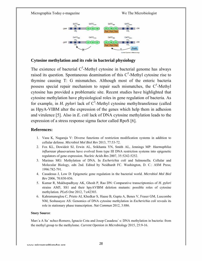
Pap-operon:
In uropathogenic E. coli there is a controlling system present called pap-operon which encodes fimbrial adhesins. The pap-ON and pap-OFF mechanism harbour DNA methylation pattern differentially in pap regulatory region. Here it contains two GATC sites with reduced processivity.
In OFF state, GATCprox is non methylated but GATCdist remains methylated. But in ON state, GATCprox is methylated and GATCdist is non methylated.
Micrographia Today e-magazine We The Microbiologist
28www.micrographiatoday.org
Cytosine methylation and its role in bacterial physiology
The existence of bacterial C5-Methyl cytosine in bacterial genome has always raised its question. Spontaneous deamination of this C5-Methyl cytosine rise to thymine causing T: G mismatches. Although most of the enteric bacteria possess special repair mechanism to repair such mismatches, the C5-Methyl cytosine has provided a problematic site. Recent studies have highlighted that cytosine methylation have physiological roles in gene regulation of bacteria. As for example, in H. pylori lack of C5-Methyl cytosine methyltransferase (called as HpyA-VIBM alter the expression of the genes which help them in adhesion and virulence [5]. Also in E. coli lack of DNA cytosine methylation leads to the expression of a stress response sigma factor called RpoS [6].
References:
1. Vasu K, Nagaraja V: Diverse functions of restriction modification systems in addition to cellular defense. Microbiol Mol Biol Rev 2013, 77:53-72.
2. Fox KL, Dowideit SJ, Erwin AL, Srikhanta YN, Smith AL, Jennings MP: Haemophilus influenzae phasevarions have evolved from type III DNA restriction systems into epigenetic regulators of gene expression. Nucleic Acids Res 2007, 35:5242-5252.
3. Marinus MG: Methylation of DNA. In Escherichia coli and Salmonella. Cellular and Molecular Biology, edn 2nd. Edited by Neidhardt FC. Washington, D. C.: ASM Press; 1996:782-791.
4. Casadesus J, Low D: Epigenetic gene regulation in the bacterial world. Microbiol Mol Biol Rev 2006, 70:830-856.
5. Kumar R, Mukhopadhyay AK, Ghosh P, Rao DN: Comparative transcriptomics of H. pyloristrains AM5, SS1 and their hpyAVIBM deletion mutants: possible roles of cytosine methylation. PLoS One 2012, 7:e42303.
6. Kahramanoglou C, Prieto AI, Khedkar S, Haase B, Gupta A, Benes V, Fraser GM, Luscombe NM, Seshasayee AS: Genomics of DNA cytosine methylation in Escherichia coli reveals its role in stationary phase transcription. Nat Commun 2012, 3:886.
Story Source:
Marı´a A Sa´ nchez-Romero, Ignacio Cota and Josep Casadesu´ s: DNA methylation in bacteria: from the methyl group to the methylome. Current Openion in Microbiology 2015, 25:9-16.
Micrographia Today e-magazine We The Microbiologist
www.micrographiatoday.org
TECHNOLOGYA New Palm Sized DNA Sequencer
A new finding which is published in DNA sequencer which would be much helpful to characterize viruses and bacteria. Scientists now highlight a new disease diagnosis tool.
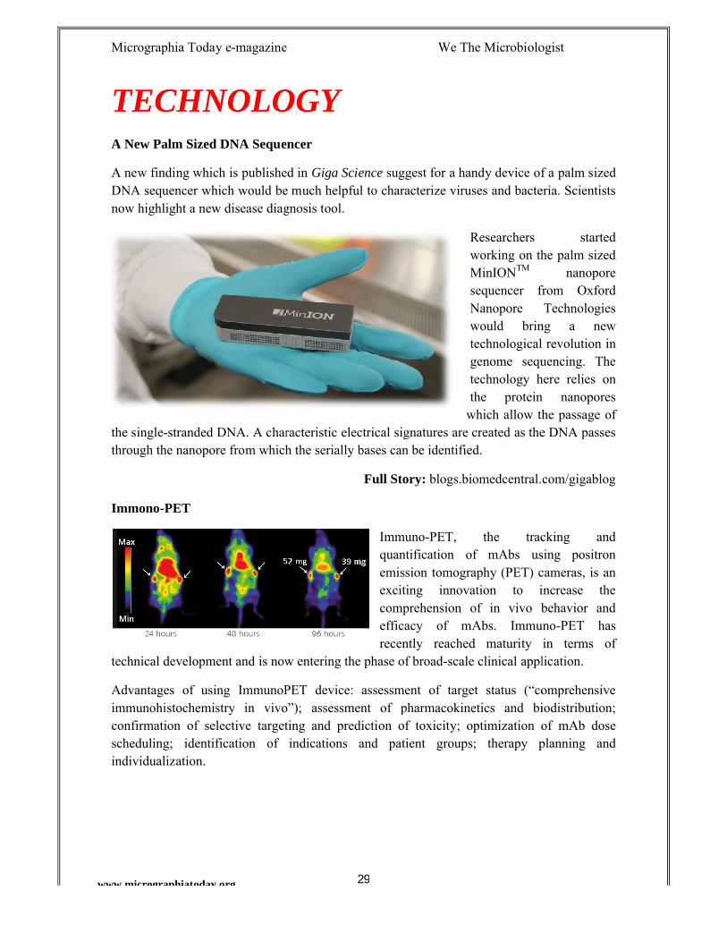
the single-stranded DNA. A characteristic electricthrough the nanopore from which the serially bases can be identified.
Immono-PET
technical development and is now entering the phase of broad
Advantages of using ImmunoPET device: immunohistochemistry in vivo”);confirmation of selective targeting and prediction of toxicity;scheduling; identification of indications and patient groups;individualization.
magazine We The Microbiologist
29
ECHNOLOGYA New Palm Sized DNA Sequencer
s published in Giga Science suggest for a handy device of a palm sized DNA sequencer which would be much helpful to characterize viruses and bacteria. Scientists now highlight a new disease diagnosis tool.
Researchers started working on the palm sized MinIONTM
sequencer from Oxford Nanopore Technologies would bring a new technological revolution in genome sequencing. The technology here relies on the protein
which allow the passage of stranded DNA. A characteristic electrical signatures are created as the DNA passes
through the nanopore from which the serially bases can be identified.
Full Story: blogs.biomedcentral.com/gigablog
Immuno-PET, the tracking and quantification of mAbs using positron emission tomography (PET) cameras, is an exciting innovation to increase the comprehension of in vivo behavior and efficacy of mAbs. Immunorecently reached maturity in terms of
technical development and is now entering the phase of broad-scale clinical application.
Advantages of using ImmunoPET device: assessment of target status (“comprehensive immunohistochemistry in vivo”); assessment of pharmacokinetics and biodistribution;confirmation of selective targeting and prediction of toxicity; optimization of mAb dose
identification of indications and patient groups; therapy planning and
magazine We The Microbiologist
suggest for a handy device of a palm sized DNA sequencer which would be much helpful to characterize viruses and bacteria. Scientists
Researchers started working on the palm sized
nanopore sequencer from Oxford Nanopore Technologies would bring a new technological revolution in genome sequencing. The technology here relies on the protein nanopores
the passage of al signatures are created as the DNA passes
blogs.biomedcentral.com/gigablog
PET, the tracking and quantification of mAbs using positron
y (PET) cameras, is an exciting innovation to increase the comprehension of in vivo behavior and efficacy of mAbs. Immuno-PET has recently reached maturity in terms of
scale clinical application.
assessment of target status (“comprehensive assessment of pharmacokinetics and biodistribution;
optimization of mAb dose therapy planning and
Micrographia Today e-magazine We The Microbiologist
www.micrographiatoday.org
INTERNATIONAL YOUNG MICROBIOLOGIST COMPETITION
Are you ready for the next world wide event of International Young Microbiologist Competition 2015? support and huge success of the International level competition organized jointly by We The Microbiologist National Academy of Young Scientists practical thinking and managerial ability to solve a major problem using microbiological techniques is what the motivation for IYMC. The organizing panel will select the candidate based on the innovative idea and the efficiency of the practical research of the Microbiological method used for a given problem.
In 2015, we are coming back for IYMC event with all new topic and new hurdle to solve. Stay connected to the organization websites and also the IYMC event website.
Important sites:
1. www.iymc.microbiologyworld.com2. www.wethemicrobiologist.in/events.html
Organization websites:
1. www.wethemicrobiologist.in2. www.microbiologyworld.com3. www.nays.com.pk (Nationa
magazine We The Microbiologist
30
INTERNATIONAL YOUNG MICROBIOLOGIST COMPETITION
2015
Are you ready for the next world wide event of International Young Microbiologist Competition 2015? IYMC 2014 had received an immense support and huge success of the International level competition organized jointly by We The Microbiologist – India, Microbiology World –National Academy of Young Scientists – Pakistan. To widen the approach of
ractical thinking and managerial ability to solve a major problem using microbiological techniques is what the motivation for IYMC. The organizing panel will select the candidate based on the innovative idea and the efficiency of
the Microbiological method used for a given problem.
In 2015, we are coming back for IYMC event with all new topic and new hurdle to solve. Stay connected to the organization websites and also the IYMC event
www.iymc.microbiologyworld.com (EVENT Page)www.wethemicrobiologist.in/events.html (IYMC 2014 Press release)
www.wethemicrobiologist.in (We The Microbiologist – India)www.microbiologyworld.com (Microbiology World – Nepal)
(National Academy of Young Scientists – Pakistan)
magazine We The Microbiologist
INTERNATIONAL YOUNG MICROBIOLOGIST COMPETITION
Are you ready for the next world wide event of International Young IYMC 2014 had received an immense
support and huge success of the International level competition organized – Nepal and
Pakistan. To widen the approach of ractical thinking and managerial ability to solve a major problem using
microbiological techniques is what the motivation for IYMC. The organizing panel will select the candidate based on the innovative idea and the efficiency of
the Microbiological method used for a given problem.
In 2015, we are coming back for IYMC event with all new topic and new hurdle to solve. Stay connected to the organization websites and also the IYMC event
(IYMC 2014 Press release)
India)
Pakistan)