microbial community of black band disease on infection, healthy, and dead part of scleractinian...
TRANSCRIPT
Microbial community of black band disease on infection ... (Ofri Johan)
1
MICROBIAL COMMUNITY OF BLACK BAND DISEASE ON INFECTION,HEALTHY, AND DEAD PART OF SCLERACTINIAN Montipora sp.
COLONY AT SERIBU ISLANDS, INDONESIA
Ofri Johan*)#, Dietriech G. Bengen**), Neviaty P. Zamani**), Suharsono***),David Smith****), Angela Mariana Lusiastuti*****), and Michael J. Sweet******)
*) Research and Development Institute for Ornamental Fish Culture, Jakarta**) Department of Marine Science and Technology, Faculty of Fisheries and Marine Science,
Bogor Agricultural University***) Research Center for Oceanography, The Indonesian Institute of Science
****) School of Biology, Newcastle University, NE1 7RU, United Kingdom*****) Center for Aquaculture Research and Development
******) Biological Sciences Research Group, University of Derby, Kedleston Road, Derby, DE22 1GB,United Kingdom
(Received 19 March 2014; Final revised 12 September 2014;Accepted 10 November 2014)
ABSTRACT
It is crucial to understand the microbial community associated with the host whenattempting to discern the pathogen responsible for disease outbreaks in scleractiniancorals. This study determines changes in the bacterial community associated withMontipora sp. in response to black band disease in Indonesian waters. Healthy,diseased, and dead Montipora sp. (n = 3 for each sample type per location) werecollected from three different locations (Pari Island, Pramuka Island, and PeteloranIsland). DGGE (Denaturing Gradient Gel Electrophoresis) was carried out to identifythe bacterial community associated with each sample type and histological analysiswas conducted to identify pathogens associated with specific tissues. VariousDesulfovibrio species were found as novelty to be associated with infection samples,including Desulfovibrio desulfuricans, Desulfovibrio magneticus, and Desulfovibriogigas, Bacillus benzoevorans, Bacillus farraginis in genus which previously associatedwith pathogenicity in corals. Various bacterial species associated with uninfectedcorals were lost in diseased and dead samples. Unlike healthy samples, coral tissuessuch as the epidermis, endodermis, zooxanthellae were not present on dead samplesunder histological observation. Liberated zooxanthellae and cyanobacteria werefound in black band diseased Montipora sp. samples.
KEYWORDS: bacteria, black band disease, Montipora sp., Seribu Islands
INTRODUCTION
Vast research throughout the Caribbeanover the past few decades has provided greatinsight into the epidemiology of coral disease,
providing a platform that can be utilised in otherregions of the world. In the mid 1990s, 18 typesof coral disease were recorded, infecting atleast 150 species of scleractinian, gorgonian,
# Corresponding author. Research and Development Institute for Ornamental Fish CultureJl. Perikanan No. 13, Pancoran Mas, Depok 16436, Indonesia. Phone: +62 21 7520482E-mail: [email protected]
ofri johan hen.pmd 3/16/2015, 4:47 PM1
Indonesian Aquaculture Journal Vol. 9 No. 2, 2014
2
and hydrozoans corals throughout the Carib-bean and Indo-Pacific. Despite this, Koch’s pos-tulates (to determine the causal agent of a dis-ease) have only been fulfilled for 5 of these 18diseases (Sutherland et al., 2004).
Bacterial infections of corals can lead todevastating large-scale losses on coral reefs,resulting in reef deterioration and a disturbancefor the ecosystem as a whole (Harvell et al.,2007).
Black Band Disease (BBD) is a classic ex-ample of such an infection, where by the dis-ease can reduce and alter the structure of coralreef communities (Rosenberg & Loya, 2004).The extent of damage from BBD in Indonesianreefs has yet to be reported.
Changes in the bacterial communities as-sociated with corals have been identified invarious infectious diseases of corals and canbe used to determine the causal agent of agiven disease, including that of Black BandDisease. BBD can be found in various coralspecies (Voss, 2006), including, the massivecorals (Frias-Lopez et al., 2004), Faviidae(Barneah et al., 2007) and coral foliose (Sato etal., 2010). The disease has also been found tohave a wide geographic distribution, havingbeen identified in the Caribbean, Indo-Pacificwaters, the Red Sea, the Great Barrier Reef (Williset al., 2004), as well as Indonesian waters(Haapkylä et al., 2007).
Previous research has proposed that theprimary cause of Black Band Disease is thebacteria Phormidium corallyticum, but furtherclarification is necessary to support this claim(Cooney et al., 2002). It has also been sug-gested that BBD is caused by a consortium ofmicroorganisms which are being dominated bycyanobacteria. Cyanobacteria are made up ofan array of species, including filamentalPhormidium corallyticum, heterotrophic bac-teria (Garrett & Ducklow, 1975), marine fungi(Ramos-Flores, 1983) and bacteria that can oxi-dizes sulfite (Beggiatoa) and sulphate reduc-ing (Desulfovibrio) (Ducklow & Mitchell, 1979;Richardson, 1996).
Black Band Disease is characterized by thepresence of a progressive black band (width5-30 mm) on the surface of a coral, leaving abare skeleton with a lack of soft tissue behind.The rate at which these bands progress canreach up to 2 cm per day (Rutzler et al., 1983),with exposed skeleton being quickly colonised
by algae. As BBD appears to be activated attemperatures exceeding 28oC, infections arefound to increase during summer months anddramatically decrease in winter (Cooney et al.,2002), suggesting the causal agent may be athermophilic organism.
The use of molecular techniques to deter-mine bacterial causal agents of coral diseases,(including BBD) has yet to be conducted fordisease cases from Indonesia. Several studieshave reported the different in bacterial com-munities found on corals with different sur-rounding environment such as aquaria andnature (Sweet et al., 2011; Sweet et al., 2012).This valuable approach can be used to analysemicrobial community structure and speciescomposition associated with diseased tissues,through the amplification of ribosomal DNA(rDNA) using specific 15S rRNA primers, viapolymerase chain reaction (PCR), followed bysequence analysis of cloned rDNA (Muyzer etal., 1993).
This study aims to identify the prevalenceof BBD across three different Indonesian reefs,all at different distances from the mainland ofJava Island. Additionally, the prokaryotic com-munity associated with healthy, BBD diseasedand dead tissue is also determined, helping toelucidate the causal agent of this disease atthese locations.
MATERIALS AND METHODS
Sample Collection
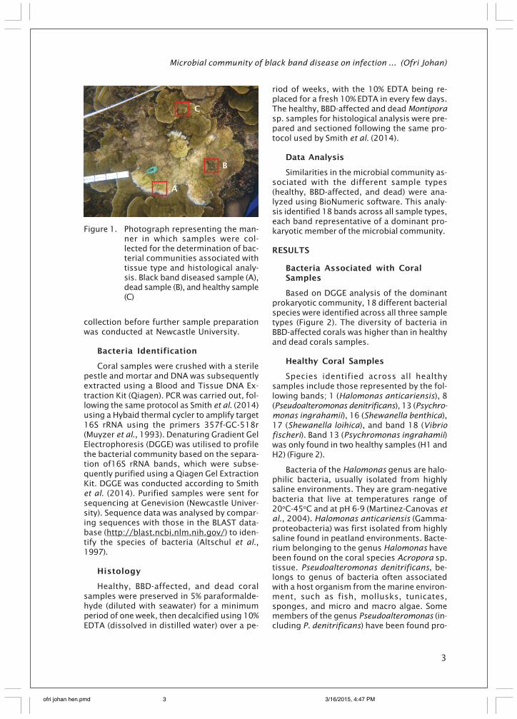
Coral samples were taken from zone 1 (PariIsland), zone 2 (Pramuka Island) and zone 3(Penjaliran Island) using hammer and punch.Three different types of sample were collect-ed, based on disease state, including healthytissue, BBD-affected tissue and dead tissuefrom the same coral colony. Samples weretaken and measured 5 cm2 in 4 replicates ofeach sample. Samples were collected for thepurposes of identifying associated bacteriaand histology (Figure 1).
Upon collection, samples for prokaryoticanalysis were placed into separate sterile con-tainers containing 70% ethanol, allowingsamples to be preserved until DNA extractioncould take place at the Coral Health and Dis-ease laboratory at the Newcastle UniversitySchool of Biology, United Kingdom.
Samples for histological analysis were sus-pended in seawater taken from the reef upon
ofri johan hen.pmd 3/16/2015, 4:47 PM2
Microbial community of black band disease on infection ... (Ofri Johan)
3
collection before further sample preparationwas conducted at Newcastle University.
Bacteria Identification
Coral samples were crushed with a sterilepestle and mortar and DNA was subsequentlyextracted using a Blood and Tissue DNA Ex-traction Kit (Qiagen). PCR was carried out, fol-lowing the same protocol as Smith et al. (2014)using a Hybaid thermal cycler to amplify target16S rRNA using the primers 357f-GC-518r(Muyzer et al., 1993). Denaturing Gradient GelElectrophoresis (DGGE) was utilised to profilethe bacterial community based on the separa-tion of16S rRNA bands, which were subse-quently purified using a Qiagen Gel ExtractionKit. DGGE was conducted according to Smithet al. (2014). Purified samples were sent forsequencing at Genevision (Newcastle Univer-sity). Sequence data was analysed by compar-ing sequences with those in the BLAST data-base (http://blast.ncbi.nlm.nih.gov/) to iden-tify the species of bacteria (Altschul et al.,1997).
Histology
Healthy, BBD-affected, and dead coralsamples were preserved in 5% paraformalde-hyde (diluted with seawater) for a minimumperiod of one week, then decalcified using 10%EDTA (dissolved in distilled water) over a pe-
riod of weeks, with the 10% EDTA being re-placed for a fresh 10% EDTA in every few days.The healthy, BBD-affected and dead Montiporasp. samples for histological analysis were pre-pared and sectioned following the same pro-tocol used by Smith et al. (2014).
Data Analysis
Similarities in the microbial community as-sociated with the different sample types(healthy, BBD-affected, and dead) were ana-lyzed using BioNumeric software. This analy-sis identified 18 bands across all sample types,each band representative of a dominant pro-karyotic member of the microbial community.
RESULTS
Bacteria Associated with CoralSamples
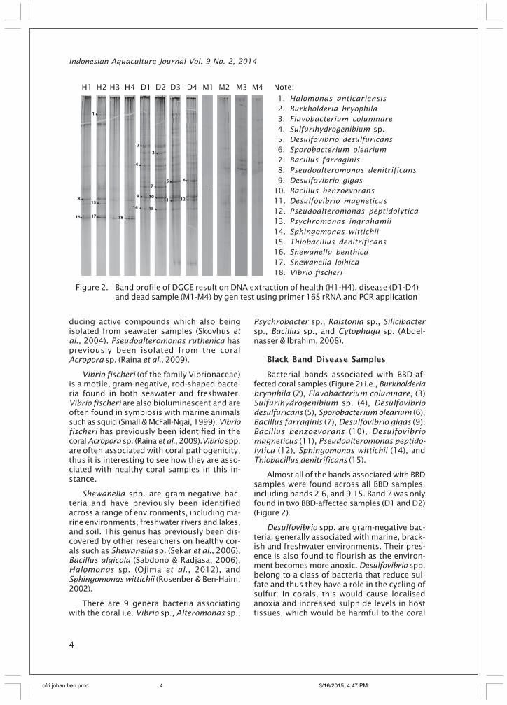
Based on DGGE analysis of the dominantprokaryotic community, 18 different bacterialspecies were identified across all three sampletypes (Figure 2). The diversity of bacteria inBBD-affected corals was higher than in healthyand dead corals samples.
Healthy Coral Samples
Species identified across all healthysamples include those represented by the fol-lowing bands; 1 (Halomonas anticariensis), 8(Pseudoalteromonas denitrificans), 13 (Psychro-monas ingrahamii), 16 (Shewanella benthica),17 (Shewanella loihica), and band 18 (Vibriofischeri). Band 13 (Psychromonas ingrahamii)was only found in two healthy samples (H1 andH2) (Figure 2).
Bacteria of the Halomonas genus are halo-philic bacteria, usually isolated from highlysaline environments. They are gram-negativebacteria that live at temperatures range of20oC-45oC and at pH 6-9 (Martinez-Canovas etal., 2004). Halomonas anticariensis (Gamma-proteobacteria) was first isolated from highlysaline found in peatland environments. Bacte-rium belonging to the genus Halomonas havebeen found on the coral species Acropora sp.tissue. Pseudoalteromonas denitrificans, be-longs to genus of bacteria often associatedwith a host organism from the marine environ-ment, such as fish, mollusks, tunicates,sponges, and micro and macro algae. Somemembers of the genus Pseudoalteromonas (in-cluding P. denitrificans) have been found pro-
Figure 1. Photograph representing the man-ner in which samples were col-lected for the determination of bac-terial communities associated withtissue type and histological analy-sis. Black band diseased sample (A),dead sample (B), and healthy sample(C)
A
C
B
ofri johan hen.pmd 3/16/2015, 4:47 PM3
Indonesian Aquaculture Journal Vol. 9 No. 2, 2014
4
ducing active compounds which also beingisolated from seawater samples (Skovhus etal., 2004). Pseudoalteromonas ruthenica haspreviously been isolated from the coralAcropora sp. (Raina et al., 2009).
Vibrio fischeri (of the family Vibrionaceae)is a motile, gram-negative, rod-shaped bacte-ria found in both seawater and freshwater.Vibrio fischeri are also bioluminescent and areoften found in symbiosis with marine animalssuch as squid (Small & McFall-Ngai, 1999). Vibriofischeri has previously been identified in thecoral Acropora sp. (Raina et al., 2009).Vibrio spp.are often associated with coral pathogenicity,thus it is interesting to see how they are asso-ciated with healthy coral samples in this in-stance.
Shewanella spp. are gram-negative bac-teria and have previously been identifiedacross a range of environments, including ma-rine environments, freshwater rivers and lakes,and soil. This genus has previously been dis-covered by other researchers on healthy cor-als such as Shewanella sp. (Sekar et al., 2006),Bacillus algicola (Sabdono & Radjasa, 2006),Halomonas sp. (Ojima et al., 2012), andSphingomonas wittichii (Rosenber & Ben-Haim,2002).
There are 9 genera bacteria associatingwith the coral i.e. Vibrio sp., Alteromonas sp.,
Psychrobacter sp., Ralstonia sp., Silicibactersp., Bacillus sp., and Cytophaga sp. (Abdel-nasser & Ibrahim, 2008).
Black Band Disease Samples
Bacterial bands associated with BBD-af-fected coral samples (Figure 2) i.e., Burkholderiabryophila (2), Flavobacterium columnare, (3)Sulfurihydrogenibium sp. (4), Desulfovibriodesulfuricans (5), Sporobacterium olearium (6),Bacillus farraginis (7), Desulfovibrio gigas (9),Bacillus benzoevorans (10), Desulfovibriomagneticus (11), Pseudoalteromonas peptido-lytica (12), Sphingomonas wittichii (14), andThiobacillus denitrificans (15).
Almost all of the bands associated with BBDsamples were found across all BBD samples,including bands 2-6, and 9-15. Band 7 was onlyfound in two BBD-affected samples (D1 and D2)(Figure 2).
Desulfovibrio spp. are gram-negative bac-teria, generally associated with marine, brack-ish and freshwater environments. Their pres-ence is also found to flourish as the environ-ment becomes more anoxic. Desulfovibrio spp.belong to a class of bacteria that reduce sul-fate and thus they have a role in the cycling ofsulfur. In corals, this would cause localisedanoxia and increased sulphide levels in hosttissues, which would be harmful to the coral
1. Halomonas anticariensis 2. Burkholderia bryophila 3. Flavobacterium columnare 4. Sulfurihydrogenibium sp. 5. Desulfovibrio desulfuricans 6. Sporobacterium olearium 7. Bacillus farraginis 8. Pseudoalteromonas denitrificans 9. Desulfovibrio gigas10. Bacillus benzoevorans11. Desulfovibrio magneticus12. Pseudoalteromonas peptidolytica13. Psychromonas ingrahamii14. Sphingomonas wittichii15. Thiobacillus denitrificans16. Shewanella benthica17. Shewanella loihica18. Vibrio fischeri
Figure 2. Band profile of DGGE result on DNA extraction of health (H1-H4), disease (D1-D4)and dead sample (M1-M4) by gen test using primer 16S rRNA and PCR application
H1 H2 H3 H4 D1 D2 D3 D4 M1 M2 M3 M4 Note:
ofri johan hen.pmd 3/16/2015, 4:47 PM4
Microbial community of black band disease on infection ... (Ofri Johan)
5
and could ultimately result in colony death(Viehman et al., 2006). Further, Viehman et al.(2006) state that many previous molecular-based studies have found several Desulfovibriospp. to be associated with black band disease.
Bacillus spp. are ubiquitous in nature andthis genus is found to include free-living andpathogenic species. Bacillus spp. are gram-positive, rod-shaped bacteria that can begrown in aerobic and anaerobic conditions.Spores from Bacillus spp. are resistant to heat(high temperature). These bacteria are knownto degrade xylan and carbohydrates, as wellas catalyse enzymes. Bacillus spp. have previ-ously been isolated from the sponge speciesPetromica citrina and Chelonaplysilla erecta(Bastos et al., 2013).
The genus Burkholderia belongs to thebetaproteobacteria group, comprising morethan 60 species which can be found in se-veral habitats such as, water, plant roots, or itcan be a causal disease in plants or humans(Santos et al., 2013). Despite this, this is thefirst report of bacteria belonging to this genusin coral reefs and diseased corals.
Flavobacterium columnare is a gram-nega-tive, rod-shaped bacteria which has previouslybeen found to cause disease in freshwater fish.This species can produce an enzyme that de-grades chondroitin sulfate (Figueiredo et al.,2005). This is the first report of this species ina diseased coral.
Sulfurihydrogenibium spp. are motile, gram-negative, rod-shaped bacteria capable of oxi-dizing sulphur, such that they are believed toplay an important role in the sulfur cycle(Nakagawa et al., 2005). These bacteria havepreviously been associated with pathogenic-ity in corals.
Sporobacterium olearium bacteria can da-mage methoxylated aromatic compounds andsyringate in significant amounts in the pre-sence of sulfide. These bacteria can be foundin both freshwater sediments and the marineenvironment (Lomans et al., 2001), althoughthere are no previous reports associating itwith corals and pathogenicity in corals.
Pseudoalteromonas peptidolytica are mo-tile, gram-negative bacteria belonging to theclass Gammaproteobacteria. This bacteria isassociated with aerobic environments and haspreviously been isolated from seawater in theSea of Japan. Interestingly, bacteria of this ge-
nus have previously been reported as patho-gens in fish (Vynne, 2011) and mussels(Venkateswaran & Dohmoto, 2000).
Pseudoalteromonas ruthenica has previ-ously been isolated from the coral Acroporasp (Raina et al., 2009).
Sphingomonas wittichii is an alphaproteobacteria and was first isolated from river water.It is known to be a good metabolizer of dibenzo-p-dioxin (Yabuuchi et al., 2001). Sphingomonasspp. are gram-negative, rod-shaped, chemo-heterotrophic bacteria usually associated withan oxygenated environment. These bacteriacan cause disease in humans (especiallySphingomonas paucimobilis). Sphingomonasbacteria are ubiquitous in nature, have beenisolated from soil, water and plant roots, largelythese bacteria are able to survive in low nutri-ent conditions. To date, these bacteria havenot been associated with pathogenicity ofcorals.
Thiobacillus denitrificans are gram-nega-tive, rod-shaped bacteria, belonging to theclass betaproteobacteria. These bacteria havebeen found in seawater sediments and are ableto oxidize sulfur to sulfate, a process in linewith the absorption of oxygen and nitrate re-duction (Aminuddin, 1979). Thiobacillus denitri-ficans is also able to reduce nitrate and nitrite(Haaijer et al., 2006). This bacterium has notbeen reported to infect corals.
Out of the bacteria found in BBD-affectedsamples, many of them belong to the groupCyanobacteria, such as Desulfovibrio desul-furicans, Desulfovibrio gigas, Desulfovibriomagneticus, Bacillus farraginis, Bacillusbenzoevorans. Cyanobacteria are a major causeof black band disease in corals (Richardson,1998).
Dead Coral Samples
Few bands were found on dead coralsamples and of those bands identified, nonewere found consistently across all deadsamples (M1-M4).
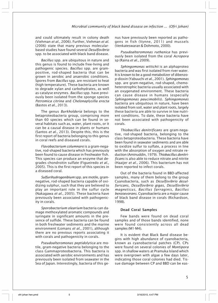
It is evident that Black Band disease be-gins with high abundance of cyanobacteria,known as cyanobacterial patches (CP). CPswere found on several colonies of Montiporaspp. in shallow waters at Pramuka Island whichwere overgrown with algae a few days later,indicating those coral colonies had died. Tis-sue damage between CP and BBD can be eas-
ofri johan hen.pmd 3/16/2015, 4:47 PM5
Indonesian Aquaculture Journal Vol. 9 No. 2, 2014
6
ily distinguished in the field, as CPs are notassociated with a black belt. Colonies foundto have a CP did not always develop BBD (Fig-ure 3). These observations are similar to thatreported by Sato et al. (2010) in the Great Bar-rier Reef.
CPs identified in Montipora sp. colonies(Figure 3A) were found to grow in size after 2weeks (Figure 3B). Exposed skeleton was thensubsequently colonised by algae (Figure 3C).Microscopic observations of CPs revealed alarge numbers of cyanobacteria filaments.
Histology Observation
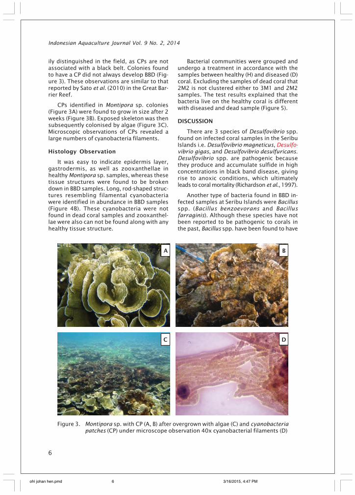
It was easy to indicate epidermis layer,gastrodermis, as well as zooxanthellae inhealthy Montipora sp. samples, whereas thesetissue structures were found to be brokendown in BBD samples. Long, rod-shaped struc-tures resembling filamental cyanobacteriawere identified in abundance in BBD samples(Figure 4B). These cyanobacteria were notfound in dead coral samples and zooxanthel-lae were also can not be found along with anyhealthy tissue structure.
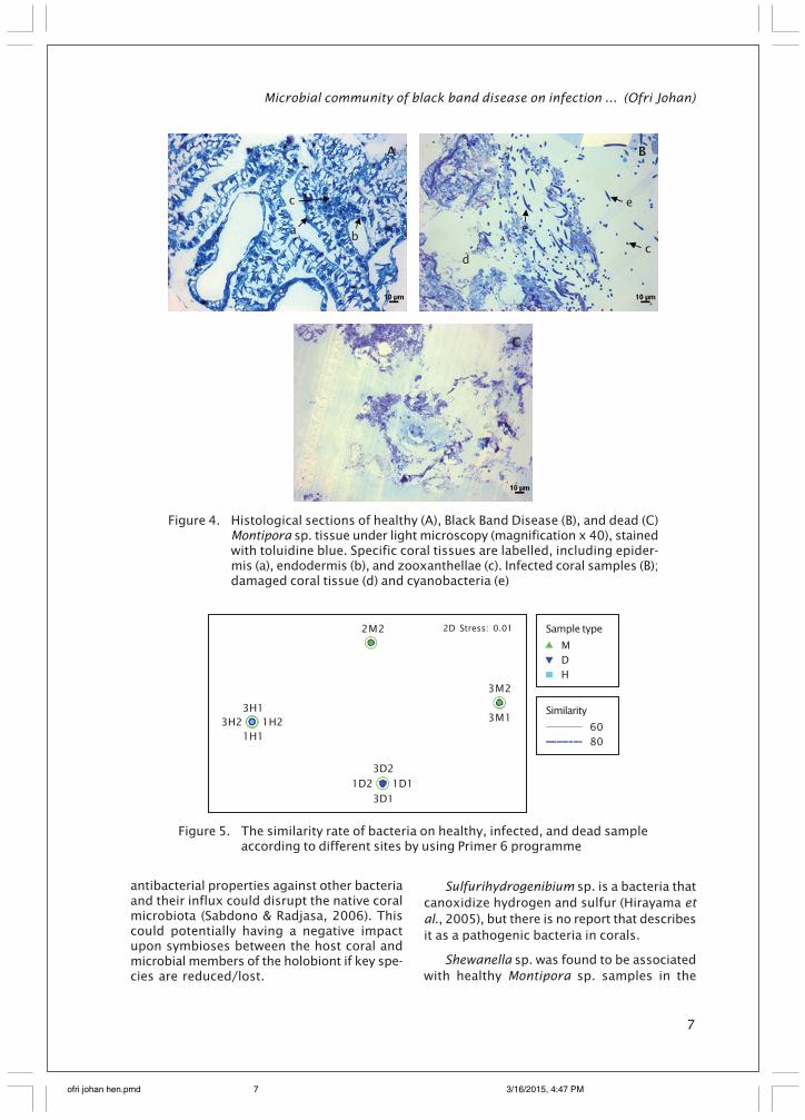
Bacterial communities were grouped andundergo a treatment in accordance with thesamples between healthy (H) and diseased (D)coral. Excluding the samples of dead coral that2M2 is not clustered either to 3M1 and 2M2samples. The test results explained that thebacteria live on the healthy coral is differentwith diseased and dead sample (Figure 5).
DISCUSSION
There are 3 species of Desulfovibrio spp.found on infected coral samples in the SeribuIslands i.e. Desulfovibrio magneticus, Desulfo-vibrio gigas, and Desulfovibrio desulfuricans.Desulfovibrio spp. are pathogenic becausethey produce and accumulate sulfide in highconcentrations in black band disease, givingrise to anoxic conditions, which ultimatelyleads to coral mortality (Richardson et al., 1997).
Another type of bacteria found in BBD in-fected samples at Seribu Islands were Bacillusspp. (Bacillus benzoevorans and Bacillusfarraginis). Although these species have notbeen reported to be pathogenic to corals inthe past, Bacillus spp. have been found to have
Figure 3. Montipora sp. with CP (A, B) after overgrown with algae (C) and cyanobacteriapatches (CP) under microscope observation 40x cyanobacterial filaments (D)
A
C
B
D
ofri johan hen.pmd 3/16/2015, 4:47 PM6
Microbial community of black band disease on infection ... (Ofri Johan)
7
antibacterial properties against other bacteriaand their influx could disrupt the native coralmicrobiota (Sabdono & Radjasa, 2006). Thiscould potentially having a negative impactupon symbioses between the host coral andmicrobial members of the holobiont if key spe-cies are reduced/lost.
Sulfurihydrogenibium sp. is a bacteria thatcanoxidize hydrogen and sulfur (Hirayama etal., 2005), but there is no report that describesit as a pathogenic bacteria in corals.
Shewanella sp. was found to be associatedwith healthy Montipora sp. samples in the
Figure 4. Histological sections of healthy (A), Black Band Disease (B), and dead (C)Montipora sp. tissue under light microscopy (magnification x 40), stainedwith toluidine blue. Specific coral tissues are labelled, including epider-mis (a), endodermis (b), and zooxanthellae (c). Infected coral samples (B);damaged coral tissue (d) and cyanobacteria (e)
d
e
e
c
a
BA
c
b
C
Figure 5. The similarity rate of bacteria on healthy, infected, and dead sampleaccording to different sites by using Primer 6 programme
3H2
2D Stress: 0.012M2
1D2 1D1
1H2
3D1
3D2
1H1
3H13M1
3M2
Sample type
MDH
Similarity
6080
ofri johan hen.pmd 3/16/2015, 4:47 PM7
Indonesian Aquaculture Journal Vol. 9 No. 2, 2014
8
present study, coinciding with previous re-ports that associate this bacteria with healthycorals (Frias-Lopez et al., 2002). Despite this,other studies have also found these bacteriato be associated with infected corals (Sekar etal., 2006), thus there is the potential thathealthy samples harbouring this bacterial spe-cies may become diseased following an influxassociated with favourable environmental con-ditions (Frias-Lopez et al., 2002; and Rohweret al., 2002).
Sphingomonas sp., found in BBD samplesin the present study, have previously beenassociated with healthy coral (Rosenberg &Ben-Haim, 2002). That said, members of thisgenus have also been associated with coralssuffering from White Plague in Florida, demon-strating their pathogenic potential (Ravindranet al., 1998).
Although Black Band Disease (BBD) hasbeen known for decades (since 1970), the mainpathogenic bacteria of the disease has yet tobe identified. Previously, it was considered thatBBD was caused by the bacteria Phormidiumcorallyticum. But, the use of molecular tech-niques to identify the microbial communityassociated with this disease has led to the dis-covery of a consortium of bacteria associatedwith BBD (Frias-Lopez et al., 2004). This con-sortium is comprised of various cyanobacteria,sulfate-reducing bacteria such as Desulfovibriospp., sulfide-oxidazing Beggiatoa spp. and ma-rine fungi. This makes it difficult to fulfil Koch’spostulates for BBD, as this approach is moresuited to diseases with single or minimal causalagents (Richardson, 1998; Sutherland et al.,2004).
Sato et al. (2010) has identified 262 casesof CP in Montipora sp. in the Great Barrier Reefand 18.7% of these cases developing into BBD.In contrast to this, CP cases in Montipora spp.in the Thousand Islands ( or Seribu Island) werenot found to develop into BBD, with a recentoutbreak of BBD reaching a recent peak (Johan,2013, pers obs) appearing to exist as a sepa-rate disease.
CONCLUSION
Bacteria belonging to the genus Bacillussp., Sulfurihydrogenibium sp. and Desulfovibriospp. are associated with BBD in Montipora sp.in Indonesian reefs, with Desulfovibrio spp.appearing to be a strong causal agent for thisdisease. Sphingomonas wittichii, Halomonas
sp., Bacillus algicola, and Shewanella sp. are allassociated with the healthy Montipora sp.holobiont in this region. Following histologicalanalysis, there are marked differences be-tween healthy, BBD-affected and dead Monti-pora sp. tissues. In BBD-affected samples, thereis mass breakdown in host tissue, zooxanthel-lae are lost and filament-shaped bacteria arepresent. These filament-shaped bacteria arelost in dead samples, thus clearly demonstrat-ing their presence is strongly associated withBBD tissues.
ACKNOWLEDGEMENTS
The authors would like to thank the Minis-try of Marine Affairs and Fisheries for provid-ing study fees and the fellowship from TheWorld Bank, tied with the Robert S. McNamaraFellowships Program (RSM), Coral Health andDisease Laboratory, Newcastle University (UK)in assisting in analyzing bacteria associatedwith coral samples. This research was also sup-ported by funding from the Center for Aqua-culture Research and Development, and BogorAgriculture University. The authors would alsolike to thank those who helped with data col-lection, Jhon Bythell who help in process toget funding in this analysis as well as theSeribu Islands National Park, Jakarta, for allow-ing use as a research location.
REFERENCES
Abdelnasser, S., & Ibrabim, S. (2008). Diversityof coral Euniceafusca associated bacteriausing culture dependent techniques. Re-search Journal of Microbiology, 3(10), 614-621.
Altschul, S.F., Madden, T.L., Schaffer, A.A.,Zhang, J., Zhang, Z., & Miller, W. (1997).Gapped BLAST and PSI-BLAST: a new gen-eration of protein database search pro-grams. Nucleic Acids Res., 25: 3389-3402.
Aminuddin, M. (1979). The oxidation of elemen-tal sulphur by Thiobacillus denitrificans.Pertanika, 2(1), 21-27.
Barneah, O., Eitan Ben-Dov, Kramarsky-Winter,E., & Kushmaro, A. (2007). Characterizationof black band disease in Red Sea stonycorals. Environmental Microbiology, 9(8),1995-2006.
Cooney, R.P., Pantos, O., Le Tissier, M.D.A.,Barer, M.R., O’Donnell, A.G., & Bythell JC.(2002). Characterization of the bacterialconsortium associated with black band dis-
ofri johan hen.pmd 3/16/2015, 4:47 PM8
Microbial community of black band disease on infection ... (Ofri Johan)
9
ease in coral using molecular microbiologi-cal techniques. Environmental microbiol-ogy, 4(7), 401-13. Retrieved from http://www.ncb i . n lm .n ih .gov/pubmed/12123476.
Ducklow, H.W., & Mitchell, R. (1979). Composi-tion of mucus released by coral reef co-elenterates. Limnol. Oceanogr., 24(4), 706-714.
Fadlallah, Y.H., & Pearse, J.S. (1982). Sexual re-production in solitary corals: synchronousgametogenesis and broadcast spawning inParacyathus stearnsii. Mar. Biol., 71, 233-239.
Figueiredo, H.C.P., Klesius, P.H., Arias, C.R.,Evans, J., Shoemaker, C.A., Pereira, Jr. D.J., &Peixoto, M.T.D. (2005). Isolation and char-acterization of strains of Flavobacteriumcolumnare from Brazil. Journal of Fish Dis-eases, 28, 199-204.
Frias-lopez, J., Bonheyo, G.T., & Fouke, B.W.(2004). Identification of differential geneexpression in bacteria associated withcoral black band disease by using RNA-Ar-bitrarily primed PCR, 70(6), 3687-3694.doi:10.1128/AEM.70.6.3687.
Frias-Lopez, J., Zerkle, A.L., Bonheyo, G.T., &Fouke, B.W. (2002). Partitioning of bacterialcommunities between sea-water andhealthy, black band diseased, and deadcoral surfaces. Appl. Environ. Microbiol., 68,2214-2228.
Garrett, P., & Ducklow, H. (1975). Coral diseasein Bermuda. Nature, 253, 349-350. doi:10.1038/253349a0.
Glynn, P.W., & De Weerdt, W.H. (1991). Elimina-tion of two reef building hydrocorals fol-lowing the 1982-83 El Niño warming event.Science, 253, 69-71.
Glynn, P.W. (1994). State of coral reefs in theGalapagos Islands: natural vs anthropo-genic impacts. Marine Pollution Bulletin, 29,131-140.
Haaijer, S.C., Van der Welle, M.E., Schmid, M.C.,Lamers, L.P., Jetten, M.S., & Op den Camp,H.J. (2006). Evidence for the involvementof betaproteobacterial Thiobacilli in thenitrate-dependent oxidation of iron sulfi-de minerals. FEMS Microbiology Ecology, 58,439-448.
Haapkylä, J., Seymour, A.S., Trebilco, J., & Smith,D. (2007). Coral disease prevalence andcoral health in the Wakatobi Marine Park,South-east Sulawesi, Indonesia. J. Mar. Biol.Ass. U.K., 87(5582), 1-12.
Harvell, D., Jordán-Dahlgren, E., Merkel, S.,Rosenberg, E., Raymundo, L., Smith, G., Weil,E., & Willis, B. (2007). Coral disease, envi-ronmental drivers, coral and microbial as-sociates balance between and the by thecoral disease working group of the globalenvironmental facility coral reef targetedresearch program. Oceanography, 20(1),172-195.
Hirayam, H., Takai, K., Inagaki, F., Nealson, K.H.,& Horikoshi, K. (2005). Thiobacter subterra-neus gen. nov., sp. nov., an obligately che-molithoautotrophic, thermophilic, sulfur-oxidizing bacterium from a subsurface hotaquifer. IJSEM, 55(1), 467-472.
Humason, G.L. (1962). Animal tissue techni-ques. W. H. Freeman and Co. San Francisco,468 pp.
Johan, O., Bengen, D.G., Zamani, N.P., Suhar-sono, & Sweet, M.J. (2013). The distribu-tion and abundance of black band diseaseand white syndrome in Kepulauan Seribu,North of Jakarta, Indonesia. Disease ofAquatic Organism, 17 pp. (in progress).
Kiernan, J.A. (1990). Histological and histoche-mical methods. 2nd Eds., Pergamon Press,New York.
Lee, K., & Ruby, E.G. (1995). Symbiotic role ofthe viable but nonculturable state of Vib-rio fischeri in Hawaiian Coastal Seawater.Appl. Environ. Microbiol., 61(1), 278-283.
Lomans, B.P., Leijdekkers, P., Wesselink, J.J.,Bakkes, P ., Pol, A., Drift, C., & Op den Camp,H.J.M. (2001). Obligate sulfide-dependentdegradation of methoxylated aromaticcompounds and formation of methanethioland dimethyl sulfide by a freshwater sedi-ment isolate, Parasporobacterium pauci-vorans gen. nov., sp. nov. Appl. Environ.Microbiol. September; 67(9), 4017-4023.doi:10.1128/AEM.67.9.4017-4023.2001.
Martinez-Canovas, M.J., Bejar, V., Martinez-Checa, F., & Quesada, E. (2004). Halomonasanticariensis sp. nov., from Fuente de Pie-dra, a saline-wetland wildfowl reserve inMa´laga, Southern Spain. International Jour-nal of Systematic and Evolutionary Micro-biology, 54, 1329-1332.
Muyzer, G., de Waal, E.C., & Uitterlinden, A.G.(1993). Profiling of complex microbial po-pulations by denaturing gradient gel elec-trophoresis analysis of polymerase chainreaction-amplified genes encoding for 16SrRNA. Appl. Environ. Microbiol., 59, 695-700.
Nakagawa, S., Shtaih, Z., Banta, A., Beveridge,
ofri johan hen.pmd 3/16/2015, 4:47 PM9
Indonesian Aquaculture Journal Vol. 9 No. 2, 2014
10
T.J., Sako, Y., & Reysenbach, A.L. (2005).Sulfurihydrogenibium yellowstonense sp.nov., an extremely thermophilic, faculta-tively heterotrophic, sulfur-oxidizing bac-terium from Yellowstone National Park, andemended descriptions of the genus Sul-furihydrogenibium, Sulfurihydrogenibiumsubterraneum and Sulfurihydrogenibiumazorense. International Journal of System-atic and Evolutionary Microbiology, 55,2263-2268.
Ojima, T., Saburi, W., Yamamoto, T., & Kudo, T.(2012). Characterization of Halomonas sp.H11 -glucosidase activated by monova-lent cations and its application for efficientsynthesis of -D-glucosylglycerol. Appl.Environ. Microbiol., 44 pp. doi:10.1128/AEM.07514-11.
Raina, J.B., Tapiolas, D., Willis, B.L., & Bourne,D.G. (2009). Coral-associated bacteria andtheir role in the biogeochemical cyclingof sulfur. Applied and Environmental Mi-crobiology, 75(11), 3492-3501.
Ramos-Flores, T. (1983). Lower marine fungusassociated with black line disease in starcorals (Montastraea annularis). Biol. Bull.,165, 429-435.
Ravindran, J., Raghukumar, C., & Raghukumar,S. (1998). Disease and stress-induced mor-tality of corals in Indian reefs and observa-tions on bleaching of corals in the Anda-mans.
Richardson, L.L., Miller, A.W., Broderick, E.,Kaczmarsky, L., Gantar, M., Stani, D., &Sekar, R. (2009). Sulfide, microcystin, andthe etiology of black band disease. Dis-eases of aquatic organisms, 87(1-2), 79-90.doi:10.3354/dao02083
Richardson, L.L. (1996). Horizontal and verti-cal migration patterns of Phormidiumcorallyticum and Beggiatoa spp. associ-ated with Black-Band Disease of corals.Microb. Ecol., 32,323-335.
Richardson, L.L. (1998). Coral disease: what isreally known? Tree, 13(11), 438-443.
Richardson, L.L., Kuta, K.G., Schnell, S., &Carlton, R.G. (1997). Ecology of the blackband disease microbial consortium. Proc.8th Intl. Coral Reef Symp. 1, 597-600.
Richardson, L.L., Miller, A.W., Broderick, E.,Kaczmarsky, L., Gantar, M., Stanic, D., &Sekar, R. (2009). Dis. Aqual. Org., 87, 79-99.
Rohwer, F., Seguritan, V., Azam, F., & Knowlton,N. (2002). Diversity and distribution of co-ral-associated bacteria. Mar. Ecol. Prog. Ser.,
243, 1-10.Rosenberg, E., & Ben-Haim, Y. (2002). Microbial
diseases of corals and global warming. En-vironmental Microbiology, 4(6), 318-326.
Rosenberg, E., & Loya, Y. (2004). Coral healthand disease. Springer, Berlin.
Sabdono, A., & Radjasa, O.K. (2006). Anti-bacte-rial property of a coral-associated bacte-rium Bacillus sp. against coral pathogenicBBD (Black Band Disease). Journal of CoastalDevelopment, 9(3), 175-182.
Santos, P., Estrada-de los, Vinuesa, P., Hirsch,A.M., Martinez-Aguilar, L., & Caballero-Mellado, J. (2013). Phylogenetic analysis ofburkholderia species by multilocus se-quence analysis. Curr. Microbiol., 10 pp.doi: 10.1007/s00284-013-0330-9.
Sato, Y., Willis, B.L., & Bourne, D.G. (2010). Suc-cessional changes in bacterial communi-ties during the development of black banddisease on the reef coral, Montipora his-pida. The ISME Journal, 4(2), 203-214. doi:10.1038/ismej.2009.103.
Sekar, R., Mills, D.K., Remily, E.R., Voss, J.D., &Richardson, L.L. (2006). Microbial commu-nities in the surface mucopolysaccharidelayer and the black band microbial mat ofblack band-diseased Siderastrea siderea.Applied and Environmental Microbiology,72(9), 5963-73. doi:10.1128/AEM.00843-06.
Smith, D., Leary, P., Bendall, M., Flach, E., & Jones,R. (2014). A novel investigation of a blister-like syndrome in aquarium Echinoporalamellosa. PLoS ONE, 9(5), e97018. doi:10.1371/j8.ournal.pone.009701
Small, A.L., & McFall-Ngai, M.J. (1999). A halideperoxidase in tissues that interact withbacteria in the host squid Euprymnascolopes. J. Cellul. Biochem., 72, 445-457.
Sweet, M.J., Croquer, A., & Bythell, J.C. (2011).Development of bacterial biofilms on arti-ficial corals comparison to surface-associ-ated microbes of hard corals. PLoS ONE,6(6), 1-14.
Sweet, M.J., Jones, R., & Bythell, J.C. (2012). Coraldiseases in aquaria and in nature. Jurnalof the Marine Biological Association of theUnited Kingdom, 92(4), 791-801.
Sutherland, K.P., Porter, J.W., & Torres, C. (2004).Disease and immunity in caribbean andIndo-Pacific zooxanthellate corals. Mar.Eco. Prog. Ser., 266, 273-302.
Venkateswaran, K., & Dohmoto, N. (2000).Pseudoalteromonas peptidolytica sp. nov.,
ofri johan hen.pmd 3/16/2015, 4:47 PM10
Microbial community of black band disease on infection ... (Ofri Johan)
11
a novel marine mussel-thread-degradingbacterium isolated from the Sea of Japan.International Journal of Systematic and Evo-lutionary Microbiology, 50, 565-574.
Viehman, S., Mills, D.K., Meichel, G.W., &Richardson, L.L. (2006). Culture and identi-fication of Desulfovibrio spp. from coralsinfected by black band disease on Domini-can and Florida Keys reefs. Dis. Aquat. Org.,69, 119-127.
Vynne, N.G. (2011). Bioactivity and phylogenyof the marine bacterial genus Pseudo-alteromonas. Technical University of Den-mark, National Food Institute, Division ofIndustrial Food Research. Ph.D. thesis.
Willis, B.L., Page, C.A., & Dinsdale, E.A. (2004).Coral disease on the Great Barrier Reef. InCoral Health and Disease, Rosenberg, E.,Loya, Y. (eds), Springer-Verlag, Berlin, p. 69-104.
Yabuuchi, E., Yamamoto, H., Terakubo, S.,Okamura, N., Naka, T., Fujiwara, N., Koba-yashi, K., Kosakoand, Y., & Hiraishi, A. (2001).Proposal of Sphingomonas wittichii sp. nov.for strain RW1T, known as a dibenzo-p-di-oxin metabolizer. International Journal ofSystematic and Evolutionary Microbiology,51, 281-292.
ofri johan hen.pmd 3/16/2015, 4:47 PM11