metric (mrenterography or ultrasound in crohn's disease): a study protocol for a multicentre,...
TRANSCRIPT
Taylor et al. BMC Gastroenterology 2014, 14:142http://www.biomedcentral.com/1471-230X/14/142
STUDY PROTOCOL Open Access
METRIC (MREnterography or ulTRasound in Crohn’sdisease): a study protocol for a multicentre,non-randomised, single-arm, prospectivecomparison study of magnetic resonanceenterography and small bowel ultrasoundcompared to a reference standard in those aged16 and overStuart Taylor1*, Susan Mallett2, Gauraang Bhatnagar1, Stuart Bloom3, Arun Gupta4, Steve Halligan1, John Hamlin5,Ailsa Hart6, Antony Higginson7, Ilan Jacobs8, Sara McCartney3, Steve Morris9, Nicola Muirhead10, Charles Murray11,Shonit Punwani1, Manuel Rodriguez-Justo12, Andrew Slater13, Simon Travis14, Damian Tolan15, Alastair Windsor16,Peter Wylie17 and Ian Zealley18
Abstract
Background: Crohn’s disease (CD) is a lifelong, relapsing and remitting inflammatory condition of the intestine.Medical imaging is crucial for diagnosis, phenotyping, activity assessment and detecting complications. Diverse smallbowel imaging tests are available but a standard algorithm for deployment is lacking. Many hospitals employ tests thatimpart ionising radiation, of particular concern to this young patient population. Magnetic resonance enterography(MRE) and small bowel ultrasound (USS) are attractive options, as they do not use ionising radiation. However, theircomparative diagnostic accuracy has not been compared in large head to head trials. METRIC aims to compare thediagnostic efficacy, therapeutic impact and cost effectiveness of MRE and USS in newly diagnosed and relapsing CD.
Methods: METRIC (ISRCTN03982913) is a multicentre, non-randomised, single-arm, prospective comparison study. Twopatient cohorts will be recruited; those newly diagnosed with CD, and those with suspected relapse. Both will undergoMRE and USS in addition to other imaging tests performed as part of clinical care. Strict blinding protocols will beenforced for those interpreting MRE and USS. The Harvey Bradshaw index, C-reactive protein and faecal calprotectin willbe collected at recruitment and 3 months, and patient experience will be assessed via questionnaires. A multidisciplinaryconsensus panel will assess all available clinical and imaging data up to 6 months after recruitment of each patient andwill define the standard of reference for the presence, localisation and activity of disease against which the diagnosticaccuracy of MRE and USS will be judged. Diagnostic impact of MRE and USS will be evaluated and cost effectiveness willbe assessed. The primary outcome measure is the difference in per patient sensitivity between MRE and USS for thecorrect identification and localisation of small bowel CD.(Continued on next page)
* Correspondence: [email protected] for Medical Imaging, University College London, 250 Euston Rd,London NW1 2PG, UKFull list of author information is available at the end of the article
© 2014 Taylor et al.; licensee BioMed Central Ltd. This is an Open Access article distributed under the terms of the CreativeCommons Attribution License (http://creativecommons.org/licenses/by/4.0), which permits unrestricted use, distribution, andreproduction in any medium, provided the original work is properly credited. The Creative Commons Public DomainDedication waiver (http://creativecommons.org/publicdomain/zero/1.0/) applies to the data made available in this article,unless otherwise stated.

Taylor et al. BMC Gastroenterology 2014, 14:142 Page 2 of 10http://www.biomedcentral.com/1471-230X/14/142
(Continued from previous page)
Discussion: The trial is open at 5 centres with 46 patients recruited. We highlight the importance of stringent blindingprotocols in order to delineate the true diagnostic accuracy of both imaging tests and discuss the difficulties of diagnosticaccuracy studies in the absence of a single standard of reference, describing our approach utilising a consensus panelwhilst minimising incorporation bias.
Trial registration: METRIC - ISRCTN03982913 – 05.11.13.
Keywords: Crohn’s disease, Inflammatory bowel disease, MRE, USS, Consensus panel
BackgroundCrohn’s disease (CD) is an inflammatory condition, with awide spectrum of intestinal manifestations ranging fromsuperficial bowel wall ulceration to deep penetratingdisease, characterised by fistulae and abscesses. Overtime, repeated inflammatory insults can result in thedevelopment of fibrosis and stricture formation. It affects200,000 people in the UK (around 1 in 500), most areyoung (diagnosed < 35 years) and the costs of directmedical care in the UK exceed £500 million [1]. A rangeof potentially toxic medical treatment options, such asimmune-modulators, or targeted surgical interventions arecurrently employed in disease management. The optimaltreatment strategy requires accurate assessment of diseasepresence, extent, activity and complications. CD typicallyaffects the small bowel, most of which is beyond the reachof conventional colonoscopy. Small bowel imaging there-fore plays a vital role in diagnosing and phenotyping CD,thereafter assessing disease activity and complications.At present there is no single imaging modality that has
been proven universally superior in either suspected orestablished CD. A plethora of small bowel investigationsare currently performed within the NHS, approximately100,000 each year, including Barium fluoroscopy (BaF),Computerised Tomography (CT), Ultrasound (USS) andMagnetic Resonance Enterography (MRE). According toa UK survey in 2010 [2], 90% of NHS radiology depart-ments routinely perform BaF to investigate patients withknown or suspected CD, 80% perform CT, 56% USS and38% MRI. Small bowel imaging tests differ in their indi-vidual attributes; for example BaF affords high qualityassessment of the bowel mucosa, whilst cross sectionaltechniques such as CT, MRE and USS facilitate evalu-ation of the bowel wall and extra-enteric tissues. An im-portant attribute is the use or otherwise of ionisingradiation. The currently most used tests, BaF and CT,impart a significant radiation dose. This is concerninggiven that CD patients are young and usually undergoserial imaging to assess disease evolution over their life-time. An audit in 2007 found 15.5% of CD patients re-ceived a cumulative radiation dose that may increasecancer risk by 7.3% [3]. USS and MRE are alternativesthat do not use ionising radiation but deployment in theNHS is currently ad hoc. The choice of small bowel
imaging investigation currently depends largely on non-evidence based decision-making, such as clinician per-sonal preference, perceived costs, available infrastructureand radiological expertise.Three systematic reviews have been published to date
evaluating the accuracy of imaging tests in the diagnosisof CD and for assessing disease activity [4-6]. All havehighlighted marked heterogeneity in the available litera-ture, with most studies being single centre and involvingrelatively small patient numbers. Variation in the appliedstandard of reference between studies is also apparent.The largest systematic review [6] incorporated 68 stud-ies, and compared the performance of CT, MRI and USSfor diagnosis, and disease activity classification. For diag-nosis of disease location sensitivity of USS ranged from75 to 93% and for MRI from 77 to 91%. Specificityranged from 98 to 100% (USS) and 60 to 100% (MRI).The diagnostic accuracy to detect active disease per pa-tient of studies USS sensitivity and specificity for detect-ing active disease was 85% (range 75 to 100%) and 91%(range 82-100%) respectively and for MRI were 80%(range 78-100%) and 82% (range 46%-100%) respectively.Only one study used the recommended direct diagnostictest comparison study design shown to reduce bias byassessing the same patients with multiple tests [7]. Miaoet al. [8] reported of sensitivity 87% for both USS andMRI and specificities of 100% (7/7 patients) and 71% (5/7 patients) respectively in a study with 30 patients (23with Crohn’s and 7 with no disease) [8].Ultimately, the optimal imaging strategy for CD remains
uncertain and single centre data is of limited utility. Un-biased, robust data to inform the implementation strategyfor newer imaging technologies are currently unavailable,although international guidelines on imaging advocateMRE for diagnosis and USS for assessing disease activity,where resource and expertise are available [9].In this article, we describe the protocol for the METRIC
study (ISRCTN03982913), a multicentre, non-randomised,single-arm, prospective comparison study of MRE andUSS in newly diagnosed CD, or established disease withsuspected relapse. The sample size of our study is tentimes larger than the only other study directly comparingMRE and USS in the same patients (8). Participating ra-diologists are members of BSGAR, British Society of
Taylor et al. BMC Gastroenterology 2014, 14:142 Page 3 of 10http://www.biomedcentral.com/1471-230X/14/142
Gastrointestinal and Abdominal Radiology. Research onthis topic has been commissioned and funded by the UKHealth Technology Assessment (HTA) programme (11/23/01). The full protocol adheres to the principals of theSPIRIT guidelines for clinical trials protocols [10].
Study objectives
� Directly to compare the diagnostic accuracies ofMRE and USS for detecting small bowel CD, andgrading of inflammatory activity. This will includesubgroup analysis of patients with a new diagnosisof CD and those suffering a relapse. The referencestandard consists of a consensus panel, withcollective review of all the available clinical andimaging data over a 6-month follow up period.
� Directly to compare diagnostic accuracies of MREand USS for detecting colonic CD in thoseundergoing colonoscopy.
� To use a novel trial design to reduce uncertainty inevaluating the impact of MRE, USS andconventional imaging methods by direct capture ofpatient management.
� To evaluate the cost effectiveness of MRE and USScompared to each other, and to conventionalimaging methods.
MethodsGeneralThis is a multi-centre prospective cohort study compar-ing the diagnostic accuracy of MRE and USS for thepresence, extent and activity of small bowel Crohn’s dis-ease. The trial framework is to detect superiority ofMRE over USS (Figure 1).
Inclusions/exclusion criteriaThe trial will recruit from two defined patient cohorts:(1) newly diagnosed CD patients (or within 3 months ofdiagnosis) and (2) those with previously confirmed CDwith a high clinical suspicion of luminal relapse, requir-ing radiological investigation.In the new diagnosis cohort, eligible patients are aged
16 years or older, have given written informed consent,undergoing or having undergone colonoscopy, and either:
� Newly diagnosed (within 3 months) with CD basedon endoscopic, histological, clinical and radiologicalfindings, [11] or
� Highly suspected of CD based on characteristicendoscopic, imaging and/or histological features butpending final diagnosis.
In the suspected relapse cohort, eligible patients areaged 16 years or older, able to give written informed
consent, with a known diagnosis of CD together with ahigh clinical suspicion of luminal relapse defined as:
� Objective markers of inflammatory activity (raisedCRP >8 mg/L OR raised calprotectin > 100 mcg/g), or
� Symptoms suggestive of luminal stenosis (includingobstructive symptoms such as colicky abdominal pain,vomiting) or abnormal endoscopy suggesting relapse.
Patients with any psychiatric or other disorder likely toaffect on informed consent and those with evidence ofsevere or uncontrolled systemic disease, which at theprincipal investigator’s discretion renders the patient un-suitable for participation in the study, will be excluded.Pregnant patients or those with contraindications toMRE (e.g. allergy to all suitable contrast agents, cardiacpacemaker, severe claustrophobia, or an inability to lieflat) are also excluded. Recruited patients with suspecteddisease whose final diagnosis is not CD, and those whoundergo surgical resection prior to colonoscopy will alsobe excluded.
Ethical arrangements and consentThe METRIC trial achieved National Health Service re-search ethics committee (NHS REC) approval in September2013 (13/SC/0394) and is being conducted in accordancewith the principles of Good Clinical Practice (GCP). In-formed consent is a prerequisite. University CollegeLondon’s Clinical Trials Unit supervises the trial.
Diagnostic interventionsMRERecruited patients will undergo MRE at their recruit-ment site. The usual site radiographer team will performthe examination providing they are deemed competentby the site radiology lead.The MRI platform (i.e. manufacturer and Tesla (T)
strength) will be decided by the local radiologist accordingto scanner availability and usual practice. It is anticipatedmost MRE will be performed at 1.5 T. Exact imaging pa-rameters will vary according to MRI platform but a mini-mum dataset of sequences will be acquired (Table 1). Thechoice of oral contrast prior to MRE will also be accordingto the usual practice of the recruitment site.In some patients MRE will have been performed as
part of usual clinical care prior to recruitment. If it hasbeen acquired within the preceding 4 weeks accordingto the minimum dataset of sequences, the MRE will beeligible for inclusion in the trial and will not need to berepeated.
USSRecruited patients will also undergo small bowel USSat their recruitment site. This will be performed by a
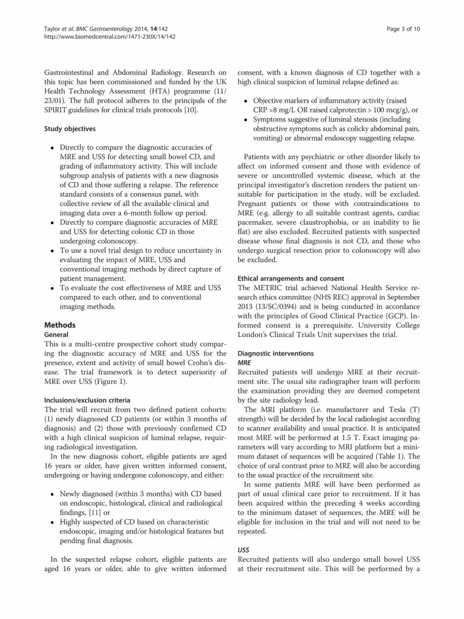
METRIC (MR Enterography or ulTRasound In Crohn’s disease)
Trial Flow Diagram
167 PatientsConfirmed new diagnosis of Crohn’s disease ORhighly suspected based on colonoscopy/imaging
* IF MRE already performed pre recruitment-blinded radiologist reviewIf US already performed pre recruitment-repeated by blinded radiologist
167 PatientsKnown Crohn’s disease with high clinical suspicion of
relapse
334 Patients-MRE and US performed by 2 blinded
independent radiologists*-blood and stool sample tests
-Repeat blood and stool sample tests 3 months following instigation of therapy.
Discrepancy for the presence of non
contiguous small bowel disease proximal
to Terminal ileum on MRE and US
Additional small bowel investigation eg Capsule
endoscopy, BaF, CT
Consensus panel composite reference standard for luminal disease presence, extent and
activity (based on all Ileo-colonoscopy, capsule endoscopy, imaging, histopathology, blood
and stool test results) leading to a final therapeutic decision.
yes
no
Ileocolonoscopy (all
patients) & conventional
small bowel imaging (BaF,
CTE) if part of site standard
clinical practice
Total patient follow up 6 months-clinical course. All standard clinical practice
diagnostic tests recorded.
Figure 1 Flow diagram outlining the stages of the METRIC trial.
Taylor et al. BMC Gastroenterology 2014, 14:142 Page 4 of 10http://www.biomedcentral.com/1471-230X/14/142
radiologist or trained sonographer fulfilling compe-tency criteria (see section below). For the purposes ofthe trial, patients will not receive any oral agent beforethe USS other than an optional 2 cups of water toimprove visualisation of the duodenum. The local
radiologist will select the USS platform according toscanner availability and usual practice. Exact imagingparameters will vary according to USS platform but aminimum probe frequency and examination techniquewill be required (Table 2).

Table 1 MRE protocol outlining the minimum andoptional MRI sequences that may be performed
Minimum Coronal true FISP
Buscopan-20 mg IV
Axial and coronal non Fat Sat HASTE
Coronal Fat Sat HASTE
Axial diffusion b values 50 and 600
Coronal pre and post gadolinium T1 (60–70 sec)
Optional Axial True FISP
Axial Fat Sat HASTE
Axial post gadolinium T1
True FIP dynamic Motility
Taylor et al. BMC Gastroenterology 2014, 14:142 Page 5 of 10http://www.biomedcentral.com/1471-230X/14/142
USS acquired up to 4 weeks prior to recruitment will berepeated unless full blinding of the original performingradiologist or sonographer to clinical and imaging datacan be assured.
Other small bowel imagingMany recruited patients will also undergo conventionalsmall bowel imaging as part of usual clinical care, not-ably BaF, CT enterography and in some cases capsuleendoscopy. The results of these tests will provide at leastone independent small bowel imaging test for the con-sensus reference standard for the proximal small bowel,or non-endoscopically visualised terminal ileum.In cases where the MRE and USS are discrepant for the
presence of disease and no other small bowel imaging hasbeen performed, a third arbiter test will be conducted. Dis-crepancy will be defined as disagreement between MREand USS for the presence of disease in non-endoscopicallyvisualised terminal ileum, or for the presence of disease inthe proximal bowel upstream of the terminal ileum. Thechoice of arbiter small bowel imaging test will be at thediscretion of the local recruitment site.
Radiologist competence and trainingA network of UK NHS hospitals with lead radiologistsaffiliated to the British Society of Gastrointestinal and
Table 2 USS imaging protocol
Preparation Nil by mouth- 4 hours
Technicalrequirements
Use of both curve-linear and high resolution probe(min 5 Mhz frequency)
Procedure Systematic review of colon and small bowel withboth probes
Review of enteric tissues
Application of colour Doppler (typical flow 6-9 m/s)
Abdominal Radiology (BSGAR) will be used, ensuringappropriate imaging expertise for the purposes of thestudy.All radiologists reporting on the trial are post-FRCR
with at least one year of sub-specialty GI experience.They have previous experience of MRE and USS thathas been supplemented with a two-day training coursefor the purposes of the study. Participating sonographerswill already be performing USS in clinical practice, hav-ing attended a trial specific workshop and be deemedcompetent by their supervising radiologist Trial centresencompass both teaching and district general hospitalsto enhance generalisability.
Blinding of trial imagingUnbiased estimates of imaging test diagnostic accuracycan only be achieved if those interpreting the tests areunaware of the findings of contemporaneous imagingand endoscopy. For example a radiologist aware of endo-scopically confirmed terminal ileal disease could not givean unbiased evaluation of subsequent USS or MRE inthe same patient. Similarly, interpretation of MRE orUSS could be influenced by knowledge of the results ofthe other test. Thus, detailed instructions for satisfactoryblinding of radiologists have been outlined in the proto-col. Notably, both MRE and USS will be interpreted bydifferent radiologists blinded to all clinical informationother than the patient cohort (relapse or new diagnosis)and surgical history. If blinding of the original reportingradiologist cannot be assured, MRE images will be re-analysed by a blinded local or central radiologist (as ap-propriate), and USS will be repeated.
Reporting of trial imagingA case report form (CRF) will be generated for MREand USS in all recruited patients. The CRF will detailthe technical quality of the examination, together withthe presence, extent and activity of Crohn’s disease. Forthe purposes of data recording, the bowel will be dividedinto duodenum, jejunum, ileum, terminal ileum andcolon (rectum, sigmoid, descending colon, transverse,ascending and caecum). Colonic segments will be de-fined using previously published definitions [12].For each segment, radiologists will indicate the pres-
ence or absence of Crohn’s disease together with theirdiagnostic confidence on a 6 point scale. Data on thelength of disease, activity, the presence of functionallysignificant stenosis, and extra-enteric complications suchas abscess or fistulae will also be recorded. Standard def-initions will be used for the identification of Crohn’s dis-ease [13,14]. All distinct sections of disease in a segmentwill be recorded separately.Disease activity on MRE and USS will be assessed using
published validated criteria [13]. Reporting radiologists
Taylor et al. BMC Gastroenterology 2014, 14:142 Page 6 of 10http://www.biomedcentral.com/1471-230X/14/142
will state if, in their opinion and based on these criteria,any disease present is active or non-active on a (per)segment(al) and per patient basis. Reporting radiologistswill also record the impact of certain additional MREsequences, such as diffusion and contrast enhancementto their decision making.
Experience questionnairesPatients recruited prior to the MRE will be issued aquestionnaire pertaining to the acceptability of the oralcontrast preparation, immediately before the MRE, dur-ing the test, and up to 48 hours later. Patients will bequestioned on their experience of other test facets suchas scanner noise.A second questionnaire will be administrated to all re-
cruited patients and will assess comparative experienceand acceptability of MRE, USS and any other imagingtests undergone during their clinical care.
Evaluating therapeutic impactAn assessment of the impact of MRE and USS on diag-nostic confidence and patient management compared toconventional imaging will be evaluated.Gastroenterologists at each site will review the clinical
data and record their diagnostic confidence for the pres-ence and location of CD, its activity, extra-enteric compli-cations, need for additional investigations and plannedtherapeutic strategy using a previously published proforma[2]. A radiologist will then present the findings of one ofthe imaging modalities (MRE, USS or more conventionalimaging such as CT, BaF (if performed), and the gastro-enterologist will re-complete the proforma in light of theseimaging findings, noting changes (if any) in their diagno-sis, diagnostic confidence or therapeutic decision. After4 weeks, the process will be repeated, although the radi-ologist will present another imaging modality. The orderof revelation of the imaging modalities for each individualpatient will be randomised centrally. The process will berepeated until all 3 modalities have been revealed.
Reference standardThere is no single reference standard that can uniformlybe employed for the phenotyping of CD. Diagnosis andphenotyping in clinical practice is made on a combin-ation of clinical, endoscopic, imaging, histopathologicaland biochemical criteria. The HTA has given guidanceregarding the evaluation of diagnostic tests when thereis no “gold standard” [15]. The current trial will use theconstruct reference standard paradigm (panel diagnosis)incorporating the concept of clinical test validation. Spe-cifically patients’ clinical course will be followed for sixmonths after recruitment, during which time the find-ings of the MRE and USS will have been acted upon by
clinicians and incorporated into their therapeuticdecision-making.Ileo-colonoscopy (combined with histological assess-
ment of tissue biopsies) is considered the most robuststandard of reference for diagnosis and phenotyping ofCD within the colon and terminal ileum (last few centi-metres of small bowel). All newly diagnosed patients willhave undergone ileo-colonoscopy as part of their normalclinical care.Consenting patients will have their Harvey Bradshaw
index, plasma CRP and faecal calprotectin measured atrecruitment and after 3 months. These data will be madeavailable to the panel to provide an objective measure ofdisease activity.Each recruitment site will convene their own consen-
sus panel, to derive the reference standard for diseasepresence, extent and activity at the time of the trial im-aging in recruited patients. The panels will consider allavailable clinical information including the results ofconventional investigations, endoscopy (conventionaland capsule), MRE, USS, surgical findings, histopath-ology (surgical resection and biopsies), HBI, CRP, andFC (and changes thereof in response to therapy), followup imaging and clinical course. Each panel will consistof at least one (and ideally two) gastroenterologists andtwo radiologists (one local to the site and one external).A histopathologist will be available to the panel if re-quired. When defining the reference standard for theprimary outcome, the panel will record their confidencein the findings of each contributing test (e.g. all avail-able imaging tests, endoscopy etc.) to allow assessmentof incorporation bias. Each panel will complete the finalreference standard CRF against which the diagnostic ac-curacy of imaging tests will be compared.
Cost effectivenessResource use data for the main drivers of hospital costswill be collected using a study-specific CRF. Additionally,resource use diaries will be administered to all patients atconsent and then once more at 3 months. The diaries willbe used to collect data on primary and community carecontacts for the 6 month period of follow up from recruit-ment. Economic costs associated with ultrasound andMRE will be extrapolated.
Outcome measuresPrimary outcome measureDifference in per patient sensitivity between MRE andUSS for the correct identification and localisation ofsmall bowel CD.The sensitivity of each test to detect presence of dis-
ease (both active and inactive disease) in the correctlocation is measured against the reference standard,consensus panel review at 6 months. There will be

Taylor et al. BMC Gastroenterology 2014, 14:142 Page 7 of 10http://www.biomedcentral.com/1471-230X/14/142
subgroup analysis for separate population of new diag-nosis versus relapse patients.
Secondary outcome measures
1. Difference in per patient specificity of MRE and USSfor the correct identification and localisation ofsmall bowel CD.
2. Comparison of USS and MRE detection of patientswith active small bowel CD.
This will include:a. Difference in sensitivity and specificity per patientbased on a consensusreview as a reference standard
b. Difference in sensitivity and specificity in theterminal ileum in those patients undergoingterminal ileostomy as a reference standard
c. Additional analysis for colonic segments inpatients with a colonoscopic referenceexamination
3. Comparison of USS and MRE diagnostic accuracy todetect presence of disease (either active or inactive)a. Difference in sensitivity and specificity per patient
in small bowel and colonic CDb. Difference in sensitivity and specificity of terminal
ileum segment in subgroup of patientsundergoing colonoscopy in small bowel andcolonic CD
c. Difference in sensitivity and specificity persegment in subgroup of patients undergoingcolonoscopy in colonic CD
d. Subgroup analysis of (i) and (ii) in patients withsmall bowel only
4. Comparative impact of MRE and USS on cliniciandiagnostic confidence for the presence of CD andinfluence on patient management
5. The lifetime incremental cost and cost-effectivenessof assessment using MRE or USS
For the above there will be subgroup analysis forseparate populations of new diagnosis versus relapsepatients.In addition several substudies are planned including
1. Diagnostic accuracy and radiologist confidence usinghydrosonography compared to conventional USS
2. Comparative patient experience of MR and USS3. Diagnostic impact of novel MRE sequences, notably
diffusion weight imaging on disease detection,diagnostic confidence and disease activityassessment
4. Inter-observer variation in the evaluation of MREand USS datasets by radiologists, to assess theimpact of diagnostic confidence on accuracy
Sample sizePower is based on the primary outcome stipulated by theHTA: diagnostic accuracy for CD extent. There are twoaspects to correctly assigning disease extent; correctly de-tecting the presence of disease and correctly assigning itssegmental location. For example a test which correctlyidentifies disease in the terminal ileum of the small bowel,but misses disease in the proximal bowel (e.g. jejunum)will conceivably result in an incorrect patient managementdecision i.e. such a test would be inaccurate for definingthe extent of CD. Power is thus based on a two facettedcompound accuracy measure (disease presence and dis-ease location) [4,16,17].A total cohort of 301 (210 patients with disease) is re-
quired to detect a 10% superiority of MRE over USS incorrectly assigning disease presence and location at 90%power (type 2 error) [18]. Allowing 10% loss to follow up(referring to patients who fail to undergo the complete setof initial imaging – colonoscopy, ultrasound and MRI fornew diagnosis patients and ultrasound and MRI for re-lapse patients – currently this lies at 12%), the total cohortis of 334 patients (167 new diagnosis patients and 167 re-lapse patients).
AnalysisA detailed statistical analysis plan will be produced andfinalised prior to data lock and transfer to trial statisti-cian. Analysis will be based on all patients in the study.The primary and secondary outcomes will be based onavailable case analysis with a sensitivity analysis usingmultiple imputations, best case and worst case analysis.Analysis for the primary outcome will use logistic re-
gression of paired binary outcomes for comparison ofdiagnostic accuracy measures of MRE and USS withinpatients, allowing adjustment for clustering by centre.95% confidence intervals will be calculated and p-valuesof <0.05 will be considered statistically significant. A simi-lar approach will be used for the secondary outcomes.There will be no adjustment of p-values for secondary
outcomes for multiple testing. STATA statistical softwarewill be used.
DiscussionBlinding of trial imagingAscertaining the true standalone diagnostic accuracy of anindividual imaging test is only possible in the absence ofexternal influences to radiological decision making. Inter-pretation of MRE or USS is likely to be influenced byknowledge of clinical parameters and findings of other im-aging tests. The study methodology has therefore beenspecifically designed to ensure strict blinding of the radiol-ogists. Radiologists’ information is restricted to the patientcohort (new diagnosis or relapse), and the history of previ-ous surgery; this information would be available in usual
Taylor et al. BMC Gastroenterology 2014, 14:142 Page 8 of 10http://www.biomedcentral.com/1471-230X/14/142
clinical care and withholding it would not reflect routineclinical practice. Each recruitment site has identified twoparticipating radiologists so the MRE and USS for each re-cruited patient can be conducted and interpreted by an in-dependent radiologist. Trial MREs will be reviewed onworkstations separate from the hospital reporting plat-forms, to ensure blinding from other imaging or reports.Similarly, during USS, where interpretation is more “realtime”, the radiologist performing USS will be isolated fromall material usually available in the clinical setting. The pa-tient and the radiologist will be advised not to conversewith the patient regarding current diagnosis during theUSS and where feasible the patient will accompanied bythe research nurse during the USS.For those patients who underwent an MRE prior to re-
cruitment, their images will be re-evaluated by an alter-nate radiologist, blinded to other patient information.For patients who underwent an USS prior to recruit-ment, their USS will be repeated by an alternate radiolo-gist (unless appropriate blinding of the radiologistperforming the original USS can be assured).
Consensus panel as reference standard and assessment ofincorporation biasThere is no single test proven to be a reference standardin the diagnosis of disease presence, extent and activityin CD within the small bowel. In such cases, a consensuspanel may be convened to judge the presence or absenceof the target condition based on multiple sources of in-formation, as recommended by the HTA [15].There is significant variation in the construct and be-
haviour of consensus panels. A recent systematic reviewof published methods and reporting of studies using ex-pert panels to define the reference standard in diagnosticstudies provides recommended options for consensuspanels composition, decision making and reporting [19].One such crucial point is whether or not to incorporatethe index tests, MRE and USS, in the consensus review.Inclusion of the index test result may overestimate itsimportance, leading to incorporation bias, and falselyhigh accuracy. Conversely, excluding the index test mayhamper the ability to make the correct diagnosis, result-ing in misclassification of the disease status. Severalstaged approaches have been proposed, such as those inwhich the panel forms an opinion without the indextests and these are then revealed for review and a finaldecision is reached. Such approaches are time andpersonnel intensive and deemed impractical on such alarge scale. In order to minimise incorporation bias, theexpertise of all panel members will be recorded as wellas the level of consensus reached (majority or unani-mous) for the primary outcome. The panel may requestreview by a second independent panel if they are unable
to reach consensus on the primary outcome referencestandard (presence and location of small bowel CD).When defining the reference standard for the primary
outcome, the panel will record their confidence in thefindings of each contributing test (e.g. all available imagingtests, endoscopy etc.). Specifically they will state if the nor-mality or otherwise of each available test is clear cut orequivocal. Such data will help detect potential incorpor-ation bias (e.g. if an equivocal MRI “overrides” a clear cutCT enterography and USS). The trial statistician and CIwill review the outcome of the panel review for the first50 recruited patients centrally. If incorporation bias isdeemed problematic, a decision will be made as to theneed for a routine second panel review of cases when thefindings of tests contributing to the primary outcome ref-erence are discrepant.To standardise the decisions of the consensus panel a
member of the central trial team will attend each con-sensus meeting to ensure similar criteria are used in de-fining disease extent.The METRIC trial is a multi-centre prospective cohort
study comparing the diagnostic accuracy of MRE and USSfor the presence, extent and activity of small bowelCrohn’s disease. The lack of a single reference test has ledto the use of a consensus panel to act as the referencestandard, with specific attention to minimize incorpor-ation bias. The trial methodology includes detailed proto-cols in order to ensure adequate blinding of radiologists.In addition the trial includes direct recording of patientmanagement decisions resulting from MRE and USS testresults, to reduce uncertainty in evaluating the compara-tive impact of modalities. The sample size of our study isten times larger than the only other study directly compar-ing MRE and USS in similar patients (8).
Trial statusThe trial was launched at the main site University CollegeHospital (London) (UCH) on the fourth of December2013 and is currently recruiting at five centres – UCH,Leeds Teaching Hospitals, Ninewells Hospital (Dundee),Northwick Park and St Mark’s Hospitals, Oxford Univer-sity Hospitals, Queen Alexandra Hospital (Portsmouth).Other sites are currently undergoing site-specific initia-tions. The trial has recruited 46 patients – 14 in the Newdiagnosis arm and 32 in the relapse arm.
AbbreviationsBaF: Barium follow through; CD: Crohn’s disease; CRF: Case report form;CRP: C Reactive protein; CT: Computed tomography; FC: Faecal calprotectin;GCP: Good clinical practice; HAI: Histological activity index; HBI: Harveybradshaw index; MRE: Magnetic resonance enterography; NHS: Nationalhealth service; REC: Research ethics committee; T: Tesla; UCH: UniversityCollege Hospital (London); USS: Ultrasound scan.
Competing interestsThe authors declare that they have no competing interests.

Taylor et al. BMC Gastroenterology 2014, 14:142 Page 9 of 10http://www.biomedcentral.com/1471-230X/14/142
Authors’ contributionsSTa is the principal investigator who conceived, developed and finalised thestudy. He drafted and finalised the manuscript. SMa is the study statisticianwho conceived, developed and finalised the study. She drafted and finalisedthe manuscript. GB is the research fellow attached to the study whocontributed to development of sub-studies and drafted and finalised themanuscript. SB contributed to developing the study concept and final studydesign. He read and approved the final manuscript. AG contributed todeveloping the study concept and final study design. He read and approvedthe final manuscript. SH conceived, developed and finalised the study. Heread and approved the final manuscript. JH developed and finalised thestudy. He read and approved the final manuscript. AHa developed andfinalised the study. She read and approved the final manuscript. AHideveloped and finalised the study. He read and approved the finalmanuscript. IJ is the patient representative on the study and developed andfinalised the study. He read and approved the final manuscript. SMcdeveloped and finalised the study. She read and approved the finalmanuscript. SMo developed and finalised the study. He read and approvedthe final manuscript. NM is the head of the clinical trials unit co-ordinatingthe study and developed and finalised the study. She read and approved thefinal manuscript. CM developed and finalised the study. He read andapproved the final manuscript. SP developed and finalised the study. He readand approved the final manuscript. MJ-R developed and finalised the study.He read and approved the final manuscript. AS developed and finalised thestudy. He read and approved the final manuscript. STr developed andfinalised the study. He read and approved the final manuscript. DTdeveloped and finalised the study. He read and approved the finalmanuscript. AW developed and finalised the study. He read and approvedthe final manuscript. PW developed and finalised the study. He read andapproved the final manuscript. IZ developed and finalised the study. He readand approved the final manuscript. All authors agree to be accountable forall aspects of the work in ensuring that questions related to the accuracy orintegrity of any part of the work are appropriately investigated and resolved.All authors read and approved the final manuscript.
AcknowledgementsThis project was funded by the National Institute for Health Research HealthTechnology Assessment (NIHR HTA) Programme (project number 11/23/01)and will be published in full in Health Technology Assessment. The project issupported by researchers at the National Institute for Health ResearchUniversity College London Hospitals Biomedical Research Centre.The views and opinions expressed therein are those of the authors and donot necessarily reflect those of the HTA programme, NIHR, NHS or theDepartment of Health.Stuart Taylor and Steve Halligan are NIHR senior investigators.
Author details1Center for Medical Imaging, University College London, 250 Euston Rd,London NW1 2PG, UK. 2Medical Statistics, Department of Primary Health CareSciences, University of Oxford, 2nd Floor Offices, 23-38 Hythe Bridge Street,Oxford, UK. 3Department of Gastroenterology, University College LondonHospital, 235 Euston Road, London, UK. 4Intestinal Imaging, St Marks Hospital,Harrow Road, London, UK. 5Gastroenterology,Leeds Teaching Hospitals NHSTrust, St James’s University Hospital, Beckett Street, Leeds, UK.6Gastroenterology, St Marks Hospital, Harrow Road, London, UK. 7MedicalImaging, Queen Alexandra Hospital, Southwick Hill Road, Cosham, UK.8Public representative, Patient forum, National Association of Crohn’s andcolitis, c/oUCL Partners CTU, Maple House, 149 Tottenham Court Rd, London,UK. 9Health Economics, UCL Research Department of Epidemiology andPublic Health, University College London, 1-19 Torrington Place, London, UK.10UCL Clinical Trials Unit, UCL Gower Street, London, UK. 11Gastroenterology,Royal Free Hospital, Pond Street, London, UK. 12Department ofGastrointestinal Pathology, University College London Hospital, 235 EustonRoad, London, UK. 13Medical Imaging, Oxford University Hospitals NHS Trust,Oxford OX3 9DU, UK. 14Translational Gastroenterology Unit, Oxford UniversityHospitals NHS Trust, Oxford OX3 9DU, UK. 15Clinical Radiology, LeedsTeaching Hospitals NHS Trust, St James’s University Hospital, Beckett Street,Leeds, UK. 16Department of Surgery, University College London Hospital, 235Euston Road, London, UK. 17Imaging, Royal Free Hospital, Pond Street,London, UK. 18Medical Imaging, Ninewells Hospital, Dundee, UK.
Received: 30 June 2014 Accepted: 1 July 2014Published: 11 August 2014
References1. Burisch J, Jess T, Martinato M, Lakatos PL, ECCO -EpiCom: The burden of
inflammatory bowel disease in Europe. J Crohns Colitis 2013, 7:322–337.2. Hafeez R, Punwani S, Boulos P, Bloom S, McCartney S, Halligan S, Taylor SA:
Diagnostic and therapeutic impact of MR enterography in Crohn’sdisease. Clin Radiol 2011, 66:1148–1158.
3. Desmond AN, O’Regan K, Curran C, McWilliams S, Fitzgerald T, Maher MM,Shanahan F: Crohn’s disease: factors associated with exposure to highlevels of diagnostic radiation. Gut 2008, 57(11):1524–1529.
4. Horsthuis K, Bipat S, Bennink RJ, Stoker J: Inflammatory bowel diseasediagnosed with US, MR, scintigraphy, and CT: meta-analysis of prospectivestudies. Radiology 2008, 247:64–79.
5. Horsthuis K, Bipat S, Stokkers PC, Stoker J: Magnetic resonance imaging forevaluation of disease activity in Crohn’s disease: a systematic review.Eur Radiol 2009, 19:1450–1460.
6. Panés J, Chaparro M, García-Sánchez V, Gisbert JP, de Guereñu Martínez B,Mendoza JL, Paredes JM, Quiroga S, Ripollés T, Rimola J: Systematic review:the use of ultrasonography, computed tomography and magneticresonance imaging for the diagnosis, assessment of activity andabdominal complications of Crohn’s disease. Aliment Pharmacol Ther 2011,34(2):125–145.
7. Takwoingi Y, Leeflang MM, Deeks JJ: Empirical evidence of the importanceof comparative studies of diagnostic test accuracy. Ann Intern Med 2013,158(7):544–554.
8. Miao YM, Koh DM, Amin Z, Healy JC, Chinn RJ, Zeegen R, Westaby D:Ultrasound and magnetic resonance imaging assessment of activebowel segments in Crohn’s disease. Clin Radiol 2002, 57(10):913–918.
9. Panes J, Bouhnik Y, Reinisch W, Stoker J, Taylor SA, Baumgart DC, Danese S,Halligan S, Marincek B, Matos C, Peyrin-Biroulet L, Rimola J, Rogler G, vanAssche G, Ardizzone S, Ba-Ssalamah A, Bali MA, Bellini D, Biancone L,Castiglione F, Ehehalt R, Grassi R, Kucharzik T, Maccioni F, Maconi G, MagroF, Martín-Comín J, Morana G, Pendsé D, Sebastian S, et al: Imagingtechniques for assessment of inflammatory bowel disease: joint ECCOand ESGAR evidence-based consensus guidelines. J Crohns Colitis 2013,7:556–585.
10. Chan A-W, Tetzlaff JM, Altman DG, Laupacis A, Gøtzsche PC, Krleža-Jerić K,Hróbjartsson A, Mann H, Dickersin K, Berlin J, Doré C, Parulekar W, Summers-kill W, Groves T, Schulz K, Sox H, Rockhold FW, Rennie D, Moher D: SPIRIT2013 statement: defining standard protocol items for clinical trials. AnnIntern Med 2013, 158:200–207.
11. Van Assche G, Dignass A, Panes J, Beaugerie L, Karagiannis J, Allez M,Ochsenkühn T, Orchard T, Rogler G, Louis E, Kupcinskas L, Mantzaris G,Travis S, Stange E, European Crohn’s and Colitis Organisation (ECCO): Thesecond European evidence-based consensus on the diagnosis andmanagement of Crohn’s disease: definitions and diagnosis. J CrohnsColitis 2010, 4:7–27.
12. Taylor SA, Halligan S, Goh V, Morley S, Bassett P, Atkin W, Bartram CI:Optimizing colonic distention for multi-detector row CT colonography:effect of hyoscine butylbromide and rectal balloon catheter. Radiology2003, 229:99–108.
13. Tolan DJ, Greenhalgh R, Zealley IA, Halligan S, Taylor SA: MR enterographicmanifestations of small bowel Crohn’s disease. Radiographics 2010,30:367–384.
14. Maconi G, Radice E, Greco S, Bianchi Porro G: Bowel ultrasound in Crohn’sdisease. Best Pract Res Clin Gastroenterol 2006, 20(1):93–112.
15. Rutjes AW, Reitsma JB, Coomarasamy A, Khan KS, Bossuyt PM: Evaluation ofdiagnostic tests when there is no gold standard. A review of methods.Health Technol Assess 2007, 11(3):9–51.
16. Fraquelli M, Colli A, Casazza G, Paggi S, Colucci A, Massironi S, Duca P,Conte D: Role of US in detection of Crohn’s disease: meta-analysis.Radiology 2005, 236(1):95–101.
17. Dignass A, Van Assche G, Lindsay JO, Lémann M, Söderholm J, Colombel JF,Danese S, D’Hoore A, Gassull M, Gomollón F, Hommes DW, Michetti P,O’Morain C, Oresland T, Windsor A, Stange EF, Travis SP, European Crohn’sand Colitis Organisation (ECCO): The second European evidence-basedConsensus on the diagnosis and management of Crohn’s disease:current management. J Crohns Colitis 2010, 4:28–62.
Taylor et al. BMC Gastroenterology 2014, 14:142 Page 10 of 10http://www.biomedcentral.com/1471-230X/14/142
18. Alonzo TA, Pepe M, Moskowitz CS: Sample size calculations forcomparative studies of medical tests for detecting presence of disease.Stat Med 2002, 21(6):835–852.
19. Bertens LC, Broekhuizen BD, Naaktgeboren CA, Rutten FH, Hoes AW, vanMourik Y, Moons KG, Reitsma JB: Use of expert panels to define thereference standard in diagnostic research: a systematic review ofpublished methods and reporting. PLoS Med 2013, 10(10):e1001531.
doi:10.1186/1471-230X-14-142Cite this article as: Taylor et al.: METRIC (MREnterography or ulTRasoundin Crohn’s disease): a study protocol for a multicentre, non-randomised,single-arm, prospective comparison study of magnetic resonanceenterography and small bowel ultrasound compared to a referencestandard in those aged 16 and over. BMC Gastroenterology 2014 14:142.
Submit your next manuscript to BioMed Centraland take full advantage of:
• Convenient online submission
• Thorough peer review
• No space constraints or color figure charges
• Immediate publication on acceptance
• Inclusion in PubMed, CAS, Scopus and Google Scholar
• Research which is freely available for redistribution
Submit your manuscript at www.biomedcentral.com/submit