methamphetamine-induced short-term increase and long-term decrease in spatial working memory affects...
TRANSCRIPT
ORIGINAL RESEARCH ARTICLEpublished: 18 December 2014
doi: 10.3389/fnbeh.2014.00438
Methamphetamine-induced short-term increase andlong-term decrease in spatial working memory affectsprotein Kinase M zeta (PKMζ), dopamine, and glutamatereceptorsStephen H. Braren1, Damian Drapala1, Ingrid K. Tulloch2 and Peter A. Serrano1,3*
1 Department of Psychology, Hunter College, City University of New York, New York, NY, USA2 Department of Psychology, Stevenson University, Baltimore, MD, USA3 Department of Psychology, The Graduate Center, City University of New York, New York, NY, USA
Edited by:
Julietta U. Frey, Georgia RegentsUniversity, USA
Reviewed by:
Yasuyuki Ishikawa, MaebashiInstitute of Technology, JapanSeth Davin Norrholm, EmoryUniversity School of Medicine, USA
*Correspondence:
Peter A. Serrano, Department ofPsychology, Hunter College, 695Park Avenue, New York, NY 10065,USAe-mail: [email protected]
Methamphetamine (MA) is a toxic, addictive drug shown to modulate learning andmemory, yet the neural mechanisms are not fully understood. We investigated the effectsof 2 weekly injections of MA (30 mg/kg) on working memory using the radial 8-armmaze (RAM) across 5 weeks in adolescent-age mice. MA-treated mice show a significantimprovement in working memory performance 1 week following the first MA injectioncompared to saline-injected controls. Following 5 weeks of MA abstinence mice werere-trained on a reference and working memory version of the RAM to assess cognitiveflexibility. MA-treated mice show significantly more working memory errors withouteffects on reference memory performance. The hippocampus and dorsal striatum wereassessed for expression of glutamate receptors subunits, GluA2 and GluN2B; dopaminemarkers, dopamine 1 receptor (D1), dopamine transporter (DAT) and tyrosine hydroxylase(TH); and memory markers, protein kinase M zeta (PKMζ) and protein kinase C zeta(PKCζ). Within the hippocampus, PKMζ and GluA2 are both significantly reduced afterMA supporting the poor memory performance. Additionally, a significant increase inGluN2B and decrease in D1 identifies dysregulated synaptic function. In the striatum, MAtreatment increased cytosolic DAT and TH levels associated with dopamine hyperfunction.MA treatment significantly reduced GluN2B while increasing both PKMζ and PKCζ withinthe striatum. We discuss the potential role of PKMζ/PKCζ in modulating dopamine andglutamate receptors after MA treatment. These results identify potential underlyingmechanisms for working memory deficits induced by MA.
Keywords: methamphetamine, working memory, protein kinase M zeta, dopamine, glutamate receptors, dorsal
striatum, hippocampus, radial arm maze
INTRODUCTIONMethamphetamine (MA) is a highly addictive drug of abuse thatis prevalent among young adults (NIDA, 2012; Talbert, 2014).Clinical studies have identified various cognitive deficits afterchronic MA exposure even when followed by years of absti-nence (Nordahl et al., 2003; Monterosso et al., 2005; Simon et al.,2010; and Morgan et al., 2012) producing deficits in attention,episodic memory, information processing, and impulse control.MA also produces memory deficits (Simon et al., 2002; Hoffmanet al., 2006; Gonzalez et al., 2007) concomitant with reducinghippocampal volume (Orikabe et al., 2011). More surprisingis that clinical studies have also identified cognitive-enhancingeffects from low doses of MA resulting in enhanced learn-ing and memory performance involving visuospatial perceptionand response speed after limited and low dose stimulant expo-sure (Johnson et al., 2000; Silber et al., 2006; Mahoney et al.,2010; Marrone et al., 2010; Hart et al., 2011; Kirkpatrick et al.,2011).
Rodent studies have also found enhancing, short-term effectson cognition from low doses of MA (Moenk and Matuszewich,2012), an effect specific to adolescent but not adult rats. Lowdoses of MA exposure during adolescence were found to produceshort-term improvements in spatial acquisition but with deficitsin spatial short-term working memory performance (McFaddenand Matuszewich, 2007). Conversely, exposing rats postnatallyover several days impairs spatial reference memory (Vorhees et al.,2000; Williams et al., 2002), but not working memory in adult-hood (Williams et al., 2003). These studies indicate that variousMA doses can selectively impair reference and working memory,but these effects are dependent on when the drug is delivered andwhen the behavioral assessments are conducted.
Various MA treatment paradigms are used in rodents to exam-ine the acute and chronic effects on the brain (see reviews Cadetand Krasnova, 2009; Hart et al., 2011). Early signs of neurotoxicdamage after MA treatments show selective damage to dopamin-ergic terminals within the dorsal striatum (Ricaurte et al., 1982,
Frontiers in Behavioral Neuroscience www.frontiersin.org December 2014 | Volume 8 | Article 438 | 1
BEHAVIORAL NEUROSCIENCE
Braren et al. Methamphetamine-induced working memory deficits
1984; O’Callaghan and Miller, 1994; Pereira et al., 2002, 2006) andhippocampus (Nash and Yamamoto, 1992; Rocher and Gardier,2001). Concomitant with dopaminergic terminal damage is adecrease in TH (Sonsalla et al., 1996; Fumagalli et al., 1998;Wallace et al., 1999; Armstrong and Noguchi, 2004; Cadet et al.,2011; North et al., 2013) and DAT levels (Hastrup et al., 2003;Baucum et al., 2004). Correlating these neurochemical effectsof MA exposure to cognitive function has identified differencesbetween bolus, binge, and escalating doses of MA exposure(Tulloch et al., 2011b). Several studies show that multiple dosesof MA reduced dopamine levels within the striatum but did notresult in cognitive impairment (Bisagno et al., 2002; Marshallet al., 2007; Belcher et al., 2008; North et al., 2013) while singleday regimens produced cognitive deficits (Friedman et al., 1998;Chapman et al., 2001; Belcher et al., 2005; Marshall et al., 2007;Belcher et al., 2008). These reports suggest that multiple dosagesacross days may provide some neuroprotection and/or delay thelong-term damage (Segal et al., 2003; O’Neil et al., 2006).
We focus our experiments on identifying the progressiveeffects of MA exposure using weekly spatial working memoryassessments to characterize short- and long-term consequences ofMA bolus dosages on cognitive function. Our behavioral resultsshow that adolescent mice treated with a bolus dose of MAdemonstrate cognitive enhancing effects on a spatial workingmemory test 1 week after treatment. In the subsequent weeks,these mice were further tested for a spatial cognitive flexibil-ity task in which MA-exposed mice show significantly moreworking memory errors but not reference memory errors com-pared to controls. Following all the behavioral assessments wefocus our molecular analyses on protein expression patternswithin the hippocampus and striatum across three distinct cat-egories that are affected by MA exposure: (1) dopamine recep-tor 1 (D1), dopamine transporter (DAT) and the precursorto dopamine, tyrosine hydroxylase (TH); (2) glutamate recep-tors: L-Alpha-amino-3-hydroxy-5-methylisoxazole-4-propionate(AMPA) GluN2B subunit and N-methyl-D-aspartate (NMDA)GluA2 subunit; (3) atypical protein kinase C zeta (PKCζ) andprotein kinase M zeta (PKMζ). We focus on these molecularmarkers since MA selectively damages DA terminals and is knownto produce excitotoxic effects involving both AMPA and NMDAreceptors (Bowers et al., 2010; Kalivas and Volkow, 2011). PKMζ
is an atypical kinase that is important for spatial learning andlong-term memory (Serrano et al., 2008; Sebastian et al., 2013b),and increases expression concomitant with improved memory(Sebastian et al., 2013a). Our results identify that 6 weeks afterMA abstinence, there are significant protein effects within thehippocampus and striatum, which identify dysregulated expres-sion of dopamine, glutamate, and PKMζ. These data could iden-tify the long-term damage associated with limited MA exposureacross multiple brain regions.
METHODSSUBJECTSMale C57BL/6 mice from Taconic Farms (Germantown, NY) werepurchased at 7 weeks of age. Subjects were randomly assigned to2 treatment conditions: MA (n = 4) and Saline (n = 4). We haveused similar sample sizes to evaluate behavioral performance and
protein expression as previously reported (Tulloch et al., 2011a;Sebastian et al., 2013a,b). Mice were housed at the Hunter Collegeanimal facility for 1 week prior to beginning any behavioralassessments with food and water ad libitum prior to behavioralshaping. Mice were housed individually and kept on a 12/12 hlight/dark cycle. All housing conditions conform to the HunterCollege guidelines outlined by the Institutional Animal Care andUse Committee (IACUC).
RADIAL 8-arm MAZE SHAPINGThe radial 8-arm maze (RAM) was used to assess both work-ing memory (experiment 1), and reference and working memory(experiment 2). The RAM consists of a center platform (15.24 cmdiameter) with 8 equivalently sized arms radiating outward. Eacharm was 38 cm in length, 6.35 cm wide with a submerged foodcup (2.0 cm diameter) at the end of the arm. Maypo (HomestatFarm, Dublin, OH), a sweetened oatmeal, was mixed in water tomake a wet mash that was used as a food reward (0.02 g portions),as previously described for rats (Serrano et al., 2008; Sebastianet al., 2013c). Prior to working memory assessments, all animalswere shaped on the RAM. Mice were food restricted to 85% offree feeding weight before being placed on the RAM for 10 minto acclimate to the maze and room cues. One hour later, all micewere given a second trial with sweetened oatmeal in the food cups.After 3 days of shaping (2 trials per day), mice were eating the foodrewards and finding all 8 baits within a 15 min maximum latency.
WORKING MEMORY ASSESSMENTBaseline working memory assessment (WMA) occurred over 6days in which individual mice were tested every other day (3 tri-als/day) with a 1 h home cage period between trials. Each trialstarted with all food cups baited. Prior to beginning each trial,mice were confined for 30 s to the center platform with a plas-tic cylinder. The sequence of arms entered to retrieve the foodrewards was recorded. To prevent a non-hippocampal strategy,mice were allowed to collect baits from up to 3 sequential armsbefore the experimenter interrupted the chaining strategy. Errorswere recorded as re-entries into arms where the food reward hadbeen collected. Maximum latency was set at 15 min. After col-lecting baseline data on working memory assessment, all micewere injected with either MA (30 mg/kg) or saline, deliveredintraperitoneally (IP). Weekly working memory assessments wereconducted on all mice for 5 weeks following MA treatment. Theseweekly assessments required that mice only be food restricted theday before testing. On the remaining days all mice were given foodchow ad libitum.
REFERENCE AND WORKING MEMORY ASSESSMENT/COGNITIVEFLEXIBILITYAfter 5 weeks of weekly working memory assessments, all micewere then trained on a reference and working memory (RWMA)version of the RAM (Serrano et al., 2008; Sebastian et al., 2013a).This paradigm had 4 baited and 4 unbaited arms in a pattern thatwas specific to each animal that remained constant throughoutthe experiment. Mice were given 6 consecutive trials per day for 10days (60 trials total). Between trials mice were confined to the cen-ter platform while the arms were re-baited and the maze cleaned.
Frontiers in Behavioral Neuroscience www.frontiersin.org December 2014 | Volume 8 | Article 438 | 2
Braren et al. Methamphetamine-induced working memory deficits
The sequence of arm entries was recorded. A reference memoryerror reflected an entry into an arm that was never baited, whilea working memory errors reflected re-entries into an arm wherethe bait had already been collected. Mice were only allowed toenter up to 3 sequential arms to prevent the non-hippocampal,chaining strategy. This version of the RAM required mice torelearn room cues associated with the baited and unbaited armsequence. The training room and room cues were identical to thatused for the WMA. One hour after their 60th trial, brains weremicrodissected, snap frozen and stored at −80◦C.
METHAMPHETAMINE TREATMENTAll mice received a 200 μl injection of either saline or 30 mg/kg(+)—methamphetamine hydrochloride (Sigma Aldrich) deliv-ered IP. Injections of MA or saline took place twice, delivered 1week apart.
TISSUE FRACTIONSTissues from hippocampus and dorsal striatum were preparedinto cytosolic and synaptic fractions as previously reported(Sebastian et al., 2013a). Briefly, tissues were thawed fromfrozen and homogenized in a TEE (Tris 50 mM; EDTA 1 mM;EGTA 1 mM) buffer containing a SigmaFast, protease inhibitorcocktail (Sigma Aldrich) diluted to contain AEBSF (2 mM),Phosphoramidon (1 μM), Bestatin (130 μM), E-64 (14 μM),Leupeptin (1 μM), Aprotinin (0.2 μM), and Pepstatin A (10 μM).Tissues were homogenized in 200 μl of the TEE-homogenizationbuffer using 20 pumps with a motorized pestle (Sacktor et al.,1993). Homogenates were transferred to Eppendorf tubes andcentrifuged at 3000 g (5 min at 4◦C), to remove the nuclear pel-let. The resulting supernatant was centrifuged at 100,000 g for30 min. After ultracentrifugation, the supernatant was collectedand stored as the cytosolic fraction. The remaining pellet wasresuspended in 100 μl of homogenizing TEE buffer containing0.001% Triton X-100, incubated on ice for 1 h and then cen-trifuged at 100,000 g for 1 h at 4◦C. The resulting pellet wasresuspended in 100 μl of TEE buffer and stored as the synapticfraction (Noguès et al., 1994). The Pierce bicinchoninic acid assay(BCA) (Thermo Scientific, Rockford, IL) was used to determineprotein concentration for each sample. Samples were reducedwith 4× Laemmli sample buffer equivalent to 25% of the totalvolume of the sample and then boiled and stored frozen at −80◦C(Sacktor et al., 1993).
IMMUNOBLOTSSamples (25 μg) were loaded onto a Tris/Gly 8% gel to resolveGAPDH (37 kDa), GluA2 (100 kDa), D1 (48 kDa), and GluN2B(166 kDa), or a 4–20% gradient gel to resolve GAPDH (37 kDa),PKMζ (55 kDa)/PKCζ (70 kDa), TH (58 kDa), and DAT (50 kDa).Gels were transferred to nitrocellulose membranes and were thenincubated in blocking solution containing 4% bovine serumalbumin (BSA) in Tris Buffered Saline with Tween-20 (TBST;0.1% Tween-20 in TBS) for 1 h at room temperature. Sampleswere incubated with the following primary antibodies overnight:GluN2B (1:1000; AbCam, Cambridge, MA), D1 (1:500; AbCam,Cambridge, MA) and with the following primary antibodiesfor 3 h at room temperature: PKMζ/PCKζ (1:5000; Santa Cruz
Biotechnology, Santa Cruz, CA); TH (1:2000; EMD Millipore,Billerica, MA); DAT (1:1000, Santa Cruz Biotechnology; SantaCruz, CA); GluA2 (1:1000; EMD Millipore, Billerica, MA);and GAPDH: (1:2000, EMD Millipore; Billerica, MA). Blotswere rinsed and probed with alkaline-phosphatase coupled sec-ondary antibody and developed with BCIP/NBT substrate (KPL,Gaithersburg, MD). Membranes were scanned for quantifica-tion with NIH Image J (Rasband, 2014). Refer to SupplementaryFigure 1 for representative immunoblots for target proteins withcorresponding molecular weight markers.
STATISTICSFor behavioral analyses, a repeated measure, Two-Way ANOVAwas used (Prism GraphPad 5.0b Statistical Package, La Jolla,California). Post-hoc analyses used a Bonferroni-corrected t-test.Western Blot analyses between MA and control treatments usedindependent samples t-tests.
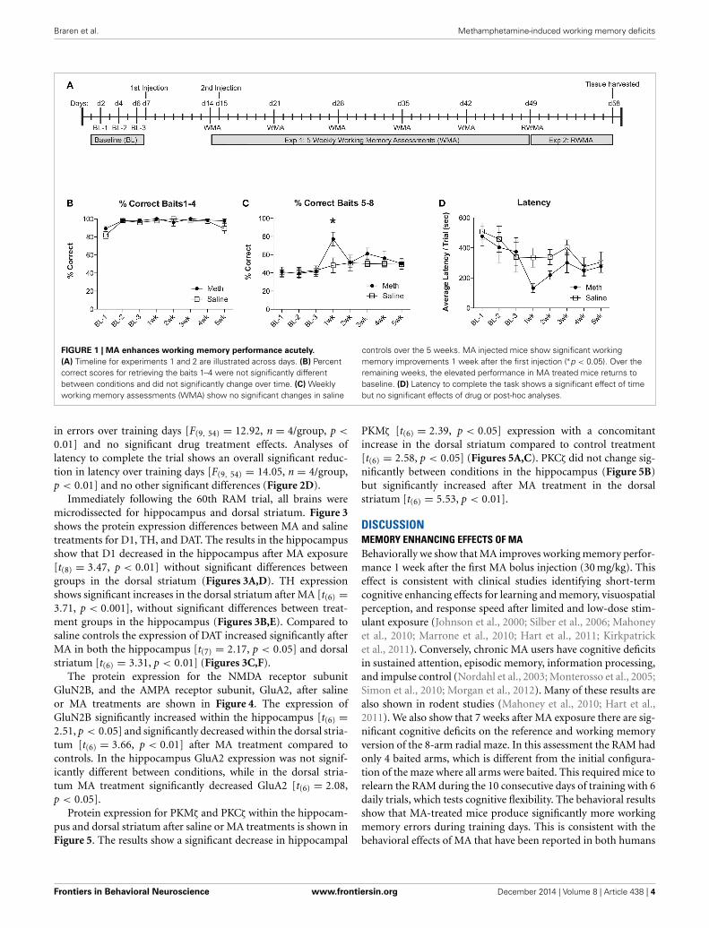
RESULTSFor experiment 1, groups of mice were injected with MA(30 mg/kg; 200 μl) or saline. One week post-injection mice wereassessed for a working memory version of the RAM. Twenty-four hours before the second working memory assessment, micewere injected again with MA (30 mg/kg; 200 μl) or saline. Forthe remaining 3 weeks, mice were assessed weekly for workingmemory performance, as illustrated in the timeline (Figure 1A).We evaluate the % correct score for each trial, which is calcu-lated as the number of total arm entries required to collect all 8food rewards divided by the number of food rewards retrieved.We show the % correct scores in two separate analyses to illus-trate the differences in number of errors committed while findingthe first 4 food rewards (Figure 1B) when the working memoryload is low, compared to the last 4 food rewards (Figure 1C)when the working memory load is high. The results shownin Figure 1C illustrate an overall significant effect of training[F(7, 49) = 3.67, n = 4/group, p = 0.003], an overall significantimprovement from MA [F(1, 49) = 5.85, n = 4/group, p = 0.04]and a significant post-hoc effect at 1 week (Bonferroni correctedt-test = 3.23, p < 0.05). In collecting baits 1–4, mice from bothtreatment conditions perform equivalently (Figure 1B). Latencyto complete the task shows an overall significant improvementover testing weeks [F(7, 49) = 4.2, n = 4/group, p = 0.0001], nosignificant effects of drug treatment and no significant post-hoccomparisons (Figure 1D).
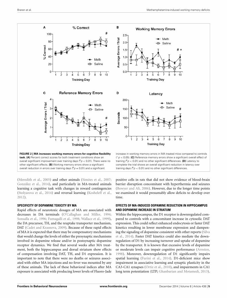
For experiment 2, all mice were re-trained on the RAM using anew configuration of four baited and four unbaited arms, which isdifferent from having all arms baited as described in experiment1. Mice were given 6 consecutive trials per day for 10 days. Theresults in Figure 2A show an overall significant improvement in% correct scores over training days [F(9, 54) = 9.3, n = 4/group,p < 0.01]. There were no significant effects of drug treatment andno significant post-hoc analyses. Analyses for working memoryerrors (Figure 2B) show a significant overall reduction in errorsover training days [F(9, 54) = 3.0, n = 4/group, p = 0.01] and asignificant increase in working memory errors in MA treated mice[F(1, 54) = 6.0, n = 4/group, p < 0.05]. Analysis of referencememory errors (Figure 2C) show a significant overall reduction
Frontiers in Behavioral Neuroscience www.frontiersin.org December 2014 | Volume 8 | Article 438 | 3
Braren et al. Methamphetamine-induced working memory deficits
FIGURE 1 | MA enhances working memory performance acutely.
(A) Timeline for experiments 1 and 2 are illustrated across days. (B) Percentcorrect scores for retrieving the baits 1–4 were not significantly differentbetween conditions and did not significantly change over time. (C) Weeklyworking memory assessments (WMA) show no significant changes in saline
controls over the 5 weeks. MA injected mice show significant workingmemory improvements 1 week after the first injection (∗p < 0.05). Over theremaining weeks, the elevated performance in MA treated mice returns tobaseline. (D) Latency to complete the task shows a significant effect of timebut no significant effects of drug or post-hoc analyses.
in errors over training days [F(9, 54) = 12.92, n = 4/group, p <
0.01] and no significant drug treatment effects. Analyses oflatency to complete the trial shows an overall significant reduc-tion in latency over training days [F(9, 54) = 14.05, n = 4/group,p < 0.01] and no other significant differences (Figure 2D).
Immediately following the 60th RAM trial, all brains weremicrodissected for hippocampus and dorsal striatum. Figure 3shows the protein expression differences between MA and salinetreatments for D1, TH, and DAT. The results in the hippocampusshow that D1 decreased in the hippocampus after MA exposure[t(8) = 3.47, p < 0.01] without significant differences betweengroups in the dorsal striatum (Figures 3A,D). TH expressionshows significant increases in the dorsal striatum after MA [t(6) =3.71, p < 0.001], without significant differences between treat-ment groups in the hippocampus (Figures 3B,E). Compared tosaline controls the expression of DAT increased significantly afterMA in both the hippocampus [t(7) = 2.17, p < 0.05] and dorsalstriatum [t(6) = 3.31, p < 0.01] (Figures 3C,F).
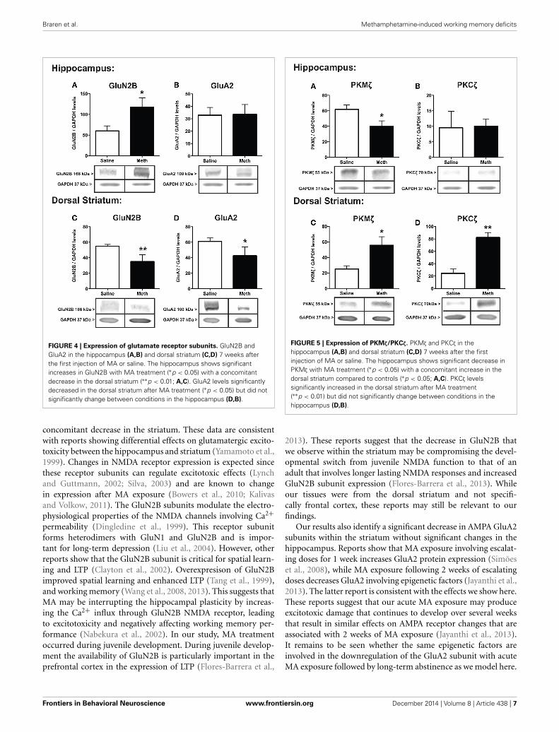
The protein expression for the NMDA receptor subunitGluN2B, and the AMPA receptor subunit, GluA2, after salineor MA treatments are shown in Figure 4. The expression ofGluN2B significantly increased within the hippocampus [t(6) =2.51, p < 0.05] and significantly decreased within the dorsal stria-tum [t(6) = 3.66, p < 0.01] after MA treatment compared tocontrols. In the hippocampus GluA2 expression was not signif-icantly different between conditions, while in the dorsal stria-tum MA treatment significantly decreased GluA2 [t(6) = 2.08,p < 0.05].
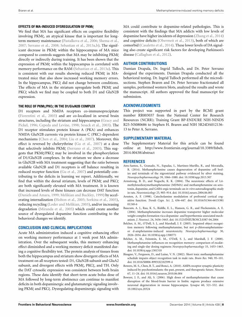
Protein expression for PKMζ and PKCζ within the hippocam-pus and dorsal striatum after saline or MA treatments is shown inFigure 5. The results show a significant decrease in hippocampal
PKMζ [t(6) = 2.39, p < 0.05] expression with a concomitantincrease in the dorsal striatum compared to control treatment[t(6) = 2.58, p < 0.05] (Figures 5A,C). PKCζ did not change sig-nificantly between conditions in the hippocampus (Figure 5B)but significantly increased after MA treatment in the dorsalstriatum [t(6) = 5.53, p < 0.01].
DISCUSSIONMEMORY ENHANCING EFFECTS OF MABehaviorally we show that MA improves working memory perfor-mance 1 week after the first MA bolus injection (30 mg/kg). Thiseffect is consistent with clinical studies identifying short-termcognitive enhancing effects for learning and memory, visuospatialperception, and response speed after limited and low-dose stim-ulant exposure (Johnson et al., 2000; Silber et al., 2006; Mahoneyet al., 2010; Marrone et al., 2010; Hart et al., 2011; Kirkpatricket al., 2011). Conversely, chronic MA users have cognitive deficitsin sustained attention, episodic memory, information processing,and impulse control (Nordahl et al., 2003; Monterosso et al., 2005;Simon et al., 2010; Morgan et al., 2012). Many of these results arealso shown in rodent studies (Mahoney et al., 2010; Hart et al.,2011). We also show that 7 weeks after MA exposure there are sig-nificant cognitive deficits on the reference and working memoryversion of the 8-arm radial maze. In this assessment the RAM hadonly 4 baited arms, which is different from the initial configura-tion of the maze where all arms were baited. This required mice torelearn the RAM during the 10 consecutive days of training with 6daily trials, which tests cognitive flexibility. The behavioral resultsshow that MA-treated mice produce significantly more workingmemory errors during training days. This is consistent with thebehavioral effects of MA that have been reported in both humans
Frontiers in Behavioral Neuroscience www.frontiersin.org December 2014 | Volume 8 | Article 438 | 4
Braren et al. Methamphetamine-induced working memory deficits
FIGURE 2 | MA increases working memory errors for cognitive flexibility
task. (A) Percent correct scores for both treatment conditions show anoverall significant improvement over training days (#p < 0.01). There were noother significant effects. (B) Working memory errors show a significantoverall reduction in errors over training days (#p = 0.01) and a significant
increase in working memory errors in MA treated mice compared to controls(∧p < 0.05). (C) Reference memory errors show a significant overall effect oftraining (#p < 0.01) and no other significant differences. (D) Latency tocomplete the trial shows an overall significant reduction in latency overtraining days (#p < 0.01) and no other significant differences.
(Meredith et al., 2005) and other animals (Simões et al., 2007;González et al., 2014), and particularly in MA-treated animalslearning a cognitive task with changes in reward contingencies(Stolyarova et al., 2014) and reversal learning (Kosheleff et al.,2012).
SPECIFICITY OF DOPAMINE TOXICITY BY MARapid effects of neurotoxic dosages of MA are associated withdecreases in DA terminals (O’Callaghan and Miller, 1994;Sonsalla et al., 1996; Fumagalli et al., 1998; Wallace et al., 1999),the DA precursor, TH, and the reuptake transporter mechanism,DAT (Cadet and Krasnova, 2009). Because of these rapid effectsof MA it is expected that there may be compensatory mechanismsthat would change the levels of either the presynaptic mechanismsinvolved in dopamine release and/or in postsynaptic dopaminereceptor dynamics. We find that several weeks after MA treat-ment, both the hippocampus and dorsal striatum show effectsof compensation involving DAT, TH, and D1 expression. It isimportant to note that there were no deaths or seizures associ-ated with either MA injections and no fever was mounted by anyof these animals. The lack of these behavioral indices after MAexposure is associated with producing lower levels of Fluoro-Jade
positive cells in rats that did not show evidence of blood-brainbarrier disruption concomitant with hyperthermia and seizures(Bowyer and Ali, 2006). However, due to the longer time pointswe examined it would presumably allow deficits to develop overtime.
EFFECTS OF MA-INDUCED DOPAMINE REDUCTION IN HIPPOCAMPUSAND DOPAMINE INCREASE IN STRIATUMWithin the hippocampus, the D1 receptor is downregulated com-pared to controls with a concomitant increase in cytosolic DATexpression. This could reflect enhanced endocytosis or faster DATkinetics resulting in lower membrane expression and dampen-ing the signaling of dopamine consistent with other reports (Silvaet al., 2014). Faster DAT kinetics could also mediate the down-regulation of D1 by increasing turnover and uptake of dopamineby the transporter. It is known that excessive levels of dopamineor moderate levels can impair cognitive performance (Arnsten,1998). Moreover, downregulation of D1 significantly impairsspatial learning (Furini et al., 2014). D1-deficient mice showimpairment in associative learning and synaptic plasticity in theCA3-CA1 synapses (Ortiz et al., 2010), and impairments in CA1long term potentiation (LTP; Ghanbarian and Motamedi, 2013).
Frontiers in Behavioral Neuroscience www.frontiersin.org December 2014 | Volume 8 | Article 438 | 5

Braren et al. Methamphetamine-induced working memory deficits
FIGURE 3 | Expression of dopamine markers. D1, TH, and DAT in thehippocampus (A–C) and dorsal striatum (D–F) 7 weeks after the first injectionof MA or saline. D1 in the hippocampus significantly decreased compared tocontrols (∗∗p < 0.01), without changing expression in the dorsal striatum
(A,D). Compared to controls, TH levels significantly increased in the dorsalstriatum (∗∗p < 0.01) without changing expression levels in the hippocampus(E,B). DAT increased significantly in both the hippocampus and dorsalstriatum (C,F; ∗p < 0.05; ∗∗p < 0.01).
Additionally, downregulation of DAT disrupts spatial learningand retention (Del’Guidice et al., 2013) as well as showing deficitsin cognitive flexibility (Morice et al., 2007). We speculate that thedownregulation of D1 and the upregulation of DAT endocyto-sis occur as a consequence of MA and is a contributing factor inspatial working memory deficits.
In the dorsal striatum there were no changes in D1 expres-sion compared to controls, rather, there was a significant increasein TH levels and DAT endocytosis. This suggests that in thestriatum, MA is upregulating presynaptic mechanisms involvingthe synthesis and degradation of dopamine. These presynapticchanges are potential compensatory mechanisms to the rapidneurotoxic effects of MA. While MA is known to damage DAterminals without affecting postsynaptic receptors (Cadet et al.,2003; Krasnova and Cadet, 2009; Sulzer, 2011), many of these DA
terminals partially recover after MA (Ares-Santos et al., 2014).The significant increase in TH and the increased endocytosis ofDAT suggests that MA induces DAT hyperfunction in the stria-tum. DAT hyperfunction has been associated with a model ofattention deficit hyperactivity disorder (ADHD) in rats that alsoshow a working memory deficit (Ruocco et al., 2014). Togetherthese data indicate that DAT, TH, and D1 dysregulation withinthe hippocampus and dorsal striatum could collectively play arole in the working memory deficit observed after weeks of MAabstinence.
EFFECTS OF MA-INDUCED GluN2B INCREASE IN HIPPOCAMPUS ANDDECREASE OF GluN2B AND GluA2 IN STRIATUMOur results show that MA treatment significantly increasedGluN2B subunit expression in the hippocampus with a
Frontiers in Behavioral Neuroscience www.frontiersin.org December 2014 | Volume 8 | Article 438 | 6
Braren et al. Methamphetamine-induced working memory deficits
FIGURE 4 | Expression of glutamate receptor subunits. GluN2B andGluA2 in the hippocampus (A,B) and dorsal striatum (C,D) 7 weeks afterthe first injection of MA or saline. The hippocampus shows significantincreases in GluN2B with MA treatment (∗p < 0.05) with a concomitantdecrease in the dorsal striatum (∗∗p < 0.01; A,C). GluA2 levels significantlydecreased in the dorsal striatum after MA treatment (∗p < 0.05) but did notsignificantly change between conditions in the hippocampus (D,B).
concomitant decrease in the striatum. These data are consistentwith reports showing differential effects on glutamatergic excito-toxicity between the hippocampus and striatum (Yamamoto et al.,1999). Changes in NMDA receptor expression is expected sincethese receptor subunits can regulate excitotoxic effects (Lynchand Guttmann, 2002; Silva, 2003) and are known to changein expression after MA exposure (Bowers et al., 2010; Kalivasand Volkow, 2011). The GluN2B subunits modulate the electro-physiological properties of the NMDA channels involving Ca2+permeability (Dingledine et al., 1999). This receptor subunitforms heterodimers with GluN1 and GluN2B and is impor-tant for long-term depression (Liu et al., 2004). However, otherreports show that the GluN2B subunit is critical for spatial learn-ing and LTP (Clayton et al., 2002). Overexpresison of GluN2Bimproved spatial learning and enhanced LTP (Tang et al., 1999),and working memory (Wang et al., 2008, 2013). This suggests thatMA may be interrupting the hippocampal plasticity by increas-ing the Ca2+ influx through GluN2B NMDA receptor, leadingto excitotoxicity and negatively affecting working memory per-formance (Nabekura et al., 2002). In our study, MA treatmentoccurred during juvenile development. During juvenile develop-ment the availability of GluN2B is particularly important in theprefrontal cortex in the expression of LTP (Flores-Barrera et al.,
FIGURE 5 | Expression of PKMζ/PKCζ. PKMζ and PKCζ in thehippocampus (A,B) and dorsal striatum (C,D) 7 weeks after the firstinjection of MA or saline. The hippocampus shows significant decrease inPKMζ with MA treatment (∗p < 0.05) with a concomitant increase in thedorsal striatum compared to controls (∗p < 0.05; A,C). PKCζ levelssignificantly increased in the dorsal striatum after MA treatment(∗∗p < 0.01) but did not significantly change between conditions in thehippocampus (D,B).
2013). These reports suggest that the decrease in GluN2B thatwe observe within the striatum may be compromising the devel-opmental switch from juvenile NMDA function to that of anadult that involves longer lasting NMDA responses and increasedGluN2B subunit expression (Flores-Barrera et al., 2013). Whileour tissues were from the dorsal striatum and not specifi-cally frontal cortex, these reports may still be relevant to ourfindings.
Our results also identify a significant decrease in AMPA GluA2subunits within the striatum without significant changes in thehippocampus. Reports show that MA exposure involving escalat-ing doses for 1 week increases GluA2 protein expression (Simõeset al., 2008), while MA exposure following 2 weeks of escalatingdoses decreases GluA2 involving epigenetic factors (Jayanthi et al.,2013). The latter report is consistent with the effects we show here.These reports suggest that our acute MA exposure may produceexcitotoxic damage that continues to develop over several weeksthat result in similar effects on AMPA receptor changes that areassociated with 2 weeks of MA exposure (Jayanthi et al., 2013).It remains to be seen whether the same epigenetic factors areinvolved in the downregulation of the GluA2 subunit with acuteMA exposure followed by long-term abstinence as we model here.
Frontiers in Behavioral Neuroscience www.frontiersin.org December 2014 | Volume 8 | Article 438 | 7
Braren et al. Methamphetamine-induced working memory deficits
EFFECTS OF MA-INDUCED DYSREGULATION OF PKMζ
We find that MA has significant effects on cognitive flexibilityinvolving PKMζ, an atypical kinase that is important for long-term memory maintenance (Pastalkova et al., 2006; Shema et al.,2007; Serrano et al., 2008; Sebastian et al., 2013a,b). The signif-icant decrease in PKMζ within the hippocampus of MA micecompared to controls suggests that MA may be inhibiting PKMζ
directly or indirectly during training. It has been shown that theexpression of PKMζ within the hippocampus is correlated withmemory performance on the RAM (Sebastian et al., 2013a). Thisis consistent with our results showing reduced PKMζ in MA-treated mice that also show increased working memory errors.In the hippocampus, PKCζ did not change between conditions.The effects of MA in the striatum upregulate both PKMζ andPKCζ which we find may be coupled to both D1 and GluN2Bexpression.
THE ROLE OF PKMζ/PKCζ IN THE D1/GluN2B COMPLEXD1 receptors and NMDA receptors co-immunoprecipitate(Fiorentini et al., 2003) and are co-localized in several brainstructures, including the striatum and hippocampus (Gracy andPickel, 1996; Cepeda and Levine, 1998; Sesack et al., 2003). TheD1 receptor stimulates protein kinase A (PKA) and enhancesNMDA GluN2B currents via protein kinase C (PKC)-dependentmechanisms (Chen et al., 2004; Liu et al., 2007). Moreover, thiseffect is reversed by chelerythrine (Gu et al., 2007) at a dosethat selectively inhibits PKMζ (Serrano et al., 2005). This sug-gests that PKMζ/PKCζ may be involved in the phosphorylationof D1/GluN2B complexes. In the striatum we show a decreasein GluN2B with MA treatment suggesting that the ratio betweenavailable GluN2B and D1 receptors is off balance resulting inreduced receptor function (Gu et al., 2007) and potentially con-tributing to the deficits in learning we report. Additionally, wefind that within the dorsal striatum, levels of PKMζ and PKCζ
are both significantly elevated with MA treatment. It is knownthat increased levels of these kinases can decrease DAT function(Daniels and Amara, 1999; Melikian and Buckley, 1999) by accel-erating internalization (Holton et al., 2005; Sorkina et al., 2005),reducing recycling (Loder and Melikian, 2003), and/or increasingdegradation (Miranda et al., 2005) which could create anothersource of dysregulated dopamine function contributing to thebehavioral changes we identify.
CONCLUSION AND CLINICAL IMPLICATIONSAcute MA administration induced a cognitive enhancing effecton working memory performance at 1 week post MA admin-istration. Over the subsequent weeks, this memory enhancingeffect diminished and a working memory deficit manifested dur-ing a cognitive flexibility test. The protein analysis of tissues fromboth the hippocampus and striatum show divergent effects of MAtreatment on all receptors tested: D1, GluN2B subunit and GluA2subunit, and divergent effects with PKMζ, PKCζ, and TH. Onlythe DAT cytosolic expression was consistent between both brainregions. These data identify that short-term acute bolus dose ofMA followed by long-term abstinence can continue to manifestdeficits in both dopaminergic and glutamatergic signaling involv-ing PKMζ and PKCζ. Dysregulating dopaminergic signaling with
MA could contribute to dopamine-related pathologies. This isconsistent with the findings that MA addicts with low levels ofdopamine have higher incidents of depression (Zhang et al., 2014)and cognitive deficits (Obermeit et al., 2013), both of which arecomorbid (Casaletto et al., 2014). These lower levels of DA signal-ing also create significant risk factors for developing Parkinson’sdisease (Callaghan et al., 2012).
AUTHOR CONTRIBUTIONSDamian Drapala, Dr. Ingrid Tulloch, and Dr. Peter Serranodesigned the experiments. Damian Drapala conducted all thebehavioral testing. Dr. Ingrid Tulloch performed all the microdi-sections. Stephen Braren and Dr. Peter Serrano fractionated allsamples, performed western blots, analyzed the results and wrotethe manuscript. All authors approved the final manuscript forsubmission.
ACKNOWLEDGMENTSThis project was supported in part by the RCMI grantnumber RR003037 from the National Center for ResearchResources (NCRR); Training Grant BP-ENDURE NIH-NINDSR25-NS080686 to Stephen H. Braren and NIH 5R24DA012136-13 to Peter A. Serrano.
SUPPLEMENTARY MATERIALThe Supplementary Material for this article can be foundonline at: http://www.frontiersin.org/journal/10.3389/fnbeh.
2014.00438/abstract
REFERENCESAres-Santos, S., Granado, N., Espadas, I., Martinez-Murillo, R., and Moratalla,
R. (2014). Methamphetamine causes degeneration of dopamine cell bod-ies and terminals of the nigrostriatal pathway evidenced by silver staining.Neuropsychopharmacology 39, 1066–1080. doi: 10.1038/npp.2013.307
Armstrong, B. D., and Noguchi, K. K. (2004). The neurotoxic effects of 3,4-methylenedioxymethamphetamine (MDMA) and methamphetamine on sero-tonin, dopamine, and GABA-ergic terminals: an in-vitro autoradiographic studyin rats. Neurotoxicology 25, 905–914. doi: 10.1016/j.neuro.2004.06.003
Arnsten, A. F. (1998). Catecholamine modulation of prefrontal cortical cog-nitive function. Trends Cogn. Sci. 2, 436–447. doi: 10.1016/S1364-6613(98)01240-6
Baucum, A. J., Rau, K. S., Riddle, E. L., Hanson, G. R., and Fleckenstein, A. E.(2004). Methamphetamine increases dopamine transporter higher molecularweight complex formation via a dopamine- and hyperthermia-associated mech-anism. J. Neurosci. 24, 3436–3443. doi: 10.1523/JNEUROSCI.0387-04.2004
Belcher, A. M., O’Dell, S. J., and Marshall, J. F. (2005). Impaired object recogni-tion memory following methamphetamine, but not p-chloroamphetamine-or d-amphetamine-induced neurotoxicity. Neuropsychopharmacology 30,2026–2034. doi: 10.1038/sj.npp.1300771
Belcher, A. M., Feinstein, E. M., O’Dell, S. J., and Marshall, J. F. (2008).Methamphetamine influences on recognition memory: comparison of escalat-ing and single-day dosing regimens Neuropsychopharmacology 33, 1453–1463.doi: 10.1038/sj.npp.1301510
Bisagno, V., Ferguson, D., and Luine, V. N. (2002). Short toxic methamphetamineschedule impairs object recognition task in male rats. Brain Res. 940, 95–101.doi: 10.1016/S0006-8993(02)02599-4
Bowers, M. S., Chen, B. T., and Bonci, A. (2010). AMPA receptor synaptic plasticityinduced by psychostimulants: the past, present, and therapeutic future. Neuron67, 11–24. doi: 10.1016/j.neuron.2010.06.004
Bowyer, J. F., and Ali, S. (2006). High doses of methamphetamine that causedisruption of the blood-brain barrier in limbic regions produce extensiveneuronal degeneration in mouse hippocampus. Synapse 60, 521–532. doi:10.1002/syn.20324
Frontiers in Behavioral Neuroscience www.frontiersin.org December 2014 | Volume 8 | Article 438 | 8
Braren et al. Methamphetamine-induced working memory deficits
Cadet, J. L., Jayanthi, S., and Deng, X. (2003). Speed kills: cellular and molecularbases of methamphetamine-induced nerve terminal degeneration and neuronalapoptosis. FASEB J. 17, 1775–1788. doi: 10.1096/fj.03-0073rev
Cadet, J. L., and Krasnova, I. N. (2009). Molecular bases of methamphetamine-induced neurodegeneration. Int. Rev. Neurobiol. 88, 101–119. doi:10.1016/S0074-7742(09)88005-7
Cadet, J. L., Brannock, C., Ladenheim, B., McCoy, M. T., Beauvais, G., Hodges, A.B., et al. (2011). Methamphetamine preconditioning causes differential changesin striatal transcriptional responses to large doses of the drug. Dose Response 9,165–181. doi: 10.2203/dose-response.10-011.Cadet
Callaghan, R. C., Cunningham, J. K., Sykes, J., and Kish, S. J. (2012). Increasedrisk of Parkinson’s disease in individuals hospitalized with conditions related tothe use of methamphetamine or other amphetamine-type drugs. Drug AlcoholDepend. 120, 35–40. doi: 10.1016/j.drugalcdep.2011.06.013
Casaletto, K. B., Obermeit, L., Morgan, E. E., Weber, E., Franklin, D. R., Grant, I.,et al. (2014). Depression and executive dysfunction contribute to a metamem-ory deficit among individuals with methamphetamine use disorders. Addict.Behav. 40C, 45–50. doi: 10.1016/j.addbeh.2014.08.007
Cepeda, C., and Levine, M. S. (1998). Dopamine and N-methyl-D-aspartatereceptor interactions in the neostriatum. Dev. Neurosci. 20, 1–18. doi:10.1159/000017294
Chapman, D. E., Hanson, G. R., Kesner, R. P., and Keefe, K. A. (2001). Long-termchanges in basal ganglia function after a neurotoxic regimen of metham-phetamine. J. Pharmacol. Exp. Ther. 296, 520–527.
Chen, G., Greengard, P., and Yan, Z. (2004). Potentiation of NMDA receptor cur-rents by dopamine D1 receptors in prefrontal cortex. Proc. Natl. Acad. Sci. U.S.A.101, 2596–2600. doi: 10.1073/pnas.0308618100
Clayton, D. A., Mesches, M. H., Alvarez, E., Bickford, P. C., and Browning, M. D.(2002). A hippocampal NR2B deficit can mimic age-related changes in long-term potentiation and spatial learning in the Fischer 344 rat. J. Neurosci. 22,3628–3637.
Daniels, G. M., and Amara, S. G. (1999). Regulated trafficking of the humandopamine transporter. Clathrin-mediated internalization and lysosomal degra-dation in response to phorbol esters. J. Biol. Chem. 274, 5794–5801. doi:10.1074/jbc.274.50.35794
Del’Guidice, T., Lemasson, M., Etiévant, A., Manta, S., Magno, L. A., Escoffier, G.,et al. (2013). Dissociations between cognitive and motor effects of psychostimu-lants and atomoxetine in hyperactive DAT-KO mice. Psychopharmacology (Berl.)231, 109–122. doi: 10.1007/s00213-013-3212-8
Dingledine, R., Borges, K., Bowie, D., and Traynelis, S. F. (1999). The glutamatereceptor ion channels. Pharmacol. Rev. 51, 7–61.
Fiorentini, C., Gardoni, F., Spano, P., Di Luca, M., and Missale, C. (2003).Regulation of dopamine D1 receptor trafficking and desensitization byoligomerization with glutamate N-methyl-D-aspartate receptors. J. Biol. Chem.278, 20196–20202. doi: 10.1074/jbc.M213140200
Flores-Barrera, E., Thomases, D. R., Heng, L. J., Cass, D. K., Caballero, A., andTseng, K. Y. (2013). Late adolescent expression of GluN2B transmission inthe prefrontal cortex is input-specific and requires postsynaptic protein kinaseA and D1 dopamine receptor signaling. Biol. Psychiatry 75, 508–516. doi:10.1016/j.biopsych.2013.07.033
Friedman, S. D., Castaneda, E., and Hodge, G. K. (1998). Long-term monoaminedepletion, differential recovery, and subtle behavioral impairment follow-ing methamphetamine induced neurotoxicity. Pharmacol. Biochem. Behav. 61,35–44. doi: 10.1016/S0091-3057(98)00066-5
Fumagalli, F., Gainetdinov, R. R., Valenzano, K. J., and Caron, M. G. (1998). Role ofdopamine transporter in methamphetamine-induced neurotoxicity: evidencefrom mice lacking the transporter. J. Neurosci. 18, 4861–4869.
Furini, C. R., Myskiw, J. C., Schmidt, B. E., Marcondes, L. A., and Izquierdo,I. (2014). D1 and D5 dopamine receptors participate on the consoli-dation of two different memories. Behav. Brain Res. 271, 212–217. doi:10.1016/j.bbr.2014.06.027
Ghanbarian, E., and Motamedi, F. (2013). Ventral tegmental area inactivation sup-presses the expression of CA1 long term potentiation in anesthetized rat. PLoSONE 8:e58844. doi: 10.1371/journal.pone.0058844
González, B., Raineri, M., Cadet, J. L., García-Rill, E., Urbano, F. J.,and Bisagno, V. (2014). Modafinil improves methamphetamine-inducedobject recognition deficits and restores prefrontal cortex ERK signalingin mice. Neuropharmacology 87, 188–197. doi: 10.1016/j.neuropharm.2014.02.002
Gonzalez, R., Bechara, A., and Martin, E. M. (2007). Executive functionsamong individuals with methamphetamine or alcohol as drugs of choice:preliminary observations. J. Clin. Exp. Neuropsychol. 29, 155–159. doi:10.1080/13803390600582446
Gracy, K. N., and Pickel, V. M. (1996). Ultrastructural immunocytochemical local-ization of the N-methyl-D-aspartate receptor and tyrosine hydroxylase in theshell of the rat nucleus accumbens. Brain Res. 739, 169–181. doi: 10.1016/S0006-8993(96)00822-0
Gu, W. H., Yang, S., Shi, W. X., Jin, G. Z., and Zhen, X. C. (2007). Requirementof PSD-95 for dopamine D1 receptor modulating glutamate NR1a/NR2Breceptor function. Acta. Pharmacol. Sin. 28, 756–762. doi: 10.1111/j.1745-7254.2007.00557.x
Hart, C. L., Marvin, C. B., Silver, R., and Smith, E. E. (2011). Is cogni-tive functioning impaired in methamphetamine users? A critical review.Neuropsychopharmacology. 37, 586–608. doi: 10.1038/npp.2011.276
Hastrup, H., Sen, N., and Javitch, J. A. (2003). The human dopamine transporterforms a tetramer in the plasma membrane: cross-linking of a cysteine in thefourth transmembrane segment is sensitive to cocaine analogs. J. Biol. Chem.278, 45045–45048. doi: 10.1074/jbc.C300349200
Hoffman, W. F., Moore, M., Templin, R., McFarland, B., Hitzemann, R. J., andMitchell, S. H. (2006). Neuropsychological function and delay discounting inmethamphetamine-dependent individuals. Psychopharmacology 188, 162–170.doi: 10.1007/s00213-006-0494-0
Holton, K. L., Loder, M. K., and Melikian, H. E. (2005). Nonclassical, dis-tinct endocytic signals dictate constitutive and PKC-regulated neurotransmittertransporter internalization. Nat. Neurosci. 8, 881–888. doi: 10.1038/nn1478
Jayanthi, S., McCoy, M. T., Chen, B., Britt, J. P., Kourrich, S., Yau, H. J.,et al. (2013). Methamphetamine downregulates striatal glutamate recep-tors via diverse epigenetic mechanisms. Biol. Psychiatry 76, 47–56. doi:10.1016/j.biopsych.2013.09.034
Johnson, B. A., Ait-Daoud, N., and Wells, L. T. (2000). Effects ofisradipine, a dihydropyridine-class calcium channel antagonist, on -methamphetamine-induced cognitive and physiological changes in humans.Neuropsychopharmacology 22, 504–512. doi: 10.1016/S0893-133X(99)00116-5
Kalivas, P. W., and Volkow, N. D. (2011). New medications for drug addic-tion hiding in glutamatergic neuroplasticity. Mol. Psychiatry 16, 974–986. doi:10.1038/mp.2011.46
Kirkpatrick, M. G., Gunderson, E. W., Perez, A. Y., Haney, M., Foltin, R. W., andHart, C. L. (2011). A direct comparison of the behavioral and physiologicaleffects of methamphetamine and 3,4-methylenedioxymethamphetamine(MDMA) in humans. Psychopharmacology (Berl.) 219, 109–122. doi:10.1007/s00213-011-2383-4
Kosheleff, A. R., Rodriguez, D., O’Dell, S. J., Marshall, J. F., and Izquierdo, A.(2012). Comparison of single-dose and escalating methamphetamine admin-istration on reversallearning in rats. Psychopharmacology (Berl.) 224, 459–467.doi: 10.1007/s00213-012-2774-1
Krasnova, I. N., and Cadet, J. L. (2009). Methamphetamine toxicity and mes-sengers of death. Brain Res. Rev. 60, 379–407. doi: 10.1016/j.brainresrev.2009.03.002
Liu, L., Wong, T. P., Pozza, M. F., Lingenhoehl, K., Wang, Y., Sheng, M., et al. (2004).Role of NMDA receptor subtypes in governing the direction of hippocampalsynaptic plasticity. Science 304, 1021–1024. doi: 10.1126/science.1096615
Liu, Y., Dore, J., and Chen, X. (2007). Calcium influx through L-type channels gen-erates protein kinase M to induce burst firing of dopamine cells in the rat ventraltegmental area. J. Biol. Chem. 282, 8594–8603. doi: 10.1074/jbc.M610230200
Loder, M. K., and Melikian, H. E. (2003). The dopamine transporter consti-tutively internalizes and recycles in a protein kinase C-regulated mannerin stably transfected PC12 cell lines. J. Biol. Chem. 278, 35168–35174. doi:10.1074/jbc.M301845200
Lynch, D. R., and Guttmann, R. P. (2002). Excitotoxicity: perspectives based on N-methyl-D-aspartate receptor subtypes. J. Pharmacol. Exp. Ther. 300, 717–723.doi: 10.1124/jpet.300.3.717
McFadden, L. M., and Matuszewich, L. (2007). The effects of methamphetamineexposure during preadolescence on male and female rats in the water maze.Behav. Brain Res. 185, 99–109. doi: 10.1016/j.bbr.2007.07.016
Mahoney, J. J., Jackson, B. J., Kalechstein, A. D., De La Garza, R., andNewton, T. F. (2010). Acute, low-dose methamphetamine administra-tion improves attention/information processing speed and working mem-ory in methamphetamine-dependent individuals displaying poorer cognitive
Frontiers in Behavioral Neuroscience www.frontiersin.org December 2014 | Volume 8 | Article 438 | 9
Braren et al. Methamphetamine-induced working memory deficits
performance at baseline. Prog. Neuropsychopharmacol. Biol. Psychiatry 35,459–465. doi: 10.1016/j.pnpbp.2010.11.034
Marrone, G. F., Pardo, J., Krauss, R. M., and Hart, C. L. (2010). Amphetamineanalogs methamphetamine and 3,4-methylenedioxymethamphetamine(MDMA) differentially affect speech. Psychopharmacology 208, 169–177. doi:10.1007/s00213-009-1715-0
Marshall, J. F., Belcher, A. M., Feinstein, E. M., and O’Dell, S. J.(2007).Methamphetamine-induced neural and cognitive changes in rodents.Addiction 102, 61–69. doi: 10.1111/j.1360-0443.2006.01780.x
Melikian, H. E., and Buckley, K. M. (1999). Membrane trafficking regulates theactivity of the human dopamine transporter. J. Neurosci. 19, 7699–7710.
Meredith, C. W., Jaffe, C., Ang-Lee, K., and Saxon, A. J. (2005). Implications ofchronic methamphetamine use: a literature review. Harv. Rev. Psychiatry 13,141–154. doi: 10.1080/10673220591003605
Miranda, M., Wu, C. C., Sorkina, T., Korstjens, D. R., and Sorkin, A. (2005).Enhanced ubiquitylation and accelerated degradation of the dopamine trans-porter mediated by protein kinase C. J. Biol. Chem. 280, 35617–35624. doi:10.1074/jbc.M506618200
Moenk, M. D., and Matuszewich, L. (2012). Juvenile but not adult metham-phetamine exposure improves performance in the Morris water maze in malerats. Int. J. Dev. Neurosci. 30, 325–331. doi: 10.1016/j.ijdevneu.2012.01.006
Monterosso, J. R., Aron, A. R., Cordova, X., Xu, J., and London, E. D. (2005).Deficits in response inhibition associated with chronic methamphetamineabuse. Drug. Alcohol Depend. 79, 273–277. doi: 10.1016/j.drugalcdep.2005.02.002
Morgan, E. E., Woods, S. P., Poquette, A. J., Vigil, O., Heaton, R. K., and Grant, I.(2012). Visual memory in methamphetamine-dependent individuals: deficientstrategic control of encoding and retrieval. Aust. N. Z. J. Psychiatry 46, 141–152.doi: 10.1177/0004867411433212
Morice, E., Billard, J. M., Denis, C., Mathieu, F., Betancur, C., Epelbaum, J., et al.(2007). Parallel loss of hippocampal LTD and cognitive flexibility in a geneticmodel of hyperdopaminergia. Neuropsychopharmacology 32, 2108–2116. doi:10.1038/sj.npp.1301354
Nabekura, J., Ueno, T., Katsurabayashi, S., Furuta, A., Akaike, N., and Okada, M.(2002). Reduced NR2A expression and prolonged decay of NMDA receptor-mediated synaptic current in rat vagal motoneurons following axotomy.J. Physiol. 539, 735–741. doi: 10.1113/jphysiol.2001.013379
Nash, J. F., and Yamamoto, B. K. (1992). Methamphetamine neurotoxicity and stri-atal glutamate release: comparison to 3,4-methylenedioxymethamphetamine.Brain Res. 581, 237–243. doi: 10.1016/0006-8993(92)90713-J
National Institute on Drug Abuse. (2012). Club Drugs. Bethesda, MD: NationalInstitutes of Health. Available online at: www.drugabuse.gov/club-drugs
Noguès, X., Micheau, J., and Jaffard, R. (1994). Protein kinase C activity in thehippocampus following spatial learning tasks in mice. Hippocampus 4, 71–77.doi: 10.1002/hipo.450040109
Nordahl, T. E., Salo, R., and Leamon, M. (2003). Neuropsychological effects ofchronic methamphetamine use on neurotransmitters and cognition: a review. J.Neuropsychiatry Clin. Neurosci. 15, 317–325. doi: 10.1176/appi.neuropsych.15.3.317
North, A., Swant, J., Salvatore, M. F., Gamble-George, J., Prins, P., Butler, B., et al.(2013).Chronic methamphetamine exposure produces a delayed, long-lastingmemory deficit. Synapse 67, 245–257. doi: 10.1002/syn.21635
Obermeit, L. C., Cattie, J. E., Bolden, K. A., Marquine, M. J., Morgan,E. E., Franklin, D. R., et al. (2013). Attention-deficit/hyperactivity disor-der among chronic methamphetamine users: frequency, persistence, andadverse effects on everyday functioning. Addict. Behav. 38, 2874–2878. doi:10.1016/j.addbeh.2013.08.010
O’Callaghan, J. P., and Miller, D. B. (1994). Neurotoxicity profiles of substitutedamphetamines in the C57BL/6Jmouse. J. Pharmacol. Exp. Ther. 270, 741–751.
O’Neil, M. L., Kuczenski, R., Segal, D. S., Cho, A. K., Lacan, G., and Melega,W. P. (2006). Escalating dose pretreatment induces pharmacodynamic and notpharmacokinetic tolerance to a subsequent high-dose methamphetamine binge.Synapse 60, 465–473. doi: 10.1002/syn.20320
Orikabe, L., Yamasue, H., Inoue, H., Takayanagi, Y., Mozue, Y., Sudo, Y.,et al. (2011). Reduced amygdala and hippocampal volumes in patientswith methamphetamine psychosis. Schizophrenia Res. 132, 183–189. doi:10.1016/j.schres.2011.07.006
Ortiz, O., Delgado-García, J. M., Espadas, I., Bah,í, A., Trullas, R., Dreyer, J. R.,et al. (2010). Associative learning and CA3-CA1 synaptic plasticity are impaired
in D1R null, Drd1a-/- mice and in hippocampal siRNA silenced Drd1a mice.J. Neurosci. 30, 12288–12300. doi: 10.1523/JNEUROSCI.2655-10.2010
Pastalkova, E., Serrano, P., Pinkhasova, D., Wallace, E., Fenton, A. A., and Sacktor,T. C. (2006). Storage of spatial information by the maintenance mechanism ofLTP. Science 313, 1141–1144. doi: 10.1126/science.1128657
Pereira, F. C., Imam, S. Z., Gough, B., Newport, G. D., Ribeiro, C. F., Slikker, W.Jr., et al. (2002). Acute changes in dopamine release and turnover in rat caudatenucleus following a single dose of methamphetamine. J. Neural. Transm. 109,1151–1158. doi: 10.1007/s00702-002-0754-z
Pereira, F. C., Lourenco, E. S., Borges, F., Morgadinho, T., Ribeiro, C. F., andMacedo, T. R. (2006). Single or multiple injections of methamphetamineincreased dopamine turnover but did not decrease tyrosine hydroxylaselevels or cleave caspase-3 in caudate-putamen. Synapse 60, 185–193. doi:10.1002/syn.20285
Rasband, W. S. (2014). ImageJ. Bethesda, MD: U. S. National Institutes of Health.Available online at: http://imagej.nih.gov/ij/
Ricaurte, G. A., Guillery, R. W., Seiden, L. S., Schuster, C. R., and Moore, R.Y. (1982). Dopamine nerve terminal degeneration produced by high doses ofmethylamphetamine in the rat brain. Brain Res. 235, 93–103. doi: 10.1016/0006-8993(82)90198-6
Ricaurte, G. A., Guillery, R. W., Seiden, L. S., and Schuster, C. R. (1984). Nerveterminal degeneration after a single injection of d-amphetamine in iprindole-treated rats: relation to selective long-lasting dopamine depletion. Brain Res.291, 378–382. doi: 10.1016/0006-8993(84)91273-3
Rocher, C., and Gardier, A. M. (2001). Effects of repeated systemic administrationof d-fenfluramine on serotonin and glutamate release in rat ventral hippocam-pus: comparison with methamphetamine using in vivo microdialysis. Naunyn.Schmiedebergs. Arch. Pharmacol. 363, 422–428. doi: 10.1007/s002100000381
Ruocco, L. A., Treno, C., Gironi Carnevale, U. A., Arra, C., Mattern, C., Huston, J.P., et al. (2014). Prepuberal intranasal dopamine treatment in an animal modelof ADHD ameliorates deficient spatial attention, working memory, aminoacid transmitters and synaptic markers in prefrontal cortex, ventral and dorsalstriatum. Amino Acids 46, 2105–2122. doi: 10.1007/s00726-014-1753-8
Sacktor, T. C., Osten, P., Valsamis, H., Jiang, X., Naik, M. U., and Sublette, E. (1993).Persistent activation of the zeta isoform of protein kinase C in the maintenanceof long-term potentiation. Proc. Natl. Acad. Sci. U.S.A. 90, 8342–8346.
Sebastian, V., Estil, J. B., Chen, D., Schrott, L. M., and Serrano, P. A. (2013b).Acute physiological stress promotes clustering of synaptic markers and altersspine morphology in the hippocampus. PLoS ONE 8:e79077. doi: 10.1371/jour-nal.pone.0079077
Sebastian, V., Diallo, A., Ling, D. S., and Serrano, P. A. (2013c). Robust trainingattenuates TBI-induced deficits in reference and working memory on the radial8-arm maze. Front. Behav. Neurosci. 7:38. doi: 10.3389/fnbeh.2013.00038
Sebastian, V., Vergel, T., Baig, R., Schrott, L. M., and Serrano, P. A. (2013a). PKMζ
differentially utilized between sexes for remote long-term spatial memory. PLoSONE 8:e81121. doi: 10.1371/journal.pone.0081121
Segal, D. S., Kuczenski, R., O’Neil, M. L., Melega, W. P., and Cho, A. K. (2003).Escalating dose methamphetamine pretreatment alters the behavioral and neu-rochemical profiles associated with exposure to a high-dose methamphetaminebinge. Neuropsychopharmacology 28, 1730–1740. doi: 10.1038/sj.npp.1300247
Serrano, P., Yao, Y., and Sacktor, T. C. (2005). Persistent phosphorylation by pro-tein kinase m zeta maintains late-phase long-term potentiation. J. Neurosci. 25,1979–1984. doi: 10.1523/JNEUROSCI.5132-04.2005
Serrano, P., Friedman, E. L., Kenney, J., Taubenfeld, S. M., Zimmerman, J. M.,Hanna, J., et al. (2008). PKMzeta maintains spatial, instrumental, and classicallyconditioned long-term memories. PLoS Biol. 6, 2698–2706. doi: 10.1371/jour-nal.pbio.0060318
Sesack, S. R., Carr, D. B., Omelchenko, N., and Pinto, A. (2003). Anatomicalsubstrates for glutamate-dopamine interactions: evidence for specificity of con-nections and extrasynaptic actions. Ann. N.Y. Acad. Sci. 1003, 36–52. doi:10.1196/annals.1300.066
Shema, R., Sacktor, T. C., and Dudai, Y. (2007). Rapid erasure of long-term memoryassociations in the cortex by an inhibitor of PKM zeta. Science 317, 951–953. doi:10.1126/science.1144334
Silber, B. Y., Croft, R. J., Papafotiou, K., and Stough, C. (2006). The acute effects ofd-amphetamine and methamphetamine on attention and psychomotor perfor-mance. Psychopharmacology 187, 154–169. doi: 10.1007/s00213-006-0410-7
Silva, A. J. (2003). Molecular and cellular cognitive studies of the role of synapticplasticity in memory. J. Neurobiol. 54, 224–237. doi: 10.1002/neu.10169
Frontiers in Behavioral Neuroscience www.frontiersin.org December 2014 | Volume 8 | Article 438 | 10
Braren et al. Methamphetamine-induced working memory deficits
Silva, C. D., Neves, A. F., Dias, A. I., Freitas, H. J., Mendes, S. M., Pita, I.,et al. (2014). A single neurotoxic dose of methamphetamine induces a long-lasting depressive-like behaviour in mice. Neurotox. Res. 25, 295–304. doi:10.1007/s12640-013-9423-2
Simões, P. F., Silva, A. P., Pereira, F. C., Marques, E., Grade, S., Milhazes, N.,et al. (2007). Methamphetamine induces alterations on hippocampal NMDAand AMPA receptor subunit levels and impairs spatial working memory.Neuroscience 150, 433–441. doi: 10.1016/j.neuroscience.2007.09.044
Simões, P. F., Silva, A. P., Pereira, F. C., Marques, E., Milhazes, N., Borges, F., et al.(2008). Methamphetamine changes NMDA and AMPA glutamate receptor sub-unit levels in the rat striatum and frontal cortex. Ann. N.Y. Acad. Sci. 1139,232–241. doi: 10.1196/annals.1432.028
Simon, S. L., Domier, C. P., Sim, T., Richardson, K., Rawson, R. A., and Ling,W. (2002). Cognitive performance of current methamphetamine and cocaineabusers. J. Addict. Dis. 21, 61–74. doi: 10.1300/J069v21n01_06
Simon, S. L., Dean, A. C., Cordova, X., Monterosso, J. R., and London, E.D. (2010). Methamphetamine dependence and neuropsychological function-ing: evaluating change during early abstinence. J. Stud. Alcohol Drugs 71,335–344.
Sonsalla, P. K., Jochnowitz, N. D., Zeevalk, G. D., Oostveen, J. A., and Hall,E. D. (1996). Treatment of mice with methamphetamine produces cell lossin the substantia nigra. Brain Res. 738, 172–175. doi: 10.1016/0006-8993(96)00995-X
Sorkina, T., Hoover, B. R., Zahniser, N. R., and Sorkin, A. (2005). Constitutiveand protein kinase C-induced internalization of the dopamine transporteris mediated by a clathrin-dependent mechanism. Traffic 6, 157–170. doi:10.1111/j.1600-0854.2005.00259.x
Stolyarova, A., O’Dell, S. J., Marshall, J. F., and Izquierdo, A. (2014). Positiveand negative feedback learning and associated dopamine and serotonin trans-porter binding after methamphetamine. Behav. Brain Res. 271, 195–202. doi:10.1016/j.bbr.2014.06.031
Sulzer, D. (2011). How addictive drugs disrupt presynaptic dopamine neurotrans-mission. Neuron 69, 628–649. doi: 10.1016/j.neuron.2011.02.010
Talbert, J. J. (2014). Club drugs: coming to a patient near you. Nurse Pract. 39,20–25. doi: 10.1097/01.NPR.0000443227.72357.72
Tang, Y. P., Shimizu, E., Dube, G. R., Rampon, C., Kerchner, G. A., Zhuo, M., et al.(1999). Genetic enhancement of learning and memory in mice. Nature 401,63–69. doi: 10.1038/43432
Tulloch, I., Afanador, L., Mexhitaj, I., Ghazaryan, N., Garzagongora, A. G., andAngulo, J. A. (2011a). A single high dose of methamphetamine induces apop-totic and necrotic striatal cell loss lasting up to 3 months in mice. Neuroscience193, 162–169. doi: 10.1016/j.neuroscience.2011.07.020
Tulloch, I., Ghazaryan, N., Mexhitaj, I., Ordonez, D., and Angulo, J. A.(2011b). Role of neurokinin-1 and dopamine receptors on the striatalmethamphetamine-induced proliferation of new cells in mice. Brain Res. 1399,33–39. doi: 10.1016/j.brainres.2011.05.017
Vorhees, C. V., Inman-Wood, S. L., Morford, L. L., Broening, H. W., Fukumura,M., and Moran, M. S. (2000). Adult learning deficits after neonatal exposure
to D-methamphetamine: selective effects on spatial navigation and memory.J. Neurosci. 20, 4732–4739.
Wallace, T. L., Gudelsky, G. A., and Vorhees, C. V. (1999). Methamphetamineinduced neurotoxicity alters locomotor activity, stereotypic behavior, and stim-ulated dopamine release in the rat. J. Neurosci. 19, 9141–9148.
Wang, H., Stradtman, G. G., Wang, X. J., and Gao, W. J. (2008). A special-ized NMDA receptor function in layer 5 recurrent microcircuitry of the adultrat prefrontal cortex. Proc. Natl. Acad. Sci. U.S.A. 105, 16791–16796. doi:10.1073/pnas.0804318105
Wang, M., Yang, Y., Wang, C. J., Gamo, N. J., Jin, L. E., Mazer, J. A., et al.(2013). NMDA receptors subserve persistent neuronal firing during work-ing memory in dorsolateral prefrontal cortex. Neuron 77, 736–749. doi:10.1016/j.neuron.2012.12.032
Williams, M. T., Vorhees, C. V., Boon, F., Saber, A. J., and Cain, D. P. (2002).Methamphetamine exposure from postnatal day 11–20 causes impairments inboth behavioral strategies and spatial learning in adults rats. Brain Res. 958,312–321. doi: 10.1016/S0006-8993(02)03620-X
Williams, M. T., Morford, L. L., Wood, S. L., Wallace, T. L., Fukumura, M.,Broening, H. W., et al. (2003). Developmental d-methamphetamine treatmentselectively induces spatial navigation impairments in reference memory in theMorris water maze while sparing working memory. Synapse 48, 138–148. doi:10.1002/syn.10159
Yamamoto, H., Kitamura, N., Lin, X. H., Ikeuchi, Y., Hashimoto, T., Shirakawa, O.,et al. (1999). Differential changes in glutamatergic transmission via N-methyl-D-aspartate receptors in the hippocampus and striatum of rats behaviourallysensitized to methamphetamine. Int. J. Neuropsychopharmacol. 2, 155–163.
Zhang, J., Xie, Y., Su, H., Tao, J., Sun, Y., Li, L., et al. (2014). Prevalence andcorrelates of depressive symptoms during early methamphetamine with-drawal in Han Chinese population. Drug Alcohol Depend. 142, 191–196. doi:10.1016/j.drugalcdep.2014.06.021
Conflict of Interest Statement: The authors declare that the research was con-ducted in the absence of any commercial or financial relationships that could beconstrued as a potential conflict of interest.
Received: 22 October 2014; accepted: 02 December 2014; published online: 18December 2014.Citation: Braren SH, Drapala D, Tulloch IK and Serrano PA (2014)Methamphetamine-induced short-term increase and long-term decrease in spa-tial working memory affects protein Kinase M zeta (PKMζ), dopamine, andglutamate receptors. Front. Behav. Neurosci. 8:438. doi: 10.3389/fnbeh.2014.00438This article was submitted to the journal Frontiers in Behavioral Neuroscience.Copyright © 2014 Braren, Drapala, Tulloch and Serrano. This is an open-access arti-cle distributed under the terms of the Creative Commons Attribution License (CC BY).The use, distribution or reproduction in other forums is permitted, provided theoriginal author(s) or licensor are credited and that the original publication in thisjournal is cited, in accordance with accepted academic practice. No use, distribution orreproduction is permitted which does not comply with these terms.
Frontiers in Behavioral Neuroscience www.frontiersin.org December 2014 | Volume 8 | Article 438 | 11