metabotropic glutamate receptors are expressed in adult human glial progenitor cells
TRANSCRIPT

Biochemical and Biophysical Research Communications 319 (2004) 120–129
BBRCwww.elsevier.com/locate/ybbrc
Metabotropic glutamate receptors are expressed in adult humanglial progenitor cells
Karen Luyt,a Aniko Varadi,b,1 Christopher A. Halfpenny,c
Neil J. Scolding,c and Elek Molnara,*
a MRC Centre for Synaptic Plasticity, Department of Anatomy, University of Bristol, School of Medical Sciences,
University Walk, Bristol BS8 1TD, UKb Department of Biochemistry, University of Bristol, School of Medical Sciences, University Walk, Bristol BS8 1TD, UK
c Department of Neurology, Institute of Clinical Neurosciences, University of Bristol, Frenchay Hospital, Bristol BS16 1LE, UK
Received 4 April 2004
Available online 10 May 2004
Abstract
Glial precursor cells (GPCs) are present in the adult human central nervous system (CNS) and they can be isolated and
maintained in culture for in vitro studies. This study analysed expression of mGluR3 and mGluR5 metabotropic glutamate receptor
(mGluR) mRNAs in GPCs. A2B5 surface antigen positive GPCs were isolated using immunomagnetic selection from dissociated
temporal lobe subcortical white matter cells. The separated GPCs were maintained in cultures and characterised by immunore-
activity for the differentiation markers A2B5 and human platelet-derived growth factor-a receptor (PDGFaR). Reverse tran-
scription followed by multiplex PCR analysis showed that the GPCs expressed both mGluR3 and mGluR5a mRNAs. Double
immunostaining for glial progenitor markers and mGluR5 proteins demonstrated that all A2B5 and PDGFaR-positive cells were
also positive for mGluR5. The results indicate that GPCs present in the adult human CNS express mGluR3 and mGluR5a. These
neurotransmitter receptors may be involved in the proliferation and differentiation of glial cells.
� 2004 Elsevier Inc. All rights reserved.
Keywords: Glutamate; Metabotropic glutamate receptor; Human glial lineage; Myelin; RT-PCR; Immunocytochemistry; A2B5; PDGFa receptor
Glial progenitor cells (GPCs) persist in the adult
nervous system and provide a limited capacity for the
restoration of structure and function in myelinated
pathways damaged by injury or disease [1]. Previous in
vitro studies have isolated GPCs from adult rat CNS.
These cells can be identified by A2B5 antibodies and, in
appropriate in vitro environments, can give rise to oli-godendrocytes [2]. GPCs also express the platelet-de-
rived growth factor-a receptor (PDGFaR) and the
integral membrane proteoglycan NG2 [3]. PDGFaRand NG2 are detected on GPCs, but not on differenti-
ated oligodendrocytes, both in vitro [4] and in devel-
* Corresponding author. Fax: +44-117-9291687.
E-mail address: [email protected] (E. Molnar).1 Present address: Centre for Research in Biomedicine, Bristol
Genomics Research Institute, Faculty of Applied Sciences, University
of the West of England, Bristol BS16 1QY, UK.
0006-291X/$ - see front matter � 2004 Elsevier Inc. All rights reserved.
doi:10.1016/j.bbrc.2004.04.158
oping rodent brain [5]. PDGFaR-positive cells remain
abundant throughout the adult rodent CNS and have
the potential to generate oligodendrocytes [5,6]. Cells
with phenotypic markers of rodent GPCs have also been
detected in adult human brain [7–13] and multiple
sclerosis lesions [14–16]. Human GPCs may be extracted
and purified from adult human white matter andmaintained in culture for in vitro analysis [9,11,12,17].
These endogenous GPCs in the adult human brain may
represent a viable target for future therapies intended to
enhance remyelination in multiple sclerosis patients.
However, this will rely on a detailed understanding of
the clinical biology of the disease and human GPCs in
particular. While a considerable amount is known about
the development and differentiation of rodent glial cells,fundamental properties of human GPCs are poorly
understood. Moreover, it is widely accepted that infor-
mation from work with rodent cells cannot be reliably
K. Luyt et al. / Biochemical and Biophysical Research Communications 319 (2004) 120–129 121
extrapolated to humans without direct experimentalsupport using human cells.
While recent evidence supports a role for ionotropic
glutamate receptors (iGluRs) in oligodendrocytes and
demyelinating diseases (reviewed in [18]), the expression
and function of metabotropic glutamate receptors
(mGluRs) have not been established in adult human
GPCs. mGluRs are a major family of class III G-pro-
tein-coupled receptors. They form a family of eightsubtypes (mGluR1-8) that regulate a variety of intra-
cellular signalling systems via activation of GTP-binding
proteins. They have been classified into three groups on
the basis of their sequence homology, pharmacological
profile, and transduction pathways [19]. Group I in-
cludes mGluR1 and mGluR5, which are coupled to
phosphoinositide hydrolysis, while group II (mGluR2
and mGluR3) and group III (mGluR4, -6, -7, and -8)mGluRs are negatively coupled to adenylyl cyclase [19].
Previous studies suggest that mGluR3 [20–23] and
mGluR5 [22–28] are the predominant mGluR subtypes
expressed in rodent astrocytes and human glioma cells.
The expression and function of mGluRs in GPCs are
less clear. Using the CG-4 rodent cell line [29] we have
recently identified that functional mGluR3 and mGluR5
with intact intracellular signal transduction pathwaysare expressed in cells of the oligodendroglial lineage [30].
While hypothetically mGluRs could represent an excit-
ing new mechanism involved in glial proliferation, dif-
ferentiation, and glial–neuronal communication in
pathological conditions, the presence of mGluRs in
human GPCs has not been reported yet due to the ex-
tremely limited supply of suitable source tissue and poor
cellular yield.In the present study we used reverse transcription,
multiplex PCR analysis, and immunocytochemistry to
identify and characterise mGluR expression in human
A2B5þ and PDGFaRþ glial precursor cells. We found
that immunomagnetically selected A2B5þ primary adult
GPC cultures express both mGluR3 and mGluR5a
mRNAs, which agrees with our previous study of the
CG-4 rodent clonal oligodendrocyte progenitor cell line[30]. In accordance with our RT-PCR analysis, immu-
nocytochemical investigation also revealed the expres-
sion of mGluR5 protein in A2B5þ and PDGFaRþ
GPCs.
Materials and methods
Materials. Male Wistar rats were from Harlan UK (Bicester,
UK). Tissue culture materials were obtained from Sigma (Poole,
Dorset, UK). DNA oligonucleotide primers were purchased from
Invitrogen Life Technologies (Paisley, UK). The Advantage-GC 2
PCR kit was from BD Biosciences Clontech (Palo Alto, CA, USA).
Restriction endonucleases were from Roche Diagnostics (Lewes,
UK). All other chemicals were of analytical grade. The mouse
monoclonal anti-human PDGFaR affinity purified IgG was from
R&D Systems Europe (Abingdon, UK). The rabbit anti-mGluR5
affinity purified IgG was obtained from Upstate Biotechnology
(Lake Placid, NY, USA). The rabbit anti-GFAP antibody was
purchased from DAKO (Glostrup, Denmark). Alexa fluor 568 goat
anti-rabbit IgG, Alexa Fluor 488 goat anti-mouse IgM, and Alexa
fluor 568 goat anti-mouse IgG were from Molecular Probes
(Eugene, OR, USA).
Adult human white matter dissociation, magnetic separation of
A2B5þ cells, and culture of GPCs. GPCs were dissociated and cultured
as previously described [7,9,12,31] from normal-appearing white mat-
ter removed at the time of anterior temporal lobe resection for
intractable epilepsy. Consent was obtained for resected tissue to be
used for research purposes, and ethical approval was obtained from
both Local and Regional Ethics Committees. Previous studies had
confirmed that comparable tissue, dissected from sites distant from the
epileptic focus, was histologically normal [9]. Small fragments of tissue
were dissected to remove meninges and identifiable blood vessels, di-
gested using trypsin, collagenase III, and DNAseI enzymes, and trit-
urated [7,9,31]. Undissociated tissue pieces were eliminated by passage
through a 40 lm stainless steel mesh followed by density centrifugation
through 9% (v/v) Optiprep I medium to remove myelin debris. Dis-
sociated cells were then suspended in NS-A medium (Euroclone,
Paignton, UK) with N2 supplements, LL-glutamine (2mM), insulin
(25mg/ml) with PDGF-AA (20 ng/ml; Peprotech, London, UK), FGF-
2 (10 ng/ml; Peprotech, London, UK), and NT-3 (2 ng/ml; Peprotech,
London, UK) and plated in 75 cm2 tissue culture flasks (Nunc, Ro-
chester, NY). After 48 h in culture, cells dissociated from adult human
white matter were collected by washing the plates with Ca2þ/Mg2þ-free
Hanks balanced salt solution (HBSS) [12]. The cells were incubated
with filtered supernatant of hybridoma cells expressing the monoclonal
IgM antibody A2B5 (clone 105; European Collection of Cell Cultures,
Salisbury, UK). Incubation proceeded for 30min at 4 �C on a rotating
mixer. The cells were washed three times with 10 times the labelling
volume in phosphate buffered saline (PBS) containing 0.5% (w/v) bo-
vine serum albumin (BSA) and 2mM EDTA. The cells were incubated
with pre-washed anti-mouse IgM coated magnetic beads (Dynabeads,
Dynal; [32]) for a further 30min at 4 �C. A2B5-positive cells were
separated by repeated selection using a Dynal magnet [32]. A2B5-po-
sitive human white matter cells were seeded onto poly-DD-lysine-coated
coverslips in fresh NS-A/N2 medium containing PDGF-AA (20 ng/
ml), FGF-2 (10 ng/ml), and NT-3 (2 ng/ml) [12]. Cells were analysed at
5 days in vitro (3 days after magnetic selection).
CG-4 oligodendrocyte progenitor cell cultures. CG-4-cells [29] were
used between passages 13 and 22 and grown in modified Sato medium
containing 30% (v/v) B104 conditioned medium as described previ-
ously [29]. The modified Sato medium consisted of: Dulbecco’s mod-
ified Eagle’s medium (DMEM), 0.1% (w/v) bovine serum albumin
fraction V, 60lg/l progesterone, 16.1mg/l putrescine, 5lg/l sodiumselenite, 400lg/l tri-iodothyronine (T3), 400lg/l LL-thyroxine (T4),
50mg/l holo-transferrin, 5mg/l insulin, and 2mM LL-glutamine. Cells
were cultured in a humidified atmosphere at 37 �C with 7% (v/v) CO2.
Glass coverslips and plastic flasks were coated with poly-LL-lysine
(100mg/l). Cells were passaged every 2–3 days, harvested using porcine
trypsin and trypsin inhibitor, and cultured for 2 days prior to RNA
and membrane preparations or imaging. B104 cells were grown in
DMEM containing 10% (v/v) heat-inactivated fetal calf serum (FCS)
and 2mM LL-glutamine until 70% confluent and then conditioned with
modified Sato medium (see above) for a period of 4 days. This con-
ditioned medium was aspirated, filtered, and frozen for use in the 30%
(v/v) B104 medium for maintaining the CG-4 cells in the proliferative
phase [29].
Human embryonic kidney cell and HeLa cell cultures. Human em-
bryonic kidney cell (HEK-293) and HeLa cells (American Type Cul-
ture Collection, Manassas, VA, USA) were cultured in DMEM
supplemented with 10% (v/v) FCS, penicillin (100 lg/ml), streptomycin
(100 lg/ml), and LL-glutamine (2mM) at 37 �C in an atmosphere of
humidified air (95%) and CO2 (5%).

122 K. Luyt et al. / Biochemical and Biophysical Research Communications 319 (2004) 120–129
Hippocampal cell cultures. Primary hippocampal neuronal cultures
were prepared from 3- to 5-day-old rat pups as previously described
[33]. The CA3–CA1 region of the hippocampus was dissected at 4 �C,and neurons were recovered by enzymatic digestion with trypsin and
mechanical dissociation. Cells were then plated at a density of �50,000
cells per 22mm glass coverslip coated with poly-LL-ornithine (25 lg/ml,
Sigma) and matrigel (1:50 dilution, Becton–Dickinson). Cultures were
maintained at 37 �C in an atmosphere of humidified air (95%) and CO2
(5%). The culture media were composed of minimal essential medium
(Gibco, Life Technologies); 38.9mM glucose, 2mM glutamine, 15mM
Hepes, 100lg/ml bovine transferrin, 30 lg/ml insulin, 0.1 lg/ml biotin,
1.5 lg/ml vitamin B12, 2lg/ml gentamicin, and 10% (v/v) FCS. From
the second day in culture, the media were supplemented with 2.5 lMcytosine-b-DD-arabinofuranoside to prevent glial cell proliferation.
Culture media were changed three times per week and after 10 days the
concentration of fetal calf serum was reduced to 5% (v/v). Neurons
were used for experiments 10–14 days after plating.
RNA isolation and cDNA synthesis. Ten magnetically selected
A2B5þ human GPCs were used for RNA extraction and reverse
transcription (RT), which was performed in one step with the Cells-to-
cDNAII kit (Ambion Europe, Huntingdon, UK) according to the
manufacturer’s instructions. Total RNA from human frontal cortex
(Ambion Europe, Huntingdon, UK) was also reverse transcribed using
methods previously described [30]. Potential contamination by geno-
mic DNA was excluded in every sample tested by performing RT re-
actions in the absence of reverse transcriptase and subjecting the
product to identical PCRs used for GPC and human cerebral cortex
cDNA (described below).
Polymerase chain reaction. cDNA was synthesised from 10 A2B5þ
human GPCs. Multiple sequences (mGluR3, mGluR5, GFAP, and b-actin) were amplified simultaneously in one tube by PCR. This was
followed by a second round of PCR during which individual sequences
were amplified simultaneously in different tubes. RT product (3.75 ll)was used as template for the first round of PCR in a final volume of
25 ll. In this PCR, the primers for all the cDNA sequences of interest
(Table 1) were included. Final concentrations of the reaction mix were
as follows: 1� Advantage-GC2 PCR buffer (BD Biosciences Clontech,
Palo Alto, USA), 0.5M GC-Melt (BD Biosciences Clontech, Palo
Alto, USA), 200lmol/l of each dNTP (Roche Diagnostics, Lewes,
UK), 2.5 pmol of each primer (Invitrogen Life Technologies, Paisley,
UK), 0.5 ll Advantage-GC 2 Polymerase Mix (BD Biosciences Clon-
tech, Palo Alto, USA), and purified water. PCR was carried out in a
programmable thermal controller (PTC-100, MJ Research, Essex, UK)
with an initial denaturation step of 3min at 94 �C, 25 cycles with stable
annealing temperature of 60 �C (56 �C for mGluR5). Each cycle con-
sisted of 30 s at 94 �C, 30 s at annealing temperature, and 1min at
72 �C. PCR was finished by a final elongation step of 3min at 72 �C.Two microliters of aliquots of the product of this first round of PCR
were used as template in a second PCR containing the primer pair of
only one of the cDNA sequences of interest in a final volume of 25llreaction mix (see above). For the second round of PCR the initial
denaturation step (3min at 94 �C) was followed by 40 cycles of 30 s at
94 �C, 30 s at annealing temperature of 60 �C (56 �C for mGluR5), and
1min at 72 �C. PCR was finished by a final elongation step of 3min at
Table 1
Oligonucleotides used as PCR primers
Isoform specificity
of primers
Primer region
Forward Reverse
mGluR3 (NM_000840) 1919–1938 2481–2499
mGluR5a (NM_000842) 2487–2506 3117–3135
mGluR5b (D28539)
b-Actin (NM_001101) 937–956 1174–1193
GFAP (AF419299) 150–170 478–498
The primers were based on human cDNA sequences. The EMBL/GenBa
72 �C. PCR products were separated on a 1.5% (w/v) agarose gel
stained with 0.5mg/ml ethidium bromide and visualised on a UV
screen. The separated bands were extracted from the gel slices and
analysed by restriction digestion as previously described [34].
We used the following multiple controls to check for possible
amplification of contaminant RNA or genomic DNA by PCR: (1)
RNA blanks taken throughout the cDNA synthesis step in the absence
of reverse transcriptase were used in every PCR, for each set of
primers; (2) samples without templates were run for every primer pair
for each PCR experiment; and (3) human frontal cortex cDNA as
positive control was used in each experiment.
Immunocytochemistry and confocal microscopy. The following pri-
mary antibodies were used: (1) mouse monoclonal anti-human
PDGFaR affinity purified IgG (16lg/ml); (2) rabbit anti-mGluR5
affinity purified IgG (1.6lg/ml); and (3) rabbit anti-GFAP antibody
(1:200 dilution).
Human GPCs, HEK 293, HeLa cells, and hippocampal neurons
cultured on glass coverslips were washed twice with phosphate buffered
saline (PBS). The cells were fixed for 20min in 4% (w/v) paraformal-
dehyde in PBS at room temperature followed by washing in PBS
(5min). Cells were washed in 100mM glycine in PBS (pH 8.5) for 5min
and then in 10% (v/v) FCS in PBS (5min) before permeabilisation in
0.2% (v/v) Triton X-100 in PBS (20min) at room temperature. This
was followed by blocking in 3% (w/v) BSA in PBS for 15min. The cells
were then incubated with the primary antibodies for 1.5 h at room
temperature in 3% (w/v) BSA in PBS. The primary antibodies were
visualised using the appropriate fluorochrome-conjugated secondary
antibody: Alexa fluor 568 goat anti-rabbit IgG antibody (1:200 dilu-
tion), Alexa fluor 488 goat anti-mouse IgM (1:200 dilution) or Alexa
fluor 568 goat anti-mouse IgG (1:200), which was applied in PBS with
3% (w/v) BSA for 30min at room temperature. Coverslips were washed
with PBS and mounted in Moviol mounting medium (Merck Bio-
sciences, Nottingham, UK).
Images were captured on a Leica (Heidelberg, Germany) TCS-NT
confocal laser-scanning microscope attached to a DM/RBE epifluo-
rescence microscope using a 63� PL Apo 1.4 NA oil-immersion ob-
jective (Leica, Heidelberg, Germany). The 488 and 568 nm laser bands
of a Kr–Ar laser were used for dual dye excitation and fluorescein
isothiocyanate/tetramethylrhodamine isothiocyanate filters for fluo-
rescence emission. With the imaging conditions used, there was no
detectable bleed through of fluorescence from one channel to the other
when we studied single-labelled specimens. Microscope settings were
adjusted so that imaging conditions for both red and green channels
were kept constant.
Results
Magnetic sorting and characterisation of A2B5þ GPCs
from adult human white matter
Previous studies established that GPCs are present in
cultures prepared from adult human subcortical white
Length of PCR
product (bp)
Annealing
temperature (�C)
581 60
649 56
745
257 60
349 60
nk database accession numbers are indicated in the first column.
K. Luyt et al. / Biochemical and Biophysical Research Communications 319 (2004) 120–129 123
matter [9,11,12]. These small bipolar cells were generallyclassified as oligodendrocyte progenitors on the basis of
their characteristic morphology, expression of the A2B5
and PDGFaR antigens, and their predominant differ-
entiation into oligodendrocytes in serum-free culture
medium [11,12]. We used immunomagnetic sorting [32]
to select A2B5þ cells from dissociated adult human
subcortical white matter [12]. These primary cultures
were negative for the neuronal marker bIII tubulin (notshown). Purified human A2B5þ precursor cells were
maintained as attached cells on poly-DD-lysine-coated
coverslips in serum-free medium [12]. We analysed
morphological and immunocytochemical characteristics
of cultured cells to confirm their GPC properties. Our
A2B5þ GPC cultures (5 days in vitro and 3 days after
immunomagnetic sorting) consisted of round, phase-
bright bi- and tri-polar cells with long processes whichcan be distinguished from the characteristically flat type
2 astrocytes. We used immunocytochemistry to confirm
the presence of GPC markers in our cultures. In addi-
tion to A2B5 antigen [35], rodent GPCs express
PDGFaR [36], and NG2 chondroitin sulphate proteo-
glycan, and antibodies against these three antigens have
been used extensively as rodent GPC markers in vitro
and in situ studies [6]. A series of double and triplestaining experiments established that in our cultures
all bi- and tri-polar cells expressed either NG2, A2B5
or PDGFaR, and the majority expressed all three of
these GPC markers at 5 days in vitro. While
PDGFaR, A2B5, and NG2 are all markers of devel-
oping GPCs, significant differences in their staining
characteristics are apparent: in the adult human brain,
there are substantially greater numbers of NG2þ cellsthan PDGFaRþ. One suggestion emerging from hu-
man developmental studies is that PDGFaR is ex-
pressed by early human GPCs, A2B5 by early and
intermediate precursors, and NG2 by intermediate and
late GPCs plus early preoligodendrocytes [13]. Because
under our experimental conditions the PDGFaRantibodies worked more consistently than NG2 anti-
bodies, we used the presence of PDGFaR immuno-reactivity in A2B5þ cell population to identify GPCs
for subsequent RT-PCR and immunocytochemical
analysis. While about 40% of the bi- and tri-polar
A2B5þ and NG2þ GPCs were labelled with the
GFAP-specific antibody at 5 days in vitro (3 days
after immunomagnetic sorting), there was generally an
increase in GFAPþ cells (characteristically with mul-
tiple processes) between 5 and 8 days in vitro. In thisstudy bi- and tri-polar A2B5 and PDGFaR immuno-
positive GPC cells were used (at 5 days in vitro and 3
days after immunomagnetic sorting) for all subsequent
experiments. Magnetically selected A2B5þ and
PDGFaRþ cells incorporated bromodeoxyuridine (not
shown), which confirms their proliferation under our
experimental conditions.
Identification of mGluR mRNAs in A2B5-positive human
GPCs using reverse transcription and multiplex PCR
analysis
To identify and characterise mGluR mRNA expres-
sion in GPCs, 10 cells were visually selected and ana-
lysed by multiplex PCR following reverse transcription.
Each of the selected cells displayed characteristic GPC
morphology with round, phase-bright bi- and tri-polarcells with long processes. The surfaces of these cells were
covered with magnetic bead-tagged anti-mouse IgM
antibodies, which allowed the visualisation of bound
anti-A2B5 antibody.
Two steps of multiplex PCR were performed. First the
cDNAs were amplified simultaneously by using all of the
primer pairs described in Table 1. For each primer pair
the sense and antisense primers were positioned on twodifferent exons [28,37]. The second round of PCR was
performed by using 2 ll of the first PCR product as a
template. In this second round each PCR product was
further amplified individually with its specific primer pair
(Table 1). We found that mRNAs for mGluR3 and
mGluR5 were present in human GPCs (Fig. 1A). In
adult rodent cerebral cortex mGluR5 is expressed as two
splice variants, mGluR5a and mGluR5b, which differ inthat mGluR5b has a 32-amino acid insert in the intra-
cellular C-terminal domain [38]. To determine the splice
variant present in human GPCs, we used a primer pair,
which amplifies the alternatively spliced region of human
mGluR5 (Table 1). The primers for mGluR5a and
mGluR5b mRNAs were designed to flank introns in the
genomic sequence [28]. Although both mGluR5a and
mGluR5b mRNAs were identified in human cerebralcortex, only the shorter mGluR5a splice variant was
detected in A2B5þ GPCs (Fig. 1A, right panel). In
agreement with previous rodent studies, the mGluR5b
splice variant appeared as the dominant form in the adult
human cerebral cortex [39,40]. The expression of GFAP
mRNA was detected in both A2B5þ GPC and human
cerebral cortical samples (Fig. 1B). The integrity of each
cDNA sample was confirmed by the detection of b-actinmRNA (data not shown). Restriction enzyme digestion
confirmed that the amplified PCR products corre-
sponded to the correct mRNA sequences (Fig. 1C). All
the PCR experiments were done in parallel with multiple
controls as described in ‘Materials and methods.’
Immunocytochemical analysis of mGluR expression in
A2B5 and PDGFaR-positive human GPCs
In addition to the RT-PCR investigations, parallel
A2B5þ sister cultures were analysed using double im-
munofluorescence labelling and confocal microscopy to
confirm the expression of mGluR proteins in individualGPCs. Magnetically separated A2B5þ cell cultures
were immunostained with the anti-PDGFaR and

Fig. 1. Detection of mGluR transcripts in human GPCs following two
rounds of PCR. (A) Receptor-specific primers (Table 1) were used to
detect mGluR3 and mGluR5 mRNA expression in A2B5þ human
GPCs. The positive controls were mRNA prepared from human ce-
rebral cortex (HCTX). Negative control PCRs were performed without
template [())C]. (B) RT-PCR analysis identified GFAP in mRNAs
isolated from primary GPC cultures. The positive controls were
mRNA prepared from human cerebral cortex (HCTX). Negative
control PCRs were performed without reverse transcription [)RT] or
template [())C]. (C) PCR products were verified by their restriction
enzyme digestion patterns using the following enzymes: AvaI
(mGluR3, GFAP), Eco47III, and SphI (mGluR5). The sizes of the
PCR products and their digestion fragments are indicated on the right.
Fig. 2. Morphological and immunocytochemical characterisation of
human GPCs in culture. (A) GPCs were separated using A2B5 anti-
body coated magnetic beads and cultured for 3 days as described under
‘Materials and methods.’ (B) GPCs were immunostained with mouse
anti-PDGFa (green) and rabbit anti-mGluR5 (red) antibodies. Overlay
of PDGFa and mGluR5 immunoreactivity (yellow) is shown on bot-
tom panels. Anti-A2B5 antibody coated beads remain tightly associ-
ated with GPCs despite extensive washing (B, arrows). Scale bars,
10 lm. (For interpretation of the references to color in this figure
legend, the reader is referred to the web version of this paper.)
124 K. Luyt et al. / Biochemical and Biophysical Research Communications 319 (2004) 120–129
anti-mGluR5 antibodies 3 days after immunomagnetic
sorting (Fig. 2). The characteristic morphological fea-
tures together with clear A2B5 and PDGFaR immu-
noreactivity were used to identify GPCs in cultures for
the assessment of mGluR5 staining. The antibody
against the conserved C-terminal 13 amino acids of rat
mGluR5 is known to cross react with the corresponding
human protein [27]. All A2B5þ and PDGFaRþ GPCs
with characteristic oligogendrocyte progenitor mor-
phology were labelled with the anti-mGluR5 antibody(Fig. 2B). Previous detailed double-labelling studies es-
tablished that astrocytes in vitro were not labelled by the
PDGFaR antibody [14]. To validate the specificity of
each immunoreaction, oligodendrocyte progenitor cells
derived from the rodent CG-4 cell line [29], primary
hippocampal neuronal cultures [33], and HEK 293, and
HeLa cells were used as positive and negative controls in
parallel experiments. The anti-A2B5 IgM antibody se-lectively labelled bipolar CG-4 oligodendrocyte pro-
genitor cells (Fig. 3A). As previously reported, the A2B5
immunoreactivity is reduced in more developed, multi-
polar oligodendrocytes (Fig. 3B, cell indicated with
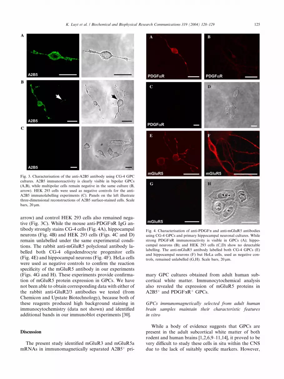
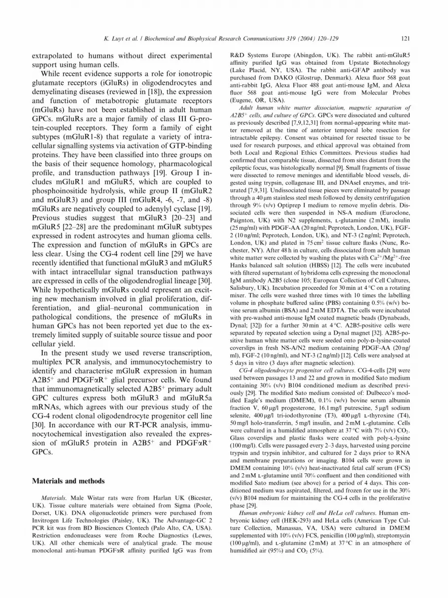
Fig. 3. Characterisation of the anti-A2B5 antibody using CG-4 GPC
cultures. A2B5 immunoreactivity is clearly visible in bipolar GPCs
(A,B), while multipolar cells remain negative in the same culture (B,
arrow). HEK 293 cells were used as negative controls for the anti-
A2B5 immunolabelling experiments (C). Panels on the left illustrate
three-dimensional reconstructions of A2B5 surface-stained cells. Scale
bars, 20 lm.
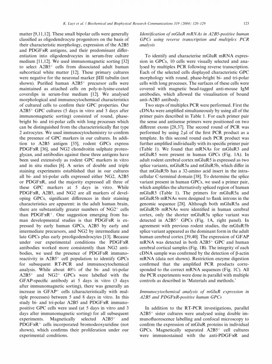
Fig. 4. Characterisation of anti-PDGFa and anti-mGluR5 antibodies
using CG-4 GPCs and primary hippocampal neuronal cultures. While
strong PDGFaR immunoreactivity is visible in GPCs (A); hippo-
campal neurons (B); and HEK 293 cells (C,D) show no detectable
labelling. The anti-mGluR5 antibody labelled both CG-4 GPCs (E)
and hippocampal neurons (F) but HeLa cells, used as negative con-
trols, remained unlabelled (G,H). Scale bars, 20 lm.
K. Luyt et al. / Biochemical and Biophysical Research Communications 319 (2004) 120–129 125
arrow) and control HEK 293 cells also remained nega-tive (Fig. 3C). While the mouse anti-PDGFaR IgG an-
tibody strongly stains CG-4 cells (Fig. 4A), hippocampal
neurons (Fig. 4B) and HEK 293 cells (Figs. 4C and D)
remain unlabelled under the same experimental condi-
tions. The rabbit anti-mGluR5 polyclonal antibody la-
belled both CG-4 oligodendrocyte progenitor cells
(Fig. 4E) and hippocampal neurons (Fig. 4F). HeLa cells
were used as negative controls to confirm the reactionspecificity of the mGluR5 antibody in our experiments
(Figs. 4G and H). These experiments provide confirma-
tion of mGluR5 protein expression in GPCs. We have
not been able to obtain corresponding data with either of
the rabbit anti-GluR2/3 antibodies we tested (from
Chemicon and Upstate Biotechnology), because both of
these reagents produced high background staining in
immunocytochemistry (data not shown) and identifiedadditional bands in our immunoblot experiments [30].
Discussion
The present study identified mGluR3 and mGluR5a
mRNAs in immunomagnetically separated A2B5þ pri-
mary GPC cultures obtained from adult human sub-
cortical white matter. Immunocytochemical analysis
also revealed the expression of mGluR5 proteins in
A2B5þ and PDGFaRþ GPCs.
GPCs immunomagnetically selected from adult human
brain samples maintain their characteristic features
in vitro
While a body of evidence suggests that GPCs are
present in the adult subcortical white matter of both
rodent and human brains [1,2,6,9–11,14], it proved to bevery difficult to study these cells in situ within the CNS
due to the lack of suitably specific markers. However,

126 K. Luyt et al. / Biochemical and Biophysical Research Communications 319 (2004) 120–129
these mitotically competent GPCs can be selectivelyisolated and maintained in vitro and recognized using
immunophenotypic markers. Rodent GPCs can be
identified by A2B5 antibodies and, in appropriate in
vitro environments, they can give rise to oligodendro-
cytes [2]. In addition to A2B5, these cells typically
express PDGFaR and the integral membrane proteo-
glycan NG2 [3]. While markers for cultured human
GPCs are not well established, some of the extensivelyused rodent GPC markers also label human precursors
[9,13,17]. In the present study we analysed primary
A2B5þ cell cultures prepared from adult human sub-
cortical white matter. Similar A2B5þ cell preparations
possess oligodendrocyte progenitor characteristics, be-
cause they develop into myelinating oligodendrocytes
following transplantation [12,41]. Our immunochemical
characterisation of these cells is consistent with previousstudies of GPCs, where the co-expression of the A2B5
antigen with NG2 and PDGFaR was reported [13,16].
The previously described potential cross-reactivities of
the anti-A2B5 and anti-PDGFaR antibodies with neu-
rons are not relevant to primary cultures from adult
human CNS, because neurons do not survive the dis-
sociation process [31]. Moreover, our A2B5þ and
PDGFaRþ cell cultures were completely negative for theneuronal marker bIII tubulin and they incorporated
bromodeoxyuridine, which also exclude their neuronal
origin. Astrocytes in vitro are not labelled by the
PDGFaR antibody [14]. As they matured, the number
of GFAPþ cells increased even under serum-free con-
ditions. Previous studies have also reported similar dif-
ferences in GFAP expression in human GPCs compare
to rat GPCs [42]. In the rat conditions have been iden-tified in which freshly isolated GPCs are GFAP negative
and remain so for several weeks [35], however, human
GPC cultures express GFAP under similar conditions
[42], which is consistent with the results of our RT-PCR
and immunocytochemical experiments. More recent
studies convincingly indicate that GFAP is not an ex-
clusive marker of astrocytes and can be expressed in
GPCs without astrocytic phenotype or function [43].
Metabotropic glutamate receptor isoforms mGluR3 and
mGluR5 are expressed in adult human GPCs
Recent evidence supports a role for glutamate re-ceptors in the pathophysiology of oligodendroglial
death in demyelinating diseases (reviewed in [18]). Oli-
godendroglial cells have been shown to be highly vul-
nerable to glutamate-mediated toxicity both in vitro and
in vivo [44,45]. Previous studies have focused specifically
on the role of iGluRs, which are ligand gated ion
channels [including a-amino-3-hydroxy-5-methyl-4-
isoxazolepropionic acid (AMPA), kainate and N -meth-yl-DD-aspartate (NMDA) receptors]. While these studies
reported the presence of AMPA, kainate and the
absence of NMDA type iGluRs in rat oligodendrocytes(reviewed in [18]), the expression and function of
mGluRs are less clear. In a recent study of the CG-4
clonal rodent oligodendrocyte progenitor cell line we
have identified and characterised functionalmGluR3 and
mGluR5 isoforms [30]. None of the other mGluRs were
present. Because the expression pattern of mGluRs in
adult human GPCs is unknown, and also to validate our
previous data obtained using the CG-4 rodent cell line[30], we used immunomagetically separated, highly ho-
mogeneous A2B5þ GPC cultures for the present study.
This strategy allows the isolation of GPCs based on cell-
surface markers and morphology. RT-PCR from very
limited number of cells requires a highly sensitive and
efficient system. Themultiplex PCRprocedure allowed us
to reliably characterise mGluR3 and mGluR5a mRNA
expression despite the extremely limited amount ofstarting tissue, which was obtained during anterior
temporal lobe resection for intractable epilepsy.
It is interesting to note that mGluR5 exists in two
splice variants, mGluR5a and mGluR5b [38]. The
mGluR5b contains a 32 amino acid fragment inserted
into the cytoplasmic tail and is the major type expressed
in different regions of adult brain [46]. There is a de-
velopmental switch from mGluR5a to mGluR5b splic-ing during the early postnatal period in both neurons
and astrocytes [22,39,40,47,48]. Our multiplex RT-PCR
analysis selectively identified the mGluR5a splice vari-
ant in adult GPCs, which is consistent with the devel-
opmentally more immature state of these cells.
We used immunofluorescence double-labelling to
confirm the expression of mGluR proteins in character-
ised GPCs. Our immunocytochemical analysis identifiedmGluR5 immunoreactivity in A2B5þ and PGDFaRþ
GPCs. Parallel positive and negative controls confirmed
the reaction specificity of each of the antibodies under
our experimental conditions. While we have not been
able to analyse the presence of mGluR3 in human GPCs
at the protein level due to the lack of suitable mGluR3-
specific antibody, previous in situ hybridisation studies
of normal rat brain identified mRNAs for mGluR3 inglial cells in various brain regions [49,50].
Astrocytes also express the mGluR3 and mGluR5
isoforms [21,23,24,26]. The astroglial expression of these
mGluRs appears to be dynamic, with expression levels
changing in response to different types of brain injury in
vivo [23,26,27,51,52] and upon exposure of cultured
astrocytes to growth factors [53,54]. The white matter
used for the present study was dissected from regionsdistant from the epileptic focus in temporal lobe. While
previous studies confirmed that comparable tissue re-
mains histologically normal [9], we cannot completely
eliminate the possibility that GPCs were exposed to
pathological glutamate concentrations in the brain,
which may lead to changes in mGluR expression levels
as seen in astrocytes.

K. Luyt et al. / Biochemical and Biophysical Research Communications 319 (2004) 120–129 127
Possible functional roles of metabotropic glutamate
receptors in human GPCs
Several observations suggest that glial mGluRs can
regulate glial function and may be involved in the in-
teraction between glia and neurons in both physiological
and pathological conditions [55–57]. It is possible that
mGluRs may play a developmental role in GPCs. Ac-
tivation of mGluR5 enhances proliferation in culturedastrocytes, whereas activation of mGluR3 inhibits this
process [58]. Therefore, the newly identified mGluR3
and mGluR5 may also be involved in the regulation of
GPC division. The mGluR5 isoform has been shown to
be involved in neurite extension in NG108-15 cells, with
the mGluR5 specific antagonist MPEP preventing ar-
borisation in these cells [59]. The same receptor could be
involved in the differentiation process in oligodendrog-lial cells where complex ramification takes place.
The presence of mGluR3 and mGluR5 in GPCs and
their recognised role in cell differentiation and prolifer-
ation in rodent glial cells may represent a novel system
for the regulation of the spontaneous remyelination
process observed in multiple sclerosis and other demy-
elinating conditions [6,12,14,16]. Previous studies
reported enhanced mGluR5 and mGluR2/3 immunore-activity in multiple sclerosis lesions [60]. It is plausible
that glutamate released from injured axons may activate
GPCs to begin the remyelination process. The increased
mGluR2/3 and mGluR5 signal may in part be due to
newly recruited GPCs migrating into the demyelinated
area in addition to reactive astrocytes [60]. Since these
receptors may be involved in the signalling process re-
quired to initiate remyelination in the human brain, theycould represent novel potential pharmacological targets
for therapeutic interventions in multiple sclerosis and
other demyelinating diseases.
Acknowledgments
This work was supported by the Department of Health, Medical
Research Council, the Wellcome Trust, and the Multiple Sclerosis
Society. We thank Dr. Maria M. Usowicz for her advice on multiplex
RT-PCR methodology. We thank the MRC for providing an Infra-
structure Award to establish the School of Medical Sciences Cell
Imaging Facility.
References
[1] N. Scolding, Glial precursor cells in the adult human brain,
Neuroscientist 4 (1998) 264–272.
[2] C. ffrench-Constant, M.C. Raff, Proliferating bipotential glial
progenitor cells in adult rat optic nerve, Nature 319 (1986) 499–
502.
[3] A. Nishiyama, A. Chang, B.D. Trapp, NG2+ glial cells: a novel
glial cell population in the adult brain, J. Neuropathol. Exp.
Neurol. 58 (1999) 1113–1124.
[4] A. Nishiyama, X.H. Lin, N. Giese, C.H. Heldin, W.B. Stallcup,
Co-localization of NG2 proteoglycan and PDGF alpha-receptor
on O2A progenitor cells in the developing rat brain, J. Neurosci.
Res. 43 (1996) 299–314.
[5] A. Nishiyama, X.H. Lin, N. Giese, C.H. Heldin, W.B. Stallcup,
Interaction between NG2 proteoglycan and PDGF alpha-receptor
on O2A progenitor cells is required for optimal response to
PDGF, J. Neurosci. Res. 43 (1996) 315–330.
[6] J.M. Levine, R. Reynolds, J.W. Fawcett, The oligodendrocyte
precursor cell in health and disease, Trends Neurosci. 24 (2001)
39–47.
[7] R.C. Armstrong, H.H. Dorn, C.V. Kufta, E. Friedman,
M. Dubois-Dalcq, Pre-oligodendrocytes from adult human
CNS, J. Neurosci. 12 (1992) 1538–1547.
[8] N. Gogate, L. Verma, J.M. Zhou, E. Milward, R. Rusten,
M. Oconnor, C. Kufta, J. Kim, L. Hudson, M. Duboisdalcq,
Plasticity in the adult human oligodendrocyte lineage, J. Neurosci.
14 (1994) 4571–4587.
[9] N.J. Scolding, P.J. Rayner, J. Sussman, C. Shaw, D.A. Compston,
A proliferative adult human oligodendrocyte progenitor, Neuro-
report 6 (1995) 441–445.
[10] N.J. Scolding, P.J. Rayner, D.A.S. Compston, Identification of
A2B5-positive putative oligodendrocyte progenitor cells and
A2B5-positive astrocytes in adult human white matter, Neurosci-
ence 89 (1999) 1–4.
[11] N.S. Roy, S. Wang, C. Harrison-Restelli, A. Benraiss,
R.A.R. Fraser, M. Gravel, P.E. Braun, S.A. Goldman, Iden-
tification, isolation, and promoter-defined separation of mitotic
oligodendrocyte progenitor cells from the adult human subcor-
tical white matter, J. Neurosci. 19 (1999) 9986–9995.
[12] M.S. Windrem, N.S. Roy, J. Wang, M. Nunes, A. Benraiss,
R. Goodman, G.M. McKhann II, S.A. Goldman, Progenitor cells
derived from the adult human subcortical white matter disperse
and differentiate as oligodendrocytes within demyelinated lesions
of the rat brain, J. Neurosci. Res. 69 (2002) 966–975.
[13] H.C. Wilson, C. Onischke, C.S. Raine, Human oligodendrocyte
precursor cells in vitro: phenotypic analysis and differential
response to growth factors, Glia 44 (2003) 143–165.
[14] N. Scolding, R. Franklin, S. Stevens, C.-H. Heldin, A. Compton,
J. Newcombe, Oligodendrocyte progenitors are present in the
normal adult human CNS and in the lesions of multiple sclerosis,
Brain 121 (1998) 2221–2228.
[15] G. Wolswijk, Chronic stage multiple sclerosis lesions contain a
relatively quiescent population of oligodendrocyte precursor cells,
J. Neurosci. 18 (1998) 601–609.
[16] A. Chang, A. Nishiyama, J. Peterson, J. Prineas, B.D. Trapp,
NG2-positive oligodendrocyte progenitor cells in adult human
brain and multiple sclerosis lesions, J. Neurosci. 20 (2000) 6404–
6412.
[17] S.-C. Zhang, B. Ge, I.D. Duncan, Tracing human oligodendrog-
lial development in vitro, J. Neurosci. Res. 59 (2000) 421–429.
[18] C. Matute, E. Aberdi, M. Domercq, F. Perez-Cerda, A. Perez-
Samartin, M.V. Sanchez-Gomez, The link between excitotoxic
oligodendroglial death and demyelinating diseases, Trends Neu-
rosci. 24 (2001) 224–230.
[19] D.D. Schoepp, D.E. Jane, J.A. Monn, Pharmacological agents
acting at subtypes of metabotropic glutamate receptors, Neuro-
pharmacology 38 (1999) 1431–1476.
[20] C.M. Testa, D.G. Standaert, A.B. Young, J.B. Penney Jr.,
Metabotropic glutamate receptor mRNA expression in the basal
ganglia of the rat, J. Neurosci. 14 (1994) 3005–3018.
[21] R.S. Petralia, Y.X. Wang, A.S. Niedzielski, R.J. Wenthold, The
metabotropic glutamate receptors, mGluR2 and mGluR3, show
unique postsynaptic, presynaptic and glial localizations, Neuro-
science 71 (1996) 949–976.
[22] G.P. Schools, H.K. Kimelberg, mGluR3 and mGluR5 are the
predominant metabotropic glutamate receptor mRNAs expressed

128 K. Luyt et al. / Biochemical and Biophysical Research Communications 319 (2004) 120–129
in hippocampal astrocytes acutely isolated from young rats,
J. Neurosci. Res. 58 (1999) 533–543.
[23] E. Aronica, E.A. van Vliet, O.A. Mayboroda, D. Troost, F.H. da
Silva, J.A. Gorter, Upregulation of metabotropic glutamate
receptor subtype mGluR3 and mGluR5 in reactive astrocytes in
a rat model of mesial temporal lobe epilepsy, Eur. J. Neurosci. 12
(2000) 2333–2344.
[24] C. Romano, M.C.T.M. Sesma, K. O’Malley, A.N. Van den Pol,
J.W. Olney, Distribution of metabotropic glutamate receptor
mGluR immunoreactivity in rat brain, J. Comp. Neurol. 355
(1995) 455–469.
[25] R. Balazs, S. Miller, C. Romano, A. de Vries, Y. Chun,
C.W. Cotman, Metabotropic glutamate receptor mGluR5 in
astrocytes: pharmacological properties and agonist regulation, J.
Neurochem. 69 (1997) 151–163.
[26] E. Aronica, M.V. Catania, J. Geurts, B. Yankaya, D. Troost,
Immunohistochemical localization of group I and II metabotropic
glutamate receptors in control and amyotrophic lateral sclerosis
human spinal cord: upregulation in reactive astrocytes, Neurosci-
ence 105 (2001) 509–520.
[27] E. Aronica, J.A. Gorter, H. Ijlst-Keizers, A.J. Rozemuller,
B. Yankaya, S. Leenstra, D. Troost, Expression and functional
role of mGluR3 and mGluR5 in human astrocytes and glioma
cells: opposite regulation of glutamate transporter proteins, Eur.
J. Neurosci. 17 (2003) 2106–2118.
[28] C. Corti, R.W.E. Clarkson, L. Crepaldi, C.F. Sala, J.H. Xuereb,
F. Ferraguti, Gene structure of the human metabotropic gluta-
mate receptor 5 and functional analysis of its multiple promoters
in neuroblastoma and astroglioma cells, J. Biol. Chem. 278 (2003)
33105–33119.
[29] J.C. Louis, E. Magal, D. Muir, M. Manthorpe, S. Varon, CG-4, a
new bipotential glial cell line from rat brain, is capable of
differentiating in vitro into either mature oligodendrocytes or
type-2 astrocytes, J. Neurosci. Res. 31 (1992) 193–204.
[30] K. Luyt, A. Varadi, E. Molnar, Functional metabotropic gluta-
mate receptors are expressed in oligodendrocyte progenitor cells,
J. Neurochem. 84 (2003) 1452–1464.
[31] V.W. Young, J.P. Antel, Culture of glial cells from human brain
biopsies, in: S. Federoff, A. Richardson (Eds.), Protocols for
Neural Cell Culture, third ed., Humana Press, Totowa, NJ, USA,
2001, pp. 129–138.
[32] A.P. Wright, J.J. Fitzgerald, R.J. Colello, Rapid purification of
glial cells using immunomagnetic separation, J. Neurosci. Meth-
ods 74 (1997) 37–44.
[33] L. Pickard, J. Noel, J.M. Henley, G.L. Collingridge, E. Molnar,
Developmental changes in synaptic AMPA and NMDA receptor
distribution and AMPA receptor subunit composition in living
hippocampal neurons, J. Neurosci. 20 (2000) 7922–7931.
[34] N.L. Brice, A. Varadi, S.J. Ashcroft, E. Molnar, Metabotropic
glutamate and GABA(B) receptors contribute to the modulation
of glucose-stimulated insulin secretion in pancreatic beta cells,
Diabetologia 45 (2002) 242–252.
[35] M.C. Raff, R.H. Miller, M. Noble, A glial progenitor cell that
develops in vitro an astrocyte or an oligodendrocyte depending on
culture medium, Nature 303 (1983) 390–396.
[36] N.P. Pringle, H.S. Mudhar, E.J. Collarini, W.D. Richardson,
PDGF receptors in the rat CNS: during late neurogenesis,
PDGF alpha-receptor expression appears to be restricted to glial
cells of the oligodendrocyte lineage, Development 115 (1992)
535–551.
[37] C. Corti, C.F. Sala, F.T. Yang, M. Corsi, J.H. Xuereb,
F. Ferraguti, Genomic organization of the human metabotropic
glutamate receptor subtype 3, J. Neurogenet. 14 (2000) 207–225.
[38] R. Minakami, F. Katsuki, H. Sugiyama, A variant of metabo-
tropic glutamate receptor subtype 5: an evolutionally conserved
insertion with no termination codon, Biochem. Biophys. Res.
Commun. 194 (1993) 622–627.
[39] C. Romano, S. Smout, J.K. Miller, K.L. O’Malley, Developmen-
tal regulation of metabotropic glutamate receptor 5b protein in
rodent brain, Neuroscience 111 (2002) 693–698.
[40] S.L. Harris, K. Cho, Z.I. Bashir, E. Molnar, Metabotropic
glutamate receptor signalling in perirhinal cortical neurons, Mol.
Cell. Neurosci. 25 (2004) 275–287.
[41] M.S. Windrem, M.C. Nunes, W.K. Rashbaum, T.H. Schwartz,
R.A. Goodman, G. McKhann II, N.S. Roy, S.A. Goldman, Fetal
and adult human oligodendrocyte progenitor cell isolates myeli-
nate the congenitally dysmyelinated brain, Nat. Med. 10 (2004)
93–97.
[42] J. Dietrich, M. Noble, M. Mayer-Proschel, Characterization of
A2B5þ glial precursor cells from cryopreserved human fetal brain
progenitor cells, Glia 40 (2002) 65–77.
[43] S. Goldman, Glia as neuronal progenitor cells, Trends Neurosci.
26 (2003) 590–596.
[44] C. Matute, M.V. Sanchez-Gomez, L. Martinez-Millan, R. Miledi,
Glutamate receptor-mediated toxicity in optic nerve oligodendro-
cytes, Proc. Natl. Acad. Sci. USA 94 (1997) 8830–8835.
[45] J.W. McDonald, S.P. Althomsons, K.L. Hyrc, D.W. Choi,
M.P. Goldberg, Oligodendrocytes from forebrain are highly
vulnerable to AMPA/kainate receptor-mediated excitotoxicity,
Nat. Med. 4 (1998) 291–297.
[46] C. Joly, J. Gomeza, I. Brabet, K. Curry, J. Bockaert, J.P. Pin,
Molecular, functional, and pharmacological characterization of
the metabotropic glutamate receptor type 5 splice variants:
comparison with mGluR1, J. Neurosci. 15 (1995) 3970–3981.
[47] C. Romano, A.N. van den Pol, K.L. O’Malley, Enhanced early
developmental expression of the metabotropic glutamate recep-
tor mGluR5 in rat brain: protein, mRNA aplice variants,
and regional distribution, J. Comp. Neurol. 367 (1996) 403–
412.
[48] Z. Cai, G.P. Schools, H.K. Kimelberg, Metabotropic glutamate
receptors in acutely isolated hippocampal astrocytes: develop-
mental changes of mGluR5 mRNA and functional expression,
Glia 29 (2000) 70–80.
[49] Y. Tanabe, M. Masu, T. Ishii, R. Shigemoto, S. Nakanishi,
A family of metabotropic glutamate receptors, Neuron 8 (1992)
169–179.
[50] H. Ohishi, R. Shigemoto, S. Nakanishi, N. Mizuno, Distribution
of the mRNA for a metabotropic glutamate receptor (mGluR3) in
the rat brain: an in situ hybridization study, J. Comp. Neurol. 335
(1993) 252–266.
[51] J. Ulas, T. Satou, K.J. Ivins, J.P. Kesslak, C.W. Cotman,
R. Balazs, Expression of metabotropic glutamate receptor 5 is
increased in astrocytes after kainate-induced epileptic seizures,
Glia 30 (2000) 352–361.
[52] F. Ferraguti, C. Corti, E. Valerio, S. Mion, J. Xuereb, Activated
astrocytes in areas of kainate-induced neuronal injury upregulate
the expression of the metabotropic glutamate receptors 2/3 and 5,
Exp. Brain Res. 137 (2001) 1–11.
[53] S. Miller, C. Romano, C.W. Cotman, Growth factor upregulation
of a phosphoinositide-coupled metabotropic glutamate receptor in
cortical astrocytes, J. Neurosci. 15 (1995) 6103–6109.
[54] T. Minoshima, S. Nakanishi, Structural organization of the mouse
metabotropic glutamate receptor subtype 3 gene and its regulation
by growth factors in cultured cortical astrocytes, J. Biochem.
(Tokyo) 126 (1999) 889–896.
[55] D.G. Winder, P.S. Ritch, R.W. Gereau IV, P.J. Conn, Novel glia-
neuronal signalling by coactivation of metabotropic glutamate
and beta-adrenergic receptors in rat hippocampus, J. Physiol. 494
(1996) 743–755.
[56] J. Cartmell, D.D. Schoepp, Regulation of neurotransmitter release
by metabotropic glutamate receptors, J. Neurochem. 75 (2000)
889–907.
[57] M. D’Onofrio, L. Cuomo, G. Battaglia, R.T. Ngomba, M. Storto,
A.E. Kingston, F. Orzi, A. De Blasi, P. Di lorio, F. Nicoletti, V.

K. Luyt et al. / Biochemical and Biophysical Research Communications 319 (2004) 120–129 129
Bruno, Neuroprotection mediated by glial group-II metabotropic
glutamate receptors requires the activation of the MAP kinase and
the phosphatidylinositol-3-kinase pathways, J. Neurochem. 78
(2001) 435–445.
[58] R. Ciccarelli, F.X. Sureda, G. Casabona, P. DiIorio, A. Caruso,
F. Spinella, D.F. Condorelli, F. Nicoletti, F. Caciagli, Opposite
influence of the metabotropic glutamate receptor subtypes
mGluR3 and -5 on astrocyte proliferation in culture, Glia 21
(1997) 390–398.
[59] S. Mion, C. Corti, A. Neki, R. Shigemoto, M. Corsi, G.
Fumagalli, F. Ferraguti, Bidirectional regulation of neurite
elaboration by alternatively spliced metabotropic glutamate
receptor 5 (mGluR5) isoforms, Mol. Cell. Neurosci. 17 (2001)
957–972.
[60] J.J.G. Geurts, G. Wolswijk, L. Bo, P. van der Valk, C.H. Polman,
D. Troost, E. Aronica, Altered expression patterns of group I and
II metabotropic glutamate receptors in multiple sclerosis, Brain
126 (2003) 1755–1766.