map2 and tau bind longitudinally along the outer ridges of microtubule protofilaments
TRANSCRIPT
The Rockefeller University Press, 0021-9525/2002/06/1187/10 $5.00The Journal of Cell Biology, Volume 157, Number 7, June 24, 2002 1187–1196http://www.jcb.org/cgi/doi/10.1083/jcb.200201048
JCB
Article
1187
MAP2 and tau bind longitudinally along the outer ridges of microtubule protofilaments
Jawdat Al-Bassam,
1
Rachel S. Ozer,
1
Daniel Safer,
2
Shelley Halpain,
1
and Ronald A. Milligan
1
1
Department of Cell Biology, Scripps Research Institute, La Jolla, CA 92037
2
Department of Physiology, School of Medicine, University of Pennsylvania, Philadelphia, PA 19104
AP2 and tau exhibit microtubule-stabilizing activities
that are implicated in the development and
maintenance of neuronal axons and dendrites. Theproteins share a homologous COOH-terminal domain,composed of three or four microtubule binding repeatsseparated by inter-repeats (IRs). To investigate how MAP2and tau stabilize microtubules, we calculated 3D maps ofmicrotubules fully decorated with MAP2c or tau usingcryo-EM and helical image analysis. Comparing thesemaps with an undecorated microtubule map revealed
additional densities along protofilament ridges on themicrotubule exterior, indicating that MAP2c and tau form
M
an ordered structure when they bind microtubules. Local-ization of undecagold attached to the second IR of MAP2cshowed that IRs also lie along the ridges, not between
protofilaments. The densities attributable to the microtubule-associated proteins lie in close proximity to helices 11 and12 and the COOH terminus of tubulin. Our data furthersuggest that the evolutionarily maintained differencesobserved in the repeat domain may be important for the
specific targeting of different repeats to either
�
or
�
tubulin.
These results provide strong evidence suggesting that MAP2cand tau stabilize microtubules by binding along individualprotofilaments, possibly by bridging the tubulin interfaces.
Introduction
The microtubule cytoskeleton plays a fundamental role in avariety of cellular processes. Microtubules assemble by lateral
association of protofilaments generated from head to tail
polymerization of
��
tubulin dimers. The tendency oftubulin to switch stochastically between polymerizationand depolymerization phases, termed dynamic instability(Mitchison and Kirschner, 1984), facilitates remodeling of
the microtubule cytoskeleton for its various roles. Microtubule-associated proteins (MAPs)* influence dynamic instabilityby binding and stabilizing microtubules (Cleveland et al.,1977). The MAP2/tau family is a unique class of structuralMAPs that modulate microtubule dynamics in neurons dur-ing the development of dendrites and axons (for reviews seeDrewes et al., 1998; Goldstein and Gunawardena, 2000).Phosphorylation of MAP2/tau proteins at specific sitesinduces dissociation from microtubules (Drewes et al.,
1997; Ozer and Halpain, 2000). However, hyperphosphor-ylation and/or specific mutations can promote aggregationof the dissociated tau into paired helical filaments, which area hallmark of certain neurodegenerative diseases such asfamilial tauopathies and Alzheimer’s disease (Crowther andGoedert, 2000; Garcia and Cleveland, 2001).
MAP2/tau proteins have dissimilar NH
2
-terminal “projec-tion” domains and homologous COOH-terminal microtubulebinding domains. Studies investigating the cytoskeleton ofneuronal processes showed that microtubules with MAP2 ortau bound are organized in parallel arrays in which the spac-ing between microtubules correlates with the length of theprojection domains of the MAP (Hirokawa, 1982; Chen etal., 1992). Rotary shadowing experiments and circulardichroism studies on isolated MAP2/tau proteins indicatethat they are highly extended polypeptides with little or nodetectable secondary structure (Voter and Erickson 1982;Schweers et al., 1994).
The MAP2/tau microtubule binding domain contains
three or four 18-residue microtubule binding repeats(MTBRs) separated by 13–14-residue inter-repeats (IRs)(Lewis et al., 1988; Himmler et al., 1989). NeighboringMTBRs within a given MAP share moderate homology;however, sequence alignment shows that repeats at identical
positions in different MAP2s and taus are highly conserved(Fig. 1 A). There is evidence that IRs as well as MTBRs con-
Address correspondence to Ronald A. Milligan, Department of Cell Biol-ogy, CB-227, Scripps Research Institute, 10550 North Torrey PinesRoad, La Jolla, CA 92037. Tel.: (858) 784-9827. Fax: (858) 784-2749.E-mail: [email protected]
*Abbreviations used in this paper: cf-MAP2c, cysteine-free MAP2c; cIR-
MAP2c, cysteine-IR-MAP2c; H11, helix 11; H12, helix 12; IR, inter-repeat; MAP, microtubule-associated protein; MTBR, microtubule bind-ing repeat.
Key words: MAP2; tau; structure; microtubule; cryo-EM
on May 31, 2014
jcb.rupress.orgD
ownloaded from
Published June 24, 2002
http://jcb.rupress.org/content/suppl/2002/06/20/jcb.200201048.DC1.html Supplemental Material can be found at:
1188 The Journal of Cell Biology
|
Volume 157, Number 7, 2002
tribute to microtubule binding (Butner and Kirschner, 1991;Goode and Feinstein, 1994; Ludin et al., 1996). The affinity ofthe repeat domain for microtubules and its polymer stabilizingactivity both increase with the number of MTBR-IR modules(Ludin et al., 1996; Goode et al., 2000). Binding to microtu-bules is mediated, in part, by the acidic tubulin COOH ter-mini (Paschal et al., 1989), because their removal by subtilisinreduces MAP binding (Serrano et al., 1984, 1985).
The prevailing model to explain how MAP2/tau familyproteins interact with microtubules presumes that eachMTBR-IR module interacts with a separate, but adjacent,tubulin monomer within the microtubule wall (Butner andKirschner, 1991; Gustke et al., 1994). Despite considerablestudy, the structural basis for MAP2/tau stabilization of mi-crotubules is not understood. It remains unclear whether in-creased microtubule stability is achieved by MAPs bindingalong protofilaments (a longitudinal binding model) orwrapping around the microtubule (a lateral binding model).As microtubule disassembly proceeds by protofilaments sep-arating and curling outwards at the ends of the microtubule(Mandelkow et al., 1991), longitudinal binding could ac-count for increased microtubule stability by strengtheningtubulin interactions along protofilaments and preventingoutward curling. Alternatively, in the lateral binding model,the wrapped MAPs would prevent protofilament separation(Ichihara et al., 2001).
Here we have used cryo-EM and helical image analysis todetermine the geometry of MAP2c and tau binding to mi-crotubules. We show that MAP2c- or tau-decorated micro-tubules have additional ordered density along protofilamentridges compared with undecorated microtubules. We usedundecagold labeling to show that the IRs lie along the ridgesand not between protofilaments. The gold labeling data sug-
gest that the MTBR-IR modules may be uniquely targeted
to
�
or
�
tubulin. Taken together, our results suggest thatMAP2 and tau proteins reduce microtubule depolymeriza-tion by bridging and stabilizing the tubulin–tubulin inter-faces along protofilaments.
Results
Cryo-EM of MAP2c- and tau-decorated microtubules
To investigate how MAPs stabilize microtubules, we haveanalyzed two distinct proteins using cryo-EM and helicalimage analysis: a recombinant three-repeat rat MAP2c(termed MAP2c throughout) and a recombinant four-repeathuman tau (termed tau throughout). The MAP2c and tauproteins are homologous in their repeat domain, but tau in-cludes an additional MTBR-IR module (Fig. 1 A; see Mate-rials and methods).
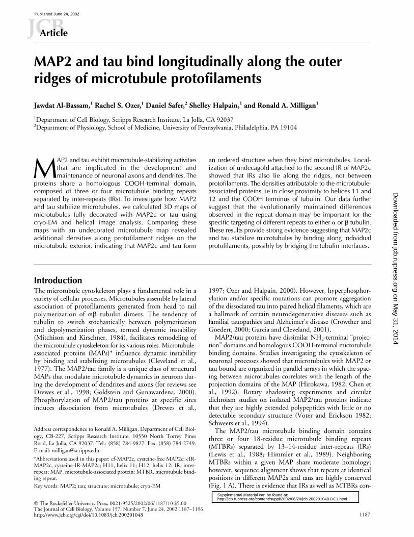
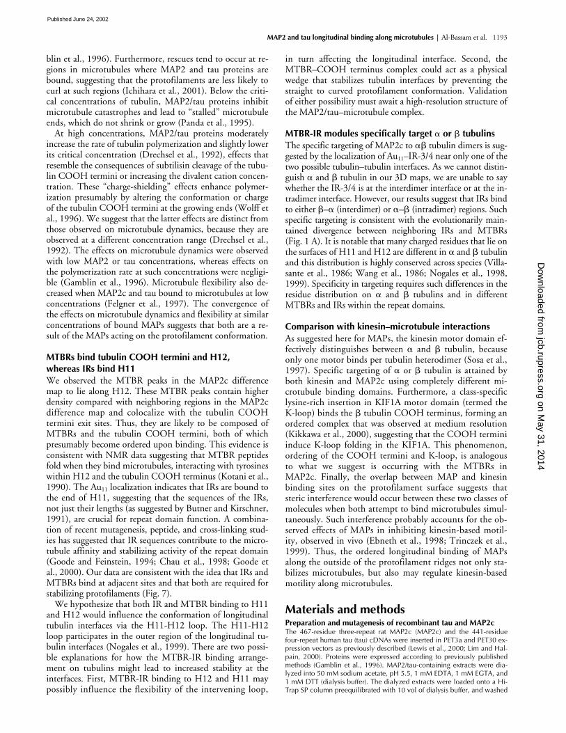
We used cosedimentation assays to determine conditionsfor full decoration of microtubules by MAP2c and tau. Undersaturating conditions, MAP2c bound microtubules at a ratioof one molecule for every 2.4 tubulin monomers (Fig. 1 B, I),and tau at one molecule for every 3.8 tubulin monomers (Fig.1 B, II). These stoichiometry values are very similar to thosedetermined by others (Gustke et al., 1994; Coffey and Purich,1995). We used the conditions found for maximal binding toprepare decorated microtubules for cryo-EM.
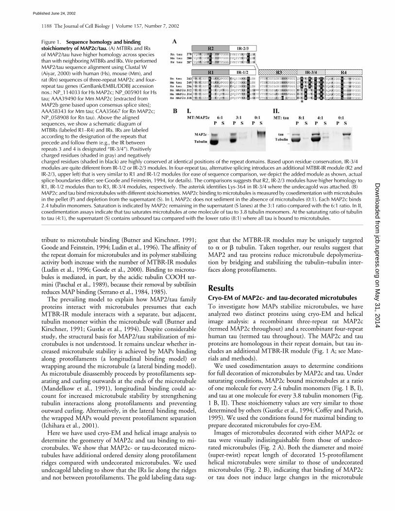
Images of microtubules decorated with either MAP2c ortau were visually indistinguishable from those of undeco-rated microtubules (Fig. 2 A). Both the diameter and moiré(super-twist) repeat length of decorated 15-protofilamenthelical microtubules were similar to those of undecoratedmicrotubules (Fig. 2 B), indicating that binding of MAP2cor tau does not induce large changes in the microtubule
Figure 1. Sequence homology and binding stoichiometry of MAP2c/tau. (A) MTBRs and IRs of MAP2/tau have higher homology across species than with neighboring MTBRs and IRs. We performed MAP2/tau sequence alignment using Clustal W (Aiyar, 2000) with human (Hs), mouse (Mm), and rat (Rn) sequences of three-repeat MAP2c and four-repeat tau genes (GenBank/EMBL/DDBJ accession nos.: NP_114033 for Hs MAP2c; NP_005901 for Hs tau; AAA39490 for Mm MAP2c [extracted from MAP2b gene based upon consensus splice sites]; AAA58343 for Mm tau; CAA35667 for Rn MAP2c; NP_058908 for Rn tau). Above the aligned sequences, we show a schematic diagram of MTBRs (labeled R1–R4) and IRs. IRs are labeled according to the designation of the repeats that precede and follow them (e.g., the IR between repeats 3 and 4 is designated “IR-3/4”). Positively charged residues (shaded in gray) and negatively charged residues (shaded in black) are highly conserved at identical positions of the repeat domains. Based upon residue conservation, IR-3/4 modules are quite different from IR-1/2 or IR-2/3 modules. In four-repeat tau, alternative splicing introduces an additional MTBR-IR module (R2 and IR-2/3, upper left) that is very similar to R1 and IR-1/2 modules (for ease of sequence comparison, we depict the added module as shown, actual splice boundaries differ; see Goode and Feinstein, 1994, for details). The comparisons suggests that R2, IR-2/3 modules have higher homology to R1, IR-1/2 modules than to R3, IR-3/4 modules, respectively. The asterisk identifies Lys-364 in IR-3/4 where the undecagold was attached. (B) MAP2c and tau bind microtubules with different stoichiometries. MAP2c binding to microtubules is measured by cosedimentation with microtubules in the pellet (P) and depletion from the supernatant (S). In I, MAP2c does not sediment in the absence of microtubules (0:1). Each MAP2c binds 2.4 tubulin monomers. Saturation is indicated by MAP2c remaining in the supernatant (S lanes) at the 3:1 ratio compared with the 6:1 ratio. In II, cosedimentation assays indicate that tau saturates microtubules at one molecule of tau to 3.8 tubulin monomers. At the saturating ratio of tubulin to tau (4:1), the supernatant (S) contains unbound tau compared with the lower ratio (8:1) where all tau is bound to microtubules.
on May 31, 2014
jcb.rupress.orgD
ownloaded from
Published June 24, 2002
MAP2 and tau longitudinal binding along microtubules |
Al-Bassam et al. 1189
structure. Although individual images of decorated and un-decorated microtubules look similar, differences were evi-dent after averaging the layer line data from a number of im-ages. In particular, layer line 1 amplitudes were strongerfor decorated microtubules (Fig. S1, available at http://www.jcb.org/cgi/content/full/jcb.200201048/DC1) with mi-nor changes in other layer lines, consistent with an increasein protofilament density. There were no additional layerlines in the decorated microtubules, as would be expected ifthe MAP projection domains were contributing to off-equa-torial diffraction (Amos, 1977).
MAP2 and tau are ordered on microtubule protofilaments
From the averaged layer lines, we calculated 3D maps ofMAP2c-decorated, tau-decorated, and undecorated microtu-bules. All three maps show the same general features as thosepreviously reported for microtubules (Sosa et al., 1997;Nogales et al., 1999). However, consistent with the differ-ences in the layer lines, there is additional density lying alongthe protofilament ridges of the decorated microtubule maps(Fig. S2). To more clearly visualize this density, which is pre-sumably attributable to MAP2c or tau binding, we performeddifference mapping (Fig. 3) and statistical analyses (Fig. S3).
The difference maps, which were statistically significant atthe
P
�
0.0001 level, show elongated density along theprotofilament ridges on the outside of the microtubule (Fig.3, C–F, and Fig. S3, A–D). The MAP2c and tau differencedensities, between which there were no statistically significantdifferences (unpublished data), have a slender cross sectioncompared with tubulin (Fig. 3, D and F). Although the differ-
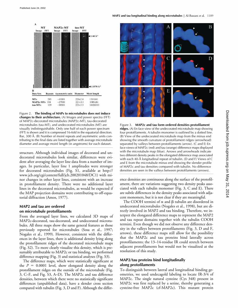
ence densities are continuous along the surface of the protofil-ament, there are variations suggesting two density peaks asso-ciated with each tubulin monomer (Fig. 3, C and E). Thereare subtle differences in the density peaks associated with adja-cent monomers, but it is not clear if they are meaningful.
The COOH termini of
�
and
�
tubulin are disordered inundecorated microtubules (Nogales et al., 1998), but are di-rectly involved in MAP2 and tau binding. Therefore, we in-terpret the elongated difference maps to represent the MAP2and tau repeat domains together with the tubulin COOHtermini. Even though we did not observe any difference den-sity in the valleys between protofilaments (Fig. 3, D and F,arrows), these difference maps still allow for the possibilitythat the MAP2c and tau proteins bind laterally acrossprotofilaments; the 13–14-residue IR could stretch betweenadjacent protofilaments but would not be visualized at theresolution of this study.
MAP2/tau proteins bind longitudinally along protofilaments
To distinguish between lateral and longitudinal binding ge-ometries, we used undecagold labeling to locate IR-3/4 ofMAP2c. The single natural cysteine (Cys 348) present inMAP2c was first replaced by a serine, thereby generating acysteine-free MAP2c (cf-MAP2c). This mutant protein
Figure 2. The binding of MAPs to microtubules does not induce changes to their architecture. (A) Images and power spectra (FFT) of MAP2c-decorated microtubules (MAP2c-MT), tau-decorated microtubules (tau-MT), and undecorated microtubules (MT) are visually indistinguishable. Only one half of each power spectrum (FFT) is shown and it is compressed 16-fold in the equatorial direction. Bar, 300 Å. (B) Number of moiré repeats and asymmetric units con-tributing to the final data are listed together with average microtubule diameter and average moiré length (in angstroms) for each dataset.
Figure 3. MAP2c and tau form ordered densities protofilament ridges. (A) En face view of the undecorated microtubule map showing four protofilaments. A tubulin monomer is outlined by a dotted line. (B) View of the undecorated microtubule map from the minus end showing the smooth curvature of protofilament ridges (arrowhead) separated by valleys between protofilaments (arrow). (C and E) En face views of MAP2c (red) and tau (orange) difference maps displayed with the microtubule map (blue). Arrows and arrowheads indicate two different density peaks in the elongated difference map associated with each 40-Å longitudinal repeat of tubulin. (D and F) Views of C and E from the microtubule minus end showing the slender profile of MAP2c and tau densities compared with tubulin. No difference densities are seen in the valleys between protofilaments (arrows).
on May 31, 2014
jcb.rupress.orgD
ownloaded from
Published June 24, 2002
1190 The Journal of Cell Biology
|
Volume 157, Number 7, 2002
bound microtubules with the same stoichiometry as MAP2c(Fig. 4 A). We then introduced a cysteine in place of lysine364 (Fig. 1 A, asterisk) in IR-3/4 of cf-MAP2c generating acysteine-IR-MAP2c mutant (cIR-MAP2c), which we conju-gated to undecagold (Au
11
) (Milligan et al., 1990; Safer,1999; Rice et al., 1999). We confirmed that neither themutation nor Au
11
attachment interferes with cIR-MAP2cbinding to microtubules (Fig. 4 B). The 3D map calculatedfrom Au
11
–cIR-MAP2c–decorated microtubules showed anadditional knob-like density on the microtubule surface.Difference mapping and statistical analysis showed that thisdensity was statistically significant at the
P
�
0.001 level(Fig. 5 and Fig. S3, E and F).
These experiments allow us to distinguish between thelongitudinal and the lateral binding models by localizationof the undecagold and by inference the IR-3/4 to which it isattached. The two possible positions of the IR–Au
11
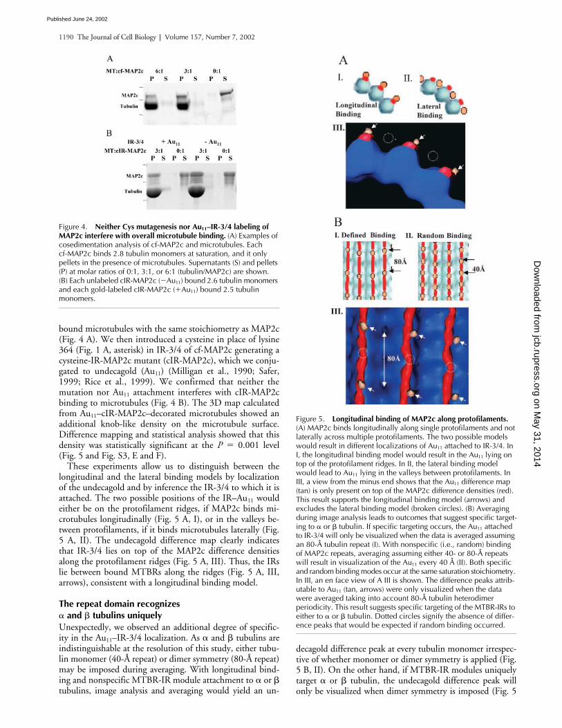
wouldeither be on the protofilament ridges, if MAP2c binds mi-crotubules longitudinally (Fig. 5 A, I), or in the valleys be-tween protofilaments, if it binds microtubules laterally (Fig.5 A, II). The undecagold difference map clearly indicatesthat IR-3/4 lies on top of the MAP2c difference densitiesalong the protofilament ridges (Fig. 5 A, III). Thus, the IRslie between bound MTBRs along the ridges (Fig. 5 A, III,arrows), consistent with a longitudinal binding model.
The repeat domain recognizes
�
and
�
tubulins uniquely
Unexpectedly, we observed an additional degree of specific-ity in the Au
11
–IR-3/4 localization. As
�
and
�
tubulins areindistinguishable at the resolution of this study, either tubu-lin monomer (40-Å repeat) or dimer symmetry (80-Å repeat)may be imposed during averaging. With longitudinal bind-ing and nonspecific MTBR-IR module attachment to
�
or
�
tubulins, image analysis and averaging would yield an un-
decagold difference peak at every tubulin monomer irrespec-tive of whether monomer or dimer symmetry is applied (Fig.5 B, II). On the other hand, if MTBR-IR modules uniquelytarget
�
or
�
tubulin, the undecagold difference peak willonly be visualized when dimer symmetry is imposed (Fig. 5
Figure 4. Neither Cys mutagenesis nor Au11–IR-3/4 labeling of MAP2c interfere with overall microtubule binding. (A) Examples of cosedimentation analysis of cf-MAP2c and microtubules. Each cf-MAP2c binds 2.8 tubulin monomers at saturation, and it only pellets in the presence of microtubules. Supernatants (S) and pellets (P) at molar ratios of 0:1, 3:1, or 6:1 (tubulin/MAP2c) are shown. (B) Each unlabeled cIR-MAP2c (�Au11) bound 2.6 tubulin monomers and each gold-labeled cIR-MAP2c (�Au11) bound 2.5 tubulin monomers.
Figure 5. Longitudinal binding of MAP2c along protofilaments. (A) MAP2c binds longitudinally along single protofilaments and not laterally across multiple protofilaments. The two possible models would result in different localizations of Au11 attached to IR-3/4. In I, the longitudinal binding model would result in the Au11 lying on top of the protofilament ridges. In II, the lateral binding model would lead to Au11 lying in the valleys between protofilaments. In III, a view from the minus end shows that the Au11 difference map (tan) is only present on top of the MAP2c difference densities (red). This result supports the longitudinal binding model (arrows) and excludes the lateral binding model (broken circles). (B) Averaging during image analysis leads to outcomes that suggest specific target-ing to � or � tubulin. If specific targeting occurs, the Au11 attached to IR-3/4 will only be visualized when the data is averaged assuming an 80-Å tubulin repeat (I). With nonspecific (i.e., random) binding of MAP2c repeats, averaging assuming either 40- or 80-Å repeats will result in visualization of the Au11 every 40 Å (II). Both specific and random binding modes occur at the same saturation stoichiometry. In III, an en face view of A III is shown. The difference peaks attrib-utable to Au11 (tan, arrows) were only visualized when the data were averaged taking into account 80-Å tubulin heterodimer periodicity. This result suggests specific targeting of the MTBR-IRs to either to � or � tubulin. Dotted circles signify the absence of differ-ence peaks that would be expected if random binding occurred.
on May 31, 2014
jcb.rupress.orgD
ownloaded from
Published June 24, 2002
MAP2 and tau longitudinal binding along microtubules |
Al-Bassam et al. 1191
B, I). We analyzed our data imposing either monomer ordimer symmetry and were only able to visualize the un-decagold when we used dimer symmetry (Fig. 5 B, III).These data strongly argue in favor of specific binding of IR-3/4 to either
�
or
�
tubulin, but not to both (Fig. 5 B, III).
The repeat domains bind helix 11 (H11), helix 12 (H12), and the COOH termini of tubulin
Although there is considerable evidence showing that MAP2and tau are unstructured in solution (Voter and Erickson,1982; Schweers et al., 1994), the data presented here show thatthey form ordered densities along the protofilament ridgeswhen they bind microtubules. Thus, it seems likely that specificresidues on the protofilament surface are required to fold theMTBR-IR modules and induce the bound density that we ob-serve. To investigate which structural elements on the tubulinsurface are likely to induce the IRs and MTBRs to fold, wemanually docked the atomic coordinates of the
��
tubulindimer into the microtubule map and displayed the result withthe MAP2c and undecagold difference maps. As the tubulinCOOH termini (18 and 10 residues for
�
and
�
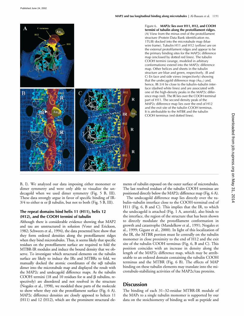
tubulins, re-spectively) are disordered and not resolved in the structure(Nogales et al., 1998), we modeled these parts of the moleculeto show where they exit the protofilament surface (Fig. 6 A).MAP2c difference densities are closely apposed to helices 11(H11) and 12 (H12), which are the prominent structural ele-
ments of tubulin exposed on the outer surface of microtubules.The last resolved residues of the tubulin COOH terminus arepositioned directly below the MAP2c difference map (Fig. 6 A).
The undecagold difference map lies directly over the tu-bulin–tubulin interface close to the COOH-terminal end ofH11 (Fig. 6, B and C). This implies that the IR, to whichthe undecagold is attached (Fig. 1 A, asterisk), also binds tothe interface, the region of the structure that has been shownto directly modulate the protofilament conformation ingrowth and catastrophe (Mandelkow et al., 1991; Nogales etal., 1999; Gigant et al., 2000). In light of this localization ofthe IR, the MTBR portion must lie centrally on the tubulinmonomer in close proximity to the end of H12 and the exitsite of the tubulin COOH terminus (Fig. 6, B and C). Thisposition coincides with an increase in density along thelength of the MAP2c difference map, which may be attrib-utable to an ordered domain containing the tubulin COOHterminus and the MTBR (Fig. 6 B). The effects of MAPbinding on these tubulin elements may translate into the mi-crotubule-stabilizing activities of the MAP2c/tau proteins.
Discussion
The binding of each 31–32-residue MTBR-IR module ofthe MAPs to a single tubulin monomer is supported by ourdata on the stoichiometry of binding as well as peptide and
Figure 6. MAP2c lies over H11, H12, and COOH termini of tubulin along the protofilament ridges. (A) View from the minus end of the protofilament structure (Protein Data Bank identification no. 1TUB) docked into the microtubule map (blue wire frame). Tubulin H11 and H12 (yellow) are on the external protofilament ridges and appear to be the primary binding sites for the MAP2c difference map (enclosed by dotted red lines). The tubulin COOH termini (orange, modeled in arbitrary conformations) extend into the MAP2c difference map. Other helices and sheets in the tubulin structure are blue and green, respectively. (B and C) En face and side views (respectively) showing that the undecagold difference map (Au11) and, hence, IR-3/4 lie close to the tubulin–tubulin inter-face (dashed white lines) and are associated with one of the high-density peaks in the MAP2c differ-ence map (red). The IR lies over the COOH-terminal part of H11. The second density peak of the MAP2c difference map lies over the end of H12 and the exit site of the tubulin COOH terminus. It is attributable to the MTBR and the tubulin COOH terminus (red dotted lines).
on May 31, 2014
jcb.rupress.orgD
ownloaded from
Published June 24, 2002
1192 The Journal of Cell Biology
|
Volume 157, Number 7, 2002
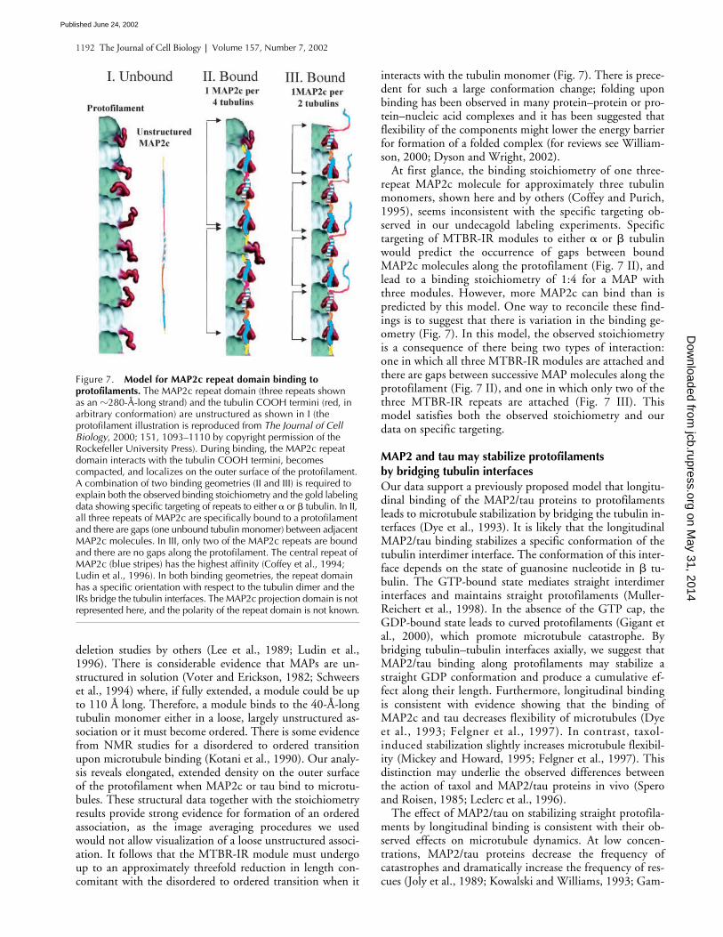
deletion studies by others (Lee et al., 1989; Ludin et al.,1996). There is considerable evidence that MAPs are un-structured in solution (Voter and Erickson, 1982; Schweerset al., 1994) where, if fully extended, a module could be upto 110 Å long. Therefore, a module binds to the 40-Å-longtubulin monomer either in a loose, largely unstructured as-sociation or it must become ordered. There is some evidencefrom NMR studies for a disordered to ordered transitionupon microtubule binding (Kotani et al., 1990). Our analy-sis reveals elongated, extended density on the outer surfaceof the protofilament when MAP2c or tau bind to microtu-bules. These structural data together with the stoichiometryresults provide strong evidence for formation of an orderedassociation, as the image averaging procedures we usedwould not allow visualization of a loose unstructured associ-ation. It follows that the MTBR-IR module must undergoup to an approximately threefold reduction in length con-comitant with the disordered to ordered transition when it
interacts with the tubulin monomer (Fig. 7). There is prece-dent for such a large conformation change; folding uponbinding has been observed in many protein–protein or pro-tein–nucleic acid complexes and it has been suggested thatflexibility of the components might lower the energy barrierfor formation of a folded complex (for reviews see William-son, 2000; Dyson and Wright, 2002).
At first glance, the binding stoichiometry of one three-repeat MAP2c molecule for approximately three tubulinmonomers, shown here and by others (Coffey and Purich,1995), seems inconsistent with the specific targeting ob-served in our undecagold labeling experiments. Specifictargeting of MTBR-IR modules to either
�
or
�
tubulinwould predict the occurrence of gaps between boundMAP2c molecules along the protofilament (Fig. 7 II), andlead to a binding stoichiometry of 1:4 for a MAP withthree modules. However, more MAP2c can bind than ispredicted by this model. One way to reconcile these find-ings is to suggest that there is variation in the binding ge-ometry (Fig. 7). In this model, the observed stoichiometryis a consequence of there being two types of interaction:one in which all three MTBR-IR modules are attached andthere are gaps between successive MAP molecules along theprotofilament (Fig. 7 II), and one in which only two of thethree MTBR-IR repeats are attached (Fig. 7 III). Thismodel satisfies both the observed stoichiometry and ourdata on specific targeting.
MAP2 and tau may stabilize protofilaments by bridging tubulin interfaces
Our data support a previously proposed model that longitu-dinal binding of the MAP2/tau proteins to protofilamentsleads to microtubule stabilization by bridging the tubulin in-terfaces (Dye et al., 1993). It is likely that the longitudinalMAP2/tau binding stabilizes a specific conformation of thetubulin interdimer interface. The conformation of this inter-face depends on the state of guanosine nucleotide in
�
tu-bulin. The GTP-bound state mediates straight interdimerinterfaces and maintains straight protofilaments (Muller-Reichert et al., 1998). In the absence of the GTP cap, theGDP-bound state leads to curved protofilaments (Gigant etal., 2000), which promote microtubule catastrophe. Bybridging tubulin–tubulin interfaces axially, we suggest thatMAP2/tau binding along protofilaments may stabilize astraight GDP conformation and produce a cumulative ef-fect along their length. Furthermore, longitudinal bindingis consistent with evidence showing that the binding ofMAP2c and tau decreases flexibility of microtubules (Dyeet al., 1993; Felgner et al., 1997). In contrast, taxol-induced stabilization slightly increases microtubule flexibil-ity (Mickey and Howard, 1995; Felgner et al., 1997). Thisdistinction may underlie the observed differences betweenthe action of taxol and MAP2/tau proteins in vivo (Speroand Roisen, 1985; Leclerc et al., 1996).
The effect of MAP2/tau on stabilizing straight protofila-ments by longitudinal binding is consistent with their ob-served effects on microtubule dynamics. At low concen-trations, MAP2/tau proteins decrease the frequency ofcatastrophes and dramatically increase the frequency of res-cues (Joly et al., 1989; Kowalski and Williams, 1993; Gam-
Figure 7. Model for MAP2c repeat domain binding to protofilaments. The MAP2c repeat domain (three repeats shown as an �280-Å-long strand) and the tubulin COOH termini (red, in arbitrary conformation) are unstructured as shown in I (the protofilament illustration is reproduced from The Journal of Cell Biology, 2000; 151, 1093–1110 by copyright permission of the Rockefeller University Press). During binding, the MAP2c repeat domain interacts with the tubulin COOH termini, becomes compacted, and localizes on the outer surface of the protofilament. A combination of two binding geometries (II and III) is required to explain both the observed binding stoichiometry and the gold labeling data showing specific targeting of repeats to either � or � tubulin. In II, all three repeats of MAP2c are specifically bound to a protofilament and there are gaps (one unbound tubulin monomer) between adjacent MAP2c molecules. In III, only two of the MAP2c repeats are bound and there are no gaps along the protofilament. The central repeat of MAP2c (blue stripes) has the highest affinity (Coffey et al., 1994; Ludin et al., 1996). In both binding geometries, the repeat domain has a specific orientation with respect to the tubulin dimer and the IRs bridge the tubulin interfaces. The MAP2c projection domain is not represented here, and the polarity of the repeat domain is not known.
on May 31, 2014
jcb.rupress.orgD
ownloaded from
Published June 24, 2002
MAP2 and tau longitudinal binding along microtubules |
Al-Bassam et al. 1193
blin et al., 1996). Furthermore, rescues tend to occur at re-gions in microtubules where MAP2 and tau proteins arebound, suggesting that the protofilaments are less likely tocurl at such regions (Ichihara et al., 2001). Below the criti-cal concentrations of tubulin, MAP2/tau proteins inhibitmicrotubule catastrophes and lead to “stalled” microtubuleends, which do not shrink or grow (Panda et al., 1995).
At high concentrations, MAP2/tau proteins moderatelyincrease the rate of tubulin polymerization and slightly lowerits critical concentration (Drechsel et al., 1992), effects thatresemble the consequences of subtilisin cleavage of the tubu-lin COOH termini or increasing the divalent cation concen-tration. These “charge-shielding” effects enhance polymer-ization presumably by altering the conformation or chargeof the tubulin COOH termini at the growing ends (Wolff etal., 1996). We suggest that the latter effects are distinct fromthose observed on microtubule dynamics, because they areobserved at a different concentration range (Drechsel et al.,1992). The effects on microtubule dynamics were observedwith low MAP2 or tau concentrations, whereas effects onthe polymerization rate at such concentrations were negligi-ble (Gamblin et al., 1996). Microtubule flexibility also de-creased when MAP2c and tau bound to microtubules at lowconcentrations (Felgner et al., 1997). The convergence ofthe effects on microtubule dynamics and flexibility at similarconcentrations of bound MAPs suggests that both are a re-sult of the MAPs acting on the protofilament conformation.
MTBRs bind tubulin COOH termini and H12, whereas IRs bind H11
We observed the MTBR peaks in the MAP2c differencemap to lie along H12. These MTBR peaks contain higherdensity compared with neighboring regions in the MAP2cdifference map and colocalize with the tubulin COOHtermini exit sites. Thus, they are likely to be composed ofMTBRs and the tubulin COOH termini, both of whichpresumably become ordered upon binding. This evidence isconsistent with NMR data suggesting that MTBR peptidesfold when they bind microtubules, interacting with tyrosineswithin H12 and the tubulin COOH terminus (Kotani et al.,1990). The Au
11
localization indicates that IRs are bound tothe end of H11, suggesting that the sequences of the IRs,not just their lengths (as suggested by Butner and Kirschner,1991), are crucial for repeat domain function. A combina-tion of recent mutagenesis, peptide, and cross-linking stud-ies has suggested that IR sequences contribute to the micro-tubule affinity and stabilizing activity of the repeat domain(Goode and Feinstein, 1994; Chau et al., 1998; Goode etal., 2000). Our data are consistent with the idea that IRs andMTBRs bind at adjacent sites and that both are required forstabilizing protofilaments (Fig. 7).
We hypothesize that both IR and MTBR binding to H11and H12 would influence the conformation of longitudinaltubulin interfaces via the H11-H12 loop. The H11-H12loop participates in the outer region of the longitudinal tu-bulin interfaces (Nogales et al., 1999). There are two possi-ble explanations for how the MTBR-IR binding arrange-ment on tubulins might lead to increased stability at theinterfaces. First, MTBR-IR binding to H12 and H11 maypossibly influence the flexibility of the intervening loop,
in turn affecting the longitudinal interface. Second, theMTBR–COOH terminus complex could act as a physicalwedge that stabilizes tubulin interfaces by preventing thestraight to curved protofilament conformation. Validationof either possibility must await a high-resolution structure ofthe MAP2/tau–microtubule complex.
MTBR-IR modules specifically target
�
or
�
tubulins
The specific targeting of MAP2c to
��
tubulin dimers is sug-gested by the localization of Au
11
–IR-3/4 near only one of thetwo possible tubulin–tubulin interfaces. As we cannot distin-guish
�
and
�
tubulin in our 3D maps, we are unable to saywhether the IR-3/4 is at the interdimer interface or at the in-tradimer interface. However, our results suggest that IRs bindto either
�
–
�
(interdimer) or
�
–
�
(intradimer) regions. Suchspecific targeting is consistent with the evolutionarily main-tained divergence between neighboring IRs and MTBRs(Fig. 1 A). It is notable that many charged residues that lie onthe surfaces of H11 and H12 are different in
�
and
�
tubulinand this distribution is highly conserved across species (Villa-sante et al., 1986; Wang et al., 1986; Nogales et al., 1998,1999). Specificity in targeting requires such differences in theresidue distribution on
�
and
�
tubulins and in differentMTBRs and IRs within the repeat domains.
Comparison with kinesin–microtubule interactions
As suggested here for MAPs, the kinesin motor domain ef-fectively distinguishes between
�
and
�
tubulin, becauseonly one motor binds per tubulin heterodimer (Sosa et al.,1997). Specific targeting of � or � tubulin is attained byboth kinesin and MAP2c using completely different mi-crotubule binding domains. Furthermore, a class-specificlysine-rich insertion in KIF1A motor domain (termed theK-loop) binds the � tubulin COOH terminus, forming anordered complex that was observed at medium resolution(Kikkawa et al., 2000), suggesting that the COOH terminiinduce K-loop folding in the KIF1A. This phenomenon,ordering of the COOH termini and K-loop, is analogousto what we suggest is occurring with the MTBRs inMAP2c. Finally, the overlap between MAP and kinesinbinding sites on the protofilament surface suggests thatsteric interference would occur between these two classes ofmolecules when both attempt to bind microtubules simul-taneously. Such interference probably accounts for the ob-served effects of MAPs in inhibiting kinesin-based motil-ity, observed in vivo (Ebneth et al., 1998; Trinczek et al.,1999). Thus, the ordered longitudinal binding of MAPsalong the outside of the protofilament ridges not only sta-bilizes microtubules, but also may regulate kinesin-basedmotility along microtubules.
Materials and methodsPreparation and mutagenesis of recombinant tau and MAP2cThe 467-residue three-repeat rat MAP2c (MAP2c) and the 441-residuefour-repeat human tau (tau) cDNAs were inserted in PET3a and PET30 ex-pression vectors as previously described (Lewis et al., 2000; Lim and Hal-pain, 2000). Proteins were expressed according to previously publishedmethods (Gamblin et al., 1996). MAP2/tau-containing extracts were dia-lyzed into 50 mM sodium acetate, pH 5.5, 1 mM EDTA, 1 mM EGTA, and1 mM DTT (dialysis buffer). The dialyzed extracts were loaded onto a Hi-Trap SP column preequilibrated with 10 vol of dialysis buffer, and washed
on May 31, 2014
jcb.rupress.orgD
ownloaded from
Published June 24, 2002
1194 The Journal of Cell Biology | Volume 157, Number 7, 2002
with 5 vol of dialysis buffer. Protein was eluted with a continuous (50–700mM) NaCl gradient. Fractions eluting at 250 mM NaCl were analyzed bySDS-PAGE and then pooled. Each purified full-length protein fraction wasdialyzed against 50 mM Tris, pH 7.6, 1 mM EGTA, 1 mM DTT at 4�C, di-vided into small aliquots, and stored at �80�C.
To generate a cysteine-free mutant of MAP2c, cysteine 348 in MAP2cR3 (Fig. 1 A) was mutated to serine (cf-MAP2c) by site-directed mutagene-sis using the primer extension method (Stratagene) with the primer5�GTTCTTTAGAGAGCCAGATTTGGAAGTC3� and its complementarystrand (Operon). The poorly conserved lysine 364 (Fig. 1 A, asterisk) ofMAP2c IR-3/4 was mutated to cysteine in cf-MAP2c (cIR-MAP2c) with theprimer 5�TACACTCTCAATGCACACGCGTCCACC3�and its complemen-tary strand (Operon). All point mutagenesis steps were confirmed by DNAsequencing. The cf-MAP2c and cIR-MAP2c mutants were bacterially ex-pressed and purified as described above.
Microtubule binding assaysMicrotubules were polymerized by incubating phosphocellulose purifiedbovine tubulin (Cytoskeleton Co.) at 5 mg/ml in 80 mM Pipes, pH 6.8, 4mM MgCl2, 1 mM EGTA, 6 mM GTP, 8% DMSO, and 50 M Taxol for 20min at 34�C. Polymerized microtubules were then diluted to 0.5 mg/mlinto binding buffer (50 mM Pipes, pH 7.6, 1 mM MgCl2, 1 mM EGTA) con-taining various concentrations of freshly dialyzed MAP2/tau proteins.MAP2/tau proteins with and without microtubules were incubated at 30�Cfor 1 h to induce microtubule binding, and then spun at 100,000 g for 10min. Supernatants and pellets were separated then analyzed on SDS-PAGE. Molar ratios of tubulin monomer to MAP2/tau proteins were deter-mined by scanning gel bands using a personal densitometer (MolecularDynamics) equipped with ImageQuant software.
Undecagold cluster labelingMonomaleimide undecagold clusters were synthesized and attached toCys 364 of cIR-MAP2c as previously described (Safer et al., 1986; Safer,1999; Rice et al., 1999). We used a ratio of 10 mol of activated un-decagold to 1 mol of cIR-MAP2c to ensure full labeling. The extent of la-beling was confirmed by comparing protein concentration determined bySDS-PAGE to undecagold cluster concentration determined by its absor-bance at 420 nm (undecagold extinction coefficient � � 470 M�1cm�1).
Sample preparation for electron microscopyMicrotubule-saturating MAP2/tau protein concentrations were determinedby microtubule binding assays as described above. The MAP2/tau satu-rated microtubule mixtures (4–5-l aliquots) were then applied to glowdischarged 400-mesh Quantifoil grids with uniform 2-m hole carbonsupport films (Signal Probe Co.). After 2 min, the grids were washed withbinding buffer containing MAP2/tau protein, which prevents the dissocia-tion of bound MAP2/tau protein, blotted, and frozen by rapidly plungingthem into liquid ethane slush (Dubochet et al., 1988). Frozen grids werestored under liquid nitrogen.
Cryo-EM and helical image analysisA Gatan cryo-stage was used for transfer and observation of frozen grids ina Philips CM200T FEG electron microscope. Electron micrographs wererecorded under low-dose conditions (�10 e/Å2 total dose) at an operatingvoltage of 120 kV and 40,000 nominal magnification.
Images of 15-protofilament, two-start helical microtubules (assuming tu-bulin dimer symmetry) were chosen for image analysis (Sosa et al., 1997).Selected micrographs were digitized on a flatbed microdensitometer (PDS1010G; Perkin-Elmer Corp.) with spot and step sizes equivalent to 4.97 Åat the specimen. The digitized images were analyzed by standard helicalreconstruction procedures (DeRosier and Moore, 1970) on Silicon Graph-ics workstations using the program software package PHOELIX (Whittakeret al., 1995; Carragher et al., 1996). An integral number of microtubulemoiré repeats (three to eight) were masked off and Fourier transforms werecalculated. Near- and far-side layer lines with Bessel orders up to �30 andto an axial resolution of 1/18 Å�1 were extracted from the transform of eachfilament. For each of the final 3D maps, datasets were averaged afterbringing them to a common phase origin. The moiré repeat and 80-Å and40-Å layer lines were used for fitting and averaging of the data. An undec-orated microtubule dataset was used as a reference for the first phase ori-gin refinement. In the second and third cycles, the average from the previ-ous cycle was used as the new reference. The axial positions of the layerlines were then refined by two cycles of “sniffing” (Morgan and DeRosier,1992). Final sets of averaged and “sniffed” layer lines were truncated at1/20 Å�1 (Fig. S1) and 3D maps were calculated by Fourier-Bessel inversionand synthesis. The following layer lines (n, l) were used to calculate the fi-
nal 3D maps (Fig. S1), where n � bessel order and l � layer line number:(0, 0); (15, 1); (30, 2); (�17, 17); (�2, 18); (13, 19); (28, 20); (�19, 35);(�4, 36); (11, 37); (26, 38); (�21, 53); (�6, 54); (9, 55); (24, 56).
Difference mapping, statistical analysis, and atomic dockingEach difference map generated was confirmed using statistical differencemapping (Milligan and Flicker, 1987). Individual datasets were moved to acommon phase origin and maps were calculated. A mean density and vari-ance were calculated for each voxel in the maps of all datasets, after whicha t test was used to compare the maps.
Tubulin �� dimer coordinates (Nogales et al., 1998; Protein Data Bankidentification no. 1TUB) were manually docked into the undecorated mi-crotubule 3D map using the program O (Jones et al., 1991) according tothe previously published orientation (Nogales et al., 1999). The MAP2 andtau difference maps were displayed at a threshold level corresponding tothe expected volume of an MTBR-IR module and the tubulin COOH termi-nus (31-residue repeat, plus 15-residue tubulin COOH terminus) bound toeach tubulin monomer with a specific density of 0.833 D/Å3. 3D mapswere rendered in Volvis (New York State University) and atomic coordi-nates docked into the 3D maps were rendered in AVS (AVS Corp.)
Online supplemental materialsFig. S1 shows the layer line data from which MAP2c-decorated, tau-deco-rated, and undecorated microtubule 3D maps were calculated. Fig. S2shows 3D maps of MAP2c-decorated, tau-decorated, and undecorated mi-crotubules. Fig. S3 shows the MAP2c, tau, and undecagold statistical dif-ference maps. Online supplemental materials are available at http://www.jcb.org/cgi/content/full/jcb.200201048/DC1.
We thank Drs. Jada Lewis and Michael Hutton (Mayo Clinic, Jacksonville,FL) for the tau construct, and David DeRosier and Nigel Unwin for helpfulcomments.
J. Al-Bassam is a fellow of the American Heart Association (AHA-0010004Y). R.S. Ozer was supported by a National Institutes of Health fel-lowship (MH-12504). This work was supported by National Institutes ofHealth grants GM-52468 (to R.A. Milligan) and MH50861 and AG-05131(to S. Halpain).
Submitted: 10 January 2002Revised: 26 April 2002Accepted: 10 May 2002
ReferencesAiyar, A. 2000. The use of CLUSTAL W and CLUSTAL X for multiple sequence
alignment. Methods Mol. Biol. 132:221–241.Amos, L.A. 1977. Arrangement of high molecular weight–associated proteins on
purified mammalian brain microtubules. J. Cell Biol. 72:642–654.Butner, K.A., and M.W. Kirschner. 1991. Tau protein binds to microtubules
through a flexible array of distributed weak sites. J. Cell Biol. 115:717–730.Carragher, B.O., M. Whittaker, and R.A. Milligan. 1996. Helical processing using
PHOELIX. J. Struct. Biol. 116:107–112.Chau, M.F., M.J. Radeke, C. de Ines, I. Barasoain, L.A. Kohlstaedt, and S.C. Fein-
stein. 1998. The microtubule-associated protein tau cross-links to two dis-tinct sites on each alpha and beta tubulin monomer via separate domains.Biochemistry. 37:17692–17703.
Chen, J., Y. Kanai, N.J. Cowan, and N. Hirokawa. 1992. Projection domains ofMAP2 and tau determine spacings between microtubules in dendrites andaxons. Nature. 360:674–677.
Cleveland, D.W., S.Y. Hwo, and M.W. Kirschner. 1977. Physical and chemicalproperties of purified tau factor and the role of tau in microtubule assembly.J. Mol. Biol. 116:227–247.
Coffey, R.L., and D.L. Purich. 1995. Non-cooperative binding of the MAP-2 mi-crotubule-binding region to microtubules. J. Biol. Chem. 270:1035–1040.
Coffey, R.L., J.C. Joly, B.D. Cain, and D.L. Purich. 1994. Exploring the microtu-bule-binding region of bovine microtubule-associated protein-2 (MAP-2):cDNA sequencing, bacterial expression, and site-directed mutagenesis. Bio-chemistry. 33:13199–13207.
Crowther, R.A., and M. Goedert. 2000. Abnormal tau-containing filaments inneurodegenerative diseases. J. Struct. Biol. 130:271–279.
DeRosier, D.J., and P.B. Moore. 1970. Reconstruction of three-dimensional im-ages from electron micrographs of structures with helical symmetry. J. Mol.Biol. 52:355–369.
on May 31, 2014
jcb.rupress.orgD
ownloaded from
Published June 24, 2002
MAP2 and tau longitudinal binding along microtubules | Al-Bassam et al. 1195
Drechsel, D.N., A.A. Hyman, M.H. Cobb, and M.W. Kirschner. 1992. Modula-tion of the dynamic instability of tubulin assembly by the microtubule-asso-ciated protein tau. Mol. Biol. Cell. 3:1141–1154.
Drewes, G., A. Ebneth, U. Preuss, E.M. Mandelkow, and E. Mandelkow. 1997.MARK, a novel family of protein kinases that phosphorylate microtubule-associated proteins and trigger microtubule disruption. Cell. 89:297–308.
Drewes, G., A. Ebneth, and E.M. Mandelkow. 1998. MAPs, MARKs and micro-tubule dynamics. Trends Biochem. Sci. 23:307–311.
Dubochet, J., M. Adrian, J.J. Chang, J.C. Homo, J. Lepault, A.W. McDowall, andP. Schultz. 1988. Cryo-electron microscopy of vitrified specimens. Q. Rev.Biophys. 21:129–228.
Dye, R.B., S.P. Fink, and R.C. Williams, Jr. 1993. Taxol-induced flexibility of mi-crotubules and its reversal by MAP2 and Tau. J. Biol. Chem. 268:6847–6850.
Dyson, H.J., and P.E. Wright. 2002. Coupling of folding and binding for unstruc-tured proteins. Curr. Opin. Struct. Biol. 12:54–60.
Ebneth, A., R. Godemann, K. Stamer, S. Illenberger, B. Trinczek, and E. Man-delkow. 1998. Overexpression of tau protein inhibits kinesin-dependenttrafficking of vesicles, mitochondria, and endoplasmic reticulum: implica-tions for Alzheimer’s disease. J. Cell Biol. 143:777–794.
Felgner, H., R. Frank, J. Biernat, E.M. Mandelkow, E. Mandelkow, B. Ludin, A.Matus, and M. Schliwa. 1997. Domains of neuronal microtubule-associatedproteins and flexural rigidity of microtubules. J. Cell Biol. 138:1067–1075.
Gamblin, T.C., K. Nachmanoff, S. Halpain, and R.C. Williams, Jr. 1996. Recom-binant microtubule-associated protein 2c reduces the dynamic instability ofindividual microtubules. Biochemistry. 35:12576–12586.
Garcia, M.L., and D.W. Cleveland. 2001. Going new places using an old MAP:tau, microtubules and human neurodegenerative disease. Curr. Opin. CellBiol. 13:41–48.
Gigant, B., P.A. Curmi, C. Martin-Barbey, E. Charbaut, S. Lachkar, L. Lebeau, S.Siavoshian, A. Sobel, and K. Knossow. 2000. The 4 Å X-ray structure of atubulin: stathmin-like domain complex. Cell. 102:809–816.
Goldstein, L.S., and S. Gunawardena. 2000. Flying through the Drosophila cyto-skeletal genome. J. Cell Biol. 150:F63–F68.
Goode, B.L., and S.C. Feinstein. 1994. Identification of a novel microtubule bind-ing and assembly domain in the developmentally regulated inter-repeat re-gion of tau. J. Cell Biol. 124:769–782.
Goode, B.L., M. Chau, P.E. Denis, and S.C. Feinstein. 2000. Structural and func-tional differences between 3-repeat and 4-repeat tau isoforms. Implicationsfor normal tau function and the onset of neurodegenetative disease. J. Biol.Chem. 275:38182–38189.
Gustke, N., B. Trinczek, J. Biernat, E.M. Mandelkow, and E. Mandelkow. 1994.Domains of tau protein and interactions with microtubules. Biochemistry.33:9511–9522.
Himmler, A., D. Drechsel, M.W. Kirschner, and D.W. Martin, Jr. 1989. Tau con-sists of a set of proteins with repeated C-terminal microtubule-binding do-mains and variable N-terminal domains. Mol. Cell. Biol. 9:1381–1388.
Hirokawa, N. 1982. Cross-linker system between neurofilaments, microtubules,and membranous organelles in frog axons revealed by the quick-freeze, deep-etching method. J. Cell Biol. 94:129–142.
Ichihara, K., H. Kitazawa, Y. Iguchi, H. Hotani, and T.J. Itoh. 2001. Visualizationof the stop of microtubule depolymerization that occurs at the high-densityregion of microtubule-associated protein 2 (MAP2). J. Mol. Biol. 312:107–118.
Joly, J.C., G. Flyn, and D.L. Purich. 1989. The microtubule-binding fragment ofmicrotubule-associated protein-2: location of the protease-accessible site andidentification of an assembly-promoting peptide. J. Cell Biol. 109:2289–2294.
Jones, T.A., J.Y. Zou, S.W. Cowan, and M. Kjeldgaard. 1991. Improved methodsfor building protein models in electron density maps and the location of er-rors in these models. Acta Crystallogr A. 47:110–119.
Kikkawa, M., Y. Okada, and N. Hirokawa. 2000. 15 A resolution model of themonomeric kinesin motor, KIF1A. Cell. 100:241–252.
Kotani, S., G. Kawai, S. Yokoyama, and H. Murofushi. 1990. Interaction mecha-nism between microtubule-associated proteins and microtubules. A protonnuclear magnetic resonance analysis on the binding of synthetic peptide totubulin. Biochemistry. 29:10049–10054.
Kowalski, R.J., and R.C. Williams, Jr. 1993. Microtubule-associated protein 2 al-ters the dynamic properties of microtubule assembly and disassembly. J.Biol. Chem. 268:9847–9855.
Leclerc, N., P.W. Baas, C.C. Garner, and K.S. Kosik. 1996. Juvenile and matureMAP2 isoforms induce distinct patterns of process outgrowth. Mol. Biol.
Cell. 7:443–455.Lee, G., R.L. Neve, and K.S. Kosik. 1989. The microtubule binding domain of tau
protein. Neuron. 2:1615–1624.Lewis, J., E. McGowan, J. Rockwood, H. Melrose, P. Nacharaju, M. Van Slegten-
horst, K. Gwinn-Hardy, M. Paul Murphy, M. Baker, X. Yu, et al. 2000.Neurofibrillary tangles, amyotrophy and progressive motor disturbance inmice expressing mutant (P301L) tau protein. Nat. Genet. 25:402–405.
Lewis, S.A., D.H. Wang, and N.J. Cowan. 1988. Microtubule-associated proteinMAP2 shares a microtubule binding motif with tau protein. Science. 242:936–939.
Lim, R.W., and S. Halpain. 2000. Regulated association of microtubule-associatedprotein 2 (MAP2) with Src and Grb2: evidence for MAP2 as a scaffoldingprotein. J. Biol. Chem. 275:20578–20587.
Ludin, B., K. Ashbridge, U. Funfschilling, and A. Matus. 1996. Functional analysisof the MAP2 repeat domain. J. Cell Sci. 109:91–99.
Mandelkow, E.M., E. Mandelkow, and R.A. Milligan. 1991. Microtubule dynam-ics and microtubule caps: a time-resolved cryo-electron microscopy study. J.Cell Biol. 114:977–991.
Mickey, B., and J. Howard. 1995. Rigidity of microtubules is increased by stabiliz-ing agents. J. Cell Biol. 130:909–917.
Milligan, R.A., and P.F. Flicker. 1987. Structural relationships of actin, myosin,and tropomyosin revealed by cryo-electron microscopy. J. Cell Biol. 105:29–39.
Milligan, R.A., M. Whittaker, and D. Safer. 1990. Molecular structure of F-actinand location of surface binding sites. Nature. 348:217–221.
Mitchison, T., and M.W. Kirschner. 1984. Dynamic instability of microtubulegrowth. Nature. 312:237–242.
Morgan, D.G., and D. DeRosier. 1992. Processing images of helical structures: anew twist. Ultramicroscopy. 46:263–285.
Muller-Reichert, T., D. Chretien, F. Severin, and A.A. Hyman. 1998. Structuralchanges at microtubule ends accompanying GTP hydrolysis: informationfrom a slowly hydrolyzable analogue of GTP, guanylyl (alpha,beta) methyl-enediphosphonate. Proc. Natl. Acad. Sci. USA. 95:3661–3666.
Nogales, E., S.G. Wolf, and K.H. Downing. 1998. Structure of the alpha beta tu-bulin dimer by electron crystallography. Nature. 391:199–203.
Nogales, E., M. Whittaker, R.A. Milligan, and K.H. Downing. 1999. High-resolu-tion model of the microtubule. Cell. 96:79–88.
Ozer, R.S., and S. Halpain. 2000. Phosphorylation-dependent localization of mi-crotubule-associated protein MAP2c to the actin cytoskeleton. Mol. Biol.Cell. 11:3573–3587.
Panda, D., B.L. Goode, S.C. Feinstein, and L. Wilson. 1995. Kinetic stabilizationof microtubule dynamics at steady state by tau and microtubule-binding do-mains of tau. Biochemistry. 34:11117–11127.
Paschal, B.M., R.A. Obar, and R.B. Vallee. 1989. Interaction of brain cytoplasmicdynein and MAP2 with a common sequence at the C terminus of tubulin.Nature. 342:569–572.
Rice, S., A.W. Lin, D. Safer, C.L. Hart, N. Naber, B.O. Carragher, S.M. Cain, E.Pechatnikova, E.M. Wilson-Kubalek, M. Whittake, et al. 1999. A structuralchange in the kinesin motor protein that drives motility. Nature. 402:778–784.
Safer, D. 1999. Undecagold cluster labeling of proteins at reactive cysteine residues.J. Struct. Biol. 127:101–105.
Safer, D., L. Bolinger, and J.S. Leigh, Jr. 1986. Undecagold clusters for site-specificlabeling of biological macromolecules: simplified preparation and model ap-plications. J. Inorg. Biochem. 26:77–91.
Schweers, O., E. Schonbrunn-Hanebeck, A. Marx, and E. Mandelkow. 1994.Structural studies of tau protein and Alzheimer paired helical filaments showno evidence for beta-structure. J. Biol. Chem. 269:24290–24297.
Serrano, L., J. Avila, and R.B. Maccioni. 1984. Controlled proteolysis of tubulinby subtilisin: localization of the site for MAP2 interaction. Biochemistry. 23:4675–4681.
Serrano, L., E. Montejo de Garcini, M.A. Hernandez, and J. Avila. 1985. Localiza-tion of the tubulin binding site for tau protein. Eur. J. Biochem. 153:595–600.
Sosa, H., D.P. Dias, A. Hoenger, M. Whittaker, E. Wilson-Kubalek, E. Sablin,R.J. Fletterick, R.D. Vale, and R.A. Milligan. 1997. A model for the micro-tubule-Ncd motor protein complex obtained by cryo-electron microscopyand image analysis. Cell. 90:217–224.
Spero, D.A., and F.J. Roisen. 1985. Neuro-2a neuroblastoma cells form neurites inthe presence of taxol and cytochalasin. Brain. Res. 355:155–159.
Trinczek, B., A. Ebneth, E.M. Mandelkow, and E. Mandelkow. 1999. Tau regu-lates the attachment/detachment but not the speed of motors in microtu-
on May 31, 2014
jcb.rupress.orgD
ownloaded from
Published June 24, 2002
1196 The Journal of Cell Biology | Volume 157, Number 7, 2002
bule-dependent transport of single vesicles and organelles. J. Cell Sci. 112:2355–2367.
Villasante, A., D. Wang, P. Dobner, P. Dolph, S.A. Lewis, and N.J. Cowan. 1986.Six mouse alpha-tubulin mRNAs encode five distinct isotypes: testis-specificexpression of two sister genes. Mol. Cell. Biol. 6:2409–2419.
Voter, W.A., and H.P. Erickson. 1982. Electron microscopy of MAP 2 (microtu-bule-associated protein 2). J. Ultrastruct. Res. 80:374–382.
Wang, D., A. Villasante, S.A. Lewis, and N.J. Cowan. 1986. The mammalian beta-tubulin repertoire: hematopoietic expression of a novel, heterologous beta-
tubulin isotype. J. Cell Biol. 103:1903–1910.Whittaker, M., B.O. Carragher, and R.A. Milligan. 1995. PHOELIX: a package
for semi-automated helical reconstruction. Ultramicroscopy. 58:245–259.Williamson, J.R. 2000. Induced fit in RNA-protein recognition. Nat. Struct. Biol.
7:834–837.Wolff, J., L. Knipling, and D.L. Sackett. 1996. Charge shielding and the “paradox-
ical” stimulation of tubulin polymerization by guanidine hydrochloride. Bio-chemistry. 35:5910–5920.
on May 31, 2014
jcb.rupress.orgD
ownloaded from
Published June 24, 2002