management of trapeziometacarpal osteoarthritis pain and dysfunction using mobilization with...
TRANSCRIPT
www.journalchiromed.com
Journal of Chiropractic Medicine (2013) 12, 79–86
Management of trapeziometacarpal osteoarthritis painand dysfunction using mobilization with movementtechnique in combination with kinesiology tape:a case reportJorge Hugo Villafañe PhD, MSc, PT a,⁎, Dolores Langford MSc, PT b, c,Isabel Maria Alguacil-Diego PhD, MD d, Josué Fernández-Carnero PhD, MSc, PT e
a Researcher, IRCCS Don Gnocchi Foundation, Milan, Italyb Physical Therapist, Department of Anesthesia, Critical Care Medicine and Pain Medicine,University of Edinburgh, College of Medicine and Veterinary Medicine, Edinburgh UKc Physical Therapist, Vancouver Coastal Health, Vancouver British Columbia, Canadad Physician, Department of Physiotherapy, Occupational Therapy, Rehabilitation and Physical Medicine,Faculty of Health Sciences. University of Rey Juan Carlos, Madrid, Spaine Physical Therapist, Department of Physiotherapy, Occupational Therapy, Rehabilitation and Physical Medicine,Faculty of Health Sciences, University of Rey Juan Carlos, Madrid, Spain
Key indexing terms: Abstract
9
(I
1h
Thumb;Osteoarthritis;Manual therapy;Hand strength
Objective: The purpose of this report is to describe the management of a patient with advancedtrapeziometacarpal (TMC) osteoarthritis (OA) using mobilization with movement technique incombination with kinesiology tape to decrease pain and improve range of motion.Clinical Features: A 52-year-old female seamstress (a career of 35 years’ duration) presented toa physiotherapy clinic with pain in the dorsal aspect of the thumb carpometacarpal region of theright (dominant) hand. Examination revealed reduced ability to abduct the right thumb,significant loss of web space, weakness of pinch grip, and deterioration of hand function.Radiographs demonstrated OA of the TMC stage IV according to the Eaton-Littler-Burtonclassification, with instability and subluxation of the joint.Intervention and Outcome: A combined treatment protocol of mobilization with movementand kinesiology tape at the TMC joint for 12 weekly sessions was performed. Outcomemeasures were assessed at baseline, immediately upon completion of treatment, and at 2-monthfollow-up and included numeric pain rating scale, range of motion, pressure pain threshold, andtip pinch strength at the TMC joint. Treatment interventions were applied for 12 sessions over aperiod of 2 months. Outcome measures indicated significant reduction of the patient’s
⁎ Corresponding author. Regione Generala 11/16. Piossasco (10045), Italy. Tel.: +39 011 9065495, +39 339 5857563; fax: +39 011065495.E-mail addresses: [email protected] (J. H. Villafañe), [email protected] (D. Langford), [email protected]
. M. Alguacil-Diego), [email protected] (J. Fernández-Carnero).
556-3707/$ – see front matter © 2013 National University of Health Sciences.ttp://dx.doi.org/10.1016/j.jcm.2013.06.001
80 J. H. Villafañe et al.
subjective pain reports and considerable improvement in functional and occupational tasks. Afollow-up visit at 4 months (2 months after last treatment) showed that the improvementwas maintained.Conclusion:A combined program of mobilization with movement and kinesiology tape reducedpain, increased range of motion, and increased tip pinch strength in a patient with severefunctional impairment related to dominant TMC OA.
© 2013 National University of Health Sciences.Introduction
Trapeziometacarpal osteoarthritis (TMC OA) is adegenerative alteration of the thumb carpometacarpaljoint, characterized by progressive deterioration of jointsurfaces and newly forming bone, presenting as painat the base of the thumb and dysfunction. 1 In Europeand the United States, the prevalence of TMC OA hasbeen reported as high as 30% to 40% of postmeno-pausal women and between 8% and 12% in the generalpopulation.2-4 Trapeziometacarpal OA frequently in-duces pain at the base of the thumb and often pro-gressive closure of the first web, which in turn causesan alteration of the thumb-index pinch and limitation inhand function.5
Evidence-based recommendations of TMC OA in-clude activity modifications, rest, nonsteroidal anti-inflammatory drugs, and thenar intrinsic and extrinsicmuscle strengthening exercises. 6 Nonpharmacologicalmanual interventions have only recently begun to bespecifically researched in advanced-stage TMC OA:recent studies investigating hypoalgesic mechanicaleffects of manual therapies in TMC OA have focusedon different passive mobilization techniques (ie,median nerve mobilization by sliding technique7) andpassive and accessory joint mobilizations. 8-10 A ran-domized controlled trial analyzing changes in pres-sure sensitivity after treatment with Kaltenbornmobilization9 or radial nerve mobilization1 in patientswith TMC OA reported that unilateral mobilizationapplied on the symptomatic hand increased pressurepain thresholds over the TMC joint immediately and atfollow-up periods. 9 Typically, in clinical situations,manual therapy interventions combine several treat-ment strategies for TMC OA; however, to ourknowledge, the benefits of kinesiology taping andMobilization With Movement (MWM)11 have not yetbeen studied in patients with TMC OA. Becausepatients often report significant pain relief and func-tional improvements from these techniques in otherconditions, 12 it was decided to investigate this approach
in a patient who was not responsive to conventionaltherapy for TMC OA.
It is hypothesized that positional faults of joints canoccur following injuries or because of physiologicalchanges secondary to degenerative conditions. 13 Mo-bilization With Movement is a manual force techniqueapplied to promote restoration of normal joint alignmentand arthrokinematics, rather than the stretching oftightened tissues. 13 The technique includes sustainedmanual correction of subtle joint malalignment, withactive movement immediately superimposed on thecorrected joint position. 12 The active movement chosenis one that previously produced pain but, when super-imposed on improved joint alignment, occurs pain-free. 14 It has been suggested that MWM can help withimmediate pain relief and improve function.15 Kinesi-ology taping is the application of thin, elastic tape topainful structures, with the goal of reducing pain, im-proving joint alignment, and improving propriocep-tion.16 The proposed benefits of taping may occurbecause of the increased circulation created by in-creasing the interstitial space between skin and under-lying connective tissues, thereby improving venous andlymphatic flow, and by stimulation of cutaneous mech-anoreceptors, reducing pain.16
At present, there are no published case reports thatdescribe the management of TMC OA using MWMand kinesiology taping. Therefore, the purpose of thisreport is to describe the management of a patient withadvanced TMC OA using MWM technique in combi-nation with kinesiology tape to decrease pain and im-prove range of movement.
Case report
Background
A 52-year-old female seamstress was referred to thephysical therapy clinic for treatment because of in-creasing pain and functional limitations in her right
81Carpometacarpal osteoarthritis in a seamstress
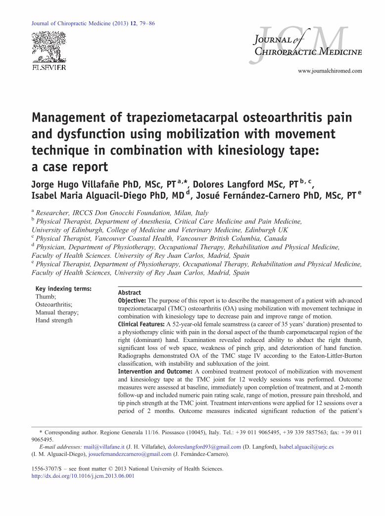
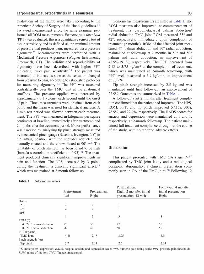
(dominant) hand. She presented with a 14-year historyof insidious and increasing pain, with progressive lackof grip and pinch strength in her right hand. A diagnosisof right hand stage IV TMC OA, with instability anddorsal subluxation of the joint (Eaton-Littler-Burtonclassification17), was confirmed by radiograph andphysical examination18,19 (Fig. 1).
Health history
At the time of assessment, the patient was not takingmedications and had no other reported medical con-ditions. Before initiation of treatment, the patient hadbeen experiencing a rapid progression of her right handsymptoms. She had consulted with her general medicalpractitioner 8 months previously, who confirmed thediagnosis of TMC OA. He prescribed a course of oralnonsteroidal anti-inflammatory medication in combi-nation with a short-arm thumb orthotic, immobilizingthe wrist in neutral and the painful thumb in abduction.In addition, the general medical practitioner hadadvised work modification, including stopping workfor 1 week, and a trial of 6 sessions of physical therapy(laser and ultrasonographic therapy). The patientreported no benefit from this medical or physical ther-apy management and had discontinued treatment6 weeks before presentation at our facility.
Pain was located along the dorsal aspect of the TMCjoint and was described as a “constant achy feeling,”
Fig. 1. Stage IV TMC OA of the right hand.
with occasional “sharp” pain with specific movements,particularly thumb abduction and flexion, and graded at7 of 10 on a numeric pain scale (NPS).
The patient’s subjective reports indicated increasedseverity of symptoms associated with her occupationaltasks, particularly cutting with scissors, such that shewas experiencing difficulty performing her occupa-tional tasks. Acetaminophen and rest provided littlerelief, whereas repetitive use (eg, gripping) aggravatedthe pain. She reported that pinching (between thethumb and her first finger), cutting with scissors, turn-ing door keys, writing, and holding cutlery utensils allaggravated her pain, with difficulty lifting even smallamounts of weight with the affected hand. Because ofher recent exacerbation of pain, she was no longer ableto maintain her occupational tasks as a seamstress.
Physical examination
The patient held the right first metacarpal in adduc-tion and flexion, with a zig-zag collapse deformity,commonly seen in advanced stages of TMC OA20,21:The right first dorsal interosseus muscle and thenarmusculature showed evidence of atrophy comparedwith the left hand. The first metacarpal base was pro-minent dorsally.
Neck, shoulder, and elbow range of motion (ROM)was within normal limits bilaterally.
Pretreatment goniometric evaluation of the righthand indicated reduced active and passive ROM ofthe TMC joint in both palmar and radial abduction ofthe right thumb, that is, 35° and 42°, respectively(compared with 57° and 58° on the asymptomatic leftthumb), with painful active and resisted opposition,adduction, and flexion.
The patient reported tenderness to palpation of theright TMC and scapholunate joints, with maximaltenderness located on the TMC joint line dorsally. Theresult of the right TMC axial compression test for OA(grind test) was positive. 22
The right (dominant) TMC joint was noted by aradiologist to have grade IV OA according to theEaton-Littler classification. 17 Asymptomatic TMC OAwas also noted in the same joint on the left (non-dominant) hand.
Results of laboratory blood tests were unremarkablefor metabolic, inflammatory, or infectious joint disease.Results of both Durkin and Tinel tests for carpal tunnelwere negative, 23 and result of Finkelstein test fortenosynovitis of the first dorsal compartment wasnegative bilaterally. 24 A confirmation of a diagnosis of
82 J. H. Villafañe et al.
TMC OA stage IV complicated by TMC joint laxitywas noted.
Treatment interventions
Before initiation of treatment, the patient was advisedabout the potential benefits of physiotherapy treatmentas well as its potential adverse effects; and writteninformed consent for treatment was obtained. Duringthe 12 intervention sessions over the course of 2months,the patient received MWM techniques to promote pain-free thumb mobility13; and Kinesiology tape was ap-plied immediately posttreatment and worn for 5 days toassist with joint repositioning posttreatment. 25
The primary treatment interventions are outlined indetail below.
Method of application of MWM13
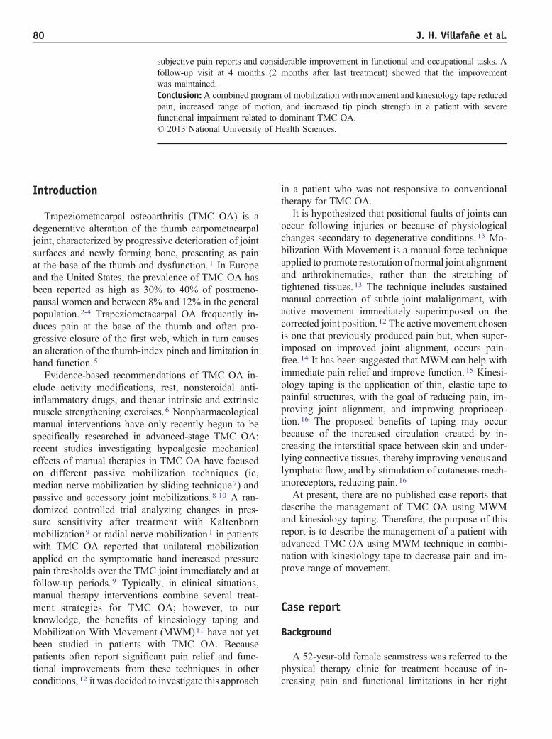
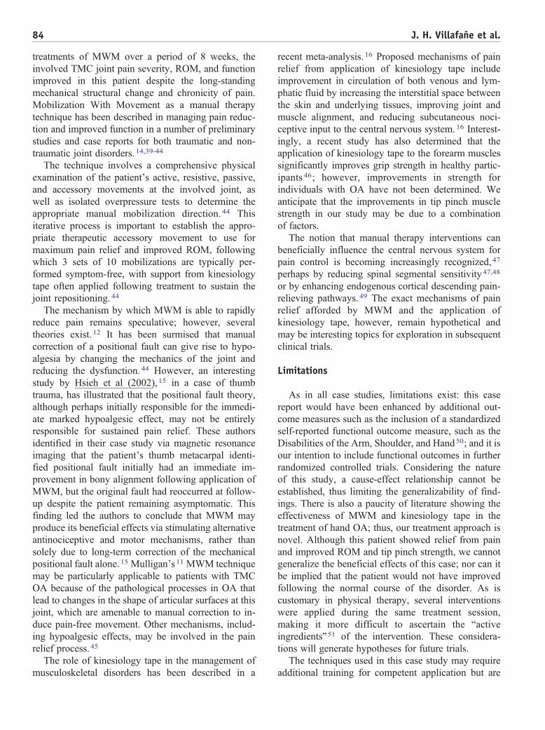
At initial assessment, active motions (TMC radialand palmar abduction and adduction) of the thumb werelimited by pain. To find the position that best allowedfor pain-free motion, an iterative process was used. Thetherapist had to experiment with and fine-tune thedirection and pressure of the imposed carpal glide usingfeedback from the patient. As the first metacarpal waspositioned toward neutral, reducing the subluxationusing manual gliding pressure, the patient’s ability tomove into previously painful ranges with pain-freeranges was noted. When the proper position, direction,and force of the thumb mobilization were established,the patient performed 3 sets of 10 repetitions of each ofthe previously painful thumb motions: TMC radial andpalmar abduction and adduction (Fig. 2).
Fig. 2. Mobilization with movement. Active thumbmotion performed with reduction of the TMC joint.
Kinesiology tape
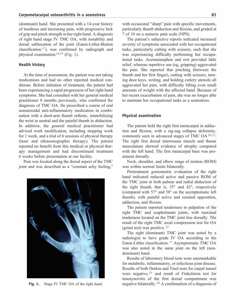
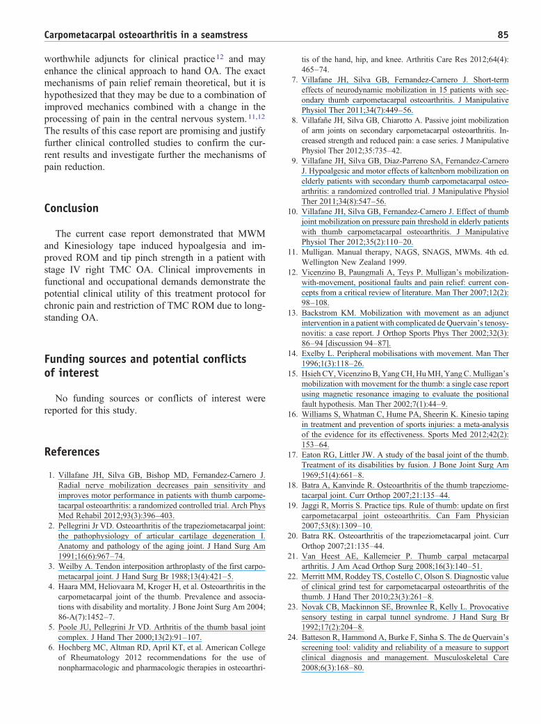
The Kinesiology tape was placed without tension fora relaxation effect 25 (Fig. 3). The goals of applicationof tape were also to facilitate proprioceptive feedbackto the involved joint, 26 to assist with circulation, and todiminish nociceptive input to the nervous system toprolong the effects of manual interventions. 16
Outcome measures
Outcome measures were performed at baseline, atthe conclusion of treatment, and again at 2-monthfollow-up.
The primary outcome measure was pain intensityof first TMC joint, which was assessed with the NPS.The NPS is a 10-cm line anchored with a numeric “0” atone end representing no pain and “10” at the other endrepresenting the worst pain imaginable, and sequentialnumbers in between.27 Pain was assessed as theparticipant performed tip pinch between the thumb andthe index finger. The NPS was selected as the primaryoutcome measure based on the ability to detectminimally important clinically important differences,noted to be 2.0 cm in this instrument.28 The HospitalAnxiety and Depression Scale (HADS) was used tocapture psychosocial adjustment. 29,30 Goniometric
Fig. 3. Kinesiology tape for TMC joint (tendons). Tape isplaced without tension for a relaxation effect from theinsertion (I) to the origin (O). Without (A) and with (B) acorrective strip over the snuff box and parallel to tendons.
83Carpometacarpal osteoarthritis in a seamstress
evaluations of the thumb were taken according to theAmerican Society of Surgery of the Hand guidelines.31
To avoid measurement error, the same examiner per-formed all ROMmeasurements.Pressure pain threshold(PPT) was evaluated: this is a quantitative sensory test oftissue sensitivity and is defined as the minimal amountof pressure that produces pain, measured via a pressurealgometer.32 Measurements were performed with aMechanical Pressure Algometer (Wagner Instruments,Greenwich, CT). The validity and reproducibility ofalgometry have been described, with higher PPTindicating lower pain sensitivity.33 The patient wasinstructed to indicate as soon as the sensation changedfrom pressure to pain, according to established protocolsfor measuring algometry.34 The PPT was measuredcontralaterally over the TMC joint at the anatomicalsnuffbox. The pressure applied was increased byapproximately 0.1 kg/cm2 each second until the onsetof pain. Three measurements were obtained from eachpoint, and the mean was used for statistical analysis. A1-min rest period was allowed between each measure-ment. The PPT was measured in kilograms per squarecentimeter at baseline, immediately after treatment, and2 months after the treatment period. Motor performancewas assessed by analyzing tip pinch strength measuredby mechanical pinch gauge (Baseline, Irvington, NY) inthe sitting position with the shoulder adducted andneutrally rotated and the elbow flexed at 90°.9,35 Thereliability of pinch strength has been found to be high(intraclass correlation coefficient = 0.93).36 The treat-ment produced clinically significant improvements inpain and function. The NPS decreased by 3 pointsduring the treatment, a clinically significant effect,37
which was maintained at 2-month follow-up.
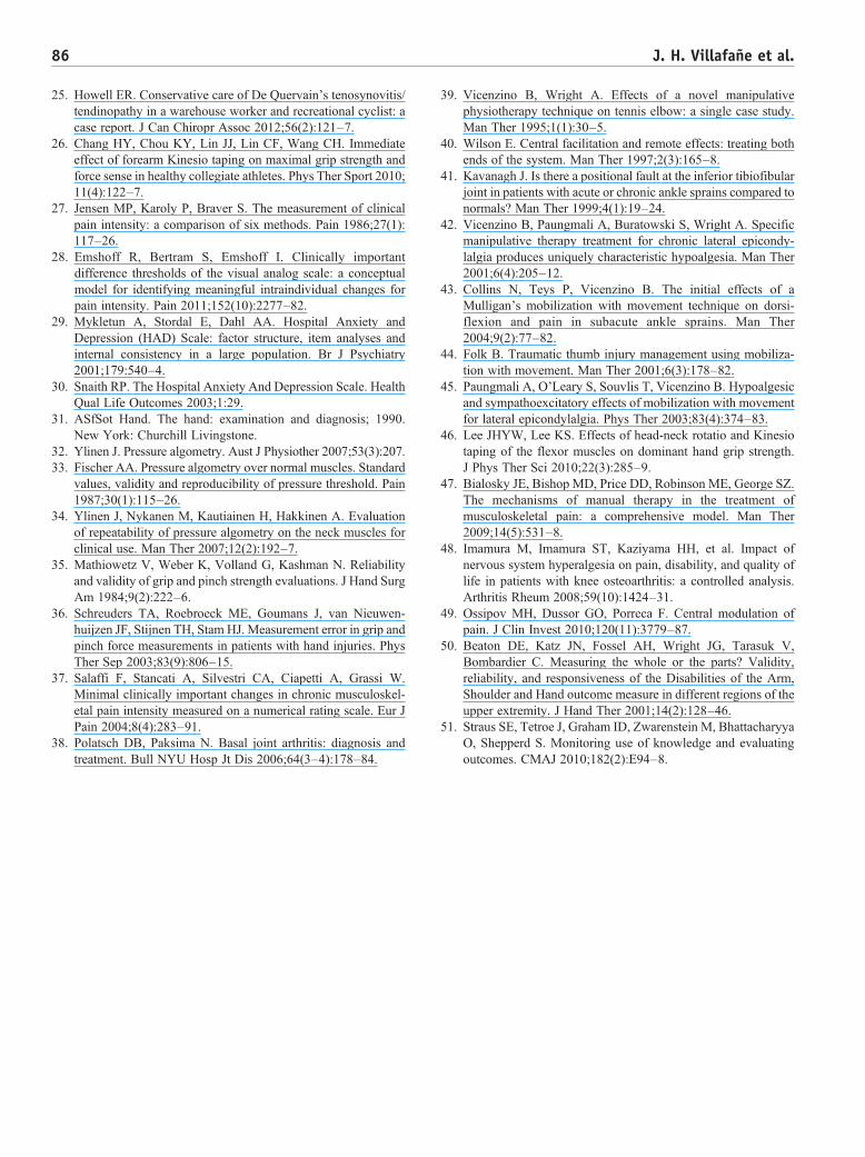
Table 1 Outcome measures
PretreatmentLeft
PretreatmentRight
HADSAS 2 2DS 2 2NPS
1 7ROM (°)1st TMC palmar abduction 57 351st TMC radial abduction 58 42PPT (kg/cm2)TMC joint 4.45 2.18Pinch strength (kg)Tip pinch 3.7 2.14
AS, anxiety; DS, depression; HADS, hospital anxiety and depression scaROM, range of motion; TMC, Trapeziometacarpal.
Goniometric measurements are listed in Table 1. TheROM measures also improved: at commencement oftreatment, first carpometacarpal palmar abduction/radial abduction TMC joint ROM measured 35° and42°, respectively. Immediately upon completion oftreatment (2 months), ROM of the affected joint mea-sured 47° palmar abduction and 50° radial abduction,maintained at follow-up at 2 months in 50° and 50°palmar and radial abduction, an improvement of42.9%/19.1%, respectively. The PPT increased from2.18 to 3.75 kg/cm2 at the completion of treatment,which was maintained at 2-month follow-up, withPPT levels measured at 3.9 kg/cm2, an improvementof 78.9%.
Tip pinch strength increased by 2.5 kg and wasmaintained until first follow-up, an improvement of22.9%. Outcomes are summarized in Table 1.
A follow-up visit 2 months after treatment comple-tion confirmed that the patient had improved. The NPS,ROM, PPT, and tip pinch improved 57.1%, 30%,78.9%, and 22.9%, respectively. The HADS scores foranxiety and depression were maintained at 1 and 1,respectively, at 2-month follow-up. The patient main-tained full treatment compliance throughout the courseof the study, with no reported adverse effects.
Discussion
This patient presented with TMC OA stage IV17
complicated by TMC joint laxity and a radiologicalpositional abnormality, a clinical presentation com-monly seen in OA of the TMC joint. 38 Following 12
PosttreatmentRight, 2 mo after initialpresentation, 12 visits
Follow-up, 4 mo afterinitial presentationRight
11
3 3
47 5050 50
3.75 3.9
2.5 2.63
le; NPS, numeric pain rating scale; PPT, pressure pain threshold;
84 J. H. Villafañe et al.
treatments of MWM over a period of 8 weeks, theinvolved TMC joint pain severity, ROM, and functionimproved in this patient despite the long-standingmechanical structural change and chronicity of pain.Mobilization With Movement as a manual therapytechnique has been described in managing pain reduc-tion and improved function in a number of preliminarystudies and case reports for both traumatic and non-traumatic joint disorders. 14,39-44
The technique involves a comprehensive physicalexamination of the patient’s active, resistive, passive,and accessory movements at the involved joint, aswell as isolated overpressure tests to determine theappropriate manual mobilization direction. 44 Thisiterative process is important to establish the appro-priate therapeutic accessory movement to use formaximum pain relief and improved ROM, followingwhich 3 sets of 10 mobilizations are typically per-formed symptom-free, with support from kinesiologytape often applied following treatment to sustain thejoint repositioning. 44
The mechanism by which MWM is able to rapidlyreduce pain remains speculative; however, severaltheories exist. 12 It has been surmised that manualcorrection of a positional fault can give rise to hypo-algesia by changing the mechanics of the joint andreducing the dysfunction. 44 However, an interestingstudy by Hsieh et al (2002), 15 in a case of thumbtrauma, has illustrated that the positional fault theory,although perhaps initially responsible for the immedi-ate marked hypoalgesic effect, may not be entirelyresponsible for sustained pain relief. These authorsidentified in their case study via magnetic resonanceimaging that the patient’s thumb metacarpal identi-fied positional fault initially had an immediate im-provement in bony alignment following application ofMWM, but the original fault had reoccurred at follow-up despite the patient remaining asymptomatic. Thisfinding led the authors to conclude that MWM mayproduce its beneficial effects via stimulating alternativeantinociceptive and motor mechanisms, rather thansolely due to long-term correction of the mechanicalpositional fault alone.15 Mulligan’s11 MWM techniquemay be particularly applicable to patients with TMCOA because of the pathological processes in OA thatlead to changes in the shape of articular surfaces at thisjoint, which are amenable to manual correction to in-duce pain-free movement. Other mechanisms, includ-ing hypoalgesic effects, may be involved in the painrelief process. 45
The role of kinesiology tape in the management ofmusculoskeletal disorders has been described in a
recent meta-analysis. 16 Proposed mechanisms of painrelief from application of kinesiology tape includeimprovement in circulation of both venous and lym-phatic fluid by increasing the interstitial space betweenthe skin and underlying tissues, improving joint andmuscle alignment, and reducing subcutaneous noci-ceptive input to the central nervous system.16 Interest-ingly, a recent study has also determined that theapplication of kinesiology tape to the forearm musclessignificantly improves grip strength in healthy partic-ipants 46; however, improvements in strength forindividuals with OA have not been determined. Weanticipate that the improvements in tip pinch musclestrength in our study may be due to a combinationof factors.
The notion that manual therapy interventions canbeneficially influence the central nervous system forpain control is becoming increasingly recognized,47
perhaps by reducing spinal segmental sensitivity47,48
or by enhancing endogenous cortical descending pain-relieving pathways. 49 The exact mechanisms of painrelief afforded by MWM and the application ofkinesiology tape, however, remain hypothetical andmay be interesting topics for exploration in subsequentclinical trials.
Limitations
As in all case studies, limitations exist: this casereport would have been enhanced by additional out-come measures such as the inclusion of a standardizedself-reported functional outcome measure, such as theDisabilities of the Arm, Shoulder, and Hand50; and it isour intention to include functional outcomes in furtherrandomized controlled trials. Considering the natureof this study, a cause-effect relationship cannot beestablished, thus limiting the generalizability of find-ings. There is also a paucity of literature showing theeffectiveness of MWM and kinesiology tape in thetreatment of hand OA; thus, our treatment approach isnovel. Although this patient showed relief from painand improved ROM and tip pinch strength, we cannotgeneralize the beneficial effects of this case; nor can itbe implied that the patient would not have improvedfollowing the normal course of the disorder. As iscustomary in physical therapy, several interventionswere applied during the same treatment session,making it more difficult to ascertain the “activeingredients”51 of the intervention. These considera-tions will generate hypotheses for future trials.
The techniques used in this case study may requireadditional training for competent application but are
85Carpometacarpal osteoarthritis in a seamstress
worthwhile adjuncts for clinical practice12 and mayenhance the clinical approach to hand OA. The exactmechanisms of pain relief remain theoretical, but it ishypothesized that they may be due to a combination ofimproved mechanics combined with a change in theprocessing of pain in the central nervous system.11,12
The results of this case report are promising and justifyfurther clinical controlled studies to confirm the cur-rent results and investigate further the mechanisms ofpain reduction.
Conclusion
The current case report demonstrated that MWMand Kinesiology tape induced hypoalgesia and im-proved ROM and tip pinch strength in a patient withstage IV right TMC OA. Clinical improvements infunctional and occupational demands demonstrate thepotential clinical utility of this treatment protocol forchronic pain and restriction of TMC ROM due to long-standing OA.
Funding sources and potential conflictsof interest
No funding sources or conflicts of interest werereported for this study.
References
1. Villafane JH, Silva GB, Bishop MD, Fernandez-Carnero J.Radial nerve mobilization decreases pain sensitivity andimproves motor performance in patients with thumb carpome-tacarpal osteoarthritis: a randomized controlled trial. Arch PhysMed Rehabil 2012;93(3):396–403.
2. Pellegrini Jr VD. Osteoarthritis of the trapeziometacarpal joint:the pathophysiology of articular cartilage degeneration I.Anatomy and pathology of the aging joint. J Hand Surg Am1991;16(6):967–74.
3. Weilby A. Tendon interposition arthroplasty of the first carpo-metacarpal joint. J Hand Surg Br 1988;13(4):421–5.
4. Haara MM, Heliovaara M, Kroger H, et al. Osteoarthritis in thecarpometacarpal joint of the thumb. Prevalence and associa-tions with disability and mortality. J Bone Joint Surg Am 2004;86-A(7):1452–7.
5. Poole JU, Pellegrini Jr VD. Arthritis of the thumb basal jointcomplex. J Hand Ther 2000;13(2):91–107.
6. Hochberg MC, Altman RD, April KT, et al. American Collegeof Rheumatology 2012 recommendations for the use ofnonpharmacologic and pharmacologic therapies in osteoarthri-
tis of the hand, hip, and knee. Arthritis Care Res 2012;64(4):465–74.
7. Villafane JH, Silva GB, Fernandez-Carnero J. Short-termeffects of neurodynamic mobilization in 15 patients with sec-ondary thumb carpometacarpal osteoarthritis. J ManipulativePhysiol Ther 2011;34(7):449–56.
8. Villafañe JH, Silva GB, Chiarotto A. Passive joint mobilizationof arm joints on secondary carpometacarpal osteoarthritis. In-creased strength and reduced pain: a case series. J ManipulativePhysiol Ther 2012;35:735–42.
9. Villafane JH, Silva GB, Diaz-Parreno SA, Fernandez-CarneroJ. Hypoalgesic and motor effects of kaltenborn mobilization onelderly patients with secondary thumb carpometacarpal osteo-arthritis: a randomized controlled trial. J Manipulative PhysiolTher 2011;34(8):547–56.
10. Villafane JH, Silva GB, Fernandez-Carnero J. Effect of thumbjoint mobilization on pressure pain threshold in elderly patientswith thumb carpometacarpal osteoarthritis. J ManipulativePhysiol Ther 2012;35(2):110–20.
11. Mulligan. Manual therapy, NAGS, SNAGS, MWMs. 4th ed.Wellington New Zealand 1999.
12. Vicenzino B, Paungmali A, Teys P. Mulligan's mobilization-with-movement, positional faults and pain relief: current con-cepts from a critical review of literature. Man Ther 2007;12(2):98–108.
13. Backstrom KM. Mobilization with movement as an adjunctintervention in a patient with complicated de Quervain's tenosy-novitis: a case report. J Orthop Sports Phys Ther 2002;32(3):86–94 [discussion 94–87].
14. Exelby L. Peripheral mobilisations with movement. Man Ther1996;1(3):118–26.
15. Hsieh CY, Vicenzino B, Yang CH, HuMH,Yang C.Mulligan'smobilization with movement for the thumb: a single case reportusing magnetic resonance imaging to evaluate the positionalfault hypothesis. Man Ther 2002;7(1):44–9.
16. Williams S, Whatman C, Hume PA, Sheerin K. Kinesio tapingin treatment and prevention of sports injuries: a meta-analysisof the evidence for its effectiveness. Sports Med 2012;42(2):153–64.
17. Eaton RG, Littler JW. A study of the basal joint of the thumb.Treatment of its disabilities by fusion. J Bone Joint Surg Am1969;51(4):661–8.
18. Batra A, Kanvinde R. Osteoarthritis of the thumb trapeziome-tacarpal joint. Curr Orthop 2007;21:135–44.
19. Jaggi R, Morris S. Practice tips. Rule of thumb: update on firstcarpometacarpal joint osteoarthritis. Can Fam Physician2007;53(8):1309–10.
20. Batra RK. Osteoarthritis of the trapeziometacarpal joint. CurrOrthop 2007;21:135–44.
21. Van Heest AE, Kallemeier P. Thumb carpal metacarpalarthritis. J Am Acad Orthop Surg 2008;16(3):140–51.
22. Merritt MM, Roddey TS, Costello C, Olson S. Diagnostic valueof clinical grind test for carpometacarpal osteoarthritis of thethumb. J Hand Ther 2010;23(3):261–8.
23. Novak CB, Mackinnon SE, Brownlee R, Kelly L. Provocativesensory testing in carpal tunnel syndrome. J Hand Surg Br1992;17(2):204–8.
24. Batteson R, Hammond A, Burke F, Sinha S. The de Quervain'sscreening tool: validity and reliability of a measure to supportclinical diagnosis and management. Musculoskeletal Care2008;6(3):168–80.
86 J. H. Villafañe et al.
25. Howell ER. Conservative care of De Quervain's tenosynovitis/tendinopathy in a warehouse worker and recreational cyclist: acase report. J Can Chiropr Assoc 2012;56(2):121–7.
26. Chang HY, Chou KY, Lin JJ, Lin CF, Wang CH. Immediateeffect of forearm Kinesio taping on maximal grip strength andforce sense in healthy collegiate athletes. Phys Ther Sport 2010;11(4):122–7.
27. Jensen MP, Karoly P, Braver S. The measurement of clinicalpain intensity: a comparison of six methods. Pain 1986;27(1):117–26.
28. Emshoff R, Bertram S, Emshoff I. Clinically importantdifference thresholds of the visual analog scale: a conceptualmodel for identifying meaningful intraindividual changes forpain intensity. Pain 2011;152(10):2277–82.
29. Mykletun A, Stordal E, Dahl AA. Hospital Anxiety andDepression (HAD) Scale: factor structure, item analyses andinternal consistency in a large population. Br J Psychiatry2001;179:540–4.
30. Snaith RP. The Hospital Anxiety And Depression Scale. HealthQual Life Outcomes 2003;1:29.
31. ASfSot Hand. The hand: examination and diagnosis; 1990.New York: Churchill Livingstone.
32. Ylinen J. Pressure algometry. Aust J Physiother 2007;53(3):207.33. Fischer AA. Pressure algometry over normal muscles. Standard
values, validity and reproducibility of pressure threshold. Pain1987;30(1):115–26.
34. Ylinen J, Nykanen M, Kautiainen H, Hakkinen A. Evaluationof repeatability of pressure algometry on the neck muscles forclinical use. Man Ther 2007;12(2):192–7.
35. Mathiowetz V, Weber K, Volland G, Kashman N. Reliabilityand validity of grip and pinch strength evaluations. J Hand SurgAm 1984;9(2):222–6.
36. Schreuders TA, Roebroeck ME, Goumans J, van Nieuwen-huijzen JF, Stijnen TH, Stam HJ. Measurement error in grip andpinch force measurements in patients with hand injuries. PhysTher Sep 2003;83(9):806–15.
37. Salaffi F, Stancati A, Silvestri CA, Ciapetti A, Grassi W.Minimal clinically important changes in chronic musculoskel-etal pain intensity measured on a numerical rating scale. Eur JPain 2004;8(4):283–91.
38. Polatsch DB, Paksima N. Basal joint arthritis: diagnosis andtreatment. Bull NYU Hosp Jt Dis 2006;64(3–4):178–84.
39. Vicenzino B, Wright A. Effects of a novel manipulativephysiotherapy technique on tennis elbow: a single case study.Man Ther 1995;1(1):30–5.
40. Wilson E. Central facilitation and remote effects: treating bothends of the system. Man Ther 1997;2(3):165–8.
41. Kavanagh J. Is there a positional fault at the inferior tibiofibularjoint in patients with acute or chronic ankle sprains compared tonormals? Man Ther 1999;4(1):19–24.
42. Vicenzino B, Paungmali A, Buratowski S, Wright A. Specificmanipulative therapy treatment for chronic lateral epicondy-lalgia produces uniquely characteristic hypoalgesia. Man Ther2001;6(4):205–12.
43. Collins N, Teys P, Vicenzino B. The initial effects of aMulligan's mobilization with movement technique on dorsi-flexion and pain in subacute ankle sprains. Man Ther2004;9(2):77–82.
44. Folk B. Traumatic thumb injury management using mobiliza-tion with movement. Man Ther 2001;6(3):178–82.
45. Paungmali A, O'Leary S, Souvlis T, Vicenzino B. Hypoalgesicand sympathoexcitatory effects of mobilization with movementfor lateral epicondylalgia. Phys Ther 2003;83(4):374–83.
46. Lee JHYW, Lee KS. Effects of head-neck rotatio and Kinesiotaping of the flexor muscles on dominant hand grip strength.J Phys Ther Sci 2010;22(3):285–9.
47. Bialosky JE, Bishop MD, Price DD, Robinson ME, George SZ.The mechanisms of manual therapy in the treatment ofmusculoskeletal pain: a comprehensive model. Man Ther2009;14(5):531–8.
48. Imamura M, Imamura ST, Kaziyama HH, et al. Impact ofnervous system hyperalgesia on pain, disability, and quality oflife in patients with knee osteoarthritis: a controlled analysis.Arthritis Rheum 2008;59(10):1424–31.
49. Ossipov MH, Dussor GO, Porreca F. Central modulation ofpain. J Clin Invest 2010;120(11):3779–87.
50. Beaton DE, Katz JN, Fossel AH, Wright JG, Tarasuk V,Bombardier C. Measuring the whole or the parts? Validity,reliability, and responsiveness of the Disabilities of the Arm,Shoulder and Hand outcome measure in different regions of theupper extremity. J Hand Ther 2001;14(2):128–46.
51. Straus SE, Tetroe J, Graham ID, Zwarenstein M, BhattacharyyaO, Shepperd S. Monitoring use of knowledge and evaluatingoutcomes. CMAJ 2010;182(2):E94–8.