making sense of eukaryotic dna replication origins
TRANSCRIPT
Making Sense of Eukaryotic DNA Replication Origins
David M. GilbertDepartment of Biochemistry and Molecular Biology, SUNY Upstate Medical University, 750 EastAdams Street, Syracuse, NY 13210, USA. E-mail:[email protected]
AbstractDNA replication is the process by which cells make one complete copy of their genetic informationbefore cell division. In bacteria, readily identifiable DNA sequences constitute the start sites or originsof DNA replication. In eukaryotes, replication origins have been difficult to identify. In some systems,any DNA sequence can promote replication, but other systems require specific DNA sequences.Despite these disparities, the proteins that regulate replication are highly conserved from yeast tohumans. The resolution may lie in a current model for once-per-cell-cycle regulation of eukaryoticreplication that does not require defined origin sequences. This model implies that the specificationof precise origins is a response to selective pressures that transcend those of once-per-cell-cyclereplication, such as the coordination of replication with other chromosomal functions. Viewed in thiscontext, the locations of origins may be an integral part of the functional organization of eukaryoticchromosomes.
Transmission of genetic information from one cell generation to the next requires the accurateand complete duplication of each DNA strand exactly once before each cell division. Typically,this process begins with the binding of an “initiator” protein to a specific DNA sequence or“replicator.” In response to the appropriate cellular signals, the initiator directs a localunwinding of the DNA double helix and recruits additional factors to initiate the process ofDNA replication. This paradigm describes most of the currently tractable replication systemsand, although derived from prokaryotic and viral systems, there is no compelling reason todoubt that it will apply to all eukaryotic organisms. In fact, the proteins that regulate replicationare highly conserved from yeast to humans, including the origin recognition complex (ORC),which binds directly to replication origin sequences in budding yeast (1, 2). However, in severaleukaryotic replication systems, it appears that any DNA sequence can function as a replicator.Those outside the field are often perplexed as to how investigators of different eukaryoticsystems can work with assumptions that range from very specific to completely random originsequence recognition, yet all agree on the basic mechanism regulating DNA replication. Thisreview summarizes our current understanding of eukaryotic replication origins and thenpresents some simple guidelines to help demystify these seemingly disparate observations,providing a framework for understanding eukaryotic origins that includes all existing data.
Conserved Initiator, Divergent ReplicatorThe structure of replication origins that have been subjected to genetic analysis in variouseukaryotic species is summarized in Fig. 1. In Saccharomyces cerevisiae, origins[autonomously replicating sequences, (ARS)] consist of an essential 11-base pair (bp) ARSconsensus sequence (ACS) and several additional elements that contribute to initiation activityand are interchangeable between origins but are not conserved in sequence. Several single–base pair mutations in the ACS can abolish initiation activity (3, 4). Saccharomycescerevisiae ORC binds in vivo to ARS elements throughout the cell cycle, and purified ORCbinds specifically to the ACS in an ATP-dependent manner (2, 5). High-resolution mappingof ARS1 has delimited the initiation point to a single nucleotide adjacent to the ORC bindingsite (6). In Schizosaccharomyces pombe, origins are much larger and consist of multipleelements that contribute partially to origin activity (2, 7). Although these elements do not sharea consensus sequence, they contain asymmetric AT stretches and can be replaced with artificial
NIH Public AccessAuthor ManuscriptScience. Author manuscript; available in PMC 2005 October 17.
Published in final edited form as:Science. 2001 October 5; 294(5540): 96–100.
NIH
-PA Author Manuscript
NIH
-PA Author Manuscript
NIH
-PA Author Manuscript

asymmetric AT stretches (e.g., A40) that reconstitute full origin activity (7). In vivo, S.pombe ORC associates with at least two separate fragments within the ARS 2004 (8).
In Drosophila, the elements required for amplification of the chorion genes have been analyzedextensively. Although the process of gene amplification involves re-replicating the same DNAsegment many times within one cell cycle, initiation requires many of the same proteins asnormal chromosomal replication does, including ORC (9). Amplification requires the 440-bpamplification control element (ACE), whereas the extent of amplification is stimulated by thepresence of several amplification-enhancing elements (AERs), of which only the most origin-proximal (AER-d) is shown in Fig. 1. ORC associates with both ACE and AER-d in vivo, andpurified Drosophila ORC exhibits preferential ATP-dependent binding to a fragmentcontaining AER-d and three separate DNA fragments within the ACE (10). It is intriguing thatreplication initiates almost exclusively within the dispensable AER-d (11–13). Furthermore,when multiple tandem copies of the ACE are integrated at ectopic sites, they recruit additionalORCs to chromatin sites in the flanking DNA that are not otherwise occupied by ORC (10).The tandem ACEs promote amplification at these ectopic sites, but the primary initiation sitesappear to reside in the flanking DNA (14, 15). Taken together, the ACE appears to containseveral ORC binding sites that promote the interaction of ORC and the initiation of replicationat specific adjacent sites.
Replication origins in multicellular organisms (metazoa) generally conform to one of twopatterns. At some loci, initiation sites are localized to within a few kilobases. At other loci,multiple dispersed origins can be identified throughout “initiation zones” of 10 to 50 kb.Preliminary genetic dissection has been carried out at two of these loci, one representative ofeach class (Fig. 1). At the human β-globin locus, replication initiates within a few kilobaseslocated between the adult δ- and β-globin genes. However, deletions of sequences greater than50 kb from the origin, as well as deletions within the initiation site itself, abolish the activityof this origin (16, 17). When the β-globin origin is transferred to an ectopic site, it can directsite-specific initiation of replication, and this activity is dependent on specific segments ofDNA within the 8-kb transferred fragment (18). Other loci where initiation sites appear to beconfined to within a few kilobases have been identified [e.g., (19, 20)]. In fact, at the lamin B2locus in human cells, a single nucleotide demarcates the major transition between leading andlagging DNA synthesis (20).
The Chinese hamster ovary (CHO) dihydrofolate reductase (DHFR) locus is representative ofthe second class of metazoan origins. The pattern of initiation sites at this locus has been thesubject of much controversy. On the one hand, high-resolution mapping of the locations ofsmall nascent DNA strands revealed only a single origin throughout a 6.1-kb fragment (21).On the other hand, two-dimensional gel electrophoresis (2D gel) analysis of replicationintermediates identified DNA structures representing replication bubbles throughout the entire55-kb intergenic zone between the DHFR gene and an adjacent gene (2BE2121) of unknownfunction (22). These results can be reconciled by considering the nature of the data obtainedwith each origin mapping technique. The 2D-gel method can search a larger area for initiationactivity but cannot accurately discern the number or precise location of initiation sites withina fragment. By contrast, small nascent strand detection analyzes a focused area in detail. Whenthe latter method was extended to cover an additional 6 kb of DNA, a second initiation sitewas revealed approximately 5 kb from the first (23). The 2D-gel method predicts that manymore such sites will eventually be identified, constituting an initiation zone. Similar broadinitiation zones have been identified at other metazoan loci (24–26).
Genetic analysis at the DHFR locus suggests the existence of specific elements that influenceorigin activity. When the most active origin is deleted (ori-β), adjacent replication origins retainor increase their activity (27). However, deletion of sequences near the 3′ end of the DHFR
Gilbert Page 2
Science. Author manuscript; available in PMC 2005 October 17.
NIH
-PA Author Manuscript
NIH
-PA Author Manuscript
NIH
-PA Author Manuscript

gene renders the entire locus inactive for early S phase–initiation activity. Like the β-globinorigin, DHFR ori-β retains initiation activity when moved to ectopic sites, and deletions ofspecific sequences within ori-β can influence this activity (28). Thus, origins found in bothbroad and localized initiation regions contain specific sequences favorable for initiating DNAreplication. However, the number and distribution of origins varies considerably at differentloci.
Perhaps the most enigmatic aspect of the field is that, in many eukaryotic systems, replicationseems to initiate within any DNA sequence. It appears that any cloned plasmid DNA willreplicate autonomously in Caenorhabditis elegans (29) and Paramecium (30). In culturedanimal cells, systematic searches for ARS elements analogous to those successfully carried outin yeast have generally failed to identify specific sequences that confer a significant replicationadvantage when reintroduced into cells (31). In one study with human cells, virtually everyDNA fragment greater than 15 kb promoted autonomous and once-per-cell-cycle replicationwith equal efficiency, and initiation sites were distributed throughout the plasmid sequences(32). Similar results were obtained in cultured Drosophila cells (33). In Xenopus andDrosophila embryos, any DNA sequence will efficiently replicate once per cell cycle up to theblastula stage of development whether microinjected into embryos or introduced into eggextracts (34). Likewise, replication of embryonic chromosomes appears to initiate within anyDNA sequence and does not become focused to specific sites until the midblastula transition,when transcription and differentiation commence (35, 36). Despite a random origin siteselection, in both Xenopus and Drosophila extracts, initiation of replication requires the ATP-dependent DNA binding activity of ORC (2, 37). How can such a precisely regulated processbe carried out without the requirement for specific start sites? The explanation is revealed inthe mechanism by which DNA replication is coordinated with the various phases of the cellcycle.
Regulated Replication Is Independent of SequenceIn all eukaryotic systems that have been amenable to study, replication is regulated by theassembly of a prereplication complex (pre-RC) of highly conserved proteins at ORC-boundDNA sites shortly after metaphase. After cells pass through the R point or START, a sharprise in the activities of S phase–promoting kinases (SPK: Cdc7/Dbf4 and B-type cyclin-Cdk)triggers the conversion of the pre-RC to an active replication complex. These high levels ofCdk activity (38), as well as a protein called geminin (1, 39, 40), persist from the onset of Sphase through metaphase and prevent the assembly of new pre-RCs. Both of these activitiesare destroyed by proteolysis during anaphase, allowing pre-RCs to reassemble. Hence,mutually exclusive periods of the cell cycle that promote either pre-RC formation or initiationensure that replication can only initiate once per cell cycle. This model (Fig. 2A) does notinvoke any requirement for specific origin sequences to accomplish accurate duplication of thegenome.
The origin spacing problem.Although initiation once per cell cycle does not require specific sequences, the positions oforigins cannot be distributed randomly, as this would run the risk that some origins might betoo far apart to complete replication of the intervening DNA within the length of a single Sphase (41). One way to solve this problem is to direct ORC to specific DNA sequences spacedat appropriate intervals. However, the origin spacing problem can also be solved without theneed for specific sequence recognition. Indeed, rapid replication in early Xenopus developmentis accomplished by initiating replication at sites that appear random with respect to sequencebut are regularly spaced every 9 to 12 kb (41). The mechanism that establishes this regularorigin spacing is unknown but, under conditions where chromatin is saturated with pre-RCs,
Gilbert Page 3
Science. Author manuscript; available in PMC 2005 October 17.
NIH
-PA Author Manuscript
NIH
-PA Author Manuscript
NIH
-PA Author Manuscript
any mechanism that prevents more than one pre-RC from assembling or firing per 10 kb wouldproduce the observed spacing.
More pre-RCs than needed.An alternative means to solve the origin spacing problem is to assemble more pre-RCs thanare necessary (Figs. 2B and 3), which appears to be the case in many eukaryotic systems. Inbudding yeast, many ARS elements function efficiently to promote the autonomous replicationof plasmid DNA but do not normally function as origins in the chromosome or are utilizedsignificantly less than once per cell cycle (42–44). However, pre-RCs are assembled on bothactive and silent origins (45). In Xenopus egg extracts, as the concentration of sperm nuclei isincreased, the number of active origins per nucleus decreases but the number of ORC- andMcm-DNA complexes assembled per nucleus remains constant (46). In mammalian cells, pre-RCs are assembled during telophase but pre-RC assembly is not sufficient to specify whichsites will function as replication origins (47). Together, these results suggest that the numberof pre-RCs assembled in eukaryotic cells exceeds the number of origins activated in each cellcycle; additional factors must select which of these pre-RCs will initiate replication.
Potentially, extraneous pre-RCs could effect the reduplication of portions of the genome if theywere to persist on daughter DNA strands after replication. However, evidence suggests thatpre-RCs are destroyed by the passage of replication forks from active origins (Fig. 2B). Silentor infrequently utilized budding yeast origins become activated when replication forks areprevented from passing through them (48, 49). In both budding and fission yeast, when two ormore origins are found in close proximity only one appears to be utilized within any given Sphase (50–54). The mechanism that determines the frequency with which a given pre-RC willbe activated is not known, however, chromosomal context and epigenetic elements clearly playa role. When identical ARS elements were placed in close proximity, one of them was utilizedmore frequently than the other, with the determinants of origin preference localized to flankingDNA sequences (52, 54). Mutations that disrupt silent chromatin at telomeres in budding yeastactivate a normally silent telomeric origin (55) and manipulations that alter the positioning ofnucleosomes on origins can directly influence the efficiency with which origins fire (56).Hence, each pre-RC has a potential to initiate replication that is influenced by a combinationof local chromatin structure and the probability that it will be activated before the passage ofa replication fork from adjacent origins.
Origin Choice Is Developmentally RegulatedThe gradual transition from random to specific origin site selection after the midblastulatransition (MBT) during Xenopus (36) and Drosophila (35) development provides a dramaticexample of origin choice during development. One possible explanation for this transition isthat the higher concentration of ORC in preblastula embryos could result in a more relaxedbinding of ORC to DNA, which would then become site-specific as the ORC:DNA ratiodecreases after the MBT. Both purified (10) and recombinant (57) Drosophila ORC bindpreferentially to specific DNA segments found near origins of gene amplification. In one case(57), the resolution was sufficient to conclude that replication initiates at the border of the ORCbinding site, as in budding yeast (6). So far, the only sequence motifs shared by these ORCbinding segments are short asymmetric AT stretches, reminiscent but not as prominent as thosefound in budding and fission yeast origins. Hence, it is presently difficult to predict the extentto which the affinity of ORC for specific DNA sequences, at any ORC:DNA ratio, can accountfor the specificity of initiation found in metazoan chromosomes. Changes in chromosomearchitecture that take place at the MBT, including chromatin condensation, the appearance ofhistone H1, cellular differentiation, and the gradual onset of transcription could also play a rolein focusing initiation to specific sites (36, 58). Differences in origin specificity have also beenobserved between cell lines from different tissues. In murine non–B cells, the entire IgH locus
Gilbert Page 4
Science. Author manuscript; available in PMC 2005 October 17.
NIH
-PA Author Manuscript
NIH
-PA Author Manuscript
NIH
-PA Author Manuscript

is replicated from a single replication fork that proceeds gradually through the locus from anorigin located downstream of the constant region genes (59). However, in pre–B cells, theentire locus is duplicated during the first hour of S phase, indicating that one or more additionalorigins are activated.
There are several means by which origin specification could be influenced or even regulatedduring development. Changes in chromatin structure that accompany key stages ofdevelopment could influence both the sequences to which ORC will preferentially bind andthe efficiency with which pre-RCs are activated. The onset of differentiation could also resultin the expression of accessory factors that interact with ORC and target it to specific sites. Forexample, the ORC4 subunit in S. pombe contains an NH2-terminal extension with nine copiesof the HMG-I(Y) related AT-hook motif, which is known to mediate binding to the minorgroove of AT tracts (60). In metazoa, interaction with accessory factors such as HMG proteinscould focus ORC to specific DNA sequences as, for example, HMG proteins increase the site-specific binding of steroid receptors to their cognate sites (61). In fact, human ORC subunitscoimmunoprecipitate with many unidentified proteins (62) and are targeted to the Epstein-Barrvirus (EBV) replication origin, apparently through an interaction with the viral origin-bindingprotein EBNA (63–65). Furthermore, extracts from differentiated Drosophila tissue culturecells contain activities that appear to increase the selectivity of ORC for specific DNAfragments (66). Assuming that there is a selective advantage to regulating the positions oforigins during development, utilizing ancillary factors to regulate the specificity of ORC couldhave circumvented the need to evolve different initiators for different tissues.
Chromosomal Traffic ControlThe fact that origins are localized in most species suggests that there is some selective pressureto initiate replication at particular sites. However, since once-per-cell-cycle replication per sedoes not require specific DNA sequences, the selective pressure must derive fromconsiderations other than genome duplication. One potential source for this selective pressureis the need to coordinate transcription with replication (67). Indeed, transcription inhibits theautonomous replication of plasmids in human cells (68). There are at least two ways in whichtranscription and replication could be mutually antagonistic. First, head-on collisions of thereplication and transcription machinery would create the need for both apparatuses to sharethe same DNA template temporarily. Bacterial genes are heavily biased toward an orientationthat places the polarity of transcription and replication in the same direction (69, 70). In bothbudding yeast and humans, a physical barrier in the 3′ region of the ribosomal genes preventsreplication forks from traveling in a direction that opposes RNA polymerase (25, 71, 72). Also,replication forks stall when they oppose the direction of yeast tRNA transcription but not whenthe tRNA genes are defective in transcription (73). However, there is some evidence inmammalian cells to suggest that transcription and replication do not take place simultaneouslyon the same DNA segments [reviewed in (74)], suggesting that polymerase collisions may notgenerally occur in mammalian cells.
A second antagonistic effect of replication and transcription could take place before replication,if pre-RCs are disrupted by passage of the transcription machinery. Although direct evidencefor this mechanism is lacking, and exceptional cases exist (origins within transcription unitsand localized origins within transcriptionally silent regions), the explanatory power isintriguing (Fig. 3). In Xenopus embryos, there is no transcriptional activity before the MBT,so there is no selective pressure to place origins at particular locations. In contrast, the vastmajority of the budding yeast genome is transcribed, and the locations of replication originsare almost exclusively restricted to intergenic regions (67). In this context, there would be astrong selective advantage for evolving specific DNA sequences that focus initiation tointergenic sites to ensure proper origin spacing (Fig. 3). This logic can also be applied to
Gilbert Page 5
Science. Author manuscript; available in PMC 2005 October 17.
NIH
-PA Author Manuscript
NIH
-PA Author Manuscript
NIH
-PA Author Manuscript

understand the different patterns of initiation at individual metazoan loci. Most solitary originsites have been identified within loci containing multiple genes (18–20). By contrast, broadinitiation zones consisting of multiple inefficient origins are observed at loci where there arelarge intergenic regions (22, 24, 25).
At present, it is difficult to predict the significance of transcription to origin localization.Clearly, we need to determine the sites where pre-RCs assemble during telophase and whetherthe onset of transcription after mitosis does, indeed, displace pre-RCs from transcription units.New applications for microarray analysis that reveal the genome-wide locations of DNA boundproteins have already been developed in budding yeast (75), and the answers to some of thesequestions in this simple model system should be forthcoming. In cases where replication doesinitiate within transcription units, it will be important to determine whether transcription isactivated after replication. Finally, the coordination of transcription with replication providesa useful working model but other roles for origin placement should be considered. For example,ORC appears to play a central role in the assembly of heterochromatin (76) and chromosomecondensation during mitosis (77, 78). The location of replication origins may be important toorganize chromosomes for sister chromatid cohesion and/or chromosome condensation.
What is clear is that the need to duplicate the genome once per cell cycle does not itself imposeselective pressure to initiate replication at specific sites. In fact, it could be argued that a moredegenerate origin recognition system would favor rapid genome evolution, allowing moreflexible rearrangement of sequences without risking large gaps of DNA without an origin, andallowing for the modulation of origin sites during development. With this revelation, thedifficulties in identifying consensus origin sequences in metazoa should come as no surprise.The more pressing question becomes why origins are localized at all in most eukaryoticsystems. The answer to this question may be different for different loci. Although this indeedcomplicates the analysis of eukaryotic origins, it should by no means discourage investigatorsfrom entertaining creative hypotheses and pursuing directions that will likely reveal newinsights into the organization of complex genomes.
Acknowledgements
I would like to thank K. Friedman, S. Fiering, M. Aladjem, J. Blow, M. Schmitt, M. Botchan, and S. Gerbi for helpfulcriticisms of this essay and H. Masukata, S. Gerbi, and M. Botchan for sharing unpublished information. Work in mylaboratory is supported by NIH grant GM-57233-01, NSF grant MCB-0077507, and American Cancer Society grantRPG-97-098-04-CCG.
References1. Diffley JF. Curr Biol 2001;11:R367. [PubMed: 11369250]2. Kelly TJ, Brown GW. Annu Rev Biochem 2000;69:829. [PubMed: 10966477]3. Marahrens Y, Stillman B. Science 1992;255:817. [PubMed: 1536007]4. Van Houten J, Newlon CS. Mol Cell Biol 1990;10:3917. [PubMed: 2196439]5. Klemm RD, Bell SP. Proc Natl Acad Sci USA 2001;98:8361. [PubMed: 11459976]6. Bielinsky AK, Gerbi SA. Mol Cell 1999;3:477. [PubMed: 10230400]7. Okuno Y, Satoh H, Sekiguchi M, Masukata H. Mol Cell Biol 1999;19:6699. [PubMed: 10490609]8. Ogawa Y, Takahashi T, Masukata H. Mol Cell Biol 1999;19:7228. [PubMed: 10490657]9. Spradling AC. Genes Dev 1999;13:2619. [PubMed: 10541547]10. Austin RJ, Orr-Weaver TL, Bell SP. Genes Dev 1999;13:2639. [PubMed: 10541550]11. Delidakis C, Kafatos FC. EMBO J 1989;8:891. [PubMed: 2542026]12. Heck MM, Spradling AC. J Cell Biol 1990;110:903. [PubMed: 2157721]13. Lu L, Zhang H, Tower J. Genes Dev 2001;15:134. [PubMed: 11157771]14. de Cicco D, Spradling AC. Cell 1984;38:45. [PubMed: 6088075]
Gilbert Page 6
Science. Author manuscript; available in PMC 2005 October 17.
NIH
-PA Author Manuscript
NIH
-PA Author Manuscript
NIH
-PA Author Manuscript

15. Carminati JL, Johnston CG, Orr-Weaver TL. Mol Cell Biol 1992;12:2444. [PubMed: 1314956]16. Cimbora DM, et al. Mol Cell Biol 2000;20:5581. [PubMed: 10891496]17. Aladjem M, et al. Science 1995;270:815. [PubMed: 7481774]18. Aladjem MI, Rodewald LW, Kolman JL, Wahl GM. Science 1998;281:1005. [PubMed: 9703500]19. Toledo F, et al. Nucleic Acids Res 1998;26:2313. [PubMed: 9580680]20. Abdurashidova G, et al. Science 2000;287:2023. [PubMed: 10720330]21. Pelizon C, Diviacco S, Falaschi A, Giacca M. Mol Cell Biol 1996;16:5358. [PubMed: 8816447]22. Dijkwel PA, Hamlin JL. Mol Cell Biol 1995;15:3023. [PubMed: 7760799]23. Kobayashi T, Rein T, DePamphilis M. Mol Cell Biol 1998;18:3266. [PubMed: 9584167]24. Ina S, Sasaki T, Yokota Y, Shinomiya T. Chromosoma 2001;109:551. [PubMed: 11305788]25. Little RD, Platt TH, Schildkraut CL. Mol Cell Biol 1993;13:6600. [PubMed: 8413256]26. Dijkwel PA, Mesner LD, Levenson VV, d’Anna J, Hamlin JL. Exp Cell Res 2000;256:150. [PubMed:
10739662]27. Kalejta RF, et al. Mol Cell 1998;2:797. [PubMed: 9885567]28. Altman AL, Fanning E. Mol Cell Biol 2001;21:1098. [PubMed: 11158297]29. Mello CC, Kramer JM, Stinchcomb D, Ambros V. EMBO J 1991;10:3959. [PubMed: 1935914]30. Kim CS, Preer JR, Polisky B. Dev Genet 1992;13:97. [PubMed: 1386793]31. Masukata H, Satoh H, Obuse C, Okazaki T. Mol Biol Cell 1993;4:1121. [PubMed: 8305734]32. Krysan PJ, Smith JG, Calos MP. Mol Cell Biol 1993;13:2688. [PubMed: 8386315]33. Smith JG, Calos MP. Chromosoma 1995;103:597. [PubMed: 7587582]34. Coverley D, Laskey RA. Annu Rev Biochem 1994;63:745. [PubMed: 7979254]35. Sasaki T, Sawado T, Yamaguchi M, Shinomiya T. Mol Cell Biol 1999;19:547. [PubMed: 9858578]36. Hyrien O, Maric C, Mechali M. Science 1995;270:994. [PubMed: 7481806]37. I. Chesnokov, M. R. Botchan, Proc. Natl. Acad. Sci. U.S.A., in press.38. Noton E, Diffley JF. Mol Cell 2000;5:85. [PubMed: 10678171]39. Wohlschlegel JA, et al. Science 2000;290:2309. [PubMed: 11125146]40. Tada S, Li A, Maiorano D, Mechali M, Blow JJ. Nature Cell Biol 2001;3:107. [PubMed: 11175741]41. Blow JJ, Gillespie PJ, Francis D, Jackson DA. J Cell Biol 2001;152:15. [PubMed: 11149917]42. Yamashita M, et al. Genes Cells 1997;2:655. [PubMed: 9491800]43. Friedman KL, Brewer BJ, Fangman WL. Genes Cells 1997;2:667. [PubMed: 9491801]44. A. Poloumienkol, A. Dershowitz, D. Jitakshi, C. S. Newlon, Mol. Biol. Cell, in press.45. Santocanale C, Diffley J. EMBO J 1996;15:6671. [PubMed: 8978693]46. Walter J, Newport J. Science 1997;275:993. [PubMed: 9020085]47. Okuno Y, McNairn AJ, den Elzen N, Pines J, Gilbert DM. EMBO J 2001;20:4263. [PubMed:
11483529]48. Wang Y, Vujcic M, Kowalski D. Mol Cell Biol 2001;21:4938. [PubMed: 11438651]49. Santocanale C, Sharma K, Diffley JF. Genes Dev 1999;13:2360. [PubMed: 10500092]50. Wohlgemuth JG, Bulboaca GH, Moghadam M, Caddle MS, Calos MP. Mol Biol Cell 1994;5:839.
[PubMed: 7803852]51. Dubey D, Zhu J, Carlson D, Sharma K, Huberman J. EMBO J 1994;13:3638. [PubMed: 8062838]52. Brewer BJ, Fangman WL. Proc Natl Acad Sci USA 1994;91:3418. [PubMed: 8159762]53. Brewer BJ, Fangman WL. Science 1993;262:1728. [PubMed: 8259517]54. Marahrens Y, Stillman B. EMBO J 1994;13:3395. [PubMed: 8045266]55. Stevenson JB, Gottschling DE. Genes Dev 1999;13:146. [PubMed: 9925638]56. Lipford JR, Bell SP. Mol Cell 2001;7:21. [PubMed: 11172708]57. A.-K. Bielinsky et al., Curr. Biol., in press.58. Gilbert DM, Miyazawa H, DePamphilis ML. Mol Cell Biol 1995;15:2942. [PubMed: 7760792]59. Ermakova OV, et al. Mol Cell 1999;3:321. [PubMed: 10198634]60. Chuang RY, Kelly TJ. Proc Natl Acad Sci USA 1999;96:2656. [PubMed: 10077566]
Gilbert Page 7
Science. Author manuscript; available in PMC 2005 October 17.
NIH
-PA Author Manuscript
NIH
-PA Author Manuscript
NIH
-PA Author Manuscript

61. Boonyaratanakornkit V, et al. Mol Cell Biol 1998;18:4471. [PubMed: 9671457]62. Dhar SK, Dutta A. J Biol Chem 2000;275:34983. [PubMed: 10945994]63. Chaudhuri B, Xu H, Todorov I, Dutta A, Yates JL. Proc Natl Acad Sci USA 2001;98:10085. [PubMed:
11517328]64. Dhar SK, et al. Cell 2001;106:287. [PubMed: 11509178]65. Schepers A, et al. EMBO J 2001;20:4588. [PubMed: 11500385]66. E. L. Beall, M. R. Botchan, personal communication.67. Brewer BJ. Curr Opin Genet Dev 1994;4:196. [PubMed: 8032196]68. Haase SB, Heinzel SS, Calos MP. Mol Cell Biol 1994;14:2516. [PubMed: 8139554]69. Brewer BJ. Cell 1988;53:679. [PubMed: 3286014]70. Tillier ER, Collins RA. Nature Genet 2000;26:195. [PubMed: 11017076]71. Brewer BJ, Lockshon D, Fangman WL. Cell 1992;71:267. [PubMed: 1423594]72. Gerber JK, et al. Cell 1997;90:559. [PubMed: 9267035]73. Deshpande AM, Newlon CS. Science 1996;272:1030. [PubMed: 8638128]74. Cook P. Science 1998;281:1466. [PubMed: 9750117]75. Ren B, et al. Science 2000;290:2306. [PubMed: 11125145]76. Pak D, et al. Cell 1997;91:311. [PubMed: 9363940]77. Loupart M, Krause S, Heck MS. Curr Biol 2000;10:1547. [PubMed: 11137005]78. Pflumm MF, Botchan MR. Development 2001;128:1697. [PubMed: 11290306]
Gilbert Page 8
Science. Author manuscript; available in PMC 2005 October 17.
NIH
-PA Author Manuscript
NIH
-PA Author Manuscript
NIH
-PA Author Manuscript
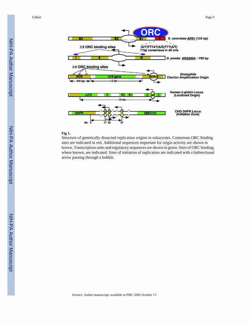
Fig 1.Structure of genetically dissected replication origins in eukaryotes. Consensus ORC bindingsites are indicated in red. Additional sequences important for origin activity are shown inbrown. Transcription units and regulatory sequences are shown in green. Sites of ORC binding,where known, are indicated. Sites of initiation of replication are indicated with a bidirectionalarrow passing through a bubble.
Gilbert Page 9
Science. Author manuscript; available in PMC 2005 October 17.
NIH
-PA Author Manuscript
NIH
-PA Author Manuscript
NIH
-PA Author Manuscript
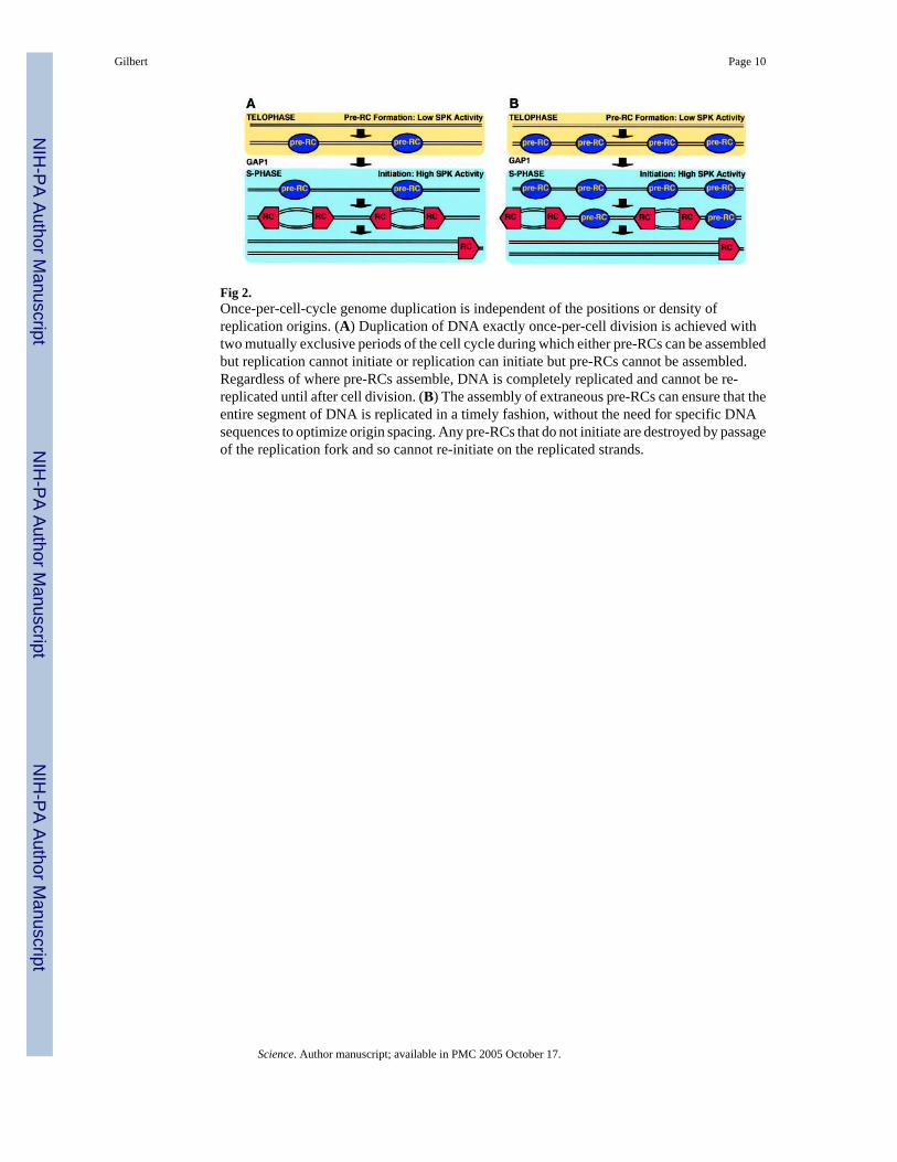
Fig 2.Once-per-cell-cycle genome duplication is independent of the positions or density ofreplication origins. (A) Duplication of DNA exactly once-per-cell division is achieved withtwo mutually exclusive periods of the cell cycle during which either pre-RCs can be assembledbut replication cannot initiate or replication can initiate but pre-RCs cannot be assembled.Regardless of where pre-RCs assemble, DNA is completely replicated and cannot be re-replicated until after cell division. (B) The assembly of extraneous pre-RCs can ensure that theentire segment of DNA is replicated in a timely fashion, without the need for specific DNAsequences to optimize origin spacing. Any pre-RCs that do not initiate are destroyed by passageof the replication fork and so cannot re-initiate on the replicated strands.
Gilbert Page 10
Science. Author manuscript; available in PMC 2005 October 17.
NIH
-PA Author Manuscript
NIH
-PA Author Manuscript
NIH
-PA Author Manuscript

Fig 3.Exclusion of pre-RCs from specific regions could create the need for origin focusingmechanisms. In this model, passage of the transcription apparatus before replication depletestranscription units (black boxes) of pre-RCs (yellow stars). (A) Without transcription, there isno selective pressure to focus initiation to specific sites as the assembly of pre-RCs at manysites ensures timely genome replication. (B) When transcription units are sparse, the assemblyof multiple pre-RCs at many sites within the intergenic regions is sufficient to accomplishgenome replication. (C) When intergenic regions are few and far between, specific DNAsequence recognition elements are required to ensure that at least one pre-RC is assembled atintervals appropriate to accomplish the timely replication of the DNA segment.
Gilbert Page 11
Science. Author manuscript; available in PMC 2005 October 17.
NIH
-PA Author Manuscript
NIH
-PA Author Manuscript
NIH
-PA Author Manuscript