macrophage identification in situ - mdpi
TRANSCRIPT
Biomedicines 2021, 9, 1393. https://doi.org/10.3390/biomedicines9101393 www.mdpi.com/journal/biomedicines
Review
Macrophage Identification In Situ Krisztina Nikovics * and Anne-Laure Favier
Imagery Unit, Department of Platforms and Technology Research, French Armed Forces Biomedical Research Institute, 91223 Brétigny-sur-Orge, France; [email protected] * Correspondence: Correspondence: [email protected]
Abstract: Understanding the processes of inflammation and tissue regeneration after injury is of great importance. For a long time, macrophages have been known to play a central role during dif-ferent stages of inflammation and tissue regeneration. However, the molecular and cellular mecha-nisms by which they exert their effects are as yet mostly unknown. While in vitro macrophages have been characterized, recent progress in macrophage biology studies revealed that macrophages in vivo exhibited distinctive features. Actually, the precise characterization of the macrophages in vivo is essential to develop new healing treatments and can be approached via in situ analyses. Nowa-days, the characterization of macrophages in situ has improved significantly using antigen surface markers and cytokine secretion identification resulting in specific patterns. This review aims for a comprehensive overview of different tools used for in situ macrophage identification, reporter genes, immunolabeling and in situ hybridization, discussing their advantages and limitations.
Keywords: macrophages; phenotype; in situ hybridization; cytokines; immunolabeling
1. Introduction In the late of 19th century, Elie Metcnikoff described macrophages for the first time
and hypothesized that these mononucleated phagocytic cells might play an important role in the immune response [1]. Later, it was discovered that macrophages have indis-pensable function during innate and adaptive immunity. Tissue injury promotes a series of well-coordinated events, which contribute to the efficient healing of damaged tissue. Reduction in inflammation and improvement in tissue regeneration is very important. Macrophages perform an essential function in both processes [2]. In the absence of mac-rophages, poor tissue healing, imperfect angiogenesis and fibrosis appears [3], resulting in an inhomogeneous population of cells detectable in every tissue of the body depending on the stimulus and the environment in which they are located [4–9]. The polarization of macrophages is determined by many biological signals (growth factors, fatty acids, pros-taglandins, and pathogen-derived molecules) in addition to cytokines located in the mi-cro-environment. Macrophages have critical biological roles from inflammation through to resolution and repair. They express and produce various molecules [10] which have different roles during different processes. The view on the characterization of macrophage polarization is rapidly changing [2,11,12]. In the beginning, in vitro culture was used for macrophage phenotype characterizations because, in this situation, the extracellular en-vironment can be easily controlled. In the 1990s, it was discovered that lipopolysaccharide (LPS), interferon (IFN)-gamma and cytokine IL-4 can induce the expression of different genes in macrophages. Two major groups were distinguished during polarization. Mac-rophages activated by LPS were named as “classically activated macrophages” and the IL-4 stimulated macrophages were named as “alternatively activated macrophages” [11,13,14]. One hundred years ago, Otto F. Warburg discovered that activation of macro-phages modified their glucose metabolism and increased lactate production [15,16]. Aside from glucose metabolism, amino acid metabolism can also be altered in the classically
Citation: Nikovics, K.; Favier, A.-L.
Macrophage Identification In Situ.
Biomedicines 2021, 9, 1393.
https://doi.org/10.3390/
biomedicines9101393
Academic Editors: Alexander N.
Orekhov, Alexei Gratchev and
Evgeny E. Bezsonov
Received: 1 September 2021
Accepted: 29 September 2021
Published: 4 October 2021
Publisher’s Note: MDPI stays neu-
tral with regard to jurisdictional
claims in published maps and insti-
tutional affiliations.
Copyright: © 2021 by the authors. Li-
censee MDPI, Basel, Switzerland.
This article is an open access article
distributed under the terms and con-
ditions of the Creative Commons At-
tribution (CC BY) license (http://crea-
tivecommons.org/licenses/by/4.0/).
Biomedicines 2021, 9, 1393 2 of 17
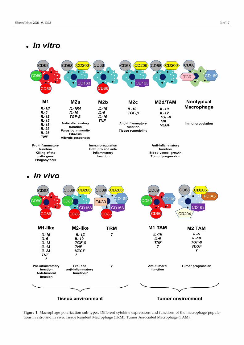
activated macrophages [17]. LPS/IFN–gamma stimulation induces arginine conversion to nitric oxide (NO). This molecule has a cytotoxic and bacteriostatic effect [18]. However, the IL-4 inhibits NO production in the alternatively activated macrophages [19]. In addi-tion to these metabolic pathways, alteration of the tricarboxylic acid cycle was also ob-served during alternative macrophage activation. High levels of succinate triggers intense inflammation in the classically activated macrophages [17]. Mills and colleagues (2000) proposed a new classification of the macrophages (M1 and M2), and the scientific com-munity decided to name the classically activated (pro-inflammatory) macrophages M1 and the alternatively activated (anti-inflammatory) macrophages M2 [20]. In vitro results showed evidence of new M2 macrophage sub-types (M2a, M2b, M2c with different func-tions and expressing different molecules (Figure 1)) [21,22].
The M1/M2 classification is not perfect because particular macrophages do not fit into any of the sub-types. These are the nontypical macrophages. Some produce T cell receptor (TCR) or CD169 protein and they do not promote phagocytosis but are involved in immune regulations [23] (Figure 1).
In 1988, the growth arrest-specific gene 6 was discovered [24]. This protein binds to the TAM (Tyro3, Axl and Mer) receptor family and plays an important role in the devel-opment of different cancers [25]. Qian and Pollard (2010) have suggested that the distri-bution of M1 and M2 macrophages is more complex, as tumor-associated macrophages (TAM macrophages) showed a different pattern compared to M1/M2 criteria [26]. Finally, TAM macrophages were classified as a new sub-type, the M2d macrophages [27]. Later it turned out that the TAM macrophage phenotype is even more complex, finally splitting into M1 and M2 TAM macrophages. M1 TAM macrophages are involved in anti-tumor-ous activity while the M2 TAM macrophages induce the tumor progression and metasta-sis formation [28–31] (Figure 1).
In that way, the in vivo landscape is more intricate compared to the in vitro situation [32–37]. The in vivo macrophages are generally called M1-like and M2-like or resolving macrophages [3]. Due to purification cell process, various immune cells are absent from in vitro environment studies, resulting in a lack of cytokine expression which is usually involved in macrophage polarization in vivo. In this context, the characterization of in vivo macrophages is still incomplete. Thus, many laboratories are interested in the iden-tification and characterization of M1-like, M2-like and tissue-resident macrophages. An increasing number of articles have pointed out that contrary to the original theory, in vivo macrophage phenotypes offered a more complex pattern of cellular markers and cytokine expression panel [3,38] (Figure 1). Various publications suggested that in vivo M2-like macrophages exhibited mixed pro- and anti-inflammatory functions [9,21,35,39–42].
Similarly, the precise function by which tissue-resident macrophages contribute to tissue regeneration is currently not fully understood, but they also play an important role in the process of wound healing (Figure 1) [3,43–45]. They contribute to maintaining ho-meostasis by constantly monitoring the internal and external signals within the body. Af-ter injury, the distinct secreted signals help to restore homeostasis. In the adult mouse, the tissue-resident macrophage populations originate equally from embryonic precursors and from bone marrow monocytes [46]. Orecchioni and colleagues (2019) have suggested, however, that in mice a majority of tissue macrophages are not monocyte-derived and mature tissue macrophages derived from embryonic precursors. This point explains why certain marker proteins used to characterize macrophages in rodent cells are not appro-priate for human macrophage identification and vice versa [34].
Biomedicines 2021, 9, 1393 3 of 17
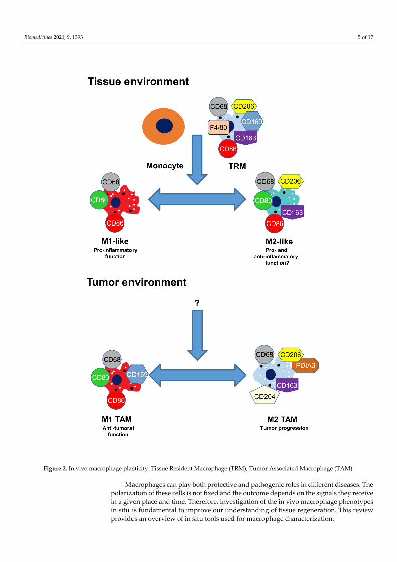
Figure 1. Macrophage polarization sub-types. Different cytokine expressions and functions of the macrophage popula-tions in vitro and in vivo. Tissue Resident Macrophage (TRM), Tumor Associated Macrophage (TAM).
Biomedicines 2021, 9, 1393 4 of 17
The in vitro and in vivo macrophages produce a wide range of secretory molecules (proteinases, chemokines, pro- and anti-inflammatory cytokines, growth factors and me-tabolites derived from oxygen, nitrogen, arachidonates) [11]. Specific macrophage pheno-types express different cytokines (Figure 1). In vitro M1 macrophages express pro-inflam-matory cytokines, such as tumor necrosis factor (TNF), interleukin-1 beta (IL-1β), inter-leukin-6 (IL-6), interleukin-12 (IL-12), interleukin-15 (IL-15), interleukin-18 (IL-18), inter-leukin 23 (IL-23) and interleukin 28 (IL-28). The M2a phenotypes produce anti-inflamma-tory cytokines, such as interleukin-10 (IL-10), interleukin-1 beta-receptor antagonist (IL-1RA) and transforming growth factor beta (TGF-β). Surprisingly, the M2b phenotypes ex-press pro-inflammatory cytokines (TNF, IL-1β and IL-6) and anti-inflammatory cytokines (IL-10). The M2c phenotypes produce exclusively anti-inflammatory cytokines IL-10 and TGF-β. Finally, the M2d subtype expresses also both pro-inflammatory (TNF and low level of IL-12), anti-inflammatory (IL-10 and TGF-β) and proangiogenic cytokines (vascu-lar endothelial growth factor (VEGF)). Cytokine expression pattern of the in vivo macro-phages have not yet been fully discovered (Figure 1) [3–6,8,9,33,42,47].
Polarization is a complex process. When un-activated macrophages (M0) in vitro are stimulated by LPS, they can undergo phenotypical and functional changes and transform into M1 macrophages [5,12,21,36]. Analysis of human macrophages showed that activa-tion by IL-4/IL-13 induce the switch to the M2 phenotype of the macrophages. However, some other processes may be involved because mouse wounds do not contain these cyto-kines [48]. Moreover, under an environmental change, M2 macrophages may reversibly revert to the original M1 polarization [49,50]. Plasticity is an essential function of macro-phages and plays an important role in the development of various diseases and cancer [7,33,51]. In vivo, monocyte and tissue-resident macrophages are also capable of polariz-ing to the M1-like macrophage phenotype and, after different environmental conditions, to the M2-like macrophage. In addition, stimulation of human M1-like macrophages with an IL-13 cytokine results in the transformation of such M2-like macrophages that gained phagocytic activity [7,52,53]. M2-like macrophages can easily revert to M1-like macro-phages. These cells lose their endocytic but not their phagocytic activity [54]. In the tumor environment, large amounts of leucocytes are obtainable, most of which are TAM macro-phages [36]. These cells play an essential role in the relationship between inflammation and cancer. In the initiation stage of the cancer, M1 TAM are located in the tumor envi-ronment and during tumor progression, they switch into M2 TAM macrophages. Anti-tumor molecules can cause M2 TAM to revert to M1 TAM macrophages (Figure 2) [26,55–57].
In order to characterize macrophages, investigating the pattern of cytokine expres-sion together with cell surface markers is essential. Diverse techniques are available, some of which focus on gene expression analysis, such as Northern blot, qPCR, microarray, flow cytometry and next-generation deep sequencing methods [58]. The limitation of these techniques is that a signal results from a mixture of diverse cells [59].
To determine the in vivo macrophage phenotypes in situ, it is crucial to identify the spatial resolution of the cytokine and cell surface markers expressing cells in the morpho-logically well-conserved tissue. To carry out this analysis, three methods are available: (i) expression of a reporter gene; (ii) classical immunolabeling techniques using antibodies against specific surface protein markers and cytokines; (iii) in situ hybridization (ISH) methods.
Biomedicines 2021, 9, 1393 5 of 17
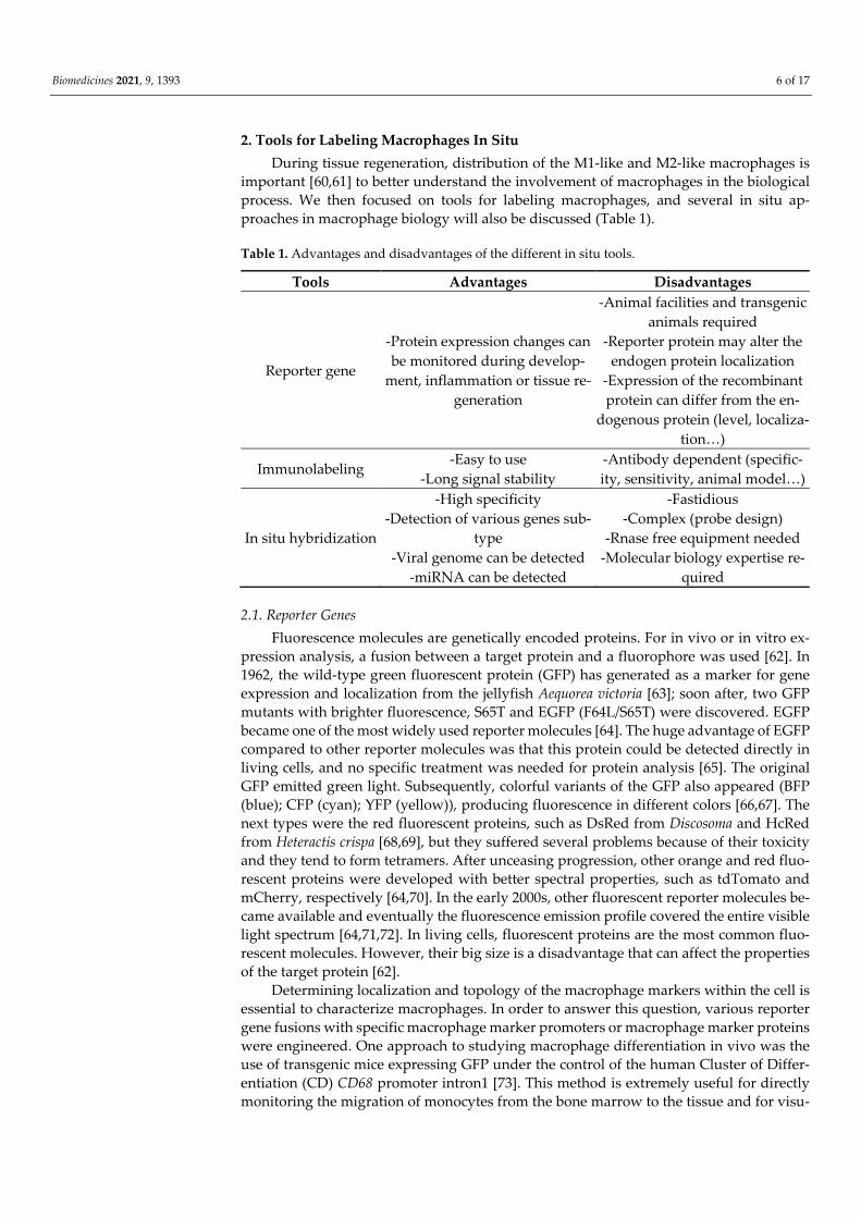
Figure 2. In vivo macrophage plasticity. Tissue Resident Macrophage (TRM), Tumor Associated Macrophage (TAM).
Macrophages can play both protective and pathogenic roles in different diseases. The polarization of these cells is not fixed and the outcome depends on the signals they receive in a given place and time. Therefore, investigation of the in vivo macrophage phenotypes in situ is fundamental to improve our understanding of tissue regeneration. This review provides an overview of in situ tools used for macrophage characterization.
Biomedicines 2021, 9, 1393 6 of 17
2. Tools for Labeling Macrophages In Situ During tissue regeneration, distribution of the M1-like and M2-like macrophages is
important [60,61] to better understand the involvement of macrophages in the biological process. We then focused on tools for labeling macrophages, and several in situ ap-proaches in macrophage biology will also be discussed (Table 1).
Table 1. Advantages and disadvantages of the different in situ tools.
Tools Advantages Disadvantages
Reporter gene
-Protein expression changes can be monitored during develop-
ment, inflammation or tissue re-generation
-Animal facilities and transgenic animals required
-Reporter protein may alter the endogen protein localization
-Expression of the recombinant protein can differ from the en-
dogenous protein (level, localiza-tion…)
Immunolabeling -Easy to use -Long signal stability
-Antibody dependent (specific-ity, sensitivity, animal model…)
In situ hybridization
-High specificity -Detection of various genes sub-
type -Viral genome can be detected
-miRNA can be detected
-Fastidious -Complex (probe design)
-Rnase free equipment needed -Molecular biology expertise re-
quired
2.1. Reporter Genes Fluorescence molecules are genetically encoded proteins. For in vivo or in vitro ex-
pression analysis, a fusion between a target protein and a fluorophore was used [62]. In 1962, the wild-type green fluorescent protein (GFP) has generated as a marker for gene expression and localization from the jellyfish Aequorea victoria [63]; soon after, two GFP mutants with brighter fluorescence, S65T and EGFP (F64L/S65T) were discovered. EGFP became one of the most widely used reporter molecules [64]. The huge advantage of EGFP compared to other reporter molecules was that this protein could be detected directly in living cells, and no specific treatment was needed for protein analysis [65]. The original GFP emitted green light. Subsequently, colorful variants of the GFP also appeared (BFP (blue); CFP (cyan); YFP (yellow)), producing fluorescence in different colors [66,67]. The next types were the red fluorescent proteins, such as DsRed from Discosoma and HcRed from Heteractis crispa [68,69], but they suffered several problems because of their toxicity and they tend to form tetramers. After unceasing progression, other orange and red fluo-rescent proteins were developed with better spectral properties, such as tdTomato and mCherry, respectively [64,70]. In the early 2000s, other fluorescent reporter molecules be-came available and eventually the fluorescence emission profile covered the entire visible light spectrum [64,71,72]. In living cells, fluorescent proteins are the most common fluo-rescent molecules. However, their big size is a disadvantage that can affect the properties of the target protein [62].
Determining localization and topology of the macrophage markers within the cell is essential to characterize macrophages. In order to answer this question, various reporter gene fusions with specific macrophage marker promoters or macrophage marker proteins were engineered. One approach to studying macrophage differentiation in vivo was the use of transgenic mice expressing GFP under the control of the human Cluster of Differ-entiation (CD) CD68 promoter intron1 [73]. This method is extremely useful for directly monitoring the migration of monocytes from the bone marrow to the tissue and for visu-
Biomedicines 2021, 9, 1393 7 of 17
alizing their transformation into macrophages and differentiation during inflamma-tion/regeneration. This tool could be a powerful resource in macrophage biology to study the process of inflammation and healing. However, it requires animal facilities and agree-ment to work with transgenic animals for the analysis and expression of the CD68 pro-moter-intron1/GFP transgene, and it showed some differences to the endogen macro-phage marker [73,74].
Another helpful approach for in situ analysis of macrophages may be the use of bac-teria containing the fluorescence reporter gene [75]. The use of such bacteria can provide important information on the details of the bacteria–macrophage interactions [6]. Fluores-cence markers containing bacteria can contain promoter-reporter genes or bacterial gene-reporter gene fusion constructs. The second type of bacteria can provide not only infor-mation on the protein localization but also about the bacterial replication status in situ [76].
Tissue-specific macrophages form a heterogeneous population. De Schepper and col-leagues (2018) have demonstrated the difference between self-maintaining and monocyte-originated macrophages [77]. A special group of tissue macrophages is microglial cells of the central nervous system (CNS) [78]. These cells have recently become a focus of interest, as they have an important function during CNS neuroinflammation and disease [79]. Dif-ferent mouse reporter lines were used, and most of them bear the fractalkine receptor gene Cx3cr1 [80]. However, this gene is expressed not only in microglia but also in other im-mune cells [79]. Nowadays, a new generation of microglial mouse reporter lines has been generated in which the reporter protein is mostly restricted to microglial cells [81–83]. These transgenic animals provide essential information about neuroinflammation.
Tumor microenvironment demonstrated immune cell penetrations. TAM macro-phage played an important role in all steps of tumor development [84]. TAM macrophages could be promising target cells for cancer therapy. However, more information on these cells, and therefore in situ characterization of TAM macrophages, is fundamental [85]. Several laboratories are using fluorescence proteins in tumor research areas to detect the interaction between TAM macrophages and cancer cells [71,86,87].
2.2. Immunolabeling In situ visualization of macrophages remains one of the most challenging tasks, and
immunolabeling is widely used to this effect. One of the most common pan-markers to label monocytes/macrophages is the CD68 protein [45,49]. CD68 is a well-known glyco-protein, intensively expressed by macrophages. However, CD68 can also be detected in other mononuclear phagocyte cells. Moreover, a weak expression can also be seen in other non-hematopoietic cells (mesenchymal stem cells, fibroblast, endothelial and tumor cells) [88].
The mouse is frequently used as an animal model in clinical research, but unfortu-nately, in many ways, mice differ from humans [78]. Indeed, rodent and human in vitro macrophages can produce different protein over-stimulations, which may potentially in-dicate diverse functions [11]. The second macrophage pan marker, successfully used in situ in human cells, is the CD11b protein [45].
The polarization of macrophages is a complex process that is highly dependent on the tissue environment [7]. Using this approach, various studies aimed at finding key markers to distinguish between M1-like and M2-like macrophages [7].
It is widely accepted that the phenotypic M1-like macrophage markers are co-stimu-latory molecules, such as CD80 and CD86 in mice [49]. Some differences need to be men-tioned between human and rodent macrophage marker gene expression. Mouse macro-phage produces F4/80 protein (also known as Epidermal Growth Factor-like module-con-taining mucin-like hormone receptor-like 1) [89]. Baud and colleagues (1995) cloned the human homolog of the F4/80 gene, the sequence homology between two genes was 68% [90]. Surprisingly, human macrophages did not produce this protein [91]. Other proteins
Biomedicines 2021, 9, 1393 8 of 17
(Ym1, Fizz1 and arginase-1) can also be detected in mouse M2 macrophages but are lack-ing in human M2 macrophages. [45,92]. Little information is currently available on mac-rophages in mammals other than rodents and humans. Consequently, it is essential to study another animal model with closer similarity to humans. The progression of diseases in pigs is similar to that in humans at metabolic and infectious levels, which makes mini-pigs an ideal model for macrophage characterization [93–95]. In the mini-pig model, un-der in situ conditions, CD80 protein is not able to accurately distinguish between M1-like and M2-like cells, since M2-like cells also produce CD80 protein [60]. Moreover, intensity of CD80 protein expression in cells depends on both the quality and the quantity of the stimulus [54]. In addition, both CD80 and CD86 expression can also be detected in other cells, such as dendritic cells (DCs), B cells and T cells [96,97] while CD86 can be detected in human M2a-like macrophages [7].
Markers such as CD163 and CD206 generally recognize M2-like macrophages. CD206 protein is a commonly accepted M2-like macrophage marker in rodent and human cells [7]. However, this protein is also expressed at low levels in satellite cells [45]. Another limit is that, depending on the anti-CD206 antibody used, different results can be obtained [98]. The CD163 is considered as an M2-like macrophage marker in human [7] and murine cells [99], but also in the human dendritic cells [100]. In situ detection of human and mouse M2 macrophages is possible by the double staining immunolabeling technique using mannose receptor C type 1 (MRC1/CD206) or CD163 combined with CD68 antibodies to-gether.
Up to now, it is assumed that the M1-like and M2-like macrophages have a monocyte origin [34]. Ginhoux and Guilliams (2016) reported that, in mice, the tissue-resident mac-rophages are of embryonic origin and persist in the tissue after cell division [101]. The role of tissue-resident macrophages in inflammation and tissue regeneration is not yet fully understood. The only exception is the microglia, which are the specialized tissue-resident macrophages of the central nervous system (CNS). This cell type has recently been inten-sively studied because of their essential function during inflammation, infection and re-sponse to injury [79]. Microglia cells produce proteins that can be detected in the periph-eral myeloid cells (CD68, CD45, CD11b, F4/80, Cx3cr1 and CSF1R) [102]. The aim of vari-ous studies was to find a specific microglia marker not produced in peripheral myeloid cells. Traditionally, Iba-1 was the microglia pan marker; however, this protein was also expressed in other myeloid cells [103,104]. Later, Bennett and colleagues (2016) showed that both mouse and human microglia cells generate Tmem119 protein for which no ex-pression in peripheral myeloid cells was detected [105]. Yet, another group has shown that the expression of this protein was not restricted to microglia cells [106]. Butovsky et al. (2014) showed that antibodies produced against P2ry12 and FCRLS proteins could make a difference between microglia and infiltrating myeloid cells [107].
In the past, tissue-resident macrophages were identified by morphological analysis or histological staining [108]; the development of various antibodies enabled the detection of macrophages. In mouse animal models, one particularly useful antibody was the anti-F4/80 antibody. F4/80 protein is a cell surface receptor selectively expressed on murine macrophages. The F4/80 protein is a useful positive control in murine macrophage biology because it is a well-resistant protein during the fixation process [46,89]. However, the very heterogeneous expression level of the F4/80 protein in the different tissues could be a dis-advantage [109]. In situ conditions make it difficult to distinguish between M2-like and tissue-resident macrophages once they co-exist in a common environment, as there is no good marker gene to make a difference. Gut tissue-resident macrophages produce CD169 proteins [43,77]. However, this protein can also be detected in TAM and in nontypical macrophages [23,110].
TAM macrophages contribute to the initiation and progression of tumor develop-ment [27]. It is usually thought that TAM macrophages originate from blood monocytes [111], but in certain brain and lung (murine model animal) cancers, the tumors originated from tissue-resident macrophages [28]. M1-like TAM macrophages express CD68, CD80,
Biomedicines 2021, 9, 1393 9 of 17
CD86 and CD169 [110,112] and the M2-like class of TAM macrophages show strong CD204, CD206 and CD163 expression [112–114]. In addition, a new marker for these cells was recently found, the disulfide-isomerase A3 (PDIA3) receptor [115].
Immunolabeling is an excellent method for protein detection in situ. It has to be men-tioned that the expression patterns differed depending on the antibody used [98]. Moreo-ver, appropriate protocols need to be developed to obtain a positive signal. Careful signal analysis has to be performed as nonspecific labeling can result from hydrophobic interac-tions of proteins, ionic and electrostatic interactions, avidin and biotin, Fc receptors or autofluorescence [61,116]. In addition, specificity of the antibody can be limited, as the peptides recognized by antibodies are small (6–10 amino acids) and this antigen can be found in other proteins [117].
2.3. In Situ Hybridization In situ hybridization (ISH) was invented simultaneously by Gall and Pardue (1969)
and by John and colleagues in 1969 [118,119]. The name “in situ” is derived from the Latin phrase for “in position”, and hybridization means hybrid formation between different molecules. The various ISH techniques in macrophage biology are summarized in Table 2. The idea of this technique is that RNA and DNA molecules can form a hydrogen bond hybrid based on the complementarity of RNA or DNA sequences. In this approach, dif-ferently labeled RNA or DNA probes make a hybrid molecule with the mRNA of interest. ISH allows a very powerful analysis of de novo gene expression because it is highly spe-cific and shows the mRNA localization in the tissue environment. The disadvantages of this technique are the complex probe design and the fastidious steps, which require some optimization for each probe and tissue [120]. An additional challenge is the conservation of the RNA because RNase enzymes are present everywhere,: on glassware, in reagents, on clothes and hands [121]. In the beginning, radioactive probes were used because radi-oisotope-labeled probes are more sensitive than non-radioactive probes. This hybridiza-tion can be detected by autoradiography. After hybridization with the radioactive probe, the sections are coated with an emulsion containing silver ions. The energy liberated from radioactive molecules can transform the silver ions to metallic silver, and this reaction results in image formation [122]. This technique was used to demonstrate that infection induces lysozyme expression in macrophages [123] and that VEGF induces the macro-phage recruitment [124]. This method was also utilized for hepatitis A viral RNA detec-tion in macrophages [125]. Unfortunately, a long exposure requirement and harmful effect on health drastically reduced this practice [126].
Table 2. Sensitivity, advantages and disadvantages of the different in situ hybridization approaches. ((+) low, (++) medium, (+++) strong sensitivity).
Tools Sensitivity Advantages Disadvantages
Radioactive probe +++ -Best sensitivity -Long exposure required -Harmful
CISH ++ -Stable signal -Cumbersome double la-beling
FISH + -Multiple labels -Not stable
HCR ++ -Multiple labels
-Increased sensitivity compared to FISH
-Not stable -Probe design strategy
RNAscope® Assay ++ -Multiple labels -Rapid method
-Not stable -High financial cost
The next method replacing the radioprobe–ISH method was the Chromogenic In Situ Hybridization (CISH). The digoxigenin-labeled probe is commonly used for CISH in mac-rophage biology [60,61,98,127,128]. The CISH method exhibits several improvements
Biomedicines 2021, 9, 1393 10 of 17
compared to the radioactive probes, such as better detection, shorter manipulation, good repeatability, easier visualization and being less harmful to health [120]. In the 1980s the Fluorescence In Situ Hybridization (FISH) method appeared [129], which was recently used for microglial signature gene analysis of the mouse brain during development and neuroinflammation [130,131].
Both techniques are based on the same principle, probe hybridization to the mRNA of interest, then detection by an appropriate system [132]. The main differences between the two methods are: (i) in the CISH, the detection is carried out by an enzymatic reaction, the signal intensity does not decrease over time but, unfortunately, only a single expres-sion can be tested in one step. To test two different mRNA productions, substantial time is required, which makes the technique complicated. Enzymatic detection generates a col-ored precipitate at the site of hybridization. Two enzymes, alkaline phosphatase (AP) and horseradish peroxidase (HRP), are generally used in this method. Although these en-zymes can directly be attached to the nucleic acid molecule, the enzyme-coupled probes have very little incorporation property. Therefore, an indirect method is usually used dur-ing hybridization, which makes this method lengthy and cumbersome [121]. (ii) In the FISH method, several fluorescent molecules can be used for detection, then multiple mRNA expressions can be analyzed in the same sample, but the signal intensity decreases over time [133] and only highly abundant mRNA can be localized. In this technique, fluor-ophores can be linked directly to the nucleic acid probe, and after hybridization this fluo-rescence is directly detectable (the method called “direct labeling”). In a second method (called the “indirect method”), the non-fluorescent molecule is linked to the probe, and this molecule can be visualized by the fluorescence molecule [134].
Characterizing in situ macrophage sub-types is not an easy task because there is no specific marker for one sub-type; therefore, characterization is based on their pattern of both cytokines and cell surface proteins (Figure 1). Two difficulties have to be noted: i, the FISH approach is not a sensitive tool, thus requiring a strong enough cytokine expression level to be detected. ii, cytokines are secreted proteins, making it complicated to link the signal with the macrophage source of expression by immunolabeling [60]. Recently, a new revolutionary in situ hybridization, the Hybridization Chain Reaction (HCR), has emerged [135]. HCR is an isothermal enzyme-independent nucleotide polymerization method. The idea of this method results in the use of two hairpin oligonucleotides linked with fluorophore. When the complementary initiator nucleotide hybridizes with mRNA, the initiator activates the hairpin molecules, and they assemble into a well-defined struc-ture providing a source of fluorescence. The main advantage of this method is that it is very sensitive and therefore suitable for the detection of the low abundant mRNAs [136–140]. This new approach was used for identification of macrophage sub-types by analysis of cytokine expression. This technique allowed the in situ visualization of M2d-like mac-rophages in the mini-pig model [60].
Another powerful technique that allows the localization of RNA to specific cells is the RNAscope®Multiplex Fluorescent Assay V2 (Advanced Cell Diagnostics, Newark, CA, USA). This technique uses paired double-Z oligonucleotide RNA probes for hybridi-zation. These oligonucleotide probes contain several linker sequences. The amplifiers and the color label probes are sequentially added to the linker sequence. It is highly sensitive, and multiple labeling can be performed on the same tissue section. This technique was used as a prognostic indicator in human kidney cancer. In certain cancers, the von Hippel Lindau tumor suppressors are inactivated. Therefore, the HIF-1α produced by M2 TAM macrophages triggers the production of proteins that are required for tumor progression. Detection of this protein by in situ hybridization can then provide important information about tumor development [141]. This method is, however, very costly, and therefore less commonly used than other ISH techniques.
Biomedicines 2021, 9, 1393 11 of 17
2.4. Imaging Mass Spectrometry In recent decades, a great new technique has emerged for in situ imaging, the Imag-
ing Mass Spectrometry [142,143]. With this technique, it is possible to detect hundreds of proteins, peptides or lipids simultaneously in different tissue sections. At the moment, this approach is still less widely used for macrophage characterization [144,145], but it is undoubtedly becoming routine and an essential tool in the coming years.
3. Conclusions During wound healing, macrophages have an essential role. Inflammation and tissue
regeneration is complex and can induce well-coordinated gene expression changes in macrophages. For proper tissue healing, the constant interplay between external and in-ternal signals is essential, including cytokine regulation, selective adhesion or import and export of transcription factors to the chromatin. Macrophages localized in the injured area have distinct phenotypes with different functions and they act at diverse times during the reparation. There are several questions that remain to be answered in order to compre-hend the repair process, such as what are the precise phenotypes of the in vivo macro-phages? To achieve breakthroughs in in vivo macrophage characterization, the develop-ment of new tools is essential, as in situ characterization of the macrophages is indispen-sable to uncover the spatial actors involved in inflammation and tissue regeneration mechanisms. These tools will also provide opportunities to explore macrophage pheno-types in vivo. Indeed, the application of distinct ISH approaches can bring new insights into the polarization of macrophages in situ to investigate tissue inflammation and regen-eration.
Author Contributions: A.-L.F. and K.N. wrote the review, contributed to the article and approved the submitted version. All authors have read and agreed to the published version of the manuscript.
Funding: This work was supported by the Délégation Générale de l’Armement (DGA) (PDH2-NRBC-4-NR-4306).
Acknowledgments: We are very grateful to Martine Miquel for her helpful advice and critical re-view of our manuscript.
Conflicts of Interest: The authors declare no conflict of interest.
Reference 1. Nathan, C. Metchnikoff’s Legacy in 2008. Nat. Immunol. 2008, 9, 695–698. https://doi.org/10.1038/ni0708-695. 2. Mosser, D.M.; Edwards, J.P. Exploring the Full Spectrum of Macrophage Activation. Nat. Rev. Immunol. 2008, 8, 958–969.
https://doi.org/10.1038/nri2448. 3. Minutti, C.M.; Knipper, J.A.; Allen, J.E.; Zaiss, D.M.W. Tissue-Specific Contribution of Macrophages to Wound Healing. Semin.
Cell Dev. Biol. 2017, 61, 3–11. https://doi.org/10.1016/j.semcdb.2016.08.006. 4. Abdelaziz, M.H.; Abdelwahab, S.F.; Wan, J.; Cai, W.; Huixuan, W.; Jianjun, C.; Kumar, K.D.; Vasudevan, A.; Sadek, A.; Su, Z.;
et al. Alternatively Activated Macrophages; a Double-Edged Sword in Allergic Asthma. J. Transl. Med. 2020, 18, 58. https://doi.org/10.1186/s12967-020-02251-w.
5. Gharib, S.A.; McMahan, R.S.; Eddy, W.E.; Long, M.E.; Parks, W.C.; Aitken, M.L.; Manicone, A.M. Transcriptional and Functional Diversity of Human Macrophage Repolarization. J. Allergy Clin. Immunol. 2019, 143, 1536–1548. https://doi.org/10.1016/j.jaci.2018.10.046.
6. Huang, L.; Nazarova, E.V.; Russell, D.G. Mycobacterium Tuberculosis: Bacterial Fitness within the Host Macrophage. Microbiol. Spectr. 2019, 7, 127–138. https://doi.org/10.1128/microbiolspec.BAI-0001-2019.
7. Atri, C.; Guerfali, F.Z.; Laouini, D. Role of Human Macrophage Polarization in Inflammation during Infectious Diseases. Int. J. Mol. Sci. 2018, 19, 1801. https://doi.org/10.3390/ijms19061801.
8. Arora, S.; Dev, K.; Agarwal, B.; Das, P.; Syed, M.A. Macrophages: Their Role, Activation and Polarization in Pulmonary Diseases. Immunobiology 2018, 223, 383–396. https://doi.org/10.1016/j.imbio.2017.11.001.
9. Wynn, T.A.; Vannella, K.M. Macrophages in Tissue Repair, Regeneration, and Fibrosis. Immunity 2016, 44, 450–462. https://doi.org/10.1016/j.immuni.2016.02.015.
10. Beyer, M.; Mallmann, M.R.; Xue, J.; Staratschek-Jox, A.; Vorholt, D.; Krebs, W.; Sommer, D.; Sander, J.; Mertens, C.; Nino-Castro, A.; et al. High-Resolution Transcriptome of Human Macrophages. PLoS ONE 2012, 7, e45466. https://doi.org/10.1371/jour-nal.pone.0045466.
Biomedicines 2021, 9, 1393 12 of 17
11. Martinez, F.O.; Gordon, S. The M1 and M2 Paradigm of Macrophage Activation: Time for Reassessment. F1000Prime Rep. 2014, 6, 13. https://doi.org/10.12703/P6-13.
12. Murray, P.J.; Allen, J.E.; Biswas, S.K.; Fisher, E.A.; Gilroy, D.W.; Goerdt, S.; Gordon, S.; Hamilton, J.A.; Ivashkiv, L.B.; Lawrence, T.; et al. Macrophage Activation and Polarization: Nomenclature and Experimental Guidelines. Immunity 2014, 41, 14–20. https://doi.org/10.1016/j.immuni.2014.06.008.
13. Nathan, C.F.; Murray, H.W.; Wiebe, M.E.; Rubin, B.Y. Identification of Interferon-Gamma as the Lymphokine That Activates Human Macrophage Oxidative Metabolism and Antimicrobial Activity. J. Exp. Med. 1983, 158, 670–689. https://doi.org/10.1084/jem.158.3.670.
14. Stein, M.; Keshav, S.; Harris, N.; Gordon, S. Interleukin 4 Potently Enhances Murine Macrophage Mannose Receptor Activity: A Marker of Alternative Immunologic Macrophage Activation. J. Exp. Med. 1992, 176, 287–292. https://doi.org/10.1084/jem.176.1.287.
15. Warburg, O.; Wind, F.; Negelein, E. The metabolism of tumors in the body. J. Gen. Physiol. 1927, 8, 519–530. https://doi.org/10.1085/jgp.8.6.519.
16. Lunt, S.Y.; Vander Heiden, M.G. Aerobic Glycolysis: Meeting the Metabolic Requirements of Cell Proliferation. Annu. Rev. Cell Dev. Biol. 2011, 27, 441–464. https://doi.org/10.1146/annurev-cellbio-092910-154237.
17. Kieler, M.; Hofmann, M.; Schabbauer, G. More than Just Protein Building Blocks: How Amino Acids and Related Metabolic Pathways Fuel Macrophage Polarization. FEBS J. 2021, 288, 3694–3714. https://doi.org/10.1111/febs.15715.
18. Granger, D.L.; Hibbs, J.B.J.; Perfect, J.R.; Durack, D.T. Specific Amino Acid (L-Arginine) Requirement for the Microbiostatic Activity of Murine Macrophages. J. Clin. Investig. 1988, 81, 1129–1136. https://doi.org/10.1172/JCI113427.
19. Corraliza, I.M.; Soler, G.; Eichmann, K.; Modolell, M. Arginase Induction by Suppressors of Nitric Oxide Synthesis (IL-4, IL-10 and PGE2) in Murine Bone-Marrow-Derived Macrophages. Biochem. Biophys. Res. Commun. 1995, 206, 667–673. https://doi.org/10.1006/bbrc.1995.1094.
20. Mills, C.D.; Kincaid, K.; Alt, J.M.; Heilman, M.J.; Hill, A.M. M-1/M-2 Macrophages and the Th1/Th2 Paradigm. J. Immunol. 2000, 164, 6166–6173. https://doi.org/10.4049/jimmunol.164.12.6166.
21. Mantovani, A.; Sica, A.; Sozzani, S.; Allavena, P.; Vecchi, A.; Locati, M. The Chemokine System in Diverse Forms of Macrophage Activation and Polarization. Trends Immunol. 2004, 25, 677–686. https://doi.org/10.1016/j.it.2004.09.015.
22. Rőszer, T. Understanding the Mysterious M2 Macrophage through Activation Markers and Effector Mechanisms. Mediat. In-flamm. 2015, 2015, 816460. https://doi.org/10.1155/2015/816460.
23. Chávez-Galán, L.; Olleros, M.L.; Vesin, D.; Garcia, I. Much More than M1 and M2 Macrophages, There Are Also CD169(+) and TCR(+) Macrophages. Front. Immunol. 2015, 6, 263. https://doi.org/10.3389/fimmu.2015.00263.
24. Schneider, W. Hematologic or hemostaseologic “paraneoplastic syndromes” as prognostic factors? Klin. Wochenschr. 1988, 66, 103–109. https://doi.org/10.1007/BF01774223.
25. Lemke, G. Biology of the TAM Receptors. Cold Spring Harb. Perspect. Biol. 2013, 5, a009076. https://doi.org/10.1101/cshper-spect.a009076.
26. Qian, B.-Z.; Pollard, J.W. New Tricks for Metastasis-Associated Macrophages. Breast Cancer Res. 2012, 14, 316. https://doi.org/10.1186/bcr3143.
27. Duluc, D.; Delneste, Y.; Tan, F.; Moles, M.-P.; Grimaud, L.; Lenoir, J.; Preisser, L.; Anegon, I.; Catala, L.; Ifrah, N.; et al. Tumor-Associated Leukemia Inhibitory Factor and IL-6 Skew Monocyte Differentiation into Tumor-Associated Macrophage-like Cells. Blood 2007, 110, 4319–4330. https://doi.org/10.1182/blood-2007-02-072587.
28. DeNardo, D.G.; Ruffell, B. Macrophages as Regulators of Tumour Immunity and Immunotherapy. Nat. Rev. Immunol. 2019, 19, 369–382. https://doi.org/10.1038/s41577-019-0127-6.
29. Lim, B.; Woodward, W.A.; Wang, X.; Reuben, J.M.; Ueno, N.T. Inflammatory Breast Cancer Biology: The Tumour Microenvi-ronment Is Key. Nat. Rev. Cancer 2018, 18, 485–499. https://doi.org/10.1038/s41568-018-0010-y.
30. Wagner, J.; Rapsomaniki, M.A.; Chevrier, S.; Anzeneder, T.; Langwieder, C.; Dykgers, A.; Rees, M.; Ramaswamy, A.; Muenst, S.; Soysal, S.D.; et al. A Single-Cell Atlas of the Tumor and Immune Ecosystem of Human Breast Cancer. Cell 2019, 177, 1330–1345.e18. https://doi.org/10.1016/j.cell.2019.03.005.
31. Tan, A.H.Y.; Tu, W.; McCuaig, R.; Hardy, K.; Donovan, T.; Tsimbalyuk, S.; Forwood, J.K.; Rao, S. Lysine-Specific Histone De-methylase 1A Regulates Macrophage Polarization and Checkpoint Molecules in the Tumor Microenvironment of Triple-Nega-tive Breast Cancer. Front. Immunol. 2019, 10, 1351. https://doi.org/10.3389/fimmu.2019.01351.
32. Yunna, C.; Mengru, H.; Lei, W.; Weidong, C. Macrophage M1/M2 Polarization. Eur. J. Pharmacol. 2020, 877, 173090. https://doi.org/10.1016/j.ejphar.2020.173090.
33. Locati, M.; Curtale, G.; Mantovani, A. Diversity, Mechanisms, and Significance of Macrophage Plasticity. Annu. Rev. Pathol. 2020, 15, 123–147. https://doi.org/10.1146/annurev-pathmechdis-012418-012718.
34. Orecchioni, M.; Ghosheh, Y.; Pramod, A.B.; Ley, K. Macrophage Polarization: Different Gene Signatures in M1(LPS+) vs. Clas-sically and M2(LPS-) vs. Alternatively Activated Macrophages. Front. Immunol. 2019, 10, 1084. https://doi.org/10.3389/fimmu.2019.01084.
35. Viniegra, A.; Goldberg, H.; Çil, Ç.; Fine, N.; Sheikh, Z.; Galli, M.; Freire, M.; Wang, Y.; Van Dyke, T.E.; Glogauer, M.; et al. Resolving Macrophages Counter Osteolysis by Anabolic Actions on Bone Cells. J. Dent. Res. 2018, 97, 1160–1169. https://doi.org/10.1177/0022034518777973.
Biomedicines 2021, 9, 1393 13 of 17
36. Shapouri-Moghaddam, A.; Mohammadian, S.; Vazini, H.; Taghadosi, M.; Esmaeili, S.-A.; Mardani, F.; Seifi, B.; Mohammadi, A.; Afshari, J.T.; Sahebkar, A. Macrophage Plasticity, Polarization, and Function in Health and Disease. J. Cell. Physiol. 2018, 233, 6425–6440. https://doi.org/10.1002/jcp.26429.
37. Sica, A.; Mantovani, A. Macrophage Plasticity and Polarization: In Vivo Veritas. J. Clin. Investig. 2012, 122, 787–795. https://doi.org/10.1172/JCI59643.
38. Klopfleisch, R. Macrophage Reaction against Biomaterials in the Mouse Model—Phenotypes, Functions and Markers. Acta Bio-mater. 2016, 43, 3–13. https://doi.org/10.1016/j.actbio.2016.07.003.
39. Wermuth, P.J.; Jimenez, S.A. The Significance of Macrophage Polarization Subtypes for Animal Models of Tissue Fibrosis and Human Fibrotic Diseases. Clin. Transl. Med. 2015, 4, 2. https://doi.org/10.1186/s40169-015-0047-4.
40. Selders, G.S.; Fetz, A.E.; Radic, M.Z.; Bowlin, G.L. An Overview of the Role of Neutrophils in Innate Immunity, Inflammation and Host-Biomaterial Integration. Regen. Biomater. 2017, 4, 55–68. https://doi.org/10.1093/rb/rbw041.
41. Snyder, R.J.; Lantis, J.; Kirsner, R.S.; Shah, V.; Molyneaux, M.; Carter, M.J. Macrophages: A Review of Their Role in Wound Healing and Their Therapeutic Use. Wound Repair Regen. 2016, 24, 613–629. https://doi.org/10.1111/wrr.12444.
42. Murray, P.J. Macrophage Polarization. Annu. Rev. Physiol. 2017, 79, 541–566. https://doi.org/10.1146/annurev-physiol-022516-034339.
43. Viola, M.F.; Boeckxstaens, G. Niche-Specific Functional Heterogeneity of Intestinal Resident Macrophages. Gut 2021, 70, 1383–1395. https://doi.org/10.1136/gutjnl-2020-323121.
44. Sreejit, G.; Fleetwood, A.J.; Murphy, A.J.; Nagareddy, P.R. Origins and Diversity of Macrophages in Health and Disease. Clin. Transl. Immunol. 2020, 9, e1222. https://doi.org/10.1002/cti2.1222.
45. Kosmac, K.; Peck, B.D.; Walton, R.G.; Mula, J.; Kern, P.A.; Bamman, M.M.; Dennis, R.A.; Jacobs, C.A.; Lattermann, C.; Johnson, D.L.; et al. Immunohistochemical Identification of Human Skeletal Muscle Macrophages. Bio Protoc. 2018, 8, e2883, https://doi.org/10.21769/BioProtoc.2883.
46. Gordon, S.; Plüddemann, A. Tissue Macrophages: Heterogeneity and Functions. BMC Biol. 2017, 15, 53. https://doi.org/10.1186/s12915-017-0392-4.
47. Van den Bossche, J.; O’Neill, L.A.; Menon, D. Macrophage Immunometabolism: Where Are We (Going)? Trends Immunol. 2017, 38, 395–406. https://doi.org/10.1016/j.it.2017.03.001.
48. Daley, J.M.; Brancato, S.K.; Thomay, A.A.; Reichner, J.S.; Albina, J.E. The Phenotype of Murine Wound Macrophages. J. Leukoc. Biol. 2010, 87, 59–67. https://doi.org/10.1189/jlb.0409236.
49. Xu, H.-T.; Lee, C.-W.; Li, M.-Y.; Wang, Y.-F.; Yung, P.S.-H.; Lee, O.K.-S. The Shift in Macrophages Polarisation after Tendon Injury: A Systematic Review. J. Orthop. Transl. 2020, 21, 24–34. https://doi.org/10.1016/j.jot.2019.11.009.
50. Xu, W.; Zhao, X.; Daha, M.R.; van Kooten, C. Reversible Differentiation of Pro- and Anti-Inflammatory Macrophages. Mol. Immunol. 2013, 53, 179–186. https://doi.org/10.1016/j.molimm.2012.07.005.
51. Aitcheson, S.M.; Frentiu, F.D.; Hurn, S.E.; Edwards, K.; Murray, R.Z. Skin Wound Healing: Normal Macrophage Function and Macrophage Dysfunction in Diabetic Wounds. Molecules 2021, 26, 4917. https://doi.org/10.3390/molecules26164917.
52. Italiani, P.; Mazza, E.M.C.; Lucchesi, D.; Cifola, I.; Gemelli, C.; Grande, A.; Battaglia, C.; Bicciato, S.; Boraschi, D. Transcriptomic Profiling of the Development of the Inflammatory Response in Human Monocytes in Vitro. PLoS ONE 2014, 9, e87680. https://doi.org/10.1371/journal.pone.0087680.
53. Rackov, G.; Hernández-Jiménez, E.; Shokri, R.; Carmona-Rodríguez, L.; Mañes, S.; Álvarez-Mon, M.; López-Collazo, E.; Mar-tínez-A, C.; Balomenos, D. P21 Mediates Macrophage Reprogramming through Regulation of P50-P50 NF-ΚB and IFN-β. J. Clin. Investig. 2016, 126, 3089–3103. https://doi.org/10.1172/JCI83404.
54. Tarique, A.A.; Logan, J.; Thomas, E.; Holt, P.G.; Sly, P.D.; Fantino, E. Phenotypic, Functional, and Plasticity Features of Classical and Alternatively Activated Human Macrophages. Am. J. Respir. Cell Mol. Biol. 2015, 53, 676–688. https://doi.org/10.1165/rcmb.2015-0012OC.
55. Belgiovine, C.; D’Incalci, M.; Allavena, P.; Frapolli, R. Tumor-Associated Macrophages and Anti-Tumor Therapies: Complex Links. Cell. Mol. Life Sci. 2016, 73, 2411–2424. https://doi.org/10.1007/s00018-016-2166-5.
56. Chittezhath, M.; Dhillon, M.K.; Lim, J.Y.; Laoui, D.; Shalova, I.N.; Teo, Y.L.; Chen, J.; Kamaraj, R.; Raman, L.; Lum, J.; et al. Molecular Profiling Reveals a Tumor-Promoting Phenotype of Monocytes and Macrophages in Human Cancer Progression. Immunity 2014, 41, 815–829. https://doi.org/10.1016/j.immuni.2014.09.014.
57. Ruffell, B.; Affara, N.I.; Coussens, L.M. Differential Macrophage Programming in the Tumor Microenvironment. Trends Immunol. 2012, 33, 119–126. https://doi.org/10.1016/j.it.2011.12.001.
58. Simmons, M.; Singhal, A.; Lu, Z. Text Mining for Precision Medicine: Bringing Structure to EHRs and Biomedical Literature to Understand Genes and Health. Adv. Exp. Med. Biol. 2016, 939, 139–166. https://doi.org/10.1007/978-981-10-1503-8_7.
59. Mossadegh-Keller, N.; Sieweke, M.H. Characterization of Mouse Adult Testicular Macrophage Populations by Immunofluo-rescence Imaging and Flow Cytometry. Bio Protoc. 2019, 9, e3178. https://doi.org/10.21769/BioProtoc.3178.
60. Nikovics, K.; Morin, H.; Riccobono, D.; Bendahmane, A.; Favier, A. Hybridization-chain-reaction Is a Relevant Method for in Situ Detection of M2d-like Macrophages in a Mini-pig Model. FASEB J. 2020, 34, 15675–15686. https://doi.org/10.1096/fj.202001496R.
61. Nikovics, K.; Favier, A.-L.; Barbier, L.; Drouet, M.; Riccobono, D. Characterization of Macrophages, Giant Cells and Granulomas during Muscle Regeneration after Irradiation. Cytokine 2021, 137, 155318. https://doi.org/10.1016/j.cyto.2020.155318.
Biomedicines 2021, 9, 1393 14 of 17
62. Guo, A.-Y.; Zhang, Y.-M.; Wang, L.; Bai, D.; Xu, Y.-P.; Wu, W.-Q. Single-Molecule Imaging in Living Plant Cells: A Methodo-logical Review. Int. J. Mol. Sci. 2021, 22, 5071. https://doi.org/10.3390/ijms22105071.
63. Chalfie, M. Green Fluorescent Protein. Photochem. Photobiol. 1995, 62, 651–656. https://doi.org/10.1111/j.1751-1097.1995.tb08712.x. 64. Shaner, N.C.; Patterson, G.H.; Davidson, M.W. Advances in Fluorescent Protein Technology. J. Cell Sci. 2007, 120, 4247–4260.
https://doi.org/10.1242/jcs.005801. 65. Belardinelli, J.M.; Jackson, M. Green Fluorescent Protein as a Protein Localization and Topological Reporter in Mycobacteria.
Tuberculosis 2017, 105, 13–17. https://doi.org/10.1016/j.tube.2017.04.001. 66. Nifosí, R.; Amat, P.; Tozzini, V. Variation of Spectral, Structural, and Vibrational Properties within the Intrinsically Fluorescent
Proteins Family: A Density Functional Study. J. Comput. Chem. 2007, 28, 2366–2377. https://doi.org/10.1002/jcc.20764. 67. Chudakov, D.M.; Matz, M.V.; Lukyanov, S.; Lukyanov, K.A. Fluorescent Proteins and Their Applications in Imaging Living
Cells and Tissues. Physiol. Rev. 2010, 90, 1103–1163. https://doi.org/10.1152/physrev.00038.2009. 68. Matz, M.V.; Fradkov, A.F.; Labas, Y.A.; Savitsky, A.P.; Zaraisky, A.G.; Markelov, M.L.; Lukyanov, S.A. Fluorescent Proteins
from Nonbioluminescent Anthozoa Species. Nat. Biotechnol. 1999, 17, 969–973. https://doi.org/10.1038/13657. 69. Gurskaya, N.G.; Savitsky, A.P.; Yanushevich, Y.G.; Lukyanov, S.A.; Lukyanov, K.A. Color Transitions in Coral’s Fluorescent
Proteins by Site-Directed Mutagenesis. BMC Biochem. 2001, 2, 6. https://doi.org/10.1186/1471-2091-2-6. 70. Shemiakina, I.I.; Ermakova, G.V.; Cranfill, P.J.; Baird, M.A.; Evans, R.A.; Souslova, E.A.; Staroverov, D.B.; Gorokhovatsky, A.Y.;
Putintseva, E.V.; Gorodnicheva, T.V.; et al. A Monomeric Red Fluorescent Protein with Low Cytotoxicity. Nat. Commun. 2012, 3, 1204. https://doi.org/10.1038/ncomms2208.
71. Hoffman, R.M.; Bouvet, M. Imaging the Microenvironment of Pancreatic Cancer Patient-Derived Orthotopic Xenografts (PDOX) Growing in Transgenic Nude Mice Expressing GFP, RFP, or CFP. Cancer Lett. 2016, 380, 349–355. https://doi.org/10.1016/j.can-let.2015.12.021.
72. Cardarelli, F. Back to the Future: Genetically Encoded Fluorescent Proteins as Inert Tracers of the Intracellular Environment. Int. J. Mol. Sci. 2020, 21, 4164. https://doi.org/10.3390/ijms21114164.
73. Iqbal, A.J.; McNeill, E.; Kapellos, T.S.; Regan-Komito, D.; Norman, S.; Burd, S.; Smart, N.; Machemer, D.E.W.; Stylianou, E.; McShane, H.; et al. Human CD68 Promoter GFP Transgenic Mice Allow Analysis of Monocyte to Macrophage Differentiation in Vivo. Blood 2014, 124, e33–e44. https://doi.org/10.1182/blood-2014-04-568691.
74. Lois, C.; Hong, E.J.; Pease, S.; Brown, E.J.; Baltimore, D. Germline Transmission and Tissue-Specific Expression of Transgenes Delivered by Lentiviral Vectors. Science 2002, 295, 868–872. https://doi.org/10.1126/science.1067081.
75. Huang, J.; Fan, Q.; Guo, M.; Wu, M.; Wu, S.; Shen, S.; Wang, X.; Wang, H. Octenidine Dihydrochloride Treatment of a Meticillin-Resistant Staphylococcus Aureus Biofilm-Infected Mouse Wound. J. Wound Care 2021, 30, 106–114. https://doi.org/10.12968/jowc.2021.30.2.106.
76. Ooi, S.-K.; Lim, T.-Y.; Lee, S.-H.; Nathan, S. Burkholderia Pseudomallei Kills Caenorhabditis Elegans through Virulence Mech-anisms Distinct from Intestinal Lumen Colonization. Virulence 2012, 3, 485–496. https://doi.org/10.4161/viru.21808.
77. De Schepper, S.; Verheijden, S.; Aguilera-Lizarraga, J.; Viola, M.F.; Boesmans, W.; Stakenborg, N.; Voytyuk, I.; Schmidt, I.; Boeckx, B.; Dierckx de Casterlé, I.; et al. Self-Maintaining Gut Macrophages Are Essential for Intestinal Homeostasis. Cell 2018, 175, 400–415.e13. https://doi.org/10.1016/j.cell.2018.07.048.
78. Haenseler, W.; Rajendran, L. Concise Review: Modeling Neurodegenerative Diseases with Human Pluripotent Stem Cell-De-rived Microglia. Stem Cells 2019, 37, 724–730. https://doi.org/10.1002/stem.2995.
79. Eme-Scolan, E.; Dando, S.J. Tools and Approaches for Studying Microglia In Vivo. Front. Immunol. 2020, 11, 583647. https://doi.org/10.3389/fimmu.2020.583647.
80. Hermann, D.M.; Kleinschnitz, C.; Gunzer, M. Role of Polymorphonuclear Neutrophils in the Reperfused Ischemic Brain: In-sights from Cell-Type-Specific Immunodepletion and Fluorescence Microscopy Studies. Ther. Adv. Neurol. Disord. 2018, 11, 1756286418798607. https://doi.org/10.1177/1756286418798607.
81. Kaiser, T.; Feng, G. Tmem119-EGFP and Tmem119-CreERT2 Transgenic Mice for Labeling and Manipulating Microglia. eNeuro 2019, 6, ENEURU.0448-18. https://doi.org/10.1523/ENEURO.0448-18.2019.
82. Ruan, C.; Sun, L.; Kroshilina, A.; Beckers, L.; De Jager, P.; Bradshaw, E.M.; Hasson, S.A.; Yang, G.; Elyaman, W. A Novel Tmem119-TdTomato Reporter Mouse Model for Studying Microglia in the Central Nervous System. Brain Behav. Immun. 2020, 83, 180–191. https://doi.org/10.1016/j.bbi.2019.10.009.
83. Masuda, T.; Amann, L.; Sankowski, R.; Staszewski, O.; Lenz, M.; Errico, P.D.; Snaidero, N.; Costa Jordão, M.J.; Böttcher, C.; Kierdorf, K.; et al. Novel Hexb-Based Tools for Studying Microglia in the CNS. Nat. Immunol. 2020, 21, 802–815. https://doi.org/10.1038/s41590-020-0707-4.
84. Ottobrini, L.; Martelli, C.; Trabattoni, D.L.; Clerici, M.; Lucignani, G. In vivo Imaging of Immune Cell Trafficking in Cancer. Eur. J. Nucl. Med. Mol. Imaging 2011, 38, 949–968. https://doi.org/10.1007/s00259-010-1687-7.
85. Heideveld, E.; Horcas-Lopez, M.; Lopez-Yrigoyen, M.; Forrester, L.M.; Cassetta, L.; Pollard, J.W. Methods for Macrophage Dif-ferentiation and in Vitro Generation of Human Tumor Associated-like Macrophages. Methods Enzymol. 2020, 632, 113–131. https://doi.org/10.1016/bs.mie.2019.10.005.
86. Esser, A.K.; Ross, M.H.; Fontana, F.; Su, X.; Gabay, A.; Fox, G.C.; Xu, Y.; Xiang, J.; Schmieder, A.H.; Yang, X.; et al. Nanotherapy Delivery of C-Myc Inhibitor Targets Protumor Macrophages and Preserves Antitumor Macrophages in Breast Cancer. Theranostics 2020, 10, 7510–7526. https://doi.org/10.7150/thno.44523.
Biomedicines 2021, 9, 1393 15 of 17
87. Suetsugu, A.; Hoffman, R.M. Color-Coded Imaging of Cancer and Stromal-Cell Interaction in the Pancreatic-Cancer Tumor Microenvironment (TME). Methods Mol. Biol. 2021, 2224, 99–111. https://doi.org/10.1007/978-1-0716-1008-4_7.
88. Chistiakov, D.A.; Killingsworth, M.C.; Myasoedova, V.A.; Orekhov, A.N.; Bobryshev, Y.V. CD68/Macrosialin: Not Just a His-tochemical Marker. Lab. Investig. 2017, 97, 4–13. https://doi.org/10.1038/labinvest.2016.116.
89. Austyn, J.M.; Gordon, S. F4/80, a Monoclonal Antibody Directed Specifically against the Mouse Macrophage. Eur. J. Immunol. 1981, 11, 805–815. https://doi.org/10.1002/eji.1830111013.
90. Baud, V.; Chissoe, S.L.; Viegas-Péquignot, E.; Diriong, S.; N’Guyen, V.C.; Roe, B.A.; Lipinski, M. EMR1, an Unusual Member in the Family of Hormone Receptors with Seven Transmembrane Segments. Genomics 1995, 26, 334–344. https://doi.org/10.1016/0888-7543(95)80218-b.
91. Hamann, J.; Koning, N.; Pouwels, W.; Ulfman, L.H.; van Eijk, M.; Stacey, M.; Lin, H.-H.; Gordon, S.; Kwakkenbos, M.J. EMR1, the Human Homolog of F4/80, Is an Eosinophil-Specific Receptor. Eur. J. Immunol. 2007, 37, 2797–2802. https://doi.org/10.1002/eji.200737553.
92. Martinez, F.O.; Helming, L.; Milde, R.; Varin, A.; Melgert, B.N.; Draijer, C.; Thomas, B.; Fabbri, M.; Crawshaw, A.; Ho, L.P.; et al. Genetic Programs Expressed in Resting and IL-4 Alternatively Activated Mouse and Human Macrophages: Similarities and Differences. Blood 2013, 121, e57–e69. https://doi.org/10.1182/blood-2012-06-436212.
93. Oishi, Y.; Manabe, I. Macrophages in Age-Related Chronic Inflammatory Diseases. NPJ Aging Mech. Dis. 2016, 2, 16018. https://doi.org/10.1038/npjamd.2016.18.
94. Linard, C.; Brachet, M.; L’homme, B.; Strup-Perrot, C.; Busson, E.; Bonneau, M.; Lataillade, J.-J.; Bey, E.; Benderitter, M. Long-Term Effectiveness of Local BM-MSCs for Skeletal Muscle Regeneration: A Proof of Concept Obtained on a Pig Model of Severe Radiation Burn. Stem Cell Res. Ther. 2018, 9, 299. https://doi.org/10.1186/s13287-018-1051-6.
95. Xu, J.; Yu, L.; Guo, J.; Xiang, J.; Zheng, Z.; Gao, D.; Shi, B.; Hao, H.; Jiao, D.; Zhong, L.; et al. Generation of Pig Induced Pluripotent Stem Cells Using an Extended Pluripotent Stem Cell Culture System. Stem Cell Res. Ther. 2019, 10, 193. https://doi.org/10.1186/s13287-019-1303-0.
96. Lorenzetti, R.; Janowska, I.; Smulski, C.R.; Frede, N.; Henneberger, N.; Walter, L.; Schleyer, M.-T.; Hüppe, J.M.; Staniek, J.; Salzer, U.; et al. Abatacept Modulates CD80 and CD86 Expression and Memory Formation in Human B-Cells. J. Autoimmun. 2019, 101, 145–152. https://doi.org/10.1016/j.jaut.2019.04.016.
97. Liu, M.; Yu, Y.; Hu, S. A Review on Applications of Abatacept in Systemic Rheumatic Diseases. Int. Immunopharmacol. 2021, 96, 107612. https://doi.org/10.1016/j.intimp.2021.107612.
98. Sicherre, E.; Favier, A.-L.; Riccobono, D.; Nikovics, K. Non-Specific Binding, a Limitation of the Immunofluorescence Method to Study Macrophages In Situ. Genes 2021, 12, 649. https://doi.org/10.3390/genes12050649.
99. Perandini, L.A.; Chimin, P.; da Silva Lutkemeyer, D.; Câmara, N.O.S. Chronic Inflammation in Skeletal Muscle Impairs Satellite Cells Function during Regeneration: Can Physical Exercise Restore the Satellite Cell Niche? FEBS J. 2018, 285, 1973–1984. https://doi.org/10.1111/febs.14417.
100. Yao, Y.; Xu, X.-H.; Jin, L. Macrophage Polarization in Physiological and Pathological Pregnancy. Front. Immunol. 2019, 10, 792. https://doi.org/10.3389/fimmu.2019.00792.
101. Ginhoux, F.; Guilliams, M. Tissue-Resident Macrophage Ontogeny and Homeostasis. Immunity 2016, 44, 439–449. https://doi.org/10.1016/j.immuni.2016.02.024.
102. Dando, S.J.; Naranjo Golborne, C.; Chinnery, H.R.; Ruitenberg, M.J.; McMenamin, P.G. A Case of Mistaken Identity: CD11c-EYFP(+) Cells in the Normal Mouse Brain Parenchyma and Neural Retina Display the Phenotype of Microglia, Not Dendritic Cells. Glia 2016, 64, 1331–1349. https://doi.org/10.1002/glia.23005.
103. Chiu, I.M.; Morimoto, E.T.A.; Goodarzi, H.; Liao, J.T.; O’Keeffe, S.; Phatnani, H.P.; Muratet, M.; Carroll, M.C.; Levy, S.; Tavazoie, S.; et al. A Neurodegeneration-Specific Gene-Expression Signature of Acutely Isolated Microglia from an Amyotrophic Lateral Sclerosis Mouse Model. Cell Rep. 2013, 4, 385–401. https://doi.org/10.1016/j.celrep.2013.06.018.
104. Dando, S.J.; Kazanis, R.; Chinnery, H.R.; McMenamin, P.G. Regional and Functional Heterogeneity of Antigen Presenting Cells in the Mouse Brain and Meninges. Glia 2019, 67, 935–949. https://doi.org/10.1002/glia.23581.
105. Bennett, M.L.; Bennett, F.C.; Liddelow, S.A.; Ajami, B.; Zamanian, J.L.; Fernhoff, N.B.; Mulinyawe, S.B.; Bohlen, C.J.; Adil, A.; Tucker, A.; et al. New Tools for Studying Microglia in the Mouse and Human CNS. Proc. Natl. Acad. Sci. USA 2016, 113, E1738–E1746. https://doi.org/10.1073/pnas.1525528113.
106. Su, N.; März, S.; Plagemann, T.; Cao, J.; Schnittler, H.-J.; Eter, N.; Heiduschka, P. Occurrence of Transmembrane Protein 119 in the Retina Is Not Restricted to the Microglia: An Immunohistochemical Study. Transl. Vis. Sci. Technol. 2019, 8, 29. https://doi.org/10.1167/tvst.8.6.29.
107. Butovsky, O.; Jedrychowski, M.P.; Moore, C.S.; Cialic, R.; Lanser, A.J.; Gabriely, G.; Koeglsperger, T.; Dake, B.; Wu, P.M.; Doykan, C.E.; et al. Identification of a Unique TGF-β-Dependent Molecular and Functional Signature in Microglia. Nat. Neurosci. 2014, 17, 131–143. https://doi.org/10.1038/nn.3599.
108. de Schaetzen, V.; Richert, B.; de la Brassinne, M. Xanthomas. Rev. Med. Liege 2004, 59, 46–50. 109. Lawson, L.J.; Perry, V.H.; Dri, P.; Gordon, S. Heterogeneity in the Distribution and Morphology of Microglia in the Normal
Adult Mouse Brain. Neuroscience 1990, 39, 151–170. https://doi.org/10.1016/0306-4522(90)90229-w. 110. Ohnishi, K.; Komohara, Y.; Saito, Y.; Miyamoto, Y.; Watanabe, M.; Baba, H.; Takeya, M. CD169-Positive Macrophages in Re-
gional Lymph Nodes Are Associated with a Favorable Prognosis in Patients with Colorectal Carcinoma. Cancer Sci. 2013, 104, 1237–1244. https://doi.org/10.1111/cas.12212.
Biomedicines 2021, 9, 1393 16 of 17
111. Kitamura, T.; Qian, B.-Z.; Pollard, J.W. Immune Cell Promotion of Metastasis. Nat. Rev. Immunol. 2015, 15, 73–86. https://doi.org/10.1038/nri3789.
112. Wu, K.; Lin, K.; Li, X.; Yuan, X.; Xu, P.; Ni, P.; Xu, D. Redefining Tumor-Associated Macrophage Subpopulations and Functions in the Tumor Microenvironment. Front. Immunol. 2020, 11, 1731. https://doi.org/10.3389/fimmu.2020.01731.
113. de Groot, A.E.; Myers, K.V.; Krueger, T.E.G.; Kiemen, A.L.; Nagy, N.H.; Brame, A.; Torres, V.E.; Zhang, Z.; Trabzonlu, L.; Bren-nen, W.N.; et al. Characterization of Tumor-Associated Macrophages in Prostate Cancer Transgenic Mouse Models. Prostate 2021, 81, 629–647. https://doi.org/10.1002/pros.24139.
114. Wang, J.; Wang, Y.; Chu, Y.; Li, Z.; Yu, X.; Huang, Z.; Xu, J.; Zheng, L. Tumor-Derived Adenosine Promotes Macrophage Pro-liferation in Human Hepatocellular Carcinoma. J. Hepatol. 2021, 74, 627–637. https://doi.org/10.1016/j.jhep.2020.10.021.
115. Staquicini, F.I.; Hajitou, A.; Driessen, W.H.; Proneth, B.; Cardó-Vila, M.; Staquicini, D.I.; Markosian, C.; Hoh, M.; Cortez, M.; Hooda-Nehra, A.; et al. Targeting a Cell Surface Vitamin D Receptor on Tumor-Associated Macrophages in Triple-Negative Breast Cancer. Elife 2021, 10, e65145. https://doi.org/10.7554/eLife.65145.
116. Ward, J.M.; Rehg, J.E. Rodent Immunohistochemistry: Pitfalls and Troubleshooting. Vet. Pathol. 2014, 51, 88–101. https://doi.org/10.1177/0300985813503571.
117. Ramos-Vara, J.A.; Miller, M.A. When Tissue Antigens and Antibodies Get along: Revisiting the Technical Aspects of Immuno-histochemistry--the Red, Brown, and Blue Technique. Vet. Pathol. 2014, 51, 42–87. https://doi.org/10.1177/0300985813505879.
118. Gall, J.G. The Origin of in Situ Hybridization—A Personal History. Methods 2016, 98, 4–9. https://doi.org/10.1016/j.ymeth.2015.11.026.
119. John, H.A.; Birnstiel, M.L.; Jones, K.W. RNA-DNA Hybrids at the Cytological Level. Nature 1969, 223, 582–587. https://doi.org/10.1038/223582a0.
120. Nehmé, B.; Henry, M.; Mouginot, D. Combined Fluorescent in Situ Hybridization and Immunofluorescence: Limiting Factors and a Substitution Strategy for Slide-Mounted Tissue Sections. J. Neurosci. Methods 2011, 196, 281–288. https://doi.org/10.1016/j.jneumeth.2011.01.018.
121. Jensen, E. Technical Review: In Situ Hybridization. Anat. Rec. 2014, 297, 1349–1353. https://doi.org/10.1002/ar.22944. 122. Looi, L.M.; Cheah, P.L. In Situ Hybridisation: Principles and Applications. Malays. J. Pathol. 1992, 14, 69–76. 123. Keshav, S.; Chung, P.; Milon, G.; Gordon, S. Lysozyme Is an Inducible Marker of Macrophage Activation in Murine Tissues as
Demonstrated by in Situ Hybridization. J. Exp. Med. 1991, 174, 1049–1058. https://doi.org/10.1084/jem.174.5.1049. 124. Wheeler, K.C.; Jena, M.K.; Pradhan, B.S.; Nayak, N.; Das, S.; Hsu, C.-D.; Wheeler, D.S.; Chen, K.; Nayak, N.R. VEGF May Con-
tribute to Macrophage Recruitment and M2 Polarization in the Decidua. PLoS ONE 2018, 13, e0191040. https://doi.org/10.1371/journal.pone.0191040.
125. Taylor, M.; Goldin, R.D.; Ladva, S.; Scheuer, P.J.; Thomas, H.C. In Situ Hybridization Studies of Hepatitis A Viral RNA in Patients with Acute Hepatitis A. J. Hepatol. 1994, 20, 380–387. https://doi.org/10.1016/s0168-8278(94)80012-x.
126. Veselinyová, D.; Mašlanková, J.; Kalinová, K.; Mičková, H.; Mareková, M.; Rabajdová, M. Selected In Situ Hybridization Meth-ods: Principles and Application. Molecules 2021, 26, 3874. https://doi.org/10.3390/molecules26133874.
127. Kwon, D.; Chae, C. Detection and Localization of Mycoplasma Hyopneumoniae DNA in Lungs from Naturally Infected Pigs by in Situ Hybridization Using a Digoxigenin-Labeled Probe. Vet. Pathol. 1999, 36, 308–313. https://doi.org/10.1354/vp.36-4-308.
128. Williams, A.M.; Whiting, C.V.; Bonhagen, K.; Reimann, J.; Bregenholt, S.; Claesson, M.H.; Bland, P.W. Tumour Necrosis Factor-Alpha (TNF-Alpha) Transcription and Translation in the CD4+ T Cell-Transplanted Scid Mouse Model of Colitis. Clin. Exp. Immunol. 1999, 116, 415–424. https://doi.org/10.1046/j.1365-2249.1999.00915.x.
129. Schramm, A.; De Beer, D.; Wagner, M.; Amann, R. Identification and Activities in Situ of Nitrosospira and Nitrospira Spp. as Dominant Populations in a Nitrifying Fluidized Bed Reactor. Appl. Environ. Microbiol. 1998, 64, 3480–3485. https://doi.org/10.1128/AEM.64.9.3480-3485.1998.
130. Hammond, T.R.; Dufort, C.; Dissing-Olesen, L.; Giera, S.; Young, A.; Wysoker, A.; Walker, A.J.; Gergits, F.; Segel, M.; Nemesh, J.; et al. Single-Cell RNA Sequencing of Microglia throughout the Mouse Lifespan and in the Injured Brain Reveals Complex Cell-State Changes. Immunity 2019, 50, 253–271.e6. https://doi.org/10.1016/j.immuni.2018.11.004.
131. Parker, L.M.; Sayyadi, N.; Staikopoulos, V.; Shrestha, A.; Hutchinson, M.R.; Packer, N.H. Visualizing Neuroinflammation with Fluorescence and Luminescent Lanthanide-Based in Situ Hybridization. J. Neuroinflamm. 2019, 16, 65. https://doi.org/10.1186/s12974-019-1451-2.
132. Chrzanowska, N.M.; Kowalewski, J.; Lewandowska, M.A. Use of Fluorescence In Situ Hybridization (FISH) in Diagnosis and Tailored Therapies in Solid Tumors. Molecules 2020, 25, 1864. https://doi.org/10.3390/molecules25081864.
133. Pothos, A.; Plastira, K.; Plastiras, A.; Vlachodimitropoulos, D.; Goutas, N.; Angelopoulou, R. Comparison of Chromogenic in Situ Hybridisation with Fluorescence in Situ Hybridisation and Immunohistochemistry for the Assessment of Her-2/Neu Onco-gene in Archival Material of Breast Carcinoma. Acta Histochem. Cytochem. 2008, 41, 59–64. https://doi.org/10.1267/ahc.07029.
134. Bayani, J.; Squire, J.A. Comparative Genomic Hybridization. Curr. Protoc. Cell Biol. 2005, Chapter 22:Unit22.6. https://doi.org/10.1002/0471143030.cb2206s25.
135. Dirks, R.M.; Pierce, N.A. Triggered Amplification by Hybridization Chain Reaction. Proc. Natl. Acad. Sci. USA 2004, 101, 15275–15278. https://doi.org/10.1073/pnas.0407024101.
136. Tsuneoka, Y.; Funato, H. Modified in Situ Hybridization Chain Reaction Using Short Hairpin DNAs. Front. Mol. Neurosci. 2020, 13, 75. https://doi.org/10.3389/fnmol.2020.00075.
Biomedicines 2021, 9, 1393 17 of 17
137. Yamaguchi, T.; Kawakami, S.; Hatamoto, M.; Imachi, H.; Takahashi, M.; Araki, N.; Yamaguchi, T.; Kubota, K. In Situ DNA-Hybridization Chain Reaction (HCR): A Facilitated in Situ HCR System for the Detection of Environmental Microorganisms. Environ. Microbiol. 2015, 17, 2532–2541. https://doi.org/10.1111/1462-2920.12745.
138. Choi, H.M.T.; Schwarzkopf, M.; Fornace, M.E.; Acharya, A.; Artavanis, G.; Stegmaier, J.; Cunha, A.; Pierce, N.A. Third-Genera-tion in Situ Hybridization Chain Reaction: Multiplexed, Quantitative, Sensitive, Versatile, Robust. Development 2018, 145, dev165753. https://doi.org/10.1242/dev.165753.
139. Choi, H.M.T.; Calvert, C.R.; Husain, N.; Huss, D.; Barsi, J.C.; Deverman, B.E.; Hunter, R.C.; Kato, M.; Lee, S.M.; Abelin, A.C.T.; et al. Mapping a Multiplexed Zoo of MRNA Expression. Development 2016, 143, 3632–3637. https://doi.org/10.1242/dev.140137.
140. Choi, H.M.T.; Beck, V.A.; Pierce, N.A. Next-Generation in Situ Hybridization Chain Reaction: Higher Gain, Lower Cost, Greater Durability. ACS Nano 2014, 8, 4284–4294. https://doi.org/10.1021/nn405717p.
141. Cowman, S.J.; Fuja, D.G.; Liu, X.-D.; Tidwell, R.S.S.; Kandula, N.; Sirohi, D.; Agarwal, A.M.; Emerson, L.L.; Tripp, S.R.; Mohlman, J.S.; et al. Macrophage HIF-1α Is an Independent Prognostic Indicator in Kidney Cancer. Clin. Cancer Res. 2020, 26, 4970–4982. https://doi.org/10.1158/1078-0432.CCR-19-3890.
142. Porta Siegel, T.; Hamm, G.; Bunch, J.; Cappell, J.; Fletcher, J.S.; Schwamborn, K. Mass Spectrometry Imaging and Integration with Other Imaging Modalities for Greater Molecular Understanding of Biological Tissues. Mol. Imaging Biol. 2018, 20, 888–901. https://doi.org/10.1007/s11307-018-1267-y.
143. Unsihuay, D.; Mesa Sanchez, D.; Laskin, J. Quantitative Mass Spectrometry Imaging of Biological Systems. Annu. Rev. Phys. Chem. 2021, 72, 307–329. https://doi.org/10.1146/annurev-physchem-061020-053416.
144. Holzlechner, M.; Strasser, K.; Zareva, E.; Steinhäuser, L.; Birnleitner, H.; Beer, A.; Bergmann, M.; Oehler, R.; Marchetti-Desch-mann, M. In Situ Characterization of Tissue-Resident Immune Cells by MALDI Mass Spectrometry Imaging. J. Proteome Res. 2017, 16, 65–76. https://doi.org/10.1021/acs.jproteome.6b00610.
145. Lipinski, M.J.; Frias, J.C.; Amirbekian, V.; Briley-Saebo, K.C.; Mani, V.; Samber, D.; Abbate, A.; Aguinaldo, J.G.S.; Massey, D.; Fuster, V.; et al. Macrophage-Specific Lipid-Based Nanoparticles Improve Cardiac Magnetic Resonance Detection and Charac-terization of Human Atherosclerosis. JACC Cardiovasc. Imaging 2009, 2, 637–647. https://doi.org/10.1016/j.jcmg.2008.08.009.