m1 polarization bias and subsequent nonalcoholic steatohepatitis progression is attenuated by nitric...
TRANSCRIPT
1521-0103/352/1/77–89$25.00 http://dx.doi.org/10.1124/jpet.114.218131THE JOURNAL OF PHARMACOLOGY AND EXPERIMENTAL THERAPEUTICS J Pharmacol Exp Ther 352:77–89, January 2015Copyright ª 2014 by The American Society for Pharmacology and Experimental Therapeutics
M1 Polarization Bias and Subsequent NonalcoholicSteatohepatitis Progression Is Attenuated by Nitric Oxide DonorDETA NONOate via Inhibition of CYP2E1-Induced OxidativeStress in Obese Mice s
Ratanesh Kumar Seth, Suvarthi Das, Sahar Pourhoseini, Diptadip Dattaroy, Stephen Igwe,Julie Basu Ray, Daping Fan, Gregory A. Michelotti, Anna Mae Diehl, and Saurabh ChatterjeeEnvironmental Health and Disease Laboratory, Department of Environmental Health Sciences, Arnold School of Public Health,University of South Carolina, Columbia, South Carolina (R.K.S., S.D., S.P., D.D., S.C.); School of Science, Technology,Engineering and Mathematics (STEM), Dillard University, New Orleans, Louisiana (S.I., J.B.R.); Department of Cell Biology andAnatomy, University of South Carolina School of Medicine, Columbia, South Carolina (D.F.); and Division of Gastroenterology,Duke University, Durham, North Carolina (G.A.M., A.M.D.)
Received July 14, 2014; accepted October 23, 2014
ABSTRACTActivation of M1 macrophages in nonalcoholic steatohepatitis(NASH) is produced by several external or endogenous factors:inflammatory stimuli, oxidative stress, and cytokines are known.However, any direct role of oxidative stress in causing M1polarization in NASH has been unclear. We hypothesized thatCYP2E1-mediated oxidative stress causes M1 polarization inexperimental NASH, and that nitric oxide (NO) donor adminis-tration inhibits CYP2E1-mediated inflammation with concomi-tant attenuation of M1 polarization. Because CYP2E1 takescenter stage in these studies, we used a toxin model of NASHthat uses a ligand and a substrate of CYP2E1 for inducingNASH. Subsequently, we used amethionine and choline–deficientdiet-induced rodent NASH model where the role of CYP2E1 indisease progression has been shown. Our results show thatCYP2E1 causes M1 polarization bias, which includes a significant
increase in interleukin-1b (IL-1b) and IL-12 in both models ofNASH, whereas CYP2E1-null mice or diallyl sulfide administrationprevented it. Administration of gadolinium chloride (GdCl3),a macrophage toxin, attenuated both the initial M1 response andthe subsequent M2 response, showing that the observed in-crease in cytokine levels is primarily from macrophages. Basedon the evidence of an adaptive NO increase, the NO donoradministration in vivo that mechanistically inhibited CYP2E1catalyzed the oxidative stress during the entire study in NASH-abrogated M1 polarization and NASH progression. The resultsobtained show the association of CYP2E1 in M1 polarization,and that inhibition of CYP2E1 catalyzed oxidative stress by an NOdonor (DETA NONOate [(Z)-1-[N-(2-aminoethyl)-N-(2-ammonioethyl)amino]diazen-1-ium-1,2-diolate]) can be a promising therapeuticstrategy in NASH.
IntroductionNonalcoholic steatohepatitis (NASH) can be characterized
by inflammation, hepatocyte necrosis, and often fibrosis(Bohinc and Diehl, 2012). If unchecked, NASH can rapidlyprogress to cirrhosis and hepatocellular carcinoma. To make
matters worse, NASH treatment is further complicated by theabsence of a proven treatment regimen owing to the silentnature of the disease and to patients reporting to clinics at anadvanced stage (Cheung and Sanyal, 2009). The two-hit ormulti-hit paradigms range from defective triglyceride pro-duction and release, free fatty acid content, oxidative stress,damage-associated molecular patterns, cytokine release, andmore recently, xenobiotic enzymes, such as cytochrome P450(Tilg and Moschen, 2008, 2010; Abdelmegeed et al., 2012,2013). A recent report of the NASH rodent model coupled withseveral research studies on alcoholic steatohepatitis clearlyascribe the role of CYP2E1 in the generation of oxygenspecies, which can act as a second hit for the progression of
This work was supported by National Institutes of Health Pathway toIndependence Award, National Institute of Environmental Health Sciences[Grant 4R00-ES019875-02] (to S.C.); and the National Institutes of HealthNational Institute of Diabetes and Digestive and Kidney Diseases [Grant R01-DK053792] (to A.M.D.).
dx.doi.org/10.1124/jpet.114.218131.s This article has supplemental material available at jpet.aspetjournals.org.
ABBREVIATIONS: 1400W, N9-[[3-(aminomethyl)phenyl]methyl]ethanimidamide; BDCM, bromodichloromethane; CRN, National Institutes of HealthNonalcoholic Steatohepatitis Clinical Research Network; DAB, 3,39-diaminobenzedine; DAS, diallyl sulfide; DETA NONOate, (Z)-1-[N-(2-aminoethyl)-N-(2-ammonioethyl)amino]diazen-1-ium-1,2-diolate; DAPI, 49,6-diamidino-2-phenylindole; DIO, diet-induced obesity mouse model;ELISA, enzyme-linked immunosorbent assay; 4-HNE, 4-hydroxynonenal; 4-HNE-His, 4-hydroxynonenal-histidine; HRP, horseradish peroxidase; IL,interleukin; KO, knockout; MCD, methionine and choline–deficient; MCS, methionine and choline sufficient diet; NASH, nonalcoholicsteatohepatitis; NO, nitric oxide; NOS, nitric oxide synthase; a-SMA, a-smooth muscle actin; V-PYRRO/NO, O2-vinyl 1-(pyrrolidin-1-yl)diazen-1-ium-1,2-diolate.
77
http://jpet.aspetjournals.org/content/suppl/2014/10/27/jpet.114.218131.DC1.htmlSupplemental material to this article can be found at:
at ASPE
T Journals on A
pril 24, 2015jpet.aspetjournals.org
Dow
nloaded from
NASH (Caro and Cederbaum, 2004; Aubert et al., 2011;Abdelmegeed et al., 2013). The second hit can couple theproinflammatory changes to the liver microenvironment andaid in the progression of severe end-stage NASH complications,which may include fibrosis and hepatocellular carcinoma.Inflammation is central to NASH progression (Tilg and
Moschen, 2008; Bohinc and Diehl, 2012; Ganz and Szabo,2013). We previously showed that inflammation linked tooxidant stress is closely associated with NASH progression(Chatterjee et al., 2013; Das et al., 2013b; Seth et al., 2014).Hepatocyte necrosis, lipid peroxidation, and activation ofpurinergic receptor P2X7r can give rise to NADPH oxidaseactivation, leading to Kupffer cell activation, a key event inNASH progression (Chatterjee et al., 2013; Das et al., 2013a).Also, proinflammatory cytokine induction depended onoxidative stress and CYP2E1 in a toxin model of NASH (Daset al., 2013a). In a recent report, Sutti et al. (2014) showedthat when mice were immunized with malonyldialdehydeadducted bovine serum albumin, it did not affect controlmice livers, but it did further stimulate transaminase release,lobular inflammation, and the hepatic expression of proin-flammatory cytokines in mice fed a methionine and choline–deficient (MCD) diet that exhibits NASH. The increasedseverity of NASH in the immunized MCD-diet mice involvedliver recruitment of the T helper (TH1) activation of CD41T cells, which, in turn, further stimulated macrophage M1 re-sponses (Sutti et al., 2014).Though the rodent models of NASH have established the
prominent role of CYP2E1 and its associated oxidative stressas key to NASH progression, the mechanisms of how CYP2E1regulates inflammation, especially the polarization of macro-phages and generation of a proinflammatory response, hasbeen unclear.Recent published reports indicate a strong M1 bias after the
inflammatory trigger (Maina et al., 2012). M1 polarization isassociated with higher levels of nitric oxide (NO), interleukin-1b(IL-1b), tumor necrosis factor-a, interferon-g, IL-12, and severalchemoattractants (Maina et al., 2012). This happens mostly inthe initial phases of liver injury in rodent models, which havebeenused to studyNASHprogression. Patientswith early phasesof NASH occurrences also have reciprocated an M1 bias (Bertolaet al., 2010). However, NASH progression is also associated witha delayed shift into profibrotic mechanisms, which might requirean M2 polarization. Inflammatory surges in many diseases areaccompanied by a late M2 shift, which can arise from higherproinflammatory cytokine levels, higher NO levels, or both (Liuet al., 2014). High NO levels have been accompanied by higherarginase activity, leading to release of transforming growthfactor-b, IL-4, and IL-13, which are primarily anti-inflammatorybut do contribute to the fibrotic process (Chatterjee et al., 2006;Liu et al., 2014). The role of higher NO levels in causinga feedback loop triggering anti-inflammatory mechanisms can bea potential therapeutic approach in NASH, given the use of anNOdonor in a single study forNASH remediation involving ob/obmice, the spontaneous knockout (KO) of leptin, where nomechanistic inputs were provided (de Oliveira et al., 2008). Alongwith studies of the boosting of NO levels are the extensive studiesabout the role of NO in inhibiting CYP2E1; the use of an NOdonor to treat NASH can be an excellent regimen (Gergel et al.,1997; Aitken et al., 2008).Based on the existing literature, we hypothesized that
CYP2E1-mediated oxidative stress causes M1 polarization in
experimental NASH and that NO donor administrationinhibits CYP2E1-mediated oxidative stress and inflammationwith concomitant attenuation of M1 polarization. BecauseCYP2E1 takes center stage in these studies, we used a toxinmodel of NASH that uses a ligand and a substrate ofCYP2E1 for inducing NASH; subsequently, we used anMCD diet–induced rodent NASH model in which the roleof CYP2E1 in its progression has been shown. Our resultsshow that CYP2E1 causes M1 polarization bias in bothmodels of NASH, but it is prevented in the CYP2E1-nullmouse model. NO donor administration inhibits CYP2E1mRNA, CYP2E1-induced oxidative stress, M1 polarization,and NASH progression.
Materials and MethodsBromodichloromethane (BDCM), gadolinium chloride (GdCl3), and
corn oil were purchased from Sigma-Aldrich (St. Louis, MO). DETANONOate [(Z)-1-[N-(2-aminoethyl)-N-(2-ammonioethyl)amino]diazen-1-ium-1,2-diolate] was purchased from Cayman Chemical (Ann Arbor,MI). Diallyl sulfide (DAS) was purchased from Santa Cruz Bio-technology (Santa Cruz, CA). Anti–4-hydroxynonenal (4-HNE), anti-–IL-1b, anti–IL-13, anti-CYP2E1 and anti–a-smooth muscle actin(a-SMA) primary and secondary antibodies conjugated with horse-radish peroxidase (HRP) were purchased from Abcam (Cambridge,MA). Anti–IL-12 antibody was purchased from Pierce Biotechnology(Rockford, IL). Secondary antibodies conjugated with fluorophore(AlexaFluor 633) were purchased from Life Technologies (Carlsbad,CA). The Nitric Oxide Fluorometric Assay Kit was purchased fromBioVision Incorporated (Milpitas, CA). The nitric oxide synthase type2 (NOS2) inhibitor, 1400W (N9-[[3-(aminomethyl)phenyl]methyl]ethanimidamide), was purchased from Cayman Chemical. HNE-histidine (HNE-His) Adduct ELISA Kit (enzyme-linked immunosor-bent assay) was purchased from Cell Biolabs (San Diego, CA). Allexperimental wild-type and gene-specific KO mice were purchasedfrom The Jackson Laboratories (Bar Harbor, ME). Animal diets werepurchased from Research Diets (New Brunswick, NJ). All otherchemicals were of analytical grade and were purchased from Sigma-Aldrich unless otherwise specified.
Mouse Models
All mice had ad libitum access to food and water and were housed ina temperature-controlled room at 23–24°C with a 12-hour light/darkcycle. All animals were treated in strict accordance with the NIHGuide for the Humane Care and Use of Laboratory Animals and localInternational Animal Care and Use Committee standards. The studywas approved by the institutional review board both at DukeUniversity and the University of South Carolina at Columbia. Aftercompletion of the treatment, mice of all study groups were sacrificedfor liver tissue and serum for the further experiments.
Mouse Model for Toxin-Induced NASH. Pathogen-free, male,C57BL/6J background mice were used as wild-type mice for diet-induced obesity model (DIO). Mice that contained the deleted CYP2E1gene (29/Sv-Cyp2e1tm1Gonz/J) (CYP2E1 KO) and that contained thedeleted NOS2 gene (B6.129P2-Nos2tm1Lau/J) (NOS2 KO) were fed witha high-fat diet and treated identically to the DIO mice. The mice werehoused one per cage before any experimental use were and fed witha high-fat diet (60% kcal) from 6 to 16 weeks.
Administration of BDCM-Induction of Liver Injury. DIOmice and high-fat diet–fed gene-specific KO mice at 16 weeks wereadministered BDCM (1.0 mmol/kg, diluted in corn oil) intraperitone-ally, twice a week for 1 week to assess the early stage of liver injury(DIO1 BDCM, 1 week) and for 4 weeks to assess the effects of chronicexposure of BDCM (DIO 1 BDCM, 4 weeks). DIO mice and high-fatdiet–fed gene-specific KOmice treated with corn oil (vehicle control ofBDCM) were used as controls.
78 Seth et al.
at ASPE
T Journals on A
pril 24, 2015jpet.aspetjournals.org
Dow
nloaded from

Mouse Model for Diet-Induced NASH. Pathogen-free, male,C57BL/6J background, wild-typemice were fed anMCD diet andwereused as models for diet-induced NASH. Mice were fed with an MCDdiet from 8 to 12 weeks of age, and the harvested livers were used tostudy early-stage liver injury (MCD, 4 weeks). Livers from mice fedwith an MCD diet from 8 to 16 weeks of age were used to study late-stage liver injury (MCD, 8 weeks). Additionally, one set of wild-typemice fed with a methionine and choline–sufficient diet (MCS; controldiet for MCD) was used as a control.
Administration of DETA NONOate, an NO Donor, and1400W. DIO mice at 16 weeks were given BDCM and treated withDETA NONOate (1.0 mg/kg diluted in phosphate-buffered saline) 1hour before each administration of BDCM for 1 week (DIO1BDCM1NO, 1 week) and for 4 weeks (DIO1BDCM1NO, 4 weeks). TheMCDdiet–fed mice were given DETA NONOate twice a week intraperito-neally from 8 to 12 weeks of age (MCD 1 NO, 4 weeks) and from 8 to16 weeks of age (MCD 1 NO, 8 weeks). We administered 1400Wintraperitoneally, as described elsewhere (Chatterjee et al., 2013).
Inhibition of CYP2E1 by DAS (CYP2E1 Inhibitor). Anotherset of MCD diet–fed mice were treated with 50 mg/kg DAS (diluted incorn oil) twice a week intraperitoneally from 8 to 12 weeks of age(MCD1 DAS, 4 weeks) and from 8 to16 weeks of age (MCD1 DAS, 8weeks) (Hu et al., 1996).
Macrophage Depletion by GdCl3. DIO mice at 16 weeks of agewere given BDCM and were injected with GdCl3 (10 mg/kg) in-travenously 24 hours before BDCM exposure (Chatterjee et al., 2013).
Laboratory Analyses
Immunohistochemistry. Formalin-fixed, paraffin-embedded livertissue from all mice groups were cut into 5-mm-thick tissue sections.Each section was deparaffinized using standard protocol as describedby Seth et al. (2013). Epitope retrieval of deparaffinized sections wasperformed using an epitope retrieval solution and steamer (IHCWorld, Woodstock, MD) following the manufacturer’s protocol.Endogenous peroxidases were blocked using 3% H2O2 for 5 minutes.After blocking with 5% normal serum, the tissue sections wereincubated with primary antibodies: 1) anti–4-hydroxynonenal and 2)anti–a-smooth muscle actin (diluted to 1:500). Species-specific anti-IgGsecondary antibodies and conjugation with HRP were performedusing Vectastain Elite ABC kit (Vector Laboratories, Burlingame,CA) following the manufacturer’s protocols. We used 3,39-diamino-benzedine (DAB) as a chromogen substrate. Sections were counter-stained with Mayer’s hematoxylin. Washing with phosphate-bufferedsaline containing 0.05% Tween 20 was performed three times betweenthe steps. Sections were mounted in the water-based mounting mediaSimpo Mount (GBI Laboratories, Mukilteo, WA) and were observedunder 10�/20� objective magnification. Morphometric analysis wasperformed using cellSens software from Olympus America (CenterValley, PA).
Immunofluorescence Microscopy. Paraffin-embedded livertissue from all the mouse groups was cut into 5-mm-thick sections.Each section was deparaffinized using a standard protocol. Heat-based epitope retrieval of deparaffinized sections was performedusing an epitope retrieval solution and steamer following themanufacturer’s protocol. The sections were treated with 0.01%triton X-100 before blocking with 5% normal serum. The primaryantibodies 1) anti–IL-1b, 2) anti–IL-12, and 3) anti–IL-13 wereused in 1:200 dilutions and incubated overnight at 4°C. Species-specific anti-IgG secondary antibody conjugated with Alexa Fluor633 was used to observe the antigen-specific immunoreactivity inthe red channel. Sections were mounted in ProLong gold antifadereagent (Life Technologies) with 49,6-diamidino-2-phenylindole(DAPI). Images were taken under 20� objective magnificationusing an Olympus BX51 microscope (Olympus America).
Quantitative Real-Time Polymerase Chain Reaction. Geneexpression levels in tissue samples were measured by two-stepquantitative real-time polymerase chain reaction. A standard protocol
as described by Seth et al. (2013) was used to extract the total RNAfrom liver tissues and to prepare cDNA from RNA extracts. Weperformed quantitative real-time polymerase chain reaction withgene-specific forward and reverse primers using SsoAdvanceduniversal SYBR Green supermix (Bio-Rad Laboratories, Hercules,CA) and a CFX96 thermal cycler (Bio-Rad Laboratories). Thethreshold cycle values for the selected genes were normalized against18S (internal expression control) value in the same sample. Eachreaction was performed in triplicate for each gene and for each tissuesample. DIO mouse liver samples were used as the control forcomparison with all other liver samples in the toxin-induced NASHgroup; an MCS-diet mouse liver sample was used as control forcomparison with all other liver samples of the diet-induced NASHgroup. The relative fold change was calculated by using CFXManagersoftware (Bio-Rad Laboratories). The sequences for the primers usedfor real-time PCR are provided in Table 1 in 59 to 39 orientation.
Western Blot. We homogenized 25 mg of tissue from each liversample in 150 ml of radioimmunoprecipitation assay buffer withprotease inhibitor (1�) (Pierce Biotechnology) using a Douncehomogenizer. The homogenate was centrifuged, and the supernatantwas collected and saved for experimental use. From each homogenate,30 mg of denatured protein was loaded per well of Novex 4%–12% Bis-Tris Gradient Gel (Life Technologiesand was subjected for SDS-PAGE. Protein bands were transferred to nitrocellulose membranesusing precut nitrocellulose/filter paper sandwiches (Bio-Rad Labora-tories) and the Trans-Blot Turbo transfer system (Bio-Rad Laborato-ries). Blots were blocked with 5% nonfat milk solution. Primaryantibodies against CYP2E1 and b-actin were used at recommendeddilutions. Species-specific anti-IgG secondary antibodies conjugatedwith HRP were used. Pierce ECL Western Blotting substrate(ThermoFisher Scientific, Rockford, IL) was used. The blot wasdeveloped using BioMax MS Films and cassettes (with intensifyingscreen (Eastman Kodak Company, Rochester, NY). The images weresubjected to densitometry analysis using Laboratory Image 2006Professional 1D gel analysis software from Kapelan Bio-ImagingSolutions (Liepzig, Germany).
ELISA. Immunoreactivity for 3-nitrotyrosine was detected in liverhomogenates using standard ELISA (Chatterjee et al., 2009). Briefly,a polystyrene, high-binding, 96-well plate was coated with 5 mg of eachprotein. The wells were blocked with 5% nonfat milk. Primary anti-body, anti–3-nitrotyrosine (1:1000 dilution), was used. Species-specific
TABLE 1Primers used for quantitative real-time polymerase chain reaction (59 to39 orientation)
Gene Primer Sequence
IL-1b Sense: CCTCGGCCAAGACAGGTCGCAntisense: TGCCCATCAGAGGCAAGGAGGA
IL-12 Sense: GCTTCTCCCACAGGAGGTTTAntisense: CTAGACAAGGGCATGCTGGT
IL-23 Sense: AAAGGATCCGCCAAGGTCTGAntisense: GCAGGCTCCCCTTTGAAGAT
Dectin-1 Sense: AGGGAGCCACCTTCTCATCTAntisense: CTTCACCTTGGAGGCCCATT
IL-4 Sense: CATCGGCATTTTGAACGAGAntisense: CGAGCTCACTCTCTGTGGTG
IL-13 Sense: CACACAAGACCAGACTCCCCAntisense: TCTGGGTCCTGTAGATGGCA
CYP2E1 Sense: GGCGCATCGTGGTCCTGCATAntisense: CCGCACGTCCTTCCATGTGGG
a-SMA Sense: GGAGAAGCCCAGCCAGTCGCAntisense: ACCATTGTCGCACACCAGGGC
NO Donor Attenuates Inflammation in NASH Liver 79
at ASPE
T Journals on A
pril 24, 2015jpet.aspetjournals.org
Dow
nloaded from
anti-IgG secondary antibody conjugated with HRP was used. TMBSubstrate Solution (Pierce Biotechnology) was used to develop color. Tostop the reaction, we used 2 N sulfuric acid. The plate was read at 450nm using a Synergy HT plate reader (BioTek Instruments, Winooski,VT). Immunoreactivity of the HNE-His adduct was detected in liverhomogenate using the OxiSelect HNE-His adduct ELISA kit (CellBiolabs) following the manufacturer’s protocol.
Histopathology. Formalin-fixed liver sections were stained withH&E as per standard protocol and were observed under the lightmicroscope using 10� objective magnification. The fibrotic stage ofthe liver tissue was evaluated by staining collagen deposition in liversections using Sirius Red. The Picrosirius Red staining of liversections was performed by using a NovaUltra Sirius Red stain kitfollowing the manufacturer’s protocol (IHC World).
Statistical Analyses
All in vivo experiments were repeated three times with three miceper group (data from each group of three mice were pooled). Allexperiments were repeated three times, and the statistical analysiswas performed by analysis of variance, including the morphometricanalysis followed by the Bonferroni post hoc correction for intergroupcomparisons. Quantitative data from mRNA expression as depictedby the relative fold change were analyzed via Student’s t test. For allanalyzes, P , 0.05 was considered statistically significant.
ResultsLipid Peroxidation and Tyrosyl Radical Formation
in NASH Progression Is Associated with the Presenceof Hepatic CYP2E1. To study the nature of the oxidativespecies generated as a result of CYP2E1 activity in models ofNASH, 4-HNE-His adducts and 3-nitrotyrosine adducts wereestimated by ELISA. Results showed that the DIO 1 BDCMgroup had statistically significantly increased 4-HNE-Hisadducts at 1 week after initiation of the study as comparedwith the DIO group (*P , 0.05) (Fig. 1A). Mice that weredeficient in the CYP2E1 gene had significantly decreased 4-HNE-His adducts as compared with the DIO 1 BDCM groupat the same time point (Fig. 1A).Because we used the toxin BDCM, which is known to
produce dihalomethyl radicals after its reactivity withCYP2E1, we used another commonly used dietary NASHmodel, the methionine and choline–deficient diet, for evalu-ating the role of oxidative stress induced by CYP2E1. Theresults showed that 4-HNE-His adducts were statisticallysignificantly high in the MCD group as compared with theMCS control group at 4 weeks (comparable to 1 week in theDIO model) (P , 0.05) (Fig. 1A). The MCD-diet mice, whentreated with the CYP2E1 inhibitor DAS, had significantlydecreased 4-HNE-His adducts, suggesting that CYP2E1 isnecessary for the generation of 4-HNE-His adducts, a directconsequence of lipid peroxidation in the liver (Fig. 1A). Earlierwork with CYP2E1 had suggested that the enzyme canproduce superoxide radicals based on its strong NADPHoxidase-like activity (Gergel et al., 1997). Similarly, Gergelet al. (1997) showed that O2
2 can react with NO to produceONOO2. Based on its ability to produce ONOO2, we studiedthe generation of 3-nitrotyrosine adducts in both toxin-treated DIO mice and MCD-diet mouse livers. Our resultsshowed that 3-nitrotyrosine adduct levels were statisticallysignificantly increased in the DIO 1 BDCM group ascompared with the DIO-only group at 1 week (*P , 0.05)(Fig. 1B). The absence of the CYP2E1 gene significantly
decreased 3-nitrotyrosine adducts as compared with the DIO1 BDCM group at the same time point (P , 0.05) (Fig. 1B).The MCD-diet mice at 1 week had similarly higher 3-nitrotyrosine adducts when compared with the control-dietmice (P, 0.05), whereas theMCD-diet mice that were treatedwith DAS had a significant decrease in the 3-nitrotyrosineadduct formation (Fig. 1B), though a significant decrease intyrosyl radical formation was not observed at 4 weeks.Our results suggest that CYP2E1 activity in NASH generates
nitro-tyrosyl radicals probably through peroxynitrite formationfrom high CYP2E1-induced O2
2, at least in the diet 1 toxinmodel of NASH.However, it is interesting that CYP2E1-induced4-HNE can also generate O2
2, thus producing tyrosyl radicalformation and 3-nitrotyrosine adducts (Whitsett et al., 2007).Figure 1C shows the localization of the immunoreactivity of
4-HNE in the livers of rodent NASHmodels. We found that 4-HNE immunoreactivity was mainly localized in the zone 3(centrilobular) regions of livers in the BDCM model of NASH(Fig. 1Ci–iii), whereas the 4-HNE immunoreactivity wasuniformly higher in the centrolobular and periportal areas oflivers from the MCD-diet model of NASH. An absence ofCYP2E1 enzyme (CYP2E1 KO) or administration of theCYP2E1 inhibitor DAS significantly decreased the 4-HNEimmunoreactivity in the NASH livers of both models (P ,0.05) (Fig. 1, C and D). Nonavailability of a direct assay methodthat would only specifically estimate CYP2E1 activity fromtissue homogenates led us to indirectly rely on CYP2E1-inducedoxidative radical formation as an index for assessing its catalyticrole in the models of NASH being used in this study. BDCM isa hepatotoxin and is metabolized by CYP2E1 by formingdihalomethyl radicals, as shown by Tomasi et al. (1985).CYP2E1-Mediated Oxidative Stress Is Associated
with M1 Polarization Bias in NASH Livers. Recentevidence involving morbidly obese patients with NASHetiology has been strongly associated with a specific increasein the liver expression of a wide array of proinflammatory M1cytokines, especially IL-1b (Bertola et al., 2010). Recentreports from our laboratory have shown that CYP2E1-induced oxidative stress causes up-regulation of patternrecognition receptor P2X7r (purinergic receptor X7), whichhas a direct role in activation of TH1 cells (Das et al., 2013b).CYP2E1-null mice also have lower F4/80-positive cells, whichare primarily macrophages, followed by levels of intrahepatictumor necrosis factor-a and monocyte chemoattractant pro-tein 1 as compared with wild-type mice that have NASH(Abdelmegeed et al., 2012). To study the patterns ofmacrophage activation and M1 polarization bias in NASH,we estimated the intrahepatic M1 cytokine levels of IL-1b, IL-12, IL-23, and Dectin-1. Because macrophages play a distinctrole in the polarization, we administered the macrophage toxinGdCl3 to deplete the residential and infiltrating macrophagesin the liver only in the toxin model of NASH to show themechanistic role of the macrophages (Chatterjee et al., 2013).NASH develops over a period of time and is progressive in
nature. An early phase of sinusoidal injury and inflammationhas been reported for most models of NASH (Pasarin et al.,2012). For the toxin (DIO 1 BDCM) model of NASH, we useda 1-week time point for measuring M1 phenotypic shift andinflammation, but we used 4 weeks for the MCD modelbecause this model extends for 8 weeks. Our results showedthat mRNA expression of IL-1b, IL-12, 1L-23, and Dectin-1was significantly upregulated in the DIO 1 BDCM group as
80 Seth et al.
at ASPE
T Journals on A
pril 24, 2015jpet.aspetjournals.org
Dow
nloaded from

Fig. 1. BDCM exposure in DIO mice and MCD diet wild-type mice generates CYP2E1-mediated oxidative stress. (A) Quantification of 4-HNE-Hisadducts (stable adducts of 4-HNE) in mouse liver homogenate by indirect ELISA of DIO mice, DIO mice exposed to BDCM for 1 week (DIO + BDCM, 1week), mice with the CYP2E1 gene deleted exposed to BDCM (CYP2E1 KO), mice fed a MCS diet, mice fed a MCD diet, and mice fed a MCD diet andtreated with diallyl disulfide (CYP2E1 inhibitor) for 4 weeks (MCD + DAS). The y-axis represents the 4-HNE-His adduct in pg/ml (n = 3). *P , 0.05 isconsidered statistically significant. (B) Indirect ELISA of 3-nitrotyrosine in mouse liver homogenate of DIO, DIO + BDCM (1 week), CYP2E1 KO, MCS,MCD, and MCD + DAS (1 week and 4 weeks). The y-axis represents the percentage levels of 3-nitrotyrosine immunoreactivity (n = 3). *P , 0.05 isconsidered statistically significant. (C) Immunohistochemistry of mouse liver slices depicting 4-HNE immunoreactivity (lipid peroxidation) in DIO, DIO+ BDCM (1 week), CYP2E1 KO, MCS, MCD, and MCD + DAS. Magnification: 10� (n = 3). (D) Morphometric analysis of 4-HNE immunoreactivity. They-axis shows the percentage of the positive immunoreactive area (n = 3, analysis of images from three separate microscopic fields). *P , 0.05 is consideredstatistically significant.
NO Donor Attenuates Inflammation in NASH Liver 81
at ASPE
T Journals on A
pril 24, 2015jpet.aspetjournals.org
Dow
nloaded from

compared with the DIO-only group (P , 0.05) (Fig. 2A).CYP2E1-null mice or mice that were treated with GdCl3 hadstatistically significantly lower M1 cytokine levels as com-pared with the DIO 1 BDCM group (P , 0.05) (Fig. 2A).
Similar trends were seen in the MCD model of NASH whereIL-1b, IL-12, IL-23, and Dectin-1 mRNA were statisticallysignificantly upregulated in the early phase of NASH pro-gression (4 weeks of the MCD diet) (P , 0.05) (Fig. 2B).
Fig. 2. M1 polarization depends on oxidative stress mediated by CYP2E1 activity. (A) Quantitative real-time polymerase chain reaction analysis of liverIL-1b, IL-12, IL-23, and Dectin-1 mRNA expression of DIO, DIO + BDCM (1 week), CYP2E1 KO, and GdCl3-treated (macrophage-depleted) mice. The
82 Seth et al.
at ASPE
T Journals on A
pril 24, 2015jpet.aspetjournals.org
Dow
nloaded from
To characterize the localization of M1 cytokines in the liver,we performed immunofluorescence microscopy on liver slicesfrom both the toxin model and MCD-diet model of NASH. Ourresults showed that IL-1b and IL-12 showed higher immuno-reactivity in the DIO 1 BDCM and MCD groups as comparedwith their controls (DIO and MCS, respectively) (Fig. 2, C andD), whereas the GdCl3- and DAS-treated groups had signifi-cantly lower immunoreactivity for both IL-1b and IL-12 (Fig. 2,C and D).We measured NO in the form of nitrite from tissue ho-
mogenates (Fig. 2E). Our results showed that the NO levelswere significantly higher in the DIO 1 BDCM group at24 hours, 1 week, and 4 weeks, while use of CYP2E1-null micedecreased the NO levels significantly (Fig. 2E). Use of 1400Wdecreased the NO levels at 24 hours. Use of NOS2 KO micedid not significantly decrease the NO levels as compared withthe DIO 1 BDCM group, suggesting the involvement of otherNOS, especially NOS3 (Fig. 2E).These data clearly show that CYP2E1-induced oxidative
stress might play a role in causing the NASH liver to shifttoward M1 type polarization bias. Further, the decrease inNO levels after 1400W-treatment (a specific NOS2 inhibitor)in the early (24-hour) phase of NASH in mice and theresistance to a decrease in NOS2KOmice at later stages showthat NOS2 may be involved in the early stage of NASHprogression, whereas other NOS isoforms, especially NOS3,may contribute to the increased NO levels at the late stages ofthe disease, probably owing to an adaptive response in theliver.M2 Phenotypic Cytokine Release in Late NASH
Stages Are Macrophage Dependent. NASH is likelyassociated with a significantly prolonged anti-inflammatoryphase, which is predicted to originate from the initial M1phase and can be shifted toward an M2 phenotype (Copaciet al., 2006). Several lines of evidence show that IL-10 and IL-13 (and IL-4 in some models) in NASH are increased in thelate stages of progression, as reported by Shimamura et al.(2008). M2 cytokines, especially IL-4 and IL-13, weresignificantly elevated in fibrosis mediated by carbon tetra-chloride (CCl4) in the liver, a feature that is linked to end-stage NASH (Shimamura et al., 2008). To show the later M2polarization shift, we studied the M2 cytokine profiles in ourmodels of NASH. Our results showed that IL-4 and IL-13 weresignificantly elevated in mice that were coexposed to thehepatotoxin BDCM and the high-fat diet (Fig. 3A) (P, 0.001).Interestingly, the mRNA levels of these cytokines weresignificantly downregulated in the MCD model of NASH(Fig. 3B). Depletion of macrophages by GdCl3 significantlyblunted the M2 phenotypic response (Fig. 3A) (P , 0.05),suggesting a direct link with the macrophage response in M2phenotype bias. This observation holds true because the M1phenotypic response was also blunted in these mice after
GdCl3 administration. Surprisingly, IL-4 and IL-13 mRNAexpression was significantly decreased in the MCD model ofNASH at 8 weeks (Fig. 3B). The IL-13 protein wassignificantly increased in both the models of NASH, asevidenced by IL-13 immunoreactivity in the centrilobularareas of the liver in the toxin model and periportal regions ofthe MCD model (Fig. 3C). Liver slices from the GdCl3administration model showed decreased immunoreactivityto IL-13, suggesting the strong link with the M2 phenotypicshift in late-stage NASH (4 weeks for the toxin model and 8weeks for the MCD model of NASH) (Fig. 3C).NO Donor Administration Attenuates CYP2E1-
Induced M1 Cytokine Production. As evidenced by bothpreclinical and clinical studies, TH1 polarization is anestablished phenomenon in early stage of NASH, but thereis sparse evidence of an M1 phenotype bias except for theKupffer cell M1/M2 transitions reported by Fukushima et al.(2009), Maina et al. (2012), and Zhou et al. (2014). Havingestablished the role of CYP2E1-induced M1 polarization biasin both the toxin-induced high-fat diet and MCD models ofNASH, we studied whether NO donor administration atten-uated the M1 polarization phenotype in both models. The roleof NOS and the generation of NO in the liver have not beenstudied with great detail. Studies by Fujita et al. (2010) showthe direct involvement of NO in the development andprogression of NASH. They hypothesized, based on thepublished studies, that increased NO may be contributing toexcess nitrotyrosine, and hence, increasing oxidative-nitrosative stress in the liver (Fujita et al., 2010). Ajamiehet al. (2012) reported that atorvastatin conferred 70%–90%hepatic protection in NASH. The increased expression ofToll-like receptor-4 and activation of nuclear factor-kB wereabrogated by the administration of atorvastatin, which wasfound to activate endothelial NOS (eNOS/NOS3) (Ajamiehet al., 2012). Prevention and reversion of nonalcoholicsteatohepatitis in ob/ob mice by S-nitroso-N-acetylcysteinetreatment has been reported in leptin deficient mice (ob/ob),but there was no mechanistic insight about the protectionseen (de Oliveira et al., 2008).Our own results, as reported in Fig. 2E, showed that
increased NO response was not due to a proinflammatoryNOS2-based response, and yet it can be justifiably assumedthat it might be an adaptive response to liver injury. Based onthis evidence, we studied the role of NO donor DETANONOate on the corresponding M1 polarization-inducedcytokine production and NASH progression. We attemptedto study the early time points where we hypothesizedpolarization-induced cytokine induction to occur (1 week forthe toxin model and 4 weeks for the MCD model). Our resultsshowed that mRNA levels of M1 markers IL-1b, IL-12, IL-23,and Dectin-1 were significantly decreased in the group givenNO donor and cotreated with a high-fat diet and toxin BDCM
y-axis represents the mRNA fold expression when normalized against the DIO-only group (n = 3). *P , 0.05 is considered statistically significant. (B)qRTPCR analysis of liver IL-1b, IL-12, IL-23, and Dectin-1 mRNA expression of MCS diet and MCD diet mice. The y-axis represents the fold of mRNAexpression when normalized against MCS-only group (n = 3). *P , 0.05 is considered statistically significant. (C) Immunofluorescence images of IL-1bimmunoreactivity (red), counterstained with DAPI (blue) of paraffin-embedded liver sections from (i) DIO, (ii) DIO + BDCM (1 week), (iii) CYP2E1KO,(iv) GdCl3, (iv) MCS, (vi) MCD, and (vii) MCD + DAS. Magnification: 20� (n = 3). (D) Immunofluorescence images of IL-12 immunoreactivity (red),counterstained with DAPI (blue) of paraffin-embedded liver sections from (i) DIO, (ii) DIO + BDCM (1 week), (iii) CYP2E1 KO, (iv) GdCl3, (v) MCS, (vi)MCD, and (vii) MCD + DAS. Magnification: 20� (n = 3). (E) NO levels measured as total nitrite (nitrite and nitrate converted to nitrite) by fluorometricassay in liver homogenates of DIO, DIO mice exposed to BDCM (DIO + BDCM) for 24 hours, DIO + BDCM for 48 hours, DIO + BDCM for 1 week, DIO +BDCM for 4 weeks, NOS2 (inducible) gene-deleted mice, mice given 1400W and exposed to BDCM for 24 hours, and NOS2 KO + BDCM for 4 weeks. They-axis represents the fold of NO levels when normalized against the DIO-only group (n = 3). *P , 0.05 is considered statistically significant.
NO Donor Attenuates Inflammation in NASH Liver 83
at ASPE
T Journals on A
pril 24, 2015jpet.aspetjournals.org
Dow
nloaded from

(as a second hit) (Fig. 4A) (*P , 0.05). Surprisingly, the MCD1 NO-donor group showed a statistically significant increasein mRNA levels of the earlier described M1 markers ascompared with MCD-only group (Fig. 4B) (*P , 0.05).Prominent M1 markers, IL-1b, IL-12, IL-23, and Dectin-1,had a significant increase in their mRNA patterns; based ontheir crucial roles in disease pathology, IL-1b and IL-12 werethen assessed by immunofluorescence microscopy for theirprotein levels and localization (Fig. 4, C and D). The resultsshowed that there was a visible decrease in IL-1b levels in NOdonor groups of both models of NASH as compared with therespective BDCM-treated or MCD-diet mice groups (Fig. 4, Cand D). These results suggest that there may be aninteresting contrast between the mRNA profiles of the M1markers in both models, but their protein profiles were foundto be similar (a decrease in M1 markers) after NO donoradministration in both models of NASH.NO Donor Administration Attenuates M1 Polariza-
tion via Blockage of CYP2E1 Protein-Induced Oxida-tive Stress. Pioneering studies by Gergel et al. (1997)showed that NO could inhibit CYP2E1 activity and oxygenradical formation. The liver-selective NO donor, O2-vinyl 1-(pyrrolidin-1-yl)diazen-1-ium-1,2-diolate (V-PYRRO/NO), pro-tects HepG2 cells against CYP2E1-dependent toxicity (Gonget al., 2004). Based on this strong argument, we studiedthe mechanism of attenuation of M1 polarization by DETANONOate.Our results showed that administration of NO donor
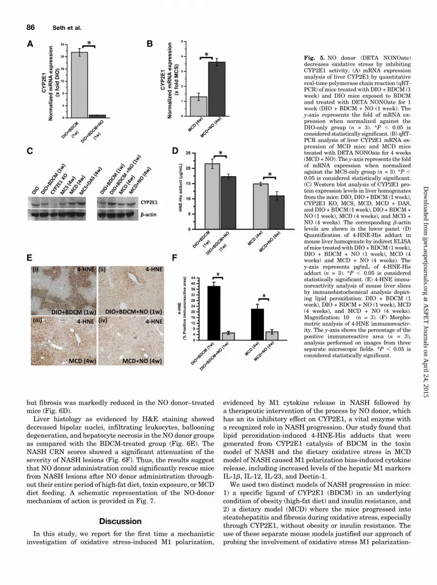
significantly decreased the mRNA levels of CYP2E1 at 1week after cotreatment with toxin and high-fat diet (termedas the initial phase) as compared with the DIO 1 BDCMgroup alone (*P , 0.05) (Fig. 5A). Surprisingly, the mRNAlevels of CYP2E1 were statistically significantly increased in
the MCD model of NASH after NO donor administration(Fig. 5B) (*P , 0.05). Western blot analysis of the CYP2E1protein showed no significant change in the liver levels of theprotein in either of the two models except in CYP2E1-nullmice (Fig. 5C).Because NO donor has been shown to inhibit CYP2E1
activity and we could not identify a direct assay system toquantify CYP2E1 activity from liver homogenates, weresorted to the CYP2E1-induced lipid peroxidation as anindex of CYP2E1 activity. ELISA and immunohistochemistrywere used to quantify the lipid peroxidation product 4-HNE-His adducts after NO donor administration. Our resultsshowed that there was a significant decrease in 4-HNE-Hisadducts in NO donor administered group as compared withthe DIO 1 BDCM group at 1 week after study initiation,a time point when both increased oxidative stress and M1polarization were observed (Fig. 5D) (*P , 0.05). Theseresults were also observed in MCD model of NASH coad-ministered with NO donor. The NO donor group of MCD-dietmice had a statistically significant decrease in 4-HNE-Hisadducts as compared with the MCD group (Fig. 5D) (*P ,0.05).Immunoreactivity to 4-HNE the lipid peroxidation product
was also statistically significantly decreased in NO donorgroup for both the BDCMmodel and theMCDmodel of NASH(Fig. 5, E and F) (*P, 0.05). These results strongly suggestedthat NO donor administration significantly decreasedCYP2E1-induced oxidative stress, which has been shown tobe directly correlated to M1 polarization and NASHprogression.Our conclusions were strengthened by supplemental data
provided by serum alanine aminotransferase and high-mobility group protein B1 mRNA expressions. The results
Fig. 3. Chronic exposure of BDCM leadsto late M2 phenotypic shift. (A) quantita-tive real-time polymerase chain reaction(qRT-PCR) analysis of liver IL-4 and IL-13mRNA expression in mice treated withDIO, DIO + BDCM (4 weeks), and GdCl3.The y-axis represents the fold of mRNAexpression when normalized against theDIO-only group (n = 3). *P , 0.05 isconsidered statistically significant. (B)qRT-PCR analysis of liver IL-4 and IL-13mRNA expression of MCS diet and MCDdiet mice. The y-axis represents the fold ofmRNA expression when normalizedagainst the MCS-only group, n = 3. *P ,0.05 is considered statistically significant.(C) Immunofluorescence images of IL-13immunoreactivity (red), counterstainedwith DAPI (blue) of paraffin embeddedliver sections from (i) DIO, (ii) DIO +BDCM (4 weeks), (iii) GdCl3, (iv) MCS,and (v) MCD. Magnification: 20� (n = 3).
84 Seth et al.
at ASPE
T Journals on A
pril 24, 2015jpet.aspetjournals.org
Dow
nloaded from

showed that serum alanine aminotransferase levels and high-mobility group protein B1 mRNA expression were signifi-cantly decreased in the NO-donor administered groups ascompared with the MCD or DIO 1 BDCM groups (Supple-mental Figs. 1 and 2), which suggests that the markeddecrease in CYP2E1 activity after NO donor administrationdecreases the bioactivation of BDCM and toxicity in MCDgroup.NO Donor Administration Rescues Mice from NASH
Progression. NASH progression is marked by a proinflam-matory stage followed mostly by a fibrotic stage (Farrell andLarter, 2006; Bohinc and Diehl, 2012). Hepatic stellate cellstransform into a more activated and proliferative phenotype(Mann and Marra, 2010; Bohinc and Diehl, 2012). There isincreased collagen deposition and a buildup of extracellularmatrix. If unchecked, this phenomenon can lead to scarring ofthe liver and ultimately can lead to liver cirrhosis (Ikejimaet al., 2007). To study whether attenuation of M1 polarizationby NO donor administration also rescued livers from the
progressive NASH phenotype, we performed experiments inboth models of NASH to study stellate cell proliferation andcollagen deposition, and we performed histopathologic exami-nations of the affected livers using the NASH CRN (NationalInstitutes of Health Nonalcoholic Steatohepatitis ClinicalResearch Network) scoring system.Our results showed that administration of NO donor in both
models of NASH significantly decreased the mRNA expres-sion of a-SMA, a biomarker for stellate cell proliferation (Fig.6, A and B) (*P, 0.05). The immunohistochemical analyses ofliver slices from both the toxin and MCD models of NASHshowed significant increased immunoreactivity of a-SMA inthe sinusoidal regions of BDCM-treated NASH mice andMCD-diet mice (Fig. 6Ci and iii); there was decreasedimmunoreactivity for a-SMA in the NO-donor treated micelivers that were either in the toxin-administered group or intheMCD-diet group (Fig. 6Cii and iv). PicroSirius red stainingfor collagen deposition in the livers showed higher periportalfibrosis in both the toxin-treated mice and theMCD-diet mice,
Fig. 4. NO donor DETA NONOateattenuates the CYP2E1-mediated M1polarization bias. (A) mRNA expressionanalysis by quantitative real-time poly-merase chain reaction (qRT-PCR) of liverIL-1b, IL-12, IL-23, and Dectin-1 fromDIO mice, DIO + BDCM (1 week) mice,and DIO mice exposed to BDCM andtreated with DETA NONOate (DIO +BDCM + NO) for 1 week. The y-axisrepresents the fold of mRNA expressionwhen normalized against the DIO-onlygroup (n = 3). *P , 0.05 is consideredstatistically significant. (B) qRT-PCRanalysis of liver IL-1b, IL-12, IL-23, andDectin-1 mRNA expression of MCS mice,MCD mice, and MCD mice treated withDETANONOate for 4 weeks (MCD +NO).The y-axis represents the fold of mRNAexpression when normalized against MCS-only group (n = 3). *P , 0.05 is consideredstatistically significant. (C) Analysis ofIL-1b immunoreactivity (red) by immuno-fluorescence imaging of paraffin-embeddedliver sections, counterstained with DAPI(blue) from (i) DIO, (ii) DIO + BDCM(1week), (iii) DIO + BDCM+NO, (iv)MCS,(v) MCD, and (vi) MCD + NO. Magnifica-tion: 20� (n = 3). (D) Immunofluorescenceimages of IL-12 immunoreactivity (red) ofparaffin-embedded liver sections counter-stained with DAPI (blue) from (i) DIO, (ii)DIO + BDCM (1 week), (iii) DIO + BDCM+NO, (iv) MCS, (v) MCD, and (vi) MCD +NO. Magnification: 20� (n = 3).
NO Donor Attenuates Inflammation in NASH Liver 85
at ASPE
T Journals on A
pril 24, 2015jpet.aspetjournals.org
Dow
nloaded from
but fibrosis was markedly reduced in the NO donor–treatedmice (Fig. 6D).Liver histology as evidenced by H&E staining showed
decreased bipolar nuclei, infiltrating leukocytes, ballooningdegeneration, and hepatocyte necrosis in the NO donor groupsas compared with the BDCM-treated group (Fig. 6E). TheNASH CRN scores showed a significant attenuation of theseverity of NASH lesions (Fig. 6F). Thus, the results suggestthat NO donor administration could significantly rescue micefrom NASH lesions after NO donor administration through-out their entire period of high-fat diet, toxin exposure, or MCDdiet feeding. A schematic representation of the NO-donormechanism of action is provided in Fig. 7.
DiscussionIn this study, we report for the first time a mechanistic
investigation of oxidative stress-induced M1 polarization,
evidenced by M1 cytokine release in NASH followed bya therapeutic intervention of the process by NO donor, whichhas an its inhibitory effect on CYP2E1, a vital enzyme witha recognized role in NASH progression. Our study found thatlipid peroxidation-induced 4-HNE-His adducts that weregenerated from CYP2E1 catalysis of BDCM in the toxinmodel of NASH and the dietary oxidative stress in MCDmodel of NASH caused M1 polarization bias-induced cytokinerelease, including increased levels of the hepatic M1 markersIL-1b, IL-12, IL-23, and Dectin-1.We used two distinct models of NASH progression in mice:
1) a specific ligand of CYP2E1 (BDCM) in an underlyingcondition of obesity (high-fat diet) and insulin resistance, and2) a dietary model (MCD) where the mice progressed intosteatohepatitis and fibrosis during oxidative stress, especiallythrough CYP2E1, without obesity or insulin resistance. Theuse of these separate mouse models justified our approach ofprobing the involvement of oxidative stress M1 polarization-
Fig. 5. NO donor (DETA NONOate)decreases oxidative stress by inhibitingCYP2E1 activity. (A) mRNA expressionanalysis of liver CYP2E1 by quantitativereal-time polymerase chain reaction (qRT-PCR) of mice treated with DIO + BDCM (1week) and DIO mice exposed to BDCMand treated with DETA NONOate for 1week (DIO + BDCM + NO (1 week). They-axis represents the fold of mRNA ex-pression when normalized against theDIO-only group (n = 3). *P , 0.05 isconsidered statistically significant. (B) qRT-PCR analysis of liver CYP2E1 mRNA ex-pression of MCD mice and MCD micetreated with DETA NONOate for 4 weeks(MCD+NO). The y-axis represents the foldof mRNA expression when normalizedagainst the MCS-only group (n = 3). *P ,0.05 is considered statistically significant.(C) Western blot analysis of CYP2E1 pro-tein expression levels in liver homogenatesfrom themice: DIO, DIO +BDCM (1 week),CYP2E1 KO, MCS, MCD, MCD + DAS,and DIO +BDCM (1week), DIO + BDCM+NO (1 week), MCD (4 weeks), and MCD +NO (4 weeks). The corresponding b-actinlevels are shown in the lower panel. (D)Quantification of 4-HNE-His adduct inmouse liver homogenate by indirect ELISAofmice treated withDIO +BDCM (1week),DIO + BDCM + NO (1 week), MCD (4weeks) and MCD + NO (4 weeks). They-axis represents pg/mL of 4-HNE-Hisadduct (n = 3). *P , 0.05 is consideredstatistically significant. (E) 4-HNE immu-noreactivity analysis of mouse liver slicesby immunohistochemical analysis depict-ing lipid peroxidation: DIO + BDCM (1week), DIO + BDCM + NO (1 week), MCD(4 weeks), and MCD + NO (4 weeks).Magnification: 10� (n = 3). (F) Morpho-metric analysis of 4-HNE immunoreactiv-ity. The y-axis shows the percentage of thepositive immunoreactive area (n = 3),analysis performed on images from threeseparate microscopic fields. *P , 0.05 isconsidered statistically significant.
86 Seth et al.
at ASPE
T Journals on A
pril 24, 2015jpet.aspetjournals.org
Dow
nloaded from

induced cytokine release without having to limit our in-terpretation to a single dietary model of NASH.We previously showed that BDCM exposure in mice fed
a high-fat diet caused NASH (Das et al., 2013a; Seth et al.,
2013). Thesemice had an increased proinflammatory cytokinerelease (at 1 week) followed by a progressive fibrotic stage (upto 4 weeks). The increased proinflammatory phase wasprimarily related to CYP2E1-mediated oxidative stress and
Fig. 6. NO donor (DETANONOate) prevents NASH progression by inhibiting CYP2E1 activity and the resultant oxidative stress. (A) mRNA expressionof liver a-SMA by quantitative real-time polymerase chain reaction (qRT-PCR) of DIO mice exposed to BDCM (DIO + BDCM) for 4 weeks, and DIO +BDCM mice treated with NO donor (DIO + BDCM + NO) for 4 weeks. The y-axis represents the fold of mRNA expression when normalized against theDIO-only group, n = 3. *P , 0.05 is considered statistically significant. (B) qRT-PCR analysis of liver a-SMA mRNA expression of MCD mice and MCDmice treated with DETA NONOate (MCD + NO) for 4 weeks. The y-axis represents the fold of mRNA expression when normalized against the MCS-onlygroup, n = 3. *P , 0.05 is considered statistically significant. (C) The a-smooth muscle actin immunoreactivity as shown by immunohistochemistry inliver slices of (i) DIO mice exposed to BDCM; (ii) DIO + BDCM + NO; (iii) MCD mice, and (iv) MCD + NO. Magnification: 20� magnification (n = 3). (D)PicroSirius red staining of liver sections of (i) DIO mice exposed to BDCM; (ii) DIO + BDCM + NO; (iii) MCD mice, and (iv) MCD + NO. Magnification:20� (n = 3). The red staining shows macrovesicular and microvesicular fibrosis. (E) H&E staining of liver sections of (i) DIO mice exposed to BDCM; (ii)DIO + BDCM + NO; (iii) MCD mice, and (iv) MCD + NO. Magnification: 10� (n = 3). (F) Stained liver sections were reviewed for stages of fibrosis usingthe criteria of the NIH Non Alcoholic Steatohepatitis Clinical Research Network (NIH NASH CRN). The table depicts the NASH CRN scores for DIO,DIO + BDCM, DIO + BDCM + NO, MCS, MCD, and MCD + NO.
NO Donor Attenuates Inflammation in NASH Liver 87
at ASPE
T Journals on A
pril 24, 2015jpet.aspetjournals.org
Dow
nloaded from

inflammosome activation due to priming of P2X7r (Das et al.,2013b). Others have studied TH1 polarization in NASH indetail (Ferreyra Solari et al., 2012; Maina et al., 2012; Suttiet al., 2014). The higher incidence of interferon-g release isprimarily from a heightened TH1 response and is found to bedue to oxidative stress–mediated malonyldialdehyde adductsthat elicit a strong autoantibody response (Sutti et al., 2014).The adaptive immune response, then, is responsible formacrophage polarization mainly through the M1 phenotype(Sutti et al., 2014).Our observation of decreased hepatic M1 markers in
CYP2E1-null mice and in mice treated with the CYP2E1inhibitor DAS in both models of NASH indicates a closeassociation of CYP2E1-induced oxidative stress in causingM1polarization bias and an increase in M1 markers. Impor-tantly, our approach to depletingmacrophages by using GdCl3in the liver was based on identifying the precise origin of theinflammatory cytokines, especially IL-1b, IL-12, IL-23, andDectin-1; we were well aware that IL-1b release has othercellular sources in the liver as well. Our results with CYP2E1-mediated M1 polarization in NASH provide insight into themechanisms of macrophage responses, which had been scarceaside from a few isolated studies where morbidly obesepatients with NASH etiology were shown to produce in-creased TH1 cytokines and Kupffer cell responses (Fukushimaet al., 2009; Bertola et al., 2010; Bieghs et al., 2013). There hasbeen preclinical evidence for the involvement of Kupffer cellswith an M1 phenotype, the former being responsible forNASH progression, but those studies did not clarify the role ofoxidative stress, especially mediated by CYP2E1, in causinga M1 polarization bias (Fukushima et al., 2009; Bieghs et al.,2013).Important from the mechanistic point of view, we observed
an increase in hepatic NO levels in the diet 1 toxin NASHmodel. NO, released from any of the three NOS upon theiractivation, plays a significant role in inflammation (Fujitaet al., 2010; Ajamieh et al., 2012). However, the inducible formof the NOS has been ascribed to the proinflammatoryphenotype more often (Fujita et al., 2010; Maina et al.,2012). Our results showed that NO levels reached a 4-foldincrease as early as 24 hours after coexposure with diet andtoxin to induce NASH, and they stayed at the same level forthe entire study period. Early use of 1400W decreased the NOlevels. Interestingly, the use of NOS2-null mice at a late stage(4 weeks) did not affect the NO levels in NASH, suggesting
that other NOS isoforms, especially NOS3 might be involvedin the sustained higher NO levels at the late stage in ourstudy. Moreover, the observed increase in NO may be anadaptive response of the injured liver. Ajamieh et al. (2012)found that there were increases in NOS3 levels and NOS3phosphorylations after liver injury in NASH, and they werefound to be protective against NASH. Based on theseobservations, we argued that though NO is consideredproinflammatory and an M1 marker, the kinetics of NOrelease and its concentration in the hepatic microenviron-ment might play an adaptive and perhaps a protective role inthe pathophysiology of NASH.To alter the NO levels we used a dual approach. We used
NOS2-null mice and a nonspecific inhibitor of NOS. Sub-sequently, we used an NO donor DETA NONOate foradministration through the intraperitoneal route to increasethe in vivo concentration of NO, assuming that the increase inNO was an adaptive response to the liver injury. Althoughblocking NO synthase did not change the levels of M1polarization markers or NASH symptoms (data not shown),treatment with an NO donor significantly lowered levels ofboth mRNA and protein expressions of M1 markers in thetoxin model but only protein levels in the MCD model ofNASH (Fig. 4, A–D), suggesting the existence of a plausiblemechanism for NO-mediated suppression of proinflammatoryevents in early NASH developmental stages.Having identified an association of CYP2E1-induced oxida-
tive stress as a mediator in M1 polarization, we studied therole of a lateM2 phenotype thatmight cause fibrosis, a featurethat is of paramount importance in the human form of thedisease. Results from our studies showed that mice with high-fat diet–induced obesity and with coexposure to the toxinBDCM had increased mRNA and protein expressions of theprominent M2 markers IL-4 and IL-13; however, the micethat were pretreated with themacrophage toxin GdCl3 did notshow the IL-4 and IL-13 increases, suggesting that themacrophage origin contributed at least partly to the M2phenotypic shift in the late stage of the disease. Because theNO donor prevented M1 polarization bias, we also wouldargue that a higher NO concentration from the initial phasesof disease development might resist the proinflammatoryevents in early stages of NASH, with a correspondingdecrease in the M2 phenotypic response, thus attenuatingNASH progression.Because M1 polarization was associated with CYP2E1-
mediated oxidative stress and CYP2E1 activity is stronglyinhibited by NO, we studied the role of NO donor (DETANONOate) in abrogating oxidative stress, mediated byCYP2E1activity. Our findings of a strong inhibition of 4-HNE-Hisadducts in both the models of NASH were in completeagreement with the pioneering study by Gergel et al. (1997),who showed that CYP2E1 catalytic activity and reactiveoxygen radical formation were inhibited by NO (Fig. 5). Ourobservation that there was no change in the CYP2E1 proteinlevel after NO donor administration also confirmed that theNO donor inhibited CYP2E1 activity in the in vivo system weused for our study.NASH pathophysiology is associated with stellate cell pro-
liferation, fibrosis, and hepatocellular necrosis (Bohinc andDiehl, 2012; Seth et al., 2013). NO donor administrationsignificantly decreased stellate cell proliferation, as evidencedby the decreased a-SMA protein levels, and decreased
Fig. 7. Schematic representation of the study. The graphics scheme showsthe proposedmechanism of action of DETANONOate. The red lines representthe checkpoints where the drug can have its potential inhibitory role.
88 Seth et al.
at ASPE
T Journals on A
pril 24, 2015jpet.aspetjournals.org
Dow
nloaded from

hepatocellular necrosis, inflammation, and fibrosis, as shownby the NASH CRN scores (Fig. 6). Thus, taken together, thisstudy showed that oxidative stress mediated by CYP2E1induced M1 polarization-induced cytokines, which, in tur,nmay have been responsible for late anti-inflammatory eventsthat resulted in fibrosis. Mechanistically, administration of NOdonor that increased the NO levels in the liver significantly(6.6-fold as compared with 3.4-fold; data not shown) could blockCYP2E1 activity and the subsequent inflammatory events thatlead to NASH pathophysiology. Our study offers a therapeuticapproach through the use ofNOdonors as prospective drugs forNASH treatment and leads the way to more detailed insightinto developing newer effective pharmacologic strategies in-volving synthetic NO donors with longer half-lives to containNASH-related comorbidities.
Acknowledgments
The authors thank Benny Davidson at the InstrumentationResource Facility (IRF), University of South Carolina School ofMedicine for technical services. The authors also thank Dr. JamesCarson, Department of Exercise Science and the IRF at the Universityof South Carolina for equipment usage and consulting services.
Authorship Contributions
Participated in research design: Chatterjee, Seth.Conducted experiments: Seth, Das, Pourhosseini, Dattaroy, Igwe.Contributed new reagents or analytic tools: Diehl, Michelotti.Performed data analysis: Chatterjee, Das, Seth.Wrote or contributed to the writing of the manuscript: Chatterjee,
Fan, Diehl, Basu-Ray, Michelotti.
References
Abdelmegeed MA, Banerjee A, Jang S, Yoo SH, Yun JW, Gonzalez FJ, KeshavarzianA, and Song BJ (2013) CYP2E1 potentiates binge alcohol-induced gut leakiness,steatohepatitis, and apoptosis. Free Radic Biol Med 65:1238–1245.
Abdelmegeed MA, Banerjee A, Yoo SH, Jang S, Gonzalez FJ, and Song BJ (2012)Critical role of cytochrome P450 2E1 (CYP2E1) in the development of high fat-induced non-alcoholic steatohepatitis. J Hepatol 57:860–866.
Aitken AE, Lee CM, and Morgan ET (2008) Roles of nitric oxide in inflammatorydownregulation of human cytochromes P450. Free Radic Biol Med 44:1161–1168.
Ajamieh H, Farrell G, Wong HJ, Yu J, Chu E, Chen J, and Teoh N (2012) Atorvastatinprotects obese mice against hepatic ischemia-reperfusion injury by Toll-like receptor-4suppression and endothelial nitric oxide synthase activation. J Gastroenterol Hepatol27:1353–1361.
Aubert J, Begriche K, Knockaert L, Robin MA, and Fromenty B (2011) Increasedexpression of cytochrome P450 2E1 in nonalcoholic fatty liver disease: mechanismsand pathophysiological role. Clin Res Hepatol Gastroenterol 35:630–637.
Bertola A, Bonnafous S, Anty R, Patouraux S, Saint-Paul MC, Iannelli A, GugenheimJ, Barr J, Mato JM, and Le Marchand-Brustel Y, et al. (2010) Hepatic expressionpatterns of inflammatory and immune response genes associated with obesity andNASH in morbidly obese patients. PLoS ONE 5:e13577.
Bieghs V, Walenbergh SM, Hendrikx T, van Gorp PJ, Verheyen F, Olde Damink SW,Masclee AA, Koek GH, Hofker MH, and Binder CJ, et al. (2013) Trapping of oxi-dized LDL in lysosomes of Kupffer cells is a trigger for hepatic inflammation. LiverInt 33:1056–1061.
Bohinc BN and Diehl AM (2012) Mechanisms of disease progression in NASH: newparadigms. Clin Liver Dis 16:549–565.
Caro AA and Cederbaum AI (2004) Oxidative stress, toxicology, and pharmacology ofCYP2E1. Annu Rev Pharmacol Toxicol 44:27–42.
Chatterjee S, Ehrenshaft M, Bhattacharjee S, Deterding LJ, Bonini MG, Corbett J,Kadiiska MB, Tomer KB, and Mason RP (2009) Immuno-spin trapping of a post-translational carboxypeptidase B1 radical formed by a dual role of xanthine oxidase andendothelial nitric oxide synthase in acute septic mice. Free Radic Biol Med 46:454–461.
Chatterjee S, Ganini D, Tokar EJ, Kumar A, Das S, Corbett J, Kadiiska M, WaalkesM, Diehl AM, and Mason RP (2013) Leptin is key to peroxynitrite-mediated oxi-dative stress and Kupffer cell activation in experimental non-alcoholic steatohepatitis.J Hepatol 58:778–784.
Chatterjee S, Premachandran S, Bagewadikar RS, Bhattacharya S, ChattopadhyayS, and Poduval TB (2006) Arginine metabolic pathways determine its therapeuticbenefit in experimental heatstroke: role of Th1/Th2 cytokine balance. Nitric Oxide15:408–416.
Cheung O and Sanyal AJ (2009) Recent advances in nonalcoholic fatty liver disease.Curr Opin Gastroenterol 25:230–237.
Copaci I, Micu L, and Voiculescu M (2006) The role of cytokines in non-alcoholicsteatohepatitis. A review. J Gastrointestin Liver Dis 15:363–373.
Das S, Kumar A, Seth RK, Tokar EJ, Kadiiska MB, Waalkes MP, Mason RP,and Chatterjee S (2013a) Proinflammatory adipokine leptin mediates disinfectionbyproduct bromodichloromethane-induced early steatohepatitic injury in obesity.Toxicol Appl Pharmacol 269:297–306.
Das S, Seth RK, Kumar A, Kadiiska MB, Michelotti G, Diehl AM, and Chatterjee S(2013b) Purinergic receptor X7 is a key modulator of metabolic oxidative stress-mediated autophagy and inflammation in experimental nonalcoholic steatohepatitis.Am J Physiol Gastrointest Liver Physiol 305:G950–G963.
de Oliveira CP, de Lima VM, Simplicio FI, Soriano FG, de Mello ES, de Souza HP,Alves VA, Laurindo FR, Carrilho FJ, and de Oliveira MG (2008) Prevention andreversion of nonalcoholic steatohepatitis in OB/OB mice by S-nitroso-N-acetylcysteine treatment. J Am Coll Nutr 27:299–305.
Farrell GC and Larter CZ (2006) Nonalcoholic fatty liver disease: from steatosis tocirrhosis. Hepatology 43(2, Suppl 1)S99–S112.
Ferreyra Solari NE, Inzaugarat ME, Baz P, De Matteo E, Lezama C, Galoppo M,Galoppo C, and Cherñavsky AC (2012) The role of innate cells is coupled to a Th1-polarized immune response in pediatric nonalcoholic steatohepatitis. J ClinImmunol 32:611–621.
Fujita K, Nozaki Y, Yoneda M, Wada K, Takahashi H, Kirikoshi H, Inamori M, SaitoS, Iwasaki T, and Terauchi Y, et al. (2010) Nitric oxide plays a crucial role in thedevelopment/progression of nonalcoholic steatohepatitis in the choline-deficient, l-amino acid-defined diet-fed rat model. Alcohol Clin Exp Res 34 (Suppl 1):S18–S24.
Fukushima J, Kamada Y, Matsumoto H, Yoshida Y, Ezaki H, Takemura T, Saji Y,Igura T, Tsutsui S, and Kihara S, et al. (2009) Adiponectin prevents progression ofsteatohepatitis in mice by regulating oxidative stress and Kupffer cell phenotypepolarization. Hepatol Res 39:724–738.
Ganz M and Szabo G (2013) Immune and inflammatory pathways in NASH. HepatolInt 7:771–781.
Gergel D, Misík V, Riesz P, and Cederbaum AI (1997) Inhibition of rat and humancytochrome P4502E1 catalytic activity and reactive oxygen radical formation bynitric oxide. Arch Biochem Biophys 337:239–250.
Gong P, Cederbaum AI, and Nieto N (2004) The liver-selective nitric oxide donor O2-vinyl 1-(pyrrolidin-1-yl)diazen-1-ium-1,2-diolate (V-PYRRO/NO) protects HepG2cells against cytochrome P450 2E1-dependent toxicity.Mol Pharmacol 65:130–138.
Hu JJ, Yoo JS, Lin M, Wang EJ, and Yang CS (1996) Protective effects of diallylsulfide on acetaminophen-induced toxicities. Food Chem Toxicol 34:963–969.
Ikejima K, Okumura K, Kon K, Takei Y, and Sato N (2007) Role of adipocytokines inhepatic fibrogenesis. J Gastroenterol Hepatol 22 (Suppl 1):S87–S92.
Liu YC, Zou XB, Chai YF, and Yao YM (2014) Macrophage polarization in in-flammatory diseases. Int J Biol Sci 10:520–529.
Maina V, Sutti S, Locatelli I, Vidali M, Mombello C, Bozzola C, and Albano E (2012)Bias in macrophage activation pattern influences non-alcoholic steatohepatitis(NASH) in mice. Clin Sci (Lond) 122:545–553.
Mann DA and Marra F (2010) Fibrogenic signalling in hepatic stellate cells. J Hepatol52:949–950.
Pasarín M, La Mura V, Gracia-Sancho J, García-Calderó H, Rodríguez-Vilarrupla A,García-Pagán JC, Bosch J, and Abraldes JG (2012) Sinusoidal endothelial dysfunc-tion precedes inflammation and fibrosis in a model of NAFLD. PLoS ONE 7:e32785.
Seth RK, Das S, Kumar A, Chanda A, Kadiiska MB, Michelotti G, Manautou J, DiehlAM, and Chatterjee S (2014) CYP2E1-dependent and leptin-mediated hepaticCD57 expression on CD81 T cells aid progression of environment-linked non-alcoholic steatohepatitis. Toxicol Appl Pharmacol 274:42–54.
Seth RK, Kumar A, Das S, Kadiiska MB, Michelotti G, Diehl AM, and Chatterjee S(2013) Environmental toxin-linked nonalcoholic steatohepatitis and hepatic met-abolic reprogramming in obese mice. Toxicol Sci 134:291–303.
Shimamura T, Fujisawa T, Husain SR, Kioi M, Nakajima A, and Puri RK (2008)Novel role of IL-13 in fibrosis induced by nonalcoholic steatohepatitis and itsamelioration by IL-13R-directed cytotoxin in a rat model. J Immunol 181:4656–4665.
Sutti S, Jindal A, Locatelli I, Vacchiano M, Gigliotti L, Bozzola C, and Albano E(2014) Adaptive immune responses triggered by oxidative stress contribute to he-patic inflammation in NASH. Hepatology 59:886–897.
Tilg H and Moschen AR (2008) Inflammatory mechanisms in the regulation of insulinresistance. Mol Med 14:222–231.
Tilg H and Moschen AR (2010) Evolution of inflammation in nonalcoholic fatty liverdisease: the multiple parallel hits hypothesis. Hepatology 52:1836–1846.
Tomasi A, Albano E, Biasi F, Slater TF, Vannini V, and Dianzani MU (1985) Acti-vation of chloroform and related trihalomethanes to free radical intermediates inisolated hepatocytes and in the rat in vivo as detected by the ESR-spin trappingtechnique. Chem Biol Interact 55:303–316.
Whitsett J, Picklo MJ, Sr, and Vasquez-Vivar J (2007) 4-Hydroxy-2-nonenal increa-ses superoxide anion radical in endothelial cells via stimulated GTP cyclohydrolaseproteasomal degradation. Arterioscler Thromb Vasc Biol 27:2340–2347.
Zhou D, Kong L, Zhou Q, and Li J (2014) Skewing KC phenotypic polarization intoM2 via the intervention of oxidized LDL as potential therapeutic implications forNASH. Liver Int 34:815–816.
Address correspondence to: Dr. Saurabh Chatterjee, EnvironmentalHealth and Disease Laboratory, Department of Environmental HealthSciences, University of South Carolina, Columbia, SC 29208. E-mail: [email protected]
NO Donor Attenuates Inflammation in NASH Liver 89
at ASPE
T Journals on A
pril 24, 2015jpet.aspetjournals.org
Dow
nloaded from