lumbar kinematic variability during gait in chronic low back pain and associations with pain,...
TRANSCRIPT

1
TITLE: Lumbar Kinematic Variability during Gait in Chronic Low Back Pain and Associations
with Pain, Disability and Isolated Lumbar Extension Strength
AUTHORS: James Steelea, Stewart Bruce-Lowa, Dave Smithb, David Jessopa, Neil Osbornec
INSTITUTIONS: Centre for Health, Exercise and Sport Science, Southampton Solent
Universitya, Department of Exercise & Sport Science, Manchester Metropolitan Universityb,
AECC Clinic, Anglo European College of Chiropracticc
CORRESPONDING AUTHOR: James Steele
CORRESPONDENCE ADDRESS: Centre for Health, Exercise and Sport Science,
Southampton Solent University, East Park Terrace, Southampton, Hampshire, UK, SO14 0YN
CORRESPONDENCE TELEPHONE: Business (Mobile): +447878127785, Home
+4402380908139
CORRESPONDENCE EMAIL: [email protected]
Abstract word count: 243
Manuscript word count: 3881
Figure count: 4
Table count: 1

2
Abstract
Background: Chronic low back pain is a multifactorial condition with a variety of dysfunctions;
one being gait variability. The lumbar spine and its musculature are involved during gait and
in chronic low back pain the lumbar extensors are often deconditioned. It was therefore of
interest to examine relationships between lumbar kinematic variability during gait, with pain,
disability and isolated lumbar extension strength in participants with chronic low back pain.
Methods: Twenty four participants with chronic low back pain were assessed for lumbar
kinematics during gait, isolated lumbar extension strength, pain, and disability. Angular
displacement and kinematic waveform pattern and offset variability were examined.
Findings: Angular displacement and kinematic waveform pattern and offset variability differed
across movement planes; displacement highest and similar in frontal and transverse planes,
and pattern variability and offset variability higher in the sagittal plane compared to frontal and
transverse planes which were similar. Spearman’s correlations showed significant correlations
between transverse plane pattern variability and isolated lumbar extension strength (r = -.411)
and disability (r = .401). However, pain was not correlated with pattern variability in any plane.
The square of the correlation coefficients suggested 80.5% to 86.3% of the variance for
significant correlations was accounted for by other variables.
Interpretation: Considering the role of the lumbar extensors in gait, the relationship between
both isolated lumbar extension strength and disability with transverse plane pattern variability
suggests gait variability may result in consequence of lumbar extensor deconditioning or
disability accompanying chronic low back pain. However, further prospective study should
examine the temporality of the relationships reported here and what other variables might
account for the unexplained variance.
Key Words: Lumbar spine; Gait variability; VICON; MedX

3
1. Introduction
Chronic low back pain (CLBP) is a highly prevalent musculoskeletal disorder [1-4] with costs
amounting to billions worldwide [5-13]. Despite its prevalence, in as much as 85% of LBP
cases no specific patho-anatomical diagnosis can be found [14]. However, more recently it is
acknowledged as a multifactorial condition with a variety of associated dysfunctions [15,16].
One of the multifactorial dysfunctions reported is gait variability [17-19]. It has been suggested
that deficiencies in motor control during gait may produce excessive stresses to the lumbar
spine, which may contribute to development of CLBP [18]. However, a recent review has
suggested there is evidence against walking itself being causally associated with CLBP [19].
Healthy participants demonstrate relatively low stride-to-stride variability in lumbar kinematic
patterns during both level and incline gait [20]. However, greater stride-to-stride variability at
the lumbar spine in all movement planes [18], greater frontal plane coordination variability of
the pelvis and trunk [21,22] and more rigid transverse plane coordination variability of the
pelvis and trunk [21,23,24] is reported in participants with CLBP compared with healthy
controls. It also appears that pain per se may not be responsible for these gait differences.
Lumbar spine kinematics during gait appear to be complex and developed over time, as
patterns are evident before pain is experienced [25] and both induced pain and fear of pain
produce little change in muscle activity in CLBP patients [26]. Indeed recently studies have
shown that even those with a previous history of CLBP who are currently asymptomatic
demonstrate abnormal gait patterns [22,27]. Thus pain per se may not be the factor
responsible. There is contrasting evidence reporting no residual effect upon gait from an
episode of low back pain in nurses returning to work with very low pain levels [28]; however
this study lacked a directly comparable control group.
Evidence instead suggests the lumbar extensor musculature might play a role in gait variability
in CLBP [21,26,29,30-35]. It appears that the kinematic patterns seen in participants with
CLBP are combined with poorer erector spinae activity adaptability to unexpected

4
perturbations [26], or walking velocity changes [23]. In fact, the findings of numerous studies
are suggestive of muscular dysfunction of the lumbar extensors during gait in those with CLBP
compared with asymptomatic controls [21,26,29,30,31]. Hanada et al. [35] also report that
where asymptomatic controls significantly activated their rectus abdominus and internal
obliques more, symptomatic participants had significantly greater activation of the lumbar
extensors. More recent work shows evidence of greater lumbar extensor activity in participants
with CLBP compared with controls [32], at a range walking velocities [33], and that neither
disability nor fear of movement is associated with this greater activity [32]. However, different
coping strategies may be associated with either greater activity (catastrophizing) or greater
relaxation during double support (distraction) suggesting some influence of cognitive control
[34].
Human gait is normally quite robust in the face of muscular weakness of the lower limbs [360].
The lumbar spine, however, may play a primary role in human bipedal gait [37]. It is possible
that the greater activation of the lumbar extensors, and altered lumbar spine kinematics during
gait in participants with CLBP, are a manifestation of the lumbar extensor deconditioning (i.e.
educed lumbar extensor strength/endurance, atrophy, and excessive fatigability) commonly
associated with CLBP [38]. Greater activation in the face of fatigue due to deconditioning could
be a compensatory attempt to maintain control of the lumbar spine during gait. Hart et al., [39]
demonstrate that inducing fatigue in the lumbar extensors impacts lumbar kinematics during
running gait of healthy participants and participants with CLBP. Arjunan et al. [40] also show
significantly greater lumbar extensor activity during running gait in participants with CLBP.
Indeed, prospective evidence has demonstrated that reduced lumbar extensor
strength/endurance, atrophy, and excessive fatigability increase risk of low back injury and
LBP in asymptomatic persons [38]. Thus it may be responsible for the development of the gait
variability associated with CLBP also.

5
Considering this it was therefore of interest in the present study to examine the relationships
between lumbar kinematic variability during gait, with pain, disability and isolated lumbar
extension (ILEX) strength. Previous research has focused upon trunk/pelvis co-ordination
[21,22,23,29,41,42,43]. Those interested in stride-to-stride variability of the lumbar spine with
respect to the pelvis instead have utilised Winter’s coefficient of variation (CV) [44] to examine
consistency of movement patterns using the ensemble average of the raw waveforms of
repeated trials [18,20]. However, a new method of differentiating between pattern and offset
variability has been recently suggested [45]. A large mean offset value effectively deflates the
value calculated for variability using the CV [45]. Because of this O’Dwyer et al. [45] have
suggested the use of methods to differentiate the offset from calculation of the variability in
the waveform pattern; the latter they suggest being far more representative of movement
replication whereas the offset incorporates a greater degree of other variance sources (i.e.
marker error). Thus this study in particular aimed to examine variation in lumbar kinematic
pattern variability in relation to pain, disability and isolated lumbar extension (ILEX) strength.
2. Methods
2.1 Study Design
The study was part of a wider investigation examining ILEX in participants with CLBP
published in part elsewhere [46]. Gait data were also collected as part of this wider
investigation. The present manuscript presents the cross-sectional data from the combined
sample of the study collected at baseline.
2.2 Participants
Thirty eight participants (males n = 21, females n = 17) were initially identified and recruited
into the wider investigation by posters, group email and word of mouth from a University and
the surrounding locality. Direct referral was also provided from a local private chiropractor
through posters in their practice. Inclusion criteria were as follows; participants suffered from
non-specific low back pain having lasted longer than 12 weeks [47] and had no medical

6
condition for which resistance training would be contraindicated. Exclusion criteria were as
follows; participants must have no medical condition for which movement therapy would be
contraindicated. These included: acute (not re-occurring) low back injury occurring within the
last 12 weeks, pregnancy, evidence of sciatic nerve root compression (sciatica), leg pain
radiating to below the knee, paraesthesia (tingling or numbness), current tension sign, lower
limb motor deficit, current disc herniation, previous vertebral fractures or other major structural
abnormalities. All participants were cleared prior to involvement in the study by either their
General Practitioner or the Chiropractor in the research group and provided written informed
consent. The study was approved by the NHS National Research Ethics Service,
Southampton & South West Hampshire Research Ethics Committee B (REC Reference:
11/H0504/9).
2.3 Equipment
Participants’ stature was measured using a stadiometer (Holtan ltd, Crymych, Dyfed), body
mass measured using scales (SECA, Germany) and Body Mass Index (BMI) calculated.
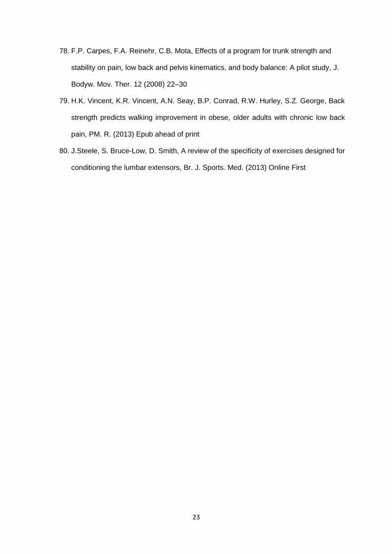
Isometric ILEX strength testing and ROM were performed using the MEDX (MedX, Ocala,
Florida; figure 1). Figure 1 shows the restraint system. The MEDX has been shown to be
reliable in assessing isometric strength at repeated angles in asymptomatic [48] and
symptomatic participants [49], and valid in measurement through removal of gravitational
effects [50] and pelvic movement [51]. Pain was measured using a 100mm point visual
analogue scale (VAS) [52], and disability measured using the revised Oswestry disability index
(ODI) [53]. Gait kinematic variables were captured at 500hz using a 10 MX T20 camera three
dimensional motion capture system (Vicon, Oxford) and analysed using both Vicon Nexus
software version 1.4.116 (Vicon, Oxford), MATLAB version R2012a (MathWorks, Cambridge)
and Microsoft Excel version 2010 (Microsoft, Reading).
2.4 Participant Testing

7
For baseline testing participants visited the lab on three occasions. Participants were required
to complete the VAS and the ODI on their first visit to the laboratory. The first two visits also
involved testing of isometric ILEX strength. This was tested on separate days (at least 72
hours apart in order to avoid the effects of residual fatigue or soreness). The first test acted
as a familiarisation and the data from the second test was used for analysis. Each test using
the lumbar extension machine involved maximal voluntary isometric contractions at various
angles through the participant’s full ROM. Details of the full test protocol using the lumbar
extension machine and details of the restraint mechanisms have been documented previously
elsewhere [48]. Because of individual differences between participants for lumbar ROM, ILEX
strength data was averaged across all angles tested. Gait data was collected using the Vicon
system during the third visit to the laboratory (again at least 72 hours after ILEX testing in
order to avoid the effects of residual fatigue or soreness).
2.5 Three dimensional motion analyses
Due to the lumbar spine’s capacity to rotate about three orthogonal axes, a three dimensional
approach was used for data collection. Ten cameras were set up and angled in a manner so
as to reduce hidden spots that might obscure data collection. The cameras were angled such
that the centre of the runway used for walking trials was the volume calibrated for data capture.
The cameras identified reflective markers attached to the participant and output three
dimensional coordinates for each marker. Data were recorded for 5 walking trials both pre and
post intervention. Participants walked barefoot from one end of a marked runway to the other
that was 8 metres in length at their free walking speed. The first full gait cycle captured where
the participants entered the calibration volume during each walking trial was used.
2.6 Biomechanical Model
The body of interest for the current study was the lumbar spine considered from S1 to T12
relative to the pelvis. For the purpose of analysis the lumbar spine was modelled as a rigid
segment. The reasoning for not considering intervertebral segment movements was due to

8
the small segments ranging from S2 to T10 always bending laterally toward the support leg
with little variation between segments during gait [54]. Lumbar spine data were collected
through three axes using the same model previously described by Scache et al. [55], which
has been shown to have high overall repeatability of angular parameters [56].
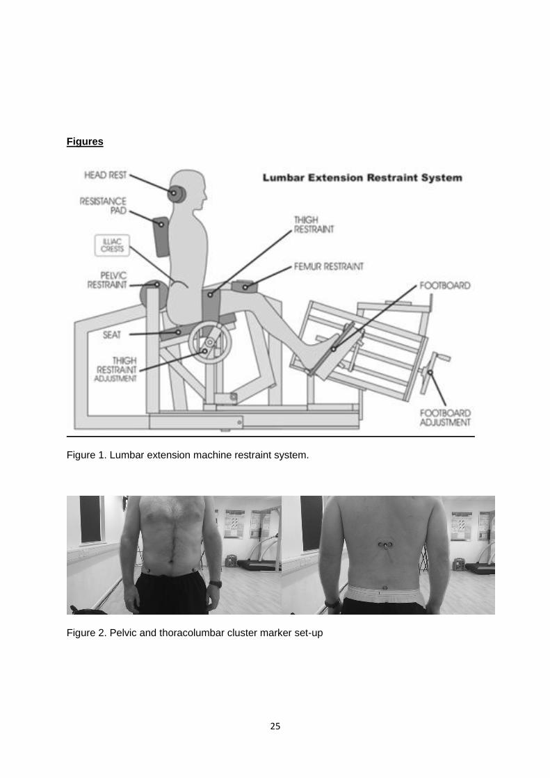
2.7 Marker Set Up
Reflective markers were placed over anatomical landmarks on the pelvis at both anterior
superior iliac spines (ASIS) and at the midpoint of the posterior superior iliac spine (PSIS).
Reflective markers were also used upon a thoracolumbar marker cluster similar to that used
by Schache et al., [55,56]. As with the biomechanical model, this marker set up has been
previously described elsewhere [55,56]. The only alteration in this present study was the use
of a flexible based wand marker for the thoraco-lumbar cluster. Two additional markers were
secured equidistant either side of the midpoint of the wand markers base. This was placed
over T12 with the mid-point of the base located over the spinous process. The ASIS and PSIS
were identified by palpation after identifying the iliac crest and palpating along its length. T12
was first located and marked using the technique suggested in Gray’s Anatomy for Students
[57]. This location was confirmed, whilst the participant was in a flexed standing position
supporting themselves upon a stool, by palpation and counting of the spinous processes from
this marked point down to the sacrum, and then double checked by counting back up to the
marked spinous process. All markers and the base of the thoracolumbar marker cluster were
secured using double sided adhesive tape. Markers were placed by the same investigator for
all gait trials. Figure 2 shows the marker set-up used.
2.8 Kinematic Data
Variability of angular kinematics of the lumbar spine about the three described axes relative
to the pelvic segment was of primary interest (i.e. movement of the thoraco-lumbar marker
cluster with respect to the pelvic markers). The Vicon Nexus software was used to run a
Bodybuilder (Vicon, Oxford) code pipeline to calculate joint angles as outputs using Cardan

9
(Euler) angles. The angles were calculated in the following order; 1) sagittal, 2) frontal, and 3)
transverse. As with the biomechanical model, the Bodybuilder code used was the same as
used by Schache et al. [55,56]. Data were filtered using a low pass Butterworth filter (fourth
order, cutoff frequency determined for each individual participant as sum of residuals closest
to zero using 4Hz, 6Hz, 8Hz, 10Hz, and 12Hz) and normalised to percentage gait cycle
corresponding to initial right heel contact (0%) and subsequent right heel contact (100%). Heel
contacts were identified as the lowest vertical displacement of a right heel marker. Stride
duration and length was also calculated using the horizontal displacement of the right heel
marker from initial right heel contact and subsequent right heel contact. Mean values for
angular displacements, stride-to-stride intra-subject variability using CVp and CVo, were
calculated for lumbar spine kinematics relative to the pelvis across all three planes of
movement.
Intra-subject variability was calculated using coefficient of variation of the ensemble average
(average of the normalised time series data at each interval). As highlighted, O’Dwyer et al.
[45] note that variability of mean offsets (CVo) and waveform pattern variability (CVp) should
be calculated separately to account for the different information they provide; CVo being
determined by the reference frame used, identification of anatomical landmarks, markers and
their configuration, whereas CVp is more representative of repeatability of motor performance.
Adding to this, the model used in this study has been examined for within-day repeatability
previously and it was reported that marker reapplication errors and their effect upon daily mean
offsets were the main source of concern [56]. Thus both CVp and CVo were also calculated
using equations from O’Dwyer et al. [45] to allow differentiation of offset variability from pattern
variability, the former being better representative of motor performance repeatability.
2.9 Data Analysis
Twenty four participants’ data (Males, n = 13; Females, n = 11) were available for analysis
after attrition. This number of participants combined with 5 trials per participant was sufficient

10
for achieving adequate statistical power in a study of this kind [58]. Isometric ILEX strength,
recorded in units of torque, was measured across the participants’ full ROM as foot pounds
(ft.llbs-1) and converted to Newton metres (Nm) using a correction of 1.356. Kinematic
variables (including means for displacements, stride-to-stride intra-subject variability using
CVp and CVo), pain, disability, and ILEX strength were examined and Spearman’s correlations
were calculated among them as well as the square of the correlation coefficient. Statistical
analysis was performed using SPSS statistics computer package (vs.20) and p<.05 set as the
limit for statistical significance.
3. Results
3.1 Participant Demographics
Participant demographics, pain, disability and ILEX strength data are shown in Table 1.
3.2 Kinematic Data
Stride duration, length and gait velocity respectively were 1.03+0.18 seconds, 1.30+0.13
metres, and 1.31+0.34 metres per second across the five walking trials. Figure 3 shows the
waveform time-series including angular displacement, CVp, and CVo. Displacement was
highest and similar in frontal and transverse planes. Contrastingly CVp and CVo were higher
in the sagittal plane than in frontal and transverse planes which were both also similar.
3.3 Correlations between Kinematic Variables, VAS, ODI, and ILEX Strength
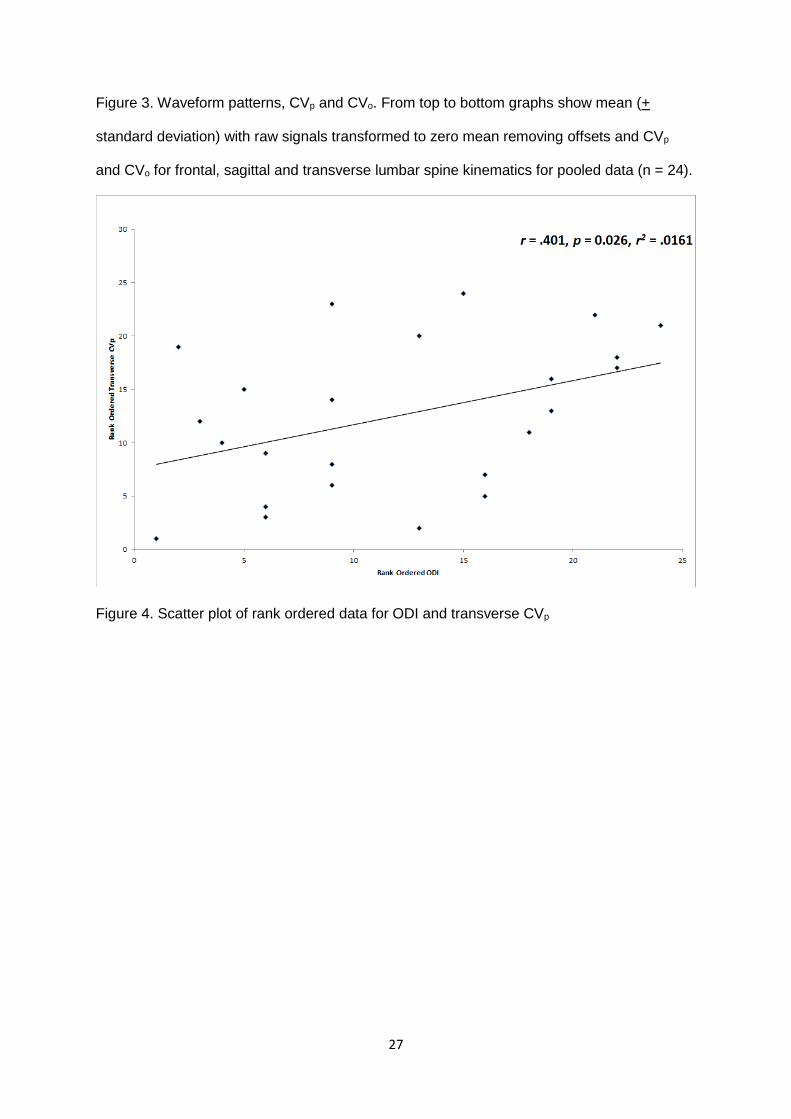
Significant moderate positive correlations were found between ODI and transverse plane CVp
(r = .401, p 0.026). Significant moderate negative correlations were also found between ILEX
strength and frontal plane CVo (r = -.370, p = 0.045), transverse plane CVp (r = -.411, p =
0.029), transverse plane CVo (r = -.378, p = 0.042) and a significant moderate positive
correlation with transverse plane displacement (r = .442, p =0.020). Figures 4, 5 and 6 present
scatter plots of the rank ordered data for transverse displacement and CVp. The square of the
correlation coefficients suggests that the degree of variance explained by ODI was 16.1%.

11
The degree of variance explained ILEX strength was 13.7% for frontal place CVo, 16.9% for
transverse plane CVp, 14.3% for transverse plane CVo, and 19.5% for transverse plane
displacement. Thus 80.5% to 86.3% of the variance was accounted for by other variables.
4. Discussion
This study of lumbar kinematic variability during gait in participants with CLBP yields several
interesting and unique results which potentially offer further understanding of the nature of the
relationships between CLBP, gait variability and lumbar extensor deconditioning. Waveform
patterns and angular displacements of the lumbar spine exhibited by participants with CLBP
in the present study were similar to those reported in other studies of both symptomatic
[18,21,24,28,] and healthy participants [20,31,59-64].
Within this study however the foremost interest was the repeatability of lumbar spine
movement patterns exhibited (intra-subject stride-to-stride variability) as, despite similar
average movements occurring at the lumbar spine, symptomatic participants appear less able
to replicate these consistently [18]. Vogt et al. [18] reported data suggesting lumbar movement
variability during gait was significantly higher in participants with CLBP compared with
asymptomatic controls, and that both sagittal and transverse plane variability was greater than
frontal plane variability. Vogt et al. [18] however did not differentiate pattern and offset
variability as we have done so here.
Schache et al. [56] have shown that although high within-day repeatability was displayed for
the model adopted in the present study, angular parameters where most susceptible to marker
reapplication errors that affected waveform offset. Our data highlights this showing that CVp
differs considerably from variation calculated using Winter’s CV reported in earlier studies [18].
Sagittal plane CVp (106.44%) is more than double the variation seen in the frontal (45.07%)
and transverse planes (42.81%).

12
CVp has not been calculated in participants with CLBP previously and thus it is not possible to
verify whether this greater sagittal plane pattern variability is a typical characteristic of their
gait. Nor is it possible to define the clinical meaning of this in comparison to healthy gait as
CVp has also not been reported on lumbar spine gait kinematics in asymptomatic participants
to the author’s knowledge. Yet, despite the high sagittal plane CVp in comparison to other
planes of movement, our correlation results suggest that there is instead a relationship
between ILEX strength and transverse plane kinematics; lower transverse displacement and
higher CVp being associated with lower ILEX strength. It might be speculated upon that this
relationship in participants with CLBP may be a consequence of the lumbar extensor
deconditioning frequently associated with this population [38]. Indeed it could be recalled that
extensor fatigue impacts upon lumbar kinematics during gait emphasising the link between
deconditioning and gait abnormality [39].
The lumbar spine has been considered to potentially play an important role in control of gait,
from the early identification of movement about the pelvis being fundamental to controlling
displacement of centre of mass during walking [65], to Gracovetsky’s [37] presentation of the
‘Spinal-Engine’ as being an important driver of human bipedal gait through all three planes of
movement. Although, the specific role of movement about the pelvis in reducing centre of
mass displacement [66-71 has been questioned, and little corroboration for its role in driving
locomotion exists [72]. Yet despite this, considerable research has demonstrated that the
lumbar spine in healthy gait shows consistently reproducible sinusoidal waveforms patterns
between participants [20,31,59-64] and that healthy participants have high intra-stride
reproducibility of these patterns [20,31].
Such fine control of the lumbar spine during healthy gait is undoubtedly aided by the
musculature. Most studies examining muscular contributions to gait have identified the active
role that the lumbar extensor musculature plays [73-75]. Thorstensson and colleagues [73]
showed that the pattern of the multifidus and longissimus activation during gait involved two

13
bursts of activity per cycle each corresponding to foot strike. They concluded that this activity
in relation to the pattern of trunk movement suggested that the lumbar extensor muscles main
function during gait is to control and restrict excessive trunk movement. Callaghan and
colleagues [75] demonstrated similar bimodal activity corresponding to greater peak in the
musculature ipsilateral to the contacting foot. The activity of the lumbar musculature appears
to follow a particular pattern seemingly to stabilise superior segments against inertial and
gravitational forces during both single foot contacts [73-75].
It seems reasonable that in a pathology such as CLBP, wherein there is an associated
deconditioning of what appears to be a critically important musculature for controlling gait, that
the deconditioning of this musculature might be considered as potentially responsible for gait
variability. Indeed our results tend towards supporting this with respect to transverse plane
CVp during gait. It might be noted that some authors have reported that transverse plane
kinematics typically show lower variability in those with CLBP [21,23,24]. However, these
studies have examined the coordination of the trunk and pelvis and variability in the phase
differences whereas the present study has instead examined the lumbar spines waveform
relative to the pelvis. This difference in methodology may account for the difference in
conclusions between these studies. Our results did also suggest that low ILEX was associated
with smaller transverse displacements. Perhaps transverse movement is more rigid in CLBP,
yet within that smaller range of movement there is poor waveform pattern repeatability. The
rigidity seen in transverse kinematic coordination in CLBP [21,23,24] may yet still be a
manifestation of lumbar extensor deconditioning and may be a gross compensatory
adjustment to reduce the impact of the reduced waveform pattern control by limiting the range
of motion experienced. Considering this it may be of future interest to examine the relationship
between ILEX and trunk/pelvis coordination in those with CLBP.
In addition, our results provide further evidence against the idea that pain per se may cause
the variability seen during gait in CLBP. No significant correlation found between VAS and

14
CVp or any other kinematic variable supporting the findings of others that pain presence
appears to not be associated with gait variability [22,25,26]. There was however also a
significant correlation between ODI and transverse plane CVp. Considering the multifactorial
nature of CLBP it would be reasonable then to consider this evidence suggests that gait
variability is potentially a dysfunction associated with CLBP that may result as a consequence
of deconditioning of the lumbar extensors or the disability accompanying CLBP. However, it
should be noted that the square of the correlation coefficients reported in the present study
indicate that the independent variables examined and found to be significantly correlated with
gait variables (ILEX strength and ODI) only accounted for a small degree of the variance.
Between 80.5% and 86.3% of the variance was accounted for by other unexamined variables.
Perhaps other cognitive variables account for some of the unexplained variance as, for
example, different coping strategies (catastrophizing or distraction) in participants with CLBP
have been shown to impact gait characteristics [34].
The results of this study however might suggest that exercise based interventions aimed at
addressing the lumbar extensor deconditioning in CLBP may be a justified approach to
address a range of dysfunctions associated with CLBP. Indeed previous studies have provided
support for exercise based interventions on improving aspects of gait variability including
muscle activation [76], ground reaction force parameters [77] and displacements during gait
[78]. However, none have examined specifically lumbar kinematic variability during gait, nor
has prior work utilised specific exercise designed to isolate the lumbar extensors. Recent work
has found that improvement in ILEX strength resulting from a strengthening program predicts
improvement in gait endurance in participants with CLBP [79]. Indeed, ILEX based exercise
has recently been suggested as optimal for conditioning of the lumbar extensors [80]. Future
studies might look to whether such exercise interventions can improve the gait variability seen
in participants with CLBP.

15
A limitation within the present study was the lack of a comparable healthy control group. Earlier
studies have suggested greater lumbar spine variability in participants with CLBP compared
to healthy controls [20,31]. Thus it might seem reasonable to speculate that CVp data would
likely be greater in the participants with CLBP in this study if compared with healthy controls
also. However, CVp has not been calculated for lumbar spine kinematics in healthy participants
as of yet to the author’s knowledge. Thus future work in healthy participants should utilise this
method [45] in order to produce normative data to conduct comparisons and also provide data
in order to judge improvement from clinical intervention.
5. Conclusions
The results of this study have provided novel information on lumbar spine kinematic variability
during gait in CLBP. These new findings are in contrast to earlier ones and instead suggest
that the highest variability is observed in sagittal plane lumbar movement during gait in CLBP.
Further to this, there was a significant relationship between both ILEX strength and ODI with
transverse plane lumbar CVp. And, a lack of relationship between VAS and CVp in any plane
measured during gait. Further research should aim to provide comparable data from healthy
controls. This would allow a better understanding of whether the pattern variability seen here
is typical or associated specifically with CLBP. In addition, the relationship between the gait
characteristics examined here and other variables should be examined in order to investigate
what might be responsible for the unexplained variance in the relationships reported. However,
the relationship found between ILEX strength and transverse plane lumbar variability suggests
that the lumbar extensors might play an important role in controlling gait in CLBP and thus
future work might examine the effect of specific exercise for this musculature upon lumbar
spine kinematic variability during gait.
6. References
1. World Health Organisation, The World Health Report 1998: Life in the 21st century: A
vision for all, Office of Publications, World Health Organisation, Geneva, 1998

16
2. B.F. Walker, The prevalence of low back pain: a systematic review of the literature
from 1966 to 1998, J. Spinal. Disord. 13(3) (2000) 205–217 .
3. G. Waddell, A.K. Burton, Occupational health guidelines for the management of low
back pain at work: evidence review, Occup. Med. 51 (2000) 126–135
4. National Institute for Health and Clinical Excellence, Low back pain: early management
of persistent non-specific low back pain, Royal College of General Practitioners,
London, 2009
5. M.W. van Tulder, B.W. Koes, L.M. Bouter, A cost-of-illness study of back pain in The
Netherlands, Pain. 62(2) (1995) 233–240
6. H.R. Guo, S. Tanaka, W.E Halperin, L.L Cameron, Back pain prevalence in US
industry and estimates of lost workdays, Am. J. Public Health. 89 (1999) 1029–1035
7. N. Maniadakis, A. Gray, The economic burden of back pain in the UK, Pain. 84(1)
(2000) 95–103.
8. M. Ekman, O. Johnell, L. Lidgren, The economic cost of low back pain in Sweden in
2001, Acta. Orthop. 76(2) (2005) 275–284
9. G. Waddell, M. Aylward, P. Sawney, Back Pain, incapacity for work and social security
benefits: an international literature review and analysis, The Royal Society of Medicine
Press Ltd, Glasgow, 2002
10. W.F. Stewart, J.A. Ricci, E. Chee, D. Morganstein, R. Lipton, Lost productive time
and cost due to common pain condition in the US workforce, JAMA. 290(18) (2003)
2443–2454
11. J.A. Ricci, W.F. Stewart, E. Chee, C. Leotta, K. Foley, M.C. Hochberg, Back pain
exacerbations and lost productive time costs in United States workers, Spine. 31(26)
(2006) 3052–3060
12. J.N. Katz, Lumbar disc disorders and low back pain: socioeconomic factors and
consequences. J. Bone. Joint. Surg. Am. 88(suppl 2) (2006) 21–24

17
13. J.K. Freburger, G.M. Holmes, R.P. Agans, A.M. Jackman, J.D. Darter, A.S. Wallace,
L.D. Castel, W.D. Kalsbeek, T.S. Carey, The rising prevalence of chronic low back
pain. Arch. Intern. Med. 169(3) (2009) 251–258
14. A.A. White, S.L. Gordon, Synopsis: Workshop on idiopathic low back pain, Spine. 7
(1982) 141–149
15. National Research Council, Work-related musculoskeletal disorders: A Review of the
evidence, National Academy Press, Washington, DC, 1998
16. National Research Council, The Institute of Medicine, Musculoskeletal disorders and
the workplace: Low back and upper extremities, National Academy Press,
Washington, DC, 2001
17. G. Waddell, G. Feder, M. Lewis, Systematic reviews of bed rest and advice to stay
active for acute low back pain. Br. J. Gen. Pract. 47 (1997) 647–652
18. L. Vogt, K. Pfeifer, M. Portscher, W. Banzer, Influences of nonspecific low back pain
on three-dimensional lumbar spine kinematics in locomotion, Spine. 26(17) (2001)
1910–1919
19. D.M. Roffey, E.K. Wai, P. Bishop, B.K. Kwon, S. Dagenais, Causal assessment of
occupational standing or walking and low back pain: results of a systematic review,
Spine. J. 10(3) (2010) 262–272
20. L. Vogt, W. Banzer, Measurement of lumbar spine kinematics in incline treadmill
walking, Gait. Posture. 9(1) (1999) 18–23
21. C.J. Lamoth, O.G. Meijer, A.D Daffersthofer, P.I. Wuisman, P.J. Beek, Effects of
chronic low back pain on trunk coordination and back muscle activity during walking:
changes in motor control, Eur. Spine. J. 15 (2006) 23–40
22. J.F. Seay, R.E. Van Emmerik, J. Hamill, Influence of low back pain status on pelvis-
trunk coordination during walking and running, Spine. 36(16) (2011) E1070–E1079
23. C.J. Lamoth, O.G. Meijer, P.I. Wuisman, J.H. van Dieen, M.F. Levin, P.J. Beek,
Pelvis-thorax coordination in the transverse plane during walking in persons with
non-specific low back pain, Spine. 27(4) (2002) E92–E99

18
24. W. van Der Hoorn, S.M. Bruijn, O.G. Meijer, P.W. Hodges, J.H van Dieen, Mechanical
coupling between transverse plane pelvis and thorax rotations during gait is higher in
people with low back pain, J. Biomech. 45 (2012) 342-347
25. C. Anders, H.C. Scholle, H. Wagner, C. Puta, R. Grassme, A. Petrovitch, Trunk
muscle co-ordination during gait: relationship between muscle function and acute low
back pain, Pathophysiology. 12(4) (2005) 243–247
26. C.J. Lamoth, A. Daffertshofer, O.G. Meijer, G. Lorimer Moseley, P.I. Wuisman, P.J.
Beek, Effects of experimentally induced pain and fear of pain on trunk coordination
and back muscle activity during walking, Clin. Biomech.19(6) (2004) 551–563
27. J. Crosbie, R. de Faria Negrao Filho, D. P Nascimento, P. Ferreira, Coordination of
spinal motion in the transverse and frontal planes during walking in people with and
without recurrent low back pain, Spine. 38(5) (2012) E286-292
28. P.J. Rowe, M. White, Three dimensional lumbar spinal kinematics during gait following
mild musculoskeletal low back pain in nurses, Gait. Posture. 4 (1997) 242–251
29. C.J. Lamoth, A.D. Daffertshofer, O.G. Meijer, P.J. Beek, How do persons with
chronic low back pain speed up and slow down? Trunk-pelvis coordination and
lumbar erector spinae activity during gait, Gait. Posture. 23(2) (2006) 230–239
30. L. Arendt-Nielsen, T. Graven-Nielsen, H. Svarrer, P. Svensson, The influence of low
back pain on muscle activity and coordination during gait: a clinical and experimental
study, Pain. 64(2) (1996) 231–240
31. L. Vogt, K. Pfeifer, W. Banzer, Neuromuscular control of walking with chronic low
back pain, Man. Ther. 8(1) (2003) 21–28
32. M. van Der Hulst, M.M. Vollenbroek-Hutten, J.S. Rietman, L. Schaake, K.G. Groothuis-
Oudshoorn, H.G. Hermens, Back muscle activation patterns in chronic low back pain
during walking: a “guarding” hypothesis, Clin. J. Pain. 26(1) (2010) 30–37
33. M. van Der Hulst, M.M. Vollenbroek-Hutten, J.S. Rietman, H.J. Hermens, Lumbar and
abdominal muscle activity during walking in subjects with chronic low back pain:
support of the “guarding” hypothesis? J. Electromyogr. Kinesiol. 20(1) (2010) 31–38

19
34. M. van Der Hulst, M.M. Vollenbroek-Hutten, K.M. Schreurs, J.S. Rietman, H.J.
Hermens, Relationships between coping strategies and lumbar muscle activity in
subjects with chronic low back pain, Eur. J. Pain. 14(6) (2010) 640–647
35. E.Y. Hanada, M. Johnson, C. Hubley-Kozey, A comparison of trunk muscle activation
amplitudes during gait in older adults with and without chronic low back pain. PM R.
3(10) (2011) 920–928
36. M.M. van Der Krogt, S.L. Delp, M.H. Schwartz, How robust is human gait to muscle
weakness? Gait. Posture. 36(1) (2012) 113–119
37. S. Gracovetsky, An hypothesis for the role of the spine in human locomotion: A
challenge to current thinking, J. Biomed. Eng. 7 (1985) 205–216
38. J. Steele, S. Bruce-Low, D. Smith, A reappraisal of the deconditioning hypothesis in
low back pain: a review of evidence from a triumvirate of research methods on specific
lumbar extensor deconditioning. Curr. Med. Res. Opin. 30(5) (2014) 865-911
39. J.M. Hart, D.C. Kerrigan, J.M. Fritz, C.D. Ingersoll, Jogging kinematics after lumbar
paraspinal muscle fatigue, J. Athl. Train. 44(5) (2009) 475–481
40. S.P. Arjunan, D.K. Kumar, W.M. Poon, H. Rudolph, Y. Hu, Variability in surface
electromyogram during gait analysis of low back pain patients, J. Med. Bio. Eng.
30(3) (2010) 133-138
41. R.W. Selles, R.C. Wagenaar, T.H. Smith, P.I. Wuisman, Disorders in trunk rotation
during walking in patients with low back pain: a dynamical systems approach, Clin.
Biomech. 16(3) (2001) 175–181
42. C.J. Lamoth, J.F. Stins, M. Pont, F. Kerckhoff, P.J. Beek, Effects of attention on the
control of locomotion in individuals with chronic low back pain, J. Neuroeng. Rehabil.
25(5) (2008) 13
43. J.F. Seay, R.E. van Emmerik, J. Hamill, Low back pain status affects pelvis-trunk
coordination and variability during walking and running, Clin. Biomech. 26(6) (2011)
572–578

20
44. D.A. Winter, Biomechanical motor pattenrs in normal walking, J. Mot. Behav. 15(4)
(1983) 302-330
45. N. O’Dwyer, R. Smith, M. Kalaki, U. Rattanaprasert, Independent assessment of
pattern and offset variability of time series waveforms, Gait. Posture. 29 (2009) 285-
289
46. J. Steele, S. Bruce-Low, D. Smith, D. Jessop, N. Osborne, A randomised controlled
trial of limited range of motion lumbar extension exercise in chronic low back pain,
Spine. 38(15) (2013) 1245-1252
47. J. Frymoyer, Back Pain and Sciatica, N. Engl. J. Med. 318 (1988) 291–300
48. J.E. Graves, M.L. Pollock, D.M. Carpenter, S.H. Leggett, A. Jones, M. MacMillan, M.
Fulton, Quantitative assessment of full range of motion isometric lumbar extension
strength, Spine. 15(4) (1990) 289–294
49. M.E. Robinson, A.F. Greene, P. O’Connor, J.E. Graves, M. MacMillan, Reliability of
lumbar isometric torque in patients with chronic low back pain, Phys. Ther. 72(3)
(1992) 186–190
50. M.L. Pollock, J.E. Graves, S.H. Leggett, W.G. Young, L. Gazzarella, D.M. Carpenter,
Accuracy of counter weighting to account for upper body mass in testing lumbar
extension strength, Med. Sci. Sport. Exerc. 23 (1991) S66
51. H. Inanami, Iwai Orthopedic Hospital rehabilitation program. Paper presented at
International Spinal Rehabilitation Update 1991 Symposium, Daytona, 1991
52. M. Ogon, M. Krismer, W. Sollner, W. Kantner-Rumplmair, A. Lampe, Chronic low back
pain measurement with visual analogue scales in different settings, Pain. 64(3) (1996)
425–428
53. J.C. Fairbank, J. Couper, J.B. Davies, J.P. O’Brien, The Oswestry low back pain
disability questionnaire, Physiotherapy. 66(8) (1980) 271–273
54. M. Syczewska, T. Oberg, D. Karlsson, Segmental movements of the spine during
treadmill walking with normal speed, Clin. Biomech. 14 (1999) 384–388

21
55. A.G. Schache, P. Blanch, D. Rath, T. Wrogley, K. Bennell, Three-dimensional angular
kinematics of the lumbar spine and pelvis during running, Hum. Mov. Sci. 21 (2002)
273–293
56. A.G. Schache, P.D. Blanch, D.A. Rath, T.V. Wrigley, R. Starr, K.L. Bennell, Intra-
subject repeatability of the three dimensional angular kinematics within the lumbo-
pelvic-hip complex during running, Gait. Posture. 15 (2002) 136–145
57. R.L. Drake, A.W. Vogl, A.W.M. Mitchell, Gray’s Anatomy for Students, Churchill
Livingstone, New York, 2010
58. B.T. Bates, J.S. Dufek, H.P. Davis, The effect of trial size on statistical power, Med.
Sci. Sports. Exerc. 24(9) (1992) 1059-1068
59. J. Crosbie, R. Vachalanthiti, R. Smith, Patterns of spinal motion during walking, Gait.
Posture. 5 (1997) 6–12
60. D.E. Krebs, D. Wong, D. Jevsevar, P. O’Riley, W.A. Hodge, Trunk kinematics during
locomotor activites, Phys. Ther. 72(7) (1992) 505–514
61. A.J. Thurston, J.D. Harris, Normal kinematics of the lumbar spine and pelvis, Spine.
8(2) (1983) 199–205
62. V.P. Stokes, C. Andersson, H. Forssberg, Rotational and translational movement
features of the pelvis and thorax during adult human locomotion, J. Biomech. 22(1)
(1989) 43–50
63. M.W. Whittle, D. Levine, Three-dimensional relationships between the movements of
the pelvis and lumbar spine during normal gait, Hum. Mov. Sci. 18 (1999), 681–692
64. N.E. Fowler, A.L.F. Rodacki, C.D. Rodacki, Changes in stature and spine kinematics
during a loaded walking task, Gait. Posture. 23 (2006) 133–141
65. J.B. Saunders, V.T. Inman, H.D. Eberhart, The major determinants in normal and
pathological gait, J. Bone. Joint. Surg. Am. 35 (1953) 543–558
66. A.G. Schache, K.L. Bennell, P.D. Blanch, T.V. Wrigley, The coordinated movement of
the lumbo-pelvic-hip complex during running: a literature review, Gait. Posture. 10(1)
(1999) 30-47

22
67. D.C. Kerrigan, U. Della Croce, M. Marciello, P.O. Riley, A refined view of the
determinants of gait: significance of heel rise, Arch. Phys. Med. Rehabil. 81(8) (2000)
1077-1080
68. U. Della Croce, P.O. Riley, J.L. Lelas, D.C. Kerrigan, A refined view of the determinants
of gait, Gait. Posture. 14(2) (2001) 79-84
69. A.D. Kuo, The six determinants of gait and the inverted pendulum analogy: A dynamic
walking perspective, Hum. Mov. Sci. 26(4) (2007) 617-656
70. C. Hayot, S. Sakka, P. Lacouture, Contribution of the six major gait determinants on
the vertical center of mass trajectory and the vertical ground reaction force, Hum. Mov.
Sci. 32(2) (2013) 279-289
71. Y. Lin, M. Gfoehler, M.G. Pandy, Quantitative evaluation of the major determinants of
human gait, J. Biomech. 47(6) (2014) 1324-331
72. J. Rice, M. Kaliszer, M. Walsh, A. Jenkinson, T. O’Brien, Movements at the low back
during normal walking, Clin. Anat. 17(8) (2004) 662-666
73. A. Thorstensson, H. Carlson, M.R. Zomlefer, J. Nilsson, Lumbar back muscle activity
in relation to trunk movements during locomotion in man, Acta. Physiol. Scand. 116
(1982) 13–20
74. J.P. Callaghan, A.E. Patla, S.M. McGill, Low back three-dimensional joint forces,
kinematics, and kinetics during walking, Clin. Biomech. 14 (1999) 203–216
75. D.A. Winter, C.D. MacKinnon, G.K. Ruder, C. Wieman, An integrated
EMG/biomechanical model of upper body balance and posture during human gait,
Prog. Brain. Res. 97 (1993) 359–367
76. H. Tsao, P.W. Hodges, Persistence of improvements in postural strategies following
motor control training in people with recurrent low back pain, J. Electromyogr.
Kinesiol. 18(4) (2008) 559–567
77. J.L. da Fonseca, M. Magini, T.H. de Freitas, Laboratory gait analysis in patients with
low back pain before and after a Pilates intervention, J. Sport. Rehabil. 18 (2009) 269-
282
23
78. F.P. Carpes, F.A. Reinehr, C.B. Mota, Effects of a program for trunk strength and
stability on pain, low back and pelvis kinematics, and body balance: A pilot study, J.
Bodyw. Mov. Ther. 12 (2008) 22–30
79. H.K. Vincent, K.R. Vincent, A.N. Seay, B.P. Conrad, R.W. Hurley, S.Z. George, Back
strength predicts walking improvement in obese, older adults with chronic low back
pain, PM. R. (2013) Epub ahead of print
80. J.Steele, S. Bruce-Low, D. Smith, A review of the specificity of exercises designed for
conditioning the lumbar extensors, Br. J. Sports. Med. (2013) Online First

24
Tables
Table 1. Participant Demographics
n = 24
Age (years) 46+14
Stature (cm) 174.01+9.68
Body Mass (Kg) 77.07+16.50
BMI (Kg/m2) 25.17+3.36
Symptom Duration (years) 14+11
VAS (mm) 40.17+24.54
ODI (pts) 32.50+11.81
Lumbar Extension Strength (Nm) 190.05+76.65
25
Figures
Figure 1. Lumbar extension machine restraint system.
Figure 2. Pelvic and thoracolumbar cluster marker set-up

26
27
Figure 3. Waveform patterns, CVp and CVo. From top to bottom graphs show mean (+
standard deviation) with raw signals transformed to zero mean removing offsets and CVp
and CVo for frontal, sagittal and transverse lumbar spine kinematics for pooled data (n = 24).
Figure 4. Scatter plot of rank ordered data for ODI and transverse CVp

28
Figure 5. Scatter plot of rank ordered data for ILEX strength and transverse CVp
Figure 6. Scatter plot of rank ordered data for ILEX strength and transverse displacement