lsp1 is an endothelial gatekeeper of leukocyte transendothelial migration
TRANSCRIPT
The
Journ
al o
f Exp
erim
enta
l M
edic
ine
JEM © The Rockefeller University Press $8.00Vol. 201, No. 3, February 7, 2005 409–418 www.jem.org/cgi/doi/10.1084/jem.20040830
ARTICLE
409
LSP1 is an endothelial gatekeeper of leukocyte transendothelial migration
Lixin Liu,
1
Denise C. Cara,
1
Jaswinder Kaur,
1
Eko Raharjo,
1
Sarah C. Mullaly,
1
Jenny Jongstra-Bilen,
2,3
Jan Jongstra,
2,3
and Paul Kubes
1
1
Immunology Research Group, Department of Physiology and Biophysics, University of Calgary, Calgary, Alberta T2N 4N1, Canada
2
Cell and Molecular Biology Division, Toronto Western Research Institute and
3
Department of Immunology, University of Toronto, Toronto, Ontario M5T 2S8, Canada
Leukocyte-specific protein 1 (LSP1), an F-actin binding protein and a major downstream substrate of p38 mitogen-activated protein kinase as well as protein kinase C, has been reported to be important in leukocyte chemotaxis. Although its distribution has been thought to be restricted to leukocytes, herein we report that LSP1 is expressed in endothelium and is essential to permit neutrophil emigration. Using intravital microscopy to directly visualize leukocyte rolling, adhesion, and emigration in postcapillary venules in LSP1-deficient (
Lsp1
���
) mice, we found that LSP1 deficiency inhibits neutrophil extravasation in response to various cytokines (tumor necrosis factor-
�
and interleukin-1
�
) and to neutrophil chemokine keratinocyte-derived chemokine in vivo. LSP1 deficiency did not affect leukocyte rolling or adhesion. Generation of
Lsp1
���
chimeric mice using bone marrow transplantation revealed that in mice with
Lsp1
���
endothelial cells and wild-type leukocytes, neutrophil transendothelial migration out of postcapillary venules is markedly restricted. In contrast,
Lsp1
���
neutrophils in wild-type mice were able to extravasate normally. Consistent with altered endothelial function was a reduction in vascular permeability to histamine in
Lsp1
���
animals. Western blot analysis and immunofluorescence microscopy examination confirmed the presence of LSP1 in wild-type but not in
Lsp1
���
mouse microvascular endothelial cells. Cultured human endothelial cells also stained positive for LSP1. Our results suggest that LSP1 expressed in endothelium regulates neutrophil transendothelial migration.
Lymphocyte-specific gene 1, found in bothmouse and humans, was initially thought to berestricted to B cells, functional T cells, and thy-mocytes (1, 2). However, more recently, it hasbeen documented in monocytes, macrophages,and neutrophils and is now referred to as leuko-cyte-specific protein 1 (LSP1; references 3–5).LSP1 is an intracellular Ca
2
�
and F-actin bindingprotein (6–9). In its carboxyl-terminal region,the molecule contains a high affinity F-actinbinding site which allows LSP1 to accumulatewithin the microfilament rich cortical cytoskele-ton. LSP1 has been shown to be a major sub-strate of the mitogen-activated protein kinase(MAPK)-activated protein (MAPKAP) kinase-2in the p38 MAPK pathway (10). MAPKAPkinase-2 and p38 MAPK were reported to beessential for neutrophil motility and chemo-taxis (11–13), suggesting that LSP1 might beimportant in chemotaxis. However, it shouldbe noted that MAPKAP kinase-2 phosphory-
lates numerous other molecules, including heatshock protein 25/27 (14), and so the impor-tance of LSP1 after p38 MAPK activation re-mains unclear. In addition, LSP1 has also beenshown to be a substrate for protein kinase C(PKC; 15, 16), which is another molecule im-plicated in numerous neutrophil functions (in-cluding adhesion and chemotaxis; reference 17),which raises the possibility that LSP1 may havemultiple roles in neutrophil recruitment.
Although recent in vitro studies usingLSP1-deficient (
Lsp1
���
) cells suggest thatLSP1 does contribute to the process of chemo-taxis, as yet unexplained opposing results havebeen observed. Jongstra-Bilen and colleaguesgenerated
Lsp1
���
mice and observed increasedchemotactic responses in
Lsp1
���
neutrophilsin vitro (18, 19). In direct contrast, Hanniganet al. reported, in an in vitro study, reducedchemotactic responses of
Lsp1
���
neutrophils,which may be associated with discontinuousprimary actin-rich cortexes and large abnormalmembrane protrusions (20). Although both
CORRESPONDENCEPaul Kubes: [email protected]
Abbreviations used: HUVEC, human umbilical vein endothelial cell; KC, keratinocyte-derived chemokine; LSP1, leukocyte-specific protein 1;
Lsp1
���
, LSP1-deficient; MAPK, mito-gen-activated protein kinase; MAPKAP, MAPK-activated protein; PKC, protein kinase C.
L. Liu and D.C. Cara contributed equally to this work.
ENDOTHELIAL LSP1 REGULATES LEUKOCYTE TRANSMIGRATION | Liu et al.
410
groups used keratinocyte-derived chemokine (KC) as achemoattractant, important differences in experimental condi-tions between these in vitro experiments included differentsubstrates and different neutrophil populations (peritoneal elic-ited or bone marrow neutrophils vs. peripheral blood neutro-phils). Clearly, a systematic examination of neutrophil func-tion in vivo in the presence and absence of LSP1 is warranted.
In vivo, neutrophil recruitment is a very complex eventthat requires that neutrophils first tether to the endothelium,and upon activation via chemokines, firmly adhere. This ap-pears to lead to cross-talk between the adherent neutrophiland the endothelium, whereby endothelial cells retract, al-lowing neutrophils to migrate across the endothelium (21,22) before chemotaxing toward the source of the injuryand/or infection. Recently, we reported that inhibition ofp38 MAPK activity dramatically limited neutrophil transmi-gration across the endothelium and subsequent neutrophilchemotaxis through the interstitium (13). However, whetherthe p38 MAPK inhibitors were affecting the endotheliumand/or the neutrophils was unclear. This is not trivial as bothp38 MAPK and MAPKAP kinase-2 have been shown toplay a role in endothelial cytoskeletal rearrangements and inincreased endothelial permeability associated with hypoxicor oxidative stress (23–25), as well as in TNF
�
or VEGF stim-ulation (26–28). However, no one to date has assessed thepossibility that LSP1 is found in endothelium.
During neutrophil recruitment, the endothelium is thoughtto actively retract to allow neutrophils to transmigrate (21,22, 29, 30). Because LSP1 is a major substrate for the p38MAPK–MAPKAP kinase-2 signaling pathway and MAPKappears to be important in both neutrophils and endothelium,we tested the hypothesis that LSP1 is an important protein inneutrophil extravasation in vivo as a result of endothelialLSP1. Indeed, our data do not support a critical role for neu-trophil LSP1 in extravasation in vivo; however, our results re-veal that endothelium does have LSP1 and it plays an essentialrole in transendothelial migration in chimeric mice whereLSP1 was selectively expressed in the endothelium.
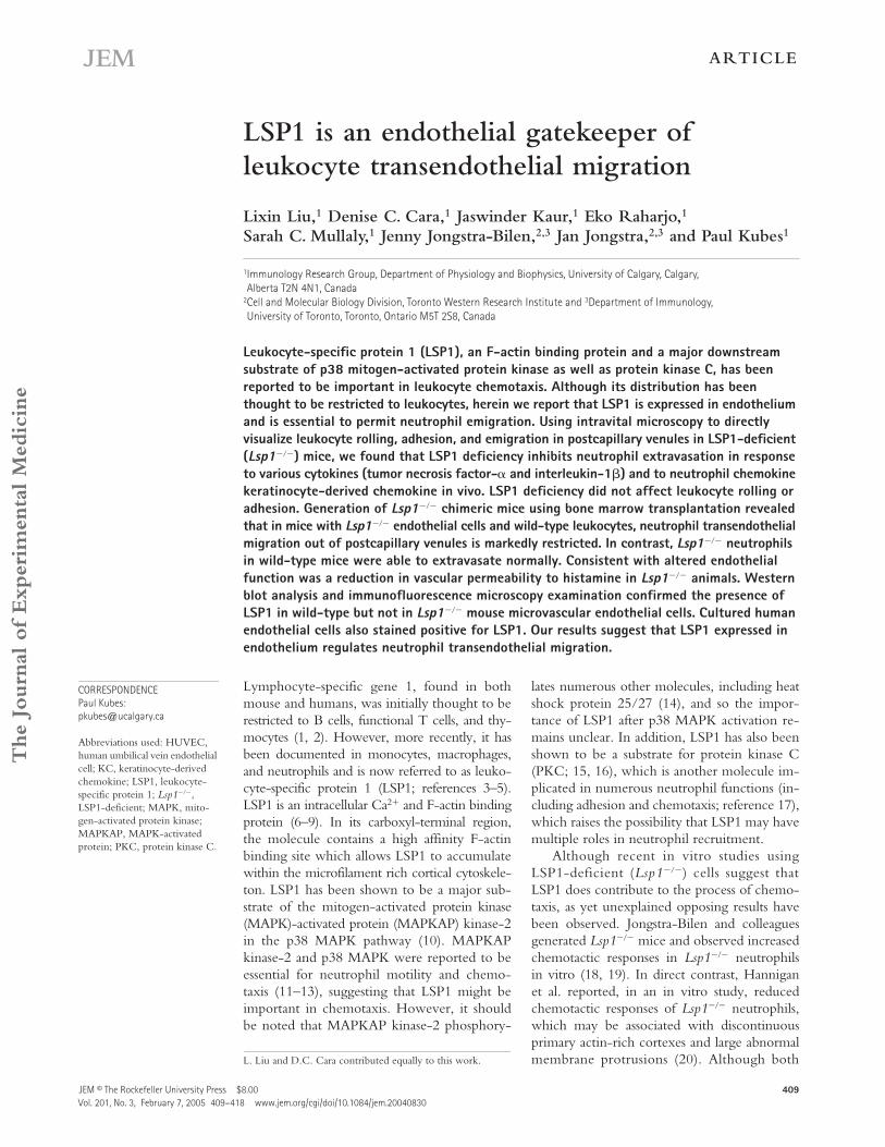
RESULTSLSP1 does not affect leukocyte rolling and adhesion, but is important for leukocyte emigration in response to TNF
�
or IL-1
�
Table I summarizes the hemodynamics of the microvascula-ture of
Lsp1
���
and WT mice 4 h after intrascrotal injectionof TNF
�
. The diameters of the chosen cremasteric venuleswere similar between WT and
Lsp1
���
mice. There was noapparent difference in shear rate, red blood cell velocity (Ta-ble I), or calculated blood flow in these postcapillary venules(not depicted). Similarly, during the induction of inflamma-tion, there was a similar decrease in blood flow in postcapil-lary venules in both WT and
Lsp1
���
mice. Therefore,changes in leukocyte behavior described herein cannot beattributed to differences in hemodynamic parameters. Therewas a small but significant increase in circulating white bloodcells in
Lsp1
���
mice.
We treated WT and
Lsp1
���
mice intrascrotally with 0.5
�
g TNF
�
and measured leukocyte rolling flux, rolling ve-locity, and the number of adherent and emigrated leukocytesin cremasteric venules 3.5, 4, and 4.5 h after cytokine injec-tion. Fig. 1 A demonstrates that
�
40
�
60 cells rolled perminute in control preparations of both WT and
Lsp1
���
mice. Exposure of the cremaster muscle microcirculation toTNF
�
induced very similar rolling flux in both WT and
Lsp1
���
mice. The rolling velocity of leukocytes was
�
80
�
m/s under control conditions in both sets of mice and avery profound 80–90% decrease in rolling velocity wasnoted in both WT and
Lsp1
���
mice after TNF
�
adminis-tration (Fig. 1 B). Fig. 1 C demonstrates a large increase inleukocyte adhesion in postcapillary venules after TNF
�
treatment, a response that was again close to identical in WTand
Lsp1
���
mice. However, a very significant difference inleukocyte transendothelial migration was noted in WT and
Lsp1
���
mice in response to TNF
�
(Fig. 1 D). Although
�
40 cells emigrated out of vessels per field of view in WT
Table I.
Hemodynamic parameters in WT and
Lsp1
���
mice 4 h after intrascrotal injection of TNF
�
(0.5
�
g,
n
= 3 in each group)
GroupVenulardiameter V
RBC
Wall shear rateWBC number(
�
10
6
cells)
�
m mm/s s
�
1
WT 31
�
2.9 2.1
�
0.3 331.8
�
27.4 5.6
�
0.2
Lsp1
���
30
�
2.9 2.0
�
0.5 320.5
�
65.5 8.1
�
0.4
a
a
P
0.05 as compared with WBC number in WT mice.WBC, white blood cell.
Figure 1. The flux of rolling leukocytes (A), rolling cell velocity (B), adherent (C), and emigrated (D) leukocytes in cremasteric venules of TNF�-treated and untreated WT and Lsp1��� mice. Leukocyte recruit-ment was induced by intrascrotal injection of TNF� (0.5 �g in 200 �l saline) and the recruitment parameters determined in cremasteric venules from WT (WT control: n 4; WT � TNF�: n 3) and Lsp1��� mice (Lsp1��� control: n 6; Lsp1��� � TNF�: n 3). *, P 0.05 and **, P 0.01, as compared with each untreated control group.
JEM VOL. 201, February 7, 2005
411
ARTICLE
mice, only 15 cells emigrated in
Lsp1
���
mice (P
0.01). Inan additional group of WT mice, one fifth the concentrationof TNF
�
was used. This caused fewer cells to adhere than in
Lsp1
���
mice treated with the higher concentration ofTNF
�
, yet the emigration was still higher in WT mice thanthat in
Lsp1
���
mice (unpublished data).Previous papers have suggested that the mechanisms un-
derlying leukocyte emigration can be quite different forTNF
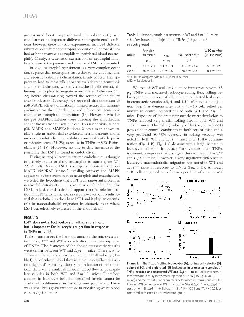
� versus, for example, IL-1� (31, 32). To determinewhether the impaired emigration was limited to TNF�, weinjected mice intrascrotally with an optimal dose (12.5 ng) ofIL-1� (31), and measured leukocyte rolling flux, rolling ve-locity, and the number of adherent and emigrated leukocytesin the tissues 3.5, 4, and 4.5 h after injection of cytokine. Af-ter IL-1� local administration, leukocyte rolling flux was in-creased in both WT and Lsp1��� mice at least twofoldgreater than untreated control mice (Fig. 2 A). Similar de-crease in rolling velocity (Fig. 2 B) and increase in adhesion(Fig. 2 C) to IL-1� was noted in WT and Lsp1��� mice. Fig.2 D demonstrates a profound 75% inhibition in leukocytetransendothelial migration in Lsp1��� mice (P 0.05).Clearly, LSP1 plays a role in leukocyte emigration in re-sponse to proinflammatory cytokines.
The impairment in Lsp1��� mice could be due to an im-pairment in cytokine signaling and subsequent synthesis ofchemokines or it could be an impairment in the emigrationprocess per se. Previous work has demonstrated that essen-tially all of the emigrated cells at 4-h TNF� or IL-1� stimu-lation are neutrophils (31, 33). Therefore, we examined re-sponses of WT and Lsp1��� neutrophils after activation of
their chemokine receptors to the neutrophil chemoattractantKC in vivo.
LSP1 is essential for neutrophil emigration in response to the chemokine KCWe measured leukocyte rolling, adhesion, and transendothelialmigration upon slow release of the chemokine KC from anagarose gel positioned 350 �m from the observed cremastericpostcapillary venule. Rolling was not affected by KC (Fig. 3A), whereas neutrophils began adhering quite rapidly after theKC-containing gel was placed on the cremaster preparation(Fig. 3 B). However, there was no significant difference in therolling flux and adhesion response between WT and Lsp1���
mice. Fig. 3 C summarizes the number of emigrated neutro-phils per field of view 60 min after local KC-containing geladdition. In WT animals, �25 neutrophils could be seen out-side the venule of study and all of the cells were migrating to-ward the KC-containing gel (unpublished data). In contrast, inthe Lsp1��� mice, the transendothelial migration was evenmore impaired in response to the chemokine KC at 60 minthan it was in response to cytokines. Less than four cells wereseen to migrate across the endothelium.
Figure 2. The flux of rolling leukocytes (A), rolling cell velocity (B), adherent (C), and emigrated (D) leukocytes in cremasteric venules of IL-1�–treated and untreated WT and Lsp1��� mice. Leukocyte re-cruitment was induced by intrascrotal injection of IL-1� (12.5 ng in 200 �l saline) and the recruitment parameters determined in cremasteric venules from WT (WT control: n 4; WT � IL-1�: n 4) and Lsp1��� mice (Lsp1��� control: n 6; Lsp1��� � IL-1�: n 4). *, P 0.05 and **, P 0.01, as compared with each untreated control group.
Figure 3. The flux of rolling leukocytes (A), adherent leukocytes (B), and emigrated leukocytes (C) induced by KC in agarose gel placed 350 �m from the observed cremasteric venule of WT (n 3) and Lsp1��� (n 4) mice. **, P 0.01 as compared with time 0 (B) or with the WT control (C).
ENDOTHELIAL LSP1 REGULATES LEUKOCYTE TRANSMIGRATION | Liu et al.412
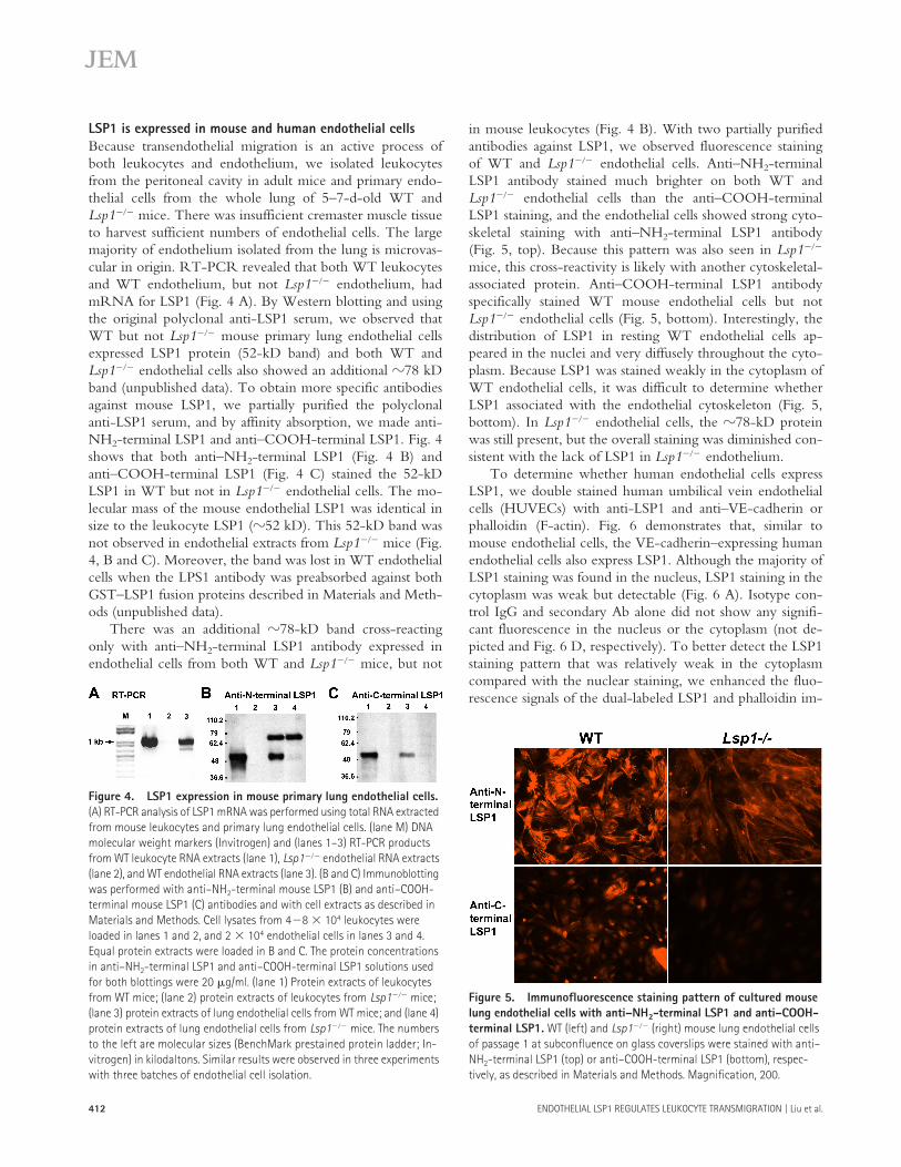
LSP1 is expressed in mouse and human endothelial cellsBecause transendothelial migration is an active process ofboth leukocytes and endothelium, we isolated leukocytesfrom the peritoneal cavity in adult mice and primary endo-thelial cells from the whole lung of 5–7-d-old WT andLsp1��� mice. There was insufficient cremaster muscle tissueto harvest sufficient numbers of endothelial cells. The largemajority of endothelium isolated from the lung is microvas-cular in origin. RT-PCR revealed that both WT leukocytesand WT endothelium, but not Lsp1��� endothelium, hadmRNA for LSP1 (Fig. 4 A). By Western blotting and usingthe original polyclonal anti-LSP1 serum, we observed thatWT but not Lsp1��� mouse primary lung endothelial cellsexpressed LSP1 protein (52-kD band) and both WT andLsp1��� endothelial cells also showed an additional �78 kDband (unpublished data). To obtain more specific antibodiesagainst mouse LSP1, we partially purified the polyclonalanti-LSP1 serum, and by affinity absorption, we made anti-NH2-terminal LSP1 and anti–COOH-terminal LSP1. Fig. 4shows that both anti–NH2-terminal LSP1 (Fig. 4 B) andanti–COOH-terminal LSP1 (Fig. 4 C) stained the 52-kDLSP1 in WT but not in Lsp1��� endothelial cells. The mo-lecular mass of the mouse endothelial LSP1 was identical insize to the leukocyte LSP1 (�52 kD). This 52-kD band wasnot observed in endothelial extracts from Lsp1��� mice (Fig.4, B and C). Moreover, the band was lost in WT endothelialcells when the LPS1 antibody was preabsorbed against bothGST–LSP1 fusion proteins described in Materials and Meth-ods (unpublished data).
There was an additional �78-kD band cross-reactingonly with anti–NH2-terminal LSP1 antibody expressed inendothelial cells from both WT and Lsp1��� mice, but not
in mouse leukocytes (Fig. 4 B). With two partially purifiedantibodies against LSP1, we observed fluorescence stainingof WT and Lsp1��� endothelial cells. Anti–NH2-terminalLSP1 antibody stained much brighter on both WT andLsp1��� endothelial cells than the anti–COOH-terminalLSP1 staining, and the endothelial cells showed strong cyto-skeletal staining with anti–NH2-terminal LSP1 antibody(Fig. 5, top). Because this pattern was also seen in Lsp1���
mice, this cross-reactivity is likely with another cytoskeletal-associated protein. Anti–COOH-terminal LSP1 antibodyspecifically stained WT mouse endothelial cells but notLsp1��� endothelial cells (Fig. 5, bottom). Interestingly, thedistribution of LSP1 in resting WT endothelial cells ap-peared in the nuclei and very diffusely throughout the cyto-plasm. Because LSP1 was stained weakly in the cytoplasm ofWT endothelial cells, it was difficult to determine whetherLSP1 associated with the endothelial cytoskeleton (Fig. 5,bottom). In Lsp1��� endothelial cells, the �78-kD proteinwas still present, but the overall staining was diminished con-sistent with the lack of LSP1 in Lsp1��� endothelium.
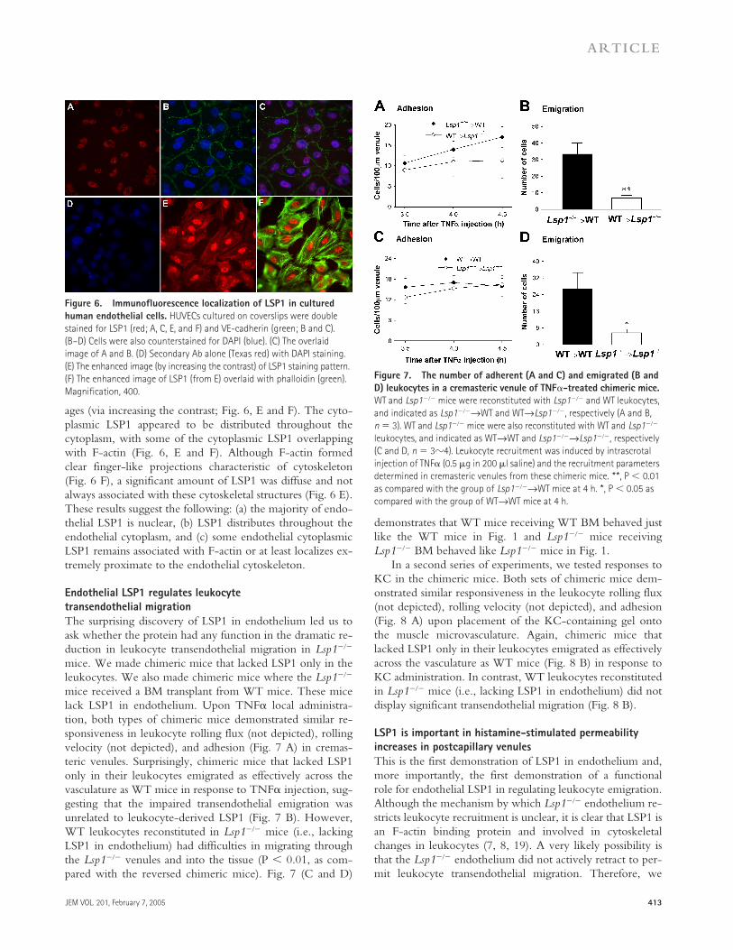
To determine whether human endothelial cells expressLSP1, we double stained human umbilical vein endothelialcells (HUVECs) with anti-LSP1 and anti–VE-cadherin orphalloidin (F-actin). Fig. 6 demonstrates that, similar tomouse endothelial cells, the VE-cadherin–expressing humanendothelial cells also express LSP1. Although the majority ofLSP1 staining was found in the nucleus, LSP1 staining in thecytoplasm was weak but detectable (Fig. 6 A). Isotype con-trol IgG and secondary Ab alone did not show any signifi-cant fluorescence in the nucleus or the cytoplasm (not de-picted and Fig. 6 D, respectively). To better detect the LSP1staining pattern that was relatively weak in the cytoplasmcompared with the nuclear staining, we enhanced the fluo-rescence signals of the dual-labeled LSP1 and phalloidin im-
Figure 4. LSP1 expression in mouse primary lung endothelial cells. (A) RT-PCR analysis of LSP1 mRNA was performed using total RNA extracted from mouse leukocytes and primary lung endothelial cells. (lane M) DNA molecular weight markers (Invitrogen) and (lanes 1–3) RT-PCR products from WT leukocyte RNA extracts (lane 1), Lsp1��� endothelial RNA extracts (lane 2), and WT endothelial RNA extracts (lane 3). (B and C) Immunoblotting was performed with anti–NH2-terminal mouse LSP1 (B) and anti–COOH-terminal mouse LSP1 (C) antibodies and with cell extracts as described in Materials and Methods. Cell lysates from 4�8 � 104 leukocytes were loaded in lanes 1 and 2, and 2 � 104 endothelial cells in lanes 3 and 4. Equal protein extracts were loaded in B and C. The protein concentrations in anti–NH2-terminal LSP1 and anti–COOH-terminal LSP1 solutions used for both blottings were 20 �g/ml. (lane 1) Protein extracts of leukocytes from WT mice; (lane 2) protein extracts of leukocytes from Lsp1��� mice; (lane 3) protein extracts of lung endothelial cells from WT mice; and (lane 4) protein extracts of lung endothelial cells from Lsp1��� mice. The numbers to the left are molecular sizes (BenchMark prestained protein ladder; In-vitrogen) in kilodaltons. Similar results were observed in three experiments with three batches of endothelial cell isolation.
Figure 5. Immunofluorescence staining pattern of cultured mouse lung endothelial cells with anti–NH2-terminal LSP1 and anti–COOH-terminal LSP1. WT (left) and Lsp1��� (right) mouse lung endothelial cells of passage 1 at subconfluence on glass coverslips were stained with anti–NH2-terminal LSP1 (top) or anti–COOH-terminal LSP1 (bottom), respec-tively, as described in Materials and Methods. Magnification, 200.
JEM VOL. 201, February 7, 2005 413
ARTICLE
ages (via increasing the contrast; Fig. 6, E and F). The cyto-plasmic LSP1 appeared to be distributed throughout thecytoplasm, with some of the cytoplasmic LSP1 overlappingwith F-actin (Fig. 6, E and F). Although F-actin formedclear finger-like projections characteristic of cytoskeleton(Fig. 6 F), a significant amount of LSP1 was diffuse and notalways associated with these cytoskeletal structures (Fig. 6 E).These results suggest the following: (a) the majority of endo-thelial LSP1 is nuclear, (b) LSP1 distributes throughout theendothelial cytoplasm, and (c) some endothelial cytoplasmicLSP1 remains associated with F-actin or at least localizes ex-tremely proximate to the endothelial cytoskeleton.
Endothelial LSP1 regulates leukocyte transendothelial migrationThe surprising discovery of LSP1 in endothelium led us toask whether the protein had any function in the dramatic re-duction in leukocyte transendothelial migration in Lsp1���
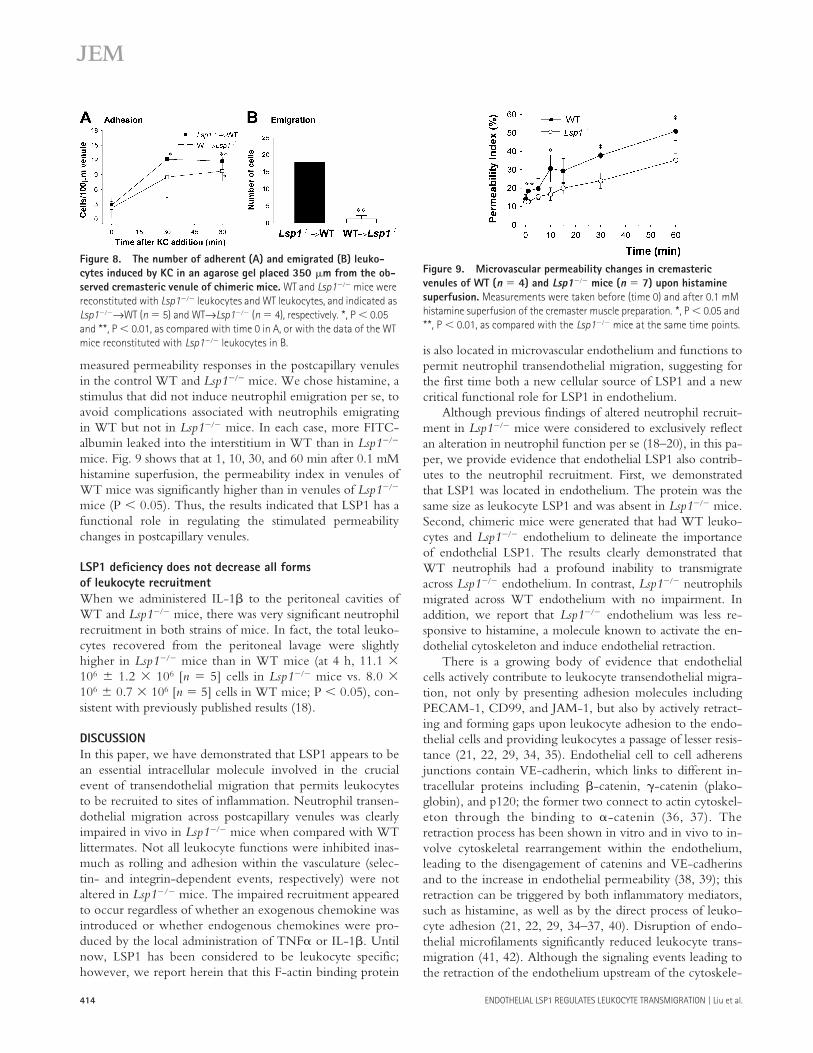
mice. We made chimeric mice that lacked LSP1 only in theleukocytes. We also made chimeric mice where the Lsp1���
mice received a BM transplant from WT mice. These micelack LSP1 in endothelium. Upon TNF� local administra-tion, both types of chimeric mice demonstrated similar re-sponsiveness in leukocyte rolling flux (not depicted), rollingvelocity (not depicted), and adhesion (Fig. 7 A) in cremas-teric venules. Surprisingly, chimeric mice that lacked LSP1only in their leukocytes emigrated as effectively across thevasculature as WT mice in response to TNF� injection, sug-gesting that the impaired transendothelial emigration wasunrelated to leukocyte-derived LSP1 (Fig. 7 B). However,WT leukocytes reconstituted in Lsp1��� mice (i.e., lackingLSP1 in endothelium) had difficulties in migrating throughthe Lsp1��� venules and into the tissue (P 0.01, as com-pared with the reversed chimeric mice). Fig. 7 (C and D)
demonstrates that WT mice receiving WT BM behaved justlike the WT mice in Fig. 1 and Lsp1��� mice receivingLsp1��� BM behaved like Lsp1��� mice in Fig. 1.
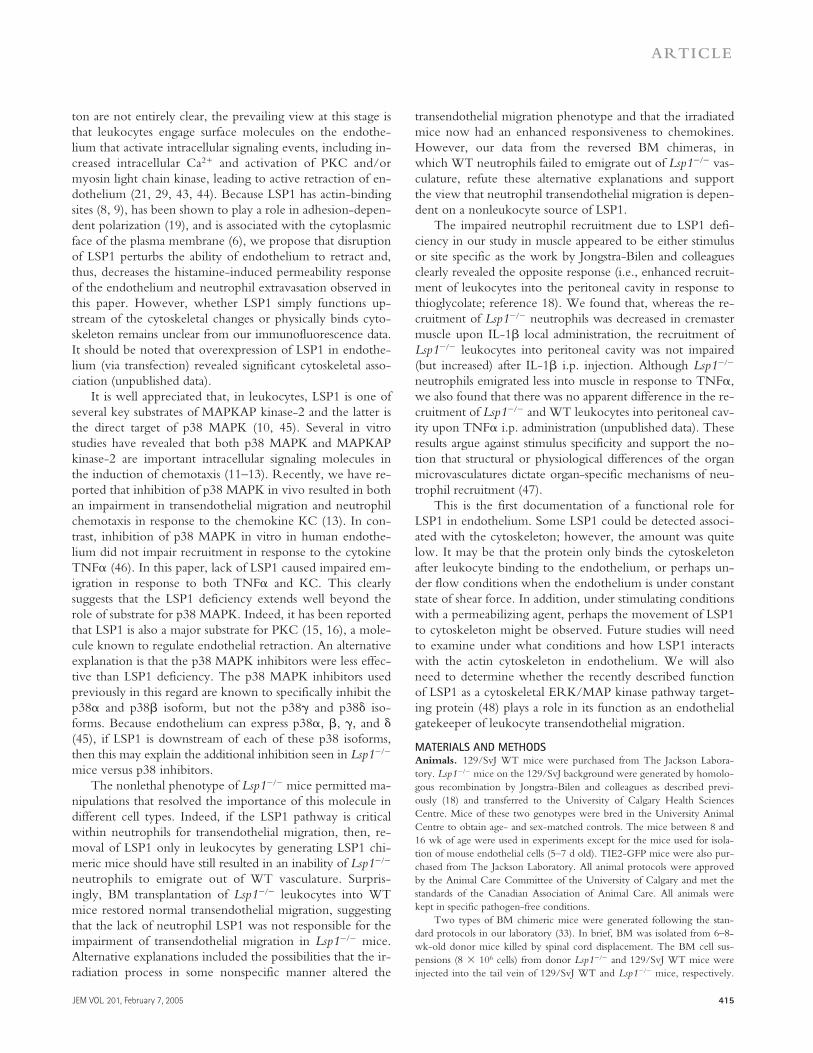
In a second series of experiments, we tested responses toKC in the chimeric mice. Both sets of chimeric mice dem-onstrated similar responsiveness in the leukocyte rolling flux(not depicted), rolling velocity (not depicted), and adhesion(Fig. 8 A) upon placement of the KC-containing gel ontothe muscle microvasculature. Again, chimeric mice thatlacked LSP1 only in their leukocytes emigrated as effectivelyacross the vasculature as WT mice (Fig. 8 B) in response toKC administration. In contrast, WT leukocytes reconstitutedin Lsp1��� mice (i.e., lacking LSP1 in endothelium) did notdisplay significant transendothelial migration (Fig. 8 B).
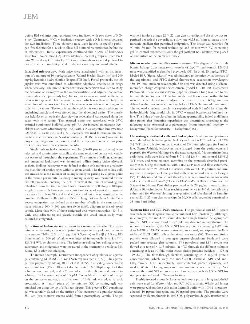
LSP1 is important in histamine-stimulated permeability increases in postcapillary venulesThis is the first demonstration of LSP1 in endothelium and,more importantly, the first demonstration of a functionalrole for endothelial LSP1 in regulating leukocyte emigration.Although the mechanism by which Lsp1��� endothelium re-stricts leukocyte recruitment is unclear, it is clear that LSP1 isan F-actin binding protein and involved in cytoskeletalchanges in leukocytes (7, 8, 19). A very likely possibility isthat the Lsp1��� endothelium did not actively retract to per-mit leukocyte transendothelial migration. Therefore, we
Figure 6. Immunofluorescence localization of LSP1 in cultured human endothelial cells. HUVECs cultured on coverslips were double stained for LSP1 (red; A, C, E, and F) and VE-cadherin (green; B and C). (B–D) Cells were also counterstained for DAPI (blue). (C) The overlaid image of A and B. (D) Secondary Ab alone (Texas red) with DAPI staining. (E) The enhanced image (by increasing the contrast) of LSP1 staining pattern. (F) The enhanced image of LSP1 (from E) overlaid with phalloidin (green). Magnification, 400.
Figure 7. The number of adherent (A and C) and emigrated (B and D) leukocytes in a cremasteric venule of TNF�-treated chimeric mice. WT and Lsp1��� mice were reconstituted with Lsp1��� and WT leukocytes, and indicated as Lsp1���→WT and WT→Lsp1���, respectively (A and B, n 3). WT and Lsp1��� mice were also reconstituted with WT and Lsp1��� leukocytes, and indicated as WT→WT and Lsp1���→Lsp1���, respectively (C and D, n 3�4). Leukocyte recruitment was induced by intrascrotal injection of TNF� (0.5 �g in 200 �l saline) and the recruitment parameters determined in cremasteric venules from these chimeric mice. **, P 0.01 as compared with the group of Lsp1���→WT mice at 4 h. *, P 0.05 as compared with the group of WT→WT mice at 4 h.
ENDOTHELIAL LSP1 REGULATES LEUKOCYTE TRANSMIGRATION | Liu et al.414
measured permeability responses in the postcapillary venulesin the control WT and Lsp1��� mice. We chose histamine, astimulus that did not induce neutrophil emigration per se, toavoid complications associated with neutrophils emigratingin WT but not in Lsp1��� mice. In each case, more FITC-albumin leaked into the interstitium in WT than in Lsp1���
mice. Fig. 9 shows that at 1, 10, 30, and 60 min after 0.1 mMhistamine superfusion, the permeability index in venules ofWT mice was significantly higher than in venules of Lsp1���
mice (P 0.05). Thus, the results indicated that LSP1 has afunctional role in regulating the stimulated permeabilitychanges in postcapillary venules.
LSP1 deficiency does not decrease all forms of leukocyte recruitmentWhen we administered IL-1� to the peritoneal cavities ofWT and Lsp1��� mice, there was very significant neutrophilrecruitment in both strains of mice. In fact, the total leuko-cytes recovered from the peritoneal lavage were slightlyhigher in Lsp1��� mice than in WT mice (at 4 h, 11.1 �106 � 1.2 � 106 [n 5] cells in Lsp1��� mice vs. 8.0 �106 � 0.7 � 106 [n 5] cells in WT mice; P 0.05), con-sistent with previously published results (18).
DISCUSSIONIn this paper, we have demonstrated that LSP1 appears to bean essential intracellular molecule involved in the crucialevent of transendothelial migration that permits leukocytesto be recruited to sites of inflammation. Neutrophil transen-dothelial migration across postcapillary venules was clearlyimpaired in vivo in Lsp1��� mice when compared with WTlittermates. Not all leukocyte functions were inhibited inas-much as rolling and adhesion within the vasculature (selec-tin- and integrin-dependent events, respectively) were notaltered in Lsp1�/� mice. The impaired recruitment appearedto occur regardless of whether an exogenous chemokine wasintroduced or whether endogenous chemokines were pro-duced by the local administration of TNF� or IL-1�. Untilnow, LSP1 has been considered to be leukocyte specific;however, we report herein that this F-actin binding protein
is also located in microvascular endothelium and functions topermit neutrophil transendothelial migration, suggesting forthe first time both a new cellular source of LSP1 and a newcritical functional role for LSP1 in endothelium.
Although previous findings of altered neutrophil recruit-ment in Lsp1��� mice were considered to exclusively reflectan alteration in neutrophil function per se (18–20), in this pa-per, we provide evidence that endothelial LSP1 also contrib-utes to the neutrophil recruitment. First, we demonstratedthat LSP1 was located in endothelium. The protein was thesame size as leukocyte LSP1 and was absent in Lsp1��� mice.Second, chimeric mice were generated that had WT leuko-cytes and Lsp1��� endothelium to delineate the importanceof endothelial LSP1. The results clearly demonstrated thatWT neutrophils had a profound inability to transmigrateacross Lsp1��� endothelium. In contrast, Lsp1��� neutrophilsmigrated across WT endothelium with no impairment. Inaddition, we report that Lsp1��� endothelium was less re-sponsive to histamine, a molecule known to activate the en-dothelial cytoskeleton and induce endothelial retraction.
There is a growing body of evidence that endothelialcells actively contribute to leukocyte transendothelial migra-tion, not only by presenting adhesion molecules includingPECAM-1, CD99, and JAM-1, but also by actively retract-ing and forming gaps upon leukocyte adhesion to the endo-thelial cells and providing leukocytes a passage of lesser resis-tance (21, 22, 29, 34, 35). Endothelial cell to cell adherensjunctions contain VE-cadherin, which links to different in-tracellular proteins including �-catenin, �-catenin (plako-globin), and p120; the former two connect to actin cytoskel-eton through the binding to �-catenin (36, 37). Theretraction process has been shown in vitro and in vivo to in-volve cytoskeletal rearrangement within the endothelium,leading to the disengagement of catenins and VE-cadherinsand to the increase in endothelial permeability (38, 39); thisretraction can be triggered by both inflammatory mediators,such as histamine, as well as by the direct process of leuko-cyte adhesion (21, 22, 29, 34–37, 40). Disruption of endo-thelial microfilaments significantly reduced leukocyte trans-migration (41, 42). Although the signaling events leading tothe retraction of the endothelium upstream of the cytoskele-
Figure 8. The number of adherent (A) and emigrated (B) leuko-cytes induced by KC in an agarose gel placed 350 �m from the ob-served cremasteric venule of chimeric mice. WT and Lsp1��� mice were reconstituted with Lsp1��� leukocytes and WT leukocytes, and indicated as Lsp1���→WT (n 5) and WT→Lsp1��� (n 4), respectively. *, P 0.05 and **, P 0.01, as compared with time 0 in A, or with the data of the WT mice reconstituted with Lsp1��� leukocytes in B.
Figure 9. Microvascular permeability changes in cremasteric venules of WT (n 4) and Lsp1��� mice (n 7) upon histamine superfusion. Measurements were taken before (time 0) and after 0.1 mM histamine superfusion of the cremaster muscle preparation. *, P 0.05 and **, P 0.01, as compared with the Lsp1��� mice at the same time points.
JEM VOL. 201, February 7, 2005 415
ARTICLE
ton are not entirely clear, the prevailing view at this stage isthat leukocytes engage surface molecules on the endothe-lium that activate intracellular signaling events, including in-creased intracellular Ca2� and activation of PKC and/ormyosin light chain kinase, leading to active retraction of en-dothelium (21, 29, 43, 44). Because LSP1 has actin-bindingsites (8, 9), has been shown to play a role in adhesion-depen-dent polarization (19), and is associated with the cytoplasmicface of the plasma membrane (6), we propose that disruptionof LSP1 perturbs the ability of endothelium to retract and,thus, decreases the histamine-induced permeability responseof the endothelium and neutrophil extravasation observed inthis paper. However, whether LSP1 simply functions up-stream of the cytoskeletal changes or physically binds cyto-skeleton remains unclear from our immunofluorescence data.It should be noted that overexpression of LSP1 in endothe-lium (via transfection) revealed significant cytoskeletal asso-ciation (unpublished data).
It is well appreciated that, in leukocytes, LSP1 is one ofseveral key substrates of MAPKAP kinase-2 and the latter isthe direct target of p38 MAPK (10, 45). Several in vitrostudies have revealed that both p38 MAPK and MAPKAPkinase-2 are important intracellular signaling molecules inthe induction of chemotaxis (11–13). Recently, we have re-ported that inhibition of p38 MAPK in vivo resulted in bothan impairment in transendothelial migration and neutrophilchemotaxis in response to the chemokine KC (13). In con-trast, inhibition of p38 MAPK in vitro in human endothe-lium did not impair recruitment in response to the cytokineTNF� (46). In this paper, lack of LSP1 caused impaired em-igration in response to both TNF� and KC. This clearlysuggests that the LSP1 deficiency extends well beyond therole of substrate for p38 MAPK. Indeed, it has been reportedthat LSP1 is also a major substrate for PKC (15, 16), a mole-cule known to regulate endothelial retraction. An alternativeexplanation is that the p38 MAPK inhibitors were less effec-tive than LSP1 deficiency. The p38 MAPK inhibitors usedpreviously in this regard are known to specifically inhibit thep38� and p38� isoform, but not the p38� and p38� iso-forms. Because endothelium can express p38�, �, �, and �(45), if LSP1 is downstream of each of these p38 isoforms,then this may explain the additional inhibition seen in Lsp1���
mice versus p38 inhibitors.The nonlethal phenotype of Lsp1��� mice permitted ma-
nipulations that resolved the importance of this molecule indifferent cell types. Indeed, if the LSP1 pathway is criticalwithin neutrophils for transendothelial migration, then, re-moval of LSP1 only in leukocytes by generating LSP1 chi-meric mice should have still resulted in an inability of Lsp1���
neutrophils to emigrate out of WT vasculature. Surpris-ingly, BM transplantation of Lsp1��� leukocytes into WTmice restored normal transendothelial migration, suggestingthat the lack of neutrophil LSP1 was not responsible for theimpairment of transendothelial migration in Lsp1��� mice.Alternative explanations included the possibilities that the ir-radiation process in some nonspecific manner altered the
transendothelial migration phenotype and that the irradiatedmice now had an enhanced responsiveness to chemokines.However, our data from the reversed BM chimeras, inwhich WT neutrophils failed to emigrate out of Lsp1��� vas-culature, refute these alternative explanations and supportthe view that neutrophil transendothelial migration is depen-dent on a nonleukocyte source of LSP1.
The impaired neutrophil recruitment due to LSP1 defi-ciency in our study in muscle appeared to be either stimulusor site specific as the work by Jongstra-Bilen and colleaguesclearly revealed the opposite response (i.e., enhanced recruit-ment of leukocytes into the peritoneal cavity in response tothioglycolate; reference 18). We found that, whereas the re-cruitment of Lsp1��� neutrophils was decreased in cremastermuscle upon IL-1� local administration, the recruitment ofLsp1��� leukocytes into peritoneal cavity was not impaired(but increased) after IL-1� i.p. injection. Although Lsp1���
neutrophils emigrated less into muscle in response to TNF�,we also found that there was no apparent difference in the re-cruitment of Lsp1��� and WT leukocytes into peritoneal cav-ity upon TNF� i.p. administration (unpublished data). Theseresults argue against stimulus specificity and support the no-tion that structural or physiological differences of the organmicrovasculatures dictate organ-specific mechanisms of neu-trophil recruitment (47).
This is the first documentation of a functional role forLSP1 in endothelium. Some LSP1 could be detected associ-ated with the cytoskeleton; however, the amount was quitelow. It may be that the protein only binds the cytoskeletonafter leukocyte binding to the endothelium, or perhaps un-der flow conditions when the endothelium is under constantstate of shear force. In addition, under stimulating conditionswith a permeabilizing agent, perhaps the movement of LSP1to cytoskeleton might be observed. Future studies will needto examine under what conditions and how LSP1 interactswith the actin cytoskeleton in endothelium. We will alsoneed to determine whether the recently described functionof LSP1 as a cytoskeletal ERK/MAP kinase pathway target-ing protein (48) plays a role in its function as an endothelialgatekeeper of leukocyte transendothelial migration.
MATERIALS AND METHODSAnimals. 129/SvJ WT mice were purchased from The Jackson Labora-tory. Lsp1��� mice on the 129/SvJ background were generated by homolo-gous recombination by Jongstra-Bilen and colleagues as described previ-ously (18) and transferred to the University of Calgary Health SciencesCentre. Mice of these two genotypes were bred in the University AnimalCentre to obtain age- and sex-matched controls. The mice between 8 and16 wk of age were used in experiments except for the mice used for isola-tion of mouse endothelial cells (5–7 d old). TIE2-GFP mice were also pur-chased from The Jackson Laboratory. All animal protocols were approvedby the Animal Care Committee of the University of Calgary and met thestandards of the Canadian Association of Animal Care. All animals werekept in specific pathogen-free conditions.
Two types of BM chimeric mice were generated following the stan-dard protocols in our laboratory (33). In brief, BM was isolated from 6–8-wk-old donor mice killed by spinal cord displacement. The BM cell sus-pensions (8 � 106 cells) from donor Lsp1��� and 129/SvJ WT mice wereinjected into the tail vein of 129/SvJ WT and Lsp1��� mice, respectively.
ENDOTHELIAL LSP1 REGULATES LEUKOCYTE TRANSMIGRATION | Liu et al.416
Before BM cell injection, recipients were irradiated with two doses of 5 Gy�-ray (Gammacell, 137Cs �-irradiation source) with a 3-h interval betweenthe two irradiations. These chimeric mice were housed in specific patho-gen-free facilities for 6–8 wk to allow full humoral reconstitution before usein experiments. Initial experiments confirmed that �99% of leukocyteswere from donor mice (33). Two additional control groups of mice (WTinto WT and Lsp1��� into Lsp1���) went through an identical protocol toensure that the transplant procedure did not cause any untoward effects.
Intravital microscopy. Male mice were anesthetized with an i.p. injec-tion of a mixture of 10 mg/kg xylazine (Animal Health; Bayer Inc.) and 200mg/kg ketamine hydrochloride (Rogar/STB Inc.). For all protocols, the leftjugular vein was cannulated to administer additional anesthetic or drugswhen necessary. The mouse cremaster muscle preparation was used to studythe behavior of leukocytes in the microcirculation and adjacent connectivetissue as described previously (49). In brief, an incision was made in the scro-tal skin to expose the left cremaster muscle, which was then carefully dis-sected free of the associated fascia. The cremaster muscle was cut longitudi-nally with a cautery. The testicle and the epididymis were separated from theunderlying muscle and were moved into the abdominal cavity. The musclewas held flat on an optically clear viewing pedestal and was secured along theedges with 4–0 suture. The exposed tissue was superfused with 37 Cwarmed bicarbonate-buffered saline, pH 7.4. An intravital microscope (Axi-olskip; Carl Zeiss MicroImaging, Inc.) with a �25 objective lens (WeltzlarL25/0.35; E. Leitz Inc.), and a �10 eyepiece was used to examine the cre-masteric microcirculation. A video camera (5100 HS; Panasonic) was used toproject the images onto a monitor, and the images were recorded for play-back analysis using a videocassette recorder.
Single unbranched cremasteric venules (25–40 �m in diameter) wereselected, and to minimize variability, the same section of cremasteric venulewas observed throughout the experiment. The number of rolling, adherent,and emigrated leukocytes was determined offline during video playbackanalysis. Rolling leukocytes were defined as those cells moving at a velocityless than that of erythrocytes within a given vessel. The flux of rolling cellswas measured as the number of rolling leukocytes passing by a given pointin the venule per minute. Leukocyte rolling velocity was measured for thefirst 20 leukocytes entering the field of view at the time of recording andcalculated from the time required for a leukocyte to roll along a 100-�mlength of venule. A leukocyte was considered to be adherent if it remainedstationary for at least 30 s, and total leukocyte adhesion was quantified as thenumber of adherent cells within a 100-�m length of venule in 5 min. Leu-kocyte emigration was defined as the number of cells in the extravascularspace within a 200 � 300-�m area (0.06 mm2), adjacent to the observedvenule. More than 90% of these emigrated cells were neutrophils (13, 31).Only cells adjacent to and clearly outside the vessel under study werecounted as emigrated.
Induction of leukocyte recruitment in cremaster muscle. To deter-mine whether emigration was impaired in response to cytokines, recombi-nant mouse TNF� (0.5 or 0.1 �g; R&D Systems) or IL-1� (12.5 ng; BDBiosciences) in 200 �l of saline was injected intrascrotally into Lsp1���,129/SvJ WT, or chimeric mice. The leukocyte rolling flux, rolling velocity,adherence, and emigration were measured in the cremasteric venule at 3.5,4, and 4.5 h after the injection.
To induce neutrophil recruitment independent of cytokines, an agarosegel containing KC (CXCL1; R&D Systems) was used (13, 50). The agarosegel was prepared by adding 10 ml of 2 � HBSS to a boiling concentratedagarose solution (4% in 10 ml of distilled water). A 100-�l aliquot of thissolution was removed, and KC was added to this aliquot and mixed toachieve a final concentration of 0.5 �M. To enable visualization of the gelon the cremaster muscle, a small amount of India ink was added to eachpreparation. A 1-mm3 piece of the mixture (KC-containing gel) waspunched out using the tip of a Pasteur pipette. This piece of KC-containinggel was carefully placed on the surface of the cremaster in a preselected area350 �m (two monitor screens wide) from a postcapillary venule. The gel
was held in place using a 22 � 22-mm glass coverslip, and the tissue was su-perfused beneath the coverslip at a slow rate (0.35 ml/min) to create a che-motactic gradient that permitted emigration. The image was recorded for90 min: 30 min for control without gel and 60 min with KC-containinggel. In control experiments, only the gel (without KC addition) was placedon the surface of the cremaster muscle.
Microvascular permeability measurement. The degree of vascular al-bumin leakage from cremasteric venules of Lsp1��� and control 129/SvJmice was quantified as described previously (51). In brief, 25 mg/kg FITC-labeled BSA (Sigma-Aldrich) was administered to the mice i.v. at the start ofthe experiment, and FITC-derived fluorescence (excitation wavelength,450–490 nm; emission wavelength, 520 nm) was detected using a silicon-intensified charge-coupled device camera (model C-2400-80; HamamatsuPhotonics). Image analysis software (Optimas; Bioscan Inc.) was used to de-termine the intensity of FITC-albumin–derived fluorescence within the lu-men of the venule and in the adjacent perivascular tissue. Background wasdefined as the fluorescence intensity before FITC-albumin administration.The exposed cremaster muscle was superfused with 0.1 mM histamine di-hydrochloride (Sigma-Aldrich) in 37 C warmed bicarbonate-buffered sa-line. The index of vascular albumin leakage (permeability index) at differenttime points after histamine superfusion was determined according to thefollowing ratio expressed as a percentage: (mean interstitial intensity �
background)/(venular intensity � background) (51).
Harvesting endothelial cells and leukocytes. Acute mouse peritonitiswas induced to obtain emigrated leukocytes from Lsp1��� and control 129/SvJ WT mice. 3 h after an i.p. injection of 1% oyster glycogen (in 1 ml sa-line; Sigma-Aldrich), leukocytes were lavaged from the peritoneum andprepared for Western blotting (see the next paragraph). Mouse primary lungendothelial cells were isolated from 5–7-d-old Lsp1��� and control 129/SvJWT mice, and were cultured according to the protocols described previ-ously (52). Using this protocol with TIE2-GFP mice and flow cytometry,we verified that �93–98% of the isolated cells were GFP positive, confirm-ing that the majority of the purified cells were of endothelial cell origin(53). Freshly isolated mouse endothelial cells were cultured in microvascularendothelial cell medium-2 (Clonetics EGM-2MV BulletKit; Cambrex BioScience) in 35-mm Petri dishes precoated with 20 �g/ml mouse laminin(Upstate Biotechnology). After reaching confluence in 5–6 d, the cells wereeither used for Western blotting or trypsinized and subcultured on laminin-coated 22 � 22-mm glass coverslips (at 30,000 cells/coverslip) contained in35-mm Petri dishes.
Western blot and RT-PCR analysis. The polyclonal anti-LSP1 serumwas made in rabbits against mouse recombinant LSP1 protein (6). Although,in leukocytes, the anti-LSP1 serum detected a single band at the appropriatesize for LSP1, a second band of �78 kD was detected in endothelium. Toremove this reactivity, the GST-LSP1 fusion proteins containing LSP1 resi-dues 1–178 or 179–330 were constructed, subcloned, and expressed in Esch-erichia coli BL21 (DE3) cells as described previously (54). These two fusionproteins were allowed to conjugate agarose-glutathione beads and werepacked into separate glass columns. The polyclonal anti-LSP1 serum wasflowed at a rate of 0.15 ml/min (at 4 C) through the different columnscontaining at least 10-fold molar excess fusion proteins (residues 1–178 or179–330). The flow-through fractions containing �1.5 mg/ml proteinconcentrations, which were the anti–COOH-terminal LSP1 and anti–NH2-terminal LSP1, respectively, were collected, pooled separately, andused in Western blotting assays and immunofluorescence microscopy. As acontrol, the anti-LSP1 serum was also absorbed against both GST-LSP1 fu-sion proteins and used in Western blotting.
Freshly isolated mouse leukocytes and mouse primary lung endothelialcells were used for Western blot and RT-PCR analysis. Whole cell lysateswere prepared from these cells using Laemmli buffer with 10% �-mercapto-ethanol, 10 �g/ml leupeptin, and 10 �g/ml aprotinin. The proteins wereseparated by electrophoresis in 10% SDS-polyacrylamide gels, transferred to
JEM VOL. 201, February 7, 2005 417
ARTICLE
a PVDF Hybond-P transfer membrane (Amersham Biosciences), and blot-ted using a specific polyclonal rabbit anti–mouse LSP1 serum (at 1:2,000 di-lution) as described previously (6) or the anti–COOH-terminal LSP1 andanti–NH2-terminal LSP1 as described before. After washing, the membranewas incubated with a secondary, horseradish peroxidase–conjugated goatanti–rabbit IgG and treated with enhanced chemiluminescence reagents(ECL kit; Amersham Biosciences). The blotted bands were detected withhigh performance autoradiography films from Amersham Biosciences. RT-PCR was performed using total RNA (100 ng for each cell type) extractedfrom freshly isolated leukocytes, mouse primary lung endothelial cells, andLSP1 primer pair A1/A4 as described previously (4). The PCR productswere electrophoresed by agarose gel, stained with ethidium bromide, andanalyzed by high sensitivity Fluor-S Multimager MAX scanner (Bio-RadLaboratories) upon dark subtraction.
Immunofluorescence microscopy. All rinsing, incubation, and dilutionof antibodies was performed in basal buffer that contained 137 NaCl, 5KCl, 1.1 Na2HPO4, 0.4 KH2PO4, 4 NaHCO3, 5.5 glucose, 4.15 PIPES di-sodium salt, 2 EGTA, and 4.15 MgCl2 in mM, pH 7.2, at room tempera-ture. Mouse lung primary endothelial cells grown on glass coverslips for 24 hwere fixed with 4% formalin, permeabilized with 0.1% Triton X-100, andincubated with 10 �g/ml glycine. Primary antibodies used were the anti–COOH-terminal LSP1 and anti–NH2-terminal LSP1 as described beforefor 30 min where the protein concentrations were both 0.4 mg/ml in thetwo blotting solutions, or the original rabbit polyclonal anti-LSP1 serum (at1:100 dilution). After rinsing three times with 0.1% Tween-20, the cover-slips were incubated with Cy3-conjugated goat anti–rabbit IgG for 30 min.After rinsing with basal buffer, the coverslips were mounted in 90% glyc-erol. Observations were performed on an Olympus IX-70 fluorescence mi-croscope (Olympus). Fluorescence images were captured using OpenLabsoftware (version 3.1.5; Improvision Inc.).
To determine whether human endothelial cells express LSP1, we iso-lated and cultured HUVECs as described previously (46). Once the HU-VECs were confluent, they were passaged onto fibronectin-coated glasscoverslips. After culture for 24–48 h, these HUVECs were stained as out-lined before with 2.5 �g/ml mouse anti–human LSP1 mAb (clone 16; BDBiosciences) and goat anti–mouse IgG conjugated with Texas Red (Molec-ular Probes). DAPI (4�,6-diamidino-2-phenylindole, dihydrochloride; Mo-lecular Probes) was used for nuclear staining. To confirm that endothelialcells and not a contaminating cell type expressed LSP1, we dual labeled theHUVECs with FITC-conjugated anti–human VE-cadherin (Bender Med-systems) in combination with LSP1 staining. To assess the association ofLSP1 with the cytoskeleton, we labeled HUVECs with anti-LSP1 and phal-loidin (Alexa Fluor conjugated; Molecular Probes).
Statistical analysis. The data are expressed as means � SEM. A Student’s ttest was applied to compare the statistical difference within two groups, andanalysis of variance was used for the comparison of the differences in morethan two groups. A p-value of 0.05 was considered statistically significant.
We thank L. Zbytnuik and K. Jorgensen for their expert assistance in animal care.This work was supported in part by a Canadian Institutes of Health Research
group grant and by the Heart and Stroke Foundation of Canada. L. Liu is supported by a fellowship from Heart and Stroke Foundation of Canada and Alberta Heritage Foundation for Medical Research. D.C. Cara has a postdoctoral fellowship from Conselho Nacional de Desenvolvimento Cientifico e Tecnologico-CNPq, Brasilia, Brazil. P. Kubes is an Alberta Heritage Foundation for Medical Research Scientist and a Canada Research Chair.
The authors have no conflicting financial interests.
Submitted: 27 April 2004Accepted: 24 November 2004
REFERENCES1. Jongstra, J., G.F. Tidmarsh, J. Jongstra-Bilen, and M.M. Davis. 1988. A
new lymphocyte-specific gene which encodes a putative Ca2�-binding
protein is not expressed in transformed T lymphocyte lines. J. Immunol.141:3999–4004.
2. Jongstra-Bilen, J., A.J. Young, R. Chong, and J. Jongstra. 1990. Hu-man and mouse LSP1 genes code for highly conserved phosphopro-teins. J. Immunol. 144:1104–1110.
3. Pulford, K., M. Jones, A.H. Banham, E. Haralambieva, and D.Y. Ma-son. 1999. Lymphocyte-specific protein 1: a specific marker of humanleucocytes. Immunology. 96:262–271.
4. Jongstra, J., M.E. Ittel, N.N. Iscove, and G. Brady. 1994. The LSP1gene is expressed in cultured normal and transformed mouse macro-phages. Mol. Immunol. 31:1125–1131.
5. Li, Y., A. Guerrero, and T.H. Howard. 1995. The actin-binding pro-tein, lymphocyte-specific protein 1, is expressed in human leukocytesand human myeloid and lymphoid cell lines. J. Immunol. 155:3563–3569.
6. Klein, D.P., J. Jongstra-Bilen, K. Ogryzlo, R. Chong, and J. Jongstra.1989. Lymphocyte-specific Ca2�-binding protein LSP1 is associatedwith the cytoplasmic face of the plasma membrane. Mol. Cell. Biol.9:3043–3048.
7. Klein, D.P., S. Galea, and J. Jongstra. 1990. The lymphocyte-specificprotein LSP1 is associated with the cytoskeleton and co-caps withmembrane IgM. J. Immunol. 145:2967–2973.
8. Jongstra-Bilen, J., P.A. Janmey, J.H. Hartwig, S. Galea, and J. Jongstra.1992. The lymphocyte-specific protein LSP1 binds to F-actin and tothe cytoskeleton through its COOH-terminal basic domain. J. CellBiol. 118:1443–1453.
9. Zhang, Q., Y. Li, and T.H. Howard. 2000. Human lymphocyte-spe-cific protein 1, the protein overexpressed in neutrophil actin dysfunc-tion with 47-kDa and 89-kDa protein abnormalities (NAD 47/89), hasmultiple F-actin binding domains. J. Immunol. 165:2052–2058.
10. Huang, C.K., L. Zhan, Y. Ai, and J. Jongstra. 1997. LSP1 is the majorsubstrate for mitogen-activated protein kinase-activated protein kinase2 in human neutrophils. J. Biol. Chem. 272:17–19.
11. Hannigan, M.O., L. Zhan, Y. Ai, A. Kotlyarov, M. Gaestel, and C.K.Huang. 2001. Abnormal migration phenotype of mitogen-activatedprotein kinase-activated protein kinase 2�/� neutrophils in Zigmondchambers containing formyl-methionyl-leucyl-phenylalanine gradients.J. Immunol. 167:3953–3961.
12. Zu, Y.L., J. Qi, A. Gilchrist, G.A. Fernandez, D. Vazquez-Abad, D.L.Kreutzer, C.K. Huang, and R.I. Sha’afi. 1998. p38 mitogen-activatedprotein kinase activation is required for human neutrophil function trig-gered by TNF-� or FMLP stimulation. J. Immunol. 160:1982–1989.
13. Cara, D.C., J. Kaur, M. Forster, D.M. McCafferty, and P. Kubes. 2001.Role of p38 mitogen-activated protein kinase in chemokine-induced emi-gration and chemotaxis in vivo. J. Immunol. 167:6552–6558.
14. Stokoe, D., K. Engel, D.G. Campbell, P. Cohen, and M. Gaestel.1992. Identification of MAPKAP kinase 2 as a major enzyme responsi-ble for the phosphorylation of the small mammalian heat shock pro-teins. FEBS Lett. 313:307–313.
15. Carballo, E., D. Colomer, J.L. Vives-Corrons, P.J. Blackshear, and J.Gil. 1996. Characterization and purification of a protein kinase C sub-strate in human B cells. Identification as lymphocyte-specific protein 1(LSP1). J. Immunol. 156:1709–1713.
16. Matsumoto, N., S. Kojima, T. Osawa, and S. Toyoshima. 1995. Pro-tein kinase C phosphorylates p50 LSP1 and induces translocation ofp50 LSP1 in T lymphocytes. J. Biochem. (Tokyo). 117:222–229.
17. Laudanna, C., D. Mochly-Rosen, T. Liron, G. Constantin, and E.C.Butcher. 1998. Evidence of � protein kinase C involvement in poly-morphonuclear neutrophil integrin-dependent adhesion and chemo-taxis. J. Biol. Chem. 273:30306–30315.
18. Jongstra-Bilen, J., V.L. Misener, C. Wang, H. Ginzberg, A. Auerbach,A.L. Joyner, G.P. Downey, and J. Jongstra. 2000. LSP1 modulates leu-kocyte populations in resting and inflamed peritoneum. Blood. 96:1827–1835.
19. Wang, C., H. Hayashi, R. Harrison, B. Chiu, J.R. Chan, H.L. Oster-gaard, R.D. Inman, J. Jongstra, M.I. Cybulsky, and J. Jongstra-Bilen.2002. Modulation of Mac-1 (CD11b/CD18)-mediated adhesion bythe leukocyte-specific protein 1 is key to its role in neutrophil polariza-tion and chemotaxis. J. Immunol. 169:415–423.
ENDOTHELIAL LSP1 REGULATES LEUKOCYTE TRANSMIGRATION | Liu et al.418
20. Hannigan, M., L. Zhan, Y. Ai, and C.K. Huang. 2001. Leukocyte-spe-cific gene 1 protein (LSP1) is involved in chemokine KC-activated cy-toskeletal reorganization in murine neutrophils in vitro. J. Leukoc. Biol.69:497–504.
21. Luscinskas, F.W., S. Ma, A. Nusrat, C.A. Parkos, and S.K. Shaw. 2002.Leukocyte transendothelial migration: a junctional affair. Semin. Immu-nol. 14:105–113.
22. Vestweber, D. 2002. Regulation of endothelial cell contacts duringleukocyte extravasation. Curr. Opin. Cell Biol. 14:587–593.
23. Kayyali, U.S., C.M. Pennella, C. Trujillo, O. Villa, M. Gaestel, andP.M. Hassoun. 2002. Cytoskeletal changes in hypoxic pulmonary en-dothelial cells are dependent on MAPK-activated protein kinase MK2.J. Biol. Chem. 277:42596–42602.
24. Park, J.H., N. Okayama, D. Gute, A. Krsmanovic, H. Battarbee, andJ.S. Alexander. 1999. Hypoxia/aglycemia increases endothelial perme-ability: role of second messengers and cytoskeleton. Am. J. Physiol. 277:C1066–C1074.
25. Huot, J., F. Houle, F. Marceau, and J. Landry. 1997. Oxidative stress-induced actin reorganization mediated by the p38 mitogen-activatedprotein kinase/heat shock protein 27 pathway in vascular endothelialcells. Circ. Res. 80:383–392.
26. Nwariaku, F.E., J. Chang, X. Zhu, Z. Liu, S.L. Duffy, N.H. Halaihel,L. Terada, and R.H. Turnage. 2002. The role of p38 MAP kinase intumor necrosis factor-induced redistribution of vascular endothelialcadherin and increased endothelial permeability. Shock. 18:82–85.
27. Laird, S.M., A. Graham, A. Paul, G.W. Gould, C. Kennedy, and R.Plevin. 1998. Tumour necrosis factor stimulates stress-activated proteinkinases and the inhibition of DNA synthesis in cultures of bovine aorticendothelial cells. Cell. Signal. 10:473–480.
28. Rousseau, S., F. Houle, J. Landry, and J. Huot. 1997. p38 MAP kinaseactivation by vascular endothelial growth factor mediates actin reorga-nization and cell migration in human endothelial cells. Oncogene. 15:2169–2177.
29. Huang, A.J., J.E. Manning, T.M. Bandak, M.C. Ratau, K.R. Hanser,and S.C. Silverstein. 1993. Endothelial cell cytosolic free calcium regu-lates neutrophil migration across monolayers of endothelial cells. J. CellBiol. 120:1371–1380.
30. Garcia, J.G., A.D. Verin, M. Herenyiova, and D. English. 1998. Ad-herent neutrophils activate endothelial myosin light chain kinase: rolein transendothelial migration. J. Appl. Physiol. 84:1817–1821.
31. Thompson, R.D., K.E. Noble, K.Y. Larbi, A. Dewar, G.S. Duncan,T.W. Mak, and S. Nourshargh. 2001. Platelet-endothelial cell adhe-sion molecule-1 (PECAM-1)–deficient mice demonstrate a transientand cytokine-specific role for PECAM-1 in leukocyte migration throughthe perivascular basement membrane. Blood. 97:1854–1860.
32. Young, R.E., R.D. Thompson, and S. Nourshargh. 2002. Divergentmechanisms of action of the inflammatory cytokines interleukin 1-�and tumour necrosis factor-� in mouse cremasteric venules. Br. J. Phar-macol. 137:1237–1246.
33. Carvalho-Tavares, J., M.J. Hickey, J. Hutchison, J. Michaud, I.T. Sut-cliffe, and P. Kubes. 2000. A role for platelets and endothelial selectinsin tumor necrosis factor-�-induced leukocyte recruitment in the brainmicrovasculature. Circ. Res. 87:1141–1148.
34. Del Maschio, A., A. Zanetti, M. Corada, Y. Rival, L. Ruco, M.G.Lampugnani, and E. Dejana. 1996. Polymorphonuclear leukocyte ad-hesion triggers the disorganization of endothelial cell-to-cell adherensjunctions. J. Cell Biol. 135:497–510.
35. Johnson-Leger, C., M. Aurrand-Lions, and B.A. Imhof. 2000. Theparting of the endothelium: miracle, or simply a junctional affair? J.Cell Sci. 113:921–933.
36. Dejana, E. 1996. Endothelial adherens junctions: implications in the
control of vascular permeability and angiogenesis. J. Clin. Invest. 98:1949–1953.
37. Dejana, E., R. Spagnuolo, and G. Bazzoni. 2001. Interendothelialjunctions and their role in the control of angiogenesis, vascular perme-ability and leukocyte transmigration. Thromb. Haemost. 86:308–315.
38. Waschke, J., W. Baumgartner, R.H. Adamson, M. Zeng, K. Aktories,H. Barth, C. Wilde, F.E. Curry, and D. Drenckhahn. 2004. Require-ment of Rac activity for maintenance of capillary endothelial barrierproperties. Am. J. Physiol. Heart Circ. Physiol. 286:H394–H401.
39. Adamson, R.H., F.E. Curry, G. Adamson, B. Liu, Y. Jiang, K. Akto-ries, H. Barth, A. Daigeler, N. Golenhofen, W. Ness, and D. Drenck-hahn. 2002. Rho and rho kinase modulation of barrier properties: cul-tured endothelial cells and intact microvessels of rats and mice. J.Physiol. 539:295–308.
40. Shaw, S.K., P.S. Bamba, B.N. Perkins, and F.W. Luscinskas. 2001.Real-time imaging of vascular endothelial-cadherin during leukocytetransmigration across endothelium. J. Immunol. 167:2323–2330.
41. Kielbassa, K., C. Schmitz, and V. Gerke. 1998. Disruption of endothe-lial microfilaments selectively reduces the transendothelial migration ofmonocytes. Exp. Cell Res. 243:129–141.
42. Hordijk, P.L., E. Anthony, F.P. Mul, R. Rientsma, L.C. Oomen, andD. Roos. 1999. Vascular-endothelial-cadherin modulates endothelialmonolayer permeability. J. Cell Sci. 112:1915–1923.
43. Su, W.H., H.I. Chen, J.P. Huang, and C.J. Jen. 2000. Endothelial[Ca2�]i signaling during transmigration of polymorphonuclear leuko-cytes. Blood. 96:3816–3822.
44. Lum, H., and A.B. Malik. 1994. Regulation of vascular endothelialbarrier function. Am. J. Physiol. 267:L223–L241.
45. Herlaar, E., and Z. Brown. 1999. p38 MAPK signalling cascades in in-flammatory disease. Mol. Med. Today. 5:439–447.
46. Kaur, J., R.C. Woodman, and P. Kubes. 2003. P38 MAPK: criticalmolecule in thrombin-induced NF-�B-dependent leukocyte recruit-ment. Am. J. Physiol. Heart Circ. Physiol. 284:H1095–H1103.
47. Liu, L., and P. Kubes. 2003. Molecular mechanisms of leukocyte re-cruitment: organ-specific mechanisms of action. Thromb. Haemost. 89:213–220.
48. Harrison, R.E., B.A. Sikorski, and J. Jongstra. 2004. Leukocyte-specificprotein 1 targets the ERK/MAP kinase scaffold protein KSR and MEK1and ERK2 to the actin cytoskeleton. J. Cell Sci. 117:2151–2157.
49. Kanwar, S., D.C. Bullard, M.J. Hickey, C.W. Smith, A.L. Beaudet,B.A. Wolitzky, and P. Kubes. 1997. The association between �4-inte-grin, P-selectin, and E-selectin in an allergic model of inflammation. J.Exp. Med. 185:1077–1087.
50. Hickey, M.J., M. Forster, D. Mitchell, J. Kaur, C. De Caigny, and P.Kubes. 2000. L-selectin facilitates emigration and extravascular loco-motion of leukocytes during acute inflammatory responses in vivo. J.Immunol. 165:7164–7170.
51. Kurose, I., P. Kubes, R. Wolf, D.C. Anderson, J. Paulson, M. Mi-yasaka, and D.N. Granger. 1993. Inhibition of nitric oxide production.Mechanisms of vascular albumin leakage. Circ. Res. 73:164–171.
52. Bowden, R.A., Z.M. Ding, E.M. Donnachie, T.K. Petersen, L.H.Michael, C.M. Ballantyne, and A.R. Burns. 2002. Role of �4 integrinand VCAM-1 in CD18-independent neutrophil migration across mousecardiac endothelium. Circ. Res. 90:562–569.
53. Motoike, T., S. Loughna, E. Perens, B.L. Roman, W. Liao, T.C.Chau, C.D. Richardson, T. Kawate, J. Kuno, B.M. Weinstein, et al.2000. Universal GFP reporter for the study of vascular development.Genesis. 28:75–81.
54. Wong, M.J., I.A. Malapitan, B.A. Sikorski, and J. Jongstra. 2003. Acell-free binding assay maps the LSP1 cytoskeletal binding site to theCOOH-terminal 30 amino acids. Biochim. Biophys. Acta. 1642:17–24.