low molecular weight spies of protein-protein interactions
TRANSCRIPT

Available online at www.sciencedirect.com
C. R. Chimie 11 (2008) 499e505http://france.elsevier.com/direct/CRAS2C/
Full paper / Memoire
Low-molecular-weight spies of proteineprotein interactions
Jascha Blobel a, Rosa Fayos a, Jesus Garcıa a, Oriol Marimon a,b,Yolanda Perez a, Miquel Pons a,b,*
a Institute for Research in Biomedicine, Parc Cientıfic de Barcelona, Josep Samitier, 1-5 08028 Barcelona, Spainb Organic Chemistry Department, University of Barcelona, Martı i Franques, 1-11 08028 Barcelona, Spain
Received 9 May 2007; accepted after revision 2 August 2007
Available online 26 November 2007
Abstract
The interaction of low-molecular-weight ligands with proteins in the search for new selective drugs is driving great efforts in thepharmaceutical industry and academic research. The same principles can be applied to solve an equally challenging task: selectivemonitoring of protein interactions by NMR at concentrations close to physiological ones in unlabelled samples. In this review, wediscuss different approaches to this problem through the measurement of either relaxation rates or chemical shifts of free ligandspies in equilibrium with their macromolecular targets. To cite this article: J. Blobel et al., C. R. Chimie 11 (2008).� 2007 Academie des sciences. Published by Elsevier Masson SAS. All rights reserved.
Resume
L’interaction de ligands de bas poids moleculaire avec les proteines pour la recherche de nouveaux medicaments selectifs est al’origine de grands efforts dans l’industrie pharmaceutique et la recherche academique. Les memes principes peuvent etre appliquespour mener a bien une tache tout aussi stimulante : l’etude selective des interactions entre proteines par RMN a des concentrationsproches des concentrations physiologiques dans les echantillons non marques. Dans cette revue, nous discutons differentesapprochesre ce probleme par la mesure des taux de relaxation ou des changements du deplacement chimique des ligands libresa l’equilibre avec leurs cibles macromoleculaires. Pour citer cet article : J. Blobel et al., C. R. Chimie 11 (2008).� 2007 Academie des sciences. Published by Elsevier Masson SAS. All rights reserved.
Keywords: Proteineligand interactions; Proteineprotein interactions by STD; Molecular spies; NMR
Mots-cles : Interaccion proteine-ligand ; Interaccion proteine-proteine par STD ; Molecular espions ; RMN
* Corresponding author.
E-mail address: [email protected] (M. Pons).
1631-0748/$ - see front matter � 2007 Academie des sciences. Published
doi:10.1016/j.crci.2007.08.015
1. Introduction
1.1. Low-molecular-weight spies
The small-molecular world includes an extremelylarge collection of chemical compounds, existing or vir-tual, sometimes called the chemical space. The size of
by Elsevier Masson SAS. All rights reserved.

500 J. Blobel et al. / C. R. Chimie 11 (2008) 499e505
chemical space is a matter of speculation, but figures ofthe order of 1060 have been suggested [1]. This numberis certainly much larger than the number of existing pro-teins and there is a reasonable chance that, for each pro-tein, one could find a specific small ligand, allowinga one-to-one connection to be made between the small-molecular weight world and the macromolecular world.Since the nuclear magnetic resonance (NMR) pro-perties of the members of the two worlds are ratherdifferent, one can envisage new approaches in whichsmall molecules are used as ‘spies’ to detect by NMRmacromolecules and their interactions (i) in complexmixtures, (ii) at low concentrations, and (iii) usingnon-isotopically labelled samples. These three limita-tions are typical of physiological conditions. DirectNMR observation of individual macromolecules underthese conditions is presently impossible for sensitivityand selectivity reasons.
Small ‘spy’ molecules should display selectivity fortheir macromolecular targets in the order that they canbe addressed individually in a complex mixture. The va-riety of chemical environments in the small-molecularworld is much larger than that in biological macromol-ecules, providing an enhanced spectroscopic resolution.This makes much easier the selective NMR observationof each of the small-molecular-weight spies in a com-plex mixture than the direct selective observation oftheir high-molecular-weight correlates (Fig. 1).
The use of small ‘spy’ molecules is directly related tothe study of ligandeprotein interactions, which is fuelledby the search for low-molecular-weight drugs. There are,however, major differences: drug candidates are usuallyexpected to bind strongly to the macromolecule and
Small molecules world: high concentrations, large
chemical universe, short correlation times
Macromolecular world: low concentrations,
long correlation times
Fig. 1. Schematic depiction of the relationship between small spy
molecules and their macromolecular counterparts. Small molecules
can detect not only the presence of specific macromolecules but
also their interactions with other macromolecular components.
change its properties (e.g., inhibit its natural activity)and to be non-toxic for the whole organism. In contrast,low-molecular-weight spies are expected to bind weaklyto the macromolecule, not perturbing its activity and, ifused in vitro, they are subjected to far less stringent con-ditions of toxicity or bioavailability.
In addition, small spy molecules should be able tosense different states of their target (e.g., different con-formations or the interaction with other macromole-cules) and encode this information in a way that canbe ‘read’ in their free state.
1.2. Ligand screening
The use of NMR to study proteineligand interac-tions is a mature field and has been extensively re-viewed [2e4]. The typical problem is to screena collection of compounds to find suitable ligands fora macromolecule of interest. One classically distin-guishes between methods based on protein observationand those that rely on ligand detection. The use ofsmall-molecular-weight spies is clearly related to li-gand detection methods used for screening purposes.In this case, a known ligandemacromolecular pair isobserved in order to detect additional perturbations inthe macromolecular component.
1.2.1. NMR experiments for lead generation in drugdiscovery
The most used ligand-based screening methods havethe desired properties of not requiring isotopicallylabelled proteins, not being limited by the molecularweight of the biomolecule, and being applicable tosamples of low macromolecular concentration.
Binding is detected through the changes in the NMRspectroscopic properties of the free ligand induced bythe temporary interaction with the macromolecular tar-get. This requires fast exchange between the free andthe bound forms and puts a lower limit to the ligandkoff, the rate constant for ligand release from thecomplex.
In the fast exchange limit, the value of the observedproperties is the weighted average of those correspond-ing to the free and bound forms and the sensitivity of themethod depends on the relative values of the measuredproperty in the two forms. Ligand chemical shifts are, ingeneral, only weakly affected by its interaction withdiamagnetic proteins. In contrast, the molecularweights of small ligands and their complexes with mac-romolecules are widely different and so are the diffu-sion coefficients (translational and rotational) of freeand bound ligands. Translational diffusion is readily
501J. Blobel et al. / C. R. Chimie 11 (2008) 499e505
measured by the attenuation of NMR signals in gradientecho experiments and is used in different ligand screen-ing experiments. Rotational diffusion governs relaxa-tion and is the basis of the most widely used NMRmethods for ligand screening: Saturation Transfer Dif-ference (STD) [5,6] and WaterLOGSY (Watereligandobserved via gradient spectroscopy) [7,8].
Both STD and WaterLOGSY are based on (i) themaintenance of a steady state away from equilibriumby the capacity to selectively saturate the macromolec-ular component without directly affecting the free li-gand, (ii) the efficient cross-relaxation in slowlytumbling molecules that extends saturation to all spinsin the complex (including those of the bound ligand)and (iii) the slow relaxation of the free ligand that al-lows the build up of the concentration of saturatedfree ligand as it exits the complex and is replacedwith non-saturated ligand from an excess free ligandpool. Accumulation of free ligand wearing the ‘mark’of its pass through a macromolecular complex dependson the balance between binding/saturation/releaseevents and free ligand relaxation.
2. Using ligands to detect proteineproteininteractions
2.1. Relaxation based detection of proteineproteincomplexes
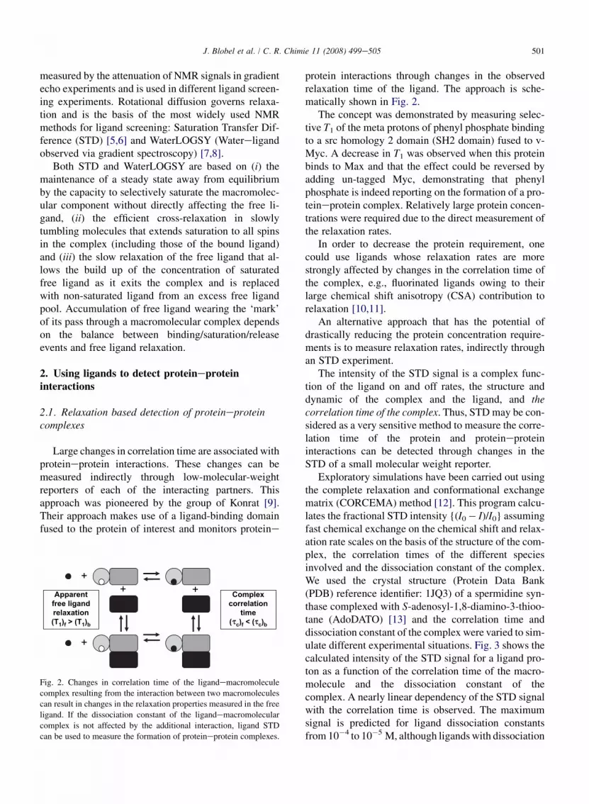
Large changes in correlation time are associated withproteineprotein interactions. These changes can bemeasured indirectly through low-molecular-weightreporters of each of the interacting partners. Thisapproach was pioneered by the group of Konrat [9].Their approach makes use of a ligand-binding domainfused to the protein of interest and monitors proteine
++ +
+
Apparent
free ligand
relaxation
(T1)f > (T
1)b
Complex
correlation
time
( c)f < (
c)b
Fig. 2. Changes in correlation time of the ligandemacromolecule
complex resulting from the interaction between two macromolecules
can result in changes in the relaxation properties measured in the free
ligand. If the dissociation constant of the ligandemacromolecular
complex is not affected by the additional interaction, ligand STD
can be used to measure the formation of proteineprotein complexes.
protein interactions through changes in the observedrelaxation time of the ligand. The approach is sche-matically shown in Fig. 2.
The concept was demonstrated by measuring selec-tive T1 of the meta protons of phenyl phosphate bindingto a src homology 2 domain (SH2 domain) fused to v-Myc. A decrease in T1 was observed when this proteinbinds to Max and that the effect could be reversed byadding un-tagged Myc, demonstrating that phenylphosphate is indeed reporting on the formation of a pro-teineprotein complex. Relatively large protein concen-trations were required due to the direct measurement ofthe relaxation rates.
In order to decrease the protein requirement, onecould use ligands whose relaxation rates are morestrongly affected by changes in the correlation time ofthe complex, e.g., fluorinated ligands owing to theirlarge chemical shift anisotropy (CSA) contribution torelaxation [10,11].
An alternative approach that has the potential ofdrastically reducing the protein concentration require-ments is to measure relaxation rates, indirectly throughan STD experiment.
The intensity of the STD signal is a complex func-tion of the ligand on and off rates, the structure anddynamic of the complex and the ligand, and thecorrelation time of the complex. Thus, STD may be con-sidered as a very sensitive method to measure the corre-lation time of the protein and proteineproteininteractions can be detected through changes in theSTD of a small molecular weight reporter.
Exploratory simulations have been carried out usingthe complete relaxation and conformational exchangematrix (CORCEMA) method [12]. This program calcu-lates the fractional STD intensity {(I0� I)/I0} assumingfast chemical exchange on the chemical shift and relax-ation rate scales on the basis of the structure of the com-plex, the correlation times of the different speciesinvolved and the dissociation constant of the complex.We used the crystal structure (Protein Data Bank(PDB) reference identifier: 1JQ3) of a spermidine syn-thase complexed with S-adenosyl-1,8-diamino-3-thioo-tane (AdoDATO) [13] and the correlation time anddissociation constant of the complex were varied to sim-ulate different experimental situations. Fig. 3 shows thecalculated intensity of the STD signal for a ligand pro-ton as a function of the correlation time of the macro-molecule and the dissociation constant of thecomplex. A nearly linear dependency of the STD signalwith the correlation time is observed. The maximumsignal is predicted for ligand dissociation constantsfrom 10�4 to 10�5 M, although ligands with dissociation
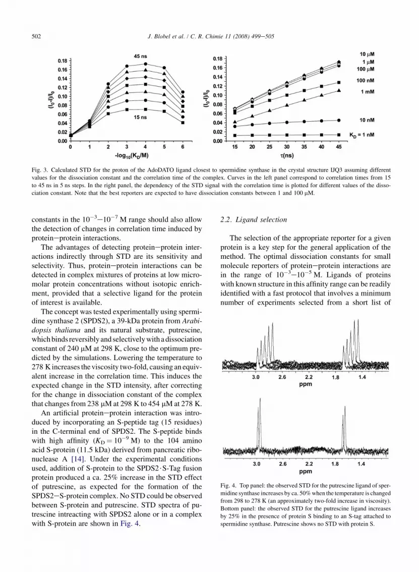
Fig. 3. Calculated STD for the proton of the AdoDATO ligand closest to spermidine synthase in the crystal structure IJQ3 assuming different
values for the dissociation constant and the correlation time of the complex. Curves in the left panel correspond to correlation times from 15
to 45 ns in 5 ns steps. In the right panel, the dependency of the STD signal with the correlation time is plotted for different values of the disso-
ciation constant. Note that the best reporters are expected to have dissociation constants between 1 and 100 mM.
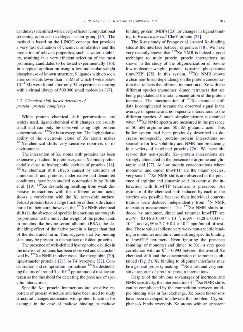
Fig. 4. Top panel: the observed STD for the putrescine ligand of sper-
midine synthase increases by ca. 50% when the temperature is changed
from 298 to 278 K (an approximately two-fold increase in viscosity).
Bottom panel: the observed STD for the putrescine ligand increases
by 25% in the presence of protein S binding to an S-tag attached to
spermidine synthase. Putrescine shows no STD with protein S.
502 J. Blobel et al. / C. R. Chimie 11 (2008) 499e505
constants in the 10�3e10�7 M range should also allowthe detection of changes in correlation time induced byproteineprotein interactions.
The advantages of detecting proteineprotein inter-actions indirectly through STD are its sensitivity andselectivity. Thus, proteineprotein interactions can bedetected in complex mixtures of proteins at low micro-molar protein concentrations without isotopic enrich-ment, provided that a selective ligand for the proteinof interest is available.
The concept was tested experimentally using spermi-dine synthase 2 (SPDS2), a 39-kDa protein from Arabi-dopsis thaliana and its natural substrate, putrescine,which binds reversibly and selectively with a dissociationconstant of 240 mM at 298 K, close to the optimum pre-dicted by the simulations. Lowering the temperature to278 K increases the viscosity two-fold, causing an equiv-alent increase in the correlation time. This induces theexpected change in the STD intensity, after correctingfor the change in dissociation constant of the complexthat changes from 238 mM at 298 K to 454 mM at 278 K.
An artificial proteineprotein interaction was intro-duced by incorporating an S-peptide tag (15 residues)in the C-terminal end of SPDS2. The S-peptide bindswith high affinity (KD¼ 10�9 M) to the 104 aminoacid S-protein (11.5 kDa) derived from pancreatic ribo-nuclease A [14]. Under the experimental conditionsused, addition of S-protein to the SPDS2$S-Tag fusionprotein produced a ca. 25% increase in the STD effectof putrescine, as expected for the formation of theSPDS2eS-protein complex. No STD could be observedbetween S-protein and putrescine. STD spectra of pu-trescine intreacting with SPDS2 alone or in a complexwith S-protein are shown in Fig. 4.
2.2. Ligand selection
The selection of the appropriate reporter for a givenprotein is a key step for the general application of themethod. The optimal dissociation constants for smallmolecule reporters of proteineprotein interactions arein the range of 10�3e10�5 M. Ligands of proteinswith known structure in this affinity range can be readilyidentified with a fast protocol that involves a minimumnumber of experiments selected from a short list of
503J. Blobel et al. / C. R. Chimie 11 (2008) 499e505
candidates identified with a very efficient computationalscreening approach developed in our group [15]. Themethod is based on the LINGO concept that providesa very fast evaluation of chemical similarities and theprediction of relevant properties, such as water solubil-ity, resulting in a very efficient selection of the mostpromising candidates to be tested experimentally [16].In a typical application using a low-molecular-weightphosphatase of known structure, 9 ligands with dissoci-ation constants lower than 1 mM (of which 4 were below10�4 M) were found after only 34 experiments startingwith a virtual library of 500 000 small molecules [17].
2.3. Chemical shift based detection ofproteineprotein complexes
While protein chemical shift perturbations arewidely used, ligand chemical shift changes are usuallysmall and can only be observed using high proteinconcentrations. 129Xe is an exception. The high polariz-ability of the electronic cloud of Xe atoms makes129Xe chemical shifts very sensitive reporters of itsenvironment.
The interaction of Xe atoms with proteins has beenextensively studied. In protein crystals, Xe binds prefer-entially close to hydrophobic cavities of proteins [18].129Xe chemical shift effects caused by solutions ofamino acids and proteins, under native and denaturedconditions, have been studied systematically by Rubinet al. [19]. 129Xe deshielding resulting from weak dis-persive interactions with the different amino acidsshows a correlation with the Xe accessible surface.Folded proteins have a large fraction of their side chainsburied in their core; however, the 129Xe NMR chemicalshifts in the absence of specific interactions are roughlyproportional to the molecular weight of the protein and,in proteins like bovine serum albumin (BSA), the de-shielding effect of the native protein is larger than thatof the denatured form. This suggests that Xe bindingsites may be present in the surface of folded proteins.
The presence of well-defined hydrophobic cavities inthe interior of proteins has been observed and character-ized by 129Xe NMR in other cases like myoglobin [20],lipid-transfer protein 1 [21], or T4 lysozyme [22]. Con-centration and composition normalised 129Xe deshield-ing factors of around 5� 10�3 ppm/mmol of residue aretaken as the threshold for detecting the presence of spe-cific interactions.
Specific Xeeprotein interactions are sensitive re-porters of protein structure and have been used to studystructural changes associated with protein function, forexample in the case of maltose binding to maltose
binding protein (MBP) [23], or changes in ligand bind-ing in Escherichia coli CheY protein [24].
The X-ray study of Prange et al. located Xe-bindingsites at the interface between oligomers [18]. We havevery recently shown that 129Xe NMR is indeed a goodtechnique to study proteineprotein interactions, asshown in the study of the oligomerization of bovinelow-molecular-weight protein tyrosine phosphatase(lmwPTP) [25]. In this system, 129Xe NMR showsa clear non-linear dependency on the protein concentra-tion that reflects the different interaction of Xe with thedifferent species (monomer, dimer, tetramer) that arebeing populated as the total concentration of the proteinincreases. The interpretation of 129Xe chemical shiftdata is complicated because the observed signal is theaverage of specific and non-specific interactions to thedifferent species. A much simpler picture is obtainedwhen 129Xe NMR spectra are measured in the presenceof 50 mM arginine and 50 mM glutamic acid. Thisbuffer system had been previously described to de-crease non-specific proteineprotein interactions re-sponsible for low solubility and NMR line broadeningin a variety of unrelated proteins [26]. We have ob-served that non-specific Xeeprotein interactions arestrongly attenuated in the presence of arginine and glu-tamic acid [27]. At low protein concentrations wheremonomer and dimer lmwPTP are the major species,very small 129Xe NMR shifts are observed in the pres-ence of arginine and glutamic acid. In contrast, the in-teraction with lmwPTP tetramers is preserved. Anestimate of the chemical shift induced by each of thespecies was possible because their individual concen-trations were deduced independently from 15N NMRrelaxation measurements. The 129Xe NMR shifts in-duced by monomer, dimer and tetramer lmwPTP areaM/N¼ 0.016� 0.007� 10�2, aD/N¼ 0.28� 0.037�10�2, and aT/N¼ 2.7� 0.4� 10�2 ppm/mmol of resi-due. These values indicate very weak non-specific bind-ing to monomer and dimer and a strong specific bindingto lmwPTP tetramers. Even ignoring the presence(binding) of monomer and dimer (to Xe), a very goodcorrelation with an R2¼ 0.993 between the overall Xechemical shift and the concentration of tetramer is ob-tained (Fig. 5). Xe binding to oligomer interfaces maybe a general property making 129Xe a fast and very sen-sitive reporter of proteineprotein interactions.
Despite of the obvious advantages of inertness andNMR sensitivity, the interpretation of 129Xe NMR shiftscan be complicated by the competition between multi-ple binding sites in fast exchange. Xe based biosensorshave been developed to alleviate this problem. Crypto-phane-A binds reversibly Xe atoms with an apparent
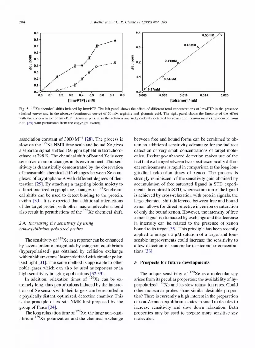
Fig. 5. 129Xe chemical shifts induced by lmwPTP. The left panel shows the effect of different total concentrations of lmwPTP in the presence
(dashed curve) and in the absence (continuous curve) of 50 mM arginine and glutamic acid. The right panel shows the linearity of the effect
with the concentration of lmwPTP tetramers present in the solution and independently detected by relaxation measurements (reproduced from
Ref. [25] with permission from the copyright owner).
504 J. Blobel et al. / C. R. Chimie 11 (2008) 499e505
association constant of 3000 M�1 [28]. The process isslow on the 129Xe NMR time scale and bound Xe givesa separate signal shifted 160 ppm upfield in tetrachoro-ethane at 298 K. The chemical shift of bound Xe is verysensitive to minor changes in its environment. This sen-sitivity is dramatically demonstrated by the observationof measurable chemical shift changes between Xe com-plexes of cryptophane-A with different degrees of deu-teration [29]. By attaching a targeting biotin moiety toa functionalized cryptophane, changes in 129Xe chemi-cal shifts can be used to detect binding to the protein,avidin [30]. It is expected that additional interactionsof the target protein with other macromolecules shouldalso result in perturbations of the 129Xe chemical shift.
2.4. Increasing the sensitivity by usingnon-equilibrium polarized probes
The sensitivity of 129Xe as a reporter can be enhancedby several orders of magnitude by using non-equilibrium(hyperpolarized) gas obtained by collision exchangewith rubidium atoms’ laser polarized with circular polar-ized light [31]. The same method is applicable to othernoble gases which can also be used as reporters or inhigh-sensitivity imaging applications [32,33].
In addition, relaxation times of 129Xe can be ex-tremely long, thus perturbations induced by the interac-tions of Xe sensors with their targets can be recorded ina physically distant, optimized, detection chamber. Thisis the principle of ex situ NMR first proposed by thegroup of Pines [34].
The long relaxation time of 129Xe, the large non-equi-librium 129Xe polarization and the chemical exchange
between free and bound forms can be combined to ob-tain an additional sensitivity advantage for the indirectdetection of very small concentrations of target mole-cules. Exchange-enhanced detection makes use of thefact that exchange between two spectroscopically differ-ent environments is rapid in comparison to the long lon-gitudinal relaxation times of xenon. The process isstrongly reminiscent of the sensitivity gain obtained byaccumulation of free saturated ligand in STD experi-ments. In contrast to STD, where saturation of the ligandis achieved by cross-relaxation with protein signals, thelarge chemical shift difference between free and boundxenon allows for direct selective inversion or saturationof only the bound xenon. However, the intensity of freexenon signal is attenuated by exchange and the decreasein intensity can be related to the presence of xenonbound to its target [35]. This principle has been recentlyapplied to image a 5 mM solution of a target and fore-seeable improvements could increase the sensitivity toallow detection of nanomolar to picomolar concentra-tions [36].
3. Prospects for future developments
The unique sensitivity of 129Xe as a molecular spyarises from its peculiar properties: the availability of hy-perpolarized 129Xe and its slow relaxation rates. Couldother molecular probes share similar desirable proper-ties? There is currently a high interest in the preparationof non-Zeeman equilibrium states in small molecules toincrease sensitivity and slow down relaxation. Bothproperties may be used to prepare more sensitive spymolecules.

505J. Blobel et al. / C. R. Chimie 11 (2008) 499e505
Solid-state dynamic nuclear polarization (DNP) atlow temperatures followed by rapid thawing and trans-fer to the NMR instrument can provide orders of mag-nitude enhancement in the polarization of smallmolecules [37]. The decreased concentration at whichthese hyperpolarized samples can be observed byNMR should allow probing their target macromoleculesat much lower concentrations.
Non-equilibrium nuclear singlet states may be cre-ated in isolated pairs of coupled spins [38] and evenin systems containing more than two coupled spins[39]. If singletetriplet exchange is prevented, the life-time of these states can be much longer than T1. Singletstates may become useful to maintain hyperpolarizedspin order generated by DNP or other methods and beused as sensitive molecular spies in a way reminiscentof the use of DQ relaxation for screening [40].
The connection between the small molecular worldand the macromolecular interactome through NMR isa growing interdisciplinary frontier where differentbranches of chemistry, molecular biology and spectros-copy can find a fruitful common ground.
Acknowledgments
We gratefully acknowledge a fruitful continuous col-laboration with Prof. Eike Brunner (University of Re-gensburg), Dr. Pau Bernado (Institute for Research inBiomedicine), Dr. Oscar Millet (CICBiogune), Dr.David Vidal and Dr. Michael Thormann (Origenis). Thiswork was supported in part by funds from the SpanishMinisterio de Educacion y Ciencia, FEDER (BIO2004-5436).
References
[1] R.S. Bohacek, C. McMartin, W.C. Guida, Med. Res. Rev. 16
(1996) 3.
[2] B.J. Stockman, C. Dalvit, Prog. NMR Spectrosc. 41 (2002) 187.
[3] J.W. Peng, J. Moore, N. Abdul-Manan, Prog. NMR Spectrosc.
44 (2004) 225.
[4] S.W. Homans, Angew. Chem. Int. Ed. 43 (2004) 290.
[5] M. Mayer, B. Meyer, Angew. Chem. Int. Ed. 38 (1999) 1784.
[6] J. Klein, R. Meinecke, M. Mayer, B. Meyer, J. Am. Chem. Soc.
121 (1999) 5336.
[7] C. Dalvit, P. Pevarello, M. Tato, M. Veronesi, A. Vulpetti,
M. Sundstrom, J. Biomol. NMR 18 (2000) 65.
[8] C. Dalvit, G.P. Fogliatto, A. Stewart, M. Veronesi, B. Stockman,
J. Biomol. NMR 21 (2001) 349.
[9] M.L. Ludwiczek, B. Baminger, R. Konrat, J. Am. Chem. Soc.
126 (2004) 1636.
[10] C. Dalvit, M. Flocco, M. Veronesi, B.J. Stockman, Comb.
Chem. High Throughput Screening 5 (2002) 605.
[11] C. Dalvit, P.E. Fagerness, D.T.A. Hadden, R.W. Sarver,
B. Stockmann, J. Am. Chem. Soc. 125 (2003) 7696.
[12] N.R. Krishna, V. Jayalakshmi, J. Magn. Reson. 155 (2002) 106.
[13] S. Korolev, Y. Ikeguchi, T. Skarina, S. Beasley, C. Arrowsmith,
A. Edwards, A. Joachimick, A.E. Pegg, A. Savchenko, Nat.
Struct. Biol. 9 (2002) 27.
[14] J.S. Kim, R.T. Raines, Protein Sci. 2 (1993) 348.
[15] D. Vidal, M. Thormann, M. Pons, J. Chem. Inf. Model. 46
(2006) 836.
[16] D. Vidal, M. Thormann, M. Pons, J. Chem. Inf. Model. 45
(2005) 386.
[17] D. Vidal, J. Blobel, Y. Perez, M. Thormann, M. Pons, Eur, J.
Med. Chem. 42 (2007) 1102.
[18] T. Prange, M. Schiltz, L. Pernot, N. Colloch, S. Longhi,
W. Bourguet, R. Fourme, Proteins 30 (1998) 61.
[19] S.M. Rubin, M.M. Spence, A. Pines, D.E. Wemmer, J. Magn.
Reson. 152 (2001) 79.
[20] S.M. Rubin, M.M. Spence, B.M. Goodson, D.E. Wemmer,
A. Pines, Proc. Natl Acad. Sci. USA 97 (2000) 9472.
[21] L. Dubois, P. Da Silva, C. Landon, J.G. Huber, M. Ponchet,
F. Vovelle, P. Berthault, H. Desvaux, J. Am. Chem. Soc. 126
(2004) 15738.
[22] H. Desvaux, L. Dubois, G. Huber, M.L. Quillin, P. Berthault,
B.W. Matthews, J. Am. Chem. Soc. 127 (2005) 11676.
[23] S.M. Rubin, M.M. Spence, I.E. Dimitrov, E.J. Ruiz, A. Pines,
D.E. Wemmer, J. Am. Chem. Soc. 123 (2001) 8616.
[24] T.J. Lowery, M. Doucleff, E.J. Ruiz, M.S. Rubin, A. Pines,
D.E. Wemmer, Protein Sci. 14 (2005) 848.
[25] P. Bernado, T. Akerud, J.G. De la Torre, M. Akke, M. Pons,
J. Am. Chem. Soc. 125 (2003) 916.
[26] A.P. Golovanov, G.M. Hautbergue, S.A. Wilson, L.-Y. Lian,
J. Am. Chem. Soc. 126 (2004) 8933.
[27] J. Blobel, S. Schmidl, D. Vidal, L. Nisius, P. Bernado, O. Millet,
E. Brunner, M. Pons, J. Am. Chem. Soc. 129 (2007) 5946.
[28] K. Bartik, M. Luhmer, J.P. Dutasta, A. Collet, J. Reisse, J. Am.
Chem. Soc. 120 (1998) 784.
[29] T. Brotin, A. Lesage, L. Emsley, A. Collet, J. Am. Chem. Soc.
122 (2000) 1171.
[30] M.M. Spence, S.M. Rubin, I.E. Dimitrov, E.J. Ruiz,
D.E. Wemmer, A. Pines, S.Q. Yao, F. Tian, P.G. Schultz,
Proc. Natl Acad. Sci. USA 98 (2001) 10654.
[31] A. Cherubini, A. Bifone, Prog. Nucl. Magn. Reson. Spectrosc.
42 (2003) 1.
[32] Z.I. Cleveland, K.F. Stupic, G.E. Pavlovskaya, J.E. Repine,
J.B. Wooten, T. Meersmann, J. Am. Chem. Soc. 129 (2007) 1784.
[33] D. Raftery, Annu. Rep. NMR Spectrosc. 57 (2006) 205.
[34] A.J. Moule, M.M. Spence, S.I. Han, J.A. Seeley, K.L. Pierce,
S. Saxena, A. Pines, Proc. Natl Acad. Sci. USA 100 (2003)
9122.
[35] M.M. Spence, E.J. Ruiz, S.M. Rubin, T.J. Lowery,
N. Winssinger, P.G. Schultz, D.E. Wemmer, A. Pines, J. Am.
Chem. Soc. 126 (2004) 15287.
[36] L. Schroder, T.J. Lowery, C. Hilty, D. Wemmer, A. Pines,
Science 314 (2006) 446.
[37] J. Wolber, F. Ellner, B. Fridlund, A. Gram, H. Johannesson,
G. Hansson, L.H. Hansson, M.H. Lerche, S. Mansson,
R. Servin, M. Thaning, K. Golman, J.H. Ardenkjaer-Larsen,
Nucl. Instrum. Methods Phys. Res. Sect. A 526 (2004) 173.
[38] M. Carravetta, M.H. Levitt, J. Am. Chem. Soc. 126 (2004)
6228.
[39] G. Pileio, M. Concistre, M. Carravetta, M.H. Levitt, J. Magn.
Reson. 182 (2006) 353.
[40] C. Dalvit, J.M. Bohlen, Annu. Rep. NMR Spectrosc. 37 (1999)
203.