lobular carcinoma in situ diagnosed by core needle biopsy: when should it be excised
TRANSCRIPT
Lobular Carcinoma In Situ Diagnosed By Core NeedleBiopsy: When Should It Be Excised?Lavinia P. Middleton, M.D., Shakeitha Grant, Tanya Stephens, M.D., Carol B. Stelling, M.D.,Nour Sneige, M.D., Aysegul A. Sahin, M.D.
Departments of Pathology (LPM, SG, NS, AAS) and Radiology (TS, CBS), The University of Texas MDAnderson Cancer Center, Houston, Texas
Core needle biopsy is the preferred technique forevaluating breast masses and abnormal mammo-graphic findings. The frequency of detection of non-invasive lobular lesions by core needle biopsy isincreasing. Historically, the diagnosis of lobular car-cinoma in situ has been considered a risk factor forthe development of invasive carcinoma, and treat-ment has consisted of careful clinical follow-up withor without chemopreventive therapeutic agentssuch as tamoxifen citrate. We retrospectively re-viewed core needle biopsy material with the pri-mary diagnoses of lobular carcinoma in situ, atyp-ical lobular hyperplasia, and lobular neoplasia inconjunction with clinical and radiographic findingsto make recommendations as to when excision maybe merited. We searched our database for core nee-dle biopsy cases with lobular carcinoma in situ,atypical lobular hyperplasia, and lobular neoplasiaas the primary diagnosis. Microcalcifications hadbeen sampled with a stereotactically guided, 11 GMammotome biopsy device, and masses had beensampled with an ultrasound guided, 18 G core nee-dle. Glass slides were reviewed and histological pa-rameters assessed. Mammographic findings werereviewed, and clinical information was obtainedfrom the medical record. When available, excisionalbiopsy material was reviewed. The 2337 breast coreneedle biopsies performed from January 1995 toDecember 2001 included 35 (1.5%) with classic lob-ular carcinoma in situ (14), lobular neoplasia (4),and atypical lobular hyperplasia (17) as the primarydiagnosis. Twelve of these 35 cases (34%) had his-tological evidence of microcalcifications directly as-
sociated with the lobular carcinoma in situ, lobularneoplasia, atypical lobular hyperplasia. Radiologicreview revealed 21 calcifications, 6 ultrasono-graphic masses, and 8 mammographic massesand/or architectural distortions. Excisional biopsyhad been performed in 17 cases (49%). In six casesdiagnosed as in situ on core needle biopsy, exci-sional biopsy revealed invasive carcinoma. All ofthese patients had radiographically detectablemasses. Eleven cases had excisional biopsies thatshowed histology similar to that of the core needlebiopsies. The most important predictor of invasivecarcinoma on excision was a synchronous mass le-sion. Lobular carcinoma in situ involving adenosisand lobular carcinoma in situ with pagetoid spreadon core needle biopsies did not show a histologicallymore aggressive lesion on excision and, therefore,may not require additional surgery. Histologicallyidentified calcifications were associated with lobu-lar lesions 34% of the time; however, their presenceinside an in situ lobular lesion did not portendworse pathology on re-excision and should not be acriterion for excision. Based on these findings, werecommend excisional biopsy of lobular carcinomain situ, atypical lobular hyperplasia or lobular neo-plasia only when it is associated with a synchronousmass lesion.
KEY WORDS: Atypical lobular hyperplasia, Coreneedle biopsy, Lobular carcinoma in situ, Pathol-ogy, Radiology.
Mod Pathol 2003;16(2):120–129
The increased use of screening mammography hasresulted in an increasing number of image-guidedbreast biopsies of mammographically suspicious le-sions. Core needle biopsy is an easy method ofsampling small nonpalpable lesions and obtainingtissue for diagnosis. The improved mammographicdetection of small and subtle (often nonpalpable)abnormalities has increased detection of in situcarcinomas and small invasive carcinomas, leadingsome to suggest that this in turn has decreased
Copyright © 2003 by The United States and Canadian Academy ofPathology, Inc.VOL. 16, NO. 2, P. 120, 2003 Printed in the U.S.A.Date of acceptance: November 8, 2002.Presented at the 91st Annual United States and Canadian Academy ofPathology Meeting, February 2002, Chicago, IL.Address reprint requests to: Lavinia P. Middleton, M.D., Department ofPathology, The University of Texas M. D. Anderson Cancer Center, 1515Holcombe Blvd., Box 85, Houston, TX, 77030; fax: 713-745-5709; e-mail:[email protected].
DOI: 10.1097/01.MP.0000051930.68104.92
120
breast cancer mortality (1). In 1941, Foote andStewart (2) described lobular carcinoma in situ as alesion that is an incidental microscopic finding thatcould not be identified clinically or by gross exam-ination. In 0.5–3% of surgical biopsy samples, lob-ular carcinoma in situ is the most significant find-ing (3, 4). Women with lobular carcinoma in situhave an elevated risk of developing invasive cancerin either breast (3, 5– 8), and their treatment hasconsisted of annual surveillance with or withoutchemopreventive agents (9). Currently, however,there are no standardized treatment guidelines forpatients with lobular carcinoma in situ diagnosedby core needle biopsy.
Herein, we describe the experience at our centerwith classic lobular carcinoma in situ, atypical lob-ular hyperplasia, and lobular neoplasia detectedmammographically, identified by core needle bi-opsy, and unassociated with a more worrisomepathologic entity. We review the findings onfollow-up excisional biopsy of these lesions to iden-tify histological and/or radiologic findings thatwhen present, should prompt the pathologist torecommend excisional biopsy. We report thefollow-up data up to 39 months on those patientsnot biopsied. Based on our data, we challenge theassertion that the diagnosis of lobular carcinoma insitu, atypical lobular hyperplasia, or lobular neopla-sia by core needle biopsy is unrelated to the mam-mographic abnormality that initiated the biopsyand thus inconsequential. We also show data thatsupports clinically following patients with lobularcarcinoma in situ and atypical lobular hyperplasiaon core needle biopsy when the lesion is not asso-ciated with a mass.
MATERIALS AND METHODS
The pathology database at the University ofTexas, M.D. Anderson Cancer Center was searched,and the results of 2337 core needle biopsies fromJanuary 1, 1995 to December 31, 2001 were retro-spectively reviewed for cases with the primary di-agnosis of classic lobular carcinoma in situ, atypicallobular hyperplasia or lobular neoplasia. Thirty-fivecases were identified. Lesions were re-reviewed andcategorized based on published histological criteria(7, 8). For the diagnosis of lobular carcinoma insitu, we required expansion of acini with atypicalmonotonous epithelial cells containing small, uni-form nuclei and lacking pleomorphism or mitoticactivity. For the diagnosis of atypical lobular hyper-plasia, we required similar histological characteris-tics, with less pronounced distension of the termi-nal duct lobular units. The diagnosis of lobularneoplasia was reserved for those rare cases that hadcytological features similar to those of lobular car-
cinoma in situ and atypical lobular hyperplasia butthat could not be definitively categorized either cat-egory either because of an underlying lesion such asadenosis or for a quantitatively borderline lesion.Cases were excluded from the study if there wasco-existing invasive carcinoma, intraductal carci-noma, atypical intraductal hyperplasia, pleomor-phic lobular carcinoma in situ, or necrosis as weroutinely recommend excisional biopsies for theseentities. Glass slides were reviewed and histologicalparameters were assessed including extent of dis-ease, number of cores involved, presence of page-toid extension and presence of microcalcifications.Attention was placed on where the calcificationswere located. Clinical information was extractedfrom the patients’ medical records.
Two types of core needle biopsies were evaluated inthis study. Microcalcifications, masses, and architec-tural distortions had been sampled with stereotacticguidance using an 11 G, vacuum-assisted biopsy(Mammotome; Ethicon EndoSurgergy, Cincinnati,OH). Solid masses had been sampled with asonographic-guided 18–20 G, cutting-needle biopsy(Biopty; Bard Urological, Covington, GA). Two to 10(average, 4), ultrasound-guided specimens had beenobtained per patient. The stereotactic biopsies hadbeen performed with the patients prone on a dedi-cated table (LoRad, Danbury, CT). From January1995, to March 1997, a 14 G cutting needle had beenused while the patient was in an upright unit. Theaverage number of vacuum-biopsy specimens per pa-tient was 11 (range, 6 to 18). A 3-mm stainless steelclip had been routinely placed in the biopsy site afterbiopsies in which most of the calcifications were re-moved by sampling. Cores containing calcificationshad been radiographed before processing, and coresdetermined to contain calcifications had beenmarked with ink.
Sonographically guided biopsies had been per-formed with the patients in the supine or supine-oblique position by using a 7.5 or VFX 13-MHzlinear array transducer and high-resolution sono-graphic equipment (Siemens Sonoline, Mountain-view, CA) in a dedicated breast-sonography unit.The number of core samples obtained was at thediscretion of the dedicated breast radiologist per-forming the procedure and was based on visualinspection of the needle placement and tissue sec-tions obtained. Stereotactic and ultrasound-guidedbiopsies were submitted in one to four blocks andlevels were cut; at least two hematoxylin and eosin–stained slides of each block (some containing mul-tiple levels) were evaluated.
Radiographic findings were reviewed to deter-mine the mammographic features of each massincluding whether architectural distortion or calci-fications were present, size, and the American Col-lege of Radiology BI-RADS classification (10). Cal-
Needle Biopsy of Lobular Carcinoma In Situ (L. Middleton et al.) 121
cified lesions were classified as calcifications, andmass lesions containing calcifications were classi-fied as masses.
When available, the excisional biopsy materialwas reviewed. The time to excisional biopsy wasrecorded. Specimen roentgenograms were ob-tained for all surgically excised material with archi-tectural distortions or calcifications (Faxitron Cab-inet X-Ray, Wheeling, IL).
RESULTS
ClinicalAll 35 patients were women. Their ages ranged
from 37 to 79 years (mean, 58 y). Fifteen womenwere premenopausal, and 20 were postmeno-pausal. Fourteen (40%) had a family history ofbreast carcinoma in a first- or second-degree fe-male relative documented in their medical records.Fifteen (43%) of the women had synchronous ormetachronous carcinoma in the contralateralbreast.
RadiographicReview of the mammograms revealed microcal-
cifications in 21 cases, masses identified by ultra-sonography in six cases, and mammographicallyidentified masses and/or architectural distortion ineight cases. The masses detected by ultrasonogra-phy revealed two invasive carcinomas on excisionalbiopsy, two cases of lobular carcinoma in situ in-volving a fibroadenoma on excisional biopsy, andlobular carcinoma in situ involving adenosis in onecase on excision. (One patient with an ultrasono-graphically detected mass identified as lobular car-cinoma in situ by core needle biopsy sought exci-sional biopsy elsewhere.) The mammographicallyidentified masses yielded invasive carcinoma onexcisional biopsy in four cases, lobular carcinomain situ involving a radial scar on excisional biopsy inone case, and lobular carcinoma in situ involvingadenosis on excisional biopsy in one case. Onepatient with a mammographically detected masssought excisional biopsy elsewhere, and one haschosen to be followed clinically with no evidence ofdisease progression after 22 months.
PathologyTwo thousand three hundred thirty-seven breast
core needle biopsies were performed at our institu-tion as a result of an indeterminate or suspiciousclinical, mammographic, or ultrasound finding be-tween January 1, 1995 and December 31, 2001. Thirty-five (1.5%) of the core biopsies had lobular carcinomain situ (14), atypical lobular hyperplasia (17), or lob-ular neoplasia (4) as their most significant histologic
diagnoses (Figs. 1–3; Table 1). This represents 29 ste-reotactic and 6 ultrasound-guided core needle biopsy.Of the lesions examined, 51% had pagetoid extensionof the atypical lobular proliferation into adjacent non-neoplastic ducts. On average, approximately 40% ofthe cores were involved by the lobular proliferation(range, 5 to 100%). Thirty-four percent of the lobularproliferations had histological evidence of microcal-cifications within the neoplastic proliferation; 29%had associated microcalcifications in adjacent colum-nar alterations with prominent apical snouts and se-cretions (11). (Table 2) Correlation of the histologicalfindings with the radiographic abnormalities con-firmed correct identification of the targeted lesions inall cases.
Excisional biopsy was performed in 17 cases (49%).The median time to excisional biopsy was 4 weeks(range, 2 to 52 weeks). Six cases diagnosed as in situlesions by core needle biopsy had invasive carcinomaon excision (Fig. 4). These cases included five withmasses detected by ultrasound and/or mammogra-phy and one with a synchronous mass lesion (Table3). Histologic review of the core needle biopsy in thesesix cases revealed that three had atypical lobular hy-perplasia and pagetoid extension, one atypical lobularhyperplasia, and two had lobular carcinoma in situwith pagetoid extension.
Pagetoid spread of atypical lobular hyperplasiaand lobular carcinoma in situ was identified in 17 of35 cases (49%). In five of the six patients who hadinvasive carcinoma on excisional biopsy, pagetoidextension was noted on core needle biopsy. Com-paratively, twelve patients who had similar or lesssignificant pathology on excisional biopsy also hadpagetoid extension of atypical lobular hyperplasiaand lobular carcinoma in situ on core needle bi-opsy. In two of six cases (33%) with invasive carci-noma on excisional biopsy, histologically con-firmed calcifications were identified within the insitu lesion on core needle biopsy.
In 11 cases, the excisional biopsies and core nee-dle biopsies showed similar histology: this subsetincludes 3 cases of lobular carcinoma in situ involv-ing adenosis (2 of which formed a mass lesion), 1case of lobular carcinoma in situ involving a radialscar; the remainder were 2 cases of lobular carci-noma in situ alone, 3 cases of atypical lobular hy-perplasia, and 2 cases of lobular neoplasia (Table4). In biopsies obtained for examination of micro-calcifications, microcalcifications were identifiedwithin the neoplastic acini in 20% of the cases.Eighteen patients who did not receive excisionalbiopsies have been followed with serial mammo-grams and biannual surveillance for a length oftime ranging from 6 to 39 months without evidenceof disease progression.
122 Modern Pathology
DISCUSSION
In 1941, Foote and Stewart (2) described the Me-morial Hospital experience with lobular carcinoma
in situ. In this sentinel paper, the authors describedtransformation of lobules filled with noncohesive,uniform cells. Foote and Stewart emphasized thatthe lesion occurred in multiple acini and was clin-
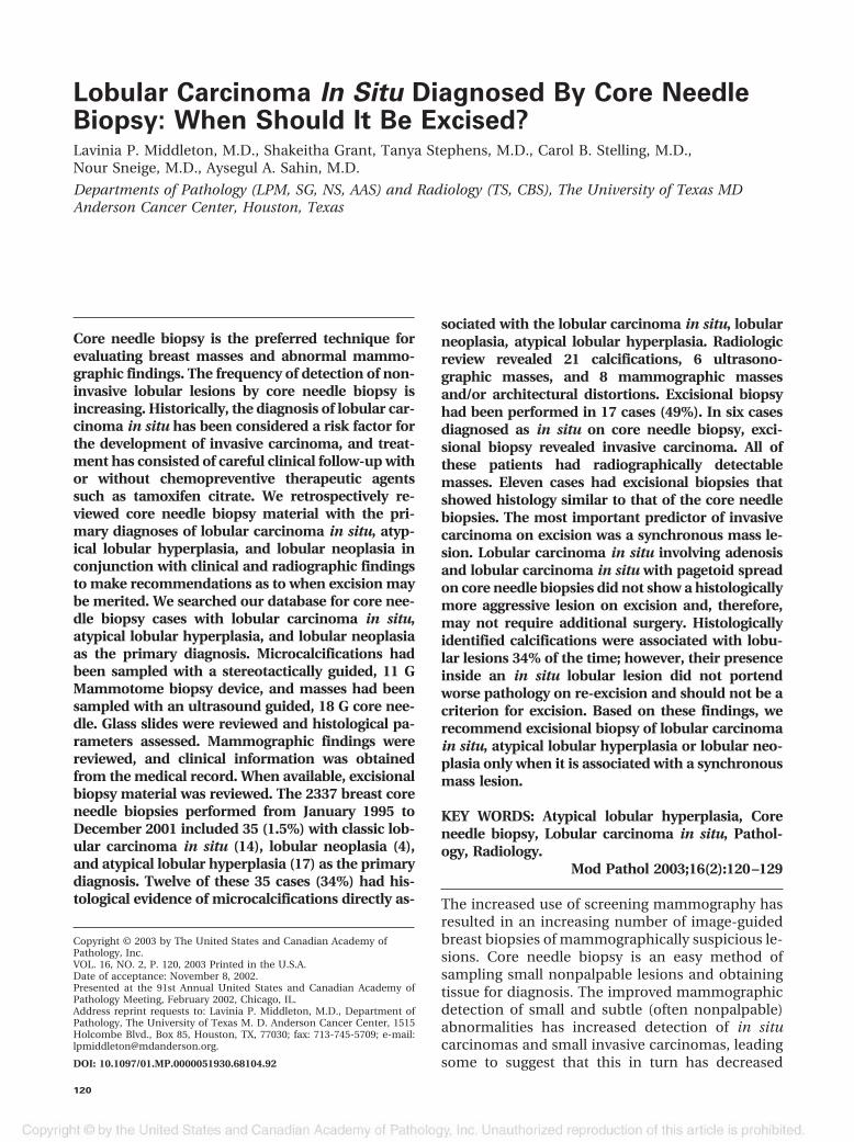
FIGURE 1. Core needle biopsy showing lobular neoplasia involving adenosis. Notice the black ink on perimeter of core indicating targetedcalcifications. Microcalcification is present in acinus.
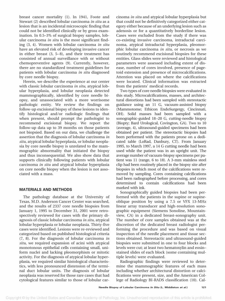
FIGURE 2. Lobular carcinoma in situ in core needle biopsy. The acini are filled with a monotonous proliferation of cells that distend and distortthe acini without secondary lumen formation.
Needle Biopsy of Lobular Carcinoma In Situ (L. Middleton et al.) 123
ically and grossly unrecognizable. Additionally, theauthors observed that the associated invasive car-cinoma could be ductal or lobular, and thus theyrecommended simple mastectomy (2).
In 1966, Snyder (12) described the mammographicfindings of 10 women who had biopsies that resultedin diagnoses of lobular carcinoma in situ. Punctatelinear calcifications were the most common mammo-graphic finding. In 1969, Hutter et al. (13) attemptedto create a clinical and pathologic correlation withmammographic findings in lobular carcinoma in situ.The patients previously reported by Synder (12) wereadded to 34 new patients with the diagnosis of lobularcarcinoma in situ (13). The authors showed thatmammographically detected finely stippled clustersof calcifications were often associated with lobularcarcinoma in situ. Similar to the results of Snyder (12),the authors conceded that these mammographicfindings were not specific for lobular carcinoma insitu and could be seen in patients with benign breastdisease. Pope et al. (14) reviewed the clinical andmammographical features of 26 patients with biopsy-proven lobular carcinoma in situ not associated withany other abnormalities. In contrast to earlier studies,those investigators concluded that not only werethere no specific radiologic findings for lobular carci-noma in situ but that the diagnosis of lobular carci-noma in situ in a biopsy obtained for calcificationswas probably an incidental finding (14).
Haagensen et al. (5) identified 211 examples of lob-ular proliferations occurring without concurrent inva-sion and introduced the term lobular neoplasia toencompass the spectrum of lobular disease rangingfrom partial lobular involvement of acini to massiveacinar distension. The authors favored the terminol-
ogy lobular neoplasia because it was thought that theappellative carcinoma should not be affixed to a be-nign disease entity. However, 17.1% of their patientsdiagnosed with lobular neoplasia and followed for amean of 14 years eventually developed carcinoma(both invasive ductal carcinoma and ductal carci-noma in situ). The authors’ recommendation fortreatment was follow-up with a clinical exam every 4months instead of mastectomy, which was the rec-ommendation at the time (5).
In our study, classic lobular carcinoma in situwas characterized by a solid proliferation of cellswith small, uniform, round-to-oval nuclei andminimal nuclear atypia. The cells were evenlyspaced, with distinct cell borders, and showedloss of cohesion. Mitoses were uncommon and,by design, necrosis was not a feature of casesincluded in this study. Cells remained within theterminal duct lobular unit or involved adjacentducts by pagetoid spread. We used the term atyp-ical lobular hyperplasia to describe lesions com-posed of an identical cell population that hadsome but not all of the features of lobular carci-noma in situ. That is, residual lumens persisted inup to 50 –75% of the lobule, and the acini wereminimally distended and distorted (7, 8). We usedthe term lobular neoplasia only for the few casesin which the lobular proliferation involved a pre-existing lesion and we could not quantify theamount of lobular involvement or where therewas partial involvement of a terminal duct lobu-lar unit that did not quantitatively fit exactly intothe category of either lobular carcinoma in situ oratypical lobular hyperplasia.
The findings of lobular carcinoma in situ on coreneedle biopsy have been studied by several groups(Tables 5 and 6), and yet there are no well-definedtreatment recommendations. Liberman et al. (15)reported 14 lesions diagnosed as lobular carcinomain situ by percutaneous biopsy in which excisionalbiopsy material was available for review (15). In 5 oftheir cases, lobular carcinoma in situ was not asso-ciated with a higher risk lesion nor had overlappingfeatures of ductal carcinoma in situ. Excisional bi-opsy of those five cases did not reveal either ductalcarcinoma in situ or invasive carcinoma histology.Additionally, Liberman et al. (15) reported the find-ings of four cases diagnosed as atypical lobularhyperplasia by core needle biopsy that were ex-cised. On excision all four lesions were shown to bebenign or lobular carcinoma in situ. Liberman andcolleagues (15) concluded, and our findings sup-port, that lobular carcinoma in situ or atypical lob-ular hyperplasia diagnosed by core needle biopsywithout a high-risk lesion or mammographicand/or histologic discordance does not necessarilyrequire surgical excision.
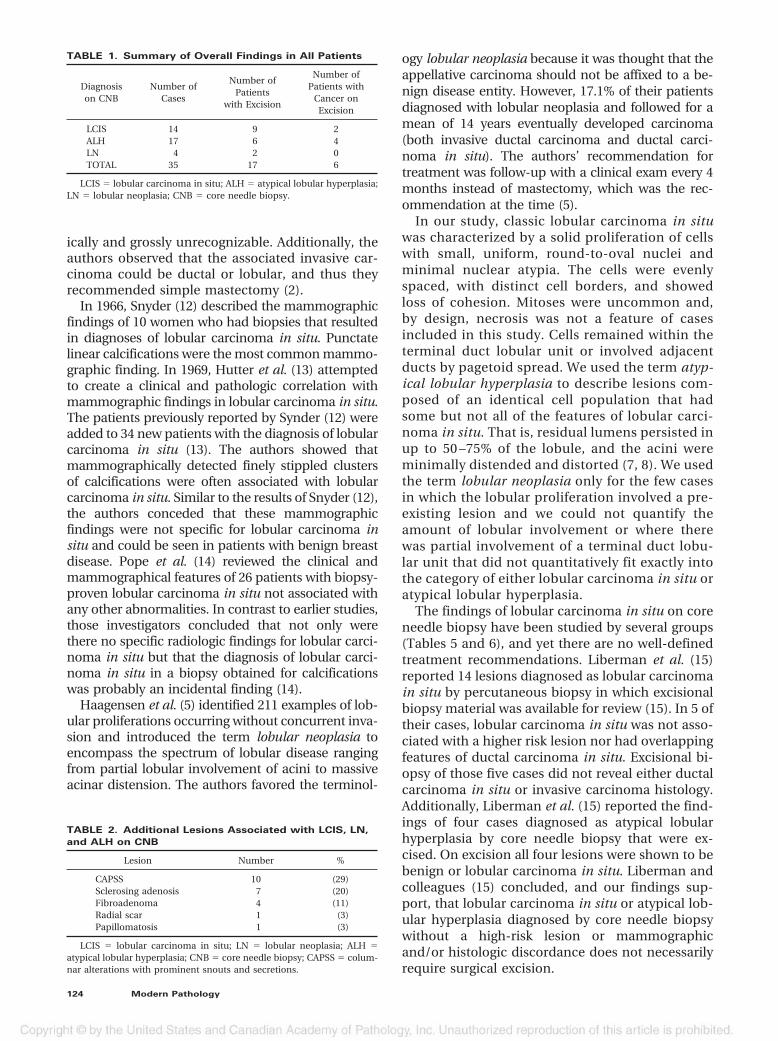
TABLE 1. Summary of Overall Findings in All Patients
Diagnosison CNB
Number ofCases
Number ofPatients
with Excision
Number ofPatients with
Cancer onExcision
LCIS 14 9 2ALH 17 6 4LN 4 2 0TOTAL 35 17 6
LCIS � lobular carcinoma in situ; ALH � atypical lobular hyperplasia;LN � lobular neoplasia; CNB � core needle biopsy.
TABLE 2. Additional Lesions Associated with LCIS, LN,
and ALH on CNB
Lesion Number %
CAPSS 10 (29)Sclerosing adenosis 7 (20)Fibroadenoma 4 (11)Radial scar 1 (3)Papillomatosis 1 (3)
LCIS � lobular carcinoma in situ; LN � lobular neoplasia; ALH �atypical lobular hyperplasia; CNB � core needle biopsy; CAPSS � colum-nar alterations with prominent snouts and secretions.
124 Modern Pathology
Lechner et al. (16) performed a multi-institutionalstudy of lobular carcinoma in situ and atypical lobularhyperplasia on core needle biopsy and found that ofthe 58 cases that were diagnosed as lobular carci-
noma in situ by core needle biopsy, 20% had inva-sive carcinoma on excisional biopsy and 14% hadductal carcinoma in situ. Similarly, of the 84 pa-tients with atypical lobular hyperplasia diagnosed
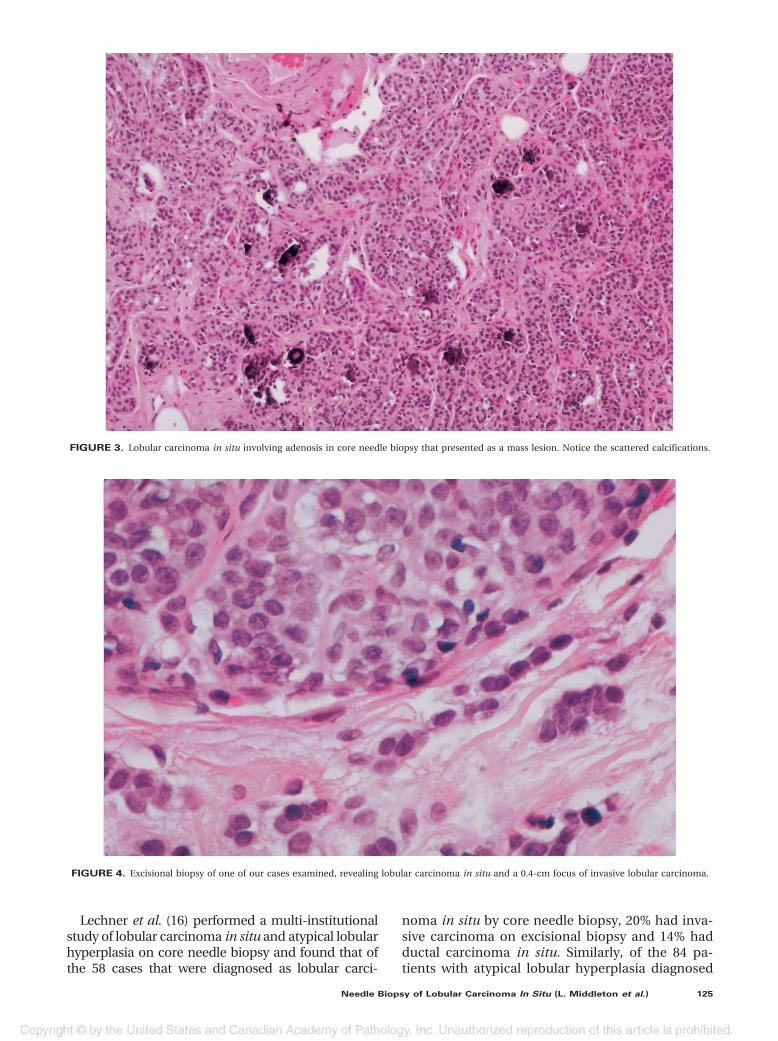
FIGURE 3. Lobular carcinoma in situ involving adenosis in core needle biopsy that presented as a mass lesion. Notice the scattered calcifications.
FIGURE 4. Excisional biopsy of one of our cases examined, revealing lobular carcinoma in situ and a 0.4-cm focus of invasive lobular carcinoma.
Needle Biopsy of Lobular Carcinoma In Situ (L. Middleton et al.) 125
by core needle biopsy, 6% had invasive carcinomaon excision and 15% had ductal carcinoma in situ.Unfortunately, the results are only presented inabstract form and there was no mention of his-topathologic slide review or radiologic review of thecases. Lechner and colleagues (16) concluded thatexcisional biopsy needs to be performed on se-lected cases of atypical lobular hyperplasia and lob-ular carcinoma in situ; but criteria for excision werenot specified.
Philpotts et al. (17) evaluated four cases of purelobular carcinoma in situ diagnosed by core needlebiopsy, which represented two mammographicallydetected masses and two patients with calcifica-tions. Excisional biopsy of these cases revealed onecase of lobular carcinoma in situ admixed withductal carcinoma in situ and associated with a masslesion, two cases of lobular carcinoma in situ, andone case of atypical lobular hyperplasia. Althoughthose investigators concluded that removal of alllobular carcinoma in situ may be prudent, if theyhad recommended excision of all mass lesions,than the one case of lobular carcinoma in situ withcoexisting ductal carcinoma in situ would havebeen excised.
O’Driscoll and colleagues (4) identified seven pa-tients with lobular carcinoma in situ as the mostsignificant pathologic diagnosis on core needle bi-opsy. On excision, one case was lobular carcinoma insitu with invasive lobular carcinoma, two cases werelobular carcinoma in situ and ductal carcinoma in
situ, one case was lobular carcinoma in situ with aradial scar, and three cases were lobular carcinoma insitu only. Again, we see that their single case of lobularcarcinoma in situ with invasive carcinoma on exci-sional biopsy had an associate spiculated opacity andthe two cases of lobular carcinoma in situ with ductalcarcinoma in situ had “indeterminate calcifications”on radiography. Notwithstanding, the authors’ finalrecommendation was that excisional biopsy be per-formed on all patients with lobular carcinoma in situon core needle biopsy.
In 2001, two studies were published describingthe findings of lobular carcinoma in situ on coreneedle biopsy and subsequent excision. Both stud-ies had different recommendations for the patient.Berg et al. (18) identified 15 examples of atypicallobular hyperplasia and 10 examples of lobular car-cinoma in situ among 1400 consecutive core needlebiopsies. Five were masses, and 20 were associatedwith mammographically detected microcalcifica-tions. Seven of the patients with atypical lobularhyperplasia on core needle biopsy went to excision.Six of the 7 had associated microcalcifications andshowed atypical lobular hyperplasia (4), fibrocysticchanges (1), and ductal carcinoma in situ (1) onexcision. The 1 patient with ductal carcinoma insitu on excisional biopsy had synchronous ductalcarcinoma in situ in the ipsilateral breast. One ofthe 7 patients with atypical lobular hyperplasia hadan associated mass lesion that on excision revealedatypical lobular hyperplasia with associated fibro-
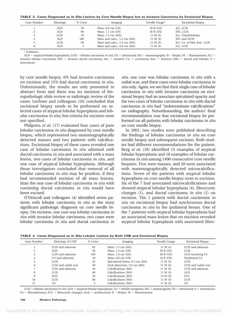
TABLE 3. Cases Diagnosed as In Situ Lesions by Core Needle Biopsy but as Invasive Carcinoma by Excisional Biopsy
Case Number Histology % Cores Imaging Needle Gauge* Excisional Biopsy
1 ALH 50 Mass, 0.9 cm (US) 18 B (US) ILC, LCIS2 ALH 66 Mass, 1.7 cm (US) 18 B (US) IDC, LCIS3 LCIS 55 Mass, 1.5 cm (MA) 11 M (S) Inv. Tubulolobular4 ALH 100 Mass and calcs., 1.5 cm (MA) 11 M (S) IDC and DCIS5 LCIS 66 Mass and calcs., 1.0 cm (MA) 11 M (S) Inv. Ca. w/D&L feat., LCIS6 ALH 5 Mass and calcs., 0.8 cm (MA) 11 M (S) ILC, LCIS
* Guidance.ALH � atypical lobular hyperplasia; LCIS � lobular carcinoma in situ; US � ultrasound; MA � mammography; B � Biopty; M � Mammotome; ILC �
invasive lobular carcinoma; IDC � invasive ductal carcinoma; Inv. � invasive; Ca. � carcinoma; feat. � features; D&L � ductal and lobular; S �stereotactic.
TABLE 4. Cases Diagnosed as In Situ Lobular Lesions by Both CNB and Excisional Biopsy
Case Number Histology of CNB % Cores Imaging Needle Gauge Excisional Biopsy
1 LCIS and adenosis 85 Mass, 1.5 cm (MA) 11 M (S) LCIS and adenosis2 ALH 30 Mass, 1.3 cm (US) 18 B (US) LCIS3 LCIS and adenosis 100 Mass, 1.6 cm (US) 20 B (US) LCIS involving FA4 LN and adenosis 10 Mass, 0.8 cm (US) 18 B (US) Hyalinized FA5 LCIS 25 Spiculated lesion, 0.5 cm (MA) 11 M (S) LCIS6 LCIS and radial scar 30 Arch distortion, 3.0 cm (MA) 11 M (S) LCIS and radial scar7 LCIS and adenosis 50 Calcifications (MA) 11 M (S) LCIS and adenosis8 LCIS 60 Calcifications (MA) 11 M (S) ALH9 ALH 30 Calcifications (MA) 14 M (S) FCC
10 LCIS 80 Calcifications (MA) 11 M (S) LCIS11 LN 5 Calcifications (MA) 11 M (S) LN
LCIS � lobular carcinoma in situ; ALH � atypical lobular hyperplasia; LN � lobular neoplasia; MA � mammogram; US � ultrasound; S � stereotactic;FA � fibroadenoma; FCC � fibrocystic changes; Arch � architectural; B � Biopty; M � Mammotome.
126 Modern Pathology
sis. Eight of the 10 lesions diagnosed as lobularcarcinoma in situ on core needle biopsy were ex-cised. Of those 8, 5 had calcifications on mammo-grams and yielded atypical ductal hyperplasia (3),lobular carcinoma in situ (1), and atypical lobularhyperplasia (1) on excision. The extent of the calci-fications identified by mammography were not de-scribed. Three patients had masses associated withlobular carcinoma in situ and were diagnosed withlobular carcinoma in situ on excisional biopsy. Bergand colleagues (18) concluded that excisional bi-opsy should be recommended when residual calci-fications remain after a diagnosis of lobular neopla-sia by core needle biopsy.
Also in 2001, Pacelli et al. (19) described the MayoClinic’s experience with lobular carcinoma in situ oncore needle biopsy: seven cases of atypical lobularhyperplasia and seven of lobular carcinoma in situthat went to excisional biopsy among 3401 consecu-
tive core needle biopsies were identified. None of the14 patients had invasive carcinoma on excisional bi-opsy. In addition, Pacelli and coworkers (19) studiedfive patients with carcinoma in situ with ductal andlobular features on core needle biopsy that went toexcisional biopsy. Three of the five (60%) had invasivecarcinoma on excision. Pacelli et al. (19) concludedthat although carcinoma in situ with ductal and lob-ular features should be excised, core needle biopsycan reliably predict the pathologic findings at surgicalexcision for atypical lobular hyperplasia and lobularcarcinoma in situ.
Shin and Rosen (20) recommended that exci-sional biopsy may be indicated when the diagnosisof lobular carcinoma in situ is made on core needlebiopsy, as 21% of patients in their series with lob-ular carcinoma in situ had a more significant lesionon excisional biopsy (20). In review of 20 cases withlobular carcinoma in situ and atypical lobular hy-perplasia on core needle biopsy and their subse-quent excisional biopsies, Shin and Rosen identi-fied a subset of eight patients with lobularcarcinoma in situ as the most significant histologicabnormality. Four of the patients had lobular car-cinoma in situ on excision, two had combined atyp-ical lobular hyperplasia and atypical ductal hyper-plasia on excision, and two of the patients hadcarcinoma on excision (one invasive ductal carci-noma and one ductal carcinoma in situ). Unfortu-nately, radiographic correlation was not included inthe analysis. Of the five cases that were reportedwith atypical lobular hyperplasia on core needlebiopsy as the most significant diagnosis, none hadinvasive carcinoma on excision. The recommenda-tion for excisional biopsy in all cases of lobularcarcinoma in situ was repeated by Hoda and Rosen(21) in a recent review outlining practice guidelinesin diagnosing breast core needle biopsies.
Herein we present 17 examples of lobular carci-noma in situ, atypical lobular hyperplasia and lobularneoplasia diagnosed by core needle biopsy with sub-sequent excisional biopsy. In six of the cases in ourcurrent series (four diagnosed as atypical lobular hy-perplasia and two as lobular carcinoma in situ) exci-sional biopsy revealed invasive carcinoma. All the pa-tients who had invasive carcinoma on excision hadmasses on mammographic review (in 1 case the masswas identified retrospectively). In 5 of the 6 cases, amass lesion was targeted and sampled. In 1 case, thepatient had a synchronous mass lesion and the tar-geted lesion and the mass lesion was excised; multi-focal invasive lobular carcinoma was identified. Wetherefore believe that the most important predictor ofinvasive carcinoma on excision of lobular carcinomain situ, atypical lobular hyperplasia, or lobular neo-plasia is a radiographically identified synchronousmass lesion. Often this lesion is best visualized byultrasound examination. Additionally, we identified
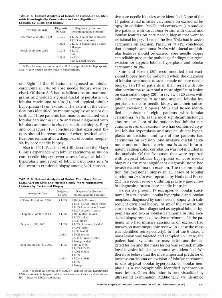
TABLE 5. Subset Analysis of Series of LCIS/ALH on CNB
with Histologically Concordant or Less Significant
Lesions by Excisional Biopsy
Investigator, YearDiagnosis
by CNBDiagnosis by Excision
(Mammographic Findings)
Liberman et al. (15), 1999 5 LCIS 4 LCIS (2 calcs and 3 masses)1 LCIS and adenosis
4 ALH 2 LCIS (3 masses and 1 calcs)2 Benign
Pacelli et al. (19), 2001 7 LCIS 5 LCIS2 ALH
7 ALH 4 ALH3 no residual disease
LCIS � lobular carcinoma in situ; ALH � atypical lobular hyperplasia;CNB � core needle biopsy; calcs � calcifications.
TABLE 6. Subset Analysis of Series That Have Shown
LCIS/ALH on CNB and Histologically More Aggressive
Lesions by Excisional Biopsy
Investigator, YearDiagnosis
by CNBDiagnosis by Excision
(Mammographic Findings)
O’Driscoll et al. (4), 2000 7 LCIS 1 ILC & LCIS (mass)2 LCIS & DCIS (indet. calcs)1 LCIS & radial scar (mass)3 LCIS (2 calcs, 1 mass)
Philpotts et al. (17), 2000 4 LCIS 1 ILC & DCIS (mass)2 LCIS (calcs)1 ALH (mass)
Berg et al. (18), 2001 8 LCIS 4 LCIS (3 masses, 1 calcs)3 ADH (calcs)1 ALH (calcs)
7 ALH 1 DCIS (calcs)5 ALH (1 mass, 4 calcs)1 Benign (calcs)
Shin and Rosen (20), 2002 8 LCIS 1 ILC & LCIS1 LCIS & DCIS2 ADH & ALH4 LCIS
5 ALH 1 LCIS & ADH1 LCIS3 Benign
LCIS � lobular carcinoma in situ; ALH � atypical lobular hyperplasia;CNB � core needle biopsy; Indet � indeterminate; Calcs � calcifications;ILC � invasive lobular carcinoma.
Needle Biopsy of Lobular Carcinoma In Situ (L. Middleton et al.) 127
microcalcifications within the lobules of lobular car-cinoma in situ, atypical lobular hyperplasia, and lob-ular neoplasia 34% of the time. Lesions targeted formicrocalcifications yielded microcalcifications associ-ated with lobular carcinoma in situ histologically in20% of the cases. On the basis of these findings, werecommend excisional biopsy lobular carcinoma insitu, atypical lobular hyperplasia, or lobular neoplasiaonly when it is associated with a synchronous masslesion or area of architectural distortion. Our recom-mendation is to excise both the area of lobular carci-noma in situ proven by core biopsy and any masslesion that may be present. Admittedly, by these cri-teria, a few cases of lobular carcinoma in situ involv-ing fibroadenomas and lobular carcinoma in situ in-volving adenosis will be unnecessarily excised, but itis best to err on the side of caution when evaluating amass lesion with an atypical lobular histology. Au-thors who have examined pure examples of lobularcarcinoma in situ and atypical lobular hyperplasiaand have correlated mammographic and clinicalfindings (4, 15, 17, 22) have had similar findings.Again, we emphasize that cases of pleomorphic lob-ular carcinoma in situ and lobular carcinoma in situwith necrosis were specifically excluded from thisstudy as we routinely recommend excision for theselobular carcinoma in situ variants.
It is important for pathologists to recognize thatlobular carcinoma in situ, and occasionally atypicallobular hyperplasia, can present as a targeted lesionon core needle biopsy and that when microcalcifica-tions are targeted on imaging, lobular carcinoma insitu or atypical lobular hyperplasia with associatedmicrocalcifications may be seen. Interestingly colum-nar alterations with prominent snouts and secretionswere identified in 29% of the cases of lobular carci-noma in situ, atypical lobular hyperplasia and lobularneoplasia. The significance of this relationship is yetto be determined. Similar to the findings of Libermanet al. (15), 40% of the patients in our series had syn-chronous or metachronous carcinoma in the con-tralateral breast. All women diagnosed with lobularcarcinoma in situ, atypical lobular hyperplasia or lob-ular neoplasia are at an increased risk of developingbreast cancer in both the ipsilateral and contralateralbreast. However, none of the women in our serieswho had invasive carcinoma on excisional biopsy hadbilateral cancer. At our institution, women with high-risk lesions are referred to the chemoprevention clinicfor counseling, surveillance, and discussion of treat-ment options.
Interestingly, our series contained slightly morepostmenopausal than premenopausal women withlobular carcinoma in situ, atypical lobular hyper-plasia, and lobular neoplasia. This finding is con-trary to published reports of lobular carcinoma insitu occurring more often in younger women (7, 8)
and may reflect our screening population at theM.D. Anderson Cancer Center.
This study reports our recent clinical experiencewith lobular carcinoma in situ and atypical ductalhyperplasia diagnosed by core needle biopsy. Ourpractice is somewhat unique in that we workclosely with dedicated breast radiologists, oncolo-gists, and surgeons. Although we only have up to 39months of follow-up for our patients treated withsurveillance, we have only noted disease progres-sion on imaging studies (both by ultrasound andmammography) in one patient after 6 months. Ourfindings on excisional biopsy material suggest thatlobular carcinoma in situ diagnosed by core needlebiopsy for calcifications and without an associatedmass lesion or area of architectural distortion maybe followed with biannual surveillance and chemo-preventive agents. Additionally, ultrasound can as-sist in the evaluation of lesions classified as lobularon core needle biopsy to see if a lesion is presentthat is not mammographically detectable.
Acknowledgments: The authors thank Leslie E. Nes-bitt, our Breast Database Coordinator, and TheresaA. Guthrie, our Breast Administrative Assistant, fortheir assistance with the collection and preparationof the materials presented here.
REFERENCES
1. Tabor L, Vitak B, Chen HH, et al. Beyond randomized con-trolled trials: organized mammographic screening substan-tially reduces breast carcinoma mortality. Cancer 2001;91:1724 –31.
2. Foote FW, Stewart FW. Lobular carcinoma in situ. A rareform of mammary cancer. Am J Pathol 1941;17:491–5.
3. Page DL, Kidd TE Jr, Dupont WD, et al. Lobular neoplasiaof the breast: higher risk for subsequent invasive cancerpredicted by more extensive disease. Hum Pathol 1991;22:1232–9.
4. O’Driscoll D, Britton P, Bobrow L, et al. Lobular carcinomain situ on core biopsy—what is the clinical significance? ClinRadiol 2001;56:216 –20.
5. Haagensen CD, Lane N, Lattes R, et al. Lobular neoplasia(so-called lobular carcinoma in situ) of the breast. Cancer1978;42:737– 69.
6. Marshall LM, Hunter DJ, Connolly JL, et al. Risk of breastcancer associated with atypical hyperplasia of lobular andductal types. Cancer Epidemiol Biomarkers Prev 1997;6:297–301.
7. Rosen PP, Kosloff C, Lieberman PH, et al. Lobular carcinomain situ of the breast: detailed analysis of 99 patients withaverage follow-up of 24 years. Am J Surg Pathol 1978;2:225–51.
8. Page DL, Anderson TJ, Rogers LN. Lobular carcinoma in situ.In: Page DL, Anderson TJ, editors. Diagnostic histopathologyof the breast. New York: Churchill Livingston, 1987:174 – 82.
9. Fisher B, Constantino JP, Wickerman DL, et al. Tamoxifen forprevention of breast cancer: report of the National SurgicalAdjuvant Breast and Bowel Project P-1 Study. J Natl CancerInst 1998;90:1371– 88.
10. Bassett L, Winchester DP, Caplan RB, et al. Stereotactic coreneedle biopsy of the breast: a report of the Joint Task Force
128 Modern Pathology
of the American College of Radiology, American College ofSurgeons, and College of American Pathologists. CA CancerJ Clin 1997;47:171–90.
11. Fraser JL, Raza S, Chorny, et al. Columnar alteration withprominent apical snouts and secretions. Am J Surg Pathol1998;22:1521–7.
12. Snyder RE. Mammography and lobular carcinoma in situ.Surg Gynecol Obstet 1966;22:225– 60.
13. Hutter RVP, Snyder RE, Lucas JC, et al. Clinical and patho-logic correlation with mammographic findings in lobularcarcinoma in situ. Cancer 1969;24:826 –39.
14. Pope TL, Fechner RE, Wilhelm MC, et al. Lobular carcinomain situ of the breast: mammographic features. Radiology1988;168:63– 6.
15. Liberman L, Sama M, Susnik B, et al. Lobular carcinoma insitu at percutaneous breast biopsy: surgical biopsy findings.AJR Am J Roentgenol 1999;173:291–9.
16. Lechner MC, Jackman RJ, Brem RF, et al. Lobular carcinomain situ and atypical lobular hyperplasia at percutaneousbiopsy with surgical correlation: a multi-institutional study.Radiology 1999;213(P):106.
17. Philpotts LE, Shaheen NA, Jain KS, et al. Uncommon high-risklesions of the breast diagnosed at stereotactic core-needle bi-opsy: clinical importance. Radiology 2000;216:831–83.
18. Berg WA, Mrose HE, Ioffe OB. Atypical lobular hyperplasia orlobular carcinoma in situ at core-needle breast biopsy. Ra-diology 2001;218:503–9.
19. Pacelli A, Rhodes DJ, Amrami KK, et al. Outcome of atypicallobular hyperplasia and lobular carcinoma in situ diagnosedby core needle biopsy: clinical and surgical follow-up of 30cases. Am J Clin Pathol 2001;116:591–2.
20. Shin SJ, Rosen PP. Excisional biopsy should be performed iflobular carcinoma in situ is seen on needle core biopsy. ArchPathol Lab Med 2002;126:697–701.
21. Hoda SA, Rosen PP. Practical considerations in the patho-logic diagnosis of needle core biopsies of breast. Am J ClinPathol 2002;118:101– 8.
22. Renshaw AE, Cartagena N, Derhagopian RP. Lobular neo-plasia in breast core needle biopsy specimens is not associ-ated with an increased risk of ductal carcinoma in situ orinvasive carcinoma. Am J Clin Pathol 2002;117:797–9.
Book Review
Travis WD, Colby TV, Koss MN, Rosado-de-Christenson ML, Müller NL, King TE, Jr.: Non-Neoplastic Disorders of the Lower Respira-tory Tract, 939 pp, Washington, DC, ArmedForces Institute of Pathology, 2002 ($195.00).
This is the second of the AFIP atlases on nontu-mor pathology. Like the other volumes of thisseries in production, it is meant to be a compan-ion to the well-established and widely used AFIPatlases of tumor pathology.
The book, printed on high-quality paper andillustrated with high-quality illustrations, is a prod-uct of a multidisciplinary approach to lung dis-eases. Thus, from one page to the next the patho-logic changes are correlated with the clinical andradiologic data. The interpretation of anatomicfindings is almost always in a clinical context andlinked to prognosis and treatment. More space isdevoted to the examination of biopsy specimensthan those obtained at autopsy. There are, how-ever, also many pictures of gross specimens ob-tained by surgery or postmortem examination todepict the main features of lung pathology.
The book is divided in 18 chapters coveringall the important pulmonary diseases from in-fancy to old age. After a short overview of em-bryology and anatomy and a chapter on han-dling and analysis of bronchioloalveolar lavageand biopsy specimens, the authors systemati-cally present the pathology of alveolar, bron-chial, bronchiolar, vascular, and pleural diseases.
Special chapters are devoted to obstructive lungdiseases, congenital disorders, infections, andoccupational lung diseases. Prominently fea-tured are diseases found in immunocompro-mised persons. Some 20 pages are devoted totransplantation pathology.
The best aspect of this book is its scope,which is trully encyclopedic. Other praiseworthyfeatures include the multidisciplinary approachand the systematic description of the pathologicmaterial. The authors have a talent for present-ing complex issues in simple terms, and onlyoccasionally do some issues remain inade-quately explained. For example, on page 849 Icould not figure out what myxotoxicosis is. Thetext is well edited, except for a few minor slipssuch as the misspelling of the Latin name ofFabry’s disease (p. 892)—I do not think that it isAngiokeratoma Corporus Diffusinum Universali.The illustrations, most of which are from theauthors’ files, complement the text. The refer-ences are well chosen and useful.
I would like to highly recommend this bookprimarily to pathologists and radiologists. It de-serves to be read also by internists and infectiousdisease specialists dealing with lung diseases andby thoracic surgeons.
Marin NolaUniversity of Zagreb School of MedicineZagreb, Croatia
Needle Biopsy of Lobular Carcinoma In Situ (L. Middleton et al.) 129