lkb1 destabilizes microtubules in myoblasts and contributes to myoblast differentiation
TRANSCRIPT
LKB1 Destabilizes Microtubules in Myoblasts andContributes to Myoblast DifferentiationIsma Mian1., Willythssa Stephie Pierre-Louis1., Neha Dole1, Renee M. Gilberti1, Kimberly Dodge-Kafka2,
Jennifer S. Tirnauer1*
1 Center for Molecular Medicine and University of Connecticut Health Center, Farmington, Connecticut, United States of America, 2 Calhoun Center for Cardiology,
University of Connecticut Health Center, Farmington, Connecticut, United States of America
Abstract
Background: Skeletal muscle myoblast differentiation and fusion into multinucleate myotubes is associated with dramaticcytoskeletal changes. We find that microtubules in differentiated myotubes are highly stabilized, but prematuremicrotubule stabilization blocks differentiation. Factors responsible for microtubule destabilization in myoblasts have notbeen identified.
Findings: We find that a transient decrease in microtubule stabilization early during myoblast differentiation precedes theultimate microtubule stabilization seen in differentiated myotubes. We report a role for the serine-threonine kinase LKB1 inboth microtubule destabilization and myoblast differentiation. LKB1 overexpression reduced microtubule elongation in aNocodazole washout assay, and LKB1 RNAi increased it, showing LKB1 destabilizes microtubule assembly in myoblasts. LKB1levels and activity increased during myoblast differentiation, along with activation of the known LKB1 substrates AMP-activated protein kinase (AMPK) and microtubule affinity regulating kinases (MARKs). LKB1 overexpression accelerateddifferentiation, whereas RNAi impaired it.
Conclusions: Reduced microtubule stability precedes myoblast differentiation and the associated ultimate microtubulestabilization seen in myotubes. LKB1 plays a positive role in microtubule destabilization in myoblasts and in myoblastdifferentiation. This work suggests a model by which LKB1-induced microtubule destabilization facilitates the cytoskeletalchanges required for differentiation. Transient destabilization of microtubules might be a useful strategy for enhancing and/or synchronizing myoblast differentiation.
Citation: Mian I, Pierre-Louis WS, Dole N, Gilberti RM, Dodge-Kafka K, et al. (2012) LKB1 Destabilizes Microtubules in Myoblasts and Contributes to MyoblastDifferentiation. PLoS ONE 7(2): e31583. doi:10.1371/journal.pone.0031583
Editor: Cara Gottardi, Northwestern University Feinberg School of Medicine, United States of America
Received August 8, 2011; Accepted January 9, 2012; Published February 14, 2012
Copyright: � 2012 Mian et al. This is an open-access article distributed under the terms of the Creative Commons Attribution License, which permitsunrestricted use, distribution, and reproduction in any medium, provided the original author and source are credited.
Funding: This work was funded from startup funds from the University of Connecticut Health Center. The funder had no role in study design, data collection andanalysis, decision to publish, or preparation of the manuscript.
Competing Interests: The authors have declared that no competing interests exist.
* E-mail: [email protected]
. These authors contributed equally to this work.
Introduction
Muscle fibers form in the developing embryo through the fusion
of myoblasts into multinucleate myotubes. In adult tissues, muscle
stem cells known as satellite cells line the surface of muscle fibers
and provide a source of myoblasts for muscle homeostasis,
hypertrophy, and repair of injury [1]. In response to differentiation
signals, myoblasts withdraw from the cell cycle, re-organize their
cytoskeleton, and ultimately fuse into multinucleate myotubes
(reviewed in [2]). Upregulation of the transcription factors MEF2
and MyoD occurs early in the process, and this is followed by
expression of myocyte specific proteins such as muscle myosin.
This differentiation process has been modeled in vitro using
myoblast cell lines, which differentiate upon switching from
standard growth media containing fetal calf serum to differenti-
ation media, which contains a lower percentage of adult horse
serum, over the course of three to four days [3].
One of the most dramatic changes observed in cultured
myoblasts during differentiation occurs in the microtubule
cytoskeleton. Microtubule organization completely changes - from
a radial array of individual microtubules that emanate from a
single central microtubule organizing center (MTOC) in myoblasts
- to a dense longitudinal linear array that originates from a diffuse,
perinuclear microtubule organizing network and/or non-centro-
somal, cytoplasmic sites in myotubes [4,5,6,7,8]. The mechanisms
of this microtubule reorganization and stabilization remain
incompletely understood, but it is clear that they play an important
role in (and are not merely a byproduct of) differentiation, because
both anti-microtubule drugs and loss of microtubule regulatory
proteins greatly impair or prevent differentiation [9,10,11,12,13,
14,15].
Myotubes contain a population of elongated, stabilized
microtubules with reduced turnover. The microtubule binding
proteins demonstrated to have positive roles in myoblast
differentiation (MAP4, EB1, EB3) all act to stabilize microtubules
and promote their elongation [10,13,14]. Thus, forced microtu-
bule stabilization might be expected to promote differentiation.
However, the converse is true: treatment of myoblasts with the
PLoS ONE | www.plosone.org 1 February 2012 | Volume 7 | Issue 2 | e31583
microtubule stabilizing drug Taxol is reported to block differen-
tiation ([16] and our data presented here). Thus, simple
microtubule stabilization is likely to be insufficient to produce this
stable, reorganized microtubule array.
Liver kinase B1 (LKB1) is a serine-threonine kinase that was
originally identified as the product of the tumor suppressor gene
mutated in the familial Peutz-Jeghers cancer syndrome (PJS) [17].
Patients who inherit a germline mutation in a single allele of the
STK11 gene that encodes LKB1 develop a syndrome of
gastrointestinal polyps; malignant tumors of the gastrointestinal
tract and other tissues; and skin pigmentation [18,19]. Somatic
mutations of LKB1 have been observed in other tumor types
(reviewed in [20,21,22]). Germline deletion of the gene encoding
LKB1 is lethal during embryogenesis, and mouse models of
heterozygous germline LKB1 mutation have been established in
which the animals develop tumors of a similar distribution to
human PJS [23].
Dramatic muscle phenotypes have not been reported in human
PJS patients or in mouse models of germline LKB1 deletion.
Together with the finding that LKB1 gene knockout in skeletal
muscle did not produce an obvious phenotype in young animals
[24], this data gave the impression that LKB1 did not play a major
role in muscle development. Subsequent genetic data, however,
has shown important roles for LKB1 in muscle. This includes the
finding that both skeletal and cardiac muscle phenotypes
developed in older LKB1 knockout mice, with decreased voluntary
running, type II muscle fiber atrophy, and loss of hind limb muscle
function [25]. LKB1 was also shown to affect the differentiation of
mouse embryonic fibroblasts (MEFs) into myofibroblasts, contrac-
tile cells that express smooth muscle actin and show acto-myosin
contractility [26,27]. Finally, activation of the LKB1 downstream
kinase AMPK was impaired in LKB1 skeletal muscle knockouts
[24], and running ability was reduced due to diminished muscle
function [28]. These data suggest that LKB1 has a role in muscle
function and might contribute to muscle development and/or
homeostasis.
Mechanisms by which LKB1 could control muscle differenti-
ation include promoting changes in cell polarity or microtubule
stability [29,30,31,32,33,34,35]. LKB1 is proposed to be at or near
the top of a network for polarity establishment in some systems
including epithelial and neuronal cells [36]. LKB1 has been
reported to reduce the stability of microtubules, because
introduction of LKB1 into LKB1-null mouse embryonic fibro-
blasts (MEFs) was able to suppress microtubule growth in an assay
that measured the elongation of microtubules following washout of
the microtubule destabilizing drug Nocodazole [37]. How or
whether these roles of LKB1 in cell polarity and microtubule
destabilization are linked is not completely clear.
In this study, we asked whether microtubule destabilization
plays a role in skeletal myoblast differentiation, and we tested the
role of LKB1 in myoblast microtubule elongation and myoblast
differentiation. We found that forced microtubule stabilization
blocks myoblast differentiation, suggesting that stabilization alone
is insufficient to drive the cytoskeletal changes associated with this
process. We found that myoblast differentiation is associated with
an initial reduction in microtubule stability that precedes the
microtubule reorganization and pronounced stabilization seen in
myotubes. We further found that LKB1 suppresses microtubule
assembly in C2C12 myoblasts, making it a potential candidate to
facilitate this reduction in microtubule stability and/or microtu-
bule reorganization. LKB1 RNAi reduced differentiation in
C2C12 cells, and LKB1 overexpression enhanced it, supporting
a positive role for LKB1 in myoblast differentiation. LKB1 is thus
a candidate to promote myoblast differentiation by reducing
microtubule stability early in the differentiation process, analogous
to factors that reduce microtubule stability early in mitosis to
facilitate formation of the mitotic spindle.
Results
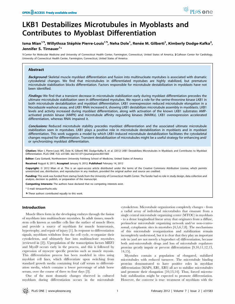
Microtubule stabilization prevents myoblastdifferentiation
Differentiated myotubes show dramatically stabilized microtu-
bules. If simple microtubule stabilization were responsible for the
cell shape changes that precede cell fusion, microtubule stabilizing
drugs might be expected to accelerate the process. We tested this
by differentiating C2C12 cells in the absence and presence of the
microtubule stabilizing drug Taxol. Cells were grown in standard
growth media until they reached near-confluence, followed by
replacement with media containing 2% horse serum (differentia-
tion media). The differentiation media was supplemented with the
dilution vehicle alone or 100 nM Taxol, a potent microtubule-
stabilizing drug. In control undifferentiated myoblasts, single
microtubules were organized in a radial array (Figure 1A). In
control differentiated myotubes, microtubules were arranged in an
array of dense, linear bundles consistent with massive stabilization,
and they expressed the differentiation marker muscle myosin
heavy chain (Figure 1B). The addition of Taxol during
differentiation also created a dense microtubule array, but it
prevented cell elongation and fusion and caused reduced myosin
expression (Figure 1C). Thus, rather than promoting myoblast
differentiation, hyperstabilization of microtubules with Taxol
completely prevented it.
We next tested whether a shorter period of exposure to
microtubule-altering drugs affected myoblast differentiation. We
cultured C2C12 cells in differentiation media containing vehicle,
200 nM Taxol, or 200 nM Nocodozole. After two days of this
incubation, culture was continued in differentiation media lacking
drugs. We found that treatment with both Nocodazole and Taxol
prevented cell elongation and fusion, consistent with an important
role for microtubules in these processes (Figure S1). Within six
hours after drug washout, cells treated with Taxol remained
rounded, but cells treated with Nocodazole showed dramatic
elongation, similar to controls. By 24 hours, cells treated with
Nocodazole differentiated as well as controls, as assessed by
morphology in phase contrast images (data not shown). This
experiment showed that exposure to Nocodazole followed by drug
washout was more conducive to differentiation than was exposure
to Taxol followed by drug washout, and could potentially be used
to synchronize cells prior to fusion. It also suggested the possibility
that microtubule destabilization could contribute to myoblast
differentiation.
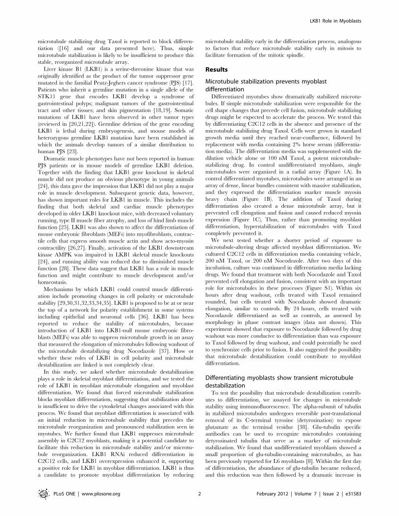
Differentiating myoblasts show transient microtubuledestabilization
To test the possibility that microtubule destabilization contrib-
utes to differentiation, we assayed for changes in microtubule
stability using immunofluorescence. The alpha-subunit of tubulin
in stabilized microtubules undergoes reversible post-translational
removal of its C-terminal tyrosine (detyrosination) to expose
glutamate as the terminal residue [38]. Glu-tubulin specific
antibodies can be used to recognize microtubules containing
detyrosinated tubulin that serve as a marker of microtubule
stabilization. We found that undifferentiated myoblasts showed a
small proportion of glu-tubulin-containing microtubules, as has
been previously reported for L6 myoblasts [8]. Within the first day
of differentiation, the abundance of glu-tubulin became reduced,
and this reduction was then followed by a dramatic increase in
LKB1 Role in Myoblasts
PLoS ONE | www.plosone.org 2 February 2012 | Volume 7 | Issue 2 | e31583
glu-tubulin as cells elongated and fused (Figure 2). This finding is
consistent with a role for a transient reduction in microtubule
stability prior to the subsequent reorganization of the microtubule
array and ultimate microtubule stabilization seen in myotubes.
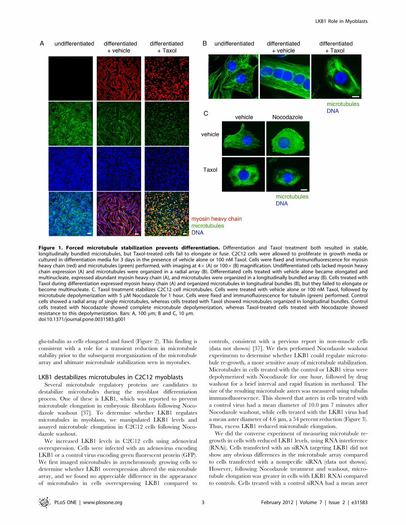
LKB1 destabilizes microtubules in C2C12 myoblastsSeveral microtubule regulatory proteins are candidates to
destabilize microtubules during the myoblast differentiation
process. One of these is LKB1, which was reported to prevent
microtubule elongation in embryonic fibroblasts following Noco-
dazole washout [37]. To determine whether LKB1 regulates
microtubules in myoblasts, we manipulated LKB1 levels and
assayed microtubule elongation in C2C12 cells following Noco-
dazole washout.
We increased LKB1 levels in C2C12 cells using adenoviral
overexpression. Cells were infected with an adenovirus encoding
LKB1 or a control virus encoding green fluorescent protein (GFP).
We first imaged microtubules in asynchronously growing cells to
determine whether LKB1 overexpression altered the microtubule
array, and we found no appreciable difference in the appearance
of microtubules in cells overexpressing LKB1 compared to
controls, consistent with a previous report in non-muscle cells
(data not shown) [37]. We then performed Nocodazole washout
experiments to determine whether LKB1 could regulate microtu-
bule re-growth, a more sensitive assay of microtubule stabilization.
Microtubules in cells treated with the control or LKB1 virus were
depolymerized with Nocodazole for one hour, followed by drug
washout for a brief interval and rapid fixation in methanol. The
size of the resulting microtubule asters was measured using tubulin
immunofluorescence. This showed that asters in cells treated with
a control virus had a mean diameter of 10.0 mm 7 minutes after
Nocodazole washout, while cells treated with the LKB1 virus had
a mean aster diameter of 4.6 mm, a 54 percent reduction (Figure 3).
Thus, excess LKB1 reduced microtubule elongation.
We did the converse experiment of measuring microtubule re-
growth in cells with reduced LKB1 levels, using RNA interference
(RNAi). Cells transfected with an siRNA targeting LKB1 did not
show any obvious differences in the microtubule array compared
to cells transfected with a nonspecific siRNA (data not shown).
However, following Nocodazole treatment and washout, micro-
tubule elongation was greater in cells with LKB1 RNAi compared
to controls. Cells treated with a control siRNA had a mean aster
Figure 1. Forced microtubule stabilization prevents differentiation. Differentiation and Taxol treatment both resulted in stable,longitudinally bundled microtubules, but Taxol-treated cells fail to elongate or fuse. C2C12 cells were allowed to proliferate in growth media orcultured in differentiation media for 3 days in the presence of vehicle alone or 100 nM Taxol. Cells were fixed and immunofluorescence for myosinheavy chain (red) and microtubules (green) performed, with imaging at 46 (A) or 1006 (B) magnification. Undifferentiated cells lacked myosin heavychain expression (A) and microtubules were organized in a radial array (B). Differentiated cells treated with vehicle alone became elongated andmultinucleate, expressed abundant myosin heavy chain (A), and microtubules were organized in a longitudinally bundled array (B). Cells treated withTaxol during differentiation expressed myosin heavy chain (A) and organized microtubules in longitudinal bundles (B), but they failed to elongate orbecome multinucleate. C. Taxol treatment stabilizes C2C12 cell microtubules. Cells were treated with vehicle alone or 100 nM Taxol, followed bymicrotubule depolymerization with 5 mM Nocodazole for 1 hour. Cells were fixed and immunofluorescence for tubulin (green) performed. Controlcells showed a radial array of single microtubules, whereas cells treated with Taxol showed microtubules organized in longitudinal bundles. Controlcells treated with Nocodazole showed complete microtubule depolymerization, whereas Taxol-treated cells treated with Nocodazole showedresistance to this depolymerization. Bars: A, 100 mm; B and C, 10 mm.doi:10.1371/journal.pone.0031583.g001
LKB1 Role in Myoblasts
PLoS ONE | www.plosone.org 3 February 2012 | Volume 7 | Issue 2 | e31583
diameter of 4.8 mm 2 minutes after Nocodazole washout, whereas
cells treated with LKB1 RNAi had a mean aster size of 8.0 mm, a
67 percent increase. This result of greater microtubule elongation
in cells with reduced LKB1 is also consistent with a role for LKB1
in suppressing microtubule elongation in myoblasts.
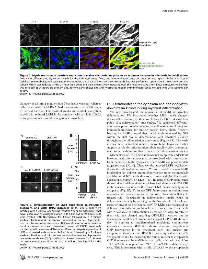
LKB1 translocates to the cytoplasm and phosphorylatesdownstream kinases during myoblast differentiation
We next investigated the regulation of LKB1 in myoblast
differentiation. We first tested whether LKB1 levels changed
during differentiation, by Western blotting for LKB1 at serial time
points of a differentiation time course. We confirmed differenti-
ation using phase contrast imaging, as well as Western blotting and
immunofluorescence for muscle myosin heavy chain. Western
blotting for LKB1 showed that LKB1 levels increased by 56%
within the first day of differentiation and remained elevated
throughout the differentiation time course (Figure 4A). This early
increase in a factor that reduces microtubule elongation further
supports a role for reduced microtubule stability prior to eventual
microtubule stabilization that occurs in the differentiation process.
Mechanisms of LKB1 activation are not completely understood;
however, activation is known to be associated with translocation
from the nucleus to the cytoplasm, where LKB1 can phosphorylate
target proteins [39,40]. Thus, we also assayed LKB1 localization
during the differentiation process. We were unable to assess LKB1
localization by indirect immunofluorescence using commercially
available anti-LKB1 antibodies, so we transfected C2C12 cells with
a plasmid encoding GFP-LKB1 [41]. Imaging of GFP fluorescence
showed that undifferentiated myoblasts had abundant GFP-LKB1
in the nucleus, consistent with reduced LKB1 kinase activity in the
cytoplasm (Fig. 4B). To image GFP fluorescence in multinucleate
myotubes, we took advantage of our own observation that cells
treated with Nocodazole in differentiation media could be
differentiated rapidly by washing out the Nocodazole. This allowed
us to circumvent the brief window of GFP-LKB1 expression and the
difficulty of transfecting multinucleate myotubes. We treated cells
with Nocodazole in differentiation media for two days, transfected
them with the plasmid encoding GFP-LKB1, washed out the
Nocodazole to allow cell fusion, and imaged GFP-LKB1 the next
day. In contrast to undifferentiated myoblasts, multinucleate
myotubes expressing GFP-LKB1 showed a greater proportion of
GFP fluorescence in the cytoplasm, such that nuclear and
cytoplasmic abundance of GFP-LKB1 were equivalent (Fig. 4C).
We quantified this by measuring the ratio of nuclear to cytoplasmic
GFP fluorescence. In undifferentiated cells, this ratio was 3.0+/
21.5 (n = 24), as opposed to 1.2+/20.3 (n = 19) in differentiated
cells. This is consistent with a shift of LKB1 to the cytoplasmic
Figure 2. Myoblasts show a transient reduction in stable microtubules prior to an ultimate increase in microtubule stabilization.Cells were differentiated by serum switch for the indicated times, fixed, and immunofluorescence for detyrosinated (glu-) tubulin, a marker ofstabilized microtubules, and tyrosinated microtubules, a marker of more dynamic microtubules, was performed. Upper panel shows detyrosinatedtubulin, which was reduced at the 24 hour time point and then progressively increased over the next two days. Short linear structures visible withthis antibody at 24 hours are primary cilia. Bottom panel shows glu- and tyrosinated tubulin immunofluorescence merged with DNA staining. Bar,10 mm.doi:10.1371/journal.pone.0031583.g002
Figure 3. Overexpression of LKB1 suppresses microtubuleassembly, and LKB1 RNAi increases it. (A) C2C12 cells wereinfected with a control adenovirus (control AV) or an adenovirus thatdrives expression of wild type human LKB1 (LKB1 AV) for 24 hours. Cellswere treated with Nocodazole for 1 hour followed by a 7 minutewashout, fixation, and microtubule immunofluorescence. Representa-tive microtubule asters are shown. (B) Quantification of aster diameterfor A, expressed as mean diameter+/2s.e.m. (C) C2C12 were cellstransfected with a control siRNA or an siRNA that targets expression ofLKB1 and treated with Nocodazole for 1 hour followed by a 2 minutewashout, fixation, and microtubule immunofluorescence. Representa-tive asters are shown. (D) Quantification of aster diameter for C. At leasttwo experiments were done for each condition. See Fig. 4 for LKB1levels.doi:10.1371/journal.pone.0031583.g003
LKB1 Role in Myoblasts
PLoS ONE | www.plosone.org 4 February 2012 | Volume 7 | Issue 2 | e31583
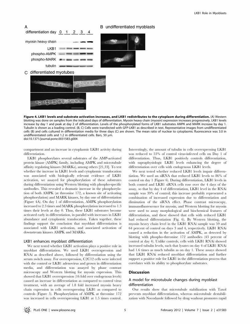
compartment and an increase in cytoplasmic LKB1 activity during
differentiation.
LKB1 phosphorylates several substrates of the AMP-activated
protein kinase (AMPK) family, including AMPK and microtubule
affinity regulating kinases (MARKs), among others [21,33]. To test
whether the increase in LKB1 levels and cytoplasmic translocation
was associated with biologically relevant evidence of LKB1
activation, we assayed for phosphorylation of these substrates
during differentiation using Western blotting with phospho-specific
antibodies. This revealed a dramatic increase in the phosphoryla-
tion of both AMPK (at threonine 172, a known site of activating
phosphorylation) and MARK kinases, by day one of differentiation
(Figure 4A). On day 1 of differentiation, AMPK phosphorylation
increased to 2.3 times and MARK phosphorylation increased to 1.3
times their levels at day 0. Thus, these LKB1 substrates become
activated early in differentiation, in parallel with increases in LKB1
abundance and cytoplasmic translocation. Taken together, these
findings support the conclusion that myoblast differentiation is
associated with LKB1 activation, and associated activation of
downstream kinases AMPK and MARKs.
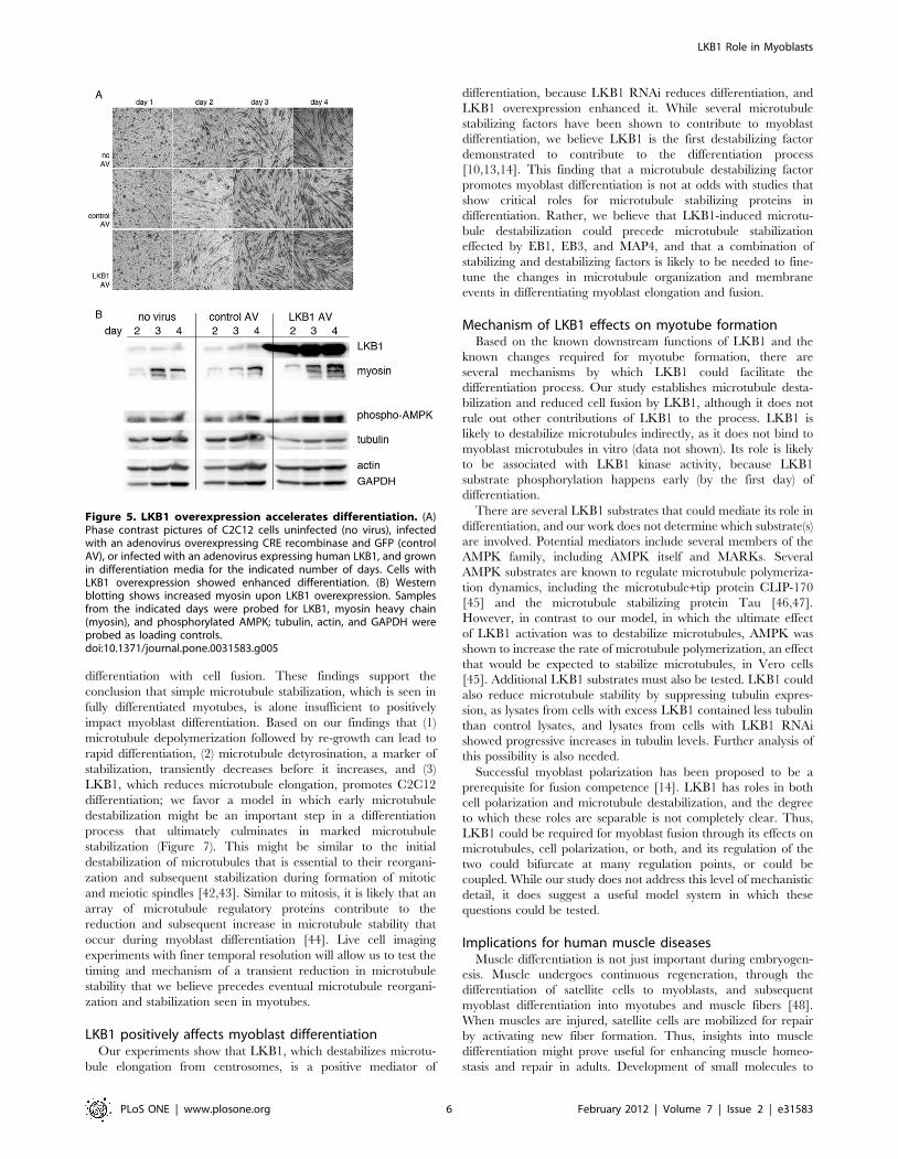
LKB1 enhances myoblast differentiationWe next tested whether LKB1 activation plays a positive role in
myoblast differentiation. We used LKB1 overexpression and
RNAi as described above, followed by differentiation using the
serum switch assay. For overexpression, C2C12 cells were infected
with the control or LKB1 adenovirus and grown in differentiation
media, and differentiation was assayed by phase contrast
microscopy and Western blotting for myosin expression. This
showed that LKB1 overexpression (16 fold over endogenous levels)
caused an increase in differentiation as compared to control virus
treatment, with an average of 1.8 fold increased myosin heavy
chain expression in cells overexpressing LKB1 as compared to
controls (Figure 5). Phosphorylation of AMPK at threonine 172
was increased in cells overexpressing LKB1 at 1.5 times control.
Interestingly, the amount of tubulin in cells overexpressing LKB1
was reduced to 53% of control virus-infected cells on Day 1 of
differentiation. Thus, LKB1 positively controls differentiation,
with supraphysiologic LKB1 levels enhancing the degree of
differentiation over cells with endogenous LKB1 levels.
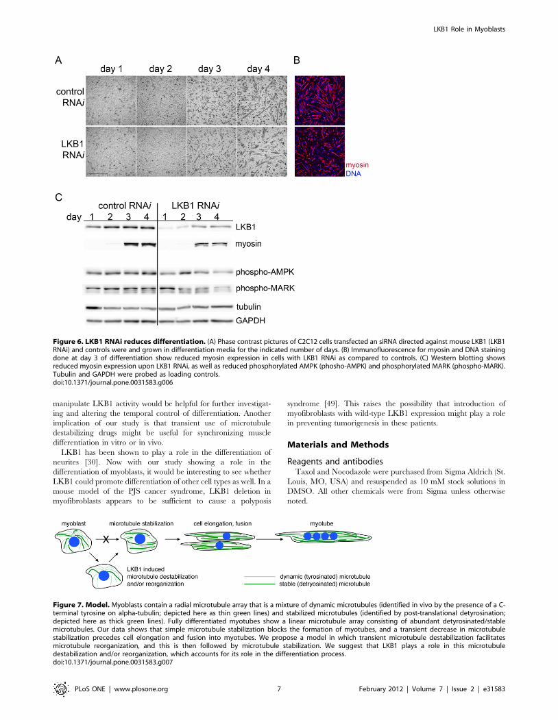
We next tested whether reduced LKB1 levels impair differen-
tiation. We used an siRNA that reduced LKB1 levels to 86% of
control on day 1 (Figure 6). During differentiation, LKB1 levels in
both control and LKB1 siRNA cells rose over the 4 days of the
assay, so that by day 4 of differentiation, LKB1 level in the RNAi
sample was 39% of control; this increase probably represented a
combination of increased expression due to differentiation and
diminution of the siRNA effect. Phase contrast microscopy,
immunofluorescence for myosin, and Western blotting for myosin
were used to assay morphological and biochemical features of
differentiation, and these showed that cells with reduced LKB1
had reduced differentiation (Fig. 6). By Western blotting, the
myosin heavy chain level in the LKB1 RNAi sample was 59 and
64 percent of control on days 3 and 4, respectively. LKB1 RNAi
caused a reduction in the activation of AMPK, as detected by
blotting with phospho-threonine 172 antibodies (43 percent of
control at day 4). Unlike controls, cells with LKB1 RNAi showed
increased tubulin levels, such that lysates on day 4 of LKB1 RNAi
had 1.6 times as much tubulin as on day 1. These assays showed
that LKB1 RNAi reduced myoblast differentiation and further
support a positive role for LKB1 in the differentiation process that
correlates with its ability to phosphorylate substrates.
Discussion
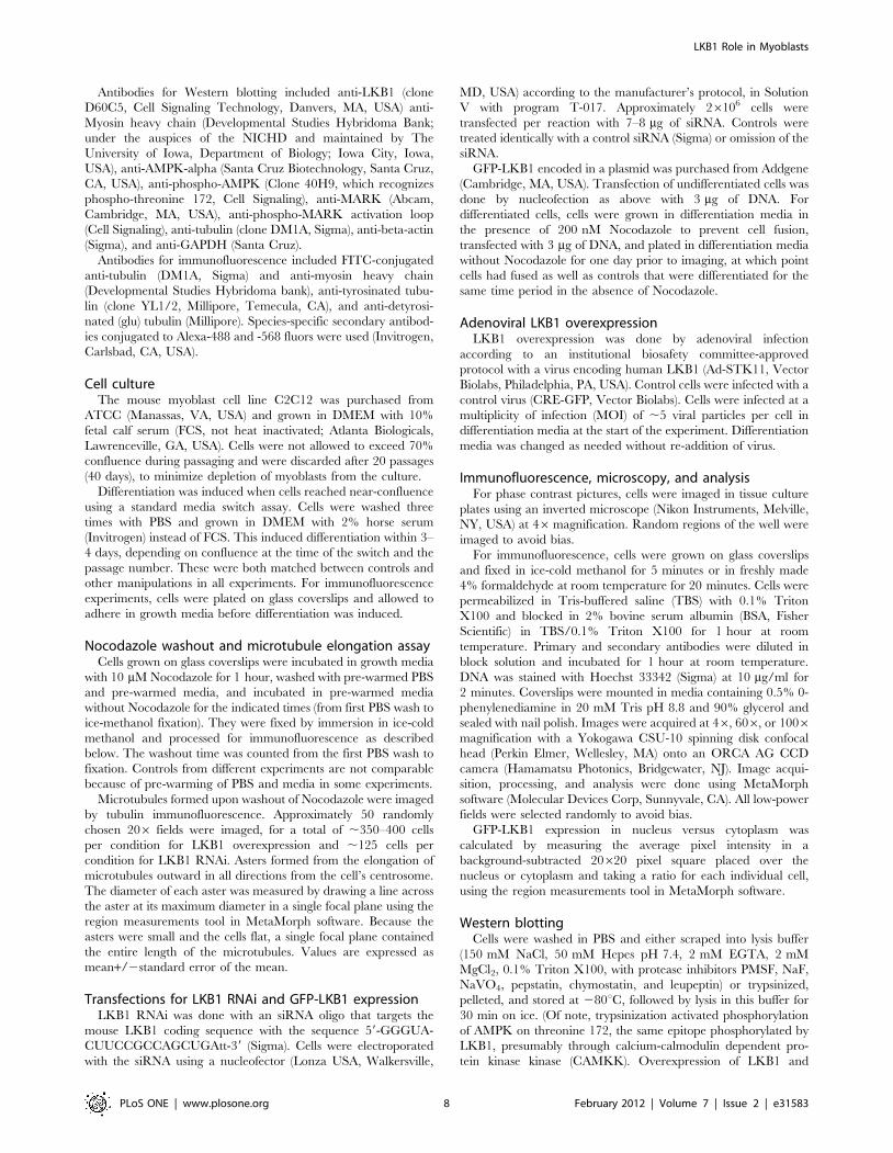
A model for microtubule changes during myoblastdifferentiation
Our results show that microtubule stabilization with Taxol
prevents myoblast differentiation, whereas microtubule destabili-
zation with Nocodazole followed by drug washout promotes rapid
Figure 4. LKB1 levels and substrate activation increases, and LKB1 redistributes to the cytoplasm during differentiation. (A) Westernblotting was done on samples from the indicated days of differentiation. Myosin heavy chain (myosin) expression increases progressively. LKB1 levelsincrease by day 1 and peak at day 2 of differentiation. Levels of the phosphorylated forms of LKB1 substrates AMPK and MARK increase by day 1.Tubulin is shown as a loading control. (B, C) Cells were transfected with GFP-LKB1 as described in text. Representative images from undifferentiatedcells (B) and cells cultured in differentiation media for three days (C) are shown. The mean ratio of nuclear to cytoplasmic fluorescence was 3.0 inundifferentiated cells and 1.2 in differentiated cells. Bars, 50 mm.doi:10.1371/journal.pone.0031583.g004
LKB1 Role in Myoblasts
PLoS ONE | www.plosone.org 5 February 2012 | Volume 7 | Issue 2 | e31583
differentiation with cell fusion. These findings support the
conclusion that simple microtubule stabilization, which is seen in
fully differentiated myotubes, is alone insufficient to positively
impact myoblast differentiation. Based on our findings that (1)
microtubule depolymerization followed by re-growth can lead to
rapid differentiation, (2) microtubule detyrosination, a marker of
stabilization, transiently decreases before it increases, and (3)
LKB1, which reduces microtubule elongation, promotes C2C12
differentiation; we favor a model in which early microtubule
destabilization might be an important step in a differentiation
process that ultimately culminates in marked microtubule
stabilization (Figure 7). This might be similar to the initial
destabilization of microtubules that is essential to their reorgani-
zation and subsequent stabilization during formation of mitotic
and meiotic spindles [42,43]. Similar to mitosis, it is likely that an
array of microtubule regulatory proteins contribute to the
reduction and subsequent increase in microtubule stability that
occur during myoblast differentiation [44]. Live cell imaging
experiments with finer temporal resolution will allow us to test the
timing and mechanism of a transient reduction in microtubule
stability that we believe precedes eventual microtubule reorgani-
zation and stabilization seen in myotubes.
LKB1 positively affects myoblast differentiationOur experiments show that LKB1, which destabilizes microtu-
bule elongation from centrosomes, is a positive mediator of
differentiation, because LKB1 RNAi reduces differentiation, and
LKB1 overexpression enhanced it. While several microtubule
stabilizing factors have been shown to contribute to myoblast
differentiation, we believe LKB1 is the first destabilizing factor
demonstrated to contribute to the differentiation process
[10,13,14]. This finding that a microtubule destabilizing factor
promotes myoblast differentiation is not at odds with studies that
show critical roles for microtubule stabilizing proteins in
differentiation. Rather, we believe that LKB1-induced microtu-
bule destabilization could precede microtubule stabilization
effected by EB1, EB3, and MAP4, and that a combination of
stabilizing and destabilizing factors is likely to be needed to fine-
tune the changes in microtubule organization and membrane
events in differentiating myoblast elongation and fusion.
Mechanism of LKB1 effects on myotube formationBased on the known downstream functions of LKB1 and the
known changes required for myotube formation, there are
several mechanisms by which LKB1 could facilitate the
differentiation process. Our study establishes microtubule desta-
bilization and reduced cell fusion by LKB1, although it does not
rule out other contributions of LKB1 to the process. LKB1 is
likely to destabilize microtubules indirectly, as it does not bind to
myoblast microtubules in vitro (data not shown). Its role is likely
to be associated with LKB1 kinase activity, because LKB1
substrate phosphorylation happens early (by the first day) of
differentiation.
There are several LKB1 substrates that could mediate its role in
differentiation, and our work does not determine which substrate(s)
are involved. Potential mediators include several members of the
AMPK family, including AMPK itself and MARKs. Several
AMPK substrates are known to regulate microtubule polymeriza-
tion dynamics, including the microtubule+tip protein CLIP-170
[45] and the microtubule stabilizing protein Tau [46,47].
However, in contrast to our model, in which the ultimate effect
of LKB1 activation was to destabilize microtubules, AMPK was
shown to increase the rate of microtubule polymerization, an effect
that would be expected to stabilize microtubules, in Vero cells
[45]. Additional LKB1 substrates must also be tested. LKB1 could
also reduce microtubule stability by suppressing tubulin expres-
sion, as lysates from cells with excess LKB1 contained less tubulin
than control lysates, and lysates from cells with LKB1 RNAi
showed progressive increases in tubulin levels. Further analysis of
this possibility is also needed.
Successful myoblast polarization has been proposed to be a
prerequisite for fusion competence [14]. LKB1 has roles in both
cell polarization and microtubule destabilization, and the degree
to which these roles are separable is not completely clear. Thus,
LKB1 could be required for myoblast fusion through its effects on
microtubules, cell polarization, or both, and its regulation of the
two could bifurcate at many regulation points, or could be
coupled. While our study does not address this level of mechanistic
detail, it does suggest a useful model system in which these
questions could be tested.
Implications for human muscle diseasesMuscle differentiation is not just important during embryogen-
esis. Muscle undergoes continuous regeneration, through the
differentiation of satellite cells to myoblasts, and subsequent
myoblast differentiation into myotubes and muscle fibers [48].
When muscles are injured, satellite cells are mobilized for repair
by activating new fiber formation. Thus, insights into muscle
differentiation might prove useful for enhancing muscle homeo-
stasis and repair in adults. Development of small molecules to
Figure 5. LKB1 overexpression accelerates differentiation. (A)Phase contrast pictures of C2C12 cells uninfected (no virus), infectedwith an adenovirus overexpressing CRE recombinase and GFP (controlAV), or infected with an adenovirus expressing human LKB1, and grownin differentiation media for the indicated number of days. Cells withLKB1 overexpression showed enhanced differentiation. (B) Westernblotting shows increased myosin upon LKB1 overexpression. Samplesfrom the indicated days were probed for LKB1, myosin heavy chain(myosin), and phosphorylated AMPK; tubulin, actin, and GAPDH wereprobed as loading controls.doi:10.1371/journal.pone.0031583.g005
LKB1 Role in Myoblasts
PLoS ONE | www.plosone.org 6 February 2012 | Volume 7 | Issue 2 | e31583
manipulate LKB1 activity would be helpful for further investigat-
ing and altering the temporal control of differentiation. Another
implication of our study is that transient use of microtubule
destabilizing drugs might be useful for synchronizing muscle
differentiation in vitro or in vivo.
LKB1 has been shown to play a role in the differentiation of
neurites [30]. Now with our study showing a role in the
differentiation of myoblasts, it would be interesting to see whether
LKB1 could promote differentiation of other cell types as well. In a
mouse model of the PJS cancer syndrome, LKB1 deletion in
myofibroblasts appears to be sufficient to cause a polyposis
syndrome [49]. This raises the possibility that introduction of
myofibroblasts with wild-type LKB1 expression might play a role
in preventing tumorigenesis in these patients.
Materials and Methods
Reagents and antibodiesTaxol and Nocodazole were purchased from Sigma Aldrich (St.
Louis, MO, USA) and resuspended as 10 mM stock solutions in
DMSO. All other chemicals were from Sigma unless otherwise
noted.
Figure 6. LKB1 RNAi reduces differentiation. (A) Phase contrast pictures of C2C12 cells transfected an siRNA directed against mouse LKB1 (LKB1RNAi) and controls were and grown in differentiation media for the indicated number of days. (B) Immunofluorescence for myosin and DNA stainingdone at day 3 of differentiation show reduced myosin expression in cells with LKB1 RNAi as compared to controls. (C) Western blotting showsreduced myosin expression upon LKB1 RNAi, as well as reduced phosphorylated AMPK (phosho-AMPK) and phosphorylated MARK (phospho-MARK).Tubulin and GAPDH were probed as loading controls.doi:10.1371/journal.pone.0031583.g006
Figure 7. Model. Myoblasts contain a radial microtubule array that is a mixture of dynamic microtubules (identified in vivo by the presence of a C-terminal tyrosine on alpha-tubulin; depicted here as thin green lines) and stabilized microtubules (identified by post-translational detyrosination;depicted here as thick green lines). Fully differentiated myotubes show a linear microtubule array consisting of abundant detyrosinated/stablemicrotubules. Our data shows that simple microtubule stabilization blocks the formation of myotubes, and a transient decrease in microtubulestabilization precedes cell elongation and fusion into myotubes. We propose a model in which transient microtubule destabilization facilitatesmicrotubule reorganization, and this is then followed by microtubule stabilization. We suggest that LKB1 plays a role in this microtubuledestabilization and/or reorganization, which accounts for its role in the differentiation process.doi:10.1371/journal.pone.0031583.g007
LKB1 Role in Myoblasts
PLoS ONE | www.plosone.org 7 February 2012 | Volume 7 | Issue 2 | e31583
Antibodies for Western blotting included anti-LKB1 (clone
D60C5, Cell Signaling Technology, Danvers, MA, USA) anti-
Myosin heavy chain (Developmental Studies Hybridoma Bank;
under the auspices of the NICHD and maintained by The
University of Iowa, Department of Biology; Iowa City, Iowa,
USA), anti-AMPK-alpha (Santa Cruz Biotechnology, Santa Cruz,
CA, USA), anti-phospho-AMPK (Clone 40H9, which recognizes
phospho-threonine 172, Cell Signaling), anti-MARK (Abcam,
Cambridge, MA, USA), anti-phospho-MARK activation loop
(Cell Signaling), anti-tubulin (clone DM1A, Sigma), anti-beta-actin
(Sigma), and anti-GAPDH (Santa Cruz).
Antibodies for immunofluorescence included FITC-conjugated
anti-tubulin (DM1A, Sigma) and anti-myosin heavy chain
(Developmental Studies Hybridoma bank), anti-tyrosinated tubu-
lin (clone YL1/2, Millipore, Temecula, CA), and anti-detyrosi-
nated (glu) tubulin (Millipore). Species-specific secondary antibod-
ies conjugated to Alexa-488 and -568 fluors were used (Invitrogen,
Carlsbad, CA, USA).
Cell cultureThe mouse myoblast cell line C2C12 was purchased from
ATCC (Manassas, VA, USA) and grown in DMEM with 10%
fetal calf serum (FCS, not heat inactivated; Atlanta Biologicals,
Lawrenceville, GA, USA). Cells were not allowed to exceed 70%
confluence during passaging and were discarded after 20 passages
(40 days), to minimize depletion of myoblasts from the culture.
Differentiation was induced when cells reached near-confluence
using a standard media switch assay. Cells were washed three
times with PBS and grown in DMEM with 2% horse serum
(Invitrogen) instead of FCS. This induced differentiation within 3–
4 days, depending on confluence at the time of the switch and the
passage number. These were both matched between controls and
other manipulations in all experiments. For immunofluorescence
experiments, cells were plated on glass coverslips and allowed to
adhere in growth media before differentiation was induced.
Nocodazole washout and microtubule elongation assayCells grown on glass coverslips were incubated in growth media
with 10 mM Nocodazole for 1 hour, washed with pre-warmed PBS
and pre-warmed media, and incubated in pre-warmed media
without Nocodazole for the indicated times (from first PBS wash to
ice-methanol fixation). They were fixed by immersion in ice-cold
methanol and processed for immunofluorescence as described
below. The washout time was counted from the first PBS wash to
fixation. Controls from different experiments are not comparable
because of pre-warming of PBS and media in some experiments.
Microtubules formed upon washout of Nocodazole were imaged
by tubulin immunofluorescence. Approximately 50 randomly
chosen 206 fields were imaged, for a total of ,350–400 cells
per condition for LKB1 overexpression and ,125 cells per
condition for LKB1 RNAi. Asters formed from the elongation of
microtubules outward in all directions from the cell’s centrosome.
The diameter of each aster was measured by drawing a line across
the aster at its maximum diameter in a single focal plane using the
region measurements tool in MetaMorph software. Because the
asters were small and the cells flat, a single focal plane contained
the entire length of the microtubules. Values are expressed as
mean+/2standard error of the mean.
Transfections for LKB1 RNAi and GFP-LKB1 expressionLKB1 RNAi was done with an siRNA oligo that targets the
mouse LKB1 coding sequence with the sequence 59-GGGUA-
CUUCCGCCAGCUGAtt-39 (Sigma). Cells were electroporated
with the siRNA using a nucleofector (Lonza USA, Walkersville,
MD, USA) according to the manufacturer’s protocol, in Solution
V with program T-017. Approximately 26106 cells were
transfected per reaction with 7–8 mg of siRNA. Controls were
treated identically with a control siRNA (Sigma) or omission of the
siRNA.
GFP-LKB1 encoded in a plasmid was purchased from Addgene
(Cambridge, MA, USA). Transfection of undifferentiated cells was
done by nucleofection as above with 3 mg of DNA. For
differentiated cells, cells were grown in differentiation media in
the presence of 200 nM Nocodazole to prevent cell fusion,
transfected with 3 mg of DNA, and plated in differentiation media
without Nocodazole for one day prior to imaging, at which point
cells had fused as well as controls that were differentiated for the
same time period in the absence of Nocodazole.
Adenoviral LKB1 overexpressionLKB1 overexpression was done by adenoviral infection
according to an institutional biosafety committee-approved
protocol with a virus encoding human LKB1 (Ad-STK11, Vector
Biolabs, Philadelphia, PA, USA). Control cells were infected with a
control virus (CRE-GFP, Vector Biolabs). Cells were infected at a
multiplicity of infection (MOI) of ,5 viral particles per cell in
differentiation media at the start of the experiment. Differentiation
media was changed as needed without re-addition of virus.
Immunofluorescence, microscopy, and analysisFor phase contrast pictures, cells were imaged in tissue culture
plates using an inverted microscope (Nikon Instruments, Melville,
NY, USA) at 46magnification. Random regions of the well were
imaged to avoid bias.
For immunofluorescence, cells were grown on glass coverslips
and fixed in ice-cold methanol for 5 minutes or in freshly made
4% formaldehyde at room temperature for 20 minutes. Cells were
permeabilized in Tris-buffered saline (TBS) with 0.1% Triton
X100 and blocked in 2% bovine serum albumin (BSA, Fisher
Scientific) in TBS/0.1% Triton X100 for 1 hour at room
temperature. Primary and secondary antibodies were diluted in
block solution and incubated for 1 hour at room temperature.
DNA was stained with Hoechst 33342 (Sigma) at 10 mg/ml for
2 minutes. Coverslips were mounted in media containing 0.5% 0-
phenylenediamine in 20 mM Tris pH 8.8 and 90% glycerol and
sealed with nail polish. Images were acquired at 46, 606, or 1006magnification with a Yokogawa CSU-10 spinning disk confocal
head (Perkin Elmer, Wellesley, MA) onto an ORCA AG CCD
camera (Hamamatsu Photonics, Bridgewater, NJ). Image acqui-
sition, processing, and analysis were done using MetaMorph
software (Molecular Devices Corp, Sunnyvale, CA). All low-power
fields were selected randomly to avoid bias.
GFP-LKB1 expression in nucleus versus cytoplasm was
calculated by measuring the average pixel intensity in a
background-subtracted 20620 pixel square placed over the
nucleus or cytoplasm and taking a ratio for each individual cell,
using the region measurements tool in MetaMorph software.
Western blottingCells were washed in PBS and either scraped into lysis buffer
(150 mM NaCl, 50 mM Hepes pH 7.4, 2 mM EGTA, 2 mM
MgCl2, 0.1% Triton X100, with protease inhibitors PMSF, NaF,
NaVO4, pepstatin, chymostatin, and leupeptin) or trypsinized,
pelleted, and stored at 280uC, followed by lysis in this buffer for
30 min on ice. (Of note, trypsinization activated phosphorylation
of AMPK on threonine 172, the same epitope phosphorylated by
LKB1, presumably through calcium-calmodulin dependent pro-
tein kinase kinase (CAMKK). Overexpression of LKB1 and
LKB1 Role in Myoblasts
PLoS ONE | www.plosone.org 8 February 2012 | Volume 7 | Issue 2 | e31583
growth in differentiation media further increased this phosphor-
ylation over and above that seen with trypsinization). Lysates were
cleared by centrifugation at 14,000 G, and protein was assayed
with Bradford reagent (Biorad, Hercules, CA, USA). Adenovirus-
infected cells were lysed by washing with PBS and scraping directly
into sample buffer, followed by bath sonication to shear DNA and
loading of equal lysate volume.
Samples were separated on 10% SDS-PAGE gels, transferred to
PVDF membranes, and blocked in 2% BSA in Tris buffered saline
(TBS) with 0.1% Tween and 0.1% Sodium Azide. Primary
antibodies were incubated for 1–2 hours at room temperature or
overnight at 4uC, and secondary antibodies were incubated for
1 hour at room temperature. The Enhanced Chemiluminescence
Fempto reagent (Thermo Scientific, Rockford, IL, USA) was used
to develop blots. Signal was imaged digitally on either a Genesnap
(Syngene USA, Frederick, MD, USA) or Biorad (Biorad) system
within the gray range of the camera. Blots were multiply probed
for proteins of different masses without stripping.
Supporting Information
Figure S1 Microtubule destabilization is more condu-cive to differentiation than is microtubule stabilization.C2C12 cells were treated with vehicle (control), 200 mm Taxol, or
200 mm Nocodozole and cultured differentiation media for two
days, followed by washout and continued differentiation. (A) Phase
contrast images at differentiation day 2 (in the presence of drugs)
and day 3 (one day after drug washout) show that both Taxol and
Nocodazole cause cell rounding and prevent myoblast fusion, but
washout of Nocodazole is associated with more substantial cell
elongation than washout of Taxol. (B) Corresponding immuno-
fluorescence images were done on cells fixed 6 hours following
drug washout. Insets show higher magnification images of
multinucleate cells in controls and Nocodazole treated cultures,
and cells with single nuclei in Taxol treated cells. This shows that
cells treated with both drugs express myosin heavy chain (MHC,
red), but only cells treated with Nocodazole show substantial cell
fusion, even at 6 hours following washout. Bar, 50 mm. (C) Fusion
index from the same time point as shown in B. Ten random 206fields were imaged for myosin heavy chain and DNA, and number
of cells with one nucleus or more than one nucleus was counted.
This showed that 60 percent of control cells expressing myosin
heavy chain had fused, while only 24 percent of Taxol treated and
40 percent of Nocodazole treated cells had fused.
(TIF)
Acknowledgments
We thank Kevin Claffey and Vladimir Rodionov for helpful comments on
the manuscript.
Author Contributions
Conceived and designed the experiments: JST KDK. Performed the
experiments: IM WPL ND RMG. Analyzed the data: JST. Contributed
reagents/materials/analysis tools: KDK. Wrote the paper: JST.
References
1. Zammit PS (2008) All muscle satellite cells are equal, but are some more equal
than others? Journal of Cell Science 121: 2975–2982.
2. Mohun T (1992) Muscle differentiation. Current Opinion in Cell Biology 4:
923–928.
3. Bains W, Ponte P, Blau H, Kedes L (1984) Cardiac actin is the major actin gene
product in skeletal muscle cell differentiation in vitro. Molecular and Cellular
Biology 4: 1449–1453.
4. Warren RH (1974) Microtubular organization in elongating myogenic cells.
Journal of Cell Science 63: 550–566.
5. Tassin AM, Maro B, Bornens M (1985) Fate of microtubule-organizing centers
during myogenesis in vitro. Journal of Cell Biology 100: 35–46.
6. Bugnard E, Zaal KJ, Ralston E (2005) Reorganization of microtubule nucleation
during muscle differentiation. Cell Motility and the Cytoskeleton 60: 1–13.
7. Musa H, Orton C, Morrison EE, Peckham M (2003) Microtubule assembly in
cultured myoblasts and myotubes following nocodazole induced microtubule
depolymerisation. Journal of Muscle Research and Cell Motility 24: 301–308.
8. Gundersen GG, Khawaja S, Bulinski JC (1989) Generation of a stable,
posttranslationally modified microtubule array is an early event in myogenic
differentiation. Journal of Cell Biology 109: 2275–2288.
9. Warren RH (1968) The effect of colchicine on myogenesis in vivo in Rana
pipiens and Rhodnius prolixus (Hemiptera). Journal of Cell Biology 39:
544–555.
10. Mangan ME, Olmsted JB (1996) A muscle-specific variant of microtubule-
associated protein 4 (MAP4) is required in myogenesis. Development 122:
771–781.
11. Spencer JA, Eliazer S, Ilaria RLJ, Richardson JA, Olson EN (2000) Regulation
of microtubule dynamics and myogenic differentiation by MURF, a striated
muscle RING-finger protein. Journal of Cell Biology 150: 771–784.
12. Chang W, Webster DR, Salam AA, Gruber D, Prasad A, et al. (2002) Alteration
of the C-terminal amino acid of tubulin specifically inhibits myogenic
differentiation. Journal of Biological Chemistry 277: 30690–30698.
13. Zhang T, Zaal KJ, Sheridan J, Mehta A, Gundersen GG, et al. (2009)
Microtubule plus-end binding protein EB1 is necessary for muscle cell
differentiation, elongation and fusion. Journal of Cell Science 122: 1401–1409.
14. Straube A, Merdes A (2007) EB3 regulates microtubule dynamics at the cell
cortex and is required for myoblast elongation and fusion. Current Biology 17:
1318–1325.
15. Conacci-Sorrell M, Ngouenet C, Eisenman RN (2010) Myc-nick: a cytoplasmic
cleavage product of Myc that promotes alpha-tubulin acetylation and cell
differentiation. Cell 142: 480–493.
16. Antin PB, Forry-Schaudies S, Friedman TM, Tapscott SJ, Holtzer H (1981)
Taxol induces postmitotic myoblasts to assemble interdigitating microtubule-
myosin arrays that exclude actin filaments. Journal of Cell Biology 90: 300–308.
17. Hemminki A, Markie D, Tomlinson I, Avizienyte E, Roth S, et al. (1998) A
serine/threonine kinase gene defective in Peutz-Jeghers syndrome. Nature 391:
184–187.
18. van Lier MG, Wagner A, Mathus-Vliegen EM, Kuipers EJ, Steyerberg EW, et
al. (2010) High cancer risk in Peutz-Jeghers syndrome: a systematic review and
surveillance recommendations. American Journal of Gastroenterology 105:
1258–1264.
19. Gammon A, Jasperson K, Kohlmann W, Burt RW (2009) Hamartomatous
polyposis syndromes. Best Practice Res Clinical Gastroenterol 23: 219–231.
20. Launonen V (2005) Mutations in the human LKB1/STK11 gene. Human
Mutation 26: 291–297.
21. Katajisto P, Vallenius T, Vaahtomeri K, Ekman N, Udd L, et al. (2007) The
LKB1 tumor suppressor kinase in human disease. Biochimica et Biophysica Acta
1775: 63–75.
22. Hezel AF, Bardeesy N (2008) LKB1; linking cell structure and tumor
suppression. Oncogene 27: 6908–6919.
23. Wei C, Amos CI, Stephens LC, Campos I, Deng JM, et al. (2005) Mutation of
Lkb1 and p53 genes exert a cooperative effect on tumorigenesis. Cancer
Research 65: 11297–11303.
24. Sakamoto K, McCarthy A, Smith D, Green KA, Hardie GD, et al. (2005)
Deficiency of LKB1 in skeletal muscle prevents AMPK activation and glucose
uptake during contraction. EMBO Journal 24: 1810–1820.
25. Thomson DM, Hancock CR, Evanson BG, Kenney SG, Malan BB, et al. (2010)
Skeletal muscle dysfunction in muscle-specific LKB1 knockout mice. J Appl
Physiol 108: 1775–1785.
26. Hinz B (2010) The myofibroblast: paradigm for a mechanically active cell.
Journal of Biomechanics 43: 146–155.
27. Vaahtomeri K, Ventela E, Laajanen K, Katajisto P, Wipff PJ, et al. (2008) Lkb1
is required for TGFbeta-mediated myofibroblast differentiation. Journal of Cell
Science 121: 3531–3540.
28. Thomson DM, Porter BB, Tall JH, Kim HJ, Barrow JR, et al. (2007) Skeletal
muscle and heart LKB1 deficiency causes decreased voluntary running and
reduced muscle mitochondrial marker enzyme expression in mice. Am J Physiol
Endocrinol Metab 292: E196–202.
29. Baas AF, Kuipers J, van der Wel NN, Batlle E, Koerten HK, et al. (2004)
Complete Polarization of Single Intestinal Epithelial Cells upon Activation of
LKB1 by STRAD. Cell 116: 457–466.
30. Asada N, Sanada K, Fukada Y (2007) LKB1 regulates neuronal migration and
neuronal differentiation in the developing neocortex through centrosomal
positioning. Journal of Neuroscience 27: 11769–11775.
31. Barnes AP, Lilley BN, Pan YA, Plummer LJ, Powell AW, et al. (2007) LKB1 and
SAD kinases define a pathway required for the polarization of cortical neurons.
Cell 129: 549–563.
LKB1 Role in Myoblasts
PLoS ONE | www.plosone.org 9 February 2012 | Volume 7 | Issue 2 | e31583
32. Shelly M, Cancedda L, Heilshorn S, Sumbre G, Poo MM (2007) LKB1/
STRAD promotes axon initiation during neuronal polarization. Cell 129:565–577.
33. Shelly M, Poo MM (2011) Role of LKB1 - SAD/MARK pathway in neuronal
polarization. Developmental Neurobiology: Epub ahead of print.34. Amin N, Khan A, St Johnston D, Tomlinson I, Martin S, et al. (2009) LKB1
regulates polarity remodeling and adherens junction formation in the Drosophilaeye. Proc Natl Acad Sci U S A 106: 8941–8946.
35. Zhang S, Schafer-Hales K, Khuri FR, Zhou W, Vertino PM, et al. (2008) The
tumor suppressor LKB1 regulates lung cancer cell polarity by mediating cdc42recruitment and activity. Cancer Research 68: 740–748.
36. Baas AF, Smit L, Clevers H (2004) LBK1 tumor suppressor protein: PARtakerin cell polarity. Trends in Cell Biology 14: 312–319.
37. Kojima Y, Miyoshi H, Clevers HC, Oshima M, Aoki M, et al. (2007)Suppression of tubulin polymerization by the LKB1-microtubule-associated
protein/microtubule affinity-regulating kinase signaling. Journal of Biological
Chemistry 282: 23532–23540.38. Verhey KJ, Gaertig J (2007) The tubulin code. Cell Cycle 6: 2152–2160.
39. Dorfman J, Macara IG (2008) STRADalpha regulates LKB1 localization byblocking access to importin-alpha, and by association with Crm1 and exportin-7.
Molecular Biology of the Cell 19: 1614–1626.
40. Baas AF, Boudeau J, Sapkota GP, Smit L, Medema R, et al. (2003) Activation ofthe tumour suppressor kinase LKB1 by the STE20-like pseudokinase STRAD.
EMBO Journal 22: 3062–3072.41. Karuman P, Gozani O, Odze RD, Zhou XC, Zhu H, et al. (2001) The Peutz-
Jegher gene product LKB1 is a mediator of p53-dependent cell death. MolecularCell 7: 1307–1319.
42. Belmont LD, Hyman AA, Sawin KE, Mitchison TJ (1990) Real-time
visualization of cell cycle-dependent changes in microtubule dynamics in
cytoplasmic extracts. Cell 62: 579–589.
43. Belmont LD, Mitchison TJ (1996) Identification of a Protein that Interacts with
Tubulin Dimers and Increases the Catastrophe Rate of Microtubules. Cell 84:
623–631.
44. Maiato H, Sampaio P, Sunkel CE (2004) Microtubule-associated proteins and
their essential roles during mitosis. International Review of Cytology 241:
53–153.
45. Nakano A, Kato H, Watanabe T, Min KD, Yamazaki S, et al. (2010) AMPK
controls the speed of microtubule polymerization and directional cell migration
through CLIP-170 phosphorylation. Nature Cell Biology 12: 583–590.
46. Vingtdeux V, Davies P, Dickson DW, Marambaud P (2011) AMPK is
abnormally activated in tangle- and pre-tangle-bearing neurons in Alzheimer’s
disease and other tauopathies. Acta Neuropathol 121: 337–349.
47. Greco SJ, Sarkar S, Johnston JM, Tezapsidis N (2009) Leptin regulates tau
phosphorylation and amyloid through AMPK in neuronal cells. Biochem
Biophys Res Commun 380: 98–104.
48. Zammit PS, Partridge TA, Yablonka-Reuveni Z (2006) The skeletal muscle
satellite cell: the stem cell that came in from the cold. Journal of Histochemistry
and Cytochemistry 54: 1177–1191.
49. Katajisto P, Vaahtomeri K, Ekman N, Ventela E, Ristimaki A, et al. (2008)
LKB1 signaling in mesenchymal cells required for suppression of gastrointestinal
polyposis. Nature Genetics 40: 455–459.
LKB1 Role in Myoblasts
PLoS ONE | www.plosone.org 10 February 2012 | Volume 7 | Issue 2 | e31583