lipid-lipid and lipid-protein interactions in chromaffin granule membranes
TRANSCRIPT

247
Biochimica et Biophysica Acta, 598 ( 1 9 8 0 ) 2 4 7 - - 2 5 9 © El sev ie r /Nor th -Hol l and Biomedica l Press
BBA 7 8 7 1 9
LIPID-LIPID AND LIPID-PROTEIN INTERACTIONS IN CHROMAFFIN GRANULE MEMBRANES
A SPIN LABEL ESR STUDY
P A M E L A F R E T T E N a, S.J. M O R R I S a, A. WATTS b and D. M A R S H b, *
a Abteilung Neurochemie und b Spektroskopie, Max-Planck-Institut fiir biophysikalische Chemie, D-3400 G6ttingen-Nikolausberg (F.R.G.)
(Received Augus t 13 th , 1979)
Key words: C hromaffin granule membrane; Lipid spin label; ESR; Phosphatidylcholine; (Bilayer)
Summary
The ESR spectra of six different positional isomers of a stearic acid and three of a phosphatidylcholine spin label have been studied as a function of temperature in chromaffin granule membranes from the bovine adrenal medulla, and in bilayers formed by aqueous dispersion of the extracted membrane lipids. Only minor differences were found between the spectra of the membranes and the extracted lipid, indicating that the major portion of the membrane lipid is organized in a bilayer arrangement which is relatively unperturbed by the presence of the membrane protein. The order parameter profile of the spin label lipid chain motion is less steep over the first half of the chain than over the section toward the terminal methyl end of the chain. This 'stiffening' effect is attributed to the high proportion of cholesterol in the membrane and becomes less marked as the temperature is raised. The isotropic hyperfine splitting factors of the various positional isomers display a profile of decreasing polarity as one penetrates further into the interior of the membrane. No marked differences are observed between the effective polarities in the intact membranes and in bilayers of the extracted membrane lipids. The previously observed temperature-induced structural change occurring in the membranes at approx. 35°C was found also in the extracted lipid bilayers, showing this to be a result of lipid-lipid interactions and not lipid-protein interactions in t he mem- brane. A steroid spin label indicated a second temperature-dependent structural change occurring in the lipid bilayers at lower temperatures. This corresponds to the onset of a more rapid rotation about the long axis of the lipid molecules
* To whom reprint requests should be addressed. Abbreviations: I(m, n), (n + 2)(4r,4t-dimethyloxazolidinc-N-oxyl)stearic acid; II(m, n), fl-(n + 2)(41,4 w- dim ethyloxaz olidine-N-oxyl)stearoyl-~-palmit oyl-~-phosphatidylchollne; In , 4 t ,4 ~-dimethylspiro( 5c¢- cholestane-3,2t-oxazolldin)-3r-yloxyl; Hepes, N-2-hydroxyethylpiperazine-Nr-2-ethanesulfonie acid.

248
at a temperature of approx. 10°C. The lipid bilayer regions probed by the spin labels used in this study may be involved in the fusion of the chromaffin granule membrane leading to hormone release by exocytosis.
Introduct ion
Chromaffin granules are the subcellular organelles responsible for the storage and release of catecholamines from the adrenal medulla. The hormone release is thought to take place by exocytosis in which part of the chromaffin granule membrane fuses with the plasma membrane of the chromaffin cell. The general features of such a mechanism probably involve both a protein-mediated associa- tion with the plasma membrane [ 1] followed by lateral displacement of the membrane proteins out of the contact region [2--4] allowing the lipid bilayers to interact [5,6]. It thus seems extremely likely that both lipid-lipid and lipid- protein interactions in the membrane may play an important part in the exo- cytotic release process. In addition these interactions may be involved in the storage process since it has previously been demonstrated that the ATPase activity in the membrane, which is coupled to catecholamine uptake [7], is sensitive to structural changes taking place in the lipid phase [8].
For these reasons we have investigated lipid-lipid and lipid-protein interac- tions in chromaffin granule membranes from bovine adrenal medulla, using a variety of different lipid spin label probes. The bilayer properties of the mem- brane have been delineated using lipid spin labels with the nitroxide reporter group situated at different positions down the hydrocarbon chain, and the motion of steroid molecules in the membrane has been studied with a spin label analogue of cholesterol. The effect of lipid-protein interactions on the properties of the bilayer have been investigated by comparing the results ob- tained with the intact membranes with those from bilayers of the extracted membrane lipids.
Materials and Methods
Chromaffin granules and lysed chromaffm granule membrane ghosts were prepared from bovine adrenal medulla as described by Cahill and Morris [9], except that dialysis overnight against 10 mM KCI, 10 mM Hepes (pH 7.0)was not performed. Material was stored at 0°C as packed, moist pellets. No prepara- tions older than 5 days post mortem were used. Ca 2÷ was removed from the membranes by shaking the membrane suspension in 30 ml buffer containing 50 mM EDTA.
Total membrane lipid extracts were prepared essentially according to Bligh and Dyer [10] with CHC13/CH3OH (2 : 1, v/v). Membrane ghosts prepared as above and stored at --20°C were used for the lipid extractions. Ca 2÷ was removed from the lipid extract by shaking the CHC13/CH3 OH solution against aqueous 500 mM EDTA (pH 8). (Ca 2÷, released from the granules, binds to acidic phospholipids.)
The stearic acid spin label isomers I(m, n) with the nitroxide group situated at a number of different positions down the hydrocarbon chain were prepared by methods similar to those of Hubbell and McConnell [11]. The corre-

249
•cO• N-0
CH~-- (CH 2 )m/" (CH2)n'-- COOH I(m,n)
f - ~ 0- I 0,,. / N - - 0 H2C-- O- - P--O --(CH2)2--1~ (CH3) 3 / C \ J U
CH3-- (CHz)m (CH2)n-- CO0 -- CH 0
I IT (m.n) CH3--(CH2) m, - - (CH2)n--COO-- CH 2
O
sponding phosphatidylcholine spin labels II(m, n) were prepared by acylation of 1-palmitoyl phosphatidylcholine (Fluka, Buchs, Switzerland) according to the general method of Boss et al. [12]. The cholestane spin label III was ob- tained from Syva, CA, U.S.A.
Membranes were spin labelled by adding a small amount of concentrated spin label solution in ethanol to membranes suspended in 5--10 ml of buffer. In all cases the ethanol concentration was less than 0.2% (v/v) and the total spin label concentration was approx. 1% with respect to membrane lipid. The mem- brane suspension was then centrifuged, the pellet resuspended in a minimal volume of buffer and transferred to a glass capillary (1 mm outer diameter, 0.1 mm wall thickness) and sealed for ESR measurements. When necessary the membrane sample was centrifuged in the capillary to improve signal-to-noise ratio. When labelling with the phosphatidycholine derivatives the membrane pellets were washed again by resuspension in 5--10 ml buffer if necessary, to remove unincorporated spin label. For labelling lipid dispersions, the spin label at 1% relative concentration was mixed with the lipid extract in CHC13/CH3OH and the organic solvent removed by evaporation in a nitrogen stream followed by vacuum desiccation. The dry lipid/spin label mixture was then suspended in buffer at a concentration of approx. 20 mg/ml by vortex mixing at room tem- perature. The buffer used throughout was 10 mM KC1, 10 mM Hepes (pH 7.0).
ESR spectra were recorded on a Varian E-12 9 GHz spectrometer with tem- perature regulation via a nitrogen gas flow system with double-wall quartz dewar. The sample capillaries were accommodated within standard 4-mm quartz ESR tubes containing silicon oil for thermal stability. Sample tempera-

250
ture was measured using a thermocouple placed in the silicon oil just above the ESR cavity.
Order parameters for the I(m, n) and II(m, n) spin labels were calculated from the following expression (see e.g. Ref. 13):
All - - A l a'o
S = A z z + A y y ) ao _ ~(Ax x . ~ (1)
where A// is the maximum hyperfine splitting, Amax, in the pseudo-powder spectrum from the unoriented membrane dispersion. Al is related to the mini- mum hyperfine splitting (Amin) in the spectrum by [14] :
A± = Amin + 1.411 - - (All - - A m i n ) / ( A z z - - ~(A,,x + Ayy))] (2)
The isotropic hyperfine splitting constant is given by:
ao = ~(A// + 2A±) (3)
and the isotropie hyperfine splitting constant corresponding to the single crys- tal environment is: a0 = 1/3 (Axx + A y y + A z z ) , where Axx, Ayy, A~z are the principal values of the nitroxide hyperfine tensor measured in a single crystal host [15].
In the limit of rapid motional narrowing, the order parameter defined in Eqn. 1 is related to the time-average angular amplitude of mot ion of the chain segment to which the spin label is at tached:
S = ~(3 <cos20> -- 1) (4)
where 0 is the angle between the normal to the plane of the membrane and the instantaneous z-axis of the labelled segment. For mot ion in the slow correla- tion time regime of spin label ESR (10 -7 s~> r > 3 . 1 0 -9 s), the effective hyperfine splittings depend not only on the amplitude of mot ion but also on the rate of motion. The following empirical relation has been obtained by spec- tral simulations, relating the correlation time for isotropic mot ion to the maxi- mum outer splitting in the ESR spectrum [16] :
r = a(1 - - A m a x / A z z ) t' (5) where a = 5.4 • 10 -l° s, b = --1.36 for Brownian motion. Thus for anisotropic motion, in the slow motional regime, the An outer splitting will depend on both the amplitude and rate of motion. For the stearic acid and phosphatidylcholine spin labels I,II(m, n), Eqn. 5 will approximate the correlation t ime for mot ion of the long molecular axis, r±, in the slow-motional regime [16]. For the cholestane label III, the outer hyperfine splitting is sensitive to rotat ion about the long axis of the molecule. Simulations, including the slow-motional regime, have been performed for the cholestane label by Polnaszek [17], and an empirical calibration established between Amax/A~z and r~, the correlation time for rotation about the long molecular axis. It is assumed that S = 1 for the long axis of the molecule, i.e. the only motion is rotation about the long axis. In the slow-motional regime the situation is similar to that given in Eqn. 5, at r~ 3 • 10 -s s extensive motional averaging of the spectrum takes place, and at ~'//~< 10 -9 s the following relation holds [13,17]:
rH(ns) = 32.3 X (Amax/Azz) --19.5 (6)

251
Results
Typical ESR spectra of the I(m, n) labels in bilayers of extracted membrane lipids are given in Fig. 1. The spectra of the same labels in chromaffin granule membranes at the same temperature are closely similar to those of Fig. 1, and this similarity holds throughout the temperature range studied. Qualitatively the decrease in extent of spectral anisotropy with increasing n represents an increase in angular amplitude of motion of the lipid chain segments on proceed- ing towards the terminal methyl end of the chain. This spin label flexibility gradient is characteristic of a lipid bilayer-type structure (see Ref. 13 for a detailed discussion) and its existence in the membrane spectra indicates that a substantial part of the lipid in the chromaffin granule membrane is present in a bilayer form. Quantitatively the lipid motions in membranes and in bilayers of the extracted lipids can be compared by means of the spin label order param- eters defined by Eqn. 1. This is done for the stearic acid spin labels I(m, n) in Fig. 2 and for the phosphatidylcholine spin labels II(m, n) in Fig. 3. In Fig. 2 it is seen that the absolute values and the temperature dependence of the stearic acid spin label order parameters are very similar for both membranes and lipid bilayers, indicating that the bilayer properties of the membrane lipid are relatively unperturbed by the presence of the membrane protein. Only in
i" 2 A - - 7 1 = ' ' i m j N
' / 13,2 ' ~ -
_ _ / ' . ' 12~ 3
9,6
5~ 10.
: i , 1
Fig. 1. ESR s p e c t r a o f t h e s t e a r l c a c i d spin labels l(m, n) in b i ] a y e r s o f t h e l i p i d s e x t r a c t e d from chromaffin granule membranes, at 24°C. Horizontal b a r r e p r e s e n t s 10 G.

252
(m,n)
(D
E
0 a..
i,_ 0
(13,2)
(12.3)
(9,6)
(7,8)
-I - ~ (5.101 0.1 (1,14)
o° 1'o° 2bo 3'0" , 'oo s'o° doo T (°C)
Fig. 2. Temperature dependence of the effect ive order pa ramete r s (S) of the stearlc acid spin labels I(m, n) in ch romaf f in granule membranes (~ . . . . . . ~) and in bi layers of the extracted m e m b r a n e lipids (o o).
the case of the I(12, 3) isomer at low temperatures and the I(5, 10) isomer at high temperatures are appreciable differences found between membrane and lipid. In contrast, with the phosphatidylcholine spin labels, II(m, n) it is found that the temperature dependence of the order parameters is very similar in both membrane and lipid, but the order parameters in the lipid bilayers are con- sistently larger than those in the membranes. This points to a small, but definite, specificity in the interaction of the protein with the phospholipid molecules of the membrane which is not detected by the stearic acid probes.
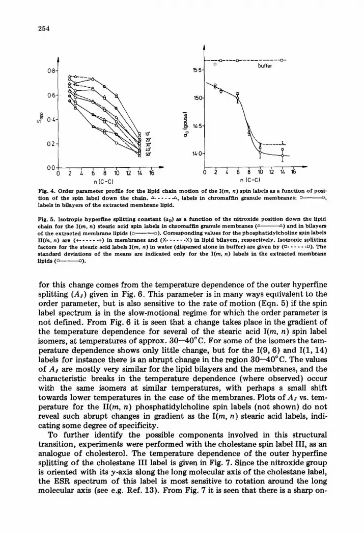
Closer detail of the lipid properties of the membrane is given in the order parameter profiles down the lipid chains in Fig. 4. Again it is seen that differ- ences between membrane and lipid only occur for the I(12, 3) and I(5, 10) isomers at low and high temperatures, respectively. The more important feature of Fig. 4 is the way in which the order parameter profiles change with increasing temperature. At lower temperatures the order parameters are approximately constant or decrease only slowly in the first part of the chain up until C-8--10 (n = 6--8) and then decrease much more rapidly towards the terminal methyl end. A change takes place in this profile in the temperature range 30--40°C; above this temperature the effective order parameters decrease approximately linearly along the entire length of the chain. The characteristic temperature for this change is similar to that found previously for a structural

253
U3
• 0.5-
E P 0~-
03- 0
0.8. \ ~ , ~
0.7-
0-6-
0.2-
0.1-
o" 2'0" 3'0" 5b" T('C)
(nxn)
(12.31
(5.10)
{1.1l.)
6'0"" Fig. 3. T e m p e r a t u r e d e p e n d e n c e o f t h e e f fec t ive o r d e r p a r a m e t e r s (S) o f t h e p h o s p h a t i d y l c h o l i n e sp in l abe l s I I (m, n) in c h r o m a f f i n g r anu l e m e m b r a n e s (~ . . . . . . A) a n d in b i l a y e r s o f the e x t r a c t e d m e m b r a n e l ip ids (o 0).
and functional transition taking place in the chromaffin granule membrane [8,18].
In parallel to the order parameter gradient is the positional dependence of the nitroxide isotropic hyperfine splitting factor, which gives the polarity profile through the membrane. The isotropic hyperfine constants, a0, for the various labels are given in Fig. 5. These values are the means of all measure- ments in the temperature range for which a0 is approximately independent of temperature (above approx. 20 ° C). The a0 dependence on n in Fig. 5 indicates a relatively high polarity in the hydrocarbon region close to the polar/apolar interface, followed by a sharp decrease in polarity leading to a region of con- stant, low polarity in the centre of the membrane. Again there is relatively little difference between membrane and extracted lipid bflayers. It appears that the polarity profile sensed by the spin labels is essentially that of bilayer regions of the membranes, undisturbed by the presence of the membrane pro- tein.
The data in Fig. 4 suggested some change taking place in the dynamic struc- ture of the membrane at approx. 30--40 ° C. This structural transition is clearly complex, since it involves a change in the effective order parameter profile, and may only be indirectly sensed by the stearic acid spin labels. Further evidence
254
0.8-
0"6"
0.4-
0.2- 10"
#
. . . . o . . . . .
150-
1/.,.5. k
14-0"
O0 2 ~ Co 8 10 1'2 14 16 = 2 l, ~ 8 10 1':) 1~, 16 ~ n [C-C) n {C-C)
Fig. 4 . O r d e r p a r a m e t e r p ro f i l e f o r t h e Hpid c h a i n m o t i o n o f t he I(m, n ) sp in l abe l s as a f u n c t i o n o f pos i - t i o n o f t h e sp in l abe l d o w n t h e c h a i n . ~ . . . . . -~, l abe l s in c h r o m a f f i n g r a n u l e m e m b r a n e s ; o o, l abe l s in b f l a y e r s o f t h e e x t r a c t e d m e m b r a n e Hpid .
Fig. 5. I s o t r o p i c h y p e r f i n e s p l i t t i n g c o n s t a n t (ao) as a f u n c t i o n o f t h e n i t r o x i d e p o s i t i o n d o w n t h e l ip id c h a i n f o r t h e I (m, n) s t ea r i c ac id sp in l abe l s in c h r o m a f f i n g r a n u l e m e m b r a n e s (~ ~) a n d in b i l a y e r s o f t h e e x t r a c t e d m e m b r a n e l ip ids (c o). C o r r e s p o n d i n g va lues f o r t he p h o s p h a t i d y l c h o l i n e sp in l abe l s II(fn, n) are (+ . . . . . -4-) in m e m b r a n e s a n d (X . . . . . . × ) in l ip id b f layers , r e spec t i ve ly . I s o t r o p i c sp l i t t i ng f a c t o r s fo r t h e s t ea r i c ac id l abe l s I (m, n ) in w a t e r (d i spe r sed a l o n e in b u f f e r ) a re given b y (o . . . . . . ~). The s t a n d a r d d e v i a t i o n s o f t h e m e a n s axe i n d i c a t e d o n l y fo r t he I (m, n ) l abe l s in the e x t r a c t e d m e m b r a n e l ip ids (o o).
for this change comes from the temperature dependence of the outer hyperfine splitting (A~) given in Fig. 6. This parameter is in many ways equivalent to the order parameter, but is also sensitive to the rate of motion (Eqn. 5) if the spin label spectrum is in the slow-motional regime for which the order parameter is not defined. From Fig. 6 it is seen that a change takes place in the gradient of the temperature dependence for several of the stearic acid I(m, n) spin label isomers, at temperatures of approx. 30--40°C. For some of the isomers the tem- perature dependence shows only little change, but for the I(9, 6) and I(1, 14) labels for instance there is an abrupt change in the region 30--40°C. The values of A~ are mostly very similar for the lipid bilayers and the membranes, and the characteristic breaks in the temperature dependence (where observed) occur with the same isomers at similar temperatures, with perhaps a small shift towards lower temperatures in the case of the membranes. Plots of A~ vs. tem- perature for the II(m, n) phosphatidylcholine spin labels (not shown) do not reveal such abrupt changes in gradient as the I(m, n) stearic acid labels, indi- cating some degree of specificity.
To further identify the possible components involved in this structural transition, experiments were performed with the cholestane spin label III, as an analogue of cholesterol. The temperature dependence of the outer hyperfine splitting of the cholestane III label is given in Fig. 7. Since the nitroxide group is oriented with its y-axis along the long molecular axis of the cholestane label, the ESR spectrum of this label is most sensitive to rotation around the long molecular axis (see e.g. Ref. 13). From Fig. 7 it is seen that there is a sharp on-

2 5 5
l
(
30.0 ~
250
200
15.0 O"
T ('C)
b: Membrane
Ira,n)
113.2]
112.31
19.61
(7. I I I
1,5,10)
(1.1LI
Trc)
Fig. 6. Temperatuze dependence of the outer hyperf ine spli t t ing (A~) of the stearic acid spin labels I(m, n): (a) in bilayers of the extracted membrane lipids (o o) and (b) in chromaffin granule mem- branes (~ ~).
set of motional averaging of the outer hyperfine splitting of the cholestane label at 10°C. The correlation times (r) calculated from the outer hyperfine splitting are also given in Fig. 7. These clearly indicate that the discontinuity in the temperature dependence of Amax corresponds to a cooperative onset of
u) D 0
30-
28-
26-
2~
22 ¸
2 0 -
18 0"
I
[] I
- 5 0
~ 0
- 2 0
Fig. 7. Temperature dependence of the outer hyperf lne spli t t ing (Amax) of the cholestane spin label III in bflayers of ehromaffin granule membrane lipids (o o) and of the correlat ion t ime (1"1) for ro ta t ion about the long molecular axis of the spin label (n . . . . . . n) deduced from the Araax hyperf lne splittings.
-30 "~
E
1°
, , ~ - F , , Ioo 20" 30" ~ so"
I" r c l

256
rapid rotation about the long axis of the cholestane molecule at 10 ° C. A discon- tinuity is not indicated in the region of 30--40 ° C, but at these temperatures the residual anisotropy in the spectra is rather small and thus the sensitivity to any further dynamic structural changes is rather low.
Discussion
The results with I(m, n) stearic acid spin labels indicate that a substantial proportion of the lipid in the chromaffin granule membrane is present as a bilayer whose properties are very little perturbed by the presence of the mem- brane protein. This can be attributed to the relatively high lipid/protein ratio of chromaffin granule membranes (approx. 1.9 pmol lipid phosphorus/mg protein; cholesterol/lipid phosphorus approx. 0.62 mol/mol, see Ref. 19), allowing for appreciable regions of lipid bilayer between neighbouring proteins. Spin-label experiments with reconstituted lipid-protein systems [20,21] and membranes with lower lipid/protein ratios [22--24] have revealed an immobilized lipid component, directly associated with the integral membrane protein, in addi- tion to the normal fluid bilayer lipid. No such immobilized component was ob- served in the present study. This does not mean that protein-immobilized lipid is not present in chromaffin granule membranes: it is possible that because of the relatively high lipid/protein ratio the fraction of lipid directly interacting with the protein is too small to be detected. For cytochrome oxidase it was estimated that the immobilized lipid was just sufficient to form a single bound- ary shell around the protein [20]. Extrapolation from the 55 immobilized lipid molecules per 200000 dalton cytochrome oxidase [21], suggests that only approx. 9% of the lipids in the chromaffin granule membrane would be immobilized. This percentage is too small to be detected, especially considering that the estimate assigns the total protein content of the membrane to integral proteins. Lipid-protein interactions are observed in these membranes, as previ- ously noted [8,18], in the response of certain membrane-associated enzymic activities [8,25,26] to the temperature-induced changes in the lipid phase which are indicated in Fig. 6. The present studies with extracted lipid indicate that this temperature-induced transition is a property of the lipid bilayer, rather than a response of the lipid to a structural change in the membrane pro- teins. The lipid spin labels have revealed several important features of these properties of the lipid bilayer component of the chromaffin granule membrane, which are discussed below.
The apparent order parameter profiles in Fig. 4 bear certain similarities with those obtained from 2H-NMR studies of specifically deuterated dipalmitoyl- phosphatidylcholine bilayers [27]. In the latter case a plateau of constant order parameter was obtained up to C-10, at a temperature of 41°C (just above the lipid phase transition} with a rapid decrease in order parameter towards the terminal methyl end of the chain. At temperatures well above the phase transi- tion (T ~ 60--80°C) the dipalmitoylphosphatidylcholine plateau is shorter and the order parameter profile is dominated more by the approximately linear fail- off toward the end of the chain. Similar patterns are seen in going from 10°C to 50°C in Fig. 4. However, the chromaffin granule membrane lipid contains approx. 40 mol% cholesterol [15] and does not display an ordered-fluid phase

257
transition like that of dipalmitoylphosphatidylcholine. In addition it is known that, in dipalmitoylphosphatidylcholine bilayers, spin labels yield a steeper order parameter profile than is observed with 2H-NMR (Refs. 11 and 27; Pates, R.P., Watts, A. and Marsh, D., unpublished results). From comparison with defined lipid mixtures (Pates, R.P., .Watts, A. and Marsh, D., unpublished results) it thus seems likely that the order parameter profiles of Fig. 4 are domi- nated by the high cholesterol content of the membranes, and possibly also by the presence of unsaturated lipid chains. The measurements at low temperature may also be approaching the slow-motion limit for isomers close to the top of the chain. This would tend to distort the apparent order parameter profile by overemphasizing the plateau region.
The measurements of the isotropic hyperfine splitting factor in Fig. 5 map out the polarity profile across the lipid regions of the membrane, also indi- cating that this is essentially identical to that of the lipid bilayers. At the upper ends of the lipid chains, closer to the polar/apolar interface, the spin labels sense a region of intermediate polarity, whereas at the centre of the membrane there is a region of very low polarity where the a0 values are close to those ob- tained for the I(m, n) labels in pure hydrocarbon solvents (a0 = 13.9 G; see e.g. Ref. 28). The sharp drop in a0 to this plateau region of low dielectric constant in the centre of the membrane constitutes the ionic permeability barrier of the membrane and is responsible, at least in part, for the catecholamine and ATP storage properties of the granules. A somewhat similar profile has been previ- ously observed for microsomal membranes [29] and appears to be a character- istic feature of lipid bilayer membranes.
The results of Fig. 6 and those of the previous study [18], indicate some structural/dynamic change taking place in the lipid phase of the membrane at around physiological temperatures. In view of the complex lipid composition of the membrane [19] it is difficult to identify the exact molecular features of this change. The I(m, n) stearic acid labels are relatively nonspecific probes and it is no tewor thy that the change is sensed less, or not at all, by the II(m, n) phosphatidylcholine spin labels, suggesting that the change may involve selec- tive rearrangement of specific lipid components in the membrane. As previ- ously mentioned the fact that the 30--40 ° change is not detected by the steroid label III does not mean that cholesterol is not involved, because the label is not very sensitive to motional changes in this temperature range.
The data of Fig. 7 show that the cholestane label detects the onset of rapid rotation about the long axis of the molecule at 10 ° C. Since the cross-sectional area of the cholestane molecule (40 •2) is similar to that of phospholipid mole- cules, and since the transition to rapid rotation takes place over a rather narrow temperature range, it appears that this corresponds to the cooperative onset of long axis rotat ion of most, if not all, lipid molecules in the bilayer. In view of its relatively low temperature it seems unlikely that the transition itself is of direct physiological significance for the functioning of chromaffin granules. This implies that at physiological temperatures most lipid molecules in the membrane are rotating rapidly around their long axes. However, the sharpness of the transition indicates that the lipid molecules are capable of interacting together in a highly cooperative manner and this may have implications for trigger mechanisms involving the lipid phase of the membrane. In addition it is

258
interesting to note that the temperature of the transition correlates reasonably well with the discontinuity observed in the Arrhenius kinetics of Ca2÷-induced membrane aggregation and with the appearance of high-affinity Ca 2÷ binding to the membrane [30].
In conclusion, the present spin-label studies have revealed the presence of substantial areas of phospholipid bilayer in chromaffin granule membranes in which the lipid molecules are capable of interacting cooperatively and are per- turbed relatively little by the presence of the membrane proteins. The process of hormone release by exocytosis consists of membrane aggregation, followed by fusion. It is known that Ca2÷-induced aggregation involves primarily the pro- tein component of chromaffin granule membranes, since rates are much slower with extracted lipid vesicles [30]. The lipid bilayer regions may be responsible for the second stage, and triggering of this fusion step result from changes in the lipid-lipid interactions studied here. This is particularly likely in view of the clearing of the protein-associated intramembranous particles observed by freeze-fracture electron microscopy prior to fusion [2], leaving apparently bare areas of phospholipid bilayer in the fusion region.
Acknowledgements
We are extremely grateful to Dr. C.F. Polnaszek for communicating the results of his simulations for the cholestane spin label III (Ref. 17), and we would like to thank Frl. U. Bottin for her technical assistance in the spin-label preparation. This work was supported in part by grant No. Ma 756/1 from the Deutsche Forschungsgemeinschaft to D.M.
References
1 Lllnas, R.R. and Heuser, J.E. (1977) Neurosci. Res. Prog. Bull. 15, 560--687 2 Schober, R., Nitsch, C., Rinne, U. and Morris, S.J. (1977) Science 195, 495---497 3 Volsky, D.J. and Loyter , A. (1978) Biochim. Biophys. Acta 514, 213--224 4 Swift, J.G. and Mukherjee, T.M. (1978) J. Cell Sci. 33, 301--316 5 Poole, A.R., Howell, J.H. and Lucy, J.A. (1970) Nature 227, 810--814 6 Haynes, D.H., Kolber, M.A. and Morris, S.J. (1979) J. Theor. Biol. 81, 713--743 7 Bashford, C.L., Casey, R.P., Radda, G.K. and Ritchie, G.A. (1976) Neuroscience 1,399---412 8 Bashford, C.L., Johnson, L.N., Radda, G.K. and Ritchie, G.A. (1976) Eur. J. Biochem. 67, 105--114 9 Cahill, A.L. and Morris, S.J. (1979) J. Nettrochem. 32, 855--867
10 Bligh, E.G. and Dyer, W.J. (1959) Can. J. Biochem. 37, 911--917 11 Hubbell, W.L. and McConnell, H.M. (1971) J. Am. Chem. Soc. 93, 314--326 12 Boss, W.F., Kelley, C.J. and Landsberger, F.R. (1975) Anal. Biochem.' 64, 289--292 13 Marsh, D. (1980) in Membrane Spectroscopy (Grell, E., ed.) Spin Label Electron Spin Resonance,
Springer-Veriag, Heidelberg, in the press 14 Griffitb, O.H. and Jost, P.C. (1976) in Spin Labelling, Theory and Applications (Berliner, L.J., ed.),
pp. 4 5 3 - - 5 2 3 , Academic Press, New York 15 Jost, P.C., Libertini, L.J., Hebert, V.C. and Griffith, O.H. (1971) J. Mol. Biol. 59, 77--98 16 Freed, J.H. (1976) in Spin Labelling, Theory and Applications (Beriiner, L.J., ed.), pp. 53--132.
Academic Press, New York 17 Polnaszek, C.F. (1977) Sixth International Sympos ium on Magnetic Resonance, Banff, Abstracts
p. 282 18 Marsh, D., Radda, G.K. and Ritchie, G.A. (1976) Eur. J. Biochem. 71, 53--61 19 Winkler, H. (1976) Neuroscience 1, 65--80 20 Jost, P.C., Griffith, O.H., Capaldi, R.A. and Vanderkooi , G. (1973) Proc. Natl. Acad. Sci. U.S.A. 70,
480---484 21 Knowles, P.F., Watts, A. and Marsh, D. (1979) Biochemistry 18, 4480--4487

259
22 Marsh, D. and Bar, antes, F.J. (1978) Proc. Natl. Acad. Sci. U.S.A. 75, 4329---4333 23 Birrell, G.B., Sistrom, W.R. and Griffith, O.H. (1978) Biochemistry 17, 3768--3773 24 Watts, A., Volotovski, I.D. and Marsh, D. (1979) Biochemistry 18, 5006--5013 25 Aunis, D., BoucHe~, M., Pescheloche, M. and Mandel, P. (1977) J. Neurochem. 29,439--477 26 Phillips, J.H. (1974) Biochem. J. 144, 311 -318 27 Seelig, A. and Seelig, J. (1974) Biochemistry 13.4839--4845 28 Seelig, J., Limacher, H. and Bader, P. (1972) J. 4km. Chem. Soc. 94, 6364--6371 29 Grifflth, O.H., Dehlinger, P.J. and Van, S.P. (1974) J. Membrane Biol. 15, 159--192 30 Morris, S.J., Chiu, V.C.K. and Haynes, D.H. (1979) Membrane Biochem. 2, 163--202