linking mechanistic and behavioral responses to sublethal esfenvalerate exposure in the endangered...
TRANSCRIPT
BioMed CentralBMC Genomics
ss
Open AcceResearch articleLinking mechanistic and behavioral responses to sublethal esfenvalerate exposure in the endangered delta smelt; Hypomesus transpacificus (Fam. Osmeridae)Richard E Connon*†1, Juergen Geist†1,4, Janice Pfeiff†2, Alexander V Loguinov†3, Leandro S D'Abronzo†1, Henri Wintz3,5, Christopher D Vulpe3 and Inge Werner1Address: 1School of Veterinary Medicine, Department of Anatomy, Physiology and Cell Biology, University of California, Davis, California 95616, USA, 2School of Veterinary Medicine, Molecular Biosciences, University of California, Davis, California 95616, USA, 3School of Nutritional Sciences and Toxicology, University of California, Berkeley, California 94720, USA, 4Unit of Functional Aquatic Ecology and Fish Biology, Department of Animal Science, Technische Universität München, D-85350 Freising, Germany and 5Biorad Laboratories, Life Science Research, Hercules, California, USA
Email: Richard E Connon* - [email protected]; Juergen Geist - [email protected]; Janice Pfeiff - [email protected]; Alexander V Loguinov - [email protected]; Leandro S D'Abronzo - [email protected]; Henri Wintz - [email protected]; Christopher D Vulpe - [email protected]; Inge Werner - [email protected]
* Corresponding author †Equal contributors
AbstractBackground: The delta smelt (Hypomesus transpacificus) is a pelagic fish species listed asendangered under both the USA Federal and Californian State Endangered Species Acts andconsidered an indicator of ecosystem health in its habitat range, which is limited to the Sacramento-San Joaquin estuary in California, USA. Anthropogenic contaminants are one of multiple stressorsaffecting this system, and among them, current-use insecticides are of major concern. Interrogativetools are required to successfully monitor effects of contaminants on the delta smelt, and toresearch potential causes of population decline in this species. We have created a microarray toinvestigate genome-wide effects of potentially causative stressors, and applied this tool to assesseffects of the pyrethroid insecticide esfenvalerate on larval delta smelt. Selected genes were furtherinvestigated as molecular biomarkers using quantitative PCR analyses.
Results: Exposure to esfenvalerate affected swimming behavior of larval delta smelt atconcentrations as low as 0.0625 μg.L-1, and significant differences in expression were measured ingenes involved in neuromuscular activity. Alterations in the expression of genes associated withimmune responses, along with apoptosis, redox, osmotic stress, detoxification, and growth anddevelopment appear to have been invoked by esfenvalerate exposure. Swimming impairmentcorrelated significantly with expression of aspartoacylase (ASPA), an enzyme involved in brain cellfunction and associated with numerous human diseases. Selected genes were investigated for theiruse as molecular biomarkers, and strong links were determined between measureddownregulation in ASPA and observed behavioral responses in fish exposed to environmentallyrelevant pyrethroid concentrations.
Published: 15 December 2009
BMC Genomics 2009, 10:608 doi:10.1186/1471-2164-10-608
Received: 25 February 2009Accepted: 15 December 2009
This article is available from: http://www.biomedcentral.com/1471-2164/10/608
© 2009 Connon et al; licensee BioMed Central Ltd. This is an Open Access article distributed under the terms of the Creative Commons Attribution License (http://creativecommons.org/licenses/by/2.0), which permits unrestricted use, distribution, and reproduction in any medium, provided the original work is properly cited.
Page 1 of 18(page number not for citation purposes)

BMC Genomics 2009, 10:608 http://www.biomedcentral.com/1471-2164/10/608
Conclusions: The results of this study show that microarray technology is a useful approach inscreening for, and generation of molecular biomarkers in endangered, non-model organisms,identifying specific genes that can be directly linked with sublethal toxicological endpoints; such aschanges in expression levels of neuromuscular genes resulting in measurable swimmingimpairments. The developed microarrays were successfully applied on larval fish exposed toesfenvalerate, a known contaminant of the Sacramento-San Joaquin estuary, and has permitted theidentification of specific biomarkers which could provide insight into the factors contributing todelta smelt population decline.
BackgroundThe delta smelt (Hypomesus transpacificus) is a pelagic fishspecies endemic to the Sacramento-San Joaquin estuary,whose abundance has dramatically declined since the1980s, and more precipitously in recent years [1-4]. It waslisted as endangered in 2009, under both the FederalEndangered Species Act (ESA) and California EndangeredSpecies Act (CESA). Considerable efforts are presentlybeing made to understand the causes of this recent popu-lation decline [4,5], especially because several otherpelagic species have shown similar population trends.Delta habitats have been compromised by a number ofcomplex factors, both known and unknown, potentiallyaffecting aquatic species throughout the Sacramento-SanJoaquin watersheds and estuary [4]. Pollution, in the formof chemicals contained in runoff from agricultural andurban areas, and old mining sites, treated wastewatereffluent, along with the effects of water exports, invasivespecies and habitat destruction are amongst potentialcauses for the population decline of several pelagic species[5].
Identifying the sublethal impacts of environmental stres-sors and their mechanistic effects on resident individualsand populations is a major challenge in ecotoxicology.Contaminants may not only affect organism survival, butcan compromise ecological fitness of individual speciesvia sublethal physiological, behavioral or immunologicaleffects (e.g. [6-10]), consequently altering food web andecosystem dynamics. However, such physiological end-points are often difficult to determine in field studies,because they either require behavioral observation andmeasurements, or because affected individuals will notsurvive in the wild. Similarly, widely used ecotoxicologi-cal tools such as standard toxicity tests [11,12] cannot eas-ily be adapted to resident species of concern, and,conversely, it is problematic to extrapolate test resultsobtained with surrogate species to resident species of con-cern [13]. Recent comparative studies have demonstrateda need for identifying effects directly in the species of con-cern, as traditional model organisms may differ in sensi-tivity and physiological response to environmentalcontaminants and other stressors [14,15].
Carefully selected molecular biomarkers can provide spe-cies-specific and sensitive, mechanistic information onthe overall health of an organism, as toxic responses areoften preceded by alterations in gene expression [16,17].In particular microarray gene profiling is a powerful toolfor defining genome-wide effects of environmentalchange on biological function [16,18,19]. The predictivevalue of microarrays as screening tools, as well as ourunderstanding of these responses and their application inthe field of ecotoxicology is rapidly growing. This technol-ogy can be applied in vertebrates and invertebrates,plants, algae, cell lines and unicellular organisms [20]. Inaddition, links are being established between specificmolecular biomarkers identified by microarray technol-ogy, and higher-level life history parameters, such asmetabolism, growth and reproduction [16,18,21,22].Gene expression studies carried out over short-term expo-sures have allowed for the prediction of chronic stressoreffects, such as reduced fecundity and embryonic arrest,somatic growth, and population dynamics [16,18,21,23].Thus, specific gene responses in studied organisms wouldnot only be indicative of health status, but when used inconservation studies, could highlight potential causes forpopulation decline. However, few biomarkers are cur-rently understood well enough to provide conclusive evi-dence of contaminant impacts on aquatic species in fieldmonitoring, and extrapolating effects seen at the biomar-ker level to individual or population-level toxicity contin-ues to be a challenge. For molecular biomarkers to be usedas successful monitoring tools of individual, populationand ecosystem damage, strong links need to continue tobe established between gene expression and health status.
To better understand the sublethal effects of contaminantsupon H. transpacificus, and to identify biomarkers forfuture field investigations, we have constructed a microar-ray with 8,448 Expressed Sequence Tags (ESTs). Nogenomic information was available on any database at thetime this project began, other than a few mitochondrialsequences used in taxonomic studies [24]. We describehere, the construction and first application of this tool toidentify genes in the delta smelt, specifically respondingto exposure to esfenvalerate, a pyrethroid insecticide, and
Page 2 of 18(page number not for citation purposes)

BMC Genomics 2009, 10:608 http://www.biomedcentral.com/1471-2164/10/608
present gene expression quantitation of selected biomark-ers, utilizing these to explain observed swimming abnor-malities. We used esfenvalerate in our study becausebiochemical responses and adverse effects on the wholeorganisms are relatively well understood [25] and there-fore would aid interpretation of results in this "proof ofprinciple" test. Esfenvalerate [(S)-a-cyano-3-phenoxyben-zyl-(S)-2-(-4-chlorophenyl)-3-methylbutyrate] is a syn-thetic pyrethroid insecticide, widely used in agriculture,with a high risk to aquatic organisms [26]. It causes neu-rological damage by blocking sodium and potassiumchannels, resulting in repetitive neurological discharge[25]. In addition, pyrethroid insecticides are highly solu-ble in myelin sheaths of nerves, causing demyelination,resulting in conduction deficiencies through nerve lesions[27], directly affecting swimming ability, and impingingon foraging and migration. Fish are highly sensitive to thisinsecticide, with for example effects on bluegill behaviorat measured concentrations as low as 0.025 μg/L-1[28].Pyrethroids have also been reported to affect growth,induce immune responses, reduce hepatic glycogen levelsand delay spawning [9,29].
The main focus of this study was not the development ofthe microarray, rather the identification of molecularbiomarkers specific to the delta smelt and stressors foundin the San Joaquin-Sacramento delta. We present hereresults from annotated genes identified through microar-ray analyses and specifically quantitative PCR analyses ofselected molecular biomarkers.
Results and DiscussionEffects of esfenvalerate exposure: mortality and swimming behaviorFish larvae are known to be highly sensitive to esfenvaler-ate, with effects on swimming performance and enhancedsusceptibility to predation resulting from concentrationsas low as 0.0625 μg/L-1[10]. Behavior alterations are con-strued as being consequential to the reported neurologicalmode of action of this pesticide, further affecting foraging,migration and reproduction [30]. Toxicity of pyrethroidsin the Sacramento-San Joaquin estuary is likely alleviatedby the presence of particles and organic matter, and todate concentrations of esfenvalerate detected in the watercolumn were low, however, concentrations in winterstorm runoff from agricultural lands have been reportedup to 0.093 μg.L-1 [31], influencing our decision to inves-tigate dose response exposures to both high and environ-mentally relevant concentrations in confirmatory studies.
In terms of mortality, 10-d old delta smelt were onlyslightly more sensitive in this study (LC50,24 h = 0.19 μg.L-
1) than 52-d old (LC50,24 h = 0.24 μg.L-1), however swim-ming performance of the younger larvae was affected at aconcentration approximating one third of that observed
affecting older fish (figure 1). Swimming abnormality in10-d old larvae, intensified with increasing esfenvalerateconcentration at 4 h, escalating significantly after 24 hexposure (figure 1a). This swimming abnormality wasalso concentration dependent in 52-d old fish, howeverswimming effects resulting from different time pointmeasurements differed only at the highest exposure con-centration of 0.250 μg.L-1 (figure 1b), where effects onmotion increased from 22.5% anomaly at 4 h to 45% at24 h. Behavioral abnormalities, reduced food intake andgrowth, as well as increased susceptibility to predationwere reported in fathead minnow larvae exposed to esfen-valerate for 4 h to concentrations above 0.455 μg.L-1 [10].Significant swimming impairments were determined inthis study at 0.250 μg.L-1, thus delta smelt are highly sen-sitive to sublethal esfenvalerate exposure. Furthermore,bioaccumulation in rainbow trout have resulted in con-centrations 400 times higher than background ambientlevels http://extoxnet.orst.edu.
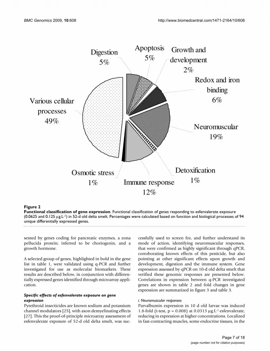
Microarray application and q-PCRThrough the application of the delta smelt microarrays,and combined analytical methods, we have identified 288ESTs, from which a number of genes of interest could beused to measure the effect of esfenvalerate and potentiallyother pyrethroid insecticides; further investigating theiruse as biomarkers in this species. Of the sequenced ESTsthat responded significantly, 118 genes were successfullyannotated; 170 matched unnamed or hypothetical pro-teins, or did not match the described annotation selectioncriteria; i.e. BLASTx searches resulted in expect-valuesgreater than 1 × 10⁻⁵ and scores below 50. Based on geneontology; molecular function, biological processes andcellular components, 94 unique genes were functionallyclassified (figure 2 and table 1) and described below.Based on the proportions of ESTs responding to success-fully annotated unique genes (33%), it is estimated that ofthe 8,448 ESTs printed on the microarray, there could beabove 2,500 unique genes identified in the delta smelt.These numbers however represent responses to a singlecontaminant, and should not be construed as final asthere will have an intrinsic bias exerted upon them, how-ever, the proportion of repeated sequences in the analyseswas very low, with a maximum of nine repetitions forCHK1 checkpoint homologue and not more than one ortwo duplication for a few others. It is also important tonote that the microarray was manufactured with incom-plete genome data, thus information presented in figure 2represents proportions of a limited number of availablegenes.
Differences between methods used allow for greater min-ing of possible biomarkers. The method by Loguinov et al[32] identified a single differentially expressed gene at0.125 μg/L-1 esfenvalerate (with no significant homol-
Page 3 of 18(page number not for citation purposes)

BMC Genomics 2009, 10:608 http://www.biomedcentral.com/1471-2164/10/608
ogy). The addition of LIMMA GUI analysis, also identify-ing this single gene, generated a broader list of genes forbiomarker selection.
A large percentage of uniquely annotated genes, respond-ing to esfenvalerate exposure; 49%, were classified asinvolved in various biological processes. These includedgenes encoding for ribosomal proteins, t-RNA synthases,
telomerases, uncoupling proteins and genes involved inchromosome maintenance. Of greater interest was theidentification of genes involved in neuromuscular activ-ity; representing 19% of identified sequences, a further12% eliciting immune responses, along with 6% relatedto oxidative stress, respiration and iron storage and 5%relevant to apoptosis. Digestion appears to have also beenaffected, along with growth and development, repre-
Swimming behavior and mortalityFigure 1Swimming behavior and mortality. Percentage swimming normality and survival in (a.) 10-d old and (b.) 52-d old H. trans-pacificus exposed to esfenvalerate, ± standard errors (n = 10). * Indicates significant reduction in survival or swimming per-formance compared to solvent control.
0
20
40
60
80
100
0.00 0.10 0.20 0.30 0.40 0.50
4-hour Survival
24-hour Survival
4-hour Normal Swimming
24-hour Normal Swimming
0
20
40
60
80
100
0.00 0.10 0.20 0.30 0.40 0.50
4-hour Survival
24-hour Survival
4-hour Normal Swimming
24-hour Normal Swimming
Perc
enta
ge s
urvi
val a
nd s
wim
min
g no
rmal
ity
Esfenvalerate concentration (μg.L-1)
**
*
*
**
*
**
*
a.
b.
*
(LC50 = 0.19 μg.L-1)
(EC50 = 0.04 μg.L-1)
(EC50 = 0.38 μg.L-1)
(LC50 = 0.24 μg.L-1)
(EC50 = 0.11 μg.L-1)
(EC50 = 0.13 μg.L-1)
0
20
40
60
80
100
0.00 0.10 0.20 0.30 0.40 0.50
4-hour Survival
24-hour Survival
4-hour Normal Swimming
24-hour Normal Swimming
0
20
40
60
80
100
0.00 0.10 0.20 0.30 0.40 0.50
4-hour Survival
24-hour Survival
4-hour Normal Swimming
24-hour Normal Swimming
Perc
enta
ge s
urvi
val a
nd s
wim
min
g no
rmal
ity
Esfenvalerate concentration (μg.L-1)
**
*
*
**
*
**
*
a.
b.
*
(LC50 = 0.19 μg.L-1)
(EC50 = 0.04 μg.L-1)
(EC50 = 0.38 μg.L-1)
(LC50 = 0.24 μg.L-1)
(EC50 = 0.11 μg.L-1)
(EC50 = 0.13 μg.L-1)
Page 4 of 18(page number not for citation purposes)

Page
5 o
f 18
(pag
e nu
mbe
r not
for c
itatio
n pu
rpos
es)
re as identified by microarray analyses.
Fold Change (+/-)
P-VALUE (or cut-off)
1.3277 0.0625 **
1.2252 0.0029
1.1884 0.0044
1.1876 0.0003
1.1720 0.0046
1.1607 0.0021
1.1368 0.0090
1.1196 0.0082
1.0860 0.0059
-1.0841 0.0045
-1.0878 0.0030
-1.1419 0.0074
-1.1675 0.0033
-1.2019 0.0625 *
-1.2121 0.0020
-1.2547 0.0625 *
-1.2602 0.0625 *
-1.3905 0.0625 *
1.2897 0.0041
1.1769 0.0003
-1.1727 0.0625 *
-1.1939 0.0625 *
-1.1945 0.0625 *
-1.1995 0.0034
-1.2245 0.0625 *
-1.2298 0.0625 *
-1.2600 0.0052
-1.2652 0.0625 *
-1.3981 0.0013
1.1235 0.0082
1.0898 0.0069
-1.0647 0.0022
BM
C G
enom
ics
2009
, 10:
608
http
://w
ww
.bio
med
cent
ral.c
om/1
471-
2164
/10/
608
Table 1: Classification of differential gene expression of esfenvalerate responding genes in 52-d old H. transpacificus following 24 h exposu
BLASTX top hit Species match Accession No. Score E-value Concentration
Neuromuscular
titin a Danio rerio ABG48500 110 4.00E-23 0.0625
smooth muscle cell-specific protein SM22 alpha Epinephelus coioides ABW04145 349 1.00E-94 0.1250
toxin-1 Oncorhynchus mykiss AAM21198 116 5.00E-25 0.1250
thymosin beta-12 Lateolabrax japonicus P33248 80 2.00E-13 0.1250
similar to 19.9 kD myosin light chain isoform 1 Danio rerio XP_685183 332 1.00E-89 0.1250
ictacalcin Ictalurus punctatus AAY86967 145 1.00E-33 0.1250
tropomyosin Theragra chalcogramma BAC44994 281 2.00E-74 0.1250
N-acylsphingosine amidohydrolase Takifugu rubripes AAM43813 367 e-100 0.1250
alanine-glyoxylate aminotransferase Platichthys flesus CAH59400 345 2.00E-93 0.1250
titin b Danio rerio ABG48499 65 5.00E-09 0.1250
alpha-2,8-polysialyltransferase IV Oncorhynchus mykiss BAC77411 70 1.00E-10 0.1250
hedgehog acyltransferase-like, a Danio rerio NP_957181 295 1.00E-78 0.1250
parvalbumin Cyprinus carpio CAC83659 173 7.00E-42 0.1250
BTEB transcription factor Pimephales promelas ABO28528 107 1.00E-21 0.0625
myosin regulatory light chain 2 Salmo salar CAD89610 330 7.00E-89 0.1250
similar to Clca1 protein Danio rerio XP_694323 198 2.00E-49 0.0625
ependymin Perca flavescens ABU49423 168 2.00E-40 0.0625
aspartoacylase Danio rerio NP_001103573 384 e-105 0.0625
Immune
carboxypeptidase B Paralichthys olivaceus BAC53789 365 2.00E-99 0.1250
fish-egg lectin (FEL) Cyprinus carpio P68512 192 2.00E-47 0.1250
procathepsin B Oncorhynchus mykiss AAK69705 346 1.00E-93 0.0625
gamma-glutamyl hydrolase Danio rerio NP_998487 223 6.00E-57 0.0625
membrane glycoprotein Human coronavirus ABD75532 53 1.00E-05 0.0625
beta-2 microglobulin Salmo salar AAG17525 176 8.00E-43 0.1250
microtubule-associated protein 1 light chain 3 alpha Danio rerio NP_999904 238 3.00E-61 0.0625
microtubule-associated protein, RP/EB family, member Danio rerio NP_998805 272 1.00E-71 0.0625
T-cell receptor beta chain ANA 11, putative Brugia malayi EDP38115 63 2.00E-08 0.1250
glycerophosphodiester phosphodiesterase domain containing 1 Danio rerio NP_001004118 322 2E-86 0.0625
CHK1 checkpoint homolog Xenopus tropicalis CAJ83813 92 2.00E-17 0.1250
Apoptosis
tissue inhibitor of metalloproteinase 2 Oncorhynchus mykiss AAU14867 265 3.00E-69 0.1250
cathepsin H Danio rerio NP_997853 300 5.00E-80 0.1250
caspase-3 Dicentrarchus labrax ABC70996 223 6.00E-68 0.1250

Page
6 o
f 18
(pag
e nu
mbe
r not
for c
itatio
n pu
rpos
es)
-1.1270 0.0061
-1.1507 0.0047
1.3333 0.0042
1.2968 0.0052
1.1769 0.0080
1.1543 0.0036
-1.1340 0.0073
-1.2429 0.0625 *
-1.1388 0.0072
-1.1568 0.0625 *
-1.2321 0.0625 *
-1.1053 0.0021
1.4959 0.0056
1.4211 0.0095
1.3204 0.0025
1.1297 0.0021
-1.0618 0.0097
nding to both 0.0625 and 0.125 μg.L1 esfenvalerate. e applied microarray.
e as identified by microarray analyses. (Continued)
BM
C G
enom
ics
2009
, 10:
608
http
://w
ww
.bio
med
cent
ral.c
om/1
471-
2164
/10/
608
caspase-1 Dicentrarchus labrax ABB05054 79 3.00E-13 0.1250
cathepsin S-like Oncorhynchus mykiss AAV32964 291 1.00E-77 0.1250
Redox and metal ion binding
hydroxymethylbilane synthase Danio rerio CAM15096 369 e-101 0.1250
hemopexin Danio rerio NP_001104617 313 2.00E-83 0.1250
transferrin Salvelinus fontinalis BAA84100 326 1.00E-87 0.1250
similar to leprecan-like 1 protein Danio rerio XP_695073 183 1.00E-44 0.1250
similar to synaptic glycoprotein SC2 Danio rerio XP_693420 430 e-119 0.1250
similar to LOC407663 protein Danio rerio XP_698537 124 6.00E-27 0.0625
Growth and development
yghl1 (Putative growth hormone like protein-1) Seriola quinqueradiata BAB62526 153 1.00E-35 0.1250
ZPA domain containing protein Oryzias latipes NP_001098216 168 3.00E-40 0.0625
Detoxification
pregnane × receptor Oncorhynchus mykiss ABP38412 206 2.00E-51 0.0625
Osmotic stress
hyperosmotic glycine rich protein Salmo salar AAO32675 134 8.00E-30 0.1250
Digestion
similar to Apoa4 protein isoform 2 (Apolipoprotein) Danio rerio XP_698920 296 7.00E-79 0.1250
chymotrypsinogen 2-like protein Sparus aurata AAT45254 460 e-128 0.1250
pancreatic carboxypeptidase A1 precursor copy 2 Tetraodon nigroviridis AAR16321 242 9.00E-63 0.1250
pancreatic protein with two somatomedin B domains Paralichthys olivaceus BAA88246 214 e-100 0.1250
chitinase (Zgc:55941) Danio rerio AAH44549 369 e-100 0.1250
Genes selected for quantitative PCR analyses are shown in bold. * indicates genes responding at 0.0625 μg.L-1 esfenvalerate and ** indicates gene respoRemaining genes responded only at 0.125 μg.L1 esfenvalerate exposure. Information represents proportions of a limited number of available genes on th
Table 1: Classification of differential gene expression of esfenvalerate responding genes in 52-d old H. transpacificus following 24 h exposur
BMC Genomics 2009, 10:608 http://www.biomedcentral.com/1471-2164/10/608
sented by genes coding for pancreatic enzymes, a zonapellucida protein; inferred to be choriogenin, and agrowth hormone.
A selected group of genes, highlighted in bold in the genelist in table 1, were validated using q-PCR and furtherinvestigated for use as molecular biomarkers. Theseresults are described below, in conjunction with differen-tially expressed genes identified through microarray appli-cation.
Specific effects of esfenvalerate exposure on gene expressionPyrethroid insecticides are known sodium and potassiumchannel modulators [25], with axon demyelinating effects[27]. This the proof-of-principle microarray assessment ofesfenvalerate exposure of 52-d old delta smelt, was suc-
cessfully used to screen for, and further understand itsmode of action, identifying neuromuscular responses,that were confirmed as highly significant through qPCR,corroborating known effects of this pesticide, but alsopointing at other significant effects upon growth anddevelopment, digestion and the immune system. Geneexpression assessed by qPCR on 10-d old delta smelt thatverified these genomic responses are presented below.Correlations in expression between q-PCR investigatedgenes are shown in table 2 and fold changes in geneexpression are summarized in figure 3 and table 3.
i. Neuromuscular responsesParvalbumin expression in 10 d old larvae was induced1.8-fold (t-test, p = 0.008) at 0.0313 μg.L-1 esfenvalerate,reducing in expression at higher concentrations. Localizedin fast-contracting muscles, some endocrine tissues, in the
Functional classification of gene expressionFigure 2Functional classification of gene expression. Functional classification of genes responding to esfenvalerate exposure (0.0625 and 0.125 μg.L-1) in 52-d old delta smelt. Percentages were calculated based on function and biological processes of 94 unique differentially expressed genes.
Redox and iron binding
6%Various cellular processes
49%
Digestion5%
Apoptosis5%
Growth and development
2%
Neuromuscular19%
Detoxification1%Immune response
12%
Osmotic stress1%
Page 7 of 18(page number not for citation purposes)

BMC Genomics 2009, 10:608 http://www.biomedcentral.com/1471-2164/10/608
nervous system in GABAergic interneurons and in thebrain [33], parvalbumin removes calcium from myofi-brils, protecting neurons from hyper-excitability and facil-itating muscle relaxation [34]. Accumulation of calciumin muscular tissue contributes to muscle degradation,muscular dystrophy and muscle fiber necrosis [35]. Estro-gen is required for parvalbumin expression, thus estrogenreceptor-β co-expresses with parvalbumin [36]. Estrogenis also required in brain development and has a protectiveneurological role, by regulating the activity of GABAergicsystems within the hippocampus, basal forebrain and
hypothalamus [37]. Differential expression of parvalbu-min on exposure to esfenvalerate may be resultant ofestrogenic effects. Pyrethroid pesticides have steroidreceptor-binding activity [38] linked with endocrine dis-ruption [39], thus exposure is likely to affect the popula-tion dynamics of wildlife not only throughneuromuscular impairments, but also by affecting repro-ductive output [29,40]. Parvalbumin, could therefore, bea good indicator of possible endocrine-disruption as wellas neuromuscular impairments.
Table 2: Pairwise correlations of gene expression in esfenvalerate exposed 10-d old delta smelt.
Aspartoacylase Titin a Microglobulin Caspase-3 Parvalbumin Hemopexin ZPA Myozenin Creatine Kinase
PXR
Aspartoacylase 1(0)
Titin a 0.490 (0.402) 1(0)
Microglobulin 0.576 (0.310) 0.954 (0.012)
1(0)
Caspase-3 0.344 (0.571) 0.919 (0.027)
0.921 (0.026) 1(0)
Parvalbumin 0.284 (0.644) 0.953 (0.012)
0.928 (0.023) 0.981 (0.003)
1(0)
Hemopexin 0.629 (0.256) 0.819 (0.090)
0.951 (0.013) 0.860 (0.061) 0.822 (0.088) 1(0)
ZPA -0.017 (0.979) 0.788 (0.113)
0.646 (0.239) 0.853 (0.066) 0.871 (0.054) 0.472 (0.422) 1(0)
Myozenin 0.309 (0.613) 0.898 (0.038)
0.949 (0.014) 0.909 (0.033)
0.943 (0.016) 0.892 (0.042)
0.677 (0.210) 1(0)
Creatine Kinase
0.255 (0.679) 0.916 (0.029)
0.865 (0.058) 0.984 (0.003)
0.978 (0.004) 0.759 (0.137) 0.932 (0.021)
0.859 (0.062)
1(0)
PXR 0.880(0.049)
0.087(0.890)
0.208(0.737)
-0.113(0.857)
-0.153(0.805)
0.307(0.616)
-0.475(0.418)
-0.038(0.952)
-0.218(0.725)
1(0)
Numbers represent correlation coefficients; r, and significance probabilities; (p), between ten selected biomarkers assessed with quantitative PCR. Bold p-values highlight significant correlations.
Table 3: Mean and standard deviations in fold changes in gene expression of ten selected biomarkers in esfenvalerate exposed, 10-d old, delta smelt, assessed by quantitative PCR.
Gene\Concentration 0.000 0.031 0.063 0.125 0.250
Aspartoacylase Mean 1.000 0.629 0.346 0.299 0.244SE 0.240 0.100 0.078 0.035 0.088
Titin Mean 1.000 1.515 0.909 0.933 0.475SE 0.529 0.198 0.095 0.192 0.096
Microglobulin Mean 1.000 1.420 0.760 0.828 0.628SE 0.404 0.127 0.104 0.154 0.221
Caspase Mean 1.000 2.024 1.136 0.818 0.670SE 0.336 0.432 0.043 0.117 0.094
Parvalbumin Mean 1.000 1.718 1.097 1.037 0.771SE 0.241 0.151 0.125 0.062 0.107
Hemopexin Mean 1.000 1.501 0.521 0.548 0.612SE 0.089 0.296 0.051 0.147 0.295
ZPA Mean 1.000 1.612 1.455 1.165 0.912SE 0.415 0.321 0.224 0.210 0.574
Myozenin Mean 1.000 1.730 0.857 1.069 0.835SE 0.212 0.205 0.121 0.093 0.188
Creatine Kinase Mean 1.000 1.799 1.265 0.968 0.750SE 0.270 0.249 0.272 0.162 0.145
PXR Mean 1.000 0.737 0.668 0.737 0.729SE 0.126 0.083 0.124 0.080 0.178
Page 8 of 18(page number not for citation purposes)

BMC Genomics 2009, 10:608 http://www.biomedcentral.com/1471-2164/10/608
Interestingly, expression of aspartoacylase (ASPA) inexposed 10-d old delta smelt larvae was significantlyaffected at all concentrations, downregulating withincreasing esfenvalerate concentration in a dose responsemanner, and correlating significantly with swimminganomaly at 24 h (r = 0.913, p = 0.029). Aspartoacylase cat-alyzes hydrolysis of N-acetyl-L-aspartate (NAA) to aspar-tate and acetate in the vertebrate brain [41]. Variations inNAA measured in urine, blood and brain, have been usedas diagnosis of nervous system diseases such as Alzhe-imer's and multiple sclerosis [42,43]. Measurements ofNAA, along with ADP levels determined by creatinekinase activity, are used to evaluate the energetic state ofthe brain, a positive linear correlation existing betweenNAA and ADP synthesis [44]. Deficiency in ASPA activityleads to degeneration of the myelin; an ensheathmentthat isolates and controls axonal activity, it is associatedwith schizophrenia [45], and is the established cause ofleukodystrophy in Canavan's disease [43]. Abnormalmyelination is known to result from acyltransferase defi-ciency [46]. Ependymin, a myelin associated glycoprotein
related to memory formation and involved in neuronalregeneration [47], was also negatively affected by esfenva-lerate. Myelin has been postulated as a probable modula-tor of ASPA activity [48] further affecting this criticalpathway of neurological function. ASPA protein activity isa strong biomarker of brain damage and neurologicalimpairment investigation, used regularly in human andveterinary disease diagnostics [49].
Creatine kinase was significantly up-regulated at 0.0313μg.L-1 esfenvalerate (t-test, p < 0.05). Creatine kinase pro-tein is used not only as a diagnosis of brain energetic valueas mentioned above, but also of diseases like cardiac inf-arction and skeletal muscle necrosis [50]. In muscle, crea-tine kinase is specifically bound to sarcoendoplasmicreticulum, and regulates calcium uptake and ATP/ADPratios [51], thus is directly involved in muscle contraction.Of interest here, are the pathway links and correlatingresponses (r = 0.98) between parvalbumin; facilitatingmuscle relaxation by binding calcium, and creatinekinase, which regulates calcium uptake. These two param-
Molecular biomarkers responsesFigure 3Molecular biomarkers responses. Fold changes in gene expression of ten selected biomarkers in esfenvalerate exposed, 10-d old, delta smelt, assessed by quantitative PCR. Significance in expression differences, as determined by One-way ANOVA, is shown in brackets in legend.
0.0
0.5
1.0
1.5
2.0
2.5
0.00 0.03 0.06 0.13 0.25
Aspartoacylase (p=0.0140)
Titin a (p=0.0858)
Microglobulin (p=0.1176)
Caspase-3 (p=0.0245)
Parvalbumin (p=0.0376)
Hemopexin (p=0.0242)
ZPA (p=0.6465)
Myozenin (p=0.0494)
Creatine Kinase (p=0.1198)
PXR (p=0.1881)
Fold
cha
nge
in g
ene
expr
essi
on
Esfenvalerate concentration (μg.L-1)
Page 9 of 18(page number not for citation purposes)

BMC Genomics 2009, 10:608 http://www.biomedcentral.com/1471-2164/10/608
eters on their own indicate muscular activity impairments,creating strong links with observed larval swimmingbehavior.
Titin expression also correlated significantly with parval-bumin (r = 0.95) and creatine kinase (r = 0.92), thoughnot statistically significant in q-PCR assessments of 10-dold smelt larvae. Titin is an important protein alsoinvolved in muscle contraction, responsible for muscleelasticity and is the molecular scaffold for thick actin fila-ment formation, forming a connection between filamentsand the muscle Z-line [52]. Myozenin, another proteininvolved in muscle contraction, was significantly influ-enced by esfenvalerate exposure (t-test, p < 0.05) in 10-dold larvae. Co-regulating with Titin (r = 0.90), myozeninis a Z-line, α-actinin- and γ-filamin-binding proteinexpressed predominantly in skeletal muscle, and has beensuggested as a biomarker for muscular dystrophy andother neuromuscular disorders [53].
ii Immune responsesIn this study we have identified a significant alteration inexpression of several genes involved in immuneresponses, most of them with links to neurological dam-age. β-microglobulin, a small protein normally found onthe surface of many cells, including lymphocytes, isknown to be involved in cell protection [54]. β-microglobulin is almost exclusively catabolized in the kid-ney and its excretion is an indication of long term neph-rotoxicity [55]. High concentrations of β-microglobulinare reported to inhibit generation of functional dendriticcells [56], thus an increased amount in the blood or urinemay be a sign of neural degeneration and of certain dis-eases, including some types of cancer, such as multiplemyeloma or lymphoma. β-microglobulin levels are alsoreported to rise following viral infection and its reducedexpression can compromise the immune system [57].Interestingly, exposure to esfenvalerate resulted in a sig-nificant increase of pathogen susceptibility in chinooksalmon [9]. β-microglobulin assessment with q-PCR didnot show any significance in expression in 10 d old larvae(t-test, p = 0.131), however, an overall increase wasobserved at low concentrations of esfenvalerate, correlat-ing significantly with expression of other genes investi-gated, further discussed below.
Multiple sclerosis is caused by an immunological attackon myelin [58], decreasing NAA and resulting in neuro-logical instability. Furthermore, oxidative stress is induc-tive of apoptosis of myelin-reactive T cells [59]. A putativeT cell receptor gene was identified through microarrayscreening, probably reacting to pyrethroid exposure, act-ing upon the myelin sheath and causing further neurolog-ical damage and cell death.
iii Apoptotic responsesCaspases (cysteine-aspartic acid protease) belong to a fam-ily of cysteine proteases that cleave other proteins, such asthe precursor forms of the inflammatory cytokines inter-leukin 1-β and interleukin 18, into active mature peptidesand are also involved in programmed cell death; or apop-tosis [60]. Enzymatic activity requires an aspartic acid res-idue, and plays a critical role in the regulation ofproinflammatory cytokines [61] that are associated withseptic shock and autoimmune syndromes [62]. Upregula-tion of proinflammatory cytokines were reported in viralinfected salmon, which further increased in expressionfollowing esfenvalerate exposure [63]. Caspases contrib-ute to the pathogenesis of neurodegenerative disorderssuch as ischemia, Krabbes and Huntington's diseases,Alzheimer's, and other leukodystrophic diseases resultingin neural degeneration [64,65]. Moreover, caspase inhibi-tors have been suggested as therapeutic treatments forneurodegenerative diseases [66]. Low concentrations ofesfenvalerate; 0.0313 μg.L-1, significantly induced cas-pase-3 expression 2-fold (t-test, p = 0.002) in 10-d olddelta smelt. As caspases are activated by aspartic acid,induction may be suggestive of increases in substrate resi-dues, along with inflammatory cytokines, probable effectsupon the immune system, and subsequent neurodegener-ation. Furthermore, a decrease in ASPA expression couldbe suggestive of reduced breakdown of NNA to aspartateand acetate, required as substrate for caspase activity, andsynthesis of proteins required in repair mechanisms.
iv. Redox and metal ion bindingUpregulation of hemopexin was confirmed by quantita-tive PCR in 10-d old delta smelt exposed to 0.0313 μg.L-1
esfenvalerate. Hemopexin-like protein, a gene sequencedisplaying vast similarities to warm-temperature-acclima-tion-related-65 protein (WAP65) on BLAST homologieswith Japanese medaka (Oryzias latipes), was identified assignificantly upregulated through microarray screening.Hemopexin is synthesized by Schwann cells followingnerve injury [67], accumulation has been reported in theperipheral nervous system following axonal lesions, andis specifically regulated during repair, returning to normallevels on axonal regeneration [68]. Wallerian degenera-tion occurs after axonal injury and is critical for repair, itis characterized by axonal and myelin degeneration [69],is accompanied by macrophage invasion and subsequentsynthesis of hemopexin [67]. Hemopexin appears to playa significant role in neural regeneration, but may beresultant of oxidative stress mediated T cell activity onmyelin sheaths (described above, under immuneresponses). Though upregulation follows nerve injury andthere are strong connections with apoptosis, we classifyhemopexin under oxidative stress as it has a high affinitywith heme and reportedly plays a strong role in bothheme transport and preventing heme-catalyzed oxidative
Page 10 of 18(page number not for citation purposes)

BMC Genomics 2009, 10:608 http://www.biomedcentral.com/1471-2164/10/608
damage [70]. Moreover, pyrethroids have been shown togenerate free radicals and induce oxidative stress [71].Heme is known to respond to nerve injury, and has beensuggested to play a role in neurodegenerative disorders[72], and hemopexin-mediated heme transport wasreported to significantly decrease levels of transferrinreceptor mRNA in HeLa cells [70]. Transferrin was alsoidentified by microarray screening as significantly upregu-lated by esfenvalerate exposure. The primary role of trans-ferrin is the delivery of iron across the blood brain barrier,and its expression in brain is not only related to myelinproduction, but may be a permissive agent in the processof myelination [73]. Furthermore, hemopexin and trans-ferrin reportedly act by similar receptor-mediated mecha-nisms [74].
v. Growth and developmentMicroarray analyses identified a gene with high homologyto egg envelope glycoproteins within the zona pellucida(ZPA) referred to as choriogenins, in fish [75]. This wassignificantly expressed in 52-d old larvae, however, no sta-tistical differences in expression of ZPA were measuredwith qPCR in 10-d old larvae exposed to esfenvalerate.Choriogenin is reportedly more sensitive to endocrine dis-rupting chemicals (EDCs) than estrogen receptors andvitellogenin [76]. Composed primarily of glycoproteinswith various functions during fertilization and develop-ment, choriogenin has been suggested as a biomarker ofexposure to endocrine disrupting chemicals, as it isinduced in late stage embryos, larvae and adult male fishexposed to estrogens [76,77]. Choriogenin is synthesizedin liver of adult females, in response to estrogen, trans-ported in blood and incorporated into the fish egg enve-lope; chorion or zona pellucida (ZPA), an extracellularmatrix that surrounds the oocyte and early embryo [78].Expression was notably elevated at low pyrethroid con-centration, and it responded in a similar fashion to creat-ine kinase (r = 0.93), though no significant links wereidentified between these two biomarkers.
vi. DetoxificationPregnane × receptor (PXR), involved in the detection oftoxic substances and a key regulator of xenobiotic metab-olism, was identified through microarray assessments, asdownregulated in 52-d old larvae. PXR is a steroid recep-tor and transcriptional regulator of detoxification mecha-nisms such as cytochrome oxidases, and phase IIconjugating enzymes such as glutathione-S-tranferases[79,80]. Downregulation of PXR expression has beenlinked with growth inhibition and cell death in rats andhuman cell lines following exposure to medroxyproges-terone and estradiol, known PXR ligands [80], furthersupporting identified apoptotic responses, steroid recep-tor-binding and endocrine disruption activity of esfenva-lerate [38,39]. PXR expression was not significantly
different in q-PCR assessments of 10-d old larvae exposedto esfenvalerate, however, overall expression declined in adose response manner, correlating with ASPA (r = 0.880;p = 0.049), making it a notable candidate of xenobioticdetection for future biomarker investigations in the deltasmelt.
vii Osmotic StressA hyperosmotic glycine rich protein was identified withthe microarrays as significantly downregulated by expo-sure to 0.0625 μg.L-1 esfenvalerate in 52-d old larvae.Osmoregulation is physiologically controlled by chemicalmessages from the endocrine system, along with cell sig-nalling and nerve transmission [81]. Pyrethroids havebeen suggested to induce osmotic imbalances in commoncarp larvae [82] which are linked to effects on ATPaseactivity responsible for maintaining the Sodium trans-membrane electrochemical gradient [83]. Larval fish areunder direct exposure to osmotic stress as their endocrinesystem is not fully developed [84]. Parvalbumin and cho-riogenin expression have indicated possible effects on theendocrine system, thus expression of this hyperosmoticglycine rich protein may be directly caused by conditionsaffecting endocrine regulation.
viii DigestionChitinase was identified through microarray analysis asbeing downregulated by esfenvalerate exposure. Chitinaseis the principal enzyme involved in digesting chitin, amajor component of insect and crustacean exoskeleton[85]. Larval smelt were fed on artemia during the pre-exposure acclimation period. Not only chitinase but,other digestive enzymes; apolipoproteins, pancreaticenzymes, carboxypeptidase precursors and chemot-rypsinogen, were also significantly upregulated followingexposure in 52-d old larvae. Effects on digestion alone willundoubtedly have significant effects on growth, whichwhen combined with hypothesized feeding reductionresulting from impaired swimming would lead to signifi-cant malnutrition. Contaminants affect a whole ecosys-tem, at all levels, and dramatic reductions in copepods,cladoceran and amphipod populations; organisms pre-dated upon by the delta smelt, have been reported in theSacramento delta [86]. Scarcity in food and reduced inges-tion ability, besides digestion will significantly affect pop-ulation dynamics of any specie.
ConclusionsMicroarray technology was used as an initial screening ofprobable genes responding to esfenvalerate exposuretherefore no multiple testing correction was applied. Wehave, however, examined and confirmed effect of esfenva-lerate upon some of the genes in a different age group oflarval delta smelt, identifying significant responses thatare primarily linked with swimming behavior. Some
Page 11 of 18(page number not for citation purposes)

BMC Genomics 2009, 10:608 http://www.biomedcentral.com/1471-2164/10/608
responding genes can be classified within different func-tional groups. Due to the measured behavioral responses,the classification approach contains a certain bias towardsunderstanding neuromuscular effects. It is interesting thatqPCR measurements have identified a greater response atthe lower concentrations, implying homeostatic altera-tions, at environmentally relevant concentrations. Mostgenes did not display a desired dose response correlationassociated with usable biomarkers, but did supportresponses within the suite of genes investigated, some-what validating their use within a broader biomarkerapproach. Hemopexin for example is known to beinvolved in axon repair, and the myelin sheath surround-ing the axon needs to be degraded for this repair to beprocessed, hypothesizing therefore that ASPA downregu-lation is resultant of neurological damage. The subse-quent decrease of hemopexin expression at higherexposure concentrations, and further decrease in ASPA,may be indicative of repair impairments.
What becomes apparent from this study is that exposureto sublethal concentrations of esfenvalerate results in neu-rological damage and a series of compensatory molecularresponses that attempt to repair nerve damage. We wouldhypothesize that induction of transcription of the genesencoding ASPA, hemopexin, parvalbumin and creatinekinase are part of a pathway of damage triggered repairmechanisms, responding to esfenvalerate insult. Reduc-tion in expression of ASPA indicates that myelin sheathsmay be degraded, resulting in a number of detrimentaleffects on the lesion sites, and similarly, muscular struc-ture and function is being compromised as measured byalterations in titin and myozenin expression. The expres-sion of β-microglobulins could be a compensatory reac-tion to toxic damage, protecting cells from infections in asusceptible immune system caused by exposure to esfen-valerate. Previous studies, carried out in esfenvalerateexposed chinook salmon have reported a compromisedimmunity and significantly higher susceptibility to infec-tion [9,63]. This is particularly important in youngerorganisms that are generally more susceptible than adults.Furthermore, polluted waters not only contain mixturesof contaminants, but also harbor multiple pathogens thatwill further affect health parameters.
Behavioral endpoints, such as swimming behavior, areamongst the most sensitive and ecologically relevantparameters to assess sublethal toxicity of neurotoxicchemicals [29]. The high susceptibility of delta smelt toesfenvalerate, mediated neurological damage resulting inimpaired swimming ability, also raises questions on thelikely effects upon their chemosensory system; olfactorysystem, important in sensing reproductive pheromones,mediating reproduction. Females synthesize sex hor-mones stimulating male reproductive behavior [87]. Neu-
rological damage affecting the olfactory nerves, the brainand or entire nervous system, could lead to further impair-ments in reproductive success following exposure to pyre-throids. Damage to the olfactory system has been used asa sublethal toxicological endpoint in fish, in studies inves-tigating behavior following pesticide exposures [88]. Pyre-throids are known to affect the olfactory system [89]. Achemosensory gene, ictacalcin, responding to esfenvaler-ate exposure was also differentially expressed on themicroarray. Ictacalcin is a gene originally identified in cat-fish (Ictalurus punctatus), involved in chemosensory tis-sues, and highly expressed in barbell, olfactory mucosaand gill [90]. Differential expression of this gene mayindicate that further behavioral parameters, not investi-gated in this study, such as recognition, alarm response,feeding, imprinting and homing, gamete release and syn-chronization, contaminant avoidance [88], and otherbehavioral parameters that are governed by chemosensorysystem, could be compromised. We could speculate thatoutside laboratory conditions, neuromuscular and chem-osensory impairments would probably result in higherecological parameters being affected through inability toswim against water currents, making them more suscepti-ble to predation and reducing their ability to obtain food.Furthermore, effects on chemosensory parameters wouldlead to migratory, reproductive, predator and contami-nant avoidance impairments.
Inhibition of repair mechanisms, leading to neuromuscu-lar damage and eventual death, was behaviorally observedthroughout exposure, as impairment in swimming ability.The ability to use molecular biomarkers of neuromusculareffect further strengthens links between mechanisticeffects with parameters of ecological relevance. Our studysupports the use of gene expression as a productive way ofunderstanding modes of actions of individual chemicalsin endangered species. Furthermore, this screening andinterrogative approach permits the identification anddevelopment of biomarkers for species of concern inwhich prior information is limited and allows for investi-gations into problems specific to the organism in ques-tion; assessing possible causes of detrimental effects andresulting influences on individual performance andhypothesizing effects upon population dynamics. A suiteof biomarkers developed in this manner, though addi-tions and subtractions are required from the presentedlist, could be used to aid monitor impacts of stressorsupon organisms within a specific environment and couldbe an essential tool in determining causative factors ofpopulation decline in the delta smelt and other threat-ened species. The selected biomarkers clearly need to befurther investigated and validated against other knowncontaminants, and suitability in field applications.
Page 12 of 18(page number not for citation purposes)
BMC Genomics 2009, 10:608 http://www.biomedcentral.com/1471-2164/10/608
MethodsMicroarray constructionWe constructed a delta smelt microarray using 8448 PCRamplified fragments from a normalized cDNA library. AcDNA library was created using expressed sequence tags(ESTs) ligated into p-BS plasmid vectors and cloned intochemically competent Escherichia coli cells (BioS&T Inc,Montreal, Quebec, Canada). RNA for library constructionwas obtained from a number of larval, juvenile and adultdelta smelt, ranging from unexposed, control conditions,to fish from exposures to high temperature (25°C), andsublethal concentrations of copper, esfenvalerate, and asix field water samples from throughout the Sacramento-San Joaquin estuary. Products were PCR amplified from 1μl bacterial suspension, and visualized on agarose gels.Purified PCR fragments ranging in size from 1-4 kb, alongwith control spots, were pin-printed in duplicate ontoepoxysilane coated glass slides (Schott-Nexterion, USA) ina 20 × 19 block format, with 48 blocks per microarray.Microarrays were printed using a Lucidea Array Spotter(Amersham) at the Array Core facility at UC Davis (sinceclosed down). Microarray control spots included anumber of hybridization tags comprised of a pooled PCRproduct from all spots on the array, H. transpacificus DNA,and four Spot Report System of alien PCR products fromArabidopsis thaliana; CAB, RCA, RBCL and LPT4 (Strata-gene, USA). Blank control spots consisted of 1× Nexterionbuffer solution.
Esfenvalerate exposuresDelta smelt larvae aged 10 d and 52 d were exposed for 24h, in two separate experiments, to a range of esfenvalerateconcentrations; 0.0313, 0.0625, 0.125, 0.250 and 0.500μg.L-1 (nominal) in laboratory control water, with corre-sponding laboratory and solvent controls. Concentrationswere measured at the start of the experiment by the WaterPollution Control Laboratory at the Department of Fishand Game (Rancho Cordova, California, USA), only sin-gle measurements were taken per treatment (results notshown), therefore we present the data in terms of nominalconcentrations. Laboratory control water consisted ofdeionized water amended to US EPA moderately hardstandards (80-100 mg.L-1 CaCO3) and 200 μl/L methanolwas used as solvent carrier. Salinity was adjusted withInstant Ocean salt to match hatchery rearing conditions(range 650 μS.cm-1 to 900 μS.cm-1).
Average wet weights of 10-d to 52-d old larval delta smeltranged from 0.5 to 2.5 mg respectively. Larvae wereobtained from the Fish Conservation and Culture Labora-tory (FCCL) UC Davis, Byron, CA, transported in cool,oxygenated 2-gallon black buckets, and held overnight inthe laboratory at 17°C and a 8 h:16 h D:L light cycle. Thefollowing day, ten larvae were transferred to each 2-Lbeaker containing 1 L of aerated control water or esfenva-
lerate treatment. Each treatment consisted of 4 replicatesand tests were performed at 8 h:16 h D:L cycle, at a watertemperature of 17°C ± 1.2°C. The pH during the tests was7.1 - 7.5. Dissolved oxygen levels were within the accept-able range for delta smelt (above 6.5 mg.L-1)[91]. Larvaewere fed rotifers the day before the test start, but not dur-ing the 24 h exposure. Rotifer cultures were obtained fromFCCL. During exposure, larvae were observed for aberrantswimming behavior, and surviving fish were scored after4 h and 24 h. Swimming behavior was assessed by observ-ing each tank for 5 min as described in Geist et. al. [17].Any pronounced deviation (> 1 min) from normal (con-trol) swimming patterns were recorded as abnormal.Effects on swimming performance (EC50) and mortality(LC50) were assessed using linear regression analysis withEnvironmental Toxicity Information System (CETIS) byTidepool Scientific Software (McKinleyville, CA, USA).
Four surviving 52 d old larvae from solvent controls, andexposures to 0.0625 and 0.125 μg.L-1 esfenvalerate, wereused for microarray analyses, hybridized in a referencedesign against a pool of RNA from all treatments. Fourreplicate 10 d old larvae from each treatment (controlsand 0.0312, 0.0625, 0.125 μg.L-1 esfenvalerate) were usedfor biomarker analyses and gene expression verificationusing quantitative PCR (q-PCR).
All experiments and use of test organisms were approvedby the UC Davis Institutional Animal Care and Use Com-mittee (Animal Use Protocol for Animal Care and Use#13361). This institution is accredited by the Associationfor Assessment and Accreditation of Laboratory AnimalCare, International (AAALAC) and has an Animal WelfareAssurance on file with the Office of Laboratory AnimalWelfare (OLAW). The Assurance Number is A3433-01.The IACUC is constituted in accordance with U.S. PublicHealth Service (PHS) Animal Welfare Policy and includesa member of the public and a non-scientist.
RNA isolation, cDNA synthesis and fluorescence labelingRNA was extracted from whole, individual organismsusing a standard phenol:chloroform protocol with TrizolReagent (Invitrogen). Fifteen micrograms of total RNAwere used for cDNA synthesis, spiked with control RNA(CAB, RCA, RBCL and LTP4 (SpotReport, Stratagene) andlabeled with Alexa fluor dyes, using SuperScript™ PlusIndirect cDNA labeling System (Invitrogen). Each experi-mental sample and control was combined with a refer-ence pool cDNA prior to hybridization using anautomated Tecan HS4800 hybridization station. Slideswere scanned using a GenePix 4000B scanner (AxonInstruments).
Microarray images and data from esfenvalerate exposeddelta smelt can be accessed at http://www.vet
Page 13 of 18(page number not for citation purposes)
BMC Genomics 2009, 10:608 http://www.biomedcentral.com/1471-2164/10/608
med.ucdavis.edu/apc/WernerLab/subpagpelagic_organism_decline.html; POD archivedata.
Microarray AnalysesNormalization and analytical methods are described inLoguinov et. al. [32] and Smyth [92]. In brief, print tipnormalization was carried out within slides and sequen-tial single slide data analysis was carried out as an alterna-tive to between-slide normalization. An α-outlier-generating model was used to identify differentiallyexpressed genes by applying the following decision rulefor multiple-slide data analysis: a given gene was selectedas a candidate if it was detected as significantly up- ordownregulated in 4 of 4 replicates (raw p-value = 0.0625using exact binomial test and considering outcomes asBernoulli trials). The approach did not use scale estimatorfor statistical inference and, therefore, it did not requirebetween-slide normalization. This method however,detected only one significant differentially expressed can-didate gene at the highest exposure concentration (0.125μg/L-1), (with no significant annotation identity - seeresults and discussion). As a result, a second analyticalmethod was applied to increase the number of probablegenes for consideration in biomarker development. Thuswe further analyzed the data using LIMMA GUI (Linearmodel for microarray analysis graphical user interface)[92], written in the R-programming language availablethrough Bioconductor http://www.Bioconductor.org.Data was normalized within arrays using print-tip Lowessand between arrays applying aquantile normalizationmethods [92]. A linear model fit was computed using the
duplicates on the arrays and the least-squares method, nomultiple assessment methods were applied to eliminatefalse positives as our aim was to increase the number ofgenes available for biomarker assessment, and qualifythese through quantitative PCR.
Sequencing and AnnotationSequencing was carried out at the CA&ES Genomic Facil-ity, UC Davis. Basic Local Alignment Search Tool; trans-lated nucleotide (BLASTx) searches were performed onspecific fragments that responded significantly to theexposure treatments. Only genes that were differentiallyexpressed following esfenvalerate exposure weresequenced. Sequences were annotated according tohomologies to protein database searches using translatednucleotide sequences and direct nucleotide queries http://blast.ncbi.nlm.nih.gov/Blast.cgi. Sequences were onlyannotated if they were found to have a BLASTx match withthe expect value smaller than 1 × 10⁻⁵ and a score above50.
Functional ClassificationsDifferentially expressed genes were classified according togene ontology http://www.uniprot.org/uniprot, andinformation gathered from literature, into functionalgroups. Classification was carried out based on geneexpression changes in respect of control subjects, regard-less of whether these were up or downregulated, or expo-sure concentrations. Specific genes of interest wereselected for further investigation using quantitative PCR.
Table 4: Primers and probes used for molecular biomarkers: Primer pairs and TaqMan probes used in q-PCR assessments.
Accession no. Gene Primer Sequences Probe No.
FJ711577 Aspartoacylase Left: ggaggcacacatgggaatg 109Right: cttcctctgaatctctgttccattatc
FJ711576 Parvalbumin Left: gaccaagacaagagtggcttca 101Right: tctggcaccagcagagaagtt
FJ711580 β-2 microglobulin Left: tctttcgcggtcatctttctc 22Right: ggttgtggccatacacctgaa
FJ711579 Hemopexin Left: catgcactacgaggacgacaag 143Right: tggtagtagctgaacaccttgctg
FJ711578 Caspase-3 Left: gagaaccggtatgaaccaacg 159Right: tccaagcttcccaaacactttc
FJ711575 Titin a Left: tgatcactggcgtgaaagagg 159Right: caagctcattggacagtttgagg
FJ711581 ZPA Left: catgcggctgagtttggataa 106Right: tgccattgatagcatcaacttca
FJ711583 Myozenin* Left: ccaatgtcgtgctggtacacc 106Right: ctgccagacattgatgtagcca
FJ711584 Creatine kinase* Left: cgatcggcgttggagatg 163Right: gccaagttcaacgagattctgg
FJ711582 PXR Left: tgaggcggtggagaagag 144Right: gaggcggtggagaagag
* indicates biomarkers obtained from preliminary studies carried out during microarray development.
Page 14 of 18(page number not for citation purposes)

BMC Genomics 2009, 10:608 http://www.biomedcentral.com/1471-2164/10/608
Biomarker developmentGenes were selected according to level of expression sig-nificance, knowledge base from literature, and functionalclassification. Primer and probes for qPCR analyses weredesigned using Roche Universal Probe Library AssayDesign Center https://www.roche-applied-science.com.Designed primers were obtained from Eurofins MWGOperon http://www.eurofinsdna.com, and TaqManprobes were supplied by Roche. Sequences for all genesassessed by qPCR analyses have been submitted to gen-bank http://www.ncbi.nlm.nih.gov. Primers and probes,and genbank accession numbers, for investigated biomar-kers are detailed in table 4. Myozenin and creatine kinase,though not resulting from the current study, were genesidentified in investigations carried out during microarraydevelopment, and added to the selected biomarkers dueto our interest in neuromuscular activity.
Quantitative PCRA total of 1.5 μg RNA was cDNA synthesized using ran-dom primers, and diluted to a total of 50 μl with nucleasefree water to generate sufficient template for qPCR analy-sis. TaqMan Universal PCR Mastermix (Applied Biosys-tems) was used in q-PCR amplifications in a reactioncontaining 10mMTris-HCl (pH 8.3), 50 mM KCl, 5 mMMgCl2, 2.5 mM deoxynucleotide triphosphates, 0.625 UAmpliTaq Gold DNA polymerase per reaction, 0.25 UAmpErase UNG per reaction and 5 μL of cDNA sample ina final volume of 12 μL. The samples were placed in 384well plates and cDNA was amplified in an automatedfluorometer (ABI PRISM 7900 Sequence Detection Sys-tem, Applied Biosystems). Amplification conditions were2 min at 50°C, 10 min at 95°C, 40 cycles of 15 s at 95°Cand 60 s at 60°C. Fluorescence of samples was measuredevery 7 s and signals were considered positive if fluores-cence intensity exceeded 10 times the standard deviationof the baseline fluorescence (threshold cycle, CT). SDS2.2.1 software (Applied Biosystems) was used to quantifytranscription.
Quantitative PCR data was analyzed using the relativequantification 2(-Delta Delta CT) method ()([93]. In theabsence of house keeping genes, expression was calcu-lated relative to the mean of controls in respective expo-sures. Surviving larvae from each replicate of the 10-d oldexposed delta smelt were used for q-PCR analyses. One-way ANOVA was used to assess differences in gene expres-sion through out the exposure concentrations, and datawere further assessed using Student's T-test at individualconcentrations in respect to solvent controls.
AbbreviationsEST: expressed sequence tag; q-PCR: real-time quantitativepolymerase chain reaction; ASPA: aspartoacylase; ESA:endangered species act; CESA: California endangered spe-
cies act; LIMMA GUI: linear model for microarray analy-sis, graphical user interface; BLAST: basic local alignmentsearch tool; GABA: gamma-aminobutyric acid; NAA: N-acetyl-L-aspartate; ADP: adenosine diphosphate; WAP65:warm-temperature-acclimation-related-65 protein; EDC:endocrine disrupting chemical; ZPA: zona pellucida; PXR:pregnane × receptor; p-BS: p-Bluescript; IPTG: isopropylβ-galactosidase; LB: Luria Bertani; FCCL: Fish Conserva-tion and Culture Laboratory; UNG: Uracil N-glycosylase;CT: cycle threshold.
Authors' contributionsAll authors were involved in designing the experiments.JG carried out esfenvalerate exposures recording swim-ming abnormality and mortality. REC designed, devel-oped and applied the microarrays with assistance from JPand HW. REC and JP prepared, hybridized and scannedmicroarrays. REC and AVL performed microarray analy-ses. REC and LSD designed qPCR assays and performedrespective assessments and analysis. REC drafted the man-uscript and all authors read, contributed intellectually andapproved the final manuscript.
AcknowledgementsWe thank E. Campbell, UC Davis, for assistance with the microarray devel-opment, J. Lindberg and B. Baskerville-Bridges (FCCL) for supplying delta smelt along with invaluable knowledge on culture and handling. UC Davis ATL was used for all exposure tests; we thank all staff for assistance. Fund-ing provided by the Interagency Ecological Program, Sacramento, California (Contract No. 4600004445 to I. Werner).
References1. Bryant M, Souza K: Summer townet and fall midwater trawl
survey status and trends. Interagency Ecological Program Newsletter2004, 17:14-17.
2. Hieb K, Bryant M, Souza K, Greiner T, Slater S: Place holder forbay and estuary species 2004 status and trends report. Inter-agency Ecological Program Newsletter 2005, 18:6-10.
3. Feyrer F, Nobriga ML, Sommer TR: Multidecal trends for threedeclining fish species: habitat patterns and mechanisms inthe San Francisco Estuary, California, USA. Journal of Fisheriesand Aquatic Sciences 2007, 64:723-734.
4. Brown LR, Kimmerer W, Brown R: Managing water to protectfish: a review of California's Environmental Water Account,2001-2005. Environmental management 2009, 43(2):357-368.
5. Sommer T, Armor C, Baxter R, Breuer R, Brown L, Chotkowski M,Culberson S, Feyerer F, Gingras M, Herbold B, et al.: The collapseof pelagic fishes in the upper san francisco estuary. Fisheries2007, 32(6):270-277.
6. Scholz NL, Truelove NK, French BL, Berejikian BA, Quinn TP, CasillasE, Collier TK: Diazinon disrupts antipredator and homingbehaviors in chinook salmon (Oncorhynchus tshawytscha).Canadian Journal of Fisheries and Aquatic Sciences 2000, 57:1911-1918.
7. de Vlaming V, Connor V, DiGiorgio C, Bailey HC, Deanovic LA, Hin-ton DE: Application of whole effluent toxicity test proceduresto ambient water quality assessment. Environmental Toxicologyand Chemistry 2000, 19(1):42-62.
8. Sandahl JF, Baldwin DH, Jenkins JJ, Scholz NL: Comparative thresh-olds for acetylcholinesterase inhibition and behavioralimpairment in coho salmon exposed to chlorpyrifos. Environ-mental toxicology and chemistry/SETAC 2005, 24(1):136-145.
9. Clifford MA, Eder KJ, Werner I, Hedrick RP: Synergistic effects ofesfenvalerate and infectious hematopoietic necrosis virus onjuvenile chinook salmon mortality. Environmental toxicology andchemistry/SETAC 2005, 24(7):1766-1772.
Page 15 of 18(page number not for citation purposes)

BMC Genomics 2009, 10:608 http://www.biomedcentral.com/1471-2164/10/608
10. Floyd EY, Geist JP, Werner I: Acute, sublethal exposure to apyrethroid insecticide alters behavior, growth, and preda-tion risk in larvae of the fathead minnow (Pimephalespromelas). Environmental toxicology and chemistry/SETAC 2008,27(8):1780-1787.
11. Rand GM: Fundamentals of Aquatic Toxicology Taylor and Francis, Bris-tol, PA; 1995.
12. U.S. EPA: Short-term Methods for Estimating the ChronicToxicity of Effluents and Receiving Waters to FreshwaterOrganisms. 4th edition. U.S. EPA OoW, Washington, DC, EPA-821-R-02-013; 2002.
13. Denslow ND, Colbourne JK, Dix D, Freedman J, Helbing C, KennedyS, Williams P: Selection of surrogate animal species for com-parative toxicogenomics. In Genomic Approaches for Cross-SpeciesExtrapolation in Toxicology Edited by: Benson WH, Di Giulio RT. Port-land, Oregon, USA: CRC Press; 2006:33-75.
14. Little EE, Calfee RD: Acute toxicity of three herbicides,DMA*41VM, Renovate 3, and Sonar A.S., to two life stagesof white sturgeon from the Kootenai River and to rainbowtrout. USGS Report: Columbia Envrionmental Research Center,Columbia, MO; 2008.
15. Little EE, Calfee RD: Toxicity of Chlorine and Copper to Rain-bow Trout and to White Sturgeon from the Kootenai Riverand Columbia River. USGS Report: Columbia EnvrionmentalResearch Center, Columbia, MO; 2008.
16. Connon R, Hooper HL, Sibly RM, Lim FL, Heckmann LH, Moore DJ,Watanabe H, Soetaert A, Cook K, Maund SJ, et al.: Linking molec-ular and population stress responses in Daphnia magnaexposed to cadmium. Environmental science & technology 2008,42(6):2181-2188.
17. Geist J, Werner I, Eder KJ, Leutenegger CM: Comparisons of tis-sue-specific transcription of stress response genes withwhole animal endpoints of adverse effect in striped bass(Morone saxatilis) following treatment with copper andesfenvalerate. Aquatic toxicology (Amsterdam, Netherlands) 2007,85(1):28-39.
18. Heckmann LH, Sibly RM, Connon R, Hooper HL, Hutchinson TH,Maund SJ, Hill CJ, Bouetard A, Callaghan A: Systems biology meetsstress ecology: linking molecular and organismal stressresponses in Daphnia magna. Genome biology 2008, 9(2):R40.
19. Denslow ND, Garcia-Reyero N, Barber DS: Fish 'n' chips: the useof microarrays for aquatic toxicology. Molecular bioSystems2007, 3(3):172-177.
20. Steinberg CE, Sturzenbaum SR, Menzel R: Genes and environment- Striking the fine balance between sophisticated biomoni-toring and true functional environmental genomics. The Sci-ence of the total environment 2008, 400(1-3):142-161.
21. Soetaert A, Vandenbrouck T, Ven K van der, Maras M, van RemortelP, Blust R, De Coen WM: Molecular responses during cad-mium-induced stress in Daphnia magna: integration of dif-ferential gene expression with higher-level effects. Aquatictoxicology (Amsterdam, Netherlands) 2007, 83(3):212-222.
22. Poynton HC, Varshavsky JR, Chang B, Cavigiolio G, Chan S, HolmanPS, Loguinov AV, Bauer DJ, Komachi K, Theil EC, et al.: Daphniamagna ecotoxicogenomics provides mechanistic insightsinto metal toxicity. Environmental science & technology 2007,41(3):1044-1050.
23. Ahluwalia A, Clodfelter KH, Waxman DJ: Sexual dimorphism ofrat liver gene expression: regulatory role of growth hor-mone revealed by deoxyribonucleic Acid microarray analy-sis. Molecular endocrinology (Baltimore, Md) 2004, 18(3):747-760.
24. Ilves KL, Taylor EB: Molecular resolution of the systematics ofa problematic group of fishes (Teleostei: Osmeridae) andevidence for morphological homoplasy. Molecular phylogeneticsand evolution 2009, 50(1):163-178.
25. Soderlund DM, Clark JM, Sheets LP, Mullin LS, Piccirillo VJ, Sargent D,Stevens JT, Weiner ML: Mechanisms of pyrethroid neurotoxic-ity: implications for cumulative risk assessment. Toxicology2002, 171(1):3-59.
26. Werner I, Geist J, Okihiro M, Rosenkranz P, Hinton DE: Effects ofdietary exposure to the pyrethroid pesticide esfenvalerateon medaka (Oryzias latipes). Marine environmental research 2002,54(3-5):609-614.
27. Lees G: Effects of pyrethroid molecules on rat nerves in vitro:potential to reverse temperature-sensitive conduction block
of demyelinated peripheral axons. British journal of pharmacology1998, 123(3):487-496.
28. Little EE, Dwyer FJ, Fairchild JF, DeLonay AJ, Zajicek JL: Survival ofbluegill and their behavioral responses during continuousand pulsed exposures to esfenvalerate, a pyrethroid insecti-cide. Environmental Toxicology and Chemistry 1993, 12(5):871-878.
29. Werner I, Moran K: Effects of pyrethroid insecticides onaquatic organisms. Synthetic pyrethroids, occurrence and behaviour inaquatic environments 2008:310-334 [http://pubs.acs.org/doi/abs/10.1021/bk-2008-0991.ch014 ]. Washington DC: American ChemicalSociety
30. Swanson C, Young P: Swimming performance of delta smelt:maximum performance, and behavioral and kinematic limi-tations on swimming at submaximal velocities. J Exp Biol 1998,201(3):333-345.
31. Bacey J, Spurlock F, Starner K, Feng H, Hsu J, White J, Tran DM: Res-idues and toxicity of esfenvalerate and permethrin in waterand sediment, in tributaries of the Sacramento and SanJoaquin rivers, California, USA. Bulletin of environmental contami-nation and toxicology 2005, 74(5):864-871.
32. Loguinov AV, Mian IS, Vulpe CD: Exploratory differential geneexpression analysis in microarray experiments with no orlimited replication. Genome biology 2004, 5(3):R18.
33. Freund TF: GABAergic septohippocampal neurons containparvalbumin. Brain Res 1989, 478(2):375-381.
34. Rewal M, Wen Y, Wilson A, Simpkins JW, Jung ME: Role of parval-bumin in estrogen protection from ethanol withdrawal syn-drome. Alcoholism, clinical and experimental research 2005,29(10):1837-1844.
35. Olive M, Rivera R, Ferrer I: Parvalbumin immunocytochemistryand calcium deposition in muscle fiber necrosis and subse-quent regeneration following intramuscular injection ofmetoclopramide. Muscle & nerve 1994, 17(5):494-499.
36. Blurton-Jones M, Tuszynski MH: Estrogen receptor-beta colocal-izes extensively with parvalbumin-labeled inhibitory neuronsin the cortex, amygdala, basal forebrain, and hippocampalformation of intact and ovariectomized adult rats. The Journalof comparative neurology 2002, 452(3):276-287.
37. Ross NR, Porter LL: Effects of dopamine and estrogen uponcortical neurons that express parvalbumin in vitro. Brain ResDev Brain Res 2002, 137(1):23-34.
38. Tyler C, Beresford N, Woning M van der, Sumpter J, Thorpe K:Metabolism and environmental degradation of pyrethroidinsecticides produce compounds with endocrine activities.Environmental Toxicology and Chemistry 2002, 19:801-809.
39. Moniz AC, Cruz-Casallas PE, Salzgeber SA, Varoli FM, Spinosa HS,Bernardi MM: Behavioral and endocrine changes induced byperinatal fenvalerate exposure in female rats. Neurotoxicologyand teratology 2005, 27(4):609-614.
40. Gutjahr-Gobell RE, Zaroogian GE, Horowitz DJ, Gleason TR, Mills LJ:Individual effects of estrogens on a marine fish, Cunner (Tau-togolabrus adspersus), extrapolated to the population level.Ecotoxicology and environmental safety 2006, 63(2):244-252.
41. Baslow MH: Functions of N-acetyl-L-aspartate and N-acetyl-L-aspartylglutamate in the vertebrate brain: role in glial cell-specific signaling. Journal of neurochemistry 2000, 75(2):453-459.
42. Bitto E, Bingman CA, Wesenberg GE, McCoy JG, Phillips GN Jr:Structure of aspartoacylase, the brain enzyme impaired inCanavan disease. Proceedings of the National Academy of Sciences ofthe United States of America 2007, 104(2):456-461.
43. Matalon R, Kaul R, Casanova J, Michals K, Johnson A, Rapin I, GashkoffP, Deanching M: SSIEM Award. Aspartoacylase deficiency: theenzyme defect in Canavan disease. Journal of inherited metabolicdisease 1989, 12(Suppl 2):329-331.
44. Pan JW, Takahashi K: Interdependence of N-acetyl aspartateand high-energy phosphates in healthy human brain. Annals ofneurology 2005, 57(1):92-97.
45. Holmes E, Tsang TM, Huang JT, Leweke FM, Koethe D, Gerth CW,Nolden BM, Gross S, Schreiber D, Nicholson JK, et al.: Metabolicprofiling of CSF: evidence that early intervention mayimpact on disease progression and outcome in schizophre-nia. PLoS medicine 2006, 3(8):e327.
46. Sztriha LS, Nork MP, Abdulrazzaq YM, al-Gazali LI, Bakalinova DB:Abnormal myelination in peroxisomal isolated dihydroxyac-etonephosphate acyltransferase deficiency. Pediatric neurology1997, 16(3):232-236.
Page 16 of 18(page number not for citation purposes)

BMC Genomics 2009, 10:608 http://www.biomedcentral.com/1471-2164/10/608
47. Adams DS, Hasson B, Boyer-Boiteau A, El-Khishin A, Shashoua VE: Apeptide fragment of ependymin neurotrophic factor usesprotein kinase C and the mitogen-activated protein kinasepathway to activate c-Jun N-terminal kinase and a functionalAP-1 containing c-Jun and c-Fos proteins in mouse NB2acells. Journal of neuroscience research 2003, 72(3):405-416.
48. Trapp BD, Peterson J, Ransohoff RM, Rudick R, Mork S, Bo L: Axonaltransection in the lesions of multiple sclerosis. The New Eng-land journal of medicine 1998, 338(5):278-285.
49. Matalon R, Michals-Matalon K: Biochemistry and molecular biol-ogy of Canavan disease. Neurochemical research 1999,24(4):507-513.
50. Jockusch H, Friedrich G, Zippel M: Serum parvalbumin, an indi-cator of muscle disease in murine dystrophy and myotonia.Muscle & nerve 1990, 13(6):551-555.
51. Rossi AM, Eppenberger HM, Volpe P, Cotrufo R, Wallimann T: Mus-cle-type MM creatine kinase is specifically bound to sarco-plasmic reticulum and can support Ca2+ uptake andregulate local ATP/ADP ratios. The Journal of biological chemistry1990, 265(9):5258-5266.
52. Tskhovrebova L, Trinick J, Sleep JA, Simmons RM: Elasticity andunfolding of single molecules of the giant muscle proteintitin. Nature 1997, 387(6630):308-312.
53. Takada F, Woude DL Vander, Tong HQ, Thompson TG, Watkins SC,Kunkel LM, Beggs AH: Myozenin: an alpha-actinin- and gamma-filamin-binding protein of skeletal muscle Z lines. Proceedingsof the National Academy of Sciences of the United States of America 2001,98(4):1595-1600.
54. Tanaka T, Ebata T, Tajima A, Kinoshita K, Okumura K, Yagita H:Beta2-microglobulin required for cell surface expression ofblastocyst MHC. Biochemical and biophysical research communica-tions 2005, 332(1):311-317.
55. Sorensen PG, Nissen MH, Groth S, Rorth M: Beta-2-microglobulinexcretion: an indicator of long term nephrotoxicity duringcis-platinum treatment? Cancer chemotherapy and pharmacology1985, 14(3):247-249.
56. Xie J, Wang Y, Freeman ME, Barlogie B, Yi Q: Beta 2-microglobulinas a negative regulator of the immune system: high concen-trations of the protein inhibit in vitro generation of func-tional dendritic cells. Blood 2003, 101(10):4005-4012.
57. Tay CH, Welsh RM, Brutkiewicz RR: NK cell response to viralinfections in beta 2-microglobulin-deficient mice. J Immunol1995, 154(2):780-789.
58. Ota K, Matsui M, Milford EL, Mackin GA, Weiner HL, Hafler DA: T-cell recognition of an immunodominant myelin basic proteinepitope in multiple sclerosis. Nature 1990, 346(6280):183-187.
59. Zettl UK, Mix E, Zielasek J, Stangel M, Hartung HP, Gold R: Apopto-sis of myelin-reactive T cells induced by reactive oxygen andnitrogen intermediates in vitro. Cellular immunology 1997,178(1):1-8.
60. Jarry A, Vallette G, Cassagnau E, Moreau A, Bou-Hanna C, LemarreP, Letessier E, Le Neel JC, Galmiche JP, Laboisse CL: Interleukin 1and interleukin 1beta converting enzyme (caspase 1) expres-sion in the human colonic epithelial barrier. Gut 1999,45(2):246-251.
61. Ghayur T, Banerjee S, Hugunin M, Butler D, Herzog L, Carter A,Quintal L, Sekut L, Talanian R, Paskind M, et al.: Caspase-1 proc-esses IFN-gamma-inducing factor and regulates LPS-induced IFN-gamma production. Nature 1997,386(6625):619-623.
62. Mariathasan S, Newton K, Monack DM, Vucic D, French DM, Lee WP,Roose-Girma M, Erickson S, Dixit VM: Differential activation ofthe inflammasome by caspase-1 adaptors ASC and Ipaf.Nature 2004, 430(6996):213-218.
63. Eder KJ, Clifford MA, Hedrick RP, Kohler HR, Werner I: Expressionof immune-regulatory genes in juvenile Chinook salmon fol-lowing exposure to pesticides and infectious hematopoieticnecrosis virus (IHNV). Fish & shellfish immunology 2008,25(5):508-516.
64. Kruidering M, Schouten T, Evan GI, Vreugdenhil E: Caspase-medi-ated cleavage of the Ca2+/calmodulin-dependent proteinkinase-like kinase facilitates neuronal apoptosis. The Journal ofbiological chemistry 2001, 276(42):38417-38425.
65. Zaka M, Wenger DA: Psychosine-induced apoptosis in a mouseoligodendrocyte progenitor cell line is mediated by caspaseactivation. Neuroscience letters 2004, 358(3):205-209.
66. Wellington CL, Hayden MR: Caspases and neurodegeneration:on the cutting edge of new therapeutic approaches. Clinicalgenetics 2000, 57(1):1-10.
67. Camborieux L, Bertrand N, Swerts JP: Changes in expression andlocalization of hemopexin and its transcripts in injured nerv-ous system: a comparison of central and peripheral tissues.Neuroscience 1998, 82(4):1039-1052.
68. Swerts JP, Soula C, Sagot Y, Guinaudy MJ, Guillemot JC, Ferrara P,Duprat AM, Cochard P: Hemopexin is synthesized in peripheralnerves but not in central nervous system and accumulatesafter axotomy. The Journal of biological chemistry 1992,267(15):10596-10600.
69. Camborieux L, Julia V, Pipy B, Swerts JP: Respective roles ofinflammation and axonal breakdown in the regulation ofperipheral nerve hemopexin: an analysis in rats and inC57BL/Wlds mice. Journal of neuroimmunology 2000, 107(1):29-41.
70. Tolosano E, Altruda F: Hemopexin: structure, function, andregulation. DNA and cell biology 2002, 21(4):297-306.
71. Kale M, Rathore N, John S, Bhatnagar D: Lipid peroxidative dam-age on pyrethroid exposure and alterations in antioxidantstatus in rat erythrocytes: a possible involvement of reactiveoxygen species. Toxicology letters 1999, 105(3):197-205.
72. Ferreira G, Moura J, Franco R: Iron metabolism: inorganic bio-chemistry and regulatory mechanisms. Germany: Wiley-VCH;1999:65-93.
73. Connor JR: Iron acquisition and expression of iron regulatoryproteins in the developing brain: manipulation by ethanolexposure, iron deprivation and cellular dysfunction. Develop-mental neuroscience 1994, 16(5-6):233-247.
74. Smith A, Hunt RC: Hemopexin joins transferrin as representa-tive members of a distinct class of receptor-mediated endo-cytic transport systems. European journal of cell biology 1990,53(2):234-245.
75. Yu RM, Wong MM, Kong RY, Wu RS, Cheng SH: Induction ofhepatic choriogenin mRNA expression in male marinemedaka: a highly sensitive biomarker for environmentalestrogens. Aquatic toxicology (Amsterdam, Netherlands) 2006,77(4):348-358.
76. Chen X, Li VW, Yu RM, Cheng SH: Choriogenin mRNA as a sen-sitive molecular biomarker for estrogenic chemicals indeveloping brackish medaka (Oryzias melastigma). Ecotoxi-cology and environmental safety 2008, 71(1):200-208.
77. Lee C, Na JG, Lee KC, Park K: Choriogenin mRNA induction inmale medaka, Oryzias latipes as a biomarker of endocrinedisruption. Aquatic toxicology (Amsterdam, Netherlands) 2002, 61(3-4):233-241.
78. Ueno T, Yasumasu S, Hayashi S, Iuchi I: Identification of chorio-genin cis-regulatory elements and production of estrogen-inducible, liver-specific transgenic Medaka. Mechanisms ofdevelopment 2004, 121(7-8):803-815.
79. Kliewer SA, Goodwin B, Willson TM: The nuclear pregnane ×receptor: a key regulator of xenobiotic metabolism. Endocrinereviews 2002, 23(5):687-702.
80. Masuyama H, Nakatsukasa H, Takamoto N, Hiramatsu Y: Down-regulation of pregnane × receptor contributes to cell growthinhibition and apoptosis by anticancer agents in endometrialcancer cells. Molecular pharmacology 2007, 72(4):1045-1053.
81. Bentley PJ: Endocrines and Osmoregulation: A ComparativeAccount in Vertebrates. Volume 1. 2nd edition. New York, NY.:Springer; 2002.
82. Aydın R, Köprücü K, Dörücü M, Köprücü SŞ, Pala M: Acute Toxic-ity of Synthetic Pyrethroid Cypermethrin on the CommonCarp (Cyprinus carpio L.) Embryos and Larvae. AquacultureInternational 2005, 13(5):451-458.
83. Tipsmark CK, Madsen SS: Rapid modulation of Na+/K+-ATPaseactivity in osmoregulatory tissues of a salmonid fish. J Exp Biol2001, 204(Pt 4):701-709.
84. Takeuchi K, Toyohara H, Sakaguchi M: A hyperosmotic stress-induced mRNA of carp cell encodes Na(+)- and Cl(-)-dependent high affinity taurine transporter. Biochimica et bio-physica acta 2000, 1464(2):219-230.
85. Krogdahl Å, Hemre G-I, Mommsen T: Carbohydrates in fishnutrition: digestion and absorption in postlarval stages. Aqua-culture Nutrition 2005, 11(2):103-122.
86. Baxter R, Breuer R, Brown L, Chotkowski M, Feyrer F, Gingras M,Herbold B, Mueller-Solger A, Nobriga M, Sommer T, et al.: Pelagic
Page 17 of 18(page number not for citation purposes)
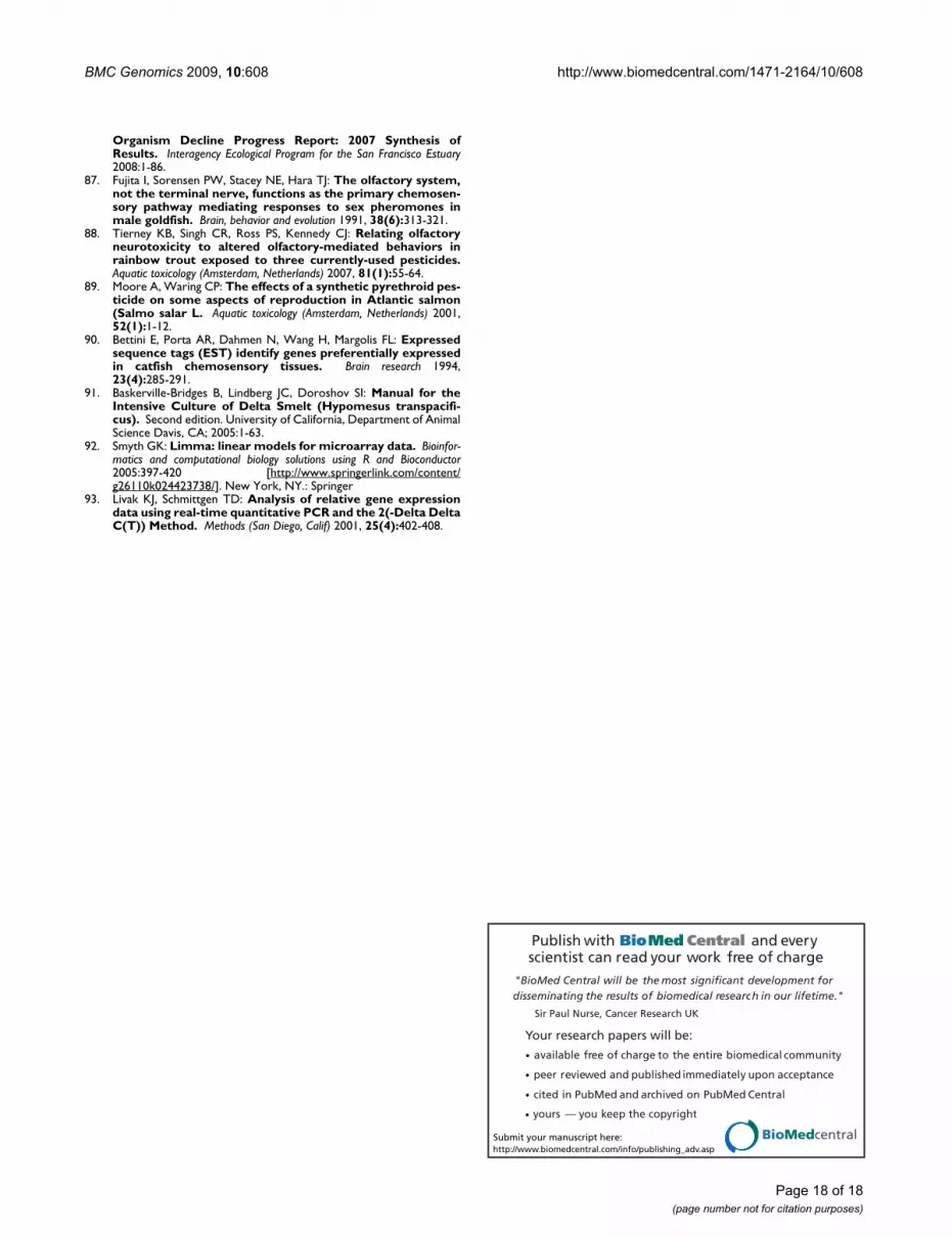
BMC Genomics 2009, 10:608 http://www.biomedcentral.com/1471-2164/10/608
Publish with BioMed Central and every scientist can read your work free of charge
"BioMed Central will be the most significant development for disseminating the results of biomedical research in our lifetime."
Sir Paul Nurse, Cancer Research UK
Your research papers will be:
available free of charge to the entire biomedical community
peer reviewed and published immediately upon acceptance
cited in PubMed and archived on PubMed Central
yours — you keep the copyright
Submit your manuscript here:http://www.biomedcentral.com/info/publishing_adv.asp
BioMedcentral
Organism Decline Progress Report: 2007 Synthesis ofResults. Interagency Ecological Program for the San Francisco Estuary2008:1-86.
87. Fujita I, Sorensen PW, Stacey NE, Hara TJ: The olfactory system,not the terminal nerve, functions as the primary chemosen-sory pathway mediating responses to sex pheromones inmale goldfish. Brain, behavior and evolution 1991, 38(6):313-321.
88. Tierney KB, Singh CR, Ross PS, Kennedy CJ: Relating olfactoryneurotoxicity to altered olfactory-mediated behaviors inrainbow trout exposed to three currently-used pesticides.Aquatic toxicology (Amsterdam, Netherlands) 2007, 81(1):55-64.
89. Moore A, Waring CP: The effects of a synthetic pyrethroid pes-ticide on some aspects of reproduction in Atlantic salmon(Salmo salar L. Aquatic toxicology (Amsterdam, Netherlands) 2001,52(1):1-12.
90. Bettini E, Porta AR, Dahmen N, Wang H, Margolis FL: Expressedsequence tags (EST) identify genes preferentially expressedin catfish chemosensory tissues. Brain research 1994,23(4):285-291.
91. Baskerville-Bridges B, Lindberg JC, Doroshov SI: Manual for theIntensive Culture of Delta Smelt (Hypomesus transpacifi-cus). Second edition. University of California, Department of AnimalScience Davis, CA; 2005:1-63.
92. Smyth GK: Limma: linear models for microarray data. Bioinfor-matics and computational biology solutions using R and Bioconductor2005:397-420 [http://www.springerlink.com/content/g26110k024423738/]. New York, NY.: Springer
93. Livak KJ, Schmittgen TD: Analysis of relative gene expressiondata using real-time quantitative PCR and the 2(-Delta DeltaC(T)) Method. Methods (San Diego, Calif) 2001, 25(4):402-408.
Page 18 of 18(page number not for citation purposes)