leveraging immunotherapy with nanomedicine - archive
TRANSCRIPT
HAL Id: hal-02965461https://hal.archives-ouvertes.fr/hal-02965461
Submitted on 17 May 2021
HAL is a multi-disciplinary open accessarchive for the deposit and dissemination of sci-entific research documents, whether they are pub-lished or not. The documents may come fromteaching and research institutions in France orabroad, or from public or private research centers.
L’archive ouverte pluridisciplinaire HAL, estdestinée au dépôt et à la diffusion de documentsscientifiques de niveau recherche, publiés ou non,émanant des établissements d’enseignement et derecherche français ou étrangers, des laboratoirespublics ou privés.
Leveraging Immunotherapy with NanomedicineVincent Mittelheisser, Mainak Banerjee, Xavier Pivot, Loïc Charbonnière,
Jacky Goetz, Alexandre Detappe
To cite this version:Vincent Mittelheisser, Mainak Banerjee, Xavier Pivot, Loïc Charbonnière, Jacky Goetz, et al.. Lever-aging Immunotherapy with Nanomedicine. Advanced Therapeutics, Wiley, 2020, 3 (12), pp.2000134.�10.1002/adtp.202000134�. �hal-02965461�
1
Published in "Advanced Therapeutics"
Leveraging immunotherapy with nanomedicine
Mittelheisser V.1,2-4, Banerjee M.1, Pivot X.1, Charbonnière L.J.5, Goetz J.G.2-4, Detappe A.1
1. Nanotranslational laboratory, Institut du Cancer Strasbourg, 67000 Strasbourg
2. INSERM UMR_S1109, Strasbourg, F-67000, France
3. Université de Strasbourg, Strasbourg, F-67000, France
4. Fédération de Médecine Translationnelle de Strasbourg (FMTS), Strasbourg, F-
67000, France
5. Institut Pluridisciplinaire Hubert Curien, CNRS UMR-7178, CNRS / Université de
Strasbourg, ECPM, 25 rue Becquerel, 67087 Strasbourg Cedex, France.
To whom correspondence should be addressed:
Alexandre Detappe, PhD
Institut du Cancer Strasbourg
3 rue de la porte de l’Hôpital, 67000 Strasbourg
Email: [email protected]
2
Abstract
Considerable progress has been made in the development and understanding of
immunotherapy, notably with the emergence of novel chimeric antigen receptor T cells (CAR-
T) which changed our perception of personalized therapy. However, cell-based
immunotherapy not only lacks therapeutic efficiency in various solid cancers but also raised
concerns related to important side effects. The convergence of immunotherapy and
nanomedicine is timely as nanoparticles can now be easily conjugated to various antibodies
and peptides enabling outstanding abilities to target specific cell populations in vivo. Here, we
describe the state-of-the art of immuno-nano-therapy that in vivo activates the immune
system, either by acting as vaccines or as tumor microenvironment (TME) activators. Then,
we discuss the development of ex vivo immune-cell surface labelling strategies to
endoctrinate/exploit immune cells as trojan horses, thereby improving the delivery of the
therapeutics in the TME. Such strategy is likely to considerably amplify the efficacy of the
immunotherapy.

3
Introduction
Cancer immunotherapies lead to specific and durable anti-cancer responses,
overcoming traditional cancer treatment limitations. Successful immunotherapy approach
aims to restore an immune response by either stimulating or suppressing the immune
system1,2. Thanks to the recent advancement (progress?) of novel monoclonal antibodies
(mAbs) and immune cell-based therapies, the field of immunotherapy has oriented our
perception of medicine towards a personalized approach.
Hence, different therapeutic modalities have been evaluated clinically to improve
cancer immunotherapy by harnessing the immune system. These include, amongst others,
the use of mAbs blocking pro-angiogenic factors or immune checkpoints inhibitors (e.g.,
NCT01274338, NCT02252042, NCT02125461). Targeting immune-checkpoints proteins with
anti-cytotoxic T lymphocyte-associated antigen-4 (CTLA-4) and anti-programmed cell death
receptor-1 (PD-1) antibodies demonstrated outstanding results when compared to cytotoxic
chemotherapies or to targeted therapies3,4. Although results remain modest in the majority of
solid cancers, immune checkpoints inhibitors have become a standard of care in some
advanced setting (e.g., melanoma, non-small-cell lung carcinoma, basal breast cancer, head
and neck cancer, etc.)5,6. These moderate effects could be partly explained because solid
tumors exhibit low immunogenicity together with primary or acquired mechanisms of
resistance7–10. In addition, the safety profile of immunotherapy remains a challenge to
overcome (e.g., cytokine release syndrome)11,12. Currently, administration of
immunotherapeutic agents is limited by the induction of systemic autoimmunity (e.g.,
myocarditis, colitis, etc.), and grade-3 or grade-4 adverse events such as gastrointestinal,
4
renal or pulmonary toxicities13,14. As such, a paramount need in improving efficacy as well as
safety in cancer immunotherapy remains.
While monoclonal antibodies have demonstrated to be powerful therapeutic
modalities, they are yet limited in efficacy and raised some safety issues (see box 1). Cell-
based therapies have recently emerged (e.g., dendritic cell (DC)-based vaccines, chimeric
antigen receptor (CAR) T and CAR NK cells, etc.) and lead to promising results in several
clinical trials (e.g., NCT03274219, NCT02498912, NCT02408016, NCT02311621, etc.)15–18.
DC-based vaccines use nanoparticles (NPs) to pulse cancer cell lysates, DNA or mRNA into
DCs to prime tumor-specific T cells19,20 while CAR T and CAR NK cells act in a HLA-
independent mechanism without requiring antigen presentation21. These recent approaches
allow to overcome the low immunogenicity of tumors that might hinder the tumor-associated
antigen presentation, and thus the cytotoxic response. However, the response remains
heterogeneous throughout the population19,22. Transition of DCs and engineered immune cells
into an in vivo immunosuppressive environment may alter their viability and functionality and
thus jeopardize their ability to induce an anti-tumor immune response. Cancer cells can also
undergo antigenic modulation (i.e., antigen loss or downregulation) that enables immune
escape22. Moreover, the efficiency of these patient-derived cell-based therapies is strongly
dependent on the patient’s immune system exhaustion level when the cells are harvested23.
Finally, such therapies have limited efficacy in patients with solid tumors22.
Nanomedicine was originally developed to improve the therapeutic index of small
molecules by decreasing their side effects or improve the specificity of the drug delivery into
the tumor24,25. Once injected in the bloodstream, the NPs were designed to passively
accumulate in the tumor by using the leaky vasculature produced by the rapid tumor
neoangiogenesis and impaired lymphatic vessels (also called enhanced permeability and
retention (EPR) effect)26. However, this passive targeting method recently raised some
concerns because of the limited amount of NPs reaching the tumor cells27. For this reason,
improving the targeted delivery of NPs to the tumor remained a challenge.
5
Liposomal NPs were the first generation of drug carriers. Although drugs encapsulation
into liposomes improve their pharmacokinetic and biodistribution in comparison to free drugs,
no marketed liposomal therapeutic agents have yet demonstrated a significant increase in
overall survival versus the standard agent28. Further liposome PEGylation allows a decreased
plasma clearance and results in a longer retention time in the bloodstream29,30. Nevertheless,
improved safety profiles justified PEG-liposomal NPs routine use in tumors where the original
active ingredient has failed to provide results (e.g., liposomal doxorubicin in ovarian cancer:
DOXIL®, CAELYX®). Liposomal deliveries are however seriously limited by their
encapsulation, their stability, and their uncontrolled drug delivery abilities (i.e., dose dumping).
Second NP generation based on micelles and nanoparticles obtained from synthetic
polymers (e.g., poly(d,l‐lactic-co-glycolic acid)-b‐poly(ethylene glycol); PLGA-PEG, organic-
based NPs, dendrimers, etc.) have been developed for several biomedical applications such
as drug delivery, or medical imaging31–34. These long-circulating polymeric NPs demonstrated
an improved therapeutic index in comparison to small molecules, but their use in the clinic
remains limited, notably because of their high liver internalization and remaining toxicity
concerns35.
More recently, theranostic NPs (i.e., carrying both imaging and therapeutic agents)
have emerged. Their conjugation with near-infrared (NIR) fluorescent dyes, magnetic
resonance imaging (MRI) tracers, or positron emission tomography (PET) contrast agents
allow to track the NPs through non-invasive and whole-body imaging and thus facilitate
triggered drug delivery to tumor site36,37. This application is only at its beginning and multiple
clinical trials are currently ongoing38,39. Altogether, the different generations of NPs aim to
improve the therapeutic window of free drugs by either reducing the toxicity, increasing the
total amount of small molecules delivered to the tumor, or enabling the delivery of highly toxic
molecules40–42. In vivo tracking of NPs further provides an additional level of understanding
and can even better improve their therapeutic benefit with personalized therapy43–45. However,
despite the advancements of nanotechnology over the last decade, a retrospective analysis
demonstrated that no more than 0.9 % of the injected dose reach the tumor cells on average27.
6
The EPR effect was questioned as to be the best route of delivery in patients. Indeed, its effect
varies significantly between both patients and tumor types (and vascularization), with some
differences within the same patient or tumor type over tumor progression46,47. These findings
also questioned the use of the appropriate animal model to better assess the NP efficacy48.
Additionally, alternatively to the known EPR effect used for the passive targeting delivery of
NPs, a recent study demonstrated that NPs tend to actually predominantly enter the tumors
through active transcytosis process through endothelial cells rather than using the
neovasculature gaps49. A deepened understanding of this mechanism may lead to its
manipulation to an improvement of NPs accumulation in the tumor. Despite all the questioning
regarding the passive uptake mechanisms, some NPs still confirm their effective tumor
internalization in patients through passive targeting delivery50,51.
To overcome the low yield of NPs reaching the tumor, some routes have been
investigated (Fig. 1A); i) The tumor blood vessel normalization approach consists in using
antiangiogenic agents to transiently normalize the tumor neovascularization to decrease the
tumor hypoxia and to increase the efficacy of conventional therapies by increasing the total
amount of NPs (smaller than 12 nm) or small molecules to be delivered52–54. ii) More recently,
the use of an external trigger combined with NPs has been investigated. This trigger could be
ultrasounds55–57, photothermal therapy (PTT)57–59, photodynamic therapy (PDT)57,60,61, or even
radiation therapy62,63. Most often, the approach consists in targeting the tumor blood vessel
using αvβ3 peptide to disrupt the neovessels with the external trigger in order to improve the
delivery of the drug in a second time (Fig. 1B). iii) An alternative approach consisting in priming
the liver with "blanked" liposomal NPs prone to be highly accumulated in this organ. A second
injection consisting in the therapeutic compound is then administrated and avoid liver
retention, resulting in an increased accumulation of NPs or small molecules to be delivered at
the tumor64,65 (Fig. 1C). Altogether, these novel passive targeting methods seem to alleviate
the NPs tumor uptake and/or improve the drug delivery.
7
In parallel, novel strategies have been studied to deliver the therapeutic agent to the
cancer cells. This is achieved through the design of therapeutic vaccines or the conjugation of
peptides and monoclonal, bispecific, or trispecific antibodies66,67 to activate immune cells (both
circulating and within the tumor microenvironment). While active targeting based on the use
of peptides, or antibodies, conjugated at the surface of the NPs was first employed to improve
the NP tumor uptake, such strategy only improved the delivery specificity of NPs to the tumor
cells without improving the total amount of tumor internalization68–70. As such, utilizing
nanomedicine to specifically target immune cells could hence improve the efficiency of
immunotherapies or, at least, enhance their toxicity.
In this review, we will discuss how nanomedicine could foster immune cell-based
therapy and overcome the usual therapeutic-limiting secondary effects by either i) activating
in vivo the immune system to turn poorly immunogenic tumors into inflamed tumors, or ii) by
modifying ex vivo the immune cells behavior before infusion to enable targeted drug delivery
as well as immune activation in the tumor site.
1. Nanomedicine to improve immunotherapy
Rather than trying to target directly the tumor, and because of the low tumor uptake of
NPs, novel strategies to specifically target circulating immune cells have emerged. The
advantage of this approach is based on the fact that, once activated, the immune cells will
propagate the message to turn the tumor into a ‘hot tumor’ and will recruit more native
unactivated immune cells, resulting in a potent immunotherapeutic response. Immune cells
targeting can be performed by conjugating peptides, antibodies, or ligands at the surface of
the NPs (Fig. 2). It was demonstrated that the biodistribution and pharmacokinetics of
ultrasmall NPs (< 5 nm diameter) are dictated by their ligand decoration, increasing their
circulation time in the body and hence improving tumor cells accumulation71. However,
because of their size, a high drug loading is difficult to achieve, orienting their use mostly for
medical imaging applications33,34,71. At the opposite, the biodistribution and pharmacokinetics
8
properties of large NPs (> 50 nm diameter) are dictated by the NP, resulting in a large
accumulation in the liver and lymph nodes72,73. This preferential accumulation into lymphatic
vessels and lymph nodes is of interest to target immune cells for vaccination approach or for
neoantigen recruitment after radiotherapy73,74.
1.1. Nanoparticles for in vivo immune cells activation.
In vivo activation of immune cells remains the main goal of immunotherapy. While
activating T cells or tumor-associated macrophages (TAMs) has resulted in great therapeutic
responses, the co-stimulation of several immune cells simultaneously is a goal not yet fully
achieved.
Circulating and tumor-infiltrating myeloid cells | Myeloid cells such as TAMs or immature
myeloid-derived suppressor cells (MDSCs) play an important role in initiating the
immunosuppressive environment that strongly suppress the function of cytotoxic immune
cells, negatively impacting the immunotherapy efficacy75,76. NPs-based therapies can either
polarize TAMs towards a more antitumor M1 phenotype or completely neutralize or kill them
(Fig. 2A). A prominent approach to reprogram TAM towards a M1 antitumor phenotype is to
deliver TLR agonists. R848, a TLR7/8 agonist, loaded in β-cyclodextrin (β-CD) NPs potently
drive TAM polarization towards the M1 phenotype, leading to efficient tumor growth control in
multiple mouse models and synergize with anti-PD-1 antibodies77. Another strategy to
reeducate TAM is to interfere with intracellular mRNAs by delivering siRNA, miRNA or mRNA
via mannosylated NPs due to high expression of mannose receptor 1 on TAMs surface78–81.
Glucan-decorated NPs allow in vivo delivery of specific anti-macrophage migration inhibitory
siRNA which results in macrophage polarization towards an antitumor phenotype expressing
pro-inflammatory cytokines, such as TNFα and IL-2, which subsequently enhanced T cell
infiltration and function at the tumor site80. More recently, mannose-modified PLGA-based NPs
allow delivery of mRNA encoding the M1-polarizing interferon regulatory factor 5 transcription
9
factor to TAMs, which reverse the immunosuppressive pro-tumorigenic phenotype of TAMs,
and reprogram them to an anti-tumor one inducing immunity and inhibiting tumor growth in
models of ovarian cancer, melanoma, and glioblastoma81.
Another approach to modulate the tumor immune microenvironment myeloid
compartment is to completely neutralize or kill TAMs by delivering them cytotoxic molecules
through ligand-decorated NPs that target the TAMs overexpressed receptors (e.g., mannose
receptor, folate receptor beta, etc.)82–84. Similarly, granulocyte-colony stimulating factor (G-
CSF) decoration of albumin NPs promote preferential in vivo accumulation in MDSCs in a 4T1
metastatic triple-negative breast mouse model85. This system could provide a cell lineage-
specific delivery of indocyanine green (ICG), an effective photothermal and photosensitizing
agent that can be used for MDSCs ablation in highly immunosuppressed patients86,87.
In parallel, cancer cells upregulate CD47 that ligates with SIRPα present on TAMs
surface and eventually inhibit their phagocytic functions. Blocking CD47 and/or SIRPα with
mAbs showed interesting but limited results notably due to low bioavailability at the tumor
site88–90. The co-delivery of CD47 and SIRPα sequestrated on a multivalent lipid-based
phagocytosis nanoenhancer (LPN) enables simultaneous engagement of TAMs and cancer
cells at significantly lower concentrations of antibodies than the ones used in recent studies
(2 mg/kg vs 5 mg/kg)91 (Fig. 2A). TAMs activation allows intra-tumoral infiltration of effector T
cells and NK cells, leading to significantly enhanced tumor growth inhibition as well as
increased survival in B16F10 melanoma tumor bearing mice.
However, widespread expression of some receptors led to readily uptake of these
targeted drug delivery systems by normal macrophages, mononuclear phagocyte system or
even liver sinusoidal endothelial cells92,93.
Circulating and tumor-infiltrating T cells | In order to override the abovementioned limitations,
NPs-based approaches have been designed to leverage T cells as drug carriers and
demonstrated greater drug levels in the tumor than ones delivered by NPs alone94–97 (Fig. 2A).
10
This could notably be explained by the ability of the T cells to freely circulate in the body, and
hence being used as trojan horse for the NPs to avoid macrophages and direct liver
accumulation after their first pass in blood system98. In one approach, immunoliposomes
decorated with engineered interleukin-2 (IL-2) molecule on an Fc framework or an antibody
F(ab’)2 fragment against a congenic cell surface receptor were used to effectively target in
vivo adoptively transferred T cells (> 95%). Using F(ab’)2 fragments to decorate NPs
demonstrated high target specificity and avidity along with little interactions with Fc receptors
expressed by the mononuclear phagocyte system, which is a major way of NPs clearance99.
Conjugation of immunoliposomes to the surface of exogenous T cells induced repeated waves
of cells expansion, improving their potency96,97. Nevertheless, cell-bound NPs become diluted
over cell proliferation, and adoptive cell transfer (ACT) remains a cumbersome and costly
procedure. In another approach, PLGA NPs were used to encapsulate either SD-208, a
TGFβR1 inhibitor, to restore T cells function, or a TLR7/TLR8 agonist to recruit lymphocytes
to non-inflamed tumors95. PLGA NPs were decorated by anti-PD-1 antibody F(ab’)2 fragments
via thiol-maleimide minute-process, such that the NPs bound approx. 5% of the circulating
and tumor-infiltrating CD8+ PD1+ T cells95,100. Targeted delivery of a TGFβR1 inhibitor led to
extended survival in a mouse model of colorectal cancer compared with free drugs at similar
dosages, while TLR7/TLR8 agonist increased the proportion of tumor-infiltrating CD8+ T cells
and sensitized tumors to anti-PD-1 therapy when compared to NPs lacking the targeting
moiety or equivalent doses of the free drugs95.
With the recent development and clinical demonstration of the efficiency of bispecific
antibodies, or at the preclinical level with tri-specific antibodies, these approaches
demonstrated tremendous results. While these antibodies have already been conjugated to
drugs to form antibody-drug conjugates, they could now be used to ease NPs decoration for
multiple targeting. Interestingly, based on a click-chemistry approach, generating dual-
targeted NPs, or tri-specific NPs is a minute-process101,102, allowing to quickly evaluate novel
targeting conjugations. NPs platform combining immune checkpoint blockade agents along
with co-stimulatory signals have been developed to overcome the autoimmune-mediated
11
dose-limiting toxicities of free mAbs14. These dual targeting systems redirect effector T cells
to recognize cancer cells while simultaneously blocking checkpoint inhibitors103–106. Various
NPs types (e.g., liposome, PEG-PLGA, etc.) have been decorated with different T cells
agonists (e.g., anti-4-1BB mAb, anti-OX40 mAb, etc.) and immune checkpoint blockade
agents (e.g., anti-PD-L1 mAb, anti-PD-1 mAb, etc.). These approaches resulted in a marketed
therapeutic activity as demonstrated by tumor regression, tumor-specific T cells expansion,
and immune responses in B16 melanoma and 4T1 breast cancer models103–106.
Targeting NPs-based immunotherapies to blood circulating and tumor-infiltrating
immune cells rather than tumor cells directly allows efficient in vivo activation of immune cells
with limited doses administration, and thus lower immune-mediated adverse events
occurrence. These NPs-based immunotherapeutic approaches can also be used to target
immune cells in the lymph node as cancer vaccination tool that elicits a potent antitumor
immune response.
1.2. Vaccines with nanomedicine.
The anti-tumor vaccines directed against tumor‐associated antigens (TAAs) or tumor
specific antigens were amongst the first immunotherapeutic agents developed since the late
1980s107,108. Cancer cell lysates, DNA or mRNA are pulsed into dendritic cells (e.g., FDA-
approved Sipuleucel-T, PROVENGE® for prostate cancer) to activate T cells by presenting
them the tumor antigen, leading to a cytotoxic T cells (CTL) response109,110. Because DCs can
be inhibited by the cancer cells or the tumor microenvironment, and because the anti-tumor
vaccines are difficult to standardize due to their requirement of highly immunogenic antigen
as well as potent adjuvant, this strategy shows decreased potency to eradicate tumors108,111.
Newly developed nanoengineered vaccines demonstrated better efficacy in cancer treatment
over former anti-tumor vaccines which come with major limitations such as poor
immunogenicity and off-target side effects112–114.
12
The size of the nanomaterial has a detrimental effect in therapeutic outcome in cancer
vaccination. NPs sizes ranging from 10 to 100 nm in diameter and negatively charged (-3 to -
15 mV) tends to preferentially accumulate into lymphatic vessels and lymph nodes73,74. The
lymph nodes-targeted NPs behave as artificial antigen presenting cells (APCs) to directly
stimulate CD8+ T cells. To achieve their activation, T cells require a T cell receptor (TCR)-Ag
recognition followed by a co-stimulatory signal procured by the interaction between the CD28
from the T cells and its receptors present at the DC surface (i.e., CD80 or CD86). A third signal
mediated through IL-2 can enhance T cells stimulation but is not required for their activation115.
Artificial APCs are coated with TAA and anti-CD28 antibodies recapitulate both mandatory
signals for T cells activations116–119 (Fig. 2B). The other benefit for nanomaterials-based
vaccines rely on their ability to co-deliver the TAAs and adjuvants simultaneously, at a
controlled ratio, and at the same location (e.g., CpG, Poly(I:C)), improving the ability of APCs
to present up taken antigens with MHC-I molecules to CD8+ T cells and enhancing CTL
responses120–122 (Fig. 2B). Although nanoengineered vaccines lead to a potent activation of
the immune system, delivery through lymphatic draining depends on NPs composition,
morphology, and surface chemistry. Alternatively, other NP designs have been sought to
target specifically the spleen to activate B lymphocytes123 or to recruit, through the use of
hydrogels, for example, dendritic cells124. These approaches enabled a specific targeting of
immune cells without decorating the NPs with conventional peptides or antibodies.
1.3. Recruitment of neoantigens post-radiotherapy.
Radiation oncology combined with immunotherapy has gained a substantial interest due
to its inherent ability to generate an abscopal effect defined as transforming "cold tumor" into
"hot tumor" as defined by the total amount of tumor antigen, APC deficit, absence of T cells
and impaired trafficking to the tumor mass post-radiation13,125. By irradiating the tumor bed,
which includes the tumor microenvironment, the radiation beams generate additional immune
response inside the tumor microenvironment. It was demonstrated that the irradiation of the
tumor leads to the liberation of TAAs that are endocytosed by APCs and then presented to
13
CD8+ T cells126. This process increases the diversity of the TCR repertoire of intra-tumoral T
cells and leads to a tumor-specific immune response directed against primary tumor and
metastatic tumor sites127,128. Unfortunately, the presence of immunosuppressive cells (for
example, MDSCs and Treg cells) producing immunosuppressive cytokines (e.g., IL-10 and
TGFβ) in the tumor microenvironment hamper the development of robust and sustained
abscopal responses even with combination approaches7–9. This combination therapy led only
to a limited abscopal effect due to T cells exhaustion mediated by the upregulation of PD-L1
on tumor cells. As such, multiple strategies have been attempted to increase this tumor
immunogenicity to boost the abscopal effect. Amongst them, targeted NPs have been used to
deliver potent immunotherapeutic compounds to the tumor microenvironment and tumor
draining lymph nodes in order to reverse immunosuppression, as illustrated by the use of CpG
oligodeoxynucleotides conjugated to a polymer NPs129. This compound was developed to
activate DCs in the lymph node, resulting in an increased activation of CD8+ T cells/Treg ratio129.
Activating DCs or tumor-infiltrating immune cells by immune checkpoint inhibitors or TLR
agonists potentiate the abscopal effect, leading to delayed tumor growth. However, little is
known about potential adverse effects emerging from combining radiotherapy with immune-
checkpoint inhibition130.
Alternatively, rather than activating the DCs by using targeted NPs, boosting the
abscopal effect through the use of radiation therapy enhancer NPs made of high-atomic
number atoms (gold, gadolinium, or hafnium NPs, among others) was also evaluated. The
radiosensitization properties of these inorganic NPs is attributed to an increase of Auger
electrons production via the photoelectric effect leading to an increased amount of reactive
oxygen species in the tumor bed131–138. Altogether, physical and biological boost effects
induced by the presence of metallic inorganic NPs during the radiation treatment increased
local DNA damage139. The local boost of radiation dose deposition is hypothesized to enable
a higher release of TAAs upon tumor cells death that potentiate the abscopal response
through an increased tumor immunogenicity140,141. Toward these findings, the development of
antigen-capturing nanoparticles (AC-NPs) to exploit the release of TAAs upon radiotherapy in
14
order to boost the abscopal effect was also performed142 (Fig. 2C). By formulating polymeric
PLGA NPs with diverse surface modifications (decoration with 1,2-dioleoyloxy-3-
(trimethylammonium)propane, DOTAP; maleimide, NH2; mPEG; or unmodified PLGA) to
determine the effects of NP surface chemistry on antigen capture efficiency, it was
demonstrated that NPs allow to capture efficiently tumor neoantigens post-radiation143.
Interestingly, these AC-NPs also captured a number of damage-associated molecular pattern
proteins (DAMPs) that potentiated the immune response by being efficiently internalized by
APCs and trafficked to lymphoid tissues. In a B16F10 melanoma bilateral tumor model, they
showed that intra-tumoral injection of NPs in one tumor significantly improved the efficacy of
anti-PD-1 antibodies and enhanced abscopal responses, generating a 20% cure rate
compared to 0% without any NPs. The mechanistic studies revealed that AC-NPs induced an
expansion of CD8+ cytotoxic T cells and increased both CD4+ T cells/Treg and CD8+ T cells/Treg
ratios.
Although harnessing the immune system to enhance the abscopal effect demonstrated
promising results, these approaches are still at their stammering. There is a great impetus for
more cross-disciplinary research combining radiotherapy and immunotherapy with NPs in
boosting the abscopal effect, thereby improving the treatment of both local and metastatic
disease.
2. Immune cells-based therapy with ex vivo nanomedicine labeling
Alongside with immune checkpoints blockade, adoptively transferred autologous T cells
have shown tremendous therapeutics effects. Patients-isolated T or NK cells can be
genetically engineered to express a CAR that recognize tumor cells in an HLA-independent
mechanism without requiring antigen presentation21. These CAR T cells, CAR NK cells, or
more recently CAR macrophages, showed impressive results in treating acute lymphoblastic
leukemias, refractory large B cell lymphomas or multiple myelomas15–17,144. However, CAR
cells remain costly, time consuming and only allow the addition of a single targeting moiety at
the time145. Moreover, the overall response to these therapies remain heterogeneous and CAR
15
immune cells have shown only moderate successes in treating solid cancers22. Once infused
into patients, adoptively transferred cells migrate to the tumor sites and require a sustained
supply of oxygen, nutrients and cytokines to support their viability, functions and proliferation.
Moreover, cells have to overcome the immunosuppressive tumor microenvironment8,9. To
promote their anti-tumoral actions, adjuvant cytokines and immune-checkpoint blockade
agents can be administered146–148. However, systemic administration of such drugs requires
repeated injection to maintain therapeutic levels resulting in dose-limiting toxicities149,150. In
order to overcome the aforementioned limitations and enhance ACT potency, cell surface
bioengineering strategies have been developed151. Immune cells are an attractive option for
cell surface bioengineering because of their natural abilities to circulate in the bloodstream
and pass challenging biological barriers before to accumulate into the tumor
microenvironment.
2.1. Cell surface conjugation routes
Cell surface conjugation strategies have to satisfy the following biocompatibility
fundamental principles: i) any cell surface modifications should not have detrimental effects
either on cell viability or cellular functions. ii) Bioengineering should further minimize
alterations in membrane fluidity or bending elasticity that are critical for cell adhesion,
migration and signaling. iii) Moreover, surface-engineered immune cells are exposed to in vivo
shear stress and hemodynamic forces that can dissociate the NPs from the cell surface. Thus,
the introduced modifications have to be compatible with the in vivo complex mechanical and
biochemical interactions. iv) Finally, they should also not lead to the apparition of severe
adverse effects such as thrombus formation after infusion152.
As such, to design novel and smarter immune cells, diverse bioengineering
methodologies have been investigated. They can be subdivided in non-covalent physical
biointeraction, and covalent chemical conjugation (Fig. 3).
16
Non-covalent non-specific biointeractions | Non-covalent conjugation of NPs can be achieved
using non-specific (e.g., hydrophobic, electrostatic, etc.) interactions or specific ligand-
receptors ones151 (Fig. 3A). Since the plasma membrane is composed of a hydrophobic lipid
bilayer, NPs conjugated with hydrophobic moieties such as glycoinositol phospholipids (GPIs)
can spontaneously be anchored into the membrane153,154. Besides being hydrophobic, the
plasma membrane is also negatively charged, as a result of phosphate groups of
phospholipids, carboxylate groups on proteins and sialic acid termination of glycoproteins155.
Thus, NPs harboring many cationic sites can bind to cell surfaces via electrostatic interactions.
Nevertheless, non-covalent non-specific conjugations have intrinsic tremendous drawbacks.
Indeed, GPI-anchored NPs recapitulate natural membrane-associated molecules turnover
rates and are thus rapidly internalized (t1/2 between 3.8 and 20 hours)156,157. Moreover, the
electrostatic interactions between the negatively charged plasma membrane and the NPs
positive surface can trigger local membrane depolarization and lead to cellular uptake158. As
such, non-specific non-covalent interactions are a rapid and easy route of conjugation but yet
suffer from in vivo shear stress exposure in the systemic circulation or endothelial diapedesis.
More specific and robust interactions have thus been designed.
Non-covalent specific biointeractions | Specific ligand-receptor interactions can be highly
specific but yet pose the risk of undesired cellular responses. Cell surface expressed
transmembrane receptors that can be targeted to conjugate NPs functionalized with their
respective ligands159,160. These ligand-receptors interactions are transient and dictated by their
intrinsic binding and dissociation kinetics (Kd) which hinder stable coupling of NPs on cell
surface161,162 (Fig. 3A).
Biotin-avidin interactions have also been extensively studied as specific, non-covalent
cell surface interactions163,164. This approach requires the introduction of a biotin group on cell
surfaces. Biotin moieties can be covalently attached whether by amide bond formation with
lysine residues165 or aldehyde groups formation through mild oxidation of cell surface
monosaccharides followed by functionalization with a hydrazide-biotin crosslinker166.
17
However, streptavidin as an immunogenic xenoprotein can elicit neutralizing antibodies that
lead to opsonization and phagocytic clearance of the engineered cell167.
Sialylated carbohydrate ligands such as sialyl-lewisX are naturally present at the
surface of leukocytes and can provide other opportunities for non-covalent, specific
biointeractions. As an example, E-selectin/TRAIL-coated or anti-NK1.1/TRAIL-coated
liposomes were designed to interact with the sialylated carbohydrates present at the surface
of the leukocytes and with the TRAIL receptors expressed by circulating tumor cells
(CTCs)168,169. This approach allowed CTCs elimination and prevention of lymph nodes
metastasis formation in patient-derived xenograft model.
Covalent chemical conjugation | Covalent approaches were performed by (i) targeting native
functional groups (e.g., thiols, amines) present on the cell surface, by (ii) chemical generation
of reactive groups (e.g., aldehydes) or by (iii) using metabolic strategies to introduce non-
natural functional groups (Fig. 3B).
Native functional groups | The naturally expressed thiol groups help to protect cells
against oxygen radicals170. Maleimide-functionalized NPs (e.g., liposomes, multilamellar
liposomes, PLGA, etc.) have efficiently been conjugated to immune cells surface171–173 and
their release can be triggered by the reduction potential of the tumor microenvironment (e.g.,
glutathione increase, etc.)174. In addition to thiol groups, aldehydes have also been
successfully targeted to covalently tether NPs on cell surfaces. However, aldehydes have to
be generated by mild oxidation of primary alcohols contained in carbohydrates natively present
on the cell surface175.
Metabolic strategies | Metabolic glycoengineering strategies use bioorthogonal
chemistry to modify natural oligosaccharides present on live cells. Alkyne- or azide-modified
monosaccharides such as sialic acid or N-acetylmannosamine are metabolically incorporated
into living cells and processed through natural biosynthetic pathways to be eventually
18
incorporated into the membrane as glycoengineered oligosaccharides that will react with
azides or alkynes groups176–178 (Fig. 3B). Up to now, glycoengineered glycan are used for
broad range of diagnostic, therapeutic or theranostic approaches177,179. However, an emerging
strategy using a dibenzocyclooctyne (DIBO) alkyne-decorated PAMAM dendrimer to target N-
azidoacetylmannosamine-expressing macrophages demonstrated the feasibility of creating
hybrid cell-NPs by bio-orthogonal chemistry to develop personalized immunotherapy180.
Although this approach exhibited no cell viability, intracellular signaling pathways, and motility
altering, its toxicity and efficacy have to be evaluated in vivo. In parallel, the recent
development of bio-orthogonal cleavage chemistry open new perspectives for NPs release
and specific drug delivery181.
Receptor genetical engineering | The aforementioned approaches result in stable
interactions between the NPs and the cell surface but provide only moderate control over the
resulting cell surface engineering. Thus, stable and controlled cell-NPs interactions have been
seeked. Immune cells naturally express receptors at their surface which can be genetically
engineered to provide an exclusive orthogonal ligand-receptor interaction182 (Fig. 3B). NK
cells, T cells, and some macrophages present the NKG2D receptors on their surfaces that
recognize the MHC class I polypeptide-related sequences (MIC) ligands family overexpressed
on cells stressed by viral infection or cancer transformation. Leveraging the natural α1-α2
binding domain of these ligands through protein engineering allows to develop an exclusive
orthogonal ligand-receptor interaction to generate the components of a universal CAR T cell
platform182. The engineered extracellular domain of the inert NKG2D receptors (iNKG2D) is
fused to the intracellular 4-1BB and CD3ζ co-signaling domains to generate the CAR while
the mutant ligand domains that specifically bind to the iNKG2D are fused to intact human
antibodies. Up to now, efficacy of rituximab-based convertible CAR T cells has been
investigated in NSG mice bearing Burkitt lymphoma and demonstrated dose-dependent
control of tumor mass. Similarly, SpyCatcher technology can be used to develop a universal
19
immune receptor183. SpyCatcher fused to the intracellular 4-1BB and CD3 ζ co-signaling
domains is used as an immune receptor that is armed with an antibody fused with SpyTag
moieties. These new approaches can be exploited for cell surface modification. NPs can be
decorated by the orthogonal ligands mutated α1-α2 domain or SpyTag moieties to be
conjugated on iNKG2D- or SpyCatcher-expressing immune cells, respectively.
Cell surface retention | As previously mentioned, cell membrane components (e.g.,
lipids and proteins) are continuously internalized, degraded and replaced184. Therefore, means
to prolong cell surface retention of NPs have been studied172,173,185–190. In a thiol-maleimide
approach, NPs-binding proteins have been identified by mass spectrometry172. The leukocyte
common antigen CD45 is predominantly bound by maleimide-functionalized NPs. Other
surface proteins such as LFA-1, CD2 or CD97 have also been identified. However, depending
on the cell type, the direct targeting of a surface receptor can trigger undesired cellular
responses. Consequently, other ways to increase cell surface retention have been
investigated185–190. Depending on their compositions and the type of interaction with the cell,
NPs can be internalized by different mechanisms (e.g., clathrin- and/or caveolae-mediated
endocytosis)191. Converging in vitro and in vivo evidences demonstrated that the antimalarial
agent chloroquine effectively inhibits NPs clathrin-mediated endocytosis by depleting the
phosphatidylinositol binding clathrin assembly protein185,186. Similar results were found by
studying the antipsychotic drug chlorpromazine187–189. Nonetheless, these results have not
been validated in in vivo models. More recent trial demonstrated that the antiemetic and
antipsychotic drug prochlorperazine (PCZ) can be repurposed to reversibly inhibit dynamin-
mediated endocytosis of membrane proteins targeted by therapeutic mAbs190. In mouse
models (squamous cell carcinoma, colon carcinoma and renal carcinoma) and in a pilot clinical
study on head and neck squamous cell carcinoma, treatment with PCZ led to increased
clustering of receptors on the cell surface, enhancing tumor cell-NK cells contacts, and finally
driving to improve antibody-dependent cell-mediated cytotoxicity in response to approved
20
IgG1 mAbs, such as cetuximab (anti-EGFR mAb) and avelumab (anti-PD-L1 mAb)190.
However, PCZ did not affect targets that are not internalized by dynamin. Thus, this
perspective might only be of interest in NPs anchoring targets with dynamin-mediated
endocytosis. Although the abovementioned molecules showed promising results in vitro and
in pilot clinical study, those molecules might have severe adverse effects in vivo (e.g.,
chloroquine: cardiac toxicities; chlorpromazine: agranulocytosis and prochlorperazine:
extrapyramidal symptoms) and must thus be carefully handled.
2.2. Application of T cells backpacking
Many cell therapy protocols require adjuvant drugs to maintain transferred cells
functions, phenotype and lifespan146–148. However, systemic administration of such drugs is
challenging due to their pleiotropic activities, leading to dose-limiting toxicities149,150. NPs-
targeting strategies with specific cell-targeting ligands, such as antibodies or small molecules
have been investigated to deliver these molecules to the tumor site192,193. Nonetheless, it has
been shown that targeting ligands do not modify the overall NPs biodistribution but rather
enable more efficient reaching of the tumor site by targeted NPs68–70. Immune cells
backpacking strategy with NPs-containing adjuvant cytokines and immune-checkpoint
blockade agents have been explored to focus administration of such drugs on tumor site94,171–
173.
Backpacking T cells with up to 100 (± 20) liposomes (300 nm in diameter) did not affect
key cellular functions (e.g., activation, transendothelial migration, tumor homing properties and
antitumor functions) and allowed a 176-fold increase in NP accumulation into the EL4 tumor
site when compared to free NP171. Loading interleukins 15 and 21 (IL-15 and IL-21,
respectively) into the surface of multilamellar liposomes supported T cells antitumor function
in an autocrine-like manner through a continuous release of bioactive interleukins over 7 days,
and hence enhanced the therapeutic efficacy by efficiently preventing tumor growth up to 30
days after treatment. Moreover, the on-site drug action allowed the use of interleukins doses
that are inefficient when systemically administered. Multilamellar liposomes loaded with SN-
21
38, the active metabolite of irinotecan, a potent topoisomerase I poison have also been used
to tether T cells94. In a Burkitt lymphoma mice model, tumor cells disseminated in lymph node
were not sensitive to treatment with free SN-38 or liposomal formulation because of the drug
poor pharmacokinetics due to a short half-life (t1/2 = 7 mins) and a rapid hepatic clearance194
along with the lack of leaky neovasculature into the lymph node and thus, a lack of EPR effect.
This lymph node homing property were exploited to deliver SN-38-loaded NPs on T cells in
lymphoid organs enriched in lymphoma cells. SN-38-loaded NPs carried by T cells
accumulated in lymph nodes 20h after infusion and SN-38 concentrations were 63-fold greater
than free NPs and were maintained for 4 days. SN-38 released in a paracrine-like manner
leading to a 60-fold reduction in tumor burden and an extended survival of mice up to 12 days
at relatively low doses (7 mg/kg) when compared to the free drug. Although these approaches
allowed to increase the treatment potency, the payloads (e.g., IL-15, IL-21, SN-38, etc.) can
passively leak out of the multilamellar liposomes and continually stimulate the T cells until their
activation-induced depletion leading to a reduction in the effective dose of T cells trafficking to
the tumor site.
To override this issue, a second generation of backpacking NPs have been
designed173. Nanogel (NG) backpacks have been engineered to transport IL-15 to tumors
together with adoptively transferred T cells. ALT-803, an IL-15 superagonist molecule were
aggregated into a NG with a linker including reduction-sensitive disulfide bonds that senses
the reducing potential of the environment195. To facilitate and prolong their cell surface
retention, small quantities of anti-CD45 mAbs and poly(ethylene glycol)-b- poly(L-lysine)
(PEG-PLL) were incorporated onto the NG surface. By engaging their cognate antigen in the
tumor, activated T cells induce thiol groups emission at the cell surface, increasing their cell
surface reduction potential that detaches the NGs. The release of IL-15 superagonist through
this approach resulted in a 16-fold expansion of T cells in tumors, as compared with free IL-
15. This approach allowed an 8-fold higher dose of cytokine to be administered without toxicity,
widening its therapeutic window and enabling a significant increased tumor clearance by ACT
22
T cells and CAR T cells. However, stimulation of cell division will eventually lead to dilution of
the backpacked NPs.
Cell-attached cargo actively transmigrate the endothelial barrier and accumulate in
tumor sites, thereby enhancing their actions on transferred cells anti-tumor abilities and limiting
systemic adverse effects. Given the plethora of available NPs tailored to deliver small
molecules drugs, proteins, siRNA or magnetic imaging agents, T cells backpacking
approaches profoundly open new perspectives for adoptive cell therapies and drug delivery
that can be extended far beyond the small molecule drugs and recombinant proteins delivery
aforementioned.
Conclusion
Nanomedicine is used in several ways to improve immunotherapy. Historically, NPs have been
used to improve in vivo activation of immune cells by enhancing vaccination or radiotherapy
efficacies, leading to an improved antitumor response. More recently, a deep focus on the
immune cell surface bioengineering to tackle the intrinsic drawbacks of ACT of autologous
immune cells or CAR cells is being investigated. These ex vivo immune cells labeling by NPs
approaches will also allow NPs to either target the tumor microenvironment, or to use it as a
trojan horse to enhance the tumor drug delivery.
23
References
1. Mellman, I., Coukos, G. & Dranoff, G. Cancer immunotherapy comes of age. Nature
(2011) doi:10.1038/nature10673.
2. Khalil, D. N., Smith, E. L., Brentjens, R. J. & Wolchok, J. D. The future of cancer
treatment: Immunomodulation, CARs and combination immunotherapy. Nature
Reviews Clinical Oncology (2016) doi:10.1038/nrclinonc.2016.25.
3. Pardoll, D. M. The blockade of immune checkpoints in cancer immunotherapy. Nature
Reviews Cancer (2012) doi:10.1038/nrc3239.
4. Topalian, S. L., Drake, C. G. & Pardoll, D. M. Immune checkpoint blockade: A common
denominator approach to cancer therapy. Cancer Cell (2015)
doi:10.1016/j.ccell.2015.03.001.
5. Yarchoan, M., Hopkins, A. & Jaffee, E. M. Tumor mutational burden and response rate
to PD-1 inhibition. New England Journal of Medicine (2017)
doi:10.1056/NEJMc1713444.
6. Syn, N. L., Teng, M. W. L., Mok, T. S. K. & Soo, R. A. De-novo and acquired resistance
to immune checkpoint targeting. The Lancet Oncology (2017) doi:10.1016/S1470-
2045(17)30607-1.
7. Garner, H. & de Visser, K. E. Immune crosstalk in cancer progression and metastatic
spread: a complex conversation. Nature Reviews Immunology (2020)
doi:10.1038/s41577-019-0271-z.
8. Binnewies, M. et al. Understanding the tumor immune microenvironment (TIME) for
24
effective therapy. Nat. Med. (2018) doi:10.1038/s41591-018-0014-x.
9. Nagarsheth, N., Wicha, M. S. & Zou, W. Chemokines in the cancer microenvironment
and their relevance in cancer immunotherapy. Nature Reviews Immunology (2017)
doi:10.1038/nri.2017.49.
10. Sharma, P., Hu-Lieskovan, S., Wargo, J. A. & Ribas, A. Primary, Adaptive, and
Acquired Resistance to Cancer Immunotherapy. Cell (2017)
doi:10.1016/j.cell.2017.01.017.
11. Santomasso, B., Bachier, C., Westin, J., Rezvani, K. & Shpall, E. J. The Other Side of
CAR T-Cell Therapy: Cytokine Release Syndrome, Neurologic Toxicity, and Financial
Burden. Am. Soc. Clin. Oncol. Educ. B. (2019) doi:10.1200/edbk_238691.
12. Brudno, J. N. & Kochenderfer, J. N. Chimeric antigen receptor T-cell therapies for
lymphoma. Nature Reviews Clinical Oncology (2018) doi:10.1038/nrclinonc.2017.128.
13. Postow, M. A. et al. Immunologic correlates of the abscopal effect in a patient with
melanoma. N. Engl. J. Med. (2012) doi:10.1056/NEJMoa1112824.
14. Martins, F. et al. Adverse effects of immune-checkpoint inhibitors: epidemiology,
management and surveillance. Nature Reviews Clinical Oncology (2019)
doi:10.1038/s41571-019-0218-0.
15. Maude, S. L. et al. Chimeric antigen receptor T cells for sustained remissions in
leukemia. N. Engl. J. Med. (2014) doi:10.1056/NEJMoa1407222.
16. Raje, N. et al. Anti-BCMA CAR T-cell therapy bb2121 in relapsed or refractory multiple
myeloma. N. Engl. J. Med. (2019) doi:10.1056/NEJMoa1817226.
17. Liu, E. et al. Use of CAR-transduced natural killer cells in CD19-positive lymphoid
tumors. N. Engl. J. Med. (2020) doi:10.1056/NEJMoa1910607.
18. Brown, C. E. et al. Regression of glioblastoma after chimeric antigen receptor T-cell
therapy. N. Engl. J. Med. (2016) doi:10.1056/NEJMoa1610497.
19. Le Gall, C. M., Weiden, J., Eggermont, L. J. & Figdor, C. G. Dendritic cells in cancer
immunotherapy. Nature Materials (2018) doi:10.1038/s41563-018-0093-6.
20. Anguille, S., Smits, E. L., Lion, E., Van Tendeloo, V. F. & Berneman, Z. N. Clinical use
25
of dendritic cells for cancer therapy. The Lancet Oncology (2014) doi:10.1016/S1470-
2045(13)70585-0.
21. Sadelain, M., Rivière, I. & Riddell, S. Therapeutic T cell engineering. Nature (2017)
doi:10.1038/nature22395.
22. Shah, N. N. & Fry, T. J. Mechanisms of resistance to CAR T cell therapy. Nature
Reviews Clinical Oncology (2019) doi:10.1038/s41571-019-0184-6.
23. Pagès, F. et al. International validation of the consensus Immunoscore for the
classification of colon cancer: a prognostic and accuracy study. Lancet (2018)
doi:10.1016/S0140-6736(18)30789-X.
24. Kim, B. Y. S., Rutka, J. T. & Chan, W. C. W. Current concepts: Nanomedicine. New
England Journal of Medicine (2010) doi:10.1056/NEJMra0912273.
25. Jain, R. K. & Stylianopoulos, T. Delivering nanomedicine to solid tumors. Nature
Reviews Clinical Oncology (2010) doi:10.1038/nrclinonc.2010.139.
26. Matsumura, Y. & Maeda, H. A New Concept for Macromolecular Therapeutics in
Cancer Chemotherapy: Mechanism of Tumoritropic Accumulation of Proteins and the
Antitumor Agent Smancs. Cancer Res. (1986).
27. Wilhelm, S. et al. Analysis of nanoparticle delivery to tumours. Nature Reviews
Materials (2016) doi:10.1038/natrevmats.2016.14.
28. Petersen, G. H., Alzghari, S. K., Chee, W., Sankari, S. S. & La-Beck, N. M. Meta-
analysis of clinical and preclinical studies comparing the anticancer efficacy of
liposomal versus conventional non-liposomal doxorubicin. J. Control. Release (2016)
doi:10.1016/j.jconrel.2016.04.028.
29. Yoo, J.-W., Chambers, E. & Mitragotri, S. Factors that Control the Circulation Time of
Nanoparticles in Blood: Challenges, Solutions and Future Prospects. Curr. Pharm. Des.
(2010) doi:10.2174/138161210791920496.
30. Allen, T. M. & Cullis, P. R. Liposomal drug delivery systems: From concept to clinical
applications. Advanced Drug Delivery Reviews (2013) doi:10.1016/j.addr.2012.09.037.
31. Gref, R. et al. Biodegradable long-circulating polymeric nanospheres. Science (80-. ).
26
(1994) doi:10.1126/science.8128245.
32. Zhang, K. et al. PEG-PLGA copolymers: Their structure and structure-influenced drug
delivery applications. Journal of Controlled Release (2014)
doi:10.1016/j.jconrel.2014.03.026.
33. Tietjen, G. T. et al. Nanoparticle targeting to the endothelium during normothermic
machine perfusion of human kidneys. Sci. Transl. Med. (2017)
doi:10.1126/scitranslmed.aam6764.
34. Chen, F. et al. Ultrasmall targeted nanoparticles with engineered antibody fragments
for imaging detection of HER2-overexpressing breast cancer. Nat. Commun. (2018)
doi:10.1038/s41467-018-06271-5.
35. Turecek, P. L., Bossard, M. J., Schoetens, F. & Ivens, I. A. PEGylation of
Biopharmaceuticals: A Review of Chemistry and Nonclinical Safety Information of
Approved Drugs. Journal of Pharmaceutical Sciences (2016)
doi:10.1016/j.xphs.2015.11.015.
36. Sun, T. et al. Engineered nanoparticles for drug delivery in cancer therapy. Angewandte
Chemie - International Edition (2014) doi:10.1002/anie.201403036.
37. Detappe, A., Bustoros, M., Mouhieddine, T. H. & Ghoroghchian, P. P. Advancements
in Nanomedicine for Multiple Myeloma. Trends in Molecular Medicine (2018)
doi:10.1016/j.molmed.2018.04.005.
38. Singh, D., Dilnawaz, F. & Sahoo, S. K. Challenges of moving theranostic nanomedicine
into the clinic. Nanomedicine (2020) doi:10.2217/nnm-2019-0401.
39. Lee, H. et al. 64Cu-MM-302 positron emission tomography quantifies variability of
enhanced permeability and retention of nanoparticles in relation to treatment response
in patients with metastatic breast cancer. Clin. Cancer Res. (2017) doi:10.1158/1078-
0432.CCR-16-3193.
40. Qi, R. et al. Nanoparticle conjugates of a highly potent toxin enhance safety and
circumvent platinum resistance in ovarian cancer. Nat. Commun. (2017)
doi:10.1038/s41467-017-02390-7.
27
41. Lancet, J. E. et al. Cpx-351 (cytarabine and daunorubicin) liposome for injection versus
conventional cytarabine plus daunorubicin in older patients with newly diagnosed
secondary acute myeloid leukemia. in Journal of Clinical Oncology (2018).
doi:10.1200/JCO.2017.77.6112.
42. Gradishar, W. J. et al. Phase III trial of nanoparticle albumin-bound paclitaxel compared
with polyethylated castor oil-based paclitaxel in women with breast cancer. J. Clin.
Oncol. (2005) doi:10.1200/JCO.2005.04.937.
43. Lammers, T., Rizzo, L. Y., Storm, G. & Kiessling, F. Personalized nanomedicine.
Clinical Cancer Research (2012) doi:10.1158/1078-0432.CCR-12-1414.
44. Theek, B., Rizzo, L. Y., Ehling, J., Kiessling, F. & Lammers, T. The theranostic path to
personalized nanomedicine. Clinical and Translational Imaging (2014)
doi:10.1007/s40336-014-0051-5.
45. Lammers, T., Aime, S., Hennink, W. E., Storm, G. & Kiessling, F. Theranostic
nanomedicine. Acc. Chem. Res. (2011) doi:10.1021/ar200019c.
46. Tanaka, N. et al. Whole-tissue biopsy phenotyping of three-dimensional tumours
reveals patterns of cancer heterogeneity. Nat. Biomed. Eng. (2017)
doi:10.1038/s41551-017-0139-0.
47. Harrington, K. J. et al. Effective targeting of solid tumors in patients with locally
advanced cancers by radiolabeled pegylated liposomes. Clin. Cancer Res. (2001).
48. Tao, Z. et al. Differences in Nanoparticle Uptake in Transplanted and Autochthonous
Models of Pancreatic Cancer. Nano Lett. (2018) doi:10.1021/acs.nanolett.7b04043.
49. Sindhwani, S. et al. The entry of nanoparticles into solid tumours. Nat. Mater. (2020)
doi:10.1038/s41563-019-0566-2.
50. Ragelle, H., Danhier, F., Préat, V., Langer, R. & Anderson, D. G. Nanoparticle-based
drug delivery systems: a commercial and regulatory outlook as the field matures. Expert
Opinion on Drug Delivery (2017) doi:10.1080/17425247.2016.1244187.
51. Bort, G. et al. EPR-mediated tumor targeting using ultrasmall-hybrid nanoparticles:
From animal to human with theranostic AGuIX nanoparticles. Theranostics (2020)
28
doi:10.7150/thno.37543.
52. Jain, R. K. Normalization of tumor vasculature: An emerging concept in antiangiogenic
therapy. Science (2005) doi:10.1126/science.1104819.
53. Martin, J. D., Cabral, H., Stylianopoulos, T. & Jain, R. K. Improving cancer
immunotherapy using nanomedicines: progress, opportunities and challenges. Nature
Reviews Clinical Oncology (2020) doi:10.1038/s41571-019-0308-z.
54. Chauhan, V. P. et al. Normalization of tumour blood vessels improves the delivery of
nanomedicines in a size-dependent manner. Nat. Nanotechnol. (2012)
doi:10.1038/nnano.2012.45.
55. Couture, O., Foley, J., Kassell, N. F., Larrat, B. & Aubry, J. F. Review of ultrasound
mediated drug delivery for cancer treatment: Updates from pre-clinical studies.
Translational Cancer Research (2014) doi:10.3978/j.issn.2218-676X.2014.10.01.
56. Boissenot, T., Bordat, A., Fattal, E. & Tsapis, N. Ultrasound-triggered drug delivery for
cancer treatment using drug delivery systems: From theoretical considerations to
practical applications. Journal of Controlled Release (2016)
doi:10.1016/j.jconrel.2016.09.026.
57. Mura, S., Nicolas, J. & Couvreur, P. Stimuli-responsive nanocarriers for drug delivery.
Nature Materials (2013) doi:10.1038/nmat3776.
58. Shenoi, M. M., Shah, N. B., Griffin, R. J., Vercellotti, G. M. & Bischof, J. C. Nanoparticle
preconditioning for enhanced thermal therapies in cancer. Nanomedicine (2011)
doi:10.2217/nnm.10.153.
59. Chauhan, D. S. et al. Enhanced EPR directed and Imaging guided Photothermal
Therapy using Vitamin E Modified Toco-Photoxil. Sci. Rep. (2018) doi:10.1038/s41598-
018-34898-3.
60. Zhen, Z. et al. Tumor vasculature targeted photodynamic therapy for enhanced delivery
of nanoparticles. ACS Nano (2014) doi:10.1021/nn501134q.
61. Paris, J. L., Villaverde, G., Gómez-Graña, S. & Vallet-Regí, M. Nanoparticles for
multimodal antivascular therapeutics: Dual drug release, photothermal and

29
photodynamic therapy. Acta Biomater. (2020) doi:10.1016/j.actbio.2019.11.004.
62. Kunjachan, S. et al. Nanoparticle Mediated Tumor Vascular Disruption: A Novel
Strategy in Radiation Therapy. Nano Lett. (2015) doi:10.1021/acs.nanolett.5b03073.
63. Kunjachan, S. et al. Selective Priming of Tumor Blood Vessels by Radiation Therapy
Enhances Nanodrug Delivery. Sci. Rep. (2019) doi:10.1038/s41598-019-50538-w.
64. Germain, M. et al. Priming the body to receive the therapeutic agent to redefine
treatment benefit/risk profile. Sci. Rep. (2018) doi:10.1038/s41598-018-23140-9.
65. Ibrahim, K. E., Bakhiet, A. O., Awadalla, M. E. & Khan, H. A. A priming dose protects
against gold nanoparticles-induced proinflammatory cytokines mRNA expression in
mice. Nanomedicine (2018) doi:10.2217/nnm-2017-0332.
66. Runcie, K., Budman, D. R., John, V. & Seetharamu, N. Bi-specific and tri-specific
antibodies- the next big thing in solid tumor therapeutics. Molecular Medicine (2018)
doi:10.1186/s10020-018-0051-4.
67. Wu, L. et al. Trispecific antibodies enhance the therapeutic efficacy of tumor-directed T
cells through T cell receptor co-stimulation. Nat. Cancer (2020) doi:10.1038/s43018-
019-0004-z.
68. Kirpotin, D. B. et al. Antibody targeting of long-circulating lipidic nanoparticles does not
increase tumor localization but does increase internalization in animal models. Cancer
Res. (2006) doi:10.1158/0008-5472.CAN-05-4199.
69. Bartlett, D. W., Su, H., Hildebrandt, I. J., Weber, W. A. & Davis, M. E. Impact of tumor-
specific targeting on the biodistribution and efficacy of siRNA nanoparticles measured
by multimodality in vivo imaging. Proc. Natl. Acad. Sci. U. S. A. (2007)
doi:10.1073/pnas.0707461104.
70. Kunjachan, S. et al. Passive versus active tumor targeting using RGD- and NGR-
modified polymeric nanomedicines. Nano Lett. (2014) doi:10.1021/nl404391r.
71. Detappe, A. et al. Antibody-targeting of ultra-small nanoparticles enhances imaging
sensitivity and enables longitudinal tracking of multiple myeloma. Nanoscale (2019)
doi:10.1039/c9nr06512a.
30
72. Blanco, E., Shen, H. & Ferrari, M. Principles of nanoparticle design for overcoming
biological barriers to drug delivery. Nature Biotechnology (2015) doi:10.1038/nbt.3330.
73. Schudel, A., Francis, D. M. & Thomas, S. N. Material design for lymph node drug
delivery. Nature Reviews Materials (2019) doi:10.1038/s41578-019-0110-7.
74. Reddy, S. T. et al. Exploiting lymphatic transport and complement activation in
nanoparticle vaccines. Nat. Biotechnol. (2007) doi:10.1038/nbt1332.
75. Engblom, C., Pfirschke, C. & Pittet, M. J. The role of myeloid cells in cancer therapies.
Nature Reviews Cancer (2016) doi:10.1038/nrc.2016.54.
76. Condamine, T. & Gabrilovich, D. I. Molecular mechanisms regulating myeloid-derived
suppressor cell differentiation and function. Trends in Immunology (2011)
doi:10.1016/j.it.2010.10.002.
77. Rodell, C. B. et al. TLR7/8-agonist-loaded nanoparticles promote the polarization of
tumour-associated macrophages to enhance cancer immunotherapy. Nat. Biomed.
Eng. (2018) doi:10.1038/s41551-018-0236-8.
78. Ortega, R. A. et al. Manipulating the NF-κB pathway in macrophages using
mannosylated, siRNA-delivering nanoparticles can induce immunostimulatory and
tumor cytotoxic functions. Int. J. Nanomedicine (2016) doi:10.2147/IJN.S93483.
79. Ortega, R. A. et al. Biocompatible mannosylated endosomal-escape nanoparticles
enhance selective delivery of short nucleotide sequences to tumor associated
macrophages. Nanoscale (2015) doi:10.1039/c4nr03962a.
80. Zhang, M., Yan, L. & Kim, J. A. Modulating mammary tumor growth, metastasis and
immunosuppression by siRNA-induced MIF reduction in tumor microenvironment.
Cancer Gene Ther. (2015) doi:10.1038/cgt.2015.42.
81. Zhang, F. et al. Genetic programming of macrophages to perform anti-tumor functions
using targeted mRNA nanocarriers. Nat. Commun. (2019) doi:10.1038/s41467-019-
11911-5.
82. Zhu, S., Niu, M., O’Mary, H. & Cui, Z. Targeting of tumor-associated macrophages
made possible by PEG-sheddable, mannose-modified nanoparticles. Mol. Pharm.
31
(2013) doi:10.1021/mp400216r.
83. Xia, W. et al. A functional folate receptor is induced during macrophage activation and
can be used to target drugs to activated macrophages. Blood (2009) doi:10.1182/blood-
2008-04-150789.
84. Puig-Kröger, A. et al. Folate receptor β is expressed by tumor-associated macrophages
and constitutes a marker for M2 anti-inflammatory/regulatory Macrophages. Cancer
Res. (2009) doi:10.1158/0008-5472.CAN-09-2050.
85. Margulis, K. et al. Nanoparticles decorated with granulocyte-colony stimulating factor
for targeting myeloid cells. Nanoscale (2020) doi:10.1039/c9nr06494j.
86. Barth, B. M. et al. Targeted indocyanine-green-loaded calcium phosphosilicate
nanoparticles for in vivo photodynamic therapy of leukemia. in ACS Nano (2011).
doi:10.1021/nn2005766.
87. Shirata, C. et al. Near-infrared photothermal/photodynamic therapy with indocyanine
green induces apoptosis of hepatocellular carcinoma cells through oxidative stress. Sci.
Rep. (2017) doi:10.1038/s41598-017-14401-0.
88. Edris, B. et al. Antibody therapy targeting the CD47 protein is effective in a model of
aggressive metastatic leiomyosarcoma. Proc. Natl. Acad. Sci. U. S. A. (2012)
doi:10.1073/pnas.1121629109.
89. Ring, N. G. et al. Anti-SIRPα antibody immunotherapy enhances neutrophil and
macrophage antitumor activity. Proc. Natl. Acad. Sci. U. S. A. (2017)
doi:10.1073/pnas.1710877114.
90. Murata, Y. et al. Anti-human SIRPα antibody is a new tool for cancer immunotherapy.
Cancer Sci. (2018) doi:10.1111/cas.13548.
91. Ramesh, A., Kumar, S., Nguyen, A., Brouillard, A. & Kulkarni, A. Lipid-based
phagocytosis nanoenhancer for macrophage immunotherapy. Nanoscale (2020)
doi:10.1039/c9nr08670f.
92. Mantovani, A., Sozzani, S., Locati, M., Allavena, P. & Sica, A. Macrophage polarization:
Tumor-associated macrophages as a paradigm for polarized M2 mononuclear
32
phagocytes. Trends in Immunology (2002) doi:10.1016/S1471-4906(02)02302-5.
93. Irache, J. M., Salman, H. H., Gamazo, C. & Espuelas, S. Mannose-targeted systems
for the delivery of therapeutics. Expert Opinion on Drug Delivery (2008)
doi:10.1517/17425247.5.6.703.
94. Huang, B. et al. Active targeting of chemotherapy to disseminated tumors using
nanoparticle-carrying T cells. Sci. Transl. Med. (2015)
doi:10.1126/scitranslmed.aaa5447.
95. Schmid, D. et al. T cell-targeting nanoparticles focus delivery of immunotherapy to
improve antitumor immunity. Nat. Commun. (2017) doi:10.1038/s41467-017-01830-8.
96. Zheng, Y. et al. In vivo targeting of adoptively transferred T-cells with antibody- and
cytokine-conjugated liposomes. J. Control. Release (2013)
doi:10.1016/j.jconrel.2013.05.037.
97. Zheng, Y., Tang, L., Mabardi, L., Kumari, S. & Irvine, D. J. Enhancing Adoptive Cell
Therapy of Cancer through Targeted Delivery of Small-Molecule Immunomodulators to
Internalizing or Noninternalizing Receptors. ACS Nano (2017)
doi:10.1021/acsnano.7b00078.
98. Kumar, B. V., Connors, T. J. & Farber, D. L. Human T Cell Development, Localization,
and Function throughout Life. Immunity (2018) doi:10.1016/j.immuni.2018.01.007.
99. Mirshafiee, V., Kim, R., Park, S., Mahmoudi, M. & Kraft, M. L. Impact of protein pre-
coating on the protein corona composition and nanoparticle cellular uptake.
Biomaterials (2016) doi:10.1016/j.biomaterials.2015.10.019.
100. Gros, A. et al. PD-1 identifies the patient-specific in filtrating human tumors. J. Clin.
Invest. (2014) doi:10.1172/JCI73639.2246.
101. Yi, G., Son, J., Yoo, J., Park, C. & Koo, H. Application of click chemistry in nanoparticle
modification and its targeted delivery. Biomaterials Research (2018)
doi:10.1186/s40824-018-0123-0.
102. Kenry & Liu, B. Bio-orthogonal Click Chemistry for In Vivo Bioimaging. Trends in
Chemistry (2019) doi:10.1016/j.trechm.2019.08.003.
33
103. Kwong, B., Liu, H. & Irvine, D. J. Induction of potent anti-tumor responses while
eliminating systemic side effects via liposome-anchored combinatorial immunotherapy.
Biomaterials (2011) doi:10.1016/j.biomaterials.2011.03.067.
104. Kwong, B., Gai, S. A., Elkhader, J., Wittrup, K. D. & Irvine, D. J. Localized
immunotherapy via liposome-anchored anti- CD137 + IL-2 prevents lethal toxicity and
elicits local and systemic antitumor immunity. Cancer Res. (2013) doi:10.1158/0008-
5472.CAN-12-3343.
105. Kosmides, A. K., Sidhom, J. W., Fraser, A., Bessell, C. A. & Schneck, J. P. Dual
Targeting Nanoparticle Stimulates the Immune System to Inhibit Tumor Growth. ACS
Nano (2017) doi:10.1021/acsnano.6b08152.
106. Mi, Y. et al. A Dual Immunotherapy Nanoparticle Improves T-Cell Activation and Cancer
Immunotherapy. Adv. Mater. (2018) doi:10.1002/adma.201706098.
107. Sahin, U. & Türeci, Ö. Personalized vaccines for cancer immunotherapy. Science
(2018) doi:10.1126/science.aar7112.
108. Hu, Z., Ott, P. A. & Wu, C. J. Towards personalized, tumour-specific, therapeutic
vaccines for cancer. Nature Reviews Immunology (2018) doi:10.1038/nri.2017.131.
109. Schmitz, M. et al. Dendritic cell-based immunotherapy for prostate cancer. Clinical and
Developmental Immunology (2010) doi:10.1155/2010/517493.
110. Di Lorenzo, G., Buonerba, C. & Kantoff, P. W. Immunotherapy for the treatment of
prostate cancer. Nature Reviews Clinical Oncology (2011)
doi:10.1038/nrclinonc.2011.72.
111. Perez, C. R. & De Palma, M. Engineering dendritic cell vaccines to improve cancer
immunotherapy. Nature Communications (2019) doi:10.1038/s41467-019-13368-y.
112. Goldberg, M. S. Improving cancer immunotherapy through nanotechnology. Nature
Reviews Cancer (2019) doi:10.1038/s41568-019-0186-9.
113. Irvine, D. J., Hanson, M. C., Rakhra, K. & Tokatlian, T. Synthetic Nanoparticles for
Vaccines and Immunotherapy. Chemical Reviews (2015)
doi:10.1021/acs.chemrev.5b00109.
34
114. Galluzzi, L., Buqué, A., Kepp, O., Zitvogel, L. & Kroemer, G. Immunogenic cell death in
cancer and infectious disease. Nature Reviews Immunology (2017)
doi:10.1038/nri.2016.107.
115. Chen, L. & Flies, D. B. Molecular mechanisms of T cell co-stimulation and co-inhibition.
Nature Reviews Immunology (2013) doi:10.1038/nri3405.
116. Shao, K. et al. Nanoparticle-Based Immunotherapy for Cancer. ACS Nano (2015)
doi:10.1021/nn5062029.
117. Irvine, D. J., Swartz, M. A. & Szeto, G. L. Engineering synthetic vaccines using cues
from natural immunity. Nature Materials (2013) doi:10.1038/nmat3775.
118. Jeanbart, L. et al. Enhancing efficacy of anticancer vaccines by targeted delivery to
tumor-draining lymph nodes. Cancer Immunol. Res. (2014) doi:10.1158/2326-
6066.CIR-14-0019-T.
119. Rhodes, K. R. & Green, J. J. Nanoscale artificial antigen presenting cells for cancer
immunotherapy. Molecular Immunology (2018) doi:10.1016/j.molimm.2018.02.016.
120. Moon, J. J. et al. Interbilayer-crosslinked multilamellar vesicles as synthetic vaccines
for potent humoral and cellular immune responses. Nat. Mater. (2011)
doi:10.1038/nmat2960.
121. Dewitte, H., Verbeke, R., Breckpot, K., De Smedt, S. C. & Lentacker, I. Nanoparticle
design to induce tumor immunity and challenge the suppressive tumor
microenvironment. Nano Today (2014) doi:10.1016/j.nantod.2014.10.001.
122. Wilson, J. T. et al. PH-responsive nanoparticle vaccines for dual-delivery of antigens
and immunostimulatory oligonucleotides. ACS Nano (2013) doi:10.1021/nn305466z.
123. Fenton, O. S. et al. Synthesis and Biological Evaluation of Ionizable Lipid Materials for
the In Vivo Delivery of Messenger RNA to B Lymphocytes. Adv. Mater. (2017)
doi:10.1002/adma.201606944.
124. Fenton, O. S. et al. Injectable Polymer-Nanoparticle Hydrogels for Local Immune Cell
Recruitment. Biomacromolecules (2019) doi:10.1021/acs.biomac.9b01129.
125. Weichselbaum, R. R., Liang, H., Deng, L. & Fu, Y. X. Radiotherapy and immunotherapy:
35
A beneficial liaison? Nature Reviews Clinical Oncology (2017)
doi:10.1038/nrclinonc.2016.211.
126. Gardner, A. & Ruffell, B. Dendritic Cells and Cancer Immunity. Trends in Immunology
(2016) doi:10.1016/j.it.2016.09.006.
127. Grass, G. D., Krishna, N. & Kim, S. The immune mechanisms of abscopal effect in
radiation therapy. Curr. Probl. Cancer (2016)
doi:10.1016/j.currproblcancer.2015.10.003.
128. Rodríguez-Ruiz, M. E., Vanpouille-Box, C., Melero, I., Formenti, S. C. & Demaria, S.
Immunological Mechanisms Responsible for Radiation-Induced Abscopal Effect.
Trends in Immunology (2018) doi:10.1016/j.it.2018.06.001.
129. Thomas, S. N., Vokali, E., Lund, A. W., Hubbell, J. A. & Swartz, M. A. Targeting the
tumor-draining lymph node with adjuvanted nanoparticles reshapes the anti-tumor
immune response. Biomaterials (2014) doi:10.1016/j.biomaterials.2013.10.003.
130. Hwang, W. L., Pike, L. R. G., Royce, T. J., Mahal, B. A. & Loeffler, J. S. Safety of
combining radiotherapy with immune-checkpoint inhibition. Nature Reviews Clinical
Oncology (2018) doi:10.1038/s41571-018-0046-7.
131. Berbeco, R. I. et al. Low Z target switching to increase tumor endothelial cell dose
enhancement during gold nanoparticle-aided radiation therapy. Med. Phys. (2016)
doi:10.1118/1.4938410.
132. McMahon, S. J. et al. Biological consequences of nanoscale energy deposition near
irradiated heavy atom nanoparticles. Sci. Rep. (2011) doi:10.1038/srep00018.
133. Hainfeld, J. F., Slatkin, D. N. & Smilowitz, H. M. The use of gold nanoparticles to
enhance radiotherapy in mice. Phys. Med. Biol. (2004) doi:10.1088/0031-
9155/49/18/N03.
134. Sancey, L. et al. The use of theranostic gadolinium-based nanoprobes to improve
radiotherapy efficacy. British Journal of Radiology (2014) doi:10.1259/bjr.20140134.
135. Detappe, A. et al. AGuIX nanoparticles as a promising platform for image-guided
radiation therapy. Cancer Nanotechnol. (2015) doi:10.1186/s12645-015-0012-3.
36
136. Detappe, A. et al. Ultrasmall Silica-Based Bismuth Gadolinium Nanoparticles for Dual
Magnetic Resonance-Computed Tomography Image Guided Radiation Therapy. Nano
Lett. (2017) doi:10.1021/acs.nanolett.6b05055.
137. Detappe, A. et al. Advanced multimodal nanoparticles delay tumor progression with
clinical radiation therapy. J. Control. Release (2016) doi:10.1016/j.jconrel.2016.07.021.
138. Detappe, A. et al. Key clinical beam parameters for nanoparticle-mediated radiation
dose amplification. Sci. Rep. (2016) doi:10.1038/srep34040.
139. Detappe, A. et al. The effect of flattening filter free delivery on endothelial dose
enhancement with gold nanoparticles. Med. Phys. (2013) doi:10.1118/1.4791671.
140. Ngwa, W. et al. Using immunotherapy to boost the abscopal effect. Nature Reviews
Cancer (2018) doi:10.1038/nrc.2018.6.
141. Moreau, M. et al. Priming the abscopal effect using multifunctional smart radiotherapy
biomaterials loaded with immunoadjuvants. Front. Oncol. (2018)
doi:10.3389/fonc.2018.00056.
142. Min, Y. et al. Antigen-capturing nanoparticles improve the abscopal effect and cancer
immunotherapy. Nat. Nanotechnol. (2017) doi:10.1038/nnano.2017.113.
143. Krysko, D. V. et al. Immunogenic cell death and DAMPs in cancer therapy. Nature
Reviews Cancer (2012) doi:10.1038/nrc3380.
144. Klichinsky, M. et al. Human chimeric antigen receptor macrophages for cancer
immunotherapy. Nat. Biotechnol. (2020) doi:10.1038/s41587-020-0462-y.
145. Bailey, S. R. & Maus, M. V. Gene editing for immune cell therapies. Nature
Biotechnology (2019) doi:10.1038/s41587-019-0137-8.
146. Morgan, R. A. et al. Cancer regression in patients after transfer of genetically
engineered lymphocytes. Science (80-. ). (2006) doi:10.1126/science.1129003.
147. Zeng, R. et al. Synergy of IL-21 and IL-15 in regulating CD8+ T cell expansion and
function. J. Exp. Med. (2005) doi:10.1084/jem.20041057.
148. Wallace, A. et al. Transforming growth factor-β receptor blockade augments the
effectiveness of adoptive T-cell therapy of established solid cancers. Clin. Cancer Res.
37
(2008) doi:10.1158/1078-0432.CCR-08-0356.
149. Thompson, J. A. et al. Recombinant interleukin 2 toxicity, pharmacokinetics, and
immunomodulatory effects in a phase I trial. Cancer Res. (1987).
150. Berger, S. C. et al. Safety and immunologic effects of IL-15 administration in nonhuman
primates. Blood (2009) doi:10.1182/blood-2008-12-189266.
151. Stephan, M. T. & Irvine, D. J. Enhancing cell therapies from the outside in: Cell surface
engineering using synthetic nanomaterials. Nano Today (2011)
doi:10.1016/j.nantod.2011.04.001.
152. Sperling, C., Fischer, M., Maitz, M. F. & Werner, C. Blood coagulation on biomaterials
requires the combination of distinct activation processes. Biomaterials (2009)
doi:10.1016/j.biomaterials.2009.05.044.
153. Paulick, M. G. & Bertozzi, C. R. The glycosylphosphatidylinositol anchor: A complex
membrane-anchoring structure for proteins. Biochemistry (2008)
doi:10.1021/bi8006324.
154. Liu, H., Kwong, B. & Irvine, D. J. Membrane anchored immunostimulatory
oligonucleotides for in vivo cell modification and localized immunotherapy. Angew.
Chemie - Int. Ed. (2011) doi:10.1002/anie.201101266.
155. Alberts, B. & Lewis, J. The Lipid Bilayer. in Molecular Biology of the Cell (2013).
156. Bojkowska, K. et al. Measuring in vivo protein half-life. Chem. Biol. (2011)
doi:10.1016/j.chembiol.2011.03.014.
157. Peterson, B. R. Synthetic mimics of mammalian cell surface receptors: Prosthetic
molecules that augment living cells. Org. Biomol. Chem. (2005) doi:10.1039/b509866a.
158. Arvizo, R. R. et al. Effect of nanoparticle surface charge at the plasma membrane and
beyond. Nano Lett. (2010) doi:10.1021/nl101140t.
159. Swiston, A. J. et al. Surface functionalization of living cells with multilayer patches. Nano
Lett. (2008) doi:10.1021/nl802404h.
160. Tietjen, G. T., Bracaglia, L. G., Saltzman, W. M. & Pober, J. S. Focus on Fundamentals:
Achieving Effective Nanoparticle Targeting. Trends in Molecular Medicine (2018)
38
doi:10.1016/j.molmed.2018.05.003.
161. Chen, W., Zarnitsyna, V. I., Sarangapani, K. K., Huang, J. & Zhu, C. Measuring
Receptor–Ligand Binding Kinetics on Cell Surfaces: From Adhesion Frequency to
Thermal Fluctuation Methods. Cell. Mol. Bioeng. (2008) doi:10.1007/s12195-008-0024-
8.
162. Kickhoefer, V. A. et al. Targeting vault nanoparticles to specific cell surface receptors.
ACS Nano (2009) doi:10.1021/nn800638x.
163. Mooney, R. et al. Conjugation of pH-responsive nanoparticles to neural stem cells
improves intratumoral therapy. J. Control. Release (2014)
doi:10.1016/j.jconrel.2014.06.015.
164. Jain, A. & Cheng, K. The principles and applications of avidin-based nanoparticles in
drug delivery and diagnosis. Journal of Controlled Release (2017)
doi:10.1016/j.jconrel.2016.11.016.
165. Cheng, H. et al. Nanoparticulate cellular patches for cell-mediated tumoritropic delivery.
in ACS Nano (2010). doi:10.1021/nn901319y.
166. Krishnamachari, Y., Pearce, M. E. & Salem, A. K. Self-assembly of cell-microparticle
hybrids. Adv. Mater. (2008) doi:10.1002/adma.200701689.
167. Paganelli, G. et al. Three-Step Monoclonal Antibody Tumor Targeting in
Carcinoembryonic Antigenpositive Patients. Cancer Res. (1991).
168. Mitchell, M. J., Wayne, E., Rana, K., Schaffer, C. B. & King, M. R. Trail-coated
leukocytes that kill cancer cells in the circulation. Proc. Natl. Acad. Sci. U. S. A. (2014)
doi:10.1073/pnas.1316312111.
169. Chandrasekaran, S., Chan, M. F., Li, J. & King, M. R. Super natural killer cells that
target metastases in the tumor draining lymph nodes. Biomaterials (2016)
doi:10.1016/j.biomaterials.2015.11.001.
170. Ulrich, K. & Jakob, U. The role of thiols in antioxidant systems. Free Radical Biology
and Medicine (2019) doi:10.1016/j.freeradbiomed.2019.05.035.
171. Stephan, M. T., Moon, J. J., Um, S. H., Bersthteyn, A. & Irvine, D. J. Therapeutic cell
39
engineering with surface-conjugated synthetic nanoparticles. Nat. Med. (2010)
doi:10.1038/nm.2198.
172. Stephan, M. T., Stephan, S. B., Bak, P., Chen, J. & Irvine, D. J. Synapse-directed
delivery of immunomodulators using T-cell-conjugated nanoparticles. Biomaterials
(2012) doi:10.1016/j.biomaterials.2012.04.029.
173. Tang, L. et al. Enhancing T cell therapy through TCR-signaling-responsive nanoparticle
drug delivery. Nat. Biotechnol. (2018) doi:10.1038/nbt.4181.
174. Wayteck, L. et al. Hitchhiking nanoparticles: Reversible coupling of lipid-based
nanoparticles to cytotoxic T lymphocytes. Biomaterials (2016)
doi:10.1016/j.biomaterials.2015.11.016.
175. Holden, C. A., Yuan, Q., Yeudall, W. A., Lebman, D. A. & Yang, H. Surface engineering
of macrophages with nanoparticles to generate a cell-nanoparticle hybrid vehicle for
hypoxia-targeted drug delivery. Int. J. Nanomedicine (2010) doi:10.2147/ijn.s8339.
176. Büll, C. et al. Steering Siglec–Sialic Acid Interactions on Living Cells using
Bioorthogonal Chemistry. Angew. Chemie - Int. Ed. (2017)
doi:10.1002/anie.201612193.
177. Agatemor, C. et al. Exploiting metabolic glycoengineering to advance healthcare.
Nature Reviews Chemistry (2019) doi:10.1038/s41570-019-0126-y.
178. Prescher, J. A., Dube, D. H. & Bertozzi, C. R. Chemical remodelling of cell surfaces in
living animals. Nature (2004) doi:10.1038/nature02791.
179. Badr, H. A. et al. Harnessing cancer cell metabolism for theranostic applications using
metabolic glycoengineering of sialic acid in breast cancer as a pioneering example.
Biomaterials (2017) doi:10.1016/j.biomaterials.2016.11.044.
180. Xu, L., Zolotarskaya, O. Y., Yeudall, W. A. & Yang, H. Click hybridization of immune
cells and polyamidoamine dendrimers. Adv. Healthc. Mater. (2014)
doi:10.1002/adhm.201300515.
181. Li, J. & Chen, P. R. Development and application of bond cleavage reactions in
bioorthogonal chemistry. Nat. Chem. Biol. (2016) doi:10.1038/nchembio.2024.
40
182. Landgraf, K. et al. convertibleCAR-T cells provide a modular universal system for dose
control of activity, flexible targeting, and versatile maintenance of CAR-cells. bioRxiv
Cancer Biology (2019). doi:10.1101/696401.
183. Minutolo, N. G. et al. Quantitative Control of Gene-Engineered T cell Activity Through
Covalent Attachment of Targeting Ligands to a Universal Immune Receptor. J. Am.
Chem. Soc. (2020) doi:10.1021/jacs.9b11622.
184. Cone, R. E., Marchalonis, J. J. & Rolley, R. T. Lymphocyte membrane dynamics:
Metabolic release of cell surface proteins. J. Exp. Med. (1971)
doi:10.1084/jem.134.6.1373.
185. Wolfram, J. et al. A chloroquine-induced macrophage-preconditioning strategy for
improved nanodelivery. Sci. Rep. (2017) doi:10.1038/s41598-017-14221-2.
186. Hu, T. Y., Frieman, M. & Wolfram, J. Insights from nanomedicine into chloroquine
efficacy against COVID-19. Nat. Nanotechnol. (2020) doi:10.1038/s41565-020-0674-9.
187. Alshehri, A., Grabowska, A. & Stolnik, S. Pathways of cellular internalisation of
liposomes delivered siRNA and effects on siRNA engagement with target mRNA and
silencing in cancer cells. Sci. Rep. (2018) doi:10.1038/s41598-018-22166-3.
188. Gao, H. et al. Ligand modified nanoparticles increases cell uptake, alters endocytosis
and elevates glioma distribution and internalization. Sci. Rep. (2013)
doi:10.1038/srep02534.
189. Bloise, N. et al. Extra-Small Gold Nanospheres Decorated With a Thiol Functionalized
Biodegradable and Biocompatible Linear Polyamidoamine as Nanovectors of
Anticancer Molecules. Front. Bioeng. Biotechnol. (2020) doi:10.3389/fbioe.2020.00132.
190. Chew, H. Y. et al. Endocytosis Inhibition in Humans to Improve Responses to ADCC-
Mediating Antibodies. Cell (2020) doi:10.1016/j.cell.2020.02.019.
191. Behzadi, S. et al. Cellular uptake of nanoparticles: Journey inside the cell. Chemical
Society Reviews (2017) doi:10.1039/c6cs00636a.
192. Park, J. et al. Combination delivery of TGF-β inhibitor and IL-2 by nanoscale liposomal
polymeric gels enhances tumour immunotherapy. Nat. Mater. (2012)
41
doi:10.1038/nmat3355.
193. Frick, S. U. et al. Interleukin-2 Functionalized Nanocapsules for T Cell-Based
Immunotherapy. ACS Nano (2016) doi:10.1021/acsnano.5b07973.
194. Atsumi, R., Okazaki, O. & Hakusui, H. Pharmacokinetics of Sn-38 ((+)-(4S)-4,11-
Diethyl-4,9-Dihydroxy-1H-Pyrano(3′,4′:6,7)-Indolizino(1,2-B)Quinoline-3,14(4H,12H)-
Dione), an Active Metabolite of Irinotecan, After a Single Intravenous Dosing of 14C-
SN-38 to Rats. Biol. Pharm. Bull. (1995) doi:10.1248/bpb.18.1114.
195. Rhode, P. R. et al. Comparison of the superagonist complex, ALT-803, to IL15 as
cancer immunotherapeutics in animal models. Cancer Immunol. Res. (2016)
doi:10.1158/2326-6066.CIR-15-0093-T.

42
Figures
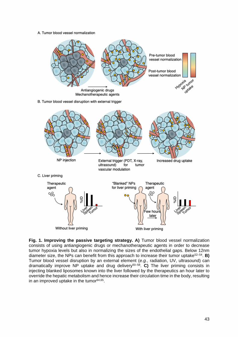
43
Fig. 1. Improving the passive targeting strategy. A) Tumor blood vessel normalization consists of using antiangiogenic drugs or mechanotherapeutic agents in order to decrease tumor hypoxia levels but also in normalizing the sizes of the endothelial gaps. Below 12nm diameter size, the NPs can benefit from this approach to increase their tumor uptake52–54. B) Tumor blood vessel disruption by an external element (e.g., radiation, UV, ultrasound) can dramatically improve NP uptake and drug delivery50–58. C) The liver priming consists in injecting blanked liposomes known into the liver followed by the therapeutics an hour later to override the hepatic metabolism and hence increase their circulation time in the body, resulting in an improved uptake in the tumor64,65.
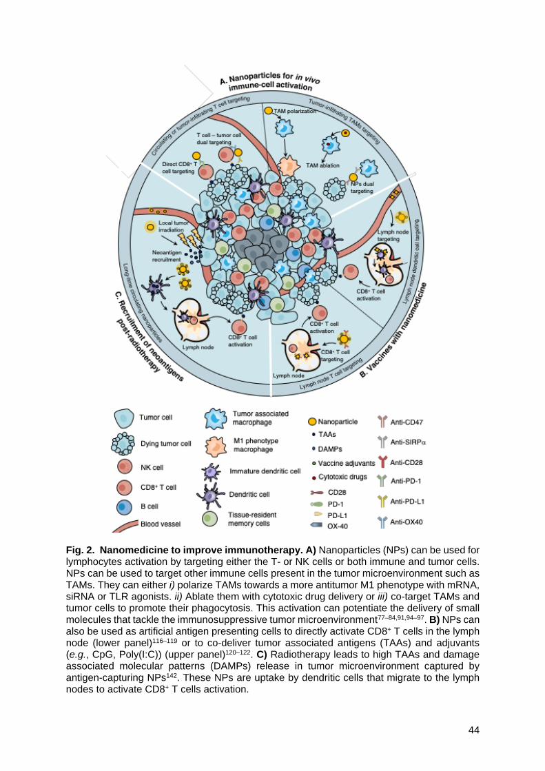
44
Fig. 2. Nanomedicine to improve immunotherapy. A) Nanoparticles (NPs) can be used for lymphocytes activation by targeting either the T- or NK cells or both immune and tumor cells. NPs can be used to target other immune cells present in the tumor microenvironment such as TAMs. They can either i) polarize TAMs towards a more antitumor M1 phenotype with mRNA, siRNA or TLR agonists. ii) Ablate them with cytotoxic drug delivery or iii) co-target TAMs and tumor cells to promote their phagocytosis. This activation can potentiate the delivery of small molecules that tackle the immunosuppressive tumor microenvironment77–84,91,94–97. B) NPs can also be used as artificial antigen presenting cells to directly activate CD8+ T cells in the lymph node (lower panel)116–119 or to co-deliver tumor associated antigens (TAAs) and adjuvants (e.g., CpG, Poly(I:C)) (upper panel)120–122. C) Radiotherapy leads to high TAAs and damage associated molecular patterns (DAMPs) release in tumor microenvironment captured by antigen-capturing NPs142. These NPs are uptake by dendritic cells that migrate to the lymph nodes to activate CD8+ T cells activation.
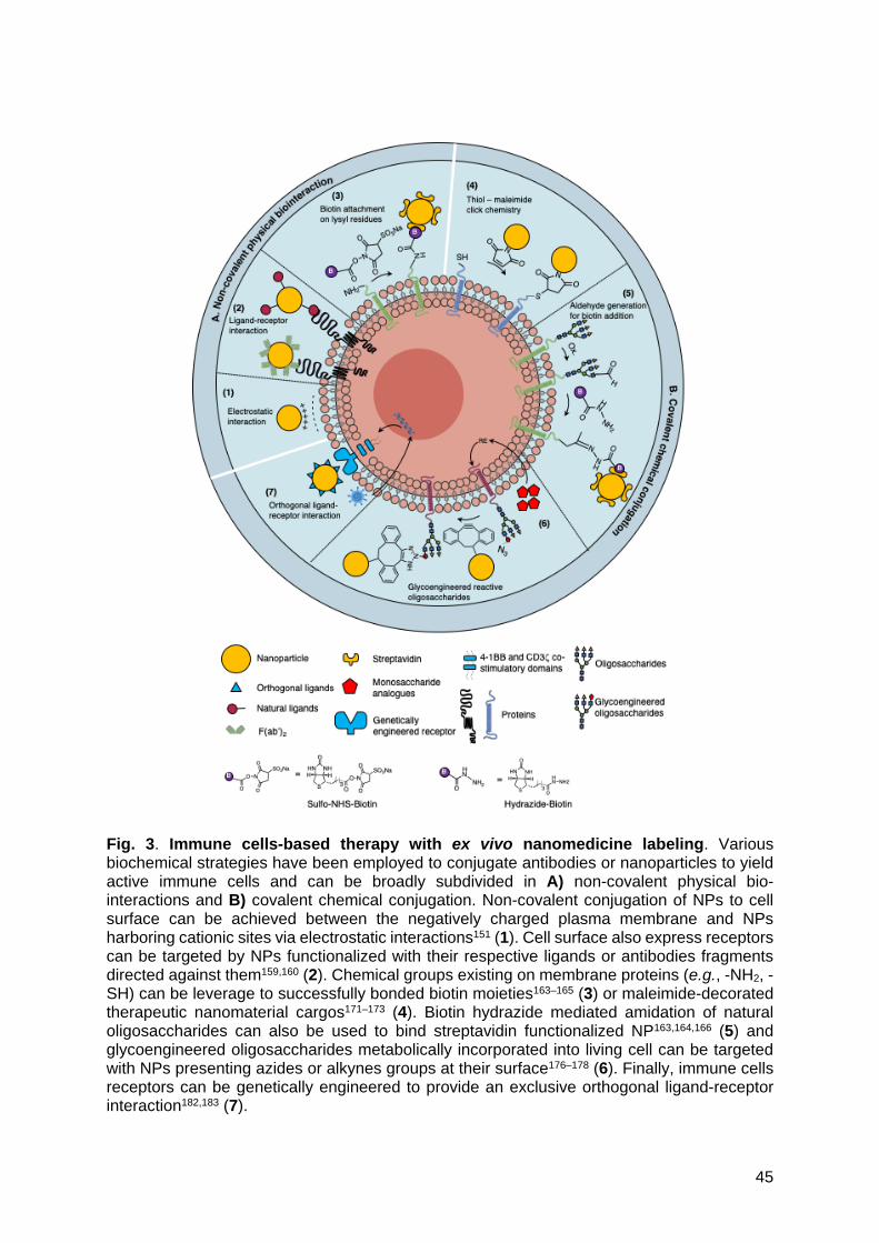
45
Fig. 3. Immune cells-based therapy with ex vivo nanomedicine labeling. Various biochemical strategies have been employed to conjugate antibodies or nanoparticles to yield active immune cells and can be broadly subdivided in A) non-covalent physical bio-interactions and B) covalent chemical conjugation. Non-covalent conjugation of NPs to cell surface can be achieved between the negatively charged plasma membrane and NPs harboring cationic sites via electrostatic interactions151 (1). Cell surface also express receptors can be targeted by NPs functionalized with their respective ligands or antibodies fragments directed against them159,160 (2). Chemical groups existing on membrane proteins (e.g., -NH2, -SH) can be leverage to successfully bonded biotin moieties163–165 (3) or maleimide-decorated therapeutic nanomaterial cargos171–173 (4). Biotin hydrazide mediated amidation of natural oligosaccharides can also be used to bind streptavidin functionalized NP163,164,166 (5) and glycoengineered oligosaccharides metabolically incorporated into living cell can be targeted with NPs presenting azides or alkynes groups at their surface176–178 (6). Finally, immune cells receptors can be genetically engineered to provide an exclusive orthogonal ligand-receptor interaction182,183 (7).