leukocyte-cancer cell fusion: initiator of the warburg effect in malignancy?
TRANSCRIPT
Chapter 8Leukocyte-Cancer Cell Fusion: Initiator of the WarburgEffect in Malignancy?
Rossitza Lazova, Ashok Chakraborty, and John M. Pawelek
Abstract The causes of metastasis remain unknown, however it has been proposed for nearly acentury that metastatic cells are generated by fusion of tumor cells with tumor-associated leukocytessuch as macrophages. Indeed, regardless of cell or tissue origin, when cancer cells in the originalin situ tumor transform to malignant, invasive cells, they generally become aneuploid and beginto express molecules and traits characteristic of activated macrophages. This includes two key fea-tures of malignancy: chemotactic motility and the use of aerobic glycolysis as a metabolic energysource (the Warburg effect). Here we review evidence that these phenomena can be well-explainedby macrophage-cancer cell fusion, as evidenced by studies of experimental macrophage-melanomahybrids generated in vitro and spontaneous host-tumor hybrids in animals and more recently humans.A key finding to emerge is that experimental and spontaneous cancer cell hybrids alike displayed ahigh degree of constitutive autophagy, a macrophage trait that is expressed under hypoxia and nutri-ent deprivation as part of the Warburg effect. Subsequent surveys of 21 different human cancers fromnearly 2,000 cases recently revealed that the vast majority (85%) exhibited autophagy and that thiswas associated with tumor proliferation and metastasis. While much work needs to be done, we positthat these findings with human cancers could be a reflection of widespread leukocyte-cancer cellfusion as an initiator of metastasis. Such fusions would generate hybrids that express the macrophagecapabilities for motility and survival under adverse conditions of hypoxia and nutrient deprivation,while at the same time maintaining the deregulated mitotic cycle of the cancer cell fusion partner.
8.1 Introduction and Background
The concept of leukocyte-tumor hybridization as a mechanism for tumor metastasis was first put forthin remarkable detail a century ago by pathologist Otto Aichel [1]. In the past 40 years, host-tumorhybrid cancer cells have been documented in numerous animal tumor models where they can be foundin metastatic tumors [2–5]. Animal and cell culture studies on cancer cell fusion, coupled with obser-vations that malignant cancer cells routinely express macrophage traits, add experimental support tothe Aichel proposal and have led to the hypothesis that leukocyte-tumor cell fusion represents a uni-fying mechanism for metastasis [2–5]. However, while it has been demonstrated that cancer cell-hostcell fusion occurs in animals and more recently in humans [6–9] little is known regarding key ques-tions such as the mechanisms through which leukocyte-cancer cell fusion and subsequent genomic
J.M. Pawelek (B)Department of Dermatology, Yale Cancer Center, Yale University School of Medicine, New Haven,CT 06520-8059, USAe-mail: [email protected]
151T. Dittmar, K.S. Zänker (eds.), Cell Fusion in Health and Disease, Advances in Experimental Medicineand Biology 714, DOI 10.1007/978-94-007-0782-5_8, C Springer Science+Business Media B.V. 2011
152 R. Lazova et al.
Table 8.1 Model for leukocyte-cancer cell hybrid formation and the generation of metastatic cells
1. Tumor cells attract bone marrow-derived cells such as macrophages, lymphocytes, neutrophils and perhaps stemcells
2. In some cases leukocyte-cancer cell fusions occur with tumor and leukocyte chromosomes becoming pooled in asingle nucleus, usually resulting in aneuploidy
3. Epigenomic gene expression patterns from both fusion partners are retained in hybrids. Some hybrids combine themigratory capabilities of the leukocyte with the de-regulated cell cycle of the cancer cell. Thus new mitoticallyactive cells emerge with high invasive and metastatic potential
4. Hybrid cells express aerobic glycolysis (the Warburg effect), a metabolic energy pathway characteristic ofmacrophages and other inflammatory cells. Aerobic glycolysis is advantageous for survival in hypoxicenvironments and also for proliferating cells [47]
5. As part of the Warburg effect, hybrids use the autophagy-lysosomal pathway for sequestration and digestion ofexternal food sources that are internalized via phagocytosis or other endocytic processes
hybridization occurs in vivo; the frequency at which hybrids are generated; how gene expression isregulated by the hybrid epigenomes from two different cell lineages; potential survival advantagesof hybrids; the role of fusion in metastasis; and metabolic energy sources for hybrids. These ques-tions and many more remain for future research and point to the difficulties of studying fusion andhybridization in vivo. A working model for in vivo cancer cell fusion and genomic hybridization ispresented in Table 8.1.
Here we summarize evidence supporting this model, and focus on how aerobic glycolysis – a traitof activated macrophages that is expressed in macrophage-tumor hybrids and malignant cancer cellsalike – may perform a central role in the regulation of hybrid metabolic energy balance followingfusion.
8.2 Macrophage-Tumor Cell Associations: The Scene of the Crime?
Tumor associated macrophages (TAMs) facilitate all aspects of cancer initiation and progression [10].Macrophages are attracted through chemotactic signals to tumors where they exert their abilities formatrix degradation, tissue remodeling, stroma deposition, neoangiogenesis and migration to distanttissues. These functions are normally employed in many physiological functions such as embryogen-esis, osteogenesis, and wound healing. Macrophages accumulate in hypoxic regions of tumors at leastin part through HIF-1-mediated upregulation of the chemokine receptor [11, 12].
However, tumor progression is not completely explained by the presence of TAMs. During transi-tion to a metastatic phenotype, tumor cells notoriously co-opt leukocytic traits [2, 3]. Malignant cellsare chemotactic, responding to chemokines and exhibiting their own matrix-degrading and angiogeniccapabilities. Like migratory leukocytes, metastatic cells exhibit loss of homotypic adhesion, and theability to transverse a basement membrane, migrate through the mesodermal matrix, intravasate intolymphatics or the blood circulatory system, extravasate from these vessels, and colonize lymph nodesand distant organs. But unlike normal leukocytes, cancer cells have deregulated mitotic cycles andtheir numbers continually increase, killing the host if left unchecked. It has been recently proposedthat expression of leukocytic traits by malignant cells can be explained by leukocyte-cancer cell fusionand genomic hybridization [2–5]. Below are considerations of this process with examples of manymacrophage or other inflammatory cell traits of malignant cells that could be explained by fusion.
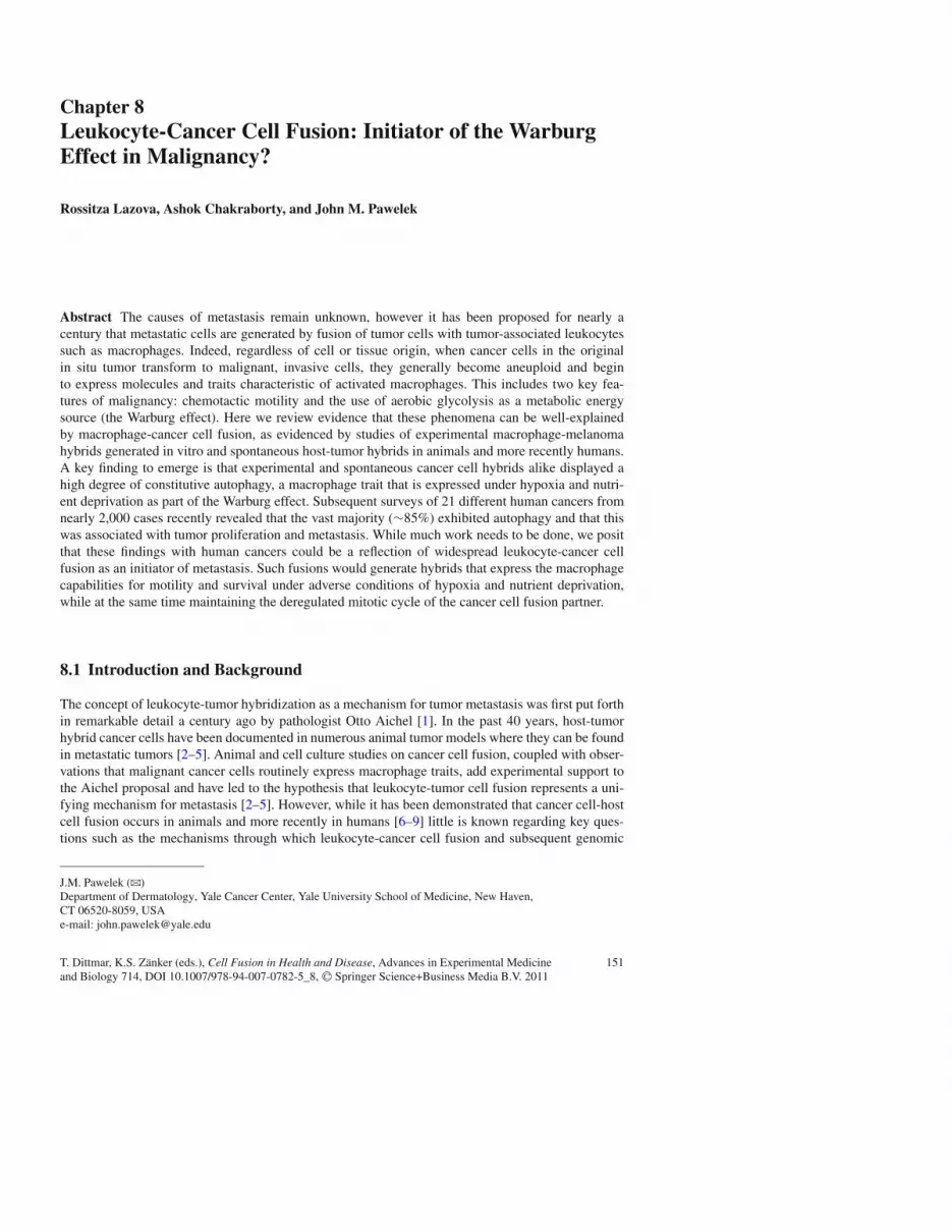
In order for macrophage-tumor cell fusion to occur, the process must be initiated by close contactbetween the two fusion partners. Such contacts are readily seen when experimental tumor xenograftsare dissociated and placed into culture. Shown in Fig. 8.1 are cultured cells obtained from a CloudmanS91 mouse melanoma tumor implanted into a DBA/2J mouse. The cells were fixed and stained fornon-specific esterase (red), a macrophage marker. At the far right is seen a red-staining macrophage
8 Leukocyte-Cancer Cell Fusion: Initiator of the Warburg Effect in Malignancy? 153
Fig. 8.1 A Cloudman S91tumor was dissociated intoindividual cells and placedin culture. Non-specificesterase stain (red) revealedmacrophages in contact withtumor cells
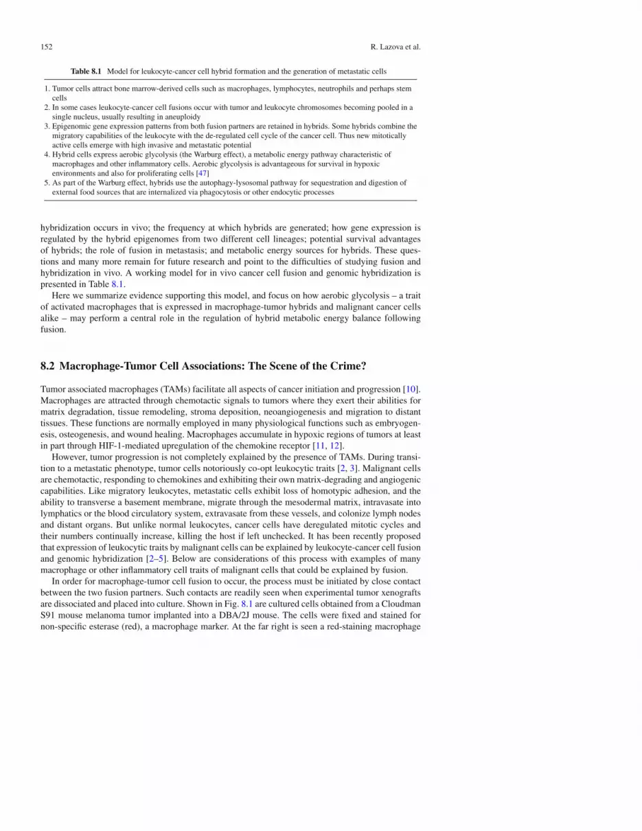
Fig. 8.2 Tumor associatedmacrophages (TAMs). (a–c)TAMs (arrows) staining withazure blue are seen in closecontact with melanoma cellsin three cases of primarycutaneous malignantmelanoma. (d) A low powerview of TAMs, (azure bluestain), are seen infiltratinginto a nest of melanoma cells(S100 stain with brownchromogen) (e) Higher powerview of (d). Note the highlyvesicular nature of themacrophages due to thepresence of autophagosomesand autolysosomes (panelsa–c) (from [13])
in close contact with a non-staining Cloudman melanoma cell. The melanoma cell is distinguishedby its abundant nucleoli compared to those of the macrophage. On the far left is another macrophagestaining red with a characteristic migratory orientation.
In pathology specimens of malignant melanomas, tumors are usually infiltrated with or surroundedby macrophages, most frequently in hyperpigmented areas of tumors [13] (Fig. 8.2a–e). MelanomaTAMs are referred to as “melanophages” due to their injestion of melanized melanoma cells andretention of incompletely digested melanin in autophagosomes or autolysosomes. High power viewsreveal that melanophages (azure blue) are found in close association with melanoma cells, with longsegments of plasma membranes of the two cell types in apposition to one another (Fig. 8.2a, b), insome cases with macrophages engulfing melanoma cells (Fig. 8.2c). Thus macrophage-tumor cellcontact – a prerequisite for cell-cell fusion – is common in human melanomas in vivo.
8.2.1 Fusion-Induced Aneuploidy
In his 1911 article, “About cell fusion with qualitatively abnormal chromosome distribution as causefor tumor formation” (from the German), Aichel first proposed the fusion theory of tumor progressionand in order to experimentally verify this idea implored future scientists to “: : : study chromosomes

154 R. Lazova et al.
Fig. 8.3 Aneuploidy in human macrophage-mouse melanoma fusion hybrids created via polyethylene glycol-induced fusion in vitro. The photograph demonstrates the presence of human and mouse chromosomes in a metaphasespread of human macrophage-mouse melanoma hybrid 96–HJP1. Human chromosomes 1, 3, 19, and X are indicatedand identified by high resolution Giemsa banding. Numerous acrocentric mouse chromosomes from the melanomafusion partner are also seen (from [18])
from all angles.” [1]. He proposed that the source of aneuploidy could stem from fusion of tumor-invading leukocytes with cancer cells, suggesting that a combination of extra chromosomes and the“qualitative differences” (now termed “epigenetic differences”) in chromosomes from the two celltypes could lead to the metastatic phenotype. With remarkably prescience he wrote, Not only wouldthe capacity for cell division increase, but after the mixing of the qualitatively different chromosomesthe different traits and capabilities of the different cells depending on the type of cells fused wouldbecome obvious in different ways in the daughter cells. In this way the daughter cell may maintainsome of the specialized function of the somatic [tumor] cell so that the tumor cell is capable to anextent to remain functional. On the other hand the traits of the leukocytes would also be incorporatedin the daughter cell, so that a new cell with new traits and capabilities would emerge, a cell thathas been thrown out of the path of the normal mother cell. The end product would be what we havelearned to understand as a malignant cell.
Decades later, the same hypothesis – that metastasis is caused by leukocyte-tumor cell fusion –was proposed independently by Meckler [14, 15] and by Goldenberg [16, 17]. Several laboratorieshave now reported that hybrids produced by fusion in vitro or in vivo were aneuploid and of highermetastatic potential (reviewed in [2–5]). As a demonstration of hybrid aneuploidy, shown in Fig. 8.3is a metaphase spread from a human macrophage-mouse melanoma hybrid 96–HJP1 experimentallyfused in vitro [18]. Seen are human chromosomes 1, 3, 19 and X along with numerous acrocentricmouse chromosomes. As discussed below, cells from this hybrid clone produced both human andmouse versions of the metastasis-related protein SPARC, indicating that the epigenomes from bothfusion partners were active (discussed below). This was in line with the above predictions of Aichelthat the cells would be aneuploid, mitotically active, and express genes from “qualitatively different”chromosomes of the leukocyte and tumor cell fusion partners.
8.2.2 Macrophage-Melanoma Fusion In Vitro Generates Altered GeneExpression and a Metastatic Phenotype In Vivo
Tumor-BMDC fusions might explain how common gene expression patterns emerge for differenttumor types. We, and others, have found that when BMDC-tumor cell hybrids were isolated in vitrowith no selective pressure other than for growth in drug-containing media, remarkably high numbersof them exhibited a metastatic phenotype in mice. Curiously, in melanoma the most metastatic clonestended to be highly melanized compared to parental melanoma cells or weakly metastatic hybrids as

8 Leukocyte-Cancer Cell Fusion: Initiator of the Warburg Effect in Malignancy? 155
Fig. 8.4 Metastatic potential of macrophage-melanoma hybrid cell lines compared to parental Cloudman S91melanoma cells. Results are shown for in vitro-generated hybrids and one spontaneous in vivo hybrid (PADA). Melaninwas estimated in pelleted cells from clones on their first passage in culture and before metastatic potential was deter-mined. A minimum of 10–20 animals was tested for each clone. In addition representative clones were tested repeatedlyduring continuous passage in culture for up to 4 years where 30–90 animals were tested for each clone with similarresults as above. Statistical analyses of metastatic potential revealed that p values for significance vs. parental melanomacells were < 0.0001 (); < 0.01 (dagger); < 0.05 (square) (from [18])
described below (Fig. 8.4) [19, 20]. This was subsequently explained by the acquisition of macrophageglycosylation patterns involving b1,6-branched oligosaccharides that were associated with many phe-notypic changes, including pigmentation [21] In two separate rounds of isolation, a total of 75 clonesof PEG-fused macrophage-melanoma hybrids were isolated in vitro. About half showed increasedchemotaxis in vitro and metastasis in mice [22]. Similar results were obtained in T-cell hybridomasfrom fusion of healthy T-lymphocytes with T-lymphoma cells [23] and in hybrids between mouseT-cell lymphoma cells and bone marrow-derived macrophages or spleen lymphocytes [24–26]. Highfrequency emergence of a common metastatic phenotype in vitro without host selective pressure wassurprising, particularly in view of the apparently chaotic nature of aneuploidy.
8.2.3 Macrophage-Melanoma Fusion Hybrids Exhibit Up-RegulatedChemotaxis In Vitro
Increased metastatic potential in macrophage-melanoma hybrids was mirrored in increased chemo-taxis in vitro (Table 8.2) [22]. In two-chambered assay systems comparing parental melanoma cellsto low and high metastatic hybrids, the hybrids with high metastatic potential showed 5- to 100-foldincreases in the rate of migration to 3T3- and lung fibroblast-conditioned media, primary lung slices,fibronectin (FN), and a 120 kDa FN fragment compared to parental melanoma cells. Unlike parental
156 R. Lazova et al.
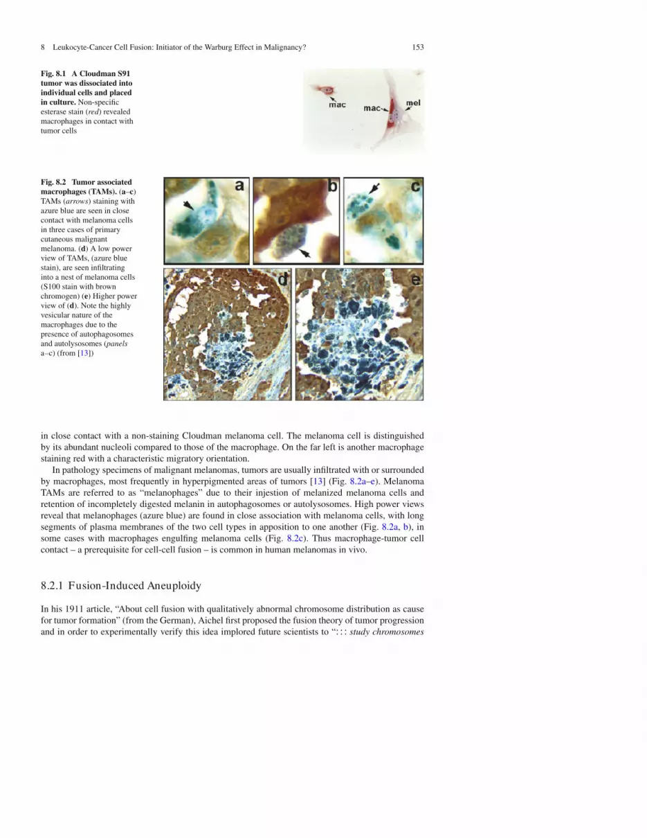
Table 8.2 Metastaticpotential in vivo vs.chemotactic migration invitro ofmacrophage-melanomahybrids
Cells migrated
Cell line Metastatic potential – MSH + MSH p-value
Parental melanoma 14 2 ± 12 ± 0 >0.050Hybrid 95-H3 0 73 ± 16 32 ± 5 >0.050Hybrid 95-H11 0 23 ± 5 42 ± 8 >0.050Hybrid 95-H19 10 50 ± 3 70 ± 2 <0.020PADA 54 87 ± 12 266 ± 20 <0.001Hybrid 95-H1 71 142 ± 14 436 ± 21 <0.001Hybrid 95-H2 78 143 ± 5 364 ± 42 <0.001
Migration to the underside of a Costar Transwell apparatus of parental Cloudmanmelanoma cells, low and high metastatic macrophage-melanoma fusion hybrids,and PADA, a spontaneous in vivo hybrid, in response to 3T3 cell-conditionedmedium (33%, vol/vol) in the lower chambers. For migration, results representmean+S.E. for triplicate assays of cells migrated/4 h. Where noted, cells were pre-treated for 72 h in culture with MSH and the cAMP phosphodiesterase inhibitorisobutylmethylxanthine to raise cyclic AMP levels. p values are for differencesbetween control vs. MSH/IBMX treatment [22].
cells, metastatic hybrids were further stimulated by pretreatment with melanocortin-1 (melanocytestimulating hormone; MSH) [22].
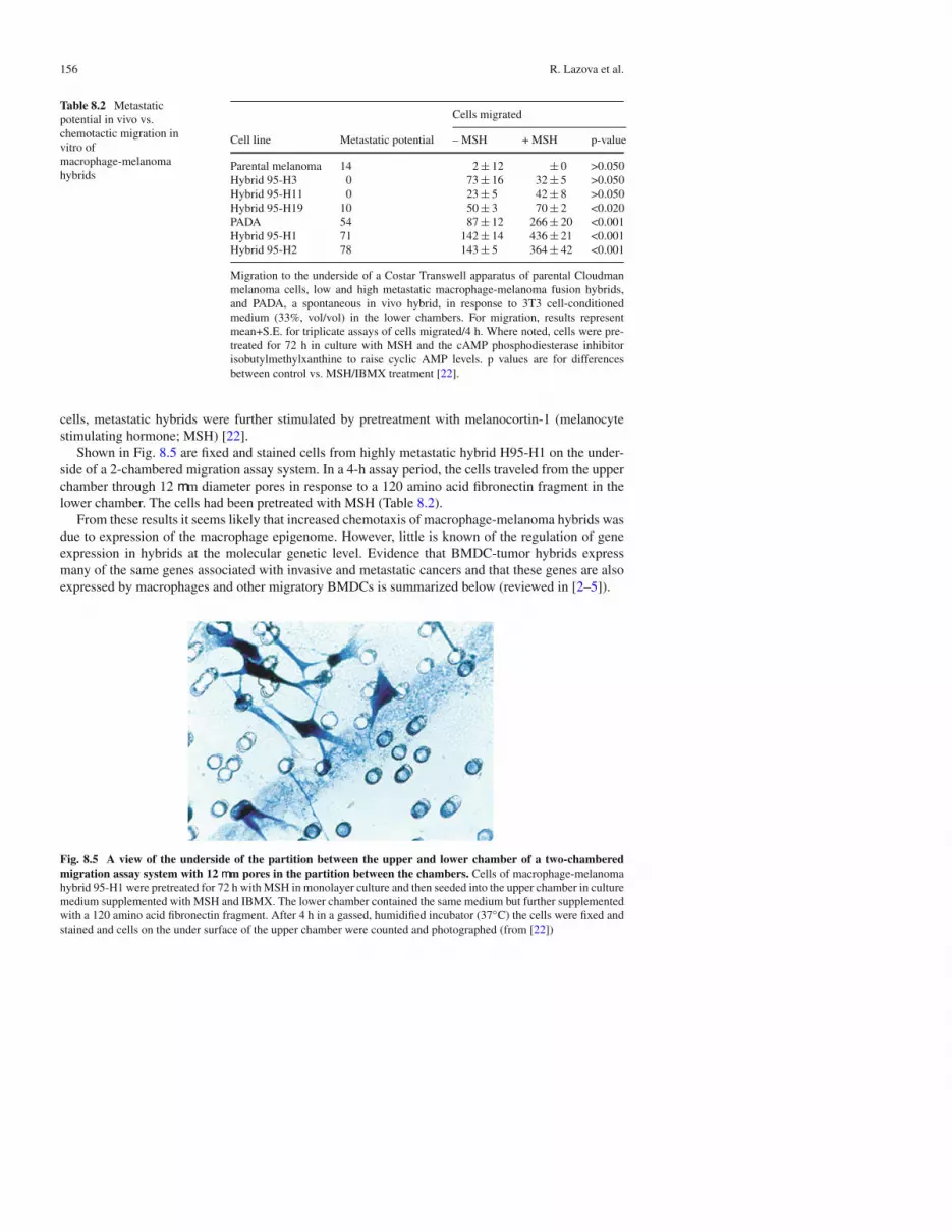
Shown in Fig. 8.5 are fixed and stained cells from highly metastatic hybrid H95-H1 on the under-side of a 2-chambered migration assay system. In a 4-h assay period, the cells traveled from the upperchamber through 12 mm diameter pores in response to a 120 amino acid fibronectin fragment in thelower chamber. The cells had been pretreated with MSH (Table 8.2).
From these results it seems likely that increased chemotaxis of macrophage-melanoma hybrids wasdue to expression of the macrophage epigenome. However, little is known of the regulation of geneexpression in hybrids at the molecular genetic level. Evidence that BMDC-tumor hybrids expressmany of the same genes associated with invasive and metastatic cancers and that these genes are alsoexpressed by macrophages and other migratory BMDCs is summarized below (reviewed in [2–5]).
Fig. 8.5 A view of the underside of the partition between the upper and lower chamber of a two-chamberedmigration assay system with 12 mm pores in the partition between the chambers. Cells of macrophage-melanomahybrid 95-H1 were pretreated for 72 h with MSH in monolayer culture and then seeded into the upper chamber in culturemedium supplemented with MSH and IBMX. The lower chamber contained the same medium but further supplementedwith a 120 amino acid fibronectin fragment. After 4 h in a gassed, humidified incubator (37C) the cells were fixed andstained and cells on the under surface of the upper chamber were counted and photographed (from [22])

8 Leukocyte-Cancer Cell Fusion: Initiator of the Warburg Effect in Malignancy? 157
8.2.4 SPARC
The SPARC (secreted protein acidic and rich in cysteine; osteonectin; BM40) gene provides anexample of gene regulation in leukocyte-tumor fusion. SPARC is expressed in macrophages, exper-imental macrophage-melanoma hybrid and is associated with tumor progression and poor outcomein melanoma and a number of carcinomas including breast, colorectal, ovarian and lung [2, 3, 27].SPARC acts as a regulator of melanoma EMT by downregulating melanoma E-cadherin with lossof homotypic adhesion, stimulates motility, and increases expression of mesenchymal markers suchas matrix metalloproteinase MMP-9 [28]. In tissue macrophages SPARC is expressed in regions ofneovascularization, for example in wound repair [29] and degenerative aortic stenosis [30]. In fusionsbetween mouse macrophages or human blood monocytes and weakly metastatic mouse CloudmanS91 melanoma cells, the total levels of SPARC mRNA were three- to four-fold higher per mg totalRNA in metastatic hybrids compared to weakly metastatic hybrids and parental melanoma cells[31]. Notably, hybrids between human monocytes and mouse melanoma cells expressed both humanand mouse SPARC mRNA [31]. This indicated that at least for SPARC, the epigenomes from thetwo different developmental lineages of the fusion partners were both active. In summary, SPARCgene expression was enhanced by hybridization of tumor cells with macrophages; high expressionwas correlated with high metastatic potential; and SPARC mRNA was produced in hybrids fromthe genomes of both parental fusion partners. That elevated SPARC expression was a character-istic of macrophage-melanoma hybrids provides a possible explanation for elevated SPARC andSPARC-mediated pathways in human melanoma and other cancers.
In addition to SPARC, macrophage-melanoma hybrids of high metastatic potential also showedmarked elevations of other macromolecules that are characteristic of macrophages and known indi-cators of metastasis, including cMet, the melanocortin 1 receptor, and the integrin subunits a3, a5,a6, av, b1, b3. We thus hypothesize that such gene expression patterns in cancer may be generatedthrough fusion with macrophages (reviewed in [2, 3]).
8.2.5 GnT-V and �1,6-Branched Oligosaccharides
N-acetylglucosaminyltransferase V (GnT-V; Mgat5; E.C.2.4.1.155) is a Golgi complex enzyme thatis highly expressed in myeloid cells and metastatic cancer cells. GnT-V and its enzymatic products,b1,6-branched oligosaccharides conjugated to N-glycoproteins, are associated with poor outcome ina number of cancers [32, 33]. b1,6-branched oligosaccharides were first purified from granulocytes[34]. From structural analyses they are composed of poly-N-acetyllactose amines that are carriers ofsialyl lewisx antigen (sialyl lex) and therein used by both leukocytes and metastatic cancer cells forbinding to E-selectin and/or galectin-3 on endothelial cells during systemic migration [35, 36].
GnT-V mRNA, protein, and/or enzymatic activity were elevated in high metastatic macrophage-melanoma hybrids formed in vitro [37], and following spontaneous host-tumor fusions in bothlymphomas and melanomas in mice [24, 37, 38]. Multiple pathways in invasion and metastasisthat are regulated by GnT-V were elevated in macrophage-melanoma hybrids – as seen below withmotility-associated integrin subunits, cell surface expression of LAMP-1, and autophagy.
8.2.6 Cell Surface Expression of Lysosome Associated Protein-1 (LAMP-1)
LAMP-1 is a preferred substrate for GnT-V and a major carrier of sialyl lex and poly-N-acetyl-lactoseamines that bind to E-selectins and galectins [35, 36]. Cell surface LAMP-1 thus mediates bindingto endothelial cells by both leukocytes and cancer cells [35, 36, 39]. Macrophage-melanomahybrids showed elevated expression of cell surface LAMP-1 [37]. This was seen in high metastatic

158 R. Lazova et al.
macrophage-melanoma hybrids as well as peritoneal macrophages compared to that in parentalmelanoma cells and low metastatic hybrids.
8.3 Coarse Melanin and Autophagy in ExperimentalMacrophage-Melanoma Hybrids
As mentioned above, the parental melanoma cells used in the production of experimental macrophage-melanoma hybrids in vitro produced little or no melanin (Fig. 8.6, left), however many hybrid clones –particularly those of high metastatic potential – were heavily pigmented with coarse, dark melaninthroughout the cytoplasm as shown for metastatic hybrid 94-H48 (Fig. 8.6, right) [40].
Electron micrographs revealed that although some melanosomes existed freely in the cyto-plasm, the coarse granular appearance of melanin in hybrid 94-H48 was due to melanosome-filledautophagasomes in various stages of maturation (Fig. 8.7) [40].
Staining for b1,6-branched oligosaccharides with the plant lectin LPHA (leukocytic phyto-hemagglutinin) revealed that the coarse, melanin – containing autophagosomes contained theseglycan structures in abundance (brown stain). The granular LPHA staining pattern (white arrows)encompassed large portions of the cytoplasm, often obscuring the nucleus (“n”) (Fig. 8.8) [40].
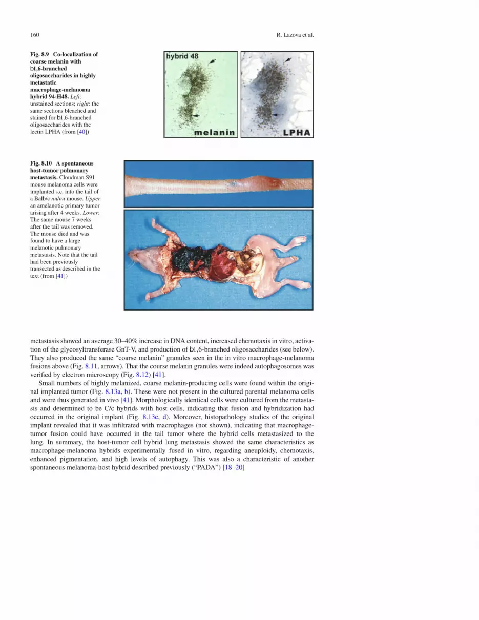
Co-localization of b1,6-branched oligosaccharides to coarse melanin is shown in Fig. 8.9. A singlecell of macrophage-melanoma hybrid H48-94 is seen in culture. The left panel shows an unstainedculture allowing for the visualization of coarse melanin. The right panel shows the same cell afterbleaching to decolorize the melanin and staining for b1,6-branched oligosaccharides with the lectinLPHA. The results demonstrate an exact correspondence between LPHA staining and coarse melaninautophagosomes [40].
8.3.1 Spontaneous Fusion In Vivo and Autophagy
There are numerous reports in animal cancer models of tumor cell fusion with host cells and manyof these implicate macrophages or other BMDC’s as host fusion partners [2–5]. An example fromour lab is seen in the development of a spontaneous melanoma metastasis to the lungs in a Balb/cnude mouse (Fig. 8.10) [41]. Balb/c mice are albino due to a homozygous mutation in tyrosinase(c/c), the rate-limiting enzyme in melanogenesis. The parental melanoma cells implanted into this
Fig. 8.6 Comparison of cultured parental Cloudman S91 melanoma cells and high metastatic macrophage-melanoma hybrid 94-H48 following staining with H&E. Left: parental Cloudman S91 cells showing lack of pigment;right: hybrid 94-H48 with abundant coarse melanin (from [40])

8 Leukocyte-Cancer Cell Fusion: Initiator of the Warburg Effect in Malignancy? 159
Fig. 8.7 Electronmicrograph of highmetastaticmacrophage-melanomahybrid 94-H48 showingcytoplasmicautophagosomes in thelower panel. The upper insetsshow individualautophagosomes andautolysosomes in variousstages of maturation (early tolate maturation stages areshown from left to right)(from [40])
Fig. 8.8 A single cell of hybrid 94-H48 in culture. The culture was fixed and decolorized of melanin by bleaching.It was then stained with the lectin LPHA for b1,6-branched oligosaccharides (brown stain) and counterstained withhematoxylin. Arrows denote the granular staining pattern characteristic of autophagosomes. The nucleus (n) is partiallyobscured by the autophagosomes (from [40])
mouse were the same clone used as the melanoma fusion partners in the in vitro fusion experimentsdescribed above. Although the melanoma clone implanted into these mice was genetically wild typefor tyrosinase (C/C), the cells produced little or no melanin in culture and formed amelanotic tumorsin mice. Metastases, though infrequent, were generally small, amelanotic tumors in the lung, and werewell tolerated by the mice [41]. However, in one experiment a mouse developed a melanin-producingin transit metastasis near the site of implantation in the tail dermis. The tail was amputated and themouse was followed to see if distant metastases developed. After 5 weeks the mouse became moribundwith a massive, highly pigmented pulmonary metastasis.
DNA analyses showed that cells from the metastasis had a genotype of C/c, indicating they werehybrids formed from fusion of the implanted tumor cells (C/C) with host cells (c/c). Cells from the
160 R. Lazova et al.
Fig. 8.9 Co-localization ofcoarse melanin withb1,6-branchedoligosaccharides in highlymetastaticmacrophage-melanomahybrid 94-H48. Left:unstained sections; right: thesame sections bleached andstained for b1,6-branchedoligosaccharides with thelectin LPHA (from [40])
Fig. 8.10 A spontaneoushost-tumor pulmonarymetastasis. Cloudman S91mouse melanoma cells wereimplanted s.c. into the tail ofa Balb/c nu/nu mouse. Upper:an amelanotic primary tumorarising after 4 weeks. Lower:The same mouse 7 weeksafter the tail was removed.The mouse died and wasfound to have a largemelanotic pulmonarymetastasis. Note that the tailhad been previouslytransected as described in thetext (from [41])
metastasis showed an average 30–40% increase in DNA content, increased chemotaxis in vitro, activa-tion of the glycosyltransferase GnT-V, and production of b1,6-branched oligosaccharides (see below).They also produced the same “coarse melanin” granules seen in the in vitro macrophage-melanomafusions above (Fig. 8.11, arrows). That the course melanin granules were indeed autophagosomes wasverified by electron microscopy (Fig. 8.12) [41].
Small numbers of highly melanized, coarse melanin-producing cells were found within the origi-nal implanted tumor (Fig. 8.13a, b). These were not present in the cultured parental melanoma cellsand were thus generated in vivo [41]. Morphologically identical cells were cultured from the metasta-sis and determined to be C/c hybrids with host cells, indicating that fusion and hybridization hadoccurred in the original implant (Fig. 8.13c, d). Moreover, histopathology studies of the originalimplant revealed that it was infiltrated with macrophages (not shown), indicating that macrophage-tumor fusion could have occurred in the tail tumor where the hybrid cells metastasized to thelung. In summary, the host-tumor cell hybrid lung metastasis showed the same characteristics asmacrophage-melanoma hybrids experimentally fused in vitro, regarding aneuploidy, chemotaxis,enhanced pigmentation, and high levels of autophagy. This was also a characteristic of anotherspontaneous melanoma-host hybrid described previously (“PADA”) [18–20]

8 Leukocyte-Cancer Cell Fusion: Initiator of the Warburg Effect in Malignancy? 161
Fig. 8.11 Histologicalsection of the pulmonoarymetastasis shown inFig. 8.10 and stained withH&E. Arrows show coarsemelanin-containing cells inthe tumor (from [41])
Fig. 8.12 Electronmicrophage of a culturedcell from the spontaneoushost-melanoma hybridshown in Fig. 8.10. Shown isa prominent autophagosome(from [41])
Thus, experimental macrophage-melanoma hybrids in cell culture and spontaneous host-melanomahybrids arising in vivo and adapted to culture all appeared to show constitutive autophagy with-out autophagic death. It is important to note that this occurred in cell culture under normoxicconditions and ample nutrients indicating that hybrids expressed the aerobic glycolysis pathway(the Warburg effect) that includes autophagy [42–48]. Below it is discussed that macrophages alsoexhibit autophagy and aerobic glycolysis in vivo and in vitro, suggesting that perhaps these traits inmacrophage-melanoma hybrids shown above may be an expression of the macrophage epigenome inthe hybrid cells.
8.3.2 Autophagy and Aerobic Glycolysis in Macrophages and Cancers
Under hypoxic conditions most cells go through apoptotic death in a mitochondrial Bcl-2 – medi-ated fashion [49–53]. However inflammatory cells and cancer cells alike survive through utilizationof the glycolytic pathway for ATP production, independent of mitochondria [54–56]. High densitiesof macrophages are found in tumors, wounds, atherosclerotic plaques, bone fractures, rheumatoidarthritis, and ischemic areas in diabetes (reviewed in [54]). Due to their reliance on glycolysis,macrophages need large amounts of glucose for ATP production. Thus, much like the vast majority(90%) of malignant solid tumors [57], macrophages in hypoxic areas can be visualized by PositronEmission Tomography (PET) due to their avid uptake of the glucose analog fluorodeoxyglucose(18F) [58–61]. Further, like cancer cells, activated macrophages exhibit glycolysis even under aer-obic conditions, a state known as aerobic glycolysis or the Warburg effect [51, 52, 62]. Glycolysis in

162 R. Lazova et al.
Fig. 8.13 Panels a and b. Histological sections of the tail implant of Cloudman S91 melanoma cells shown in Fig. 8.10(H&E stain). Arrows denote coarse melanin containing melanoma cells. Panels c and d. Phase contrast photos of coarsemelanin-containing cells cultured from the pulmonary metastasis seen in Fig. 8.10. The original clone of Cloudmancells used in this study was amelanotic in culture even though it was wild type for tyrosinase (C/C) Thus, the pigmentedmelanoma cells were generated in the mouse (c/c), potentially through macrophage-tumor cell fusion as seen in exper-imentally fused hybrids (NRC). As predicted, cells cloned from the metastasis were fround to be C/c fusion hybrids(from [41])
macrophages is accompanied by dramatic changes in gene expression patterns and phenotype, includ-ing activation of pathways for motility and phagocytosis and markedly increased cell viability [52,54, 63–67]. The transcription factor HIF-1a is a master regulator of the hypoxic phenotype [55, 68,69]. HIF-1a is regulated by the serine/threonine kinase Akt, which also mediates the switch to aerobicglycolysis [70].
The advantage of cancer cells for utilizing glycolysis over oxidative respiration has long beenunclear, since the Krebs cycle is far more efficient than glycolysis in ATP production. However arecent proposal argues that the glycolysis pathway is best suited to proliferating cells which constantlyneed to increase biomass and thus depend on the uptake and/or production of glucose, nucleotides,amino acids, and lipids [49].
8.3.3 Autophagy in Macrophages
Melanophages are macrophages filled with melanized vesicles are similar under light microscopicexamination to coarse melanin seen in melanoma cells nearby in the same tissue (Figs. 8.2 and8.14). These vesicles are presumably generated from phagocytic engulfment by the macrophageof melanoma cells containing melanin, followed by transfer of the engulfed cellular debris into

8 Leukocyte-Cancer Cell Fusion: Initiator of the Warburg Effect in Malignancy? 163
Fig. 8.14 Co-localization ofgranular melanin withheavily melanized vesiclesin melanophages. Melanin inunstained sections isdisplayed in the left columnand the corresponding stainedfields are shown in the rightcolumn. Arrows denoteexamples of co-localizedvesicles. (a) A section stainedfor LC3B. (b) A sectionstained for the Golgi 58kprotein. (c) A section stainedfor b1,6-branchedoligosaccharides withbiotinylated LPHA(from [71])
autophagosomes. Co-localization studies showed that the autophagosome marker LC3B (Fig. 8.14a),the Golgi 58k protein (Fig. 8.14b), and b1,6-branched oligosaccharides (Fig. 8.14c) were allconstituents of the melanized vesicles in melanophages, indicative of autophagy [71].
Electron micrographs of melanophages confirmed that the vesicles were autophagosomes(Fig. 8.15) [71]. These autophagosomes in the melanophages are limited by double membranes and,similar to autophagosomes in macrophage-melanoma hybrids, contain what appears to be partiallydigested, melanized melanosomes, and other cellular debris. The melanophages were surrounded bycollagen bundles (labeled “c”) confirming their dermal location beneath a melanoma tumor in theepidermis (melanoma in situ) seen under the light microscope in pathology specimens.
8.3.4 Autophagy in Human Cancers
The above findings of autophagy in macrophages and in macrophage-melanoma hybrids motivated usto launch a larger pathology survey of melanomas and other human cancers as summarized below.
8.3.5 Normal Epidermis
In regions of normal epidermis, melanocytes and keratinocytes did not stain for LC3B orb1,6-branched oligosaccharides and showed no signs of autophagy confirming previous findings thatthese cells do not appear to produce b1,6-branched oligosaccharides [32, 71, 72].

164 R. Lazova et al.
Fig. 8.15 Electron micrographs of a dermal melanophage. (a) Low power view showing numerous melanin-containing vesicles within the cytoplasm of a melanophage. Collagen bundles in the adjacent dermis are labeled by“c”. (b) High power view of a vesicle, which is an autophagosome, enveloped by a double membrane (arrow), andcontaining partially digested, heavily melanized melanosomes (from [71])
8.3.6 EarlyMelanoma In Situ (MIS)
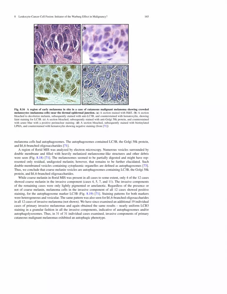
Early MIS presents as a subtle proliferation of atypical melanocytes (melanoma cells) disposed as sin-gle units as well as in a few small nests at the dermal epidermal junction and above it (Fig. 8.16a–d).Similar to normal melanocytes, melanoma cells in regions of early MIS did not show prominentmelanin in their cytoplasm as seen with H&E staining (Fig. 8.16a). Cells of early MIS did not stainor stained only weakly for LC3B (Fig. 8.16b) and for b1,6-branched oligosaccharides (Fig. 8.3d).However, similar to normal melanocytes they did stain for the Golgi 58k protein in a globularperinuclear pattern (Fig. 8.16c) [71].
8.3.7 Autophagy in Florid MIS
In florid MIS there is an irregular, asymmetric, and poorly circumscribed proliferation of melanomacells. There are nests of melanoma cells that vary markedly in size and shape, which are not equidis-tant from one another. Single melanoma cells predominate over nests in some high power fields andthere are individual melanoma cells as well as melanocytic nests above the dermal-epidermal junc-tion. Melanoma cells are also seen down adnexal structures. In our study melanoma cells in florid MISof all 13 cases produced coarse melanin to at least some extent (Fig. 8.17a). In addition, in all casesmost if not all of the cells of florid MIS stained for the autophagosome marker LC3B with a hetero-geneous vesicular pattern in the cytoplasm, indicating the presence of autophagosomes (Fig. 8.17b).Surprisingly, the Golgi 58k protein was distributed not in a globular perinuclear pattern, characteristicof normal cells and early MIS (above), but in a heterogeneous vesicular pattern, similar to that ofcoarse melanin and LC3B (Fig. 8.17c). In previous studies such a vesicular pattern for Golgi staininghas been described as Golgi “fragmentation” or “vesiculation” [27, 34–36]. Unlike melanocytes innormal epidermis and melanoma cells in early MIS, the nested melanoma cells in florid MIS pro-duced b1,6-branched oligosaccharides, which, like the LC3B and Golgi 58k protein, also stainedin a heterogeneous vesicular pattern (Fig. 8.4d). In summary of florid MIS, most if not all of the
8 Leukocyte-Cancer Cell Fusion: Initiator of the Warburg Effect in Malignancy? 165
Fig. 8.16 A region of early melanoma in situ in a case of cutaneous malignant melanoma showing crowdedmelanocytes (melanoma cells) near the dermal epidermal junction. (a) A section stained with H&E. (b) A sectionbleached to decolorize melanin, subsequently stained with anti-LC3B, and counterstained with hematoxylin, showingfaint staining for LC3B. (c) A section bleached, subsequently stained with anti-Golgi 58k protein, and counterstainedwith azure blue with a positive perinuclear staining. (d) A section bleached, subsequently stained with biotinylatedLPHA, and counterstained with hematoxylin showing negative staining (from [71])
melanoma cells had autophagosomes. The autophagosomes contained LC3B, the Golgi 58k protein,and b1,6-branched oligosaccharides [71].
A region of florid MIS was analyzed by electron microscopy. Numerous vesicles surrounded bydouble membrane and filled with heavily melanized melanosome-like structures and other debriswere seen (Fig. 8.18) [71]. The melanosomes seemed to be partially digested and might have rep-resented only residual, undigested melanin; however, that remains to be further elucidated. Suchdouble-membraned vesicles containing cytoplasmic organelles are defined as autophagosomes [73].Thus, we conclude that coarse melanin vesicles are autophagosomes containing LC3B, the Golgi 58kprotein, and b1,6-branched oligosaccharides.
While coarse melanin in florid MIS was present in all cases to some extent, only 4 of the 12 casesshowed coarse melanin in the invasive component (cases 4, 5, 7, and 11). The invasive componentsof the remaining cases were only lightly pigmented or amelanotic. Regardless of the presence ornot of coarse melanin, melanoma cells in the invasive component of all 12 cases showed positivestaining, for the autophagosome marker LC3B (Fig. 8.19) [71]. Staining patterns for both markerswere heterogeneous and vesicular. The same pattern was also seen for b1,6-branched oligosaccharidesin all 12 cases of invasive melanoma (not shown). We have since examined an additional 19 individualcases of primary invasive melanomas and again obtained the same results – nearly uniform LCB3staining in a granular fashion in all the invasive components, indicative of autophagosomes and/orautophagolysosomes. Thus, in 31 of 31 individual cases examined, invasive components of primarycutaneous malignant melanomas exhibited an autophagic phenotype.

166 R. Lazova et al.
Fig. 8.17 Florid MIS in malignant melanoma. Small nests of melanoma cells were bleached, stained with antibodyor lectin, and counterstained with hematoxylin. Section c was not bleached, but directly stained with the antibody andcounterstained with azure blue. Arrows denote coarse melanin (a) and vesicular staining pattern (b–d). (a) A sectionstained with H&E. (b) A section stained for LC3B. (c) A section stained for the Golgi 58k protein. (d) A section stainedfor b1,6-branched oligosaccharides with biotinylated LPHA (from [71])
Fig. 8.18 Electron micrographs of coarse melanin vesicles in a region of florid MIS. (a) Low power view showingvesicles with heavily melanized melanosome-like structures. (b) and (c) High power views of individual vesicles. Thearrows show that the vesicles are bordered by double membranes and by definition represent autophagosomes (from[71])
8.3.8 Autophagy in Tumor Progression
The above pathology studies on autophagosomes in primary melanomas has recently been expanded toa larger study including nearly 2,000 solid tumor pathology specimens from 20 different cancers [71].In summary, regardless of tumor type a large majority of cancer cells contained autophagosomes, asassessed by LC3B granular staining and, in the case of melanoma, by electron microscopy (Fig. 8.18).

8 Leukocyte-Cancer Cell Fusion: Initiator of the Warburg Effect in Malignancy? 167
Fig. 8.19 LC3B staining in the invasive components of 12 malignant melanoma cases. The panel numbers indicatedifferent cases. Case 2 was stained with anti-LC3B and counterstained with azure blue. The remaining cases were firstbleached to decolorize any melanin, stained with anti-LC3B, and counterstained with hematoxylin. The arrow in case8 denotes a mitotic figure in an autophagic cell (from [71])
The presence of autophagosomes was associated with tumor proliferation, spread, and worse patientoutcome. Further supporting a role for autophagy in proliferation, mitoses were widely present inautophagosome-containing cells. For example a mitotic figure in an autophagic cell is seen in Fig. 8.19(case 8, arrow). The findings support the recent proposal by Vander Heiden et al. that the Warburgeffect provides a metabolic advantage for proliferating cancer cells since they have a constant needfor anabolic precursors to support cell division [47]. Since autophagy is an evolutionarily-conservedmechanism for providing such precursors [44, 74], it would appear that it plays an important role inthis process by providing fuel for cell division and ultimately an increase in tumor mass. However, thiscould not be sustained by self cannibalism alone and we propose that in order to maintain a positiveenergy balance autophagy is likely to be linked to phagocytosis or other endocytic processes as it isin macrophages, providing a mechanism for ingesting and digesting exogenous food sources.
8.4 Conclusions and Future Directions
Could autophagy in human cancer result from fusions between cancer cells and macrophages or otherphagocytes? In fact, macrophages express active autophagy as a part of the pathway for digestionof phagocytosed microorganisms and cells and autophagy in macrophages is linked to phagocy-tosis, another characteristic of metastatic cancers [75–80]. Therefore, activation of phagocytic andautophagic pathways in human cancers could reflect expression of imprinted genes of myeloid lineage

168 R. Lazova et al.
in macrophage-tumor cell fusion hybrids. We propose that should cancer cell autophagy be linked tophagocytosis or other endocytic processes as it is in macrophages, nutrients could be continuouslyinternalized from external sources and digested through the autophagy-lysosome pathways, providingan exogenous food source for proliferation and migration.
In summary, this chapter has reviewed evidence for the leukocyte-cancer cell theory of metastasisand how leukocyte-cancer cell fusion could be an underlying initiator of aerobic glycolysis or theWarburg effect in cancer. Results from several laboratories support the points of the model for cancercell fusion presented in Table 8.1. A cartoon model for this is presented below (Fig. 8.20) [2].
Fig. 8.20 A model for generation of a metastatic phenotype following fusion of a melanoma cell with amacrophage. (a) A macrophage is attracted to a non-migratory melanoma cell in situ. The epigenomes of the two cellsreflect their myeloid and melanocytic lineages respectively. The melanoma cell produces “fine” or “dusty” melanin –individual melanosomes in the cytoplasm, generally with a golden-brown color. Melanoma-associated macrophagesare known as melanophages because they are laden with autophagolysosomal vesicles containing melanin from injestedmelanoma cells, and thus at times difficult to distinguish from melanoma cells at the light microscope level. (b) Themacrophage and melanoma plasma membranes form close appositional contacts, normally as a prelude to injestionand destruction of the melanoma cell. However in some cases rather than the macrophage digesting the melanomacell, the two cells fuse. (c) Following fusion a heterokaryon is formed with the two nuclei separate in the cyto-plasm. (d) Genomic hybridization occurs and a mononuclear macrophage/melanoma hybrid emerges. From studiesof macrophage/melanoma hybrids generated experimentally in vitro and of melanoma/host hybrids generated sponta-neously in mice, such hybrids have a deregulated cell cycle, are aneuploid and exhibit epigenomes of both parentallineages. Some exhibit the myeloid capability for chemotaxis in vitro and tropism in vivo, common characteristics ofmetastatic cells (from [2])

8 Leukocyte-Cancer Cell Fusion: Initiator of the Warburg Effect in Malignancy? 169
It should be noted that we did not attempt to incorporate concepts of cancer stem cells and fusioninto this review. While we have used macrophages as examples for cancer cell fusion partners invivo, there is no reason to rule out other leukocytes or stem cells that have fusion and hybridizationcapabilities [81]. It is interesting in this regard that, like tissue macrophages, hematopoietic stem cellsin the bone marrow exhibit constitutive aerobic glycolysis [82].
Many more studies are needed to establish a role for cell fusion in human cancer. However it isinteresting that the studies of cell fusion in animals and cell culture lead us to the findings of extensiveautophagy in human cancers. Whether or not this was caused by leukocyte-cancer cell fusion, theresults suggest that autophagy may play a key role in fueling cell division, and thus may representan area of great vulnerability to cancer cells, underlining the emerging therapeutic importance of thismetabolic pathway. We hope that the observations and speculations presented herein will stimulatenew research in these most interesting areas.
Acknowledgments We thank Vincent Klump, Yale Dermatopathology for the excellent immunohistochemistry.Funded in part by a generous gift from the Amway Corporation.
References
1. Aichel O (1911) Über Zellverschmelzung mit Qualitativ Abnormer Chromosomenverteilung als Ursache derGeschwulstbildung. In Roux W (ed) Vorträge und Aufsätze über Entwickelungsmechanik Der Organism,pp. 1–115. Wilhelm Engelmann. Leipzig, Germany, Chapter XIII
2. Pawelek JM (2000) Tumor cell hybridization and metastasis revisited. Melanoma Res 10:507–5143. Pawelek J (2005) Tumor cell fusion as a source of myeloid traits in cancer. Lancet Oncol 6:988–9934. Pawelek JM, Chakraborty AK (2008) Fusion of tumour cells with bone marrow-derived cells: a unifying
explanation for metastasis. Nat Rev Cancer 8:377–3865. Pawelek JM, Chakraborty AK (2008) The cancer cell–leukocyte fusion theory of metastasis. Adv Cancer Res
101:397–4446. Chakraborty A, Lazova R, Davies S et al (2004) Donor DNA in a renal cell carcinoma metastasis from a bone
marrow transplant recipient. Bone Marrow Transplant 34:183–1867. Yilmaz Y, Lazova R, Qumsiyeh M et al (2005) Donor Y chromosome in renal carcinoma cells of a female BMT
recipient: visualization of putative BMT-tumor hybrids by FISH. Bone Marrow Transplant 35:1021–10248. Andersen TL, Boissy P, Sondergaard TE et al (2007) Osteoclast nuclei of myeloma patients show chromo-
some translocations specific for the myeloma cell clone: a new type of cancer-host partnership? J Pathol 211:10–17
9. Andersen TL, Søe K, Sondergaard TE et al (2010) Myeloma cell-induced disruption of bone remodellingcompartments leads to osteolytic lesions and generation of osteoclast-myeloma hybrid cells. Br J Haematol148:551–561
10. Qian BZ, Pollard JW (2010) Macrophage diversity enhances tumor progression and metastasis. Cell 141:39–5111. Lin EY, Nguyen AV, Russell RG et al (2001) Colony-stimulating factor 1 promotes progression of mammary
tumors to malignancy. J Exp Med 193:727–74012. Pollard JW (2004) Tumour-educated macrophages promote tumour progression and metastasis. Nat Rev Cancer
4:71–7813. Handerson T, Berger A, Harigopol M et al (2007) Melanophages reside in hypermelanotic, aberrantly glycosylated
tumor areas and predict improved outcome in primary CMM. J Cutan Pathol 34:667–73814. Mekler LB (1968) A general theory of oncogenesis. Materials of symposia on general immunol. The club of
immunologists of NF Gamaleya. Inst of Epidemiol and Microbiol 3:91–10015. Mekler LB (1971) Hybridization of transformed cells with lymphocytes as 1 of the probable causes of the
progression leading to the development of metastatic malignant cells. Vestn Acad Med Nauk SSR 26:80–8916. Goldenberg DM (1968) On the progression of malignity: a hypothesis. Klin Wochenschr 46:898–89917. Goldenberg DM, Götz H (1968) On the ‘human’ nature of highly malignant heterotransplantable tumors of human
origin. Eur J Cancer 4:547–54818. Rachkovsky MS, Sodi S, Chakraborty A et al (1998) Melanomamacrophage hybrids with enhanced metastatic
potential. Clin Exp Metastasis 16:299–31219. Sodi SA, Chakraborty AK, Platt JT et al (1998) Melanoma macrophage fusion hybrids acquire increased
melanogenesis and metastatic potential: altered N-glycosylation as an underlying mechanism. Pigment Cell Res11:299–309

170 R. Lazova et al.
20. Pawelek JM, Chakraborty AK, Rachkovsky ML et al (2000) Altered N-glycosylation in macrophagemelanomafusion hybrids. Cell Mol Biol (Noisy-Le-Grand) 45:1011–1027
21. Chakraborty AK, Funasaka Y, Ichihashi M et al (2009) Upregulation of alpha and beta integrin subunits inmetastatic macrophage-melanoma fusion hybrids. Melanoma Res 19:343–349
22. Rachkovsky M, Pawelek J (1999) Acquired melanocyte stimulating hormone-inducible chemotaxis followingmacrophage fusion with cloudman S91 melanoma cells. Cell Growth Diff 10:515–524
23. Roos E, La Rivière G, Collard JG et al (1985) Invasiveness of T-cell hybridomas in vitro and their metastaticpotential in vivo. Cancer Res 45:6238–6243
24. Kerbel RS, Lagarde AE, Dennis JW et al (1983) Spontaneous fusion in vivo between normal host and tumor cells:possible contribution to tumor progression and metastasis studied with a lectin-resistant mutant tumor. Mol CellBiol 3:523–538
25. Larizza L, Schirrmacher V, Stöhr M (1984) Inheritance of immunogenicity and metastatic potential in murine cellhybrids from the T-lymphoma ESb08 and normal spleen lymphocytes. J Natl Cancer Inst 72:1371–1381
26. Larizza L, Schirrmacher V, Graf L et al (1984) Suggestive evidence that the highly metastatic variant ESb of theT-cell lymphoma eb is derived from spontaneous fusion with a host macrophage. Int J Cancer 34:699–707
27. Robert G, Gaggioli C, Bailet O et al (2006) SPARC represses E-cadherin and induces mesenchymal transitionduring melanoma development. Cancer Res 66:7516–7523
28. Alonso SR, Tracey L, Ortiz P et al (2007) A high-throughput study in melanoma identifies epithelial-mesenchymaltransition as a major determinant of metastasis. Cancer Res 67:3450–3460
29. Reed MJ, Puolakkainen P, Lane TF et al (1993) Differential expression of SPARC and thrombospondin 1 in woundrepair: immunolocalization and in situ hybridization. J Histochem Cytochem 41:1467–1477
30. Charest A, Pépin A, Shetty R et al (2006) Distribution of SPARC during neovascularisation of degenerative aorticstenosis. Heart 92:1844–1849
31. Chakraborty AK, de Freitas Sousa J, Espreafico EM et al (2001) Human monocytemouse melanoma fusionhybrids express human gene. Gene 275:103–106
32. Handerson T, Pawelek JM (2003) b1,6-Branched oligosaccharides and coarse vesicles: a common and pervasivephenotype in melanoma and other human cancers. Cancer Res 63:5363–5369
33. Handerson T, Camp R, Harigopal M et al (2005) b1,6-Branched oligosaccharides are associated with metastasisand predict poor outcome in breast carcinoma. Clin Cancer Res 11:2969–2973
34. Fukuda M, Spooncer E, Oates JE et al (1984) Structure of sialylated fucosyl lactosaminoglycan isolated fromhuman granulocytes. J Biol Chem 25:10925–10935
35. Sawada R, Lowe JB, Fukuda M (1993) E-selectin-dependent adhesion efficiency of colonic carcinoma cells isincreased by genetic manipulation of their cell surface lysosomal membrane glycoprotein-1 expression levels.J Biol Chem 268:12675–12681
36. Sarafian V, Jadot M, FoidartJ M et al (1998) Expression of lamp-1 and lamp-2 and their interactions with galectin-3in human tumor cells. Int J Cancer 75:105–111
37. Chakraborty AK, Pawelek J, Ikeda Y et al (2001) Fusion hybrids with macrophage and melanoma cell up-regulateN-acetylglucosaminyltransferase V, b1-6 branching, and metastasis. Cell Growth Diff 12:623–630
38. Dennis JW, Waller CA, Schirrmacher V (1984) Identification of asparagine-linked oligosaccharides involved intumor cell adhesion to laminin and type IV collagen. J Cell Biol 99:1416–1423
39. Chang MH, Hua CT, Isaac EL et al (2004) Transthyretin interacts with the lysosome-associated membrane protein(LAMP-1) in circulation. Biochem J 382:481–489
40. Rupani R, Handerson T, Pawelek J (2004) Co-localization of b1,6-branched oligosaccharides and coarse melaninin macrophage-melanoma fusion hybrids and human melanoma cells in vitro. Pigment Cell Res 17:281–288
41. Chakraborty A, Sodi S, Rachkovsky M et al (2000) A spontaneous murine melanoma lung metastasis comprisedof hosttumor hybrids. Cancer Res 60:2512–2519
42. Warburg O (1930) Über den Stoffwechsel der Tumoren. Constable: London43. Sarbassov DD, Ali SM, Sabatini DM (2005) Growing roles for the mTOR pathway. Curr Opin Cell Biol 17:
596–60344. Yang Z, Klionsky DJ (2010) Eaten alive: a history of macroautophagy. Nat Cell Biol 12:814–82245. Ogata M, Hino S, Saito A et al (2006) Autophagy is activated for cell survival after endoplasmic reticulum stress.
Mol Cell Biol 26:9220–923146. Yorimitsu T, Klionsky DJ (2007) Endoplasmic reticulum stress: a new pathway to induce autophagy. Autophagy
3:160–16247. Vander Heiden MG et al (2009) Understanding the warburg effect: the metabolic requirements of cell proliferation.
Science 324:1029–103348. Jones RG, Thompson CB (2009) Tumor suppressors and cell metabolism: a recipe for cancer growth. Genes Dev
23:537–548

8 Leukocyte-Cancer Cell Fusion: Initiator of the Warburg Effect in Malignancy? 171
49. Santore MT, McClintock DS, Lee VY et al (2002) Anoxia-induced apoptosis occurs through a mitochondria-dependent pathway in lung epithelial cells. Am J Physiol Lung Cell Mol Physiol 282:L727–734
50. Lee VY, McClintock DS, Santore MT et al (2002) Hypoxia sensitizes cells to nitric oxide-induced apoptosis. J BiolChem 277:16067–16074
51. van Loo G, Saelans X, van Gurp M et al (2002) The role of mitochondrial factors in apoptosis: a russian roulettewith more than one bullet. Cell Death Differ 10:1031–1042
52. Roiniotis J, Dinh H, Masendycz P et al (2009) Hypoxia prolongs monocyte/macrophage survival and enhancedglycolysis is associated with their maturation under aerobic conditions. J Immunol 182:7974–7981
53. Plas DR, Talapatra S, Edlinger AL et al (2001) Akt and bcl-xL promote growth factor-independent survival throughdistinct effects on mitochondrial physiology. J Biol Chem 276:12041–12048
54. Lewis JS, Lee JA, Underwood JC et al (1999) Macrophage responses to hypoxia: relevance to disease mechanisms.J Leukoc Biol 66:889–900
55. Cramer T, Yamanishi Y, Clausen BE et al (2003) HIF-1alpha is essential for myeloid cell-mediated inflammation.Cell 112:645–657
56. Gatenby RA, Gillies RJ (2004) Why do cancers have high aerobic glycolysis? Nat Rev Cancer 4:891–89957. Czernin J, Phelps ME (2002) Positron emission tomography scanning: current and future applications. Annu Rev
Med 53:89–11258. Nair-Gill E, Wiltzius SM, Wei XX et al (2010) PET probes for distinct metabolic pathways have different cell
specificities during immune responses in mice. J Clin Invest 120:2005–201559. Laing R, Nair-Gill E, Witte ON et al (2010) Visualizing cancer and immune cell function with metabolic positron
emission tomography. Curr Opin Genet Dev 20:100–10560. Radu CG, Shu CJ, Nair-Gill E et al (2008) Molecular imaging of lymphoid organs and immune activation by
positron emission tomography with a new [18f]-labeled 20-deoxycytidine analog. Nat Med 14:783–73861. Nair-Gill ED, Shu CJ, Radu CG et al (2008) Non-invasive imaging of adaptive immunity using positron emission
tomography. Immunol Rev 221:214–22862. Garedew A, Henderson SO, Moncada S (2010) Activated macrophages utilize glycolytic ATP to maintain
mitochondrial membrane potential and prevent apoptotic cell death. Cell Death Differ 17:1540–155063. Butterick CJ, Williams DA, Boxer LA et al (1981) Changes in energy metabolism, structure and function in
alveolar macrophages under anaerobic conditions. Br J Haematol 48:523–53264. Hannah S, Mecklenburgh K, Rahmen I et al (1995) Hypoxia prolongs neutrophil survival in vitro. FEBS Lett
372:233–23765. Murdoch C, Muthana M, Lewis CE (2005) Hypoxia regulates macrophage functions in inflammation. J Immunol
175:6257–626366. Murdoch C, Lewis CE (2005) Macrophage migration and gene expression in response to tumor hypoxia. Int J
Cancer 117:701–70867. Walmsley SR, Print C, Farahi N et al (2005) Hypoxia-induced neutrophil survival is mediated by HIF-1alpha-
dependent NF-kappaB activity. J Exp Med 201:105–11568. Semenza GL (2010) Oxygen homeostasis. Wiley Interdiscip Rev Syst Biol Med 2:336–36169. Imtiyaz HZ, Simon MC (2010) Hypoxia-inducible factors as essential regulators of inflammation. Curr Top
Microbiol Immunol 810:105–12070. Elstrom RL, Bauer DE, Buzzai M et al (2004) Akt stimulates aerobic glycolysis in cancer cells. Cancer Res
64:3892–389971. Lazova R, Klump V, Pawelek J (2010) Autophagy in cutaneous malignant melanoma. J Cutan Pathol 37:
256–26872. Lazova R, Pawelek J (2009) Why do melanomas get so dark? Exp Dermatol 18:934–93873. Klionsky DJ, Abeliovich H, Agostinis P et al (2008) Guidelines for the use and interpretation of assays for
monitoring autophagy in higher eukaryotes. Autophagy 4:151–17574. Martinou JC, Kroemer G (2009) Autophagy: evolutionary and pathophysiological insights. Biochim Biophys Acta
1793:1395–139675. Amer AO, Swanson MS (2009) Autophagy is an immediate macrophage response to legionella pneumophila. Cell
Microbiol 7:765–77876. Amer AO, Byrne BG, Swanson MS (2005) Macrophages rapidly transfer pathogens from lipid raft vacuoles to
autophagosomes. Autophagy 1:53–5877. Sanjuan MA, Dillon CP, Tait SW et al (2007) Toll-like receptor signalling in macrophages links the autophagy
pathway to phagocytosis. Nature 450:1253–125778. Sanjuan MA, Green DR (2008) Eating for good health: linking autophagy and phagocytosis in host defense.
Autophagy 4:607–61179. Shui W, Sheu L, Liu J et al (2008) Membrane proteomics of phagosomes suggests a connection to autophagy. Proc
Natl Acad Sci USA 105:16952–16957

172 R. Lazova et al.
80. Deretic V (2008) Autophagosome and phagosome. Methods Mol Biol 445:1–1081. Bjerkvig R, Tysnes BB, Aboody KS et al (2005) Opinion: the origin of the cancer stem cell: current controversies
and new insights. Nat Rev Cancer 5:899–90482. Simsek T, Kocabas F, Zheng J et al (2010) The distinct metabolic profile of hematopoietic stem cells reflects their
location in a hypoxic niche. Cell Stem Cell 7:380–390