leg tendon glands in male bumblebees (bombus terrestris): structure, secretion chemistry, and...
TRANSCRIPT

1 23
NaturwissenschaftenThe Science of Nature ISSN 0028-1042Volume 99Number 12 Naturwissenschaften (2012)99:1039-1049DOI 10.1007/s00114-012-0986-1
Leg tendon glands in male bumblebees(Bombus terrestris): structure, secretionchemistry, and possible functions
Stefan Jarau, Petr Žáček, Jan Šobotník,Vladimír Vrkoslav, Romana Hadravová,Audrey Coppée, Soňa Vašíčková, PavelJiroš, et al.

1 23
Your article is protected by copyright and
all rights are held exclusively by Springer-
Verlag Berlin Heidelberg. This e-offprint is
for personal use only and shall not be self-
archived in electronic repositories. If you
wish to self-archive your work, please use the
accepted author’s version for posting to your
own website or your institution’s repository.
You may further deposit the accepted author’s
version on a funder’s repository at a funder’s
request, provided it is not made publicly
available until 12 months after publication.

ORIGINAL PAPER
Leg tendon glands in male bumblebees (Bombus terrestris):structure, secretion chemistry, and possible functions
Stefan Jarau & Petr Žáček & Jan Šobotník &
Vladimír Vrkoslav & Romana Hadravová &
Audrey Coppée & Soňa Vašíčková & Pavel Jiroš &
Irena Valterová
Received: 13 September 2012 /Revised: 18 October 2012 /Accepted: 18 October 2012 /Published online: 31 October 2012# Springer-Verlag Berlin Heidelberg 2012
Abstract Among the large number of exocrine glands de-scribed in bees, the tarsal glands were thought to be thesource of footprint scent marks. However, recent studiesshowed that the compounds used for marking by stinglessbees are secreted by leg tendon instead of tarsal glands.Here, we report on the structure of leg tendon glands inmales of Bombus terrestris, together with a description ofthe chemical composition of their secretions and respectivechanges of both during the males’ lives. The ultrastructureof leg tendon glands shows that the secretory cells arelocated in three independent regions, separated from eachother by unmodified epidermal cells: in the femur, tibia, andbasitarsus. Due to the common site of secretion release, theorgan is considered a single secretory gland. The secretionof the leg tendon glands of B. terrestris males differs in itscomposition from those of workers and queens, in particularby (1) having larger proportions of compounds with longer
chain lengths, which we identified as wax esters; and (2) bythe lack of certain hydrocarbons (especially long chaindienes). Other differences consist in the distribution of doublebond positions in the unsaturated hydrocarbons that are pre-dominantly located at position 9 in males but distributed atseven to nine different positions in the female castes. Doublebond positions may change chemical and physical propertiesof a molecule, which can be recognized by the insects and,thus, may serve to convey specific information. The functionof male-specific compounds identified from their tendonglands remains elusive, but several possibilities are discussed.
Keywords Bumblebee . Hydrocarbons . Leg tendonglands . Sex specific secretion .Wax esters
Introduction
The daily life of social insects largely relies upon chemicalcommunication between the members of a colony, betweenthe sexes for the purpose of mating, between individualsfrom different nests, or for orientation, e.g., towards foodsources or the nests (Free 1987; Hölldobler and Wilson1990; Wilson 1990; Giglio et al. 2005; Detrain andDeneubourg 2009; Goulson 2009; Jarau 2009; Reinhardand Srinivasan 2009; Slaa and Hughes 2009). The vastmajority of the involved chemical compounds are secretedfrom exocrine glands located virtually everywhere in insectbodies, from antennae to posterior abdomen (e.g.,Hölldobler and Wilson 1990; Billen and Morgan 1998;Cruz-Landim et al. 2005; Billen 2009). Regarding exocrineglands, the best studied social insects undoubtedly are ants.For example, a recent review focusing just on the ants’ legglands revealed a stunning diversity of 20 different glandu-lar structures or gland complexes (Billen 2009). The number
Communicated by: Sven Thatje
Stefan Jarau and Petr Žáček contributed equally to this work.
S. JarauInstitute for Experimental Ecology, University of Ulm,Albert-Einstein-Allee 11,89081 Ulm, Germany
P. Žáček : J. Šobotník (*) :V. Vrkoslav :R. Hadravová :A. Coppée : S. Vašíčková : P. Jiroš : I. ValterováInstitute of Organic Chemistry and Biochemistry,Academy of Sciences of the Czech Republic,Flemingovo nám. 2,166 10 Prague, Czech Republice-mail: [email protected]
P. ŽáčekDepartment of Analytical Chemistry, Faculty of Science,Charles University,Albertov 8,128 40 Prague, Czech Republic
Naturwissenschaften (2012) 99:1039–1049DOI 10.1007/s00114-012-0986-1
Author's personal copy

of different leg glands in bees seems to be smaller, butglandular structures have been described from all segmentsof all three pairs of legs (Cruz-Landim et al. 1998, 2005;Cruz-Landim and Franco 2000, 2001). However, a functionof the secretions from leg glands of bees in chemical com-munication was mainly assigned to the tarsal glands(Schmitt et al. 1991; Stout et al. 1998; Goulson et al.2000). These glands are sac-like structures, formed by class1 secretory cells (sensu Noirot and Quennedey 1974), locat-ed within the fifth tarsomeres (Dahl 1885; Arnhart 1923).Ironically, several studies, in which the structure of tarsalglands was examined, unequivocally showed that they donot possess openings to release their secretions to the out-side (honey bees: Arnhart 1923; Lensky et al. 1985; Federleet al. 2001; bumblebees: Pouvreau 1991; stingless bees:Cruz-Landim et al. 1998; Jarau et al. 2005). Instead, tarsalgland secretions are used to inflate the arolium in order toincrease adhesion to smooth surfaces during walking(Federle et al. 2001). The seeming contradiction betweenthe use of footprint secretions in chemical communicationand the lack of openings of tarsal glands was resolved in thestingless bee Melipona seminigra Friese 1903. Jarau et al.(2004, 2005) discovered that the footprint hydrocarbons,which are left behind wherever a bee walks, are secretedfrom glandular systems associated with the claw retractortendon of each leg (henceforth called leg tendon glands).The secretion produced by glandular epithelia in the femurand tibia of a leg accumulates in the hollow tendon and isreleased from an opening at the base of the unguitractorplate, i.e., at the leg tip (Jarau et al. 2004). Preliminaryobservations revealed the presence of similar glandularstructures in the legs of the bumblebees Bombus terrestris(L. 1758), along with differences in the chemical composi-tion of the gland secretions between males and workers(Baliet and Jarau, unpublished). These chemical differencesindicate a special function of the male-specific compoundsthat is most likely linked to bumblebee mating behavior.
In order to better understand the function of male-specificleg tendon gland secretions, we describe the structure ofthese glands in males of B. terrestris, as well as the chemicalcomposition of their secretions. Furthermore, we report onage-dependent changes in the structure of the glands and inthe composition of their secretions, which was previouslyalso shown for the labial glands of B. terrestris males(Šobotník et al. 2008; Žáček et al. 2009).
Material and methods
Material origin
All B. terrestris individuals used for this study originatedfrom two laboratory breeds (Institute for Experimental
Ecology, University of Ulm, Germany; Faculty of Science,Masaryk University, Brno, Czech Republic). This workfocuses on males because preliminary data had shown thepresence of male-specific chemical compounds in leg ten-don glands (Baliet and Jarau, unpublished). The structure ofthe glands and the composition of their secretion wereinvestigated in males that differed in age (1, 7, and 15 days,respectively) and originated from the same nest (colony A).For a comparison of the chemical composition of leg tendonand tarsal gland secretions with the cuticular hydrocarbonsrepresented by wing washes (see Martin et al. 2010) in males,workers, and queens, we used individuals of the same age(7 days) originating from a single colony (colony B).
Light microscopy
Five males and workers from a B. terrestris colony werekilled by freezing at −40 °C and their legs were cut off(Fig. 1). The femora, tibiae, and tarsi were separated andfixed in separate vials using Duboscq–Brasil fixative for5 days, subsequently washed in 80 % ethanol, dehydratedin a series of ethanol with increasing concentration, andfinally embedded in Spurr’s resin (Serva ElectrophoresisGmbH, Heidelberg, Germany) using the suppliers’ standardprotocol for hard resin. The resin was polymerized for 20 hat 62 °C.
Longitudinal and cross sections 7–8 μm thick were cutwith a steel knife on a rotation microtome (model 1130, R.Jung, Heidelberg, Germany). Sections were mounted onslides, stained with 0.1 % toluidine blue in 2.5 % boraxsolution for 6 min at 40 °C, and embedded in DPXmountant.
Fig. 1 The hind leg of a Bombus terrestris male (a) and the dissectedhind leg tendon (b). bt basitarsus, co coxa, dt distitarsus, fe femur, ptpretarsus, sbt secretory epithelium within basitarsus, sfe secretoryepithelium within femur, sti secretory epithelium within tibia, ti tibia,tr trochanter
1040 Naturwissenschaften (2012) 99:1039–1049
Author's personal copy

Scanning electron microscopy
To observe the openings of leg tendon glands at the leg tipsof bumblebees, the last tarsomeres of three freshly killedmales were cut off, dehydrated in a series of ethanol mix-tures with increasing concentration (starting with 70 %),placed in acetone for 3 days, and finally dried chemicallyusing hexamethyldisilazane. The preparations weremounted on aluminum stubs, sputter-coated with platinum(20 nm), and examined in a Zeiss DSM962 scanning elec-tron microscope.
Ultrastructural study
Legs were cut off from living males, put into a droplet offixative (mixture of 2 % glutaraldehyde and 2.5 % formal-dehyde in 0.1 M phosphate buffer, pH 7.2), and separatedinto the following parts with a razor blade: the fifth tarso-mere together with pretarsus, remaining tarsomeres, tibia,and femur. The respective parts from all three leg pairs werefixed at ambient temperature for 1 day, washed in 0.1 Mphosphate buffer, and subsequently postfixed in 1.5 % os-mium tetroxide in the same buffer for 2 h. After fixation, theleg parts were washed in ultrapure (HPLC) water, dehy-drated in a series of ethanol mixtures with increasing con-centration, and embedded in Araldite resin. Ultrathin (about50 nm) cross sections of all leg parts were prepared with aReichert Ultracut ultramicrotome, stained with uranyl ace-tate and lead citrate (standard recipe after Reynolds 1963),and examined with a Jeol 1011 transmission electronmicroscope.
Sample preparation for chemical analysis
Pooled extracts of the leg tendon glands dissected from 30legs of five 1-, 7-, and 15-day-old males each were preparedfor the elucidation of the chemical structures of their secre-tion’s components. The entire claw retractor tendons withtheir glandular tissues were dissected in ultrapure (HPLC)water using a dissecting microscope. Prior to dissection, thefifth tarsomere containing the whole tarsal gland (Pouvreau1991) was removed in order to prevent possible contamina-tion of the leg tendon gland extracts with tarsal gland com-pounds. All tendons and their gland epithelia were carefullyseparated from other tissues, except for a few muscle fibersthat were tightly attached to them (Fig. 1). The 30 legtendon glands of each age class were extracted in hexane(400 μl), ultrasonicated for 6 min, left in the solvent for20 h, and evaporated under an argon flow to 50 μl. Extractsfrom the tarsal glands located within the fifth tarsomereswere prepared by cutting of the entire tarsomeres and pre-tarsi and extracting the whole structures in hexane in thesame way as the tendons.
The compositions of the leg tendon and tarsal glandsecretions and of cuticular hydrocarbons were comparedbetween 7-day-old males, queens, and workers (N05 each,for material origin see above). The composition of thecompounds present on the cuticle surface was studied usingright fore- and hindwing extracts (see Martin et al. 2010).The glands and wings of an individual were placed sepa-rately into 400 μl of hexane, ultrasonicated for 6 min, andleft for 20 h in a freezer (−30 °C). Prior to analyses, allextracts were evaporated to a volume of 50 μl under anargon flow.
TLC fractionation
This approach was only used for samples studied by meansof MALDI-TOF (see below). The leg tendon and tarsalgland extracts of 15-day-old males were fractionated onpre-cleaned glass thin layer chromatography plates (coatedwith Adsorbosil-Plus purchased from Applied ScienceLabs), using a mixture of hexane/diethylether093:7 as mo-bile phase. Spots were visualized by spraying Rhodamine6 G solution (0.05 % in ethanol). Two fractions wereobtained: (1) hydrocarbons (Rf00.94) and (2) wax esters(Rf00.68). Each fraction was extracted with 1.5 ml of dieth-ylether and cleaned on silica gel columns. The extracts weresubsequently dried under an argon stream and the remainsdissolved in 10 μl of chloroform.
MALDI mass spectrometry
MALDI-TOF experiments were performed on Reflex IV(Bruker Daltonik GmbH, Bremen, Germany) operated inthe reflectron mode with an acceleration voltage of 20 kVand 200 ns extraction pulse. Desorption and ionization wasachieved using a nitrogen UV laser (337.1 nm). Matrix ionswere suppressed below m/z 200. Data were collected andanalyzed using FlexAnalysis 3.0 software (Bruker DaltonikGmbH, Bremen, Germany). Spectra were averaged from500 laser shots. Samples dissolved in chloroform were spot-ted on the target using thin layer technique: Matrix(6LiDHB, 10 mg/ml in acetone/chloroform02:1, 0.8 μl)was spotted on the MALDI plate and the matrix solventwas dried at ambient lab temperature. Each sample (0.8 μl)was then applied on top of the matrix and the solvent(chloroform) dried in the fume box at ambient lab temper-ature. The mass spectra were externally calibrated using amixture of PEG 400, 600, and 1000.
GC×GC–MS analysis
We added 1 μl of internal standard (1-bromoicosane;1.1 mg/ml of hexane) to each sample prior to injection.The quantification was carried out by external calibration
Naturwissenschaften (2012) 99:1039–1049 1041
Author's personal copy

of pentacosane. The GC×GC–MS analyses were per-formed on Pegasus III (LECO Corporation, St. Joseph,MI, USA). The first dimension column was a 30-m×0.25-mm×0.25-μm Rxi-5 ms (Restek, Bellefonte, PA,USA), and the second dimension column was a 2.5-m×0.10-mm × 0.10-μm BPX-50 (SGE, Ringwood ,Austral ia) . The columns were connected by aPresstight Connector (Restek, Bellefonte, PA, USA).The operating conditions were as follows: gas chroma-tography, primary oven temperature 50 °C (2 min),10 °C/min to 320 °C (15 min); secondary oven tem-perature 10 °C above the primary oven temperature;modulator temperature 30 °C above the primary oventemperature; modulation period 4 s; the carrier gas washelium (corrected constant flow 1 ml/min); injectionmode splitless for 30 s; injection temperature 320 °C,injection volume 1 μl; ToF-MS, electron ionization(−70 eV); ion source temperature 220 °C; acquiredmass range 30–600m/z; acquisition rate 100 spectra/s;solvent delay 420 s; and transfer line temperature 280 °C.ChromaToF software version 2.32 (LECO Corporation, St.Joseph, MI, USA) was used for system control, data ac-quisition, and processing. The NIST/EPA/NIH MassSpectral Library was used for identification of compounds(NIST 2008).
Double bond position and configuration determination
To identify the double bond positions in alkenes foundin the leg tendon glands of all castes and tarsal glandsof males, derivatization with dimethyl disulfide (DMDS)was done according to a standard procedure (Carlson etal. 1989). Positions of the double bonds in alkadienes(C31 and C33) and wax esters could not be determinedbecause the masses of their DMDS derivatives were toolarge to be transferred into the gas phase of the GCequipment.
A Fourier transform infrared instrument (Bruker model55) and the KBr pellet (1.5 mm) technique were used todetermine double bond configurations in the most abundanthydrocarbons. The Z/E configuration could not be deter-mined in unsaturated compounds if their relative abundancein the extracts was below 4 %.
Nomenclature of hydrocarbons and wax esters
For unambiguous denomination of the identified com-pounds, we have used abbreviated nomenclature. In thepresented bar graphs, for example, the abbreviation(Z)-C29:1 represents a hydrocarbon with straight carbonchain containing 29 carbons with one double bond inthe Z configuration. For designation of wax esters,16:0–16:1 denotes hexadecyl hexadecenoate.
Statistics
To test for significance in dissimilarities between the hydro-carbon profiles extracted from leg tendon glands, tarsalglands, and from the cuticle surface (wings) within eachsex or caste, we conducted one-way analysis of similarities(ANOSIM) with 10,000 permutations based on Bray–Curtisdistances using the program PAST version 2.15 (Hammer etal. 2001). Significance values (p) were adjusted according tothe Bonferroni method.
Results
Leg tendon gland structure
The claw retractor tendons in all three pairs of legs of B.terrestris males, workers, and queens extend from the midfemur to the proximal part of the unguitractor plate, wherean opening to the outside is located (Figs. 1 and 2). Twogroups of muscles are attached to a tendon, the first to itsproximal end within the femur and the second in the prox-imal tibia. The entire tendon is a hollow rod, to which threeareas of glandular tissue are attached. Glandular cells arelocated (1) in the distal part of the femur, (2) the middle part ofthe tibia, and (3) within the entire basitarsus (Figs. 1 and 3). Inthe other parts, the tendon is either surrounded by flat, nearlyorganelle-free cells (proximal and distal tibia and basitarsus,articulations), by a basal lamina only (in tarsomeres other thanthe basitarsus), or penetrated bymuscle attachments (proximalfemur, small part in tibia). The cuticle overlying the secretorycells consists of chitin cuticle and epicuticle. The chitin cuticleis about 1.5 μm thick and does not show any particular layers.Pore canals through the cuticle are enlarged and containnumerous pore canal filaments. The epicuticle consists of aninner (about 30 nm thick) and an outer (about 10 nm thick)epicuticular layer, which is perforated by tiny pores. The
Fig. 2 a Ventral view of the fifth tarsomere and pretarsus of a mid-legfrom a Bombus terrestris male. The arrow indicates the opening of theleg tendon gland at the base of the unguitractor plate, which is enlargedin b. Scale bars represent 200 μm (a) or 10 μm (b). Abbreviations: ararolium, cl claw, u unguitractor plate
1042 Naturwissenschaften (2012) 99:1039–1049
Author's personal copy

nonglandular part of the claw retractor tendon shows a similarcuticle structure, but its chitin layer is about two times thicker,lacks marked pore canals, and is devoid of epicuticleperforations.
The following description of the glandular epithelium isbased on observations of 15-day-old males. The thickness ofthe glandular epithelium ranges from 5 to 12 μm. It consists
of a single type of class 1 secretory cells (according to theclassification by Noirot and Quennedey 1974) approximate-ly cubic in shape. The apical parts form irregular projectionsand reach the tendon cuticle (Fig. 4). The basal parts of thecells form weakly developed invaginations. The basementmembrane is formed by a single lamina, about 70 nm thick(locally two to three laminae of the same thickness were
Fig. 3 Histological sections of the leg tendon gland within the mid-legof a B. terrestris male. a Cross section in the range of distal femur. bOblong section in the range of the mid tibia. c Cross section in the
range of the basal basitarsus. Scale bars represent 50 μm. Note thedifference between the strong cuticular part of the tendon (td) and theglandular tissue (gt) with its large nuclei (arrow heads); ln leg nerve
Fig. 4 Ultrastructure of legtendon gland cells. a Gland cellin tibia of a 7-day-old male.Note the presence of numeroussecretory vesicles. b Gland cellin basitarsus of a 15-day-oldmale. Note smooth endoplasmicreticulum oriented into a whorl.c Gland cell in basitarsus of a15-day-old male. Note numer-ous lipid droplets (asterisks). dDetail of a secretory cell inbasitarsus of a 7-day-old male.Note mitochondria inserted intoapical projections and enlargedpore canals in the cuticle. Scalebars represent 1 μm. Abbrevia-tions: c cuticle, m mitochondria,n nucleus, rer rough endoplas-mic reticulum, ser smooth en-doplasmic reticulum, tl tendongland lumen, v electron-lucentvesicle
Naturwissenschaften (2012) 99:1039–1049 1043
Author's personal copy
observed), and is connected to the secretory cells by hemi-desmosomes. Intercellular junctions are formed by long apicalseptate junctions (Fig. 4) and rare gap junctions; substantialparts of the membranes of neighboring cells are free (approx-imately three quarters measured from the cells’ base).Numerous microtubules, about 15 nm in diameter and orientedpredominantly apico-basally, are present in the secretory cells.They are more abundant at the gland margins, close to areaswhere the secretory epithelium changes into nonsecretory ep-ithelium. The nuclei of the gland cells are about 5 μm in theirlargest dimension, spherical or ovoid in shape, and located inthe cells’ center (Fig. 4). Mitochondria are moderately abun-dant, scattered throughout the cells, but more numerous in theapical projections (Fig. 4). The most abundant secretory organ-elles are smooth endoplasmic reticulum (ER) and less exten-sive rough ER scattered among the tubules of the smooth ER(Fig. 4). Inclusions consist of electron-lucent vesicles (0.5–1 μm in diameter), electron-dense granules (about 600 nm indiameter) delimited by a membrane, and small lipid-like drop-lets (150–300 nm in diameter) localized predominantly at thecell bases (Fig. 4). The electron-dense granules were frequentlyobserved to turn into lipid-like droplets (Fig. 4).
Only slight differences in the structure of the glandulartissue were observed according to the age of males. Thethickness of the secretory epithelium increases with age andis particularly low in 1-day-old males (3–8 μm). The amountof smooth ER and the abundance of lipid-like droplets are alsolower in younger males. Pinocytotic activity was rarely ob-served at the cell bases of 1-day-old males only.
Chemical analyses
The leg tendon gland extracts are dominated by linear sat-urated and unsaturated hydrocarbons that are qualitatively
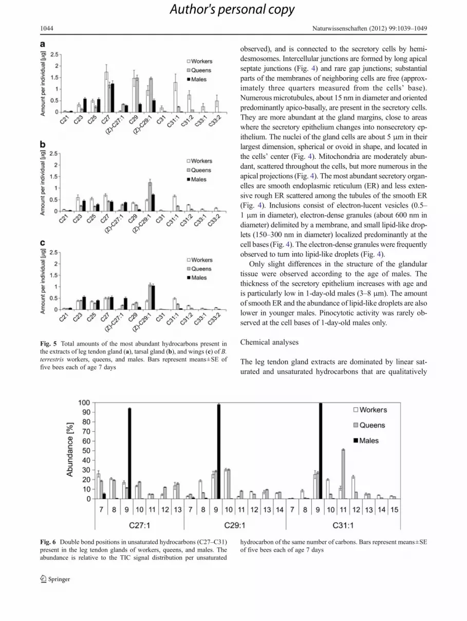
Fig. 5 Total amounts of the most abundant hydrocarbons present inthe extracts of leg tendon gland (a), tarsal gland (b), and wings (c) of B.terrestris workers, queens, and males. Bars represent means±SE offive bees each of age 7 days
Fig. 6 Double bond positions in unsaturated hydrocarbons (C27–C31)present in the leg tendon glands of workers, queens, and males. Theabundance is relative to the TIC signal distribution per unsaturated
hydrocarbon of the same number of carbons. Bars represent means±SEof five bees each of age 7 days
1044 Naturwissenschaften (2012) 99:1039–1049
Author's personal copy

similar in all castes but differ in their quantities and relativeproportions (Fig. 5a). Differences between the castes arefound in the relative abundances of the common hydro-carbons, as well as in hydrocarbons with longer chainlengths and two double bonds that are present only in work-ers and queens (Fig. 5a). The leg tendon glands of males andfemales also differ in regard to the double bond positions inthe unsaturated hydrocarbons. Males produce compoundswith a double bond almost exclusively in position 9 (27:1,29:1, and 31:1), whereas the double bond position is dis-tributed among seven to nine different positions in queensand workers (Fig. 6). All double bonds present in hydro-carbons with an abundance of more than 4 % in the crudeextract were in the Z configuration. In addition, the legtendon gland extracts of the males contain wax esters(Table 1, Fig. 7) that are lacking in the glands of workersand queens.
The relative proportions of the hydrocarbons in the tarsalgland extracts (Fig. 5b) and of the cuticular hydrocarbons(Fig. 5c) are similar within each caste, whereas the compo-sition of hydrocarbons in leg tendon glands differs fromthese two sources (Fig. 5a). ANOSIM results of pairwisecomparisons confirm significant dissimilarities between theleg tendon gland hydrocarbon composition to that of the tarsalglands or cuticular surface, respectively, for males, workers,and queens alike (all Bonferroni corrected p values<0.04). Nosignificant dissimilarities were found by ANOSIM betweentarsal gland and cuticular hydrocarbons for any sex or caste(all Bonferroni corrected p values>0.08). The composition ofthe males’ leg tendon gland secretion shows age-dependentchanges in the absolute amounts of particular hydrocarbonsresulting in differences in their relative composition (Fig. 8).The majority of the compounds considerably increase inamount with the age of a male, except for the hydrocarbons(Z)-nonacos-9-ene and hentriacont-9-ene.
We analyzed in detail the structures of heavier nonvola-tile compounds from the males’ leg tendon and tarsal glands(Table 1). The most abundant wax esters were hexadecylhexadecanoate and hexadecyl octadecenoate in bothextracts. The extracts of the fifth tarsomeres also containedterpenic compounds (2,3-dihydrofarnesol and its esters) thatwere absent from leg tendon glands. The wax esters with thehighest molecular weight identified by GC×GC–MScontained 34 carbons. However, the presence of heavierwax esters with up to 50 carbon atoms was confirmed byMALDI analysis but was not further identified.
Discussion
The leg tendon gland we describe here represents a newmember of the exocrine gland set known from bumblebees.The general structure and location of the leg tendon glands
is similar to that described for the stingless bee Meliponaseminigra (Jarau et al. 2004), with the difference that in B.terrestris the secretory cells are located also in the basitarsusin addition to femur and tibia. The leg tendon gland isclearly an exocrine organ, as evidenced (1) by the increased
Table 1 List of the compounds found in leg tendon and tarsal glandextracts prepared from 7-day-old bumblebee males (extracts of 30glands from five males)
Compounds LRIa Abundance [%]b
Tendongland
Tarsalgland
2,3-Dihydrofarnesol 1694 1.0
Icosene 1995 3.7 2.2
Henicosane 2100 1.5 1.2
Tricos-9-ene 2275 0.9 1.7
Tricosane 2300 13.1 14.7
Tetracosane 2400 2.0 0.6
Pentacos-9-ene 2482 0.5 1.9
Pentacosane 2500 14.6 10.1
Hexacos-9-ene 2581 0.5 0.6
Hexacosane 2600 2.1 1.0
(Z)-Heptacos-9-ene 2682 4.4 11.7
Heptacosane 2700 23.8 13.2
2,3-Dihydrofarnesyl dodecanoate 2760 0.4
Octacos-9-ene 2779 0.6 1.4
Docosenamide 2790 0.5 1.0
Octacosane 2800 1.2 0.5
Squalene 2820 1.8 1.2
(Z)-Nonacos-9-ene 2880 7.4 20.4
Nonacosane 2900 8.9 3.1
Hexadecyl dodecanoate 2973 0.3
2,3-Dihydrofarnesyl tetradecenoate 2964 0.2
Hentriacont-9-ene 3077 0.5 1.3
Hentriacontane 3100 0.3 0.3
Hexadecyl tetradecanoate 3170 0.6 0.2
2,3-Dihydrofarnesyl hexadecanoate 3164 0.5
2,3-Dihydrofarnesyl hexadecenoate 3166 0.2
2,3-Dihydrofarnesyl octadecenoate 3350 0.7
2,3-Dihydrofarnesyl octadecatrienoate 3359 0.4
Hexadecyl hexadecanoate 3365 6.0 1.8
Hexadecyl hexadecenoate 3365 1.8 1.2
Hexadecyl octadecenoate 3537 2.2 3.2
Hexadecyl octadecadienoate 3539 0.5
Hexadecyl octadecatrienoate 3557 0.3 1.4
Hexadecyl octadecanoate 3557 0.4
Hexadecenyl octadecenoate 3557 0.3
a Linear Retention Indexes (LRI)b Abundance of the compounds according to total ion chromatogram(TIC) signal distribution
Naturwissenschaften (2012) 99:1039–1049 1045
Author's personal copy

volume of the secretory cells due to the presence of well-developed secretory organelles (smooth and rough ER), (2)by the presence of projections of the apical plasma mem-brane, and (3) by modifications of the cuticle overlying thesecretory cells, having enlarged pore canals and perforationsthrough the epicuticle allowing the transport of secretionfrom the cells into the gland’s reservoir formed by thehollow tendon. Although the secretory cells are located inthree independent and well-separated regions, we considerthe gland as a single exocrine secretory organ according tothe same ultrastructure of the secretory cells and the jointdelivery of their secretion via the opening of the hollowtendon at the base of the unguitractor plate. Our observa-tions suggest that the leg tendon gland secretion is primarilysynthesized by smooth ER, which is the dominant secretoryorganelle in B. terrestris males’ labial (Šobotník et al. 2008)and leg tendon glands, and is also known to be the mostimportant source of nonpolar compounds including hydro-carbons and wax esters (Fawcett 1966; Percy-Cunninghamand MacDonald 1987; Tillman et al. 1999).
The secretion of the leg tendon glands of B. terrestrismales differs in its composition from that of workers andqueens, supporting our hypothesis that leg tendon glandsecretions of males are used for specific purposes. In partic-ular, they have larger proportions of heavier wax esters withaliphatic alcohol parts. In addition, the composition of theleg tendon gland hydrocarbons differs between the sexes,showing remarkable differences in the double bond posi-tions in the unsaturated hydrocarbons. Interestingly, thedouble bond position in alkenes found in the secretion ofmale leg tendon glands is highly conserved, being almostexclusively at position 9, whereas females synthesizealkenes with double bonds at various positions in theirglands. The position of a double bond influences the chem-ical and physical properties of a molecule, which can beperceived by insects and, thus, can serve to convey specificinformation (Martin et al. 2010). For example, the male sexpheromones from labial gland secretions of the related spe-cies Bombus ruderarius and Bombus sylvarum, seeminglysimilar in composition, contain components with differentdouble bond positions (hexadec-9-enol and octadec-11-enolin B. ruderarius and hexadec-7-enol and octadec-9-enol inB. sylvarum; Terzo et al. 2005). These differences likelyconvey species specificity in the pheromone.
The chemical analyses had shown that the secretion ofthe leg tendon glands differs from both the secretion of thetarsal glands and from the cuticular hydrocarbon profile(represented by wing washes). Nevertheless, leg tendonand tarsal glands share certain compounds that may repre-sent constituent parts of the bumblebee cuticle. We assumethat compounds found in leg tendon gland extracts thatincrease in quantity with bumblebee age are produced bythe glands, while compounds showing unchanged amounts
Fig. 7 Gas chromatographicseparation of the compoundsextracted from leg tendonglands of a Bombus terrestrismale, worker, and queen. Waxesters are predominantly foundin male secretions
Fig. 8 Age-dependent changes of the most abundant compounds fromleg tendon gland extracts of Bombus terrestris males. Bars representmeans±SE of five bees studied per age group (1, 7, and 15 days old).Note that the quantity of all compounds is gradually increasing, exceptfor (Z)-9-C29 and 9-C31
1046 Naturwissenschaften (2012) 99:1039–1049
Author's personal copy

may have been extracted from the cuticle. For example, theamount of (Z)-nonacos-9-ene in the leg tendon glands ofmales does not change with age and also represents the mostabundant hydrocarbon in wing washes. The same applies tohentriacont-9-ene that was detected only in trace amounts inall samples prepared from males. More importantly, howev-er, the amount of the remaining compounds from leg tendongland secretions increased with the age of males, coincidingwith an increased attractiveness of their labial gland secre-tions to virgin queens with the males’ age (Coppée et al.2011).
The function of male-specific compounds from leg ten-don gland secretions remains elusive; however, several pos-sibilities can be assumed. Differences in the composition ofthe secretion as compared to queens and workers stronglysuggest that these compounds serve a communication pur-pose related to sexual behavior. Male leg tendon glandsecretions may be used to mark flight routes, to which virginqueens are attracted for mating, in addition to labial glandsecretions of males known to serve this purpose (Kullenberget al. 1973; Free 1987; Bergman and Bergström 1997; Kindlet al. 1999). The amount of leg tendon gland secretion at amarking spot may tell how many males are using this spot,or how much time has elapsed since marking, based on thehigher volatility of male’s labial gland secretion comparedto the secretion of leg tendon glands. The compounds couldthen influence a queen’s decision of whether to stay or to flyon looking for another marked spot. In that sense, leg tendongland secretions could act as an “arrestment signal.” Thegland secretion may also act as anti-aphrodisiac depositedby the males on queens during mating. The role of anti-aphrodisiacs to render mated females unattractive has al-ready been reported for some bee species, as well as forother insects (Gilbert 1976; Kukuk 1985; Ayasse et al.2001). Interestingly, B. terrestris males regularly drum theirfront legs on a queen’s body during copulation (Jarau andŠobotník, unpublished observation), which may facilitatethe deposition of leg tendon gland secretions on her body.Furthermore, males first investigate the head and abdomenof a queen with their antennae when approaching her in amating attempt (Djegham et al. 1994). This behavior mayserve to asses a queen’s mating status. Since queens of B.terrestris mate only with a single male (Estoup et al. 1995;Schmid-Hempel and Schmid-Hempel 2000) that transfers amating plug to their genital tract during copulation(Duvoisin et al. 1999; Baer et al. 2001; Sauter et al. 2001),any male approaching a queen could gain advantage byquickly identifying whether she is already mated beforemounting and trying to copulate. The mating plug itself,which contains palmitic, linoleic, oleic, and stearic acid, aswell as the cyclic peptide cycloprolylproline (Baer et al.2000), does not alter the mating motivation of males (Baeret al. 2001). Whether specific compounds from leg tendon
glands play a role in species or kin discrimination, to arrestvirgin queens at the males’ marking spots, or serve asinformation about a queen’s mating status needs to be testedin field bioassays.
Our study revealed only slight age-dependent changes inboth the structure of the leg tendon gland tissue and in thechemical composition of the gland secretion in B. terrestrismales. Although the gland ultrastructure is already welldeveloped in 1-day-old males, the overall activity, asevidenced by increased amounts of both smooth ER andlipid droplets, increases as a male gets older. This trendcorresponds to observations on the labial gland structureand changes in composition of their secretions in bumblebeemales, which clearly correlates with the attractiveness ofmales towards virgin queens, which is highest in youngmales, roughly between the age of 5 and 15 days(Šobotník et al. 2008; Žáček et al. 2009; Coppée et al.2011).
Acknowledgments This work was financially supported by theTechnological Agency of the Czech Republic (project no.TA01020969) and by the Academy of Sciences of the Czech Republic(research project RVO: 61388963). S.J. is supported by a researchgrant from the German Research Council, DFG (project no. JA 1715/3-1). The authors also wish to thank Alena Bučánková for providingpart of the biological material and to the staff of the Laboratory ofElectron microscopy (Charles University in Prague) for their help withTEM sample preparation.
References
Arnhart L (1923) Das Krallenglied der Honigbiene. Arch Bienenkunde5:37–86
Ayasse M, Paxton R, Tengö J (2001) Mating behavior and chemicalcommunication in the order Hymenoptera. Annu Rev Entomol46:31–78
Baer B, Maile R, Schmid-Hempel P, Morgan ED, Jones GR (2000)Chemistry of a mating plug in bumblebees. J Chem Ecol26:1869–1875
Baer B, Morgan ED, Schmid-Hempel P (2001) A nonspecific fatty acidwithin the bumblebee mating plug prevents females from remat-ing. PNAS 98:3926–3928
Bergman P, Bergström G (1997) Scent marking, scent origin, andspecies specificity in male premating behavior of two Scandina-vian bumblebees. J Chem Ecol 23:1235–1251
Billen J (2009) Occurrence and structural organization of theexocrine glands in the legs of ants. Arthropod Struct Dev38:2–15
Billen J, Morgan ED (1998) Pheromone communication in socialinsects: sources and secretions. In: Vander Meer RK, Breed MD,Winston ML, Espelie KE (eds) Pheromone communication insocial insects: ants, wasps, bees, and termites. Westview, Boulder,pp 3–33
Carlson DA, Roan C-S, Yost RA, Hector J (1989) Dimethyl disulfidederivatives of long chain alkenes, alkadienes, and alkatrienes forgas chromatography/mass spectrometry. Anal Chem 61:1564–1571
Naturwissenschaften (2012) 99:1039–1049 1047
Author's personal copy

Coppée A, Mathy T, Cammaerts M-C, Verheggen FJ, Terzo M, IserbytS, Valterová I, Rasmont P (2011) Age-dependent attractivity ofmales’ sexual pheromones in Bombus terrestris (L.) [Hymenop-tera, Apidae]. Chemoecology 21:75–82
da Cruz-Landim C, Franco AC (2000) Epithelial bags inside thetibia and femur of males of Centris (Hymenoptera, Antho-phoridae): localization and ultrastructure. Rev Bras Entomol44:97–103
da Cruz-Landim C, Franco AC (2001) Light and electron microscopicaspects of glands and pseudoglandular structures in the legs ofbees (Hymenoptera, Apinae, Euglossini). Braz J morphol Sci18:81–90
da Cruz-Landim C, de Moraes RLMS, Salles HC, Reginato RD (1998)Note on glands present in Meliponinae (Hymenoptera, Apidae)bees legs. Rev Bras Zool 15:159–165
da Cruz-Landim C, Abdalla FC, Gracioli-Vitti LF (2005) Morpholog-ical and functional aspects of volatile-producing glands in bees(Hymenoptera: Apidae). Insect Sci 12:467–480
Dahl F (1885) Die Fussdrüsen der Insekten. Arch mikrosk Anat25:236–262
Detrain C, Deneubourg J-L (2009) Social cues and adaptive foragingstrategies in ants. In: Jarau S, Hrncir M (eds) Food exploitation bysocial insects. Ecological, behavioral, and theoretical approaches.CRC, Boca Raton, pp 29–51
Djegham Y, Verhaeghe JC, Rasmont P (1994) Copulation of Bombusterrestris L. (Hymenoptera: Apidae) in captivity. J Apic Res33:15–20
Duvoisin N, Baer B, Schmid-Hempel P (1999) Sperm transferand male competition in a bumblebee. Anim Behav 58:743–749
Estoup A, Scholl A, Pouvreau A, Solignac M (1995) Monoandryand polyandry in bumble bees (Hymenoptera; Bombinae) asevidenced by highly variable microsatellites. Mol Ecol 4:89–94
Fawcett DW (1966) The cell. Its organelles and inclusions. Saunders,Philadelphia
Federle W, Brainerd EL, McMahon TA, Hölldobler B (2001) Biome-chanics of the movable pretarsal adhesive organ in ants and bees.PNAS 98:6215–6220
Free JB (1987) Pheromones of social bees. Cornell University Press,Ithaca
Giglio A, Ferrero EA, Brandmayr TZ (2005) Ultrastructural identifi-cation of the antennal gland complement in Siagona europaeaDejean 1826, a myrmecophagous carabid beetle. Acta Zool86:195–203
Gilbert LE (1976) Postmating female odor in Heliconius butter-flies: a male contributed antiaphrodisiac? Science 193:419–420
Goulson D (2009) The use of scent marks by foraging bumble bees. In:Jarau S, Hrncir M (eds) Food exploitation by social insects.Ecological, behavioral, and theoretical approaches. CRC, BocaRaton, pp 251–260
Goulson D, Stout JC, Langley J, Hughes WOH (2000) Identity andfunction of scent marks deposited by foraging bumblebees. JChem Ecol 26:2897–2911
Hammer Ø, Harper DAT, Ryan PD (2001) PAST: paleontologicalstatistics software package or education and data analysis. Palae-ontol Electron 4:9, http://palaeo-electronica.org/2001_1/past/issue1_01.htm
Hölldobler B, Wilson EO (1990) The ants. Harvard University Press,Cambridge
Jarau S (2009) Chemical communication during food exploitation instingless bees. In: Jarau S, Hrncir M (eds) Food exploitation bysocial insects. Ecological, behavioral, and theoretical approaches.CRC, Boca Raton, pp 223–249
Jarau S, Hrncir M, Ayasse M, Schulz C, Francke W, Zucchi R, BarthFG (2004) A stingless bee (Melipona seminigra) marks foodsources with a pheromone from its claw retractor tendons. J ChemEcol 30:793–804
Jarau S, Hrncir M, Zucchi R, Barth FG (2005) Morphology andstructure of the tarsal glands of the stingless bee Melipona semi-nigra. Naturwissenschaften 92:147–150
Kindl J, Hovorka O, Urbanová K, Valterová I (1999) Scent marking inmale premating behavior of Bombus confuses. J Chem Ecol25:1489–1500
Kukuk P (1985) Evidence for an antiaphrodisiac in the sweatbee Lasioglossum (Dialictus) zephyrum. Science 227:656–657
Kullenberg B, Bergström G, Bringer B, Carlberg B, Cederberg B(1973) Observations on scent marking by Bombus Latr. andPsithyrus Lep. males (Hym., Apidae) and localization of site ofproduction of the secretion. Zoon Suppl 1:23–30
Lensky Y, Cassier P, Finkel A, Delorme-Joulie C, Levinsohn M(1985) The fine structure of the tarsal glands of the honeybeeApis mellifera L. (Hymenoptera). Cell Tissue Res 240:153–158
Martin JS, Carruthers JM, Williams PH, Drijfhout FP (2010)Host specific social parasites (Psithyrus) indicate chemicalrecognition system in bumblebees. J Chem Ecol 36:855–863
NIST Mass Spec Data Center SES (2008) Mass spectra, 6th edn.National Institute of Standards and Technology, Gaithersburg,http://webbook.nist.gov/chemistry
Noirot C, Quennedey A (1974) Fine structure of insect epidermalglands. Annu Rev Entomol 19:61–80
Percy-Cunningham JE, MacDonald JA (1987) Biology and ultrastruc-ture of sex pheromone-producing glands. In: Prestwich GD,Blomquist GJ (eds) Pheromone biochemistry. Academic,Orlando, pp 27–69
Pouvreau A (1991) Morphology and histology of tarsal glands inbumble bees of the genera Bombus, Pyrobombus, and Megabom-bus. Can J Zool 69:866–872
Reinhard J, Srinivasan MV (2009) The role of scents in honey beeforaging and recruitment. In: Jarau S, Hrncir M (eds) Food ex-ploitation by social insects. Ecological, behavioral, and theoreticalapproaches. CRC, Boca Raton, pp 165–182
Reynolds ES (1963) The use of lead citrate at high pH as anelectron opaque stain in electron microscopy. J Cell Biol17:208–212
Sauter A, Brown MJF, Baer B, Schmid-Hempel P (2001) Males ofsocial insects can prevent queens from multiple mating. Proc RSoc Lond B 268:1449–1454
Schmid-Hempel R, Schmid-Hempel P (2000) Female mating fre-quencies in Bombus spp. from Central Europe. Insect soc47:36–41
Schmitt U, Lübke G, Francke W (1991) Tarsal secretion marks foodsources in bumblebees (Hymenoptera: Apidae). Chemoecology2:35–40
Slaa EJ, Hughes WOH (2009) Local enhancement, local inhibition,eavesdropping, and the parasitism of social insect communica-tion. In: Jarau S, Hrncir M (eds) Food exploitation by socialinsects. Ecological, behavioral, and theoretical approaches.CRC, Boca Raton, pp 147–164
Šobotník J, Kalinová B, Cahlíková L, Weyda F, Ptáček V, Valterová I(2008) Age-dependent changes in structure and function of themale labial gland in Bombus terrestris. J Insect Physiol 54:204–214
Stout JC, Goulson D, Allen JA (1998) Repellent scent-marking offlowers by a guild of foraging bumble bees (Bombus spp.). BehavEcol Sociobiol 43:317–326
1048 Naturwissenschaften (2012) 99:1039–1049
Author's personal copy

Terzo M, Urbanová K, Valterová I, Rasmont P (2005) Intra andinterspecific variability of the cephalic labial glands’ secretionsin male bumblebees: the case of Bombus (Thoracobombus) ruder-arius and B. (Thoracobombus) sylvarum [Hymenoptera, Apidae].Apidologie 36:85–96
Tillman JA, Seybold SJ, Jurenka RA, Blomquist GJ (1999) Insectpheromones—an overview of biosynthesis and endocrine regula-tion. Insect Biochem Mol Biol 29:481–514
Wilson EO (1990) Success and dominance in ecosystems: the case ofthe social insects. In: Kinne O (ed) Excellence in ecology, book 2.Ecology Institute, Oldendorf/Luhe
Žáček P, Kalinová B, Šobotník J, Hovorka O, Ptáček V, CoppéeA, Verheggen F, Valterová I (2009) Comparison of age-dependent quantitative changes in the male labial gland se-cretion of Bombus terrestris and Bombus lucorum. J ChemEcol 35:698–705
Naturwissenschaften (2012) 99:1039–1049 1049
Author's personal copy