laser in situ keratomileusis for myopia and astigmatism
TRANSCRIPT
Laser in situ keratomileusis for myopia and astigmatism: 6 month results
David D. Dulaney, MD, Ronald W. Barnet, MD, Scott A. Perkins, MD, Guy M. Kezirian, MD
ABSTRACT
Purpose: To evaluate the visual and refractive results of laser in situ keratomi leusis (LASIK) for mild to moderate myopia with or without astigmatism.
Setting: Barnet-Dulaney Eye Center, Phoenix, Arizona, USA.
Methods: Data were prospectively collected on 124 consecutive eyes having LASIK over 12 weeks. Eyes with a preoperative spherical equivalent (SE) from - 1.35 to - 10.00 diopters (D) (mean - 4.81 D ± 2.21 [SDJ) and cyl inder from 0 to 5.00 D (mean 1.12 ± 1.12 D) were entered in the study. Th irty-one eyes had spherical corrections. Ninety-three eyes had spherocylinder corrections; preoperative astigmatism in these eyes ranged from 0.50 to 5.00 D (mean 1.47 ± 1.09 D). Surgery included creation of a corneal flap using an automated microkeratome with a 160 J..Lm plate followed by photoablation on the exposed stromal bed . Photoablation was performed using five zones varying from 5.0 to 6.6 mm in eyes with 6.25 D of myopia or less and with five passes at a 5.0 mm zone in eyes with 6.50 D of myopia or more. Astigmatism was corrected using a single-pass ablation through a 6.0 mm slit of varying diameter.
Results: Six month follow-up was obtained in 89 eyes (72%). All eyes were completely re-epithelial ized by the fi rst postoperative day. Uncorrected visual acuity was 20/40 or better in 81 % of eyes at 1 day and in 91 % at 6 months. At 6 months, the mean SE was - 0.35 ± 0. 77 D; 83% were within ± 1 .00 D of plano. Postoperative astigmatism in the 93 eyes having cylinder correction ranged from 0 to 1.22 D (mean 0.38 ± 0.42 D). No eye lost more than two lines of best spectacle-corrected visual acuity. Three eyes (2%) required surgical intervention for cap problems. Visually significant corneal haze was not observed .
Conclusion: In eyes with myopia with or without astigmatism, LASIK provided rapid visual recovery with satisfactory visual and refractive outcomes. The effect of LASIK on visual function (night glare, contrast sensitivity) awaits further study. J Cataract Refract Surg 1998; 24:758- 764
I n laser in situ keratomileusis (LASIK) , the excimer laser is used to perform refractive photoablation on
the corneal stroma beneath a corneal flap that is raised
with a microkeratome. 1 Modern microkeratomes permit the surgeon to vary the flap thickness and make smoother dissections than are possible with manual techniques. 2 A pedicle flap can be created by stopping the keratome before it completes its excursion across the cornea, facilitating flap realignment and protecting against cap loss in the event of a dislocation. 3
Reprint requests to Guy M. Kezirian, MD, SurgiVision Consultants, Inc., 4601 East Mockingbird Lane, Paradise Valley, Arizona 85253, USA.
758 J CATARACT REFRACT SURG-VOL 24, JUNE 1998
LASIK FOR MYOPIA AND ASTIGMATISM
In this series, LASIK for myopia with or without
astigmatism was performed with a self-constructed laser
using an ablation algorithm developed by two of the
authors (D.D.D., R.WB.) specifically for LASIK. Abla
tion diameters varied from 5.0 to 6.6 mm, according to
preoperative refraction. Multizone and multipass treat
ments were used.
The use of various photoablation diameters in
LASIK has been reported.4 Diameters vary from 4.5 to
6.0 mm,5- 7 decreasing with increasing amounts of
correction in most studies. In general, it is desirable to
use an ablation diameter that results in an effective
optical zone that is larger than the pupil diameter in
scotopic conditions. However, to obtain a given correc
tion, the depth of the photoablation must be increased
as the ablation diameter increases. The general relation
ship of ablation depth to diameter is described by
Munnerlyn's formula8: Ablation Depth = DS2/3, where
D = intended dioptric correction and S = diameter of
the intended ablation zone. The depth required to
achieve a given correction increases as the square of the
increase in diameter. Because corneal ectasia occurs
with ablation depths greater than 50%,9 optical zone
size is limited by this relationship. Multizone ablations divide the total ablation into
smaller ablations of varying diameter. Dividing abla
tions into several passes theoretically results in smoother
ablations because of averaging of errors that may exist
in the optical delivery of laser energy. 10 The term
multipass has been used to describe broad-beam excimer
laser ablations that are divided into parts but are
performed at the same diameter. 11
Patients and Methods
This prospective study evaluated the results of
LASIK for mild to moderate myopia with or without
astigmatism. Local institutional review board approval
was obtained. Consecutive eyes having surgery at the
Barnet-Dulaney Eye Center between October 7 and
December 31, 1995, were entered into the study
according to the following criteria: 18 years or older;
preoperative cycloplegic spherical equivalent (SE) be
tween -1.00 and -10.00 diopters (D); preoperative
astigmatism of 5.00 D or less; stable refraction for at
least 6 months documented by objective criteria (refrac-
tion, spectacle power); preoperative best spectacle-cor
rected visual acuity (BSCVA) of 20/20 or better; sur
gery between October 7 and December 31, 1995.
Patients who were pregnant and those with a history of
ocular disease or surgery, keratoconus, connective tissue
disease, or corneal scarring were excluded.
Procedures were performed by two surgeons
(D.D.D., R.WB.). Postoperative care was at the Barnet
Dulaney Eye Center. Patients had LASIK after the
surgeon determined their eligibility and suitability and
after giving informed consent confirming their under
standing of the methods being used.
Methods of Examination Preoperative examination included uncorrected vi
sual acuity (UCVA), intraocular pressure measurement
by applanation tonometry, retinoscopy, manifest and
cycloplegic refractions performed at the spectacle plane,
BSCVA, slitlamp evaluation, dilated fundus evaluation,
and video-assisted corneal topography (EyeSys Labora
tories and Technomed Technology, Inc.).
Patients were instructed to remove soft contact
lenses at least 1 week and hard or rigid gas-permeable
lenses at least 3 weeks before surgery. The corneas of
contact lens patients had to be stable before surgery as
documented through serial topographic examinations.
Visual acuity was tested in dim room conditions
using a Snellen chart projected with an American
Ophthalmics projector at a distance of 10 feet. Line
acuity was recorded as the line with the smallest letters
on which the letters were correctly discerned, with no
more than one letter missed. All refractions were cycloplegic.
Surgical Laser The laser used in this series was a self-constructed,
193 nm broad-beam excimer laser that was subse
quently enrolled in a U.S. Food and Drug Administra
tion Investigational Device Exemption study. The tube
was a Nova Tube manufactured by Lambda-Physik.
The laser was calibrated at a fluence of 130 m]/cm2,
with a frequency of 10 Hz. Fluence was monitored with
a Molectron model EM400 fluence meter (Molectron
Detector, Inc.) during each pass of the laser. Fluence
blocks (Chiron Ophthalmics) were used to confirm
fluence calibration between each case.
J CATARACT REFRACT SURG-VOL 24, JUNE 1998 759
LASIK FOR MYOPIA AND ASTIGMATISM
The treatment algorithms were developed by the Barner-Dulaney Eye Center in conjunction with Lambda-Physik using in-house programmers and a commercially available operating system (Windows 95®, Microsoft Corp.). Eyes with myopia of 0.50 to 6.25 D were treated using a multizone treatment of five zones (3.0, 3.9, 4.8, 5.7, and 6.6 mm), with each zone administered in a single pass. Eyes with myopia of 6.50 to 10.00 D were treated with a single-zone multipass algorithm divided into five equal passes using a 5.0 mm zone. All zones in both treatment algorithms were administered with a collapsing diaphragm to 2.0 mm. A programming delay of approximately 15 seconds occurred between each treatment zone.
Eyes with 0.50 D of cylinder were treated for astigmatism using a 6.0 mm single-pass rectangular slit. The diameter of the beam varied from 0.5 to 4.7 mm across the axis of treatment.
Surgical Technique Preoperative medications were limited to adminis
tration of topical anesthesia (proparacaine 0.5% drops) in the operative eye until anesthetized. The patient was positioned for surgery beneath the microscope, and a Barraquer-style wire speculum was placed. Patients were asked to fixate on a red diode light that was nearly coaxial with the laser beam. A reticule in the surgeon's microscope ocular assisted the surgeon in centering the patient's eye in the operative field.
The cornea was marked with a pararadial marker (Chiron Vision) to facilitate centration. The Steinway Automated Corneal Shaper (Chiron) was used to create a hinged flap using a 160 pm plate and an 8 mm nonadjustable ring (Steinway/Chiron). Flaps were slightly decentered toward the nasal cornea to provide clearance for the stromal photoablation. The photorefractive ablation was performed with the cap reflected over the nasal limbus and the hinge protected with an iris spatula.
Ablations were briefly halted during treatment to permit removal of visible surface moisture using a Merocel sponge. At the conclusion of the photoablation passes, the stromal surface was irrigated briefly with balanced saline solution and the cap replaced by floating it into position. Excess moisture was sponged from the area, and the cap was examined for adherence.
Postoperative Procedure Patients were kept for 30 minutes after surgery and
examined under the operating microscope for cap smoothness and adherence before they were discharged. A Fox shield was placed, but an occlusive dressing was not used.
A slitlamp examination was performed and visual acuity was measured in all eyes the day after surgery and then daily until complete re-epithelialization. Reoperations were limited to cap manipulation; no refractive treatments were performed. Eyes were also examined at 1, 3, and 6 months postoperatively for visual acuity, cycloplegic refraction, tonometry, and computer-assisted corneal topography.
Data Analysis and Reporting The results are presented according to the guide
lines recommended by Waring12 for refractive data presentation. Vector analysis13 was performed on the astigmatism results because it considers cylinder axis when evaluating astigmatic corrections and prevents axis changes from masking overcorrections.
Results
The study included 124 eyes of 68 patients ( 67 right eyes, 57 left). The male:female ratio was 64:60. Preoperative SE ranged from -1.35 to -10.00 D (mean -4.81 D :±:: 2.21 [SO]). Thirty-four eyes had SEs from -1.35 to -3.00 D (mean -2.14 :±:: 0.50 D); 50 eyes, from -3.10 to -6.00 D (mean -4.48 :±:: 0.77 D); and 40 eyes, from -6.10 to -10.00 D (mean -7.48 :±:: 1.21 D). Thirty-one eyes had spherical corrections and 96, spherocylindrical correction for myopia with myopic astigmatism. The preoperative refractive astigmatism in these eyes ranged from 0.50 to 5.00 D (mean 1.47 :±:: 1.09 D).
One surgeon (D.D.D.) operated on 95 eyes and another (R.WB.), on 29. There were no significant differences in the results of the two surgeons in any parameter measured.
The surgical procedure was completed in all 124 eyes. No procedures were terminated for intraoperative complications.
All eyes were seen at 1 day postoperatively. One month examinations were performed in 103 eyes (83%), and 89 eyes (72%) were examined at 3 and 6 months.
760 J CATARACT REFRACT SURG-VOL 24, JUNE 1998
LASIK FOR MYOPIA AND ASTIGMATISM
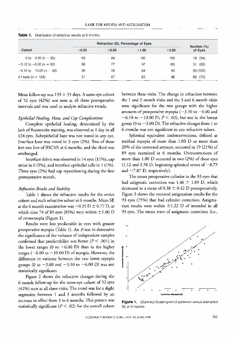
Table 1. Distribution of refractive results at 6 months.
Refraction (D), Percentage of Eyes Number(%)
Cohort ::':: 0.25 ::'::0.50
0 to -3.00 (n = 35) 63 84
-3.12 to -6.00 (n = 50) 58 77
-6.12 to -10.00 (n = 39) 36 56
All eyes (n = 124) 51 67
Mean follow-up was 139 ± 55 days. A same-eye cohort of 52 eyes (42%) was seen at all three postoperative
intervals and was used to analyze refractive trends.
Epithelial Healing, Haze, and Cap Complications Complete epithelial healing, determined by the
lack of fluorescein staining, was observed at 1 day in all
124 eyes. Subepithelial haze was not noted in any eye. Interface haze was noted in 3 eyes (2%). Two of these lost one line ofBSCVA at 6 months, and the third was
unchanged. Interface debris was observed in 14 eyes (11 %), cap
striae in 6 (5%), and interface epithelial cells in 1 (1 %).
Three eyes (2%) had cap repositioning during the first
postoperative month.
Refractive Results and Stability Table 1 shows the refractive results for the entire
cohort and each refractive subset at 6 months. Mean SE at the 6 month examination was -0.35 D ± 0.77 D, at
which time 74 of 89 eyes (83%) were within ± 1.00 D
of emmetropia (Figure 1). Results were less predictable in eyes with greater
preoperative myopia (Table 1). An F-test to determine the significance of the variance of independent samples confirmed that predictability was better (P < .001) in the lower ranges (O to -6.00 D) than in the higher ranges (-6.00 to -10.00 D) of myopia. However, the
difference in variance between the two lower myopia
groups (0 to -3.00 and -3.10 to -6.00 D) was not
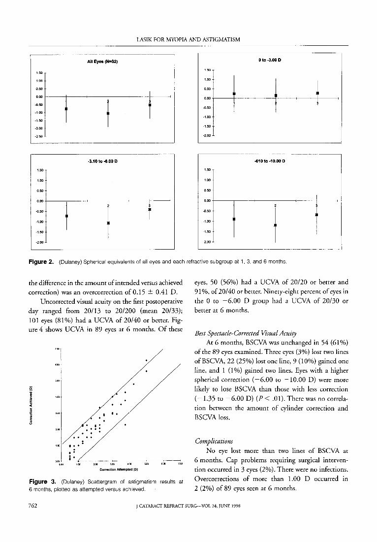
statistically significant. Figure 2 shows the refractive changes during the
6 month follow-up for the same-eye cohort of 52 eyes
(42%) seen at all three visits. The trend was for a slight
regression between 1 and 3 months followed by an increase in effect from 3 to 6 months. This pattern was statistically significant (P < .02) for the overall cohort
::':: 1.00 ::':: 2.00 of Eyes
100 100 19 (54)
97 100 31 (62)
64 90 39 (100)
83 96 89 (72)
between these visits. The change in refraction between
the 1 and 3 month visits and the 3 and 6 month visits
were significant for the two groups with the higher amounts of preoperative myopia (-3.10 to -6.00 and
-6.10 to -10.00 D; P< .02), but not in the lowest
group (0 to -3.00 D). The refractive changes from 1 to
6 months was not significant in any refractive subset.
Spherical equivalent undercorrections, defined as residual myopia of more than 1.00 D or more than
20% of the corrected amount, occurred in 19 (21%) of
89 eyes examined at 6 months. Overcorrections of more than 1.00 D occurred in two (2%) of these eyes (1.12 and 1.50 D, beginning spherical errors of -8.75
and -7.87 D, respectively).
The mean preoperative cylinder in the 93 eyes that
had astigmatic correction was 1.46 ± 1.09 D, which decreased to a mean of 0.38 ± 0.42 D postoperatively.
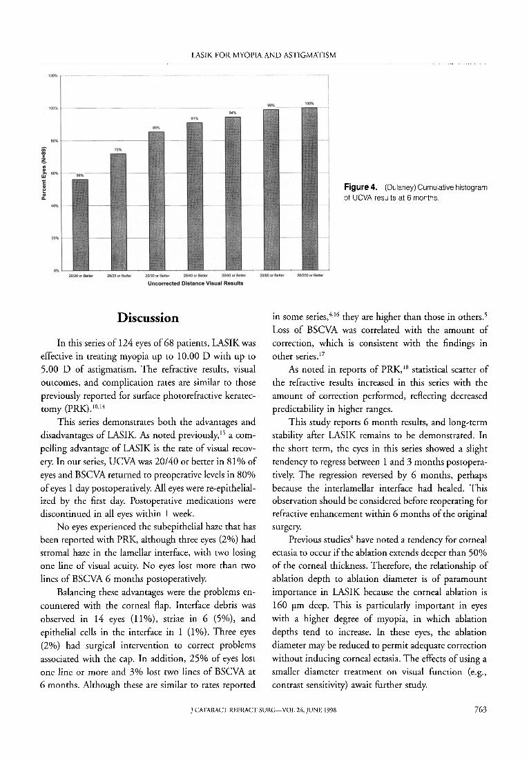
Figure 3 shows the vectored astigmatism results for the 93 eyes (75%) that had cylinder correction. Astigmatism results were within ± 1.22 D of intended in all
93 eyes. The mean error of astigmatic correction (i.e.,
.l~-~· 1----~-----·------+-----·-~----------+--------·--·
0 ' , 3 4 5 6 7 8
Co~Hctfon Attempted (0}
Figure 1. (Dulaney) Scattergram of achieved versus attempted SEat 6 months.
J CATARACT REFRACT SURG-VOL 24, JUNE 1998 761
LASIK FOR MYOPIA AND ASTIGMATISM
All Eyes (N=52) o to -3.00 D
1.50 1.50
1.00 1.00
0.50 0.50
-0.50
-1.00
-1.50
-2.00 t I 0.00
-0.50
-1.00
-1.50
0.00 t---t------<------+--
-2.50 -2.00
·3.10 to -6.00 D -610 to -10.00 D
1.50 1.50
1.00 1.00
0.50 0.50
0
·
00
-1-------;---1 ----+------12-----+------+--0.50
-1.00
-1.50
-2.00
0.00
I 2
-0.50
t -1.00
-1.50
-2.00
Figure 2. (Dulaney) Spherical equivalents of all eyes and each refractive subgroup at 1, 3, and 6 months.
the difference in the amount of intended versus achieved
correction) was an overcorrection of 0.15 ::!:: 0.41 D.
Uncorrected visual acuity on the first postoperative
day ranged from 20/13 to 20/200 (mean 20/33);
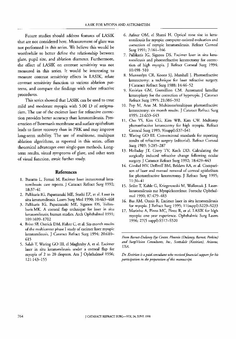
101 eyes (81 %) had a UCVA of 20/40 or better. Figure 4 shows UCVA in 89 eyes at 6 months. Of these
'"I
0.00
• • • . . . . . . . . . . . ••• . • •
j_. -+---- -- ' ------2.00
. . ••
3.00 4.00
Correction Attempted (D)
5.00 6.00 1.00
Figure 3. (Dulaney) Scattergram of astigmatism results at
6 months, plotted as attempted versus achieved.
eyes, 50 (56%) had a UCVA of 20/20 or better and
91%, of20/40 or better. Ninety-eight percent of eyes in
the 0 to -6.00 D group had a UCVA of 20/30 or
better at 6 months.
Best Spectacle-Corrected Visual Acuity At 6 months, BSCVA was unchanged in 54 (61 %)
of the 89 eyes examined. Three eyes (3%) lost two lines
ofBSCVA, 22 (25%) lost one line, 9 (10%) gained one
line, and 1 (1 %) gained two lines. Eyes with a higher
spherical correction ( -6.00 to -10.00 D) were more
likely to lose BSCVA than those with less correction
(-1.35 to -6.00 D) (P< .01). There was no correla
tion between the amount of cylinder correction and BSCVA loss.
Complications No eye lost more than two lines of BSCVA at
6 months. Cap problems requiring surgical interven
tion occurred in 3 eyes (2%). There were no infections.
Overcorrections of more than 1.00 D occurred in
2 (2%) of 89 eyes seen at 6 months.
762 J CATARACT REFRACT SURG-VOL 24, JUNE 1998
LASIK FOR MYOPIA AND ASTIGMATISM
120% - -- ~-·
"'"" '"""
Figure 4. (Dulaney) Cumulative histogram
of UCVA results at 6 months.
20.120 or Bettor 20125 or Bett.f 20130 or Beltltr 20{40 or Belief 20f60 or Better 20{80 or Better 20{2()() or Benet
Uncorrected Distance Visual Results
Discussion
In this series of 124 eyes of 68 patients, LASIK was
effective in treating myopia up to 10.00 D with up to 5.00 D of astigmatism. The refractive results, visual
outcomes, and complication rates are similar to those
previously reported for surface photorefractive keratec
tomy (PRK). 10•14
This series demonstrates both the advantages and
disadvantages of LASIK. As noted previously, 15 a com
pelling advantage of LASIK is the rate of visual recov
ery. In our series, UCVA was 20/40 or better in 81% of
eyes and BSCVA returned to preoperative levels in 80%
of eyes 1 day postoperatively. All eyes were re-epithelialized by the first day. Postoperative medications were discontinued in all eyes within 1 week.
No eyes experienced the subepithelial haze that has been reported with PRK, although three eyes (2%) had stromal haze in the lamellar interface, with two losing one line of visual acuity. No eyes lost more than two
lines of BSCVA 6 months postoperatively.
Balancing these advantages were the problems en
countered with the corneal flap. Interface debris was
observed in 14 eyes (11 %), striae in 6 (5%), and
epithelial cells in the interface in 1 (1 %). Three eyes
(2%) had surgical intervention to correct problems
associated with the cap. In addition, 25% of eyes lost one line or more and 3% lost two lines of BSCVA at
6 months. Although these are similar to rates reported
in some series,4•16 they are higher than those in others.5
Loss of BSCVA was correlated with the amount of
correction, which is consistent with the findings in other series. 17
As noted in reports of PRK, 10 statistical scatter of the refractive results increased in this series with the
amount of correction performed, reflecting decreased predictability in higher ranges.
This study reports 6 month results, and long-term
stability after LASIK remains to be demonstrated. In
the short term, the eyes in this series showed a slight
tendency to regress between 1 and 3 months postopera
tively. The regression reversed by 6 months, perhaps
because the interlamellar interface had healed. This observation should be considered before reoperating for refractive enhancement within 6 months of the original surgery.
Previous studies9 have noted a tendency for corneal ectasia to occur if the ablation extends deeper than 50% of the corneal thickness. Therefore, the relationship of
ablation depth to ablation diameter is of paramount
importance in LASIK because the corneal ablation is
160 rm deep. This is particularly important in eyes
with a higher degree of myopia, in which ablation
depths tend to increase. In these eyes, the ablation
diameter may be reduced to permit adequate correction
without inducing corneal ectasia. The effects of using a smaller diameter treatment on visual function (e.g.,
contrast sensitivity) await further study.
] CATARACT REFRACT SURG-VOL 24, JUNE 1998 763
LASIK FOR MYOPIA AND ASTIGMATISM
Future studies should address features of LASIK that are not considered here. Measurement of glare was
not performed in this series. We believe this would be worthwhile to better define the relationship between
glare, pupil size, and ablation diameter. Furthermore,
the effect of LASIK on contrast sensitivity was not measured in this series. It would be interesting to
measure contrast sensitivity effects in LASIK, relate
contrast sensitivity function to various ablation pat
terns, and compare the findings with other refractive
procedures. This series showed that LASIK can be used to treat
mild and moderate myopia with 5.00 D of astigma
tism. The use of the excimer laser for refractive correction provides better accuracy than keratomileusis. Pres
ervation of Bowman's membrane and surface epithelium
leads to faster recovery than in PRK and may improve
long-term stability. The use of multizone, multipass ablation algorithms, as reported in this series, offers
theoretical advantages over single-pass methods. Longterm results, visual symptoms of glare, and other tests
of visual function, await further study.
References
1. Buratto L, Ferrari M. Excimer laser intrastromal keratomileusis: case reports. J Cataract Refract Surg 1992; 18:37-41
2. Pallikaris IG, Papatzanaki ME, Stathi EZ, et al. Laser in situ keratomileusis. Lasers Surg Med 1990; 10:463-468
3. Pallikaris IG, Papatzanaki ME, Siganos OS, Tsilimbaris MK. A corneal flap technique for laser in situ keratomileusis; human studies. Arch Ophthalmol 1991; 109:1699-1702
4. Brim SF, Ostrick DM, Fisher C, et al. Six-month results of the multicenter phase I study of excimer laser myopic keratomileusis. J Cataract Refract Surg 1994; 20:610-615
5. Salah T, Waring GO III, el Maghraby A, et al. Excimer laser in situ keratomileusis under a corneal flap for myopia of 2 to 20 diopters. Am ] Ophthalmol 1996; 121:143-155
6. Asfour OM, el Shami H. Optical zone size in keratomileusis for myopia: computer-assisted evaluation and correction of myopic keratomileusis. Refract Corneal Surg 1991; 7:161-166
7. Pallikaris IG, Siganos DS. Excimer laser in situ keratomileusis and photorefractive keratectomy for correction of high myopia. J Refract Corneal Surg 1994; 10:498-510
8. Munnerlyn CR, Koons SJ, Marshall J. Photorefractive keratectomy: a technique for laser refractive surgery. J Cataract Refract Surg 1988; 14:46-52
9. Kezirian GM, Gremillion CM. Automated lamellar keratoplasty for the correction of hyperopia. ] Cataract Refract Surg 1995; 21:386-392
10. Pop M, Aras M. Multizone/multipass photorefractive keratectomy: six month results. ] Cataract Refract Surg 1995; 21:633-643
11. Cho YS, Kim CG, Kim WB, Kim CW Multistep photorefractive keratectomy for high myopia. Refract Corneal Surg 1993; 9(suppl):S37-S41
12. Waring GO III. Conventional standards for reporting results of refractive surgery (editorial). Refract Corneal Surg 1989; 5:285-287
13. Holladay JT, Cravy Tv, Koch DD. Calculating the surgically induced refractive change following ocular surgery. J Cataract Refract Surg 1992; 18:429-443
14. Gimbel HV, DeBroffBM, Beldavs RA, et al. Comparison of laser and manual removal of corneal epithelium for photorefractive keratectomy. ] Refract Surg 1995; 11:36-41
15. Seiler T, Kahle G, Kriegerowski M, Wollensak J. Laserkeratomileusis zur Myopiekorrektur. Fortschr Ophthalmol 1990; 87:479-483
16. Bas AM, Onnis R. Excimer laser in situ keratomileusis for myopia.] Refract Surg 1995; 11(suppl):S229-S233
17. Marinho A, Pinto MC, Pinto R, et al. LASIK for high myopia: one year experience. Ophthalmic Surg Lasers 1996; 27(5 suppl):S517-S520
From Barnet-Dulaney Eye Center, Phoenix (Dulaney, Barnet, Perkins) and SurgiVision Consultants, Inc., Scottsdale (Kezirian), Arizona, USA.
Dr. Kezirian is a paid consultant who received financial support for his participation in the preparation of this manuscript.
764 J CATARACT REFRACT SURG-VOL 24, JUNE 1998