laser cleaning experiences on sculptures’ materials: terracotta, plaster, wood, and wax
TRANSCRIPT
Laser Cleaning Experiences on Sculptures’ Materials: Terracotta, Plaster, Wood and Wax
C. Pelosi*a, D. Fodarob, L. Sforzinic, A. Lo Monacod aDepartment of Cultural Heritage Sciences (DISBEC), University of Tuscia, Largo dell’Università
01100 Viterbo, Italy; bIstituto Superiore per la Conservazione e il Restauro, Roma, Italy; cLivia Sforzini restauro e conservazione, Genova, Italy; dDepartment of science and technology for
Agriculture, Forests, Nature and Energy (DAFNE), University of Tuscia, Via San Camillo de Lellis, 01100 Viterbo, Italy
ABSTRACT
The focus of this paper is to show the work experiences with laser cleaning on sculptures made of terracotta, plaster, wood and wax. These materials exhibit peculiar features that often prevent the use of traditional cleaning procedures to remove the surface dirt, soot or carbonaceous deposits and other materials coming from environment or ancient conservative interventions.
To overcome the difficulties in the cleaning of the above mentioned materials, laser technology was tested. The laser irradiation and cleaning tests were carried out with a Q-switched Nd:YAG system under the following conditions: wavelength 1064 nm and 532 nm; energy 4-28 mJ; pulse duration 10 ns; spot diameter 2-8 mm; frequency 5 Hz. The irradiated surfaces were analyzed before and after the laser tests, with the aid of a video microscope and a reflectance spectrophotometer, in order to evaluate the morphology and colour changes of the surfaces.
Before starting with the cleaning intervention, some diagnostic analysis was performed on the sculptures in order to obtain the identification of the original materials and of the surface deposits. Concerning this, Fourier Transform Infrared spectroscopy, X-ray fluorescence spectroscopy, and internal micro stratigraphic analysis were performed.
This research demonstrated that the laser cleaning is an effective method to remove the surface deposits preserving the original patina of the sculptures and the opacity of the wax.
The results gathered in this work encourage to continue the research in order to better understand the interactions between the laser beam and the surfaces and to find the most appropriate laser conditions to clean the sculptures.
Keywords: laser cleaning, sculpture materials, Fourier Transform Infrared spectroscopy, X-Ray fluorescence spectroscopy, internal micro stratigraphic analysis
1. INTRODUCTION Laser cleaning of artworks and antiquities, relying on the use of different types of lasers, has proven successful in many cases; it was widely applied especially for the removal of black crusts or other unwanted layers from stone sculptures and architectural surfaces [1-2]. On the other hand few applications of laser cleaning can be found regarding terracotta objects [1, 3-5] and particularly plaster [6], wood [7-8] and wax artefacts [9]. Nevertheless there is an increasing interest of the conservators in testing the laser cleaning on different kinds of materials in addition to the stone surfaces.
The focus of this paper is to show the work experiences with laser cleaning on sculptures made of terracotta, plaster, wood and wax. These materials exhibit peculiar features that often prevent the use of traditional cleaning procedures to remove the surface dirt, soot or carbonaceous deposits and other materials coming from environment or ancient conservation interventions.
In particular, the use of water or other solvents on terracotta’s objects can easily cause the formation of stains that are difficult to remove.
*[email protected]; phone 0039 0761357684; fax 0039 0761357182; www.disbec.unitus.it
Invited Paper
Fundamentals of Laser-Assisted Micro- and Nanotechnologies 2013, edited by Vadim P. Veiko, Tigran A. Vartanyan,Proc. of SPIE Vol. 9065, 90650X · © 2013 SPIE · CCC code: 0277-786X/13/$18 · doi: 10.1117/12.2052812
Proc. of SPIE Vol. 9065 90650X-1
Downloaded From: http://proceedings.spiedigitallibrary.org/ on 12/09/2013 Terms of Use: http://spiedl.org/terms
The plaster, as material of the sculpture, is particularly difficult to clean for the inherent fragility, the extreme porosity and the water sensibility. The plaster surfaces can be easily damaged by the mechanical cleaning.
Wood is very porous material that can absorb the organic solvents and the water solutions usually adopted by the restores for the surface cleaning. Moreover the wood surface can be easily scratched. Wax is a very malleable and delicate material. The main problem with the cleaning of wax objects is that the surface becomes glossy even using simple cotton flocks.
To overcome the difficulties in the cleaning of the above mentioned materials, laser technology was tested. The laser irradiation and cleaning tests were carried out with a Q-switched Nd:YAG system under the following conditions: wavelength 1064 nm and 532 nm; energy 4-28 mJ; pulse duration 10 ns; spot diameter 2-8 mm; frequency 5 Hz. The irradiated surfaces were analyzed before and after the laser tests, with the aid of a video microscope and a reflectance spectrophotometer, in order to evaluate the morphology and colour changes of the surfaces.
Before starting with the cleaning intervention, some diagnostic analysis was performed in order to obtain the identification of the original materials and of the surface deposits. Concerning this, Fourier Transform Infrared spectroscopy (FTIR), X-ray fluorescence spectroscopy (XRF), and internal micro stratigraphic analysis were performed.
The results of the scientific investigation, together with the examination of the ancient technical manuals, were very useful to produce some laboratory samples in a similar way to the sculptures and their surface finishing [3].
2. METHODOLOGY X-Ray fluorescence spectroscopy was performed by means of a Surface Monitor instrument supplied by Assing. The XRF spectra were obtained with the following experimental conditions: Mo tube operating at 25 kV voltage and 300 μA beam current; scan time 60 s; distance 95 mm.
FTIR spectra were obtained using a Nicolet Avatar 360 Fourier transform spectrometer. For each sample 128 scans were recorded in the 4000 to 400 cm-1 spectral range in diffuse reflection modality (DRIFT) with a resolution of 4 cm-1. Samples were ground with spectrophotometric grade KBr (1% sample in KBr) in an agate mortar. Binders and other materials were identified through infrared spectrometry by comparing the experimental spectra with literature data, spectral databases [10-12] and standards created in our laboratory.
The internal microstratigraphic analysis was performed on a part of each sample mounted as polished cross-sections embedded in polyester resin. The samples were examined using a Zeiss Axioskop polarizing microscope at 25-400x magnification both in incident visible and in UV light. For the UV excitation Zeiss filter 01 (excitation: BP 365/12, beam splitter: FT 395, emission: LP 397) was used. Photomicrographs were taken with a digital Zeiss AxioCam MR.
Thin sections of the wood samples were examined under a Polyvar 100 optical microscope equipped with a PIXeLINK digital camera. They were described according to the standard of the IAWA list of microscopic features for hardwood and softwood identification and the wood taxa identification was carried out according to literature dichotomous keys, as previously described [13].
The laser irradiation and cleaning tests were carried out with the MDTT45 Q-switched Nd:YAG system supplied by MEDICAM. Laser tests were performed under the following conditions, depending on the material to be irradiated: wavelength 1064 nm and 532 nm, energy from 4 mJ to 28 mJ, fluence from 0.01 J/cm2 to 0.90 J/cm2, pulse duration 10 ns, spot diameter from 2 to 8 mm, frequency 5 Hz, and distance from 15 to 30 cm.
Color measurements before and after the laser irradiation were obtained by using an X-Rite CA22 reflectance spectrophotometer [14-15]. The characteristics of the color measuring instrument are the following: color scale CIEL*a*b*; illuminant D65; standard observer 10°; geometry of measurement 45°/0°; spectral range 400-700 nm; spectral resolution 10 nm; measurement diameter 4 mm; white reference supplied with the instrument. Ten measures for each point were performed, then average values and standard deviations were calculated. The CIELAB color system was used where L* describes the lightness while a* and b* describe the chromatic coordinates on the green-red and blue-yellow axes, respectively. The differences in lightness (ΔL*), chromatic coordinates (Δa* and Δb*), and total color (ΔE*) were then calculated using these parameters according to Normal 14/93 (1993) and EN 15886 (2010). The total color difference, ΔE*, between two measurements (L*1a*1b*1 and L*2a*2b*2) is the geometrical distance between their
Proc. of SPIE Vol. 9065 90650X-2
Downloaded From: http://proceedings.spiedigitallibrary.org/ on 12/09/2013 Terms of Use: http://spiedl.org/terms

positions in CIELAB color space. It is calculated using the following equation:
.b*)( + a*)( + L*)( *E 2221,2 ΔΔΔ=Δ
3. RESULTS AND DISCUSSION 3.1 Terracotta sculptures
The laser cleaning of terracotta sculptures was tested and applied on several artefacts belonging to the extraordinary collection of Palazzo Venezia in Rome [3, 16-17]. The experience gathered on these sculptures allowed to find the laser conditions suitable to remove the dirt layers avoiding morphological and chromatic changes of the terracotta. Moreover, the characterization of the surface finishing materials, by different analytical techniques, allowed to choose the most appropriate cleaning systems. In fact, the presence of pigments highly sensitive to the laser irradiation, prevented the use of this system for the cleaning intervention in the areas where the pigments were detected, as for example in the Nile sculpture [3]. In the cases of the terracotta sculptures, the laser cleaning allowed to carry out the conservation work preserving the structural stability of the ceramic bodies and avoiding the pre-consolidation of the artefact.
The terracotta sculptures presented in this paper are: the bas-relief representing Samuel who has king Agar killed (1754, Figure 1) by the sculptor Giuseppe Maria Mazzuoli (1727-1781), and the bas-relief representing Abraham and the Angels (1783, Figure 2) by Filippo Rega (1761-1833), an engraver and medalist. Both sculptures won the Clementine competition at the Accademia di San Luca in Rome.
Figure 1. Photograph of the bas-relief (59x85 cm) representing Samuel who has king Agar killed by Mazzuoli with the colour measuring points, before the cleaning.
The sculpture by Mazzuoli was covered by air pollution deposits strongly sticking to the terracotta, as it was exposed for about 80 years in the porch of Carpegna palace, seat of the Accademia di San Luca.
The FTIR analysis revealed the presence of di-hydrate calcium sulphate (FTIR bands at 3544 cm-1, 3406 cm-1, 1622 cm-1, 1144 cm-1, 672 cm-1 and 603 cm-1), oxalate bands at 1324 cm-1 and 786 cm-1), an acrylic resin (bands at 2921 cm-1, 2854 cm-1, 1731 cm-1, and 1383 cm-1) probably used as protective in past restorations, and traces of proteins (bands at 1648 cm-1 and 1452 cm-1, ) (Figure 3).
Proc. of SPIE Vol. 9065 90650X-3
Downloaded From: http://proceedings.spiedigitallibrary.org/ on 12/09/2013 Terms of Use: http://spiedl.org/terms

%R
eflectanceoo
Ñrn
óoo
1144
3544
3406
29212854
17311648-
1622-
14521383
1324°
672603
786
Figure 2. Photograph of the bas-relief (56x73 cm) representing Abraham and the Angels by Rega with the colour measuring points, before the laser cleaning.
Figure 3. FTIR spectrum of a microsample taken from the surface (near point 2 of Figure 1) of the bas-relief Samuel who has king Agar killed by Mazzuoli.
Laser tests were performed in the points 1-3 and in the same areas colour was measured. The following conditions were used: wavelength 1064 nm, distance 30 cm, spot diameter 8 mm, energy 20 mJ, frequency 5 Hz, fluence 0.04 J/cm2.
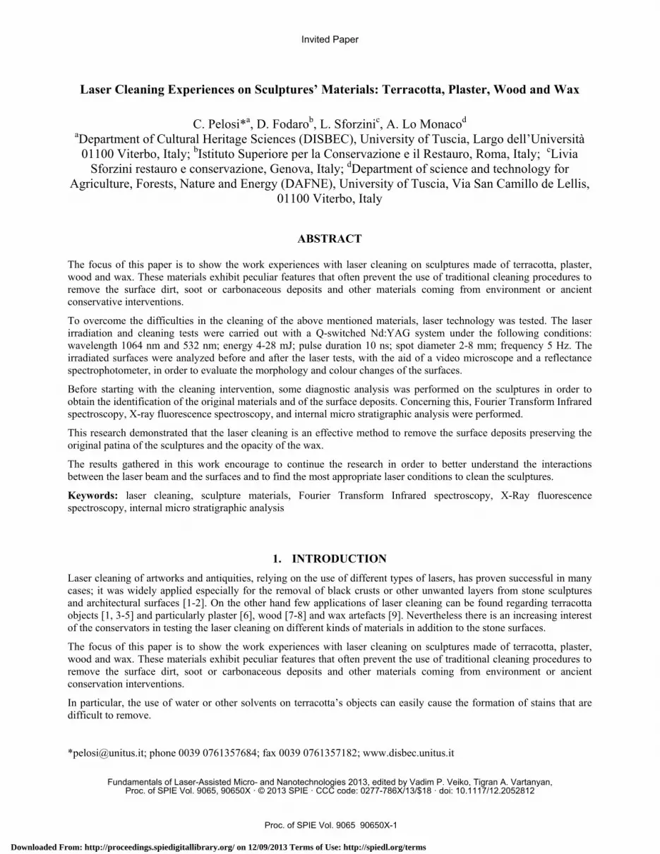
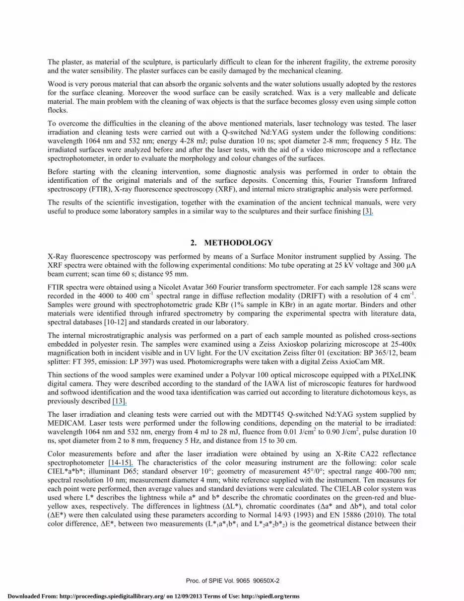
Colour measurements, performed before and after the laser tests, clearly show that the surface reflectance was increased (Figure 4), by removing the black crust, and also the red-yellow components underwent a significant increase (a* and b* coordinates, Figure 5).
Proc. of SPIE Vol. 9065 90650X-4
Downloaded From: http://proceedings.spiedigitallibrary.org/ on 12/09/2013 Terms of Use: http://spiedl.org/terms
40
35 -
30 -
g25-
I 20-
R'A4*404r4 ,arJ
Xrr' }J;rrrrfXr
Z15 -
a rr}}r+x-' ¡.ra+r Ari~+Li=T-'+.- -kIO rr `44
>Fr3l5
400 450 500 550 600 650 700
1(nm)
--*- -Before cleaning 1
--n -- After cleaning l
--i -- Before cleaning 2
--x -- After cleaning 2
--w -- Before cleaning 3
--e -- After cleaning 3
Figure 4. Reflectance spectra of the three measured points on the bas-relief Samuel who has king Agar killed by Mazzuoli, before and after the laser tests.
Figure 5. a*b* colour space showing the measured points on the bas-relief Samuel who has king Agar killed by Mazzuoli, before and after the laser tests.
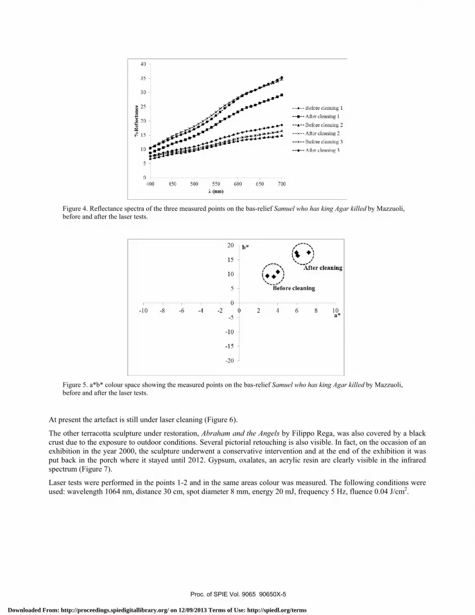
At present the artefact is still under laser cleaning (Figure 6).
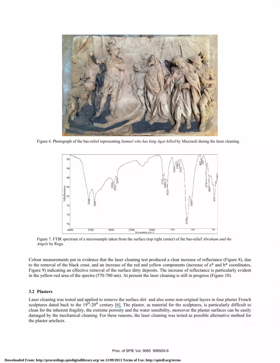
The other terracotta sculpture under restoration, Abraham and the Angels by Filippo Rega, was also covered by a black crust due to the exposure to outdoor conditions. Several pictorial retouching is also visible. In fact, on the occasion of an exhibition in the year 2000, the sculpture underwent a conservative intervention and at the end of the exhibition it was put back in the porch where it stayed until 2012. Gypsum, oxalates, an acrylic resin are clearly visible in the infrared spectrum (Figure 7).
Laser tests were performed in the points 1-2 and in the same areas colour was measured. The following conditions were used: wavelength 1064 nm, distance 30 cm, spot diameter 8 mm, energy 20 mJ, frequency 5 Hz, fluence 0.04 J/cm2.
Proc. of SPIE Vol. 9065 90650X-5
Downloaded From: http://proceedings.spiedigitallibrary.org/ on 12/09/2013 Terms of Use: http://spiedl.org/terms
74
70
1
l----. 0M.
4000 3500 3000 2500 2000Wavenumbers (cm-1)
1500 1000 500
Figure 6. Photograph of the bas-relief representing Samuel who has king Agar killed by Mazzuoli during the laser cleaning.
Figure 7. FTIR spectrum of a microsample taken from the surface (top right corner) of the bas-relief Abraham and the Angels by Rega.
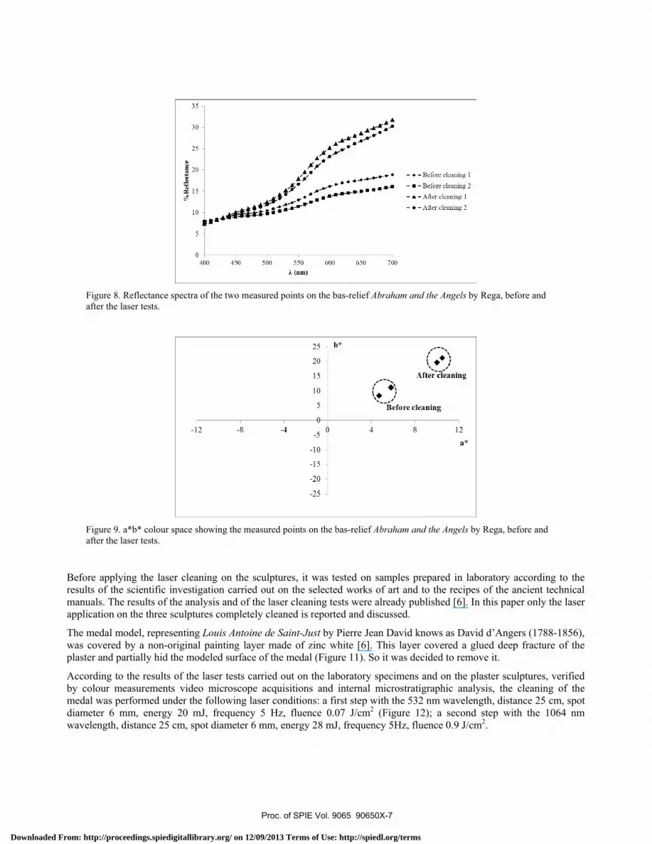
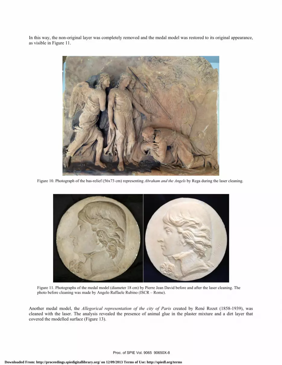
Colour measurements put in evidence that the laser cleaning test produced a clear increase of reflectance (Figure 8), due to the removal of the black crust, and an increase of the red and yellow components (increase of a* and b* coordinates, Figure 9) indicating an effective removal of the surface dirty deposits. The increase of reflectance is particularly evident in the yellow-red area of the spectra (570-700 nm). At present the laser cleaning is still in progress (Figure 10).
3.2 Plasters
Laser cleaning was tested and applied to remove the surface dirt and also some non-original layers in four plaster French sculptures dated back to the 19th-20th century [6]. The plaster, as material for the sculptures, is particularly difficult to clean for the inherent fragility, the extreme porosity and the water sensibility, moreover the plaster surfaces can be easily damaged by the mechanical cleaning. For these reasons, the laser cleaning was tested as possible alternative method for the plaster artefacts.
Proc. of SPIE Vol. 9065 90650X-6
Downloaded From: http://proceedings.spiedigitallibrary.org/ on 12/09/2013 Terms of Use: http://spiedl.org/terms
35 -
5
0
400 450 500 550
(nm)600 650 700
- - Before cleaning 1
- - Before cleaning 2
- - After cleaning 1
- - After cleaning 2
k,After cleaning
*`
Before cleaning
-12 -8 -4
Figure 8. Reflectance spectra of the two measured points on the bas-relief Abraham and the Angels by Rega, before and after the laser tests.
Figure 9. a*b* colour space showing the measured points on the bas-relief Abraham and the Angels by Rega, before and after the laser tests.
Before applying the laser cleaning on the sculptures, it was tested on samples prepared in laboratory according to the results of the scientific investigation carried out on the selected works of art and to the recipes of the ancient technical manuals. The results of the analysis and of the laser cleaning tests were already published [6]. In this paper only the laser application on the three sculptures completely cleaned is reported and discussed.
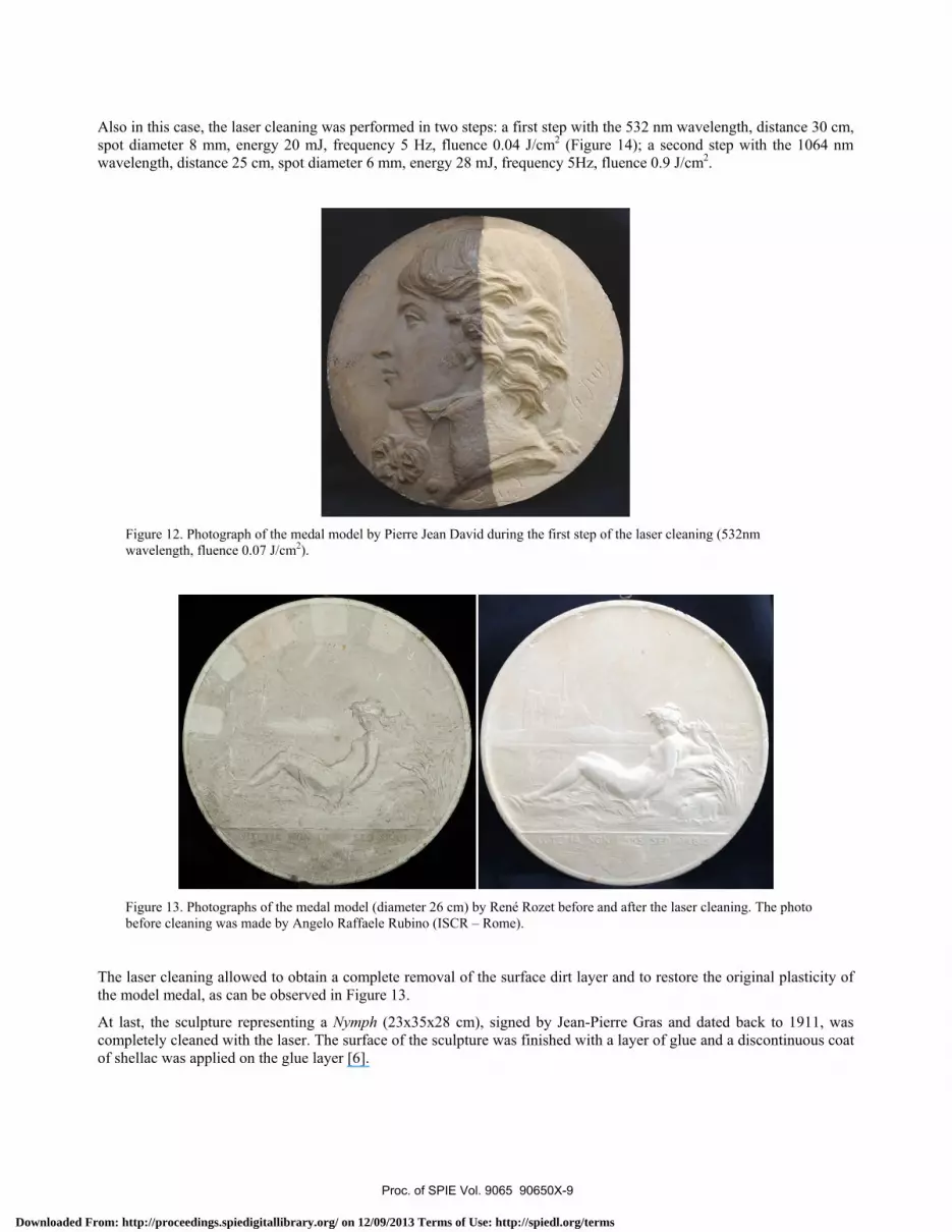
The medal model, representing Louis Antoine de Saint-Just by Pierre Jean David knows as David d’Angers (1788-1856), was covered by a non-original painting layer made of zinc white [6]. This layer covered a glued deep fracture of the plaster and partially hid the modeled surface of the medal (Figure 11). So it was decided to remove it.
According to the results of the laser tests carried out on the laboratory specimens and on the plaster sculptures, verified by colour measurements video microscope acquisitions and internal microstratigraphic analysis, the cleaning of the medal was performed under the following laser conditions: a first step with the 532 nm wavelength, distance 25 cm, spot diameter 6 mm, energy 20 mJ, frequency 5 Hz, fluence 0.07 J/cm2 (Figure 12); a second step with the 1064 nm wavelength, distance 25 cm, spot diameter 6 mm, energy 28 mJ, frequency 5Hz, fluence 0.9 J/cm2.
Proc. of SPIE Vol. 9065 90650X-7
Downloaded From: http://proceedings.spiedigitallibrary.org/ on 12/09/2013 Terms of Use: http://spiedl.org/terms
In this way, the non-original layer was completely removed and the medal model was restored to its original appearance, as visible in Figure 11.
Figure 10. Photograph of the bas-relief (56x73 cm) representing Abraham and the Angels by Rega during the laser cleaning.
Figure 11. Photographs of the medal model (diameter 18 cm) by Pierre Jean David before and after the laser cleaning. The photo before cleaning was made by Angelo Raffaele Rubino (ISCR – Rome).
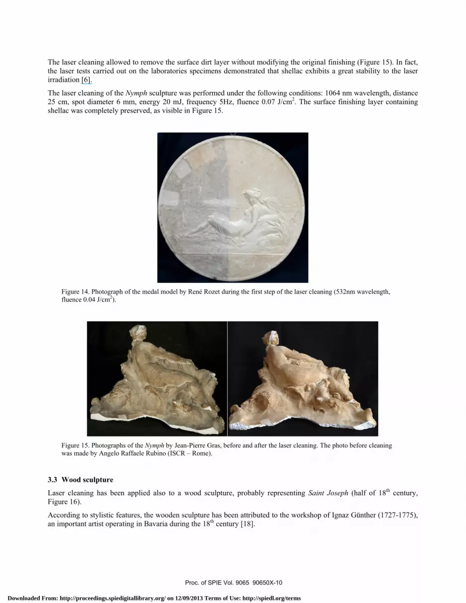
Another medal model, the Allegorical representation of the city of Paris created by René Rozet (1858-1939), was cleaned with the laser. The analysis revealed the presence of animal glue in the plaster mixture and a dirt layer that covered the modelled surface (Figure 13).
Proc. of SPIE Vol. 9065 90650X-8
Downloaded From: http://proceedings.spiedigitallibrary.org/ on 12/09/2013 Terms of Use: http://spiedl.org/terms
Also in this case, the laser cleaning was performed in two steps: a first step with the 532 nm wavelength, distance 30 cm, spot diameter 8 mm, energy 20 mJ, frequency 5 Hz, fluence 0.04 J/cm2 (Figure 14); a second step with the 1064 nm wavelength, distance 25 cm, spot diameter 6 mm, energy 28 mJ, frequency 5Hz, fluence 0.9 J/cm2.
Figure 12. Photograph of the medal model by Pierre Jean David during the first step of the laser cleaning (532nm wavelength, fluence 0.07 J/cm2).
Figure 13. Photographs of the medal model (diameter 26 cm) by René Rozet before and after the laser cleaning. The photo before cleaning was made by Angelo Raffaele Rubino (ISCR – Rome).
The laser cleaning allowed to obtain a complete removal of the surface dirt layer and to restore the original plasticity of the model medal, as can be observed in Figure 13.
At last, the sculpture representing a Nymph (23x35x28 cm), signed by Jean-Pierre Gras and dated back to 1911, was completely cleaned with the laser. The surface of the sculpture was finished with a layer of glue and a discontinuous coat of shellac was applied on the glue layer [6].
Proc. of SPIE Vol. 9065 90650X-9
Downloaded From: http://proceedings.spiedigitallibrary.org/ on 12/09/2013 Terms of Use: http://spiedl.org/terms
The laser cleaning allowed to remove the surface dirt layer without modifying the original finishing (Figure 15). In fact, the laser tests carried out on the laboratories specimens demonstrated that shellac exhibits a great stability to the laser irradiation [6].
The laser cleaning of the Nymph sculpture was performed under the following conditions: 1064 nm wavelength, distance 25 cm, spot diameter 6 mm, energy 20 mJ, frequency 5Hz, fluence 0.07 J/cm2. The surface finishing layer containing shellac was completely preserved, as visible in Figure 15.
Figure 14. Photograph of the medal model by René Rozet during the first step of the laser cleaning (532nm wavelength, fluence 0.04 J/cm2).
Figure 15. Photographs of the Nymph by Jean-Pierre Gras, before and after the laser cleaning. The photo before cleaning was made by Angelo Raffaele Rubino (ISCR – Rome).
3.3 Wood sculpture
Laser cleaning has been applied also to a wood sculpture, probably representing Saint Joseph (half of 18th century, Figure 16).
According to stylistic features, the wooden sculpture has been attributed to the workshop of Ignaz Günther (1727-1775), an important artist operating in Bavaria during the 18th century [18].
Proc. of SPIE Vol. 9065 90650X-10
Downloaded From: http://proceedings.spiedigitallibrary.org/ on 12/09/2013 Terms of Use: http://spiedl.org/terms
CN , 0 0 CN 0 0 0 v 0 0 0
%R
efle
ctan
ce00
0000
00,C
,,C
)s.
0w
,00
WV
i0
1421
29222852
3541
1089
1738 1 10
47
__ -9-8
-4-7
"
681.
_637
--.
612
839
y77
2 3 r'
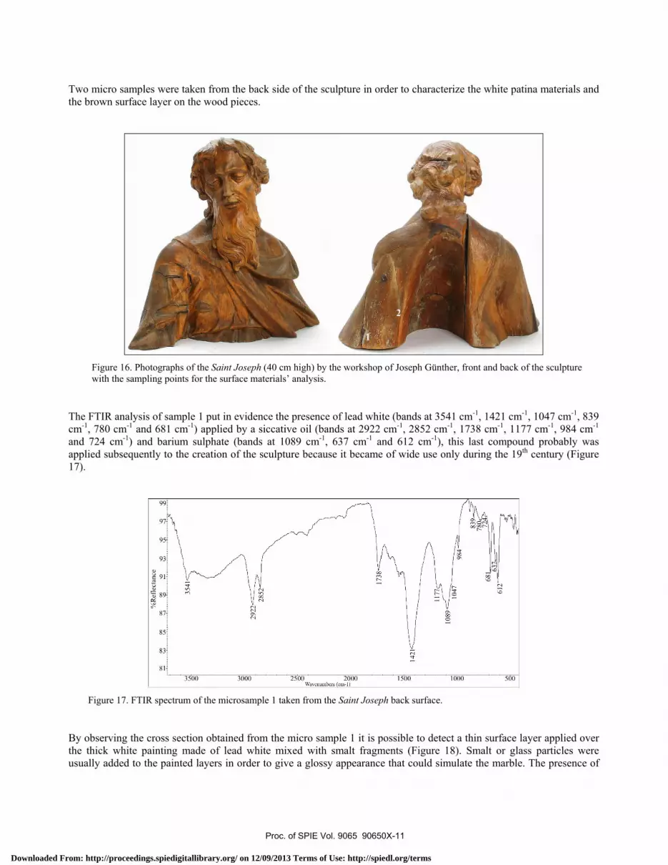
Two micro samples were taken from the back side of the sculpture in order to characterize the white patina materials and the brown surface layer on the wood pieces.
Figure 16. Photographs of the Saint Joseph (40 cm high) by the workshop of Joseph Günther, front and back of the sculpture with the sampling points for the surface materials’ analysis.
The FTIR analysis of sample 1 put in evidence the presence of lead white (bands at 3541 cm-1, 1421 cm-1, 1047 cm-1, 839 cm-1, 780 cm-1 and 681 cm-1) applied by a siccative oil (bands at 2922 cm-1, 2852 cm-1, 1738 cm-1, 1177 cm-1, 984 cm-1 and 724 cm-1) and barium sulphate (bands at 1089 cm-1, 637 cm-1 and 612 cm-1), this last compound probably was applied subsequently to the creation of the sculpture because it became of wide use only during the 19th century (Figure 17).
Figure 17. FTIR spectrum of the microsample 1 taken from the Saint Joseph back surface.
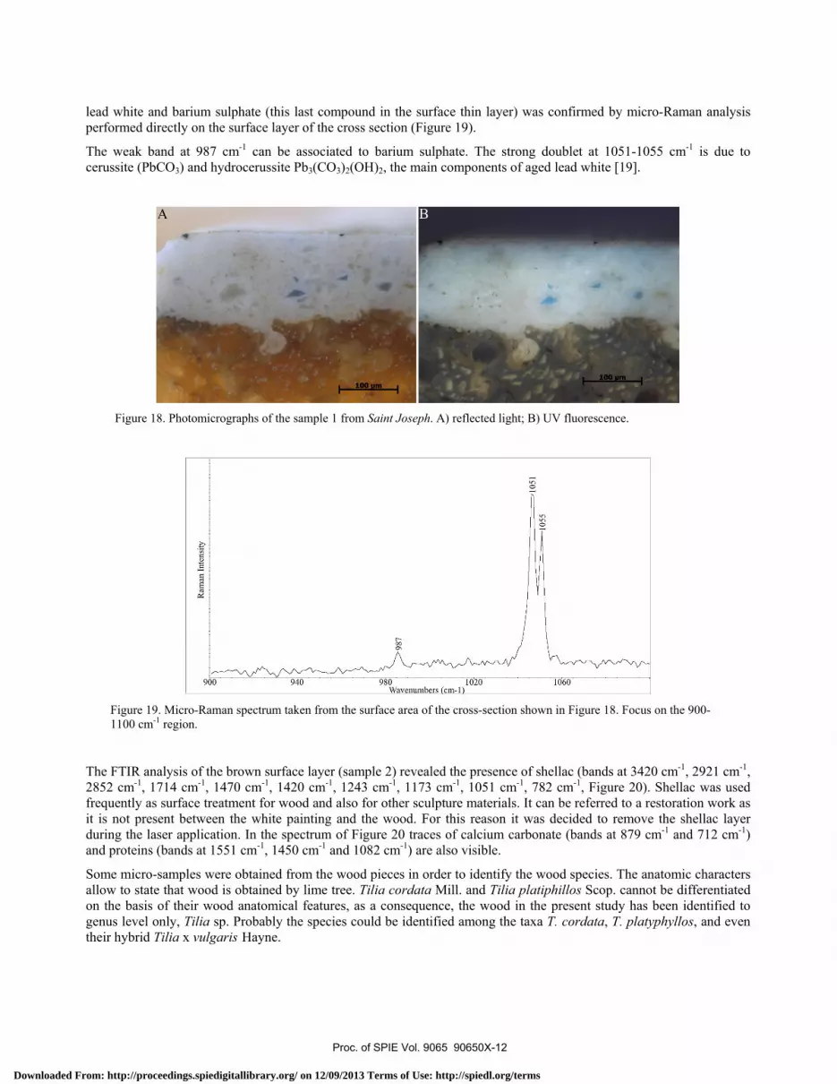
By observing the cross section obtained from the micro sample 1 it is possible to detect a thin surface layer applied over the thick white painting made of lead white mixed with smalt fragments (Figure 18). Smalt or glass particles were usually added to the painted layers in order to give a glossy appearance that could simulate the marble. The presence of
Proc. of SPIE Vol. 9065 90650X-11
Downloaded From: http://proceedings.spiedigitallibrary.org/ on 12/09/2013 Terms of Use: http://spiedl.org/terms
Ram
an I
nten
sity
987
1051
-105
5
lead white and barium sulphate (this last compound in the surface thin layer) was confirmed by micro-Raman analysis performed directly on the surface layer of the cross section (Figure 19).
The weak band at 987 cm-1 can be associated to barium sulphate. The strong doublet at 1051-1055 cm-1 is due to cerussite (PbCO3) and hydrocerussite Pb3(CO3)2(OH)2, the main components of aged lead white [19].
Figure 18. Photomicrographs of the sample 1 from Saint Joseph. A) reflected light; B) UV fluorescence.
Figure 19. Micro-Raman spectrum taken from the surface area of the cross-section shown in Figure 18. Focus on the 900-1100 cm-1 region.
The FTIR analysis of the brown surface layer (sample 2) revealed the presence of shellac (bands at 3420 cm-1, 2921 cm-1, 2852 cm-1, 1714 cm-1, 1470 cm-1, 1420 cm-1, 1243 cm-1, 1173 cm-1, 1051 cm-1, 782 cm-1, Figure 20). Shellac was used frequently as surface treatment for wood and also for other sculpture materials. It can be referred to a restoration work as it is not present between the white painting and the wood. For this reason it was decided to remove the shellac layer during the laser application. In the spectrum of Figure 20 traces of calcium carbonate (bands at 879 cm-1 and 712 cm-1) and proteins (bands at 1551 cm-1, 1450 cm-1 and 1082 cm-1) are also visible.
Some micro-samples were obtained from the wood pieces in order to identify the wood species. The anatomic characters allow to state that wood is obtained by lime tree. Tilia cordata Mill. and Tilia platiphillos Scop. cannot be differentiated on the basis of their wood anatomical features, as a consequence, the wood in the present study has been identified to genus level only, Tilia sp. Probably the species could be identified among the taxa T. cordata, T. platyphyllos, and even their hybrid Tilia x vulgaris Hayne.
Proc. of SPIE Vol. 9065 90650X-12
Downloaded From: http://proceedings.spiedigitallibrary.org/ on 12/09/2013 Terms of Use: http://spiedl.org/terms
%R
efle
ctan
ces.
o N00
1714
1551
1470
1450
1420
1321`
1173
1243
1126
1082 10
51
879 71
61
78
Figure 20. FTIR spectrum of the microsample 2 taken from the Saint Joseph back surface.
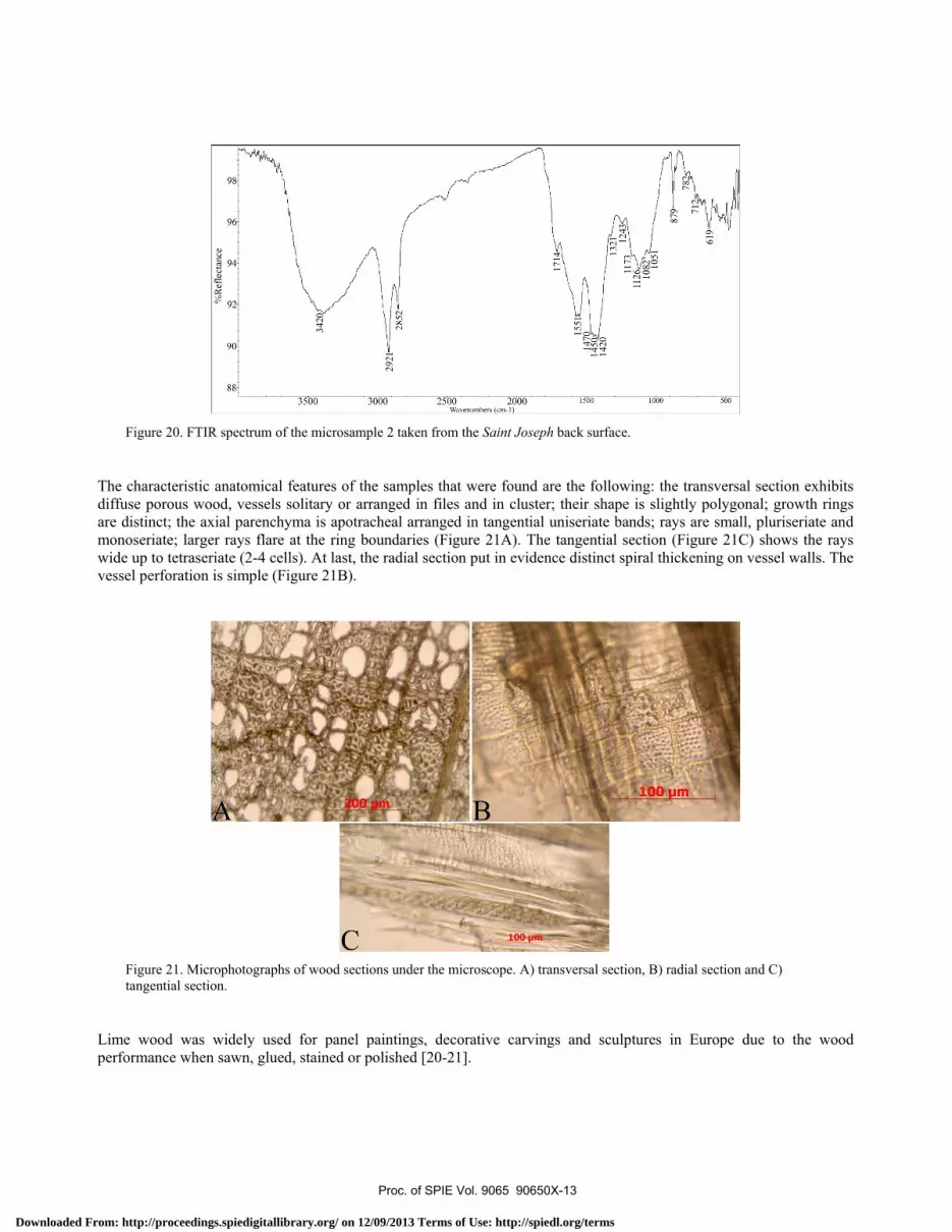
The characteristic anatomical features of the samples that were found are the following: the transversal section exhibits diffuse porous wood, vessels solitary or arranged in files and in cluster; their shape is slightly polygonal; growth rings are distinct; the axial parenchyma is apotracheal arranged in tangential uniseriate bands; rays are small, pluriseriate and monoseriate; larger rays flare at the ring boundaries (Figure 21A). The tangential section (Figure 21C) shows the rays wide up to tetraseriate (2-4 cells). At last, the radial section put in evidence distinct spiral thickening on vessel walls. The vessel perforation is simple (Figure 21B).
Figure 21. Microphotographs of wood sections under the microscope. A) transversal section, B) radial section and C) tangential section.
Lime wood was widely used for panel paintings, decorative carvings and sculptures in Europe due to the wood performance when sawn, glued, stained or polished [20-21].
Proc. of SPIE Vol. 9065 90650X-13
Downloaded From: http://proceedings.spiedigitallibrary.org/ on 12/09/2013 Terms of Use: http://spiedl.org/terms
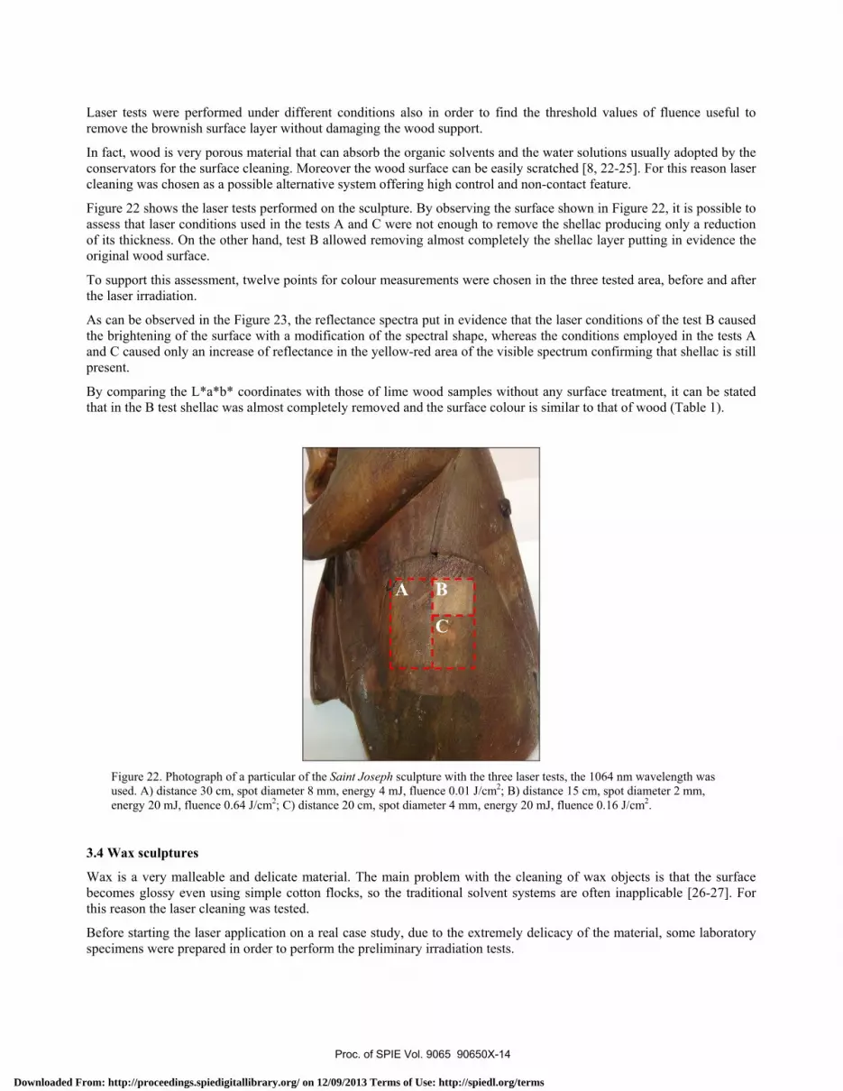
Laser tests were performed under different conditions also in order to find the threshold values of fluence useful to remove the brownish surface layer without damaging the wood support.
In fact, wood is very porous material that can absorb the organic solvents and the water solutions usually adopted by the conservators for the surface cleaning. Moreover the wood surface can be easily scratched [8, 22-25]. For this reason laser cleaning was chosen as a possible alternative system offering high control and non-contact feature.
Figure 22 shows the laser tests performed on the sculpture. By observing the surface shown in Figure 22, it is possible to assess that laser conditions used in the tests A and C were not enough to remove the shellac producing only a reduction of its thickness. On the other hand, test B allowed removing almost completely the shellac layer putting in evidence the original wood surface.
To support this assessment, twelve points for colour measurements were chosen in the three tested area, before and after the laser irradiation.
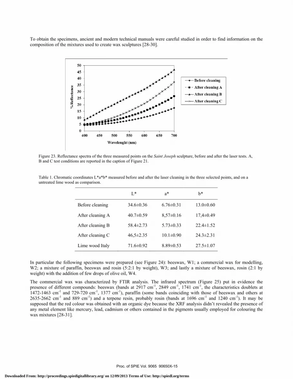
As can be observed in the Figure 23, the reflectance spectra put in evidence that the laser conditions of the test B caused the brightening of the surface with a modification of the spectral shape, whereas the conditions employed in the tests A and C caused only an increase of reflectance in the yellow-red area of the visible spectrum confirming that shellac is still present.
By comparing the L*a*b* coordinates with those of lime wood samples without any surface treatment, it can be stated that in the B test shellac was almost completely removed and the surface colour is similar to that of wood (Table 1).
Figure 22. Photograph of a particular of the Saint Joseph sculpture with the three laser tests, the 1064 nm wavelength was used. A) distance 30 cm, spot diameter 8 mm, energy 4 mJ, fluence 0.01 J/cm2; B) distance 15 cm, spot diameter 2 mm, energy 20 mJ, fluence 0.64 J/cm2; C) distance 20 cm, spot diameter 4 mm, energy 20 mJ, fluence 0.16 J/cm2.
3.4 Wax sculptures
Wax is a very malleable and delicate material. The main problem with the cleaning of wax objects is that the surface becomes glossy even using simple cotton flocks, so the traditional solvent systems are often inapplicable [26-27]. For this reason the laser cleaning was tested.
Before starting the laser application on a real case study, due to the extremely delicacy of the material, some laboratory specimens were prepared in order to perform the preliminary irradiation tests.
Proc. of SPIE Vol. 9065 90650X-14
Downloaded From: http://proceedings.spiedigitallibrary.org/ on 12/09/2013 Terms of Use: http://spiedl.org/terms
50
45
40
35
30
25
20
15
10
5
0
400 450 500 550 600 650 700
Wavelenght (nm)
- -+ -- Before cleaning
-- - After cleaning A
- --A--- After cleaning B
- --x -- After cleaning C
To obtain the specimens, ancient and modern technical manuals were careful studied in order to find information on the composition of the mixtures used to create wax sculptures [28-30].
Figure 23. Reflectance spectra of the three measured points on the Saint Joseph sculpture, before and after the laser tests. A, B and C test conditions are reported in the caption of Figure 21.
Table 1. Chromatic coordinates L*a*b* measured before and after the laser cleaning in the three selected points, and on a untreated lime wood as comparison.
L* a* b*
Before cleaning 34.6±0.36 6.76±0.31 13.0±0.60
After cleaning A 40.7±0.59 8,57±0.16 17,4±0.49
After cleaning B 58.4±2.73 5.73±0.33 22.4±1.52
After cleaning C 46,5±2.35 10.1±0.90 24.3±2.31
Lime wood Italy 71.6±0.92 8.89±0.53 27.5±1.07
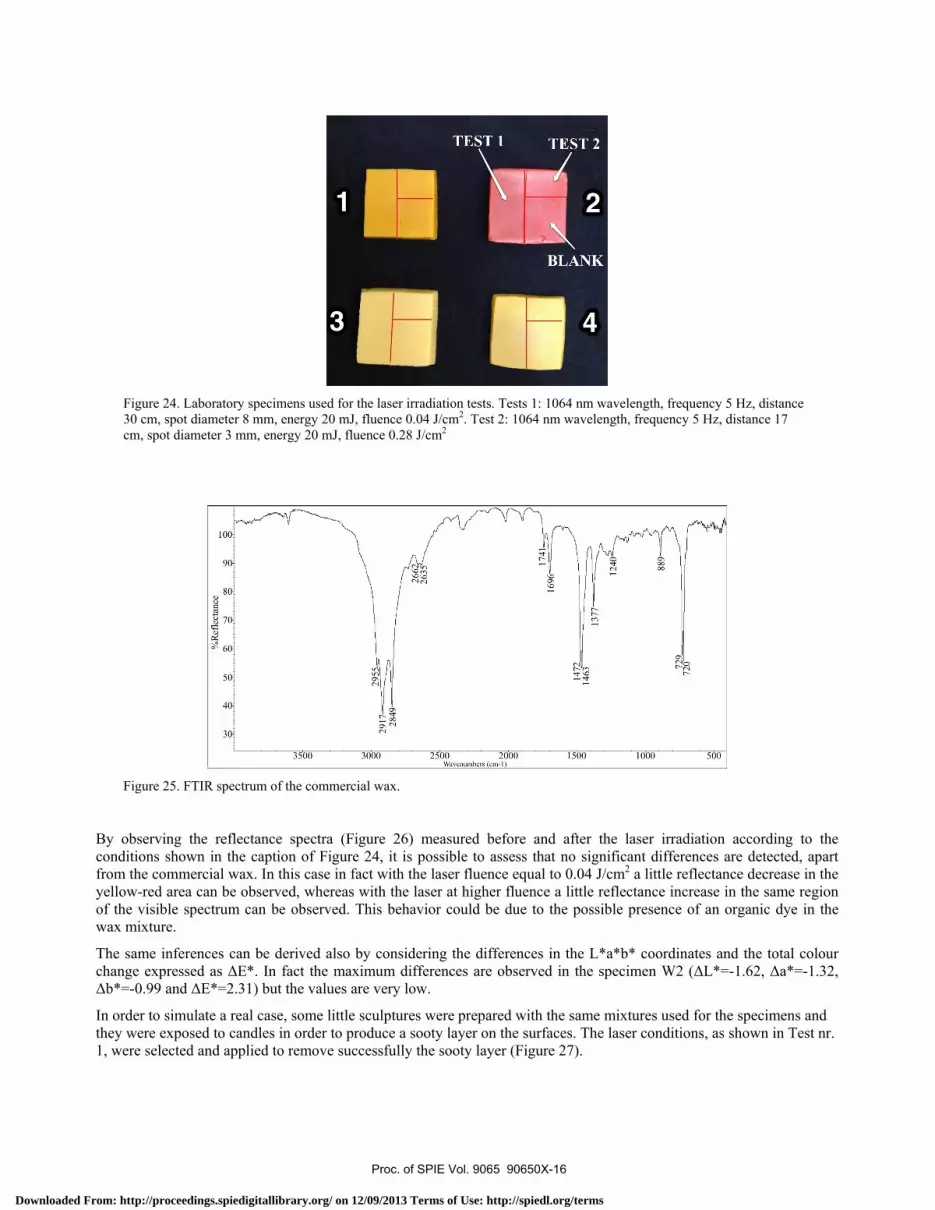
In particular the following specimens were prepared (see Figure 24): beeswax, W1; a commercial wax for modelling, W2; a mixture of paraffin, beeswax and rosin (5:2:1 by weight), W3; and lastly a mixture of beeswax, rosin (2:1 by weight) with the addition of few drops of olive oil, W4.
The commercial wax was characterized by FTIR analysis. The infrared spectrum (Figure 25) put in evidence the presence of different compounds: beeswax (bands at 2917 cm-1, 2849 cm-1, 1741 cm-1, the characteristics doublets at 1472-1463 cm-1 and 729-720 cm-1, 1377 cm-1), paraffin (some bands coinciding with those of beeswax and others at 2635-2662 cm-1 and 889 cm-1) and a terpene resin, probably rosin (bands at 1696 cm-1 and 1240 cm-1). It may be supposed that the red colour was obtained with an organic dye because the XRF analysis didn’t revealed the presence of any metal element like mercury, lead, cadmium or others contained in the pigments usually employed for colouring the wax mixtures [28-31].
Proc. of SPIE Vol. 9065 90650X-15
Downloaded From: http://proceedings.spiedigitallibrary.org/ on 12/09/2013 Terms of Use: http://spiedl.org/terms
%R
efle
ctan
cew
.1,.
,..t,
rn,
00,c
,ó
w v 0 O W1ó O
2955
2917
¡ -_
_T28
49
N to Ó< á C O
' ;_ N O O O
266
263
1741
-,16
96-
ó14
720
1463
____
__T
h
190
7 2
720
1240
889
} 2
Figure 24. Laboratory specimens used for the laser irradiation tests. Tests 1: 1064 nm wavelength, frequency 5 Hz, distance 30 cm, spot diameter 8 mm, energy 20 mJ, fluence 0.04 J/cm2. Test 2: 1064 nm wavelength, frequency 5 Hz, distance 17 cm, spot diameter 3 mm, energy 20 mJ, fluence 0.28 J/cm2
Figure 25. FTIR spectrum of the commercial wax.
By observing the reflectance spectra (Figure 26) measured before and after the laser irradiation according to the conditions shown in the caption of Figure 24, it is possible to assess that no significant differences are detected, apart from the commercial wax. In this case in fact with the laser fluence equal to 0.04 J/cm2 a little reflectance decrease in the yellow-red area can be observed, whereas with the laser at higher fluence a little reflectance increase in the same region of the visible spectrum can be observed. This behavior could be due to the possible presence of an organic dye in the wax mixture.
The same inferences can be derived also by considering the differences in the L*a*b* coordinates and the total colour change expressed as ΔE*. In fact the maximum differences are observed in the specimen W2 (ΔL*=-1.62, Δa*=-1.32, Δb*=-0.99 and ΔE*=2.31) but the values are very low.
In order to simulate a real case, some little sculptures were prepared with the same mixtures used for the specimens and they were exposed to candles in order to produce a sooty layer on the surfaces. The laser conditions, as shown in Test nr. 1, were selected and applied to remove successfully the sooty layer (Figure 27).
Proc. of SPIE Vol. 9065 90650X-16
Downloaded From: http://proceedings.spiedigitallibrary.org/ on 12/09/2013 Terms of Use: http://spiedl.org/terms
400 450 500 550
(nm)600 650 700
- e -W1 BLANK
- G - W1 LASER TEST 1
- e - W1 LASER TEST2
- - W2 BLANK-<- W2 LASER TEST1
-- W2 LASER TEST2- -+ -- W3 BLANK
- - -- -- W3 LASER TEST 1
- -- -- W3 LASER TEST 2
--.- W4 BLANK
-i- W4LASERTEST 1--A- W4 LASER TEST 2
Figure 26. Reflectance spectra of the measured area on the wax specimens. A comparison between the not-irradiated and the laser irradiated surface is reported.
Figure 27. Laboratory sculptures prepared with the same mixtures of samples W1-W4. The vertical dashed lines indicate the separation between the non-irradiated and laser treated areas (1064 nm wavelength, frequency 5 Hz, distance 30 cm, spot diameter 8 mm, energy 20 mJ, fluence 0.04 J/cm2).
Some tests were performed on a real case study, a late 19th – early 20th century female portrait attributed to Ballereau (Figure 28). The sculpture was created on a wood panel, covered by linen prepared with a gypsum layer. The surface appears blackened and several missing parts are clearly visible on the background.
The FTIR analysis put in evidence the presence of beeswax and trace of proteins, probably due to the setting of the wax modelling (Figure 29). The band at 530 cm-1 can be associated to Pb-O bond in red lead.
The XRF analysis confirmed the presence of red lead revealing high counts of lead in all the micro samples analysed. Red lead was frequently used, starting from the Renaissance, to turn red the wax mixtures used to create sculptures and models [28-29, 31]. In a masterpiece by Michelangelo Buonarroti, “A slave” about 1516-19, stored in the Victoria and Albert Museum of London, a clear blackening of the surface has been observed, probably due to red lead alteration [32].
The presence of red lead, a pigment highly sensible to the laser irradiation, prevents the use of the laser cleaning on this artefact.
Proc. of SPIE Vol. 9065 90650X-17
Downloaded From: http://proceedings.spiedigitallibrary.org/ on 12/09/2013 Terms of Use: http://spiedl.org/terms
- «,AP!
b
0 0
2918
2850
%Reflectance
00
00
00
Lo
O0
N°:
1737
165
1639
1545
1472
1511
1463
1420 13
78
1246
117
1079
1024
7371
9
56 530
931 86
2
Figure 28. Photograph of the Female portrait (23 cm high) by F. Ballereau, a late 19th – early 20th century wax sculpture.
Figure 29. FTIR spectrum of a micro-sample detached from the background, near the nose of the female profile.
4. CONCLUSIONS
In this paper the use of laser was tested on different kinds of materials with the aim at finding the most appropriate conditions to clean the surfaces preserving the original substrates and finishing. The materials selected in this work exhibit peculiar features that often prevent the use of traditional cleaning procedures to remove the surface dirt, soot or
Proc. of SPIE Vol. 9065 90650X-18
Downloaded From: http://proceedings.spiedigitallibrary.org/ on 12/09/2013 Terms of Use: http://spiedl.org/terms
carbonaceous deposits and other materials coming from environment or ancient conservation treatments. For this reasons the laser cleaning was used as possible alternative method to the solvent or mechanical systems.
The methodological approach used in this work, hardly ever performed in the daily routine of a conservation laboratory, was based on a preliminary characterization of the surfaces to be irradiated with the laser. In fact, from a conservative point of view, it is of primary importance to know the constitutive materials of the objects to be cleaned in order to choose the most appropriate system (both laser and solvent systems). When possible, laboratory samples were prepared on the basis of the results of the analysis and of the recipes in ancient technical manuals in order to test the laser irradiation before applying it on the real sculptures. This procedure was used for plaster and wax and it proved very useful to verify the applicability of the laser on these materials. The effectiveness of the laser cleaning was explored by means of reflectance spectrophotometry, a simple method giving immediate and easy to understand data to evaluate the surface changes.
A wide experience was gathered on the terracotta sculptures, thanks to the possibility of studying and restoring several artefacts from Palazzo Venezia in Rome [3, 16-17]. This experience allowed performing easily the laser cleaning of two bas-reliefs from the Accademia di San Luca in Rome. The two sculptures were exposed to outdoor and the laser cleaning revealed highly effective to remove the black crust preserving the time oxalate patina.
The laser cleaning of the plaster models demonstrated a powerful method to remove the non-original layers preserving the patinas and the gypsum material. In this case, the laser system was the only possible cleaning method as the aqueous solutions caused the removal of the original materials from the plaster surface [6].
Laser tests were performed on a lime wood sculpture covered by a dark brown layer due to the photodegradation of shellac. The analysis put in evidence that the shellac layer was not original but is was applied subsequently to the creation of the artefact. In fact, the internal micro stratigraphic analysis of a micro sample from a white painting, visible on the back of the sculpture and made of lead white, showed that shellac is not present between the wood and the white painting.
The laser tests, performed under different conditions, allowed finding the suitable parameters to almost completely remove the shellac layer without damaging the wood surface.
At last, laser cleaning was tested on wax. In this case, due to extreme delicacy of the material, some laboratory specimens were prepared in order to perform the preliminary irradiation tests. The results of the laser tests on the laboratory samples put in evidence that no surface modification occurred as a consequence of the laser irradiation and the wax maintained its characteristic opacity. The same results were obtained on little sculptures, prepared in the laboratory and exposed to candles in order to produce a sooty layer on the surfaces. The laser removed completely the soot preserving the wax surface.
Some tests were performed on a real case study, a late 19th – early 20th century female portrait created on a wood panel, covered by linen prepared with a gypsum layer. Unfortunately, in this case, the laser cleaning was inapplicable due to the presence of red lead mixed with the beeswax. In fact, red lead is a pigment highly sensible to the laser irradiation; it was frequently used, starting from the Renaissance, to turn red the wax mixtures used to create sculptures and models.
To conclude, it is possible to assess that the laser cleaning is an effective method to remove the surface deposits preserving the original patina, when present, of the sculptures and the opacity of the wax.
According to our opinion, the laser cleaning, appropriately tested and verified, is an extremely precise and controlled method to remove the dirt layer in a selective and highly controlled modality from different kinds of materials used for sculptures.
The results gathered in this work encourage to continue the research in order to better understand the interactions between the laser beam and the surfaces and to find the most appropriate laser conditions to clean the sculptures.
5. ACKNOWLEDGMENTS The authors would like to thank Prof. Pietro Baraldi of Modena and Reggio Emilia University for the Raman analysis on the cross section from Saint Joseph sculpture and Prof. Angela Cipriani of Accademia di San Luca in Rome.
Proc. of SPIE Vol. 9065 90650X-19
Downloaded From: http://proceedings.spiedigitallibrary.org/ on 12/09/2013 Terms of Use: http://spiedl.org/terms
REFERENCES
[1] Cooper, M., editor, [Laser cleaning in conservation: An introduction], Butterworth-Heineman, Oxford, 1-98 (1998).
[2] Fotakis, C., Anglos, D., Zafiropulos, V., Georgiou, S., Tornari, V., [Lasers in the Preservation of Cultural Heritage. Principles and Applications], Taylor&Francis, New York, 1-365 (2007).
[3] Baraldi, P., Barberini, M.G., Fodaro, D., Martino, L.P.M., Pelosi, C., Sforzini, L., “Use of laser technology for the conservation project of an eighteenth-century terracotta sculpture by Gaspare Sibilla”, Proc. Lasers in the Conservation of Artworks IX, 75-83 (2013).
[4] Oujja, M., Rebollar, E., Castillejo, M., Domingo, C., Cirujano, C. and Guerra-Librero, F., “Laser cleaning of terracotta decorations of the portal of Palos of the Cathedral of Seville”, Journal of cultural heritage 6, 321-327 (2005).
[5] Larson, J., Cooper, M., [The use of laser energy for cleaning architectural terracotta decoration], Architectural ceramics: their history, manufacture and conservation, James and James, London 92-100 (1996).
[6] Pelosi, C., Fodaro, D., Sforzini, L., Rubino, A.R., Falqui, A., “Study of Laser Cleaning on Plaster Sculpures. The Effect of Laser Irradiation on the Surfaces”, Optics and Spectroscopy 114 (6), 917-928 (2013).
[7] Aligizaki, E.M., Melessanaki, K., Pournou, A., “The use of lasers for the removal of shellac from wood”, e-Preservation Science 5, 36-40 (2008).
[8] Wiedemann, G., Schulz, M., Hauptmann, J., Kusch, H.G., Muller, S., Panzner, M., Wust, H., “Laser cleaning applied in the restoration of a medieval wooden panel chamber at Pirna”, Journal of Cultural Heritage 1, 247–258 (2000).
[9] Moreno, T., “Laser Cleaning of Fifteenth and Early Sixteenth Century A.D. Wax Votive Images from St. Peter's Cathedral, Exeter, England”, MRS Proceedings, 852, 002.7 (2004).
[10] The Infrared and Raman user Group (IRUG) Spectral Database, Edition 2000 [http://www.irug.org]. [11] Derrick, M.R., Stulik, D., Landry, J.M., [Infrared Spectroscopy in Conservation Science], The Getty
Conservation Institute, Los Angeles, 1-235 (1999). [12] http://rruff.info. [13] Lo Monaco, A., Mattei, E., Pelosi, C., Santancini, M., “The scientific investigation for the study and
conservation of the wooden Model of S. Maria della Consolazione’s church (Todi, Italy), Journal of Cultural Heritage 14, 537-543 (2013).
[14] Lo Monaco, A., Marabelli, M., Pelosi, C., Picchio, R., "Colour measurements of surfaces to evaluate the restoration materials", Proc. SPIE 8084, 1-14 (2011).
[15] Pelosi, C., Agresti, G., Calienno, L., Lo Monaco, A., Picchio, R., Santamaria, U., Vinciguerra, V., “Application of spectroscopic techniques for the study of the surface changes in poplar wood and possible implications in conservation of wooden artefacts”, Proc. SPIE 8790, 1-14 (2013).
[16] Barberini M.G., Giometti C., Falcucci C., Fodaro D., Pelosi C., Sforzini L., “Sedici terrecotte dalla collezione del Museo Nazionale del Palazzo di Venezia a Roma dalla metà del XVI alla fine del XIX secolo”. Restituzioni – Tesori d’arte restaurati a cura di Carlo Bertelli, Edizioni Intesa San Paolo, Marsilio Editore, Venezia, 256-283 (2011).
[17] Falcucci C., Pelosi C., “Indagini diagnostiche su alcune sculture in terracotta nell’ambito del progetto di ricerca: Il Museo di Palazzo Venezia a Roma e le sue collezioni di scultura”. Sculture in terracotta a cura di Cristiano Giometti, Gangemi, Roma, 190-197 (2011).
[18] Christiane H., “Pygmalion in Bavaria: the sculptor Ignaz Günther and eighteenth-century aesthetic art theory”, University Park, Pa, The Pennsylvania State University Press, 2011.
[19] Bouchard, M., Smith, D.C., “Catalogue of 45 reference Raman spectra of minerals concerning research in art history or archaeology, especially on corroded metals and coloured glass”, Spectrochimica Acta Part A 59, 2247-2266 (2003).
[20] Ministero delle risorse agricole, alimentari e idriche, [FEDERLEGNO-ARREDO Progetto cultura. Il legno nell’arte], IGER, Roma, 1-151 (1995).
[21] Macchioni, N., Lazzeri, S., Sozzi, L., Vitiello, R., “Wooden Sculptures from XVII to XVIII Centuries in the Region of Asti (Italy): Scientific Identification of the Species”, International Journal of Conservation Science 2 (4), 251-260 (2010).
[22] Panzner, M., Wiedemann, G., Henneberg, K., Fischer, R., Wittke, Th., Dietsch, R., “Experimental investigation of the laser ablation process on wood surfaces”, Applied Surface Science 127-129, 787-792 (1998).
Proc. of SPIE Vol. 9065 90650X-20
Downloaded From: http://proceedings.spiedigitallibrary.org/ on 12/09/2013 Terms of Use: http://spiedl.org/terms
[23] Gaspar, P., Rocha, M., Kearns, A., Watkins, K., Vilar, R., “A study of the effect of the wavelength in the Q-switched Nd:YAG laser cleaning of gilded wood”, Journal of Cultural Heritage 1, 133-144 (2000).
[24] Cooper, M., Solajic. M., Usher, G., Ostapkowicz, J., “The application of laser technology to the conservation of a Haida totem pole”, Journal of Cultural Heritage 4, 165s-173s (2003).
[25] Acquaviva, S., D’Anna, E., De Giorgi, M.L., Della Patria, A., Pezzati, L., Pasca, D., Vicari, L., Bloisi, F. Califano, V., “Laser clenaing of gilded wood: A comparative study of colour variations induced by irradiation at different wavelengths”, Applied Surface Science 253, 7715-7718 (2007).
[26] Berzioli, M., [An analytical and applicative approach to the cleaning of artworks], PhD Thesis, University of Parma, Italy, 1-133 (2011).
[27] Berzioli, M., Casoli, A., Cremonesi, P., Fratelli, M., Riggiardi, D., Zorzetti, I., “Verifica analitica dell’idoneità delle soluzioni acquose nella pulitura di statue in cera”, Quaderni Cesmar, 7, Il Prato, Padova pg # (2010).
[28] Lacombe, J., [Dizionario portatile delle belle arti (Portable dictionary of fine arts)], Bassano, Italy, pg # (1781). [29] Milizia, F., [Dizionario delle belle arti del disegno (Dictionary of fine arts of drawing)], Bologna, pg # (1827) [30] Giuffredi, A., [Formatura e Fonderia. Guida ai processi di lavorazione (Moulding and foundry. A Giude to the
working processes)], Alinea, Firenze, pg # (2010). [31] Vasari, G., [Le vite de' piu eccellenti pittori scultori, e architettori], Firenze, pg # (1568). [32] MacLagan, E., “The Wax Models by Michael Angelo in the Victoria and Albert Museum”, The Burlington
Magazine for Connoisseurs 44 (250), 4-16 (1924).
Proc. of SPIE Vol. 9065 90650X-21
Downloaded From: http://proceedings.spiedigitallibrary.org/ on 12/09/2013 Terms of Use: http://spiedl.org/terms