laguncularia racemosa leaves from a mangrove of
TRANSCRIPT

Page 1/33
Laguncularia Racemosa Leaves from a Mangrove ofthe Southeast Atlantic Coast, Brazil: EpicuticularWax, Morphoanatomical Traits and MineralsCristiane Pimentel Victório ( [email protected] )
UEZO https://orcid.org/0000-0002-2815-3725Mayara Silva dos Santos
UEZO: Centro Universitario Estadual da Zona OesteAimêe Cordeiro Dias
UEZO: Centro Universitario Estadual da Zona OesteJoão Pedro Silvério Pena Bento
UFMS: Universidade Federal de Mato Grosso do SulMarcelo da Costa Souza
UFRRJ: Universidade Federal Rural do Rio de JaneiroNaomi Kato Simas
UFRJ: Universidade Federal do Rio de JaneiroRosani do Carmo de Oliveira Arruda
UFMS: Universidade Federal de Mato Grosso do Sul
Research Article
Keywords: Sepetiba Bay, epicuticular wax, leaf cuticle, mangroves, heavy metals, micromorphology
Posted Date: July 23rd, 2021
DOI: https://doi.org/10.21203/rs.3.rs-540578/v1
License: This work is licensed under a Creative Commons Attribution 4.0 International License. Read Full License

Page 2/33
AbstractLeaves of Laguncularia racemosa (L.) Gaertn. f. were collected from the following mangroves alongBrazil’s southeastern Atlantic coast: Coroa Grande (CG), Pedra de Guaratiba (PG) and Marambaia (M).This work aimed to evaluate the presence of minerals by Energy Dispersive X-Ray Spectroscopy (EDS)and Inductively coupled plasma - optical emission spectrometry (ICP-OES); the chemical composition ofepicuticular waxes by gas chromatography–mass spectrometry (GC-MS) and the leaf morphoanatomicalfeatures. Results revealed variation in metal contents among mangroves in the following ranges: Al(0.307–0.73), Cd (0.004–0.016) and Pb (0.095–0.325) mg/mL by ICP-OES. Leaf epicuticular waxcontained more than 50% of triterpenes, in particular the pentacyclic triterpenes lupeol (41.61–55.63%)and β-amyrin (8.81–16.35%), as well as n-alkanes, such as hentriacontane and tetratetracontane. Inparticular, we observed differences in the micromorphology of the epicuticular wax in the leaves of plantsfrom each of the three evaluated sites, especially around stomatal entrances. Histochemical reactionindicated the presence of zinc in �ber cell walls and druse crystals of leaves.
1. IntroductionMangroves are affected by domestic and industrial pollutants. For example, domestic waste increasesphosphates and plasticizers in the environment, while industrial waste varies by industry. These residuesare dumped in rivers and �ow into the mangroves, tending to accumulate as a result of the large amountof organic matter in sediments and/or �ltered particulate matter with the help of tree roots (Souza et al.2018). Such residues may also enter and accumulate into living organisms or come into contact withmicrobes inhabiting the soil and, as a consequence, affect different trophic levels (Fernandes 2012).
Leaves correspond to most of the primary production in the mangrove ecosystem and are the mainconstituents of the litter (Clough et al. 2000), comprising, in turn, the food resource of insects andarboreal crabs. Leaf evaluation allows researchers to detect pollutants in different ecosystems by variousmorphoanatomical changes or changes in plant metabolism (Bargagli et al. 1998; Flores et al. 2018;Victório et al. 2020). Different studies of mangroves have shown the presence of heavy metals andplasticizers in leaves (Alzahrani et al. 2018; Almahasheer et al. 2018; Victório et al. 2020; Victório et al.2021; Flores et al. 2021). These substances are taken up by plants through their leaves, but roots are the�rst and most common organs in contact with pollutants (Cheng et al., 2017). In the roots, pollutants arefreely diffused via apoplast or symplast or carried across cells to xylem from which contaminants aretransported throughout the plant (Kvesitadze et al. 2015).
Sepetiba Bay is an aquatic saline environment surrounded by a large restinga and a mangrove,ecosystems that have been severely impacted by anthropic activities since the 1970s. This Bay hasexperienced an increase in industrialization with the construction of the Sepetiba Industrial Complex, theItaguaí Harbour area and the Industrial District of Santa Cruz. These areas were and are the sites ofseveral metallurgical, petrochemical, and pyrometallurgical smelters, as well as chemical, textile,beverage, and paper plants (SEMA 1998; Wasserman et al. 2013; Victório et al. 2020; Silva and Victório

Page 3/33
2021). The entire region is dubbed the “sacri�ce zone” by the extraordinary damage caused to suchcoastal systems as mangroves, not to mention societal issues arising from the ever-expanding industrialcomplex and port that receives industrial waste (Viégas 2006). Metal smelting, including Zn, Cd, Al, Fe,and alloy steel are the major economic activities located in Sepetiba Bay's basin, followed by thechemical and paper industries (Lacerda et al. 2004; Wasserman et al. 2001, 2013; Tonhá et al. 2020).Previous studies reported improper disposal of solid wastes, such as Zn and Cd, which come from anindustrial site on Madeira Island, one of the main sources of heavy metal to Sepetiba Bay (Fonseca et al.2012). Most heavy metal inputs to the bay arrive from rivers, mainly as drainage from the mostindustrialized and urbanized areas of Sepetiba Bay (Guandu River, Guarda River and Sao FranciscoChannel) (Marins et al. 1999; Fonseca et al. 2012). Cyclical periods and the action of marine tidalcurrents create a dynamism that alters the chemical and physical condition of mangroves; and bringpollutants directly to mangroves (Silvan and Madureira 2012). Heavy metals also reach Sepetiba Baythrough the atmosphere. Atmospheric deposition of pollutants emitted outside the Bay area may furthercontribute to the total heavy metal load (Marins et al. 1999). Heavy metals are not degraded; instead, theybecome concentrated in water, sediments and plants themselves posing a threat to the entire food chain(Almahasheer et al. 2018).
Laguncularia racemosa (L.) Gaertn. f. (Combretaceae) is an arboreal plant which occurs in mangroveswamps on the Atlantic coasts of the Americas and West Africa (Sugiyama 1995, Nyananyo et at. 2009).A typical pioneer found in the interior of the mangroves and in the transition to the restinga forest, L.racemosa is popularly known as mangue-branco. The leaves are slightly succulent with red or purplishpetiole. L. racemosa exhibits a complex root system with �ve types of roots: anchoring, cable,pneumatophores that form pneumathodes at the tip with hypertrophied lenticels, feeding, and lateralaerial roots arising from the pneumatophores (Angeles et al. 2002) that contain lenticels favoringoxygenation in a �ooded environment (Tomlinson 1986). This plant does not have barriers in roots toprotect against the entry of salt that comes from seawater; instead, it is excreted through specializedglands in the leaves, which, in addition, produce solutes in their tissues that contribute to themaintenance of osmotic balance (Kathiresan and Bingham 2001). It is well known that mangrove plantscan absorb, accumulate, degrade/transform and volatilize metals by green remediation processes(Fernandes 2012).
In this study, we investigated the presence of minerals and heavy metals in leaves of L. racemosacollected in different mangroves of Rio de Janeiro situated around Sepetiba Bay - Marambaia, Pedra deGuaratiba and Coroa Grande. In addition, we analyzed the leaf epicuticular waxes for chemicalcomposition and micromorphology, including histochemical tests to evidence the presence of zinc ininternal tissues.
2. Materials And Methods2.1. Plant materials and mangrove areas

Page 4/33
Leaves of Laguncularia racemosa (L.) Gaertn. f. used in the present study were collected from �ve plantsduring the reproductive stage at mangroves around Sepetiba Bay between June and July 2012: CoroaGrande-CG (22°54'42.23"S and 43°52'48.88" W); Pedra de Guaratiba-PG (23° 0'27.98"S and43°37'22.41"W); and Marambaia -M (23° 2'32.46"S and 43°35'43.99"W) (Figs. 1, 2).
Approximately 30 expanded and mature leaves (Fig. 2) were collected from the third to �fth node, fromthe apex of the branches, at a height of approximately 1.75 cm above substrate level, from �veindividuals for each mangrove site. Vouchers are deposited at the Herbarium RBR at UFRRJ, Rio deJaneiro, Brazil.
2.2. Analysis of Sepetiba Bay: physicochemistry and heavy metals
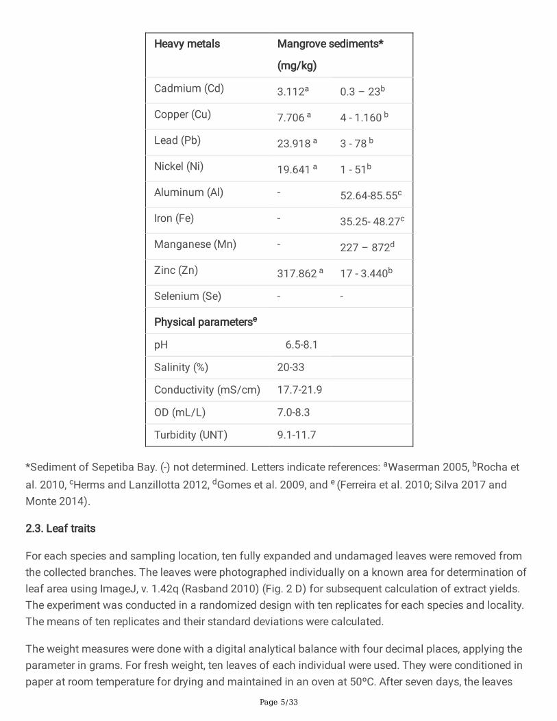
Sepetiba Bay has brackish water according to resolution n. 357/2005 of CONAMA, Brazil (ConselhoNacional do Meio Ambiente) and considering OD, salinity and pH (see Table 1).
Table 1. Total concentration of heavy metals in sediments from Sepetiba Bay, as analyzed by ICP-OESand physicochemical measurements of water at different sampling stations in mangroves aroundSepetiba Bay.
Page 5/33
Heavy metals Mangrove sediments*
(mg/kg)
Cadmium (Cd) 3.112a 0.3 – 23b
Copper (Cu) 7.706 a 4 - 1.160 b
Lead (Pb) 23.918 a 3 - 78 b
Nickel (Ni) 19.641 a 1 - 51b
Aluminum (Al) - 52.64-85.55c
Iron (Fe) - 35.25- 48.27c
Manganese (Mn) - 227 – 872d
Zinc (Zn) 317.862 a 17 - 3.440b
Selenium (Se) - -
Physical parameterse
pH 6.5-8.1
Salinity (%) 20-33
Conductivity (mS/cm) 17.7-21.9
OD (mL/L) 7.0-8.3
Turbidity (UNT) 9.1-11.7
*Sediment of Sepetiba Bay. (-) not determined. Letters indicate references: aWaserman 2005, bRocha etal. 2010, cHerms and Lanzillotta 2012, dGomes et al. 2009, and e (Ferreira et al. 2010; Silva 2017 andMonte 2014).
2.3. Leaf traits
For each species and sampling location, ten fully expanded and undamaged leaves were removed fromthe collected branches. The leaves were photographed individually on a known area for determination ofleaf area using ImageJ, v. 1.42q (Rasband 2010) (Fig. 2 D) for subsequent calculation of extract yields.The experiment was conducted in a randomized design with ten replicates for each species and locality.The means of ten replicates and their standard deviations were calculated.
The weight measures were done with a digital analytical balance with four decimal places, applying theparameter in grams. For fresh weight, ten leaves of each individual were used. They were conditioned inpaper at room temperature for drying and maintained in an oven at 50ºC. After seven days, the leaves

Page 6/33
were unwrapped and weighed to calculate the dry weight. Later, they were weighed for con�rmation of thevalues. Speci�c leaf area (SLA) was calculated using the ratio of the leaf area to the corresponding leafdry weight. These procedures were repeated in the three areas, and the average dry and fresh weight foreach species was calculated.
2.4. Analysis of heavy metals from ashes of leaves by Energy Dispersive X-Ray Spectroscopy (EDS)
The ashes for metal presence analysis were obtained by burning the leaves in a mu�e furnace, asdescribed by the Association of O�cial Analytical Chemists (AOAC, 1995). Six leaves from each studyarea were deposited in porcelain crucibles and previously dried at 60ºC in an air circulation oven for onehour, followed by storage at room temperature. Afterwards, porcelain crucibles that contained leaves wereput in an EDG 3P-S mu�e furnace for 60 min at 800ºC.
Ashes from the burning leaves were analyzed by Energy Dispersive X-Ray Spectroscopy (EDS or EDX,Thermo®, Noran System Six model, coupled to SEM, Jeol JSM-6380L), following the protocol of Resende(2013) with adaptations. The setup works such that the main electron beam of the Scanning ElectronMicroscope (SEM) focuses on the sample. Its atoms emit X-rays. Each element of the periodic table hasspeci�c and well-de�ned energy peaks; consequently, it is possible to identify elements present in thesample. The detector will capture the X-ray photons emitted by the sample and measure its energy inelectron volts (Ev), counting how many photons are detected with certain energy content. From theseresults, a spectrum is produced indicating the number of photons for each X-ray-emitted value. An Ev isthe amount of energy gained by a single electron when accelerated by a power of one volt in vacuum.Qualitative data were obtained by means of replicates.
2.5. Analysis of minerals by spectrometry
Nitric acid digestion of leaves was based on protocols found in the literature (Raposo Junior et al. 2007).In order to determine the minerals, mainly heavy metals (metals with high atomicweight/number/density), leaf samples were dried in an oven at 60°C with air circulation, crushed andsubmitted to digestion with nitric acid 65% ultrapure (Merck) in a sealed Erlenmeyer. To an Erlenmeyerwere transferred 0.2 grams of leaves to which 10 mL of HNO3 were added under an exhaust hood. Then,the samples were placed in an ultrasound apparatus for 30 minutes. Following this, samples weretransferred to an oven to dry at 60°C for 60 minutes. After cooling, an additional 5 mL of HNO3 wereadded and evaporated off at 95±5°C for 2 more hours. Digested samples were �ltered through aquantitative �lter (n0 40), diluted to 50 ml with distilled-deionized water plus 5 drops of HNO3, and storedin Falcon tubes at low temperature until analysis.
Samples were analyzed by Inductively Coupled Plasma Optical Emission Spectrometry (ICP-OES) 2100using a PerkinElmer Optima 7300 V ICP/OES apparatus. To prepare the standard curves, a multi-elementstandard of concentration of 100 mg/L with the same elements (Spex CertiPrep or Fluka®) was used.The calibration curves were adjusted according to the analysis of elements. For Al, Ba, Fe, Zn, Mg, Mn,and Sr, the curves ranged from 0.01 to 1.0 mg/L; for Co, Cr, Cu, Ti, V, Li, Mo, and Ni, they ranged from

Page 7/33
0.005 to 0.2 mg/L; for B, Ca, Na, and K, they ranged from 0.100 to 1.000 mg/L; and for Se, As, Pb, Be, Cd,and Sb, they ranged from 0.002 to 0.025 mg/L. For K, Ca, Na, and Mg, the curves ranged from 1 to 50mg/L, for Si from 1 to 50 mg/L, and for Sn, the curves ranged from 0.01 to 0.1 mg/L. Highly e�cient ionextraction and transport through the mass spectrometer, as well as detection, afford ICP-OES its ultra-trace elemental detection features. Data were obtained by means of three replicates from three to �veindividuals of each mangrove.
2.6. Extraction of epicuticular waxes and chemical analysis by gas chromatography (GC) coupled withmass spectrometry (MS)
After leaf area determination, each leaf was placed in a separate pre-weighed �ask containing 50 mL ofchloroform (spectroscopy grade; Tedia®) and maintained for 30 seconds under gentle manual agitation.This procedure extracts only surface chloroform-soluble compounds without disturbing the leaf interior.Chloroform extracts were maintained at room temperature for solvent evaporation to obtain the solidresidue. The amount of wax was expressed per unit leaf area (µg/mm2). The experiment was conductedin a randomized design with ten replicates for each individual and locality. The means of three replicatesand their standard deviations were calculated. Two mg of dried extract were dissolved in 200 mLchloroform. Subsequently, 1 µL was injected into a gas chromatograph (GC-2010-Shimadzu) equippedwith a flame ionization detector (FID). The analyses were performed on a DB-1MS capillary column (30 mx 0.25 mm x 0.2 mm), using He as carrier gas at 1 mL/min and a split ratio (50:1). Temperature wasincreased by 10oC/min from 140 to 300oC and then maintained at 300oC for 15 min. The injector wasmaintained at 290oC and the detector at 300oC. Quantification was performed from GC/FID profiles,using relative area (%). Identification of n-alkanes was based on injection of commercial standards(Sigma Fluka® Alkane standard solution C21-C40). Analysis of a subsample was performed in a GC/MSQP 2010 Plus Shimadzu mass detector, using the same operating conditions as above (except thecolumn; ZB-5MS column 30 m x 0.25 mm x 0.2 mm), and the MS scanned for 50–650 amu at 2sec/decade with an electron impact ionization potential of 70 eV. Triterpenoid compounds were identifiedby comparison of the corresponding mass spectra with library data (Spectrum Libraries: NIST05.LIB)complemented with proton nuclear magnetic resonance (1H NMR) spectrometry (Bruker DRX 400 MHz),using deuterated chloroform as solvent.
2.7. Microscopy analyses
For light microscopic observations and measurements, three leaves from each site were �xed in FAA,dehydrated in ethanol series, and stored in ethanol 70% (Johansen, 1940). Middle portions of leaves werecut in appropriate pieces and embedded in glycolmethacrylate (Historesin®), cross sectioned (8 µmthickness) in a rotary microtome (Leica RM 2155), and stained with Toluidine Blue 1% (O'Brien et al.1965). Photomicrographs of leaf sections were made using light microscopy (Leica) �tted with a digitalcamera. The measurements were taken from images obtained from leaf cross sections using a NikonEclipse CI microscope equipped with a digital camera (Moticam Pro 252b). To identify the presence ofzinc in plant tissues, free-hand cuts were performed on collected leaves, dehydrated at room temperature

Page 8/33
and exposed to Zincon® reagent, following the method of Seregin et al. (2015) and Seregin andKozhevnikova (2011). For scanning electron microscopy observations (SEM), segments of dry leaveswere mounted on stubs and coated with a thin layer of gold (Denton vacuum Desk IV, LLC). The abaxialand adaxial surfaces of leaves were analyzed with a JEOL-JSM 6390 LV scanning electron microscopy(JEOL, Tokyo, Japan). To analyze the epidermis in frontal view and count the secretory glands/mm2, leafepidermises were obtained after treatment with a dissociation solution of hydrogen peroxide and glacialacetic acid (1: 1) and heating in an oven at 60ºC for 24h (Franklin 1945). Afterwards, the epidermiseswere washed in distilled water many times to remove the dissociation solution. Then, they were stainedwith 0.05% basic fuchsine aqueous and mounted in 50% glycerin on semi-permanent slides (Kraus andArduin 1997).
2.8. Statistical analyses
Morphoanatomical data from leaves and mineral content obtained by ICP were subjected to a two-wayanalysis of variance (ANOVA), multiple comparisons, considering data for each mangrove (CG, PG andM), followed by Tukey`s test. A difference was considered to be statistically signi�cant when P< 0.05. Allstatistical analyses were performed with GraphPad Prism software, version 8.0 for Windows.
3. Results3.1. Morphological leaf traits
Leaf weight of L. racemosa from Marambaia showed high values in comparison with leaves from CGand PG. Meanwhile, the leaves from PG presented low values for weight and area (Fig. 3). Data from leafarea, together with leaf weight, con�rm the low average development of L. racemosa in PG compared toCG and M. Signi�cant difference attributable to leaf area was observed when the data from threelocations were analyzed, showing that leaves from PG had smaller values. Leaves from M presentedlower speci�c leaf area (SLA) than that from CG and PG, suggesting the in�uence of pollutants on plantdevelopment since CG and PG are surrounded by widespread industrialization.
3.2. Epicuticular wax chemical composition
The amount of triterpene content in epicuticular wax per leaf follows the descending order of CG> PG.(Table 2). Epicuticular wax composition presented the major pentacyclic triterpene fagarasterol (lupeol)and -amyrin found in plants of L. racemosa from CG and PG mangroves. Considering lupeol and -amyrin, the chemical pro�les of L. racemosa leaf waxes were consistent. The n-alkanes hentriacontaneand 8-octadecanone were identi�ed in leaves from CG, while tetratetracontane was only found in leafsamples from PG. This is the �rst time the epicuticular wax of L. racemosa from mangroves of thesoutheast Atlantic coast has been chemically pro�led.
Table 2. Composition of triterpenes identi�ed in epicuticular wax of Laguncularia racemosa collected inCoroa Grande (CG), Pedra de Guaratiba (PG) and Marambaia (M) mangroves, Rio de Janeiro (Brazil), as

Page 9/33
obtained by gas chromatography.
Constituent RT (min) Relative area (%)*
CG PG M
Hentriacontane 61.50 28.01 nd nd
Tetratetracontane 66.17 nd 10.93 nd
8-octadecanone 70.16 6.31 nd nd
β-amyrin 74.63 8.81 16.35 nd
Fagarasterol (lupeol) 75.57 53.63 41.61 nd
Total triterpenes 62.44 57.96 nd
*Mean amount of wax extracted with chloroform. RT – Retention time obtained by GC. nd- not detected.
3.3. Mineral content analyzed by EDS
The analysis of leaf ash made it possible to quantify the ATOM (%), which is the percentage in number ofatoms of the elements present in the sample. Using EDS, the major essential mineral levels found inleaves of L. racemosa collected in M, CG and PG were Na>Cl>K>Ca>K. These minerals are common insaline ecosystems that have input of seawater (Fig. 4a).
The presence of heavy metals was found mainly in leaves of L. racemosa from CG and PG. For leafsamples from M, only Al, Si and Fe were detected. Zn was detected only in leaves from CG, the sitenearest the industrial center (CG) (Fig. 4b). Cd was not detected.
3.4. Mineral content analyzed by ICP- OES
As shown in Table 3, it was possible to observe higher levels of calcium, magnesium and potassium, bothin CG and PG, when compared to the levels of the same minerals in M.
Among the heavy metals in leaf samples, Cd and Zn were identi�ed. The levels of the heavy metals As, Al,Cr, Cu, Pb and Se were similar for L. racemosa samples from all three mangroves evaluated. PG and CGsamples showed equal results in terms of the order that they bioaccumulated in the leaves:Fe>Al>Zn>Mn>Pb>Cu>Cd (Table 3), with Cd varying by having a higher level in PG (0.016 mg/L)compared to CG (0.0012 mg/L). The order of accumulation of metals in leaves of L. racemosa fromMarambaia was similar to that found in the other mangroves; however, Al (0.37 mg/L) was detected at ahigher concentration than Fe (0.35 mg/L). For comparison, the total concentration of heavy metals withina normal range for plants is presented in Table 3. Speci�cally, the normal range in plants is 0.03 to 0.9mg/kg for Cd and 50.8 mg/kg for Zn (Table 3). When compared to chemical �ngerprinting of referenceplants, L. racemosa leaves contain a lower concentration of heavy metals (Table 3). For analysis ofminerals by ICP, collections were made at two points in CG. Differences between the two CG points

Page 10/33
(CG1 and CG2) were observed; speci�cally, Al and Na presented higher content in CG2, while Sr and Capresented lower content in CG2 when compared to CG1 (Table 3).
Table 3. Content of minerals, heavy metals and other chemical elements in leaves (mg/kg) ofLaguncularia racemosa by ICP-OES compared to reference plants. Micro- and macronutrients areseparated based on physiological use by plants. Leaves were collected in mangroves around SepetibaBay, Rio de Janeiro (Brazil): Coroa Grande (CG), Pedra de Guaratiba (PG) and Marambaia (M).

Page 11/33
Mineral(mg/kg)
CG1 CG2 PG M Referenceplants†
Metals
Al 0.31±0.31 0.74±0.45 0.38±0.1 0.37±0.061 80
As 0.002±0 0.002±0 0.002±0 0.002±0
Ba 0.025±0.007 0.027±0.004 0.039±0.0005 0.025±0.004 40
Be 0.002±0 0.002±0 0.002±0 0.002±0
Cd 0.004±0.003 0.032±0.01 0.016±0.01 0.009±0.02 0.03-0.9
Pb 0.095±0.16 0.32±0.13 0.16±0.12 0.1±0.17 1.54
Li** 0.005±0 0.005±0 0.005±0 0.005±0
Sr 0.46±0.07 0.29±0.036 0.45±0.147 0.24±0.07
Cr 0.005±0 0.005±0 0.005±0 0.005±0
Sn 0.01±0 0.01±0 0.01±0 0.01±0
Ti** 0.008±0.002 0.006±0.003 0.005±0 0.005±0 5.0
V 0.005±0 0.005±0.0 0.005±0 0.005±0
Micronutrients
Co 0.005±0 0.005±0.0 0.005±0 0.005±0 0.2
B 0.126±0.45 0.1±0.0 0.21±0.06 0.35±0.79
Mn* 0.088±0.0026 0.091±0.007 0.20±0.02 0.13±0.033 15-100
Mo 0.01±0 0.01±0.0 0.1±1.69 0.01±0
Ni* 0.007±0.0081 0.005±0.0 0.087±0.141 0.005±0.004 3.10
Zn* 0.30±0.0587 0.26±0.11 0.33±0.025 0.25±0.16 50.80
Cu* 0.024±0.0051 0.028±0.005 0026±0.002 0.018±0.003 10.0
Fe* 0.27±0.0982 0.33±0.08 0.51±0.006 0.36±0.09 89.22
Macronutrients
Na 31.988±6.84 68.71±5.512 38.33±4.73 92.0±11.56
Mg 15.778±3.03 12.718±1.13 14.034±3.83 9.71±3.006
Ca 62.94±11.10 37.25±3.10 69.23±19.85 42.18±8.50
K 27.09±5.43 29.126±1.78 26.025±2.63 29.063±7.23

Page 12/33
Other elements
Se 0.005±0.0 0.007±0.001 0.006±0.001 0.006±0.001
Si 0.602±0.16 0.5±0.07 0.55±0.04 0.76±0.24 0.1
Sb*** 0.002±0.0 0.002±0.001 0.002±0.0 0.002±0.0 (<0.5) 1.5††
* Also heavy metals, but considered micronutrients used by plant. **Light metal. ***Antimony (or stibium)metalloid. †References: Markert 1992, values in mg/Kg dry weight. ††Levels in leaves of different plants:Pérez-Sirvent et al. 2011. 1,2For analysis of minerals by ICP, collections of leaves were made at two pointsin CG.
3.5. Microscopic diagnosis
The qualitative and quantitative parameters evaluated in L. racemosa leaves from the three sites arepresented in Fig. 6.
In investigated plants, the epicuticular wax layer covering the leaf surface presented distinct patterns,those being plates, granules, rodlets and crusts, according to the site of collection (Fig. 5). Leaves fromMarambaia presented epicuticular wax in crusts and granules covering most of the surface area, but inscales near stomata (Fig. 5a, b). In leaves from Pedra de Guaratiba, the wax layer consisted of crusts andplates throughout the surface, including stomatal cells (Fig. 5c, d). In leaves from Coroa Grande, theepicuticular wax consisted of granules over common epidermal cells, but a rod pattern near stomata (Fig.5e, f). Pores of the cavity where salt secretory glands are located were seen in both surfaces (Fig. 5g, h).
In cross section, the epicuticular wax and cuticular layer are not easily distinguished under lightmicroscopy, so they were measured together. The thickness of wax+cuticular layer was smaller in Mcompared to that for CG for both sides with few differences on the adaxial side between CG and PG (Fig.6).
Laguncularia racemosa has amphistomatic leaves; the ordinary epidermal cells present straight walls(Fig. 6a, b). Unicellular trichomes were detected only in plants collected in CG (Fig. 6a). In cross section,the epidermis consisted of a single layer of epidermal cells (Fig. 6d-f). Common epidermal cells storedphenolic compounds in both surfaces more intensely as detected in plants from CG (Fig. 6e). The adaxialepidermal cells in individuals from M were higher than those of individuals from PG (Fig. 6).
Salt-secreting glands are located in epidermal cavities that invade the mesophyll (Fig. 6 b, c-e). Thenumber of glandular trichomes was constant between the two faces of the leaf and also between thethree sites evaluated (2-3 glands/mm2). The secretory structure is supported by short peduncle cells with3-4 (basal cells) sustaining the secretory cells that form a nearly globose structure (Fig. 6b-c, e). Theepidermal cells that surround the opening of the cavity (pore) are palisade-like in cross section, with

Page 13/33
tabular shape, containing phenolics. The salts, mucilage and other substances excreted by this secretorystructure �rst accumulate in the cavity and thereafter are likely released to the external environmentthrough the epidermal pore (Fig. 6e).
The mesophyll is heterogeneous and isolateral in leaves from the three sites investigated and wascomprised of two layers of adaxial and abaxial palisade parenchyma with shorter cells when comparedwith adaxial layers and 4-5 layers of spongy, or water storage, parenchyma (Fig. 6d-f). The thickness ofthe mesophyll was greater in leaves from PG, but less so in plants from M (Fig. 7). The length of palisadeparenchyma cells (adaxial and abaxial) was similar in the leaves evaluated for individuals of the samesite; however, when different sites were compared, longer cells were observed in PG and shorter cells inCG. In the �rst layer of palisade parenchyma under both sides of the epidermis, a strong reaction tophenolic compounds was observed (Fig. 6d-f). The median part of mesophyll is occupied by a spongyparenchyma, similar to water storage parenchyma, with voluminous cells with few, or no, chloroplasts(Fig. 6c). This tissue is thicker in leaves from CG than that from PG. In all samples evaluated, the cell wallof water storage parenchyma presented intense reaction to the mucilage test. The mucilage wasobserved spread between cells in the mesophyll (Fig. 6f). Idioblasts with druse crystals were observed inthe cells of the spongy parenchyma, proportionally more abundant in PG leaves (Fig. 6). Druse crystalspresented positive reaction to the Zincon® histochemical test with more intensity in leaves from CG andPG, indicating the presence of this metal (Fig. 6i-j).
The vascular bundles are the collateral type with �bers in the most developed units, forming a capprotecting the phloem (Fig. 6c). Positive reaction to the presence of zinc was observed in vascular �bersin CG and PG leaves (Fig. 6i-j). A parenchymal sheath (endodermis) is present and is better observed inthe larger bundles. It does not touch the epidermis. Phenolic compounds and idioblasts with drusecrystals are associated with vascular tissue. Regarding the vascular system, we did not observe anydifference among leaf samples from different collection sites.
Midvein - The epidermal cells on the adaxial face had a rectangular shape (Fig. 6g), and those on theabaxial face were square to papillose. The height of the epidermal cells and the cuticle layer was higherin PG and lower in M. Under adaxial and abaxial epidermal sides, we observed 6-8 layers and 1-2 layers,respectively, of regular parenchyma storing phenolic compounds and crystals. In the region of the mainvein, the parenchyma cells are isodiametric, separated by intercellular spaces �lled with mucilage (Fig.6g). The main vascular bundle is the collateral type, with a reniform shape, surrounded by �bers andcollenchyma (Fig. 6h). The width of the main rib is proportional to the length in height of the mainvascular bundle, longer in PG and shorter in CG. The width of the main rib was longer in M and shorter inCG.
DiscussionAmong the studied mangroves of Sepetiba Bay, Marambaia is thought to be the most preserved since itlies east of the Coroa Grande and Santa Cruz industrial complex and, hence, the most distant mangrove

Page 14/33
from anthropic pressure. It is also located in an area under military protection. In 2010 Ferreira et al.found that the concentration of heavy metals in Sepetiba Bay did not exceed the limits recommended inCONAMA Resolution No. 344/2004. However, Cd, Zn and Cu in the water did show unsatisfactory valuesin some parts of Sepetiba Bay when assessed according to resolution Nº357/2005. One of the manyfactors that alter the concentration of minerals in Sepetiba Bay is the frequent movement of tidewaterand sediment from water entering the bay. In addition, industrial activities have production rates that canalternately reduce and increase pollution. Sepetiba Bay is a coastal area sensitive to regionalenvironmental changes that can create an ecotone where interactions between land and water take place.According to Ribeiro et al. (2013), metal mobility may also change in the short term. The average of dryweight showed a decreasing pattern following the order M> CG> PG (Fig. 4).
Severoglu et al. (2015) suggest that heavy metals constitute one of the main abiotic agents related togrowth reduction and alteration in physiological processes. The high SLA value of L. racemosa collectedin CG and PG mangroves is consistent with the pollution data based on proximity to the industrialcomplex. SLA is an important indicator of the impact of pollution in the environment, and it can may be aparameter involved in protection and adaptation of plants (Wuytack et al. 2011).
The amount of Na is three times greater in leaves of Marambaia compared to leaves examined from CGand PG mangroves. This result could be attributed to the degree of salinity in the region where a highhydrodynamism is known to signi�cantly change salinity. Soil salinity can interfere with the uptake andexchange of ions in the soil-plant system, which, in turn, is re�ected in the morphology and structure ofplants (Bartz et al. 2015).
Leaf structure of L. racemosa was analyzed by Francisco et al. (2009) who reported the presence of leafglands and trichomes, as well as salt secretory glands. They considered these features to be adaptationsto the saline ecosystem. Here we added information about the quantity of salt secretory glands in theleaves of L. racemosa plants growing in different mangroves around Sepetiba Bay and data about themicromorphology of epicuticular wax. Some microscopic views showed the high salt content on leavesand around salt glands (Fig. 5). Evidence suggests that mature leaves of L. racemosa can secrete saltaccording to its concentration in the soil. That is, if salt increases in the substrate, then salt secretion willbe higher in leaves (Sobrado 2004). Through salt glands, salt-tolerant plants also can expel heavy metals(Arrivabene et al. 2016). Leaf structures are particularly relevant to the ecological success of this species,which occupies environments where salinity is high. No leaf samples from mangroves evaluated showedmorphological or structural damage.
The plant cuticle consists of nonpolar substances such as cutin, which is the matrix associated withwaxes. It is secreted by cells of the epidermis and deposited on an organ of the plant’s surface, such asthe leaf. It also seals �owers, stems, and fruits, protecting them from biotic and abiotic stresses (Kunstand Samuels 2009). Intracuticular wax is embedded into the cuticle, and epicuticular wax is found on thecuticle's surface (Koch and Ensikat, 2005). Cuticular waxes are involved in guarding against excessiveleaf water loss (residual transpiration) (Hasanuzzaman et al. 2017). The presence of n-alkanes and

Page 15/33
triterpenes, as the main constituents of epicuticular wax, is recurrent among restinga and mangroveplants that use residual transpiration as a mechanism of tolerance under salinity stress (Oku et al. 2003;Zorat et al. 2011; Hasanuzzaman et al. 2017; Victório et al. 2020). Pentacyclic triterpenoids, such as 𝛽-amyrin and lupeol, which are representative of the oleanane and lupane groups, are widely distributed inmangrove plants associated with features tolerant to salt (Basyuni et al. 2012). Pentacyclic triterpeneswere identi�ed as the main components of A. shaueriana epicuticular wax (Victório et al. 2020), as wellas the main components of L. racemosa leaves in this study. However, our results are different fromthose of Ra�i et al. (1996) who veri�ed only trace amounts of triterpenes in leaf wax of L. racemosa fromGuyana (western Atlantic coast) and 8.5% in wax extract in a population from Gabon (eastern Atlanticcoast) that did not present lupeol.
The presence of hydrocarbons in leaf epicuticular wax has proven valuable in chemotaxonomic studiesof different botanic families. Alkanes were the most abundant compounds among the hydrocarbons,highlighting hentriacontane (28.01-45.36%) in leaf wax collected in CG. The hydrocarbons hentriacontaneand octadecanone are present in leaves of Rhizophora mangle from Africa (Dodd et al. 1995), andhentriacontane is present in leaves of A. shaueriana (Victório et al. 2020). The presence of thehydrocarbon hentriacontane is recorded for the epicuticular waxes of several plant species (Wang et al.1999), but this was the �rst evidence of its presence in the Laguncularia genus. As constituents ofepicuticular wax, n-alkanes, but not triterpenes, seem to be related to a reduction in cuticular water loss(Buschhaus and Jetter 2012).
In studies with conifer (Pinus sylvestris L.) seedlings, Burkhardt and Pariyar (2014) veri�ed that airpollution degraded (´erosion`) epicuticular waxes that revealed an amorphous appearance resulting inlow drought tolerance. These symptoms, which are easily visible through an analysis of the leaf surfaceby scanning electron microscopy (SEM), are provoked by very diverse chemical environments, such asacid rain or fog, simultaneous exposure to SO2 and NH3 or car exhaust, as pointed out by Viskari et al.(2000). Studies of Arrivabene et al. (2015) detected a higher amount of particulate material, includingiron, on the leaf surface of A. schaueriana and L. racemosa, the leaves of which contain salt glands incomparison to Rhizophora mangle. The presence of hydrophobic epicuticular wax covering the leafblades may be associated with the absorption of chemicals of equal polarity, such as phthalates,pesticides and others. Plasticizers were detected in the leaf epicuticular wax of A. shaueriana and R.mangle from the same mangroves (Victório et al. 2021). Cuticular waxes also act in controlling loss anduptake of polar solutes (Hasanuzzaman et al. 2017).
Cutin is integrated into superimposed waxes; it is an extracellular layer that lines the epidermal cellsexternally. When plant cells are subjected to stress, the results show up later as alterations in thesynthesis of wax, in turn affecting gene expression. Apart from internal processes, biotic and abioticstresses are involved in the regulation of plant cuticle biosynthesis (Fich et al. 2016; Tafolla-Arellano et al.2018). Some reports show consistency between the morphology of wax and its composition (Koch andEnsikat 2008). However, variations in wax types caused by changing environmental conditions have been

Page 16/33
suggested during crystallization, and genes associated with cuticle formation have shownresponsiveness to environmental conditions (Koch and Ensikat 2008).
It has been reported that uptake of metals from both soil and leaves in�uences cuticular wax layer andpermeability. A positive correlation between transpiration rate and cadmium (Cd) concentration wasreported in Beta vulgaris plants, potentially affecting cuticle biosynthesis and composition. Consequently,changes in chemical composition are re�ected in permeability which can then result in high water lossesthrough the cuticle (Greger and Johansson 1992). Evidence suggests that heavy metals alter the cuticle,e.g., the expression of genes involved in Cd tolerance, while Fe de�ciency was shown to reduce theamount of cuticular lipids that in�uence water retention, solute permeability, pathogen infection anddisease resistance (Fernández et al. 2008; Tafolla-Arellano et al. 2018).
Mangroves are also exposed to pollution of particulate material through the atmosphere owing to theircoastal distribution and proximity to urban centers (Bayen 2012). Particulate pollutants deposited on theleaf surface may be absorbed by leaf tissues and alter the chemical structure of epicuticular waxes,thereby causing morphoanatomical damage, such as chlorosis, necrosis, reduction in photosynthesis andgas exchange (Prasad and Hagemeyer 1999; Naidoo and Chirkoot 2004; Arrivabene et al. 2015). Suchabsorption may also induce physiological responses of plants and alter production of metabolites. Instudies reporting on the epicuticular waxes of Coffea arabica, Lichston and Godoy (2006) veri�ed adecrease in wax content and morphological alteration of wax when leaves were exposed to a copper-based fungicide.
Plants from mangroves, mainly in polluted areas, incorporate some metals in organ tissues. However,results show that metal concentration attained in biota is only high if the plants have accumulatedfeatures and are hyperaccumulators (Saenger and McConchie, 2004). Laguncularia racemosa plantsaccumulate high concentrations of Cr in roots, but low mobility of this element results in correspondinglylow content in leaves (Rocha et al. 2009). Saenger and McConchie (2004) also indicate that young leavesaccumulate more metals than mature leaves. The metal contents in leaves of mangroves sampled in thisstudy are lower than those observed in normal reference plants grown in uncontaminated soils. Machadoet al. (2002) conducted a study with L. racemosa in Guanabara Bay (RJ) and found that heavy metalstend to accumulate in chemical forms that reduce their mobility and, hence, absorption by biota. Inaddition, a low translocation of heavy metals was observed from the leaves of the litter to other trophiclevels, suggesting the low bioavailability of these heavy metals. Leaves may accumulate heavy metals ormetabolize them. When leaves fall in the mangrove substrate, a low amount of metals present in the leaflitter is returned to the soil by decomposition and mineralization processes, becoming bioavailable in theenvironment in a way that culminates in biomagni�cation processes along the food chain. However, therelease of metals from fallen leaves is low (Saenger and McConchie 2004). This ability to keep heavymetals in unavailable forms, together with resistance to these elements in the sediment, may explain thelow levels of some heavy metals found in the aerial part of L. racemosa. Still, leaves are important organsin the analysis of heavy metals, essentially because many processes of primary and secondarymetabolism occur in leaves. In addition, because of transpiration, the water rises from the root carrying

Page 17/33
the pollutants dissolved in the water when they are not deposited on the leaf owing to their presence inthe atmosphere (Liang et al. 2017).
EDS analysis evidenced high concentrations of calcium (Ca), magnesium (Mg), potassium (K) andsodium (Na) (Fig. 4), indicating the high salinity of the mangrove environment surrounding Sepetiba Baywhere the marine in�uence is greater (Lacerda et al. 1985). The ability to tolerate high concentrations ofsalt may be associated with tolerance to heavy metals since both depend, in part, on commonphysiological mechanisms (Manousaki and Kalogerakis 2011). Ca, K, and Mg will work by inhibiting theabsorption of heavy metals, and their levels will increase whenever stress is caused by heavy metals.Potassium will act to restore osmotic pressure, and the plant will protect itself by increasing the levels ofCa and Mg to a threshold, after which they start to decrease (Severeglu et al. 2015). Using EDS, Zn wasdetected only in leaves from CG (0.39 ATOM%). Its absence in other mangroves may be associated withexperimental conditions or optimization of the analysis by EDS since the presence of Zn for allmangroves was veri�ed in analyses by ICP-OES. Comparing these methods, a large number of mineralswere identi�ed through ICP, revealing that this type of analysis is more accurate than that performed withEDS. However, the use of standards is required in EDS analysis to better indicate the concentration ofminerals.
Considering the analysis by ICP-OES, it follows that CG, as the mangrove area nearest the industrialcomplex, would have high values of heavy metals, as veri�ed by both spectroscopy and spectrometry.The high concentrations of Zn (0.335 mg/L) and Cd (0.016 m/L) for PG samples in relation to othermangroves also suggest the in�uence of industrial pollutants in this area, in contrast to Marambaia,which would be the most distant from industrial pollution. Through the analysis by ICP-OES, only highconcentrations of Zn were con�rmed in CG. Zn is a metal present in the lithosphere, but it also indicatesanthropogenic pollution. Zn is a naturally occurring metal, but it is often associated with anthropogenicsources, and it can be considered a key indicator of polluted areas, such as contaminated urban areas(Alharbi et al. 2019). Also, Zn is probably associated with the waste from the metallurgical plants in theWest Zone of Rio de Janeiro and Coroa Grande (Itaguai City). The higher concentration of Zn (<50 ppm)was also observed in leaves of L. racemosa in experiments carried out by Bernini et al. (2010) fromleaves collected in an estuarine mangrove of the São Mateus River, Espírito Santo, Brazil.
According to Ramos and Silva (2006), mangrove forests are important biochemical �lters of heavymetals to coastal areas since these elements would otherwise be accumulated in the organs ofperennials, such as branches and leaves, with high renewal rate. Heavy metals like Cd, Cr, Pb and Znpresent high toxicity to environment, and their signi�cant bioaccumulation causes many problems inecosystems because they are not biologically degraded, but rather accumulate in biota and in abioticenvironments as trapped particulate in mangrove sediments (Mathivanan and Rajaram 2013).
Mineral resources correspond to an important material base for socioeconomic progress. Besides Zn andCd, other metals, such as Ba, Si, Sb, Ti and V, are employed in different industries, in particular, those inthe Sepetiba Bay area, and thus also found in leaves of L. racemosa. For example, Ti, Sb and V are used

Page 18/33
in the production of metal alloys. Because of the lower values from some minerals, it is possible that theydo not originate from industrial sources, but, instead, are the result of biogeochemical cycling.
Barium (Ba) is a toxic chemical used in various industries, such as oil well drilling, production of rubberand paper, �reworks, manufacture of glass, paints and pigments, composition of batteries, and thecomposition of �uorescent lamps, and it causes many disturbances in plant development (Sleimi et al.2021). Silicon (Si) has important applications in computers and the production of silicone polymers.Antimony (Sb) is employed mainly in metal alloys, and some �re-resistant compounds are also used inpaintings, ceramics, enamels, rubber vulcanization and �reworks. However, Sb is potentially toxic and hasno role in biological functions. Sb in plants can lead to toxicity, so low to moderate concentrations canresult in damage to plant growth and development, including photosynthesis, lipid peroxidation andoxidative stress (Natasha et al. 2019). The maximum value reported in leaves is 1.5 mg/kg, but in mostsamples, like our data (0.2 mg/kg), the concentrations were below 0.5 mg/kg (Pérez-Sirvent et al. 2011).These chemical elements are indicators of industrial development and were detected in samples of L.racemosa leaves, but not in concentrations considered toxic.
Vanadium (V) is a metal widely present in the environment and distributed in leaf organs of plants in lowconcentrations. Compared to other species, the leaves of L. racemosa accumulated it at very lowconcentration (0.05 mg/kg). The lowest registered to leaves was found in smilograss - Piptatherummiliaceum (0.1 mg/kg of V) (Aihemaiti et al. 2020). Studies indicated that some phosphate fertilizerspresent high concentrations of V (90-180 mg/kg) as a contaminant (Vachirapatama et al. 2002),suggesting that this element is widespread in soils, water and vegetables through phosphorusfertilization. For plants, depending on concentration, V can be harmful to development in highconcentrations by disrupting energy metabolism and matter cycling. It can also inhibit some enzymes,protein synthesis, and ion transport, as well as reduce growth rate, cause root and shoot abnormalities, oreven death of plants. In low levels, the results may be positive by elevating plant height, root length, andbiomass production associated with increased chlorophyll content, seed germination, essential mineraluptake, and assimilation of nitrogen and its utilization (Aihemaiti et al. 2020).
From an anatomical point of view, the L. racemosa leaf presented tissue organization similar to thatdescribed for the family and genus mentioned in the classical literature (Metcalfe and Chalk 1950). Theanatomical pattern observed in the samples of L. racemosa differed slightly from that described by Silvaet al. (2010), who evaluated the same species in a mangrove in the state of São Paulo by comparing ahighly impacted area with a non-impacted one. In the epidermis, for example, the authors observed onlyepicuticular wax in granules. Baker (1980) suggests that epicuticular wax in plates are produced byprimary alcohols, such as triterpenoids, resulting in amorphous morphology, as we observed here.
Salt-secreting structures in the epidermis seem to be an unusual feature. It has been suggested thatplants evolutionarily tend to adapt to the saline condition rather than having salt glands. Only a feworders of Angiosperms such as Poales and Myrtales, including Combretaceae, Caryophyllales, Lamiales,and Solanales have salt glands (Flowers et al. 2010), indicating an independent evolutive origin. The
Page 19/33
secretory glands actively eliminate salts, keeping them within certain limits (Larcher 1995). In our study,the number of salt glands varied (2-3 glands/mm2), different from what was observed by Silva et al.(2010) who found less than 1 gland/mm2. This reduction may be related to the levels of salinity found ineach collection site, especially from the proximity of large rivers that �ow into the site investigated bySilva et al. (2010) and which would have reduced the salinity of the soil. The very high content of Na inleaves from M, compared to that found in leaf samples from CG and PG mangroves, is simply suggestiveof high salinity as a characteristic of the local ecosystem. This study did not verify any changes in thepatterns of salt gland distribution for L. racemosa. The anatomical characteristics of the salt glands,which are located at the bottom of an epidermal cavity, corroborate the description provided by Franciscoet al. (2009) and Dassanayake and Larkin (2017) regarding cell organization and composition.
In the mesophyll of L. racemosa herein evaluated, we observed that the thickness varied from 397.94 to430.13 µm, values similar to those found in leaves collected in northern Brazil (Lucena et al. 2011) in aplace not affected by industrial pollution, suggesting that the leaves of the species evaluated here did notpresent signi�cant alterations in the organization of the palisade and spongy parenchyma. In leavesinvestigated here, we observed an isolateral structure in the three sites selected, even as we acknowledgethe dorsiventral mesophyll cited by Lima et al. (2013) and Lucena et al. (2011). Isolateral leaves arefound in plants living in sites where incident light is received from upper and lower orientations, possiblyimproving the photosynthetic process in an otherwise growth-limiting environment. As part of themesophyll, the spongy parenchyma, similar to an aquiferous tissue, is a common, but no less remarkable,tissue in halophytes and has a fundamental role in the dilution of salts that are absorbed together withwater and that can accumulate in levels toxic to the plant (Silva et al. 2020; Larcher 1996).
In the epidermis and mesophyll, we observed an intense reaction pointing to the presence of phenoliccompounds in all collection areas. The occurrence of phenolic compounds, including �avonoids andderived phenolic acids, in the epidermal cells and in the palisade parenchyma close to the adaxial andabaxial surfaces may be related to photoprotection of the underlying tissues, guaranteeing their integrity,even under conditions of intense luminosity, as they relieve the photo-oxidative stress and limit theformation of reactive oxygen species (ROS) in chloroplasts or reduce their formation (Zhang et al. 2018).
Histochemical analyses revealed the presence of Zn associated with druses in the parenchymatic tissueof the leaves. According to Silva et al. (2010), most vascular plants store some type of mineralizedmaterial, with druses being the most common form. One of the functions of the deposition of calciumoxalate in the leaves, the main substance of the composition of druses, is to maintain a lowconcentration of Ca in the vicinity of the cells of the stoma. Ca is engaged during the opening of thestomata, and the greater number of druses may be related to several changes in metabolism (Silva et al.2010), including those caused by heavy metal stress, which explains the large number of druses inhistological analyses.
Conclusion

Page 20/33
The L. racemosa epicuticular wax studied revealed a high concentration of the pentacyclic triterpenesfagarasterol (lupeol) and β-amirine, as well as n-alkanes, such as hentriacontane (28.01%), in CoroaGrande mangrove. This study showed variability in wax layer micromorphology among leaves from eachmangrove investigated, including the morphological patterns mentioned for plants with the presence ofterpenoids in the wax composition. The anatomical features correspond to the physiology of plantsgrowing in saline environments under intense luminosity, as indicated by isolateral structure, notdescribed up to now for L. racemosa.
In general, the results showed that accumulation of heavy metals in leaves of L. racemosa, as revealed byICP-OES, did not reach phytotoxic concentrations or toxic levels, according to the reference values. Themetal content found in L. racemosa leaves is instructive insofar as pollutants associated with anthropicactivity in the study area. We herein validated an association with industrial plants engaged in metallurgy,electronics, and other industries that deposit toxic materials into the water and soil. Thus, the values ofPb and Cd indicated signi�cant anthropogenic contributions to pollution of Sepetiba Bay.
Through histochemical tests, it was possible to observe the presence of Zn in such leaf tissues assclerenchyma associated with vascular system and crystals, suggesting in�uence of heavy metals onleaf metabolism. However, other variations in physical conditions may change leaf metabolism in thesame Bay. These data also point to point to this plant as a bioindicator and/or bioaccumulator inaccordance with the proposal of Ramos and Geraldo (2007). Further studies are necessary to analyze theconcentration limits of heavy metals in the organs of this species. Nonetheless, it can be speculated thatthe leaves of such plants can serve as monitors of environmental quality.
DeclarationsNot applicable
Ethics approval and consent to participate
Not applicable
Consent for publication
Not applicable
Availability of data and materials
All data generated or analysed during this study are included in this published article [and itssupplementary information �les]
Competing interests
The authors declare that they have no competing interests.

Page 21/33
Funding
Fundação Carlos Chagas Filho de Amparo à Pesquisa do Estado do Rio de Janeiro (FAPERJ, RJ, Brazil):E-26/111.687/2011 and E-26/110.781/2011.
Authors’ contribution
We declare that all work performed in compiling this article was done by the authors.
C.P.V. designed the study, collected plant material, conducted the leaf extractions and analysis ofminerals and epicuticular wax, interpreted the results, and drafted the manuscript.
M.S.S. evaluated morphological parameters, extracted the epicuticular wax for analysis, and preparedleaf samples for mineral analysis.
A.C.D. evaluated morphological parameters and prepared leaf samples for mineral analysis.
J.P.S.P.B. prepared anatomical slides and descriptions and performed leaf tissue measurements and lightphotomicrography.
M.C.S. collected and identi�ed plant material.
N.K.S. analyzed the composition of epicuticular wax and interpreted the results.
R. C. O. A. conducted the anatomical analysis and description, prepared the histochemical tests,interpreted the results, and drafted the manuscript.
Acknowledgements
We are grateful to the Brazilian Army for providing access to the Marambaia restinga to collect rawmaterial used in this study. We also acknowledge the technician Luiz Felipe Plaça from UFMS for hissupport in the SEM-EDS analyses and Companhia de Pesquisa de Recursos Minerais – CPRM, RJ for theICP-OES analysis.
References1. Aihemaiti A, Gao Y, Meng Y, Chen X, Liu J, Xiang H, Xu Y, Jiang J (2019) Review of plant-vanadium
physiological interactions, bioaccumulation, and bioremediation of vanadium-contaminated sites.Sci Total Environ 712: 135637. https://doi.org/10.1016/j.scitotenv.2019.135637
2. Alharbi H, Pasha MJ, Al-Shams MAS (2019) In�uence of different urban structures on metalcontamination in two metropolitan cities. Sci Rep 9:4920. https://doi.org/10.1038/s41598-019-40180-x
3. Almahasheer H, Serrano O, Duarte CM, Irigoien X (2018) Remobilization of Heavy Metals byMangrove Leaves. Front Mar Sci 5:1–10. https://doi.org/10.3389/fmars.2018.00484

Page 22/33
4. Alzahrani DA, Selim E-MM, El-Sherbiny MM (2018) Ecological assessment of heavy metals in thegrey mangrove (Avicennia marina) and associated sediments along the red sea coast of SaudiArabia. Oceanologia 60:513–526. https://doi.org/10.1016/j.oceano.2018.04.002
5. Alcaraz CF, Sánchez MF, Giménez JL (1991) Ascorbato de titânio, fertilizante foliar. Agricultura708:636–638.
�. Angeles G, López-Portillo J, Ortega-Escalona F (2002) Functional anatomy of the secondary xylem ofroots of the mangrove Laguncularia racemosa (L.) Gaertn. (Combretaceae). Trees 16:338–345.
7. Baker, EA (1980) Chemistry and morphology of plant epicuticular waxes in The Plant Cuticle CutlerDF, Alvin KL, Price CE Academic Press, London, pp 139-165.
�. Bargagli R (1998) Plants as Biomonitors In: Bargagli R (ed) Trace elements in terrestrial plants: anecophysiological approach to biomonitoring and biorecovery, 1ª edn. Springer, Berlin Heidelberg, NewYork, pp 79.
9. Basyuni M, Baba S, Kinjo Y, Putri LAP, Hakim L, Oku H (2012) Salt-dependent increase in triterpenoidsis reversible upon transfer to fresh water in mangrove plants Kandelia candel and Bruguieragymnorrhiza. J Plant Physiol 169:1903–1908. https://doi.org/10.1016/j.jplph.2012.08.005
10. Bartz MC, Melo Júnior JCF, Larcher L (2015) Variação morfológica de Laguncularia racemosa (L.) C.F. Gaertn. (Combretaceae) em áreas de manguezal e de transição entre manguezal e �oresta derestinga. Biotemas 28:21–29. https://doi.org/10.5007/2175-7925.2015v28n1p21
11. Bernini E, da Silva MAB, Do Carmo TMS, Cuzzuol GRF (2010) Spatial and temporal variation of thenutrients in the sediment and leaves of two Brazilian mangrove species and their role in the retentionof environmental heavy metals. Braz J Plant Physiol 22:177–187. https://doi.org/10.1590/S1677-04202010000300005
12. Burkhardt J, Pariyar S (2014) Particulate pollutants are capable to ‘degrade’ epicuticular waxes andto decrease the drought tolerance of Scots pine (Pinus sylvestris L.). Environ Pollut 184:659–667.https://doi.org/10.1016/j.envpol.2013.04.041
13. Buschhaus C, Jetter R (2012) Composition and physiological function of the wax layers coatingArabidopsis leaves: β-amyrin negatively affects the intracuticular water barrier. Plant Physiol160:1120–1129. https://doi.org/10.1104/pp.112.198473
14. Clough BF, Tan DT, Phuong DX (2000) Canopy leaf area index and litter fall in stands of themangrove Rhizophora apiculata of different age in the Mekong Delta, Vietnam. Aquat Bot 66:311–320. https://doi.org/10.1016/S0304-3770(99)00081-9
15. Dassanayake M, Larkin JC (2017) Making Plants Break a Sweat: The Structure, Function, andEvolution of Plant Salt Glands. Front Plant Sci volume 8:406.https://doi.org/10.3389/fpls.2017.00406
1�. Ferreira AP, Horta MAP, Cunha CLN (2010) Avaliação das concentrações de metais pesados nosedimento, na água e nos órgãos de Nycticorax nycticorax (Garça-da-noite) na Baía de Sepetiba, RJ,Brasil. J Integrated Coastal Zone Management 10:229–241.

Page 23/33
17. Fich EA, Segerson NA, Rose JKC (2016) The plant polyester cutin: biosynthesis, structure, andbiological roles. Annu Rev Plant Biol 67:207–233. https://doi.org/10.1146/annurev-arplant-043015-111929
1�. Flores VR; Victório CP, Direito ICN, Cardoso AM (2018) Heavy Metals Accumulation in Banana (Musaspp.) Leaves from Industrial Area in Rio de Janeiro. Orbital: The Electronic Journal of Chemistry10:364–366. http://dx.doi.org/10.17807/orbital.v10i4.1054
19. Flores VR, Berbert L, Succar JB, Victório CP, Direito IC, Cardoso AM (in press) Avicennia schauerianaas a highly e�cient accumulator for manganese in Sepetiba Bay, Brazil. Intern J Environ Sci Technol
20. Fonseca EF, Baptista Neto JA, Silva CG (2012) Heavy metal accumulation in mangrove sedimentssurrounding a large waste reservoir of a local metallurgical plant, Sepetiba Bay, SE, Brazil. EnvironEarth Sci 70:643–650. https://doi.org/10.1007/s12665-012-2148-3
21. Francisco AM, Díaz M, Romano M, Sánchez F (2009) Descripción morfoanatómica de los tipos deglândulas foliares en el mangle blanco Laguncularia racemosa L. Gaertn (f.). Acta Micros 18:237 –252.
22. Gomes FC, Godoy JM, Godoy MLDP et al (2009) Metal 112 concentrations, �uxes, inventories andchronologies in sediments from Sepetiba and Ribeira Bays: a comparative study. Mar Pollut Bull59:123–133. https://doi.org/10.1016/j.marpolbul.2009.03.015
23. Hasanuzzaman M, Davies NW, Shabala L, Zhou M, Brodribb TJ, Shabala S (2017) Residualtranspiration as a component of salinity stress tolerance mechanism: a case study for barley. BMCPlant Biol 17:1–12. https://doi.org/10.1186/s12870-017-1054-y
24. Herms FW, Lanzillotta HAA (2012) In�uência de atividades industriais na população por metais noRio Guandu, Baía de Sepetiba – RJ. In: Tubbs Filho D, Antunes JCO, Vettorazzi JS. BaciaHidrográ�ca dos rios Guandu, da Guarda e Guandu-Mirim – Experiências para a gestão dos recursoshídricos. INEA (ed.), Rio de Janeiro
25. Kabata-Pendias A, Pendias H (1984) Trace Elements in Soils and Plants. CRC Press, Boca Ratón,Florida.
2�. Kathiresan K, Bingham BL (2001) Biology of mangroves and mangrove ecosystems. Advances inMarine Biology 40: 81–251
27. Koch K, Ensikat H (2008) The hydrophobic coatings of plant surfaces: Epicuticular wax crystals andtheir morphologies, crystallinity and molecular self-assembly. Micron 39:759–772.https://doi.org/10.1016/j.micron.2007.11.010
2�. Kvesitadze G, Khatisashvili G, Sadunishvili T, Kvesitadze E (2015) Plants for remediation: Uptake,translocation and transformation of organic pollutants. In: Öztürk M, Ashraf M, Aksoy A, Ahmad MSA, Hakeem KR (eds.) Plants, Pollutants and Remediation. Springer Netherlands, USA, pp. 241–305.
29. Lacerda LD, Rezende CE, José DV, Wasserman JC, Francisco MC (1985) Mineral concentration inleaves of mangrove trees. Biotropica 17:260–262.
30. Lacerda LD, Marins RV, Barcellos C, Molisani MM (2004) Sepetiba Bay: A Case Study of theEnvironmental Geochemistry of Heavy Metals in a Subtropical Coastal Lagoon. In: Drude de Lacerda
Page 24/33
L, Santelli RE, Duursma EK, Abrão JJ. (eds) Environmental Geochemistry in Tropical and SubtropicalEnvironments. Environmental Science. Springer, Berlin, Heidelberg, pp. 293–318https://doi.org/10.1007/978-3-662-07060-4_21
31. Larcher W (1996) Plant under stress in Physiological Plant Ecology: Ecophysiology and StressPhysiology of Functional Groups. Springer, Verlag
32. Liang J, Fang HL, Zhang TL, Wang XX, Liu YD (2017) Heavy metal in leaves of twelve plant speciesfrom seven different areas in Shanghai, China. Urban For Urban Green 27:390–398. https://doi:10.1016/j.ufug.2017.03.006
33. Lichston JE, Godoy SAP (2006) Morphology and epicuticular wax content of coffee leaves afterfungicide application. Pesq Agropec Bras 41:919–926. https://doi.org/10.1590/S0100-204X2006000600004
34. Lima CS, Torres-Boeger MR, Carvalho LL, Pelozzo A, So�atti P (2013) Sclerophylly in mangrove treespecies from South Brazil. Rev Mex Biodiv 84:1159–1166. https://doi.org/10.7550/rmb.32149
35. Lucena I, Maciel VEO, Silva JB, Galvincio, JD, Pimentel RMM (2011) Leaf structure of mangrovespecies to understand the spectral responses. RSE 2:19–31
3�. Machado W, Silva-Filho EV, Oliveira RR, Lacerda LD (2002) Trace metal retention in mangroveecosystems in Guanabara Bay, SE Brazil. Mar Pollut Bull 44:1277–1280.https://doi.org/10.1016/S0025-326X(02)00232-1
37. Marins RV, Lacerda LO, Villas Boas RC (1999) Relative importance of non-point sources of mercury toan industrialized coastal system, Sepetiba Bay, SE Brazil. In: Ebinghaus R, Turner RR, Lacerda LD,Vasiliev O, Salomons W (eds) Mercury contaminated sites. Springer-Verlag, Berlin, pp 207–220
3�. Markert B (1992) Establishing of ´reference plant` for inorganic characterization of different plantspecies by chemical �ngerprinting. Water, Air, Soil Pollut 64:533–538.https://doi.org/10.1007/BF00483363
39. Mathivanan K, Rajaram R (2013) Anthropogenic in�uences on toxic metals in water and sedimentsamples collected from industrially polluted Cuddalore coast, Southeast coast of India. Environ EarthSci 72:997–1010. https://doi.org/10.1007/s12665-013-3017-4
40. Monte CN (2014) Análise da biodisponibilidade de metais a partir de ensaio de ressuspensão desedimentos da Baía de Sepetiba, Rio de Janeiro. Dissertação apresentada ao Curso de Pós-graduação em Geociências da Universidade Federal Fluminense, Mestrado. Área de concentração:Geoquímica Ambiental (in Portuguese)
41. Naidoo G, Chirkoot D (2004) The effects of coal dust on photosynthetic performance of themangrove, Avicennia marina in Richards Bay, South Africa. Environ Pollut 127:359–366.https://doi.org/10.1016/j.envpol.2003.08.018
42. Navarro E, Baun A, Behra R, Hartmann NB, Filser J, Miao A, Quigg A, Santschi PH, Sigg L (2008)Environmental behavior and ecotoxicity of engineered nanoparticles to algae, plants, andfungi. Ecotoxicol 17:372–386. https://doi.org/10.1007/s10646-008-0214-0

Page 25/33
43. Nyananyo BL, Bryai FO, Kiesekime SGG (2009) Laguncularia recemosa (L.) Gaertner f. (FamilyCombretaceae): gross morphology, phytochemistry, ecology and distribution in the Niger Delta. JAppl Sci Environ Manag 13:47–49.
44. Oku H, Baba S, Koga H, Takara K, Iwasaki H (2003) Lipid composition of mangrove and its relevanceto salt tolerance. J Plant Res 116:37–45. https://doi.org/10.1007/s10265-002-0069-z
45. Prasad MNV, Hagemeyer J (1999) Heavy metal stress in plants — from molecules to ecosystems.Springer, Berlin, pp 1–27
4�. Pérez-Sirvent C, Martínez-Sánchez MJ, Martínez-López S, Hernández-Córdoba M (2011)Antimony distribution in soils and plants near an abandoned mining site. Microchem J 97:52–56.https://doi.org/10.1016/j.microc.2010.05.009
47. Ra�i ZA, Dodd RS, Fromard F (1996) Biogeographic variation in foliar waxes of mangrove species.Biochem Syst Ecol 24:341–345. https://doi.org/10.1016/0305-1978(96)00029-4
4�. Ribeiro AP, Figueiredo AMG, dos Santos JO, Dantas E, Cotrim MEB, Figueira RCL, Filho EVS,Wasserman JC (2013) Combined SEM/AVS and attenuation of concentration models for theassessment of bioavailability and mobility of metals in sediments of Sepetiba Bay (SE Brazil). MarPollut Bull 68:55–6. https://doi.org/10.1016/j.marpolbul.2012.12.023
49. Rocha AC, Canal EC, Campostrini E, Reis FO, Cuzzuol GRF (2009) In�uence of chromium inLaguncularia racemosa (L). Gaertn f. physiology. Braz J Plant Physiol 21(2):87-94.https://doi.org/10.1590/S1677-04202009000200001
50. Rocha DS, Cunha BCA, Geraldes CM et al (2010) Metais pesados em sedimentos da Baía deSepetiba, RJ: implicações sobre fontes e dinâmica da distribuição pelas correntes de maré. GeochimBras 24:63–70.
51. Saenger P, McConchie D (2004) Heavy metals in mangroves: methodology, monitoring andmanagement. Envis Forest Bulletin 4:52–62.
52. Severoglu Z, Ozyigit II, Dogan I, Kurmanbekova G, Demir G, Yalcin IE, Kari GK (2015) The usability ofJuniperus virginiana L. as a biomonitor of heavy metal pollution in Bishkek City, Kyrgyzstan.Biotechnol Biotechnol Equip 29:1104–1112. https://doi.org/10.1080/13102818.2015.1072478
53. Seregin IV, Koszhevnikova A (2011) Histochemical methods for detection of heavy metals andstrontium in the tissues of higher plants. Russ J Plant Physiol 58:721–727.https://doi.org/10.1134/S1021443711040133
54. Seregin I, Koszhevnikova A, Schat H (2015) Histochemical detection of Zn in plant tissues. Bio-protocol 5:1–6.
55. Shahid M, Khalid S, Dumat C, Pierart A, Niazi NK (2019) Biogeochemistry of antimony in soil-plantsystem: ecotoxicology and human health. Appl Geochem 106: 45–59.https://doi.org/10.1016/j.apgeochem.2019.04.006
5�. Silva BO, Victório CP, Arruda RCO (2020) Anatomical and Micromorphological Traits in Leaf Blade ofHalophytes from a Brazilian Sandy Coastal Plain. Handbook of Halophytes 1–30.https://doi.org/10.1007/978-3-030-17854-3_30-1

Page 26/33
57. Silva JM, Martin MBG, Cavalheiro AJ (2010) Caracterização anatômica e per�s químicos de folhasde Avicennia schaueriana Stapf. & Leech. ex Moldenke e Rhizophora mangle L. de manguezaisimpactados e não impactados do litoral paulista. Insula 39:14–33.
5�. Silva CA, Madureira LA (2012) Source correlation of biomarkers in a mangrove ecosystem on SantaCatarina Island in southern Brazil. An Acad Bras Ciênc 84:589–604. https://doi.org/10.1590/S0001-37652012005000042
59. Silva FS (2017) Avaliação da qualidade da água da Baía de Sepetiba próximo aos canais Dom-Pedro Guandu, São Francisco e Itá. Dissertation, Universidade Estadual da Zona Oeste - UEZO (inPortuguese)
�0. Silva LTDM; Victório CP (2021) Áreas verdes na Zona Oeste do Rio de Janeiro: patrimônio ambientalde Mata Atlântica| Green areas in the West Zone of Rio de Janeiro: the environmental heritage ofAtlantic Forest. Meio Ambiente (Brasil) 3:112–136.
�1. Sleimi N, Kouki R, Ammar MH, Ferreira R, Pérez‐Clemente R (2021) Barium effect on germination,plant growth, and antioxidant enzymes in Cucumis sativus L. plants. Food Sci Nutr 9:2086–2094.https://doi.org/10.1002/fsn3.2177
�2. Sobrado MA (2004) In�uence of external salinity on the osmolality of xylem sap, leaf tissue and leafgland secretion of the mangrove Laguncularia racemosa (L.) Gaertn. Trees 18:422–427. https://doi:10.1007/s00468-004-0320-4
�3. Sugiyama M (1995) A �ora do manguezal. In: Schaeffer-Novelli Y (coord.) Manguezal ecossistemaentre a terra e o mar. Caribbean Ecol Res, São Paulo, pp 17-20
�4. Tafolla-Arellano JC, Báez-Sañudo R, Tiznado-Hernández ME. 2018. The cuticle as a key factor in thequality of horticultural crops. Sci Horticult 232:145–152.https://doi.org/10.1016/j.scienta.2018.01.005
�5. Tomlinson PB (1986) The Botany of Mangroves. Cambridge University Press, Cambridge, U.K.
��. Tonhá MS, Garnier J, Araújo DF, Cunha BCA, Machado W, Dantas E, Araújo R, Kutterf VT, Bonnet M-P,Seyler P (2020) Behavior of metallurgical zinc contamination in coastal environments: A survey ofZn from electroplating wastes and partitioning in sediments. Sci Total Environ 743, 140610.https://doi.org/10.1016/j.scitotenv.2020.140610
�7. Victório CP, dos Santos MS, de Mello MC, Bento JPSP, Souza MC, Simas NK, Arruda RCO (2020) Thepresence of heavy metals in Avicennia schaueriana Stapf & Leechman ex Moldenke leaf andepicuticular wax from different mangroves around Sepetiba Bay, Rio de Janeiro, Brazil. Environ SciPollut Res 27: 23714–23729. https://doi.org/10.1007/s11356-020-08606-6
��. Victório CP, dos Santos MS, Simas NK (2021) Phthalates: environmental pollutants detected in leafepicuticular wax of Avicennia schaueriana and Rhizophora mangle from a mangrove ecosystem, IntJ Environ Studies, 1–10. https://doi.org/10.1080/00207233.2021.1875298
�9. Viégas RN (2006) Desigualdade ambiental e “zonas de sacrifício”. Mapa dos con�itos ambientais noestado do Rio de Janeiro. Rio de Janeiro, FASE/IPPUR 1:1–21

Page 27/33
70. Wasserman JC, Figueiredo AMG, Pellegatti F, Silva-Filho EV (2001) Elemental composition ofsediment cores from a mangrove environment using neutron activation analysis. J GeochemExploration 72:129–146. https://doi.org/10.1016/S0375-6742(01)00158-3
71. Wasserman JC (2005) O impacto da mobilização química de metais durante um serviço dedragagem na Baía de Sepetiba para o terminal marítimo da CSA. Mobilidade de Metais em Sepetiba.Relatório de dragagem para a Companhia Siderúrgica do Atlântico (in Portuguese)
72. Wasserman JC, Barros SR, Lima GBA (2013) Planning dredging services in contaminated sedimentsfor balanced environmental and investment costs. J Environ Manag 121:48–56.https://doi.org/10.1016/j.jenvman.2013.02.024
73. Wuytack T, Wuyts K, Van Dongen S et al (2011) The effect of air pollution and other environmentalstressors on leaf �uctuating asymmetry and speci�c leaf area of Salix alba L. Environ Pollut159:2405– 2411
74. Zayed A, Terry N (2003) Chromium in the environment: factors affecting biological remediation.Plant Soil 249:139–56. https://doi.org/10.1023/A:1022504826342
75. Zhang C, Feng Y, Liu Y-W, Chang H-q, Li Z-j, Xue J-m (2017) Uptake and translocation of organicpollutants in plants: A review. J Integr Agricult 16:1659–1668. https://doi.org/10.1016/S2095-3119(16)61590-3
Figures

Page 28/33
Figure 1
A. Rio de Janeiro City. B. Location of mangroves around Sepetiba Bay (Baía de Sepetiba, Rio de Janeiro)where Laguncularia racemosa leaves were collected (*). The black stars indicate mangroves: CoroaGrande (Itaguaí City), Pedra de Guaratiba (Rio de Janeiro City) and Marambaia (Rio de Janeiro City). InItaguaí and Santa Cruz, there are industrial centers.
Figure 2

Page 29/33
Plant habit, collection sites and leaves of Laguncularia racemosa in Rio de Janeiro State, Brazil, aroundSepetiba Bay. A-C. Tree with pneumatophores (in detail) from Coroa Grande Mangrove. C. Red petiole ofleaves (in detail). D. Morphological variation in leaves of L. racemosa in mangrove and organized to scanfor analysis of leaf area (D).
Figure 3
Fresh and dry weight (g), leaf area (mm2) and speci�c leaf area (SLA) of Laguncularia racemosacollected in mangroves of Marambaia (M), Coroa Grande (CG) and Pedra de Guaratiba PG), aroundSepetiba Bay. *Signi�cant difference, p< 0.0002.

Page 30/33
Figure 4
Relative quantity in number of atoms per mineral (chemical element) expressed in ATOM (%) of leaf ofLaguncularia racemosa by analysis of ashes of leaves using Energy Dispersive X-Ray Spectroscopy(EDS). Al: aluminum; Si: Silicon, Fe: iron; Mn: manganese; Cu: copper, Zn: zinc; Ca: calcium; K: potassium;Mg: magnesium; Na: sodium, Cl: chlorine; P: phosphorous. Mangroves around Sepetiba Bay, Rio deJaneiro (Brazil): Coroa Grande (CG), Marambaia (M) and Pedra de Guaratiba (PG). A. Essential minerals;B. Heavy metals. *Signi�cant difference Cl (CG and PG, p< 0.01) and Ca (CG and PG, p< 0.0001). C.Correspondence analysis of the number of atoms per mineral (chemical element) expressed in ATOM (%)of L. racemosa leaves collected in the mangroves.

Page 31/33
Figure 5
Leaf micromorphology of Laguncularia racemosa from three mangrove areas in Rio de Janeiro State(Brazil), showing different epicuticular wax deposition, stomata and pore of salt secretory structurelocated in a cavity by electronic microscopy. A-B. Marambaia (M) mangrove: epicuticular wax layer incrusts (cr) and scales near a stoma; C-D. Pedra de Guaratiba (PG) mangrove: epicuticular wax in plates(pl) in all surface, including next stoma. E-H. Coroa Grande (CG) mangrove: epicuticular wax in crusts,

Page 32/33
some rodlets (rd) over ordinary epidermal cells and stoma. G-H: abaxial epidermis of L. racemosa fromCG mangrove showing opening of epidermal cavity (co) where secretory multicellular glandular trichomesare located. A, C-H. Abaxial surface. B. Adaxial surface.
Figure 6
Anatomical analysis of Laguncularia racemosa leaves collected in different mangroves in Rio de Janeiro(Brazil), under light microscopy. A-B. epidermises in frontal view showing ordinary epidermal cells,

Page 33/33
stomata, unicellular non-glandular trichome (tt), and secretory cavity (cv) after removal of the mesophyllwith a salt secretory trichome at the bottom (*); C. Leaf cross section showing cavity (cv) on the abaxialepidermal surface where the salt secretory trichome (*) is located, with opening to external environment(black arrowhead), palisade parenchyma (pp) and spongy parenchyma (sp). D. Leaf from Coroa Grande(CG): palisade parenchyma (pp) and spongy parenchyma (sp), crystal idioblasts with druses (blackarrowheads). E. Leaf from Pedra de Guaratiba (PG): adaxial secretory cavity (cv) with pore (blackarrowheads). F. Leaf from Marambaia (M): palisade parenchyma (pp), spongy parenchyma, collateralvascular bundles (*), black arrowheads point to mucilage from spongy parenchyma cells. G. Midveincross section showing cortical and medullary parenchyma storing phenolic compounds (*). H. Mainvascular bundle evidencing phenolic idioblasts and mucilage (black arrowheads). I. Cross section of leafwith Zincon® reagent; plant from Marambaia. J. Cross section with leaf with Zincon® reagent,evidencing zinc in �bers and idioblasts with druse crystals; plant from CG. A, E. Coroa Grande mangrove;B, F, H. Marambaia mangrove and D, G. Pedra de Guaratiba mangrove.
Figure 7
Leaf anatomical parameters from intercostal region (epidermis and mesophyll) and midvein (epidermisand vascular system) from leaf samples of Laguncularia racemosa collected in the Coroa Grande (CG),Pedra de Guaratiba (PG) and Marambaia (M) mangroves of Rio de Janeiro (Brazil). No signi�cantdifferences were veri�ed (Tukey`s test, p< 0.05). A. wax (w) + cuticle (cut) + epidermis (ep) intercostalregion (IR). B. mesophyll IR: palisade parenchyma – pp, water parenchyma -wp. C. cuticle + epidermismidvein region (MR). ↑- adaxial ↓- abaxial, D. Mh - Midvein height, VSh - Vascular system height, VSw -Vascular system width.