laboratory report 2 basic medical microbiology sbp3403 cell biology unit department of biomedical...
TRANSCRIPT
LABORATORY REPORT 2
BASIC MEDICAL MICROBIOLOGY
SBP3403
Staining of bacteria, microscopy and microscopic
measurement
LECTURER: PROF. DR. MOHD NASIR MOHD DESA
Date : 26 SEPTEMBER 2013
CELL BIOLOGY UNIT
DEPARTMENT OF BIOMEDICAL AND HEALTH SCIENCES
FACULTY OF MEDICINE AND HEALTH SCIENCES
UNIVERSITI PUTRA MALAYSIA
LEARNING OBJECTIVES
1. To study microscope components and handling for observation and measurement
of microorganism.
2. To understand the basic bacterial staining techniques.
3. To describe microscopic characteristics of microbial cells.
INTRODUCTION
The compound microscope is important in microbiology because of the
small size of the microorganisms. Its correct usage is an important skill to be
learnt by the students. The purpose of this practical is not to learn the mechanism
of microscope, but to learn its optimal use.
Staining of bacteria is important to increase the visualization of the cell or
certain cellular components under a microscope. The examples of staining of
bacteria are simple stain, gram stain, negative stain, endospore staining and acid-
fast stain.
In simple staining, its purpose is to observe the shape and arrangement of
the bacteria cells. It is involved staining the bacterial smear using a reagent such
as crystal violet, methylene blue and carbolfuchsin to differentiate the bacteria
and its background.
Negative staining is an easy method for examining the structure of isolated
organelles, individual macromolecules and viruses or useful for determining cell
size and arrangement. Nigrosin is an acidic stain and used in negative staining.
Since the surface of most bacterial cells is negatively charged, the cell surface
repels the stain. The glass of the slide will stain, but the bacterial cells will not.
The bacteria will show up as clear spots against a dark background.
For the gram staining, it divides bacterial cell into two major groups
which are gram negative and gram positive. Gram negative bacteria have a thin
peptidoglycan layer and an outer membrane that contains lipopolysaccharide,
phospholipids, and proteins. The gram negative bacteria are red in colours
because of the effect of safranin. Gram positive bacteria have a thick
peptidoglycan layer and therefore it will retain the blue colour of methylene blue
even though it is already being decolourised by alcohol.
A large number of bacteria have the property of resistant bodies called
endospore. In order to stain bacterial endospore, the dye must be driven into the
spore using heat. When the dye already introduced into the spore, the spore may
not easily decolorized, but easily wash out for the vegetative cell. Therefore,
when the stain smear is wash out and being introduced with second dye, the spore
will retain with the first stain while the vegetative cells will retain with the second
stain.
Acid-fast bacteria are the bacteria which can retain the treatment with
dilute acid or acid alcohol. In acid-fast staining, wetting agents such as dimethyl
sulfoxide are incorporated into primary stain and serve to transport the
carbolfuchsin into the mycobacterial without applying heat.
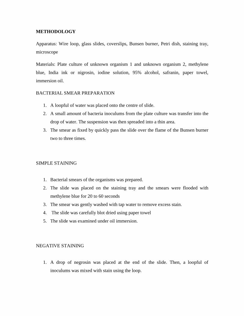
METHODOLOGY
Apparatus: Wire loop, glass slides, coverslips, Bunsen burner, Petri dish, staining tray,
microscope
Materials: Plate culture of unknown organism 1 and unknown organism 2, methylene
blue, India ink or nigrosin, iodine solution, 95% alcohol, safranin, paper towel,
immersion oil.
BACTERIAL SMEAR PREPARATION
1. A loopful of water was placed onto the centre of slide.
2. A small amount of bacteria inoculums from the plate culture was transfer into the
drop of water. The suspension was then spreaded into a thin area.
3. The smear as fixed by quickly pass the slide over the flame of the Bunsen burner
two to three times.
SIMPLE STAINING
1. Bacterial smears of the organisms was prepared.
2. The slide was placed on the staining tray and the smears were flooded with
methylene blue for 20 to 60 seconds
3. The smear was gently washed with tap water to remove excess stain.
4. The slide was carefully blot dried using paper towel
5. The slide was examined under oil immersion.
NEGATIVE STAINING
1. A drop of negrosin was placed at the end of the slide. Then, a loopful of
inoculums was mixed with stain using the loop.
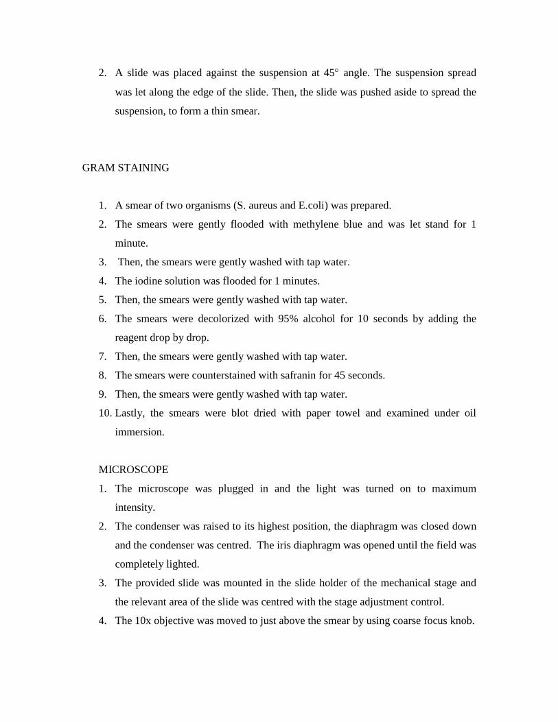
2. A slide was placed against the suspension at 45 angle. The suspension spread
was let along the edge of the slide. Then, the slide was pushed aside to spread the
suspension, to form a thin smear.
GRAM STAINING
1. A smear of two organisms (S. aureus and E.coli) was prepared.
2. The smears were gently flooded with methylene blue and was let stand for 1
minute.
3. Then, the smears were gently washed with tap water.
4. The iodine solution was flooded for 1 minutes.
5. Then, the smears were gently washed with tap water.
6. The smears were decolorized with 95% alcohol for 10 seconds by adding the
reagent drop by drop.
7. Then, the smears were gently washed with tap water.
8. The smears were counterstained with safranin for 45 seconds.
9. Then, the smears were gently washed with tap water.
10. Lastly, the smears were blot dried with paper towel and examined under oil
immersion.
MICROSCOPE
1. The microscope was plugged in and the light was turned on to maximum
intensity.
2. The condenser was raised to its highest position, the diaphragm was closed down
and the condenser was centred. The iris diaphragm was opened until the field was
completely lighted.
3. The provided slide was mounted in the slide holder of the mechanical stage and
the relevant area of the slide was centred with the stage adjustment control.
4. The 10x objective was moved to just above the smear by using coarse focus knob.
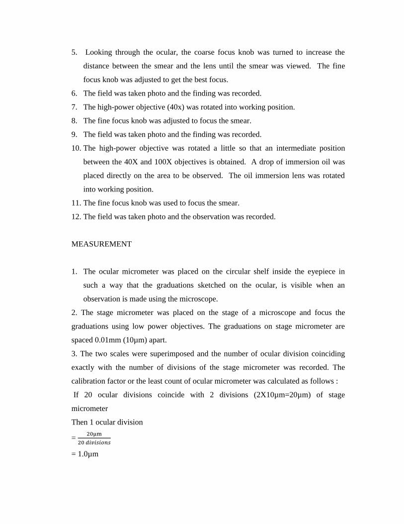
5. Looking through the ocular, the coarse focus knob was turned to increase the
distance between the smear and the lens until the smear was viewed. The fine
focus knob was adjusted to get the best focus.
6. The field was taken photo and the finding was recorded.
7. The high-power objective (40x) was rotated into working position.
8. The fine focus knob was adjusted to focus the smear.
9. The field was taken photo and the finding was recorded.
10. The high-power objective was rotated a little so that an intermediate position
between the 40X and 100X objectives is obtained. A drop of immersion oil was
placed directly on the area to be observed. The oil immersion lens was rotated
into working position.
11. The fine focus knob was used to focus the smear.
12. The field was taken photo and the observation was recorded.
MEASUREMENT
1. The ocular micrometer was placed on the circular shelf inside the eyepiece in
such a way that the graduations sketched on the ocular, is visible when an
observation is made using the microscope.
2. The stage micrometer was placed on the stage of a microscope and focus the
graduations using low power objectives. The graduations on stage micrometer are
spaced 0.01mm (10µm) apart.
3. The two scales were superimposed and the number of ocular division coinciding
exactly with the number of divisions of the stage micrometer was recorded. The
calibration factor or the least count of ocular micrometer was calculated as follows :
If 20 ocular divisions coincide with 2 divisions (2X10µm=20µm) of stage
micrometer
Then 1 ocular division
= 20µm
20 𝑑𝑖𝑣𝑖𝑠𝑖𝑜𝑛𝑠
= 1.0µm
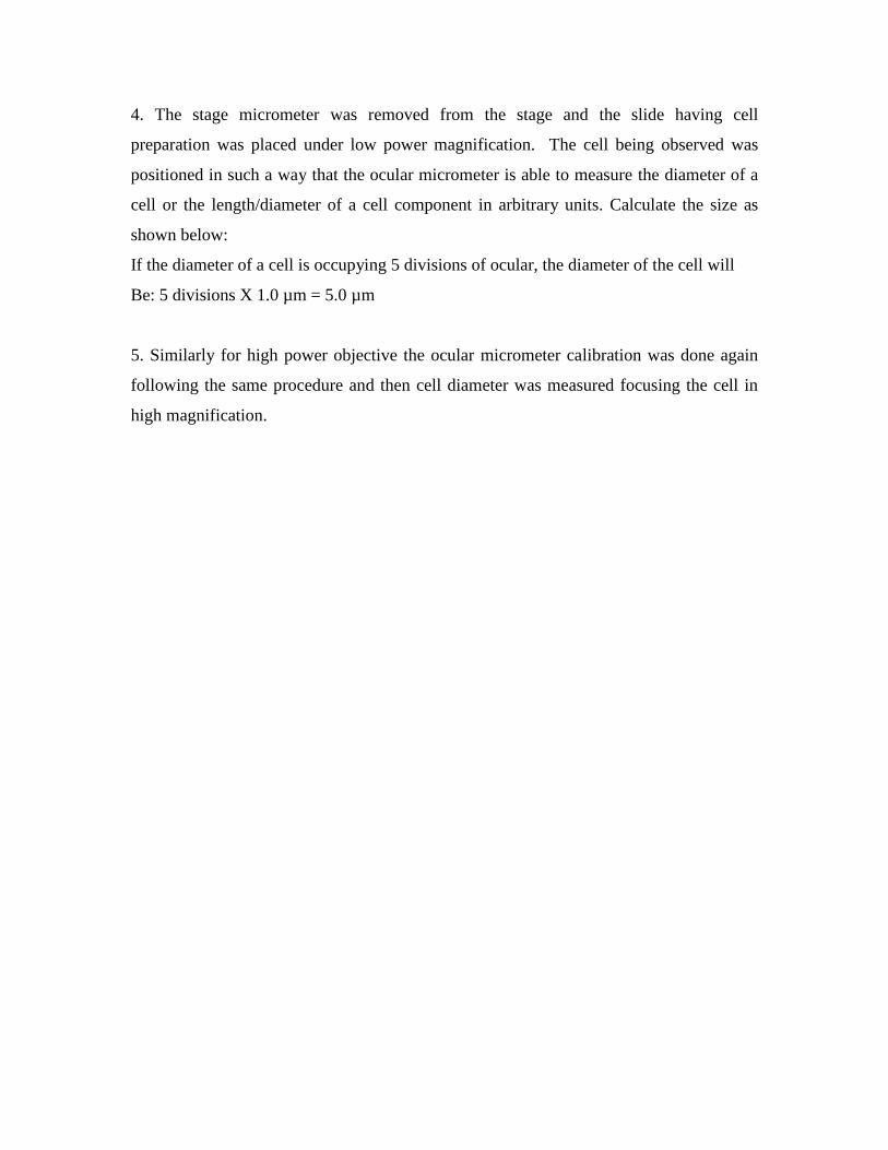
4. The stage micrometer was removed from the stage and the slide having cell
preparation was placed under low power magnification. The cell being observed was
positioned in such a way that the ocular micrometer is able to measure the diameter of a
cell or the length/diameter of a cell component in arbitrary units. Calculate the size as
shown below:
If the diameter of a cell is occupying 5 divisions of ocular, the diameter of the cell will
Be: 5 divisions X 1.0 µm = 5.0 µm
5. Similarly for high power objective the ocular micrometer calibration was done again
following the same procedure and then cell diameter was measured focusing the cell in
high magnification.
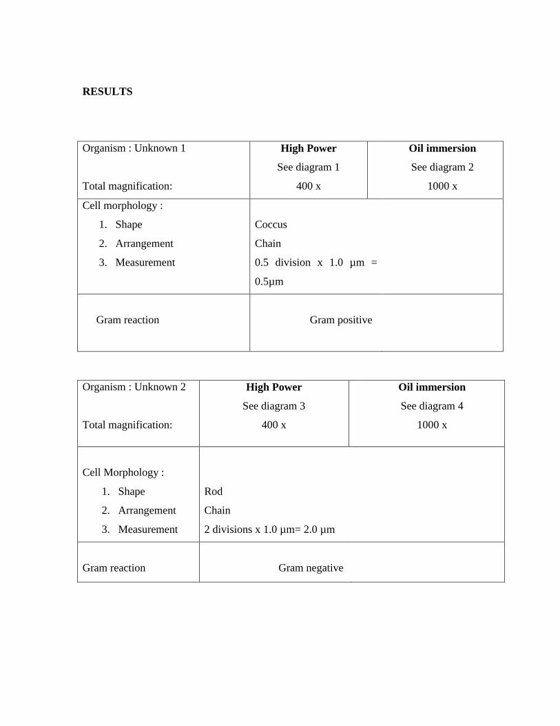
RESULTS
Organism : Unknown 1
Total magnification:
High Power
See diagram 1
400 x
Oil immersion
See diagram 2
1000 x
Cell morphology :
1. Shape
2. Arrangement
3. Measurement
Coccus
Chain
0.5 division x 1.0 µm =
0.5µm
Gram reaction
Gram positive
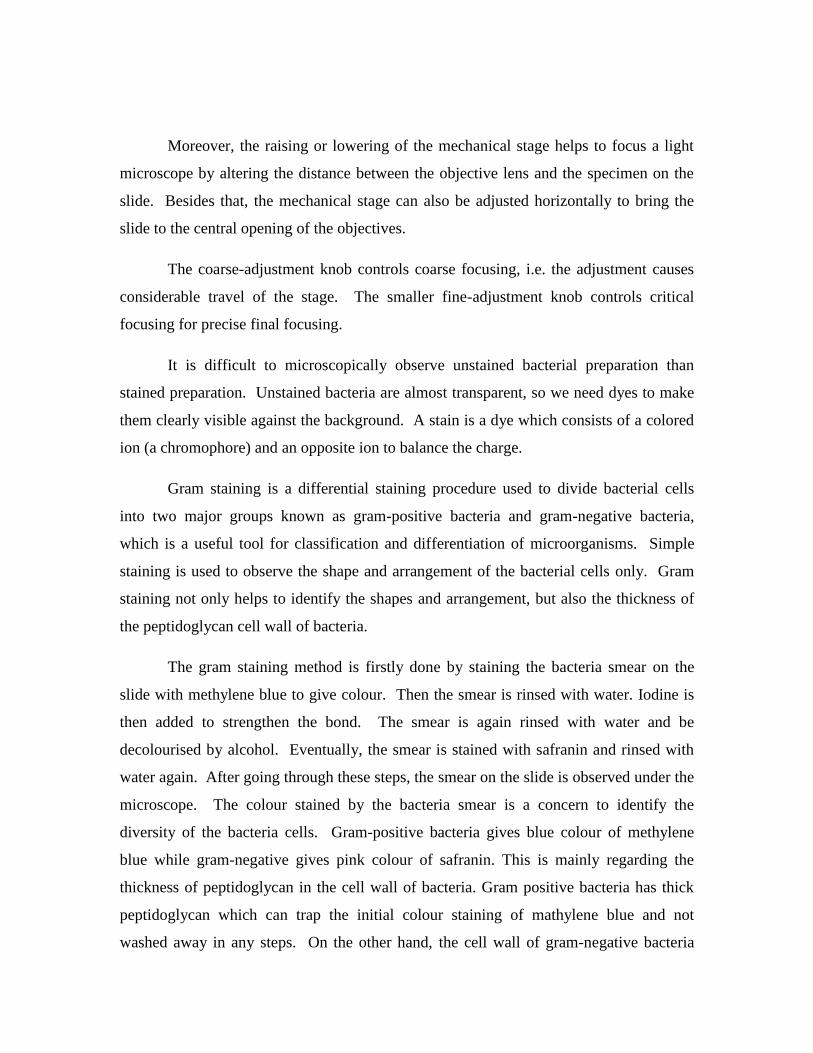
Organism : Unknown 2
Total magnification:
High Power
See diagram 3
400 x
Oil immersion
See diagram 4
1000 x
Cell Morphology :
1. Shape
2. Arrangement
3. Measurement
Rod
Chain
2 divisions x 1.0 µm= 2.0 µm
Gram reaction
Gram negative

DISCUSSION
The compound microscope consists of two systems that interact to produce the
enlarged image:
(a) Illumination system
Proper illumination of the specimen is critical if the maximum usefulness
of the microscope is to be achieved. Modern light microscopes use inbuilt
lamps for illumination. The amount of light in the system can be controlled
by a substage condenser knob. This light is collected by substage condenser,
which then passes the light through the iris diaphragm and then to the
specimen.
The iris diaphragm has different sized holes and is used to vary the
intensity and size of the light that projected into the slide. Furthermore, the
substage condenser is a lens that serves to concentrate light for the
illumination sources.
(b) Lens system
Then lens system of the microscope produces a magnified image which
allows visualization of the microorganisms. Each of the objective lenses has a
number of magnification: 10x, 40x and 100x. The eyepiece usually has a
magnification of 10. Thus, with 100x objective the magnification at eyepoint
is 10x100, i.e. 1000.
Under 1000x magnification, the immersion oil is used because it has the
same refractive index with the lens. Therefore, it can avoid refraction in the
lens which will occur if air is in between the lens and the specimen.
Moreover, the raising or lowering of the mechanical stage helps to focus a light
microscope by altering the distance between the objective lens and the specimen on the
slide. Besides that, the mechanical stage can also be adjusted horizontally to bring the
slide to the central opening of the objectives.
The coarse-adjustment knob controls coarse focusing, i.e. the adjustment causes
considerable travel of the stage. The smaller fine-adjustment knob controls critical
focusing for precise final focusing.
It is difficult to microscopically observe unstained bacterial preparation than
stained preparation. Unstained bacteria are almost transparent, so we need dyes to make
them clearly visible against the background. A stain is a dye which consists of a colored
ion (a chromophore) and an opposite ion to balance the charge.
Gram staining is a differential staining procedure used to divide bacterial cells
into two major groups known as gram-positive bacteria and gram-negative bacteria,
which is a useful tool for classification and differentiation of microorganisms. Simple
staining is used to observe the shape and arrangement of the bacterial cells only. Gram
staining not only helps to identify the shapes and arrangement, but also the thickness of
the peptidoglycan cell wall of bacteria.
The gram staining method is firstly done by staining the bacteria smear on the
slide with methylene blue to give colour. Then the smear is rinsed with water. Iodine is
then added to strengthen the bond. The smear is again rinsed with water and be
decolourised by alcohol. Eventually, the smear is stained with safranin and rinsed with
water again. After going through these steps, the smear on the slide is observed under the
microscope. The colour stained by the bacteria smear is a concern to identify the
diversity of the bacteria cells. Gram-positive bacteria gives blue colour of methylene
blue while gram-negative gives pink colour of safranin. This is mainly regarding the
thickness of peptidoglycan in the cell wall of bacteria. Gram positive bacteria has thick
peptidoglycan which can trap the initial colour staining of mathylene blue and not
washed away in any steps. On the other hand, the cell wall of gram-negative bacteria
which composed mainly of lipid layer compared to peptidoglycan, is able to trap the final
colour staining of safranin and gives pink colour.
Some precautionary steps are to be undertaken to ensure the accuracy of the
experiments. While preparing for the bacteria smear, aseptic technique has to be used to
ensure there is no contamination on the bacteria culture which might influence the results
of the staining. Once the bacteria is transferred to the slide, it is brought to the flame for
heat fixing process. The slide has to be clamped to a clipper and slided quickly over the
flame. This is to prevent overheating on the slide, causing the cell wall composition of
the bacterial cells to be broken down. On the other hand, the staining processes must not
be done for too long, each staining can only be done for 45 seconds or only within 1
minute. This is to ensure the staining processes are not too extreme or over in giving
specific colour to the bacteria cells. For decolourisation, alcohol can only be added to the
smear for at most 10 seconds to prevent over-decolourisation of the smear which might
contribute to the inaccuracy of the experimental results.
CONCLUSION
As the conclusion, in simple staining, positively charged dyes attracted to the negatively
charged materials of the microbial cytoplasm. However, in negative staining, negatively
charged dyes are repelled by the same charge of cytoplasm, and gather around cell,
leaving inside cell clearly unstained. The staining technique involves four steps.
Microorganisms that resist decolorizing in the third step appear blue microscopically are
called Gram-positive. Conversely, microorganisms that decolorize, i.e. accept safranin
and appear red are called Gram-negative. In this experiment, the organism 1 is gram-
positive and organism 2 is gram-negative.
REFERENCES
1. Cappuccino J.G, Sherman N, 2005. Microbiology - A Laboratory Manual,
7thed. Pearson-Benjamin Cummings: Singapore.
2. Gerard J.T, Berdell R.F, Christine L.C, 2007.Microbiology – An Introduction,
9thed. Pearson-Benjamin Cummings: United States. pp. 160-183.
3. Karen M.K, William C.P, Teresa A. T, 2011. Clinical Laboratory
Microbiology: A Practical Approach. Prentice Hall: U.S.A.
4. Monica Z. Bruckner. Gram Staining [online]
Available at:
http://serc.carleton.edu/microbelife/research_methods/microscopy/gramstain.h
tml
Accessed on 2 October 2013
5. BioRadLifeScience. Gram Staining. [online]
Available at: http://www.youtube.com/watch?v=sxa46xKfIOY
Accessed on 2 October 2013