journal of the anatomical society of india
TRANSCRIPT
Journal of the Anatomical Society of India
Print ISSN: 0003-2778
GENERAL INFORMATIONAbout the JournalJournal of the Anatomical Society of India (ISSN: Print 0003-2778) is peer-reviewed journal. The journal is owned and run by Anatomical Society of India. The journal publishes research articles related to all aspects of Anatomy and allied medical/surgical sciences. Pre-Publication Peer Review and Post-Publication Peer Review Online Manuscript Submission System Selection of articles on the basis of MRS system Eminent academicians across the globe as the Editorial board members Electronic Table of Contents alerts Available in both online and print form. The journal is published quarterly in the months of January, April, July and October.
Scope of the JournalThe aim of the Journal of the Anatomical Society of India is to enhance and upgrade the research work in the field of anatomy and allied clinical subjects. It provides an integrative forum for anatomists across the globe to exchange their knowledge and views. It also helps to promote communication among fellow academicians and researchers worldwide. The Journal is devoted to publish recent original research work and recent advances in the field of Anatomical Sciences and allied clinical subjects. It provides an opportunity to academicians to disseminate their knowledge that is directly relevant to all domains of health sciences.
The Editorial Board comprises of academicians across the globe.
JASI is indexed in Scopus, available in Science Direct.
Abstracting and Indexing InformationThe journal is registered with the following abstracting partners:
Baidu Scholar, CNKI (China National Knowledge Infrastructure), EBSCO Publishing's Electronic Databases, Ex Libris – Primo Central, Google Scholar, Hinari, Infotrieve, Netherlands ISSN center, ProQuest, TdNet, Wanfang Data
The journal is indexed with, or included in, the following:
SCOPUS, Science Citation Index Expanded, IndMed, MedInd, Scimago Journal Ranking, Emerging Sources Citation Index.
Impact Factor® as reported in the 2020 Journal Citation Reports® (Clarivate Analytics, 2021): 0.15
Information for AuthorsArticle processing and publication charges will be communicated by the editorial office. All manuscripts must be submitted online at https://review.jow.medknow.com/jasi.
Subscription Information A subscription to JASI comprises 4 issues. Prices include postage. Annual Subscription Rate for non-members-
Rates of Membership (with effect from 1.1.2019)
India International
Ordinary membership INR 1500 US $ 100 Couple membership INR 2250 Life membership INR 8000 US $ 900 Subscription Rates (till 31st August) Individual INR 4500 US $ 600 Library/Institutional
Trade discount of 10% for agencies only
INR 10000 US $ 900
Subscription Rates (after 31st August) Individual INR 5000 Library/Institutional INR 10500
The Journal of Anatomical Society of India (ISSN: 0003-2778) is published quarterly Subscriptions are accepted on a prepaid basis only and are entered on a calendar year basis. Issues are sent by standard mail Priority rates are available upon request.
Information to Members/SubscribersAll members and existing subscribers of the Anatomical Society of India are requested to send their membership/existing subscription fee for the current year to the Treasurer of the Society on the following address: Prof (Dr.) Punit Manik, Treasurer, ASI, Department of Anatomy, KGMU, Lucknow - 226003. Email: [email protected]. All payments should be made through an account payee bank draft drawn in favor of the Treasurer, Anatomical Society of India, payable at Lucknow only, preferably for Allahabad Bank, Medical College Branch, Lucknow. Outstation cheques/drafts must include INR 70 extra as bank collection charges. All complaints regarding non-receipt of journal issues should be addressed to the Editor-in-Chief, JASI at [email protected]. The new subscribers may, please contact [email protected].
Requests of any general information like travel concession forms, venue of next annual conference, etc. should be addressed to the General Secretary of the Anatomical Society of India.
For mode of payment and other details, please visit www.medknow.com/subscribe.aspClaims for missing issues will be serviced at no charge if received within 60 days of the cover date for domestic subscribers, and 3 months for subscribers outside India. Duplicate copies cannot be sent to replace issues not delivered because of failure to notify publisher of change of address. The journal is published and distributed by Wolters Kluwer India Pvt. Ltd. Copies are sent to subscribers directly from the publisher’s address. It is illegal to acquire copies from any other source. If a copy is received for personal use as a member of the association/society, one cannot resale or give-away the copy for commercial or library use.The copies of the journal to the subscribers are sent by ordinary post. The editorial board, association or publisher will not be responsible for non receipt of copies. If any subscriber wishes to receive the copies by registered post or courier, kindly contact the publisher’s office. If a copy returns due to incomplete, incorrect or changed address of a subscriber on two consecutive occasions, the names of such subscribers will be deleted from the mailing list of the journal. Providing complete, correct and up-to-date address is the responsibility of the subscriber.
Nonmembers: Please send change of address information to [email protected].
Advertising PoliciesThe journal accepts display and classified advertising. Frequency discounts and special positions are available. Inquiries about advertising should be sent to Wolters Kluwer India Pvt. Ltd, advertise@medknow. com.
The journal reserves the right to reject any advertisement considered unsuitable according to the set policies of the journal.
The appearance of advertising or product information in the various sections in the journal does not constitute an endorsement or approval by the journal and/or its publisher of the quality or value of the said product or of claims made for it by its manufacturer.CopyrightThe entire contents of the JASI are protected under Indian and international copyrights. The Journal, however, grants to all users a free, irrevocable, worldwide, perpetual right of access to, and a license to copy, use, distribute, perform and display the work publicly and to make and distribute derivative works in any digital medium for any reasonable non-commercial purpose, subject to proper attribution of authorship and ownership of the rights. The journal also grants the right to make small numbers of printed copies for their personal non-commercial use.PermissionsFor information on how to request permissions to reproduce articles/information from this journal, please visit www.jasi.org.in.DisclaimerThe information and opinions presented in the Journal reflect the views of the authors and not of the Journal or its Editorial Board or the Publisher. Publication does not constitute endorsement by the journal. Neither the JASI nor its publishers nor anyone else involved in creating, producing or delivering the JASI or the materials contained therein, assumes any liability or responsibility for the accuracy, completeness, or usefulness of any information provided in the JASI, nor shall they be liable for any direct, indirect, incidental, special, consequential or punitive damages arising out of the use of the JASI. The JASI, nor its publishers, nor any other party involved in the preparation of material contained in the JASI represents or warrants that the information contained herein is in every respect accurate or complete, and they are not responsible for any errors or omissions or for the results obtained from the use of such material. Readers are encouraged to confirm the information contained herein with other sources.
AddressesEditorial OfficeDr. Vishram Singh, Editor-in-Chief, JASI OC-5/103, 1st floor, Orange County Society, Ahinsa Khand-I, Indirapuram, Ghaziabad, Delhi, NCR- 201014. Email: [email protected]
Published byWolters Kluwer India Pvt. Ltd A-202, 2nd Floor, The Qube, C.T.S. No.1498A/2 Village Marol, Andheri (East), Mumbai - 400 059, India. Phone: 91-22-66491818 Website: www.medknow.comPrinted atNikeda Art Printers Pvt. Ltd Bhandup (W) , Mumbai - 400078, India.
Journal of the Anatomical Society of India
Journal of the Anatomical Society of India ¦ Volume 70 ¦ Issue 3 ¦ July-September 2021 i
Journal of the Anatomical Society of India
Print ISSN: 0003-2778
EDITORIAL BOARD
Editor-in-Chief Dr. Vishram Singh, MBBS, MS, PhD (hc), FASI, FIMSA
Adjunct Professor, Department of Anatomy, KMC, Mangalore, Manipal Academy of Higher Education, Manipal, KarnatakaJoint-Editor
Dr. Murlimanju B.V Associate Professor, Department of Anatomy, KMC, Mangalore, Manipal Academy of Higher Education, Manipal, Karnataka
Managing Editor Dr. C. S. Ramesh Babu
Associate Professor, Department of Anatomy, Muzaffarnagar Medical College, Muzaffarnagar, Uttar PradeshAssociate Editor
Dr. D. Krishna Chaitanya Reddy Assistant Professor, Department of Anatomy, Kamineni Academy of Medical Sciences and Research Center, Hyderabad
Section EditorsClinical Anatomy Dr. Vishy Mahadevan, PhD, FRCS(Ed), FRCS Prof of Surgical Anatomy, The Royal College of Surgeons of England, London, UK
Histology Dr. G.P. Pal, MS, DSc, Prof & Head, Department of Anatomy, MDC & RC, Indore, India
Gross and Imaging Anatomy Dr. Srijit Das, Department of Human and Clinical Anatomy, College of Medicine and Health Sciences, Sultan Qaboos University, Muscat, Oman
Medical Education Dr. Deepa Singh Professor, Department of Anatomy, HIMS, Swami Rama Himalayan University, Jolly Grant, Dehradun, Uttarakhand
Neuroanatomy Dr. T.S. Roy, MD, PhD Prof & Head, Department of Anatomy, AIIMS, New Delhi
Embryology Dr. Gayatri Rath, MS, FAMS Professor and Head, Department of Anatomy, NDMC Medical College, New Delhi
Genetics Dr. Rima Dada, MD, PhD Prof, Department of Anatomy, AIIMS, New Delhi, India
Dental Sciences Dr. Praveen B Kudva Professor and Head, Department of Periodontology, Jaipur Dental College, Jaipur, Rajasthan
National Editorial BoardDr. S.D. Joshi, Indore Dr. G.S. Longia, Jaipur Dr. A.K. Srivastava, Lucknow Dr. Daksha Dixit, Belgaum Dr. S.K. Jain, Moradabad Dr. P.K. Sharma, Lucknow Dr. S. Senthil Kumar, Chennai Dr. Daisy Sahani, Chandigarh Dr. N. Damayanti Devi, Imphal
Dr. Renu Chauhan, Delhi Dr. Ashok Sahai, Agra Dr. Ramesh Babu, Muzzafarnagar Dr. T.C. Singel, Ahmedabad Dr. P.K. Verma, Hyderabad Dr. S.L. Jethani, Dehradun Dr. Surajit Ghatak, Jodhpur Dr. Brijendra Singh, Rishikesh Dr. P. Vatsala Swamy, Pune
International Editorial BoardDr. Yun-Qing Li, China Dr. In-Sun Park, Korea Dr. K.B. Swamy, Malaysia Dr. Syed Javed Haider, Saudi Arabia Dr. Pasuk Mahakknaukrauh, Thailand Dr. Tom Thomas R. Gest, USA
Dr. Chris Briggs, Australia Dr. Petru Matusz, Romania Dr. Min Suk Chung, South Korea Dr. Veronica Macchi, Italy Dr. Gopalakrishnakone, Singapore Dr. Sunil Upadhyay, UK
ii Journal of the Anatomical Society of India ¦ Volume 70 ¦ Issue 3 ¦ July-September 2021
EXECUTIVE COMMITTEE
Dr. Avinash Abhaya (Chandigarh) Dr. Sumit T. Patil (Portblair ) Dr. Mirnmoy Pal (Agartala) Dr. Manish R. Gaikwad (Bhubaneswar) Dr. Sudhir Eknath Pawar (Ahmednagar) Dr. Rekha Lalwani (Bhopal) Dr. Anshu Sharma (Chandigarh) Dr. Rakesh K Diwan (Lucknow) Dr. A. Amar Jayanthi (Trichur) Dr. Ranjan Kumar Das (Baripada)
Dr. Rajani Singh (Rishikesh) Dr. Anu Sharma (Ludhiana) Dr. Pradeep Bokariya (Sevagram) Dr. B. Prakash Babu (Manipal) Dr. Ruchira Sethi (Varanasi) Dr. Ashok Nirvan (Ahmedabad) Dr. S K Deshpande (Dharwad) Dr. Sunita Athavale (Bhopal) Dr. Sharmistha Biswas (Kolkatta)
Office Bearers
President Dr. Brijendra Singh (Rishikesh)
Vice President Dr. G. P. Pal (Indore)
Gen. Secretary Dr. S.L. Jethani (Dehradun)
Joint. Secretary Dr. Jitendra Patel (Ahmedabad)
Treasurer Dr. Punita Manik (Lucknow)
Joint-Treasurer Dr. R K Verma (Lucknow)
Editor-in-Chief Dr. Vishram Singh (Mangalore)
Joint-Editor Dr. Murlimanju B.V (Mangalore)
Members
Journal of the Anatomical Society of India
Print ISSN: 0003-2778
Journal of the Anatomical Society of India ¦ Volume 70 ¦ Issue 3 ¦ July-September 2021 iii
Volume 70 | Issue 3 | July-September 2021
Journal of the Anatomical Society of India
CONTENTS
EDITORIALEvolving Trends in Anatomy Teaching across the Globe: A New PerspectiveVishram Singh, Rashi Singh ......................................................................................................................................................................129
ORIGINAL ARTICLESEducational Resources Used by 1st‑Year Medical StudentsHimel Mondal, Sumita Dutta, Shaikat Mondal, Manas Ranjan Sahoo, Koushik Saha, Sarika Mondal ...................................................130
Proliferative Capacity of Retinal Progenitor Cells in Human Fetal RetinaPrakash Mane, Anjali Satyen Sabnis .........................................................................................................................................................136
Wax versus Plastinated Models in Teaching Human Anatomy to Health‑care Professionals. A Randomized Crossover TrialRafael Boscolo‑Berto, Cinzia Tortorella, Veronica Macchi, Andrea Porzionato, Raffaele De Caro .........................................................140
Three‑Dimensional Evaluation of Maxillary Sinuses in the Turkmen Population, North of IranArash Mollaali, Mohammad Hadi Gharib, Jahangir Ghorbani, Mohammad Jafar Golalipour .................................................................146
Variation of the Superior Articular Facet of Atlas and Their SignificanceNeeru Goyal, Anjali Jain ...........................................................................................................................................................................151
Assessing Differences in Hand Dominance by Testing Hand Preference against Hand PerformancePamela Mandela Idenya, Peter Gichangi, A. Ogeng’o Julius ....................................................................................................................156
Association of Chiari Type 1 Malformation and Cervical Spine Curve ChangesMuhammed Alpaslan, Sercan Özkaçmaz, Yeliz Dadalı, İlyas Uçar ..........................................................................................................162
Harbin’s Index: Morphological Evaluation of Caudate‑to‑Right Lobe Ratio in Human Cadaveric LiverJaikumar B. Contractor, Vipul D. Patel, V. H. Vaniya ...............................................................................................................................168
CASE REPORTSA Rare Case of Anomalous Origin of Bilateral Testicular Arteries: An Anatomical and Developmental OverviewArthi Ganapathy, Aritra Banerjee, Saroj Kaler Jhajhria, Seema Singh .....................................................................................................173
Anophthalmic Cyclopia with Proboscis, Acardia, Amelia, Sirenomelia – Case Report Bini Markose, Deepti Shastri, B. Rajesh, Jinu Merlin Koshy ...................................................................................................................176
Bilateral Persistent Primitive Olfactory Artery Incidentally Detected by Computed Tomography Angiography and Digital Subtraction Angiography: An Extremely Rare Case ReportAnshu Mahajan, Apratim Chatterjee, Gaurav Goel ...................................................................................................................................183
LETTER TO EDITORA Woman with “Lobster‑Claw” Hands – Isolated Nonsyndromic Ectrodactyly of Both HandsH. S. Kiran, H. S. Rajani, N. Rashmi ........................................................................................................................................................186
iv Journal of the Anatomical Society of India ¦ Volume 70 ¦ Issue 3 ¦ July-September 2021
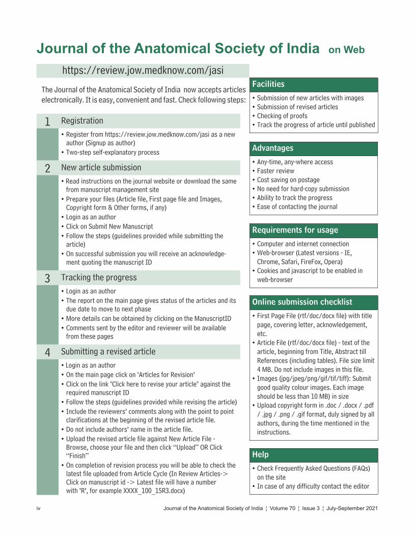
The Journal of the Anatomical Society of India now accepts articles electronically. It is easy, convenient and fast. Check following steps:
Journal of the Anatomical Society of India on Web
Facilities
• Submission of new articles with images• Submission of revised articles• Checking of proofs• Track the progress of article until published
Advantages
• Any-time, any-where access• Faster review• Cost saving on postage• No need for hard-copy submission• Ability to track the progress• Ease of contacting the journal
Requirements for usage
• Computer and internet connection• Web-browser (Latest versions - IE,
Chrome, Safari, FireFox, Opera)• Cookies and javascript to be enabled in
web-browser
Online submission checklist
• First Page File (rtf/doc/docx file) with title page, covering letter, acknowledgement, etc.
• Article File (rtf/doc/docx file) - text of the article, beginning from Title, Abstract till References (including tables). File size limit 4 MB. Do not include images in this file.
• Images (jpg/jpeg/png/gif/tif/tiff): Submit good quality colour images. Each image should be less than 10 MB) in size
• Upload copyright form in .doc / .docx / .pdf / .jpg / .png / .gif format, duly signed by all authors, during the time mentioned in the instructions.
Help
• Check Frequently Asked Questions (FAQs) on the site
• In case of any difficulty contact the editor
1 Registration• Register from https://review.jow.medknow.com/jasi as a new
author (Signup as author)• Two-step self-explanatory process
2 New article submission• Read instructions on the journal website or download the same
from manuscript management site• Prepare your files (Article file, First page file and Images,
Copyright form & Other forms, if any)• Login as an author• Click on Submit New Manuscript• Follow the steps (guidelines provided while submitting the
article)• On successful submission you will receive an acknowledge-
ment quoting the manuscript ID
3 Tracking the progress• Login as an author• The report on the main page gives status of the articles and its
due date to move to next phase• More details can be obtained by clicking on the ManuscriptID• Comments sent by the editor and reviewer will be available
from these pages
4 Submitting a revised article• Login as an author• On the main page click on ‘Articles for Revision’• Click on the link "Click here to revise your article" against the
required manuscript ID• Follow the steps (guidelines provided while revising the article)• Include the reviewers’ comments along with the point to point
clarifications at the beginning of the revised article file. • Do not include authors’ name in the article file. • Upload the revised article file against New Article File -
Browse, choose your file and then click “Upload” OR Click “Finish”
• On completion of revision process you will be able to check the latest file uploaded from Article Cycle (In Review Articles-> Click on manuscript id -> Latest file will have a number with ‘R’, for example XXXX_100_15R3.docx)
https://review.jow.medknow.com/jasi
© 2021 Journal of the Anatomical Society of India | Published by Wolters Kluwer - Medknow 129
Evolving Trends in Anatomy Teaching across the Globe: A New Perspective
The basic purpose of all governments is to produce competent medical graduates who should be able to perform basic medical and surgical procedures efficiently with precision and competence.
To achieve this various curricula have been prepared by different national agencies concerned with medical education. Most of these curricula included two things in common: Didactic classroom lectures and dissection on cadavers so that medical students must acquire good anatomical knowledge to build a solid background for future clinical practices.
The application of classical anatomy teaching was impossible to carry out during the COVID‑19 period.[1] Further how long it would take for this pandemic to end was not certain. In order to overcome this problem new teaching and learning methods were evolved during this period, which were multimodal teaching and digitalization of Anatomy.
The video‑based learning with dissection were posted on YouTube for team‑based (or individual) self‑learning using computers and other technologies.
Now in Anatomy teaching the use of e‑technology has become a common norm in the form of models, simulations, and e‑books[2] (Triepels et al. 2020).
With these changes in place a subject like Anatomy which was considered a dull subject because it was mainly concerned with the memorization of anatomical facts.[3], has been made more fascinating by making it more interactive and engaging to obtain deeper learning so that the students could apply their knowledge in the clinical context.
In our opinion, the future is digital and the next step should be the evolution of virtual teaching platforms demonstrating common medical procedures such as intradermal, intramuscular, intravenous injections etc to be shown on videos.
The common basic surgical procedures such as treatment of hydrocele, hernia, lipoma (cyst), transllectomy etc must also be explained and demonstrated in virtually.
There should also be an inclusion of technical procedures such as cardiopulmonary resuscitation, use of oxygen cylinder, ventilator, rhinoscopy, and endoscopy that may be demonstrated it on dummies/cadavers.
Furthermore, must not be forgotten that once the COVID period is over, all efforts should be made to resume the
manual dissection on cadavers, as it is must for Anatomy teaching and thus shall create true blended learning.
This would not only make Anatomy teaching more interesting but also enable the production of competent medical graduates.
Vishram Singh, Rashi Singh1
Department of Anatomy, Kasturba Medical College, Mangalore, Manipal Academy of Higher Education, Manipal, Karnataka, 1Department
of Paediatric and Preventive Dentistry, Santosh Dental College and Hospital, Ghaziabad, Uttar Pradesh, India
Address for correspondence: Prof. Vishram Singh, OC 5/103, 1st Floor, Orange County Society, Ahinsa Khand I,
Indirapuram, Ghaziabad, Delhi NCR 201014, Uttar Pradesh, India. E‑mail: [email protected]
References1. Agnihotri G. The anatomy of a ‘positively novel’ medical teacher
in covid‑19 times. Int J Anat Res 2020;8(3):1.2. Triepels CP, Smeets CF, Notten KJ, Kruitwagen RF, Futterer JJ,
Vergeldt TF, et al. Does three‑dimensional anatomy improve student understanding? Clin Anat 2020;33:25‑33.
3. Dawson AG, Bruce SA, Heys SD, Stewart IJ. Student views on the introduction of anatomy teaching packages into clinical attachments. Clin Anat 2009;22:267‑72.
This is an open access journal, and articles are distributed under the terms of the Creative Commons Attribution‑NonCommercial‑ShareAlike 4.0 License, which allows others to remix, tweak, and build upon the work non‑commercially, as long as appropriate credit is given and the new creations are licensed under the identical terms.
Access this article onlineQuick Response Code:
Website: www.jasi.org.in
DOI: 10.4103/jasi.jasi_156_21
How to cite this article: Singh V, Singh R. Evolving trends in anatomy teaching across the globe: A new perspective. J Anat Soc India 2021;XX:XX-XX.
Received: 08 September 2021Accepted: 08 September 2021Available online: ***
Article Info
Editorial
130 © 2021 Journal of the Anatomical Society of India | Published by Wolters Kluwer - Medknow
Address for correspondence: Dr. Himel Mondal, Department of Physiology, Santiniketan Medical College, Bolpur ‑ 731 204, West Bengal, India. E‑mail: [email protected]
Access this article online
Website: www.jasi.org.inDOI: 10.4103/JASI.JASI_16_20
Quick Response Code:
AbstractIntroduction: A dynamic teaching–learning environment is being established in Indian medical institutions with the implementation of a competency‑based medical education curriculum. This new curriculum may change the previous pattern of usage of educational resources by the medical students. We aimed to explore the pattern of usage of resources by the 1st‑year medical students. Material and Methods: We invited 1st‑year medical students of three medical colleges for an online survey. There were 17 statements in the questionnaire with 5‑point Likert‑type response options to collect data on the preference of type of classes, frequency of collection of notes, pattern of reading, and usage of multimedia. Results: A total of 127 (response rate 42.6%) students participated in the survey. Practical classes were the most preferred type of class followed by small group teaching. Students preferred to take notes from 1‑h lectures than making notes while reading books. Traditional textbooks were the most preferred material read by the students followed by the question–answer type book. E‑book downloaded on the smartphone was preferred over the online e‑book. Internet searches and watching YouTube™ videos were popular than watching e‑content provided with the textbook. Discussion and Conclusion: In the age of smartphones and the internet, traditional learning resources are still popular among 1st‑year medical students. However, learning is reinforced by widely available electronic content. Hence, blended teaching with both traditional and e‑resource may be considered by medical teachers.
Keywords: Learning, medical education, medical students, reading, smartphone
Educational Resources Used by 1st‑Year Medical Students
Himel Mondal, Sumita Dutta1, Shaikat Mondal2, Manas Ranjan Sahoo3, Koushik Saha1, Sarika Mondal4
Department of Physiology, Santiniketan Medical College, Bolpur, 1Department of Anatomy, Rampurhat Government Medical College and Hospital, Rampurhat, 4Freelance Medical Writer, Kolkata, West Bengal, 2Department of Physiology, Raiganj Government Medical College and Hospital, Raiganj, 3Department of Psychiatry, Fakir Mohan Medical College and Hospital, Balasore, Odisha, India
How to cite this article: Mondal H, Dutta S, Mondal S, Sahoo MR, Saha K, Mondal S. Educational resources used by 1st-year medical students. J Anat Soc India 2021;XX:XX-XX.
IntroductionA competency‑based, dynamic, and learner‑centric undergraduate curriculum has been introduced to train Indian medical graduates.[1] Learning basic medical sciences has been enriched with early clinical exposure and self‑directed learning.[2,3] A sudden change in the curriculum is a challenging situation for both teachers and students.[4] Traditional textbooks and manuals may lack targeted content and arrangement. The teachers may get adequate time for collecting resources for teaching. However, the students may not get that much time to collect resources for learning a topic.
Wynter et al. have explored the resources used by medical students in a sample of Australian medical students. They found that the majority of the students learn a topic with the help of textbooks and written notes along with the use of varieties of e‑learning tools.[5]
Punja et al. have ascertained the perception of Indian 1st‑year medical students for learning anatomy and found that small group teaching and early clinical exposure were the most favored mode of learning.[6]
To the best of our knowledge, no study has been conducted to find out the resources used by the 1st‑year medical students after the introduction of the competency‑based medical education curriculum. Hence, in this study, we aimed to know the pattern of usage of different resources by 1st‑year medical students. The knowledge would help the teachers to understand how students are learning a topic. This would facilitate the teachers to teach in a balanced manner with the blending of different resources.
Material and MethodsThis questionnaire‑based, cross‑sectional, observational study was conducted with 1st‑year medical students.
Survey questionnaire
After reviewing relevant literature about the resources used by medical students,[5‑10] we
This is an open access journal, and articles are distributed under the terms of the Creative Commons Attribution‑NonCommercial‑ShareAlike 4.0 License, which allows others to remix, tweak, and build upon the work non‑commercially, as long as appropriate credit is given and the new creations are licensed under the identical terms.
For reprints contact: [email protected]
Received: 31 January 2020Accepted: 25 July 2021Available online: ***
Article Info
Original Article
Mondal, et al.: Resource used by medical students
Journal of the Anatomical Society of India ¦ Volume 70 ¦ Issue 3 ¦ July-September 2021 131
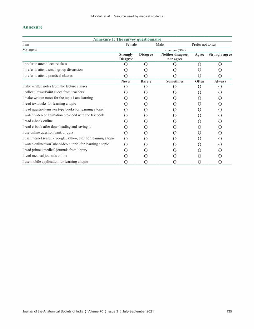
designed a questionnaire that collects data on (a) preference of classes, (b) frequency of collection of notes, (c) pattern of reading, and (d) frequency of multimedia usage. The questionnaire was composed of 17 statements with 5‑point Likert‑type response options. The questionnaire is available in Annexure 1. The questionnaire was created on Google Forms, an online platform which can be used to collect survey response.[11] The questionnaire has an informed consent statement at the beginning and agreement to the consent allows a user to submit the survey response.
Participants
The survey links were probably exposed (we did not distribute the links directly to the students) to 298 1st‑year medical students (maximum number of 1st‑year medical students in three medical colleges) of three government‑run medical colleges in Eastern India.
Data collection method
A quick response (QR) code was generated with the web link of the survey questionnaire, and posters were made with the QR code with a message for voluntary participation in the survey. The posters were put up on a wall that is easily visible to the 1st‑year medical students. They were also informed that an online survey is being conducted on which they can provide their response without revealing their identity. Willing students scanned the QR code which lands them to the consent form for participation and the questionnaire. The submitted response was collected from Google Forms after a period of 1 week. After collecting the data, the questionnaire was closed for further response in Google Forms.
Statistical analysis
The survey response was coded for making it quantitative data with the extreme agreement as 5 and extreme disagreement as 1 (i.e. strongly agree = 5, agree = 4, neutral/neither agree nor disagree = 3, disagree = 2, strongly disagree = 1). Data was presented in percentages and mean and standard deviation. The mean between males and females was compared by unpaired t‑test. For the entire statistical test, a P < 0.05 was considered statistically significant. The data coding and analysis were carried out in Microsoft Excel® 2010 (Microsoft Corporation, USA) and GraphPad Prism 6.01 (GraphPad Software, CA, USA).
ResultsA total of 127 students (female 61 [48.03%], male 66 [51.97%]) with mean age 18.64 ± 1.25 years (female 18.75 ± 1.36 years and male 18.64 ± 1.25 years) participated in the survey (response rate 42.6%).
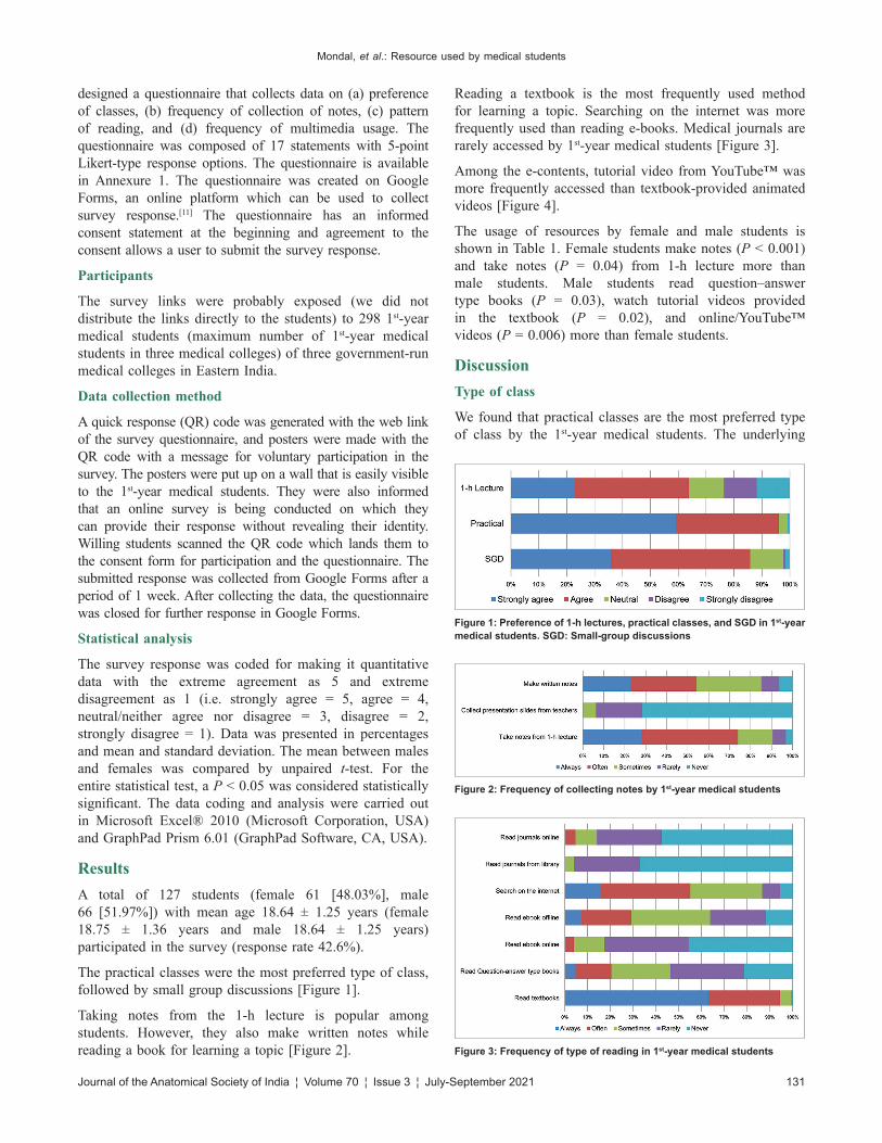
The practical classes were the most preferred type of class, followed by small group discussions [Figure 1].
Taking notes from the 1‑h lecture is popular among students. However, they also make written notes while reading a book for learning a topic [Figure 2].
Reading a textbook is the most frequently used method for learning a topic. Searching on the internet was more frequently used than reading e‑books. Medical journals are rarely accessed by 1st‑year medical students [Figure 3].
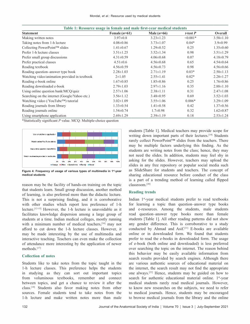
Among the e‑contents, tutorial video from YouTube™ was more frequently accessed than textbook‑provided animated videos [Figure 4].
The usage of resources by female and male students is shown in Table 1. Female students make notes (P < 0.001) and take notes (P = 0.04) from 1‑h lecture more than male students. Male students read question–answer type books (P = 0.03), watch tutorial videos provided in the textbook (P = 0.02), and online/YouTube™ videos (P = 0.006) more than female students.
DiscussionType of class
We found that practical classes are the most preferred type of class by the 1st‑year medical students. The underlying
Figure 1: Preference of 1‑h lectures, practical classes, and SGD in 1st‑year medical students. SGD: Small‑group discussions
Figure 2: Frequency of collecting notes by 1st‑year medical students
Figure 3: Frequency of type of reading in 1st‑year medical students
Mondal, et al.: Resource used by medical students
132 Journal of the Anatomical Society of India ¦ Volume 70 ¦ Issue 3 ¦ July-September 2021
reason may be the facility of hands‑on training on the topic that students learn. Small group discussion, another method of learning, is also preferred more than the didactic lecture. This is not a surprising finding, and it is corroborative with other studies which report less preference of 1‑h lecture.[12‑15] However, the 1‑h lecture is unavoidable as it facilitates knowledge dispersion among a large group of students at a time. Indian medical colleges, mostly running with a minimum number of medical teachers,[16] may not afford to cut down the 1‑h lecture classes. However, it may be made interesting by the use of multimedia and interactive teaching. Teachers can even make the collection of attendance more interesting by the application of newer methods.[17]
Collection of notes
Students like to take notes from the topic taught in the 1‑h lecture classes. This preference helps the students in studying as they can sort out important topics from voluminous textbooks, remember and connect between topics, and get a chance to review it after the class.[18] Students also favor making notes from other sources. Female students tend to take notes from the 1‑h lecture and make written notes more than male
students [Table 1]. Medical teachers may provide scope for writing down important parts of their lectures.[19] Students rarely collect PowerPoint™ slides from the teachers. There may be multiple factors underlying this finding. As the students are writing notes from the class; hence, they may not need the slides. In addition, students may feel shy in asking for the slides. However, teachers may upload the slides in any free repository or popular social media such as SlideShare for students and teachers. The concept of sharing educational resource before conduct of the class is a part of a trending method of learning called flipped classroom.[20]
Reading trends
Indian 1st‑year medical students prefer to read textbooks for learning a topic than question–answer type books and e‑resources. Among the students, male students read question–answer type books more than female students [Table 1]. All other reading patterns did not show any gender difference. This is corroborative to a study conducted by Ahmad and Asif.[21] E‑books are available online or in downloaded form. We found that students prefer to read the e‑books in downloaded form. The usage of e‑book (both online and downloaded) is less preferred over searching the topic on the internet. The reason behind this behavior may be easily available information from search results provided by search engines. Although there are various authentic sources of educational material on the internet, the search result may not find the appropriate one always.[22] Hence, students may be guided on how to search for authentic educational material online. 1st‑year medical students rarely read medical journals. However, to know new researches on the subjects, we need to refer to medical journals. Hence, students may be encouraged to browse medical journals from the library and the online
Table 1: Resource usage in female and male first‑year medical studentsStatement Female (n=61) Male (n=66) t‑test P OverallMaking written notes 3.97±0.8 3.23±1.23 <0.001* 3.58±1.10Taking notes from 1‑h lecture 4.08±0.86 3.73±1.07 0.04* 3.9±0.99Collecting PowerPoint™ slides 1.41±0.67 1.29±0.52 0.25 1.35±0.60Prefer 1‑h lecture classes 3.51±1.25 3.52±1.34 0.98 3.51±1.29Prefer small group discussions 4.31±0.59 4.06±0.68 0.07 4.18±0.79Prefer practical classes 4.51±0.6 4.56±0.68 0.65 4.54±0.64Reading textbook 4.56±0.59 4.56±0.73 0.98 4.56±0.66Reading question–answer type book 2.28±1.03 2.71±1.19 0.03* 2.50±1.13Watching video/animation provided in textbook 2±1.05 2.55±1.41 0.02* 2.28±1.27Reading e‑book online 1.67±0.85 1.85±0.86 0.25 1.76±0.86Reading downloaded e‑book 2.79±1.03 2.97±1.16 0.35 2.88±1.10Using online question bank/MCQ/quiz 2.57±1.06 2.38±1.11 0.31 2.47±1.08Searching on the internet (Google/Yahoo etc.) 3.56±1.12 3.48±0.95 0.69 3.52±1.03Watching video (/YouTube™) tutorial 3.02±1.09 3.55±1.06 0.006* 3.29±1.09Reading journals from library 1.33±0.54 1.41±0.58 0.42 1.37±0.56Reading journals online 1.54±0.74 1.7±0.98 0.32 1.62±0.87Using smartphone application 2.69±1.29 2.39±1.19 0.18 2.53±1.24*Statistically significant P value. MCQ: Multiple‑choice question
Figure 4: Frequency of usage of various types of multimedia in 1st‑year medical students
Mondal, et al.: Resource used by medical students
Journal of the Anatomical Society of India ¦ Volume 70 ¦ Issue 3 ¦ July-September 2021 133
repository or freely available bibliographic databases such as Directory of Open Access Journals and PubMed Central.
Usage of multimedia
We found that watching online videos on YouTube™ is more preferred than the multimedia provided in the textbook. Male students tend to watch videos, be it textbook‑provided or YouTube™, more than female students [Table 1]. Overall, watching YouTube™ videos has been increased for academic learning in recent times.[23] Medical teachers and institutions may gradually build a collection of authentic tutorial videos available free on YouTube™. These videos not only make learning interesting, but it may also help in revising a topic or may be helpful for students who remain absent in a class. Smartphone use has been increased in recent years for teaching and learning due to the easy availability of smartphones and internet connection.[24] These new technologies should be adapted to make the students more interested. However, a perfect balance is necessary between traditional and modern‑age technology of teaching.[25]
Novelty and limitations of the study
In this study, we have explored the resources used by 1st‑year medical students for learning basic medical science after the introduction of the competency‑based undergraduate medical curriculum. This information would help the medical teachers in designing their teaching strategies. This online survey did not collect any identification of the respondents, except the age and sex to reduce any response bias. However, this pilot study was conducted with a sample of three medical colleges from Eastern India. A future study is needed with participants from multiple colleges all over India for a more generalized result.
ConclusionTraditional learning resources such as reading textbooks, taking notes from lecture classes, and making written notes are still popular among the 1st‑year medical students. Along with these, students watch online tutorial videos and use smartphone applications for learning basic medical sciences. Medical teachers may use a combination of both traditional and new technologies to make teaching more acceptable to the students.
Acknowledgment
We thank all the students who took part in the survey and provided their response.
Financial support and sponsorship
Nil.
Conflicts of interest
There are no conflicts of interest.
References1. UG Curriculum. Medical Council of India. Available from:
https://www.mciindia.org/CMS/information‑desk/for‑colleges/ug‑curriculum.
2. Kar M, Kar C, Roy H, Goyal P. Early clinical exposure as a learning tool to teach neuroanatomy for first year MBBS students. Int J Appl Basic Med Res 2017;7:S38‑41.
3. Premkumar K, Vinod E, Sathishkumar S, Pulimood AB, Umaefulam V, Prasanna Samuel P, et al. Self‑directed learning readiness of Indian medical students: A mixed method study. BMC Med Educ 2018;18:134.
4. Basheer A. Competency‑based medical education in India: Are we ready? J Curr Res Sci Med 2019;5:1‑3.
5. Wynter L, Burgess A, Kalman E, Heron JE, Bleasel J. Medical students: What educational resources are they using? BMC Med Educ 2019;19:36.
6. Punja R, Sumalatha S, Hosapatna M. Perspective of the 1st year undergraduate medical students in learning anatomy. J Anat Soc India 2019;68:129‑32.
7. Brennan N, Edwards S, Kelly N, Miller A, Harrower L, Mattick K. Qualified doctor and medical students’ use of resources for accessing information: What is used and why? Health Info Libr J 2014;31:204‑14.
8. Scott K, Morris A, Marais B. Medical student use of digital learning resources. Clin Teach 2018;15:29‑33.
9. Judd T, Elliott K. Selection and use of online learning resources by first‑year medical students: Cross‑sectional study. JMIR Med Educ 2017;3:e17.
10. Tain M, Schwartzstein R, Friedland B, Park SE. Dental and medical students’ use and perceptions of learning resources in a human physiology course. J Dent Educ 2017;81:1091‑7.
11. Mondal H, Mondal S, Ghosal T, Mondal S. Using Google forms for medical survey: A technical note. Int J Clin Exp Physiol 2018;5:216‑8.
12. Holambe VM, Thakur NA, Giri PA. Student’s preferences for learning in medical education. Int J Community Med Public Health 2015;2:328‑30.
13. Buşan AM. Learning styles of medical students – Implications in education. Curr Health Sci J 2014;40:104‑10.
14. Jain A, Bansal R, Singh K, Kumar A. Attitude of medical and dental first year students towards teaching methods in a medical college of Northern India. J Clin Diagn Res 2014;8:C05‑8.
15. Shrewsbury D, Wiskin C. Medical student preference in teaching methods and educational support. Health Soc Care Educ 2013;2:11‑5.
16. Mudur G. Faculty shortages may thwart India’s plans for more AIIMS‑like institutions in every state. BMJ 2014;349:g4822.
17. Mondal H, Mondal S. Students’ engagement during collection of attendance: An experience of a pilot study. J Med Res Innov 2018;2:e000097.
18. How To Take Study Notes: 5 Effective Note Taking Methods. Oxford Learning. Available from: https://www.oxfordlearning.com/5‑effective‑note‑taking‑methods/.
19. Weimer M. How to Help Students Improve Their Note‑Taking Skills. Faculty Focus. Available from: https://www.facultyfocus.com/articles/teaching‑and‑learning/help‑students‑improve‑note‑taking‑skills/.
20. Fan JY, Tseng YJ, Chao LF, Chen SL, Jane SW. Learning outcomes of a flipped classroom teaching approach in an adult‑health nursing course: A quasi‑experimental study. BMC Med Educ 2020;20:317.
21. Ahmad HN, Asif M. Medical student’s learning habits: A mixed

Mondal, et al.: Resource used by medical students
134 Journal of the Anatomical Society of India ¦ Volume 70 ¦ Issue 3 ¦ July-September 2021
method study during clinical rotation in general surgery. J Pak Med Assoc 2018;68:600‑5.
22. Wang L, Wang J, Wang M, Li Y, Liang Y, Xu D. Using Internet search engines to obtain medical information: A comparative study. J Med Internet Res 2012;14:e74.
23. O’Malley D, Barry DS, Rae MG. How much do preclinical medical students utilize the internet to study physiology? Adv
Physiol Educ 2019;43:383‑91.24. Gavali MY, Khismatrao DS, Gavali YV, Patil KB. Smartphone,
the new learning aid amongst medical students. J Clin Diagn Res 2017;11:C05‑8.
25. Liu Q Peng W, Zhang F, Hu R, Li Y, Yan W. The effectiveness of blended learning in health professions: Systematic review and meta‑analysis. J Med Internet Res 2016;18:e2.
Mondal, et al.: Resource used by medical students
Journal of the Anatomical Society of India ¦ Volume 70 ¦ Issue 3 ¦ July-September 2021 135
Annexure
Annexure 1: The survey questionnaireI am Female Male Prefer not to sayMy age is ………. years
Strongly Disagree
Disagree Neither disagree, nor agree
Agree Strongly agree
I prefer to attend lecture class Ο Ο Ο Ο ΟI prefer to attend small group discussion Ο Ο Ο Ο ΟI prefer to attend practical classes Ο Ο Ο Ο Ο
Never Rarely Sometimes Often AlwaysI take written notes from the lecture classes Ο Ο Ο Ο ΟI collect PowerPoint slides from teachers Ο Ο Ο Ο ΟI make written notes for the topic i am learning Ο Ο Ο Ο ΟI read textbooks for learning a topic Ο Ο Ο Ο ΟI read question–answer type books for learning a topic Ο Ο Ο Ο ΟI watch video or animation provided with the textbook Ο Ο Ο Ο ΟI read e‑book online Ο Ο Ο Ο ΟI read e‑book after downloading and saving it Ο Ο Ο Ο ΟI use online question bank or quiz Ο Ο Ο Ο ΟI use internet search (Google, Yahoo, etc.) for learning a topic Ο Ο Ο Ο ΟI watch online/YouTube video tutorial for learning a topic Ο Ο Ο Ο ΟI read printed medical journals from library Ο Ο Ο Ο ΟI read medical journals online Ο Ο Ο Ο ΟI use mobile application for learning a topic Ο Ο Ο Ο Ο

136 © 2021 Journal of the Anatomical Society of India | Published by Wolters Kluwer - Medknow
Address for correspondence: Dr. Anjali Satyen Sabnis, Department of Anatomy, MGM Medical College and Hospital, Kamothe, Navi Mumbai ‑ 410 209, Maharashtra, India. E‑mail: [email protected]
Access this article online
Website: www.jasi.org.inDOI: 10.4103/jasi.jasi_100_21
Quick Response Code:
AbstractIntroduction: Retina is an innermost, delicate, and photosensitive layer of the eyeball, which is composed of 10 layers and 8 specialized cells which are involved in paramount function of the body like vision. Retinal neurogenesis commences from the layers of optic cup, which forms from optic vesicle. Progenitor cells are the tissue‑specific cells which give rise to all different types of retinal cells. Progenitor cells in fetal retina proliferate at specific time during development of retina. Knowledge of the highest proliferative capacity interval of progenitor cells will be valuable for transplantation. Material and Methods: Twenty‑eight fetuses of spontaneous abortions of 13th–40th week were collected from MGM Hospital after ethical and scientific approval of the institute. After fixation of fetuses, eyeballs were extracted and fixed in buffer solution. Sections were taken and the retina was treated with Ki‑67 immunohistochemistry marker to observe proliferative capacity of retinal progenitor cells (RPCs). Seven groups (A to G) of 4 weeks were made and observations of each group were noted. Results: It was observed that the highest proliferative capacity of RPCs was in B group (17–20 weeks) and the highest proliferative capacity of RPCs was maximum at 19th week of gestation. Discussion and Conclusion: Characteristics of progenitor cells in retina are well studied. Their highest proliferation period can be utilized to make the procedure of transplantation more refined.
Keywords: Fetal retina, progenitor cells, proliferation, retina
Proliferative Capacity of Retinal Progenitor Cells in Human Fetal Retina
Prakash Mane, Anjali Satyen Sabnis1
Department of Anatomy, MGM Medical College, 1Department of Anatomy, MGM Medical College and Hospital, Navi Mumbai, Maharashtra, India
How to cite this article: Mane P, Sabnis AS. Proliferative capacity of retinal progenitor cells in human fetal retina. J Anat Soc India 2021;XX:XX-XX.
IntroductionRetina is a specialized, thin membrane of the eyeball, composed of chain of distinct cells which are involved in receiving light signal from exterior, converting into visual signal, and conveying to brain for perception. Two layers such as neurosensory and pigment epithelium of the retina are derived from the inner and outer part of the neuroectoderm, respectively. Retinal pigment epithelium, supporting cells such as Muller cells and astrocytes, photoreceptors such as rods and cones, supporting neurons such as amacrine and horizontal cells, and neurons such as bipolar and ganglionic cells show a specific arrangement, tremendous coordination for carrying out indispensable function of vision. Damage to any individual neuron within the neural retina could lead to disruption of the retinal function and vision loss.[1] Pathologies of the neural retina represent some of the most common causes of vision impairment and blindness.[2] Retinitis pigmentosa is one of the diseases
of the retina where there is degeneration and death of photoreceptor cells, and it affects 1/3000–4000 individuals younger than 60 years of age.[3] Regeneration in retina is possible, which was shown in the 18th and 19th century, and it was confirmed that retinal pigment epithelium could regenerate newt retina.[4] The ciliary marginal zone, retinal pigment epithelium, muller cells, and ciliary epithelial cells have the capacity to reenter the cell cycle, and they express several genes which are expressed in retinal progenitor cells (RPCs).[5] The use of RPCs for neuronal regeneration is an active area of investigation.[6] Progenitor cells in retina are tissue‑specific cells which are competent to make cells of desire in retina and this has wide application in retinal transplantation if different retinal disorders. Studies regarding generation of RPCs in rat,[7] xenopus,[8] mice,[9] human,[10] and isolation and efficacy of RPCs,[11] factors involved in differentiation and proliferation,[12] and therapeutic potential of human RPCs[13] have been done. All the dimensions of RPCs studied are of great value. Knowledge of maximum proliferation activity of RPCs in specific gestational age would be considered for
Received: 30 May 2021Accepted: 23 August 2021Available online: ***
Article Info
This is an open access journal, and articles are distributed under the terms of the Creative Commons Attribution‑NonCommercial‑ShareAlike 4.0 License, which allows others to remix, tweak, and build upon the work non‑commercially, as long as appropriate credit is given and the new creations are licensed under the identical terms.
For reprints contact: [email protected]
Original Article
Mane and Sabnis: Progenitor cells in fetal retina
Journal of the Anatomical Society of India ¦ Volume 70 ¦ Issue 3 ¦ July-September 2021 137
retinal transplantation. In the study, proliferative capacity of RPCs in fetal retina is studied in different gestational ages with Ki67 immunohistochemistry marker.
Material and MethodsType of the study: Exploratory study design
Fifty‑six eyeballs (right and left) from 28 fetuses of both sexes of gestational age ranging from 13th to 40th week were obtained from MGM Kalamboli Hospital through spontaneous abortion and medical termination of pregnancy after taking parent’s consent and obtaining ethical and scientific approval of institution. Macerated or decomposed fetuses, fetuses with congenital anomalies, and fetuses with a maternal history of any disorders were not included in the study. Fetuses were preserved in 10% formalin. Horizontal and vertical incision was taken on the eyelid and the eyelid was removed. Ten percent neutral buffer formalin was injected around the eyeball. Eyeballs were taken out through the orbit from its anterior aspect with help of special dural forceps. To understand the side of eyeball after enucleation, marking was done with eosin stain on superior and temporal side of eyeball. It was kept in Davidson’s fixative solution for 24 h. The eyeballs were cut in horizontal section at the center of cornea and optic nerve projection, and the eyeballs were immersed in 10% neutral formalin for 1 day. Tissue was fixed and treated with immunohistochemistry marker Ki67.
Immunohistochemistry protocol
1. Section cutting: 3–4 micrometer thick sections of the slides were taken on saline or poly‑L‑lysine coated slide
2. It was then transferred to three changes of xylene for 30 min
3. Later rehydrate with decreasing grades of alcohol absolute, 95%, 70%, and 50%
4. Finally, the sections were washed under tap water for 30 min
5. Antigen retrievalThe demonstration of many antigens can be significantly
improved by the pretreatment with the antigen retrieval reagents that break the protein cross‑link formed by formalin fixation and thereby uncover hidden antigenic sites.
This was by adding citrate buffer pH 6 and antigen retrieval reagents. Heat‑mediated antigen retrieval was done in the microwave oven at 800 watt for 10 min, following 420 watt for 10 min and 360 watt for 5 min.
6. Immunostaininga. Peroxidase block with 3% hydrogen peroxide in
methanol 5 minb. Power block: Incubate sections for 10 with
primary antibody: anti‑CD34/ki‑67 (Santa Cruz Biotechnology, Santa Cruz, CA) at 0.025/g/ml
c. CD34/ki‑67 for 30 min at room temperatured. Wash in Tris buffer solution pH 7.4 – 10 mine. Super enhancer: Incubate with super enhancer for
10 minf. Wash in Tris buffer solution pH 7.4 – 10 ming. Poly HRP: Incubate with poly HRP for 30 minh. Wash in Tris buffer solution pH 7.4 – 10 mini. Substrate: Incubate with substrate DAB and check
for the color change, brown color appears within 5–10 min
j. Wash in Tris buffer solution pH 7.4 – 10 min.
7. Transfer to tap water for 10–20 min8. Put into increasing grades of alcohol 50%, 70%, and
95% and absolute alcohol9. Transfer to three changes of xylene.
Slides were prepared, dried, mounted in DPX, and covered with coverslip. Seven groups were categorized as per gestational age. Each group was composed of 4 weeks of gestation. They were labelled A to G. the slides were scanned under light microscope at 10 X and 40 X. All the sections were studied under the compound light research microscope and microphotography was done with USB camera.
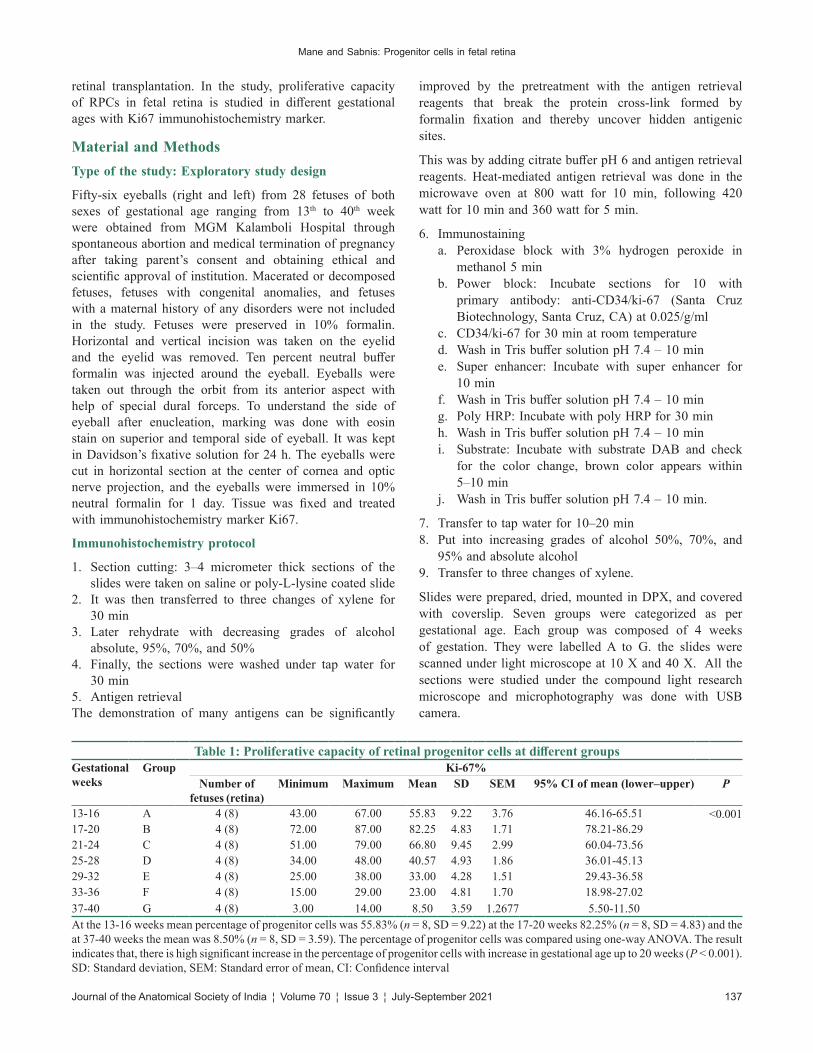
Table 1: Proliferative capacity of retinal progenitor cells at different groupsGestational weeks
Group Ki‑67%Number of
fetuses (retina)Minimum Maximum Mean SD SEM 95% CI of mean (lower–upper) P
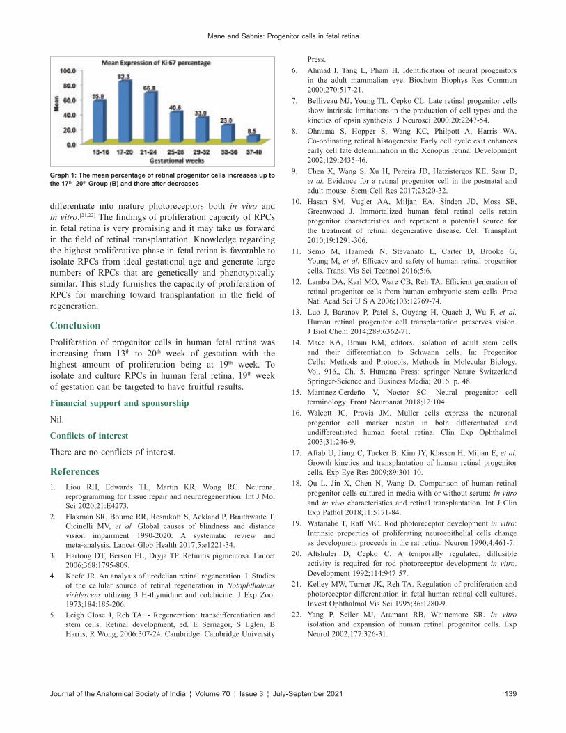
13‑16 A 4 (8) 43.00 67.00 55.83 9.22 3.76 46.16‑65.51 <0.00117‑20 B 4 (8) 72.00 87.00 82.25 4.83 1.71 78.21‑86.2921‑24 C 4 (8) 51.00 79.00 66.80 9.45 2.99 60.04‑73.5625‑28 D 4 (8) 34.00 48.00 40.57 4.93 1.86 36.01‑45.1329‑32 E 4 (8) 25.00 38.00 33.00 4.28 1.51 29.43‑36.5833‑36 F 4 (8) 15.00 29.00 23.00 4.81 1.70 18.98‑27.0237‑40 G 4 (8) 3.00 14.00 8.50 3.59 1.2677 5.50‑11.50At the 13‑16 weeks mean percentage of progenitor cells was 55.83% (n = 8, SD = 9.22) at the 17‑20 weeks 82.25% (n = 8, SD = 4.83) and the at 37‑40 weeks the mean was 8.50% (n = 8, SD = 3.59). The percentage of progenitor cells was compared using one‑way ANOVA. The result indicates that, there is high significant increase in the percentage of progenitor cells with increase in gestational age up to 20 weeks (P < 0.001). SD: Standard deviation, SEM: Standard error of mean, CI: Confidence interval
Mane and Sabnis: Progenitor cells in fetal retina
138 Journal of the Anatomical Society of India ¦ Volume 70 ¦ Issue 3 ¦ July-September 2021
Data analysis for ki‑67: The number of progenitor cells was assessed in sections of the retina. We recorded 20 measurements/fetus. The counts were expressed as average percentage of progenitor cells positive for Ki‑67 marker in the retina of each fetus. Then, the mean percentage of progenitor cells was determined for each group.
Results1. Observations of right and left retina were similar2. Proliferative capacity of RPCs was highest in B
group (17–20 weeks) and it was maximum at 19th week of gestation [Table 1].
DiscussionStem cells are the cells which are capable of proliferation, self‑renewal, and differentiation into various types of cells. These are called totipotent cells when isolated from fertilized oocyte, pluripotent when isolated from blastocyst, and multipotent when isolated from fully developed adult tissue.[14] Multipotent stem cells are referred as progenitor cells.[15] Ability of progenitor cells to get transform into various cells explores new avenue in medicine where regenerative therapy is essential. Retinal degenerative conditions like retinitis pigmentosa, age related macula degeneration where progressive visual decline results because of continuing loss of photoreceptor cells. Injecting progenitor cells in such cases is a promising attempt which would help to restore vision as progenitor cells have peculiarity to convert into various type of cells of retina. Progenitor cells are able to retain their progenitor status and these cells are not tumorigenic over the period. They may provide renewable, stable, and consistent supply of transplantable cells in the treatment of retinal degeneration.[10] Transplantation of progenitor cells of fetal retina into degenerative retinal diseases would be fruitful in protecting visual function.[13] Human embryonic stem cells can be selectively directed to neural retinal fate and may be useful in the treatment of retinal degeneration.[12] RPCs are obtained from fetal eyes at 16 to 18 weeks of gestation when retinal differentiation has been well defined.
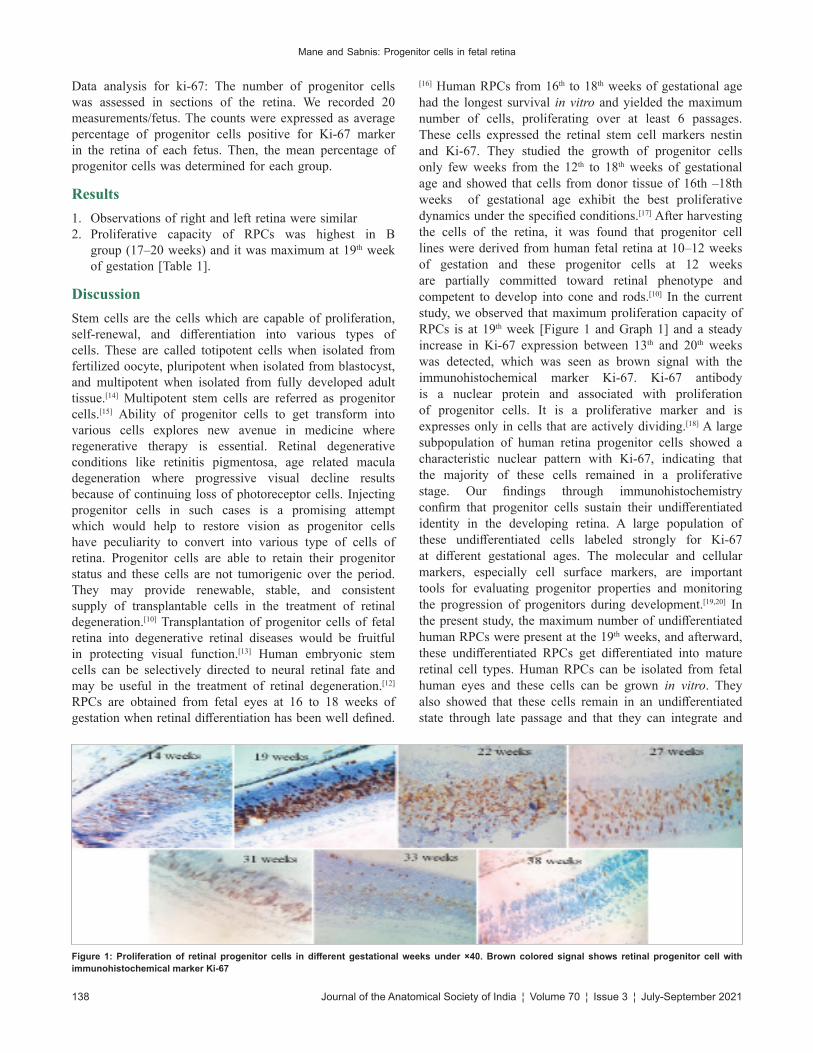
[16] Human RPCs from 16th to 18th weeks of gestational age had the longest survival in vitro and yielded the maximum number of cells, proliferating over at least 6 passages. These cells expressed the retinal stem cell markers nestin and Ki‑67. They studied the growth of progenitor cells only few weeks from the 12th to 18th weeks of gestational age and showed that cells from donor tissue of 16th –18th weeks of gestational age exhibit the best proliferative dynamics under the specified conditions.[17] After harvesting the cells of the retina, it was found that progenitor cell lines were derived from human fetal retina at 10–12 weeks of gestation and these progenitor cells at 12 weeks are partially committed toward retinal phenotype and competent to develop into cone and rods.[10] In the current study, we observed that maximum proliferation capacity of RPCs is at 19th week [Figure 1 and Graph 1] and a steady increase in Ki‑67 expression between 13th and 20th weeks was detected, which was seen as brown signal with the immunohistochemical marker Ki‑67. Ki‑67 antibody is a nuclear protein and associated with proliferation of progenitor cells. It is a proliferative marker and is expresses only in cells that are actively dividing.[18] A large subpopulation of human retina progenitor cells showed a characteristic nuclear pattern with Ki‑67, indicating that the majority of these cells remained in a proliferative stage. Our findings through immunohistochemistry confirm that progenitor cells sustain their undifferentiated identity in the developing retina. A large population of these undifferentiated cells labeled strongly for Ki‑67 at different gestational ages. The molecular and cellular markers, especially cell surface markers, are important tools for evaluating progenitor properties and monitoring the progression of progenitors during development.[19,20] In the present study, the maximum number of undifferentiated human RPCs were present at the 19th weeks, and afterward, these undifferentiated RPCs get differentiated into mature retinal cell types. Human RPCs can be isolated from fetal human eyes and these cells can be grown in vitro. They also showed that these cells remain in an undifferentiated state through late passage and that they can integrate and
Figure 1: Proliferation of retinal progenitor cells in different gestational weeks under ×40. Brown colored signal shows retinal progenitor cell with immunohistochemical marker Ki‑67
Mane and Sabnis: Progenitor cells in fetal retina
Journal of the Anatomical Society of India ¦ Volume 70 ¦ Issue 3 ¦ July-September 2021 139
differentiate into mature photoreceptors both in vivo and in vitro.[21,22] The findings of proliferation capacity of RPCs in fetal retina is very promising and it may take us forward in the field of retinal transplantation. Knowledge regarding the highest proliferative phase in fetal retina is favorable to isolate RPCs from ideal gestational age and generate large numbers of RPCs that are genetically and phenotypically similar. This study furnishes the capacity of proliferation of RPCs for marching toward transplantation in the field of regeneration.
ConclusionProliferation of progenitor cells in human fetal retina was increasing from 13th to 20th week of gestation with the highest amount of proliferation being at 19th week. To isolate and culture RPCs in human feral retina, 19th week of gestation can be targeted to have fruitful results.
Financial support and sponsorship
Nil.
Conflicts of interest
There are no conflicts of interest.
References1. Liou RH, Edwards TL, Martin KR, Wong RC. Neuronal
reprogramming for tissue repair and neuroregeneration. Int J Mol Sci 2020;21:E4273.
2. Flaxman SR, Bourne RR, Resnikoff S, Ackland P, Braithwaite T, Cicinelli MV, et al. Global causes of blindness and distance vision impairment 1990‑2020: A systematic review and meta‑analysis. Lancet Glob Health 2017;5:e1221‑34.
3. Hartong DT, Berson EL, Dryja TP. Retinitis pigmentosa. Lancet 2006;368:1795‑809.
4. Keefe JR. An analysis of urodelian retinal regeneration. I. Studies of the cellular source of retinal regeneration in Notophthalmus viridescens utilizing 3 H‑thymidine and colchicine. J Exp Zool 1973;184:185‑206.
5. Leigh Close J, Reh TA. ‑ Regeneration: transdifferentiation and stem cells. Retinal development, ed. E Sernagor, S Eglen, B Harris, R Wong, 2006:307‑24. Cambridge: Cambridge University
Press.6. Ahmad I, Tang L, Pham H. Identification of neural progenitors
in the adult mammalian eye. Biochem Biophys Res Commun 2000;270:517‑21.
7. Belliveau MJ, Young TL, Cepko CL. Late retinal progenitor cells show intrinsic limitations in the production of cell types and the kinetics of opsin synthesis. J Neurosci 2000;20:2247‑54.
8. Ohnuma S, Hopper S, Wang KC, Philpott A, Harris WA. Co‑ordinating retinal histogenesis: Early cell cycle exit enhances early cell fate determination in the Xenopus retina. Development 2002;129:2435‑46.
9. Chen X, Wang S, Xu H, Pereira JD, Hatzistergos KE, Saur D, et al. Evidence for a retinal progenitor cell in the postnatal and adult mouse. Stem Cell Res 2017;23:20‑32.
10. Hasan SM, Vugler AA, Miljan EA, Sinden JD, Moss SE, Greenwood J. Immortalized human fetal retinal cells retain progenitor characteristics and represent a potential source for the treatment of retinal degenerative disease. Cell Transplant 2010;19:1291‑306.
11. Semo M, Haamedi N, Stevanato L, Carter D, Brooke G, Young M, et al. Efficacy and safety of human retinal progenitor cells. Transl Vis Sci Technol 2016;5:6.
12. Lamba DA, Karl MO, Ware CB, Reh TA. Efficient generation of retinal progenitor cells from human embryonic stem cells. Proc Natl Acad Sci U S A 2006;103:12769‑74.
13. Luo J, Baranov P, Patel S, Ouyang H, Quach J, Wu F, et al. Human retinal progenitor cell transplantation preserves vision. J Biol Chem 2014;289:6362‑71.
14. Mace KA, Braun KM, editors. Isolation of adult stem cells and their differentiation to Schwann cells. In: Progenitor Cells: Methods and Protocols, Methods in Molecular Biology. Vol. 916., Ch. 5. Humana Press: springer Nature Switzerland Springer‑Science and Business Media; 2016. p. 48.
15. Martínez‑Cerdeño V, Noctor SC. Neural progenitor cell terminology. Front Neuroanat 2018;12:104.
16. Walcott JC, Provis JM. Müller cells express the neuronal progenitor cell marker nestin in both differentiated and undifferentiated human foetal retina. Clin Exp Ophthalmol 2003;31:246‑9.
17. Aftab U, Jiang C, Tucker B, Kim JY, Klassen H, Miljan E, et al. Growth kinetics and transplantation of human retinal progenitor cells. Exp Eye Res 2009;89:301‑10.
18. Qu L, Jin X, Chen N, Wang D. Comparison of human retinal progenitor cells cultured in media with or without serum: In vitro and in vivo characteristics and retinal transplantation. Int J Clin Exp Pathol 2018;11:5171‑84.
19. Watanabe T, Raff MC. Rod photoreceptor development in vitro: Intrinsic properties of proliferating neuroepithelial cells change as development proceeds in the rat retina. Neuron 1990;4:461‑7.
20. Altshuler D, Cepko C. A temporally regulated, diffusible activity is required for rod photoreceptor development in vitro. Development 1992;114:947‑57.
21. Kelley MW, Turner JK, Reh TA. Regulation of proliferation and photoreceptor differentiation in fetal human retinal cell cultures. Invest Ophthalmol Vis Sci 1995;36:1280‑9.
22. Yang P, Seiler MJ, Aramant RB, Whittemore SR. In vitro isolation and expansion of human retinal progenitor cells. Exp Neurol 2002;177:326‑31.
Graph 1: The mean percentage of retinal progenitor cells increases up to the 17th–20th Group (B) and there after decreases
140 © 2021 Journal of the Anatomical Society of India | Published by Wolters Kluwer - Medknow
Address for correspondence: Prof. Veronica Macchi, Department of Neurosciences, Institute of Human Anatomy, University of Padova, Via A. Gabelli 65, Padova 35127, Italy. E‑mail: [email protected]
Access this article online
Website: www.jasi.org.inDOI: 10.4103/JASI.JASI_17_20
Quick Response Code:
AbstractIntroduction: Teaching anatomy moved from teacher‑centered to student‑centered learning. Three‑dimensional models help to improve self‑learning of basic concepts other than anatomical spatial relationships. Wax and plastinated models were compared for appropriateness and safety in teaching human anatomy to health‑care professionals. Material and Methods: Randomized crossover trial. The CONSORT checklist for randomized crossover trials was followed. Eighteen volunteer physiotherapy students at the University of Padova were randomized into two crossing‑over groups applying to wax and plastinated heart models. Final Likert survey scales were administered. Results: They reported that the wax models presented a more pleasant smell, a better chromatic appearance, and superior ease of handling than plastinated models, with a higher degree of perceived biological safety. Wax models were judged less suitable for educational use and for clarifying anatomical doubts, both in assessing external and internal anatomical details. Discussion and Conclusion: Overall, the plastinated models were considered more suitable for educational use in teaching internal and external anatomical details. The wax models showed a better appearance, ease of handling, and a minor perceived biological hazard.
Keywords: Ceroplastic, health‑care professionals, plastinated model, randomized crossover trial, wax model
Wax versus Plastinated Models in Teaching Human Anatomy to Health‑care Professionals. A Randomized Crossover Trial
Rafael Boscolo‑Berto, Cinzia Tortorella, Veronica Macchi, Andrea Porzionato, Raffaele De CaroDepartment of Neurosciences, Institute of Human Anatomy, University of Padova, Padova, Italy
How to cite this article: Boscolo-Berto R, Tortorella C, Macchi V, Porzionato A, De Caro R. Wax versus plastinated models in teaching human anatomy to health-care professionals. A randomized crossover trial. J Anat Soc India 2021;XX:XX-XX.
IntroductionHistorically, the dissection of human bodies has been the most relevant teaching paradigm for gross anatomy learning since the late 16th century. A body donation program was implemented at the Institute of Human Anatomy of the University of Padova to make health‑care professionals specifically, anatomy education and research activities on real human models.[1] However, the exponential growth of interest in anatomical education on cadavers by students and postgraduates, jointly with the increase of class sizes, makes the number of available bodies insufficient to meet current needs fully.
The paradigm shift has moved from passive and teacher‑centered learning toward active and student‑centered learning by integrating supplementary multimodal teaching resources.[2] Interactive three‑dimensional models may improve the long‑lasting retaining of anatomical details and understand their spatial arrangement and
neighborhood relationships. Indeed, they are increasingly required by health‑care students, with a favorable impact on student’s perception and learning.[2‑4] Among the tools traditionally used for this purpose, there are both wax and plastinated models.[5‑8] They are widely used for didactical purposes, attempting to reproduce and clarify the human body’s three‑dimensional organization and complexity. They recall the actions and results of an anatomic dissection as performed by an anatomist, being a potential option to reproduce and manipulate specimens as close as possible to reality.[9]
Mainly, anatomical wax modeling (also named ceroplastics or less frequently moulage) referred to a traditional technique involving wax obtained by bees or other animals/vegetable sources to manufacture high‑quality artworks colored so realistic that they resemble almost lifelike.[7,10] They require unique expertise and are time‑ and money‑consuming.[11]
This is an open access journal, and articles are distributed under the terms of the Creative Commons Attribution‑NonCommercial‑ShareAlike 4.0 License, which allows others to remix, tweak, and build upon the work non‑commercially, as long as appropriate credit is given and the new creations are licensed under the identical terms.
For reprints contact: [email protected]
Original Article
Received: 31 January 2020 Revised: 26 January 2021 Accepted: 07 July 2021 Available online: ***
Article Info
Boscolo‑Berto, et al.: Wax and plastinated models in teaching anatomy
Journal of the Anatomical Society of India ¦ Volume 70 ¦ Issue 3 ¦ July-September 2021 141
On the other hand, anatomical plastination was introduced in the late 1970s using silicone rubber, polyester, or epoxy resin to produce treated specimens of high durability that are extensively used in anatomical research and medical teaching.[12,13] However, one negative point is the complexity of the operations necessary for the production of the models, resulting in an articulate and expensive process with low chromatic credibility.[5]
The present study compared three‑dimensional wax versus plastinated models to investigate their impact on practical experience. Mainly, it explores which one is perceived as more useful, safe, and appropriate for the study of basic human anatomy in a university course devoted to health professionals such as physiotherapists.
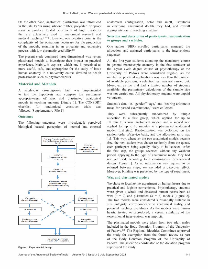
Material and MethodsA single‑day crossing‑over trial was implemented to test the hypothesis and compare the usefulness/appropriateness of wax and plastinated anatomical models in teaching anatomy [Figure 1]. The CONSORT checklist for randomized crossover trials was followed [Supplementary File 1].
Outcomes
The following outcomes were investigated: perceived biological hazard, perception of internal and external
anatomical configuration, color and smell, usefulness in clarifying anatomical doubts they had, and overall appropriateness in teaching anatomy.
Selection and description of participants, randomization to groups and variables.
One author (BBR) enrolled participants, managed the allocation, and assigned participants to the interventions sequence.
All the first‑year students attending the mandatory course in general macroscopic anatomy in the first semester of the 3‑year cycle degree course of physiotherapy at the University of Padova were considered eligible. As the number of potential applications was less than the number of available positions, a selection test was not carried out. Moreover, as the trial had a limited number of students available, the preliminary calculation of the sample size was not carried out. All physiotherapy students were unpaid volunteers.
Student’s data, i.e. “gender,” “age,” and “scoring arithmetic mean for passed examinations,” were collected.
They were subsequently randomized by simple allocation to a first group, which applied for up to 10 min to a wax anatomical model, and a second one applied for up to 10 minutes to a plastinated anatomical model (first step). Randomization was performed on the random‑order‑of‑service basis, and the allocation ratio was 1:1. This way, whenever the two anatomical models became free, the next student was chosen randomly from the queue, each participant being equally likely to be selected. After the first step, the groups reversed without any washout period, applying to the type of anatomical model they had not yet used, according to a crossing‑over experimental design [Figure 1]. As no information was required to be retained between steps, we excluded a carryover effect. Moreover, blinding was prevented by the type of experiment.
Wax and plastinated models
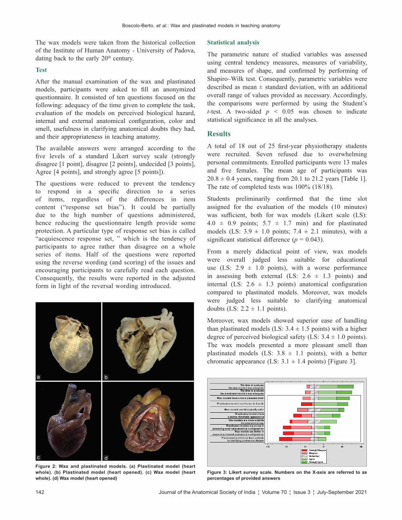
We chose to focalize the experiment on human hearts due to practical and logistic convenience. Physiotherapy students were given a whole and dissected human hearts both as wax (n = 2) and plastinated (n = 2) models [Figure 2]. The two models were considered substantially suitable in size, integrity, correspondence to anatomical reality, and potential teaching usefulness. As the models were human hearts, treated or reproduced, a certain similarity of the experimental interventions was implicit.
The plastinated models were taken from two adult males included in the Body Donation Program of the University of Padova.[14] The Regional Bioethics Committee approved the study for exemption from the formal review as part of the Body Donation Program of the University of Padova. The scientific coordinator of the donation program supervised the study.Figure 1: Experimental design
Boscolo‑Berto, et al.: Wax and plastinated models in teaching anatomy
142 Journal of the Anatomical Society of India ¦ Volume 70 ¦ Issue 3 ¦ July-September 2021
The wax models were taken from the historical collection of the Institute of Human Anatomy ‑ University of Padova, dating back to the early 20th century.
Test
After the manual examination of the wax and plastinated models, participants were asked to fill an anonymized questionnaire. It consisted of ten questions focused on the following: adequacy of the time given to complete the task, evaluation of the models on perceived biological hazard, internal and external anatomical configuration, color and smell, usefulness in clarifying anatomical doubts they had, and their appropriateness in teaching anatomy.
The available answers were arranged according to the five levels of a standard Likert survey scale (strongly disagree [1 point], disagree [2 points], undecided [3 points], Agree [4 points], and strongly agree [5 points]).
The questions were reduced to prevent the tendency to respond in a specific direction to a series of items, regardless of the differences in item content (“response set bias”). It could be partially due to the high number of questions administered, hence reducing the questionnaire length provide some protection. A particular type of response set bias is called “acquiescence response set, ” which is the tendency of participants to agree rather than disagree on a whole series of items. Half of the questions were reported using the reverse wording (and scoring) of the issues and encouraging participants to carefully read each question. Consequently, the results were reported in the adjusted form in light of the reversal wording introduced.
Statistical analysis
The parametric nature of studied variables was assessed using central tendency measures, measures of variability, and measures of shape, and confirmed by performing of Shapiro–Wilk test. Consequently, parametric variables were described as mean ± standard deviation, with an additional overall range of values provided as necessary. Accordingly, the comparisons were performed by using the Student’s t‑test. A two‑sided p < 0.05 was chosen to indicate statistical significance in all the analyses.
ResultsA total of 18 out of 25 first‑year physiotherapy students were recruited. Seven refused due to overwhelming personal commitments. Enrolled participants were 13 males and five females. The mean age of participants was 20.8 ± 0.4 years, ranging from 20.1 to 21.2 years [Table 1]. The rate of completed tests was 100% (18/18).
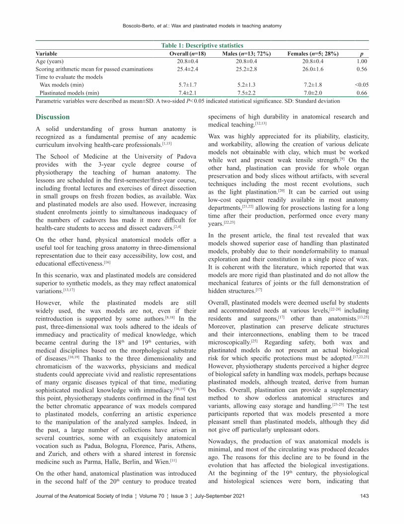
Students preliminarily confirmed that the time slot assigned for the evaluation of the models (10 minutes) was sufficient, both for wax models (Likert scale (LS): 4.0 ± 0.9 points; 5.7 ± 1.7 min) and for plastinated models (LS: 3.9 ± 1.0 points; 7.4 ± 2.1 minutes), with a significant statistical difference (p = 0.043).
From a merely didactical point of view, wax models were overall judged less suitable for educational use (LS: 2.9 ± 1.0 points), with a worse performance in assessing both external (LS: 2.6 ± 1.3 points) and internal (LS: 2.6 ± 1.3 points) anatomical configuration compared to plastinated models. Moreover, wax models were judged less suitable to clarifying anatomical doubts (LS: 2.2 ± 1.1 points).
Moreover, wax models showed superior ease of handling than plastinated models (LS: 3.4 ± 1.5 points) with a higher degree of perceived biological safety (LS: 3.4 ± 1.0 points). The wax models presented a more pleasant smell than plastinated models (LS: 3.8 ± 1.1 points), with a better chromatic appearance (LS: 3.1 ± 1.4 points) [Figure 3].
Figure 3: Likert survey scale. Numbers on the X‑axis are referred to as percentages of provided answers
Figure 2: Wax and plastinated models. (a) Plastinated model (heart whole). (b) Plastinated model (heart opened). (c) Wax model (heart whole). (d) Wax model (heart opened)
dc
ba
Boscolo‑Berto, et al.: Wax and plastinated models in teaching anatomy
Journal of the Anatomical Society of India ¦ Volume 70 ¦ Issue 3 ¦ July-September 2021 143
DiscussionA solid understanding of gross human anatomy is recognized as a fundamental premise of any academic curriculum involving health‑care professionals.[1,15]
The School of Medicine at the University of Padova provides with the 3‑year cycle degree course of physiotherapy the teaching of human anatomy. The lessons are scheduled in the first‑semester/first‑year course, including frontal lectures and exercises of direct dissection in small groups on fresh frozen bodies, as available. Wax and plastinated models are also used. However, increasing student enrolments jointly to simultaneous inadequacy of the numbers of cadavers has made it more difficult for health‑care students to access and dissect cadavers.[2,4]
On the other hand, physical anatomical models offer a useful tool for teaching gross anatomy in three‑dimensional representation due to their easy accessibility, low cost, and educational effectiveness.[16]
In this scenario, wax and plastinated models are considered superior to synthetic models, as they may reflect anatomical variations.[13,17]
However, while the plastinated models are still widely used, the wax models are not, even if their reintroduction is supported by some authors.[8,18] In the past, three‑dimensional wax tools adhered to the ideals of immediacy and practicality of medical knowledge, which became central during the 18th and 19th centuries, with medical disciplines based on the morphological substrate of diseases.[10,19] Thanks to the three dimensionality and chromaticism of the waxworks, physicians and medical students could appreciate vivid and realistic representations of many organic diseases typical of that time, mediating sophisticated medical knowledge with immediacy.[10,19] On this point, physiotherapy students confirmed in the final test the better chromatic appearance of wax models compared to plastinated models, conferring an artistic experience to the manipulation of the analyzed samples. Indeed, in the past, a large number of collections have arisen in several countries, some with an exquisitely anatomical vocation such as Padua, Bologna, Florence, Paris, Athens, and Zurich, and others with a shared interest in forensic medicine such as Parma, Halle, Berlin, and Wien.[11]
On the other hand, anatomical plastination was introduced in the second half of the 20th century to produce treated
specimens of high durability in anatomical research and medical teaching.[12,13]
Wax was highly appreciated for its pliability, elasticity, and workability, allowing the creation of various delicate models not obtainable with clay, which must be worked while wet and present weak tensile strength.[9] On the other hand, plastination can provide for whole organ preservation and body slices without artifacts, with several techniques including the most recent evolutions, such as the light plastination.[20] It can be carried out using low‑cost equipment readily available in most anatomy departments,[21,22] allowing for prosections lasting for a long time after their production, performed once every many years.[22,23]
In the present article, the final test revealed that wax models showed superior ease of handling than plastinated models, probably due to their nondeformability to manual exploration and their constitution in a single piece of wax. It is coherent with the literature, which reported that wax models are more rigid than plastinated and do not allow the mechanical features of joints or the full demonstration of hidden structures.[17]
Overall, plastinated models were deemed useful by students and accommodated needs at various levels,[22‑24] including residents and surgeons,[17] other than anatomists.[13,25] Moreover, plastination can preserve delicate structures and their interconnections, enabling them to be traced microscopically.[25] Regarding safety, both wax and plastinated models do not present an actual biological risk for which specific protections must be adopted.[17,22,23] However, physiotherapy students perceived a higher degree of biological safety in handling wax models, perhaps because plastinated models, although treated, derive from human bodies. Overall, plastination can provide a supplementary method to show odorless anatomical structures and variants, allowing easy storage and handling.[23‑25] The test participants reported that wax models presented a more pleasant smell than plastinated models, although they did not give off particularly unpleasant odors.
Nowadays, the production of wax anatomical models is minimal, and most of the circulating was produced decades ago. The reasons for this decline are to be found in the evolution that has affected the biological investigations. At the beginning of the 19th century, the physiological and histological sciences were born, indicating that
Table 1: Descriptive statisticsVariable Overall (n=18) Males (n=13; 72%) Females (n=5; 28%) pAge (years) 20.8±0.4 20.8±0.4 20.8±0.4 1.00Scoring arithmetic mean for passed examinations 25.4±2.4 25.2±2.8 26.0±1.6 0.56Time to evaluate the models
Wax models (min) 5.7±1.7 5.2±1.3 7.2±1.8 <0.05Plastinated models (min) 7.4±2.1 7.5±2.2 7.0±2.0 0.66
Parametric variables were described as mean±SD. A two‑sided P<0.05 indicated statistical significance. SD: Standard deviation
Boscolo‑Berto, et al.: Wax and plastinated models in teaching anatomy
144 Journal of the Anatomical Society of India ¦ Volume 70 ¦ Issue 3 ¦ July-September 2021
medicine was already directed toward the anatomo‑clinical approach, further developed in the following decades with the introduction of histopathology, cell pathology, and radiology.[9,19]
In this scenario, the gross anatomic institutions have taken a back seat in scientific research for many decades, although they continued to be active and still considered the mainstay of medical education. Moreover, the prohibitive costs related to the type of processing and the time required have made the wax models the almost exclusive prerogative of the institutions with the most significant funds available. Furthermore, historically, the major boost to wax models was the lack of valid methods for preserving bodies, so the dissections were concentrated in the winter season and over a few days.[11] With the development of more modern fixation techniques in the second half of the 19th century (i.e., formalin instead of alcoholic solutions), demonstrative dissections became more feasible. The treated samples could last for weeks, by the use of much cheaper and faster processes than the productions of wax models, which lost much of their teaching appeal.[9] The wax models kept a role in the reproduction of rare diseases for a longer time so that students had an always‑available reference to recognize unfrequent diseases. Dedicated series of wax models were produced for several medical disciplines, aimed to show mainly the pathological modifications of the organs induced by the diseases studied and treated by the specialists.[10,19] Overall, wax models in modern medical education seemed to be still of interest for learning on diseases, being integrated with other teaching tools to improve diagnostic skills.[7,11] At the same time, this is valid for studying normal surface anatomy, enabling learners to improve visualization of structures, especially those of the musculoskeletal system, hence contributing to a better anatomical learning experience by familiarising with surface landmarks.[15,26]
Physiotherapy students here reported that, from a merely didactical point of view, wax models were overall less suitable for educational use compared to plastinated models, with a worse performance in assessing both external and internal anatomical details. In this view, wax models were judged less suitable to clarify anatomical doubts than plastinated ones.
Indeed, for health‑care students who are not provided with the opportunity to dissect human bodies, plastinated specimens might represent a valid alternative.[27] Although three‑dimensional models cannot replace traditional anatomic dissection in every circumstance, the anatomical section of fresh frozen bodies probably is beyond the scope for physiotherapy students.[17,18,22]
Wax models are artistic works of great value that should still find a prominent place in the diagnostic teaching of even unusual pathological cases, while plastinated models can be more useful in daily teaching of anatomy to students needing basic notions.
The present study is affected by the following limitations.
First, the study population was restricted to first‑year physiotherapy students. Whether these findings could be generalized to more senior ones needs to be determined. In the same way, external validity should be tested.
Second, students were not tested for their spatial ability, i.e. the aptitude for understanding three‑dimensional structure, which could interfere with the spatial evaluation apart from the type of educational materials involved.[28]
Third, participation in the trial was voluntary, potentially leading to biases in various directions concerning which students chose to attend the present experiment.
Fourth, just heart models were used in this trial. The inference of these results to other anatomical regions is to be verified.
Finally, the current results arise from a single‑center study. The sample size is limited because the degree course of physiotherapy includes few students. Moreover, their attendance in the classrooms or handling of anatomical material was restricted due to the COVID pandemic.
ConclusionWax and plastinated models are significant educational, research, and cultural tools in the medical world to integrate teaching and learning anatomy in conjunction with traditional gross dissection, which remains the milestone reference. They could help maximize the impact of practical experience and overcome the contraction of economic resources and the shortage of available bodies.
Despite wax models showing a better chromatic appearance, superior ease of handling, and a minor perceived biological hazard against the plastinated models, the latter were judged more suitable for educational use in teaching internal and external anatomical details.
Nevertheless, further studies are needed to validate these findings, to expand the trials to models of other anatomical districts, other than examining the long‑term impacts on the learning process also for other health‑care professionals.
Key findings:
• Wax and plastinated models are still useful educational, research, and cultural tools.
• They could be of help in maximizing the impact of practical experience.
• Plastinated models were judged more suitable for educational use in teaching anatomy.
Acknowledgments
The authors wish to thank Gloria Sarasin for her administrative and technical support in scheduling the experimental activities and administering
Boscolo‑Berto, et al.: Wax and plastinated models in teaching anatomy
Journal of the Anatomical Society of India ¦ Volume 70 ¦ Issue 3 ¦ July-September 2021 145
questionnaires to the students, and Aron Emmi for retrieving museum materials.
Financial support and sponsorship
Nil.
Conflicts of interest
There are no conflicts of interest.
References1. Macchi V, Porzionato A, Stecco C, De Caro R. Evolution of the
anatomical theatre in Padova. Anat Sci Educ. 2014;7:487‑93. doi:10.1002/ase.1447.
2. McLachlan JC, Patten D. Anatomy teaching: Ghosts of the past, present and future. Med Educ 2006;40:243‑53.
3. Triepels CP, Koppes DM, Van Kuijk SM, Popeijus HE, Lamers WH, van Gorp T, et al. Medical students’ perspective on training in anatomy. Ann Anat 2018;217:60‑5.
4. McLachlan JC, Bligh J, Bradley P, Searle J. Teaching anatomy without cadavers. Med Educ 2004;38:418‑24.
5. Haenssgen K, Makanya AN, Djonov V. Casting materials and their application in research and teaching. Microsc Microanal 2014;20:493‑513.
6. Kravetz RE. Anatomic wax model. Am J Gastroenterol 2008;103:1339.
7. Parish LC, Geiges ML. The wax model. Skinmed 2011;9:275‑7.8. Toutous Trellu L, Stahl Gretsch LI, Wenger A. A new life for old
anatomical wakes. Rev Med Suisse 2019;15:662‑5.9. Chen JC, Amar AP, Levy ML, Apuzzo ML. The development of
anatomic art and sciences: The ceroplastica anatomic models of La Specola. Neurosurgery 1999;45:883‑91.
10. Rodriguez O, Parish LC. Wax models in dermatology: Updated through 2020. Clin Dermatol 2020;38:555‑62.
11. Cooke RA. A moulage museum is not just a museum: Wax models as teaching instruments. Virchows Arch 2010;457:513‑20.
12. von Hagens G, Tiedemann K, Kriz W. The current potential of plastination. Anat Embryol (Berl) 1987;175:411‑21.
13. Jones DG. Re‑inventing anatomy: The impact of plastination on how we see the human body. Clin Anat 2002;15:436‑40.
14. Porzionato A, Macchi V, Stecco C, Mazzi A, Rambaldo A,
Sarasin G, et al. Quality management of body donation program at the University of Padova. Anat Sci Educ. 2012;5:264‑72. doi:10.1002/ase.1285.
15. Leonard R. A clinical anatomy curriculum for the medical student of the 21st century: Gross anatomy. Clin Anat 1999;9:71‑99.
16. Yammine K, Violato C. The effectiveness of physical models in teaching anatomy: A meta‑analysis of comparative studies. Adv Health Sci Educ Theory Pract 2016;21:883‑95.
17. Riederer BM. Plastination and its importance in teaching anatomy. Critical points for long‑term preservation of human tissue. J Anat 2014;224:309‑15.
18. Perry M, Maffulli N, Willson S, Morrissey D. The effectiveness of arts‑based interventions in medical education: A literature review. Med Educ 2011;45:141‑8.
19. Zampieri F, Comacchio F, Zanatta A. Ophthalmologic wax models as an educational tool for 18th‑century vision scientists. Acta Ophthalmol 2017;95:852‑7.
20. Steinke H, Rabi S, Saito T, Sawutti A, Miyaki T, Itoh M, et al. Light‑weight plastination. Ann Anat 2008;190:428‑31.
21. O’Sullivan E, Mitchell BS. Plastination for gross anatomy teaching using low cost equipment. Surg Radiol Anat 1995;17:277‑81.
22. Estai M, Bunt S. Best teaching practices in anatomy education: A critical review. Ann Anat 2016;208:151‑7.
23. Latorre RM, García‑Sanz MP, Moreno M, Hernández F, Gil F, López O, et al. How useful is plastination in learning anatomy? J Vet Med Educ 2007;34:172‑6.
24. Fruhstorfer BH, Palmer J, Brydges S, Abrahams PH. The use of plastinated prosections for teaching anatomy – The view of medical students on the value of this learning resource. Clin Anat 2011;24:246‑52.
25. Jones DG, Whitaker MI. Engaging with plastination and the Body Worlds phenomenon: A cultural and intellectual challenge for anatomists. Clin Anat 2009;22:770‑6.
26. Standring S. Evidence‑based surface anatomy. Clin Anat 2012;25:813‑5.
27. Sah S, Bhandari K, Acharya S, Thapa P, Goel N, Nimmagada H. Plastination: A miracle in the preservation of biological specimen. Eur J Pharm Med Res 2017;4:276‑81.
28. Garg AX, Norman G, Sperotable L. How medical students learn spatial anatomy. Lancet 2001;357:363‑4.
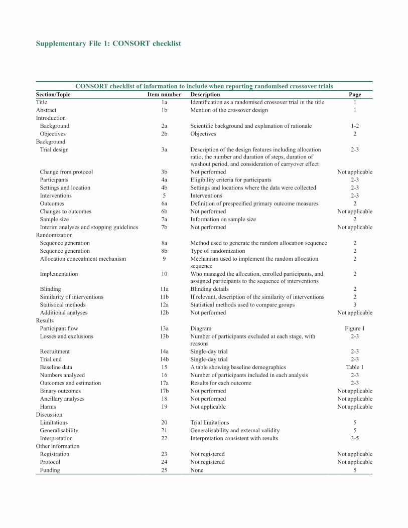
CONSORT checklist of information to include when reporting randomised crossover trialsSection/Topic Item number Description PageTitle 1a Identification as a randomised crossover trial in the title 1Abstract 1b Mention of the crossover design 1Introduction
Background 2a Scientific background and explanation of rationale 1‑2Objectives 2b Objectives 2
BackgroundTrial design 3a Description of the design features including allocation
ratio, the number and duration of steps, duration of washout period, and consideration of carryover effect
2‑3
Change from protocol 3b Not performed Not applicableParticipants 4a Eligibility criteria for participants 2‑3Settings and location 4b Settings and locations where the data were collected 2‑3Interventions 5 Interventions 2‑3Outcomes 6a Definition of prespecified primary outcome measures 2Changes to outcomes 6b Not performed Not applicableSample size 7a Information on sample size 2Interim analyses and stopping guidelines 7b Not performed Not applicable
RandomizationSequence generation 8a Method used to generate the random allocation sequence 2Sequence generation 8b Type of randomization 2Allocation concealment mechanism 9 Mechanism used to implement the random allocation
sequence2
Implementation 10 Who managed the allocation, enrolled participants, and assigned participants to the sequence of interventions
2
Blinding 11a Blinding details 2Similarity of interventions 11b If relevant, description of the similarity of interventions 2Statistical methods 12a Statistical methods used to compare groups 3Additional analyses 12b Not performed Not applicable
ResultsParticipant flow 13a Diagram Figure 1Losses and exclusions 13b Number of participants excluded at each stage, with
reasons2‑3
Recruitment 14a Single‑day trial 2‑3Trial end 14b Single‑day trial 2‑3Baseline data 15 A table showing baseline demographics Table 1Numbers analyzed 16 Number of participants included in each analysis 2‑3Outcomes and estimation 17a Results for each outcome 2‑3Binary outcomes 17b Not performed Not applicableAncillary analyses 18 Not performed Not applicableHarms 19 Not applicable Not applicable
DiscussionLimitations 20 Trial limitations 5Generalisability 21 Generalisability and external validity 5Interpretation 22 Interpretation consistent with results 3‑5
Other informationRegistration 23 Not registered Not applicableProtocol 24 Not registered Not applicableFunding 25 None 5
Supplementary File 1: CONSORT checklist
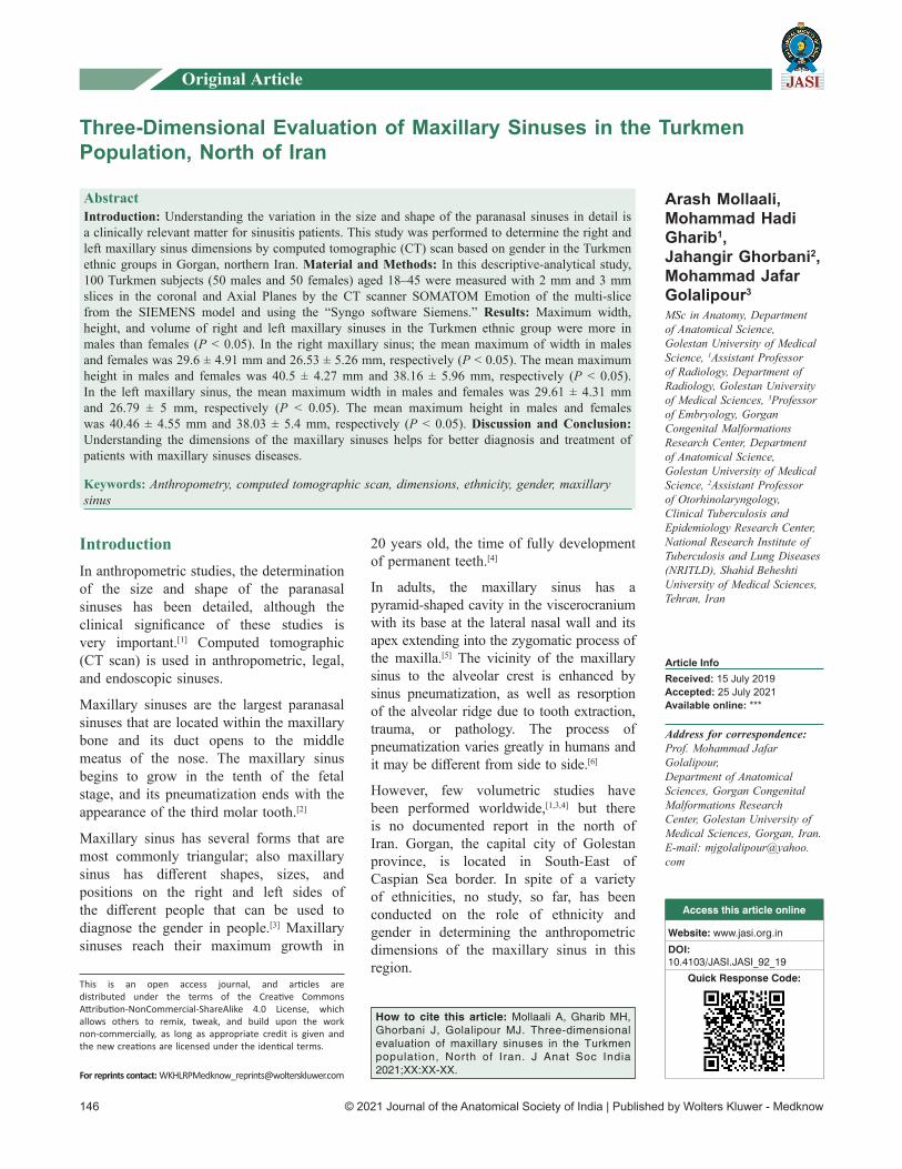
146 © 2021 Journal of the Anatomical Society of India | Published by Wolters Kluwer - Medknow
Address for correspondence: Prof. Mohammad Jafar Golalipour, Department of Anatomical Sciences, Gorgan Congenital Malformations Research Center, Golestan University of Medical Sciences, Gorgan, Iran. E‑mail: [email protected]
Access this article online
Website: www.jasi.org.inDOI: 10.4103/JASI.JASI_92_19
Quick Response Code:
AbstractIntroduction: Understanding the variation in the size and shape of the paranasal sinuses in detail is a clinically relevant matter for sinusitis patients. This study was performed to determine the right and left maxillary sinus dimensions by computed tomographic (CT) scan based on gender in the Turkmen ethnic groups in Gorgan, northern Iran. Material and Methods: In this descriptive‑analytical study, 100 Turkmen subjects (50 males and 50 females) aged 18–45 were measured with 2 mm and 3 mm slices in the coronal and Axial Planes by the CT scanner SOMATOM Emotion of the multi‑slice from the SIEMENS model and using the “Syngo software Siemens.” Results: Maximum width, height, and volume of right and left maxillary sinuses in the Turkmen ethnic group were more in males than females (P < 0.05). In the right maxillary sinus; the mean maximum of width in males and females was 29.6 ± 4.91 mm and 26.53 ± 5.26 mm, respectively (P < 0.05). The mean maximum height in males and females was 40.5 ± 4.27 mm and 38.16 ± 5.96 mm, respectively (P < 0.05). In the left maxillary sinus, the mean maximum width in males and females was 29.61 ± 4.31 mm and 26.79 ± 5 mm, respectively (P < 0.05). The mean maximum height in males and females was 40.46 ± 4.55 mm and 38.03 ± 5.4 mm, respectively (P < 0.05). Discussion and Conclusion: Understanding the dimensions of the maxillary sinuses helps for better diagnosis and treatment of patients with maxillary sinuses diseases.
Keywords: Anthropometry, computed tomographic scan, dimensions, ethnicity, gender, maxillary sinus
Three‑Dimensional Evaluation of Maxillary Sinuses in the Turkmen Population, North of Iran
Arash Mollaali, Mohammad Hadi Gharib1, Jahangir Ghorbani2, Mohammad Jafar Golalipour3
MSc in Anatomy, Department of Anatomical Science, Golestan University of Medical Science, 1Assistant Professor of Radiology, Department of Radiology, Golestan University of Medical Sciences, 3Professor of Embryology, Gorgan Congenital Malformations Research Center, Department of Anatomical Science, Golestan University of Medical Science, 2Assistant Professor of Otorhinolaryngology, Clinical Tuberculosis and Epidemiology Research Center, National Research Institute of Tuberculosis and Lung Diseases (NRITLD), Shahid Beheshti University of Medical Sciences, Tehran, Iran
How to cite this article: Mollaali A, Gharib MH, Ghorbani J, Golalipour MJ. Three-dimensional evaluation of maxillary sinuses in the Turkmen population, North of Iran. J Anat Soc India 2021;XX:XX-XX.
IntroductionIn anthropometric studies, the determination of the size and shape of the paranasal sinuses has been detailed, although the clinical significance of these studies is very important.[1] Computed tomographic (CT scan) is used in anthropometric, legal, and endoscopic sinuses.
Maxillary sinuses are the largest paranasal sinuses that are located within the maxillary bone and its duct opens to the middle meatus of the nose. The maxillary sinus begins to grow in the tenth of the fetal stage, and its pneumatization ends with the appearance of the third molar tooth.[2]
Maxillary sinus has several forms that are most commonly triangular; also maxillary sinus has different shapes, sizes, and positions on the right and left sides of the different people that can be used to diagnose the gender in people.[3] Maxillary sinuses reach their maximum growth in
20 years old, the time of fully development of permanent teeth.[4]
In adults, the maxillary sinus has a pyramid‑shaped cavity in the viscerocranium with its base at the lateral nasal wall and its apex extending into the zygomatic process of the maxilla.[5] The vicinity of the maxillary sinus to the alveolar crest is enhanced by sinus pneumatization, as well as resorption of the alveolar ridge due to tooth extraction, trauma, or pathology. The process of pneumatization varies greatly in humans and it may be different from side to side.[6]
However, few volumetric studies have been performed worldwide,[1,3,4] but there is no documented report in the north of Iran. Gorgan, the capital city of Golestan province, is located in South‑East of Caspian Sea border. In spite of a variety of ethnicities, no study, so far, has been conducted on the role of ethnicity and gender in determining the anthropometric dimensions of the maxillary sinus in this region.
This is an open access journal, and articles are distributed under the terms of the Creative Commons Attribution‑NonCommercial‑ShareAlike 4.0 License, which allows others to remix, tweak, and build upon the work non‑commercially, as long as appropriate credit is given and the new creations are licensed under the identical terms.
For reprints contact: [email protected]
Received: 15 July 2019Accepted: 25 July 2021Available online: ***
Article Info
Original Article
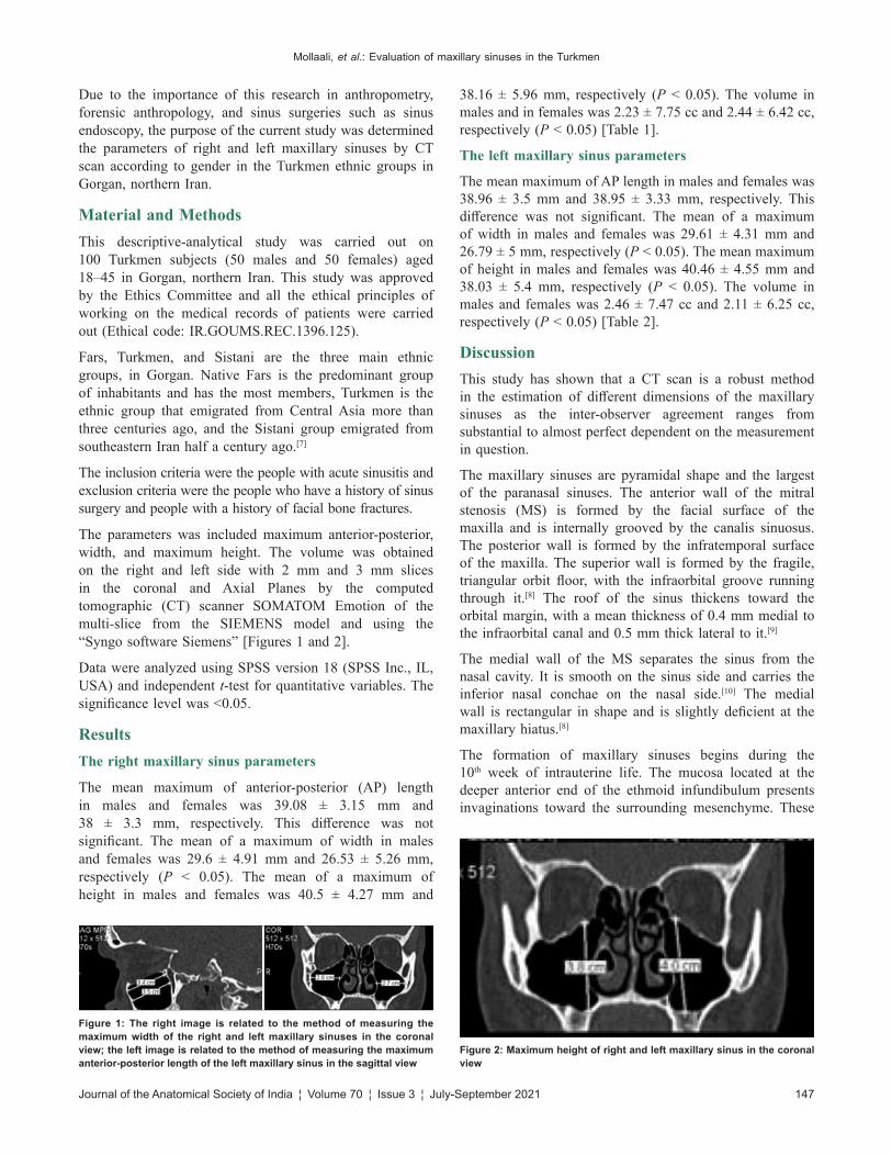
Mollaali, et al.: Evaluation of maxillary sinuses in the Turkmen
Journal of the Anatomical Society of India ¦ Volume 70 ¦ Issue 3 ¦ July-September 2021 147
Due to the importance of this research in anthropometry, forensic anthropology, and sinus surgeries such as sinus endoscopy, the purpose of the current study was determined the parameters of right and left maxillary sinuses by CT scan according to gender in the Turkmen ethnic groups in Gorgan, northern Iran.
Material and MethodsThis descriptive‑analytical study was carried out on 100 Turkmen subjects (50 males and 50 females) aged 18–45 in Gorgan, northern Iran. This study was approved by the Ethics Committee and all the ethical principles of working on the medical records of patients were carried out (Ethical code: IR.GOUMS.REC.1396.125).
Fars, Turkmen, and Sistani are the three main ethnic groups, in Gorgan. Native Fars is the predominant group of inhabitants and has the most members, Turkmen is the ethnic group that emigrated from Central Asia more than three centuries ago, and the Sistani group emigrated from southeastern Iran half a century ago.[7]
The inclusion criteria were the people with acute sinusitis and exclusion criteria were the people who have a history of sinus surgery and people with a history of facial bone fractures.
The parameters was included maximum anterior‑posterior, width, and maximum height. The volume was obtained on the right and left side with 2 mm and 3 mm slices in the coronal and Axial Planes by the computed tomographic (CT) scanner SOMATOM Emotion of the multi‑slice from the SIEMENS model and using the “Syngo software Siemens” [Figures 1 and 2].
Data were analyzed using SPSS version 18 (SPSS Inc., IL, USA) and independent t‑test for quantitative variables. The significance level was <0.05.
ResultsThe right maxillary sinus parameters
The mean maximum of anterior‑posterior (AP) length in males and females was 39.08 ± 3.15 mm and 38 ± 3.3 mm, respectively. This difference was not significant. The mean of a maximum of width in males and females was 29.6 ± 4.91 mm and 26.53 ± 5.26 mm, respectively (P < 0.05). The mean of a maximum of height in males and females was 40.5 ± 4.27 mm and
38.16 ± 5.96 mm, respectively (P < 0.05). The volume in males and in females was 2.23 ± 7.75 cc and 2.44 ± 6.42 cc, respectively (P < 0.05) [Table 1].
The left maxillary sinus parameters
The mean maximum of AP length in males and females was 38.96 ± 3.5 mm and 38.95 ± 3.33 mm, respectively. This difference was not significant. The mean of a maximum of width in males and females was 29.61 ± 4.31 mm and 26.79 ± 5 mm, respectively (P < 0.05). The mean maximum of height in males and females was 40.46 ± 4.55 mm and 38.03 ± 5.4 mm, respectively (P < 0.05). The volume in males and females was 2.46 ± 7.47 cc and 2.11 ± 6.25 cc, respectively (P < 0.05) [Table 2].
DiscussionThis study has shown that a CT scan is a robust method in the estimation of different dimensions of the maxillary sinuses as the inter‑observer agreement ranges from substantial to almost perfect dependent on the measurement in question.
The maxillary sinuses are pyramidal shape and the largest of the paranasal sinuses. The anterior wall of the mitral stenosis (MS) is formed by the facial surface of the maxilla and is internally grooved by the canalis sinuosus. The posterior wall is formed by the infratemporal surface of the maxilla. The superior wall is formed by the fragile, triangular orbit floor, with the infraorbital groove running through it.[8] The roof of the sinus thickens toward the orbital margin, with a mean thickness of 0.4 mm medial to the infraorbital canal and 0.5 mm thick lateral to it.[9]
The medial wall of the MS separates the sinus from the nasal cavity. It is smooth on the sinus side and carries the inferior nasal conchae on the nasal side.[10] The medial wall is rectangular in shape and is slightly deficient at the maxillary hiatus.[8]
The formation of maxillary sinuses begins during the 10th week of intrauterine life. The mucosa located at the deeper anterior end of the ethmoid infundibulum presents invaginations toward the surrounding mesenchyme. These
Figure 1: The right image is related to the method of measuring the maximum width of the right and left maxillary sinuses in the coronal view; the left image is related to the method of measuring the maximum anterior‑posterior length of the left maxillary sinus in the sagittal view
Figure 2: Maximum height of right and left maxillary sinus in the coronal view
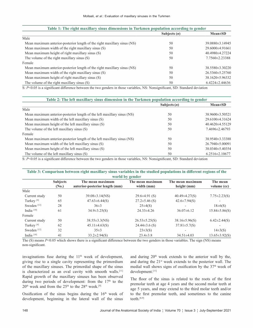
Mollaali, et al.: Evaluation of maxillary sinuses in the Turkmen
148 Journal of the Anatomical Society of India ¦ Volume 70 ¦ Issue 3 ¦ July-September 2021
invaginations fuse during the 11th week of development, giving rise to a single cavity representing the primordium of the maxillary sinuses. The primordial shape of the sinus is characterized as an oval cavity with smooth walls.[11] Rapid growth of the maxillary sinuses has been observed during two periods of development: from the 17th to the 20th week and from the 25th to the 28th week.[9]
Ossification of the sinus begins during the 16th week of development, beginning in the lateral wall of the sinus
and during 20th week extends to the anterior wall by the, and during the 21st week extends to the posterior wall. The medial wall shows signs of ossification by the 37th week of development.[11]
The floor of the sinus is related to the roots of the first premolar teeth at age 4 years and the second molar teeth at age 5 years, and may extend to the third molar teeth and/or to the first premolar teeth, and sometimes to the canine teeth.[8,9]
Table 1: The right maxillary sinus dimensions in Turkmen population according to genderSubjects (n) Mean±SD
MaleMean maximum anterior‑posterior length of the right maxillary sinus (NS) 50 39.0880±3.14945Mean maximum width of the right maxillary sinus (S) 50 29.6000±4.91661Mean maximum height of right maxillary sinus (S) 50 40.4980±4.27224The volume of the right maxillary sinus (S) 50 7.7560±2.23388
FemaleMean maximum anterior‑posterior length of the right maxillary sinus (NS) 50 38.5580±3.30220Mean maximum width of the right maxillary sinus (S) 50 26.5360±5.25760Mean maximum height of right maxillary sinus (S) 50 38.1620±5.96332The volume of the right maxillary sinus (S) 50 6.4224±2.44636
S: P<0.05 is a significant difference between the two genders in those variables, NS: Nonsignificant, SD: Standard deviation
Table 2: The left maxillary sinus dimension in the Turkmen population according to genderSubjects (n) Mean±SD
MaleMean maximum anterior‑posterior length of the left maxillary sinus (NS) 50 38.9600±3.50521Mean maximum width of the left maxillary sinus (S) 50 29.6100±4.31624Mean maximum height of the left maxillary sinus (S) 50 40.4620±4.55129The volume of the left maxillary sinus (S) 50 7.4696±2.46793
FemaleMean maximum anterior‑posterior length of the left maxillary sinus (NS) 50 38.9540±3.33388Mean maximum width of the left maxillary sinus (S) 50 26.7940±5.00091Mean maximum height of the left maxillary sinus (S) 50 38.0340±5.40354The volume of the left maxillary sinus (S) 50 6.2516±2.10677
S: P<0.05 is a significant difference between the two genders in those variables, NS: Nonsignificant, SD: Standard deviation
Table 3: Comparison between right maxillary sinus variables in the studied populations in different regions of the world by gender
Subjects (No.)
The mean maximum anterior‑posterior length (mm)
The mean maximum width (mm)
The mean maximum height (mm)
The mean volume (cc)
MaleCurrent study 50 39.08±3.14(NS) 29.6±4.91 (S) 40.49±4.27(S) 7.75±2.23(S)Turkey [4] 65 47.63±6.44(S) 27.2±5.46 (S) 42.6±7.94(S) ‑Sweden [12] 28 36±3 25±4(S) ‑ 18±6(S)India [10] 61 34.9±3.25(S) 24.33±4.26 36.07±6.12 15.84±5.86(S)
FemaleCurrent study 50 38.55±3.3(NS) 26.53±5.25(S) 38.16±5.96(S) 6.42±2.44(S)Turkey [4] 62 45.11±4.63(S) 24.44±3.6 (S) 37.81±5.7(S) ‑Sweden [12] 32 35±3 23±3(S) ‑ 14±3(S)India [10] 41 33.2±2.94(S) 23.4±3.8 34.51±4.03 13.65±3.92(S)
The (S) means P<0.05 which shows there is a significant difference between the two genders in those variables. The sign (NS) means non‑significant.
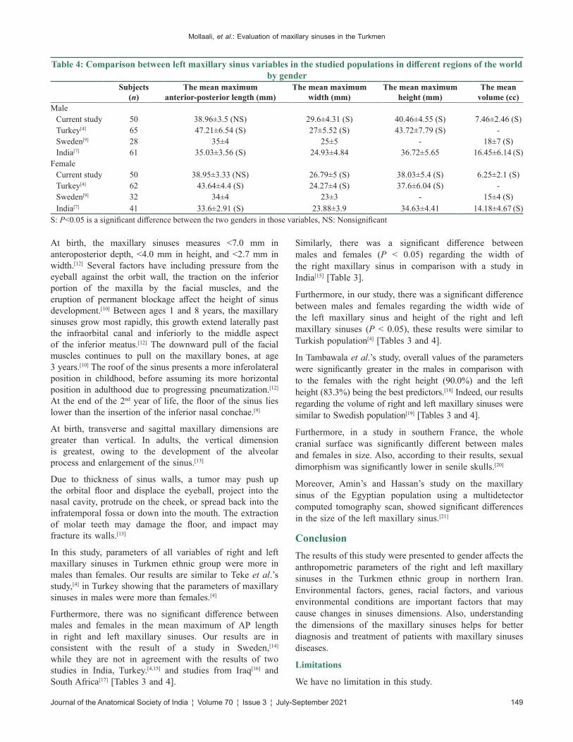
Mollaali, et al.: Evaluation of maxillary sinuses in the Turkmen
Journal of the Anatomical Society of India ¦ Volume 70 ¦ Issue 3 ¦ July-September 2021 149
At birth, the maxillary sinuses measures <7.0 mm in anteroposterior depth, <4.0 mm in height, and <2.7 mm in width.[12] Several factors have including pressure from the eyeball against the orbit wall, the traction on the inferior portion of the maxilla by the facial muscles, and the eruption of permanent blockage affect the height of sinus development.[10] Between ages 1 and 8 years, the maxillary sinuses grow most rapidly, this growth extend laterally past the infraorbital canal and inferiorly to the middle aspect of the inferior meatus.[12] The downward pull of the facial muscles continues to pull on the maxillary bones, at age 3 years.[10] The roof of the sinus presents a more inferolateral position in childhood, before assuming its more horizontal position in adulthood due to progressing pneumatization.[12] At the end of the 2nd year of life, the floor of the sinus lies lower than the insertion of the inferior nasal conchae.[9]
At birth, transverse and sagittal maxillary dimensions are greater than vertical. In adults, the vertical dimension is greatest, owing to the development of the alveolar process and enlargement of the sinus.[13]
Due to thickness of sinus walls, a tumor may push up the orbital floor and displace the eyeball, project into the nasal cavity, protrude on the cheek, or spread back into the infratemporal fossa or down into the mouth. The extraction of molar teeth may damage the floor, and impact may fracture its walls.[13]
In this study, parameters of all variables of right and left maxillary sinuses in Turkmen ethnic group were more in males than females. Our results are similar to Teke et al.’s study,[4] in Turkey showing that the parameters of maxillary sinuses in males were more than females.[4]
Furthermore, there was no significant difference between males and females in the mean maximum of AP length in right and left maxillary sinuses. Our results are in consistent with the result of a study in Sweden,[14] while they are not in agreement with the results of two studies in India, Turkey.[4,15] and studies from Iraq[16] and South Africa[17] [Tables 3 and 4].
Similarly, there was a significant difference between males and females (P < 0.05) regarding the width of the right maxillary sinus in comparison with a study in India[15] [Table 3].
Furthermore, in our study, there was a significant difference between males and females regarding the width wide of the left maxillary sinus and height of the right and left maxillary sinuses (P < 0.05), these results were similar to Turkish population[4] [Tables 3 and 4].
In Tambawala et al.’s study, overall values of the parameters were significantly greater in the males in comparison with to the females with the right height (90.0%) and the left height (83.3%) being the best predictors.[18] Indeed, our results regarding the volume of right and left maxillary sinuses were similar to Swedish population[19] [Tables 3 and 4].
Furthermore, in a study in southern France, the whole cranial surface was significantly different between males and females in size. Also, according to their results, sexual dimorphism was significantly lower in senile skulls.[20]
Moreover, Amin’s and Hassan’s study on the maxillary sinus of the Egyptian population using a multidetector computed tomography scan, showed significant differences in the size of the left maxillary sinus.[21]
ConclusionThe results of this study were presented to gender affects the anthropometric parameters of the right and left maxillary sinuses in the Turkmen ethnic group in northern Iran. Environmental factors, genes, racial factors, and various environmental conditions are important factors that may cause changes in sinuses dimensions. Also, understanding the dimensions of the maxillary sinuses helps for better diagnosis and treatment of patients with maxillary sinuses diseases.
Limitations
We have no limitation in this study.
Table 4: Comparison between left maxillary sinus variables in the studied populations in different regions of the world by gender
Subjects (n)
The mean maximum anterior‑posterior length (mm)
The mean maximum width (mm)
The mean maximum height (mm)
The mean volume (cc)
MaleCurrent study 50 38.96±3.5 (NS) 29.6±4.31 (S) 40.46±4.55 (S) 7.46±2.46 (S)Turkey[4] 65 47.21±6.54 (S) 27±5.52 (S) 43.72±7.79 (S) ‑Sweden[9] 28 35±4 25±5 ‑ 18±7 (S)India[7] 61 35.03±3.56 (S) 24.93±4.84 36.72±5.65 16.45±6.14 (S)
FemaleCurrent study 50 38.95±3.33 (NS) 26.79±5 (S) 38.03±5.4 (S) 6.25±2.1 (S)Turkey[4] 62 43.64±4.4 (S) 24.27±4 (S) 37.6±6.04 (S) ‑Sweden[9] 32 34±4 23±3 ‑ 15±4 (S)India[7] 41 33.6±2.91 (S) 23.88±3.9 34.63±4.41 14.18±4.67 (S)
S: P<0.05 is a significant difference between the two genders in those variables, NS: Nonsignificant
Mollaali, et al.: Evaluation of maxillary sinuses in the Turkmen
150 Journal of the Anatomical Society of India ¦ Volume 70 ¦ Issue 3 ¦ July-September 2021
Financial support and sponsorship
This study has funded by the Deputy of Research of Golestan University of Medical Sciences, Gorgan, Iran. (Grant No: 960628168).
Conflicts of interest
There are no conflicts of interest.
References1. Park IH, Song JS, Choi H, Kim TH, Hoon S, Lee SH, et al.
Volumetric study in the development of paranasal sinuses by CT imaging in Asian: A pilot study. Int J Pediatr Otorhinolaryngol 2010;74:1347‑50.
2. Putz R, Pabst R. Sobotta‑Atlas of Human Anatomy: Head, Neck, Upper Limb, Thorax, Abdomen, Pelvis, Lower Limb; Two‑Volume Set. Urban & Fischer; Churchill Livingstone, 2006. P61‑65.
3. Bangi BB, Ginjupally U, Nadendla LK, Vadla B. 3D evaluation of maxillary sinus using computed tomography: A sexual dimorphic study. Int J Dent 2017;2017:9017078.
4. Teke HY, Duran S, Canturk N, Canturk G. Determination of gender by measuring the size of the maxillary sinuses in computerized tomography scans. Surg Radiol Anat 2007;29:9‑13.
5. McGowan DA, Baxter PW, James J. The Maxillary Sinus and Its Dental Implications. Oxford: Wright, Butter‑Worth‑Heinemann; 1993. p. 1‑25.
6. Orhan K, Kusakci Seker B, Aksoy S, Bayindir H, Berberoğlu A, Seker E. Cone beam CT evaluation of maxillary sinus septa prevalence, height, location and morphology in children and an adult population. Med Princ Pract 2013;22:47‑53.
7. Mirfazeli A, Kaviany N, Hosseinpoor K, Aryaie M, Golalipour MJ. Birth defects in Northern Iran (2008‑2013). Iran J Public Health 2018;47:413‑7.
8. Standring S. Gray’s Anatomy: The Anatomical Basis of Clinical Practice. 41st ed. London: Elsevier Health Sciences; 2015.
9. Iwanaga J, Wilson C, Lachkar S, Tomaszewski KA, Walocha JA,
Tubbs RS. Clinical anatomy of the maxillary sinus: Application to sinus floor augmentation. Anat Cell Biol 2019;52:17‑24.
10. Chanavaz M. Maxillary sinus: Anatomy, physiology, surgery, and bone grafting related to implantology – Eleven years of surgical experience (1979‑1990). J Oral Implantol 1990;16:199‑209.
11. Nuñez‑Castruita A, López‑Serna N, Guzmán‑López S. Prenatal development of the maxillary sinus: A perspective for paranasal sinus surgery. Otolaryngol Head Neck Surg 2012;146:997‑1003.
12. Duncavage J. The Maxillary Sinus: Medical and Surgical Management. New York: Thieme Medical Publishers; 2011.
13. Williams PL, Warwick R, Dyson M, Bannister LH. Gray's Anatomy 37th ed, Edinburgh: Churchill Livingstone; International Edition. 1997: pp 389‑390.
14. Čechová M, Dupej J, Brůžek J, Bejdová Š, Horák M, Velemínská J. Sex estimation using external morphology of the frontal bone and frontal sinuses in a contemporary Czech population. Int J Legal Med 2019;133:1285‑94.
15. Sharma SK, Jehan M, Kumar A. Measurements of maxillary sinus volume and dimensions by computed tomography scan for gender determination. J Anat Soc India 2014;63:36‑42.
16. Uthman AT, Al‑Rawi NH, Al‑Naaimi AS, Al‑Timimi JF. Evaluation of maxillary sinus dimension in determine using helical CT scaning. J Forensic Sci 2011;56:403‑8.
17. Lee F. Forensic ethnic identification of cranial the role of the maxillary sinus. Am J Forensic Med Pathol 2004;25:302‑13.
18. Tambawala SS, Karjodkar FR, Sansare K, Prakash N. Sexual dimorphism of maxillary sinus using cone beam computed tomography. Egypt J Forensic Sci 2016;6:120‑5.
19. Sahlstrand‑Johnson P, Jannert M, Strömbeck A, Abul‑Kasim K. Computed tomography measurements of different dimensions of maxillary and frontal sinuses. BMC Med Imaging 2011;11:8.
20. Musilová B, Dupej J, Velemínská J, Chaumoitre K, Bruzek J. Exocranial surfaces for sex assessment of the human cranium. Forensic Sci Int 2016;269:70‑7.
21. Amin MF, Hassan EI. Sex identification in Egyptian population using multidetector computed tomography of the maxillary sinus. J Forensic Leg Med 2012;19:65‑9.
© 2021 Journal of the Anatomical Society of India | Published by Wolters Kluwer - Medknow 151
Address for correspondence: Dr. Neeru Goyal, Department of Anatomy, Christian Medical College, Ludhiana, Punjab, India. E‑mail: [email protected]
Access this article online
Website: www.jasi.org.inDOI: 10.4103/JASI.JASI_125_19
Quick Response Code:
AbstractIntroduction: Atlas helps in complex biomechanical movements of the skull along with weight transmission of skull to spine. Recent developments in fixation technologies and minimally invasive surgical approaches have encouraged further studies of the region. Objectives of this study are to explore the shape, size, and symmetry of the superior articular facet of atlas. Good knowledge of the variations of the facet is important for orthopedicians, physiotherapists, and neurosurgeons. Material and Methods: The study was conducted on 110 dried adult atlas vertebrae. Shape, size, and number of the superior articular facets on each atlas were recorded. Results: Different shapes observed were kidney‑shaped, oval‑shaped, irregular, rectangular, comma‑shaped, sinuous, sole/8‑shaped, and two/three separate facets. Length and width of the facet were similar on two sides. In 31.82% of cases, the facets on the two sides were not symmetrical. Discussion and Conclusion: Variations of the superior articular facets have been extensively described. While planning treatment plans in cases of craniovertebral joint dysfunction, morphology and variations of the region should be kept in mind.
Keywords: Atlas, spine, superior articular facet, vertebra
Variation of the Superior Articular Facet of Atlas and Their Significance
Neeru Goyal, Anjali JainDepartment of Anatomy, Christian Medical College, Ludhiana, Punjab, India
How to cite this article: Goyal N, Jain A. Variation of the superior articular facet of atlas and their significance. J Anat Soc India 2021;XX:XX-XX.
IntroductionAtlas occupies the most important and vital position interfacing skull cranially and axis caudally constituting atlanto‑occipital and atlantoaxial joints. Hence, it helps in complex biomechanical movements of the skull and neck along with weight transmission of skull to spine. Besides this, it paves the way for spinomedullary junction through the vertebral foramen. Vertebral artery and first cervical nerve pass over the superior surface of its posterior arch, making it more vulnerable.[1] The atlas consists of two lateral masses connected by a shorter anterior and a longer posterior arch. The lateral masses are ovoid, their long axes converging anteriorly. Each bears a kidney‑shaped superior articular facet for the respective occipital condyle, which is sometimes completely divided into a larger anterior and a smaller posterior part.[2]
The atlanto‑occipital joints are responsible for about one‑half of all cervical flexion and extension.[3] The stability of the cervical spine is violated by various traumatic and nontraumatic causes. Instability of craniovertebral junction needs surgical
correction or long‑term immobility to attain a solid fusion. Therefore, reduction and rebuilding of the stability of this complex are important. A short‑segment posterior fixation technique is often adopted to preserve the motion of the craniovertebral junction.[4] Morphometric characterization of the atlas has been found to be useful in operative management of atlantoaxial complex and occipitocervical instability.[5,6]
Most of the surgical theories and techniques consider the atlas to be a source or contributing factor to many pathologies, including cervicogenic headaches, disequilibrium, and otalgia when the biomechanics of the atlanto‑occipital and/or atlantoaxial joints are disturbed.[7] Fitz‑Ritson[8] reported a high correspondence of upper cervical joint fixations in patients suffering from cervicogenic vertigo.
Variations of the superior articular facet have been extensively described.[4,9‑15] Still, more is being added to it day by day due to its contribution to the movements of head. Recent developments in fixation technologies and minimally invasive surgical approaches have encouraged further studies of the region. The objectives of this study are to explore the This is an open access journal, and articles are
distributed under the terms of the Creative Commons Attribution‑NonCommercial‑ShareAlike 4.0 License, which allows others to remix, tweak, and build upon the work non‑commercially, as long as appropriate credit is given and the new creations are licensed under the identical terms.
For reprints contact: [email protected]
Received: 06 September 2020Accepted: 25 July 2021Available online: ***
Article Info
Original Article
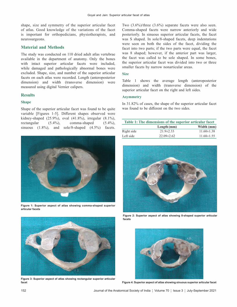
Goyal and Jain: Superior articular facet of atlas
152 Journal of the Anatomical Society of India ¦ Volume 70 ¦ Issue 3 ¦ July-September 2021
shape, size and symmetry of the superior articular facet of atlas. Good knowledge of the variations of the facet is important for orthopedicians, physiotherapists, and neurosurgeons.
Material and MethodsThe study was conducted on 110 dried adult atlas vertebrae available in the department of anatomy. Only the bones with intact superior articular facets were included, while damaged and pathologically abnormal bones were excluded. Shape, size, and number of the superior articular facets on each atlas were recorded. Length (anteroposterior dimension) and width (transverse dimension) were measured using digital Vernier calipers.
ResultsShape
Shape of the superior articular facet was found to be quite variable [Figures 1‑5]. Different shapes observed were kidney‑shaped (25.9%), oval (41.8%), irregular (8.1%), rectangular (5.4%), comma‑shaped (5.4%), sinuous (1.8%), and sole/8‑shaped (4.5%) facets.
Two (3.6%)/three (3.6%) separate facets were also seen. Comma‑shaped facets were narrow anteriorly and wide posteriorly. In sinuous superior articular facets, the facet was S shaped. In sole/8‑shaped facets, deep indentations were seen on both the sides of the facet, dividing the facet into two parts; if the two parts were equal, the facet was 8 shaped; however, if the anterior part was larger, the facet was called to be sole shaped. In some bones, the superior articular facet was divided into two or three smaller facets by narrow nonarticular areas.
Size
Table 1 shows the average length (anteroposterior dimension) and width (transverse dimension) of the superior articular facet on the right and left sides.
Asymmetry
In 31.82% of cases, the shape of the superior articular facet was found to be different on the two sides.
Table 1: The dimensions of the superior articular facetLength (mm) Width (mm)
Right side 21.9±2.33 11.60±1.38Left side 22.09±2.62 11.68±1.55
Figure 4: Superior aspect of atlas showing sinuous superior articular facet
Figure 1: Superior aspect of atlas showing comma‑shaped superior articular facets
Figure 2: Superior aspect of atlas showing 8‑shaped superior articular facets
Figure 3: Superior aspect of atlas showing rectangular superior articular facet
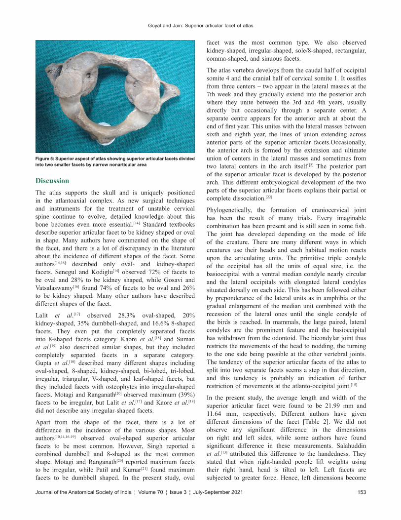
Goyal and Jain: Superior articular facet of atlas
Journal of the Anatomical Society of India ¦ Volume 70 ¦ Issue 3 ¦ July-September 2021 153
DiscussionThe atlas supports the skull and is uniquely positioned in the atlantoaxial complex. As new surgical techniques and instruments for the treatment of unstable cervical spine continue to evolve, detailed knowledge about this bone becomes even more essential.[14] Standard textbooks describe superior articular facet to be kidney shaped or oval in shape. Many authors have commented on the shape of the facet, and there is a lot of discrepancy in the literature about the incidence of different shapes of the facet. Some authors[14,16] described only oval‑ and kidney‑shaped facets. Senegul and Kodiglu[14] observed 72% of facets to be oval and 28% to be kidney shaped, while Gosavi and Vatsalaswamy[16] found 74% of facets to be oval and 26% to be kidney shaped. Many other authors have described different shapes of the facet.
Lalit et al.[17] observed 28.3% oval‑shaped, 20% kidney‑shaped, 35% dumbbell‑shaped, and 16.6% 8‑shaped facets. They even put the completely separated facets into 8‑shaped facets category. Kaore et al.[18] and Suman et al.[19] also described similar shapes, but they included completely separated facets in a separate category. Gupta et al.[10] described many different shapes including oval‑shaped, 8‑shaped, kidney‑shaped, bi‑lobed, tri‑lobed, irregular, triangular, V‑shaped, and leaf‑shaped facets, but they included facets with osteophytes into irregular‑shaped facets. Motagi and Ranganath[20] observed maximum (39%) facets to be irregular, but Lalit et al.[17] and Kaore et al.[18] did not describe any irregular‑shaped facets.
Apart from the shape of the facet, there is a lot of difference in the incidence of the various shapes. Most authors[10,14,16‑19] observed oval‑shaped superior articular facets to be most common. However, Singh reported a combined dumbbell and 8‑shaped as the most common shape. Motagi and Ranganath[20] reported maximum facets to be irregular, while Patil and Kumar[21] found maximum facets to be dumbbell shaped. In the present study, oval
facet was the most common type. We also observed kidney‑shaped, irregular‑shaped, sole/8‑shaped, rectangular, comma‑shaped, and sinuous facets.
The atlas vertebra develops from the caudal half of occipital somite 4 and the cranial half of cervical somite 1. It ossifies from three centers – two appear in the lateral masses at the 7th week and they gradually extend into the posterior arch where they unite between the 3rd and 4th years, usually directly but occasionally through a separate center. A separate centre appears for the anterior arch at about the end of first year. This unites with the lateral masses between sixth and eighth year, the lines of union extending across anterior parts of the superior articular facets.Occasionally, the anterior arch is formed by the extension and ultimate union of centers in the lateral masses and sometimes from two lateral centers in the arch itself.[2] The posterior part of the superior articular facet is developed by the posterior arch. This different embryological development of the two parts of the superior articular facets explains their partial or complete dissociation.[22]
Phylogenetically, the formation of craniocervical joint has been the result of many trials. Every imaginable combination has been present and is still seen in some fish. The joint has developed depending on the mode of life of the creature. There are many different ways in which creatures use their heads and each habitual motion reacts upon the articulating units. The primitive triple condyle of the occipital has all the units of equal size, i.e. the basioccipital with a ventral median condyle nearly circular and the lateral occipitals with elongated lateral condyles situated dorsally on each side. This has been followed either by preponderance of the lateral units as in amphibia or the gradual enlargement of the median unit combined with the recession of the lateral ones until the single condyle of the birds is reached. In mammals, the large paired, lateral condyles are the prominent feature and the basioccipital has withdrawn from the odontoid. The bicondylar joint thus restricts the movements of the head to nodding, the turning to the one side being possible at the other vertebral joints. The tendency of the superior articular facets of the atlas to split into two separate facets seems a step in that direction, and this tendency is probably an indication of further restriction of movements at the atlanto‑occipital joint.[15]
In the present study, the average length and width of the superior articular facet were found to be 21.99 mm and 11.64 mm, respectively. Different authors have given different dimensions of the facet [Table 2]. We did not observe any significant difference in the dimensions on right and left sides, while some authors have found significant difference in these measurements. Salahuddin et al.[13] attributed this difference to the handedness. They stated that when right‑handed people lift weights using their right hand, head is tilted to left. Left facets are subjected to greater force. Hence, left dimensions become
Figure 5: Superior aspect of atlas showing superior articular facets divided into two smaller facets by narrow nonarticular area
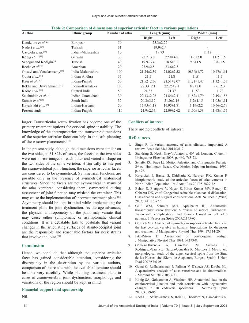
Goyal and Jain: Superior articular facet of atlas
154 Journal of the Anatomical Society of India ¦ Volume 70 ¦ Issue 3 ¦ July-September 2021
larger. Transarticular screw fixation has become one of the primary treatment options for cervical spine instability. The knowledge of the anteroposterior and transverse dimensions of the superior articular facet can help in the safe planning of these screw placements.[18]
In the present study, although the dimensions were similar on the two sides, in 31.82% of cases, the facets on the two sides were not mirror images of each other and varied in shape on the two sides of the same vertebra. Historically to interpret the craniovertebral joint function, the superior articular facets are considered to be symmetrical. Symmetrical functions are possible only in the presence of symmetrical anatomical structures. Since the facets are not symmetrical in many of the atlas vertebrae, considering them, symmetrical during assessment of joint function may mislead the examiner. This may cause the implementation of incorrect treatment plans.[13] Asymmetry should be kept in mind while implementing the treatment plans for joint dysfunction. As the age advances, the physical anthropometry of the joint may variate that may cause either symptomatic or asymptomatic clinical conditions. It is a debatable task to postulate that variant changes in the articulating surfaces of atlanto‑occipital joint are the responsible and reasonable factors for neck strains that involve the joint.[20]
ConclusionHence, we conclude that although the superior articular facet has gained considerable attention, considering the discrepancy in the description by the various authors, comparison of the results with the available literature should be done very carefully. While planning treatment plans in cases of craniovertebral joint dysfunction, morphology and variations of the region should be kept in mind.
Financial support and sponsorship
Nil.
Conflicts of interest
There are no conflicts of interest.
References1. Singh R. Is variant anatomy of atlas clinically important? A
review. Basic Sci Med 2014;3:1‑7.2. Standring S. Neck. Gray’s Anatomy. 40th ed. London: Churchill
Livingstone Elsevier; 2008. p. 460, 763‑73.3. Schafer RC, Faye LJ. Motion Palpation and Chiropractic Technic.
2nd ed. Huntington Beach, CA: Motion Palpation Institute; 1990. p. 426.
4. Kayalvizhi I, Bansal S, Dhidharia K, Narayan RK, Kumar P. Morphometric study of the articular facets of atlas vertebra in North Indian Population. Int J Anat Res 2017;5:3829‑32.
5. Behari S, Bhargava V, Nayak S, Kiran Kumar MV, Banerji D, Chhabra DK, et al. Congenital reducible atlantoaxial dislocation: Classification and surgical considerations. Acta Neurochir (Wien) 2002;144:1165‑77.
6. Gluf WM, Schmidt MH, Apfelbaum RI. Atlantoaxial transarticular screw fixation: A review of surgical indications, fusion rate, complications, and lessons learned in 191 adult patients. J Neurosurg Spine 2005;2:155‑63.
7. Gottlieb MS. Absence of symmetry in superior articular facets on the first cervical vertebra in humans: Implications for diagnosis and treatment. J Manipulative Physiol Ther 1994;17:314‑20.
8. Fitz‑Ritson D. Assessment of cervicogenic vertigo. J Manipulative Physiol Ther 1991;14:193‑8.
9. Gómez‑Olivencia A, Carretero JM, Arsuaga JL, Rodríguez‑García L, García‑González R, Martínez I. Metric and morphological study of the upper cervical spine from the Sima de los Huesos site (Sierra de Atapuerca, Burgos, Spain). J Hum Evol 2007;53:6‑25.
10. Gupta C, Radhakrishnan P, Palimar V, D’souza AS, Kiruba NL. A quantitative analysis of atlas vertebrae and its abnormalities. J Morphol Sci 2017;30:77‑81.
11. König SA, Goldammer A, Vitzthum HE. Anatomical data on the craniocervical junction and their correlation with degenerative changes in 30 cadaveric specimens. J Neurosurg Spine 2005;3:379‑85.
12. Rocha R, Safavi‑Abbasi S, Reis C, Theodore N, Bambakidis N,
Table 2: Comparison of dimensions of superior articular facet in various populationsAuthor Ethnic group Number of atlas Length (mm) Width (mm)
Right Left Right LeftKandziora et al.[23] European 50 25.3±2.22 ‑Naderi et al.[24] Turkish 31 19.9±2.4 ‑Cacciola et al.[25] Indian‑Maharashtra 10 19.73 11.12König et al.[11] German 30 22.7±3.0 22.8±4.2 11.6±2.0 11.2±1.5Senegul and Kodiglu[14] Turkish 40 19.9±3.4 18.6±3.2 9.6±1.9 9.8±1.5Rocha et al.[12] American 20 23.9±2.5 23.6±2.5Gosavi and Vatsalaswamy[16] India‑Maharashtra 100 21.24±2.39 21.02±2.52 10.36±1.72 10.47±1.61Gupta et al.[10] Indian‑Andhra 35 21.5 21.8 11.8 11.5Kaur et al.[26] Indian‑Punjab 50 21.52±2.36 21.51±2.07 11.21±1.47 11.32±1.53Rekha and Divya Shanthi[27] Indian‑Karnataka 100 22.33±2.1 22.25±2.1 8.7±2.0 9.6±2.3Kaore et al.[18] Central India 50 21.33 21.37 11.53 11.72Salahuddin et al.[13] Indian‑Uttarakhand 30 22.13±2.26 21.84±2.11 11.82±1.79 12.19±1.58Suman et al.[19] South India 32 20.3±3.12 21.0±2.16 11.7±1.15 11.05±1.11Kayalvizhi et al.[4] Indian‑Haryana 50 16.95±1.18 16.95±1.81 11.19±2.2 10.66±2.79Present study Indian‑Punjab 110 21.9±2.33 22.09±2.62 11.60±1.38 11.68±1.55
Goyal and Jain: Superior articular facet of atlas
Journal of the Anatomical Society of India ¦ Volume 70 ¦ Issue 3 ¦ July-September 2021 155
de Oliveira E, et al. Working area, safety zones, and angles of approach for posterior C‑1 lateral mass screw placement: A quantitative anatomical and morphometric evaluation. J Neurosurg Spine 2007;6:247‑54.
13. Salahuddin A, Mukesh S, Binaya KB, Nilotpal C. A study on the morphometric asymmetry of atlas vertebrae. J Surg Acad 2016;6:18‑24.
14. Senegul G, Kodiglu HH. Morphometric anatomy of atlas and axis vertebra. Turk Neurosurg 2006;16:69‑76.
15. Singh S. Variations of the superior articular facets of atlas vertebrae. J Anat 1965;99:565‑71.
16. Gosavi SN, Vatsalaswamy P. Morphometric study of the atlas vertebra using manual method. Malays Orthop J 2012;6:18‑20.
17. Lalit M, Piplani S, Kullar JS, Arora AK, Mannan R. The morphological analysis of the superior articular facet of the adult human atlas vertebra. J Clin Diagn Res 2011;5:274‑7.
18. Kaore A, Kamdi AU, Kasote AP, Kamdi NY, Fulpatil MP. A study of morphometry of superior articular facet of atlas and its clinical implication in central India. Int J Anat Res 2016;4:2750‑6.
19. Suman P, Cariappa LC, Mahato RK. Morphometric analysis of superior articular facets of atlas vertebra and its clinical applications. J Evol Med Dent Sci 2017;6:4912‑6.
20. Motagi MV, Ranganath V. Morphometric analysis of superior articular facets of atlas vertebra and its clinical applications
in ergonomics of atlanto‑occipital joints. J Clin Diagn Res 2013;7:2674‑6.
21. Patil GV, Kumar S. Superior articular facets of atlas vertebra‑A morphological study. Int J Sci Res 2014;3:364‑5.
22. Paraskevas G, Papaziogas B, Tzaveas A, Natsis K, Spanidou S, Kitsoulis P. Morphological parameters of the superior articular facets of the atlas and potential clinical significance. Surg Radiol Anat 2008;30:611‑7.
23. Kandziora F, Schulze‑Stahl N, Khodadadyan‑Klostermann C, Schröder R, Mittlmeier T. Screw placement in transoral atlantoaxial plate systems: An anatomical study. J Neurosurg (Spine) 2001;95:80‑7.
24. Naderi S, Cakmakçi H, Acar F, Arman C, Mertol T, Arda MN. Anatomical and computed tomographic analysis of C1 vertebra. Clin Neurol Neurosurg 2003;105:245‑8.
25. Cacciola F, Phalke U, Goel A. Vertebral artery in relationship to C1‑C2 vertebrae: An anatomical study. Neurol India 2004;52:178‑84.
26. Kaur J, Grewal H, Singh P, Kumar A. Morphometric study of the articular facets of atlas and axis vertebrae. Unique J Med Dent Sci 2014;2:83‑9.
27. Rekha BS, Divya Shanthi D’Sa. Morphometric anatomy of the atlas (C1) Vertebra among Karnataka population in India. Int J Anat Res 2016;4:1981‑4.
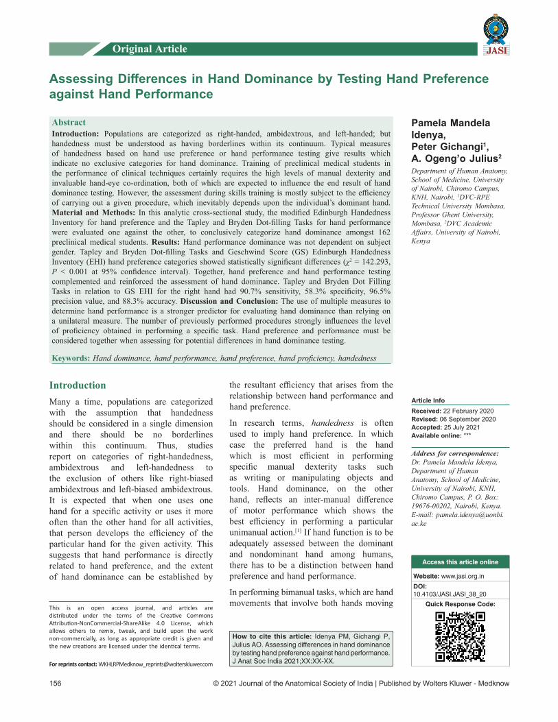
156 © 2021 Journal of the Anatomical Society of India | Published by Wolters Kluwer - Medknow
Address for correspondence: Dr. Pamela Mandela Idenya, Department of Human Anatomy, School of Medicine, University of Nairobi, KNH, Chiromo Campus, P. O. Box: 19676‑00202, Nairobi, Kenya. E‑mail: [email protected]
Access this article online
Website: www.jasi.org.inDOI: 10.4103/JASI.JASI_38_20
Quick Response Code:
AbstractIntroduction: Populations are categorized as right‑handed, ambidextrous, and left‑handed; but handedness must be understood as having borderlines within its continuum. Typical measures of handedness based on hand use preference or hand performance testing give results which indicate no exclusive categories for hand dominance. Training of preclinical medical students in the performance of clinical techniques certainly requires the high levels of manual dexterity and invaluable hand‑eye co‑ordination, both of which are expected to influence the end result of hand dominance testing. However, the assessment during skills training is mostly subject to the efficiency of carrying out a given procedure, which inevitably depends upon the individual’s dominant hand. Material and Methods: In this analytic cross‑sectional study, the modified Edinburgh Handedness Inventory for hand preference and the Tapley and Bryden Dot‑filling Tasks for hand performance were evaluated one against the other, to conclusively categorize hand dominance amongst 162 preclinical medical students. Results: Hand performance dominance was not dependent on subject gender. Tapley and Bryden Dot‑filling Tasks and Geschwind Score (GS) Edinburgh Handedness Inventory (EHI) hand preference categories showed statistically significant differences (χ2 = 142.293, P < 0.001 at 95% confidence interval). Together, hand preference and hand performance testing complemented and reinforced the assessment of hand dominance. Tapley and Bryden Dot Filling Tasks in relation to GS EHI for the right hand had 90.7% sensitivity, 58.3% specificity, 96.5% precision value, and 88.3% accuracy. Discussion and Conclusion: The use of multiple measures to determine hand performance is a stronger predictor for evaluating hand dominance than relying on a unilateral measure. The number of previously performed procedures strongly influences the level of proficiency obtained in performing a specific task. Hand preference and performance must be considered together when assessing for potential differences in hand dominance testing.
Keywords: Hand dominance, hand performance, hand preference, hand proficiency, handedness
Assessing Differences in Hand Dominance by Testing Hand Preference against Hand Performance
Pamela Mandela Idenya, Peter Gichangi1, A. Ogeng’o Julius2
Department of Human Anatomy, School of Medicine, University of Nairobi, Chiromo Campus, KNH, Nairobi, 1DVC‑RPE Technical University Mombasa, Professor Ghent University, Mombasa, 2DVC Academic Affairs, University of Nairobi, Kenya
How to cite this article: Idenya PM, Gichangi P, Julius AO. Assessing differences in hand dominance by testing hand preference against hand performance. J Anat Soc India 2021;XX:XX-XX.
IntroductionMany a time, populations are categorized with the assumption that handedness should be considered in a single dimension and there should be no borderlines within this continuum. Thus, studies report on categories of right‑handedness, ambidextrous and left‑handedness to the exclusion of others like right‑biased ambidextrous and left‑biased ambidextrous. It is expected that when one uses one hand for a specific activity or uses it more often than the other hand for all activities, that person develops the efficiency of the particular hand for the given activity. This suggests that hand performance is directly related to hand preference, and the extent of hand dominance can be established by
the resultant efficiency that arises from the relationship between hand performance and hand preference.
In research terms, handedness is often used to imply hand preference. In which case the preferred hand is the hand which is most efficient in performing specific manual dexterity tasks such as writing or manipulating objects and tools. Hand dominance, on the other hand, reflects an inter‑manual difference of motor performance which shows the best efficiency in performing a particular unimanual action.[1] If hand function is to be adequately assessed between the dominant and nondominant hand among humans, there has to be a distinction between hand preference and hand performance.
In performing bimanual tasks, which are hand movements that involve both hands moving This is an open access journal, and articles are
distributed under the terms of the Creative Commons Attribution‑NonCommercial‑ShareAlike 4.0 License, which allows others to remix, tweak, and build upon the work non‑commercially, as long as appropriate credit is given and the new creations are licensed under the identical terms.
For reprints contact: [email protected]
Received: 22 February 2020Revised: 06 September 2020Accepted: 25 July 2021Available online: ***
Article Info
Original Article
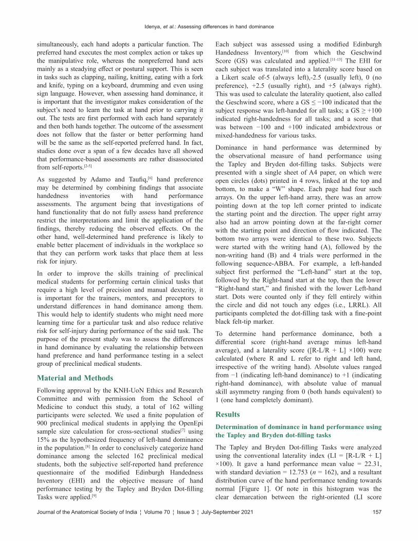
Idenya, et al.: Assessing differences in hand dominance
Journal of the Anatomical Society of India ¦ Volume 70 ¦ Issue 3 ¦ July-September 2021 157
simultaneously, each hand adopts a particular function. The preferred hand executes the most complex action or takes up the manipulative role, whereas the nonpreferred hand acts mainly as a steadying effect or postural support. This is seen in tasks such as clapping, nailing, knitting, eating with a fork and knife, typing on a keyboard, drumming and even using sign language. However, when assessing hand dominance, it is important that the investigator makes consideration of the subject’s need to learn the task at hand prior to carrying it out. The tests are first performed with each hand separately and then both hands together. The outcome of the assessment does not follow that the faster or better performing hand will be the same as the self‑reported preferred hand. In fact, studies done over a span of a few decades have all showed that performance‑based assessments are rather disassociated from self‑reports.[2‑5]
As suggested by Adamo and Taufiq,[6] hand preference may be determined by combining findings that associate handedness inventories with hand performance assessments. The argument being that investigations of hand functionality that do not fully assess hand preference restrict the interpretations and limit the application of the findings, thereby reducing the observed effects. On the other hand, well‑determined hand preference is likely to enable better placement of individuals in the workplace so that they can perform work tasks that place them at less risk for injury.
In order to improve the skills training of preclinical medical students for performing certain clinical tasks that require a high level of precision and manual dexterity, it is important for the trainers, mentors, and preceptors to understand differences in hand dominance among them. This would help to identify students who might need more learning time for a particular task and also reduce relative risk for self‑injury during performance of the said task. The purpose of the present study was to assess the differences in hand dominance by evaluating the relationship between hand preference and hand performance testing in a select group of preclinical medical students.
Material and MethodsFollowing approval by the KNH‑UoN Ethics and Research Committee and with permission from the School of Medicine to conduct this study, a total of 162 willing participants were selected. We used a finite population of 900 preclinical medical students in applying the OpenEpi sample size calculation for cross‑sectional studies[7] using 15% as the hypothesized frequency of left‑hand dominance in the population.[8] In order to conclusively categorize hand dominance among the selected 162 preclinical medical students, both the subjective self‑reported hand preference questionnaire of the modified Edinburgh Handedness Inventory (EHI) and the objective measure of hand performance testing by the Tapley and Bryden Dot‑filling Tasks were applied.[9]
Each subject was assessed using a modified Edinburgh Handedness Inventory,[10] from which the Geschwind Score (GS) was calculated and applied.[11‑13] The EHI for each subject was translated into a laterality score based on a Likert scale of‑5 (always left),‑2.5 (usually left), 0 (no preference), +2.5 (usually right), and +5 (always right). This was used to calculate the laterality quotient, also called the Geschwind score, where a GS ≤ −100 indicated that the subject response was left‑handed for all tasks; a GS ≥ +100 indicated right‑handedness for all tasks; and a score that was between −100 and +100 indicated ambidextrous or mixed‑handedness for various tasks.
Dominance in hand performance was determined by the observational measure of hand performance using the Tapley and Bryden dot‑filling tasks. Subjects were presented with a single sheet of A4 paper, on which were open circles (dots) printed in 4 rows, linked at the top and bottom, to make a “W” shape. Each page had four such arrays. On the upper left‑hand array, there was an arrow pointing down at the top left corner printed to indicate the starting point and the direction. The upper right array also had an arrow pointing down at the far‑right corner with the starting point and direction of flow indicated. The bottom two arrays were identical to these two. Subjects were started with the writing hand (A), followed by the non‑writing hand (B) and 4 trials were performed in the following sequence‑ABBA. For example, a left‑handed subject first performed the “Left‑hand” start at the top, followed by the Right‑hand start at the top, then the lower “Right‑hand start,” and finished with the lower Left‑hand start. Dots were counted only if they fell entirely within the circle and did not touch any edges (i.e., LRRL). All participants completed the dot‑filling task with a fine‑point black felt‑tip marker.
To determine hand performance dominance, both a differential score (right‑hand average minus left‑hand average), and a laterality score ([R‑L/R + L] ×100) were calculated (where R and L refer to right and left hand, irrespective of the writing hand). Absolute values ranged from −1 (indicating left‑hand dominance) to +1 (indicating right‑hand dominance), with absolute value of manual skill asymmetry ranging from 0 (both hands equivalent) to 1 (one hand completely dominant).
ResultsDetermination of dominance in hand performance using the Tapley and Bryden dot‑filling tasks
The Tapley and Bryden Dot‑filling Tasks were analyzed using the conventional laterality index (LI = [R‑L/R + L] ×100). It gave a hand performance mean value = 22.31, with standard deviation = 12.753 (n = 162), and a resultant distribution curve of the hand performance tending towards normal [Figure 1]. Of note in this histogram was the clear demarcation between the right‑oriented (LI score
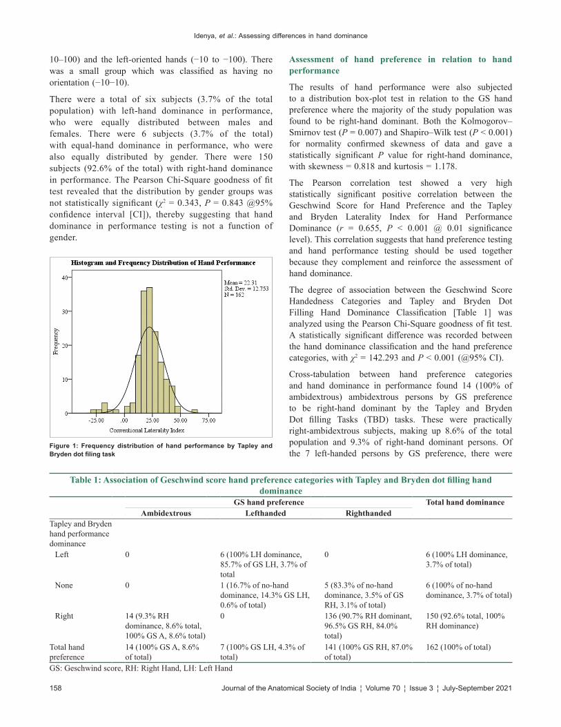
Idenya, et al.: Assessing differences in hand dominance
158 Journal of the Anatomical Society of India ¦ Volume 70 ¦ Issue 3 ¦ July-September 2021
10–100) and the left‑oriented hands (−10 to −100). There was a small group which was classified as having no orientation (−10−10).
There were a total of six subjects (3.7% of the total population) with left‑hand dominance in performance, who were equally distributed between males and females. There were 6 subjects (3.7% of the total) with equal‑hand dominance in performance, who were also equally distributed by gender. There were 150 subjects (92.6% of the total) with right‑hand dominance in performance. The Pearson Chi‑Square goodness of fit test revealed that the distribution by gender groups was not statistically significant (χ2 = 0.343, P = 0.843 @95% confidence interval [CI]), thereby suggesting that hand dominance in performance testing is not a function of gender.
Assessment of hand preference in relation to hand performance
The results of hand performance were also subjected to a distribution box‑plot test in relation to the GS hand preference where the majority of the study population was found to be right‑hand dominant. Both the Kolmogorov–Smirnov test (P = 0.007) and Shapiro–Wilk test (P < 0.001) for normality confirmed skewness of data and gave a statistically significant P value for right‑hand dominance, with skewness = 0.818 and kurtosis = 1.178.
The Pearson correlation test showed a very high statistically significant positive correlation between the Geschwind Score for Hand Preference and the Tapley and Bryden Laterality Index for Hand Performance Dominance (r = 0.655, P < 0.001 @ 0.01 significance level). This correlation suggests that hand preference testing and hand performance testing should be used together because they complement and reinforce the assessment of hand dominance.
The degree of association between the Geschwind Score Handedness Categories and Tapley and Bryden Dot Filling Hand Dominance Classification [Table 1] was analyzed using the Pearson Chi‑Square goodness of fit test. A statistically significant difference was recorded between the hand dominance classification and the hand preference categories, with χ2 = 142.293 and P < 0.001 (@95% CI).
Cross‑tabulation between hand preference categories and hand dominance in performance found 14 (100% of ambidextrous) ambidextrous persons by GS preference to be right‑hand dominant by the Tapley and Bryden Dot filling Tasks (TBD) tasks. These were practically right‑ambidextrous subjects, making up 8.6% of the total population and 9.3% of right‑hand dominant persons. Of the 7 left‑handed persons by GS preference, there were
Table 1: Association of Geschwind score hand preference categories with Tapley and Bryden dot filling hand dominance
GS hand preference Total hand dominanceAmbidextrous Lefthanded Righthanded
Tapley and Bryden hand performance dominance
Left 0 6 (100% LH dominance, 85.7% of GS LH, 3.7% of total
0 6 (100% LH dominance, 3.7% of total)
None 0 1 (16.7% of no‑hand dominance, 14.3% GS LH, 0.6% of total)
5 (83.3% of no‑hand dominance, 3.5% of GS RH, 3.1% of total)
6 (100% of no‑hand dominance, 3.7% of total)
Right 14 (9.3% RH dominance, 8.6% total, 100% GS A, 8.6% total)
0 136 (90.7% RH dominant, 96.5% GS RH, 84.0% total)
150 (92.6% total, 100% RH dominance)
Total hand preference
14 (100% GS A, 8.6% of total)
7 (100% GS LH, 4.3% of total)
141 (100% GS RH, 87.0% of total)
162 (100% of total)
GS: Geschwind score, RH: Right Hand, LH: Left Hand
Figure 1: Frequency distribution of hand performance by Tapley and Bryden dot filing task
Idenya, et al.: Assessing differences in hand dominance
Journal of the Anatomical Society of India ¦ Volume 70 ¦ Issue 3 ¦ July-September 2021 159
6 (85.7% of left‑handed) who were left‑hand dominant. These were seemingly pure left‑handed subjects, making up 3.7% of the total population. There was 1 left‑handed person (14.3% of left‑handed) found to show no‑hand dominance (16.7% of no hand dominance), who was practically left‑ambidextrous. Of the 141 (87% of total population) right‑handed persons, there were 136 (84% of total population and 96.5% of right‑handed persons) who were right‑hand dominant. These were seemingly pure right‑handed subjects. The other 5 (3.5% of right‑handed persons) right‑handed persons were found to have no‑hand dominance. These were correctly right‑ambidextrous, making up 83.3% of the no‑hand dominance group and 3.1% of the total population. In summary, the resultant categorization of hand function following assessment of dominance using both hand performance and hand preference testing is shown in Table 2.
The characteristics and relevance of the Tapley and Bryden Dot Filling Tasks in relation to the GS EHI right hand revealed a sensitivity of 90.7% with a specificity of 58.3%. The positive predictive value (and precision value) was 96.5% with an accuracy of 88.3%. This suggests that in the absence of the EHI‑GS, the Tapley and Bryden Dot Filling Task has a 90.7% chance of correctly classifying a person to be right‑hand dominant in 96.5% cases; and this classification would be 88.3% accurate.
DiscussionAs various researches have revealed, in young healthy subjects self‑rated hand dominance, hand preference and hand efficiency are all highly correlated based on the level of motor performance.[3,14,15] Nevertheless, the differences in the performance of the dominant and nondominant hands are expected to diminish with increasing age and to become more balanced in early adulthood. If hand preference is considered over hand performance, there is a likelihood of misclassifying ambidextrous (or apparently neutral‑handed) persons as pure‑handed (be it right‑or left‑handed, ambidextrous or ambivalent), thereby omitting significant details about efficient hand use by the said mixed‑or cross‑handed individual.
As the results in the present study have showed, those who were initially classified by the EHI GS as ambidextrous, were all reclassified by the combined tests as right‑ambidextrous.
This is a significant change that can influence results in proficiency testing. It is also likely that the individual who had to be reclassified as left‑ambidextrous might have a degree of mixed proficiency results given that there was no‑hand dominance in the performance testing. The results of the present study are also in favour of reporting more mixed‑and cross‑handedness than pure ambidextrous as suggested by EHI‑GS categories. The most consistent result was that of the left‑handed individuals, which gave a 3.7% pure left‑handed population. This proportion might appear small, but it could as well be clinically/practically significant when assessing the skills of these individuals in training using right‑biased tools.
It is noteworthy that the results of the current study support the view by Corey et al.[3] about performance‑based measures such as the Tapley and Bryden Dot Filling Tasks not to be considered independently when predicting hand dominance. It is necessary to combine such performance tests with other hand preference tests, even if they are from self‑reported questionnaires such as the EHI, to be able to more clearly distinguish hand dominance. This is signified by the 96.5% precision value and the 88.3% accuracy rate of determining hand dominance obtained by combining the GS hand preference with the dot‑filling tasks.
The results in the present study are similar to the findings by Brown et al.,[2] which indicated that the combination of the results from a handedness questionnaire and a series of performance measures gives the best predictors of hand dominance. The current study further enforces the thought that habitual use of either the right or left hand can typically be observed during the performance of everyday tasks. However, it should be acknowledged that task‑specific training effects may influence the extent to which one identifies and describes their dominant hand.
It is clear that measures of hand dominance are based on individual hand use preference or hand performance testing; but there really are no clear categories for each one, such as being exclusively left‑or right‑hand dominant.[16] As demonstrated earlier, each individual shows a preference for the use of one hand for a given manual action, even though it is not always the same hand that is preferred for two different actions.[17] Now, whether one is right‑or left‑hand dominant is therefore a function of the given task at hand, and not a generalization of the dominant hand. Furthermore, there are several studies which report of individuals who are specialized in highly skilled and complex tasks who demonstrate very strong correlations between different tasks.[18‑22]
In their study of handedness and musical ability in a group of professional musicians, Aggleton et al.[23] demonstrated that the dominant hand performs tasks requiring force or a series of rapid movements while the other hand offers stabilization and support. There is no doubt that the habitual use of the same hand for a given task contributes
Table 2: Hand dominance classification from combined hand preference and hand performance testing
Hand dominance Proportionate distribution (percentage of total population)
Pure right‑handed 136 (84)Pure left‑handed 6 (3.7)Pure ambidextrous 0Right‑ambidextrous 19 (11.7)Left‑ambidextrous 1 (0.6)
Idenya, et al.: Assessing differences in hand dominance
160 Journal of the Anatomical Society of India ¦ Volume 70 ¦ Issue 3 ¦ July-September 2021
and promotes skills development of the same hand for the particular tasks; yet, habitual use does not appear to transfer the acquired skill to other tasks. It is therefore possible for one to gain proficiency in the use of a certain tool, but this level of proficiency does not transfer to other tools with similar physical properties.
Since hand preference for some tasks could also be modified by social or religious influences, the tasks selected for use in hand assessments should apply typical tools amongst the said specific populations from different cultures so that hand dominance variations in a given population can be adequately studied. Although researchers like Bishop[24] allege that hand dominance might be a by‑product of brain lateralization in human beings, this notion does not explain the 5%–15% left‑handed population that is reported in every culture.
The present study evaluates of hand dominance among preclinical medical students echoes the thought by others like Baldwin et al.,[25] that a combination of hand preference and hand performance testing should be used in assessing manual dexterity skills that are required during precise surgical procedures. The results also support the notion that self‑reported questionnaires by experienced clinicians may identify a specific skill that is needed to perform a given task, but the students must still be subjected to an objective test that is used when measuring their skills performance.[26] This viewpoint was likewise expressed by the research which experimented with virtual reality laparoscopy simulators,[27] and another one which used a motion‑tracking system to assess laparoscopic suturing skills.[28]
ConclusionSuffice to say that the use of multiple measures to determine hand performance is a stronger predictor for determining hand dominance than reliance on a single measure. However, it must be kept in mind that the number of previously performed procedures will strongly influence the level of proficiency reached in performing specific tasks. This therefore means that hand preference and hand performance must be considered together when assessing for potential differences in performance outcomes of the dominant and nondominant hands.
AcknowledgmentWe acknowledge contributions made by Mr. Martin I. Inyimili and Ms. Esther W. Mburu in recruiting subjects and ensuring complete data collection. Appreciation to all preclinical students of the University of Nairobi who willingly gave consent and participated in the study.
Financial support and sponsorship
Nil.
Conflicts of interest
There are no conflicts of interest.
References1. Serrien DJ, Ivry RB, Swinnen SP. Dynamics of hemispheric
specialization and integration in the context of motor control. Nat Rev Neurosci 2006;7:160‑6.
2. Brown SG, Roy EA, Rohr LE, Snider BR, Bryden PJ. Preference and performance measures of handedness. Brain Cogn 2004;55:283‑5.
3. Corey DM, Hurley MM, Foundas AL. Right and left handedness defined: A multivariate approach using hand preference and hand performance measures. Neuropsychiatry Neuropsychol Behav Neurol 2001;14:144‑52.
4. Peters M, Durding BM. Handedness measured by finger tapping: A continuous variable. Can J Psychol 1978;32:257‑61.
5. Woo TL, Pearson K. Dexterity and sinistrality of hand and eye. Biometrika 1927;19:165‑99.
6. Adamo DE, Taufiq A. Establishing hand preference: why does it matter? Hand (N Y) 2011;6:295‑303.
7. Dean AG, Sullivan KM, Soe MM. OpenEpi: Open Source Epidemiologic Statistics for Public Health, Version 3.01; 2013. Available from: www.OpenEpi.com.
8. Mathiowetz V, Wiemer D, Federman S. Grip and pinch strength: Norms for 6 to 19‑year‑olds. Am J Occup Ther 1986;40:705‑11.
9. Tapley SM, Bryden MP. A group test for the assessment of performance between the hands. Neuropsychologia 1985;23:215‑21.
10. Oldfield RC. The assessment and analysis of handedness: The Edinburgh inventory. Neuropsychologia 1971;9:97‑113.
11. Tan U. The relationship between nonverbal intelligence, familial sinistrality and Geschwind scores in right‑handed female subjects. Int J Neurosci 1988;43:177‑82.
12. Kulaksiz G, Gözil R. The effect of hand preference on hand anthropometric measurements in healthy individuals. Ann Anat 2002;184:257‑65.
13. Barut C, Ozer CM, Sevinc O, Gumus M, Yunten Z. Relationships between hand and foot preferences. Int J Neurosci 2007;117:177‑85.
14. Bishop DV, Ross VA, Daniels MS, Bright P. The measurement of hand preference: A validation study comparing three groups of right‑handers. Br J Psychol 1996;87 (Pt 2):269‑85.
15. Henkel V, Mergl R, Juckel G, Rujescu D, Mavrogiorgou P, Giegling I, et al. Assessment of handedness using a digitizing tablet: A new method. Neuropsychologia 2001;39:1158‑66.
16. McManus IC. Handedness. The Blackwell Dictionary of Neuropsychology. In: Beaumont JG, Kenealy PM, Rogers MJ, editors. Blackwell: Oxford, UK; 1996. p. 367‑76.
17. Salmaso D, Longoni AM. Problems in the assessment of hand preference. Cortex 1985;22:533‑49.
18. Bryden MP. Measuring handedness with questionnaires. Neuropsychologia 1977;13:617‑624.
19. Wood CJ, Aggleton JP. Handedness in ‘fast ball’ sports: Do left‑handers have an innate advantage? Br J Psychol 1989;80:227‑40.
20. Connolly KJ, Bishop DV. The measurement of handedness: A cross‑cultural comparison of samples from England and Papua New Guinea. Neuropsychologia 1992;30:13‑26.
21. Marchant LF, McGrew WC, Eibl‑Eibesfeldt I. Is human handedness universal? Ethological analyses from three traditional cultures. Ethology 1995;101:239‑58.

Idenya, et al.: Assessing differences in hand dominance
Journal of the Anatomical Society of India ¦ Volume 70 ¦ Issue 3 ¦ July-September 2021 161
22. Marchant LF, McGrew WC. Human handedness: An ethological perspective. J Hum Evol 1998;13:221‑8.
23. Aggleton JP, Kentridge RW, Good JM. Handedness and musical ability: A study of professional orchestral players, composers and choir members. Psychol Music 1994;22:148‑56.
24. Bishop DV. Individual differences in handedness and specific speech and language impairment: Evidence against a genetic link. Behav Genet 2001;31:339‑51.
25. Baldwin PJ, Paisley AM, Brown SP. Consultant surgeons’ opinion of the skills required of basic surgical trainees. Br J Surg 1999;86:1078‑82.
26. Gallagher AG, Richie K, McClure N, McGuigan J. Objective psychomotor skills assessment of experienced, junior, and novice laparoscopists with virtual reality. World J Surg 2001;25:1478‑83.
27. Thijssen AS, Schijven MP. Contemporary virtual reality laparoscopy simulators: Quicksand or solid grounds for assessing surgical trainees? Am J Surg 2010;199:529‑41.
28. Yamaguchi S, Yoshida D, Kenmotsu H, Yasunaga T, Konishi K, Ieiri S, et al. Objective assessment of laparoscopic suturing skills using a motion‑tracking system. Surg Endosc 2011;25:771‑5.
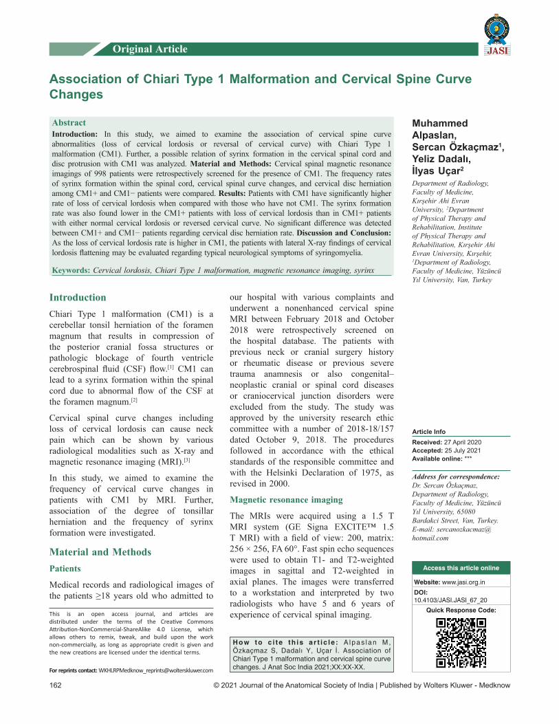
162 © 2021 Journal of the Anatomical Society of India | Published by Wolters Kluwer - Medknow
Address for correspondence: Dr. Sercan Özkaçmaz, Department of Radiology, Faculty of Medicine, Yüzüncü Yıl University, 65080 Bardakci Street, Van, Turkey. E‑mail: [email protected]
Access this article online
Website: www.jasi.org.inDOI: 10.4103/JASI.JASI_67_20
Quick Response Code:
AbstractIntroduction: In this study, we aimed to examine the association of cervical spine curve abnormalities (loss of cervical lordosis or reversal of cervical curve) with Chiari Type 1 malformation (CM1). Further, a possible relation of syrinx formation in the cervical spinal cord and disc protrusion with CM1 was analyzed. Material and Methods: Cervical spinal magnetic resonance imagings of 998 patients were retrospectively screened for the presence of CM1. The frequency rates of syrinx formation within the spinal cord, cervical spinal curve changes, and cervical disc herniation among CM1+ and CM1− patients were compared. Results: Patients with CM1 have significantly higher rate of loss of cervical lordosis when compared with those who have not CM1. The syrinx formation rate was also found lower in the CM1+ patients with loss of cervical lordosis than in CM1+ patients with either normal cervical lordosis or reversed cervical curve. No significant difference was detected between CM1+ and CM1− patients regarding cervical disc herniation rate. Discussion and Conclusion: As the loss of cervical lordosis rate is higher in CM1, the patients with lateral X‑ray findings of cervical lordosis flattening may be evaluated regarding typical neurological symptoms of syringomyelia.
Keywords: Cervical lordosis, Chiari Type 1 malformation, magnetic resonance imaging, syrinx
Association of Chiari Type 1 Malformation and Cervical Spine Curve Changes
Muhammed Alpaslan, Sercan Özkaçmaz1, Yeliz Dadalı, İlyas Uçar2
Department of Radiology, Faculty of Medicine, Kırşehir Ahi Evran University, 2Department of Physical Therapy and Rehabilitation, Institute of Physical Therapy and Rehabilitation, Kırşehir Ahi Evran University, Kırşehir, 1Department of Radiology, Faculty of Medicine, Yüzüncü Yıl University, Van, Turkey
How to c i t e th is a r t i c le : A lpas lan M , Özkaçmaz S, Dadalı Y, Uçar İ. Association of Chiari Type 1 malformation and cervical spine curve changes. J Anat Soc India 2021;XX:XX-XX.
IntroductionChiari Type 1 malformation (CM1) is a cerebellar tonsil herniation of the foramen magnum that results in compression of the posterior cranial fossa structures or pathologic blockage of fourth ventricle cerebrospinal fluid (CSF) flow.[1] CM1 can lead to a syrinx formation within the spinal cord due to abnormal flow of the CSF at the foramen magnum.[2]
Cervical spinal curve changes including loss of cervical lordosis can cause neck pain which can be shown by various radiological modalities such as X‑ray and magnetic resonance imaging (MRI).[3]
In this study, we aimed to examine the frequency of cervical curve changes in patients with CM1 by MRI. Further, association of the degree of tonsillar herniation and the frequency of syrinx formation were investigated.
Material and MethodsPatients
Medical records and radiological images of the patients ≥18 years old who admitted to
our hospital with various complaints and underwent a nonenhanced cervical spine MRI between February 2018 and October 2018 were retrospectively screened on the hospital database. The patients with previous neck or cranial surgery history or rheumatic disease or previous severe trauma anamnesis or also congenital–neoplastic cranial or spinal cord diseases or craniocervical junction disorders were excluded from the study. The study was approved by the university research ethic committee with a number of 2018‑18/157 dated October 9, 2018. The procedures followed in accordance with the ethical standards of the responsible committee and with the Helsinki Declaration of 1975, as revised in 2000.
Magnetic resonance imaging
The MRIs were acquired using a 1.5 T MRI system (GE Signa EXCITE™ 1.5 T MRI) with a field of view: 200, matrix: 256 × 256, FA 60°. Fast spin echo sequences were used to obtain T1‑ and T2‑weighted images in sagittal and T2‑weighted in axial planes. The images were transferred to a workstation and interpreted by two radiologists who have 5 and 6 years of experience of cervical spinal imaging.This is an open access journal, and articles are
distributed under the terms of the Creative Commons Attribution‑NonCommercial‑ShareAlike 4.0 License, which allows others to remix, tweak, and build upon the work non‑commercially, as long as appropriate credit is given and the new creations are licensed under the identical terms.
For reprints contact: [email protected]
Received: 27 April 2020Accepted: 25 July 2021Available online: ***
Article Info
Original Article
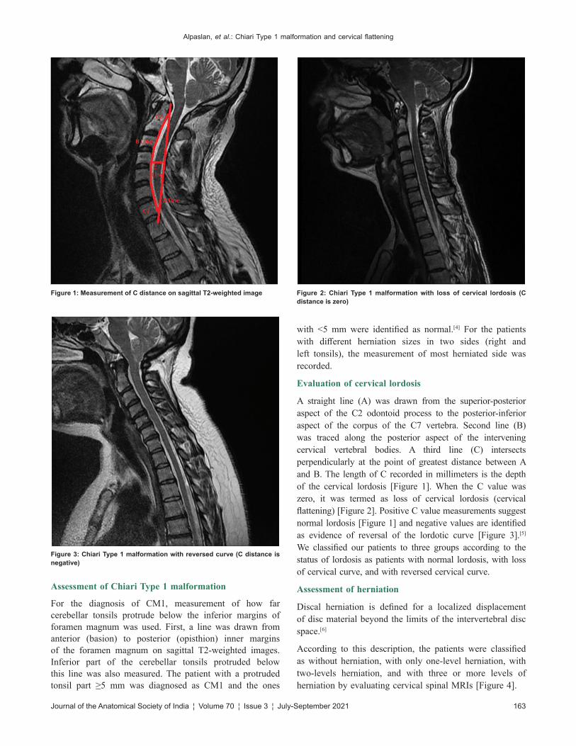
Alpaslan, et al.: Chiari Type 1 malformation and cervical flattening
Journal of the Anatomical Society of India ¦ Volume 70 ¦ Issue 3 ¦ July-September 2021 163
Assessment of Chiari Type 1 malformation
For the diagnosis of CM1, measurement of how far cerebellar tonsils protrude below the inferior margins of foramen magnum was used. First, a line was drawn from anterior (basion) to posterior (opisthion) inner margins of the foramen magnum on sagittal T2‑weighted images. Inferior part of the cerebellar tonsils protruded below this line was also measured. The patient with a protruded tonsil part ≥5 mm was diagnosed as CM1 and the ones
with <5 mm were identified as normal.[4] For the patients with different herniation sizes in two sides (right and left tonsils), the measurement of most herniated side was recorded.
Evaluation of cervical lordosis
A straight line (A) was drawn from the superior‑posterior aspect of the C2 odontoid process to the posterior‑inferior aspect of the corpus of the C7 vertebra. Second line (B) was traced along the posterior aspect of the intervening cervical vertebral bodies. A third line (C) intersects perpendicularly at the point of greatest distance between A and B. The length of C recorded in millimeters is the depth of the cervical lordosis [Figure 1]. When the C value was zero, it was termed as loss of cervical lordosis (cervical flattening) [Figure 2]. Positive C value measurements suggest normal lordosis [Figure 1] and negative values are identified as evidence of reversal of the lordotic curve [Figure 3].[5] We classified our patients to three groups according to the status of lordosis as patients with normal lordosis, with loss of cervical curve, and with reversed cervical curve.
Assessment of herniation
Discal herniation is defined for a localized displacement of disc material beyond the limits of the intervertebral disc space.[6]
According to this description, the patients were classified as without herniation, with only one‑level herniation, with two‑levels herniation, and with three or more levels of herniation by evaluating cervical spinal MRIs [Figure 4].
Figure 3: Chiari Type 1 malformation with reversed curve (C distance is negative)
Figure 1: Measurement of C distance on sagittal T2‑weighted image Figure 2: Chiari Type 1 malformation with loss of cervical lordosis (C distance is zero)
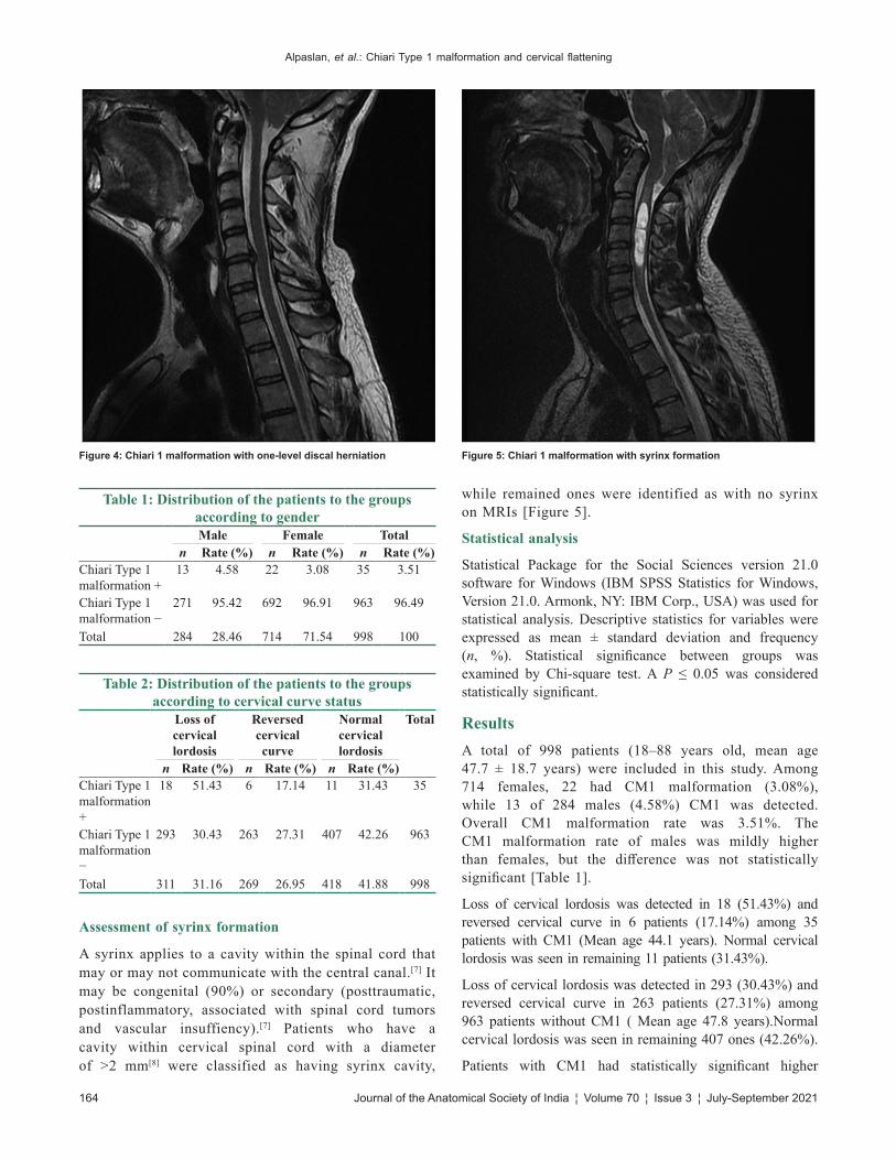
Alpaslan, et al.: Chiari Type 1 malformation and cervical flattening
164 Journal of the Anatomical Society of India ¦ Volume 70 ¦ Issue 3 ¦ July-September 2021
Assessment of syrinx formation
A syrinx applies to a cavity within the spinal cord that may or may not communicate with the central canal.[7] It may be congenital (90%) or secondary (posttraumatic, postinflammatory, associated with spinal cord tumors and vascular insuffiency).[7] Patients who have a cavity within cervical spinal cord with a diameter of >2 mm[8] were classified as having syrinx cavity,
while remained ones were identified as with no syrinx on MRIs [Figure 5].
Statistical analysis
Statistical Package for the Social Sciences version 21.0 software for Windows (IBM SPSS Statistics for Windows, Version 21.0. Armonk, NY: IBM Corp., USA) was used for statistical analysis. Descriptive statistics for variables were expressed as mean ± standard deviation and frequency (n, %). Statistical significance between groups was examined by Chi‑square test. A P ≤ 0.05 was considered statistically significant.
ResultsA total of 998 patients (18–88 years old, mean age 47.7 ± 18.7 years) were included in this study. Among 714 females, 22 had CM1 malformation (3.08%), while 13 of 284 males (4.58%) CM1 was detected. Overall CM1 malformation rate was 3.51%. The CM1 malformation rate of males was mildly higher than females, but the difference was not statistically significant [Table 1].
Loss of cervical lordosis was detected in 18 (51.43%) and reversed cervical curve in 6 patients (17.14%) among 35 patients with CM1 (Mean age 44.1 years). Normal cervical lordosis was seen in remaining 11 patients (31.43%).
Loss of cervical lordosis was detected in 293 (30.43%) and reversed cervical curve in 263 patients (27.31%) among 963 patients without CM1 ( Mean age 47.8 years).Normal cervical lordosis was seen in remaining 407 ones (42.26%).
Patients with CM1 had statistically significant higher
Table 1: Distribution of the patients to the groups according to genderMale Female Total
n Rate (%) n Rate (%) n Rate (%)Chiari Type 1 malformation +
13 4.58 22 3.08 35 3.51
Chiari Type 1 malformation −
271 95.42 692 96.91 963 96.49
Total 284 28.46 714 71.54 998 100
Table 2: Distribution of the patients to the groups according to cervical curve status
Loss of cervical lordosis
Reversed cervical curve
Normal cervical lordosis
Total
n Rate (%) n Rate (%) n Rate (%)Chiari Type 1 malformation +
18 51.43 6 17.14 11 31.43 35
Chiari Type 1 malformation −
293 30.43 263 27.31 407 42.26 963
Total 311 31.16 269 26.95 418 41.88 998
Figure 4: Chiari 1 malformation with one‑level discal herniation Figure 5: Chiari 1 malformation with syrinx formation
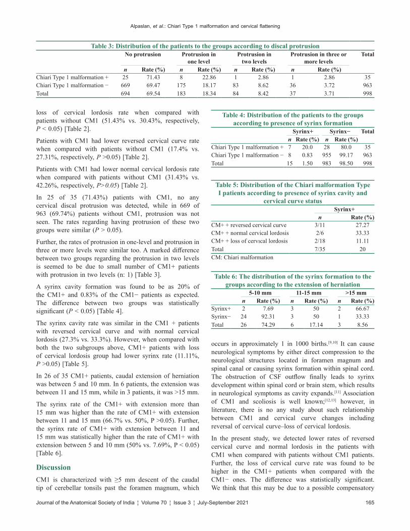
Alpaslan, et al.: Chiari Type 1 malformation and cervical flattening
Journal of the Anatomical Society of India ¦ Volume 70 ¦ Issue 3 ¦ July-September 2021 165
loss of cervical lordosis rate when compared with patients without CM1 (51.43% vs. 30.43%, respectively, P < 0.05) [Table 2].
Patients with CM1 had lower reversed cervical curve rate when compared with patients without CM1 (17.4% vs. 27.31%, respectively, P >0.05) [Table 2].
Patients with CM1 had lower normal cervical lordosis rate when compared with patients without CM1 (31.43% vs. 42.26%, respectively, P>0.05) [Table 2].
In 25 of 35 (71.43%) patients with CM1, no any cervical discal protrusion was detected, while in 669 of 963 (69.74%) patients without CM1, protrusion was not seen. The rates regarding having protrusion of these two groups were similar (P > 0.05).
Further, the rates of protrusion in one‑level and protrusion in three or more levels were similar too. A marked difference between two groups regarding the protrusion in two levels is seemed to be due to small number of CM1+ patients with protrusion in two levels (n: 1) [Table 3].
A syrinx cavity formation was found to be as 20% of the CM1+ and 0.83% of the CM1− patients as expected. The difference between two groups was statistically significant (P < 0.05) [Table 4].
The syrinx cavity rate was similar in the CM1 + patients with reversed cervical curve and with normal cervical lordosis (27.3% vs. 33.3%). However, when compared with both the two subgroups above, CM1+ patients with loss of cervical lordosis group had lower syrinx rate (11.11%, P >0.05) [Table 5].
In 26 of 35 CM1+ patients, caudal extension of herniation was between 5 and 10 mm. In 6 patients, the extension was between 11 and 15 mm, while in 3 patients, it was >15 mm.
The syrinx rate of the CM1+ with extension more than 15 mm was higher than the rate of CM1+ with extension between 11 and 15 mm (66.7% vs. 50%, P >0.05). Further, the syrinx rate of CM1+ with extension between 11 and 15 mm was statistically higher than the rate of CM1+ with extension between 5 and 10 mm (50% vs. 7.69%, P < 0.05) [Table 6].
DiscussionCM1 is characterized with ≥5 mm descent of the caudal tip of cerebellar tonsils past the foramen magnum, which
occurs in approximately 1 in 1000 births.[9,10] It can cause neurological symptoms by either direct compression to the neurological structures located in foramen magnum and spinal canal or causing syrinx formation within spinal cord. The obstruction of CSF outflow finally leads to syrinx development within spinal cord or brain stem, which results in neurological symptoms as cavity expands.[11] Association of CM1 and scoliosis is well known;[12,13] however, in literature, there is no any study about such relationship between CM1 and cervical curve changes including reversal of cervical curve–loss of cervical lordosis.
In the present study, we detected lower rates of reversed cervical curve and normal lordosis in the patients with CM1 when compared with patients without CM1 patients. Further, the loss of cervical curve rate was found to be higher in the CM1+ patients when compared with the CM1− ones. The difference was statistically significant. We think that this may be due to a possible compensatory
Table 3: Distribution of the patients to the groups according to discal protrusionNo protrusion Protrusion in
one levelProtrusion in
two levelsProtrusion in three or
more levelsTotal
n Rate (%) n Rate (%) n Rate (%) n Rate (%)Chiari Type 1 malformation + 25 71.43 8 22.86 1 2.86 1 2.86 35Chiari Type 1 malformation − 669 69.47 175 18.17 83 8.62 36 3.72 963Total 694 69.54 183 18.34 84 8.42 37 3.71 998
Table 4: Distribution of the patients to the groups according to presence of syrinx formation
Syrinx+ Syrinx− Totaln Rate (%) n Rate (%)
Chiari Type 1 malformation + 7 20.0 28 80.0 35Chiari Type 1 malformation − 8 0.83 955 99.17 963Total 15 1.50 983 98.50 998
Table 5: Distribution of the Chiari malformation Type I patients according to presence of syrinx cavity and
cervical curve statusSyrinx+
n Rate (%)CM+ + reversed cervical curve 3/11 27.27CM+ + normal cervical lordosis 2/6 33.33CM+ + loss of cervıcal lordosis 2/18 11.11Total 7/35 20CM: Chiari malformation
Table 6: The distribution of the syrinx formation to the groups according to the extension of herniation
5‑10 mm 11‑15 mm >15 mmn Rate (%) n Rate (%) n Rate (%)
Syrinx+ 2 7.69 3 50 2 66.67Syrinx− 24 92.31 3 50 1 33.33Total 26 74.29 6 17.14 3 8.56
Alpaslan, et al.: Chiari Type 1 malformation and cervical flattening
166 Journal of the Anatomical Society of India ¦ Volume 70 ¦ Issue 3 ¦ July-September 2021
mechanism which decreases the compression of spinal cord. Flexion or extension of occipito–cervical junction may increase the compression of the spinal cord by cerebellar tonsils at the level of foramen magnum, while the loss of cervical curve can minimize this compression. As there are no any data about this issue, such possible association must be further evaluated with large series.
Cervical syrinx cavity occurs due to congenital (myelomeningocele, CM1, CM2, Dandy‑Walker malformation, Klippel–Feil syndrome) or acquired (cervical canal stenosis, postinflammatory, secondary to a spinal cord tumor, secondary to a hemorrhage, due to vascular insufficiency) conditions.[7]
In a study, syringohydromyelia was detected in 39 (36.1%) of 108 symptomatic CM1 patients. They suggested craniocervical junction osseous anomalies (platybasia, retroverted odontoid, short hypoplastic clivus, basilar invagination) as most predictive of syrinx formation that can create a point of mechanical stress and tension, leading to anterior brain stem compression.[14] Our syrinx frequency in the CM1+ patients was significantly higher than the frequency in CM1− patients as expected. (20% vs. 0.83%, respectively). However, the syrinx frequency in the CM1+ patients with loss of cervical lordosis was lower than both in CM1+ patients with reversed cervical curve or with normal cervical lordosis (11.1% vs. 27,3% and 33.3%, respectively). We think that this lower frequency may be due to a vertical position of proximal medulla spinalis segments, while anteflexion or retroflexion of spinal cord can contribute the compression of medulla spinalis in the level of foramen magnum and also leads to the CSF flow blockage in subarachnoid space, which finally results in syrinx formation. This possible association must be further examined with larger series.
In the literature, there are some studies which suggest that increased amount of tonsillar herniation was associated with a greater likelihood of an associated spinal syrinx,[4,15,16] while a study did not detect any association of syrinx formation and degree of tonsillar herniation.[14] Our results also supported that the extension size of the tonsils would affects the syrinx cavity formation. The syrinx frequency in CM1+ patients with a tonsil herniation between 5 and 10 mm was significantly lower than in the patients with a herniation between 11 and 15 mm and also with a herniation of >15 mm (7.69% vs. 50% and 66.67%, respectively). As herniation extension size increase, the syrinx cavity frequency increases significantly may suggest that a longer compressed segment of the spinal cord can be associated with increased risk of syrinx formation.
The cervical discal protrusion incidences of the CM1+ and CM1− patients were similar with no statistically significant difference which suggests that CM1 or syrinx cavity are not associated with cervical discal herniation. In the literature, resolution of cervical syrinx cavity after a surgical
treatment of discal hernia was reported in a patient without CM1, which can suggest a possible association of discal protrusion with syrinx formation, leading to spinal stenosis or direct compression of spinal cord.[17] For this reason, a cervical syrinx cavity in a CM1+ patients, in whom cervical discal herniation is detected must be carefully evaluated before a surgery for accurate identification of the cause of syrinx cavity.
The major limitations of the study are the retrospective design and small patient number. Especially in some subgroups, small patient number did not allow an optimal comparison. Further, the results were not correlated with clinical symptoms and signs.
ConclusionCM1 must be kept in mind for the patients with cervical flattening seen on X‑ray and computed tomographic images, especially when symptoms and signs associated with syrinx formation are present.
Financial support and sponsorship
Nil.
Conflicts of interest
There are no conflicts of interest.
References1. Nash J, Cheng JS, Meyer GA, Remler BF. Chiari type I
malformation: Overview of diagnosis and treatment. WMJ 2002;101:35‑40.
2. Armonda RA, Citrin CM, Foley KT, Ellenbogen RG. Quantitative cine‑mode magnetic resonance ımaging of Chiari I malformations: An analysis of cerebrospinal fluid dynamics. Neurosurgery 1994;35:214‑24.
3. Lippa L, Lippa L, Cacciola F. Loss of cervical lordosis: What is the prognosis? J Craniovertebr Junction Spine 2017;8:9‑14.
4. Elster AD, Chen MY. Chiari I malformations: Clinical and radiologic reappraisal. Radiology 1992;183:347‑53.
5. Borden AG, Rechtman AM, Gershon‑Cohen J. The normal cervical lordosis. Radiology 1960;74:806‑9.
6. Fardon DF, Williams AL, Dohring EJ, Murtagh FR, Gabriel Rothman SL, Sze GK. Lumbar disc nomenclature: version 2.0: Recommendations of the combined task forces of the North American Spine Society, the American Society of Spine Radiology and the American Society of Neuroradiology. Spine J. 2014;14(11):2525‑2545. doi:10.1016/j.spinee.2014.04.022.
7. Batnitzky S, Price HI, Gaughan MJ, Hall PV, Rosenthal SJ. The radiology of syringohydromyelia. RadioGraphics 1983;3:585‑611.
8. Sherman JL, Barkovich AJ, Citrin CM. The MR Appearan’ce of Syringomyelia: New Observations. AJNR 1986;7:985‑95.
9. Hidalgo JA, Tork CA, Varacallo M. Arnold chiari malformation. In: StatPearls. Treasure Island (FL): StatPearls Publishing; 2020. Available from: https://www.ncbi.nlm.nih.gov/books /NBK431076/.
10. Jayamanne C, Fernando L, Mettananda S. Chiari malformation type 1 presenting as unilateral progressive foot drop: A case report and review of literature. BMC Pediatr 2018;18:34.

Alpaslan, et al.: Chiari Type 1 malformation and cervical flattening
Journal of the Anatomical Society of India ¦ Volume 70 ¦ Issue 3 ¦ July-September 2021 167
11. Sabba MF, Renor BS, Ghizoni E, Tedeschi H, Joaquim AF. Posterior fossa decompression with duraplasty in Chiari surgery: A technical note. Rev Assoc Med Bras (1992) 2017;63:946‑9.
12. Kelly MP, Guillaume TJ, Lenke LG. Spinal deformity associated with chiari malformation. Neurosurg Clin N Am 2015;26:579‑85.
13. Brockmeyer DL. Editorial. Chiari malformation Type I and scoliosis: The complexity of curves. J Neurosurg Pediatr 2011;7:22‑3.
14. Gad KA, Yousem DM. Syringohydromyelia in patients with chiari I malformation: A retrospective analysis. AJNR Am J Neuroradiol 2017;38:1833‑8.
15. Pillay PK, Awad IA, Little JR, Hahn JF. Symptomatic Chiari malformation in adults: A new classification based on magnetic resonance imaging with clinical and prognostic significance. Neurosurgery 1991;28:639‑645.
16. Strahle J, Muraszko KM, Kapurch J, Bapuraj JR, Garton HJ, Maher CO. Chiari malformation Type I and syrinx in children undergoing magnetic resonance imaging. J Neurosurg Pediatr 2011;8:205‑13.
17. Yaman ME, Eylen A, Ayberk G. Resolution of isolated syringomyelia after treatment of cervical disc herniation: Association or coincidence? Bratisl Med J 2012;113:500‑2.
168 © 2021 Journal of the Anatomical Society of India | Published by Wolters Kluwer - Medknow
Address for correspondence: Dr. Jaikumar B. Contractor, B‑7, Rushi Tenaments, Behind Munjal Park, Samta, Vadodara ‑ 390 023, Gujarat, India. E‑mail: [email protected]
Access this article online
Website: www.jasi.org.inDOI: 10.4103/jasi.jasi_25_21
Quick Response Code:
AbstractBackground: Liver cirrhosis is essentially an end stage liver fibrosis that develops as a continuation of normal wound healing in response to chronic liver injury. While 1/4th of cirrhotic livers are morphologically normal in size and configuration, on computerized tomography, over 1/3rd are diffusely atrophic and almost 50% manifest focal hypertrophy most commonly in the caudate lobe concomitant with segmental atrophy of the right lobe. Aims and Objectives: To analyze the morphometric measurements of the right lobe and determine the C/RL in human cadaveric liver and compare the values of C/RL ratio to previously documented studies in an attempt to provide baseline data. Material and Methods: The study was conducted on 100 human cadaveric livers at Anatomy Department, Medical College Baroda, Vadodara, Gujarat. Morphometric measurements were observed in mm with threads and digital Vernier Caliper. Results: The transverse diameter of the right lobe averaged 78.22 ± 12.17 mm, with values ranging at 55.06–98.30 mm. The longitudinal diameter of the right lobe averaged 126.31 ± 20.24 with values ranging at 90.54–172.18 mm. Harbin’s Index was calculated as the ratio of the CT to right lobe, i.e., (CT/RT), and averaged 0.38 ± 0.12 with values ranging at 0.17–0.61. Discussion and Conclusion: While nodular regeneration within liver parenchyma may be difficult to recognize on ultrasonography and any irregularity of the liver surface may be apparent only with macro nodules or ascitic effusion, caudate lobe hypertrophy is a consistent finding with liver cirrhosis or other chronic liver disease.
Keywords: Caudate to right lobe ratio, Harbin’s index, liver cirrhosis
Harbin’s Index: Morphological Evaluation of Caudate‑to‑Right Lobe Ratio in Human Cadaveric Liver
Jaikumar B. Contractor, Vipul D. Patel1, V. H. Vaniya2
Assistant Professor, Department of Anatomy, Pramukhswami Medical College, Bhaikaka University, Karamsad, 1Associate Professor, Department of Anatomy, Government Medical College, Surat, 2Professor, Department of Anatomy, Baroda Medical College, Vadodara, Gujarat, India
How to cite this article: Contractor JB, Patel VD, Vaniya VH. Harbin’s index: Morphological evaluation of caudate-to-right lobe ratio in human cadaveric liver. J Anat Soc India 2021;XX:XX-XX.
IntroductionGlobally, hepatitis is considered one of the most common causes of chronic liver disease and continues to affect a large fraction of the population. Overall, 1/5th of the acute hepatitis C cases proceed to cirrhosis and coupled with ascites, encephalopathy, and altered liver function tests, such cases, invariably warrant liver transplantation.[1] Hence, it becomes imperative to have thorough knowledge of the liver anatomy and its variations to achieve successful surgical outcomes, especially in the era of diagnostic imaging and minimally invasive surgeries.[2]
Liver cirrhosis is essentially an end‑stage liver fibrosis that develops as a continuation of normal wound healing in response to chronic liver injury. Histologically, it presents as hepatocyte islands in the form of regenerative nodules that are devoid of central vein and are surrounded by fibrotic
septa. Functionally, the exchange between hepatic sinusoids and adjacent parenchyma is compromised through shunting of portal and arterial blood directly into the hepatic outflow. Impaired liver function, portal hypertension, hepatocellular carcinoma are few well‑known sequelae of liver cirrhosis.[3]
Currently, liver biopsy is the choice of investigation for diagnosing liver cirrhosis.[4] However, sampling errors in needle biopsies, incomplete interpretation of semi‑quantitative scores, intricacies in recognizing a fully developed stage of cirrhosis in needle biopsy specimens raise substantial challenges.[5] On the other hand, while 1/4th of end‑stage cirrhotic livers are morphologically normal in size and configuration, on computerized tomography, over 1/3rd are diffusely atrophic and almost 50% manifest focal hypertrophy most commonly in the caudate lobe concomitant with segmental atrophy of the right lobe.[6]
Anatomically, caudate lobe is identified on the liver as a prominence on its inferior and This is an open access journal, and articles are
distributed under the terms of the Creative Commons Attribution‑NonCommercial‑ShareAlike 4.0 License, which allows others to remix, tweak, and build upon the work non‑commercially, as long as appropriate credit is given and the new creations are licensed under the identical terms.
For reprints contact: [email protected]
Received: 12 February 2021Accepted: 11 August 2021Available online: ***
Article Info
Original Article
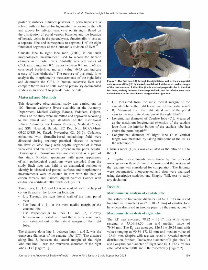
Contractor, et al.: Caudate to right lobe ratio in human cadaveric liver
Journal of the Anatomical Society of India ¦ Volume 70 ¦ Issue 3 ¦ July-September 2021 169
posterior surfaces. Situated posterior to porta hepatis it is related with the fissure for ligamentum venosum on the left and groove for inferior vena cava on its right. Based on the distribution of portal venous branches and the location of hepatic veins in the parenchyma, functionally, it acts as a separate lobe and corresponds to segment I of the eight functional segments of the Couinaud’s division of liver.[7]
Caudate lobe to right lobe ratio (C/RL) is one such morphological measurement used to record the hepatic changes in cirrhotic livers. Globally accepted values of C/RL ratio range to <0.6, values between 0.6 and 0.65 are considered borderline, and any value >0.65 can indicate a case of liver cirrhosis.[8] The purpose of this study is to analyze the morphometric measurements of the right lobe and determine the C/RL in human cadaveric liver and compare the values of C/RL ratio to previously documented studies in an attempt to provide baseline data.
Material and MethodsThis descriptive observational study was carried out on 100 Human cadaveric livers available at the Anatomy Department, Medical College Baroda, Vadodara, Gujarat. Details of the study were submitted and approved according to the ethical and legal standards of the Institutional Ethics Committee for Human Research, Medical College and SSG Hospital, Baroda (EC Reg. No.: ECR/85/Inst/GJ/2013/RR‑16, Dated: November 02, 2017). Cadavers, embalmed with formalin‑based embalming fluid were dissected during anatomy practical classes to remove the liver en bloc along with hepatic segment of inferior vena cava and the structures present in the porta hepatis. Demographic information was not collected as a part of this study. Nineteen specimens with gross appearance of any pathological conditions were excluded from the study. Each liver was held in an anatomical position to identify its visceral and parietal surfaces and morphometric measurements were calculated in mm with the help of cotton threads and Kristeel digital Vernier Caliper with calibration certificate 200 mm/8 inch (2917).
Three lines, L1, L2, and L3 were marked with the help of cotton threads at the following locations:• L1: Through the right lateral wall of the main portal
vein• L2: Parallel to L1 at the most medial margin of the
caudate lobe• L3: Perpendicular to lines L1 and L2, midway
between main portal vein and the inferior vena cava, and extended out to the lateral margin of the right lobe.
The distance along line 3, between lines 1 and 2, was the transverse diameter of the caudate lobe (CT). The distance along line 3, between the lateral margin of the right lobe and line 1, was the transverse diameter of the right lobe (RT)[9] [Figure 1].
• CT: Measured from the most medial margin of the caudate lobe to the right lateral wall of the portal vein[8]
• RT: Measured from the right lateral wall of the portal vein to the most lateral margin of the right lobe[8]
• Longitudinal diameter of Caudate lobe (CL): Measured as the maximum longitudinal extension of the caudate lobe from the inferior border of the caudate lobe just above the porta hepatis[9]
• Longitudinal diameter of Right lobe (RL): Vertical length was measured by taking mid‑point of the RT as the reference.[10]
Harbin’s index (CT/RT) was calculated as the ratio of CT to the RT.
All hepatic measurements were taken by the principal investigator on three different occasions and the average of the readings was considered for computation. The findings were documented, photographed and data were analyzed using descriptive statistics and Shapiro–Wilk test to study any deviation.
ResultsMorphometric analysis of caudate lobe
The values of transverse diameter (28.69 ± 7.73 mm) and longitudinal diameter (54.97 ± 10.73 mm) of caudate lobe have been discussed in another paper by the same author.[11]
Morphometric analysis of right lobe
The RT was averaged 78.22 ± 12.17 mm with values ranging at 55.06–98.30 mm and median value of 79.84 mm. The RL was averaged 126.31 ± 20.24 mm with values ranging at 90.54–172.18 mm and median value of 126.54 mm. Shapiro‑wilk test was used to revealed normal distribution, for both, Transverse diameter of Right lobe (RT) and Longitudinal diameter of Right lobe (RL). The P values calculated were 0.001 and 0.02 respectively [Figure 2].
Figure 1: The first line (L1) through the right lateral wall of the main portal vein. A second line (L2) is marked parallel to L1 at the most medial margin of the caudate lobe. A third line (L3) is marked perpendicular to the first two lines, midway between the main portal vein and the inferior vena cava extended out to the most lateral margin of the right lobe
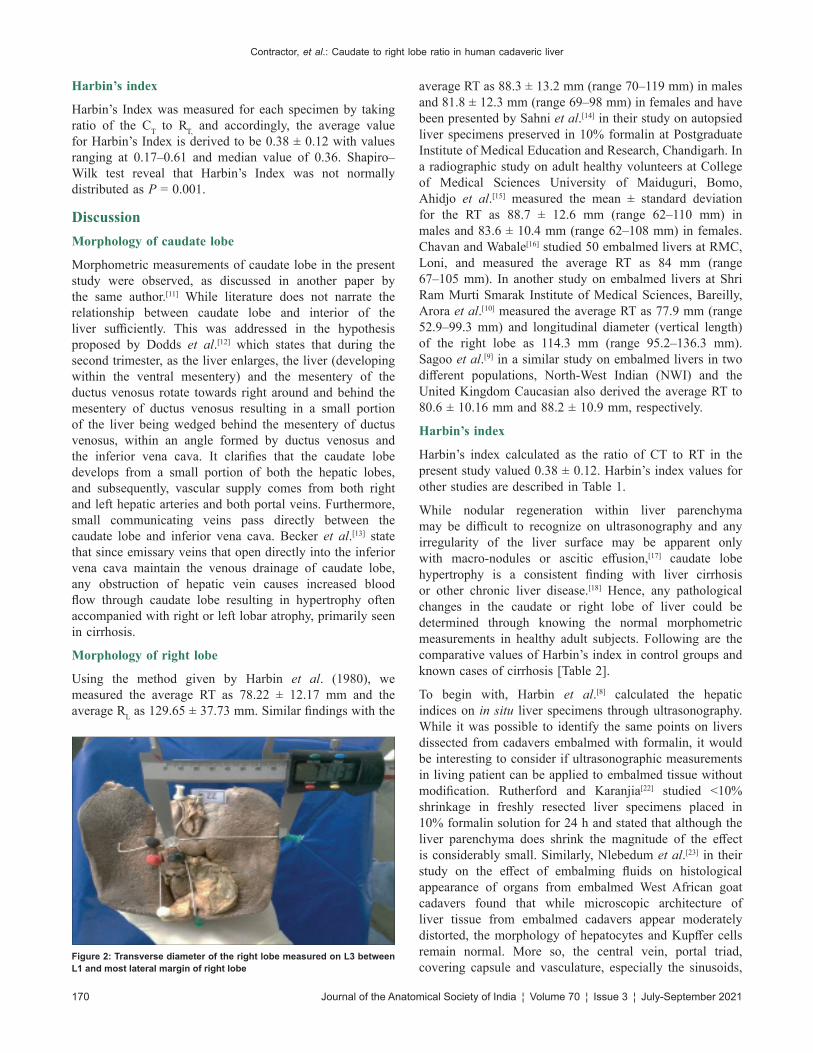
Contractor, et al.: Caudate to right lobe ratio in human cadaveric liver
170 Journal of the Anatomical Society of India ¦ Volume 70 ¦ Issue 3 ¦ July-September 2021
Harbin’s index
Harbin’s Index was measured for each specimen by taking ratio of the CT to RT, and accordingly, the average value for Harbin’s Index is derived to be 0.38 ± 0.12 with values ranging at 0.17–0.61 and median value of 0.36. Shapiro–Wilk test reveal that Harbin’s Index was not normally distributed as P = 0.001.
DiscussionMorphology of caudate lobe
Morphometric measurements of caudate lobe in the present study were observed, as discussed in another paper by the same author.[11] While literature does not narrate the relationship between caudate lobe and interior of the liver sufficiently. This was addressed in the hypothesis proposed by Dodds et al.[12] which states that during the second trimester, as the liver enlarges, the liver (developing within the ventral mesentery) and the mesentery of the ductus venosus rotate towards right around and behind the mesentery of ductus venosus resulting in a small portion of the liver being wedged behind the mesentery of ductus venosus, within an angle formed by ductus venosus and the inferior vena cava. It clarifies that the caudate lobe develops from a small portion of both the hepatic lobes, and subsequently, vascular supply comes from both right and left hepatic arteries and both portal veins. Furthermore, small communicating veins pass directly between the caudate lobe and inferior vena cava. Becker et al.[13] state that since emissary veins that open directly into the inferior vena cava maintain the venous drainage of caudate lobe, any obstruction of hepatic vein causes increased blood flow through caudate lobe resulting in hypertrophy often accompanied with right or left lobar atrophy, primarily seen in cirrhosis.
Morphology of right lobe
Using the method given by Harbin et al. (1980), we measured the average RT as 78.22 ± 12.17 mm and the average RL as 129.65 ± 37.73 mm. Similar findings with the
average RT as 88.3 ± 13.2 mm (range 70–119 mm) in males and 81.8 ± 12.3 mm (range 69–98 mm) in females and have been presented by Sahni et al.[14] in their study on autopsied liver specimens preserved in 10% formalin at Postgraduate Institute of Medical Education and Research, Chandigarh. In a radiographic study on adult healthy volunteers at College of Medical Sciences University of Maiduguri, Bomo, Ahidjo et al.[15] measured the mean ± standard deviation for the RT as 88.7 ± 12.6 mm (range 62–110 mm) in males and 83.6 ± 10.4 mm (range 62–108 mm) in females. Chavan and Wabale[16] studied 50 embalmed livers at RMC, Loni, and measured the average RT as 84 mm (range 67–105 mm). In another study on embalmed livers at Shri Ram Murti Smarak Institute of Medical Sciences, Bareilly, Arora et al.[10] measured the average RT as 77.9 mm (range 52.9–99.3 mm) and longitudinal diameter (vertical length) of the right lobe as 114.3 mm (range 95.2–136.3 mm). Sagoo et al.[9] in a similar study on embalmed livers in two different populations, North‑West Indian (NWI) and the United Kingdom Caucasian also derived the average RT to 80.6 ± 10.16 mm and 88.2 ± 10.9 mm, respectively.
Harbin’s index
Harbin’s index calculated as the ratio of CT to RT in the present study valued 0.38 ± 0.12. Harbin’s index values for other studies are described in Table 1.
While nodular regeneration within liver parenchyma may be difficult to recognize on ultrasonography and any irregularity of the liver surface may be apparent only with macro‑nodules or ascitic effusion,[17] caudate lobe hypertrophy is a consistent finding with liver cirrhosis or other chronic liver disease.[18] Hence, any pathological changes in the caudate or right lobe of liver could be determined through knowing the normal morphometric measurements in healthy adult subjects. Following are the comparative values of Harbin’s index in control groups and known cases of cirrhosis [Table 2].
To begin with, Harbin et al.[8] calculated the hepatic indices on in situ liver specimens through ultrasonography. While it was possible to identify the same points on livers dissected from cadavers embalmed with formalin, it would be interesting to consider if ultrasonographic measurements in living patient can be applied to embalmed tissue without modification. Rutherford and Karanjia[22] studied <10% shrinkage in freshly resected liver specimens placed in 10% formalin solution for 24 h and stated that although the liver parenchyma does shrink the magnitude of the effect is considerably small. Similarly, Nlebedum et al.[23] in their study on the effect of embalming fluids on histological appearance of organs from embalmed West African goat cadavers found that while microscopic architecture of liver tissue from embalmed cadavers appear moderately distorted, the morphology of hepatocytes and Kupffer cells remain normal. More so, the central vein, portal triad, covering capsule and vasculature, especially the sinusoids,
Figure 2: Transverse diameter of the right lobe measured on L3 between L1 and most lateral margin of right lobe
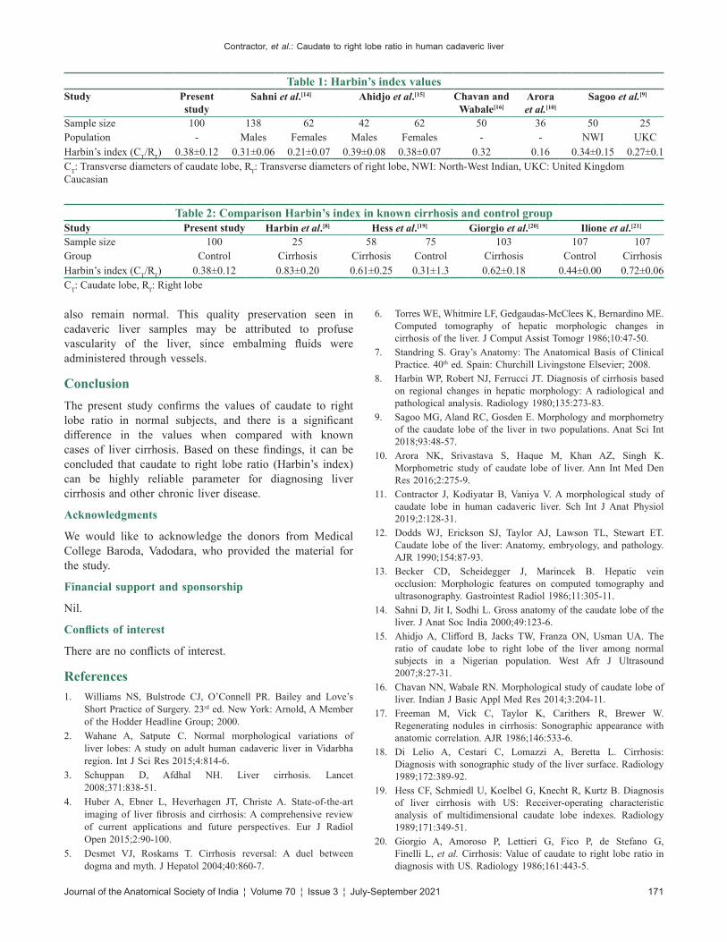
Contractor, et al.: Caudate to right lobe ratio in human cadaveric liver
Journal of the Anatomical Society of India ¦ Volume 70 ¦ Issue 3 ¦ July-September 2021 171
also remain normal. This quality preservation seen in cadaveric liver samples may be attributed to profuse vascularity of the liver, since embalming fluids were administered through vessels.
ConclusionThe present study confirms the values of caudate to right lobe ratio in normal subjects, and there is a significant difference in the values when compared with known cases of liver cirrhosis. Based on these findings, it can be concluded that caudate to right lobe ratio (Harbin’s index) can be highly reliable parameter for diagnosing liver cirrhosis and other chronic liver disease.
Acknowledgments
We would like to acknowledge the donors from Medical College Baroda, Vadodara, who provided the material for the study.
Financial support and sponsorship
Nil.
Conflicts of interest
There are no conflicts of interest.
References1. Williams NS, Bulstrode CJ, O’Connell PR. Bailey and Love’s
Short Practice of Surgery. 23rd ed. New York: Arnold, A Member of the Hodder Headline Group; 2000.
2. Wahane A, Satpute C. Normal morphological variations of liver lobes: A study on adult human cadaveric liver in Vidarbha region. Int J Sci Res 2015;4:814‑6.
3. Schuppan D, Afdhal NH. Liver cirrhosis. Lancet 2008;371:838‑51.
4. Huber A, Ebner L, Heverhagen JT, Christe A. State‑of‑the‑art imaging of liver fibrosis and cirrhosis: A comprehensive review of current applications and future perspectives. Eur J Radiol Open 2015;2:90‑100.
5. Desmet VJ, Roskams T. Cirrhosis reversal: A duel between dogma and myth. J Hepatol 2004;40:860‑7.
6. Torres WE, Whitmire LF, Gedgaudas‑McClees K, Bernardino ME. Computed tomography of hepatic morphologic changes in cirrhosis of the liver. J Comput Assist Tomogr 1986;10:47‑50.
7. Standring S. Gray’s Anatomy: The Anatomical Basis of Clinical Practice. 40th ed. Spain: Churchill Livingstone Elsevier; 2008.
8. Harbin WP, Robert NJ, Ferrucci JT. Diagnosis of cirrhosis based on regional changes in hepatic morphology: A radiological and pathological analysis. Radiology 1980;135:273‑83.
9. Sagoo MG, Aland RC, Gosden E. Morphology and morphometry of the caudate lobe of the liver in two populations. Anat Sci Int 2018;93:48‑57.
10. Arora NK, Srivastava S, Haque M, Khan AZ, Singh K. Morphometric study of caudate lobe of liver. Ann Int Med Den Res 2016;2:275‑9.
11. Contractor J, Kodiyatar B, Vaniya V. A morphological study of caudate lobe in human cadaveric liver. Sch Int J Anat Physiol 2019;2:128‑31.
12. Dodds WJ, Erickson SJ, Taylor AJ, Lawson TL, Stewart ET. Caudate lobe of the liver: Anatomy, embryology, and pathology. AJR 1990;154:87‑93.
13. Becker CD, Scheidegger J, Marincek B. Hepatic vein occlusion: Morphologic features on computed tomography and ultrasonography. Gastrointest Radiol 1986;11:305‑11.
14. Sahni D, Jit I, Sodhi L. Gross anatomy of the caudate lobe of the liver. J Anat Soc India 2000;49:123‑6.
15. Ahidjo A, Clifford B, Jacks TW, Franza ON, Usman UA. The ratio of caudate lobe to right lobe of the liver among normal subjects in a Nigerian population. West Afr J Ultrasound 2007;8:27‑31.
16. Chavan NN, Wabale RN. Morphological study of caudate lobe of liver. Indian J Basic Appl Med Res 2014;3:204‑11.
17. Freeman M, Vick C, Taylor K, Carithers R, Brewer W. Regenerating nodules in cirrhosis: Sonographic appearance with anatomic correlation. AJR 1986;146:533‑6.
18. Di Lelio A, Cestari C, Lomazzi A, Beretta L. Cirrhosis: Diagnosis with sonographic study of the liver surface. Radiology 1989;172:389‑92.
19. Hess CF, Schmiedl U, Koelbel G, Knecht R, Kurtz B. Diagnosis of liver cirrhosis with US: Receiver‑operating characteristic analysis of multidimensional caudate lobe indexes. Radiology 1989;171:349‑51.
20. Giorgio A, Amoroso P, Lettieri G, Fico P, de Stefano G, Finelli L, et al. Cirrhosis: Value of caudate to right lobe ratio in diagnosis with US. Radiology 1986;161:443‑5.
Table 1: Harbin’s index valuesStudy Present
studySahni et al.[14] Ahidjo et al.[15] Chavan and
Wabale[16]Arora et al.[10]
Sagoo et al.[9]
Sample size 100 138 62 42 62 50 36 50 25Population ‑ Males Females Males Females ‑ ‑ NWI UKCHarbin’s index (CT/RT) 0.38±0.12 0.31±0.06 0.21±0.07 0.39±0.08 0.38±0.07 0.32 0.16 0.34±0.15 0.27±0.1CT: Transverse diameters of caudate lobe, RT: Transverse diameters of right lobe, NWI: North‑West Indian, UKC: United Kingdom Caucasian
Table 2: Comparison Harbin’s index in known cirrhosis and control groupStudy Present study Harbin et al.[8] Hess et al.[19] Giorgio et al.[20] Ilione et al.[21]Sample size 100 25 58 75 103 107 107Group Control Cirrhosis Cirrhosis Control Cirrhosis Control CirrhosisHarbin’s index (CT/RT) 0.38±0.12 0.83±0.20 0.61±0.25 0.31±1.3 0.62±0.18 0.44±0.00 0.72±0.06CT: Caudate lobe, RT: Right lobe

Contractor, et al.: Caudate to right lobe ratio in human cadaveric liver
172 Journal of the Anatomical Society of India ¦ Volume 70 ¦ Issue 3 ¦ July-September 2021
21. Ilione T, Ohagwu CC, Ogolodom MP. Computed tomography evaluation of the caudate‑to‑right lobe ratio in patients with liver cirrhosis and subjects with normal liver in Benin City, Edo State, Nigeria. Health Sci J 2019;13:672.
22. Rutherford EE, Karanjia ND. The measurement of liver resection
margins. HPB (Oxford) 2004;6:18‑20.23. Nlebedum U, Ikpegbu E, Nnadozie O, Agbakwuru I. Effect of
embalming fluid on histological appearance of organs from embalmed West African Dwarf Goat Cadavers. Histologic 2013;XLVI: 16.
© 2021 Journal of the Anatomical Society of India | Published by Wolters Kluwer - Medknow 173
Address for correspondence: Dr. Saroj Kaler Jhajhria, Department of Anatomy, AIIMS, New Delhi, India. E‑mail: [email protected]
Access this article online
Website: www.jasi.org.inDOI: 10.4103/JASI.JASI_140_19
Quick Response Code:
AbstractAn extensive knowledge of the origin and course of testicular arteries (TAs) is indispensable during various surgical procedures such as renal transplant, intra‑abdominal surgeries, and even in orthopedic surgeries such as spine surgery. With the advent of new intra‑abdominal therapeutic and diagnostic techniques, the anatomy of TAs has assumed much more significance. Although the variations of the testicular vein are well documented, the variations of the TA are not so frequent in incidence. We report a rare case of bilateral aberrant origin of the TA from polar renal arteries. Though anomalies of the polar arteries supplying the kidney are common, bilateral origin of TAs from them is a rare presentation. We also discuss its developmental basis. Such anomalies if left unnoticed will lead to serious intraoperative complications during procedures on retroperitoneal organs. Any damage to the TAs will compromise the function of the gonads.
Keywords: Abdominal aorta, inferior renal polar artery, renal artery, suprarenal arteries
A Rare Case of Anomalous Origin of Bilateral Testicular Arteries: An Anatomical and Developmental Overview
Arthi Ganapathy, Aritra Banerjee, Saroj Kaler Jhajhria, Seema SinghDepartment of Anatomy, AIIMS, New Delhi, India
How to cite this article: Ganapathy A, Banerjee A, Jhajhria SK, Singh S. A rare case of anomalous origin of bilateral testicular arteries: An anatomical and developmental overview. J Anat Soc India 2020;69:XX-XX.
IntroductionAbdominal aorta being the sole purveyor of all organs in the abdomen is quite legendary for anomalies not only of its own but also due to its fellow branches. This provokes different types of pathogenesis of different organs; hence, an utmost knowledge of varying patterns of abdominal aorta and its branches evokes great importance in medical stream. Varying patterns of testicular artery (TA) is one of them.
Each TA originates from the abdominal aorta, at the level of second lumbar vertebra, 2.5–5 cm inferior to the origin of the renal artery. It traverses inferolaterally under the parietal peritoneum, along psoas major, toward the pelvis. It then enters the deep (internal) inguinal ring and travels along the ipsilateral spermatic cord in the inguinal canal to the scrotum and supplies ipsilateral testis.[1] Though the variations of testicular vein are well documented, the variations of TA are not so frequent in incidence. Most of these anomalies are related to the origin of TA. The knowledge of varying patterns of TA is not only significant in testicular and renal pathology but is also of utmost importance in various surgical procedures.
Case ReportDuring the routine dissection of an embalmed elderly male cadaver in the Department of Anatomy, AIIMS, New Delhi, we encountered an anomalous origin of bilateral TAs from the polar arteries which were supplying the lower pole of the corresponding kidneys. The renal arteries were seen separately originating from the abdominal aorta. On each side, the TA traversed inferolaterally under the parietal peritoneum with the testicular vein, along psoas major. It then crossed the ureter, toward the pelvis. Finally, it entered the deep inguinal ring to supply the testis [Figure 1]. The length of the right TA was 17.6 cm and that of the left was 17.6 cm from its origin up to the deep inguinal ring. There were no visible structural anomalies in the kidneys and testicles.
DiscussionAsala et al. reported anatomical variations of the TA in 4.7% of 150 cadavers. The variations reported by them were related to the origin of TA, which were either from unusually high levels of the abdominal aorta or from the renal artery. In four cases, they reported that the right TA was a branch of the right renal artery and in This is an open access journal, and articles are
distributed under the terms of the Creative Commons Attribution‑NonCommercial‑ShareAlike 4.0 License, which allows others to remix, tweak, and build upon the work non‑commercially, as long as appropriate credit is given and the new creations are licensed under the identical terms.
For reprints contact: [email protected]
Received: 09 October 2019 Accepted: 03 December 2020 Available online: ***
Article Info
Case Report
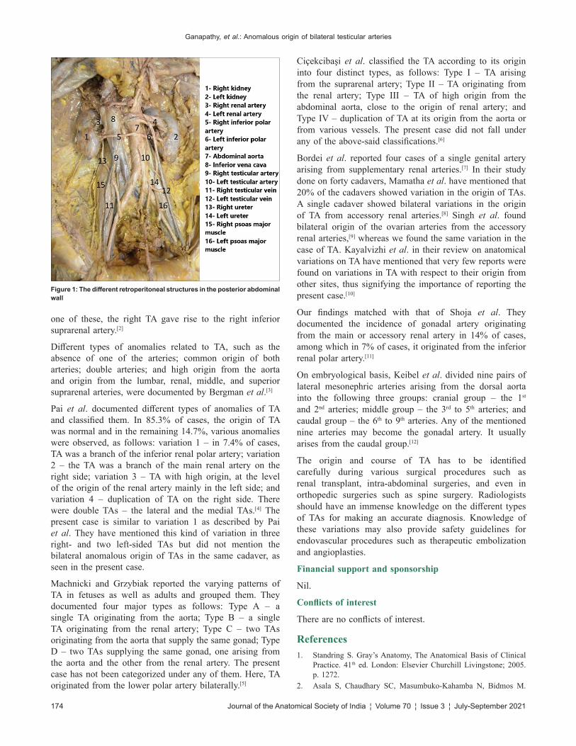
Ganapathy, et al.: Anomalous origin of bilateral testicular arteries
174 Journal of the Anatomical Society of India ¦ Volume 70 ¦ Issue 3 ¦ July-September 2021
one of these, the right TA gave rise to the right inferior suprarenal artery.[2]
Different types of anomalies related to TA, such as the absence of one of the arteries; common origin of both arteries; double arteries; and high origin from the aorta and origin from the lumbar, renal, middle, and superior suprarenal arteries, were documented by Bergman et al.[3]
Pai et al. documented different types of anomalies of TA and classified them. In 85.3% of cases, the origin of TA was normal and in the remaining 14.7%, various anomalies were observed, as follows: variation 1 – in 7.4% of cases, TA was a branch of the inferior renal polar artery; variation 2 – the TA was a branch of the main renal artery on the right side; variation 3 – TA with high origin, at the level of the origin of the renal artery mainly in the left side; and variation 4 – duplication of TA on the right side. There were double TAs – the lateral and the medial TAs.[4] The present case is similar to variation 1 as described by Pai et al. They have mentioned this kind of variation in three right‑ and two left‑sided TAs but did not mention the bilateral anomalous origin of TAs in the same cadaver, as seen in the present case.
Machnicki and Grzybiak reported the varying patterns of TA in fetuses as well as adults and grouped them. They documented four major types as follows: Type A – a single TA originating from the aorta; Type B – a single TA originating from the renal artery; Type C – two TAs originating from the aorta that supply the same gonad; Type D – two TAs supplying the same gonad, one arising from the aorta and the other from the renal artery. The present case has not been categorized under any of them. Here, TA originated from the lower polar artery bilaterally.[5]
Ciçekcibaşi et al. classified the TA according to its origin into four distinct types, as follows: Type I – TA arising from the suprarenal artery; Type II – TA originating from the renal artery; Type III – TA of high origin from the abdominal aorta, close to the origin of renal artery; and Type IV – duplication of TA at its origin from the aorta or from various vessels. The present case did not fall under any of the above‑said classifications.[6]
Bordei et al. reported four cases of a single genital artery arising from supplementary renal arteries.[7] In their study done on forty cadavers, Mamatha et al. have mentioned that 20% of the cadavers showed variation in the origin of TAs. A single cadaver showed bilateral variations in the origin of TA from accessory renal arteries.[8] Singh et al. found bilateral origin of the ovarian arteries from the accessory renal arteries,[9] whereas we found the same variation in the case of TA. Kayalvizhi et al. in their review on anatomical variations on TA have mentioned that very few reports were found on variations in TA with respect to their origin from other sites, thus signifying the importance of reporting the present case.[10]
Our findings matched with that of Shoja et al. They documented the incidence of gonadal artery originating from the main or accessory renal artery in 14% of cases, among which in 7% of cases, it originated from the inferior renal polar artery.[11]
On embryological basis, Keibel et al. divided nine pairs of lateral mesonephric arteries arising from the dorsal aorta into the following three groups: cranial group – the 1st and 2nd arteries; middle group – the 3rd to 5th arteries; and caudal group – the 6th to 9th arteries. Any of the mentioned nine arteries may become the gonadal artery. It usually arises from the caudal group.[12]
The origin and course of TA has to be identified carefully during various surgical procedures such as renal transplant, intra‑abdominal surgeries, and even in orthopedic surgeries such as spine surgery. Radiologists should have an immense knowledge on the different types of TAs for making an accurate diagnosis. Knowledge of these variations may also provide safety guidelines for endovascular procedures such as therapeutic embolization and angioplasties.
Financial support and sponsorship
Nil.
Conflicts of interest
There are no conflicts of interest.
References1. Standring S. Gray’s Anatomy, The Anatomical Basis of Clinical
Practice. 41th ed. London: Elsevier Churchill Livingstone; 2005. p. 1272.
2. Asala S, Chaudhary SC, Masumbuko‑Kahamba N, Bidmos M.
Figure 1: The different retroperitoneal structures in the posterior abdominal wall

Ganapathy, et al.: Anomalous origin of bilateral testicular arteries
Journal of the Anatomical Society of India ¦ Volume 70 ¦ Issue 3 ¦ July-September 2021 175
Anatomical variations in the human testicular blood vessels. Ann Anat 2001;183:545‑9.
3. Bergman RA, Thompson SA, Afifi A K. Catalog of Human Variation. Baltimore: Urban and Schwarzenberg; 1984. p. 119.
4. Pai MM, Vadgaonkar R, Rai R, Nayak SR, Jiji PJ, Ranade A, et al. A cadaveric study of the testicular artery in the south Indian population. Singapore Med J 2008;49:551‑5.
5. Machnicki A, Grzybiak M. Variations in testicular arteries in foetuses and adults. Folia Morphol (Warsz) 1997;56:277‑85.
6. Ciçekcibaşi AE, Salbacak A, Seker M, Ziylan T, Büyükmumcu M, Uysal II, et al. The origin of gonadal arteries in human fetuses: Anatomical variations. Ann Anat 2002;184:275‑9.
7. Bordei P, Sapte E, Iliescu D. Double renal arteries originating from the aorta. Surg Radiol Anat 2004;26:474‑9.
8. Mamatha H, D’Souza AS, Vinodhini P, Ray B, Suhani, Pallavi.
A Cadaveric Study about the Anomalous origin of testicular arteries arising from the accessory renal arteries. Indian J Surg 2015;77:111‑6.
9. Singh G, Ng YK, Bay BH. Bilateral accessory renal arteries associated with some anomalies of the ovarian arteries: A case study. Clin Anat 1998;11:417‑20.
10. Kayalvizhi I, Narayan RK, Kumar P. Anatomical variations of testicular artery: A review. Folia Morphol (Warsz) 2017;76:541‑50.
11. Borderud SP, Li Y, Burkhalter JE, Sheffer CE, Ostroff JS. Electronic cigarette use among patients with cancer: Characteristics of electronic cigarette users and their smoking cessation outcomes. Cancer 2014;120:3527‑35.
12. Keibel F, Mall F. Manual of Human Embryology II. Philadelphia: J. B. Lippincott; 1912. p. 820‑5.

Case Report
176 © 2021 Journal of the Anatomical Society of India | Published by Wolters Kluwer - Medknow
Address for correspondence: Dr. Jinu Merlin Koshy, Department of Anatomy, Sree Balaji Medical College and Hospital, 7, Works Road, Chrompet, Chennai ‑ 600 044, Tamil Nadu, India. E‑mail: jinumerlinkoshy@ gmail.com
Access this article online
Website: www.jasi.org.inDOI: 10.4103/JASI.JASI_19_20
Quick Response Code:
AbstractTrue or primary anophthalmic cyclopia is an extremely rare and severe malformation of eye. It is the apparent absence of eyeball in a median orbit and it results from failure of the optic vesicle (optic primordial) to form from the cerebral or prosencephalic vesicles. Proboscis is a skin‑covered median tubular appendage above the anophthalmic median orbit. It results from the defective development of the olfactory placodes. Sirenomelia, or mermaid syndrome, is a rare abnormality characterized by complete or incomplete fusion of lower limbs. Acardiacus is a fatal complication of monozygotic twin pregnancy. The acardiacus maintains its circulation through the heart of its normal twin reversed arterial perfusion mate. This manuscript reports on a severely malformed monozygotic stillborn twin with anophthalmic cyclopia, proboscis, acardia, absence of upper limb, sirenomelia, and aprosencephaly; agenesis of diaphragm, respiratory system, genitourinary system, lymphatic system, endocrine system, and external genitalia; and intestinal atresia, liver atresia, vascular atresia, hypoplastic skeletal system, and muscular system. In general, in all these fetal abnormalities, there are various degrees of malformation causing group of groups of anomalies. Probable cause of these anomalies is discussed.
Keywords: Monochorionic‑monoamniotic twin, twin reversed arterial perfusion, twin–twin transfusion syndrome
Anophthalmic Cyclopia with Proboscis, Acardia, Amelia, Sirenomelia – Case Report
Bini Markose1,2, Deepti Shastri3, B. Rajesh4, Jinu Merlin Koshy5
1Research Scholar, Department of Anatomy, Bharath Institute of Higher Education and Research, 5Department of Anatomy, Sree Balaji Medical College and Hospital, Bharath Institute of Higher Education and Research, Chennai, 3Department of Anatomy, Vinayaka Mission’s Kirupananda Variyar Medical College, Salem, Tamil Nadu, 2Department of Anatomy, Amala Institute of Medical Sciences, Thrissur, Kerala, 4Department of Anatomy, Sri Lakshmi Narayana Institute of Medical Sciences, Bharath Institute of Higher Education and Research, Puducherry, India
How to cite this article: Markose B, Shastri D, Rajesh B, Koshy JM. Anophthalmic cyclopia with proboscis, acardia, amelia, sirenomelia – Case report. J Anat Soc India 2021;70:XX-XX.
IntroductionTrue or primary anophthalmic cyclopia with proboscis, sirenomelia, acardia, and associated malformations, is extremely rare and severe malformation occurring in early stages of development.[1] The monozygotic twins occur in approximately 1 in 200 births, which represent the most common aberration of morphogenesis noted in the human.[2] The prevalence of anophthalmic cyclopia is approximately 30 per 100,000, and sirenomelia is found in approximately 1 out of every 100,000 live births, 100 times more likely to occur in identical twins than single birth or fraternal twin.[3] There are approximately 300 cases of sirenomelia reported in the literature, 15% of which are associated with twinning, more often monozygotic.[4] “Monochorionic‑monoamniotic (MoMo)” twins are very rare with an occurrence of 1 in 35,000 to 1 in 60,000 pregnancies corresponding to about 1% of twin pregnancy. Acardiac twinning is thought to affect 1 in 100 monozygotic twin
gestation and 1 in 35,000 pregnancies overall. In monozygotic twinning, if the division occurs unequally, one component may become dependent on its co‑twin’s cardiovascular system and can develop into an acardiac fetus.[5] The anomalous monster twin was a heterogeneous mass of tissue with a well‑developed skull, ribs, sternum, dural folds, and spinal cord. All the remaining tissues were either absent or poorly developed. An acardiacus refers to a hemodynamically disadvantaged nonviable twin which occurs in association with a twin reversed arterial perfusion (TRAP) sequence which corresponds to twin–twin transfusion syndrome (TTTS).[6] In TTTS, arterial‑arterial and venous‑venous anastomoses in the placenta permit twin–twin transfusion and reversal of blood flow in the umbilical vessels and aorta of the recipient twin. The anomalous twin had a cephalic attachment of the umbilical cord so that the head has got maximum tissue differentiation compared to other regions. The present article describes the details of the anomalous with the help of dissection and X‑rays.
This is an open access journal, and articles are distributed under the terms of the Creative Commons Attribution‑NonCommercial‑ShareAlike 4.0 License, which allows others to remix, tweak, and build upon the work non‑commercially, as long as appropriate credit is given and the new creations are licensed under the identical terms.
For reprints contact: [email protected]
Received: 31 January 2020Accepted: 15 February 2021Available online: ***
Article Info
Markose, et al.: Anophthalmic cyclopia with proboscis, acardia, amelia, sirenomelia – Case report
Journal of the Anatomical Society of India ¦ Volume 70 ¦ Issue 3 ¦ July-September 2021 177
Case ReportA 28‑year‑old female, with consanguineous marriage, G1P1, with an unremarkable family history was admitted to a peripheral hospital, at 35 weeks of gestation with labor pain. Antenatal ultrasound revealed a monozygotic twin pregnancy. Antenatal diagnosis by ultrasound examination was confirmed the monozygotic twin (MoMo twin) gestation and detected most of the associated malformations in the anomalous twin; the co‑twin was a normal fetus. Other than ultrasound scan during the second trimester, the parents never did any investigations because of economic and social status. The selective termination of the anomalous twin was not done because the patient’s family did not allow to do any intrauterine interventions. Because of the economic and social status, the family never did any investigations during the pregnancy. Only when she was suffering from labor pain, they came to the hospital. She had undergone a lower‑segment cesarean section, because of delay in normal progress of the labor. The first of the twin was an apparently healthy male neonate, with 3.2 kg birth weight. On examination, the co‑twin was found to be a monstrous stillborn fetus having a dysmorphic face, with a median single eye, absence of nose, micrognathia, and a proboscis, all of which made anophthalmic cyclopia, and associated malformations the possible initial diagnosis. Anomalous twin had a crown–rump length of 19.5 cm, weighs 1000 g, and shows the following anomalies which are described in Table 1. Two umbilical cords were noticed.
The umbilical cord of the normal twin was long with a pair of umbilical arteries and one umbilical vein. The anomalous twin had a short umbilical cord of 7.5 cm long with a single small umbilical artery and a big umbilical vein. The umbilical cord was attached to the head–trunk junction of the fetus. There was no obvious neck, but there was a transverse linear constriction on ventral region. The anomalous twin was very soft, edematous with sloughed‑off skin. All the clinical features were confirmed by the radiological pictures and the dissection photographs of the body. Chromosomal analysis was not carried out as consent was not given by the parents.
DiscussionWe have reported on a 36‑week gestation stillborn fetus with anophthalmic cyclopia with associated malformation. To our knowledge, this is a bizarre form of anomalies and the fetus can be the victim of monozygotic twinning. In this case, no apparent comparable cases could be extracted from the literature for comparison. Cyclopia represents a severe deficit in early midline facial development, the eyes become fused, and the olfactory placodes consolidate into a single tube‑like proboscis above the eye. Anophthalmic cyclopia occurs due to failure in the development of optic primordia. There is failure in the cleavage of the prosencephalon resulting in the absence of the forebrain (aprosencephaly). The absence of an organ may be due to its disappearance in the subsequent development owing either to an inherent
Table 1: Dissection findings of the variant twinRegion/parts Gross featuresHead [Figure 1] Umbilical cord was attached to the head. Approximately normal size for a full‑term;
anophthalmic cyclopia with proboscis (1.7 cm, no cavity), arhinia, rudimentary low set ears without external acoustic meatus, mouth with clefted lower lip, and microglossia. Normal gum and cheeks. Scalp thickness at bregma ‑ 1.08 cm, at lamda ‑ 2.55 cm and extra subcutaneous connective tissue in the occipital region
Brain [Figure 2] Cranial cavity almost empty except brain stem, dural folds with dural venous sinuses. Absence of forebrain (aprosencephaly). Anteroposterior diameter of cranial cavity is 7.9 cm, and transverse diameter is 6.2 cm. No pituitary gland in the fossa, rudimentary cranial nerves. Optic canal (4 cm long) connecting anterior cranial fossa to median anophthalmic orbit
Neck [Figure 1] No obvious neckUpper limb Figure 1 Totally absentDiaphragm [Figure 2a and b] Totally absentUpper ½ of trunk [Figure 2a and b] Body wall with normal ribs, and sternum, vertebrae. No visceraLower ½ of trunk [Figure 2a and b] Almost empty except a network of blood vessels, blind‑ended coiled tube of intestine (10 cm),
small lobulated liver. All other viscera absentExternal genitalia [Figure 1a] Totally absentLower limb Figures 1 and 2 Both limbs are of different lengths; left ‑ 4.5 cm, right ‑ 5.5 cm; fused proximally like a
mermaid (sirenomelia), hypoplastic contracted pelvis, normal hip bone, normal femora, hypoplastic tibiae and absent fibulae. Foot skeleton ‑ 3 toes on left and 2 toes on right. Left femur, tibia thinner and shorter than right
Vertebral column [Figure 3] Apparent scoliosis, normal vertebra except sacrum (hypoplastic)Spinal cord [Figure 2] 11 cm long fully occupies the vertebral canal. Normal spinal meninges and spinal nerves. No
brachial plexus, but lumbosacral plexus is well seen with proportionate thicknessRadiological finding [Figure 3] Skull shows all bones except ethmoid and nasal bones. No upper limb bones, scapula, and
clavicle, hyoid bone. Ribs, sternum, vertebrae well seen. Lower limb bones were hypoplastic
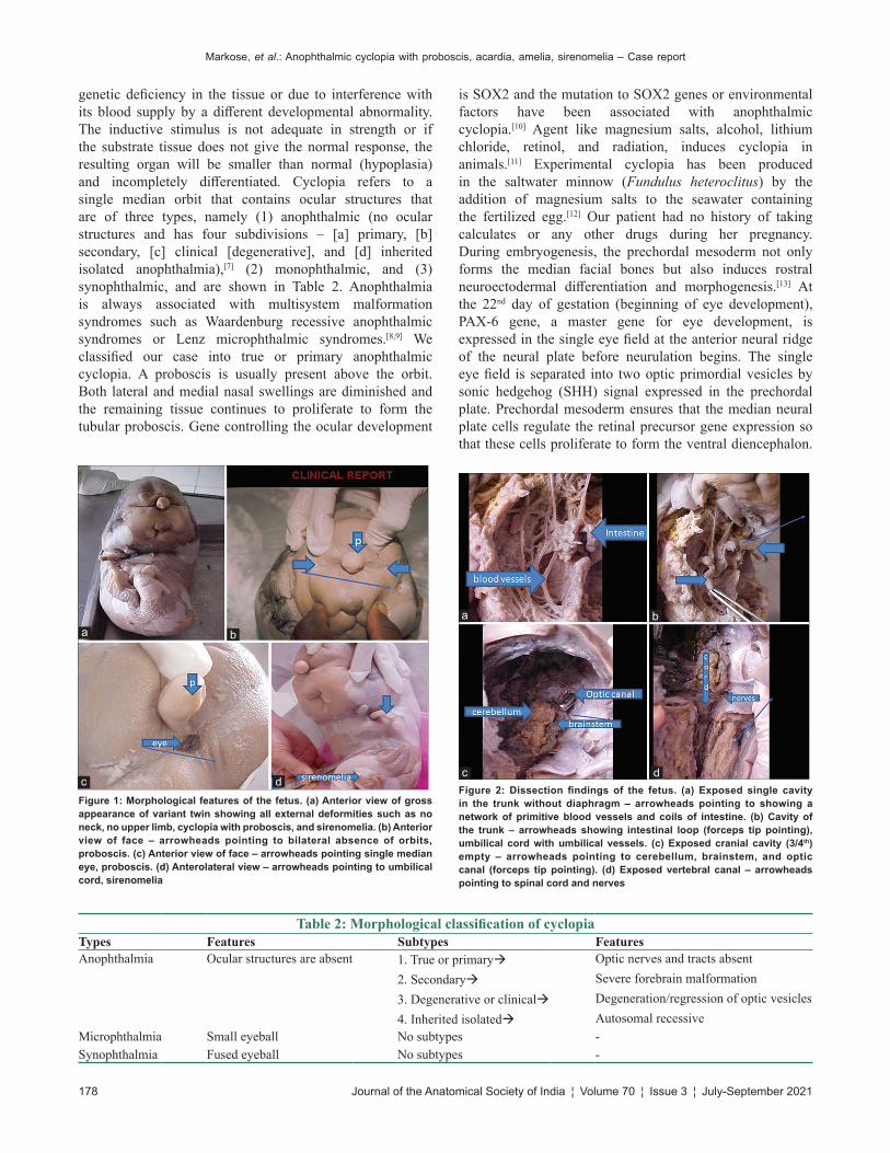
Markose, et al.: Anophthalmic cyclopia with proboscis, acardia, amelia, sirenomelia – Case report
178 Journal of the Anatomical Society of India ¦ Volume 70 ¦ Issue 3 ¦ July-September 2021
genetic deficiency in the tissue or due to interference with its blood supply by a different developmental abnormality. The inductive stimulus is not adequate in strength or if the substrate tissue does not give the normal response, the resulting organ will be smaller than normal (hypoplasia) and incompletely differentiated. Cyclopia refers to a single median orbit that contains ocular structures that are of three types, namely (1) anophthalmic (no ocular structures and has four subdivisions – [a] primary, [b] secondary, [c] clinical [degenerative], and [d] inherited isolated anophthalmia),[7] (2) monophthalmic, and (3) synophthalmic, and are shown in Table 2. Anophthalmia is always associated with multisystem malformation syndromes such as Waardenburg recessive anophthalmic syndromes or Lenz microphthalmic syndromes.[8,9] We classified our case into true or primary anophthalmic cyclopia. A proboscis is usually present above the orbit. Both lateral and medial nasal swellings are diminished and the remaining tissue continues to proliferate to form the tubular proboscis. Gene controlling the ocular development
is SOX2 and the mutation to SOX2 genes or environmental factors have been associated with anophthalmic cyclopia.[10] Agent like magnesium salts, alcohol, lithium chloride, retinol, and radiation, induces cyclopia in animals.[11] Experimental cyclopia has been produced in the saltwater minnow (Fundulus heteroclitus) by the addition of magnesium salts to the seawater containing the fertilized egg.[12] Our patient had no history of taking calculates or any other drugs during her pregnancy. During embryogenesis, the prechordal mesoderm not only forms the median facial bones but also induces rostral neuroectodermal differentiation and morphogenesis.[13] At the 22nd day of gestation (beginning of eye development), PAX‑6 gene, a master gene for eye development, is expressed in the single eye field at the anterior neural ridge of the neural plate before neurulation begins. The single eye field is separated into two optic primordial vesicles by sonic hedgehog (SHH) signal expressed in the prechordal plate. Prechordal mesoderm ensures that the median neural plate cells regulate the retinal precursor gene expression so that these cells proliferate to form the ventral diencephalon.
Table 2: Morphological classification of cyclopiaTypes Features Subtypes FeaturesAnophthalmia Ocular structures are absent 1. True or primary
2. Secondary3. Degenerative or clinical4. Inherited isolated
Optic nerves and tracts absentSevere forebrain malformationDegeneration/regression of optic vesiclesAutosomal recessive
Microphthalmia Small eyeball No subtypes ‑Synophthalmia Fused eyeball No subtypes ‑
Figure 1: Morphological features of the fetus. (a) Anterior view of gross appearance of variant twin showing all external deformities such as no neck, no upper limb, cyclopia with proboscis, and sirenomelia. (b) Anterior view of face – arrowheads pointing to bilateral absence of orbits, proboscis. (c) Anterior view of face – arrowheads pointing single median eye, proboscis. (d) Anterolateral view – arrowheads pointing to umbilical cord, sirenomelia
dc
ba
Figure 2: Dissection findings of the fetus. (a) Exposed single cavity in the trunk without diaphragm – arrowheads pointing to showing a network of primitive blood vessels and coils of intestine. (b) Cavity of the trunk – arrowheads showing intestinal loop (forceps tip pointing), umbilical cord with umbilical vessels. (c) Exposed cranial cavity (3/4th) empty – arrowheads pointing to cerebellum, brainstem, and optic canal (forceps tip pointing). (d) Exposed vertebral canal – arrowheads pointing to spinal cord and nerves
dc
ba
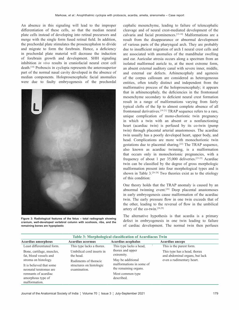
Figure 3: Radiological features of the fetus – total radiograph showing cranium, well‑developed vertebral column with scoliosis, ribs, and the remaining bones are hypoplastic
Markose, et al.: Anophthalmic cyclopia with proboscis, acardia, amelia, sirenomelia – Case report
Journal of the Anatomical Society of India ¦ Volume 70 ¦ Issue 3 ¦ July-September 2021 179
An absence in this signaling will lead to the improper differentiation of these cells, so that the median neural plate cells instead of developing into retinal precursors and merge with the single form fused retinal field. In addition, the prechordal plate stimulates the prosencephalon to divide and migrate to form the forebrain. Hence, a deficiency in prechordal plate material will decrease the induction of forebrain growth and development. SHH signaling inhibition in vivo results in craniofacial neural crest cell death.[14] Proboscis in cyclopia represents the anterosuperior part of the normal nasal cavity developed in the absence of median components. Holoprosencephalic facial anomalies were due to faulty embryogenesis of the prechordal
cephalic mesenchyme, leading to failure of telencephalic cleavage and of neural crest‑mediated development of the calvaria and facial prominences.[15‑18] Malformations are a result from the disappearance or abnormal development of various parts of the pharyngeal arch. They are probably due to insufficient migration of arch I neural crest cells and are associated with anomalies of the mandibular swelling and ear. Auricular atresia occurs along a spectrum from an isolated malformed auricle to, at the most extreme form, an absent external auditory canal with severe inner, middle, and external ear defects. Arhinencephaly and agenesis of the corpus callosum are considered as heterogeneous entities, often totally distinct and independent from the malformative process of the holoprosencephaly; it appears that in arhinencephaly, the deficiencies in the frontonasal mesenchyme secondary to deficient neural crest formation result in a range of malformations varying from fairly typical clefts of the lip to almost complete absence of all frontonasal derivatives.[19‑21] TRAP sequence refers to a rare, unique complication of mono‑chorionic twin pregnancy in which a twin with an absent or a nonfunctioning heart (acardiac twin) is perfused by its co‑twin (pump twin) through placental arterial anastomoses. The acardiac twin usually has a poorly developed heart, upper body, and head. Complications are more with monochorionic twin gestations due to placental sharing.[22] The TRAP sequence, also known as acardiac twinning, is a malformation that occurs only in monochorionic pregnancies, with a frequency of about 1 per 35,000 deliveries.[23‑25] Acardiac twin can be classified by the degree of gross morphologic malformation present into four morphological types and is shown in Table 3.[26‑28] Two theories exist as to the etiology of this condition:
One theory holds that the TRAP anomaly is caused by an abnormal twinning event.[26] Deep placental anastomoses in early embryogenesis cause malformation of the acardiac twin. The early pressure flow in one twin exceeds that of the other, leading to the reversal of flow in the umbilical artery of the co‑twin.[28,29]
The alternative hypothesis is that acardia is a primary defect in embryogenesis in one twin leading to failure of cardiac development. The normal twin then perfuses
Table 3: Morphological classification of Acardiacus TwinAcardius amorphous Acardius acormus Acardius acephalus Acardius anceps
Least differentiated form.Bone, cartilage, muscles, fat, blood vessels and stroma on histology.It is believed that some neonatal teratomas are remnants of acardiac amorphous type of malformation.
This type lacks a thorax.Umbilical cord inserts in the head.Rudiments of thoracic structures on histologic examination.
This type lacks a head, thorax and upper extremity.May be additional malformations in some of the remaining organs.Most common type described.
` This is the purest form.This type has a head, thorax and abdominal organs, but lack even a rudimentary heart.
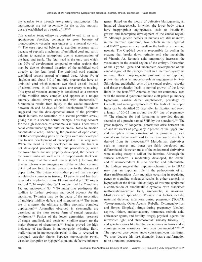
Markose, et al.: Anophthalmic cyclopia with proboscis, acardia, amelia, sirenomelia – Case report
180 Journal of the Anatomical Society of India ¦ Volume 70 ¦ Issue 3 ¦ July-September 2021
the acardiac twin through artery‑artery anastomoses. The anastomoses are not responsible for the cardiac anomaly but are established as a result of it.[28,29]
The acardiac twin, otherwise destined to end in an early spontaneous abortion, continues to grow because of monochorionic vascular anastomoses to a normal co‑twin.[26] The case reported belongs to acardius acormus partly because of cephalic attachment of umbilical cord and partly belongs to acardius amorphous due to nonseparation of the head and trunk. The fetal head is the only part which has 50% of development compared to other regions that may be due to abnormal insertion of the umbilical cord directly to the fetal head. The umbilical cord contains two blood vessels instead of normal three. About 1% of singleton and about 5% of multiple pregnancies have an umbilical cord which contains two blood vessels instead of normal three. In all these cases, one artery is missing. This type of vascular anomaly is considered as a remnant of the vitelline artery complex and appears as a unique anomaly almost always associated with sirenomelia.[30] Sirenomelia results from injury to the caudal mesoderm between 28 and 32 days of fetal development.[31] Studies suggested that the developmental arrest of the primitive streak initiates the formation of a second primitive streak, giving rise to a second normal embryo. This may account for the high incidence of sirenomelia in twins. There was a canal communicating the anterior cranial fossa to median anophthalmic orbit, indicating the presence of optic canal, but the corresponding parts of the eyes were not developed due to non development of diencephalon. It is very rare. When the head is fully developed in size, the brain is not developed proportionately, but paradoxically, when the lower limbs are not properly developed, the nerves to the lower limbs are well seen in proportionate thickness. It is strange that the spinal nerves (C5‑T1) forming the brachial plexus were emerging out of the vertebral column, but it did not form brachial plexus due to the absence of upper limbs. The cytogenetic studies proved that cyclopia is relatively common in trisomy 13 patients and has been reported in triploidy, trisomy 18 combined dup 1q32 →qter and del 7q34 →qter, dup 3p21 →3pter, del 18 P and ring 18, and monosomy G.[1,25] Twinning may predispose the midline to further problems and could account for the anomalies. Twinning may be the cause of the association of multiple midline defects and sirenomelia.[32] The twins are in a sense, the ultimate midline anomaly: complete duplication![33] Anomalies observed in sirenomelia are described as the most severe form of caudal regression syndrome.[34] Fusion of the lower extremities, presence of single umbilical, and persistent vitelline artery are the major features of sirenomelia.[35] Studies proved the high incidence of acardiacus in monozygotic twinning. Early malformation in monozygotic twins is due to reversed or disrupted vascular shunts between monozygotic twins, vascular disruption or hypoperfusion, and defective inherent
genes. Based on the theory of defective blastogenesis, an impaired blastogenesis, in which the lower body organs have inappropriate angiogenesis, leads to insufficient growth and incomplete development of the caudal region.[36] Although genetic defects in humans are still unknown in the mermaid syndrome, two defects in the Cyp26a1 and BMP7 genes in mice result in the birth of a mermaid neonate. The Cyp26a1 gene is responsible for coding the enzyme that breaks down retinoic acid (the metabolite of Vitamin A). Retinoic acid temporarily increases the vasculature in the caudal region of the embryo. Disruption of the Cyp26a1 gene and incomplete development of the caudal region of the embryo result in a mermaid syndrome in mice. Bone morphogenetic protein‑7 is an important protein that plays an important role in angiogenesis in vitro. Stimulating endothelial cells of the caudal region, vascular and tissue production leads to normal growth of the lower limbs in the fetus.[37‑39] Anomalies that are commonly seen with the mermaid syndrome include cleft palate pulmonary hypoplasia, cardiac defect omphalocele, pentalogy of Cantrell, and meningomyelocele.[39] The buds of the upper limbs can be identified 26 days after fertilization and reach a length of 20–22 mm around the 53rd day of pregnancy.[40] The stimulus for bud formation is provided through secretion of a protein named SHH by the notochord.[41] The great majority of congenital deformities arise between the 4th and 8th weeks of pregnancy. Agenesis of the upper limb and disruption or malformation of the primitive streak’s caudal vasculature could lead to malformation of structures derived from its mesoderm.[42] Mesodermal derivatives such as muscles and bones are fairly developed and differentiated. However, most of the endodermal derivatives were missing except a coil of the intestine. Although the surface ectoderm is moderately developed, the cranial end of neuroectoderm fails to develop and differentiate. The findings suggest that hypoxia‑ischemia due to TRAP may play an important role in the pathogenesis of all these malformations. Any mutation occurring in regulating genes or signaling molecules results in either agenesis or hypoplasia of the tissue. The etiology of this rare syndrome, a combination of anophthalmic cyclopia, with associated malformation‑acardiac twin, sirenomelia, is unknown. Most cases are sporadic.[43] Possible risk factors include: maternal diabetes, infections during pregnancy {TORCH ‑Toxoplasmosis, Other Agents, Rubella, Cytomegalovirus, and Herpes Simplex}, drugs during pregnancy (alcohol, aspirin, lithium, anticonvulsants, hormones, retinoic acid, anticancer agents, and fertility drugs), physical agents like ultraviolet light, and chromosomal3 (mostly trisomy 13) and genetic causes like familial occurrences in twins and in consanguineous marriages have been documented[10‑16,43,44] The reported case comes under consanguineous marriages. We must deduce this rare, dramatic, bizarre malformation to be a random occurrence.
Markose, et al.: Anophthalmic cyclopia with proboscis, acardia, amelia, sirenomelia – Case report
Journal of the Anatomical Society of India ¦ Volume 70 ¦ Issue 3 ¦ July-September 2021 181
ConclusionIt is a rare case of monozygotic twin fetus with multiple anomalies. In all the cases so far known in the past (after 1535), wherever there is a reduction in size of the brain, there is an invariable reduction in size of the skull as seen in anencephaly or microcephaly. Here, in this case, the skull is of normal size for a full‑term fetus and the major parts of the brain are missing. It is important to recognize these syndromes clinically and to determine their relationship to various chromosomal abnormalities. The recognition of carrier states with the help of advanced imaging techniques and in utero cytogenetic studies of amniotic cells may spare the unfortunate parents of such monstrosities from similar traumatic experiences. One more aspect of unrevealed secrets of nature is unfolded in this work.
Consent
Informed consent was obtained from parents for the publication of this case report and any accompanying images. Ethical clearance was obtained from the institutional ethical committee. The fetus was donated to the department and was preserved in the Department of Anatomy, VMKV Medical College, Salem, Tamil Nadu, India.
Declaration of patient consent
The authors certify that they have obtained all appropriate patient consent forms. In the form, the patient has given her consent for her images and other clinical information to be reported in the journal. The patient understands that her name and initials will not be published and due efforts will be made to conceal identity, but anonymity cannot be guaranteed.
Acknowledgment
The authors are thankful to Late Dr. Muthukrishnan, Professor of Anatomy, VMKV Medical College, Salem, for the most valuable guidance throughout this work and helped us to achieve the best paper presentation award in Tamil Nadu State Anatomy conference. We sincerely acknowledge Dr. T. A. Ajith, Professor of Biochemistry, AIMS, Thrissur, for his expert and sincere help in the preparation of this work. We are grateful to Dr. Sugathan for his great help in the proofreading. We are grateful to Mrs. Leena and Mrs. Rengini K. V. for their help in typing the manuscript. We are thankful to the staff of the Department of Anatomy, AIMS, Thrissur, and VMKV Medical College, Salem.
Financial support and sponsorship
Nil.
Conflicts of interest
There are no conflicts of interest.
References1. Stevenson R, Hall J, Everman D, Solomon B. (Eds.), Human
Malformations and Related Anomalies. Oxford, UK: Oxford University Press. Available from: https://oxfordmedicine.com/view/10.1093/med/9780199386031.001.0001/med‑9780199386031. [Last accessed on 2019 Sept 12].
2. Stein RA. Smith's recognizable patterns of human malformation, 6th edition. Arch Dis Child. 2007;92:562. PMCID: PMC2066177.
3. Källén B, Castilla EE, Lancaster PA, Mutchinick O, Knudsen LB, Martínez‑Frías ML, et al. The cyclops and the mermaid: An epidemiological study of two types of rare malformation. Med Genet 1992;29:30‑5.
4. Stocker JT, Heifetz SA. Sirenomelia. A morphological study of 33 cases and review of the literature. Perspect Pediatr Pathol 1987;10:7‑50.
5. Hamilton, Boyd and Mossman's Human Embryology. Fourth edition. By W. J. HAMILTON and H. W. MOSSMAN. Cambridge: Heffer. 1972.
6. Athwal S, Millard K, Lakhoo K. Twin reversed arterial perfusion (TRAP) sequence in association with VACTERL association: A case report. Med Case Rep 2010;4:411.
7. Kohn G, El Shawwa R, El Rayyes E. Isolated “clinical anophthalmia” in an extensively affected Arab kindred. Clin Genet 1988;33:321‑4.
8. Traboulsi EI, Nasr AM, Fahd SD, Jabbour NM, Der Kaloustian VM. Waardenburg’s recessive anophthalmia syndrome. Ophthalmic Paediatr Genet 1984;4:13‑8.
9. Traboulsi EI, Lenz W, Gonzales‑Ramos M, Siegel J, Macrae WG, Maumenee IH. The Lenz microphthalmia syndrome. Am J Ophthalmol 1988;105:40‑5.
10. Fantes J, Ragge NK, Lynch SA, McGill NI, Collin JR, Howard‑Peebles PN, et al. Mutations in SOX2 cause anophthalmia. Nat Genet 2003;33:462‑3.
11. Mollica F, Pavone L, Sorge G. Maternal drug ingestion and cyclopia. J Pediatr 1981;98:680.
12. Stockard CR. Developmental rate and structural expression: An experimental study of twins, double monsters and single deformities, and the interaction among embryonic organs during their origin and development. Am J Anat 1921;28:115‑277.
13. Arathi N, Mahadevan A, Santosh V, Yasha TC, Shankar SK. Holoprosencephaly with cyclopia – Report of a pathological study. Neurol India 2003;51:279‑82.
14. Ahlgren SC, Bronner‑Fraser M. Inhibition of Sonic hedgehog signaling in vivo results in craniofacial neural crest cell death. Curr Biol 1999;9:1304‑14.
15. Adelmann HB. The problem of cyclopia I & 11. Q Rev Biol 1936;11:284.
16. Kallen B. Contribution of the knowledge of the regulation of the proliferation processes in the vertebrate brain during ontogenesis. Acta Anat (Basel) 1956;27:351‑60.
17. DeMyer WE, Zeman W, Palmer CG. The face predicts the brain: Diagnostic significance of median facial anomalies for holoprosencephaly (arhinencephaly). Pediatrics 1964;34:256‑63.
18. Johnston MC, Morriss GM, Kushner DC, Bingle GJ. Abnormal organogenesis of facial structures. In: Handbook of Teratology. US: Springer; 1977. p. 421‑51.
19. Marin‑Padilla M, Hoefnagel D, Benirschke K. Anatomic and histologic study of two cases of DI (13‑15) trisomy. Cytogenetics 1964;3:258‑84.
20. Sedano HO, Cohen MM Jr., Jirasek J, Gorlin RJ. Frontonasal dysplasia. J Pediatr 1970;76:906‑13.
21. Delezoide AL, Narcy F, Larroche JC. Cerebral midline
Markose, et al.: Anophthalmic cyclopia with proboscis, acardia, amelia, sirenomelia – Case report
182 Journal of the Anatomical Society of India ¦ Volume 70 ¦ Issue 3 ¦ July-September 2021
developmental anomalies: Spectrum and associated features. Genet Couns 1990;1:197‑210.
22. Srivastava N, Bhatia T, Narshetty J, Sushil Kumar. Acardiac twin: A rare case report. International Journal of Infertility and Fetal Medicine. 2016;7:109‑10.
23. James WH. A note on the epidemiology of acardiac monsters. Teratology 1977;16:211‑6.
24. Vandenbusseche F, Dprest JA, Klumper F. Minimal invasive intra‑uterine chirurgische behandeling bij vier monochoriale tweelingzwangersc happenge compliceerd dor een acardiacus. Nedrlands. Tijdschrift Voor Geneeskunde 2003;147:931‑936.
25. Sullivan AE, Varner MW, Ball RH, Jackson M, Silver RM. The management of acardiac twins: A conservative approach. Am J Obs Gynecol 2003;189:1310‑3.
26. Gabbe SG, Niebyl JR, Simpson JL, Landon MB, Galan HL, Jauniaux ERM, et al. Obstetrics: Normal and Problem Pregnancies. Philadelphia, Pa: Elsevier;2012.
27. Prameela RC, Ranganath P, Nivedita S, PoojaGY, Prajwal S. A rare case of acardiac twin: a case report. Int J Sci Stud. 2014;2:254‑7.
28. Van Allen MI, Smith DW, Shepard TH. Twin reversed arterial perfusion (TRAP) sequence: A study of 14 twin pregnancies with acardius. Semin Perinatol 1983;7:285‑93.
29. Athwal S, Millard K, Lakhoo K. Twin reversed arterial perfusion (TRAP) sequence in association with VACTERL association: A case report. J Med Case Rep 2010;4:411.
30. Seidahmed MZ, Abdelbasit OB, Alhussein KA, Miqdad AM, Khalil MI, Salih MA. Sirenomelia and severe caudal regression syndrome. Saudi Med J 2014;35 Suppl 1:S36‑43.
31. Salama GS, Kaabneh MA, Al‑Raqad MK, Al‑Abdallah IM, Shakkoury AG, Halaseh RA. Cyclopia: A rare condition with unusual presentation – A case report. Clin Med Insights Pediatr 2015;9:19‑23.
32. Potter’s Pathology of the Fetus and Infant E‑Book: 2‑Volume Set – Google Books. Available from: https://books.google.co.in/books?id=ZwklCwAAQBAJ&pg=PA94&lpg=PA94&dq=Opitz,+Gilbert+Ef(+1982),+Am+J+Med+Genet;+12+443‑455.&source=bl&ots=Byz3f_N7tL&sig=ACfU3U3oVLi8m5og0pVMGcoKfOdhpt3Avg&hl=en&sa=X&ved=2ahUKEwj3iIivwq3nAhWO4zgGH
Zd5C7gQ6AEwAXoECAkQAQ#v=one. [Last accessed on 2020 Jan 31].
33. Lubinsky MS. Midline developmental “weakness” as a consequence of determinative field properties. Am J Med Genet Suppl 1987;3:23‑8.
34. Duhamel B. From the mermaid to anal imperforation: The syndrome of caudal regression. Arch Dis Child 1961;36:152‑5.
35. Twickler D, Budorick N, Pretorius D, Grafe M, Currarino G. Caudal regression versus sirenomelia: Sonographic clues. Ultrasound Med 1993;12:323‑30.
36. Opitz JM, Zanni G, Reynolds JF, Gilbert‑Barness E. Defects of blastogenesis. Am J Med Genet 2002;115:269‑86.
37. Zakin L, Reversade B, Kuroda H, Lyons KM, De Robertis EM. Sirenomelia in Bmp7 and Tsg compound mutant mice: Requirement for Bmp signaling in the development of ventral posterior mesoderm. Development 2005;132:2489‑99.
38. Ribes V, Fraulob V, Petkovich M, Dollé P. The oxidizing enzyme CYP26A1 tightly regulates the availability of retinoic acid in the gastrulating mouse embryo to ensure proper head development and vasculogenesis. Dev Dyn 2007;236:644‑53.
39. Sheng N, Xie Z, Wang C, Bai G, Zhang K, Zhu Q, et al. Retinoic acid regulates bone morphogenic protein signal duration by promoting the degradation of phosphorylated Smad1. Proc Natl Acad Sci U S A 2010;107:18886‑91.
40. In Wolfe SW, In Hotchkiss RN, In Pederson WC, In Kozin SH, In Cohen MS. Green's operative hand surgery. 5th ed. Philadelphia: Elsevier Chur‑Chill Livingstone; 2017.p.1375‑9.
41. Al‑Qattan MM, Yang Y, Kozin SH. Embryology of the upper limb. J Hand Surg Am 2009;34:1340‑50.
42. Garrido‑Allepuz C, Haro E, González‑Lamuño D, Martínez‑Frías ML, Bertocchini F, Ros MA. A clinical and experimental overview of sirenomelia: Insight into the mechanisms of congenital limb malformations. Dis Model Mech 2011;4:289‑99.
43. Chervenak FA, Isaacson G, Hobbins JC, Chitkara U, Tortora M, Berkowitz RL. Diagnosis and management of fetal holoprosencephaly. Obstet Gynecol 1985;66:322‑6.
44. Johnson CY, Rasmussen SA. Non‑genetic risk factors for holoprosencephaly. Am J Med Genet C Semin Med Genet 2010;154C: 73‑85.
© 2021 Journal of the Anatomical Society of India | Published by Wolters Kluwer - Medknow 183
Address for correspondence: Dr. Gaurav Goel, Department of Neurosciences, Medanta ‑ The Medicity, Gurgaon, Haryana, India. E‑mail: [email protected]
Access this article online
Website: www.jasi.org.inDOI: 10.4103/JASI.JASI_131_20
Quick Response Code:
AbstractWe present a rare case of bilateral persistent primitive olfactory artery incidentally detected on computed tomography angiography and digital subtraction angiography in a 53‑year‑old female with symptomatic left‑sided tight internal carotid artery stenosis. This imaging finding can be useful to the literature.
Keywords: Anterior cerebral artery, internal carotid artery, persistent primitive olfactory artery
Bilateral Persistent Primitive Olfactory Artery Incidentally Detected by Computed Tomography Angiography and Digital Subtraction Angiography: An Extremely Rare Case Report
Anshu Mahajan, Apratim Chatterjee, Gaurav GoelDepartment of Neurosciences, Medanta ‑ The Medicity, Gurgaon, Haryana, India
How to cite this article: Mahajan A, Chatterjee A, Goel G. Bilateral persistent primitive olfactory artery incidentally detected by computed tomography angiography and digital subtraction angiography: An extremely rare case report. J Anat Soc India 2021;70:XX-XX.
IntroductionPersistent primitive olfactory artery (PPOA) is an extremely rare anomaly of anterior cerebral artery (ACA), in which the anomalous artery courses anteroinferomedially along the ipsilateral olfactory tract and makes a hairpin to turn posterior to the olfactory bulb to continue as A2 segment of ACA.[1] The clinical significance of this anomaly is its association with cerebral aneurysm. It is also very important for the neurosurgeon to recognize this anomaly before performing the surgery at the anterior skull base to avoid any catastrophic complication.[2] We present a rare case of bilateral PPOA incidentally detected on computed tomography angiography (CTA) and digital subtraction angiography (DSA) in a patient with symptomatic left internal carotid artery (ICA) tight stenosis who was treated with left carotid stenting.
Case ReportA 53‑year‑old hypertensive female presented with a history of two episodes of transient right‑sided weakness for 1 month which recovered completely. Noncontrast computed tomography head was normal and CTA of brain and neck vessels showed >90% stenosis of left carotid bulb and an incidental anomalous course of bilateral ACAs [Figure 1a]. We performed
magnetic resonance imaging (MRI) brain which showed small microvascular ischemic changes in brain parenchyma however no fresh infarct. We planned carotid stenting for her symptoms. Successful left carotid stenting was done and DSA (Axiom Artis Zee; Siemens, Erlangen, Germany) of cerebral vessels showed incidental anomalous anteroinferior course of bilateral ACA which then turns posteriorly and superiorly to continue as A2 segment of ACAs suggestive of PPOA [Figure 1b‑e]. Detailed review of angiography was done which showed no evidence of associated aneurysm in our case.
DiscussionAt around 4–5.7 mm embryo length, cranial and caudal divisions of ICA are formed. Cranial division of the ICA is termed as PPOA. Anterior choroidal artery and middle cerebral artery branches off from the primitive olfactory artery (POA). As the embryo reaches 11.5–18 mm length, there are two branches from the POA, the original one is to the nasal fossa and other one represents the future continuation of the ACA. Later in the end of the stage, the opposite artery joins with its fellow by the plexiform anastomosis which then forms the future anterior communicating artery (ACoA). The original POA forms an anastomosis with the ACA and later POA regress in size and the two arteries,
This is an open access journal, and articles are distributed under the terms of the Creative Commons Attribution‑NonCommercial‑ShareAlike 4.0 License, which allows others to remix, tweak, and build upon the work non‑commercially, as long as appropriate credit is given and the new creations are licensed under the identical terms.
For reprints contact: [email protected]
Received: 07 August 2020Accepted: 19 August 2021Available online: ***
Article Info
Case Report
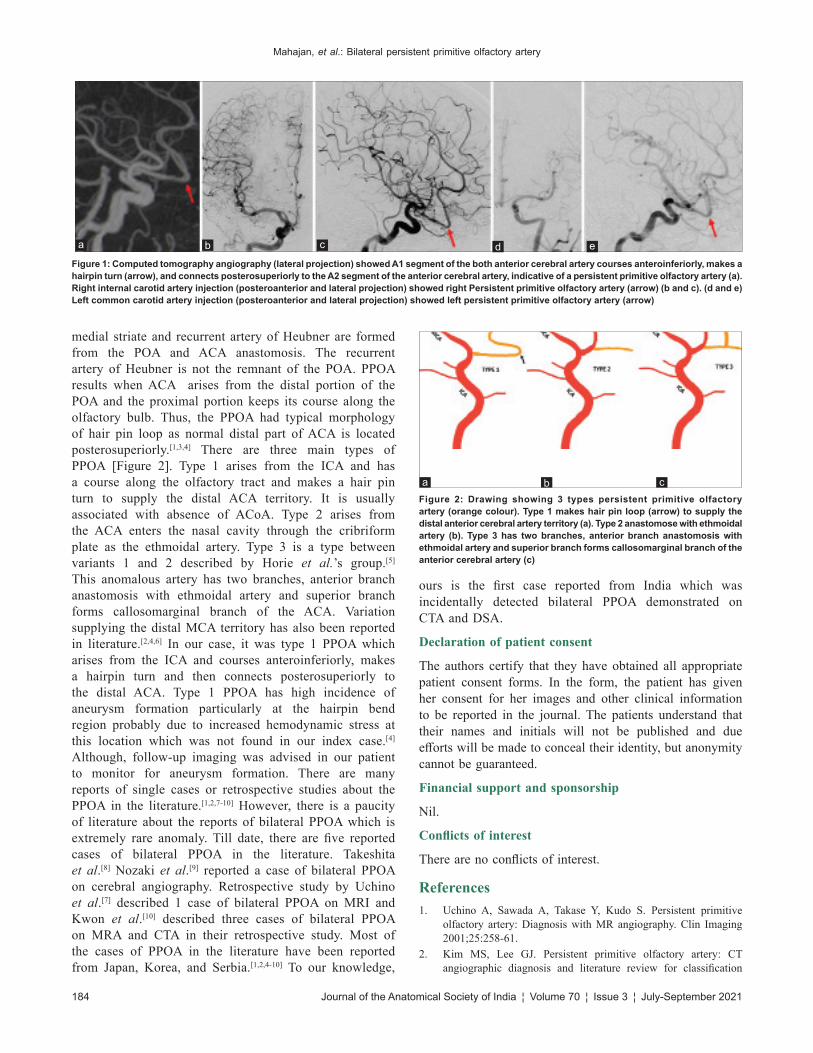
Mahajan, et al.: Bilateral persistent primitive olfactory artery
184 Journal of the Anatomical Society of India ¦ Volume 70 ¦ Issue 3 ¦ July-September 2021
medial striate and recurrent artery of Heubner are formed from the POA and ACA anastomosis. The recurrent artery of Heubner is not the remnant of the POA. PPOA results when ACA arises from the distal portion of the POA and the proximal portion keeps its course along the olfactory bulb. Thus, the PPOA had typical morphology of hair pin loop as normal distal part of ACA is located posterosuperiorly.[1,3,4] There are three main types of PPOA [Figure 2]. Type 1 arises from the ICA and has a course along the olfactory tract and makes a hair pin turn to supply the distal ACA territory. It is usually associated with absence of ACoA. Type 2 arises from the ACA enters the nasal cavity through the cribriform plate as the ethmoidal artery. Type 3 is a type between variants 1 and 2 described by Horie et al.’s group.[5] This anomalous artery has two branches, anterior branch anastomosis with ethmoidal artery and superior branch forms callosomarginal branch of the ACA. Variation supplying the distal MCA territory has also been reported in literature.[2,4,6] In our case, it was type 1 PPOA which arises from the ICA and courses anteroinferiorly, makes a hairpin turn and then connects posterosuperiorly to the distal ACA. Type 1 PPOA has high incidence of aneurysm formation particularly at the hairpin bend region probably due to increased hemodynamic stress at this location which was not found in our index case.[4] Although, follow‑up imaging was advised in our patient to monitor for aneurysm formation. There are many reports of single cases or retrospective studies about the PPOA in the literature.[1,2,7‑10] However, there is a paucity of literature about the reports of bilateral PPOA which is extremely rare anomaly. Till date, there are five reported cases of bilateral PPOA in the literature. Takeshita et al.[8] Nozaki et al.[9] reported a case of bilateral PPOA on cerebral angiography. Retrospective study by Uchino et al.[7] described 1 case of bilateral PPOA on MRI and Kwon et al.[10] described three cases of bilateral PPOA on MRA and CTA in their retrospective study. Most of the cases of PPOA in the literature have been reported from Japan, Korea, and Serbia.[1,2,4‑10] To our knowledge,
ours is the first case reported from India which was incidentally detected bilateral PPOA demonstrated on CTA and DSA.
Declaration of patient consent
The authors certify that they have obtained all appropriate patient consent forms. In the form, the patient has given her consent for her images and other clinical information to be reported in the journal. The patients understand that their names and initials will not be published and due efforts will be made to conceal their identity, but anonymity cannot be guaranteed.
Financial support and sponsorship
Nil.
Conflicts of interest
There are no conflicts of interest.
References1. Uchino A, Sawada A, Takase Y, Kudo S. Persistent primitive
olfactory artery: Diagnosis with MR angiography. Clin Imaging 2001;25:258‑61.
2. Kim MS, Lee GJ. Persistent primitive olfactory artery: CT angiographic diagnosis and literature review for classification
Figure 1: Computed tomography angiography (lateral projection) showed A1 segment of the both anterior cerebral artery courses anteroinferiorly, makes a hairpin turn (arrow), and connects posterosuperiorly to the A2 segment of the anterior cerebral artery, indicative of a persistent primitive olfactory artery (a). Right internal carotid artery injection (posteroanterior and lateral projection) showed right Persistent primitive olfactory artery (arrow) (b and c). (d and e) Left common carotid artery injection (posteroanterior and lateral projection) showed left persistent primitive olfactory artery (arrow)
dcba e
Figure 2: Drawing showing 3 types persistent primitive olfactory artery (orange colour). Type 1 makes hair pin loop (arrow) to supply the distal anterior cerebral artery territory (a). Type 2 anastomose with ethmoidal artery (b). Type 3 has two branches, anterior branch anastomosis with ethmoidal artery and superior branch forms callosomarginal branch of the anterior cerebral artery (c)
cba
Mahajan, et al.: Bilateral persistent primitive olfactory artery
Journal of the Anatomical Society of India ¦ Volume 70 ¦ Issue 3 ¦ July-September 2021 185
and clinical significance. Surg Radiol Anat 2014;36:663‑7.3. Okahara M, Kiyosue H, Mori H, Tanoue S, Sainou M,
Nagatomi H. Anatomic variations of the cerebral arteries and their embryology: A pictorial review. Eur Radiol 2002;12:2548‑61.
4. Sato Y, Kashimura H, Takeda M, Chida K, Kubo Y, Ogasawara K. Aneurysm of the A1 segment of the anterior cerebral artery associated with the persistent primitive olfactory artery. World Neurosurg 2015;84:9.e7‑9.
5. Horie N, Morikawa M, Fukuda S, Hayashi K, Suyama K, Nagata I. New variant of persistent primitive olfactory artery associated with a ruptured aneurysm. J Neurosurg 2012;117:26‑8.
6. Kim MS. Persistent primitive olfactory artery connected with middle cerebral artery: Case report. Surg Radiol Anat
2013;35:849‑52.7. Uchino A, Saito N, Kozawa E, Mizukoshi W, Inoue K. Persistent
primitive olfactory artery: MR angiographic diagnosis. Surg Radiol Anat 2011;33:197‑201.
8. Takeshita G, Katada K, Koga S, Sano K, Jinno T. An anomalous course of proximal portion of the anterior cerebral artery. Jpn J Clin Radiol 1988;33:593‑6.
9. Nozaki K, Taki W, Kawakami O, Hashimoto N. Cerebral aneurysm associated with persistent primitive olfactory artery aneurysm. Acta Neurochir (Wien) 1998;140:397‑401.
10. Kwon BR, Yeo SH, Chang HW, Kim MJ, Kim E, Kim MK, et al. Magnetic resonance angiography and CT angiography of persistent primitive olfactory artery: Incidence and association rate with aneurysm in Korea. J Korean Soc Radiol 2012;66:493‑9.
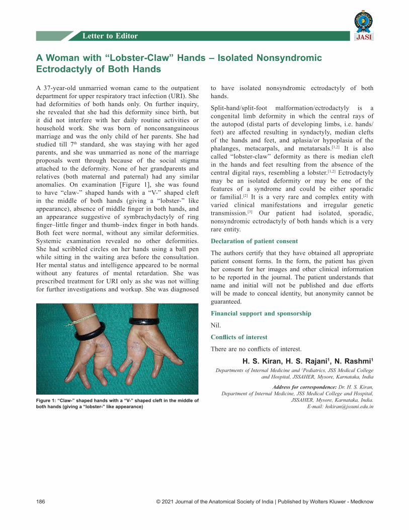
186 © 2021 Journal of the Anatomical Society of India | Published by Wolters Kluwer - Medknow
A Woman with “Lobster‑Claw” Hands – Isolated Nonsyndromic Ectrodactyly of Both Hands
A 37‑year‑old unmarried woman came to the outpatient department for upper respiratory tract infection (URI). She had deformities of both hands only. On further inquiry, she revealed that she had this deformity since birth, but it did not interfere with her daily routine activities or household work. She was born of nonconsanguineous marriage and was the only child of her parents. She had studied till 7th standard, she was staying with her aged parents, and she was unmarried as none of the marriage proposals went through because of the social stigma attached to the deformity. None of her grandparents and relatives (both maternal and paternal) had any similar anomalies. On examination [Figure 1], she was found to have “claw‑” shaped hands with a “V‑” shaped cleft in the middle of both hands (giving a “lobster‑” like appearance), absence of middle finger in both hands, and an appearance suggestive of symbrachydactyly of ring finger–little finger and thumb–index finger in both hands. Both feet were normal, without any similar deformities. Systemic examination revealed no other deformities. She had scribbled circles on her hands using a ball pen while sitting in the waiting area before the consultation. Her mental status and intelligence appeared to be normal without any features of mental retardation. She was prescribed treatment for URI only as she was not willing for further investigations and workup. She was diagnosed
to have isolated nonsyndromic ectrodactyly of both hands.
Split‑hand/split‑foot malformation/ectrodactyly is a congenital limb deformity in which the central rays of the autopod (distal parts of developing limbs, i.e. hands/feet) are affected resulting in syndactyly, median clefts of the hands and feet, and aplasia/or hypoplasia of the phalanges, metacarpals, and metatarsals.[1,2] It is also called “lobster‑claw” deformity as there is median cleft in the hands and feet resulting from the absence of the central digital rays, resembling a lobster.[1,2] Ectrodactyly may be an isolated deformity or may be one of the features of a syndrome and could be either sporadic or familial.[2] It is a very rare and complex entity with varied clinical manifestations and irregular genetic transmission.[3] Our patient had isolated, sporadic, nonsyndromic ectrodactyly of both hands which is a very rare entity.
Declaration of patient consent
The authors certify that they have obtained all appropriate patient consent forms. In the form, the patient has given her consent for her images and other clinical information to be reported in the journal. The patient understands that name and initial will not be published and due efforts will be made to conceal identity, but anonymity cannot be guaranteed.
Financial support and sponsorship
Nil.
Conflicts of interest
There are no conflicts of interest.
H. S. Kiran, H. S. Rajani1, N. Rashmi1
Departments of Internal Medicine and 1Pediatrics, JSS Medical College and Hospital, JSSAHER, Mysore, Karnataka, India
Address for correspondence: Dr. H. S. Kiran, Department of Internal Medicine, JSS Medical College and Hospital,
JSSAHER, Mysore, Karnataka, India. E‑mail: [email protected]
Letter to Editor
Figure 1: “Claw‑” shaped hands with a “V‑” shaped cleft in the middle of both hands (giving a “lobster‑” like appearance)

Letter to Editor
Journal of the Anatomical Society of India ¦ Volume 70 ¦ Issue 3 ¦ July-September 2021 187
References1. Jindal G, Parmar VR, Gupta VK. Ectrodactyly/split hand feet
malformation. Indian J Hum Genet 2009;15:140‑2.2. Duijf PH, van Bokhoven H, Brunner HG. Pathogenesis of
split‑hand/split‑foot malformation. Hum Mol Genet 2003;12 Spec No 1:R51‑60.
3. Umair M, Hayat A. Nonsyndromic split‑hand/foot malformation: Recent classification. Mol Syndromol 2020;10:243‑54.
How to cite this article: Kiran HS, Rajani HS, Rashmi N. A woman with “lobster-claw” hands – Isolated nonsyndromic ectrodactyly of both hands. J Anat Soc India 2021;XX:XX-XX.© 2021 Journal of the Anatomical Society of India | Published by Wolters Kluwer - Medknow
This is an open access journal, and articles are distributed under the terms of the Creative Commons Attribution‑NonCommercial‑ShareAlike 4.0 License, which allows others to remix, tweak, and build upon the work non‑commercially, as long as appropriate credit is given and the new creations are licensed under the identical terms.
Access this article onlineQuick Response Code:
Website: www.jasi.org.in
DOI: 10.4103/jasi.jasi_26_21
Received: 15 February 2021Accepted: 02 July 2021Available online: ***
Article Info