issue20202.pdf - our dermatology online
TRANSCRIPT
1. w
Volume 11, Number 2 April 2020 ISSN: 2081-9390
p. 113 - 223 DOI: 10.7241/ourd
Issue online since Thursday April 02, 2020
Dermatology Online www.odermatol.com
Our
- Soraya Aouali, Imane Alouani, Hanane Ragragui, Nada Zizi, Siham Dikhaye A case of epithelioid angiosarcoma in a young man with chronic lymphedema
- Iyda El Faqyr, Maria Dref, Sara Zahid, Jamila Oualla, Nabil Mansouri, Hanane Rais, Ouafa Hocar, Said AmalSyringocystadenoma papilliferum presented as an ulcerated nodule of the vulva in a patient with Neurofibromatosis type 1
- FMonisha Devi Selvakumari, Bittanakurike Narasappa Raghavendra, Anjan Kumar Patra
A case of infantile Sweet’s syndrome
- Anissa Zaouak, Leila Bouhajja, Houda Hammami, Samy FennichePenile annular lichen planus
2 / 2020
Editorial Pages
Quarterly published since 01/06/2010 yearsOur Dermatol Online
www.odermatol.com
Editor in Chief: Publisher:Piotr Brzeziński, MD Ph.D Our Dermatology Online
Address: Address:ul. Braille’a 50B, 76200 Słupsk, Poland ul. Braille’a 50B, 76200 Słupsk, Polandtel. 48 692121516, fax. 48 598151829 tel. 48 692121516, fax. 48 598151829e-mail: [email protected] e-mail: [email protected]
Associate Editor:Ass. Prof. Viktoryia Kazlouskaya (USA)
Indexed in:Universal Impact Factor for year 2012 is = 0.7319
system of opinion of scientific periodicals INDEX COPERNICUS (8,69)(Academic Search) EBSCO
(Academic Search Premier) EBSCOMNiSW (kbn)-Ministerstwo Nauki i Szkolnictwa Wyższego (7.00)
DOAJ (Directory of Open Acces Journals)Geneva Foundation for Medical Education and Research (GFMER), Google Scholar, Open J-Gate, NewJour,
International Committee of Medical Journal Editors (ICMJE), Genamics JournalSeek, Hinari,Bielefeld Academic Search Engine (BASE), WorldCat, e -journal, WorldWideScience.org, National Science Library,
LibSearch, Sciencegate, Virtual Science Library (VSL), Wanfang Data, COnnecting REpositories (CORE),CAB Abstracts, Global Health, Journal Indexed in Directory of Research Journals Indexing,OAIster: The Open Access Initiative, OAJSE - Open Access Journals Search Engine, Scirus
Previous website: issue 1.2010 www.ndermatol.like.pl since issue 2.2010 to issue 3.2011 www.odermatol.like.pl since issue 4.2011 www.odermatol.comPrevious shortcut: since issue 1.2010 to issue 3.2011 N Dermatol Online since issue 4.2011 Our Dermatol Online
Open access journal:This is an open access journal which means that all content is freely available without charge to the user or his/her institution. Users are allowed to read, download, copy, distribute, print, search, or link to the fullor texts of the articles in this journal without asking prior permission from the publisher or the author.Our Dermatology Online is a international journal that publishes original contributions in the field of dermatology, including papers on biochemistry, morphology and immunology of the skin.The journal is among the few not related to dermatological associations or belonging to respective societies which guarantees complete independence. Offers a platform for review articles in areas of interest for dermatologists.OurDermatologyOnline offers article in English as well as in other languages. This is in accordance with the BOAI definition of open access.
e-ISSN: 2081-9390DOI: 10.7241/ourd
Editorial Board
Abdel-Naser, Mohamed Badawy, Prof. (Egypt)Abdul-Lateef Mousa Haider, MD (Iraq)Al Aboud Khalid, MD (Saudi Arabia)Al-Kamel Mohamed A., MD (Yemen)Al-Mashaleh Manal Sulaiman, MD (Jordan)Abreu-Velez Ana Maria, Prof. (USA)Abreu Hilda, MD (Urugway)Adaskevich Uladzimir, Prof. (Belarus)Afifi Mustafa, MD (United Arab Emirates)Aghaei Shahin, Ass. Prof. (Iran)Akpaka Patrick Eberechi, Prof. (Trinidad and Tobago)Akyshbayeva Kulbarshin, Prof. (Kazakhstan)Amichai Boaz, MD (Israel)Arakelyan Hayk S. Prof. (Armenia)Arenas Roberto, Prof. (Mexico)Arif Tasleem, MD (India)Asuquo Maurice Efana, Prof. (Nigeria)Auto James, Ass. Prof. (Solomon Islands)Fatou Barro-Traoré, Prof. (Burkina Faso)Christian Muteba Baseke, MD (Democratic Republic of the Congo)Beigi Pooya Khan Mohammad, Prof. (Canada)Bharti Rakesh, MD (India)Bonifaz Alexandro, Prof. (Mexico)Borowska Katarzyna, Ass. Prof. (Poland)Borruto Franco, Prof. (Monaco)Bouadjar Bakar, Prof. (Algeria)Bukhari Iqbal A., Prof. (Saudi Arabia)Cabo Horacio, Prof. (Argentina)Chamcheu Jean Christopher, Ph.D (USA)Chang Patricia, MD Ph.D (Guatemala)Chihanga Simon, MD (Botswana)Choon Siew Eng, MD (Malaysia)Chuh An Tung Antonio, Prof. (Hong Kong)Crump Vincent, MD (New Zealand)Daboul Mohamed Wael, MD (Syria)Daisley Hubert, Prof. (Trinidad and Tobago)Darlenski Razvigor, MD Ph.D (Bulgaria)Diouf Assane, Ass. Prof. (Senegal)Dobrev Hristo, Prof. (Bulgaria)Doganay Mehmet, Prof. (Turkey)Dong Huiting, Prof. (China)Dori Geme Urge, PhD (Ethiopia)Draganita Ana Maria, MD PhD (Romania)Drljević Irdina, MD, Ph.D. Ass. Prof. (Bosnia and Herzegovina)Dubakienė Rūta, Prof. (Lithuania)Edwards Carl, Ass. Prof. (USA)Elhassan Elizabeth, MD (Senegal)Farkas Arpad, MD PhD (Hungary)Fernandez-Flores Angel, MD Ph.D (Spain)Fortuna Giulio, Ass. Prof. (USA)
Gołąb Elżbieta, Prof. (Poland)Gómez Cuevas Alina, Prof. MD (Nicaragua)Grattan Clive (United Kingdom)Grivcheva-Panovska Vesna, Prof. (Macedonia)Guzmán Antonio, MD (Paraguay)Hashimoto Takashi, Prof. (Japan)Hassan Iffat, Prof. (India)Hegyi Vladimir, Prof. (Slovakia)Hidalgo-Matlock Benjamin, MD (Costa Rica)Hysi Katerina, MD (Albania)Janjua Shahbaz, MD (Pakistan)Jeseňák Miloš, Ass. Prof. (Slovakia)Jeewon Rajesh, Ph.D. (Mauritius)Jordán Rodriguez Ramiro, Prof. (Bolivia)Julian Rolando, Prof. (El Salvador)Kaszuba Andrzej, Prof. (Poland)Kaštelan Marija, Prof. (Croatia)Katsambas Andreas, Prof. (Greece) Khawaja Shakeel Ahmed, PhD (Eritrea)Kibbi Abdul-Ghani, Prof. (Lebanon) Kossi Metowogo, Ph.D (Togo)Kuiate Jules-Roger, Prof. (Cameroon)Lan Cheng-Che E., Ass. Prof. (Taiwan) Lopez-Granja Jorge, MD (Belize) Lotti Torello, Prof. (Italy)Mahassadi Alassan Kouamé, Ass. Prof. (Côte d’Ivoire’)Mahdi Juma Husain Ali, MD (Bahrain)Maibach Howard I., Prof (USA)Maio Paula, MD (Portugal) Mekokishvili Lali, Prof. (Georgia) Mikkelsen Carsten Sauer, MD (Denmark) Mourad Mokni, Prof. (Tunisia)Mota Luiz Alberto Alves, Prof. (Brazil) Mrisho Fatma, MD (Tanzania) Muvunyi Claude Mambo, MD (Rwanda) Ndugwa Christopher, Prof. (Uganda) Nedelciuc Boris, Ass. Prof. (Moldova) Nhlengethwa Winnie, Prof. (Swaziland) Nigam Pramod Kumar, Prof. (India) Nikolic Milos, Prof. (Serbia)Nowicki Roman, Prof. (Poland)Nwabudike Lawrence Chukwudi, MD Ph.D (Romania)Odeh Samuel, Prof. (Gabon)Olszański Romuald, Prof. (Poland)Oranje Arnold, Prof. (Netherlands) Parajuli Sudip, MD (Nepal) Parvin Rukhsana, MD (Bangladesh)du Plessis Jeanetta, Prof. (South Africa) Puri Neerja, MD (India)Pusahai-Riman Paula, BSc, MS (Papua New Guinea)
Editorial Board
Qurashi Mohd, MD (Sudan)Riedl Elisabeth, Ass. Prof. (Austria)Ríos Yuil José Manuel, Prof. (Panama) Ranotsi Amelia, PhD (Lesotho) Rubio-Texeira Marta Ph.D. (Belgium) Rusnak Martin, Prof. (Slovakia) Sayad Ibrahim, Prof. (Kuwait) Sharquie Khalifa E., Prof. (Iraq) Shawa Mary, MD (Malawi)Shkilna Mariia, MD Ph.D (Ukraine)Sinclair Rodney Daniel, Prof. (Australia) Singh Harjeet, MD (Qatar)Slavic Vjerolsva, MD PhD (Montenegro)Srinivasan Sundaramoorthy, Prof. (India)Sumathipala Gayan Saranga, MD (Sri Lanka) Tapia Felix J., Ass. Prof. (Venezuela)
Tatu Alin, MD (Romania)Teixeira Roni Leonardo, MD (Brazil) Tincopa-Wong Oscar Wilfredo, MD (Peru) Tresh Amani, MD (Libya)Tylewska-Wierzbanowska Stanisława, Prof. (Poland)Uraga Pazmiño Enrique, MD (Ecuador)Usha Rani Anaparthy, Prof. (India) Valdebran Manuel, MD (Dominican Republic) Vok Marko, MD (Slovenia)Win Oo Soe, MD (Myanmar)Wollina Uwe, Prof. (Germany) Wortsman Ximena, Ass. Prof. (Chile) Yamamoto Toshiyuki, Prof. (Japan) Yuil de Ríos Emma, MD (Panama) Zabielski Stanisław, Prof. (Poland) Zawar Vijay, Prof (India)
© Our Dermatol Online 2.2020 i
Contents
ORIGINAL ARTICLES
Psychotherapeutic methods in psoriasis ............................................................................................................. 113Aneta Gruchała, Konrad Marski, Anna Zalewska-Janowska
Investigating the role of Interleukin-33 and Soluble ST2 in pediatric atopic dermatitis ..................................... 120Ilona Paulauskaitė, Audronė Eidukaitė, Odilija Rudzevičienė, Rasa Orentaitė
Epidemiological and clinical aspects of skin diseases observed in workers handling cement inBurkina Faso ..................................................................................................................................................... 126Issouf Konaté, Nina Nessine Korsaga/Somé, Arsène Hema, Samira Véronique Sanfo, Boukary Diallo,Jean Baptiste Andonaba, Fatou Barro/Traoré, Pascal Niamba, Adama Traoré
BRIEF REPORT
Scabies of the child: Epidemiological, clinical, therapeutic and evolutionary aspects in theservice of Dermatology of the Regional Hospital of Th iès (Senegal): About 69 cases (2012-2017) ..................... 135Haby Dione, Pauline Diousse, Agbogbenkou Tévi Déla-dem Lawson, Mariama Bammo,Mame Diasse Ndiaye, Pape Souleymane Toure, Fatou Seck, Ndeye Ramatoulaye Diagne Gueye,Sylvie Audrey Diop, Bernard Marcel Diop, Mamadou Mourtalla Ka
CASE REPORTS
Microneedling for androgenetic alopecia not responding to conventional treatment .......................................... 140Sudip Parajuli, Upama Paudel
A case of infantile Sweet’s syndrome .................................................................................................................. 143Monisha Devi Selvakumar, Bittanakurike Narasappa Raghavendra, Anjan Kumar Patra
A case of acute hemorrhagic edema of infancy ................................................................................................... 146Aastha Wadhwa, Anjan Kumar Patra, Bijina Kolukulangara Dharman
Combined treatment of keloids and scars with Nd:YAG 1064 nm laser andcryotherapy: Report of clinical cases .................................................................................................................. 149César Bimbi, Piotr Brzeziński
Rhinoscleroma: a diagnosis not to be ignored .................................................................................................... 154Nadia Baali, Sofia Berrada, Nadia Akhdari, Ouafa Hocar, Hanane Rais, Said Amal
Lichen planus pigmentosus inversus associated with oral lichen planus.............................................................. 156Antonio Carlos Bastos Gomes, César Bimbi, Piotr Brzezinski
Solitary purpuric plaque: clinical and dermoscopic evolution of lichen aureus ................................................... 158Asmae Rasso, Jihane Ziani, Sara Oukarfi, Hanane Baybay, Sara Elloudi, Fatima Zahra Mernissi
Cutaneous epithelioid angiosarcoma of the head and neck. A case report of an unusualaggressive entity with limited treatment options ................................................................................................ 161Sandra Gonzalez, Raul Gerardo Mendez, Bertha Lissette Sotelo, Francisco Solano
A case of epithelioid angiosarcoma in a young man with chronic lymphedema .................................................. 165Soraya Aouali, Imane Alouani, Hanane Ragragui, Nada Zizi, Siham Dikhaye
Syringoma, hormone receptors and associated endocrinopathies. Are they clinically relevant? ........................... 168Ayse Tülin Mansur, Saime Ramadan
Digital pacinian neuroma in a patient with myeloma ........................................................................................ 171Yosra Soua, Kamar Belhareth, Hayet Akkari, Mahbouba Jguirim, Hichem Belhadjali,Nouha Ben Abdeljelil, Monia Youssef, Jameleddine Zili
© Our Dermatol Online 2.2020 ii
Contents
Syringocystadenoma papilliferum presented as an ulcerated nodule of the vulva in a patient with Neurofi bromatosis type 1 .................................................................................................................................. 174Iyda El Faqyr, Maria Dref, Sara Zahid, Jamila Oualla, Nabil Mansouri, Hanane Rais,Ouafa Hocar, Said Amal
Herpes simplex mastitis: A rare complication of paediatric orolabial herpes ....................................................... 177Radhika Varma, Rony Mathew, Amitha Abraham
HIV and venous thromboembolism risk: Report of 3 cases with review of the literature .................................... 180Mohamed El Amraoui, Rachid Frikh, Naoufal Hjira, Mohammed Boui
A case of positive HIV serology revealed by a demodicosis ................................................................................ 183Ibtissam Assenhaji, Mounia Bennani, Sara Elloudi, Hanane Baybay, Fatima Zahra Mernissi
OPINION ARTICLE
Parental roles and childhood sun safety ............................................................................................................. 186Tessa Li Chyin Lim
CLINICAL IMAGES
Drug Reaction with Eosinophilia and Systemic Symptoms (DRESS) syndrome with ciprofl oxacin ................... 189Sameh Sayhi, Tayssir Ben Achour
Multiple scrotal epidermal cysts: A clinical case ................................................................................................. 190Cheymae Saadani Hassani, Sara Elloudi, Fatima Zahra Mernissi
LETTER TO THE EDITORS
Vulvar pruritus: A view over a life ...................................................................................................................... 191Aicha Nassiri, Kaoutar Moustaide, Sara Elloudi, Hanane Baybay, Fatima Zahra Mernissi
Topical corticosteroid abuse among pediatric population - a prospective study .................................................. 194Mrinal Gupta
Epidemio-clinical, etiological, therapeutic and evolutive profi le of hyperhidrosis:Moroccan experience ......................................................................................................................................... 197Mohamed El Amraoui, Abdelhafid Achbouk, Rachid Frikh, Naoufal Hjira, Mohammed Boui
Rapid dramatic improvement of pustulotic arthro-osteitis by guselkumab in apatient with palmoplantar pustulosis: a real-world experience............................................................................ 199Nobuyuki Kikuchi, Toshiyuki Yamamoto
Association atopic dermatitis and psoriasis in Moroccan children ...................................................................... 201Fatima Zahra Elfatoiki, Meriem Mansouri, Hayat Dahbi Skali, Fouzia Hali, Soumia Chiheb
Can demodex incite pseudolymphoma?: A dermoscopic revelation .................................................................. 204Subrata Malakar, Ashwini R Mahesh
Clinical and dermoscopic features of a verrucous epidermal neavus ................................................................... 206Sara Oukarfi, Rhizlane Chaoui, Salim Gallouj, Fatima Zahra Mernissi
Recurrent blistering of the fi ngers as a sign of carpal tunnel syndrome: a fi rst report from Japan ....................... 208Mai Endo, Toshiyuki Yamamoto
Molluscum contagiosum mimicking sebacious hyperplasia ................................................................................ 210Bobak Hedayati, Manuel Valdebran, Amira Elbendary, Ruzeng Xue
© Our Dermatol Online 2.2020 iii
Contents
A strange presentation of molluscum contagiosum in an immunocompetent child ............................................ 212Khadija Elboukhari, Kaoutar Achehboune, Sara Elloudi, Hanane Baybay, Fatima Zahra Mernissi
Onychopapilloma .............................................................................................................................................. 214Siham Mansouri, Amina Kissou, Meriem Meziane, Badr Hassam
Systemic juvenile idiopathic arthritis with skin eruption and dactylitis .............................................................. 216Takako Miura, Toshiyuki Yamamoto
Penile annular lichen planus .............................................................................................................................. 218Anissa Zaouak, Leila Bouhajja, Houda Hammami, Samy Fenniche
Usefulness of dermoscopy in a pediatric case of lichen aureus ............................................................................ 220Mouna Korbi, Maha Lahouel, Yosra Soua, Hichem Belhadjali, Monia Youssef, Rym Hadhri, Jameleddine Zili
HISTORICAL ARTICLE
Dermatology Eponyms – sign –Lexicon (Y) ...................................................................................................... 222Piotr Brzeziński, Lorenzo Martini, Mufutau Muphy Oripelaye
Our Dermatology Online
© Our Dermatol Online 2.2020 113
How to cite this article: Gruchała A, Marski K, Zalewska-Janowska A. Psychotherapeutic methods in psoriasis. Our Dermatol Online. 2020;11(2):113-119.Submission: 11.09.2019; Acceptance: 15.11.2019DOI: 10.7241/ourd.20202.1
INTRODUCTION
Psoriasis is a chronic immune–mediated inflammatory disease which affects around 2–4% of the population [1]. Occurrence of psoriasis is believed to be associated with genetic, epigenetic, environmental and lifestyle factors [2]. The course of the disease is punctuated by periods of exacerbations and remissions [3]. Psychological stress or an abnormal response to stressors is reported as a trigger of exacerbation and it might have a role in developing the disease in predisposed individuals [4–7]. Feelings of stigmatization, higher levels of social anxiety, negative emotional attitude towards the body and higher levels of depressive symptoms are observed in patients with psoriasis, especially when psoriatic lesions are present on the arms and hands, and on the head and neck [8]. Psychotherapy and stress relief techniques can be helpful in the
treatment of the majority of dermatological disorders such as psoriasis, atopic dermatitis, acne vulgaris and alopecia [9,10]. Psychotherapeutic methods reduce stress and anxiety, which enhances the quality of everyday life. Moreover, psychological therapy could provide chances for longer remission in treatment of psoriasis and other chronic dermatological diseases. The aim of the study was to systematize and evaluate the psychotherapeutic methods which could be used in patients with psoriasis based on literature review and meta-analysis.
MATERIAL AND METHODS
We performed rapid literature review using streamlined approach to systematically identify and summarize studies based on guidelines outlined in the preferred reporting Items for Systematic Reviews and Meta-Analyses (PRISMA) statement [10].
ABSTRACT
Background: Psoriasis is a chronic inflammatory disease, which is associated with genetic, environmental and lifestyle factors. It is characterized by periods of exacerbations and remissions. Psychological stress or an abnormal response to stressors is reported as an important trigger of exacerbation. The study attempted to systematize and evaluate the psychotherapeutic methods used in treatment of psoriasis based on relevant literature review and meta-analysis model.Materials and Methods: We searched the PubMed database from its inception to August 2, 2019 and summarized studies based on guidelines outlined in the preferred reporting Items for Systematic Reviews and Meta–Analyses (PRISMA) statement. Results: The number of articles concerning psychotherapeutic interventions in patients with psoriasis is rather limited. Before screening, seventy four potentially relevant articles were identified. In our study, we included 24 articles: 13 controlled trials, 5 reviews and 6 case reports. Following interventions are described in patients with psoriasis: cognitive behavioural therapy, biofeedback, psychotherapy, meditation, hypnosis, music therapy, exploratory and psychodynamic therapy, emotional writing, systemic family therapy, and support groups. Conclusions: It can be concluded that in patients with frequent exacerbations of psoriasis, combination of both standard (topical agents, UV phototherapy, systemic agents) and psychotherapeutic intervention, could be of some benefit. More studies are required to show how these approaches could be used in clinical practice. However, at this stage more definite conclusions cannot be drawn.
Key words: Psoriasis; Treatment; Psychotherapeutic methods; Relaxation techniques; Cognitive behavioural therapy
Original Article
Psychotherapeutic methods in psoriasisPsychotherapeutic methods in psoriasisAneta Gruchała, Konrad Marski, Anna Zalewska-Janowska
Psychodermatology Department, Clinical Immunology and Rheumatology, Medical University of Lodz, Lodz, Poland
Corresponding author: Dr. Aneta Gruchała, E-mail: [email protected]
www.odermatol.com
© Our Dermatol Online 2.2020 114
We searched the PubMed database from its inception to August 2, 2019. PubMed search terms were as follows.• (((psychotherapy[Title/Abstract]) OR psychological
treatment[Title/Abstract]) AND psoriasis[Title/Abstract]) AND english[Language]
• (((CBT[Title/Abstract]) OR “cognitive behavioural therapy”[Title/Abstract]) AND psoriasis[Title/Abstract]) AND English[Language]
• (((((((((stress reduction[Title/Abstract]) OR stress management[Title/Abstract]) OR yoga[Title/Abstract]) OR meditation[Title/Abstract]) OR emotional disclosure[Title/Abstract]) OR hypnosis[Title/Abstract]) OR music[Title/Abstract]) OR biofeedback[Title/Abstract]) AND psoriasis[Title/Abstract]) AND english[Language]
We included controlled trials, reviews and case reports. Only publications in English were included.
RESULTS
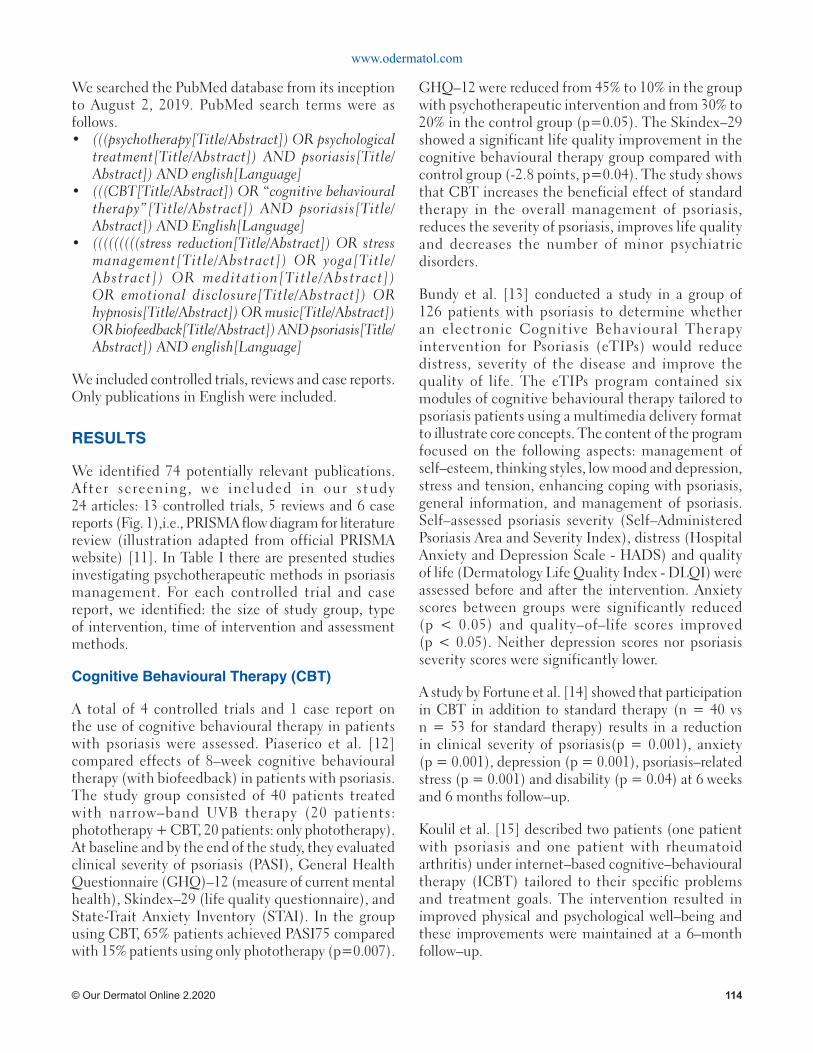
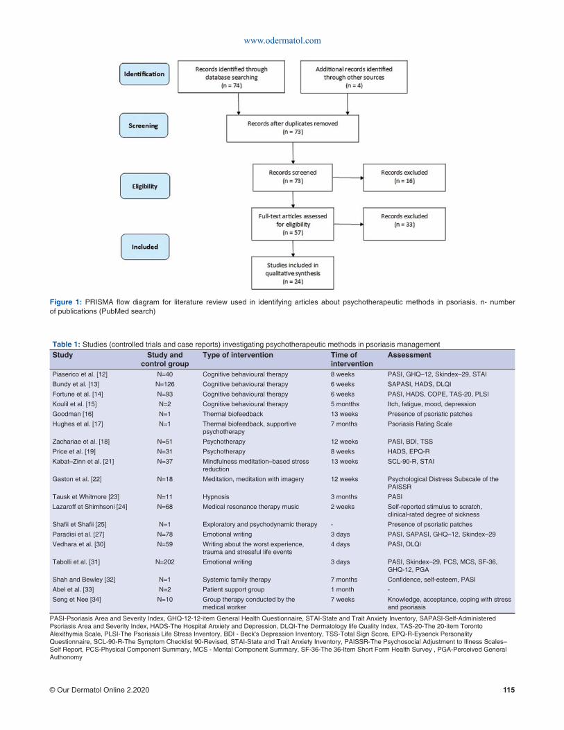
We identified 74 potentially relevant publications. After screening, we included in our study 24 articles: 13 controlled trials, 5 reviews and 6 case reports (Fig. 1),i.e., PRISMA flow diagram for literature review (illustration adapted from official PRISMA website) [11]. In Table I there are presented studies investigating psychotherapeutic methods in psoriasis management. For each controlled trial and case report, we identified: the size of study group, type of intervention, time of intervention and assessment methods.
Cognitive Behavioural Therapy (CBT)
A total of 4 controlled trials and 1 case report on the use of cognitive behavioural therapy in patients with psoriasis were assessed. Piaserico et al. [12] compared effects of 8–week cognitive behavioural therapy (with biofeedback) in patients with psoriasis. The study group consisted of 40 patients treated with narrow–band UVB therapy (20 patients: phototherapy + CBT, 20 patients: only phototherapy). At baseline and by the end of the study, they evaluated clinical severity of psoriasis (PASI), General Health Questionnaire (GHQ)–12 (measure of current mental health), Skindex–29 (life quality questionnaire), and State-Trait Anxiety Inventory (STAI). In the group using CBT, 65% patients achieved PASI75 compared with 15% patients using only phototherapy (p=0.007).
GHQ–12 were reduced from 45% to 10% in the group with psychotherapeutic intervention and from 30% to 20% in the control group (p=0.05). The Skindex–29 showed a significant life quality improvement in the cognitive behavioural therapy group compared with control group (-2.8 points, p=0.04). The study shows that CBT increases the beneficial effect of standard therapy in the overall management of psoriasis, reduces the severity of psoriasis, improves life quality and decreases the number of minor psychiatric disorders.
Bundy et al. [13] conducted a study in a group of 126 patients with psoriasis to determine whether an electronic Cognitive Behavioural Therapy intervention for Psoriasis (eTIPs) would reduce distress, severity of the disease and improve the quality of life. The eTIPs program contained six modules of cognitive behavioural therapy tailored to psoriasis patients using a multimedia delivery format to illustrate core concepts. The content of the program focused on the following aspects: management of self–esteem, thinking styles, low mood and depression, stress and tension, enhancing coping with psoriasis, general information, and management of psoriasis. Self–assessed psoriasis severity (Self–Administered Psoriasis Area and Severity Index), distress (Hospital Anxiety and Depression Scale - HADS) and quality of life (Dermatology Life Quality Index - DLQI) were assessed before and after the intervention. Anxiety scores between groups were significantly reduced (p < 0.05) and quality–of–life scores improved (p < 0.05). Neither depression scores nor psoriasis severity scores were significantly lower.
A study by Fortune et al. [14] showed that participation in CBT in addition to standard therapy (n = 40 vs n = 53 for standard therapy) results in a reduction in clinical severity of psoriasis(p = 0.001), anxiety (p = 0.001), depression (p = 0.001), psoriasis–related stress (p = 0.001) and disability (p = 0.04) at 6 weeks and 6 months follow–up.
Koulil et al. [15] described two patients (one patient with psoriasis and one patient with rheumatoid arthritis) under internet–based cognitive–behavioural therapy (ICBT) tailored to their specific problems and treatment goals. The intervention resulted in improved physical and psychological well–being and these improvements were maintained at a 6–month follow–up.
www.odermatol.com
© Our Dermatol Online 2.2020 115
Figure 1: PRISMA fl ow diagram for literature review used in identifying articles about psychotherapeutic methods in psoriasis. n- number of publications (PubMed search)
Table 1: Studies (controlled trials and case reports) investigating psychotherapeutic methods in psoriasis managementStudy Study and
control groupType of intervention Time of
interventionAssessment
Piaserico et al. [12] N=40 Cognitive behavioural therapy 8 weeks PASI, GHQ–12, Skindex–29, STAI
Bundy et al. [13] N=126 Cognitive behavioural therapy 6 weeks SAPASI, HADS, DLQI
Fortune et al. [14] N=93 Cognitive behavioural therapy 6 weeks PASI, HADS, COPE, TAS-20, PLSI
Koulil et al. [15] N=2 Cognitive behavioural therapy 5 montths Itch, fatigue, mood, depression
Goodman [16] N=1 Thermal biofeedback 13 weeks Presence of psoriatic patches
Hughes et al. [17] N=1 Thermal biofeedback, supportive psychotherapy
7 months Psoriasis Rating Scale
Zachariae et al. [18] N=51 Psychotherapy 12 weeks PASI, BDI, TSS
Price et al. [19] N=31 Psychotherapy 8 weeks HADS, EPQ-R
Kabat–Zinn et al. [21] N=37 Mindfulness meditation–based stress reduction
13 weeks SCL-90-R, STAI
Gaston et al. [22] N=18 Meditation, meditation with imagery 12 weeks Psychological Distress Subscale of the PAISSR
Tausk et Whitmore [23] N=11 Hypnosis 3 months PASI
Lazaroff et Shimhsoni [24] N=68 Medical resonance therapy music 2 weeks Self-reported stimulus to scratch, clinical-rated degree of sickness
Shafi i et Shafi i [25] N=1 Exploratory and psychodynamic therapy - Presence of psoriatic patches
Paradisi et al. [27] N=78 Emotional writing 3 days PASI, SAPASI, GHQ–12, Skindex –29
Vedhara et al. [30] N=59 Writing about the worst experience, trauma and stressful life events
4 days PASI, DLQI
Tabolli et al. [31] N=202 Emotional writing 3 days PASI, Skindex–29, PCS, MCS, SF-36, GHQ-12, PGA
Shah and Bewley [32] N=1 Systemic family therapy 7 months Confi dence, self-esteem, PASI
Abel et al. [33] N=2 Patient support group 1 month -
Seng et Nee [34] N=10 Group therapy conducted by the medical worker
7 weeks Knowledge, acceptance, coping with stress and psoriasis
PASI-Psoriasis Area and Severity Index, GHQ-12-12-item General Health Questionnaire, STAI-State and Trait Anxiety Inventory, SAPASI-Self-Administered Psoriasis Area and Severity Index, HADS-The Hospital Anxiety and Depression, DLQI-The Dermatology life Quality Index, TAS-20-The 20-item Toronto Alexithymia Scale, PLSI-The Psoriasis Life Stress Inventory, BDI - Beck's Depression Inventory, TSS-Total Sign Score, EPQ-R-Eysenck Personality Questionnaire, SCL-90-R-The Symptom Checklist 90-Revised, STAI-State and Trait Anxiety Inventory, PAISSR-The Psychosocial Adjustment to Illness Scales–Self Report, PCS-Physical Component Summary, MCS - Mental Component Summary, SF-36-The 36-Item Short Form Health Survey , PGA-Perceived General Authonomy
www.odermatol.com
© Our Dermatol Online 2.2020 116
Biofeedback
Goodman [16] described a case report of a 56-year-old Caucasian female who has failed standard medical treatment for psoriasis for seven years. Following 13 weekly one-hour finger/hand thermal biofeedback treatments, all 11 presenting psoriasis lesions (2-6 cm) had disappeared. Interestingly, patient was unmedicated for psoriasis during our treatment and continues to be unmedicated and asymptomatic at 12-month follow-up.
Hughes et al. [17] presented a case of a 31-year-old white male with multiple psoriatic plaques. It was resistant to previous dermatological treatments. During 7 months, each treatment session consisted of 20 minutes of skin temperature training at the target plaque site and following supportive psychotherapy. The photographs using the Psoriasis Rating Scale indicated marked improvement of the dermatological signs.
Relaxation and Meditation Techniques
Zachariae et al. [18] conducted a study in a group of patients (n = 51) with psoriasis. The treatment group participated in seven individual psychotherapy sessions in 12 weeks. Intervention techniques included stress management, guided imagery and relaxation. They observed slight but significant changes in Psoriasis Area Severity Index (PASI), Total Sign Score (TSS) and Laser Doppler Skin Blood Flow (LDBF) in the group of patients which attended psychotherapy sessions.
Price et al. [19] reported that patients with psoriasis are a noticeably anxious group compared to the general population. They conducted psychological meetings, in which the patients discussed among themselves problems caused by the disease. They were also taught specific relaxation techniques for use whenever they felt under stress. This reduced significantly the level of anxiety by the end of the study. Moreover, a modest trend towards physical improvement was also observed.
Bonadonna [20] highlighted that meditation is a good addition to conventional medical therapy in psoriasis. It reduces anxiety, pain, stress and enhances mood and self–esteem.
Kabat–Zinn et al. [21] conducted a study on 37 patients with psoriasis undergoing ultraviolet phototherapy (UVB) or photochemotherapy (PUVA). The study group took part in mindfulness meditation–based
stress reduction intervention guided by audio–taped instructions during light treatment. The control group had light treatment alone without the audio–taped instructions. Results showed that patients in the tape group reached the Halfway Point (p = 0.013) and the Clearing Point (p = 0.033) significantly more rapidly than those in the no–tape situation, for both UVB and PUVA treatments. The findings of this research indicating that relaxation and meditation techniques increase the resolution of psoriatic lesions in patients with psoriasis are consistent with previous studies.
Gaston et al. [22] conducted a study in a group of 18 patients with psoriasis symptoms on the scalp. They assigned patients to four groups meditation (n = 5), meditation and imagery (n = 4), waiting list (n = 5) and no treatment control group (n = 4). The intervention lasted 12 weeks, with 4 weeks pre– and post–baseline periods. They confirm that stress reduction techniques can be beneficial in patients with psoriasis.
Hypnosis
Tausk et Whitmore [23] performed a 3-month randomized controlled trial of the use of hypnosis in adults with psoriasis vulgaris. They used highly or moderately hypnotizable subjects. Patients received either hypnosis with active suggestions of improvement (5 patients) or neutral hypnosis with no mention of their disease process (6 patients). Results of the study suggest that hypnosis may be a useful therapeutic methods in psoriasis.
Medical Resonance Therapy Music
Lazaroff et Shimshoni [24] measured the parameters of blood pressure, heart rate, stimulus to scratch and the degree of sickness in the group of 68 patients in total (two experimental groups - psoriasis and neurodermatitis and two control groups). The experimental groups were additionally treated with 3 x 30 minutes of Medical Resonance Therapy Music per day. In the experimental groups was observed a reduction of blood pressure and heart rate, reduction of the stimulus to scratch and reduction in the degree of sickness. Interestingly the effects of therapy were stronger for the patients with psoriasis than for patients with neurodermatitis.
Exploratory and Psychodynamic Therapy
Shafii and Shafii [25] report that the techniques of developing a therapeutic alliance, therapeutic
www.odermatol.com
© Our Dermatol Online 2.2020 117
confrontation, clarification, dynamic interpretation, and exploration of intrapsychic and interpersonal conflicts, which are in accord with concepts of exploratory and psychodynamic therapy, can be beneficial in patients with psoriasis.
Emotional Writing (EW)
There are 3 controlled trials evaluating emotional writing in patients with psoriasis.
Nyssen et al. [26] summarized the results of these studies. The main intervention for all studies was the emotional writing including disease–focused writing, including worst experience, trauma and stressful life events. All interventions were delivered in 3 to 4 consecutive days periods for 20 minutes each day and by handwriting.
Paradisi et al. [27] conducted a study on a group of 40 patients with psoriasis undergoing ultraviolet B (UVB) therapy. Besides emotional writing (according to Pennebaker [28]), they assessed one other active intervention based on the emotional positive writing technique focused on the best possible future self and achieving life goals (according to King [29]), Disease severity (PASI and SAPASI scores), psychological distress (GHQ–12 scores) and quality of life (Skindex–29) were assessed at baseline, halfway through and at the end of UVB treatment and 4 months after emotional disclosure intervention. Significant differences in Skindex–29 values between emotional writing group and others were reported. Furthermore, patients allocated to the EW group had a longer period of remission after phototherapy.
Vedhara et al. [30] conducted a study in a group of 59 patients with plaque–type psoriasis involving more 10% of the body area (mean age: 50 years, 32 men and 27 women, mean length of diagnosis: 22 years). Disease severity and quality of life improved in both groups over the follow–up period (at baseline and at 2, 8 and 12 weeks post–intervention).
Tabolli et al. [31] tested the efficacy of Pennebaker’s emotional writing intervention in 67 patients with psoriasis treated with systemic therapy. Total follow–up period for each individual was equal to 12 months. The intervention had little or no effect on the severity of the disease (psoriasis area severity index, Physician Global Assessment Score), as well as generic and dermatology–specific quality life questionnaires.
Systemic Family Therapy
Shah and Bewley [32] underlined that cognitive behavioural therapy (CBT) is not appropriate for everyone. They described a case report showing benefits of a psychological intervention using the principles of systemic family therapy (SFT). The key is the understanding of problems in the context of family and social relationships, and how reciprocal dynamics influence problems. Authors report that problems do not exist only within individuals, however, they are the product of the interactions between people and wider systems, e.g.,communities.
Support Groups
Abel et al. [33] described an experience of patient support group at Stanford, led by a psychiatrist, which is an integral part of the Psoriasis Day Care program. Common discussions topics include lifestyle changes, stressful relationships, associated emotional reactions, occupational limitations and treatment concerns. Psycho–social support systems, stress reduction and enhanced coping skills acquired through shared experiences enhance treatment response.
Seng et Nee [34] used a structured program in a group of 10 patients with psoriasis. The program covered knowledge of psoriasis, feelings of acceptance, stress management, and coping with daily living. Most patients found that the program helped them to cope better with the disease.
Moreover, there were two reviews in which there are described methods of psychological interventions in psoriasis based on literature. Winchell et al. [35] mention about relaxation, hypnosis and biofeedback. Qureshi et al. [36] show numerous methods which could be useful in patients with psoriasis: cognitive behavioural therapy, mindfulness-based therapies, motivational interviewing, educational and interdisciplinary interventions.
DISCUSSION
Despite an extensive review of literature, the number of published articles concerning psychotherapeutic interventions in patients with psoriasis turned out to be rather limited. What is more, issues concerning diagnostic strategies or small study groups in certain works are beyond the scope of this review.
www.odermatol.com
© Our Dermatol Online 2.2020 118
We i d e n t i f y a s p r o m i s i n g t h e f o l l o w i n g psychotherapeutic intervention methods, which can be used in addition to standard therapy in psoriasis patients: cognitive behavioural therapy, biofeedback, psychotherapy, meditation, hypnosis, music therapy, exploratory and psychodynamic therapy, emotional writing, systemic family therapy, and support groups. Thanks to the application of these techniques, one can observe beneficial effects such as reduced severity of psoriasis, improved life quality and decreased incidence of minor psychiatric disorders. For this reason, it can be concluded that patients with frequent exacerbations of psoriasis should use both standard and psychotherapeutic treatment. That seems a good idea that future management of psoriasis should involve multidisciplinary teams that help patients to manage the physical and psychological aspects of psoriasis [37]. More studies are however required to show how these approaches could be used in clinical practice owing to, i.a., (i) the limited number of published works, (ii) small study group sizes, (iii) a lack of comparisons between various methods, (iv) a lack of formulated guidelines.
REFERENCES
1. Parisi R, Symmons DP, Griffiths CE, Ashcroft DM. Global epidemiology of psoriasis: a systematic review of incidence and prevalence. J Invest Dermatol. 2013;133:377-85.
2. Albanesi C, Madonna S, Gisondi P, Girolomoni G. The interplay between keratinocytes and immune cells in the pathogenesis of psoriasis. Front Immunol. 2018;9:1549.
3. Gaikwad R, Deshpande S, Raje S, Dhamdhere DV, Ghate MR. Evaluation of functional impairment in psoriasis. Indian J Dermatol Venereol Leprol. 2006;72:37-40.
4. Heller MM, Lee ES, Koo JY. Stress as an infl uencing factor in psoriasis. Skin Therapy Lett. 2011;16:1-4.
5. Rousset L, Halioua B. Stress and psoriasis. Int J Dermatol. 2018;57:1165-72.
6. Hunter HJ, Griffi ths CE, Kleyn CE. Does psychosocial stress play a role in the exacerbation of psoriasis? Br J Dermatol. 2013;169:965-74.
7. Malhotra SK, Mehta V. Role of stressful life events in induction or exacerbation of psoriasis and chronic urticaria. Indian J Dermatol Venereol Leprol. 2008;74:594-9.
8. Lakuta P, Marcinkiewicz K, Bergler-Czop B, Brzezinska-Wcislo L, Slomian A. Associations between site of skin lesions and depression, social anxiety, body-related emotions and feelings of stigmatization in psoriasis patients. Adv Dermatol Allergol. 2018;35:60-6.
9. Shenefelt PD. Mindfulness-Based Cognitive Hypnotherapy and Skin Disorders. Am J Clin Hypn. 2018;61:34-44.
10. Shenefelt PD. Use of hypnosis, meditation, and biofeedback in dermatology. Clin Dermatol. 2017;35:285-91.
11. Moher D, Liberati A, Tetzlaff J, Altman DG. Preferred reporting items for systematic reviews and meta-analyses: the PRISMA statement. PLoS Med. 2009;6:e1000097.
12. Piaserico S, Marinello E, Dessi A, Linder MD, Coccarielli D, Peserico A. Effi cacy of biofeedback and cognitive-behavioural
therapy in psoriatic patients a single-blind, randomized and controlled study with added narrow-band ultraviolet b therapy. Acta Derm Venereol. 2016;96:91-5.
13. Bundy C, Pinder B, Bucci S, Reeves D, Griffi ths CE, Tarrier N. A novel, web-based, psychological intervention for people with psoriasis: the electronic Targeted Intervention for Psoriasis (eTIPs) study. Br J Dermatol. 2013;169:329-36.
14. Fortune DG, Richards HL, Kirby B, Bowcock S, Main CJ, Griffi ths CE. A cognitive-behavioural symptom management programme as an adjunct in psoriasis therapy. Br J Dermatol. 2002;146:458-65.
15. Koulil SS, Ferwerda M, van Beugen S, van Middendorp H, van de Kerkhof PCM, van Riel PLCM, et al. Tailored therapist-guided internet-based cognitive-behavioural treatment for psoriasis and rheumatoid arthritis: two case reports. Acta Derm Venereol. 2018;98:225-33.
16. Goodman M. An hypothesis explaining the successful treatment of psoriasis with thermal biofeedback: a case report. Biofeedback Self Regul. 1994;19:347-52.
17. Hughes HH, England R, Goldsmith DA. Biofeedback and psychotherapeutic treatment of psoriasis: a brief report. Psychol Rep. 1981;48:99-102.
18. Zachariae R, Oster H, Bjerring P, Kragballe K. Effects of psychologic intervention on psoriasis: a preliminary report. J Am Acad Dermatol. 1996;34:1008-15.
19. Price ML, Mottahedin I, Mayo PR. Can psychotherapy help patients with psoriasis? Clin Exp Dermatol. 1991;16:114-7.
20. Bonadonna R. Meditation’s impact on chronic illness. Holist Nurs Pract. 2003;17:309-19.
21. Kabat-Zinn J, Wheeler E, Light T, Skillings A, Scharf MJ, Cropley TG, et al. Infl uence of a mindfulness meditation-based stress reduction intervention on rates of skin clearing in patients with moderate to severe psoriasis undergoing phototherapy (UVB) and photochemotherapy (PUVA). Psychosom Med. 1998;60:625-32.
22. Gaston L, Crombez JC, Lassonde M, Bernier-Buzzanga J, Hodgins S. Psychological stress and psoriasis: experimental and prospective correlational studies. Acta Derm Venereol Suppl (Stockh). 1991;156:37-43.
23. Tausk F, Whitmore SE. A pilot study of hypnosis in the treatment of patients with psoriasis. Psychother Psychosom. 1999;68:221-5.
24. Lazaroff I, Shimshoni R. Effects of Medical Resonance Therapy Music on patients with psoriasis and neurodermatitis-a pilot study. Integr Physiol Behav Sci. 2000;35:189-98.
25. Shafi i M, Shafi i SL. Exploratory psychotherapy in the treatment of psoriasis. Twelve hundred years ago. Arch Gen Psychiatry. 1979;36:1242-5.
26. Nyssen OP, Taylor SJ, Wong G, Steed E, Bourke L, Lord J, et al. Does therapeutic writing help people with long-term conditions? Systematic review, realist synthesis and economic considerations. Health Technol Assess. 2016;20:1-367.
27. Paradisi A, Abeni D, Finore E, Di Pietro C, Sampogna F, Mazzanti C, et al. Effect of written emotional disclosure interventions in persons with psoriasis undergoing narrow band ultraviolet B phototherapy. Eur J Dermatol. 2010;20:599-605.
28. Pennebaker JW. Writing about emotional experiences as a therapeutic process. Psychological Science. 1997;8:162–6.
29. King LA. The health benefi ts of writing about life goals. Personal Social Psychol Bull. 2001;27:798–807.
30. Vedhara K, Morris RM, Booth R, Horgan M, Lawrence M, Birchall N. Changes in mood predict disease activity and quality of life in patients with psoriasis following emotional disclosure. J Psychosom Res. 2007;62:611-9.
31. Tabolli S, Naldi L, Pagliarello C, Sampogna F, di Pietro C, Spagnoli A, et al. Evaluation of the impact of writing exercises interventions on quality of life in patients with psoriasis undergoing systemic treatments. Br J Dermatol. 2012;167:1254-64.
32. Shah R, Bewley A. Psoriasis: ‘the badge of shame’. A case report of

www.odermatol.com
© Our Dermatol Online 2.2020 119
a psychological intervention to reduce and potentially clear chronic skin disease. Clin Exp Dermatol. 2014;39:600-3.
33. Abel EA, Moore US, Glathe JP. Psoriasis patient support group and self-care effi cacy as an adjunct to day care center treatment. Int J Dermatol. 1990;29:640-3.
34. Seng TK, Nee TS. Group therapy: a useful and supportive treatment for psoriasis patients. Int J Dermatol. 1997;36:110-2.
35. Winchell SA, Watts RA. Relaxation therapies in the treatment of psoriasis and possible pathophysiologic mechanisms. J Am Acad Dermatol. 1988;18:101-4.
36. Qureshi AA, Awosika O, Baruffi F, Rengifo-Pardo M, Ehrlich A. Psychological therapies in management of psoriatic skin disease: a
systematic review. Am J Clin Dermatol. 2019;20:607-24.37. Liluashvili S, Kituashvili T. Dermatology Life Quality Index and
disease coping strategies in psoriasis patients. Adv Dermatol Allergol. 2019;XXXVI:419-24.
Copyright by Aneta Gruchała, et al. This is an open-access article distributed under the terms of the Creative Commons Attribution License, which permits unrestricted use, distribution, and reproduction in any medium, provided the original author and source are credited.Source of Support: Nil, Confl ict of Interest: None declared.
Our Dermatology Online
© Our Dermatol Online 2.2020 120
INTRODUCTION
Atopic dermatitis (AD) is a complex inflammatory cutaneous disorder characterized by immune-mediated inflammation and epidermal barrier dysfunction [1]. AD has a complex etiology, it develops in early childhood and has age dependent distribution [2]. AD affects approximately 20% of children and up to 3% of adults, data shows that it’s prevalence is still increasing [3]. Most of the AD end up in remission, small number of childhood cases, especially severe ones persist into adulthood [4]. Studies show, that AD predispose to a higher risk of atopic and other than atopy comorbidities [5,6].
AD is heterogeneous in its pathophysiological pathways. Studies demonstrate that different T-helper subsets and cytokines drive pathological cutaneous
inflammation [7]. Children with AD have a dominant Th2 activation and expansion [8]. Determining the correct cellular subset could have a potential in not only providing a better understanding in AD pathophysiology, but also be a potential diagnostic and prognostic tool [9].
Interleukin-33 (IL-33) is a member of interleukin-1 family cytokines, it is found in the nuclei of various tissue and immune cells in human body [10]. IL-33 is highly expressed in barrier tissues, which are exposed to the environment (e.g., skin, gut, lungs), those tissues are considered to be the major sources of IL-33 in human body [11]. IL-33 extracellularly can be detected following cellular damage or stress due to infection, allergen exposure [12]. IL-33 expression can be induced by inflammation, environmental triggers [13]. IL-33
How to cite this article: Paulauskaitė I, Eidukaitė A, Rudzevičienė O, Orentaitė R. Investigating the role of Interleukin-33 and Soluble ST2 in pediatric atopic dermatitis. Our Dermatol Online. 2020;11(2):120-125.Submission: 14.10.2019; Acceptance: 15.12.2019DOI: 10.7241/ourd.20202.2
Original Article
Investigating the role of Interleukin-33 and Soluble Investigating the role of Interleukin-33 and Soluble ST2 in pediatric atopic dermatitisST2 in pediatric atopic dermatitisIlona Paulauskaitė1, Audronė Eidukaitė1,2, Odilija Rudzevičienė2, Rasa Orentaitė2
1Immunology Department, Centre for Innovative Medicine, Santariškių str. 5, LT-08406, Vilnius, Lithuania, 2Children‘s Hospital, Affi liate of Vilnius University Hospital Santaros Klinikos, Santariškių str. 7, LT-08406, Vilnius, Lithuania
Corresponding author: Dr. Ilona Paulauskaitė, E-mail: [email protected]
ABSTRACT
Background: Atopic dermatitis (AD) is a chronic, inflammatory skin disease, common in children. Pathologic cutaneous inflammation is driven by activated T-helper cells. Studies demonstrate that childhood AD is associated with Th2 immune activation. IL-33 is an intracellular cytokine, abundantly expressed in tissues, which are exposed to the environment. Cellular damage, due to scratching, encounter to infectious pathogens or exposure to allergens, trigger the release of IL-33. Extracellularly IL-33 acts as an activator for Th2 lymphocytes. Soluble ST2 (sST2) is a decoy receptor for IL-33. Combined to sST2, IL-33 loses its biological functions, which results in the alleviation of Th2 immune response. With this study we wanted to investigate the role of IL-33 and sST2 in pediatric AD. Material and Methods: Blood and stool samples from children with AD and healthy controls were tested for IL-33 and sST2 concentrations. Results: Children with AD presented significantly higher blood IL-33 concentrations, compared to healthy controls: 18,21 pg/ml vs 0, p<0,05. Stool IL-33 levels demonstrated no significant difference between the two groups: 12,43 pg/ml vs 45,94 pg/ml, p>0,05. Blood and stool sST2 concentrations showed no significant difference: 67,58 pg/ml vs 74,96 pg/ml, p>0,05; 0 vs 0, p>0,05, respectively. Conclusion: IL-33 is associated with pediatric AD. Blood, but not stool IL-33 testing can be used as a biomarker. sST2 showed no difference in AD.
Key words: Atopic Dermatitis; Interleukin-33; Soluble ST2 Receptor
www.odermatol.com
© Our Dermatol Online 2.2020 121
activates Th2 lymphocytes, promotes IL-4, IL-5, IL-13 cytokine secretion, acts as a chemoattractant for Th2 lymphocytes [14,15]. Soluble serum stimulation 2 receptor (sST2) is an alternative splice variant of the gene, encoding ST2. Combined to sST2 IL-33 loses its biological functions, which results in the alleviation of Th2 immune response [16].
Data, concerning pediatric AD and prospective biomarkers is lacking. With this study we wanted to evaluate the role of IL-33/sST2 axis in pediatric AD.
MATERIALS AND METHODS
Study Population and Ethical Considerations
A total of 68 children were invited to take part in the study. Study population comprised of 51 participants with clinically proved atopic dermatitis, hospitalized in Children’s Pulmonology and Allergology Department and 17 healthy control subjects, who had no history of atopic diseases, nor any current inflammatory diseases.
Sample Collection and Laboratory Analysis
Venous blood and stool samples were obtained from test and control subjects. All the samples were collected as part of the routine clinical practice. Automated blood test, allergen specific IgE tests were performed at once. Complete blood count was obtained from venous blood using automated hematology analyzer (Sysmex XT 4000i, Roche, Germany). Allergen specific IgE tests were performed using Phadia Immunocap 100 analyzer (Phadia, Uppsala, Sweden). The rest of the samples were stored at -80°C for further IL-33 and sST2 testing.
Measurement of Serum and Fecal IL-33, sST2
Frozen stool and serum samples were completely defrosted prior testing. Suspensions were prepared from stool samples: 0,1 g stool was suspended in 1 ml phosphate saline buffer (PBS, pH=7,2). Suspensions were thoroughly vortexed, left to sit at room temperature for 15 min, then once again vortexed and centrifuged (10000 x g, 20 min). Supernatants were used for the test procedure. Supernatants and serum samples were tested for IL-33 using Human IL-33 ELISA kit (Elabscience, China), sST2 - Human IL-1 R4 (IL1RL1) ELISA kit (Thermo Fisher Scientific, USA). Assay procedures were performed according to manufacturer’s recommendations.
Data Management and Statistical Analysis
MS Office Excel, MedCalc software were used for data management and statistical analysis. Nonparametric data were expressed with median and range. Mann-Whitney U test was used for compared two groups of variables. Categorical data were expressed with a number and percentage, difference was determined using Chi-Square test. Difference between the groups was considered significant when p<0,05.
Ethics Statement
Participants parents or legal guardians provided their agreement in participating in the study by signing a written informed consent form. Ethics approval for the research study was obtained (No. 158200-16-834-352).
RESULTS
Baseline Characteristics
Test and control subjects were a match according to age and gender (Table 1). Total leukocyte count, absolute neutrophil and eosinophil counts in venous blood were measured. Test subjects were tested for food specific IgE. 43% (n=22) had non detectable food specific IgE, 57% (n=29) were sensitized to food (food specific IgE ≥ 0,35 kUA/l). Sensitization was mostly detected to cow‘s milk and hen‘s egg. Detailed information is provided in Table 1.
IL-33 and sST2 Concentrations
Children with AD had higher blood IL-33 levels (median: 18,21 pg/ml, range: 0 – 392,89 pg/ml),
Table 1: Patient characteristics Characteristics AD patients
n=51
Control subjects
n=17
p value
Age (mo)(range)
20,0(5–77)
28,0(4–80)
0,137*
Gender (male/female) (%)
36/1571%/29%
12/571%/29%
0,827§
Sensitisation to food allergensnon sensitized/sensitized (%)
25/2649%/51%
- -
Laboratory analysisWBC (x 109/l)(range)ANC (cells/μl)(range)Blood eosinophils (%)(range)
10,93(6,45–13,66)
5,27(1,81–10,07)
3,6
(0,3–10,2)
8,18(5,6–16,59)
2,41(1,51–6,87)
0,24
(0,11–0,53)
0,024*
0,0466*
0,0001*
*- Mann-Whitney test for independent samples; § - Chi-square test; WBC – white blood cell count; ANC – absolute neutrophil count
www.odermatol.com
© Our Dermatol Online 2.2020 122
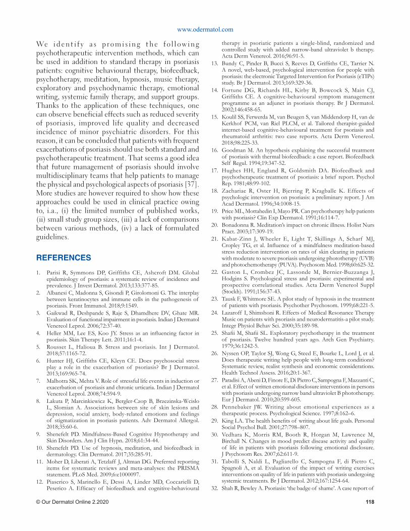
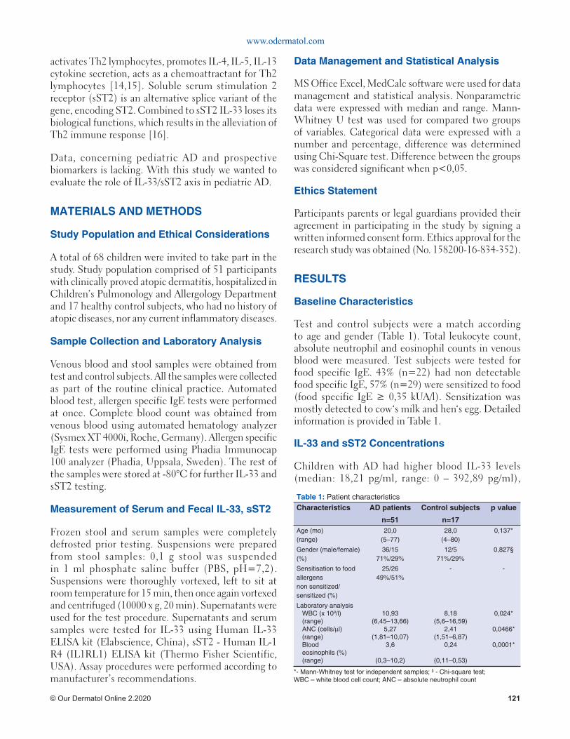
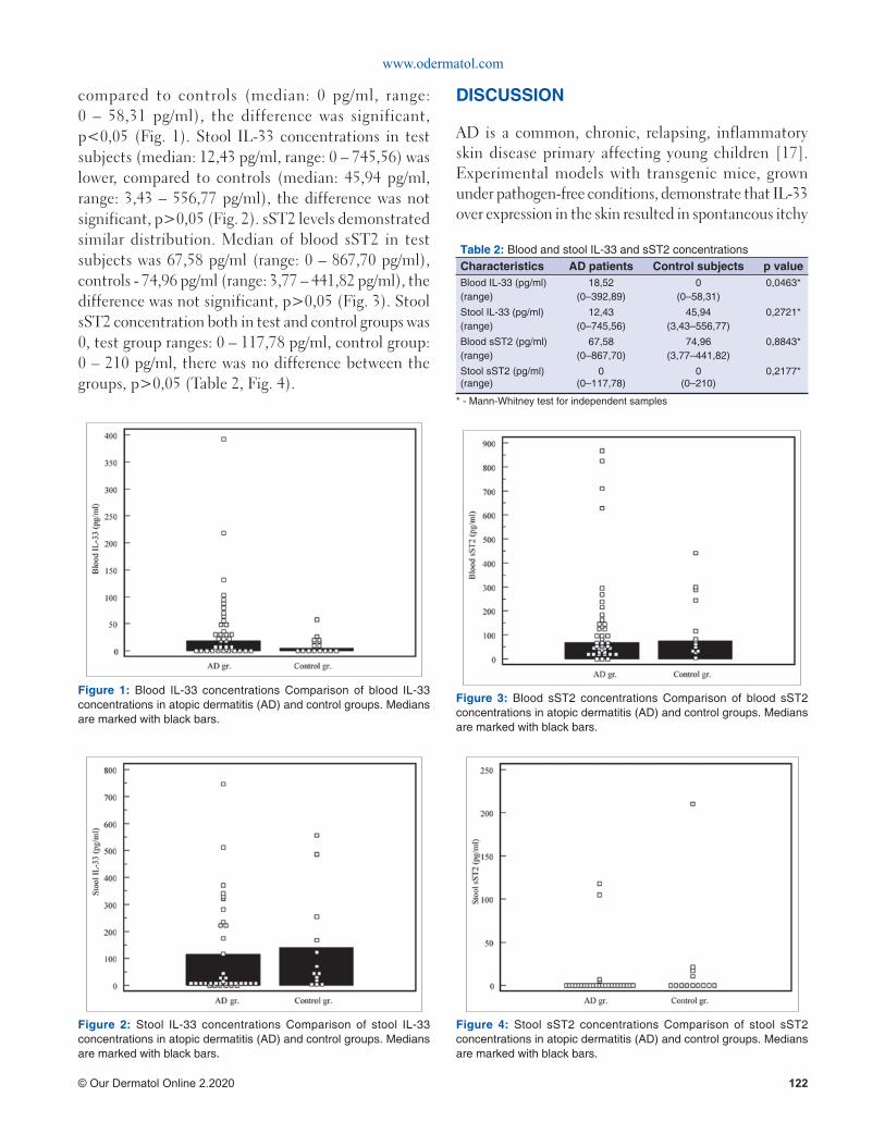
compared to controls (median: 0 pg/ml, range: 0 – 58,31 pg/ml), the difference was significant, p<0,05 (Fig. 1). Stool IL-33 concentrations in test subjects (median: 12,43 pg/ml, range: 0 – 745,56) was lower, compared to controls (median: 45,94 pg/ml, range: 3,43 – 556,77 pg/ml), the difference was not significant, p>0,05 (Fig. 2). sST2 levels demonstrated similar distribution. Median of blood sST2 in test subjects was 67,58 pg/ml (range: 0 – 867,70 pg/ml), controls - 74,96 pg/ml (range: 3,77 – 441,82 pg/ml), the difference was not significant, p>0,05 (Fig. 3). Stool sST2 concentration both in test and control groups was 0, test group ranges: 0 – 117,78 pg/ml, control group: 0 – 210 pg/ml, there was no difference between the groups, p>0,05 (Table 2, Fig. 4).
DISCUSSION
AD is a common, chronic, relapsing, inflammatory skin disease primary affecting young children [17]. Experimental models with transgenic mice, grown under pathogen-free conditions, demonstrate that IL-33 over expression in the skin resulted in spontaneous itchy
Figure 1: Blood IL-33 concentrations Comparison of blood IL-33 concentrations in atopic dermatitis (AD) and control groups. Medians are marked with black bars.
Figure 2: Stool IL-33 concentrations Comparison of stool IL-33 concentrations in atopic dermatitis (AD) and control groups. Medians are marked with black bars.
Figure 3: Blood sST2 concentrations Comparison of blood sST2 concentrations in atopic dermatitis (AD) and control groups. Medians are marked with black bars.
Figure 4: Stool sST2 concentrations Comparison of stool sST2 concentrations in atopic dermatitis (AD) and control groups. Medians are marked with black bars.
Table 2: Blood and stool IL-33 and sST2 concentrationsCharacteristics AD patients Control subjects p valueBlood IL-33 (pg/ml)(range)
18,52(0–392,89)
0(0–58,31)
0,0463*
Stool IL-33 (pg/ml)(range)
12,43(0–745,56)
45,94(3,43–556,77)
0,2721*
Blood sST2 (pg/ml)(range)
67,58(0–867,70)
74,96(3,77–441,82)
0,8843*
Stool sST2 (pg/ml)(range)
0(0–117,78)
0(0–210)
0,2177*
* - Mann-Whitney test for independent samples
www.odermatol.com
© Our Dermatol Online 2.2020 123
dermatitis [18]. Human studies also demonstrate, that IL-33 expression is significantly increased in the lesional skin [19]. IL-33 signaling is crucial in the development of experimental models of AD [20]. Mechanical damage, due to scratching, infection, exposure to allergens, triggers the release of IL-33 [21]. C. Galand et al. in his experimental murine model demonstrates that serum IL-33 levels were significantly increased after a tape stripping experiment [22]. In our study children with AD had significantly higher serum IL-33 concentration compared to controls (18,52 pg/ml vs 0 pg/ml, p<0,05). R. Tanagawa-Mineoka et al. reports, that IL-33 was significantly higher in patients with AD compared to patients with chronic idiopathic urticaria, psoriasis and healthy controls [23]. AD is a multifactorial disease, its main pathogenetic factor is immune mediated inflammation. IL-33 is associated with Th2 immune activation. According to T. Czarnowicki et al. children with AD had a markedly expanded Th2 type lymphocyte population in their blood [8]. Other studies also present data that AD is associated with an expansion and activation of Th2 lymphocytes in peripheral blood [24]. U. Nyagaard et al. demonstrates, that serum levels of IL-33 and sST2 were elevated in adults and children with AD compared to healthy controls [25]. The researchers also found that serum IL-33 levels were much higher in children compared to adults with AD. On the contrary, adults with AD presented with significantly higher sST2 values compared to children with AD. This could mean that there might be an age dependent distribution of IL-33 and sST2. Although available data are missing, this should be taken in consideration comparing studies with children and adults.
IL-33 biological functions manifest through its receptor - ST2. It exists in two main isoforms: a membrane bound (ST2L) and soluble (sST2) [26]. Small intestine, heart, kidney, lung tissues display the highest expression and are considered to be the main sources of sST2 in human body [27]. Immune cells normally do not secrete sST2, but under certain conditions they can also be a significant source of sST2 [28]. We did not detect any differences in blood sST2 concentrations compared AD patients to controls: 67,57 pg/ml vs 78,58 pg/ml, p>0,05. Relationship between IL-33 and sST2 in is still under investigation. Studies demonstrate that both IL-33 and sST2 were elevated in asthmatic children [29]. There are not many data, concerning AD. U. According to Nygaard et al. increased IL-33, but not sST2 levels, were associated with AD. sST2 did not correlate with disease
activity [25]. According to literature, inflammatory conditions activate sST2 synthesis and secretion in tissue cells. D. Diaz-Jimenez et al. states, that increased sST2 concentration reflected inflammatory activity in patients with ulcerative colitis [30]. It is not clear, whether sST2 provides negative regulation for the exacerbation of IL-33 biological functions in the pathogenesis of AD and what could influence those processes. P. E. Pfeffer et al. investigated the effect of vitamin D on IL-33/sST2 axis in experimental cell model. According to the researchers, vitamin D selectively upregulated sST2 expression and impeded IL-33 biological functions [31]. Although, vitamin D affects multiple systems, evidence suggests that it has a beneficial effect on AD course [32].
Recently, there has been a growing interest in gut microbiome and its possible immunomodulatory effect on systemic disorders [33]. Evidence supporting the gut-skin axis is inconclusive, but there are studies demonstrating that alterations in microbiome might be a triggering signal for the dysregulation of the immune response [34]. As well as epithelial cells in the skin, gastrointestinal tract enterocytes can also be a significant source of IL-33 [35]. Exposure of the intestinal epithelial cells to food allergens increases IL-33 expression [36]. Hoewer, there are still questions remaining about its active secretion. Half of our test subjects were sensitized to food. Stool testing for IL-33 and sST2 could reflect the immune reactions ongoing in the gut mucosa. It has a potential as a noninvasive method. However, we found no significant difference in stool IL-33 concentrations compared patients with AD and healthy controls: 14,11 pg/ml vs 57,83 pg/ml, p>0,05. Stool sST2 levels also demonstrated no difference between the groups. Literature provides no data concerning stool testing for IL-33 and sST2. According to J. Penders et al. study, intestinal microbiota plays an important role in the development of AD in early childhood [37]. An experimental murine model demonstrates that germ-free mice fail to develop oral tolerance, presence of gut microbiota have a protective role against AD formation [38,39]. According to our study, stool testing did not reveal any activation of IL-33/sST2 axis in AD patients, further testing is needed.
CONCLUSION
In conclusion: the increase in IL-33 concentration is associated with pediatric AD. Blood, but not stool IL-33
www.odermatol.com
© Our Dermatol Online 2.2020 124
testing can be used as a biomarker. sST2 showed no difference in pediatric AD, further investigations are needed.
Statement of Human and Animal Rights
All procedures followed were in accordance with the ethical standards of the responsible committee on human experimentation (institutional and national) and with the Helsinki Declaration of 1975, as revised in 2008.
Statement of Informed Consent
Informed consent was obtained from all patients for being included in the study.
REFERENCES
1. Guttman-Yassky E, Waldman A, Ahluwalia J, Ong PY, Eichenfi eld LF. Atopic dermatitis: pathogenesis. Semin Cutan Med Surg. 2017;36:100-3.
2. Lyons JJ, Milner JD, Stone KD. Atopic dermatitis in children: clinical features, pathophysiology and treatment. Immunol Allergy Clin North Am. 2015;35:161-83.
3. Flohr C, Mann J. New insights into epidemiology of childhood atopic dermatitis. Allergy. 2014;69:3-16.
4. Kim JP, Chao LX, Simpson EL, Silverberg JI. Persistence of atopic dermatitis (AD): A systematic review and meta-analysis. J Am Acad Dermatol. 2016;75:681-7.
5. Binyamin ST, Algamal F, Yamani AN, Labani MS, Alaqbi FH, Baeshen AA, et al. Prevalence and determinants of eczema among females aged 21 to 32 years in Jeddah city - Saudi Arabia. Our Dermatol Online. 2017;8:22-6.
6. Lenga Loumingou IA, Loumingou R. Meaning of microalbuminuria during the atopic dermatitis of the child. Our Dermatol Online. 2019;10:251-4.
7. Wang A, Landen NX. New insights into T cells and their signature cytokines in atopic dermatitis. IUBMB Life. 2015;67:601-10.
8. Czarnowicki T, Esaki H, Gonzalez J, Malajian D, Shemer A, Noda S, et al. Early pediatric atopic dermatitis shows only a CLA+ Th2/Th1 imbalance, while adults acquire CLA+ Th22/Tc22 subsets. J Allergy Clin Immunol. 2015;136:941-51.
9. Ungar B, Garcet S, Gonzalez J, Dhingra N, Correa da Rosa J, Shemer A, et al. An integrated model of atopic dermatitis biomarkers highlights the systemic nature of the disease. J Invest Dermatol. 2017;137:603-13.
10. Carrasco TG, Morales RA, Pérez F, Terraza C, Yáñez L, Campos-Mora M, et al. Alarmin’ immunologists: IL-33 as a putative target for modulating T cell-dependent responses. Front Immunol. 2015;6:1-8.
11. Liew FY, Girard JP, Turnquist HR. Interleukin-33 in health and disease. Nat Rev Immunol. 2016;16:676-89.
12. Cayrol C, Girard JP. IL-33: An alarmin cytokine with crucial roles in innate immunity, infl ammation and allergy. Curr Opin Immunol. 2014;31:31-7.
13. Gautier V, Cayrol C, Farache D, Roga S, Monsarrat B, Gonzalez De Peredo A, et al. Extracellular IL-33 cytokine, but not endogenous nuclear IL-33, regulates protein expression in endothelial cells. Sci Rep. 2016;6:1-12.
14. Murakami-Satsutani N, Ito T, Nakanishi T, Inagaki N, Tanaka A,
Vien PT, et al. IL-33 promotes the induction and maintenance of Th2 immune responses by enhancing the function of OX40 ligand. Allergol Int. 2014;63:443-55.
15. Komai-Koma M, Xu D, Li Y, McKenzie A, McInnes IB, Liew FY, et al. IL-33 is a chemoattractant for human Th2 cells. Eur J Immunol. 2007;37:2779-86.
16. Griesenauer B, Paczesny S. The ST2/IL-33 axis in immune cells during infl ammatory diseases. Front Immunol. 2017;8:1-17.
17. Thomsen SF. Atopic dermatitis: natural history, diagnosis, and treatment. ISRN Allergy. 2014;2014:1-7.
18. Imai Y, Yasuda K, Sakaguchi Y, Haneda T, Mizutami H, Yoshimoto T, et al. Skin-specifi c expression of IL-33 activates group 2 innate lymphoid cells and elicits atopic dermatitis-like infl ammation in mice. Proc Natl Acad Sci U.S.A. 2013;110:13921-6.
19. Abdel Hay RM, Ibrahim NH, Metwally D, Rashed LA. The role of interleukin-1ß and interleukin-33 in atopic dermatitis. Our Dermatol Online. 2013;4:11-4.
20. Li C, Maillet I, Mackowiak C, Viala C, Padova F, Li M, et al. Experimental atopic dermatitis depends on IL-33R signaling via MyD88 in dendritic cells. Cell Death Dis. 2017;8:1-11.
21. Savinko T, Matikainen S, Saarialho-Kere U, Lehto M, Wang G, Lehtimäki S, et al. IL-33 and ST2 in atopic dermatitis: Expression profi les and modulation by triggering factors. J Investig Dermatol. 2012;132:1392-400.
22. Galand C, Leyva-Castillo JM, Yoon J, Han A, Lee MS, McKenzie ANJ, et al. IL-33 promotes food anaphylaxis in epicutaneously sensitized mice by targeting mast cells. J Allergy Clin Immunol. 2016;138:1356-66.
23. Tamagawa-Mineoka R, Okuzawa Y, Masuda K, Katoh N. Increased serum levels of interleukin 33 in patients with atopic dermatitis. J Am Acad Dermatol. 2014;70:882-8.
24. Czarnowicki T, Gonzalez J, Shemer A, Malajian D, Xu H, Zhenh X, et al. Severe atopic dermatitis is characterized by selective expansion of circulating TH2/TC2 and TH22/TC22, but not TH17/TC17, cells within the skin-homing T-cell population. J Allergy Clin Immunol. 2015;136:104-15.
25. Nygaard U, Hvid M, Johansen C, Buchner M, Fölster-Holst M, Deleuran M, et al. TSLP, IL-31, IL-33 and sST2 are new biomarkers in endophenotypic profiling of adult and childhood atopic dermatitis. J Eur Acad Dermatol Venereol. 2016;30:1930-8.
26. Zhao Q, Chen G. Role of IL-33 and its receptor in T cell-mediated autoimmune diseases. Biomed Res Int. 2014;2014:1-10.
27. Mildner M, Storka A, Lichtenauer M, Mlitz V, Ghannadan M, Hoetzenecker K, et al. Primary sources and immunological prerequisites for sST2 secretion in humans. Cardiovasc Res. 2010;87:769-77.
28. Zhang J, Ramada AM, Griesenauer B, Li W, Turner MJ, Liu C, et al. ST2 blockade reduces sST2-producing T cells while maintaining protective mST2-expressing T cells during graft-versus-host disease. Sci Transl Med. 2015;7:1-27.
29. Chu MA, Lee HJ, Lee EJ, Hong SJ, Park HJ, Lee KH, et al. Increased serum soluble ST2 in asthmatic children and recurrent early wheezers. Allergy Asthma Respir Dis. 2013;1:314-20.
30. Díaz-Jiménez D, De la Fuente M, Dubois-Camacho K, Landskron G, Fuentes J, Pérez T, et al. Soluble ST2 is a sensitive clinical marker of ulcerative colitis evolution. BMC Gastroenterol. 2016;16:1-10.
31. Pfeffer PE, Chen YH, Woszczek G, Matthews NC, Chevretton E, Gupta A, et al. Vitamin D enhances production of soluble ST2, inhibiting the action of IL-33. J Allergy Clin Immunol. 2015;135:824-7.
32. Filippo PD, Scaparrotta A, Rapino D, Cingolani A, Attanasi M, Petrosino MI, et al. Vitamin D supplementation modulates the immune system and improves atopic dermatitis in children. J Allergy Clin Immunol. 2015;166:91-6.
33. Lee SY, Lee E, Park YM, Hong SJ. Microbiome in the gut-skin axis in atopic dermatitis. Allergy Asthma Immunol Res. 2018;10:354-62.
www.odermatol.com
© Our Dermatol Online 2.2020 125
34. Vaughn AR, Notay M, Clark AK, Sivamani RK. Skin-gut axis: The relationship between intestinal bacteria and skin health. World J Dermatol. 2017;6:52-8.
35. Pascual-Reguant A, Bayat Sarmad J, Baumann C, Noster R, Cirera-Salinas D, Curato D, et al. TH17 cells express ST2 and are controlled by the alarmin IL-33 in the small intestine. Mucosal Immunol. 2017;10:1431-42.
36. Starkl P, Krishnamurthy D, Szalai K, Felix F, Oberthuer D, Sampson HA, et al. Heating affects structure, enterocyte adsorption and signalling, as well as immunogenicity of the peanut allergen ara h 2. Open Allergy J. 2011;4:24-34.
37. Penders J, Gerhold K, Stobberingh EE, Thijs C, Zimmermann K, Lau S, et al. Establishment of the intestinal microbiota and its role for atopic dermatitis in early childhood. J Allergy Clin Immunol. 2013;132:601-7.
Copyright by Ilona Paulauskaitė, et al. This is an open-access article distributed under the terms of the Creative Commons Attribution License, which permits unrestricted use, distribution, and reproduction in any medium, provided the original author and source are credited.Source of Support: Nil, Confl ict of Interest: None declared.
38. Rodriguez B, Prioult G, Bibiloni G, Nicolis I, Mercenier A, Butel MJ, et al. Germ-free status and altered caecal subdominant microbiota are associated with a high susceptibility to cow’s milk allergy in mice. FEMS Microbiol Ecol. 2011;76:133-44.
39. Zachariassen LF, Krych L, Engkilde K, Nielsen DS, Kot W, Hansen CHF, et al. Sensitivity to oxazolone induced dermatitis is transferable with gut microbiota in mice. Sci Rep. 2017;7:1-11.
Our Dermatology Online
© Our Dermatol Online 2.2020 126
ABSTRACT
Background: The building and public works sector is booming in recent years in Burkina Faso and exposes workers to different materials including cement. The aim of this study was to describe the epidemiological and clinical characteristics of skin diseases observed among cement workers in Burkina Faso. Patients and Methods: We conducted a cross-sectional descriptive study, from April to June 2015, on 22 sites in the cities of Ouagadougou and Bobo Dioulasso. The study concerned workers on construction sites, handling cement and giving their agreement. A standardized survey form was designed for socio-demographic and dermatological examination data collection. Results: The study included 300 workers, all male. The median age was 29 years old. The extreme ages were 16 and 66 years old. The workers were masons (39.7%) and apprentice masons (34.7%). Two hundred and sixteen workers had a history of cement dermatitis and 56 had already observed a temporary cessation of work. Of the 300 workers, 265 had at least one dermatoses. The dermatoses prevalence was 88.3%. We recorded a prevalence of 12% cement contact dermatitis including 8.7% irritation contact dermatitis and 3.3% allergic contact dermatitis. We observed a total of 369 dermatoses including 237 keratoderma and 69 mycotic dermatoses. Conclusion: Dermatosis is common among workers who handle cement. They are dominated by keratoderma, mycotic dermatosis and cement contact dermatitis. Etiological investigations will be necessary in our context.
Key words: Skin diseases; Cement workers; Burkina Faso
How to cite this article: Konaté I, Korsaga/Somé NN, Hema A, Sanfo VS, Diallo B, Andonaba JB, Barro/Traoré F, Niamba P, Traoré A. Epidemiological and clinical aspects of skin diseases observed in workers handling cement in Burkina Faso. Our Dermatol Online. 2020;11(2):126-134.Submission: 05.11.2019; Acceptance: 06.01.2020DOI: 10.7241/ourd.20202.3
Epidemiological and clinical aspects of skin diseases Epidemiological and clinical aspects of skin diseases observed in workers handling cement in Burkina Fasoobserved in workers handling cement in Burkina FasoIssouf Konaté1,2,3, Nina Nessine Korsaga/Somé4,5, Arsène Hema3,Samira Véronique Sanfo1, Boukary Diallo1,3, Jean Baptiste Andonaba1,3,Fatou Barro/Traoré4,5, Pascal Niamba4,5, Adama Traoré4,5
1High Institute of Health Sciences; Nazi BONI University, Bobo-Dioulasso, Burkina Faso; 2Centre Muraz, National Institute of Public Health, Bobo-Dioulasso, Burkina Faso; 3Souro Sanou University Hospital Center, Bobo-Dioulasso, Burkina Faso; 4Faculty of Health Sciences, Joseph KI-ZERBO University, Ouagadougou, Burkina Faso; 5Service of Dermatology and Venereology, Yalgado Ouedraogo University Hospital Center, Ouagadougou, Burkina Faso
Corresponding author: Dr. Issouf Konaté, E-mail: [email protected]
Original Article
Our Dermatology Online
© Our Dermatol Online 2.2020 127
Original Article
Aspects epidemiologiques et cliniques des dermatoses Aspects epidemiologiques et cliniques des dermatoses observees chez les ouvriers manipulant le ciment au observees chez les ouvriers manipulant le ciment au Burkina FasoBurkina FasoIssouf Konaté1,2,3, Nina Nessine Korsaga/Somé4,5, Arsène Hema3,Samira Véronique Sanfo1, Boukary Diallo1,3, Jean Baptiste Andonaba1,3,Fatou Barro/Traoré4,5, Pascal Niamba4,5, Adama Traoré4,5
1High Institute of Health Sciences; Nazi BONI University, Bobo-Dioulasso, Burkina Faso; 2Centre Muraz, National Institute of Public Health, Bobo-Dioulasso, Burkina Faso; 3Souro Sanou University Hospital Center, Bobo-Dioulasso, Burkina Faso; 4Faculty of Health Sciences, Joseph KI-ZERBO University, Ouagadougou, Burkina Faso; 5Service of Dermatology and Venereology, Yalgado Ouedraogo University Hospital Center, Ouagadougou, Burkina Faso
Corresponding author: Dr. Issouf Konaté, E-mail: [email protected]
RESUME
Background: Le secteur du bâtiment et des travaux publics connaît un essor important ces dernières années au Burkina Faso et expose les ouvriers à différents matériaux dont le ciment. Le but de cette étude était de décrire les caractéristiques épidémiologiques et cliniques des dermatoses observées chez les ouvriers qui manipulent le ciment au Burkina Faso. Patients et Méthodes: Nous avons mené une étude descriptive transversale qui s’est déroulée du 08 Avril au 1er Juin 2015, sur 22 chantiers des villes de Ouagadougou et de Bobo Dioulasso. L’étude concernait les ouvriers se trouvant sur les chantiers de construction, manipulant le ciment et ayant donné leur accord. Une fiche d’enquête standardisée était conçue pour la collecte des données socio démographiques, et des données de l’examen dermatologique. Résultats: L’étude a inclus 300 ouvriers, tous de sexe masculin. L’âge médian des ouvriers était de 29 ans. Les âges extrêmes étaient de 16 et 66 ans. Les ouvriers enquêtés étaient maçons (39,7 %) et apprenti-maçons (34,7 %). Deux cent seize ouvriers avaient un antécédent de dermatose due au ciment et 56 avaient déjà observé un arrêt temporaire du travail. Parmi les 300 ouvriers, 265 présentaient au moins une affection dermatologique, soit une prévalence des dermatoses de 88,3 %. Nous avons enregistré une prévalence de 12 % de dermite de contact au ciment dont 8,7 % de dermites de contact d’irritation et 3,3 % de dermites de contact allergique. Nous avons observé en tout 369 dermatoses dont 237 kératodermies et 69 dermatoses mycosiques. Conclusion: Les dermatoses sont fréquentes chez les ouvriers qui manipulent le ciment. Elles sont dominées par les kératodermies, les dermites de contact au ciment et les dermatoses mycosiques. Des enquêtes étiologiques seront nécessaires dans notre contexte.
Mots clés: Dermatoses; Ouvriers; Ciment; Burkina Faso
How to cite this article: Konaté I, Korsaga/Somé NN, Hema A, Sanfo VS, Diallo B, Andonaba JB, Barro/Traoré F, Niamba P, Traoré A. Aspects epidemiologiques et cliniques des dermatoses observees chez les ouvriers manipulant le ciment au Burkina Faso. Our Dermatol Online. 2020;11(2):126-134.Submission: 05.11.2019; Acceptance: 06.01.2020DOI: 10.7241/ourd.20202.3
INTRODUCTION
Dans le secteur du travail, les maladies professionnelles résultent des dysfonctionnements provenant des interactions entre les facteurs humains, les facteurs techniques, l’environnement de travail et les facteurs liés à l’organisation du travail [1].
De nos jours, environ 20 à 34 % des maladies professionnelles en Europe sont des dermatoses [2].
Les dermatoses professionnelles représentent plus de 10 % de la pathologie cutanée en France et touchent 1 à 2 % des salariés en activité [3].
Les dermatoses professionnelles relèvent de causes multiples parmi lesquelles on peut distinguer des agents infectieux, des agents physiques et des facteurs chimiques [4]. Le secteur de la construction représente un des secteurs les plus à risque de dermatoses professionnelles avec le ciment comme premier
www.odermatol.com
© Our Dermatol Online 2.2020 128
facteur causal, agissant à la fois comme irritant et allergisant [2,5-7].
Au Burkina Faso, les dermites irritatives et les dermites eczématiformes figurent sur la liste des maladies professionnelles indemnisables [1]. Le secteur du Bâtiment et des Travaux Publics (BTP) connaît un essor important ces dernières années. Il s’agit d’un secteur important pour l’économie du pays. Cette activité expose à de nombreuses poussières et à différents matériaux dont le ciment. Le contact avec le ciment reste important, surtout chez dans les organisations informelles, dans les petites entreprises où le travail se réalise dans des conditions difficiles [8]. Le but de la présente étude était de décrire les caractéristiques épidémiologiques et cliniques des dermatoses observées chez les ouvriers usagers du ciment sur les chantiers du BTP à Ouagadougou et à Bobo-Dioulasso.
MATERIELS ET METHODES
Type, Période et Cadre de L’étude
Il s’est agi d’une étude descriptive transversale qui s’est déroulée du 08 Avril au 1er Juin 2015. Elle a eu pour cadre les chantiers du BTP dans les deux principales villes du Burkina Faso qui sont Ouagadougou la capitale politique et Bobo Dioulasso la capitale économique.
Echantillonnage et Méthode de Sélection Des Ouvriers
Le calcul de la taille minimale de notre échantillon était basé sur la formule de Swartzch [9] selon laquelle n = [Z² p (1-p)/i²]. Elle permet d’estimer la proportion des dermatoses chez les ouvriers du ciment avec une précision donnée. Dans cette formule: n = taille de l’échantillon, Z = Loi normale réduite. Nous avons choisi un risqué α = 5 %, soit un intervalle de confiance de 95 % donc zα = 1,96. «p» est la proportion supposée de la variable qualitative. Pour notre étude, nous avons considéré la prévalence de 12,48 % des dermites de contact au ciment en nous basant sur l’étude de Kuruvila et coll. à Mangalore en Inde [10]. «i» est la précision désirée et pour notre étude, nous avons choisi 4 %.
La prise en compte de ces valeurs a permis de trouver n=263. Pour notre étude, nous avons examiné au total 300 ouvriers sur différents chantiers.
De façon raisonnée, nous nous sommes tout d’abord adressé à la Direction régionale du ministère de l’habitat de Bobo Dioulasso puis de Ouagadougou, pour cibler les chantiers du BTP. Au niveau des chantiers, nous avons proposé systématiquement à tous les ouvriers consentant de les examiner et ce jusqu’à l’obtention du nombre de sujets nécessaires.
Variables Étudiées, Collecte et Analyse Des Données
Nous nous sommes intéressés aux variables épidémiologiques (sexe, âge, religion, statut marital, niveau de scolarisation, poste de travail, ville d’exercice, durée de l’exposition au ciment, régularité de l’exposition au ciment, port d’Equipements de Protection Individuels (EPI)), et cliniques (antécédents de dermatoses liée au ciment, antécédent d’arrêt de travail pour dermatose liée au ciment, issue de la dermatose liée au ciment après l’arrêt de travail, présence d’atopie personnel et/ou familiale, consommation d’alcool, consommation de tabac, présence de dermatoses à l’interrogatoire et à l’examen physique et type de dermatose).
La collecte des données était faite à partir d’une fiche d’enquête standardisée, renseignée par un entretien direct, en face à face avec l’ouvrier, et complétée par les données de l’examen physique.
Les données étaient analysées avec le logiciel stata12. Pour les facteurs associés, nous avons utilisé un seuil de signification de 5 %. En analyse uni variée, nous avons utilisé le test du Chi carré de Pearson. En multivariée, nous avons utilisé une régression logistique. Le seuil d’inclusion des variables dans le model était de 30%.
Aspects Éthiques
Nous avons obtenu les autorisations des responsables des entreprises qui ont servi de site d’étude. Nous avons recueilli le consentement des ouvriers avant leur inclusion. L’anonymat des ouvriers a été respecté. Lorsqu’une dermatose était retrouvée chez un ouvrier, des conseils étaient prodigués ainsi qu’une prescription médicale gratuite. La retro information sur les recommandations validées étaient faite aux entreprises.
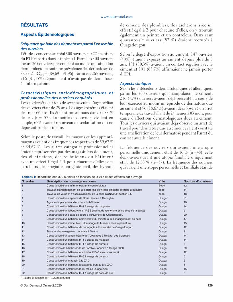
www.odermatol.com
© Our Dermatol Online 2.2020 129
RÉSULTATS
Aspects Épidémiologiques
Fréquence globale des dermatoses parmi l’ensemble des ouvriersL’étude a concerné au total 300 ouvriers sur 22 chantiers du BTP répartis dans le tableau I. Parmi les 300 ouvriers inclus, 265 ouvriers présentaient au moins une affection dermatologique, soit une prévalence des dermatoses de 88,33 %, IC95%= [84,69 – 91,96]. Parmi ces 265 ouvriers, 216 (81,15%) répondaient n’avoir pas de dermatoses à l’interrogatoire.
Caractérist iques sociodémographiques et professionnelles des ouvriers enquêtésLes ouvriers étaient tous de sexe masculin. L’âge médian des ouvriers était de 29 ans. Les âges extrêmes étaient de 16 et 66 ans. Ils étaient musulmans dans 52,33 % des cas (n=157). La moitié des ouvriers vivaient en couple, 67% avaient un niveau de scolarisation qui ne dépassait pas le primaire.
Selon le poste de travail, les maçons et les apprenti-maçons avaient des fréquences respectives de 39,67 % et 34,67 %. Les autres catégories professionnelles étaient représentées par des magasiniers de ciment, des électriciens, des techniciens du bâtiment avec un effectif égal à 3 pour chacune d’elles; des carreleurs, des stagiaires en génie civil, des livreurs
de ciment, des plombiers, des tacherons avec un effectif égal à 2 pour chacune d’elles; on y trouvait également un peintre et un contrôleur. Deux cent quarante-six ouvriers (82 %) étaient recrutés à Ouagadougou.
Selon le degré d’exposition au ciment, 147 ouvriers (49%) étaient exposés au ciment depuis plus de 5 ans, 151 (50,3%) avaient un contact régulier avec le ciment et 191 (63,7%) affirmaient ne jamais porter d’EPI.
Aspects cliniquesSelon les antécédents dermatologiques et allergiques, parmi les 300 ouvriers qui manipulaient le ciment, 216 (72%) ouvriers avaient déjà présenté au cours de leur exercice au moins un épisode de dermatose due au ciment et 56 (18,67 %) avaient déjà observé un arrêt temporaire de travail allant de 24 heures à 03 mois, pour cause d’affections dermatologiques dues au ciment. Tous les ouvriers qui avaient déjà observé un arrêt de travail pour dermatose due au ciment avaient constaté une amélioration de leur dermatose pendant l’arrêt du contact avec le ciment.
La fréquence des ouvriers qui avaient une atopie personnelle uniquement était de 16 % (n=48), celle des ouvriers ayant une atopie familiale uniquement était de 12,33 % (n=37). La fréquence des ouvriers qui avaient une atopie personnelle et familiale était de
Tableau I: Répartition des 300 ouvriers en fonction de la ville et des effectifs par ouvrageN° ordre Description de l’ouvrage en cours Ville Nombre d’ouvriers1 Construction d’une infi rmerie pour le centre Muraz Bobo* 12
2 Travaux d’aménagement de la plateforme du village artisanal de bobo Dioulasso bobo 14
3 Travaux de voirie et d’assainissement de la zone SONATUR section 447 bobo 16
4 Construction d’une agence de Coris Banque à Gounghin Ouaga** 21
5 Agence de placement d’ouvriers du bâtiment Ouaga 10
6 Construction d’un bâtiment R+1 à usage de magasins Ouaga 14
7 Construction d’un laboratoire à l’IRSS (institut de recherche en science de la santé) Ouaga 9
8 Construction d’une salle de cours à l’université de Ouagadougou Ouaga 20
9 Construction d’un bâtiment administratif du ministère de l’enseignement de base Ouaga 17
10 Construction d’un immeuble R+2 à usage de bureaux pour la primature Ouaga 45
11 Construction d’un bâtiment de pédagogie à l’université de Ouagadougou Ouaga 12
12 Travaux d’aménagement de voirie à Saaba Ouaga 9
13 Construction d’un amphithéâtre de 700 places à l’Institut des Sciences Ouaga 14
14 Construction d’un bâtiment R+1 à usage de magasin Ouaga 14
15 Construction d’un bâtiment R+1 à usage de bureaux Ouaga 7
16 Construction de l’Ambassade de l’Arabie Saoudite à Ouaga 2000 Ouaga 20
17 Construction d’un bâtiment administratif R+3 avec sous terrain Ouaga 12
18 Construction d’un bâtiment R+3 à usage de bureaux Ouaga 6
19 Construction d’un magasin à la ZAD Ouaga 5
20 Construction d’un bâtiment à usage de bureau à la ZAD Ouaga 6
21 Construction de l’Ambassade du Mali à Ouaga 2000 Ouaga 15
22 Construction d’un bâtiment R+1 à usage de boite de nuit Ouaga 2
(*)=Bobo-Dioulasso et (**)=Ouagadougou
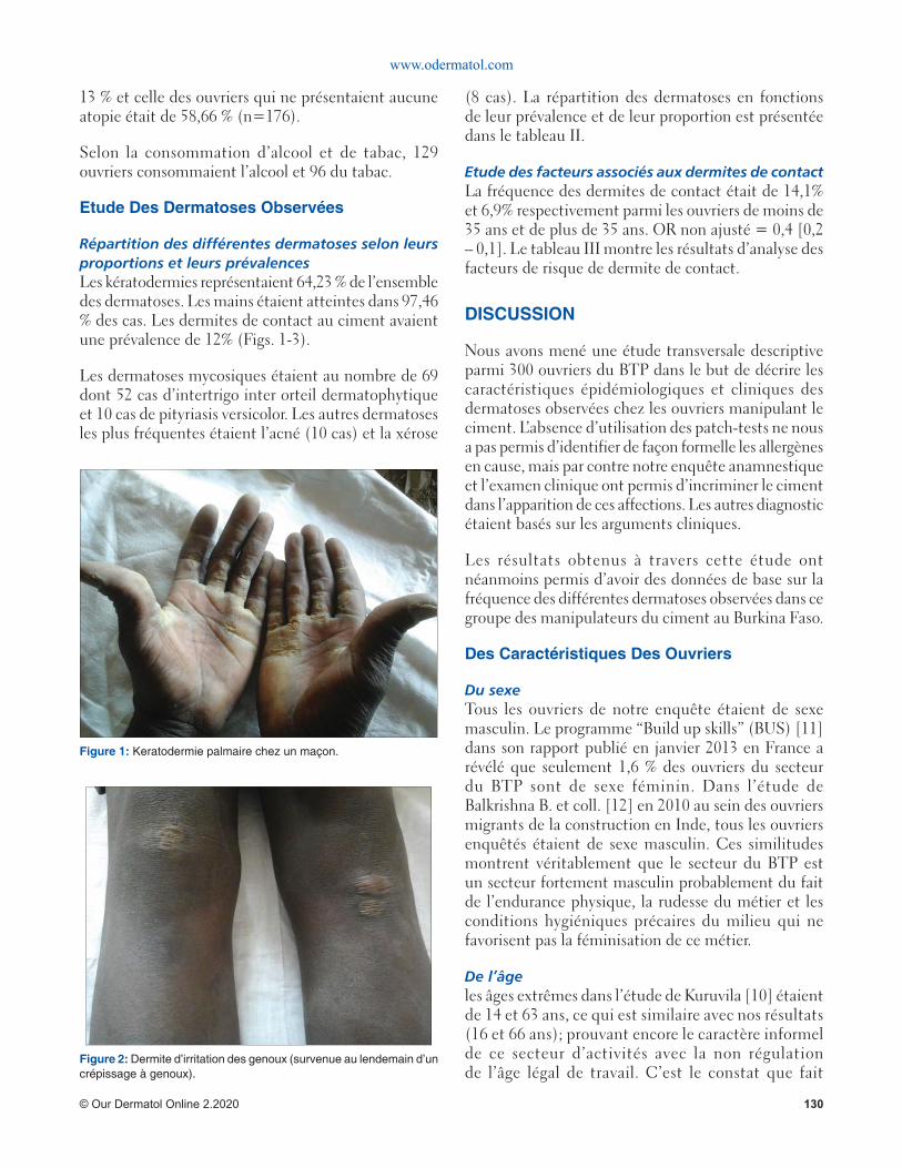
www.odermatol.com
© Our Dermatol Online 2.2020 130
13 % et celle des ouvriers qui ne présentaient aucune atopie était de 58,66 % (n=176).
Selon la consommation d’alcool et de tabac, 129 ouvriers consommaient l’alcool et 96 du tabac.
Etude Des Dermatoses Observées
Répartition des différentes dermatoses selon leurs proportions et leurs prévalencesLes kératodermies représentaient 64,23 % de l’ensemble des dermatoses. Les mains étaient atteintes dans 97,46 % des cas. Les dermites de contact au ciment avaient une prévalence de 12% (Figs. 1-3).
Les dermatoses mycosiques étaient au nombre de 69 dont 52 cas d’intertrigo inter orteil dermatophytique et 10 cas de pityriasis versicolor. Les autres dermatoses les plus fréquentes étaient l’acné (10 cas) et la xérose
(8 cas). La répartition des dermatoses en fonctions de leur prévalence et de leur proportion est présentée dans le tableau II.
Etude des facteurs associés aux dermites de contactLa fréquence des dermites de contact était de 14,1% et 6,9% respectivement parmi les ouvriers de moins de 35 ans et de plus de 35 ans. OR non ajusté = 0,4 [0,2 – 0,1]. Le tableau III montre les résultats d’analyse des facteurs de risque de dermite de contact.
DISCUSSION
Nous avons mené une étude transversale descriptive parmi 300 ouvriers du BTP dans le but de décrire les caractéristiques épidémiologiques et cliniques des dermatoses observées chez les ouvriers manipulant le ciment. L’absence d’utilisation des patch-tests ne nous a pas permis d’identifier de façon formelle les allergènes en cause, mais par contre notre enquête anamnestique et l’examen clinique ont permis d’incriminer le ciment dans l’apparition de ces affections. Les autres diagnostic étaient basés sur les arguments cliniques.
Les résultats obtenus à travers cette étude ont néanmoins permis d’avoir des données de base sur la fréquence des différentes dermatoses observées dans ce groupe des manipulateurs du ciment au Burkina Faso.
Des Caractéristiques Des Ouvriers
Du sexeTous les ouvriers de notre enquête étaient de sexe masculin. Le programme “Build up skills” (BUS) [11] dans son rapport publié en janvier 2013 en France a révélé que seulement 1,6 % des ouvriers du secteur du BTP sont de sexe féminin. Dans l’étude de Balkrishna B. et coll. [12] en 2010 au sein des ouvriers migrants de la construction en Inde, tous les ouvriers enquêtés étaient de sexe masculin. Ces similitudes montrent véritablement que le secteur du BTP est un secteur fortement masculin probablement du fait de l’endurance physique, la rudesse du métier et les conditions hygiéniques précaires du milieu qui ne favorisent pas la féminisation de ce métier.
De l’âgeles âges extrêmes dans l’étude de Kuruvila [10] étaient de 14 et 63 ans, ce qui est similaire avec nos résultats (16 et 66 ans); prouvant encore le caractère informel de ce secteur d’activités avec la non régulation de l’âge légal de travail. C’est le constat que fait
Figure 1: Keratodermie palmaire chez un maçon.
Figure 2: Dermite d’irritation des genoux (survenue au lendemain d’un crépissage à genoux).
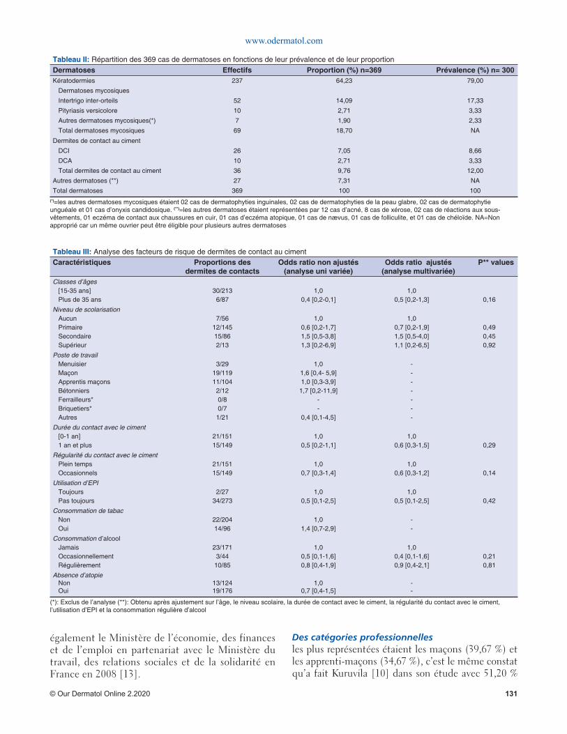
www.odermatol.com
© Our Dermatol Online 2.2020 131
également le Ministère de l’économie, des finances et de l’emploi en partenariat avec le Ministère du travail, des relations sociales et de la solidarité en France en 2008 [13].
Des catégories professionnellesles plus représentées étaient les maçons (39,67 %) et les apprenti-maçons (34,67 %), c’est le même constat qu’a fait Kuruvila [10] dans son étude avec 51,20 %
Tableau II: Répartition des 369 cas de dermatoses en fonctions de leur prévalence et de leur proportionDermatoses Effectifs Proportion (%) n=369 Prévalence (%) n= 300Kératodermies 237 64,23 79,00
Dermatoses mycosiques
Intertrigo inter-orteils 52 14,09 17,33
Pityriasis versicolore 10 2,71 3,33
Autres dermatoses mycosiques(*) 7 1,90 2,33
Total dermatoses mycosiques 69 18,70 NA
Dermites de contact au ciment
DCI 26 7,05 8,66
DCA 10 2,71 3,33
Total dermites de contact au ciment 36 9,76 12,00
Autres dermatoses (**) 27 7,31 NA
Total dermatoses 369 100 100(*)=les autres dermatoses mycosiques étaient 02 cas de dermatophyties inguinales, 02 cas de dermatophyties de la peau glabre, 02 cas de dermatophytie unguéale et 01 cas d’onyxis candidosique. (**)=les autres dermatoses étaient représentées par 12 cas d’acné, 8 cas de xérose, 02 cas de réactions aux sous-vêtements, 01 eczéma de contact aux chaussures en cuir, 01 cas d’eczéma atopique, 01 cas de nævus, 01 cas de folliculite, et 01 cas de chéloïde. NA=Non approprié car un même ouvrier peut être éligible pour plusieurs autres dermatoses
Tableau III: Analyse des facteurs de risque de dermites de contact au cimentCaractéristiques Proportions des
dermites de contactsOdds ratio non ajustés
(analyse uni variée)Odds ratio ajustés
(analyse multivariée)P** values
Classes d’âges[15-35 ans]Plus de 35 ans
30/2136/87
1,00,4 [0,2-0,1]
1,00,5 [0,2-1,3] 0,16
Niveau de scolarisationAucunPrimaireSecondaireSupérieur
7/5612/14515/862/13
1,00,6 [0,2-1,7]1,5 [0,5-3,8]1,3 [0,2-6,9]
1,00,7 [0,2-1,9]1,5 [0,5-4,0]1,1 [0,2-6,5]
0,490,450,92
Poste de travailMenuisierMaçonApprentis maçonsBétonniersFerrailleurs*Briquetiers*Autres
3/2919/11911/104
2/120/80/7
1/21
1,01,6 [0,4- 5,9]1,0 [0,3-3,9]
1,7 [0,2-11,9]--
0,4 [0,1-4,5]
-------
Durée du contact avec le ciment[0-1 an]1 an et plus
21/15115/149
1,00,5 [0,2-1,1]
1,00,6 [0,3-1,5] 0,29
Régularité du contact avec le cimentPlein tempsOccasionnels
21/15115/149
1,00,7 [0,3-1,4]
1,00,6 [0,3-1,2] 0,14
Utilisation d’EPIToujoursPas toujours
2/2734/273
1,00,5 [0,1-2,5]
1,00,5 [0,1-2,5] 0,42
Consommation de tabacNonOui
22/20414/96
1,01,4 [0,7-2,9]
--
Consommation d’alcoolJamaisOccasionnellementRégulièrement
23/1713/44
10/85
1,00,5 [0,1-1,6]0,8 [0,4-1,9]
1,00,4 [0,1-1,6]0,9 [0,4-2,1]
0,210,81
Absence d’atopieNonOui
13/12419/176
1,00,7 [0,4-1,5]
--
(*): Exclus de l’analyse (**): Obtenu après ajustement sur l’âge, le niveau scolaire, la durée de contact avec le ciment, la régularité du contact avec le ciment, l’utilisation d’EPI et la consommation régulière d’alcool
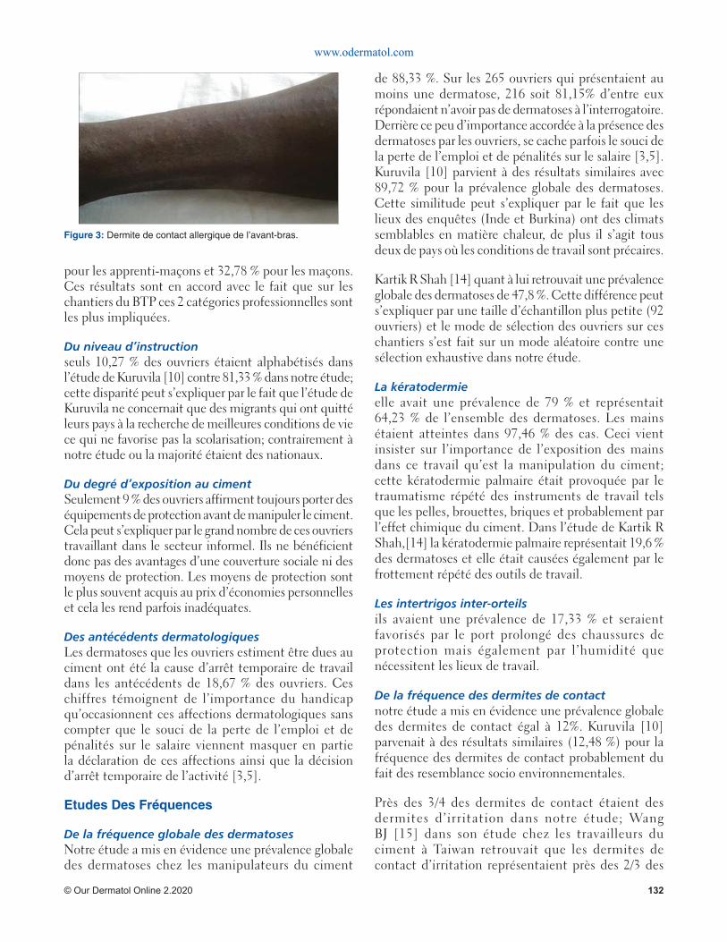
www.odermatol.com
© Our Dermatol Online 2.2020 132
pour les apprenti-maçons et 32,78 % pour les maçons. Ces résultats sont en accord avec le fait que sur les chantiers du BTP ces 2 catégories professionnelles sont les plus impliquées.
Du niveau d’instructionseuls 10,27 % des ouvriers étaient alphabétisés dans l’étude de Kuruvila [10] contre 81,33 % dans notre étude; cette disparité peut s’expliquer par le fait que l’étude de Kuruvila ne concernait que des migrants qui ont quitté leurs pays à la recherche de meilleures conditions de vie ce qui ne favorise pas la scolarisation; contrairement à notre étude ou la majorité étaient des nationaux.
Du degré d’exposition au cimentSeulement 9 % des ouvriers affirment toujours porter des équipements de protection avant de manipuler le ciment. Cela peut s’expliquer par le grand nombre de ces ouvriers travaillant dans le secteur informel. Ils ne bénéficient donc pas des avantages d’une couverture sociale ni des moyens de protection. Les moyens de protection sont le plus souvent acquis au prix d’économies personnelles et cela les rend parfois inadéquates.
Des antécédents dermatologiquesLes dermatoses que les ouvriers estiment être dues au ciment ont été la cause d’arrêt temporaire de travail dans les antécédents de 18,67 % des ouvriers. Ces chiffres témoignent de l’importance du handicap qu’occasionnent ces affections dermatologiques sans compter que le souci de la perte de l’emploi et de pénalités sur le salaire viennent masquer en partie la déclaration de ces affections ainsi que la décision d’arrêt temporaire de l’activité [3,5].
Etudes Des Fréquences
De la fréquence globale des dermatosesNotre étude a mis en évidence une prévalence globale des dermatoses chez les manipulateurs du ciment
de 88,33 %. Sur les 265 ouvriers qui présentaient au moins une dermatose, 216 soit 81,15% d’entre eux répondaient n’avoir pas de dermatoses à l’interrogatoire. Derrière ce peu d’importance accordée à la présence des dermatoses par les ouvriers, se cache parfois le souci de la perte de l’emploi et de pénalités sur le salaire [3,5]. Kuruvila [10] parvient à des résultats similaires avec 89,72 % pour la prévalence globale des dermatoses. Cette similitude peut s’expliquer par le fait que les lieux des enquêtes (Inde et Burkina) ont des climats semblables en matière chaleur, de plus il s’agit tous deux de pays où les conditions de travail sont précaires.
Kartik R Shah [14] quant à lui retrouvait une prévalence globale des dermatoses de 47,8 %. Cette différence peut s’expliquer par une taille d’échantillon plus petite (92 ouvriers) et le mode de sélection des ouvriers sur ces chantiers s’est fait sur un mode aléatoire contre une sélection exhaustive dans notre étude.
La kératodermieelle avait une prévalence de 79 % et représentait 64,23 % de l’ensemble des dermatoses. Les mains étaient atteintes dans 97,46 % des cas. Ceci vient insister sur l’importance de l’exposition des mains dans ce travail qu’est la manipulation du ciment; cette kératodermie palmaire était provoquée par le traumatisme répété des instruments de travail tels que les pelles, brouettes, briques et probablement par l’effet chimique du ciment. Dans l’étude de Kartik R Shah,[14] la kératodermie palmaire représentait 19,6 % des dermatoses et elle était causées également par le frottement répété des outils de travail.
Les intertrigos inter-orteilsils avaient une prévalence de 17,33 % et seraient favorisés par le port prolongé des chaussures de protection mais également par l’humidité que nécessitent les lieux de travail.
De la fréquence des dermites de contactnotre étude a mis en évidence une prévalence globale des dermites de contact égal à 12%. Kuruvila [10] parvenait à des résultats similaires (12,48 %) pour la fréquence des dermites de contact probablement du fait des resemblance socio environnementales.
Près des 3/4 des dermites de contact étaient des dermites d’irritation dans notre étude; Wang BJ [15] dans son étude chez les travailleurs du ciment à Taiwan retrouvait que les dermites de contact d’irritation représentaient près des 2/3 des
Figure 3: Dermite de contact allergique de l’avant-bras.
www.odermatol.com
© Our Dermatol Online 2.2020 133
dermites de contact. Ceci corrobore l’épidémiologie de ces affections quand on sait que la DCI atteint la majorité des sujets en contact avec le produit et la DCA n’atteint que quelques sujets du fait de la sensibilisation préalable.
Etude Des Facteurs Associés à La Survenue Des Dermites de Contact
L’âgeen analyse bi variée avec un seuil de signification de 5%, les ouvriers de plus de 35 ans avaient 2,5 fois moins de dermite de contact que les autres. On pourrait penser que les ouvriers jeunes seraient moins prudents et moins expérimentés en matière de prévention des dermites dues au ciment.
Après une régression logistique, en ajustant sur l’âge, le niveau scolaire, la durée de contact avec le ciment, la régularité du contact avec le ciment, l’utilisation d’EPI et la consommation régulière d’alcool aucun facteur n’était associé à la survenue des dermites de contact.
Le poste de travaill’étude de Guo YL [16] isolait les carreleurs comme étant la catégorie professionnelle la plus à risque et dans l’étude de Kuruvila les dermites de contact étaient plus observées chez les maçons que chez les autres catégories professionnelles; ce constat peut s’expliquer par le fait que sur les chantiers qui ont fait l’objet de notre étude, les taches n’étaient pas toujours effectuées par la catégorie professionnelle indiquée, ainsi par exemple un menuisier de formation pouvait se retrouver à exécuter les taches d’un maçon ou d’un apprenti-maçon suivant l’étape du chantier et vice versa pour se trouver des revenus.
L’ancienneté de contact avec le cimentIl n’existe pas d’association entre la présence de dermite de contact et l’ancienneté de contact avec le ciment dans notre étude. Guo YL [16] a quant à lui retrouvé une association entre l’ancienneté de travail et la survenue des dermites de contact; cette différence peut s’expliquer par le fait que malgré parfois la longue expérience de travail avec le ciment, il ne s’est pas agi en réalité d’un contact permanent et régulier, en effet près de la moitié des ouvriers travaillaient de façon occasionnelle avec le ciment (49,66 %). Les ouvriers ne sont pas employés à plein temps par des entreprises avec des périodes de flottements parfois importants entre deux chantiers contraignant l’ouvrier à se tourner vers d’autres types d’emplois.
L’utilisation des EPIWang BJ [15] a retrouvé que la moindre utilisation des gants était un facteur associé aux dermites de contact et Poppe H [17] retrouve dans son étude que ce sont les pauvres mesures de protection qui sont associés aux dermites de contact.
Par contre notre étude n’observe pas de lien statistique entre l’utilisation des EPI et l’actuelle présence de dermite de contact; cela pourrait traduire une utilisation non optimale des EPI.
CONCLUSION
Les dermatoses sont fréquentes, variées et le plus souvent méconnues chez les ouvriers qui manipulent le ciment. Beaucoup de ces affections sont liées à l’environnement de travail. Les dermatoses directement liées au contact avec le ciment encore appelées dermites de contact étaient fréquentes avec de multiples arrêt de travail si on prend en compte les antécédents des ouvriers. Au Burkina Faso, le secteur du BTP est un milieu où domine le secteur informel et où les conditions de travail sont parfois difficiles et précaires. Il conviendrait de mieux structurer cette catégorie professionnelle et de les doter d’un organe administratif afin de mieux prendre en compte leurs difficultés et notamment leurs multiples problèmes de santé. Notre étude n’a pas permis de mettre en évidence un lien entre les dermites de contact et des facteurs associés à leur survenue. D’autres enquêtes étiologiques sont nécessaires dans notre contexte afin d’isoler des facteurs de causalité de ces dermatoses.
Statement of Human and Animal Rights
All procedures followed were in accordance with the ethical standards of the responsible committee on human experimentation (institutional and national) and with the Helsinki Declaration of 1975, as revised in 2008.
Statement of Informed Consent
Informed consent was obtained from all patients for being included in the study.
REFERENCES
1. Conseil des Ministres du Burkina Faso. Décret N°2015-866/PRES-TRANS/PM/MFPTSS/MS portant liste des maladies professionnelles au Burkina Faso. JO N° 42 du 15 octobre 2015;p37.
2. P Frimat, S Fantoni-Quinton. La main du travailleur: aspect
www.odermatol.com
© Our Dermatol Online 2.2020 134
socio-économiques. Quelle réparation ? Quel reclassement ?. Prog Dermato-allergol Bruxelles. 2001:65-78.
3. Dezfoulian B. Dermatoses professionnelles. 2011. Disponible sur: http://www.stes-apes.med.ulg.ac.be/strav/stenseignement/stformationcontinue/Dermatoses%20profle.pdf. [Consulté le 21/10/2019]
4. Géraut C, Tripodi D. Dermatoses professionnelles. Encyclopédie Médico-Chirurgicale (Elsevier, Paris), Toxicologie-Pathologie Professionnelle, 16-533-A10, Dermatologie, 98-795-A10, 2001. p. 27.
5. Crépy MN. Dermatoses professionnelles au ciment. INRS Documents pour le médecin du travail. 2001;88:419–29.
6. Macedo MS, de Avelar Alchorne AO, Costa EB, Montesano FT. Contact allergy in male construction workers in Sao Paulo, Brazil, 2000-2005. Contact Derm. 2007;56:232-4.
7. Niang SO, Cissé M, Gaye FMC, Diallo M, Boye A, Dieng MT, et al. [Occupational allergic contact dermatitis in Dakar]. Mali Med. 2007;22:34–7.
8. Frimat P. Les dermatoses professionnelles dans le bâtiment et les travaux publics. RevFrançaise d’Allergol d’Immunol Clin. 2007;47:153-7.
9. Ancelle T. Statistique, épidémiologie. 3e édition. Collection sciences fondamentales; 2011. p 80-81.
10. Kuruvila M, Dubey S, Gahalaut P. Pattern of skin diseases among migrant construction workers in Mangalore. Indian J Dermatol Venereol Leprol. 2006;72:129–32.
11. Build Up Skills. France: analyse du statu quo national: organiser la montée en compétences des ouvriers et artisans du batiment [Internet]. 2013 Jan p. 9. Available from: http://www.cercad.fr/
Copyright by Issouf Konaté, et al. This is an open-access article distributed under the terms of the Creative Commons Attribution License, which permits unrestricted use, distribution, and reproduction in any medium, provided the original author and source are credited.Source of Support: Nil, Confl ict of Interest: None declared.
IMG/pdf/bus_etat_des_lieux.pdf12. Balkrishna B. Adsul, Payal S. Laad, Prashant V. Howal, Ramesh M.
Chaturvedi. Health problems among migrant construction workers: A unique public–private partnership project. Indian J Occup Envir Med. 2011;15:29–32.
13. République française, Ministère de l’économie des fi nances et de l’emploi, Ministère du travail, des relations sociales et de la solidarité. Les ouvriers du bâtiment et des travaux publics : des contraintes physiques et des expositions aux produits chimiques importantes, une autonomie assez élevée dans le travail. Premières synthèses, DARES. 2008;N°07.3:2.
14. Kartik R Shah, Rajnarayan R Tiwari. Occupational skin problems in construction workers. Indian J Dermatol. 2010;55:348–51.
15. Wang B-J, Wu J-D, Sheu S-C, Shih T-S, Chang H-Y, Guo Y-L, et al. Occupational hand dermatitis among cement workers in Taiwan. J Formos Med Assoc. 2011;110:775–9.
16. Guo YL, Wang BJ, Yeh KC, Wang JC, Kao HH, Wang MT, et al. Dermatoses in cement workers in southern Taiwan. Contact Derm. 1999;40:1–7.
17. Poppe H, Poppe LM, Bröcker E-B, Trautmann A. Do-it-yourself cement work: the main cause of severe irritant contact dermatitis requiring hospitalization. Contact Derm. 2013;68:111–5.
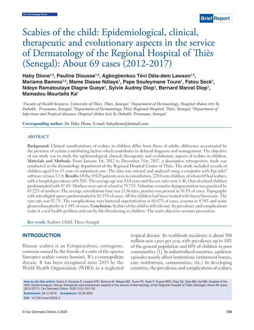
Our Dermatology Online
© Our Dermatol Online 2.2020 135
INTRODUCTION
Human scabies is an Ectoparasitosis, contagious, common caused by the female of a mite of the species Sarcoptes scabiei variety hominis. It’s a cosmopolitan disease. It has been recognized since 2013 by the World Health Organization (WHO) as a neglected
tropical disease. Its worldwide incidence is about 300 million new cases per year, with prevalence up to 10% of the general population and 60% of children in poor communities [1]. In industrialized countries, epidemic episodes mainly affect institutions (retirement homes, care institutions, communities, etc.) In developing countries, the prevalence and complications of scabies,
ABSTRACT
Background: Clinical manifestations of scabies in children differ from those of adults: difference accentuated by the presence of certain contributing factors which contributes to delayed diagnosis and management. The objective of our study was to study the epidemiological, clinical, therapeutic and evolutionary aspects of scabies in children. Materials and Methods: From January 1st, 2012 to December 31st, 2017, a descriptive retrospective study was conducted in the dermatology department of the Regional Hospital Center of Thiès. The study included records of children aged 0 to 15 years in outpatient care. The data was entered and analyzed using a computer with Epi info7 software version 3.5.4. Results: Of the 19129 patients seen in consultation, 2284 were children, of whom 69 had scabies, with a hospital prevalence of 0.36%. The average age was 4.64 years and the sex ratio were 1.46. Out-of-school children predominated with 47.8%. Mothers were out of school in 79.71%. Voluntary cosmetic depigmentation was practiced by 65.22% of mothers. The average consultation time was 21.86 days, pruritus was present in 56.5% of cases. Topography with interdigital spaces predominated in 81.15% of cases. All the children had been treated with benzyl benzoate. The cure rate was 92.7%. The complications were bacterial superinfection in 60.87% of cases, eczema in 4.34% and acute glomerulonephritis in 1.44% of cases. Conclusion: Scabies of the child is still relevant. Its prevalence and complications make it a real health problem and can be life-threatening in children. The main objective remains prevention.
Key words: Scabies; Child; Thies; Senegal
How to cite this article: Dione H, Diousse P, Lawson ATD, Bammo M, Ndiaye MD, Toure PS, Seck F, Gueye NRD, Diop SA, Diop BM, Ka MM. Scabies of the child: Epidemiological, clinical, therapeutic and evolutionary aspects in the service of Dermatology of the Regional Hospital of Thiès (Senegal): About 69 cases (2012-2017). Our Dermatol Online. 2020;11(2):135-139.Submission: 26.11.2019; Acceptance: 02.02.2020DOI: 10.7241/ourd.20202.4
Brief Report
Scabies of the child: Epidemiological, clinical, Scabies of the child: Epidemiological, clinical, therapeutic and evolutionary aspects in the service therapeutic and evolutionary aspects in the service of Dermatology of the Regional Hospital of Th iès of Dermatology of the Regional Hospital of Th iès (Senegal): (Senegal): About 69 cases (2012-2017)bout 69 cases (2012-2017)Haby Dione1,2, Pauline Diousse1,3, Agbogbenkou Tévi Déla-dem Lawson1,4, Mariama Bammo1,3, Mame Diasse Ndiaye1, Pape Souleymane Toure1, Fatou Seck1, Ndeye Ramatoulaye Diagne Gueye1, Sylvie Audrey Diop1, Bernard Marcel Diop1, Mamadou Mourtalla Ka1
1Faculty of Health Sciences, University of Thiès, Thiès, Senegal, 2Department of Dermatology, Hospital Abdou Aziz Sy Dabakh, Tivaouane, Senegal, 3Department of Dermatology, Thiès Regional Hospital, Thiès, Senegal, 4Department of Infectious and Tropical diseases, Hospital Abdou Aziz Sy Dabakh, Tivaouane, Senegal
Corresponding author: Dr. Haby Dione, E-mail: [email protected]
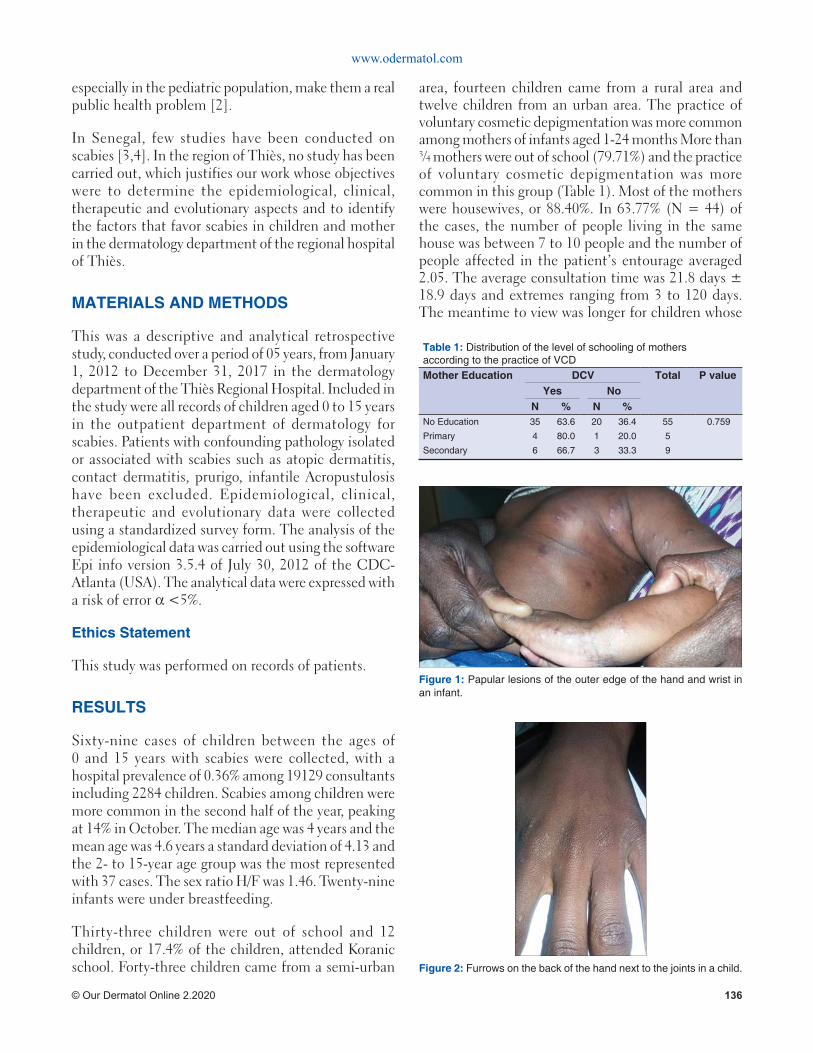
www.odermatol.com
© Our Dermatol Online 2.2020 136
especially in the pediatric population, make them a real public health problem [2].
In Senegal, few studies have been conducted on scabies [3,4]. In the region of Thiès, no study has been carried out, which justifies our work whose objectives were to determine the epidemiological, clinical, therapeutic and evolutionary aspects and to identify the factors that favor scabies in children and mother in the dermatology department of the regional hospital of Thiès.
MATERIALS AND METHODS
This was a descriptive and analytical retrospective study, conducted over a period of 05 years, from January 1, 2012 to December 31, 2017 in the dermatology department of the Thiès Regional Hospital. Included in the study were all records of children aged 0 to 15 years in the outpatient department of dermatology for scabies. Patients with confounding pathology isolated or associated with scabies such as atopic dermatitis, contact dermatitis, prurigo, infantile Acropustulosis have been excluded. Epidemiological, clinical, therapeutic and evolutionary data were collected using a standardized survey form. The analysis of the epidemiological data was carried out using the software Epi info version 3.5.4 of July 30, 2012 of the CDC-Atlanta (USA). The analytical data were expressed with a risk of error α <5%.
Ethics Statement
This study was performed on records of patients.
RESULTS
Sixty-nine cases of children between the ages of 0 and 15 years with scabies were collected, with a hospital prevalence of 0.36% among 19129 consultants including 2284 children. Scabies among children were more common in the second half of the year, peaking at 14% in October. The median age was 4 years and the mean age was 4.6 years a standard deviation of 4.13 and the 2- to 15-year age group was the most represented with 37 cases. The sex ratio H/F was 1.46. Twenty-nine infants were under breastfeeding.
Thirty-three children were out of school and 12 children, or 17.4% of the children, attended Koranic school. Forty-three children came from a semi-urban
area, fourteen children came from a rural area and twelve children from an urban area. The practice of voluntary cosmetic depigmentation was more common among mothers of infants aged 1-24 months More than ¾ mothers were out of school (79.71%) and the practice of voluntary cosmetic depigmentation was more common in this group (Table 1). Most of the mothers were housewives, or 88.40%. In 63.77% (N = 44) of the cases, the number of people living in the same house was between 7 to 10 people and the number of people affected in the patient’s entourage averaged 2.05. The average consultation time was 21.8 days ± 18.9 days and extremes ranging from 3 to 120 days. The meantime to view was longer for children whose
Figure 1: Papular lesions of the outer edge of the hand and wrist in an infant.
Figure 2: Furrows on the back of the hand next to the joints in a child.
Table 1: Distribution of the level of schooling of mothers according to the practice of VCDMother Education DCV Total P value
Yes NoN % N %
No Education 35 63.6 20 36.4 55 0.759
Primary 4 80.0 1 20.0 5
Secondary 6 66.7 3 33.3 9
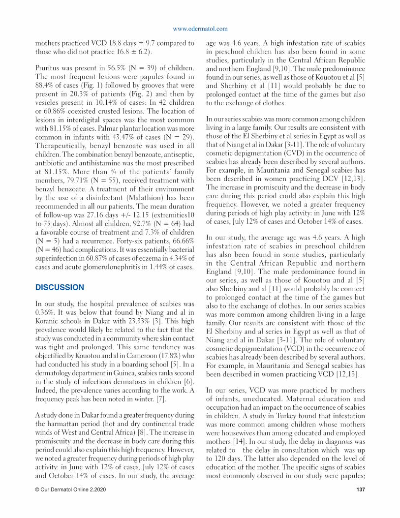
www.odermatol.com
© Our Dermatol Online 2.2020 137
mothers practiced VCD 18.8 days ± 9.7 compared to those who did not practice 16.8 ± 6.2).
Pruritus was present in 56.5% (N = 39) of children. The most frequent lesions were papules found in 88.4% of cases (Fig. 1) followed by grooves that were present in 20.3% of patients (Fig. 2) and then by vesicles present in 10.14% of cases: In 42 children or 60.86% coexisted crusted lesions. The location of lesions in interdigital spaces was the most common with 81.15% of cases. Palmar plantar location was more common in infants with 43.47% of cases (N = 29). Therapeutically, benzyl benzoate was used in all children. The combination benzyl benzoate, antiseptic, antibiotic and antihistamine was the most prescribed at 81.15%. More than ¾ of the patients’ family members, 79.71% (N = 55), received treatment with benzyl benzoate. A treatment of their environment by the use of a disinfectant (Malathion) has been recommended in all our patients. The mean duration of follow-up was 27.16 days +/- 12.15 (extremities10 to 75 days). Almost all children, 92.7% (N = 64) had a favorable course of treatment and 7.3% of children (N = 5) had a recurrence. Forty-six patients, 66.66% (N = 46) had complications. It was essentially bacterial superinfection in 60.87% of cases of eczema in 4.34% of cases and acute glomerulonephritis in 1.44% of cases.
DISCUSSION
In our study, the hospital prevalence of scabies was 0.36%. It was below that found by Niang and al in Koranic schools in Dakar with 23.33% [3]. This high prevalence would likely be related to the fact that the study was conducted in a community where skin contact was tight and prolonged. This same tendency was objectified by Kouotou and al in Cameroon (17.8%) who had conducted his study in a boarding school [5]. In a dermatology department in Guinea, scabies ranks second in the study of infectious dermatoses in children [6]. Indeed, the prevalence varies according to the work. A frequency peak has been noted in winter. [7].
A study done in Dakar found a greater frequency during the harmattan period (hot and dry continental trade winds of West and Central Africa) [8]. The increase in promiscuity and the decrease in body care during this period could also explain this high frequency. However, we noted a greater frequency during periods of high play activity: in June with 12% of cases, July 12% of cases and October 14% of cases. In our study, the average
age was 4.6 years. A high infestation rate of scabies in preschool children has also been found in some studies, particularly in the Central African Republic and northern England [9,10]. The male predominance found in our series, as well as those of Kouotou et al [5] and Sherbiny et al [11] would probably be due to prolonged contact at the time of the games but also to the exchange of clothes.
In our series scabies was more common among children living in a large family. Our results are consistent with those of the El Sherbiny et al series in Egypt as well as that of Niang et al in Dakar [3-11]. The role of voluntary cosmetic depigmentation (CVD) in the occurrence of scabies has already been described by several authors. For example, in Mauritania and Senegal scabies has been described in women practicing DCV [12,13]. The increase in promiscuity and the decrease in body care during this period could also explain this high frequency. However, we noted a greater frequency during periods of high play activity: in June with 12% of cases, July 12% of cases and October 14% of cases.
In our study, the average age was 4.6 years. A high infestation rate of scabies in preschool children has also been found in some studies, particularly in the Central African Republic and northern England [9,10]. The male predominance found in our series, as well as those of Kouotou and al [5] also Sherbiny and al [11] would probably be connect to prolonged contact at the time of the games but also to the exchange of clothes. In our series scabies was more common among children living in a large family. Our results are consistent with those of the El Sherbiny and al series in Egypt as well as that of Niang and al in Dakar [3-11]. The role of voluntary cosmetic depigmentation (VCD) in the occurrence of scabies has already been described by several authors. For example, in Mauritania and Senegal scabies has been described in women practicing VCD [12,13].
In our series, VCD was more practiced by mothers of infants, uneducated. Maternal education and occupation had an impact on the occurrence of scabies in children. A study in Turkey found that infestation was more common among children whose mothers were housewives than among educated and employed mothers [14]. In our study, the delay in diagnosis was related to the delay in consultation which was up to 120 days. The latter also depended on the level of education of the mother. The specific signs of scabies most commonly observed in our study were papules;
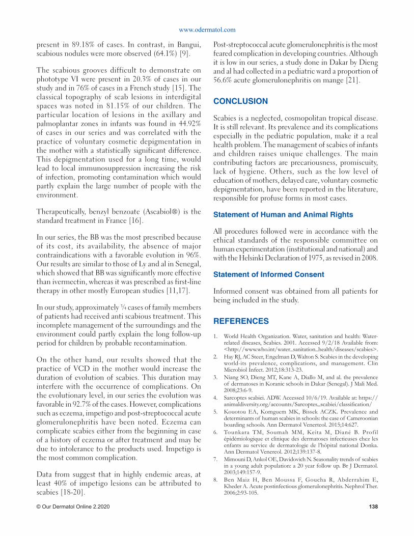
www.odermatol.com
© Our Dermatol Online 2.2020 138
present in 89.18% of cases. In contrast, in Bangui, scabious nodules were more observed (64.1%) [9].
The scabious grooves difficult to demonstrate on phototype VI were present in 20.3% of cases in our study and in 76% of cases in a French study [15]. The classical topography of scab lesions in interdigital spaces was noted in 81.15% of our children. The particular location of lesions in the axillary and palmoplantar zones in infants was found in 44.92% of cases in our series and was correlated with the practice of voluntary cosmetic depigmentation in the mother with a statistically significant difference. This depigmentation used for a long time, would lead to local immunosuppression increasing the risk of infection, promoting contamination which would partly explain the large number of people with the environment.
Therapeutically, benzyl benzoate (Ascabiol®) is the standard treatment in France [16].
In our series, the BB was the most prescribed because of its cost, its availability, the absence of major contraindications with a favorable evolution in 96%. Our results are similar to those of Ly and al in Senegal, which showed that BB was significantly more effective than ivermectin, whereas it was prescribed as first-line therapy in other mostly European studies [11,17].
In our study, approximately ¾ cases of family members of patients had received anti scabious treatment. This incomplete management of the surroundings and the environment could partly explain the long follow-up period for children by probable recontamination.
On the other hand, our results showed that the practice of VCD in the mother would increase the duration of evolution of scabies. This duration may interfere with the occurrence of complications. On the evolutionary level, in our series the evolution was favorable in 92.7% of the cases. However, complications such as eczema, impetigo and post-streptococcal acute glomerulonephritis have been noted. Eczema can complicate scabies either from the beginning in case of a history of eczema or after treatment and may be due to intolerance to the products used. Impetigo is the most common complication.
Data from suggest that in highly endemic areas, at least 40% of impetigo lesions can be attributed to scabies [18-20].
Post-streptococcal acute glomerulonephritis is the most feared complication in developing countries. Although it is low in our series, a study done in Dakar by Dieng and al had collected in a pediatric ward a proportion of 56.6% acute glomerulonephritis on mange [21].
CONCLUSION
Scabies is a neglected, cosmopolitan tropical disease. It is still relevant. Its prevalence and its complications especially in the pediatric population, make it a real health problem. The management of scabies of infants and children raises unique challenges. The main contributing factors are precariousness, promiscuity, lack of hygiene. Others, such as the low level of education of mothers, delayed care, voluntary cosmetic depigmentation, have been reported in the literature, responsible for profuse forms in most cases.
Statement of Human and Animal Rights
All procedures followed were in accordance with the ethical standards of the responsible committee on human experimentation (institutional and national) and with the Helsinki Declaration of 1975, as revised in 2008.
Statement of Informed Consent
Informed consent was obtained from all patients for being included in the study.
REFERENCES
1. World Health Organization. Water, sanitation and health: Water-related diseases, Scabies. 2001. Accessed 9/2/18 Available from: <http://www.who.int/water_sanitation_health/diseases/scabies>.
2. Hay RJ, AC Steer, Engelman D, Walton S. Scabies in the developing world-its prevalence, complications, and management. Clin Microbiol Infect. 2012;18:313-23.
3. Niang SO, Dieng MT, Kane A, Diallo M, and al. the prevalence of dermatoses in Koranic schools in Dakar (Senegal). J Mali Med. 2008;23:6-9.
4. Sarcoptes scabiei. ADW. Accessed 10/6/19. Available at: https://animaldiversity.org/accounts/Sarcoptes_scabiei/classifi cation/
5. Kouotou EA, Komguem MK, Bissek ACZK. Prevalence and determinants of human scabies in schools: the case of Cameroonian boarding schools. Ann Dermatol Venerteol. 2015;14:627.
6. Tounkara TM, Soumah MM, Keita M, Diané B. Profil épidémiologique et clinique des dermatoses infectieuses chez les enfants au service de dermatologie de l’hôpital national Donka. Ann Dermatol Venereol. 2012;139:137-8.
7. Mimouni D, Ankol OE, Davidovich N. Seasonality trends of scabies in a young adult population: a 20 year follow up. Br J Dermatol. 2003;149:157-9.
8. Ben Maiz H, Ben Moussa F, Goucha R, Abderrahim E, Kheder A. Acute postinfectious glomerulonephritis. Nephrol Ther. 2006;2:93-105.

www.odermatol.com
© Our Dermatol Online 2.2020 139
Copyright by Haby Dione, et al. This is an open-access article distributed under the terms of the Creative Commons Attribution License, which permits unrestricted use, distribution, and reproduction in any medium, provided the original author and source are credited.Source of Support: Nil, Confl ict of Interest: None declared.
9. Kobangué L, Guéréndo P, Abéyé J, Namdito P, Mballa MD, Gresenguet G. Gale sarcoptique: aspects épidémiologiques, cliniques et thérapeutiques à Bangui. Bull Pathol Exot. 2014;107:10-4.
10. Church RE, Knowleden J. Scabies in Sheffi eld: a family infestation. Br Med J. 1978;1:761-3.
11. Naglaa A, El Sherbiny, Talal A, Hassan M, Hassan NS, Zeiada AN. Epidemiological Study of Scabies in Primary Schools, Fayoum Governorate- Egypt. J Prim Health Care Gen Pract. 2017;1:1-5.
12. Mahe A, Ly F, Aymard G, Dangou JM. Skin diseases associated with the cosmetic use of bleaching products in women from Dakar, Senegal. Br J Dermatol. 2003;148:493-500.
13. Kebe M, Yahya S, Lo B, M. Ball. Study of complications of artifi cial depigmentation in Nouakchott, Mauritania. Mali Medical. 2015;3:38-40.
14. Ciftci IH, Karaca S, Dogru O, Cetinkaya Z, Kulac M. Prevalence of pediculosis and scabies in preschool nursery children of Afyon, Turkey. Korean J Parasitol. 2006;44:95-8.
15. Pouessel G, Dumortier J, Lagrée M, Pierre MH, Ganga-Zandzou PS, Ythier H, et al. Scabies: a common infection in pediatrics. Pediat Arch. 2012;19:1259-60.
16. Royer M, CM Latre, Paul C, Mazereeuw-Hautier J. The French Society of Pediatric Dermatology, “Infant scabies”. Ann Dermatol Venerteol. 2008;135:876-81.
17. Brooks PA, Grace RF. Ivermectin is better than benzyl benzoate for children scabies in developing countries. J Paediatr Child H. 2002;38:401-4.
18. Aung PTZ, Cuningham W, Hwang K, Andrews RM, Carapetis JR, Kearns T, et al. Australian Aboriginals: A self-controlled case series study. PLoS Negl Trop Dis. 2018;12:6668.
19. Mason DS, Marks M, Sokana O, Solomon AW, Mabey DC, Romani L, et al. The prevalence of scabies and impetigo in the Solomon Islands: A population-based survey. PLoS Negl Trop Dis. 2016;10:4803.
20. Romani L, AC Steer, Whitfeld MJ, Kaldor JM. Prevalence of scabies and impetigo worldwide: A systematic review. Lancet Infect Dis. 2015;15:960-7.
21. Dieng MT, Ndiaye B, Ndiaye AM. Scabies complicated by acute glomerulonephritis in children: 114 cases observed in two years in a pediatric ward in Dakar. Dakar Med. 1998;43:201-4.
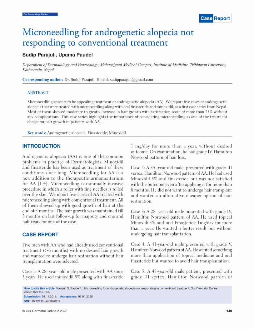
Our Dermatology Online
© Our Dermatol Online 2.2020 140
How to cite this article: Parajuli S, Paudel U. Microneedling for androgenetic alopecia not responding to conventional treatment. Our Dermatol Online. 2020;11(2):140-142.Submission: 01.11.2019; Acceptance: 07.01.2020DOI: 10.7241/ourd.20202.5
Microneedling for androgenetic alopecia not Microneedling for androgenetic alopecia not responding to conventional treatmentresponding to conventional treatmentSudip Parajuli, Upama Paudel
Department of Dermatology and Venereology, Maharajgunj Medical Campus, Institute of Medicine, Tribhuvan University, Kathmandu, Nepal
Corresponding author: Dr. Sudip Parajuli, E-mail: [email protected]
INTRODUCTION
Androgenetic alopecia (AA) is one of the common problems in practice of Dermatologists. Minoxidil and finasteride has been used as treatment of these conditions since long. Microneedling for AA is a new addition to the therapeutic armamentarium for AA [1-4]. Microneedling is minimally invasive procedure in which a roller with fine needles is rolled over the skin. We report five cases of AA treated with microneedling along with conventional treatment. All of them showed up with good growth of hair at the end of 3 months. The hair growth was maintained till 3 months on last follow-up for majority and one and half years for one of the case.
CASE REPORT
Five men with AA who had already used conventional treatment (>6 months) with no desired hair growth and wanted to undergo hair restoration without hair transplantation were selected.
Case 1: A 28- year -old male presented with AA since 5 years. He used minoxidil 5% along with finasteride
1 mg/day for more than a year, without desired outcome. On examination, he had grade IV, Hamilton Norwood pattern of hair loss.
Case 2: A 35 -year old male, presented with grade III vertex, Hamilton Norwood pattern of AA. He had used Minoxidil 5% and finasteride but was not satisfied with the outcome even after applying it for more than 8 months. He did not want to undergo hair transplant and wanted an alternative cheaper option of hair restoration.
Case 3: A 28- year-old male presented with grade IV, Hamilton Norwood pattern of AA. He used topical Minoxidil5% and oral Finasteride 1mg/day for more than a year. He wanted a better result but without undergoing hair transplantation.
Case 4: A 41-year-old male presented with grade V, Hamilton Norwood pattern of AA. He wanted something more than application of topical medicine and oral finasteride but wanted to avoid hair transplantation.
Case 5: A 45-year-old male patient, presented with grade III vertex, Hamilton Norwood pattern of
ABSTRACT
Microneedling appears to be appealing treatment of androgenetic alopecia (AA). We report five cases of androgenetic alopecia that were treated with microneedling along with oral finasteride and minoxidil, as a first case series from Nepal. Most of them showed moderate to greatly increase in hair growth with satisfaction score of more than 75% without any complications. This case series highlights the importance of considering microneedling as one of the treatment choice for hair growth in patients with AA.
Key words: Androgenetic alopecia; Finasteride; Minoxidil
Case Report
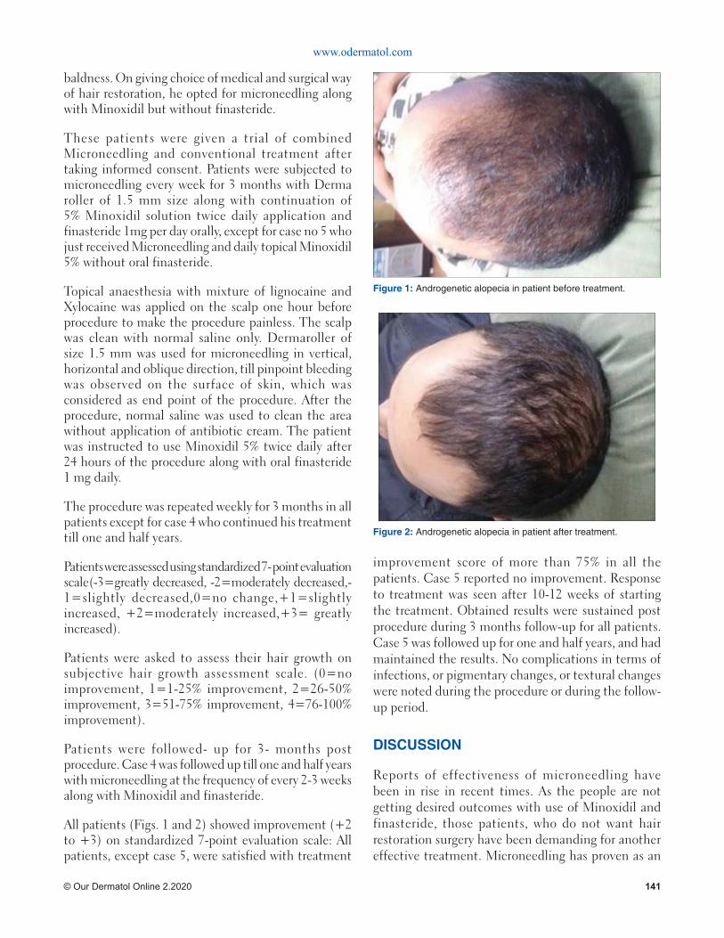
www.odermatol.com
© Our Dermatol Online 2.2020 141
baldness. On giving choice of medical and surgical way of hair restoration, he opted for microneedling along with Minoxidil but without finasteride.
These patients were given a trial of combined Microneedling and conventional treatment after taking informed consent. Patients were subjected to microneedling every week for 3 months with Derma roller of 1.5 mm size along with continuation of 5% Minoxidil solution twice daily application and finasteride 1mg per day orally, except for case no 5 who just received Microneedling and daily topical Minoxidil 5% without oral finasteride.
Topical anaesthesia with mixture of lignocaine and Xylocaine was applied on the scalp one hour before procedure to make the procedure painless. The scalp was clean with normal saline only. Dermaroller of size 1.5 mm was used for microneedling in vertical, horizontal and oblique direction, till pinpoint bleeding was observed on the surface of skin, which was considered as end point of the procedure. After the procedure, normal saline was used to clean the area without application of antibiotic cream. The patient was instructed to use Minoxidil 5% twice daily after 24 hours of the procedure along with oral finasteride 1 mg daily.
The procedure was repeated weekly for 3 months in all patients except for case 4 who continued his treatment till one and half years.
Patients were assessed using standardized 7- point evaluation scale(-3=greatly decreased, -2=moderately decreased,-1=slightly decreased,0=no change,+1=slightly increased, +2=moderately increased,+3= greatly increased).
Patients were asked to assess their hair growth on subjective hair growth assessment scale. (0=no improvement, 1=1-25% improvement, 2=26-50% improvement, 3=51-75% improvement, 4=76-100% improvement).
Patients were followed- up for 3- months post procedure. Case 4 was followed up till one and half years with microneedling at the frequency of every 2-3 weeks along with Minoxidil and finasteride.
All patients (Figs. 1 and 2) showed improvement (+2 to +3) on standardized 7-point evaluation scale: All patients, except case 5, were satisfied with treatment
improvement score of more than 75% in all the patients. Case 5 reported no improvement. Response to treatment was seen after 10-12 weeks of starting the treatment. Obtained results were sustained post procedure during 3 months follow-up for all patients. Case 5 was followed up for one and half years, and had maintained the results. No complications in terms of infections, or pigmentary changes, or textural changes were noted during the procedure or during the follow-up period.
DISCUSSION
Reports of effectiveness of microneedling have been in rise in recent times. As the people are not getting desired outcomes with use of Minoxidil and finasteride, those patients, who do not want hair restoration surgery have been demanding for another effective treatment. Microneedling has proven as an
Figure 1: Androgenetic alopecia in patient before treatment.
Figure 2: Androgenetic alopecia in patient after treatment.
www.odermatol.com
© Our Dermatol Online 2.2020 142
effective alternative treatment modality for these types of patients.
The principle of microneedling was first described by Orentreich, who used it for the acne scar. Since then, its use has been extended to other conditions like rhytides, striae distensae, alopecia and others. In androgenetic alopecia, it is supposed to work by stimulation of epidermal growth factor, platelet derived growth factor, hair bulb stem cells and activation of Wnt signaling pathway [5].
Our cases showed moderate to great increase in hair growth without any complications associated with the procedure with exception of one patient. This case series adds to existing literature on beneficial effects of microneedling for AA. Though all the cases, except for case 4, could not be followed for long period of time, the response in case 4 seen at 18 months highlights that the response of hair growth would be maintained if the procedure is continued in intermittent pattern for long period of time.
To conclude, micro needling is one of the effective and cheap therapeutic interventions for AA. This is the first case series reported from Nepal highlighting the effectiveness of microneedling procedure in androgenetic alopecia. Though we see promising results of microneedlingthe in small numbers of cases in our series, a large cohort, and longer follow -up and comparative studies would be required in our population in future for ascertaining real effectiveness of microneedling in AA.
Consent
The examination of the patient was conducted according to the Declaration of Helsinki principles.
The authors certify that they have obtained all appropriate patient consent forms. In the form the patient(s) has/have given his/her/their consent for his/her/their images and other clinical information to be reported in the journal. The patients understand that their names and initials will not be published and due efforts will be made to conceal their identity, but anonymity cannot be guaranteed.
REFERENCES
1. Dhurat R, Mathapati S. Response to Microneedling treatment in men with androgenetic alopecia who failed to respond to conventional therapy. Indian J Dermatol. 2015;60:260-63.
2. Valente Duarte de Sousa IC, Tosti A. New investigational drugs for androgenetic alopecia. Expert Opin Investig Drugs. 2013;22:579-89.
3. Kim YS, Jeong KH, Kim JE, Woo YJ, Kim BJ, Kang H. Repeated microneedle stimulation induces enhanced hair growth in a murine model. Ann Dermatol. 2016;28:586-92.
4. Dhurat R, Sukesh MS, Avhad G, Dandale A, Pal A, Pund P. A randomized evaluator blinded study of effect of microneedling in androgenetic alopecia. A pilot study. Int J Trichology. 2013;5:6-11.
5. Fertig RM, Gamret AC, Cervantes J, Tosti A. Microneedling for treatment of hair loss? JEADV. 2018:32:564-9.
Copyright by Sudip Parajuli, et al. This is an open-access article distributed under the terms of the Creative Commons Attribution License, which permits unrestricted use, distribution, and reproduction in any medium, provided the original author and source are credited.Source of Support: Nil, Confl ict of Interest: None declared.
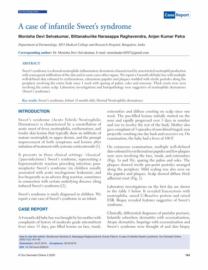
Our Dermatology Online
© Our Dermatol Online 2.2020 143
How to cite this article: Selvakumar Monisha D, Narasappa Raghavendra B, Kumar Patra A. A case of infantile Sweet’s syndrome. Our Dermatol Online. 2020;11(2):143-145.Submission: 04.07.2019; Acceptance: 08.09.2019DOI: 10.7241/ourd.20202.6
INTRODUCTION
Sweet’s syndrome (Acute Febrile Neutrophilic Dermatoses) is characterized by a constellation of acute onset of fever, neutrophilia, erythematous, and tender skin lesions that typically show an infiltrate of mature neutrophils in upper dermis, and the prompt improvement of both symptoms and lesions after initiation of treatment with systemic corticosteroids [1].
It presents in three clinical settings: ‘classical’ (’para-infectious’) Sweet’s syndrome, representing a hypersensitivity reaction preceding infection; para-neoplastic Sweet’s syndrome (in children usually associated with acute myelogenous leukemia); and less frequently as an adverse drug reaction, sometimes in connection with certain underlying diseases (drug induced Sweet’s syndrome)[2].
Sweet’s syndrome is rarely diagnosed in children. We report a rare case of Sweet’s syndrome in an infant.
CASE REPORT
A 4-month old baby boy was brought by his mother with complaints of history of moderate grade intermittent fever since 15 days, pus filled lesions on face, trunk,
extremities and diffuse crusting on scalp since one week. The pus-filled lesions initially started on the nose and rapidly progressed over 3 days in number and size to involve the rest of the body. Mother also gave complaint of 3 episodes of non-blood tinged, non projectile vomiting one day back and excessive cry. On examination, the baby had a fever of 100 F.
On cutaneous examination, multiple well-defined skin-coloured to erythematous papules and few plaques were seen involving the face, trunk, and extremities (Figs. 1a and 1b), sparing the palms and soles. The plaques showed sterile pin-point pustules arranged along the periphery. Mild scaling was also seen on the papules and plaques. Scalp showed diffuse thick adherent crust (Fig. 2).
Laboratory investigations on the first day are shown in the table 1 below. It revealed leucocytosis with neutrophilia, raised C-Reactive protein and raised ESR. Biopsy revealed features suggestive of Sweet’s syndrome.
Clinically, differential diagnoses of pustular psoriasis, Infantile seborrheic dermatitis with eczematisation, Atopic dermatitis, Impetigo with eczematisation and Sweet’s syndrome were thought of and skin biopsy
ABSTRACT
Sweet’s syndrome is a dermal neutrophilic inflammatory dermatosis characterized by unrestricted neutrophil production with consequent infiltration of the skin and in some cases other organs. We report a 4 month old baby boy with multiple well-defined skin coloured to erythematous, edematous papules and plaques studded with sterile pustules along the periphery involving the entire body since 1 week with sparing of palms, soles and mucosae. Thick crusts were seen involving the entire scalp. Laboratory investigations and histopathology were suggestive of neutrophilic dermatoses (Sweet’s syndrome).
Key words: Sweet’s syndrome; Infant (4 month old); Dermal Neutrophilic dermatoses
Case Report
A case of infantile Sweet’s syndromeA case of infantile Sweet’s syndromeMonisha Devi Selvakumar, Bittanakurike Narasappa Raghavendra, Anjan Kumar Patra
Department of Dermatology, MVJ Medical College and Research Hospital, Bangalore, India
Corresponding author: Dr. Monisha Devi Selvakumar, E-mail: [email protected]
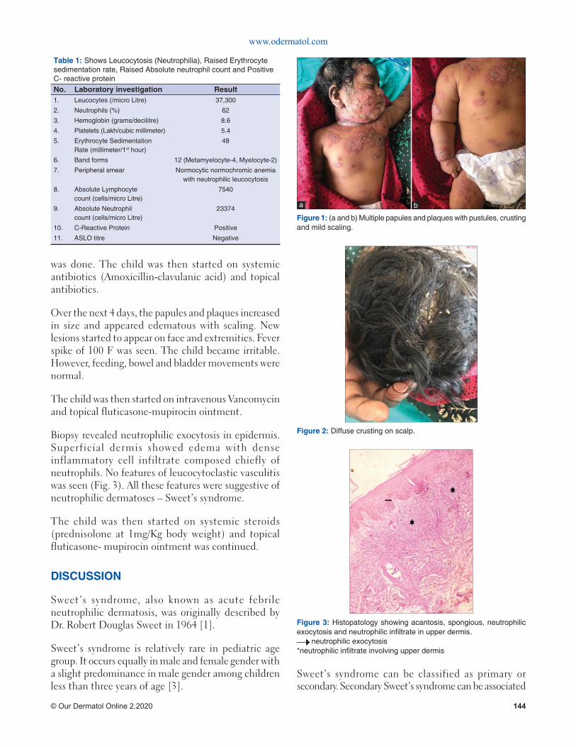
www.odermatol.com
© Our Dermatol Online 2.2020 144
was done. The child was then started on systemic antibiotics (Amoxicillin-clavulanic acid) and topical antibiotics.
Over the next 4 days, the papules and plaques increased in size and appeared edematous with scaling. New lesions started to appear on face and extremities. Fever spike of 100 F was seen. The child became irritable. However, feeding, bowel and bladder movements were normal.
The child was then started on intravenous Vancomycin and topical fluticasone-mupirocin ointment.
Biopsy revealed neutrophilic exocytosis in epidermis. Superficial dermis showed edema with dense inflammatory cell infiltrate composed chiefly of neutrophils. No features of leucocytoclastic vasculitis was seen (Fig. 3). All these features were suggestive of neutrophilic dermatoses – Sweet’s syndrome.
The child was then started on systemic steroids (prednisolone at 1mg/Kg body weight) and topical fluticasone- mupirocin ointment was continued.
DISCUSSION
Sweet’s syndrome, also known as acute febrile neutrophilic dermatosis, was originally described by Dr. Robert Douglas Sweet in 1964 [1].
Sweet’s syndrome is relatively rare in pediatric age group. It occurs equally in male and female gender with a slight predominance in male gender among children less than three years of age [3].
Sweet’s syndrome can be classified as primary or secondary. Secondary Sweet’s syndrome can be associated
Figure 2: Diffuse crusting on scalp.
Figure 3: Histopatology showing acantosis, spongious, neutrophilic exocytosis and neutrophilic infi ltrate in upper dermis. neutrophilic exocytosis*neutrophilic infi ltrate involving upper dermis
Table 1: Shows Leucocytosis (Neutrophilia), Raised Erythrocyte sedimentation rate, Raised Absolute neutrophil count and Positive C- reactive proteinNo. Laboratory investigation Result1. Leucocytes (/micro Litre) 37,300
2. Neutrophils (%) 62
3. Hemoglobin (grams/decilitre) 8.6
4. Platelets (Lakh/cubic millimeter) 5.4
5. Erythrocyte Sedimentation Rate (millimeter/1st hour)
48
6. Band forms 12 (Metamyelocyte-4, Myelocyte-2)
7. Peripheral smear Normocytic normochromic anemia with neutrophilic leucocytosis
8. Absolute Lymphocyte count (cells/micro Litre)
7540
9. Absolute Neutrophil count (cells/micro Litre)
23374
10. C-Reactive Protein Positive
11. ASLO titre Negative
Figure 1: (a and b) Multiple papules and plaques with pustules, crusting and mild scaling.
ba
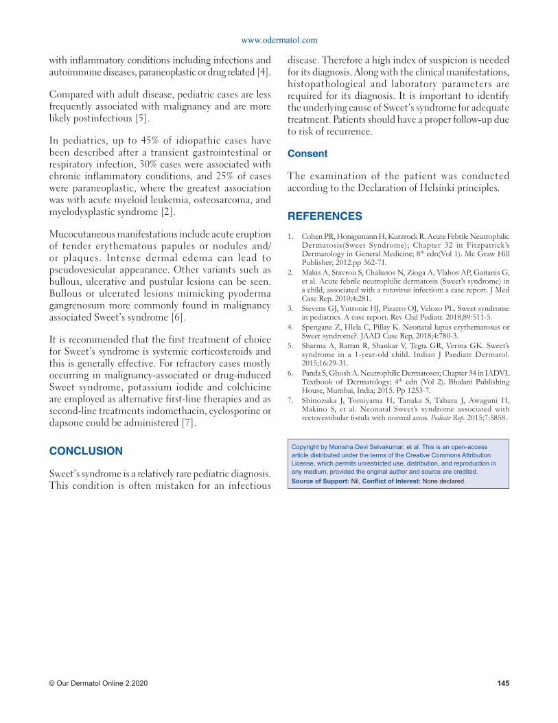
www.odermatol.com
© Our Dermatol Online 2.2020 145
with inflammatory conditions including infections and autoimmune diseases, paraneoplastic or drug related [4].
Compared with adult disease, pediatric cases are less frequently associated with malignancy and are more likely postinfectious [5].
In pediatrics, up to 45% of idiopathic cases have been described after a transient gastrointestinal or respiratory infection, 30% cases were associated with chronic inflammatory conditions, and 25% of cases were paraneoplastic, where the greatest association was with acute myeloid leukemia, osteosarcoma, and myelodysplastic syndrome [2].
Mucocutaneous manifestations include acute eruption of tender erythematous papules or nodules and/or plaques. Intense dermal edema can lead to pseudovesicular appearance. Other variants such as bullous, ulcerative and pustular lesions can be seen. Bullous or ulcerated lesions mimicking pyoderma gangrenosum more commonly found in malignancy associated Sweet’s syndrome [6].
It is recommended that the first treatment of choice for Sweet’s syndrome is systemic corticosteroids and this is generally effective. For refractory cases mostly occurring in malignancy-associated or drug-induced Sweet syndrome, potassium iodide and colchicine are employed as alternative first-line therapies and as second-line treatments indomethacin, cyclosporine or dapsone could be administered [7].
CONCLUSION
Sweet’s syndrome is a relatively rare pediatric diagnosis. This condition is often mistaken for an infectious
disease. Therefore a high index of suspicion is needed for its diagnosis. Along with the clinical manifestations, histopathological and laboratory parameters are required for its diagnosis. It is important to identify the underlying cause of Sweet’s syndrome for adequate treatment. Patients should have a proper follow-up due to risk of recurrence.
Consent
The examination of the patient was conducted according to the Declaration of Helsinki principles.
REFERENCES
1. Cohen PR, Honigsmann H, Kurzrock R. Acute Febrile Neutrophilic Dermatosis(Sweet Syndrome); Chapter 32 in Fitzpatrick’s Dermatology in General Medicine; 8th edn(Vol 1). Mc Graw Hill Publisher; 2012.pp 362-71.
2. Makis A, Stavrou S, Chaliasos N, Zioga A, Vlahos AP, Gaitanis G, et al. Acute febrile neutrophilic dermatosis (Sweet’s syndrome) in a child, associated with a rotavirus infection: a case report. J Med Case Rep. 2010;4:281.
3. Stevens GJ, Yutronic HJ, Pizarro OJ, Velozo PL. Sweet syndrome in pediatrics. A case report. Rev Chil Pediatr. 2018;89:511-5.
4. Spengane Z, Hlela C, Pillay K. Neonatal lupus erythematosus or Sweet syndrome?. JAAD Case Rep, 2018;4:780-3.
5. Sharma A, Rattan R, Shankar V, Tegta GR, Verma GK. Sweet’s syndrome in a 1-year-old child. Indian J Paediatr Dermatol. 2015;16:29-31.
6. Panda S, Ghosh A. Neutrophilic Dermatoses; Chapter 34 in IADVL Textbook of Dermatology; 4th edn (Vol 2). Bhalani Publishing House, Mumbai, India; 2015. Pp 1253-7.
7. Shinozuka J, Tomiyama H, Tanaka S, Tahara J, Awaguni H, Makino S, et al. Neonatal Sweet’s syndrome associated with rectovestibular fi stula with normal anus. Pediatr Rep. 2015;7:5858.
Copyright by Monisha Devi Selvakumar, et al. This is an open-access article distributed under the terms of the Creative Commons Attribution License, which permits unrestricted use, distribution, and reproduction in any medium, provided the original author and source are credited.Source of Support: Nil, Confl ict of Interest: None declared.
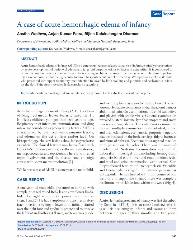
Our Dermatology Online
© Our Dermatol Online 2.2020 146
INTRODUCTION
Acute hemorrhagic edema of infancy (AHEI) is a form of benign cutaneous leukocyteclastic vasculitis [1]. It affects children younger than two years of age. Respiratory tract infections, immunization, and drug intake are considered as precipitating factors. AHEI is characterized by fever, ecchymotic,purpuric lesions, and edema on the extremities and/or face. On histopathology, the skin lesions show leukocytoclastic vasculitis. The clinical features may be confused with Henoch-Schönlein purpura, erythema multiforme, meningococcemia, and septicemia. There is no internal organ involvement, and the disease runs a benign course with spontaneous resolution [2].
We Report a case of AHEI in a one year old male child.
CASE REPORT
A one year old male child presented to our opd with complaint of red raised Itchy lesions over lower limbs, buttocks, right arm and ear pinna since one day (Figs. 1 and 2). He had symptoms of upper respiratory tract infection, swelling of lower limb, initially started over the right foot and gradually progressed to involve the left foot and both legs till knee, and fever one episode
and vomiting four days prior to the eruption of the skin lesions. He had no complaints of diarrhea, joint pain, or abdominal pain. On examination, the child was active and playful with stable vitals. General examination revealed bilateral inguinal lymphadenopathy and grade two non-pitting edema. The cutaneous examination showed multiple symmetrically distributed, round and oval, edematous, ecchymotic, purpuric, targetoid plaques localized on the both feet, legs, thighs, buttocks and pinna of right ear. Erythematous targetoid macules were present on the soles. There was no mucosal involvement. Systemic Examination was normal. Laboratory investigations, including hemoglobin, complete blood count, liver and renal function tests, and stool and urine examination, were normal. Skin Biopsy showed features of leucocytoclastic Vasculitis and Dermal edema (Fig. 3). DIF showed perivascular C3 deposits. He was treated with short course of oral steroids and supportive therapy there was complete resolution of the skin lesions within one week (Fig. 4).
DISCUSSION
Acute Hemorrhagic edema of infancy was first described by Snow in 1913 [3]. It is an acute Leukocytoclastic vasculitis occurring in infants and young children between the ages of three months and two years.
ABSTRACT
Acute hemorrhagic edema of infancy (AHEI) is a cutaneous leukocytoclastic vasculitis of infants, clinically characterized by acute development of peripheral edema and targetoid purpuric lesions on face and extremities. It is considered to be an uncommon form of cutaneous vasculitis occurring in children younger than two years old. The clinical picture has a violent onset, a short benign course followed by spontaneous complete recovery. We report a case of a male child who presented with upper respiratory tract infection followed by limb swelling and purpuric and ecchymotic lesions on the skin. Skin biopsy revealed leukocytoclastic vasculitis.
Key words: Acute hemorrhagic edema of infancy; Ecchymoses; Leukocytoclastic vasculitis; Purpura
Case Report
A case of acute hemorrhagic edema of infancyA case of acute hemorrhagic edema of infancyAastha Wadhwa, Anjan Kumar Patra, Bijina Kolukulangara Dharman
Department of Dermatology, MVJ Medical College and Research Hospital, Bangalore, India
Corresponding author: Dr. Aastha Wadhwa, E-mail: [email protected]
How to cite this article: Wadhwa A, Patra AK, Dharman BK. A case of acute hemorrhagic edema of infancy. Our Dermatol Online. 2020;11(2):146-148.Submission: 04.07.2019; Acceptance: 05.09.2019DOI: 10.7241/ourd.20202.7
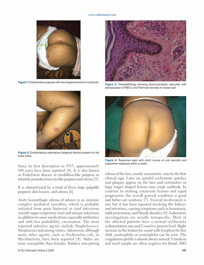
www.odermatol.com
© Our Dermatol Online 2.2020 147
Since its first description in 1913, approximately 100 cases have been reported [4]. It is also known as Finkelstein disease or medallion-like purpura or infantile postinfectious iris-like purpura and edema [5].
It is characterized by a triad of fever, large palpable purpuric skin lesions, and edema [6].
Acute hemorrhagic edema of infancy is an immune complex mediated vasculitis, which is probably initiated from prior bacterial or viral infections (mostly upper respiratory tract and urinary infection) in addition to some medications especially antibiotics and with less probability, vaccination. The most reported infective agents include Staphylococci, Streptococci and among viruses, Adenovirus, although many other agents, such as Escherichia coli, or Mycobacteria, have been reported [4]. Males are more susceptible than females. Painless non-pitting
edema of the face, mostly asymmetric, may be the first clinical sign. Later on, painful ecchymotic patches and plaques appear on the face and extremities or large target shaped lesions may erupt suddenly. In contrast to striking cutaneous lesions and rapid progression, the overall general condition is good and babies are nontoxic [7]. Visceral involvement is rare but it has been reported involving the kidneys and intestines, causing symptoms such as hematuria, mild proteinuria, and bloody diarrhea [8]. Laboratory investigations are usually nonspecific. Most of the affected patients have a normal erythrocyte sedimentation rate and C-reactive protein level. Slight increase in the leukocytic count with lymphocytic line shift, neutrophilia or eosinophilia can be seen. The coagulation profile is almost always normal. Urinalysis and stool sample are often negative for blood. ASO
Figure 1: Erythematous papules with few targetoid lesions on buttocks.
Figure 2: Erythematous edematous targetoid lesions present on the lower limbs.
Figure 3: Histopathology showing leucocytoclastic vasculitis with extravasation of RBC’s and Fibrinoid necrosis of vessel wall.
Figure 4: Response seen with short course of oral steroids and supportive measures within a week.

www.odermatol.com
© Our Dermatol Online 2.2020 148
titer, antinuclear antibodies, antideoxyribonucleic acid, and rheumatoid factor are usually negative. High serum levels of gamma globulins and immune complexes have been described [9]. Histologic features of AHEI are consistent with small vessel vasculitis of both capillaries and post capillary venules of the upper and the middle dermis, showing typical leukocytoclastic vasculitis with or without fibrinoid necrosis and a deep perivascular and interstitial infiltrate composed mostly of neutrophils with abundant nuclear dust [4]. The chief differential diagnosis of AHEI is Henoch schonlein purpura. The age of onset of Acute haemorrhagic edema of infancy is two to twenty four months is lower when compared to Henoch schonlein purpura four to seven years. Systemic complications (arthralgia, gastrointestinal bleeding, and nephritis) are common in Henoch schonlein purpura. The disease runs a benign course with complete spontaneous recovery occurring in one to three weeks, although relapses have been reported rarely. Treatment with oral corticosteroids has been reported, but this is unnecessary due to its spontaneous recovery [10].
CONCLUSION
Acute haemorrhagic edema of infancy is a benign disorder despite its dramatic appearance There is a contrast between the acuteness of skin lesions and good general condition of the patient. It is an unique disorder and needs to be differentiated from Henoch schonlein purpura.
Consent
The examination of the patient was conducted according to the Declaration of Helsinki principles.
REFERENCES
1. Taieb A, Legrain V. Acute hemorrhagic edema of the skin in infancy. In: Textbook of pediatric Dermatology (2nd ed). United Kingdom. Blackwell Science; 2002:1569-73.
2. Dongre A, Adhe V, Kothari D, Kardekar S, Khopkar U. Acute hemorrhagic edema of infancy: A report of two cases. Indian J Dermatol Venereol Leprol. 2012;78:121.
3. Snow IM. Purpura, urticaria, and angioneurotic edema of the hands and feet in a nursing baby. JAMA. 1913;61:18-9.
4. Saraclar Y, Tinaztepe K, Adalioglu G, Tuncer A. Acute hemorrhagic edema of infancy (AHEI) – A variant of Henoch-Schönlein purpura or a distinct clinical entity? J Allergy Clin Immunol. 1990;86:473-83.
5. Shah D, Goraya JS, Poddar B, Parmar VR. Acute infantile hemorrhagic edema and Henoch-Schönlein purpura overlap in a child. Pediatr Dermatol. 2002;19:92-3.
6. McDougall CM, Ismail SK, Osmerod A. Acute hemorrhagic oedema of infancy. Arch Dis Child. 2005;90:316.
7. da Silva Manzoni AP, Viecili JB, de Andrade CB, Kruse RL, Bakos L, et al. Acute hemorrhagic edema of infancy: A case report. Int J Dermatol. 2004;43:48-51.
8. Garty BZ, Ofer I, Finkelstein Y. Acute hemorrhagic edema of infancy. Isr Med Assoc J. 2002;4:228-9.
9. Alharbi MS. Role of systemic steroids in acute hemorrhagic edema of infancy: report of two cases. Int J Adv Med. 2019;6:537-41.
10. Jindal SR, Kura MM. Acute hemorrhagic edema of infancy-a rare entity. Indian Dermatol Online J. 2013;4:106-8.
Copyright by Aastha Wadhwa, et al. This is an open-access article distributed under the terms of the Creative Commons Attribution License, which permits unrestricted use, distribution, and reproduction in any medium, provided the original author and source are credited.Source of Support: Nil, Confl ict of Interest: None declared.
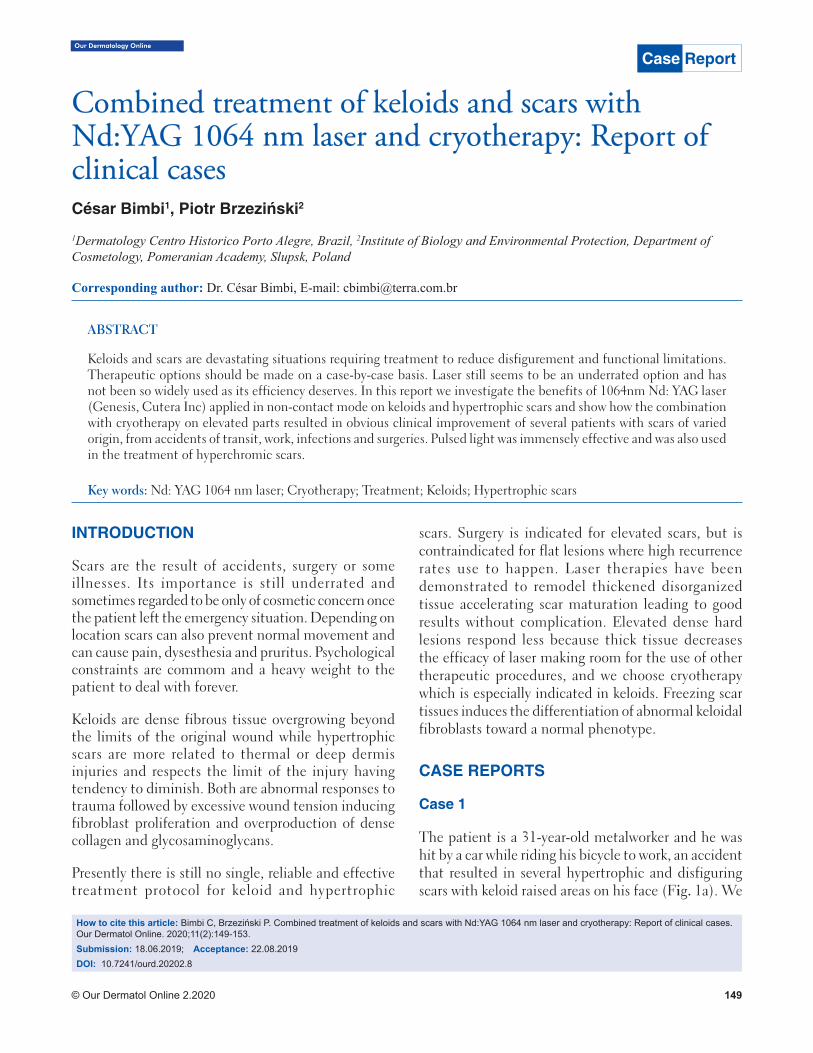
Our Dermatology Online
© Our Dermatol Online 2.2020 149
INTRODUCTION
Scars are the result of accidents, surgery or some illnesses. Its importance is still underrated and sometimes regarded to be only of cosmetic concern once the patient left the emergency situation. Depending on location scars can also prevent normal movement and can cause pain, dysesthesia and pruritus. Psychological constraints are commom and a heavy weight to the patient to deal with forever.
Keloids are dense fibrous tissue overgrowing beyond the limits of the original wound while hypertrophic scars are more related to thermal or deep dermis injuries and respects the limit of the injury having tendency to diminish. Both are abnormal responses to trauma followed by excessive wound tension inducing fibroblast proliferation and overproduction of dense collagen and glycosaminoglycans.
Presently there is still no single, reliable and effective treatment protocol for keloid and hypertrophic
scars. Surgery is indicated for elevated scars, but is contraindicated for flat lesions where high recurrence rates use to happen. Laser therapies have been demonstrated to remodel thickened disorganized tissue accelerating scar maturation leading to good results without complication. Elevated dense hard lesions respond less because thick tissue decreases the efficacy of laser making room for the use of other therapeutic procedures, and we choose cryotherapy which is especially indicated in keloids. Freezing scar tissues induces the differentiation of abnormal keloidal fibroblasts toward a normal phenotype.
CASE REPORTS
Case 1
The patient is a 31-year-old metalworker and he was hit by a car while riding his bicycle to work, an accident that resulted in several hypertrophic and disfiguring scars with keloid raised areas on his face (Fig. 1a). We
ABSTRACT
Keloids and scars are devastating situations requiring treatment to reduce disfigurement and functional limitations. Therapeutic options should be made on a case-by-case basis. Laser still seems to be an underrated option and has not been so widely used as its efficiency deserves. In this report we investigate the benefits of 1064nm Nd: YAG laser (Genesis, Cutera Inc) applied in non-contact mode on keloids and hypertrophic scars and show how the combination with cryotherapy on elevated parts resulted in obvious clinical improvement of several patients with scars of varied origin, from accidents of transit, work, infections and surgeries. Pulsed light was immensely effective and was also used in the treatment of hyperchromic scars.
Key words: Nd: YAG 1064 nm laser; Cryotherapy; Treatment; Keloids; Hypertrophic scars
Case Report
Combined treatment of keloids and scars with Combined treatment of keloids and scars with Nd:YAG 1064 nm laser and cryotherapy: Report of Nd:YAG 1064 nm laser and cryotherapy: Report of clinical casesclinical casesCésar Bimbi1, Piotr Brzeziński2
1Dermatology Centro Historico Porto Alegre, Brazil, 2Institute of Biology and Environmental Protection, Department of Cosmetology, Pomeranian Academy, Slupsk, Poland
Corresponding author: Dr. César Bimbi, E-mail: [email protected]
How to cite this article: Bimbi C, Brzeziński P. Combined treatment of keloids and scars with Nd:YAG 1064 nm laser and cryotherapy: Report of clinical cases. Our Dermatol Online. 2020;11(2):149-153.Submission: 18.06.2019; Acceptance: 22.08.2019DOI: 10.7241/ourd.20202.8
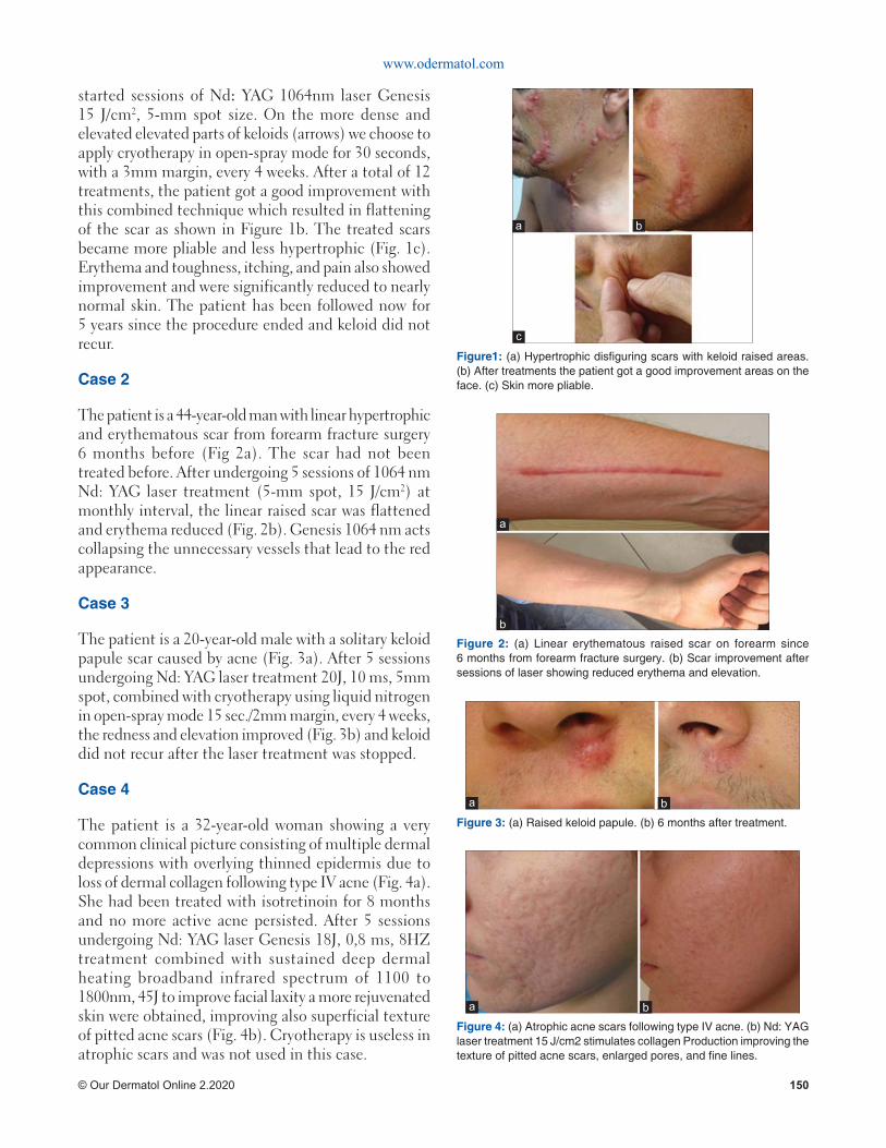
www.odermatol.com
© Our Dermatol Online 2.2020 150
started sessions of Nd: YAG 1064nm laser Genesis 15 J/cm2, 5-mm spot size. On the more dense and elevated elevated parts of keloids (arrows) we choose to apply cryotherapy in open-spray mode for 30 seconds, with a 3mm margin, every 4 weeks. After a total of 12 treatments, the patient got a good improvement with this combined technique which resulted in flattening of the scar as shown in Figure 1b. The treated scars became more pliable and less hypertrophic (Fig. 1c). Erythema and toughness, itching, and pain also showed improvement and were significantly reduced to nearly normal skin. The patient has been followed now for 5 years since the procedure ended and keloid did not recur.
Case 2
The patient is a 44-year-old man with linear hypertrophic and erythematous scar from forearm fracture surgery 6 months before (Fig 2a). The scar had not been treated before. After undergoing 5 sessions of 1064 nm Nd: YAG laser treatment (5-mm spot, 15 J/cm2) at monthly interval, the linear raised scar was flattened and erythema reduced (Fig. 2b). Genesis 1064 nm acts collapsing the unnecessary vessels that lead to the red appearance.
Case 3
The patient is a 20-year-old male with a solitary keloid papule scar caused by acne (Fig. 3a). After 5 sessions undergoing Nd: YAG laser treatment 20J, 10 ms, 5mm spot, combined with cryotherapy using liquid nitrogen in open-spray mode 15 sec./2mm margin, every 4 weeks, the redness and elevation improved (Fig. 3b) and keloid did not recur after the laser treatment was stopped.
Case 4
The patient is a 32-year-old woman showing a very common clinical picture consisting of multiple dermal depressions with overlying thinned epidermis due to loss of dermal collagen following type IV acne (Fig. 4a). She had been treated with isotretinoin for 8 months and no more active acne persisted. After 5 sessions undergoing Nd: YAG laser Genesis 18J, 0,8 ms, 8HZ treatment combined with sustained deep dermal heating broadband infrared spectrum of 1100 to 1800nm, 45J to improve facial laxity a more rejuvenated skin were obtained, improving also superficial texture of pitted acne scars (Fig. 4b). Cryotherapy is useless in atrophic scars and was not used in this case.
Figure 4: (a) Atrophic acne scars following type IV acne. (b) Nd: YAG laser treatment 15 J/cm2 stimulates collagen Production improving the texture of pitted acne scars, enlarged pores, and fi ne lines.
ba
Figure 3: (a) Raised keloid papule. (b) 6 months after treatment.
ba
Figure 2: (a) Linear erythematous raised scar on forearm since 6 months from forearm fracture surgery. (b) Scar improvement after sessions of laser showing reduced erythema and elevation.
b
a
Figure1: (a) Hypertrophic disfi guring scars with keloid raised areas. (b) After treatments the patient got a good improvement areas on the face. (c) Skin more pliable.
c
ba
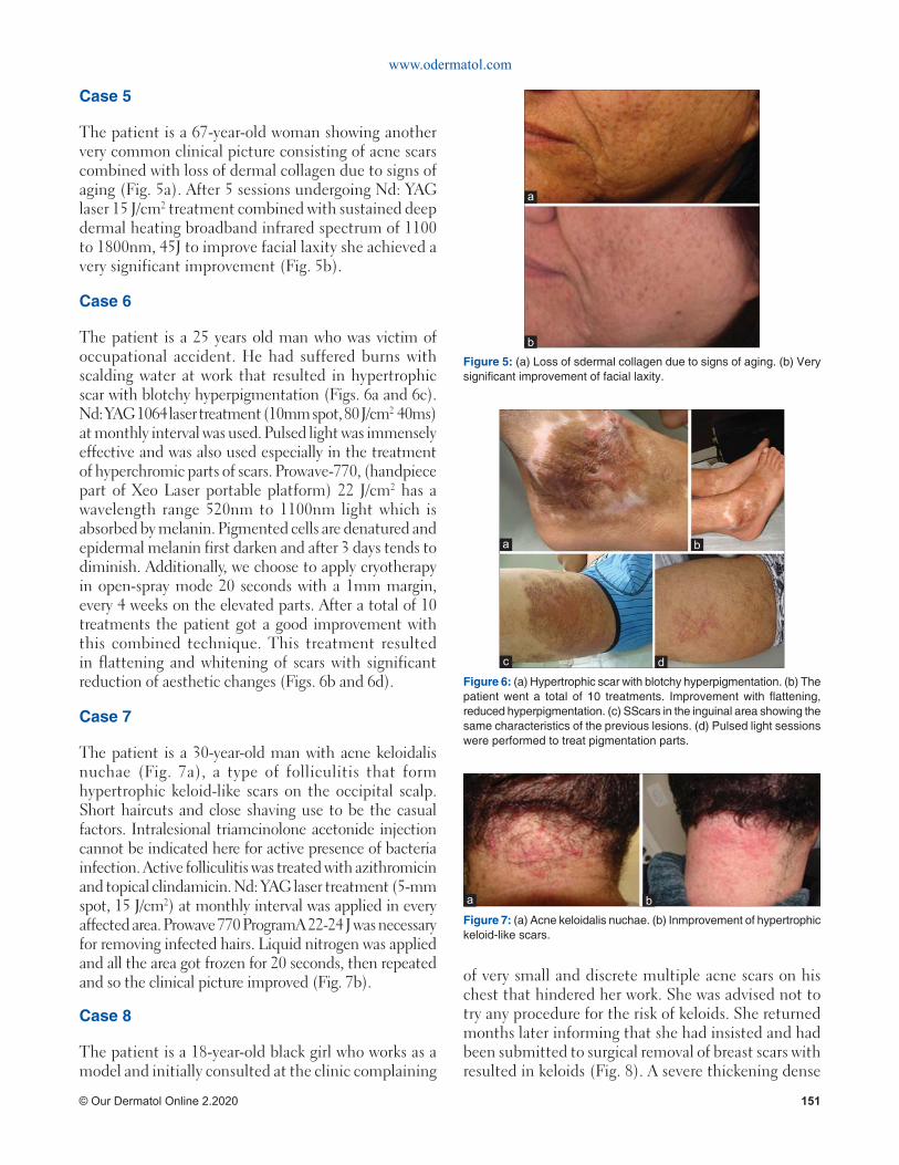
www.odermatol.com
© Our Dermatol Online 2.2020 151
Case 5
The patient is a 67-year-old woman showing another very common clinical picture consisting of acne scars combined with loss of dermal collagen due to signs of aging (Fig. 5a). After 5 sessions undergoing Nd: YAG laser 15 J/cm2 treatment combined with sustained deep dermal heating broadband infrared spectrum of 1100 to 1800nm, 45J to improve facial laxity she achieved a very significant improvement (Fig. 5b).
Case 6
The patient is a 25 years old man who was victim of occupational accident. He had suffered burns with scalding water at work that resulted in hypertrophic scar with blotchy hyperpigmentation (Figs. 6a and 6c). Nd: YAG 1064 laser treatment (10mm spot, 80 J/cm2 40ms) at monthly interval was used. Pulsed light was immensely effective and was also used especially in the treatment of hyperchromic parts of scars. Prowave-770, (handpiece part of Xeo Laser portable platform) 22 J/cm2 has a wavelength range 520nm to 1100nm light which is absorbed by melanin. Pigmented cells are denatured and epidermal melanin first darken and after 3 days tends to diminish. Additionally, we choose to apply cryotherapy in open-spray mode 20 seconds with a 1mm margin, every 4 weeks on the elevated parts. After a total of 10 treatments the patient got a good improvement with this combined technique. This treatment resulted in flattening and whitening of scars with significant reduction of aesthetic changes (Figs. 6b and 6d).
Case 7
The patient is a 30-year-old man with acne keloidalis nuchae (Fig. 7a), a type of folliculitis that form hypertrophic keloid-like scars on the occipital scalp. Short haircuts and close shaving use to be the casual factors. Intralesional triamcinolone acetonide injection cannot be indicated here for active presence of bacteria infection. Active folliculitis was treated with azithromicin and topical clindamicin. Nd: YAG laser treatment (5-mm spot, 15 J/cm2) at monthly interval was applied in every affected area. Prowave 770 ProgramA 22-24 J was necessary for removing infected hairs. Liquid nitrogen was applied and all the area got frozen for 20 seconds, then repeated and so the clinical picture improved (Fig. 7b).
Case 8
The patient is a 18-year-old black girl who works as a model and initially consulted at the clinic complaining
of very small and discrete multiple acne scars on his chest that hindered her work. She was advised not to try any procedure for the risk of keloids. She returned months later informing that she had insisted and had been submitted to surgical removal of breast scars with resulted in keloids (Fig. 8). A severe thickening dense
Figure 7: (a) Acne keloidalis nuchae. (b) Inmprovement of hypertrophic keloid-like scars.
ba
Figure 6: (a) Hypertrophic scar with blotchy hyperpigmentation. (b) The patient went a total of 10 treatments. Improvement with fl attening, reduced hyperpigmentation. (c) SScars in the inguinal area showing the same characteristics of the previous lesions. (d) Pulsed light sessions were performed to treat pigmentation parts.
dc
ba
Figure 5: (a) Loss of sdermal collagen due to signs of aging. (b) Very signifi cant improvement of facial laxity.
b
a
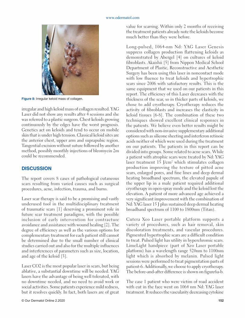
www.odermatol.com
© Our Dermatol Online 2.2020 152
irregular and high keloid mass of collagen resulted. YAG Laser did not show any results after 4 sessions and she was referred to a plastic surgeon. Chest keloids growing continuously by the edges have the worst prognosis. Genetics act on keloids and tend to occur on mobile skin that is under high tension. Classical keloid sites are the anterior chest, upper arm and suprapubic region. Tangential excision without suture followed by another method, possibly monthly injections of bleomycin 2m could be recommended.
DISCUSSION
The report covers 8 cases of pathological cutaneous scars resulting from varied causes such as surgical procedures, acne, infection, trauma, and burns.
Laser scar therapy is said to be a promising and vastly underused tool in the multidisciplinary treatment of traumatic scars [1] deserving a prominent role in future scar treatment paradigms, with the possible inclusion of early intervention for contracture avoidance and assistance with wound healing [2]. The degree of efficiency as well as the various options for complementary treatment for each patient still cannot be determined due to the small number of clinical studies carried out and also for the multiple influences and interferences of parameters such as size, location, and age of the keloid [3].
Laser CO2 is the most popular laser in scars, but being ablative, a substantial downtime will be needed. YAG lasers have the advantage of being well tolerated, with no downtime needed, and no need to avoid work or social activities. Some patients experience mild redness, but it resolves quickly. In fact, both lasers are of great
value for scarring. Within only 2 months of receiving the treatment patients already note the keloids become much better than they were before.
Long-pulsed, 1064-nm Nd: YAG Laser Genesis suppress collagen production flattening keloids as demonstrated by Abergel [4] on cultures of keloid fibroblasts. Akaishii [5] from Nippon Medical School Department of Plastic, Reconstructive and Aesthetic Surgery has been using this laser in noncontact mode with low fluence to treat keloids and hypertrophic scars since 2006 with satisfactory results. This is the same equipment that we used on our patients in this report. The efficiency of this Laser decreases with the thickness of the scar, so in thicker parts of keloids, we chose to add cryotherapy. Cryotherapy reduces the activity of fibroblasts and increases the elasticity in keloid tissues [6-8]. The combination of these two techniques showed excellent clinical responses in the patients. We believe even better results might be considered with non-invasive supplementary additional options such as silicone sheeting and interferon retinoic acids neither of which were used during the treatment on our patients. The patients in this report can be divided into groups. Some related to acne scars. While a patient with atrophic scars were treated by Nd: YAG laser treatment 15 J/cm2 which stimulates collagen production improving the texture of pitted acne scars, enlarged pores, and fine lines and deep dermal heating broadband spectrum, the elevated papule of the upper lip in a male patient required additional cryotherapy in open-spray mode and the keloid lost the elevation. A patient of more advanced age achieved a very significant improvement with the combination of Nd: YAG laser 15 J plus sustained deep dermal heating broadband spectrum of 1100 to 1800nm (Titan).
Cutera Xeo Laser portable platform supports a variety of procedures, such as hair removal, skin discoloration treatments, and vascular procedures. Pigmented hypertrophic scars are a difficult condition to treat. Pulsed light has utility in hyperchromic scars. LimeLight handpiece (part of Xeo Laser portable platform) has a wavelength range 520nm to 1100nm light which is absorbed by melanin. Pulsed light sessions were performed to treat pigmentation parts of patient-6. Additionally, we choose to apply cryotherapy. The before-and-after difference is shown on figure6a-b.
The case 1 patient who were victim of road accident with cut in the face went on 1064 nm Nd: YAG laser treatment. It reduces the vascularity decreasing cytokine
Figure 8: Irregular keloid mass of collagen.
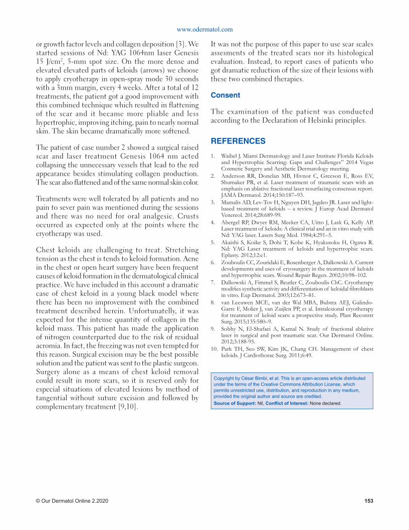
www.odermatol.com
© Our Dermatol Online 2.2020 153
or growth factor levels and collagen deposition [3]. We started sessions of Nd: YAG 1064nm laser Genesis 15 J/cm2, 5-mm spot size. On the more dense and elevated elevated parts of keloids (arrows) we choose to apply cryotherapy in open-spray mode 30 seconds with a 3mm margin, every 4 weeks. After a total of 12 treatments, the patient got a good improvement with this combined technique which resulted in flattening of the scar and it became more pliable and less hypertrophic, improving itching, pain to nearly normal skin. The skin became dramatically more softened.
The patient of case number 2 showed a surgical raised scar and laser treatment Genesis 1064 nm acted collapsing the unnecessary vessels that lead to the red appearance besides stimulating collagen production. The scar also flattened and of the same normal skin color.
Treatments were well tolerated by all patients and no pain to sever pain was mentioned during the sessions and there was no need for oral analgesic. Crusts occurred as expected only at the points where the cryotherapy was used.
Chest keloids are challenging to treat. Stretching tension as the chest is tends to keloid formation. Acne in the chest or open heart surgery have been frequent causes of keloid formation in the dermatological clinical practice. We have included in this account a dramatic case of chest keloid in a young black model where there has been no improvement with the combined treatment described herein. Unfortunatelly, it was expected for the intense quantity of collagen in the keloid mass. This patient has made the application of nitrogen counterparted due to the risk of residual acromia. In fact, the freezing was not even tempted for this reason. Surgical excision may be the best possible solution and the patient was sent to the plastic surgeon. Surgery alone as a means of chest keloid removal could result in more scars, so it is reserved only for especial situations of elevated lesions by method of tangential without suture excision and followed by complementary treatment [9,10].
It was not the purpose of this paper to use scar scales assesments of the treated scars nor its histological evaluation. Instead, to report cases of patients who got dramatic reduction of the size of their lesions with these two combined therapies.
Consent
The examination of the patient was conducted according to the Declaration of Helsinki principles.
REFERENCES
1. Waibel J. Miami Dermatology and Laser Institute Florida Keloids and Hypertrophic Scarring: Gaps and Challenges” 2014 Vegas Cosmetic Surgery and Aesthetic Dermatology meeting.
2. Anderson RR, Donelan MB, Hivnor C, Greeson E, Ross EV, Shumaker PR, et al. Laser treatment of traumatic scars with an emphasis on ablative fractional laser resurfacing consensus report. JAMA Dermatol. 2014;150:187–93.
3. Mamalis AD, Lev-Tov H, Nguyen DH, Jagdeo JR. Laser and light-based treatment of keloids – a review. J Europ Acad Dermatol Venereol. 2014;28:689-99.
4. Abergel RP, Dwyer RM, Meeker CA, Uitto J, Lask G, Kelly AP. Laser treatment of keloids: A clinical trial and an in vitro study with Nd: YAG laser. Lasers Surg Med. 1984;4:291–5.
5. Akaishi S, Koike S, Dohi T, Kobe K, Hyakusoku H, Ogawa R. Nd: YAG Laser treatment of keloids and hypertrophic scars. Eplasty. 2012;12:e1.
6. Zouboulis CC, Zouridaki E, Rosenberger A, Dalkowski A. Current developments and uses of cryosurgery in the treatment of keloids and hypertrophic scars. Wound Repair Regen. 2002;10:98–102.
7. Dalkowski A, Fimmel S, Beutler C, Zouboulis ChC. Cryotherapy modifi es synthetic activity and differentiation of keloidal fi broblasts in vitro. Exp Dermatol. 2003;12:673–81.
8. van Leeuwen MCE, van der Wal MBA, Bulstra AEJ, Galindo-Garre F, Molier J, van Zuijlen PP, et al. Intralesional cryotherapy for treatment of keloid scars: a prospective study. Plast Reconstr Surg. 2015;135:580–9.
9. Sobhy N, El-Shafaei A, Kamal N. Study of fractional ablative laser in surgical and post traumatic scar. Our Dermatol Online. 2012;3:188-95.
10. Park TH, Seo SW, Kim JK, Chang CH. Management of chest keloids. J Cardiothorac Surg. 2011;6:49.
Copyright by César Bimbi, et al. This is an open-access article distributed under the terms of the Creative Commons Attribution License, which permits unrestricted use, distribution, and reproduction in any medium, provided the original author and source are credited.Source of Support: Nil, Confl ict of Interest: None declared.
Our Dermatology Online
© Our Dermatol Online 2.2020 154
How to cite this article: Baali N, Berrada S, Akhdari N, Hocar O, Rais H, Amal S. Rhinoscleroma: a diagnosis not to be ignored. Our Dermatol Online. 2020;11(2):154-155.Submission: 12.07.2019; Acceptance: 15.09.2019DOI: 10.7241/ourd.20202.9
INTRODUCTION
Rhinoscleroma is a chronic and specific granulomatous disease caused by enterobacteria of the family Klebsiella (Klebsiella rhinoscleromatis) with a tropism for the upper airways. The purpose of our work is to describe, through an observation, the epidemiological-clinical profile of this pathology and to demonstrate its peculiarities of management.
CASE REPORT
Patientaged 47, living in very poor hygienic condition, agriculture by professio, having as antecedent smoking and chronic alcoholism.He was admitted to our formation for a unilateral nasal obstruction, progressive installation, associated with persistent rhinitis evolving since 04 years.Clinical examination revealed a crusted nodular lesion of the right wing of the nose, which had been evolving for 4 years, increasing very gradually in volume (Fig. 1).
The rhino-nasofibroscopy visualized an almost complete obstruction by an ulcerated tumor of the right nasal fossa, with a septal deviation. The oral cavity, pharynx and larynx were normal. HIV, TPHA/VDRL serologies were negative. The diagnosis of rhinoscleroma was confirmed
by the presence of MIKULICZ cells in the pathological study (Figs. 2 and 3).
The first control evolution at 3 weeks was marked by a slight improvement but with persistence of the nasal obstruction, additional surgery was necessary in this case and the patient was referred to the department of otorhinolaryngological surgery.
DISCUSSION
The literature review shows that rhinoscleroma is particularly prevalent in tropical Africa, the Maghreb, Latin America, southern Asia and central Europe [1]. The pathogenesis of the affection is not yet well understood. Genetic predisposition and/or immunodeficiency have been advanced by some authors [2,3].
The diagnosis of rhinoscleroma can be made at the catarrhal stage, by identification of K. rhinoscleromatis on nasal smear culture on specific media. However, most cases are diagnosed, as in our patient, at the granulomatous stage. The germ can be isolated at this stage on the granulated tissue crushers. The histological study of the biopsy pieces makes it possible to highlight the Mikulicz cells, considered as characteristic of the affection[4].These are foamy histiocytes with a single and uniform nucleus, small,
ABSTRACT
Rhinoscleroma is a chronic indolent granulomatous infection of the nose and the upper respiratory tract caused by Klebsiella rhinoscleromatis; this condition is endemic to many regions of the world including North Africa. We present a case of rhinoscleroma in a 47-year-old man with clinically diagnosed nodule in the nasal cavity. The histopathological examination revealed granulation tissue with plasma cells and Mikulicz’s cells. The clinical and morphological picture of the case is a rare opportunity to recall a disease that is often overlooked by dermatologists.
Key words: Rhinoscleroma; Morocco; Klebsiella rhinoscleromatis
Rhinoscleroma: a diagnosis not to be ignoredRhinoscleroma: a diagnosis not to be ignoredNadia Baali1, Sofi a Berrada2, Nadia Akhdari1, Ouafa Hocar1, Hanane Rais2, Said Amal1
1Department of Dermatology, Mohammed VI CHU, Marrakech, Morocco, 2Department of Pathological Anatomy and Cytology, CHU Mohamed VI, Marrakech, Morocco
Corresponding author: Dr. Nadia Baali, E-mail: [email protected]
Case Report
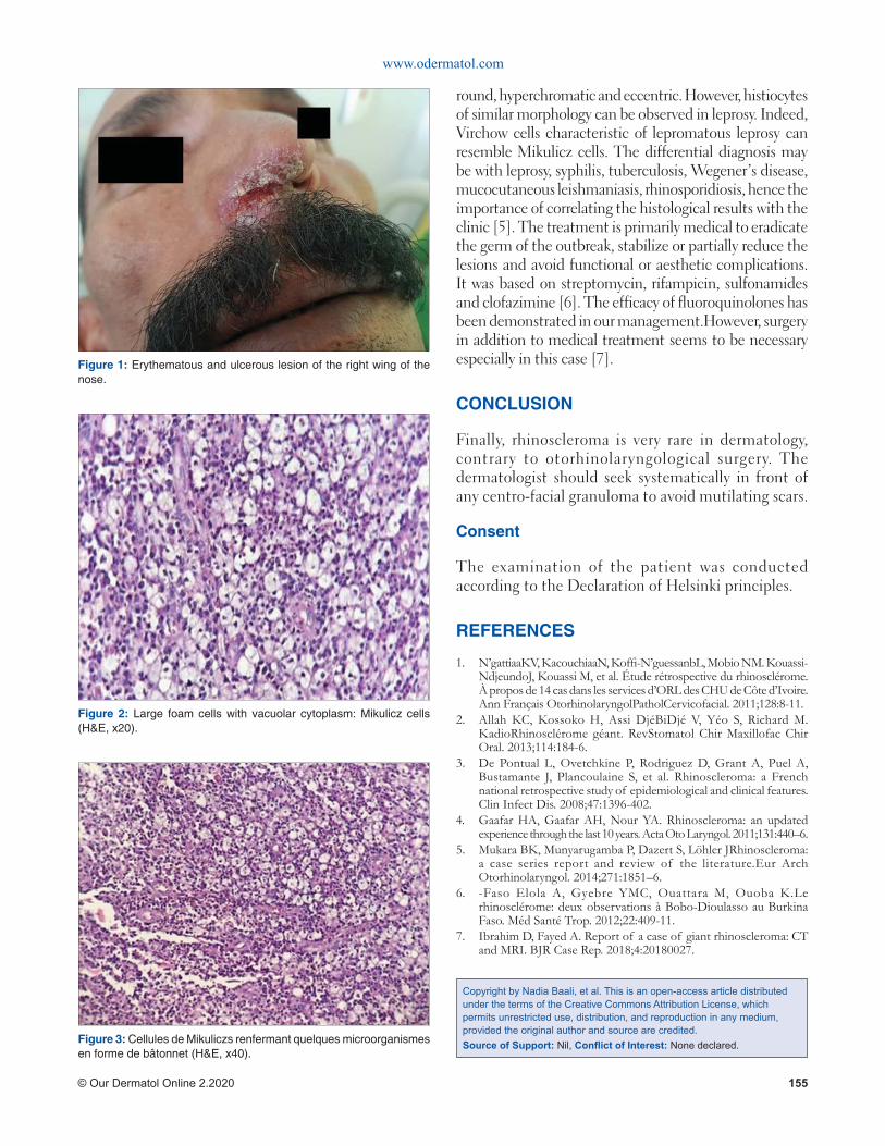
www.odermatol.com
© Our Dermatol Online 2.2020 155
round, hyperchromatic and eccentric. However, histiocytes of similar morphology can be observed in leprosy. Indeed, Virchow cells characteristic of lepromatous leprosy can resemble Mikulicz cells. The differential diagnosis may be with leprosy, syphilis, tuberculosis, Wegener’s disease, mucocutaneous leishmaniasis, rhinosporidiosis, hence the importance of correlating the histological results with the clinic [5]. The treatment is primarily medical to eradicate the germ of the outbreak, stabilize or partially reduce the lesions and avoid functional or aesthetic complications. It was based on streptomycin, rifampicin, sulfonamides and clofazimine [6]. The efficacy of fluoroquinolones has been demonstrated in our management.However, surgery in addition to medical treatment seems to be necessary especially in this case [7].
CONCLUSION
Finally, rhinoscleroma is very rare in dermatology, contrary to otorhinolaryngological surgery. The dermatologist should seek systematically in front of any centro-facial granuloma to avoid mutilating scars.
Consent
The examination of the patient was conducted according to the Declaration of Helsinki principles.
REFERENCES
1. N’gattiaaKV, KacouchiaaN, Koffi -N’guessanbL, Mobio NM. Kouassi-NdjeundoJ, Kouassi M, et al. Étude rétrospective du rhinosclérome. À propos de 14 cas dans les services d’ORL des CHU de Côte d’Ivoire. Ann Français OtorhinolaryngolPatholCervicofacial. 2011;128:8-11.
2. Allah KC, Kossoko H, Assi DjéBiDjé V, Yéo S, Richard M. KadioRhinosclérome géant. RevStomatol Chir Maxillofac Chir Oral. 2013;114:184-6.
3. De Pontual L, Ovetchkine P, Rodriguez D, Grant A, Puel A, Bustamante J, Plancoulaine S, et al. Rhinoscleroma: a French national retrospective study of epidemiological and clinical features. Clin Infect Dis. 2008;47:1396-402.
4. Gaafar HA, Gaafar AH, Nour YA. Rhinoscleroma: an updated experience through the last 10 years. Acta Oto Laryngol. 2011;131:440–6.
5. Mukara BK, Munyarugamba P, Dazert S, Löhler JRhinoscleroma: a case series report and review of the literature.Eur Arch Otorhinolaryngol. 2014;271:1851–6.
6. -Faso Elola A, Gyebre YMC, Ouattara M, Ouoba K.Le rhinosclérome: deux observations à Bobo-Dioulasso au Burkina Faso. Méd Santé Trop. 2012;22:409-11.
7. Ibrahim D, Fayed A. Report of a case of giant rhinoscleroma: CT and MRI. BJR Case Rep. 2018;4:20180027.
Figure 1: Erythematous and ulcerous lesion of the right wing of the nose.
Figure 2: Large foam cells with vacuolar cytoplasm: Mikulicz cells (H&E, x20).
Figure 3: Cellules de Mikuliczs renfermant quelques microorganismes en forme de bâtonnet (H&E, x40).
Copyright by Nadia Baali, et al. This is an open-access article distributed under the terms of the Creative Commons Attribution License, which permits unrestricted use, distribution, and reproduction in any medium, provided the original author and source are credited.Source of Support: Nil, Confl ict of Interest: None declared.
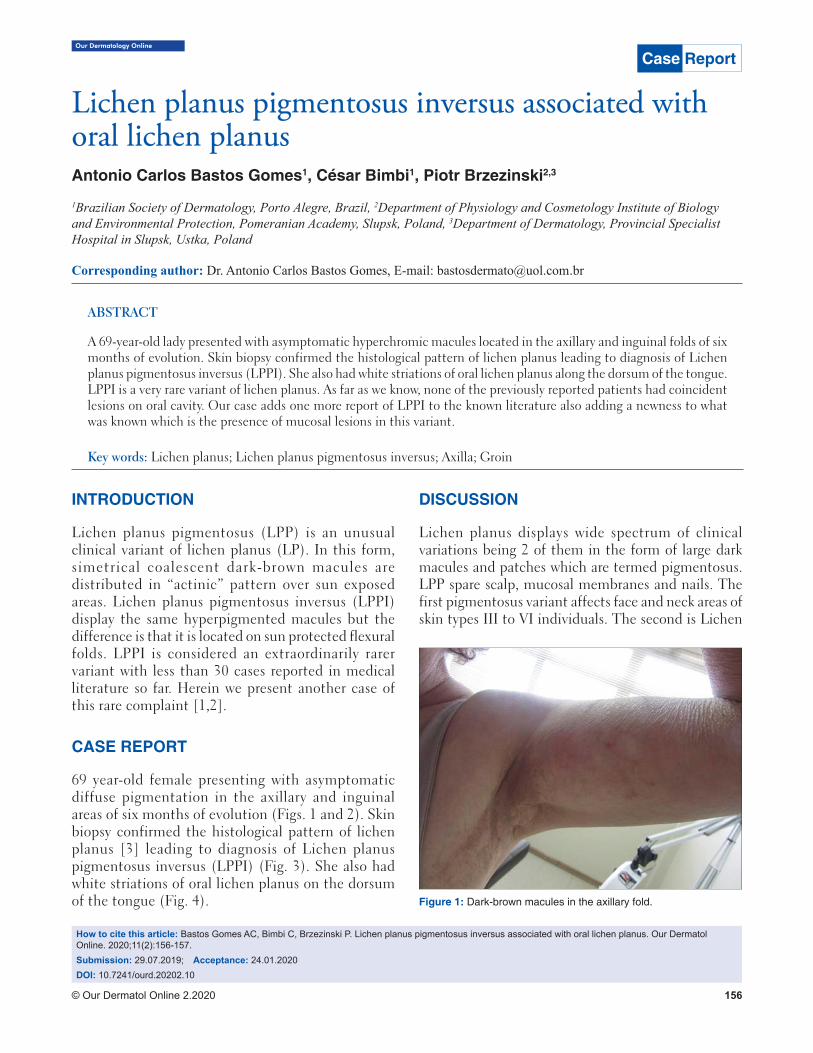
Our Dermatology Online
© Our Dermatol Online 2.2020 156
How to cite this article: Bastos Gomes AC, Bimbi C, Brzezinski P. Lichen planus pigmentosus inversus associated with oral lichen planus. Our Dermatol Online. 2020;11(2):156-157.Submission: 29.07.2019; Acceptance: 24.01.2020DOI: 10.7241/ourd.20202.10
INTRODUCTION
Lichen planus pigmentosus (LPP) is an unusual clinical variant of lichen planus (LP). In this form, simetrical coalescent dark-brown macules are distributed in “actinic” pattern over sun exposed areas. Lichen planus pigmentosus inversus (LPPI) display the same hyperpigmented macules but the difference is that it is located on sun protected flexural folds. LPPI is considered an extraordinarily rarer variant with less than 30 cases reported in medical literature so far. Herein we present another case of this rare complaint [1,2].
CASE REPORT
69 year-old female presenting with asymptomatic diffuse pigmentation in the axillary and inguinal areas of six months of evolution (Figs. 1 and 2). Skin biopsy confirmed the histological pattern of lichen planus [3] leading to diagnosis of Lichen planus pigmentosus inversus (LPPI) (Fig. 3). She also had white striations of oral lichen planus on the dorsum of the tongue (Fig. 4).
DISCUSSION
Lichen planus displays wide spectrum of clinical variations being 2 of them in the form of large dark macules and patches which are termed pigmentosus. LPP spare scalp, mucosal membranes and nails. The first pigmentosus variant affects face and neck areas of skin types III to VI individuals. The second is Lichen
ABSTRACT
A 69-year-old lady presented with asymptomatic hyperchromic macules located in the axillary and inguinal folds of six months of evolution. Skin biopsy confirmed the histological pattern of lichen planus leading to diagnosis of Lichen planus pigmentosus inversus (LPPI). She also had white striations of oral lichen planus along the dorsum of the tongue. LPPI is a very rare variant of lichen planus. As far as we know, none of the previously reported patients had coincident lesions on oral cavity. Our case adds one more report of LPPI to the known literature also adding a newness to what was known which is the presence of mucosal lesions in this variant.
Key words: Lichen planus; Lichen planus pigmentosus inversus; Axilla; Groin
Lichen planus pigmentosus inversus associated with Lichen planus pigmentosus inversus associated with oral lichen planusoral lichen planusAntonio Carlos Bastos Gomes1, César Bimbi1, Piotr Brzezinski2,3
1Brazilian Society of Dermatology, Porto Alegre, Brazil, 2Department of Physiology and Cosmetology Institute of Biology and Environmental Protection, Pomeranian Academy, Slupsk, Poland, 3Department of Dermatology, Provincial Specialist Hospital in Slupsk, Ustka, Poland
Corresponding author: Dr. Antonio Carlos Bastos Gomes, E-mail: [email protected]
Case Report
Figure 1: Dark-brown macules in the axillary fold.
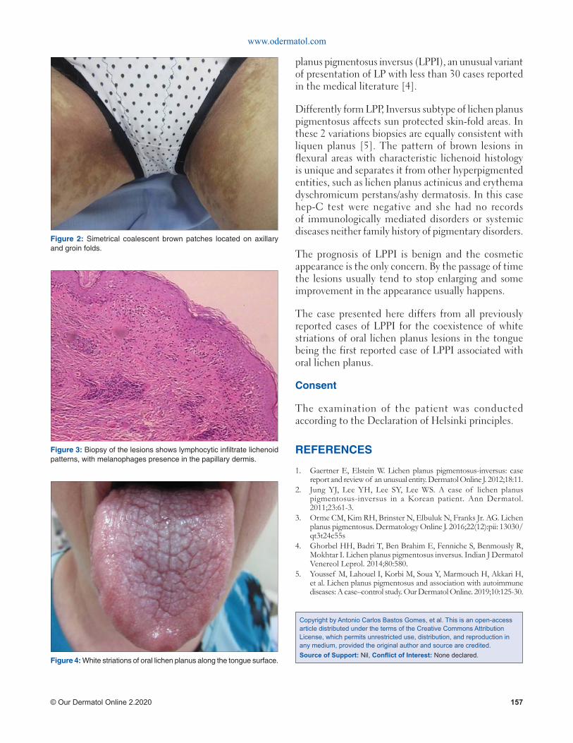
www.odermatol.com
© Our Dermatol Online 2.2020 157
planus pigmentosus inversus (LPPI), an unusual variant of presentation of LP with less than 30 cases reported in the medical literature [4].
Differently form LPP, Inversus subtype of lichen planus pigmentosus affects sun protected skin-fold areas. In these 2 variations biopsies are equally consistent with liquen planus [5]. The pattern of brown lesions in flexural areas with characteristic lichenoid histology is unique and separates it from other hyperpigmented entities, such as lichen planus actinicus and erythema dyschromicum perstans/ashy dermatosis. In this case hep-C test were negative and she had no records of immunologically mediated disorders or systemic diseases neither family history of pigmentary disorders.
The prognosis of LPPI is benign and the cosmetic appearance is the only concern. By the passage of time the lesions usually tend to stop enlarging and some improvement in the appearance usually happens.
The case presented here differs from all previously reported cases of LPPI for the coexistence of white striations of oral lichen planus lesions in the tongue being the first reported case of LPPI associated with oral lichen planus.
Consent
The examination of the patient was conducted according to the Declaration of Helsinki principles.
REFERENCES
1. Gaertner E, Elstein W. Lichen planus pigmentosus-inversus: case report and review of an unusual entity. Dermatol Online J. 2012;18:11.
2. Jung YJ, Lee YH, Lee SY, Lee WS. A case of lichen planus pigmentosus-inversus in a Korean patient. Ann Dermatol. 2011;23:61-3.
3. Orme CM, Kim RH, Brinster N, Elbuluk N, Franks Jr. AG. Lichen planus pigmentosus. Dermatology Online J. 2016;22(12):pii: 13030/qt3t24c55s
4. Ghorbel HH, Badri T, Ben Brahim E, Fenniche S, Benmously R, Mokhtar I. Lichen planus pigmentosus inversus. Indian J Dermatol Venereol Leprol. 2014;80:580.
5. Youssef M, Lahouel I, Korbi M, Soua Y, Marmouch H, Akkari H, et al. Lichen planus pigmentosus and association with autoimmune diseases: A case–control study. Our Dermatol Online. 2019;10:125-30.
Figure 2: Simetrical coalescent brown patches located on axillary and groin folds.
Figure 3: Biopsy of the lesions shows lymphocytic infi ltrate lichenoid patterns, with melanophages presence in the papillary dermis.
Figure 4: White striations of oral lichen planus along the tongue surface.
Copyright by Antonio Carlos Bastos Gomes, et al. This is an open-access article distributed under the terms of the Creative Commons Attribution License, which permits unrestricted use, distribution, and reproduction in any medium, provided the original author and source are credited.Source of Support: Nil, Confl ict of Interest: None declared.
Our Dermatology Online
© Our Dermatol Online 2.2020 158
How to cite this article: Rasso A, Ziani J, Oukarfi S, Baybay H, Elloudi S, Mernissi FZ. Solitary purpuric plaque: clinical and dermoscopic evolution of lichen aureus. Our Dermatol Online. 2020;11(2):158-160.Submission: 16.06.2019; Acceptance: 16.12.2019DOI: 10.7241/ourd.20202.11
INTRODUCTION
Lichen aureus (LA) is a rare variant of persistent pigmented purpuric dermatitis (PPPD), and is characterized by the appearance of a solitary golden-yellow, lichenoid macule [1,2]. We report a clinical and demoscopic evolutionary aspect of lichen aureus in our patient.
CASE REPORT
A 53-year-old woman with a history of diabetes who had been on insulin for 10 years, consulted for the management of asymptomatic leg-level lesions that had been evolving for 3 months. The dermatological examination revealed a purpuric infiltrated placard on the anterior surface of the left leg (Fig. 1). Dermoscopy showed a red blood cells, with whitish linear streaks, and erythematous-purplish fundus (Figs. 2 and 3). The skin biopsy showed slightly increased number of superficial vascular plexus vessels in the dermis, associated with dense perivascular inflammatory sheaths consisting mainly of lymphocytes accompanied
by certain histiocytes.in places lymphocyte penetration of the wall of the capillary vessels by lymphocytes but without parietal necrosis (Figs. 4-6). In places, a slight extravasation of red blood cells in the interstitium around the vessels. Doppler ultrasound of the vessels of the lower limbs revealed a mediacalcosis of the fibular artery. Blood chemistry and full blood count results were normal. Based on these findings, we made a diagnosis of segmental Lichen aureus. the patient was treated with topical corticosteroid with improvement. Three months after the purplish erythematous appearance is replaced by a coppery brown color, the dermoscopy revealed a copper-red pigmentation background, more red blood cells than white streaks (Fig. 3).
DISCUSSION
The segmental lichen aureus is a “tattoo” of hemosiderin resulting from an altered local venous return, clinically, we have macules, an erythematous or purpuric, unilateral, solitary plaque. And is characterized histopathologically by no alteration of the epidermis, a lichenoid lymphocytic infiltrate
ABSTRACT
Lichen aureus is a pigmented purpuric dermatosis that typically presents with the acute onset of a solitary, unilateral, purple to rust-yel- low colored lichenoid patch or plaque on lower extremities1. We presented a 53-year-old woman consulted for the management of asymptomatic lesions. The dermatological examination revealed a purpuric infiltrated placard on the anterior surface of the left leg, dermoscopy and skin biopsy confirmed diagnostic of lichen aureus. Lichen aureus is a rare entity whose diagnosis remains difficult from where the interest of the dermoscopy to retain the diagnosis.
Key words: Dermoscopy; Lichen aureus; Purpuric dermatosis
Solitary purpuric plaque: clinical and dermoscopic Solitary purpuric plaque: clinical and dermoscopic evolution of lichen aureusevolution of lichen aureusAsmae Rasso, Jihane Ziani, Sara Oukarfi , Hanane Baybay, Sara Elloudi, Fatima Zahra Mernissi
Department of Dermatology CHU Hassan II, Fez, Morocco
Corresponding author: Dr. Asmae Rasso, E-mail: [email protected]
Case Report
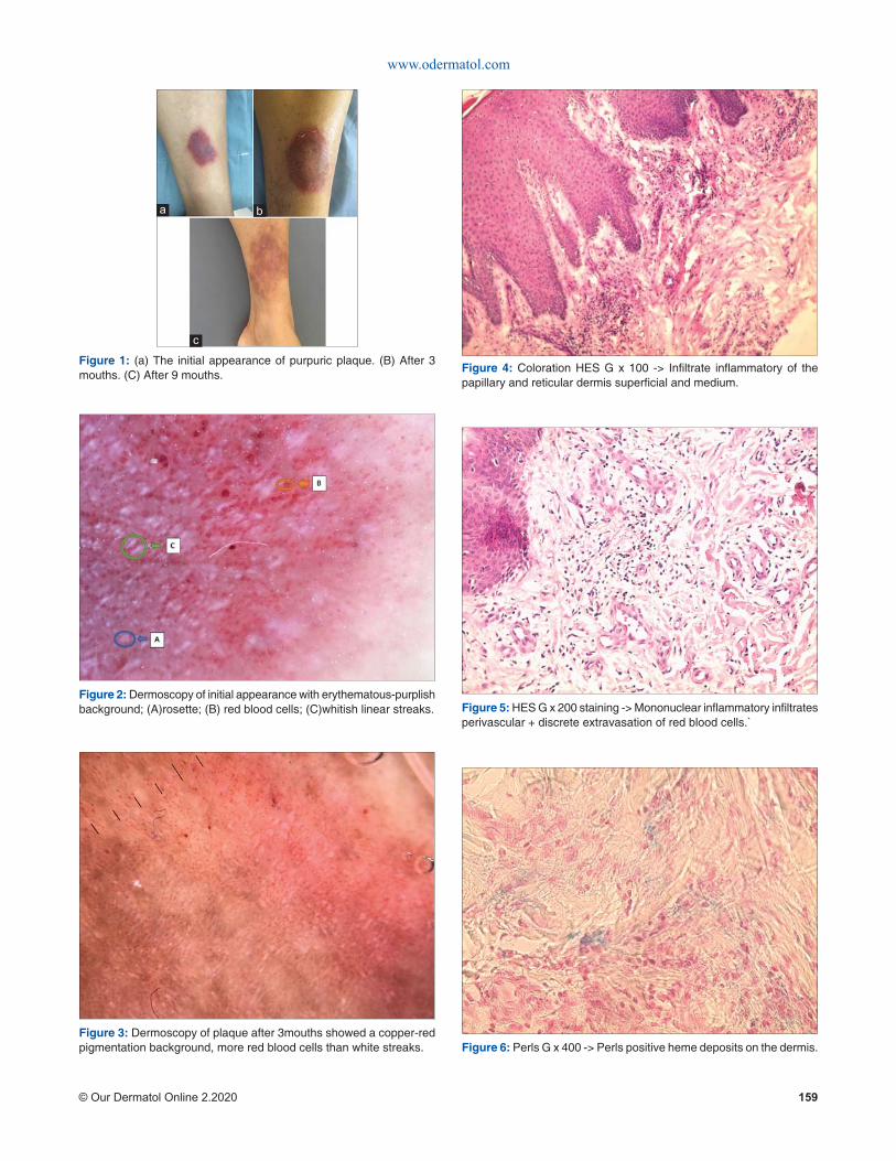
www.odermatol.com
© Our Dermatol Online 2.2020 159
Figure 2: Dermoscopy of initial appearance with erythematous-purplish background; (A)rosette; (B) red blood cells; (C)whitish linear streaks.
Figure 3: Dermoscopy of plaque after 3mouths showed a copper-red pigmentation background, more red blood cells than white streaks.
Figure 1: (a) The initial appearance of purpuric plaque. (B) After 3 mouths. (C) After 9 mouths.
c
ba
Figure 4: Coloration HES G x 100 -> Infi ltrate infl ammatory of the papillary and reticular dermis superfi cial and medium.
Figure 5: HES G x 200 staining -> Mononuclear infl ammatory infi ltrates perivascular + discrete extravasation of red blood cells.`
Figure 6: Perls G x 400 -> Perls positive heme deposits on the dermis.
www.odermatol.com
© Our Dermatol Online 2.2020 160
Copyright by Asmae Rasso, et al. This is an open-access article distributed under the terms of the Creative Commons Attribution License, which permits unrestricted use, distribution, and reproduction in any medium, provided the original author and source are credited.Source of Support: Nil, Confl ict of Interest: None declared.
and extravasated erythrocytes and hemosiderin in the dermis. Dermatoscopy showed a copper-red pigmentation background, with brownish networks of interconnected lines and rounded to oval red blood cells [3]. Zeballos et al. reported that LA showed four characteristic dermoscopic features: (i) brownish or coppery-red diffuse background coloration; (ii) round to oval red dots, globules or patches; (iii) grey dots; and (iv) a network of brownish to grey interconnected lines [4]. The lesions usually persist for several years and then spontaneously disappear, the treatment is based on the dermocorticoid, puvatherapy. the patient treated by dermocorticoid with improvement.
CONCLUSION
clinical appearance of lichen aureus is variable, the dermoscopy is a less rapid non-invasive help to retain the diagnosis, as it allows to follow up as the case of our patient.
Consent
The examination of the patient was conducted according to the Declaration of Helsinki principles.
REFERENCES
1. Erreola AW Jauregui IE, Ló pez Zaldo JB, Huerta Rivera G, Soria Orozco M, Bonnafoux Alcaraz M. A case of lichen aureus successfully treated with 595 nm wavelength pulsed-dye laser. J Cosmet Dermatol. 2019;00:1–3.
2. Saito Y, Shimomura Y, Orime M, Kariya N, Abe R. Segmental lichen aureus in infancy. Clin Exp Dermatol. 2017;42:215-7.
3. Portela PS, Ormiga P, Carvalho de Freitas N. Dermoscopy of lichen aureus. An Bras Dermatol. 2013;88:253-5.
4. Zaballos P, Puig S, Malvehy J. Dermoscopy of pigmented purpuric dermatoses (lichen aureus): a useful tool for clinical diagnosis. Arch Dermatol. 2004;140:1290-1.
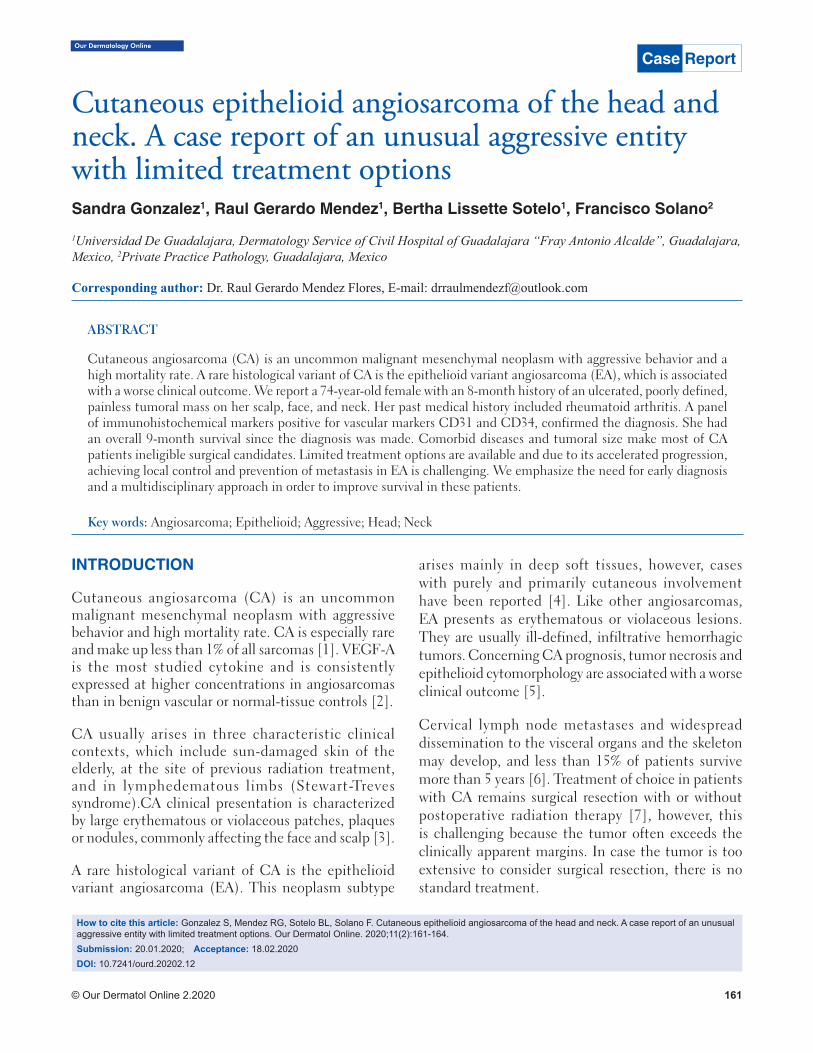
Our Dermatology Online
© Our Dermatol Online 2.2020 161
Cutaneous epithelioid angiosarcoma of the head and Cutaneous epithelioid angiosarcoma of the head and neck. A case report of an unusual aggressive entity neck. A case report of an unusual aggressive entity with limited treatment optionswith limited treatment optionsSandra Gonzalez1, Raul Gerardo Mendez1, Bertha Lissette Sotelo1, Francisco Solano2
1Universidad De Guadalajara, Dermatology Service of Civil Hospital of Guadalajara “Fray Antonio Alcalde”, Guadalajara, Mexico, 2Private Practice Pathology, Guadalajara, Mexico
Corresponding author: Dr. Raul Gerardo Mendez Flores, E-mail: [email protected]
INTRODUCTION
Cutaneous angiosarcoma (CA) is an uncommon malignant mesenchymal neoplasm with aggressive behavior and high mortality rate. CA is especially rare and make up less than 1% of all sarcomas [1]. VEGF-A is the most studied cytokine and is consistently expressed at higher concentrations in angiosarcomas than in benign vascular or normal-tissue controls [2].
CA usually arises in three characteristic clinical contexts, which include sun-damaged skin of the elderly, at the site of previous radiation treatment, and in lymphedematous limbs (Stewart-Treves syndrome).CA clinical presentation is characterized by large erythematous or violaceous patches, plaques or nodules, commonly affecting the face and scalp [3].
A rare histological variant of CA is the epithelioid variant angiosarcoma (EA). This neoplasm subtype
arises mainly in deep soft tissues, however, cases with purely and primarily cutaneous involvement have been reported [4]. Like other angiosarcomas, EA presents as erythematous or violaceous lesions. They are usually ill-defined, infiltrative hemorrhagic tumors. Concerning CA prognosis, tumor necrosis and epithelioid cytomorphology are associated with a worse clinical outcome [5].
Cervical lymph node metastases and widespread dissemination to the visceral organs and the skeleton may develop, and less than 15% of patients survive more than 5 years [6]. Treatment of choice in patients with CA remains surgical resection with or without postoperative radiation therapy [7], however, this is challenging because the tumor often exceeds the clinically apparent margins. In case the tumor is too extensive to consider surgical resection, there is no standard treatment.
ABSTRACT
Cutaneous angiosarcoma (CA) is an uncommon malignant mesenchymal neoplasm with aggressive behavior and a high mortality rate. A rare histological variant of CA is the epithelioid variant angiosarcoma (EA), which is associated with a worse clinical outcome. We report a 74-year-old female with an 8-month history of an ulcerated, poorly defined, painless tumoral mass on her scalp, face, and neck. Her past medical history included rheumatoid arthritis. A panel of immunohistochemical markers positive for vascular markers CD31 and CD34, confirmed the diagnosis. She had an overall 9-month survival since the diagnosis was made. Comorbid diseases and tumoral size make most of CA patients ineligible surgical candidates. Limited treatment options are available and due to its accelerated progression, achieving local control and prevention of metastasis in EA is challenging. We emphasize the need for early diagnosis and a multidisciplinary approach in order to improve survival in these patients.
Key words: Angiosarcoma; Epithelioid; Aggressive; Head; Neck
Case Report
How to cite this article: Gonzalez S, Mendez RG, Sotelo BL, Solano F. Cutaneous epithelioid angiosarcoma of the head and neck. A case report of an unusual aggressive entity with limited treatment options. Our Dermatol Online. 2020;11(2):161-164.Submission: 20.01.2020; Acceptance: 18.02.2020DOI: 10.7241/ourd.20202.12
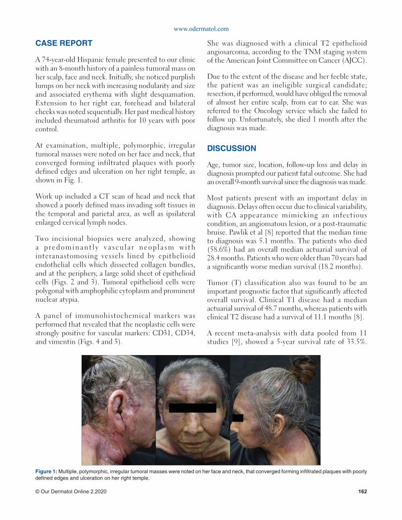
www.odermatol.com
© Our Dermatol Online 2.2020 162
CASE REPORT
A 74-year-old Hispanic female presented to our clinic with an 8-month history of a painless tumoral mass on her scalp, face and neck. Initially, she noticed purplish lumps on her neck with increasing nodularity and size and associated erythema with slight desquamation. Extension to her right ear, forehead and bilateral cheeks was noted sequentially. Her past medical history included rheumatoid arthritis for 10 years with poor control.
At examination, multiple, polymorphic, irregular tumoral masses were noted on her face and neck, that converged forming infiltrated plaques with poorly defined edges and ulceration on her right temple, as shown in Fig. 1.
Work up included a CT scan of head and neck that showed a poorly defined mass invading soft tissues in the temporal and parietal area, as well as ipsilateral enlarged cervical lymph nodes.
Two incisional biopsies were analyzed, showing a p redominant l y va scu la r neop la sm wi th interanastomosing vessels lined by epithelioid endothelial cells which dissected collagen bundles, and at the periphery, a large solid sheet of epithelioid cells (Figs. 2 and 3). Tumoral epithelioid cells were polygonal with amphophilic cytoplasm and prominent nuclear atypia.
A panel of immunohistochemical markers was performed that revealed that the neoplastic cells were strongly positive for vascular markers: CD31, CD34, and vimentin (Figs. 4 and 5).
She was diagnosed with a clinical T2 epithelioid angiosarcoma, according to the TNM staging system of the American Joint Committee on Cancer (AJCC).
Due to the extent of the disease and her feeble state, the patient was an ineligible surgical candidate; resection, if performed, would have obliged the removal of almost her entire scalp, from ear to ear. She was referred to the Oncology service which she failed to follow up. Unfortunately, she died 1 month after the diagnosis was made.
DISCUSSION
Age, tumor size, location, follow-up loss and delay in diagnosis prompted our patient fatal outcome. She had an overall 9-month survival since the diagnosis was made.
Most patients present with an important delay in diagnosis. Delays often occur due to clinical variability, with CA appearance mimicking an infectious condition, an angiomatous lesion, or a post-traumatic bruise. Pawlik et al [8] reported that the median time to diagnosis was 5.1 months. The patients who died (58.6%) had an overall median actuarial survival of 28.4 months. Patients who were older than 70 years had a significantly worse median survival (18.2 months).
Tumor (T) classification also was found to be an important prognostic factor that significantly affected overall survival. Clinical T1 disease had a median actuarial survival of 48.7 months, whereas patients with clinical T2 disease had a survival of 11.1 months [8].
A recent meta-analysis with data pooled from 11 studies [9], showed a 5-year survival rate of 33.5%.
Figure 1: Multiple, polymorphic, irregular tumoral masses were noted on her face and neck, that converged forming infi ltrated plaques with poorly defi ned edges and ulceration on her right temple.
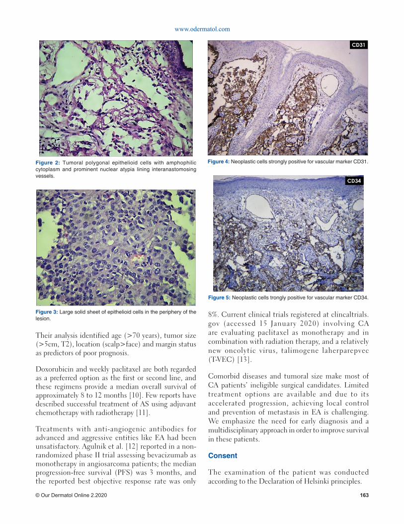
www.odermatol.com
© Our Dermatol Online 2.2020 163
Their analysis identified age (>70 years), tumor size (>5cm, T2), location (scalp>face) and margin status as predictors of poor prognosis.
Doxorubicin and weekly paclitaxel are both regarded as a preferred option as the first or second line, and these regimens provide a median overall survival of approximately 8 to 12 months [10]. Few reports have described successful treatment of AS using adjuvant chemotherapy with radiotherapy [11].
Treatments with anti-angiogenic antibodies for advanced and aggressive entities like EA had been unsatisfactory. Agulnik et al. [12] reported in a non-randomized phase II trial assessing bevacizumab as monotherapy in angiosarcoma patients; the median progression-free survival (PFS) was 3 months, and the reported best objective response rate was only
8%. Current clinical trials registered at clincaltrials.gov (accessed 15 January 2020) involving CA are evaluating paclitaxel as monotherapy and in combination with radiation therapy, and a relatively new oncolytic virus, talimogene laherparepvec (T-VEC) [13].
Comorbid diseases and tumoral size make most of CA patients’ ineligible surgical candidates. Limited treatment options are available and due to its accelerated progression, achieving local control and prevention of metastasis in EA is challenging. We emphasize the need for early diagnosis and a multidisciplinary approach in order to improve survival in these patients.
Consent
The examination of the patient was conducted according to the Declaration of Helsinki principles.
Figure 2: Tumoral polygonal epithelioid cells with amphophilic cytoplasm and prominent nuclear atypia lining interanastomosing vessels.
Figure 4: Neoplastic cells strongly positive for vascular marker CD31.
Figure 3: Large solid sheet of epithelioid cells in the periphery of the lesion.
Figure 5: Neoplastic cells trongly positive for vascular marker CD34.
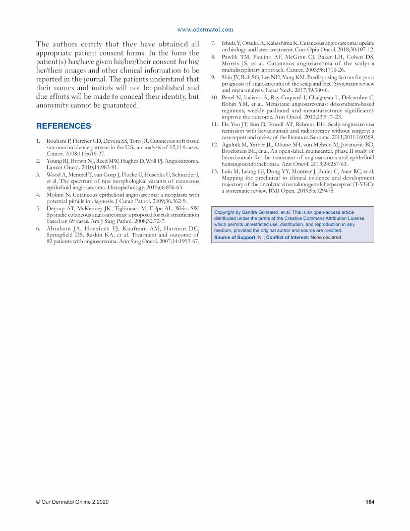
www.odermatol.com
© Our Dermatol Online 2.2020 164
The authors certify that they have obtained all appropriate patient consent forms. In the form the patient(s) has/have given his/her/their consent for his/her/their images and other clinical information to be reported in the journal. The patients understand that their names and initials will not be published and due efforts will be made to conceal their identity, but anonymity cannot be guaranteed.
REFERENCES
1. Rouhani P, Fletcher CD, Devesa SS, Toro JR. Cutaneous soft tissue sarcoma incidence patterns in the U.S.: an analysis of 12,114 cases. Cancer. 2008;113:616-27.
2. Young RJ, Brown NJ, Reed MW, Hughes D, Woll PJ. Angiosarcoma. Lancet Oncol. 2010;11:983-91.
3. Wood A, Mentzel T, van Gorp J, Flucke U, Huschka U, Schneider J, et al. The spectrum of rare morphological variants of cutaneous epithelioid angiosarcoma. Histopathology. 2015;66:856-63.
4. Mobini N. Cutaneous epithelioid angiosarcoma: a neoplasm with potential pitfalls in diagnosis. J Cutan Pathol. 2009;36:362-9.
5. Deyrup AT, McKenney JK, Tighiouart M, Folpe AL, Weiss SW. Sporadic cutaneous angiosarcomas: a proposal for risk stratifi cation based on 69 cases. Am J Surg Pathol. 2008;32:72-7.
6. Abraham JA, Hornicek FJ, Kaufman AM, Harmon DC, Springfi eld DS, Raskin KA, et al. Treatment and outcome of 82 patients with angiosarcoma. Ann Surg Oncol. 2007;14:1953-67.
7. Ishida Y, Otsuka A, Kabashima K. Cutaneous angiosarcoma: update on biology and latest treatment. Curr Opin Oncol. 2018;30:107-12.
8. Pawlik TM, Paulino AF, McGinn CJ, Baker LH, Cohen DS, Morris JS, et al. Cutaneous angiosarcoma of the scalp: a multidisciplinary approach. Cancer. 2003;98:1716-26.
9. Shin JY, Roh SG, Lee NH, Yang KM. Predisposing factors for poor prognosis of angiosarcoma of the scalp and face: Systematic review and meta-analysis. Head Neck. 2017;39:380-6.
10. Penel N, Italiano A, Ray-Coquard I, Chaigneau L, Delcambre C, Robin YM, et al. Metastatic angiosarcomas: doxorubicin-based regimens, weekly paclitaxel and metastasectomy significantly improve the outcome. Ann Oncol. 2012;23:517–23.
11. De Yao JT, Sun D, Powell AT, Rehmus EH. Scalp angiosarcoma remission with bevacizumab and radiotherapy without surgery: a case report and review of the literature. Sarcoma. 2011;2011:160369.
12. Agulnik M, Yarber JL, Okuno SH, von Mehren M, Jovanovic BD, Brockstein BE, et al. An open-label, multicenter, phase II study of bevacizumab for the treatment of angiosarcoma and epithelioid hemangioendotheliomas. Ann Oncol. 2013;24:257-63.
13. Lalu M, Leung GJ, Dong YY, Montroy J, Butler C, Auer RC, et al. Mapping the preclinical to clinical evidence and development trajectory of the oncolytic virus talimogene laherparepvec (T-VEC): a systematic review. BMJ Open. 2019;9:e029475.
Copyright by Sandra Gonzalez, et al. This is an open-access article distributed under the terms of the Creative Commons Attribution License, which permits unrestricted use, distribution, and reproduction in any medium, provided the original author and source are credited.Source of Support: Nil, Confl ict of Interest: None declared.
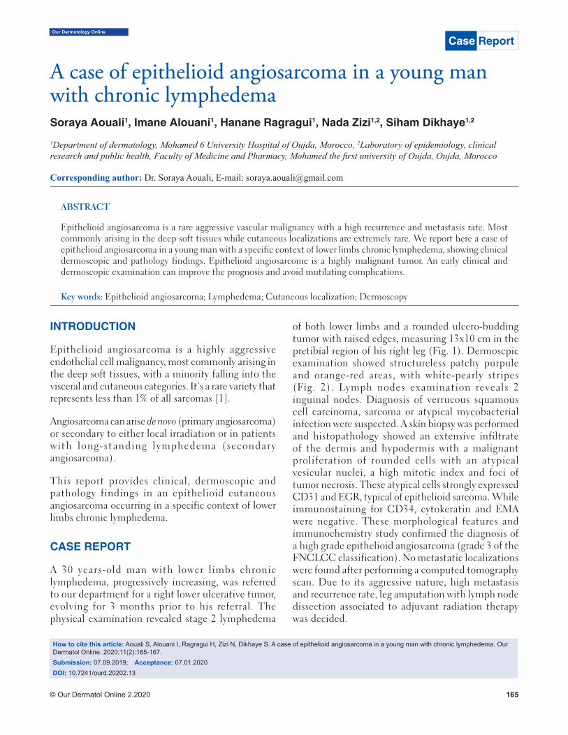
Our Dermatology Online
© Our Dermatol Online 2.2020 165
How to cite this article: Aouali S, Alouani I, Ragragui H, Zizi N, Dikhaye S. A case of epithelioid angiosarcoma in a young man with chronic lymphedema. Our Dermatol Online. 2020;11(2):165-167.Submission: 07.09.2019; Acceptance: 07.01.2020DOI: 10.7241/ourd.20202.13
A case of epithelioid angiosarcoma in a young man A case of epithelioid angiosarcoma in a young man with chronic lymphedemawith chronic lymphedemaSoraya Aouali1, Imane Alouani1, Hanane Ragragui1, Nada Zizi1,2, Siham Dikhaye1,2
1Department of dermatology, Mohamed 6 University Hospital of Oujda, Morocco, 2Laboratory of epidemiology, clinical research and public health, Faculty of Medicine and Pharmacy, Mohamed the fi rst university of Oujda, Oujda, Morocco
Corresponding author: Dr. Soraya Aouali, E-mail: [email protected]
INTRODUCTION
Epithelioid angiosarcoma is a highly aggressive endothelial cell malignancy, most commonly arising in the deep soft tissues, with a minority falling into the visceral and cutaneous categories. It’s a rare variety that represents less than 1% of all sarcomas [1].
Angiosarcoma can arise de novo (primary angiosarcoma) or secondary to either local irradiation or in patients with long-standing lymphedema (secondary angiosarcoma).
This report provides clinical, dermoscopic and pathology findings in an epithelioid cutaneous angiosarcoma occurring in a specific context of lower limbs chronic lymphedema.
CASE REPORT
A 30 years-old man with lower limbs chronic lymphedema, progressively increasing, was referred to our department for a right lower ulcerative tumor, evolving for 3 months prior to his referral. The physical examination revealed stage 2 lymphedema
of both lower limbs and a rounded ulcero-budding tumor with raised edges, measuring 13x10 cm in the pretibial region of his right leg (Fig. 1). Dermoscpic examination showed structureless patchy purpule and orange-red areas, with white-pearly stripes (Fig. 2). Lymph nodes examination reveals 2 inguinal nodes. Diagnosis of verrucous squamous cell carcinoma, sarcoma or atypical mycobacterial infection were suspected. A skin biopsy was performed and histopathology showed an extensive infiltrate of the dermis and hypodermis with a malignant proliferation of rounded cells with an atypical vesicular nuclei, a high mitotic index and foci of tumor necrosis. These atypical cells strongly expressed CD31 and EGR, typical of epithelioid sarcoma. While immunostaining for CD34, cytokeratin and EMA were negative. These morphological features and immunochemistry study confirmed the diagnosis of a high grade epithelioid angiosarcoma (grade 3 of the FNCLCC classification). No metastatic localizations were found after performing a computed tomography scan. Due to its aggressive nature, high metastasis and recurrence rate, leg amputation with lymph node dissection associated to adjuvant radiation therapy was decided.
ABSTRACT
Epithelioid angiosarcoma is a rare aggressive vascular malignancy with a high recurrence and metastasis rate. Most commonly arising in the deep soft tissues while cutaneous localizations are extremely rare. We report here a case of epithelioid angiosarcoma in a young man with a specific context of lower limbs chronic lymphedema, showing clinical dermoscopic and pathology findings. Epithelioid angiosarcome is a highly malignant tumor. An early clinical and dermoscopic examination can improve the prognosis and avoid mutilating complications.
Key words: Epithelioid angiosarcoma; Lymphedema; Cutaneous localization; Dermoscopy
Case Report
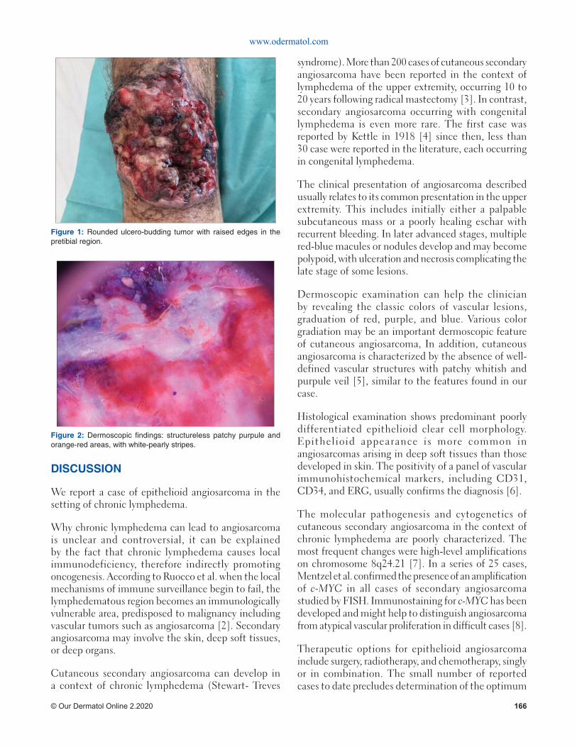
www.odermatol.com
© Our Dermatol Online 2.2020 166
DISCUSSION
We report a case of epithelioid angiosarcoma in the setting of chronic lymphedema.
Why chronic lymphedema can lead to angiosarcoma is unclear and controversial, it can be explained by the fact that chronic lymphedema causes local immunodeficiency, therefore indirectly promoting oncogenesis. According to Ruocco et al. when the local mechanisms of immune surveillance begin to fail, the lymphedematous region becomes an immunologically vulnerable area, predisposed to malignancy including vascular tumors such as angiosarcoma [2]. Secondary angiosarcoma may involve the skin, deep soft tissues, or deep organs.
Cutaneous secondary angiosarcoma can develop in a context of chronic lymphedema (Stewart- Treves
syndrome). More than 200 cases of cutaneous secondary angiosarcoma have been reported in the context of lymphedema of the upper extremity, occurring 10 to 20 years following radical mastectomy [3]. In contrast, secondary angiosarcoma occurring with congenital lymphedema is even more rare. The first case was reported by Kettle in 1918 [4] since then, less than 30 case were reported in the literature, each occurring in congenital lymphedema.
The clinical presentation of angiosarcoma described usually relates to its common presentation in the upper extremity. This includes initially either a palpable subcutaneous mass or a poorly healing eschar with recurrent bleeding. In later advanced stages, multiple red-blue macules or nodules develop and may become polypoid, with ulceration and necrosis complicating the late stage of some lesions.
Dermoscopic examination can help the clinician by revealing the classic colors of vascular lesions, graduation of red, purple, and blue. Various color gradiation may be an important dermoscopic feature of cutaneous angiosarcoma, In addition, cutaneous angiosarcoma is characterized by the absence of well-defined vascular structures with patchy whitish and purpule veil [5], similar to the features found in our case.
Histological examination shows predominant poorly differentiated epithelioid clear cell morphology. Epithelioid appearance is more common in angiosarcomas arising in deep soft tissues than those developed in skin. The positivity of a panel of vascular immunohistochemical markers, including CD31, CD34, and ERG, usually confirms the diagnosis [6].
The molecular pathogenesis and cytogenetics of cutaneous secondary angiosarcoma in the context of chronic lymphedema are poorly characterized. The most frequent changes were high-level amplifications on chromosome 8q24.21 [7]. In a series of 25 cases, Mentzel et al. confirmed the presence of an amplification of c-MYC in all cases of secondary angiosarcoma studied by FISH. Immunostaining for c-MYC has been developed and might help to distinguish angiosarcoma from atypical vascular proliferation in difficult cases [8].
Therapeutic options for epithelioid angiosarcoma include surgery, radiotherapy, and chemotherapy, singly or in combination. The small number of reported cases to date precludes determination of the optimum
Figure 1: Rounded ulcero-budding tumor with raised edges in the pretibial region.
Figure 2: Dermoscopic fi ndings: structureless patchy purpule and orange-red areas, with white-pearly stripes.
www.odermatol.com
© Our Dermatol Online 2.2020 167
treatment regime at this stage, although where possible, wide excision is recommended. The need for adjuvant therapy is determined on an individual basis.
The overall prognosis of epithelioid angiosarcoma is poor, with a high rate of local recurrence and distant metastasis, regardless of the chosen treatment.
CONCLUSION
In summary, epithelioid angiosarcoma is a rare, aggressive vascular malignancy with a high recurrence and metastasis rate. An early dermatological evaluation and appropriate dermoscopic examination may help avoid a delayed diagnosis, to get a better prognosis with less mutilating complications.
REFERENCES
1. Rouhani P, Fletcher CDM, Devesa SS, Toro JR. Cutaneous soft tissue sarcoma incidence patterns in the U.S.: an analysis of 12,114 cases. Cancer. 2008;113:616– 27.
2. Ruocco V, Schwartz RA, Ruocco E. Lymphedema: an immunologically vulnerable site for development of neoplasms.
J Am Acad Dermatol. 2002; l47:124–7.3. Fayette J, Martin E, Piperno-Neumann S, Le Cesne A, Robert C,
Bonvalot S, et al. Angiosarcomas, a heterogeneous group of sarcomas with specifi c behavior depending on primary site: a retrospective study of 161 cases. Ann Oncol. 2007;18:2030–6.
4. Offori TW, Platt CC, Stephens M, Hopkinson GB. Angiosarcoma in congenital hereditary lymphoedema (Milroy’s disease)—diagnostic beacons and a review of the literature. Clin Exp Dermatol. 1993;18:174–77.
5. Cozzani E, Chinazzo C, Ghigliotti G, Pastorino C, Gasparini G, Parodi A. Cutaneous angiosarcoma: the role of dermoscopy to reduce the risk of a delayed diagnosis. Int J Dermatol. 2018;57:996-7.
6. Shavit E, Alav A, Limacher JJ, Sibbald RG Angiosarcoma complicating lower leg elephantiasis in a male patient: An unusual clinical complication, case report and literature review. SAGE Open Med Case Rep. 2018;6:2050313X18796343.
7. Itakura E, Yamamoto H, Oda Y, Tsuneyoshi M, Detec- tion and characterization of vascular endothelial growth factors and their receptors in a series of angiosarcomas. J Surg Oncol. 2008;9774–81.
8. Fernandez AP, Sun Y, Tubbs RR, Goldblum JR, Billings SD. FISH for MYC amplifi cation and anti-MYC immuno- histochemistry: useful diagnostic tools in the assessment of secondary angiosarcoma and atypical vascular proliferations. J Cut Pathol. 2012;39234–42.
Copyright by Soraya Aouali, et al. This is an open access article distributed under the terms of the Creative Commons Attribution License, which permits unrestricted use, distribution, and reproduction in any medium, provided the original author and source are credited.Source of Support: Nil, Confl ict of Interest: None declared.
Our Dermatology Online
© Our Dermatol Online 2.2020 168
Syringoma, hormone receptors and associated Syringoma, hormone receptors and associated endocrinopathies. Are they clinically relevant?endocrinopathies. Are they clinically relevant?Ayse Tülin Mansur1, Saime Ramadan2
1Department of Dermatology, Baskent University Istanbul Hospital, Istanbul, Turkey, 2Department of Pathology, Baskent University Istanbul Hospital, Istanbul, Turkey
Corresponding author: Dr. Ayse Tülin Mansur, E-mail: [email protected]
INTRODUCTION
Syringomas are benign neoplasms of the intraepidermal portion of the eccrine sweat ducts, seen mainly in women and characterized by skin-coloured or slightly yellowish papules. The etiopathogenesis of syringomas is not clear yet, but hormonal influences have been suspected [1]. There are some reported cases of syringomas manifesting lesional estrogen receptor (ER) and progesterone receptor (PR) positivity [1-4]. Herein, we present a case of vulvar syringoma (VS) and in this context, we review the endocrinological abnormalities associated with syringomas, reported up to date, and discuss the clinical importance of lesional ER and PR positivity.
CASE REPORT
A 19-year-old female patient presented to the dermatology outpatient clinic for evaluation of
asymptomatic genital papules developed 6 years ago, with the onset of puberty. The lesions had been increasing in size and extent since then. Medical history revealed polycystic ovary syndrome diagnosed 4 years ago, and she had been under treatment with a combination of 2 mg cyproterone acetate and 0.035 mg ethinyl estradiol for the past 2 years. No other members of her family had similar skin lesions.
On dermatological examination, symmetrical skin-coloured or slightly hyperpigmented papules of approximately 0.5 cm in diameter were observed on both labia majora and pubis. On the lower areas of the labia, the lesions formed thick plaques with smooth surfaces. She had a few papules on her axillary vaults clinically similar to genital ones. Papulopustular acne lesions were seen on the face and back, and facial hirsutism was evident on the chin and sideburns.
ABSTRACT
Syringomas are benign eccrine sweat gland tumors characterized by skin-colored or slightly yellowish papules. The lesions are predominantly located on the face, neck, and trunk, while vulvar involvement is relatively rare. There are some reported cases of syringomas manifesting lesional estrogen and/or progesterone receptor positivity. On the other hand, syringomas associated with endocrinopathies have infrequently been reported. We describe a 19-year-old female patient with asymptomatic skin coloured papules on both labia majora and pubis, present for 6 years. As polycystic ovary syndrome was diagnosed 4 years ago, she had been under treatment with a combination of cyproterone acetate and ethinyl estradiol for the past 2 years. Clinical, dermatoscopic, and histopathologic findings of the lesions were consistent with syringoma. Immunohistochemical stainings for estrogen and progesterone receptors were negative. To the best of our knowledge, there has been no previously reported case of vulvar syringoma associated with polycystic ovary syndrome. Hereby, we review the association of endocrinopathies with syringomas, together with the presence and clinical importance of hormone receptor positivity in syringoma lesions.
Key words: Syringoma; Vulvar; Estrogen receptor; Progesterone receptor; Endocrinopathy
Case Report
How to cite this article: Mansur AT, Ramadan S. Syringoma, hormone receptors and associated endocrinopathies. Are they clinically relevant?. Our Dermatol Online. 2020;11(2):168-170.Submission: 04.08.2019; Acceptance: 07.10.2019DOI: 10.7241/ourd.20202.14
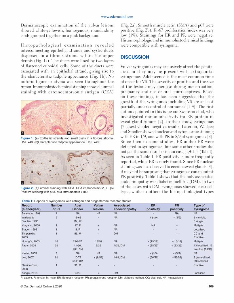
www.odermatol.com
© Our Dermatol Online 2.2020 169
Dermatoscopic examination of the vulvar lesions showed white-yellowish, homogenous, round, shiny clods grouped together on a pink background.
H i s t o p a t h o l o g i c a l ex a m i n a t i o n re v e a l e d interconnecting epithelial strands and cystic ducts dispersed in a fibrous stroma within the upper dermis (Fig. 1a). The ducts were lined by two layers of flattened cuboidal cells. Some of the ducts were associated with an epithelial strand, giving rise to the characteristic tadpole appearance (Fig. 1b). No mitotic figure or atypia was seen throughout the tumor. Immunohistochemical staining showed luminal staining with carcinoembryonic antigen (CEA)
(Fig. 2a). Smooth muscle actin (SMA) and p63 were positive (Fig. 2b). Ki-67 proliferation index was very low (1%). Stainings for ER and PR were negative. Histomorphologic and immunohistochemical findings were compatible with syringoma.
DISCUSSION
Vulvar syringomas may exclusively affect the genital area, or they may be present with extragenital syringomas. Adolescence is the most common time of onset for VS. The severity of pruritus and the size of the lesions may increase during menstruation, pregnancy and use of oral contraceptives. Based on these findings, it has been suggested that the growth of the syringomas including VS are at least partially under control of hormones [1-4]. The first authors pointed to this issue are Swanson et al, who investigated immunoreactivity for ER protein in sweat gland tumors [2]. In their study, syringomas (7 cases) yielded negative results. Later on, Wallace and Smoller showed nuclear and cytoplasmic staining with ER in 1/9, and with PR in 8/9 of syringomas [3]. Since then in some studies, ER and/or PR were detected in syringomas, but some other studies did not get the same result as in our case [1,4-11] (Tab. I). As seen in Table 1, PR positivity is more frequently reported, while ER is rarely found. Since PR nuclear staining was also observed in eccrine sweat glands [3], it may not be surprising that syringomas can manifest PR positivity. Table 1 shows that the only associated endocrinopathy was diabetes mellitus (DM). In two of the cases with DM, syringomas showed clear cell type, while in others the histopathological types
Table 1: Reports of syringomas with estrogen and progesterone receptor studiesReport (author/year)
Number of Ps
Age/Gender
Vulvar lesions
Associated endocrinopathy
ER positivity
PR positivity
Type of syringoma
Swanson, 1991 7 NA NA NA - NA NA
Wallace &Smoller, 1995
9 16-682M, 7F
- NA + (1/9) + (8/9) 5 multiple,3 single
Yorganci, 2000 1 27, F + NA NA + Multiple
Trager, 1999 1 8, F + NA - - Localized
Timpanidis, 2003
1 55, M - DM - + CC and Eruptive
Huang Y, 2003 18 21-60/F 18/18 NA - (15/18) - (15/18) Multiple
Fathy, 2005 25 11-36, 20F, 5M
2/25 1/25, DM - (25/25) + (23/25) 13 localized, 12 eruptive (1 CC)
Kariya, 2005 3 NA NA NA + (1/3) + (3/3) NA
Lee, 2007 61 10-7253 F, 8M
+ (8/53) 1/61, DM - (56/56) - (56/56) 8 generalized,53 localized
Garrido-Ruiz, 2008
1 31, M - - - - Eruptive
Akoğlu, 2013 1 40/F + DM - - Localized
P: patient, F: female, M: male, ER: Estrogen receptor, PR: progesterone receptor, DM: diabetes mellitus, CC: clear cell, NA: not available
Figure 1: (a) Epithelial strands and small cysts in a fi brous stroma. H&E x40. (b)Characteristic tadpole appearance. H&E x400.
a b
Figure 2: (a)Luminal staining with CEA. CEA immunostain x100. (b)Positive staining with p63. p63 immunostain x100.
a b
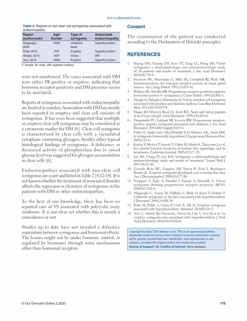
www.odermatol.com
© Our Dermatol Online 2.2020 170
were not mentioned. The cases associated with DM were either PR positive or negative, indicating that hormone receptor positivity and DM presence seems to be unrelated.
Reports of syringomas associated with endocrinopathy are limited in number. Association with DM has mostly been reported in eruptive and clear cell variants of syringomas. It has even been suggested that multiple or eruptive clear cell syringomas may be considered as a cutaneous marker for DM [6]. Clear cell syringoma is characterized by clear cells with a vacuolated cytoplasm containing glycogen, besides other typical histological findings of syringomas. A deficiency or decreased activity of phosphorylase due to raised glucose level was suggested for glycogen accumulation in clear cells [6].
Endocrinopathies associated with non-clear cell syringomas are scant and listed in Table 2 [4,12-14]. It is not known whether the treatment of associated disorder affects the regression or clearance of syringomas in the patients with DM or other endocrinopathies.
To the best of our knowledge, there has been no reported case of VS associated with polycystic ovary syndrome. It is not clear yet whether this is merely a coincidence or not.
Studies up to date have not revealed a definitive association between syringomas and hormonal effects. The lesions might not be under hormone control, or regulated by hormones through some mechanisms other than hormonal receptors.
Consent
The examination of the patient was conducted according to the Declaration of Helsinki principles.
REFERENCES
1. Huang YH, Chuang YH, Kuo TT, Yang LC, Hong HS. Vulvar syringoma: a clinicopathologic and immunohistologic study of 18 patients and results of treatment. J Am Acad Dermatol. 2003;48:735-9.
2. Swanson PE, Mazoujian G, Mills SE, Campbell RJ, Wick MR. Immunoreactivity for estrogen receptor protein in sweat gland tumors. Am J Surg Pathol. 1991;15:835-41.
3. Wallace ML, Smoller BR. Progesterone receptor positivity supports hormonal control of syringomas. J Cutan Pathol. 1995:22:442-5.
4. Akoglu G, Ibiloglu I, Durmazlar N. Vulvar nonclear cell syringoma associated with pruritus and diabetes mellitus. Case Rep Dermatol Med. 2013;2013:418794.
5. Trager JD, Silvers J, Reed JA, Scott RA. Neck and vulvar papules in an 8-year-old girl. Arch Dermatol. 1999;135:203-6.
6. Timpanidis PC, Lakhani SR, Groves RW. Progesterone receptor-positive eruptive syringoma associated with diabetes. J Am Acad Dermatol. 2003;48(5 Suppl):S103-4.
7. Fathy H, Abdel Aziz AM, Elhanbly S, El-Hawary AK, Amin MM. Is syringoma hormonally dependent? J Egypt wom Dermatol Soc. 2005;2:12-6.
8. Kariya Y, Moriya T, Suzuki T, Chiba M, Ishida K, Takeyama J, et al. Sex steroid hormon receptors in human skin appandage and its neoplasms. Endocrine Journal. 2005;52:317-25.
9. Lee JH, Chang JY, Lee KH. Syringoma: a clinicopathologic and immunohistologic study and results of treatment. Yonsei Med J. 2007;48:35-40.
10. Garrido-Ruiz MC, Enguita AB, Navas R, Polo I, Rodríguez Peralto JL. Eruptive syringoma developed over a waxing skin area. Am J Dermatopathol. 2008;30:377-80.
11. Yorganci A, Kale A, Dunder I, Ensari A, Sertcelik A. Vulvar syringoma showing progesterone receptor positivity. BJOG. 2000;107:292-4.
12. Aliagaoglu C, Atasoy M, Yildirim U, Balik O, Koca T, Erdem T. Unilateral syringoma of the face associated with hyperthyroidism. J Dermatol. 2004;31:828-30.
13. Polat M, Pelitli A, Oztaş P, Unal T, Alli N. Eruptive syringoma associated with hyperthyroidism. Skinmed. 2010;8:124-5.
14. Avcı A, Altınor SŞ, Özyurt K, Atasoy M, Ulaş Y, Avcı D, et al. An eruptive syringoma case associated with hyperthyroidism. J Turk Acad Dermatol. 2016;10:16102c8.
Table 2: Reports of non-clear cell syringomas associated with endocrinopathyReport (author/year)
Age/Gender
Type of syringoma
Associated endocrinopathy
Aliagaoğlu, 2004
34/M Unilateral facial
Hyperthyroidism
Polat, 2010 45/F Eruptive Hyperthyroidism
Akoğlu, 2013 40/F Vulvar DM
Avcı, 2016 59/M Eruptive Hyperthyroidism
F: female, M: male, DM: diabetes mellitus
Copyright by Ayse Tülin Mansur, et al. This is an open-access article distributed under the terms of the Creative Commons Attribution License, which permits unrestricted use, distribution, and reproduction in any medium, provided the original author and source are credited.Source of Support: Nil, Confl ict of Interest: None declared.
Our Dermatology Online
© Our Dermatol Online 2.2020 171
INTRODUCTION
Pacinian corpuscles (PC), a rapidly adapting pressure-sensitive mechanoreceptor, found in several organs and are mainly distributed in the dermis of the fingers and palm of the hand. Reactive enlargement of these PC is known as Pacinian neuroma (PN): an extremely painful condition with only few cases reported in the literature. It is characterized by formation of painful nodules in connective tissue of the hand. The etiopathogenesis is still not clear. However, it is of non-malign nature and frequently reported after local or repetitive trauma. We present here a case in which PN was associated with multiple myeloma.
CASE REPORT
A 57-year-old previously healthy man presented with a 6-month history of severe pain and swelling located over the volar surface of his fingers. He reported no previous history of trauma or hand injury. He didn’t present any
neurological symptoms. Physical examination revealed definitely palpable nodules and moderate swelling over the first phalanx of his left thumb and all his fingers (Fig. 1).
A biopsy specimen was performed. It revealed multiple hyperplastic Pacinian corpuscles in the lower dermis and subcutaneous fat (Fig. 2). A diagnosis of PN was made. The patient also reported chronic back pain and muscle weakness. Lumbar spine X-ray showed a severe demineralization. MM was suspected. On laboratory evaluation, the results of complete blood count and biochemistry panels were within normal limits. There was no anemia, hypercalcemia, renal impairment or lytic bone lesions. Otherwise, immunoelectrophoresis demonstrated monoclonal light chains in blood and urine. Bone marrow biopsy was obtained and was consistent with plasma cell myeloma. The patient successfully underwent an autologous stem cell transplant with a very good response. Two months after his myeloma remission,
ABSTRACT
We present a case of Pacinian Neuroma (PN) associated with multiple myeloma (MM) in a 57-year-old man with a 6-month history of painful swelling of his fingers. Physical examination revealed definite palpable nodules and moderate swelling over the first phalanx of all his fingers. A biopsy confirmed the diagnosis of PN. The patient reported chronic back pain. Radiologically, a severe demineralization was found. MM was suspected. The results of complete blood count and biochemistry panels were within normal limits. Otherwise, immunoelectrophoresis demonstrated monoclonal light chains in blood and urine. Bone marrow biopsy was consistent with MM. The patient successfully underwent an autologous stem cell transplant with good response. A slight decrease in the size in PN was noted. To the best of our knowledge, no case of PN associated with MM had been reported. This occurrence may be accidental. However, further studies are required in order to explain this association.
Key words: Pacinian corpuscles; Pacinian neuroma; Multiple myeloma
How to cite this article: Soua Y, Belhareth K, Akkari H, Jguirim M, Belhadjali H, Abdeljelil NB, Youssef M, Zili J. Digital pacinian neuroma in a patient with myeloma. Our Dermatol Online. 2020;11(2):171-173.Submission: 06.10.2019; Acceptance: 24.01.2020DOI: 10.7241/ourd.20202.15
Case Report
Digital pacinian neuroma in a patient with myelomaDigital pacinian neuroma in a patient with myelomaYosra Soua1, Kamar Belhareth1, Hayet Akkari1, Mahbouba Jguirim2, Hichem Belhadjali1, Nouha Ben Abdeljelil3, Monia Youssef1, Jameleddine Zili1
1Dermatology Department, Fattouma Bourguiba University Hospital, University of Monastir, Tunisia, 2Rhumatology Department, Fattouma Bourguiba University Hospital, University of Monastir, Tunisia, 3Anatomopathology Department, Fattouma Bourguiba University Hospital, University of Monastir, Tunisia
Corresponding author: Dr. Kamar Belhareth, E-mail: [email protected]
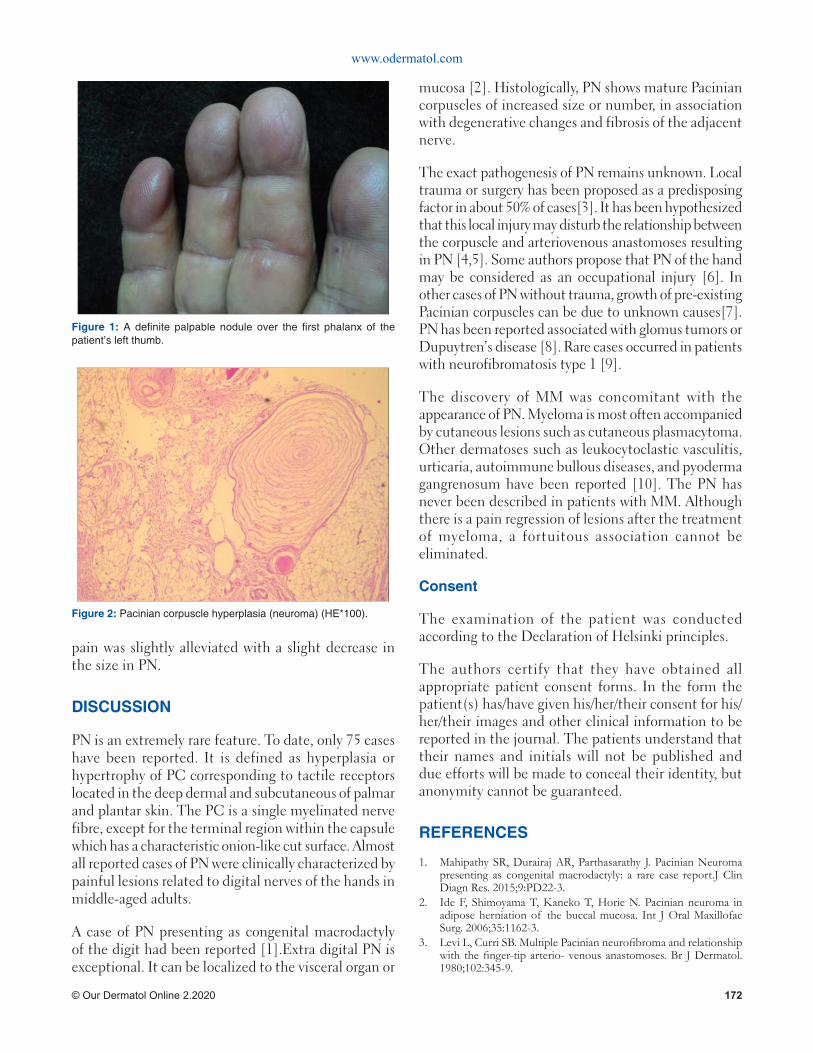
www.odermatol.com
© Our Dermatol Online 2.2020 172
pain was slightly alleviated with a slight decrease in the size in PN.
DISCUSSION
PN is an extremely rare feature. To date, only 75 cases have been reported. It is defined as hyperplasia or hypertrophy of PC corresponding to tactile receptors located in the deep dermal and subcutaneous of palmar and plantar skin. The PC is a single myelinated nerve fibre, except for the terminal region within the capsule which has a characteristic onion-like cut surface. Almost all reported cases of PN were clinically characterized by painful lesions related to digital nerves of the hands in middle-aged adults.
A case of PN presenting as congenital macrodactyly of the digit had been reported [1].Extra digital PN is exceptional. It can be localized to the visceral organ or
mucosa [2]. Histologically, PN shows mature Pacinian corpuscles of increased size or number, in association with degenerative changes and fibrosis of the adjacent nerve.
The exact pathogenesis of PN remains unknown. Local trauma or surgery has been proposed as a predisposing factor in about 50% of cases[3]. It has been hypothesized that this local injury may disturb the relationship between the corpuscle and arteriovenous anastomoses resulting in PN [4,5]. Some authors propose that PN of the hand may be considered as an occupational injury [6]. In other cases of PN without trauma, growth of pre-existing Pacinian corpuscles can be due to unknown causes[7]. PN has been reported associated with glomus tumors or Dupuytren’s disease [8]. Rare cases occurred in patients with neurofibromatosis type 1 [9].
The discovery of MM was concomitant with the appearance of PN. Myeloma is most often accompanied by cutaneous lesions such as cutaneous plasmacytoma. Other dermatoses such as leukocytoclastic vasculitis, urticaria, autoimmune bullous diseases, and pyoderma gangrenosum have been reported [10]. The PN has never been described in patients with MM. Although there is a pain regression of lesions after the treatment of myeloma, a fortuitous association cannot be eliminated.
Consent
The examination of the patient was conducted according to the Declaration of Helsinki principles.
The authors certify that they have obtained all appropriate patient consent forms. In the form the patient(s) has/have given his/her/their consent for his/her/their images and other clinical information to be reported in the journal. The patients understand that their names and initials will not be published and due efforts will be made to conceal their identity, but anonymity cannot be guaranteed.
REFERENCES
1. Mahipathy SR, Durairaj AR, Parthasarathy J. Pacinian Neuroma presenting as congenital macrodactyly: a rare case report.J Clin Diagn Res. 2015;9:PD22-3.
2. Ide F, Shimoyama T, Kaneko T, Horie N. Pacinian neuroma in adipose herniation of the buccal mucosa. Int J Oral Maxillofac Surg. 2006;35:1162-3.
3. Levi L, Curri SB. Multiple Pacinian neurofi broma and relationship with the fi nger-tip arterio- venous anastomoses. Br J Dermatol. 1980;102:345-9.
Figure 1: A defi nite palpable nodule over the fi rst phalanx of the patient’s left thumb.
Figure 2: Pacinian corpuscle hyperplasia (neuroma) (HE*100).
www.odermatol.com
© Our Dermatol Online 2.2020 173
4. Narayanamurthy V, Winston AT, Gupta A. A rare case of Pacinian corpuscle neuroma. Can J Plast Surg. 2005;13:43-5.
5. Vijayaraghavan R, Chandrashekar R, Belagavi CS, Murthy NB. Painful digital Pacinian corpuscle hyperplasia following dog bite: a case report. Int J Surg. 2008;6:e42-5.
6. Zanardi F, Cooke RM, Maiorana A.”Is this case of a very rare disease work-related?” A review of reported cases of Pacinian neuroma. Scand J Work Environ Health. 2011;37:253-8.
7. Fletcher CD, Theaker JM.Digital pacinian neuroma: a distinctive hyperplastic lesion. Histopathology. 1989;15:249-56.
8. Von Campe A, Mende K, Omaren H, Meuli-Simmen C. Painful nodules and cords in Dupuytren disease. J Hand Surg Am. 2012;37:1313-8.
9. Friedrich RE, Hagel C.Painful Vater-Pacini neuroma of the digit in neurofi bromatosis type 1.GMS Interdiscip Plast Reconstr Surg DGPW. 2019 Feb 8;8:Doc03. doi: 10.3205/iprs000129. eCollection 2019.
10. Behera B, Pat tna ik M, Sahu B,Mohanty P, Jena S, Mohapatra L. Cutaneous Manifestations of multiple myeloma. Indian J Dermatol. 2016;61:668-71.
Copyright by Yosra Soua, et al. This is an open-access article distributed under the terms of the Creative Commons Attribution License, which permits unrestricted use, distribution, and reproduction in any medium, provided the original author and source are credited.Source of Support: Nil, Confl ict of Interest: None declared.
Our Dermatology Online
© Our Dermatol Online 2.2020 174
INTRODUCTION
Syringocystadenoma papilliferum (SCAP) is an uncommon benign adnexal tumor, which frequently arises from a nevus sebaceous. This tumor occurs usually at birth or during early childhood. It’s most commonly located on the scalp or face, however SCAP in other areas have been reported [1]. Location in the vulva is rare and had only been reported in very few cases[2]. Neurofibromatosis type1 (NF1) is a frequent neurocutaneous syndrome that predisposes for various benign and malignant tumors. We report a case of SCAP located in the vulva in a NF1 patient.
CASE REPORT
We report the case of a 36 years old patient, treated one year ago for a breast carcinoma in situ. With a family history of neurofibromatosis type 1 in the father, the dead brother and the sister. Physical examination showed many flat, uniformly
hyperpigmented macules with well-defined borders over the body, corresponding to café-au-lait macules (Fig. 1). Skinfold freckling were present all over the trunk (Fig. 1). Examination of the genital area objectified a pinkish to erythematous bordered elevated ulcerated nodule measuring about 1 cm in diameter, unpainful, located in the inside of the labia majora (Fig. 2). There were no other genital or perineal lesions. Ophthalmological examination found several bilateral Lisch nodules. The patient meted National Institutes of Health criteria for NF14 (4/7 criteria). Other physical examination showed dosrsolumbar scoliosis. The vulvar lesion was fully excised, histopathology revealed a tumor with endophytic configuration, composed of papillary projections and ductal-like structures that extend as invaginations from the surface epithelium into the underlying dermis. The papillary projections covered by regular cubic epithelium. The ducts and papillary projections are covered by an inner layer of columnar epithelium and an outer layer of cuboidal or flattened
ABSTRACT
A 36-year-old female presented to the dermatology service with a pinkish to erythematous ulcerated nodule measuring about 1 cm in diameter, located on the labia majora. The lesion has been slowly increasing in size. Physical examination found features compatible with the diagnosis of neurofibromatosis type 1. Excisional biopsy demonstrated the histological picture of syringocystadenoma papilliferum. The interest of this report lies in the rarity of syringocystadenoma papilliferum, its unusual presentation in the vulva, and it’s association with neurofibromatosis type 1. To our knowledge, this association has not been described in the medical literature.
Key words: Syringocystadenoma papilliferum; Neurofibromatosis type 1; Vulva
Case Report
Syringocystadenoma papilliferum presented as Syringocystadenoma papilliferum presented as an ulcerated nodule of the vulva in a patient with an ulcerated nodule of the vulva in a patient with Neurofi bromatosis type 1Neurofi bromatosis type 1Iyda El Faqyr1, Maria Dref2, Sara Zahid1, Jamila Oualla1, Nabil Mansouri2, Hanane Rais2, Ouafa Hocar1, Said Amal1
1Department of Dermatology, Mohammed VI University Hospital, University Cadi Ayyad, Marrakech, Morocco, 2Department of Pathology, Mohammed VI University Hospital, University Cadi Ayyad, Marrakech, Morocco
Corresponding author: Dr. Iyda El Faqyr, E-mail: [email protected]
How to cite this article: El Faqyr I, Dref M, Zahid S, Oualla J, Mansouri N, Rais H, Hocar O, Amal S. Syringocystadenoma papilliferum presented as an ulcerated nodule of the vulva in a patient with Neurofi bromatosis type 1. Our Dermatol Online. 2020;11(2):174-176.Submission: 19.12.2019; Acceptance: 14.02.2020DOI: 10.7241/ourd.20202.16
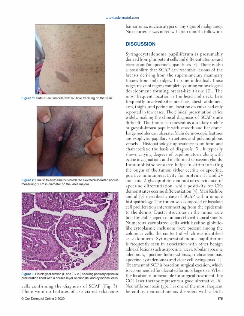
www.odermatol.com
© Our Dermatol Online 2.2020 175
cells confirming the diagnosis of SCAP (Fig. 3). There were no features of associated sebaceous
hamartoma, nuclear atypia or any signs of malignancy. No recurrence was noted with four months follow-up.
DISCUSSION
Syringocystadenoma papilliferum is presumably derived from pluripotent cells and differentiates toward eccrine and/or apocrine apparatuses [3]. There is also a possibility that SCAP can resemble lesions of the breasts deriving from the supernumerary mammary tissues from milk ridges. In some individuals those ridges may not regress completely during embryological development forming breast-like tissue [2]. The most frequent location is the head and neck. Less frequently involved sites are face, chest, abdomen, arm, thighs, and perineum, location on vulva had only reported in few cases. The clinical presentation varies widely, making the clinical diagnosis of SCAP quite difficult. The tumor can present as a solitary nodule or greyish-brown papule with smooth and flat dome. Large nodules can ulcerate. Main dermoscopic features are exophytic papillary structures and polymorphous vessels1. Histopathologic appearance is uniform and characteristic the basis of diagnosis [3]. It typically shows varying degrees of papillomatosis along with cystic invaginations and malformed sebaceous glands. Immunohistochemistry helps in differentiating the origin of the tumor, either eccrine or apocrine, positive immunoreactivity for proteins 15 and 24 and zinc-2 glycoprotein demonstrates evidence of apocrine differentiation, while positivity for CKs demonstrates eccrine differentiation [4]. Mari Kishibe and al [5] described a case of SCAP with a unique histopathology. The tumor was composed of basaloid cell proliferation interconnecting from the epidermis to the dermis. Ductal structures in the tumor were lined by club-shaped columnar cells with apical snouts. Numerous vacuolated cells with hyaline globule-like cytoplasmic inclusions were present among the columnar cells, the content of which was identified as sialomucin. Syringocystadenoma papilliferum is frequently seen in association with other benign adnexal lesions such as apocrine naevi, tubular apocrine adenomas, apocrine hidrocystomas, trichoadenomas, apocrine cystadenomas and clear cell syringomas [1]. Treatment of SCP is based on surgical excision, which is recommended for ulcerated forms or large size. When the location is unfavorable for surgical treatment, the CO2 laser therapy represents a good alternative [6]. Neurofibromatosis type 1 is one of the most frequent hereditary neurocutaneous disorders with a birth
Figure 1: Café-au-lait macule with multiple freckling on the trunk.
Figure 2: Pinkish to erythematous bordered elevated ulcerated nodule measuring 1 cm in diameter on the labia majora.
Figure 3: Histological section (H and E × 20) showing papillary epithelial proliferation lined with a double layer of cuboidal and cylindrical cells.
www.odermatol.com
© Our Dermatol Online 2.2020 176
incidence of about 1/2500. It is caused by heterozygous germ-line mutations in the tumor suppressor gene NF1 which codes for neurofibromin, a negative regulator of the RAS proto-oncogene [7]. This makes NF1 patients at risk to develop various benign and malignant tumors. Authors [7] revealed that the range of NF1 associated tumors extends beyond nervous system and includes cancers of the lung, thyroid gland, skin, ovary and gastrointestinal tract. Our patient with the history of removed breast cancer, developed SCAP. To our knowledge, the association between NF1 and SCAP has not been described yet in the medical literature. Although this tumor had relatively benign course, rare cases of malignant transformation have been reported.
CONCLUSION
This case report contributes to extending NF1 associated tumors, and reconsider differential diagnoses of a chronic ulcerated lesion in the vulva. Our patient requires continuous and permanent monitoring to detect other neoplasms.
Consent
The examination of the patient was conducted according to the Declaration of Helsinki principles.
The authors certify that they have obtained all appropriate patient consent forms. In the form the
patient(s) has/have given his/her/their consent for his/her/their images and other clinical information to be reported in the journal. The patients understand that their names and initials will not be published and due efforts will be made to conceal their identity, but anonymity cannot be guaranteed.
REFERENCES
1. Mara L, Simonetta P, Caterina L, Borsari S, Persechino F, Argenziano G, et al. Dermoscopy of syringocystadenoma papilliferum. Austral J Dermatol. 2017;59:e59-e61.
2. Oleksandr S, Nirmala C, Frank L. Syringocysadenoma papilliferum of the vulva: a rarity in gynaecology. BMJ Case Rep. 2014;2014.pii: bcr2014203902.
3. Mohamed M, Belhadjali M, Hadhri R, Youssef M, Zili J. Clinical aspects of syringocystadenoma papilliferum. Our Dermatol Online. 2017;8:348.
4. Shah PA, Singh VS, Bhalekar S, Sudhamani S, Paramjit E. Syringocystadenoma papilliferum: A rare case report with review of literature. J Sci Soc. 2016;43:96-8.
5. Mari K, Takeshi I, Ichiro T, Akemi IY. Intracytoplasmic eosinophilic inclusion bodies with embryonic folliculosebaceous-apocrine unit differentiation in syringocystadenoma papilliferum. J Cutan Pathol. 2018;45:923-6.
6. Bettioui A, Bouyahyaoui Y, Gallouj S. Syringocystadénome papillifère congénital. Ann Dermatol Vénéréol. 2012;139:647-51.
7. Rosenbaum T, Wimmer K. Neurofi bromatosis type 1 (NF1) and Associated tumors. Klin Padiatr. 2014;226:309–15.
Copyright by Iyda El Faqyr, et al. This is an open access article distributed under the terms of the Creative Commons Attribution License, which permits unrestricted use, distribution, and reproduction in any medium, provided the original author and source are credited.Source of Support: Nil, Confl ict of Interest: None declared.
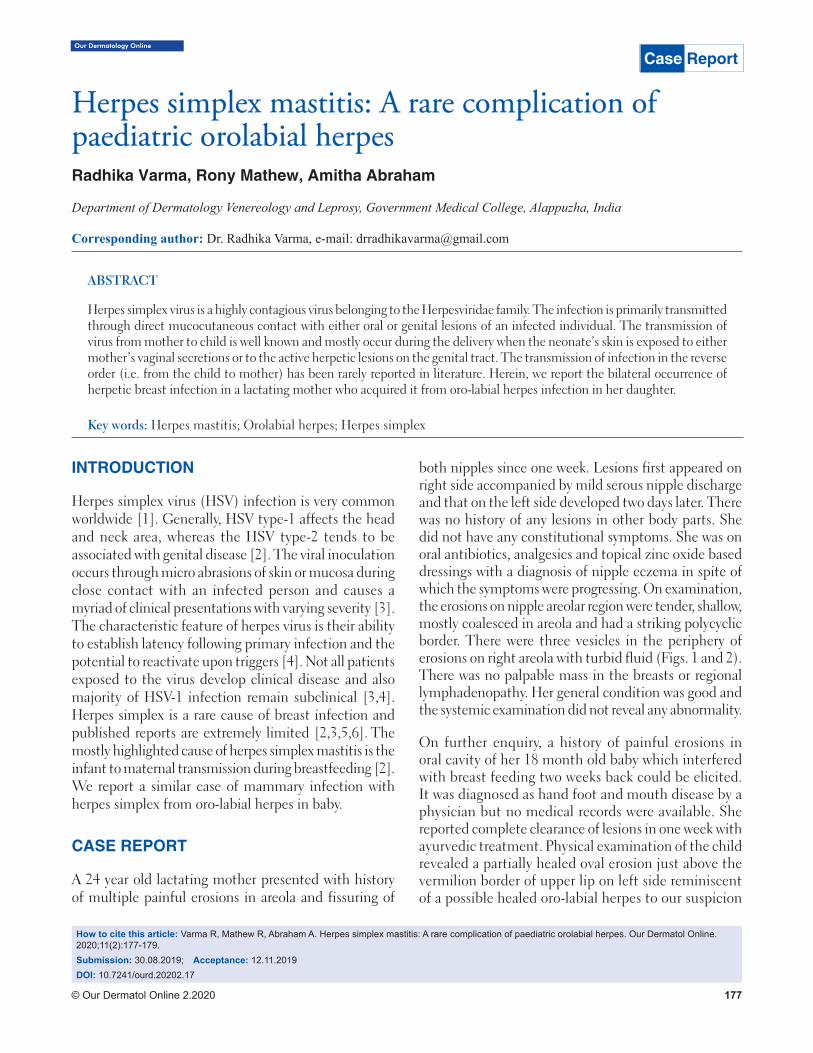
Our Dermatology Online
© Our Dermatol Online 2.2020 177
How to cite this article: Varma R, Mathew R, Abraham A. Herpes simplex mastitis: A rare complication of paediatric orolabial herpes. Our Dermatol Online. 2020;11(2):177-179.Submission: 30.08.2019; Acceptance: 12.11.2019DOI: 10.7241/ourd.20202.17
Herpes simplex mastitis: A rare complication of Herpes simplex mastitis: A rare complication of paediatric orolabial herpespaediatric orolabial herpesRadhika Varma, Rony Mathew, Amitha Abraham
Department of Dermatology Venereology and Leprosy, Government Medical College, Alappuzha, India
Corresponding author: Dr. Radhika Varma, e-mail: [email protected]
INTRODUCTION
Herpes simplex virus (HSV) infection is very common worldwide [1]. Generally, HSV type-1 affects the head and neck area, whereas the HSV type-2 tends to be associated with genital disease [2]. The viral inoculation occurs through micro abrasions of skin or mucosa during close contact with an infected person and causes a myriad of clinical presentations with varying severity [3]. The characteristic feature of herpes virus is their ability to establish latency following primary infection and the potential to reactivate upon triggers [4]. Not all patients exposed to the virus develop clinical disease and also majority of HSV-1 infection remain subclinical [3,4]. Herpes simplex is a rare cause of breast infection and published reports are extremely limited [2,3,5,6]. The mostly highlighted cause of herpes simplex mastitis is the infant to maternal transmission during breastfeeding [2]. We report a similar case of mammary infection with herpes simplex from oro-labial herpes in baby.
CASE REPORT
A 24 year old lactating mother presented with history of multiple painful erosions in areola and fissuring of
both nipples since one week. Lesions first appeared on right side accompanied by mild serous nipple discharge and that on the left side developed two days later. There was no history of any lesions in other body parts. She did not have any constitutional symptoms. She was on oral antibiotics, analgesics and topical zinc oxide based dressings with a diagnosis of nipple eczema in spite of which the symptoms were progressing. On examination, the erosions on nipple areolar region were tender, shallow, mostly coalesced in areola and had a striking polycyclic border. There were three vesicles in the periphery of erosions on right areola with turbid fluid (Figs. 1 and 2). There was no palpable mass in the breasts or regional lymphadenopathy. Her general condition was good and the systemic examination did not reveal any abnormality.
On further enquiry, a history of painful erosions in oral cavity of her 18 month old baby which interfered with breast feeding two weeks back could be elicited. It was diagnosed as hand foot and mouth disease by a physician but no medical records were available. She reported complete clearance of lesions in one week with ayurvedic treatment. Physical examination of the child revealed a partially healed oval erosion just above the vermilion border of upper lip on left side reminiscent of a possible healed oro-labial herpes to our suspicion
ABSTRACT
Herpes simplex virus is a highly contagious virus belonging to the Herpesviridae family. The infection is primarily transmitted through direct mucocutaneous contact with either oral or genital lesions of an infected individual. The transmission of virus from mother to child is well known and mostly occur during the delivery when the neonate’s skin is exposed to either mother’s vaginal secretions or to the active herpetic lesions on the genital tract. The transmission of infection in the reverse order (i.e. from the child to mother) has been rarely reported in literature. Herein, we report the bilateral occurrence of herpetic breast infection in a lactating mother who acquired it from oro-labial herpes infection in her daughter.
Key words: Herpes mastitis; Orolabial herpes; Herpes simplex
Case Report
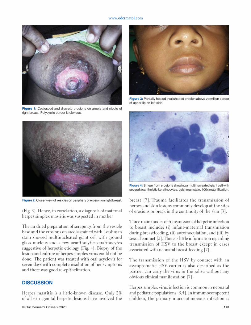
www.odermatol.com
© Our Dermatol Online 2.2020 178
(Fig. 3). Hence, in correlation, a diagnosis of maternal herpes simplex mastitis was suspected in mother.
The air dried preparation of scrapings from the vesicle base and the erosions on areola stained with Leishman stain showed multinucleated giant cell with ground glass nucleus and a few acantholytic keratinocytes suggestive of herpetic etiology (Fig. 4). Biopsy of the lesion and culture of herpes simplex virus could not be done. The patient was treated with oral acyclovir for seven days with complete resolution of her symptoms and there was good re-epithelization.
DISCUSSION
Herpes mastitis is a little-known disease. Only 2% of all extragenital herpetic lesions have involved the
breast [7]. Trauma facilitates the transmission of herpes and skin lesions commonly develop at the sites of erosions or break in the continuity of the skin [3].
Three main modes of transmission of herpetic infection to breast include: (i) infant-maternal transmission during breastfeeding, (ii) autoinoculation, and (iii) by sexual contact [2]. There is little information regarding transmission of HSV to the breast except in cases associated with neonatal breast feeding [7].
The transmission of the HSV by contact with an asymptomatic HSV carrier is also described as the partner can carry the virus in the saliva without any obvious clinical manifestation [7].
Herpes simplex virus infection is common in neonatal and pediatric populations [3,4]. In immunocompetent children, the primary mucocutanoeous infection is
Figure 1: Coalesced and discrete erosions on areola and nipple of right breast. Polycyclic border is obvious.
Figure 2: Closer view of vesicles on periphery of erosion on right breast.
Figure 3: Partially healed oval shaped erosion above vermilion border of upper lip on left side.
Figure 4: Smear from erosions showing a multinucleated giant cell with several acantholytic keratinocytes. Leishman stain, 100x magnifi cation.
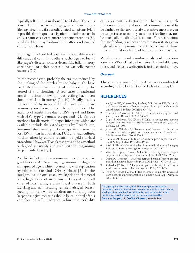
www.odermatol.com
© Our Dermatol Online 2.2020 179
typically self limiting in about 10 to 21 days. The virus remain latent in nerve or the ganglion cells and causes lifelong infection with episodic clinical symptoms [4]. It is possible that frequent antigenic stimulation occurs in at least some cases of recurrent herpetic infections [5].
Viral shedding may continue even after resolution of clinical symptoms.
The diagnosis of isolated herpes simplex mastitis is very difficult as it can mimic others pathologies of breast like paget’s disease, contact dermatitis, inflammatory carcinoma, or other bacterial or varicella zoster mastitis [2,7].
In the present case, probably the trauma induced by the sucking of the nipples by the baby might have facilitated the development of lesions during the period of viral shedding. A few cases of maternal breast infection following breastfeeding have been documented in literature [2,6-10]. Usually, lesions are restricted to areola although cases with entire mammary involvement have been described. The majority of mastitis are due to HSV type-1 and those with HSV type-2 remain exceptional [2]. Various methods for diagnosis of herpes infections which are available include the cytodiagnosis by Tzanck test, immunohistochemistry of tissue specimen, serology for HSV, in-situ hybridisation, PCR and viral culture. Viral isolation by culture remains the gold standard procedure. However, Tzanck test prove to be a method with good sensitivity and specificity for diagnosing herpetic infection [2,7].
As this infection is uncommon, no therapeutic guidelines exists. Acyclovir, a guanosine analogue is an approved agent which reduces the viral replication by inhibiting the viral DNA synthesis [2]. In the background of our case, we highlight the need for a high index of suspicion of this entity in all cases of non healing erosive breast disease in both lactating and non-lactating females. Also, all breast-feeding mothers whose children are suffering from herpetic gingivostomatitis should be cautioned of this complication well in advance to limit the morbidity
of herpes mastitis. Factors other than trauma which influences this unusual mode of transmission need to be studied so that appropriate preventive measures can be suggested as refraining from breast feeding may not be practically possible in all scenarios. Future directions for safe feeding practices and vaccination strategies in high risk lactating women need to be explored to limit the substantial morbidity of herpes simplex mastitis.
We also recommend a routine analysis of suspicious lesions by a Tzanck test as it remains a fairly reliable, easy, quick, and inexpensive diagnostic test in clinical settings.
Consent
The examination of the patient was conducted according to the Declaration of Helsinki principles.
REFERENCES
1. Xu F, Lee FK, Morrow RA, Stenberg MR, Luther KE, Dubin G, et al. Seroprevalence of herpes simplex virus type 1 in children in United states. J Pediatr. 2007;151:374-77.
2. Toussaint A, Simonson C, Valla C. Herpes mastitis: diagnosis and management. Breast J. 2016;22:335–38.
3. Gupta S, Malhotra AK, Dash SS. Child to mother transmission of herpes simplex virus-1 infection at an unusual site. JEADV. 2008;22,:875–903.
4. James SH, Whitley RJ. Treatment of herpes simplex virus infections in pediatric patients: current status and future needs. Clin Pharmacol Ther. 2010;88:720-4.
5. Nahmias AJ, Roizman B. Infection with herpes-simplex viruses 1 and 2. N Engl J Med. 1973;289:719–25.
6. Soo MS, Ghate S. Herpes simplex virus mastitis: clinical and imaging fi ndings. AJR Am J Roentgenol. 2000;174:1087–88.
7. Mardi K. Gupta N, Sharma S, Gupta S. Cytodiagnosis of herpes simplex mastitis. Report of a rare case. J Cytol. 2009;26:149-50.
8. Quinn PT, Lofberg JV. Maternal herpetic breast infection: another hazard of neonatal herpes simplex. Med J Aust. 1978;2:411–12.
9. Sealander JY, Kerr CP. Herpes simplex of the nipple: infant-to-mother transmission. Am Fam Physician. 1989;39:111-13.
10. Dekio S, Kawasaki Y, Jidoi J. Herpes simplex on nipples inoculated from herpetic gingivostomatitis of a baby. Clin Exp Dermatol. 1986;11:664-6.
Copyright by Radhika Varma, et al. This is an open-access article distributed under the terms of the Creative Commons Attribution License, which permits unrestricted use, distribution, and reproduction in any medium, provided the original author and source are credited.Source of Support: Nil, Confl ict of Interest: None declared.
Our Dermatology Online
© Our Dermatol Online 2.2020 180
How to cite this article: El Amraoui M, Frikh R, Hjira N, Boui M. HIV and venous thromboembolism risk: Report of 3 cases with review of the literature. Our Dermatol Online. 2020;11(2):180-182.Submission: 20.07.2019; Acceptance: 26.09.2019DOI: 10.7241/ourd.20202.18
INTRODUCTION
HIV infection, with about 37 million cases globally and 22000 nationally, is a major health problem. Thromboembolic disease is increasingly reported during infection with the HIV. It interests the venous territory with predilection. We present three cases with review of the literature.
CASE REPORT
Case 1
A 40-year-old man, followed for retroviral infection for 8 years, with poor treatment adherence, was admitted for febrile dyspnea. Chest X-ray showed well-circumscribed right basal opacity and thoracic CT was in favor of massive, bilateral pulmonary embolism with infarction and superinfection of the right lower lobe (Figs. 1, 2a-2b). The patient was put under a treatment based on antibiotics and anticoagulants with a favorable evolution over two years of decline.
Case 2
A 50-year-old patient with a history of repeated STIs was hospitalized for left femoro-popliteal thrombophlebitis with cachexia and general impairment. The report showed a retroviral infection at the AIDS stage. The patient was put under tritherapy and anticoagulants with a favorable evolution over two years of decline.
Case 3
55 year old man, followed for retroviral infection at the AIDS stage under triple therapy. Presented an elongated Activated Cephalin Time, hyperfibrinogemia with presence of an anti-phospholipid antibody (lupus anticoagulant). The patient was placed on platelet antiaggregants with a favorable evolution over two years of decline.
DISCUSSION
The risk of venous thromboembolism in HIV-infected patients has been described since the 1990s and it’s 2
ABSTRACT
Thromboembolic disease is particularly increased in HIV-infected patients for multiple reasons as shown by several recent studies. We present three cases with literature review. Case 1: Young patient followed for HIV infection with poor treatment adherence, presented with febrile dyspnea with acute chest pain, radiography and chest CT were in favor of pulmonary embolism,the evolution was favorable with anticoagulants. Case 2: young patient admitted to the emergency department for thrombophlebitis of the lower limb with cachexia and alteration of the general state whose etiological assessment was in favor of an HIV infection, the evolution was favorable under tritherapy and anticoagulants. Case 3: young patient followed for HIV infection, had an active lengthening of cephalin time with hyperfibrinogenemia and the presence of antiphospholipid antibodies (lupus anticoagulant), the evolution was favorable under platelet antiaggregants. The risk of thromboembolism especially venous is serious and must be integrated into the overall care of patients.
Key words: HIV; AIDS; Thromboembolism; Venous
HIV and venous thromboembolism risk: Report of 3 HIV and venous thromboembolism risk: Report of 3 cases with review of the literaturecases with review of the literatureMohamed El Amraoui, Rachid Frikh, Naoufal Hjira, Mohammed Boui
Dermatology-Venereology Department, Mohammed V Military Teaching Hospital, Rabat, Morocco
Corresponding author: Dr. Mohamed El Amraoui, E-mail: [email protected]
Case Report
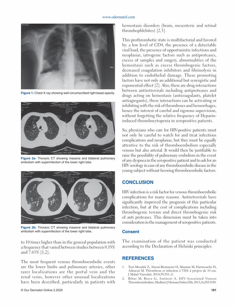
www.odermatol.com
© Our Dermatol Online 2.2020 181
to 10 times higher than in the general population with a frequency that varied between studies between 0.19% and 7.63% [1,2].
The most frequent venous thromboembolic events are the lower limbs and pulmonary arteries, other rarer localizations are the portal vein and the renal veins, however other unusual localizations have been described, particularly in patients with
hemostasis disorders (brain, mesenteric and retinal thrombophlebites) [2,3].
This prothrombotic state is multifactorial and favored by: a low level of CD4, the presence of a detectable viral load, the presence of opportunistic infections and neoplasias, iatrogenic factors such as antiproteases, excess of samples and surgery, abnormalities of the hemostasis such as excess thrombogenic factors, decreased coagulation inhibitors and fibrinolysis in addition to endothelial damage. These promoting factors have not only an additional but synergistic and exponential effect [2]. Also, there are drug interactions between antiretrovirals including antiproteases and drugs acting on hemostasis (anticoagulants, platelet antiagregants), these interactions can be activating or inhibiting with the risk of thromboses and hemorrhages, hence the interest of careful and rigorous supervision, without forgetting the relative frequency of Heparin-induced thrombocytopenia in seropositive patients.
So, physicians who care for HIV-positive patients must not only be careful to watch for and treat infectious complications and neoplasias, but they must be equally attentive to the risk of thromboembolism especially venous but also arterial. It would then be justifiable to raise the possibility of pulmonary embolism in the event of any dyspnea in the seropositive patient and to ask for an HIV serology in case of any thromboembolic disease in the young subject without favoring thromboembolic factors.
CONCLUSION
HIV infection is a risk factor for venous thromboembolic complications for many reasons. Antiretrovirals have significantly improved the prognosis of this particular infection, but at the cost of complications including thrombogenic terrain and direct thrombogenic risk of anti proteases. This dimension must be taken into consideration in the management of seropositive patients.
Consent
The examination of the patient was conducted according to the Declaration of Helsinki principles.
REFERENCES
1. Tazi-Mezalek Z, Alaoui-Bennasser H, Maamar M, Harmouche H, Adnaoui M. Thrombose et infection à VIH: à propos de 10 cas. J Malad Vasculair. 2014;39:351–2.
2. Bibas M, Biava G, Antinori A. HIV-Associated Venous Thromboembolism. Mediterr J Hematol Infect Dis. 2011;3:e2011030.
Figure 1: Chest X-ray showing well-circumscribed right basal opacity.
Figure 2a: Thoracic CT showing massive and bilateral pulmonary embolism with superinfection of the lower right lobe.
Figure 2b: Thoracic CT showing massive and bilateral pulmonary embolism with superinfection of the lower right lobe.

www.odermatol.com
© Our Dermatol Online 2.2020 182
3. Ahonkhai AA, Gebo KA, Streiff MB, Moore RD, Segal JB. Venous Thromboembolism in Patients with HIV/AIDS A Case-Control Study. J Acquir Immune Defi c Syndr. 2008;48:310–4.
4. Singer EJ, Valdes-Sueiras M, Commins DL, Yong W, Carlson M. HIV stroke risk: evidence and implications. Ther Adv Chronic Dis. 2013;4:61–70.
Copyright by Mohamed El Amraoui, et al. This is an open-access article distributed under the terms of the Creative Commons Attribution License, which permits unrestricted use, distribution, and reproduction in any medium, provided the original author and source are credited.Source of Support: Nil, Confl ict of Interest: None declared.
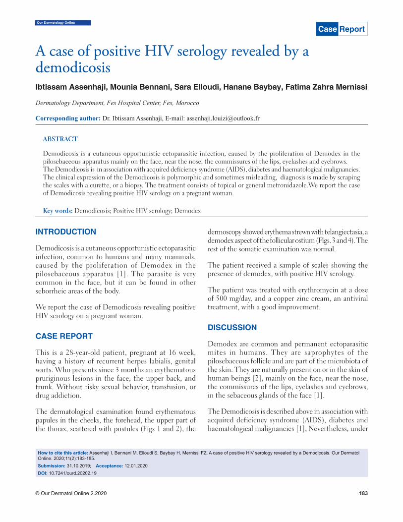
Our Dermatology Online
© Our Dermatol Online 2.2020 183
INTRODUCTION
Demodicosis is a cutaneous opportunistic ectoparasitic infection, common to humans and many mammals, caused by the proliferation of Demodex in the pilosebaceous apparatus [1]. The parasite is very common in the face, but it can be found in other seborrheic areas of the body.
We report the case of Demodicosis revealing positive HIV serology on a pregnant woman.
CASE REPORT
This is a 28-year-old patient, pregnant at 16 week, having a history of recurrent herpes labialis, genital warts. Who presents since 3 months an erythematous pruriginous lesions in the face, the upper back, and trunk. Without risky sexual behavior, transfusion, or drug addiction.
The dermatological examination found erythematous papules in the cheeks, the forehead, the upper part of the thorax, scattered with pustules (Figs 1 and 2), the
dermoscopy showed erythema strewn with telangiectasia, a demodex aspect of the follicular ostium (Figs. 3 and 4). The rest of the somatic examination was normal.
The patient received a sample of scales showing the presence of demodex, with positive HIV serology.
The patient was treated with erythromycin at a dose of 500 mg/day, and a copper zinc cream, an antiviral treatment, with a good improvement.
DISCUSSION
Demodex are common and permanent ectoparasitic mites in humans. They are saprophytes of the pilosebaceous follicle and are part of the microbiota of the skin. They are naturally present on or in the skin of human beings [2], mainly on the face, near the nose, the commissures of the lips, eyelashes and eyebrows, in the sebaceous glands of the face [1].
The Demodicosis is described above in association with acquired deficiency syndrome (AIDS), diabetes and haematological malignancies [1], Nevertheless, under
How to cite this article: Assenhaji I, Bennani M, Elloudi S, Baybay H, Mernissi FZ. A case of positive HIV serology revealed by a Demodicosis. Our Dermatol Online. 2020;11(2):183-185.Submission: 31.10.2019; Acceptance: 12.01.2020DOI: 10.7241/ourd.20202.19
Case Report
A case of positive HIV serology revealed by a A case of positive HIV serology revealed by a demodicosisdemodicosisIbtissam Assenhaji, Mounia Bennani, Sara Elloudi, Hanane Baybay, Fatima Zahra Mernissi
Dermatology Department, Fes Hospital Center, Fes, Morocco
Corresponding author: Dr. Ibtissam Assenhaji, E-mail: [email protected]
ABSTRACT
Demodicosis is a cutaneous opportunistic ectoparasitic infection, caused by the proliferation of Demodex in the pilosebaceous apparatus mainly on the face, near the nose, the commissures of the lips, eyelashes and eyebrows. The Demodicosis is in association with acquired deficiency syndrome (AIDS), diabetes and haematological malignancies. The clinical expression of the Demodicosis is polymorphic and sometimes misleading, diagnosis is made by scraping the scales with a curette, or a biopsy. The treatment consists of topical or general metronidazole.We report the case of Demodicosis revealing positive HIV serology on a pregnant woman.
Key words: Demodicosis; Positive HIV serology; Demodex
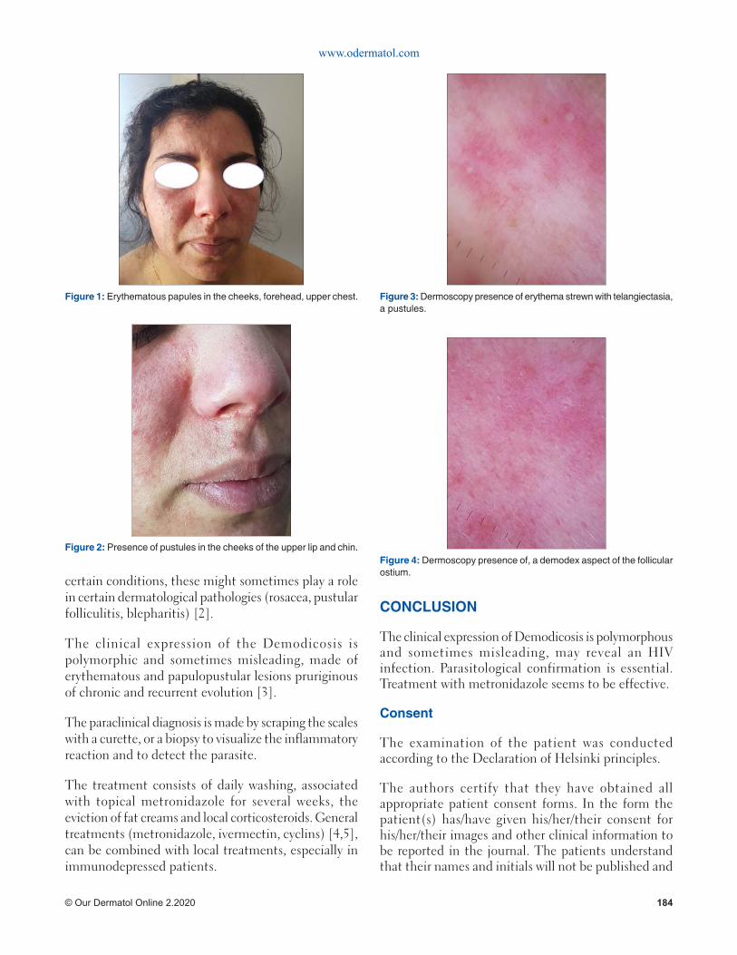
www.odermatol.com
© Our Dermatol Online 2.2020 184
certain conditions, these might sometimes play a role in certain dermatological pathologies (rosacea, pustular folliculitis, blepharitis) [2].
The clinical expression of the Demodicosis is polymorphic and sometimes misleading, made of erythematous and papulopustular lesions pruriginous of chronic and recurrent evolution [3].
The paraclinical diagnosis is made by scraping the scales with a curette, or a biopsy to visualize the inflammatory reaction and to detect the parasite.
The treatment consists of daily washing, associated with topical metronidazole for several weeks, the eviction of fat creams and local corticosteroids. General treatments (metronidazole, ivermectin, cyclins) [4,5], can be combined with local treatments, especially in immunodepressed patients.
CONCLUSION
The clinical expression of Demodicosis is polymorphous and sometimes misleading, may reveal an HIV infection. Parasitological confirmation is essential. Treatment with metronidazole seems to be effective.
Consent
The examination of the patient was conducted according to the Declaration of Helsinki principles.
The authors certify that they have obtained all appropriate patient consent forms. In the form the patient(s) has/have given his/her/their consent for his/her/their images and other clinical information to be reported in the journal. The patients understand that their names and initials will not be published and
Figure 1: Erythematous papules in the cheeks, forehead, upper chest.
Figure 2: Presence of pustules in the cheeks of the upper lip and chin.
Figure 3: Dermoscopy presence of erythema strewn with telangiectasia, a pustules.
Figure 4: Dermoscopy presence of, a demodex aspect of the follicular ostium.
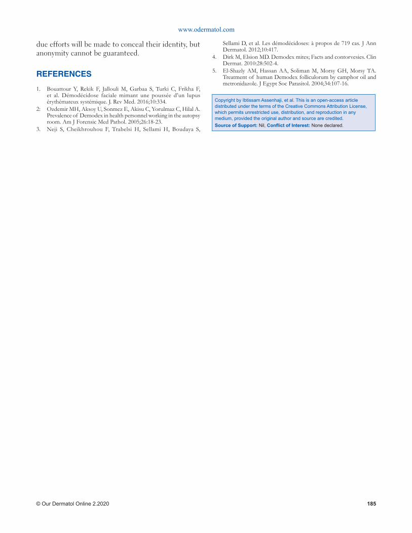
www.odermatol.com
© Our Dermatol Online 2.2020 185
Copyright by Ibtissam Assenhaji, et al. This is an open-access article distributed under the terms of the Creative Commons Attribution License, which permits unrestricted use, distribution, and reproduction in any medium, provided the original author and source are credited.Source of Support: Nil, Confl ict of Interest: None declared.
due efforts will be made to conceal their identity, but anonymity cannot be guaranteed.
REFERENCES
1. Bouattour Y, Rekik F, Jallouli M, Garbaa S, Turki C, Frikha F, et al. Démodécidose faciale mimant une poussée d’un lupus érythémateux systémique. J. Rev Med. 2016;10:334.
2: Ozdemir MH, Aksoy U, Sonmez E, Akisu C, Yorulmaz C, Hilal A. Prevalence of Demodex in health personnel working in the autopsy room. Am J Forensic Med Pathol. 2005;26:18-23.
3. Neji S, Cheikhrouhou F, Trabelsi H, Sellami H, Boudaya S,
Sellami D, et al. Les démodécidoses: à propos de 719 cas. J Ann Dermatol. 2012;10:417.
4. Dirk M, Elsion MD. Demodex mites; Facts and contorvesies. Clin Dermat. 2010:28:502-4.
5. El-Shazly AM, Hassan AA, Soliman M, Morsy GH, Morsy TA. Treatment of human Demodex folliculorum by camphor oil and metronidazole. J Egypt Soc Parasitol. 2004;34:107-16.
Our Dermatology Online
© Our Dermatol Online 2.2020 186
OPINION ARTICLE
Statistics by the Centers for Disease Control and Prevention (CDC) in 2013 show that only 10.1% of adolescents in the United States (U.S.) wear sunscreen regularly when they are outside on a sunny day for more than an hour. Furthermore, the Healthy People 2020 has failed to reach its target to increase the sun protection among teens from 9.3% in 2009 to 11.2% by 2020 [1]. Ultraviolet radiation (UVR) exposure and sunburns during the age of 10 to 15 is linked to all kinds of skin cancer, in particular malignant melanoma, the second most common cancer among females aged 15 to 29 years old [2,3]. Studies have also shown that primary school-aged boys and children aged 8 and above are less protected against UVR and have higher reported sunburns [2]. Many sun protection programs focus on children due to their constant exposure to UVR and due to the fact that there is a higher probability that children would change their behavior [4]. As the skin of children is more sensitive to UVR, sun protection
should begin in early childhood, an important phase to establish health behaviors and make a difference in children’s lives [2].
Acknowledging the essential role of parents, some of the strategies parents can take include setting a good example for their children by reassessing their sun safety practices. A research by Behrens et al. showed that children aged 13 and above have an 85% increase in the risk of sunburn when parents favor tanned skin [2]. Other literature also shows that the use of indoor tanning among youth is strongly co-related to parental permission as well as parental use of tanning beds [3]. Another research by Mayer et. al (2011) which investigated the extent of tanning among teens aged 14 to 17 years old in various cities in the U.S. found out that about 1 in 5 girls and 3 in 100 boys had used indoor tanning within the last year. The same research found that the use of indoor tanning is predicted by several factors. These include being a white female, parental use and their beliefs on indoor
ABSTRACT
Statistics by the Centers for Disease Control and Prevention (CDC) in 2013 show that only 10.1% of adolescents in the United States (U.S.) wear sunscreen regularly when they are outside on a sunny day for more than an hour. Furthermore, the Healthy People 2020 has failed to reach its target to increase the sun protection among teens from 9.3% in 2009 to 11.2% by 2020. Ultraviolet radiation (UVR) exposure and sunburns during the age of 10 to 15 is linked to all kinds of skin cancer, in particular malignant melanoma, the second most common cancer among females aged 15 to 29 years old. Studies have also shown that primary school-aged boys and children aged 8 and above are less protected against UVR and have higher reported sunburns. Many sun protection programs focus on children due to their constant exposure to UVR and due to the fact that there is a higher probability that children would change their behavior. As the skin of children is more sensitive to UVR, sun protection should begin in early childhood, an important phase to establish health behaviors and make a difference in children’s lives. Parents play an imperative part in promoting childhood sun safety. Promoting this can take several forms: parents setting a good example for their children and parents keeping themselves and their children educated on sun safety. All these are simply yet effective measures which can go a long way in helping to reduce the burden of skin cancer.
Key words: Melanoma; Malignant melanoma; Sun protection factor
How to cite this article: Lim, TLC. Parental roles and childhood sun safety. Our Dermatol Online. 2020;11(2):186-188.Submission: 10.01.2020; Acceptance: 03.02.2020DOI: 10.7241/ourd.20202.20
Opinion Article
Parental roles and childhood sun safetyParental roles and childhood sun safetyTessa Li Chyin Lim
Monash University Malaysia Campus, Malaysia
Corresponding author: Tessa Li Chyin Lim, E-mail: [email protected]
www.odermatol.com
© Our Dermatol Online 2.2020 187
tanning. In a nutshell, parents were a major factor in whether their children partake in indoor tanning. This is in line with other research which showed that parents’ attitudes can impact their children’s future behavior [5]. According to American Cancer Society, youths are especially susceptible to the misleading claims of tanning industry. Some claim that there is no scientific proof about dangers of UV from tanning beds and it is not a big problem and no different from being under the sun. Thus, they have the misconception that melanoma is not a disease of the young. Contrary to all these popular beliefs, frequent exposure of the UVA by indoor tanning before 35 years old increases the risk of melanoma by almost 60%, which is remarkable. In addition, 12.5% of all indoor tanning-related acute injuries treated in U.S. hospital emergency departments annually is related to adolescents below 18 years old [3]. Therefore, it is important for parents to know about the well-established dangers of UV radiation exposure from tanning beds and be a role model for their children. Short term effects can include syncope, keratitis and corneal burns, sunburn and immune system suppression. On the other hand, long term effects include solar keratosis, premature skin aging, wrinkles, skin and ocular malignancies, and permanent visual loss. Getting a base tan also does not prevent sunburn and it is not possible to get enough Vitamin D from tanning beds. There is also a real risk of addiction to tanning beds [3].
The second point is that parents ought to educate themselves and their children about sun protection. According to Thoonen and colleagues, sun safety measures should focus on both parents and their children and it is warranted that boys have specific stimulation of sun protection. Establishing good childhood sun protection behaviors will increase the likelihood of habitual behavior in the future. It is found that children who reach adolescence have weakened sun-protective attitudes and behaviours [2]. Several studies also found that generally adolescents do not apply sun protection practices consistently. An analysis of literature about children’s sun safety which included parental influence over children programs was done by Buller and colleagues. It was concluded that there were better outcomes for the whole family when programs aimed to increase the parents’ knowledge about sun protection and focused on behavioral adjustment in order to protect themselves and not just their children [4]. Therefore, parental influence is necessary and should not be taken lightly. The most recommended sun safe behaviors include seeking
shade, applying sunscreen and wearing UV-protective outfits with a hat and sunglasses [2]. The CDC recommends indoor activities during mid-day and if possible, seek shade under umbrellas, trees or pop-up tents. Sunscreens must be at least SPF 15, applied half an hour before heading outdoors and reapplied every 2 hours even during cloudy days. There is also another recommendation to cover up with long-sleeved shirts, long pants and skirts which are dry, dark-colored and made from tightly woven fabric. Furthermore, it is recommended that hats should shade the face, scalp, eats and neck [6].
Another way is for parents to educate themselves about sun protection to be able to differentiate facts from myths. These are some of the crucial facts about sun protection according to the Cancer Council Australia. Fact number 1 is that sun damage can still have on cool, windy and cloudy days. Fact number 2 is that a tan does not protect the skin from the sun. Fact number 3 is that people with any skin type can get skin cancer. Fact number 4 is that car window does not prevent sun burns. Fact number 5 is that there is no such thing as a safe tan. Fact number 6 is that skin cancer treatment is more serious than just the removal of lesion. It can include chemotherapy or even surgery. Fact number 6 is that prolonged sun exposure does not increase the Vitamin D levels in the body further. Lastly, sunscreen should not be used to extend the amount of time under the sun [7]. If parents know all these common facts about sun protection, it will most probably benefit their children as well.
In summary, parents play an imperative part in promoting childhood sun safety. Promoting this can take several forms: parents setting a good example for their children and parents keeping themselves and their children educated on sun safety. All these are simply yet effective measures which can go a long way in helping to reduce the burden of skin cancer in the U.S. This is a step in the right direction.
REFERENCES
1. Centers for Disease Control and Prevention. Sun Protection. https://www.cdc.gov/cancer/skin/statistics/behavior/sun-protection.htm. Accessed January 4, 2020.
2. Thoonen K, Schneider F, Candel M, de Vries H, van Osch L. Childhood sun safety at different ages: relations between parental sun protection behavior towards their child and children’s own sun protection behavior. BMC Public Health. 2019;19:1044.
3. American Cancer Society. Just the Facts: Teens and the Dangers of Indoor Tanning. https://www.fi ghtcancer.org/sites/default/fi les/FINAL%20-%20Just%20the%20Facts%20-%20Teens%20and%20

www.odermatol.com
© Our Dermatol Online 2.2020 188
Indoor%20Tanning%2006.15.16.pdf. Accessed January 4, 2020.4. Buller DB, Borland R. Skin cancer prevention for children: A critical
review. Health Educat Beh. 1999;26:317–43.5. Mayer JA, Woodruff SI, Slymen DJ, Sallis JF, Forster JL, Clapp EJ,
et al. Adolescents’ use of indoor tanning: a large-scale evaluation of psychosocial, environmental, and policy-level correlates. Am J Public Health. 2011;101:930-8.
6. Centers for Disease Control and Prevention. How Can I Protect My Children from the Sun. https://www.cdc.gov/cancer/skin/basic_info/children.htm. Accessed January 4, 2020.
7. Cancer Council Australia. 10 myths about sun protection. https://www.cancer.org.au/preventing-cancer/sun-protection/10-myths-about-sun-protection.html. Accessed January 4, 2020.
Copyright by Tessa Li Chyin Lim. This is an open-access article distributed under the terms of the Creative Commons Attribution License, which permits unrestricted use, distribution, and reproduction in any medium, provided the original author and source are credited.Source of Support: Nil, Confl ict of Interest: None declared.
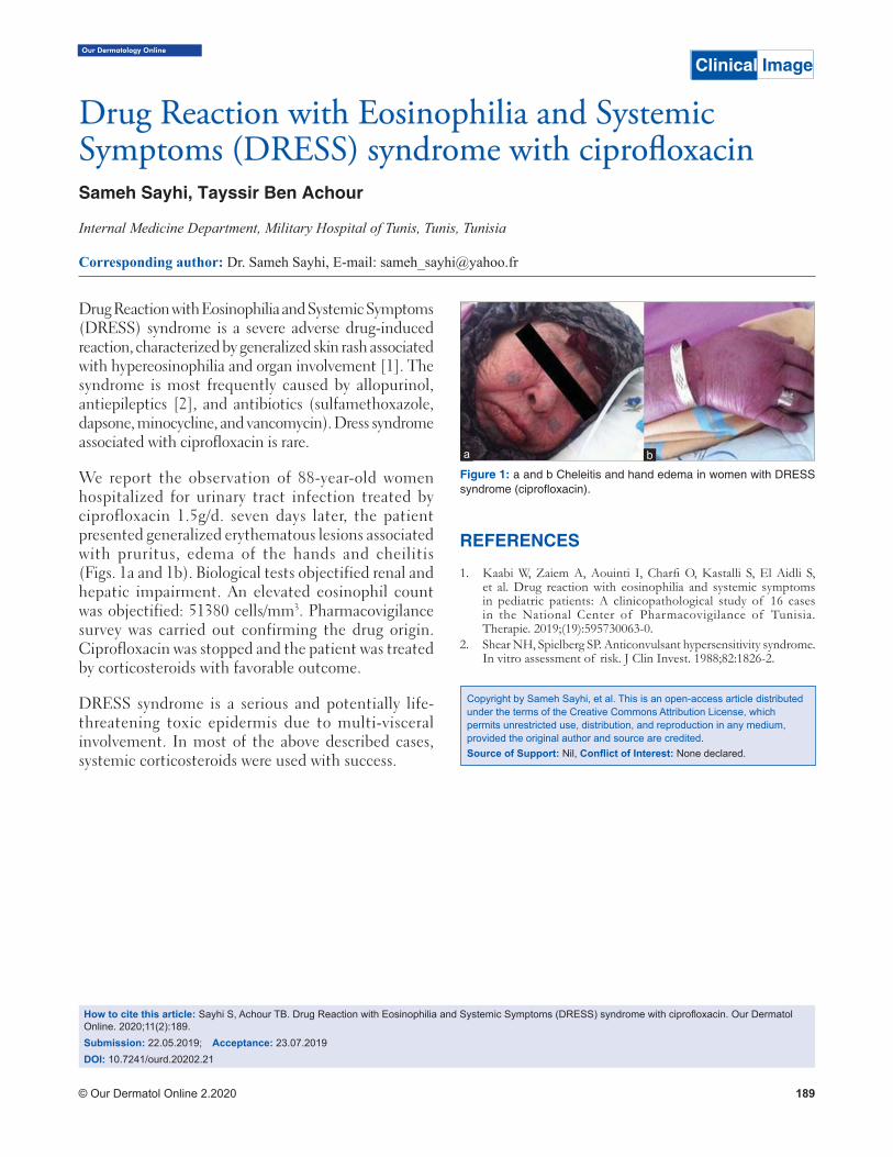
Our Dermatology Online
© Our Dermatol Online 2.2020 189
Drug Reaction with Eosinophilia and Systemic Symptoms (DRESS) syndrome is a severe adverse drug-induced reaction, characterized by generalized skin rash associated with hypereosinophilia and organ involvement [1]. The syndrome is most frequently caused by allopurinol, antiepileptics [2], and antibiotics (sulfamethoxazole, dapsone, minocycline, and vancomycin). Dress syndrome associated with ciprofloxacin is rare.
We report the observation of 88-year-old women hospitalized for urinary tract infection treated by ciprofloxacin 1.5g/d. seven days later, the patient presented generalized erythematous lesions associated with pruritus, edema of the hands and cheilitis (Figs. 1a and 1b). Biological tests objectified renal and hepatic impairment. An elevated eosinophil count was objectified: 51380 cells/mm3. Pharmacovigilance survey was carried out confirming the drug origin. Ciprofloxacin was stopped and the patient was treated by corticosteroids with favorable outcome.
DRESS syndrome is a serious and potentially life-threatening toxic epidermis due to multi-visceral involvement. In most of the above described cases, systemic corticosteroids were used with success.
REFERENCES
1. Kaabi W, Zaiem A, Aouinti I, Charfi O, Kastalli S, El Aidli S, et al. Drug reaction with eosinophilia and systemic symptoms in pediatric patients: A clinicopathological study of 16 cases in the National Center of Pharmacovigilance of Tunisia. Therapie. 2019;(19):595730063-0.
2. Shear NH, Spielberg SP. Anticonvulsant hypersensitivity syndrome. In vitro assessment of risk. J Clin Invest. 1988;82:1826-2.
Copyright by Sameh Sayhi, et al. This is an open-access article distributed under the terms of the Creative Commons Attribution License, which permits unrestricted use, distribution, and reproduction in any medium, provided the original author and source are credited.Source of Support: Nil, Confl ict of Interest: None declared.
Clinical Image
Drug Reaction with Eosinophilia and Systemic Drug Reaction with Eosinophilia and Systemic Symptoms (DRESS) syndrome with ciprofl oxacinSymptoms (DRESS) syndrome with ciprofl oxacinSameh Sayhi, Tayssir Ben Achour
Internal Medicine Department, Military Hospital of Tunis, Tunis, Tunisia
Corresponding author: Dr. Sameh Sayhi, E-mail: [email protected]
How to cite this article: Sayhi S, Achour TB. Drug Reaction with Eosinophilia and Systemic Symptoms (DRESS) syndrome with ciprofl oxacin. Our Dermatol Online. 2020;11(2):189.Submission: 22.05.2019; Acceptance: 23.07.2019DOI: 10.7241/ourd.20202.21
Figure 1: a and b Cheleitis and hand edema in women with DRESS syndrome (ciprofl oxacin).
ba
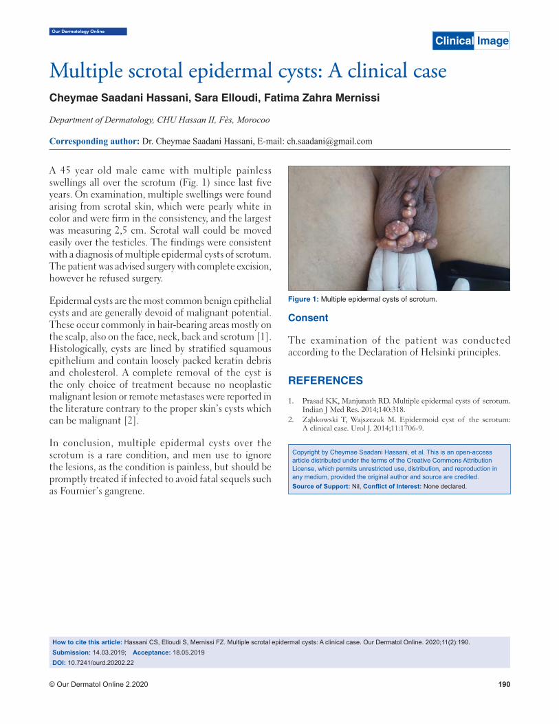
Our Dermatology Online
© Our Dermatol Online 2.2020 190
How to cite this article: Hassani CS, Elloudi S, Mernissi FZ. Multiple scrotal epidermal cysts: A clinical case. Our Dermatol Online. 2020;11(2):190.Submission: 14.03.2019; Acceptance: 18.05.2019DOI: 10.7241/ourd.20202.22
Multiple scrotal epidermal cysts: A clinical caseMultiple scrotal epidermal cysts: A clinical caseCheymae Saadani Hassani, Sara Elloudi, Fatima Zahra Mernissi
Department of Dermatology, CHU Hassan II, Fès, Morocoo
Corresponding author: Dr. Cheymae Saadani Hassani, E-mail: [email protected]
A 45 year old male came with multiple painless swellings all over the scrotum (Fig. 1) since last five years. On examination, multiple swellings were found arising from scrotal skin, which were pearly white in color and were firm in the consistency, and the largest was measuring 2,5 cm. Scrotal wall could be moved easily over the testicles. The findings were consistent with a diagnosis of multiple epidermal cysts of scrotum. The patient was advised surgery with complete excision, however he refused surgery.
Epidermal cysts are the most common benign epithelial cysts and are generally devoid of malignant potential. These occur commonly in hair-bearing areas mostly on the scalp, also on the face, neck, back and scrotum [1]. Histologically, cysts are lined by stratified squamous epithelium and contain loosely packed keratin debris and cholesterol. A complete removal of the cyst is the only choice of treatment because no neoplastic malignant lesion or remote metastases were reported in the literature contrary to the proper skin’s cysts which can be malignant [2].
In conclusion, multiple epidermal cysts over the scrotum is a rare condition, and men use to ignore the lesions, as the condition is painless, but should be promptly treated if infected to avoid fatal sequels such as Fournier’s gangrene.
Consent
The examination of the patient was conducted according to the Declaration of Helsinki principles.
REFERENCES
1. Prasad KK, Manjunath RD. Multiple epidermal cysts of scrotum. Indian J Med Res. 2014;140:318.
2. Ząbkowski T, Wajszczuk M. Epidermoid cyst of the scrotum: A clinical case. Urol J. 2014;11:1706-9.
Figure 1: Multiple epidermal cysts of scrotum.
Copyright by Cheymae Saadani Hassani, et al. This is an open-access article distributed under the terms of the Creative Commons Attribution License, which permits unrestricted use, distribution, and reproduction in any medium, provided the original author and source are credited.Source of Support: Nil, Confl ict of Interest: None declared.
Clinical Image
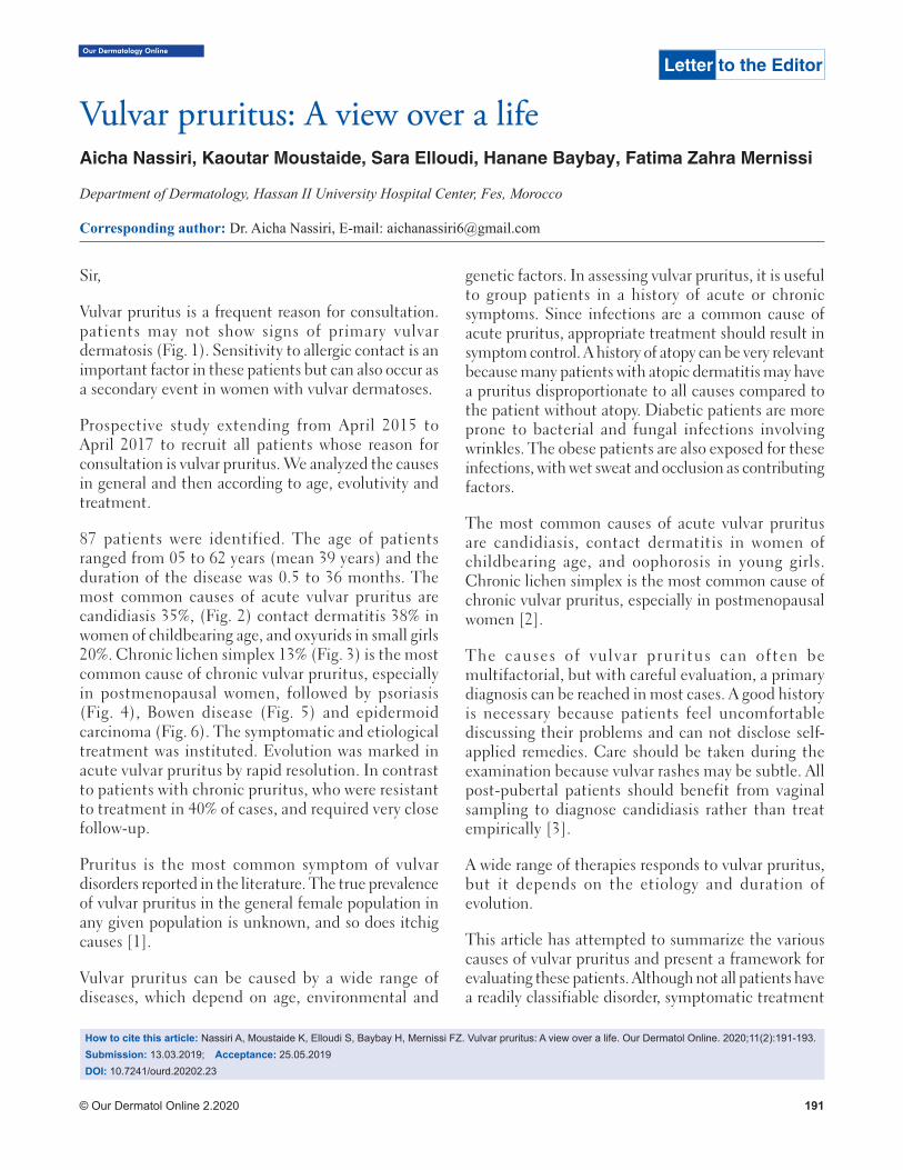
Our Dermatology Online
© Our Dermatol Online 2.2020 191
Vulvar pruritus: A view over a lifeVulvar pruritus: A view over a lifeAicha Nassiri, Kaoutar Moustaide, Sara Elloudi, Hanane Baybay, Fatima Zahra Mernissi
Department of Dermatology, Hassan II University Hospital Center, Fes, Morocco
Corresponding author: Dr. Aicha Nassiri, E-mail: [email protected]
Sir,
Vulvar pruritus is a frequent reason for consultation. patients may not show signs of primary vulvar dermatosis (Fig. 1). Sensitivity to allergic contact is an important factor in these patients but can also occur as a secondary event in women with vulvar dermatoses.
Prospective study extending from April 2015 to April 2017 to recruit all patients whose reason for consultation is vulvar pruritus. We analyzed the causes in general and then according to age, evolutivity and treatment.
87 patients were identified. The age of patients ranged from 05 to 62 years (mean 39 years) and the duration of the disease was 0.5 to 36 months. The most common causes of acute vulvar pruritus are candidiasis 35%, (Fig. 2) contact dermatitis 38% in women of childbearing age, and oxyurids in small girls 20%. Chronic lichen simplex 13% (Fig. 3) is the most common cause of chronic vulvar pruritus, especially in postmenopausal women, followed by psoriasis (Fig. 4), Bowen disease (Fig. 5) and epidermoid carcinoma (Fig. 6). The symptomatic and etiological treatment was instituted. Evolution was marked in acute vulvar pruritus by rapid resolution. In contrast to patients with chronic pruritus, who were resistant to treatment in 40% of cases, and required very close follow-up.
Pruritus is the most common symptom of vulvar disorders reported in the literature. The true prevalence of vulvar pruritus in the general female population in any given population is unknown, and so does itchig causes [1].
Vulvar pruritus can be caused by a wide range of diseases, which depend on age, environmental and
genetic factors. In assessing vulvar pruritus, it is useful to group patients in a history of acute or chronic symptoms. Since infections are a common cause of acute pruritus, appropriate treatment should result in symptom control. A history of atopy can be very relevant because many patients with atopic dermatitis may have a pruritus disproportionate to all causes compared to the patient without atopy. Diabetic patients are more prone to bacterial and fungal infections involving wrinkles. The obese patients are also exposed for these infections, with wet sweat and occlusion as contributing factors.
The most common causes of acute vulvar pruritus are candidiasis, contact dermatitis in women of childbearing age, and oophorosis in young girls. Chronic lichen simplex is the most common cause of chronic vulvar pruritus, especially in postmenopausal women [2].
The causes of vulvar pruritus can often be multifactorial, but with careful evaluation, a primary diagnosis can be reached in most cases. A good history is necessary because patients feel uncomfortable discussing their problems and can not disclose self-applied remedies. Care should be taken during the examination because vulvar rashes may be subtle. All post-pubertal patients should benefit from vaginal sampling to diagnose candidiasis rather than treat empirically [3].
A wide range of therapies responds to vulvar pruritus, but it depends on the etiology and duration of evolution.
This article has attempted to summarize the various causes of vulvar pruritus and present a framework for evaluating these patients. Although not all patients have a readily classifiable disorder, symptomatic treatment
How to cite this article: Nassiri A, Moustaide K, Elloudi S, Baybay H, Mernissi FZ. Vulvar pruritus: A view over a life. Our Dermatol Online. 2020;11(2):191-193.Submission: 13.03.2019; Acceptance: 25.05.2019DOI: 10.7241/ourd.20202.23
Letter to the Editor
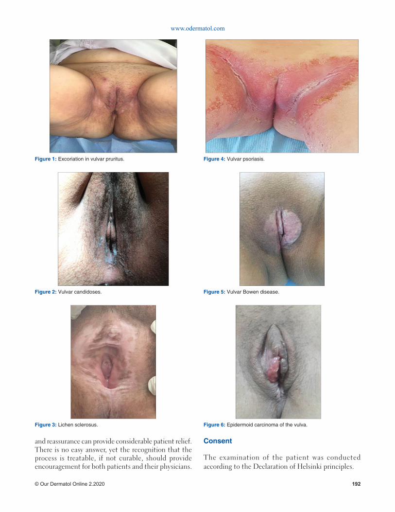
www.odermatol.com
© Our Dermatol Online 2.2020 192
and reassurance can provide considerable patient relief. There is no easy answer, yet the recognition that the process is treatable, if not curable, should provide encouragement for both patients and their physicians.
Consent
The examination of the patient was conducted according to the Declaration of Helsinki principles.
Figure 6: Epidermoid carcinoma of the vulva.
Figure 1: Excoriation in vulvar pruritus.
Figure 2: Vulvar candidoses.
Figure 3: Lichen sclerosus.
Figure 4: Vulvar psoriasis.
Figure 5: Vulvar Bowen disease.

www.odermatol.com
© Our Dermatol Online 2.2020 193
REERENCES
1. Bohl TG. Overview of Vulvar Pruritus Through the Life Cycle. Clin Obstet Gynecol. 2005;48:786-807.
2. Kaur J, Kalsy J. Study of pruritus vulvae in geriatric age group in tertiary hospital. Indian J Sex Transm Dis AIDS. 2017;38:15-21.
3. Carrillo-Meléndrez H, Villamil-Cerda D, Espinoza-Hernández J, Lacy-Niebla RM. Vulvar pruritus: determination of the most
common causes and their treatments]. Ginecol Obstet Mex. 2015;83:179-88.
Copyright by Aicha Nassiri, et al. This is an open-access article distributed under the terms of the Creative Commons Attribution License, which permits unrestricted use, distribution, and reproduction in any medium, provided the original author and source are credited.Source of Support: Nil, Confl ict of Interest: None declared.
Our Dermatology Online
© Our Dermatol Online 2.2020 194
Sir,
Topical corticosteroids (TCs) are one of the most commonly used preparations in dermatology practice. Their rapid anti-inflammatory, immunosuppressive and anti-pruritic activity has made them the drug of choice for a large number of dermatoses [1]. Apart from the well documented uses of TCs, they can also cause a wide array of adverse effects if used indiscriminately or for long duration without supervision which include steroid rosacea, acneiform eruption, hypertrichosis [2]. TCs misuse is a common problem in our country owing to their easy availability as over the counter medication and preparations and lack of awareness among the general population. TCs are commonly being used as fairness and anti acne medications by the general population without any dermatological consultation which has led to a significant number of patients presenting with cutaneous adverse effects of TCs to the dermatologists [2,3]. Corticosteroid abuse is also being observed in pediatric population where topical steroids are being misused by the caregivers either as self-medication or by using them for duration more that that advised by a dermatologist.
This study was carried out in a dermatology outpatient centre with the main aims of studying the clinic-epidemiological features of TCs misuse among the pediatric population.
This was a prospective, questionnaire based study carried out over a period of one and half year in our centre in which the patients were questioned and assessed for misuse of TCs in terms of indication, frequency, duration and source of recommendation. All the patients who had been self-medicated with TCs or where the parents/caregivers had used TCs beyond
the prescribed time advised by the dermatologists were included in the study. After taking an informed consent, the patients were assessed regarding the formulation, frequency, duration or indications of TCs use for different skin conditions and the various cutaneous adverse effects, wherever present, were also noted.
A total of 50 patients (M: F 29:21) were included in our study. The age range of patients varied from 6 months to 18 years with a mean age of 11.35 years. Most of the patients were aged >12 years (54%). The age distribution and the indications for steroid use are enumerated in Table 1. The most common indication for TCs use in our study was eczemas (32%), fungal infections (20%), bacterial and viral infections (18%), acne (12%) and facial pigmentation (10%). The most commonly abused corticosteroids were mometasone (36%), clobetasol (26%) and betamethasone (22%) (Table 2). The duration of TCs use varied from three days to one year in our study group whereas the frequency of use varied from thrice a week to thrice a day. Mometasone based Kligman’s combination and salicyclic acid-clobetasol combinations were also being used over the face in pediatric population for indications like acne and acne scarring.
The lack of awareness among the population can be gauged from the fact that only 18% patients (n=9) were aware of the adverse effects of TCs misuse. A large majority of patients (32%, n=16) were using topical corticosteroids on advice of friends or relatives, 28% (n=14) on the advice of pharmacists or paramedical personnel; 6 (12%) patients were using them based on information from internet or other sources, 4 (8%) were advised by beauty parlors and beauticians. Ten patients (20%) were using the TCs advised by the dermatologists, but had been using them for a period beyond the advised time frame or had been using it
Letter to the Editor
Topical corticosteroid abuse among pediatric Topical corticosteroid abuse among pediatric population - a prospective studypopulation - a prospective studyMrinal Gupta
Department of Dermatology, Treatwell Skin Centre, Jammu and Kashmir, India
Corresponding author: Dr. Mrinal Gupta, E-mail: [email protected]
How to cite this article: Gupta M. Topical corticosteroid abuse among pediatric population - a prospective study. Our Dermatol Online. 2020;11(2):194-196.Submission: 02.05.2019; Acceptance: 12.08.2019DOI: 10.7241/ourd.20202.24
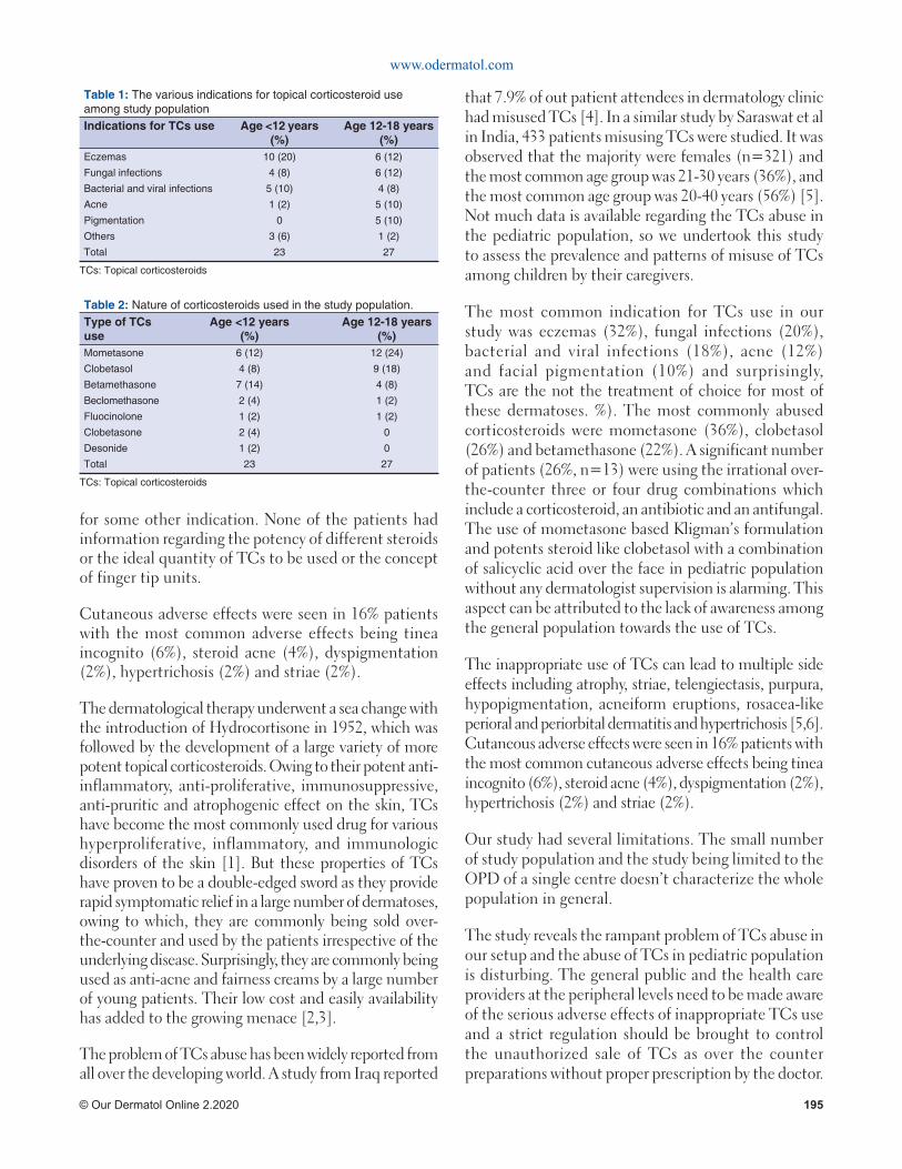
www.odermatol.com
© Our Dermatol Online 2.2020 195
for some other indication. None of the patients had information regarding the potency of different steroids or the ideal quantity of TCs to be used or the concept of finger tip units.
Cutaneous adverse effects were seen in 16% patients with the most common adverse effects being tinea incognito (6%), steroid acne (4%), dyspigmentation (2%), hypertrichosis (2%) and striae (2%).
The dermatological therapy underwent a sea change with the introduction of Hydrocortisone in 1952, which was followed by the development of a large variety of more potent topical corticosteroids. Owing to their potent anti-inflammatory, anti-proliferative, immunosuppressive, anti-pruritic and atrophogenic effect on the skin, TCs have become the most commonly used drug for various hyperproliferative, inflammatory, and immunologic disorders of the skin [1]. But these properties of TCs have proven to be a double-edged sword as they provide rapid symptomatic relief in a large number of dermatoses, owing to which, they are commonly being sold over-the-counter and used by the patients irrespective of the underlying disease. Surprisingly, they are commonly being used as anti-acne and fairness creams by a large number of young patients. Their low cost and easily availability has added to the growing menace [2,3].
The problem of TCs abuse has been widely reported from all over the developing world. A study from Iraq reported
that 7.9% of out patient attendees in dermatology clinic had misused TCs [4]. In a similar study by Saraswat et al in India, 433 patients misusing TCs were studied. It was observed that the majority were females (n=321) and the most common age group was 21-30 years (36%), and the most common age group was 20-40 years (56%) [5]. Not much data is available regarding the TCs abuse in the pediatric population, so we undertook this study to assess the prevalence and patterns of misuse of TCs among children by their caregivers.
The most common indication for TCs use in our study was eczemas (32%), fungal infections (20%), bacterial and viral infections (18%), acne (12%) and facial pigmentation (10%) and surprisingly, TCs are the not the treatment of choice for most of these dermatoses. %). The most commonly abused corticosteroids were mometasone (36%), clobetasol (26%) and betamethasone (22%). A significant number of patients (26%, n=13) were using the irrational over-the-counter three or four drug combinations which include a corticosteroid, an antibiotic and an antifungal. The use of mometasone based Kligman’s formulation and potents steroid like clobetasol with a combination of salicyclic acid over the face in pediatric population without any dermatologist supervision is alarming. This aspect can be attributed to the lack of awareness among the general population towards the use of TCs.
The inappropriate use of TCs can lead to multiple side effects including atrophy, striae, telengiectasis, purpura, hypopigmentation, acneiform eruptions, rosacea-like perioral and periorbital dermatitis and hypertrichosis [5,6]. Cutaneous adverse effects were seen in 16% patients with the most common cutaneous adverse effects being tinea incognito (6%), steroid acne (4%), dyspigmentation (2%), hypertrichosis (2%) and striae (2%).
Our study had several limitations. The small number of study population and the study being limited to the OPD of a single centre doesn’t characterize the whole population in general.
The study reveals the rampant problem of TCs abuse in our setup and the abuse of TCs in pediatric population is disturbing. The general public and the health care providers at the peripheral levels need to be made aware of the serious adverse effects of inappropriate TCs use and a strict regulation should be brought to control the unauthorized sale of TCs as over the counter preparations without proper prescription by the doctor.
Table 1: The various indications for topical corticosteroid use among study populationIndications for TCs use Age <12 years
(%)Age 12-18 years
(%)Eczemas 10 (20) 6 (12)
Fungal infections 4 (8) 6 (12)
Bacterial and viral infections 5 (10) 4 (8)
Acne 1 (2) 5 (10)
Pigmentation 0 5 (10)
Others 3 (6) 1 (2)
Total 23 27
TCs: Topical corticosteroids
Table 2: Nature of corticosteroids used in the study population.Type of TCs use
Age <12 years (%)
Age 12-18 years (%)
Mometasone 6 (12) 12 (24)
Clobetasol 4 (8) 9 (18)
Betamethasone 7 (14) 4 (8)
Beclomethasone 2 (4) 1 (2)
Fluocinolone 1 (2) 1 (2)
Clobetasone 2 (4) 0
Desonide 1 (2) 0
Total 23 27
TCs: Topical corticosteroids
www.odermatol.com
© Our Dermatol Online 2.2020 196
Statement of Human and Animal Rights
All procedures followed were in accordance with the ethical standards of the responsible committee on human experimentation (institutional and national) and with the Helsinki Declaration of 1975, as revised in 2008.
Statement of Informed Consent
Informed consent was obtained from all patients for being included in the study.
REFERENCES
1. Becke r DE. Ba s i c and c l i n i c a l pha r maco log y o f glucocorticosteroids. Anesth Prog. 2013;60:25–31.
2. Dey VK. Misuse of topical corticosteroids: A clinical study of adverse effects. Indian Dermatol Online J. 2014;5:436–40.
3. Jha AK, Sinha R, Prasad S. Misuse of topical corticosteroids on the face: A cross-sectional study among dermatology outpatients. Indian Dermatol Online J. 2016;7:259–63.
4. Al-Dhalimi MA, Aljawahiri N. Misuse of topical corticosteroids: A clinical study from an Iraqi hospital. East Mediterr Health J. 2006;12:847-52.
5. Saraswat A, Lahiri K, Chatterjee M, Barua S, Coondoo A, Mittal A, et al. Topical corticosteroid abuse on the face: A prospective, multicenter study of dermatology outpatients. Indian J Dermatol Venereol Leprol. 2011;77:160-6.
6. Hengge UR, Ruzicka T, Schwartz RA, Cork MJ. Adverse effects of topical glucocorticosteroids. J Am Acad Dermatol. 2006;54:1-15.
Copyright by Mrinal Gupta. This is an open-access article distributed under the terms of the Creative Commons Attribution License, which permits unrestricted use, distribution, and reproduction in any medium, provided the original author and source are credited.Source of Support: Nil, Confl ict of Interest: None declared.
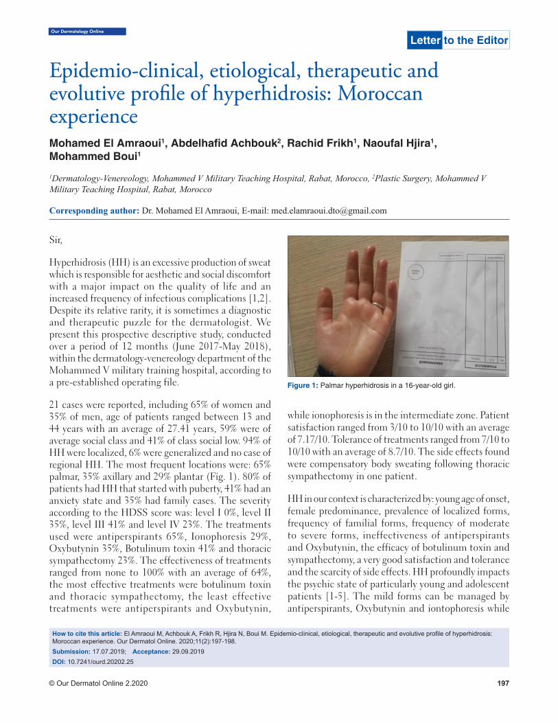
Our Dermatology Online
© Our Dermatol Online 2.2020 197
How to cite this article: El Amraoui M, Achbouk A, Frikh R, Hjira N, Boui M. Epidemio-clinical, etiological, therapeutic and evolutive profi le of hyperhidrosis: Moroccan experience. Our Dermatol Online. 2020;11(2):197-198.Submission: 17.07.2019; Acceptance: 29.09.2019DOI: 10.7241/ourd.20202.25
Sir,
Hyperhidrosis (HH) is an excessive production of sweat which is responsible for aesthetic and social discomfort with a major impact on the quality of life and an increased frequency of infectious complications [1,2]. Despite its relative rarity, it is sometimes a diagnostic and therapeutic puzzle for the dermatologist. We present this prospective descriptive study, conducted over a period of 12 months (June 2017-May 2018), within the dermatology-venereology department of the Mohammed V military training hospital, according to a pre-established operating file.
21 cases were reported, including 65% of women and 35% of men, age of patients ranged between 13 and 44 years with an average of 27.41 years, 59% were of average social class and 41% of class social low. 94% of HH were localized, 6% were generalized and no case of regional HH. The most frequent locations were: 65% palmar, 35% axillary and 29% plantar (Fig. 1). 80% of patients had HH that started with puberty, 41% had an anxiety state and 35% had family cases. The severity according to the HDSS score was: level I 0%, level II 35%, level III 41% and level IV 23%. The treatments used were antiperspirants 65%, Ionophoresis 29%, Oxybutynin 35%, Botulinum toxin 41% and thoracic sympathectomy 23%. The effectiveness of treatments ranged from none to 100% with an average of 64%, the most effective treatments were botulinum toxin and thoracic sympathectomy, the least effective treatments were antiperspirants and Oxybutynin,
while ionophoresis is in the intermediate zone. Patient satisfaction ranged from 3/10 to 10/10 with an average of 7.17/10. Tolerance of treatments ranged from 7/10 to 10/10 with an average of 8.7/10. The side effects found were compensatory body sweating following thoracic sympathectomy in one patient.
HH in our context is characterized by: young age of onset, female predominance, prevalence of localized forms, frequency of familial forms, frequency of moderate to severe forms, ineffectiveness of antiperspirants and Oxybutynin, the efficacy of botulinum toxin and sympathectomy, a very good satisfaction and tolerance and the scarcity of side effects. HH profoundly impacts the psychic state of particularly young and adolescent patients [1-5]. The mild forms can be managed by antiperspirants, Oxybutynin and iontophoresis while
Letter to the Editor
Epidemio-clinical, etiological, therapeutic and Epidemio-clinical, etiological, therapeutic and evolutive profi le of hyperhidrosis: Moroccan evolutive profi le of hyperhidrosis: Moroccan experienceexperienceMohamed El Amraoui1, Abdelhafi d Achbouk2, Rachid Frikh1, Naoufal Hjira1, Mohammed Boui1
1Dermatology-Venereology, Mohammed V Military Teaching Hospital, Rabat, Morocco, 2Plastic Surgery, Mohammed V Military Teaching Hospital, Rabat, Morocco
Corresponding author: Dr. Mohamed El Amraoui, E-mail: [email protected]
Figure 1: Palmar hyperhidrosis in a 16-year-old girl.
www.odermatol.com
© Our Dermatol Online 2.2020 198
moderate and severe forms justify the use of botulinum toxin or even thoracic sympathectomy and hence the value of multidisciplinary collaboration.
Consent
The examination of the patient wasconductedaccording to the Declaration of Helsinki principles.
REFERENCES
1. Maillard H, Lecoufl et M. Management of hyperhidrosis. Ann Dermatol Vénéréol. 2015;142:252-61.
2. Kouris A, Ar myra K, Christodoulou C, Karimal i P,
Copyright by Mohamed El Amraoui, et al. This is an open-access article distributed under the terms of the Creative Commons Attribution License, which permits unrestricted use, distribution, and reproduction in any medium, provided the original author and source are credited.Source of Support: Nil, Confl ict of Interest: None declared.
Karypidis D, Kontochristopoulos G. Quality of life in patients with focal hyperhidrosis before and after treatment with botulinum toxin A. ISRN Dermatol. 2014;2014:308650.
3. Menzinger S, QuenanS. Evaluation et prise en charge de l’hyperhidrose. Rev Med Suisse. 2017;13:710-4.
4. Mordant P, Castier Y, Maury JM, Karsenti A, Paraskevas N, Cerceau P, et al. Prise en charge de l’hyperhidrose primitive invalidante. Sang Thrombose Vaisseaux. 2010;22:302-10.
5. Kettle C, Freiberg A. Axillary hyperhidrosis treatment by simple skin excision and undermining. Can J Plast Surg. 1999;7:267-72.
Our Dermatology Online
© Our Dermatol Online 2.2020 199
How to cite this article: Kikuchi N, Yamamoto T. Rapid dramatic improvement of pustulotic arthro-osteitis by guselkumab in a patient with palmoplantar pustulosis: a real-world experience. Our Dermatol Online. 2020;11(2):199-200.Submission: 18.07.2019; Acceptance: 22.09.2019DOI: 10.7241/ourd.20202.26
Sir,
A 59-year-old female with palmoplantar pustulosis (PPP) of 20 years’ duration was referred to our hospital. Although she had been treated with topical corticosteroid ointment, skin lesions had waxed and waned during this time period. She was a past smoker (three cigarettes a day for 1 year), but quit smoking soon after she was diagnosed as PPP. She occasionally had a sore throat when she was a child. Her mother also had PPP. The patient further developed arthralgia involving the left clavicle two years previously. Physical examination showed palmoplantar erythema with scales and a small number of pustules and vesiculopustules (Fig. 1). Skin atrophy due to long-term topical corticosteroid therapy was also observed. She did not have acne. Laboratory examination showed slight increases in inflammatory markers (C-reactive protein; 2.0 mg/dl, erythrocyte sedimentation rate; 34 mm/h); however, anti-thyroid, microsome, and nuclear antibodies were all within normal limits. Examination using technetium-99m bone scintigraphy revealed increased uptake in the left clavicle (Fig. 2). Otolaryngological examination revealed tonsillar hypertrophy. For joint pain, non-steroidal anti-inflammatory drugs (NSAIDs), methotrexate (6 mg/week) for 10 months, and cyclosporine (100-200 mg/day) for 8 months were administered, all of which resulted in little effect. The patient refused tonsillectomy, and guselkumab was initiated for severe joint pain. A few days after the first subcutaneous injection of guselkumab (100 mg), the patient’s joint pain was dramatically improved, and NSAIDs were not needed. Her visual analogue scale score for joint pain was decreased to 0.
Joint manifestation is a major comorbidity of PPP, and is known as pustulotic arthro-osteitis (PAO) [1]. The clavicles, sternum, and sternoclavicular joints are mostly affected, but peripheral joints are also sometimes involved. PAO is also triggered by focal infection. Previous studies have shown that PAO occurs in around 20 to 30% of all PPP patients [2]. To date, there are few satisfactory therapies for PAO, which severely impairs the patient’s quality of life. Patients with PPP have difficulties in daily activities requiring the use of hands and feet, such as routine daily work, professional work, and walking, as well as cosmetic concerns. Furthermore, PAO patients have a high burden of illness.
NSAIDs, methotrexate, cyclosporine, potassium iodide, bisphosphonate, and antibiotics have been used in the treatment of PAO; however, conventional therapies have been disappointing in many cases. Recently, biologics may be a good candidate for refractory cases of PPP, although to date, reports on the efficacy are limited. There are several studies of ustekinumab, alefacept, or combination therapy with adalimumab and ustekinumab for refractory and/or severe PPP [3-5]. Recently, guselkumab, an anti-IL-23 monoclonal antibody, was reported to significantly reduce the mean PPP severity index total scores from baseline at week 16 in 25 patients with PPP, and the improvement was maintained through 24 weeks [6]. Biologics may be an effective new therapy for PPP; however, their effects on PAO are largely unknown, and only a few case reports have been reported [7].
Our patient occasionally had a sore throat when she was young. Tonsillectomy was therefore recommended,
Letter to the Editor
Rapid dramatic improvement of pustulotic Rapid dramatic improvement of pustulotic arthro-osteitis by guselkumab in a patient with arthro-osteitis by guselkumab in a patient with palmoplantar pustulosis: a real-world experiencepalmoplantar pustulosis: a real-world experienceNobuyuki Kikuchi, Toshiyuki Yamamoto
Department of Dermatology, Fukushima Medical University, Fukushima 960-1295, Japan
Corresponding author: Prof. Toshiyuki Yamamoto, E-mail: [email protected]
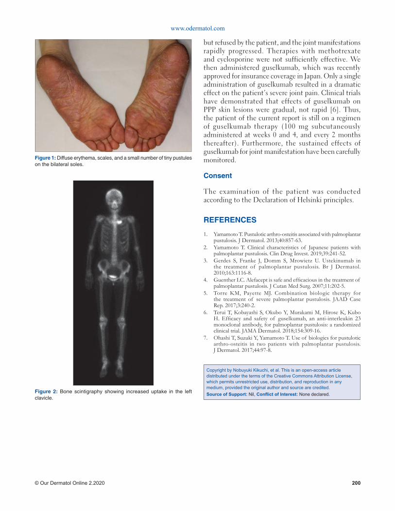
www.odermatol.com
© Our Dermatol Online 2.2020 200
but refused by the patient, and the joint manifestations rapidly progressed. Therapies with methotrexate and cyclosporine were not sufficiently effective. We then administered guselkumab, which was recently approved for insurance coverage in Japan. Only a single administration of guselkumab resulted in a dramatic effect on the patient’s severe joint pain. Clinical trials have demonstrated that effects of guselkumab on PPP skin lesions were gradual, not rapid [6]. Thus, the patient of the current report is still on a regimen of guselkumab therapy (100 mg subcutaneously administered at weeks 0 and 4, and every 2 months thereafter). Furthermore, the sustained effects of guselkumab for joint manifestation have been carefully monitored.
Consent
The examination of the patient was conducted according to the Declaration of Helsinki principles.
REFERENCES
1. Yamamoto T. Pustulotic arthro-osteitis associated with palmoplantar pustulosis. J Dermatol. 2013;40:857-63.
2. Yamamoto T. Clinical characteristics of Japanese patients with palmoplantar pustulosis. Clin Drug Invest. 2019;39:241-52.
3. Gerdes S, Franke J, Domm S, Mrowietz U. Ustekinumab in the treatment of palmoplantar pustulosis. Br J Dermatol. 2010;163:1116-8.
4. Guenther LC. Alefacept is safe and effi cacious in the treatment of palmoplantar pustulosis. J Cutan Med Surg. 2007;11:202-5.
5. Torre KM, Payette MJ. Combination biologic therapy for the treatment of severe palmoplantar pustulosis. JAAD Case Rep. 2017;3:240-2.
6. Terui T, Kobayashi S, Okubo Y, Murakami M, Hirose K, Kubo H. Effi cacy and safety of guselkumab, an anti-interleukin 23 monoclonal antibody, for palmoplantar pustulosis: a randomized clinical trial. JAMA Dermatol. 2018;154:309-16.
7. Ohashi T, Suzuki Y, Yamamoto T. Use of biologics for pustulotic arthro-osteitis in two patients with palmoplantar pustulosis. J Dermatol. 2017;44:97-8.
Figure 1: Diffuse erythema, scales, and a small number of tiny pustules on the bilateral soles.
Figure 2: Bone scintigraphy showing increased uptake in the left clavicle.
Copyright by Nobuyuki Kikuchi, et al. This is an open-access article distributed under the terms of the Creative Commons Attribution License, which permits unrestricted use, distribution, and reproduction in any medium, provided the original author and source are credited.Source of Support: Nil, Confl ict of Interest: None declared.
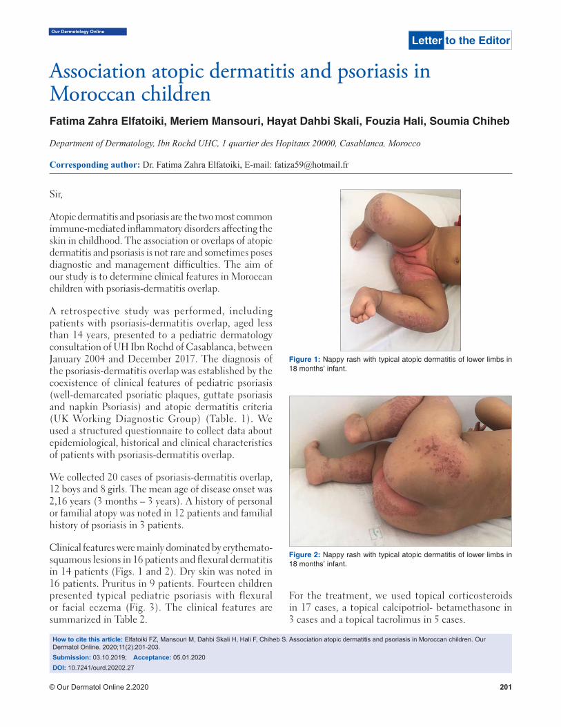
Our Dermatology Online
© Our Dermatol Online 2.2020 201
Sir,
Atopic dermatitis and psoriasis are the two most common immune-mediated inflammatory disorders affecting the skin in childhood. The association or overlaps of atopic dermatitis and psoriasis is not rare and sometimes poses diagnostic and management difficulties. The aim of our study is to determine clinical features in Moroccan children with psoriasis-dermatitis overlap.
A retrospective study was performed, including patients with psoriasis-dermatitis overlap, aged less than 14 years, presented to a pediatric dermatology consultation of UH Ibn Rochd of Casablanca, between January 2004 and December 2017. The diagnosis of the psoriasis-dermatitis overlap was established by the coexistence of clinical features of pediatric psoriasis (well-demarcated psoriatic plaques, guttate psoriasis and napkin Psoriasis) and atopic dermatitis criteria (UK Working Diagnostic Group) (Table. 1). We used a structured questionnaire to collect data about epidemiological, historical and clinical characteristics of patients with psoriasis-dermatitis overlap.
We collected 20 cases of psoriasis-dermatitis overlap, 12 boys and 8 girls. The mean age of disease onset was 2,16 years (3 months – 3 years). A history of personal or familial atopy was noted in 12 patients and familial history of psoriasis in 3 patients.
Clinical features were mainly dominated by erythemato-squamous lesions in 16 patients and flexural dermatitis in 14 patients (Figs. 1 and 2). Dry skin was noted in 16 patients. Pruritus in 9 patients. Fourteen children presented typical pediatric psoriasis with flexural or facial eczema (Fig. 3). The clinical features are summarized in Table 2.
For the treatment, we used topical corticosteroids in 17 cases, a topical calcipotriol- betamethasone in 3 cases and a topical tacrolimus in 5 cases.
How to cite this article: Elfatoiki FZ, Mansouri M, Dahbi Skali H, Hali F, Chiheb S. Association atopic dermatitis and psoriasis in Moroccan children. Our Dermatol Online. 2020;11(2):201-203.Submission: 03.10.2019; Acceptance: 05.01.2020DOI: 10.7241/ourd.20202.27
Association atopic dermatitis and psoriasis in Association atopic dermatitis and psoriasis in Moroccan childrenMoroccan childrenFatima Zahra Elfatoiki, Meriem Mansouri, Hayat Dahbi Skali, Fouzia Hali, Soumia Chiheb
Department of Dermatology, Ibn Rochd UHC, 1 quartier des Hopitaux 20000, Casablanca, Morocco
Corresponding author: Dr. Fatima Zahra Elfatoiki, E-mail: [email protected]
Letter to the Editor
Figure 1: Nappy rash with typical atopic dermatitis of lower limbs in 18 months’ infant.
Figure 2: Nappy rash with typical atopic dermatitis of lower limbs in 18 months’ infant.
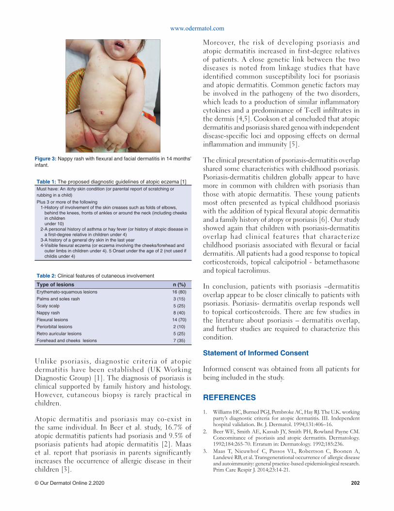
www.odermatol.com
© Our Dermatol Online 2.2020 202
Unlike psoriasis, diagnostic criteria of atopic dermatitis have been established (UK Working Diagnostic Group) [1]. The diagnosis of psoriasis is clinical supported by family history and histology. However, cutaneous biopsy is rarely practical in children.
Atopic dermatitis and psoriasis may co-exist in the same individual. In Beer et al. study, 16.7% of atopic dermatitis patients had psoriasis and 9.5% of psoriasis patients had atopic dermatitis [2]. Maas et al. report that psoriasis in parents significantly increases the occurrence of allergic disease in their children [3].
Moreover, the risk of developing psoriasis and atopic dermatitis increased in first-degree relatives of patients. A close genetic link between the two diseases is noted from linkage studies that have identified common susceptibility loci for psoriasis and atopic dermatitis. Common genetic factors may be involved in the pathogeny of the two disorders, which leads to a production of similar inflammatory cytokines and a predominance of T-cell infiltrates in the dermis [4,5]. Cookson et al concluded that atopic dermatitis and psoriasis shared genoa with independent disease-specific loci and opposing effects on dermal inflammation and immunity [5].
The clinical presentation of psoriasis-dermatitis overlap shared some characteristics with childhood psoriasis. Psoriasis-dermatitis children globally appear to have more in common with children with psoriasis than those with atopic dermatitis. These young patients most often presented as typical childhood psoriasis with the addition of typical flexural atopic dermatitis and a family history of atopy or psoriasis [6]. Our study showed again that children with psoriasis-dermatitis overlap had clinical features that characterize childhood psoriasis associated with flexural or facial dermatitis. All patients had a good response to topical corticosteroids, topical calcipotriol - betamethasone and topical tacrolimus.
In conclusion, patients with psoriasis –dermatitis overlap appear to be closer clinically to patients with psoriasis. Psoriasis- dermatitis overlap responds well to topical corticosteroids. There are few studies in the literature about psoriasis – dermatitis overlap, and further studies are required to characterize this condition.
Statement of Informed Consent
Informed consent was obtained from all patients for being included in the study.
REFERENCES
1. Williams HC, Burned PGJ, Pembroke AC, Hay RJ. The U.K. working party’s diagnostic criteria for atopic dermatitis. III. Independent hospital validation. Br. J. Dermatol. 1994;131:406–16.
2. Beer WE, Smith AE, Kassab JY, Smith PH, Rowland Payne CM. Concomitance of psoriasis and atopic dermatitis. Dermatology. 1992;184:265-70. Erratum in: Dermatology. 1992;185:236.
3. Maas T, Nieuwhof C, Passos VL, Robertson C, Boonen A, Landewé RB, et al. Transgenerational occurrence of allergic disease and autoimmunity: general practice-based epidemiological research. Prim Care Respir J. 2014;23:14-21.
Figure 3: Nappy rash with fl exural and facial dermatitis in 14 months’ infant.
Table 2: Clinical features of cutaneous involvement
Type of lesions n (%)Erythemato-squamous lesions 16 (80)
Palms and soles rash 3 (15)
Scaly scalp 5 (25)
Nappy rash 8 (40)
Flexural lesions 14 (70)
Periorbital lesions 2 (10)
Retro auricular lesions 5 (25)
Forehead and cheeks lesions 7 (35)
Table 1: The proposed diagnostic guidelines of atopic eczema [1]Must have: An itchy skin condition (or parental report of scratching or rubbing in a child)
Plus 3 or more of the following 1- History of involvement of the skin creases such as folds of elbows,
behind the knees, fronts of ankles or around the neck (including cheeks in childrenunder 10)
2- A personal history of asthma or hay fever (or history of atopic disease in a fi rst-degree relative in children under 4)
3-A history of a general dry skin in the last year4- Visible fi exural eczema (or eczema involving the cheeks/forehead and
outer limbs in children under 4). 5 Onset under the age of 2 (not used if childis under 4)

www.odermatol.com
© Our Dermatol Online 2.2020 203
Copyright by Fatima Zahra Elfatoiki, et al. This is an open-access article distributed under the terms of the Creative Commons Attribution License, which permits unrestricted use, distribution, and reproduction in any medium, provided the original author and source are credited.Source of Support: Nil, Confl ict of Interest: None declared.
4. Wilsmann-Theis D, Hagemann T, Jordan J, Bieber T, Novak N, Bieber T, Novak N. Facing psoriasis and atopic dermatitis: are there more similarities or more differences? Eur. J. Dermatol. 2008;18:172–80.
5. Cookson WO, Ubhi B, Lawrence R, Abecasis GR, Walley AJ, Cox HE, et al. Genetic linkage of childhood atopic dermatitis to psoriasis susceptibility loci. Nat. Genet. 2001;27:372–3.
6. Kapila S, Hong E, Fischer G. A comparative study of childhood psoriasis and atopic dermatitis and greater understanding of the
overlapping condition, psoriasis-dermatitis. Australas J Dermatol. 2012;53:98-105.
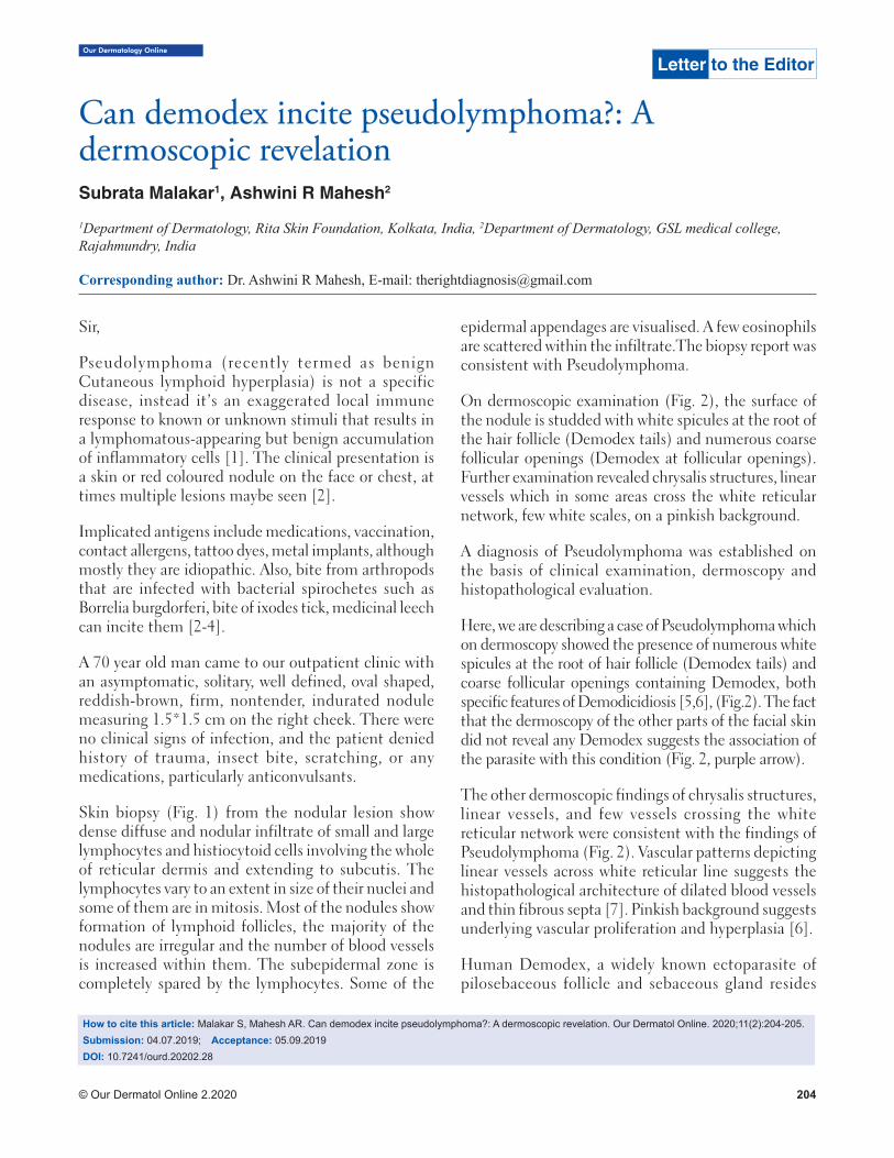
Our Dermatology Online
© Our Dermatol Online 2.2020 204
Sir,
Pseudolymphoma (recently termed as benign Cutaneous lymphoid hyperplasia) is not a specific disease, instead it’s an exaggerated local immune response to known or unknown stimuli that results in a lymphomatous-appearing but benign accumulation of inflammatory cells [1]. The clinical presentation is a skin or red coloured nodule on the face or chest, at times multiple lesions maybe seen [2].
Implicated antigens include medications, vaccination, contact allergens, tattoo dyes, metal implants, although mostly they are idiopathic. Also, bite from arthropods that are infected with bacterial spirochetes such as Borrelia burgdorferi, bite of ixodes tick, medicinal leech can incite them [2-4].
A 70 year old man came to our outpatient clinic with an asymptomatic, solitary, well defined, oval shaped, reddish-brown, firm, nontender, indurated nodule measuring 1.5*1.5 cm on the right cheek. There were no clinical signs of infection, and the patient denied history of trauma, insect bite, scratching, or any medications, particularly anticonvulsants.
Skin biopsy (Fig. 1) from the nodular lesion show dense diffuse and nodular infiltrate of small and large lymphocytes and histiocytoid cells involving the whole of reticular dermis and extending to subcutis. The lymphocytes vary to an extent in size of their nuclei and some of them are in mitosis. Most of the nodules show formation of lymphoid follicles, the majority of the nodules are irregular and the number of blood vessels is increased within them. The subepidermal zone is completely spared by the lymphocytes. Some of the
epidermal appendages are visualised. A few eosinophils are scattered within the infiltrate.The biopsy report was consistent with Pseudolymphoma.
On dermoscopic examination (Fig. 2), the surface of the nodule is studded with white spicules at the root of the hair follicle (Demodex tails) and numerous coarse follicular openings (Demodex at follicular openings).Further examination revealed chrysalis structures, linear vessels which in some areas cross the white reticular network, few white scales, on a pinkish background.
A diagnosis of Pseudolymphoma was established on the basis of clinical examination, dermoscopy and histopathological evaluation.
Here, we are describing a case of Pseudolymphoma which on dermoscopy showed the presence of numerous white spicules at the root of hair follicle (Demodex tails) and coarse follicular openings containing Demodex, both specific features of Demodicidiosis [5,6], (Fig.2). The fact that the dermoscopy of the other parts of the facial skin did not reveal any Demodex suggests the association of the parasite with this condition (Fig. 2, purple arrow).
The other dermoscopic findings of chrysalis structures, linear vessels, and few vessels crossing the white reticular network were consistent with the findings of Pseudolymphoma (Fig. 2). Vascular patterns depicting linear vessels across white reticular line suggests the histopathological architecture of dilated blood vessels and thin fibrous septa [7]. Pinkish background suggests underlying vascular proliferation and hyperplasia [6].
Human Demodex, a widely known ectoparasite of pilosebaceous follicle and sebaceous gland resides
Letter to the Editor
Can demodex incite pseudolymphoma?: A Can demodex incite pseudolymphoma?: A dermoscopic revelation dermoscopic revelation Subrata Malakar1, Ashwini R Mahesh2
1Department of Dermatology, Rita Skin Foundation, Kolkata, India, 2Department of Dermatology, GSL medical college, Rajahmundry, India
Corresponding author: Dr. Ashwini R Mahesh, E-mail: [email protected]
How to cite this article: Malakar S, Mahesh AR. Can demodex incite pseudolymphoma?: A dermoscopic revelation. Our Dermatol Online. 2020;11(2):204-205.Submission: 04.07.2019; Acceptance: 05.09.2019DOI: 10.7241/ourd.20202.28
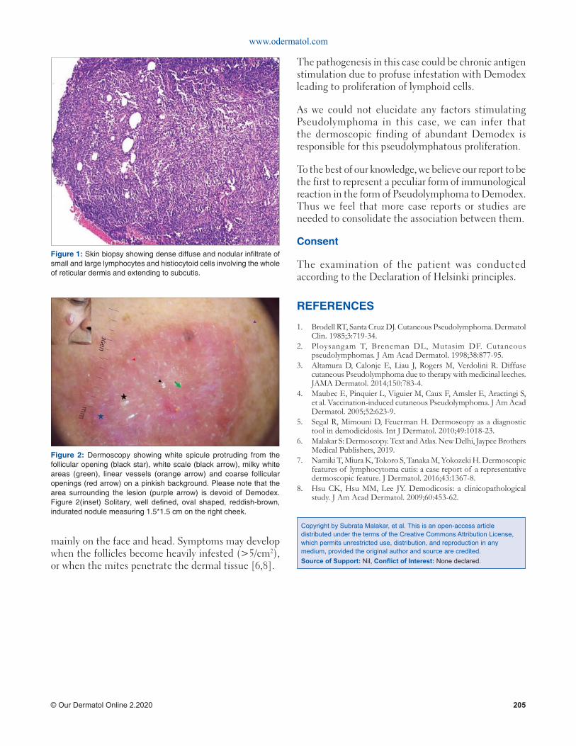
www.odermatol.com
© Our Dermatol Online 2.2020 205
mainly on the face and head. Symptoms may develop when the follicles become heavily infested (>5/cm2), or when the mites penetrate the dermal tissue [6,8].
The pathogenesis in this case could be chronic antigen stimulation due to profuse infestation with Demodex leading to proliferation of lymphoid cells.
As we could not elucidate any factors stimulating Pseudolymphoma in this case, we can infer that the dermoscopic finding of abundant Demodex is responsible for this pseudolymphatous proliferation.
To the best of our knowledge, we believe our report to be the first to represent a peculiar form of immunological reaction in the form of Pseudolymphoma to Demodex. Thus we feel that more case reports or studies are needed to consolidate the association between them.
Consent
The examination of the patient was conducted according to the Declaration of Helsinki principles.
REFERENCES
1. Brodell RT, Santa Cruz DJ. Cutaneous Pseudolymphoma. Dermatol Clin. 1985;3:719-34.
2. Ploysangam T, Breneman DL, Mutasim DF. Cutaneous pseudolymphomas. J Am Acad Dermatol. 1998;38:877-95.
3. Altamura D, Calonje E, Liau J, Rogers M, Verdolini R. Diffuse cutaneous Pseudolymphoma due to therapy with medicinal leeches. JAMA Dermatol. 2014;150:783-4.
4. Maubec E, Pinquier L, Viguier M, Caux F, Amsler E, Aractingi S, et al. Vaccination-induced cutaneous Pseudolymphoma. J Am Acad Dermatol. 2005;52:623-9.
5. Segal R, Mimouni D, Feuerman H. Dermoscopy as a diagnostic tool in demodicidosis. Int J Dermatol. 2010;49:1018-23.
6. Malakar S: Dermoscopy. Text and Atlas. New Delhi, Jaypee Brothers Medical Publishers, 2019.
7. Namiki T, Miura K, Tokoro S, Tanaka M, Yokozeki H. Dermoscopic features of lymphocytoma cutis: a case report of a representative dermoscopic feature. J Dermatol. 2016;43:1367-8.
8. Hsu CK, Hsu MM, Lee JY. Demodicosis: a clinicopathological study. J Am Acad Dermatol. 2009;60:453-62.
Figure 1: Skin biopsy showing dense diffuse and nodular infi ltrate of small and large lymphocytes and histiocytoid cells involving the whole of reticular dermis and extending to subcutis.
Figure 2: Dermoscopy showing white spicule protruding from the follicular opening (black star), white scale (black arrow), milky white areas (green), linear vessels (orange arrow) and coarse follicular openings (red arrow) on a pinkish background. Please note that the area surrounding the lesion (purple arrow) is devoid of Demodex. Figure 2(inset) Solitary, well defi ned, oval shaped, reddish-brown, indurated nodule measuring 1.5*1.5 cm on the right cheek.
Copyright by Subrata Malakar, et al. This is an open-access article distributed under the terms of the Creative Commons Attribution License, which permits unrestricted use, distribution, and reproduction in any medium, provided the original author and source are credited.Source of Support: Nil, Confl ict of Interest: None declared.
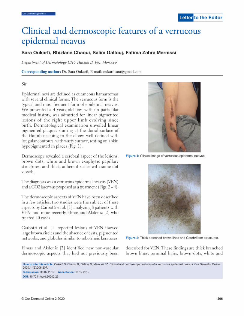
Our Dermatology Online
© Our Dermatol Online 2.2020 206
How to cite this article: Oukarfi S, Chaoui R, Gallouj S, Mernissi FZ. Clinical and dermoscopic features of a verrucous epidermal neavus. Our Dermatol Online. 2020;11(2):206-207.Submission: 30.07.2019; Acceptance: 18.12.2019DOI: 10.7241/ourd.20202.29
Clinical and dermoscopic features of a verrucous Clinical and dermoscopic features of a verrucous epidermal neavusepidermal neavusSara Oukarfi , Rhizlane Chaoui, Salim Gallouj, Fatima Zahra Mernissi
Department of Dermatology CHU Hassan II, Fez, Morocco
Corresponding author: Dr. Sara Oukarfi , E-mail: oukarfi [email protected]
Sir
Epidermal nevi are defined as cutaneous hamartomas with several clinical forms. The verrucous form is the typical and most frequent form of epidermal neavus. We presented a 4 years old boy, with no particular medical history, was admitted for linear pigmented lesions of the right upper limb evolving since birth. Dermatological examination unveiled linear pigmented plaques starting at the dorsal surface of the thumb reaching to the elbow, well defined with irregular contours, with warty surface, resting on a skin hypopigmented in places (Fig. 1).
Dermoscopy revealed a cerebral aspect of the lesions, brown dots, white and brown exophytic papillary structures, and thick, adherent scales with some dot vessels.
The diagnosis was a verrucous epidermal neavus (VEN) and a CO2 laser was proposed as a treatment (Figs. 2 – 4).
The dermoscopic aspects of VEN have been described in a few articles; two studies were the subject of these aspects by Carbotti et al. [1] analyzing 8 patients with VEN, and more recently Elmas and Akdeniz [2] who treated 20 cases.
Carbotti et al. [1] reported lesions of VEN showed large brown circles and the absence of cysts, pigmented networks, and globules similar to seborrheic keratoses.
Elmas and Akdeniz [2] identified new non-vascular dermoscopic aspects that had not previously been
described for VEN. These findings are thick branched brown lines, terminal hairs, brown dots, white and
Letter to the Editor
Figure 1: Clinical image of verrucous epidermal neavus.
Figure 2: Thick branched brown lines and Cerebriform structures.
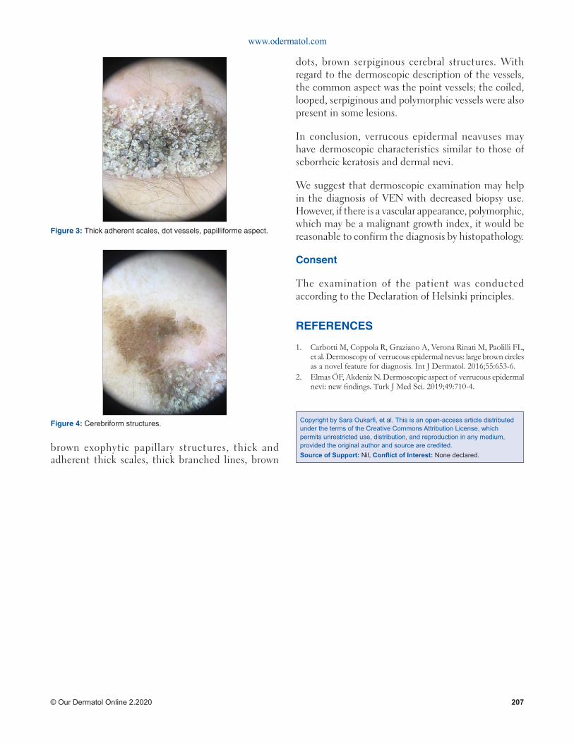
www.odermatol.com
© Our Dermatol Online 2.2020 207
brown exophytic papillary structures, thick and adherent thick scales, thick branched lines, brown
dots, brown serpiginous cerebral structures. With regard to the dermoscopic description of the vessels, the common aspect was the point vessels; the coiled, looped, serpiginous and polymorphic vessels were also present in some lesions.
In conclusion, verrucous epidermal neavuses may have dermoscopic characteristics similar to those of seborrheic keratosis and dermal nevi.
We suggest that dermoscopic examination may help in the diagnosis of VEN with decreased biopsy use. However, if there is a vascular appearance, polymorphic, which may be a malignant growth index, it would be reasonable to confirm the diagnosis by histopathology.
Consent
The examination of the patient was conducted according to the Declaration of Helsinki principles.
REFERENCES
1. Carbotti M, Coppola R, Graziano A, Verona Rinati M, Paolilli FL, et al. Dermoscopy of verrucous epidermal nevus: large brown circles as a novel feature for diagnosis. Int J Dermatol. 2016;55:653-6.
2. Elmas ÖF, Akdeniz N. Dermoscopic aspect of verrucous epidermal nevi: new fi ndings. Turk J Med Sci. 2019;49:710-4.
Figure 3: Thick adherent scales, dot vessels, papilliforme aspect.
Figure 4: Cerebriform structures. Copyright by Sara Oukarfi , et al. This is an open-access article distributed under the terms of the Creative Commons Attribution License, which permits unrestricted use, distribution, and reproduction in any medium, provided the original author and source are credited.Source of Support: Nil, Confl ict of Interest: None declared.
Our Dermatology Online
© Our Dermatol Online 2.2020 208
How to cite this article: Endo M, Yamamoto T. Recurrent blistering of the fi ngers as a sign of carpal tunnel syndrome: a fi rst report from Japan. Our Dermatol Online. 2020;11(2):208-209.Submission: 20.08.2019; Acceptance: 24.10.2019DOI: 10.7241/ourd.20202.30
Recurrent blistering of the fi ngers as a sign of carpal Recurrent blistering of the fi ngers as a sign of carpal tunnel syndrome: a fi rst report from Japantunnel syndrome: a fi rst report from JapanMai Endo, Toshiyuki Yamamoto
Department of Dermatology, Fukushima Medical University Hikarigaoka 1, Fukushima 960-1295, Japan
Corresponding author: Dr. Mai Endo, E-mail: [email protected]
Sir,
Carpal tunnel syndrome is an entrapment neuropathy caused by compression of the median nerve. Dermatological signs associated with carpal tunnel syndrome are rare, and to date, ulceration, blistering, hypohidrosis, Raynaud’s phenomenon, and irritant contact dermatitis have been reported. Among them, blistering of the fingers is extremely rare. We herein report a patient with carpal tunnel syndrome, who developed recurrent blisters on the fingers.
A 75-year-old woman, who was suffering from carpal tunnel syndrome for 15 years, was referred to our department complaining of skin eruptions on the fingers. For the previous 6 years, blisters repeatedly appeared on the digits of her right hand and healed in about a month after ulceration. Therefore, she was referred to our department. On physical examination, she had no blisters other than an erosion in her right third finger (Fig. 1). Symptoms of local infection were not observed. The site where the blistering occurred had become reddish after erosion. There were no blisters other than those on the fingers. She denied Raynaud’s phenomenon, and neither finger coldness nor swelling was observed. Just before the blisters appeared, she had a complaint of slight pain at the same site. She stated that blisters had occurred on all fingers except the little finger. After a few months, the blister occurs and collapses in a few days. It is not related to the season, there is no special timing, and there was no history of trauma or local infection.
Cutaneous manifestation associated with carpal tunnel syndrome has been poorly described. To date,
there are several cases of carpal tunnel syndrome showing cutaneous manifestations in English literature [1–6]; however, to our knowledge, there are no reports from Japan. In all cases, the lesions were found in fingers, and in severe cases, bone and nail changes were also induced [3]. In cases that underwent surgical decompression for carpal tunnel syndrome, skin lesions had disappeared [1,2,4]. In the present case, blisters repeatedly appeared on the right third finger innervated by the median nerve, although other fingers were also involved. According to the patient’s complaint, the blisters waxed and waned; however, the triggering event of the blister appearance was unknown. Unfortunately, we could not have any chance to perform biopsy; however, bullous pemphigoid was excluded because bullous formation was restricted to the fingers and disease course showed recurrent spontaneous regression and relapse. Other diseases such as diabetic bulla, blistering digital dactylitis, and herpetic whitlow were all differentiated.
Although the mechanism by which blisters can occur is not disclosed, ischemic changes due to autonomic neuropathy and vasomotor dysfunction are suggested to play a major role, and minor trauma and secondary infection may also be involved. On the other hand, nerve compression in carpal tunnel syndrome is a privileged site to become acquired immunocompromised district [7]. Baroni et al. speculated that the areas innervated by median nerve fiber compression are an obstacle to the normal passage of immunocompetent cells through lymphatic channels or an interference with neuropeptide signaling by peripheral nerves to the membrane receptors of immunocompetent cells, caused by nerve
Letter to the Editor
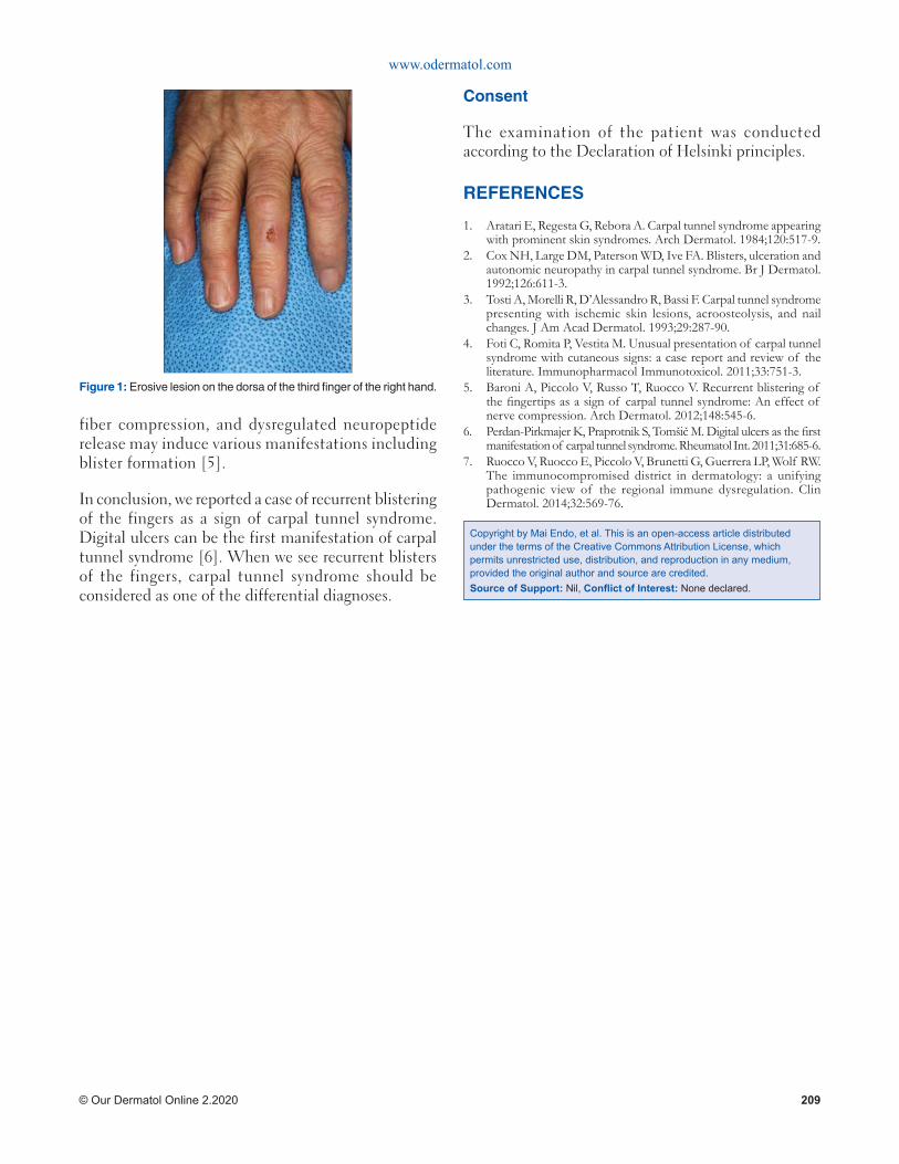
www.odermatol.com
© Our Dermatol Online 2.2020 209
fiber compression, and dysregulated neuropeptide release may induce various manifestations including blister formation [5].
In conclusion, we reported a case of recurrent blistering of the fingers as a sign of carpal tunnel syndrome. Digital ulcers can be the first manifestation of carpal tunnel syndrome [6]. When we see recurrent blisters of the fingers, carpal tunnel syndrome should be considered as one of the differential diagnoses.
Consent
The examination of the patient was conducted according to the Declaration of Helsinki principles.
REFERENCES
1. Aratari E, Regesta G, Rebora A. Carpal tunnel syndrome appearing with prominent skin syndromes. Arch Dermatol. 1984;120:517-9.
2. Cox NH, Large DM, Paterson WD, Ive FA. Blisters, ulceration and autonomic neuropathy in carpal tunnel syndrome. Br J Dermatol. 1992;126:611-3.
3. Tosti A, Morelli R, D’Alessandro R, Bassi F. Carpal tunnel syndrome presenting with ischemic skin lesions, acroosteolysis, and nail changes. J Am Acad Dermatol. 1993;29:287-90.
4. Foti C, Romita P, Vestita M. Unusual presentation of carpal tunnel syndrome with cutaneous signs: a case report and review of the literature. Immunopharmacol Immunotoxicol. 2011;33:751-3.
5. Baroni A, Piccolo V, Russo T, Ruocco V. Recurrent blistering of the fi ngertips as a sign of carpal tunnel syndrome: An effect of nerve compression. Arch Dermatol. 2012;148:545-6.
6. Perdan-Pirkmajer K, Praprotnik S, Tomšič M. Digital ulcers as the fi rst manifestation of carpal tunnel syndrome. Rheumatol Int. 2011;31:685-6.
7. Ruocco V, Ruocco E, Piccolo V, Brunetti G, Guerrera LP, Wolf RW. The immunocompromised district in dermatology: a unifying pathogenic view of the regional immune dysregulation. Clin Dermatol. 2014;32:569-76.
Figure 1: Erosive lesion on the dorsa of the third fi nger of the right hand.
Copyright by Mai Endo, et al. This is an open-access article distributed under the terms of the Creative Commons Attribution License, which permits unrestricted use, distribution, and reproduction in any medium, provided the original author and source are credited.Source of Support: Nil, Confl ict of Interest: None declared.
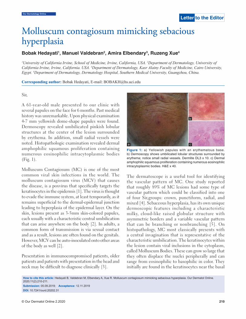
Our Dermatology Online
© Our Dermatol Online 2.2020 210
How to cite this article: Hedayati B, Valdebran M, Elbendary A, Xue R. Molluscum contagiosum mimicking sebacious hyperplasia. Our Dermatol Online. 2020;11(2):210-211. Submission: 05.09.2019; Acceptance: 12.11.2019DOI: 10.7241/ourd.20202.31
Sir,
A 61-year-old male presented to our clinic with several papules on the face for 6 months. Past medical history was unremarkable. Upon physical examination 4-7 mm yellowish dome-shape papules were found. Dermoscopy revealed umbilicated pinkish lobular structures at the center of the lesion surrounded by erythema. In addition, small radial vessels were noted. Histopathologic examination revealed dermal amphophilic squamous proliferation containing numerous eosinophilic intracytoplasmic bodies (Fig. 1).
Molluscum Contagiosum (MC) is one of the most common viral skin infections in the world. The molluscum contagiosum virus (MCV) that causes the disease, is a poxvirus that specifically targets the keratinocytes in the epidermis [1]. The virus is thought to evade the immune system, at least temporarily, as it remains superficial to the dermal-epidermal junction leading to hyperplasia of the epidermal layer. On the skin, lesions present as 3-5mm skin-colored papules, each usually with a characteristic central umbilication that can arise anywhere on the body [2]. In adults, a common form of transmission is via sexual contact and as a result, lesions are often found on the genitals. However, MCV can be auto-inoculated onto other areas of the body as well [2].
Presentation in immunocompromised patients, older patients and patients with presentation in the head and neck may be difficult to diagnose clinically [3].
The dermatoscope is a useful tool for identifying the vascular pattern of MC. One study reported that roughly 89% of MC lesions had some type of vascular pattern which could be classified into one of four Sir,groups: crown, punctiform, radial, and mixed [4]. Sebaceous hyperplasia, has its own unique dermoscopic features including a characteristic milky, cloud-like raised globular structure with asymmetric borders and a variable vascular pattern that can be branching or nonbranching [5]. On histopathology, MC most classically presents with a central invagination that is representative of the characteristic umbilication. The keratinocytes within the lesion contain viral inclusions in the cytoplasm, called Molluscum Bodies. These can grow so large that they often displace the nuclei peripherally and can range from eosinophilic to basophilic in color. They initially are found in the keratinocytes near the basal
Molluscum contagiosum mimicking sebacious Molluscum contagiosum mimicking sebacious hyperplasiahyperplasiaBobak Hedayati1, Manuel Valdebran2, Amira Elbendary3, Ruzeng Xue4
1University of California Irvine, School of Medicine, Irvine, California, USA. 2Department of Dermatology, University of California Irvine, Irvine, California. USA. 3Department of Dermatology, Kasr Alainy Faculty of Medicine, Cairo University, Egypt. 4Department of Dermatology, Dermatology Hospital, Southern Medical University, Guangzhou, China.
Corresponding author: Bobak Hedayati, E-mail: [email protected]
Letter to the Editor
Figure 1: a) Yellowish papules with an erythematous base. b) Dermoscopy shows umbilicated lobular structures surrounded by erythema; notice small radial vessels. Dermlite DL3 x 10. c) Dermal amphophilic squamous proliferation containing numerous eosinophilic intracytoplasmic bodies. H&E x 40.
c
b
a
www.odermatol.com
© Our Dermatol Online 2.2020 211
REFERENCES
1. Chen X, Anstey A V, Bugert JJ. Molluscum contagiosum virus infection. Lancet Infect Dis. 2013;13:877-8.
2. Nguyen HP, Franz E, Stiegel KR, Hsu S, Tyring SK. Treatment of molluscum contagiosum in adult, pediatric, and immunodefi cient populations. J Cutan Med Surg. 2014;18:299-306.
3. Ishikawa MK, Arps DP, Chow C, Hocker TL, Fullen DR. Histopathological features of molluscum contagiosum other than molluscum bodies. Histopathology. 2015;67:836-42.
4. Ianhez M, Cestari S da CP, Enokihara MY, Seize MB de PM. Padrões dermatoscópicos do molusco contagioso: estudo de 211 lesões confi rmadas por exame histopatológico. An Bras Dermatol. 2011;86:74-9.
5. Bryden AM, Dawe RS, Fleming C. Dermatoscopic features of benign sebaceous proliferation. Clin Exp Dermatol. 2004;29:676-7.
layer then progress toward the more superficial layers of the epidermis [3].
Our patient has a somewhat unique presentation for a few different reasons. First, his lesions ranged from 4-7mm. We observed pinkish lobular structures at the center of the lesion surrounded by erythema.
In conclusion, MC is a self-resolving disease in immunocompetent individuals, however, it can be spread via direct skin contact. Therefore, it is important to properly diagnose these lesions when they present. As with many diseases, MC has classic and non-classic presentations. It is imperative that physicians are aware of the less common clinical, dermatoscopic and histopathologic variations that can manifest from this disease.
Copyright by Bobak Hedayati, et al. This is an open access article distributed under the terms of the Creative Commons Attribution License, which permits unrestricted use, distribution, and reproduction in any medium, provided the original author and source are credited.Source of Support: Nil, Confl ict of Interest: None declared.
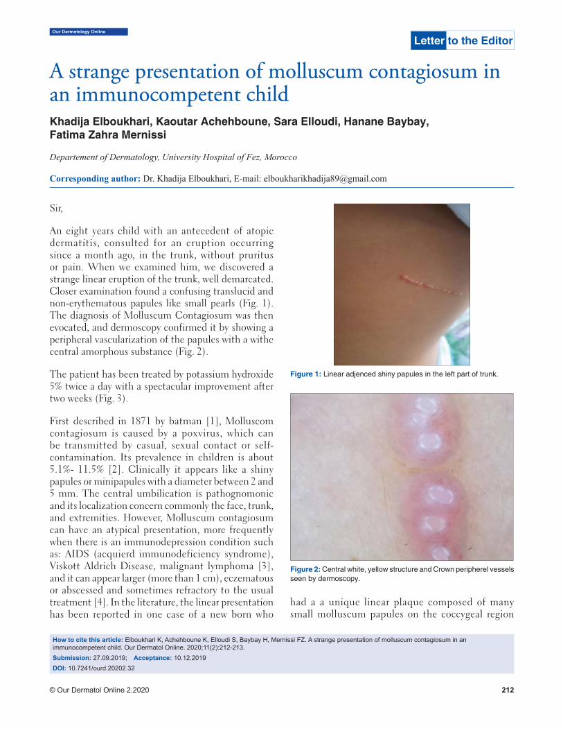
Our Dermatology Online
© Our Dermatol Online 2.2020 212
How to cite this article: Elboukhari K, Achehboune K, Elloudi S, Baybay H, Mernissi FZ. A strange presentation of molluscum contagiosum in an immunocompetent child. Our Dermatol Online. 2020;11(2):212-213.Submission: 27.09.2019; Acceptance: 10.12.2019DOI: 10.7241/ourd.20202.32
A strange presentation of molluscum contagiosum in A strange presentation of molluscum contagiosum in an immunocompetent childan immunocompetent childKhadija Elboukhari, Kaoutar Achehboune, Sara Elloudi, Hanane Baybay, Fatima Zahra Mernissi
Departement of Dermatology, University Hospital of Fez, Morocco
Corresponding author: Dr. Khadija Elboukhari, E-mail: [email protected]
Sir,
An eight years child with an antecedent of atopic dermatitis, consulted for an eruption occurring since a month ago, in the trunk, without pruritus or pain. When we examined him, we discovered a strange linear eruption of the trunk, well demarcated. Closer examination found a confusing translucid and non-erythematous papules like small pearls (Fig. 1). The diagnosis of Molluscum Contagiosum was then evocated, and dermoscopy confirmed it by showing a peripheral vascularization of the papules with a withe central amorphous substance (Fig. 2).
The patient has been treated by potassium hydroxide 5% twice a day with a spectacular improvement after two weeks (Fig. 3).
First described in 1871 by batman [1], Molluscom contagiosum is caused by a poxvirus, which can be transmitted by casual, sexual contact or self-contamination. Its prevalence in children is about 5.1%- 11.5% [2]. Clinically it appears like a shiny papules or minipapules with a diameter between 2 and 5 mm. The central umbilication is pathognomonic and its localization concern commonly the face, trunk, and extremities. However, Molluscum contagiosum can have an atypical presentation, more frequently when there is an immunodepression condition such as: AIDS (acquierd immunodeficiency syndrome), Viskott Aldrich Disease, malignant lymphoma [3], and it can appear larger (more than 1 cm), eczematous or abscessed and sometimes refractory to the usual treatment [4]. In the literature, the linear presentation has been reported in one case of a new born who
Letter to the Editor
Figure 1: Linear adjenced shiny papules in the left part of trunk.
Figure 2: Central white, yellow structure and Crown peripherel vessels seen by dermoscopy.
had a a unique linear plaque composed of many small molluscum papules on the coccygeal region
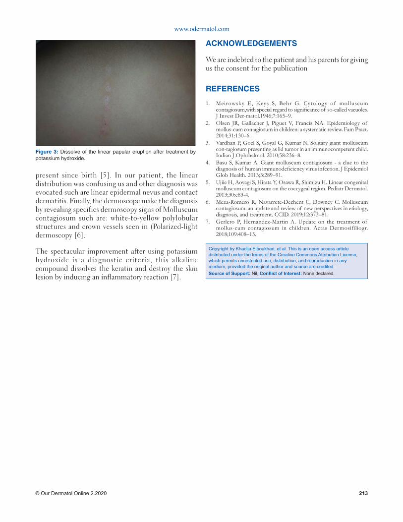
www.odermatol.com
© Our Dermatol Online 2.2020 213
present since birth [5]. In our patient, the linear distribution was confusing us and other diagnosis was evocated such are linear epidermal nevus and contact dermatitis. Finally, the dermoscope make the diagnosis by revealing specifics dermoscopy signs of Molluscum contagiosum such are: white-to-yellow polylobular structures and crown vessels seen in (Polarized-light dermoscopy [6].
The spectacular improvement after using potassium hydroxide is a diagnostic criteria, this alkaline compound dissolves the keratin and destroy the skin lesion by inducing an inflammatory reaction [7].
Figure 3: Dissolve of the linear papular eruption after treatment by potassium hydroxide.
ACKNOWLEDGEMENTS
We are indebted to the patient and his parents for giving us the consent for the publication
REFERENCES
1. Meirowsky E, Keys S, Behr G. Cytology of molluscum contagiosum,with special regard to signifi cance of so-called vacuoles. J Invest Der-matol.1946;7:165–9.
2. Olsen JR, Gallacher J, Piguet V, Francis NA. Epidemiology of mollus-cum contagiosum in children: a systematic review. Fam Pract. 2014;31:130–6.
3. Vardhan P, Goel S, Goyal G, Kumar N. Solitary giant molluscum con-tagiosum presenting as lid tumor in an immunocompetent child. Indian J Ophthalmol. 2010;58:236–8.
4. Basu S, Kumar A. Giant molluscum contagiosum - a clue to the diagnosis of human immunodefi ciency virus infection. J Epidemiol Glob Health. 2013;3:289–91.
5. Ujiie H, Aoyagi S, Hirata Y, Osawa R, Shimizu H. Linear congenital molluscum contagiosum on the coccygeal region. Pediatr Dermatol. 2013;30:e83-4.
6. Meza-Romero R, Navarrete-Dechent C, Downey C. Molluscum contagiosum: an update and review of new perspectives in etiology, diagnosis, and treatment. CCID. 2019;12:373–81.
7. Gerlero P, Hernandez-Martin A. Update on the treatment of mollus-cum contagiosum in children. Actas Dermosifiliogr. 2018;109:408–15.
Copyright by Khadija Elboukhari, et al. This is an open access article distributed under the terms of the Creative Commons Attribution License, which permits unrestricted use, distribution, and reproduction in any medium, provided the original author and source are credited.Source of Support: Nil, Confl ict of Interest: None declared.
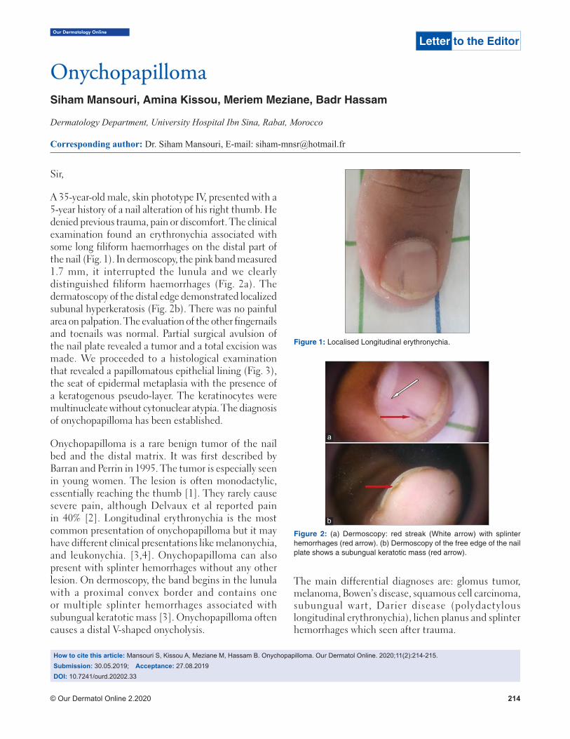
Our Dermatology Online
© Our Dermatol Online 2.2020 214
How to cite this article: Mansouri S, Kissou A, Meziane M, Hassam B. Onychopapilloma. Our Dermatol Online. 2020;11(2):214-215.Submission: 30.05.2019; Acceptance: 27.08.2019DOI: 10.7241/ourd.20202.33
OnychopapillomaOnychopapillomaSiham Mansouri, Amina Kissou, Meriem Meziane, Badr Hassam
Dermatology Department, University Hospital Ibn Sina, Rabat, Morocco
Corresponding author: Dr. Siham Mansouri, E-mail: [email protected]
Sir,
A 35-year-old male, skin phototype IV, presented with a 5-year history of a nail alteration of his right thumb. He denied previous trauma, pain or discomfort. The clinical examination found an erythronychia associated with some long filiform haemorrhages on the distal part of the nail (Fig. 1). In dermoscopy, the pink band measured 1.7 mm, it interrupted the lunula and we clearly distinguished filiform haemorrhages (Fig. 2a). The dermatoscopy of the distal edge demonstrated localized subunal hyperkeratosis (Fig. 2b). There was no painful area on palpation. The evaluation of the other fingernails and toenails was normal. Partial surgical avulsion of the nail plate revealed a tumor and a total excision was made. We proceeded to a histological examination that revealed a papillomatous epithelial lining (Fig. 3), the seat of epidermal metaplasia with the presence of a keratogenous pseudo-layer. The keratinocytes were multinucleate without cytonuclear atypia. The diagnosis of onychopapilloma has been established.
Onychopapilloma is a rare benign tumor of the nail bed and the distal matrix. It was first described by Barran and Perrin in 1995. The tumor is especially seen in young women. The lesion is often monodactylic, essentially reaching the thumb [1]. They rarely cause severe pain, although Delvaux et al reported pain in 40% [2]. Longitudinal erythronychia is the most common presentation of onychopapilloma but it may have different clinical presentations like melanonychia, and leukonychia. [3,4]. Onychopapilloma can also present with splinter hemorrhages without any other lesion. On dermoscopy, the band begins in the lunula with a proximal convex border and contains one or multiple splinter hemorrhages associated with subungual keratotic mass [3]. Onychopapilloma often causes a distal V-shaped onycholysis.
The main differential diagnoses are: glomus tumor, melanoma, Bowen’s disease, squamous cell carcinoma, subungual wart, Darier disease (polydactylous longitudinal erythronychia), lichen planus and splinter hemorrhages which seen after trauma.
Letter to the Editor
Figure 1: Localised Longitudinal erythronychia.
Figure 2: (a) Dermoscopy: red streak (White arrow) with splinter hemorrhages (red arrow). (b) Dermoscopy of the free edge of the nail plate shows a subungual keratotic mass (red arrow).
a
b
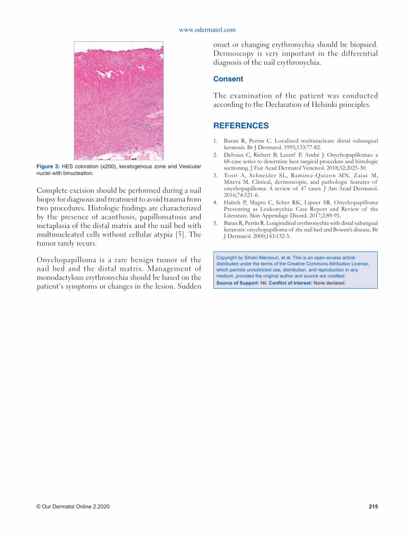
www.odermatol.com
© Our Dermatol Online 2.2020 215
Complete excision should be performed during a nail biopsy for diagnosis and treatment to avoid trauma from two procedures. Histologic findings are characterized by the presence of acanthosis, papillomatosis and metaplasia of the distal matrix and the nail bed with multinucleated cells without cellular atypia [5]. The tumor rarely recurs.
Onychopapilloma is a rare benign tumor of the nail bed and the distal matrix. Management of monodactylous erythronychia should be based on the patient’s symptoms or changes in the lesion. Sudden
onset or changing erythronychia should be biopsied. Dermoscopy is very important in the differential diagnosis of the nail erythronychia.
Consent
The examination of the patient was conducted according to the Declaration of Helsinki principles.
REFERENCES
1. Baran R, Perrin C. Localized multinucleate distal subungual keratosis. Br J Dermatol. 1995;133:77-82.
2. Delvaux C, Richert B, Lecerf P, André J. Onychopapillomas: a 68-case series to determine best surgical procedure and histologic sectioning. J Eur Acad Dermatol Venereol. 2018;32:2025-30.
3. Tosti A, Schneider SL, Ramirez-Quizon MN, Zaiac M, Miteva M. Clinical, dermoscopic, and pathologic features of onychopapilloma: A review of 47 cases. J Am Acad Dermatol. 2016;74:521-6.
4. Halteh P, Magro C, Scher RK, Lipner SR. Onychopapilloma Presenting as Leukonychia: Case Report and Review of the Literature. Skin Appendage Disord. 2017;2:89-91.
5. Baran R, Perrin R. Longitudinal erythronychia with distal subungual keratosis: onychopapilloma of the nail bed and Bowen’s disease. Br J Dermatol. 2000;143:132-5.
Copyright by Siham Mansouri, et al. This is an open-access article distributed under the terms of the Creative Commons Attribution License, which permits unrestricted use, distribution, and reproduction in any medium, provided the original author and source are credited.Source of Support: Nil, Confl ict of Interest: None declared.
Figure 3: HES coloration (x200), keratogenous zone and Vesicular nuclei with binucleation.
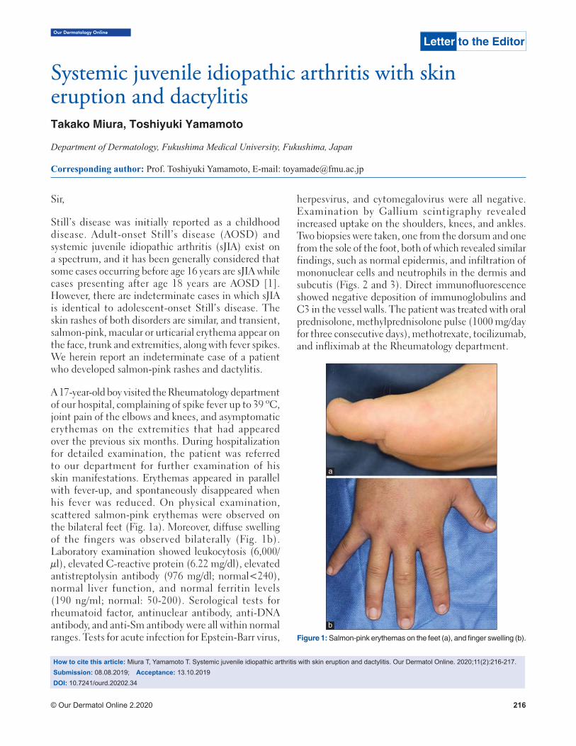
Our Dermatology Online
© Our Dermatol Online 2.2020 216
How to cite this article: Miura T, Yamamoto T. Systemic juvenile idiopathic arthritis with skin eruption and dactylitis. Our Dermatol Online. 2020;11(2):216-217.Submission: 08.08.2019; Acceptance: 13.10.2019DOI: 10.7241/ourd.20202.34
Systemic juvenile idiopathic arthritis with skin Systemic juvenile idiopathic arthritis with skin eruption and dactylitiseruption and dactylitisTakako Miura, Toshiyuki Yamamoto
Department of Dermatology, Fukushima Medical University, Fukushima, Japan
Corresponding author: Prof. Toshiyuki Yamamoto, E-mail: [email protected]
Sir,
Still’s disease was initially reported as a childhood disease. Adult-onset Still’s disease (AOSD) and systemic juvenile idiopathic arthritis (sJIA) exist on a spectrum, and it has been generally considered that some cases occurring before age 16 years are sJIA while cases presenting after age 18 years are AOSD [1]. However, there are indeterminate cases in which sJIA is identical to adolescent-onset Still’s disease. The skin rashes of both disorders are similar, and transient, salmon-pink, macular or urticarial erythema appear on the face, trunk and extremities, along with fever spikes. We herein report an indeterminate case of a patient who developed salmon-pink rashes and dactylitis.
A 17-year-old boy visited the Rheumatology department of our hospital, complaining of spike fever up to 39 ºC, joint pain of the elbows and knees, and asymptomatic erythemas on the extremities that had appeared over the previous six months. During hospitalization for detailed examination, the patient was referred to our department for further examination of his skin manifestations. Erythemas appeared in parallel with fever-up, and spontaneously disappeared when his fever was reduced. On physical examination, scattered salmon-pink erythemas were observed on the bilateral feet (Fig. 1a). Moreover, diffuse swelling of the fingers was observed bilaterally (Fig. 1b). Laboratory examination showed leukocytosis (6,000/μl), elevated C-reactive protein (6.22 mg/dl), elevated antistreptolysin antibody (976 mg/dl; normal<240), normal liver function, and normal ferritin levels (190 ng/ml; normal: 50-200). Serological tests for rheumatoid factor, antinuclear antibody, anti-DNA antibody, and anti-Sm antibody were all within normal ranges. Tests for acute infection for Epstein-Barr virus,
herpesvirus, and cytomegalovirus were all negative. Examination by Gallium scintigraphy revealed increased uptake on the shoulders, knees, and ankles. Two biopsies were taken, one from the dorsum and one from the sole of the foot, both of which revealed similar findings, such as normal epidermis, and infiltration of mononuclear cells and neutrophils in the dermis and subcutis (Figs. 2 and 3). Direct immunofluorescence showed negative deposition of immunoglobulins and C3 in the vessel walls. The patient was treated with oral prednisolone, methylprednisolone pulse (1000 mg/day for three consecutive days), methotrexate, tocilizumab, and infliximab at the Rheumatology department.
Letter to the Editor
Figure 1: Salmon-pink erythemas on the feet (a), and fi nger swelling (b).
a
b
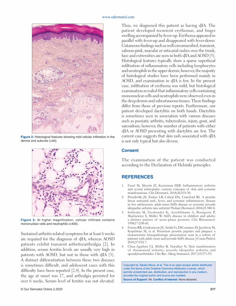
www.odermatol.com
© Our Dermatol Online 2.2020 217
Sustained arthritis-related symptoms for at least 6 weeks are required for the diagnosis of sJIA, whereas AOSD patients exhibit transient arthritis/arthralgia [2]. In addition, serum ferritin levels are usually very high in patients with AOSD, but not in those with sJIA [3]. A distinct differentiation between these two diseases is sometimes difficult, and adolescent cases with this difficulty have been reported [2,4]. In the present case, the age at onset was 17, and arthralgia persisted for over 6 weeks. Serum level of ferritin was not elevated.
Thus, we diagnosed this patient as having sJIA. The patient developed recurrent erythemas, and finger swelling accompanied by fever-up. Erythema appeared in parallel with fever-up and disappeared with fever-down. Cutaneous findings such as well-circumscribed, transient, salmon-pink, macular or urticarial rashes over the trunk, face and extremities are seen in both sJIA and AOSD [5]. Histological features typically show a sparse superficial infiltration of inflammatory cells including lymphocytes and neutrophils in the upper dermis; however, the majority of histological studies have been performed mainly in AOSD, and examination in sJIA is few. In the present case, infiltration of erythema was mild, but histological examination revealed that inflammatory cells containing mononuclear cells and neutrophils were observed even in the deep dermis and subcutaneous tissues. These findings differ from those of previous reports. Furthermore, our patient developed dactylitis on both hands. Dactylitis is sometimes seen in association with various diseases such as psoriatic arthritis, tuberculosis, injury, gout, and sarcoidosis; however, the number of patients with either sJIA or AOSD presenting with dactylitis are few. The current case suggests that skin rash associated with sJIA is not only typical but also diverse.
Consent
The examination of the patient was conducted according to the Declaration of Helsinki principles.
REFERENCES
1. Fazel M, Merola JF, Kurtzman DJB. Inflammatory arthritis and crystal arthropathy: current concepts of skin and systemic manifestations. Clin Dermatol. 2018;36:533-50.
2. Prendiville JS, Tucker LB, Cabral DA, Crawford RI. A pruritic linear urticarial rash, fever, and systemic infl ammatory disease in fi ve adolescents: adult-onset Still’s disease or systemic juvenile idiopathic arthritis sine arthritis? Pediatr Dermatol. 2004;21:580-8.
3. Sobieska M, Fassbender K, Aeschlimann A, Bourgeois P, Mackiewicz S, Müller W. Still’s disease in children and adults: a distinct pattern of acute-phase proteins. Clin Rheumatol. 1998;17:258-60.
4. Fortna RR, Gudjonsson JE, Seidel G, DiCostanzo D, Jacobson M, Kopelman M, et al. Persistent pruritic papules and plaques: a characteristic histopathologic presentation seen in a subset of patients with adult-onset and juvenile Still’s disease. J Cutan Pathol. 2010;37:932-7.
5. Chua-Aguilera CJ, Möller B, Yawalkar N. Skin manifestation of rheumatoid arthritis, juvenile idiopathic arthritis, and spondyloarthritides. Clin Rev Allerg Immunol. 2017;53:371-93.
Figure 2: Histological features showing mild cellular infi ltration in the dermis and subcutis (×40).
Figure 3: At higher magnification, cellular infiltrate contains mononuclear cells and neutrophils (×400).
Copyright by Takako Miura, et al. This is an open-access article distributed under the terms of the Creative Commons Attribution License, which permits unrestricted use, distribution, and reproduction in any medium, provided the original author and source are credited.Source of Support: Nil, Confl ict of Interest: None declared.
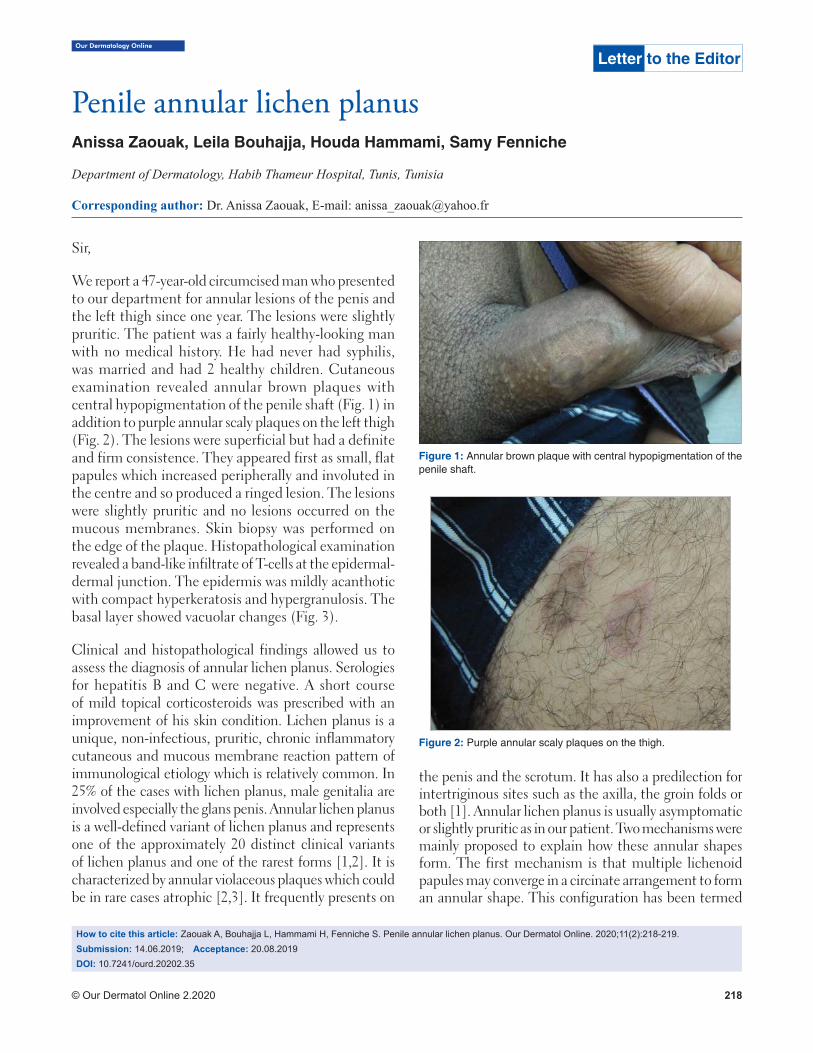
Our Dermatology Online
© Our Dermatol Online 2.2020 218
How to cite this article: Zaouak A, Bouhajja L, Hammami H, Fenniche S. Penile annular lichen planus. Our Dermatol Online. 2020;11(2):218-219.Submission: 14.06.2019; Acceptance: 20.08.2019DOI: 10.7241/ourd.20202.35
Penile annular lichen planusPenile annular lichen planusAnissa Zaouak, Leila Bouhajja, Houda Hammami, Samy Fenniche
Department of Dermatology, Habib Thameur Hospital, Tunis, Tunisia
Corresponding author: Dr. Anissa Zaouak, E-mail: [email protected]
Sir,
We report a 47-year-old circumcised man who presented to our department for annular lesions of the penis and the left thigh since one year. The lesions were slightly pruritic. The patient was a fairly healthy-looking man with no medical history. He had never had syphilis, was married and had 2 healthy children. Cutaneous examination revealed annular brown plaques with central hypopigmentation of the penile shaft (Fig. 1) in addition to purple annular scaly plaques on the left thigh (Fig. 2). The lesions were superficial but had a definite and firm consistence. They appeared first as small, flat papules which increased peripherally and involuted in the centre and so produced a ringed lesion. The lesions were slightly pruritic and no lesions occurred on the mucous membranes. Skin biopsy was performed on the edge of the plaque. Histopathological examination revealed a band-like infiltrate of T-cells at the epidermal-dermal junction. The epidermis was mildly acanthotic with compact hyperkeratosis and hypergranulosis. The basal layer showed vacuolar changes (Fig. 3).
Clinical and histopathological findings allowed us to assess the diagnosis of annular lichen planus. Serologies for hepatitis B and C were negative. A short course of mild topical corticosteroids was prescribed with an improvement of his skin condition. Lichen planus is a unique, non-infectious, pruritic, chronic inflammatory cutaneous and mucous membrane reaction pattern of immunological etiology which is relatively common. In 25% of the cases with lichen planus, male genitalia are involved especially the glans penis. Annular lichen planus is a well-defined variant of lichen planus and represents one of the approximately 20 distinct clinical variants of lichen planus and one of the rarest forms [1,2]. It is characterized by annular violaceous plaques which could be in rare cases atrophic [2,3]. It frequently presents on
the penis and the scrotum. It has also a predilection for intertriginous sites such as the axilla, the groin folds or both [1]. Annular lichen planus is usually asymptomatic or slightly pruritic as in our patient. Two mechanisms were mainly proposed to explain how these annular shapes form. The first mechanism is that multiple lichenoid papules may converge in a circinate arrangement to form an annular shape. This configuration has been termed
Letter to the Editor
Figure 1: Annular brown plaque with central hypopigmentation of the penile shaft.
Figure 2: Purple annular scaly plaques on the thigh.
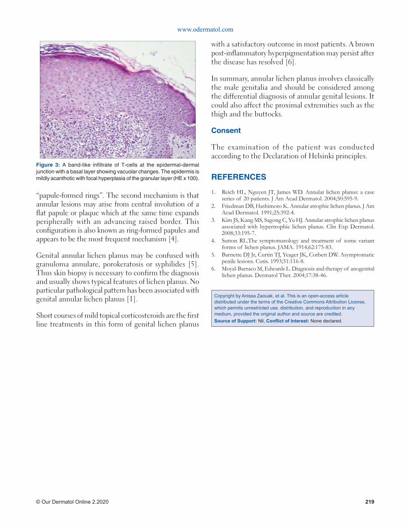
www.odermatol.com
© Our Dermatol Online 2.2020 219
“papule-formed rings”. The second mechanism is that annular lesions may arise from central involution of a flat papule or plaque which at the same time expands peripherally with an advancing raised border. This configuration is also known as ring-formed papules and appears to be the most frequent mechanism [4].
Genital annular lichen planus may be confused with granuloma annulare, porokeratosis or syphilides [5]. Thus skin biopsy is necessary to confirm the diagnosis and usually shows typical features of lichen planus. No particular pathological pattern has been associated with genital annular lichen planus [1].
Short courses of mild topical corticosteroids are the first line treatments in this form of genital lichen planus
with a satisfactory outcome in most patients. A brown post-inflammatory hyperpigmentation may persist after the disease has resolved [6].
In summary, annular lichen planus involves classically the male genitalia and should be considered among the differential diagnosis of annular genital lesions. It could also affect the proximal extremities such as the thigh and the buttocks.
Consent
The examination of the patient was conducted according to the Declaration of Helsinki principles.
REFERENCES
1. Reich HL, Nguyen JT, James WD. Annular lichen planus: a case series of 20 patients. J Am Acad Dermatol. 2004;50:595-9.
2. Friedman DB, Hashimoto K. Annular atrophic lichen planus. J Am Acad Dermatol. 1991;25:392-4.
3. Kim JS, Kang MS, Sagong C, Yu HJ. Annular atrophic lichen planus associated with hypertrophic lichen planus. Clin Exp Dermatol. 2008;33:195-7.
4. Sutton RL.The symptomatology and treatment of some variant forms of lichen planus. JAMA. 1914;62:175-83.
5. Barnette DJ Jr, Curtin TJ, Yeager JK, Corbett DW. Asymptomatic penile lesions. Cutis. 1993;51:116-8.
6. Moyal-Barraco M, Edwards L. Diagnosis and therapy of anogenital lichen planus. Dermatol Ther. 2004;17:38-46.
Figure 3: A band-like infi ltrate of T-cells at the epidermal-dermal junction with a basal layer showing vacuolar changes. The epidermis is mildly acanthotic with focal hyperplasia of the granular layer (HE x 100).
Copyright by Anissa Zaouak, et al. This is an open-access article distributed under the terms of the Creative Commons Attribution License, which permits unrestricted use, distribution, and reproduction in any medium, provided the original author and source are credited.Source of Support: Nil, Confl ict of Interest: None declared.
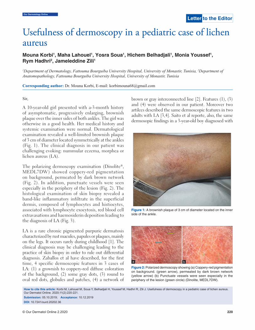
Our Dermatology Online
© Our Dermatol Online 2.2020 220
Sir,
A 10-year-old girl presented with a-3-month history of asymptomatic, progressively enlarging, brownish plaque over the inner sides of both ankles. The girl was otherwise in a good health. Her medical history and systemic examination were normal. Dermatological examination revealed a well-limited brownish plaque of 3 cm of diameter located symmetrically at the ankles (Fig. 1). The clinical diagnosis in our patient was challenging evoking: nummular eczema, morphea or lichen aureus (LA).
The polarizing dermoscopy examination (Dinolite®, MEDL7DW) showed coppery-red pigmentation on background, permeated by dark brown network (Fig. 2). In addition, punctuate vessels were seen especially in the periphery of the lesion (Fig. 2). The histological examination of skin biopsy revealed a band-like inflammatory infiltrate in the superficial dermis, composed of lymphocytes and histiocytes, associated with lymphocyte exocytosis, red blood cell extravasations and haemosiderin deposition leading to the diagnosis of LA (Fig. 3).
LA is a rare chronic pigmented purpuric dermatosis characterized by rust macules, papules or plaques, mainly on the legs. It occurs rarely during childhood [1]. The clinical diagnosis may be challenging leading to the practice of skin biopsy in order to rule out differential diagnosis. Zaballos et al have described, for the first time, 4 specific dermoscopic features in 3 cases of LA: (1) a grownish to coppery-red diffuse coloration of the background, (2) some gray dots, (3) round to oval red dots, globules and patches, (4) a network of
brown or gray interconnected line [2]. Features (1), (3) and (4) were observed in our patient. Moreover two artilces described the same dermoscopic features in two adults with LA [3,4]. Saito et al reporte, also, the same dermosopic findings in a 3-year-old boy diagnosed with
How to cite this article: Korbi M, Lahouel M, Soua Y, Belhadjali H, Youssef M, Hadhri R, Zili J. Usefulness of dermoscopy in a pediatric case of lichen aureus. Our Dermatol Online. 2020;11(2):220-221.Submission: 05.10.2019; Acceptance: 10.12.2019DOI: 10.7241/ourd.20202.36
Letter to the Editor
Usefulness of dermoscopy in a pediatric case of lichen Usefulness of dermoscopy in a pediatric case of lichen aureusaureusMouna Korbi1, Maha Lahouel1, Yosra Soua1, Hichem Belhadjali1, Monia Youssef1, Rym Hadhri2, Jameleddine Zili1
1Department of Dermatology, Fattouma Bourguiba University Hospital, University of Monastir, Tunisia, 2Department of Anatomopathology, Fattouma Bourguiba University Hospital, University of Monastir, Tunisia
Corresponding author: Dr. Mouna Korbi, E-mail: [email protected]
Figure 1: A brownish plaque of 3 cm of diameter located on the inner side of the ankle.
Figure 2: Polarized dermoscopy showing (a) Coppery-red pigmentation on background. (green arrow), permeated by dark brown network (yellow arrow) (b) Punctuate vessels were seen especially in the periphery of the lesion (green circle) (Dinolite, MEDL7DW).
ba
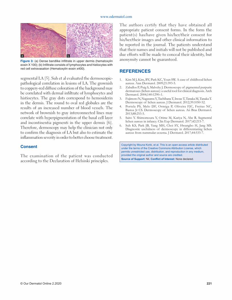
www.odermatol.com
© Our Dermatol Online 2.2020 221
segmental LA [5]. Suh et al evaluated the dermoscopic-pathological correlation in lesions of LA. The grownish to coppery-red diffuse coloration of the background may be correlated with dermal infiltrate of lymphocytes and histiocytes. The gray dots correspond to hemosiderin in the dermis. The round to oval red globules are the results of an increased number of blood vessels. The network of brownish to gray interconnected lines may correlate with hyperpigmentation of the basal cell layer and incontinentia pigmenti in the upper dermis [6]. Therefore, dermoscopy may help the clinician not only to confirm the diagnosis of LA but also to estimate the inflammation severity in order to better choose treatment.
Consent
The examination of the patient was conducted according to the Declaration of Helsinki principles.
The authors certify that they have obtained all appropriate patient consent forms. In the form the patient(s) has/have given his/her/their consent for his/her/their images and other clinical information to be reported in the journal. The patients understand that their names and initials will not be published and due efforts will be made to conceal their identity, but anonymity cannot be guaranteed.
REFERENCES
1. Kim M J, Kim, BY, Park KC, Youn SW. A case of childhood lichen aureus. Ann Dermatol. 2009;21:393-5.
2. Zaballos P, Puig S, Malvehy J. Dermoscopy of pigmented purpuric dermatoses (lichen aureus): a useful tool for clinical diagnosis. Arch Dermatol. 2004;140:1290–1.
3. Fujimoto N, Nagasawa Y, Tachibana T, Inoue T, Tanaka M, Tanaka T. Dermoscopy of lichen aureus. J Dermatol. 2012;39:1050-52.
4. Portela PS, Melo DF, Ormiga P, Oliveira FJC, Freitas NC, Bastos Jr CS. Dermoscopy of lichen aureus. An Bras Dermatol. 2013;88:253-5.
5. Saito Y. Shimomoura Y, Orime M, Kariya N, Abe R. Segmental lichen aureus in infancy. Clin Exp Dermatol. 2017;42:215-7.
6. Suh KS, Park JB, Yang MH, Choi SY, Hwangbo H, Jang MS. Diagnostic usefulness of dermoscopy in differentiating lichen aureus from nummular eczema. J Dermatol. 2017;44:533-7.
Copyright by Mouna Korbi, et al. This is an open-access article distributed under the terms of the Creative Commons Attribution License, which permits unrestricted use, distribution, and reproduction in any medium, provided the original author and source are credited.Source of Support: Nil, Confl ict of Interest: None declared.
Figure 3: (a) Dense bandlike infi ltrate in upper dermis (hematoxylin eosin X 100); (b) Infi ltrate consists of lymphocytes and histiocytes with red cell extravasation (Hematoxylin eosin x400).
ba
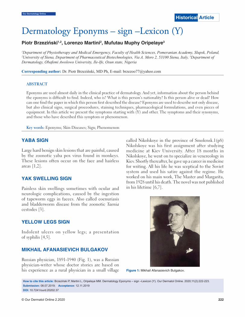
Our Dermatology Online
© Our Dermatol Online 2.2020 222
How to cite this article: Brzeziński P, Martini L, Oripelaye MM. Dermatology Eponyms – sign –Lexicon (Y). Our Dermatol Online. 2020;11(2):222-223.Submission: 08.07.2019; Acceptance: 12.11.2019DOI: 10.7241/ourd.20202.37
Historical Article
Dermatology Eponyms – sign –Lexicon (Y)Dermatology Eponyms – sign –Lexicon (Y)Piotr Brzeziński1,2, Lorenzo Martini2, Mufutau Muphy Oripelaye3
1Department of Physiotherapy and Medical Emergency, Faculty of Health Sciences, Pomeranian Academy, Slupsk, Poland, 2University of Siena, Department of Pharmaceutical Biotechnologies, Via A. Moro 2, 53100 Siena, Italy, 3Department of Dermatology, Obafemi Awolowo University, Ile-Ife, Osun state, Nigeria
Corresponding author: Dr. Piotr Brzeziński, MD Ph, E-mail: [email protected]
YABA SIGN
Large hard benign skin lesions that are painful, caused by the zoonotic yaba pox virus found in monkeys. These lesions often occur on the face and hairless areas [1,2].
YAK SWELLING SIGN
Painless skin swellings sometimes with ocular and neurologic complications, caused by the ingestion of tapeworm eggs in facees. Also called eoenuriasis and bladderworm disease from the zoonotic Taenia cestodes [3].
YELLOW LEGS SIGN
Indolent ulcers on yellow legs; a presentation of syphilis [4,5].
MIKHAIL AFANASIEVICH BULGAKOV
Russian physician, 1891-1940 (Fig. 1), was a Russian physician-writer whose doctor stories are based on his experience as a rural physician in a small village
called Nikolskoye in the province of Smolensk.1(p8) Nikolskoye was his first assignment after studying medicine at Kiev University. After 18 months in Nikolskoye, he went on to specialize in venereology in Kiev. Shortly thereafter, he gave up a career in medicine for writing. All his life he was sceptical to the Soviet system and used his satire against the regime. He worked on his main work, The Master and Margarita, from 1928 until his death. The novel was not published in his lifetime [6,7].
ABSTRACT
Eponyms are used almost daily in the clinical practice of dermatology. And yet, information about the person behind the eponyms is difficult to find. Indeed, who is? What is this person’s nationality? Is this person alive or dead? How can one find the paper in which this person first described the disease? Eponyms are used to describe not only disease, but also clinical signs, surgical proceodures, staining techniques, pharmacological formulations, and even pieces of equipment. In this article we present the symptoms starting with (Y) and other. The symptoms and their synonyms, and those who have described this symptom or phenomenon.
Key words: Eponyms; Skin Diseases; Sign; Phenomenon
Figure 1: Mikhail Afanasievich Bulgakov.
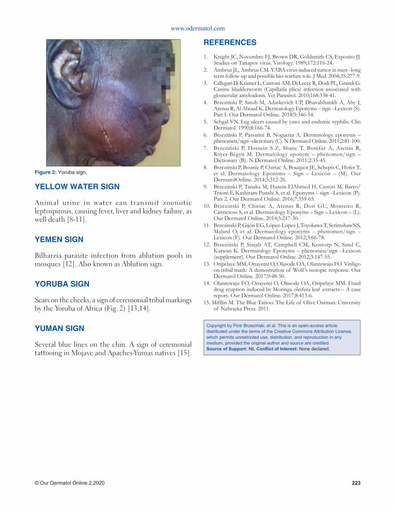
www.odermatol.com
© Our Dermatol Online 2.2020 223
YELLOW WATER SIGN
Animal urine in water can transmit zoonotic leptospirosis, causing fever, liver and kidney failure, as well death [8-11].
YEMEN SIGN
Bilharzia parasite infection from ablution pools in mosques [12]. Also known as Ablution sign.
YORUBA SIGN
Scars on the cheeks, a sign of ceremonial tribal markings by the Yoruba of Africa (Fig. 2) [13,14].
YUMAN SIGN
Several blue lines on the chin. A sign of ceremonial tattooing in Mojave and Apaches-Yumas natives [15].
REFERENCES
1. Knight JC, Novembre FJ, Brown DR, Goldsmith CS, Esposito JJ. Studies on Tanapox virus. Virology. 1989;172:116-24.
2. Ambrus JL, Ambrus CM. YABA virus induced tumor in men--long term follow-up and possible bio-warfare role. J Med. 2004;35:277-9.
3. Callegari D, Kramer L, Cantoni AM, Di Lecce R, Dodi PL, Grandi G. Canine bladderworm (Capillaria plica) infection associated with glomerular amyloidosis. Vet Parasitol. 2010;168:338-41.
4. Brzeziński P, Satoh M, Adaskevich UP, Dhavalshankh A, Aby J, Arenas R, Al Aboud K. Dermatology Eponyms – sign –Lexicon (S). Part I. Our Dermatol Online. 2018;9:346-54.
5. Sehgal VN. Leg ulcers caused by yaws and endemic syphilis. Clin Dermatol. 1990;8:166-74.
6. Brzeziński P, Passarini B, Nogueira A. Dermatology eponyms – phenomen/sign –dictionary (C). N Dermatol Online. 2011;2:81-100.
7. Brzeziński P, Hossein S-Z, Shane T, Bonifaz A, Arenas R, Royer-Bégyn M. Dermatology eponym – phenomen/sign – Dictionary (B). N Dermatol Online. 2011;2:35-45.
8. Brzezinski P, Bourée P, Chiriac A, Bouquot JE, Schepis C, Hofer T, et al. Dermatology Eponyms – Sign – Lexicon – (M). Our DermatolOnline. 2014;5:312-26.
9. Brzezinski P, Tanaka M, Husein-ElAhmed H, Castori M, Barro/Traoré F, Kashiram Punshi S, et al. Eponyms – sign –Lexicon (P). Part 2. Our Dermatol Online. 2016;7:359-65.
10. Brzezinski P, Chiriac A, Arenas R, Dori GU, Monteiro R, Cairncross S, et al. Dermatology Eponyms – Sign – Lexicon – (L). Our Dermatol Online. 2014;5:217-30.
11. Brzezinski P, Gijón EG, López-López J, Toyokawa T, ScrimshawNS, Malard O, et al. Dermatology eponyms – phenomen/sign – Lexicon (F). Our Dermatol Online. 2012;3:66-78.
12. Brzeziński P, Sinjab AT, Campbell CM, Kentorp N, Sand C, Karwan K. Dermatology Eponyms – phenomen/sign –Lexicon (supplement). Our Dermatol Online. 2012;3:147-55.
13. Oripelaye MM, Onayemi O, Olasode OA, Olanrewaju FO. Vitiligo on tribal mark: A demostration of Wolf ’s isotopic response. Our Dermatol Online. 2017;9:48-50.
14. Olanrewaju FO, Onayemi O, Olasode OA, Oripelaye MM. Fixed drug eruption induced by Moringa oleifera leaf extracts – A case report. Our Dermatol Online. 2017;8:413-6.
15. Miffl in M. The Blue Tattoo: The Life of Olive Oatman. University of Nebraska Press. 2011.
Figure 2: Yoruba sign.
Copyright by Piotr Brzeziński, et al. This is an open-access article distributed under the terms of the Creative Commons Attribution License, which permits unrestricted use, distribution, and reproduction in any medium, provided the original author and source are credited.Source of Support: Nil, Confl ict of Interest: None declared.
O u r D e r m a t o l o g y O n l i n e
w w w . o d e r m a t o l . c o m2.2020 (02.April.2020)