isocostunolide, a sesquiterpene lactone, induces mitochondrial membrane depolarization and...
TRANSCRIPT

Isocostunolide, a sesquiterpene lactone, induces mitochondrial
membrane depolarization and caspase-dependent apoptosis
in human melanoma cells
Chia-Nan Chen a, Hsin-Hsiu Huang a, Chia-Li Wu b, Coney P.C. Lin c,
John T.A. Hsu c,d, Hsing-Pang Hsieh c, Shuang-En Chuang a, Gi-Ming Lai a,*
a Divsion of Cancer Research, National Health Research Institutes (NHRI), 7F, No. 161, Sec. 6, Min-Chuan East Road, Taipei 114, Taiwan, ROCb Department of Chemistry, Tamkang University, Tamsui 251, Taiwan, ROC
c Division of Biotechnology and Pharmaceutical Research, NHRI, 35, Keyan Road, Zhunan Town, Miaoli County 350, Taiwan, ROCd Department of chemical Engineering, National Tsing Hua University, Hsinchu, Taiwan, ROC
Received 21 February 2006; received in revised form 22 February 2006; accepted 1 March 2006
Abstract
Isocostunolide is a sesquiterpene lactone isolated from the roots of Inula helenium. Its chemical structure was determined by
NMR and FAB-MS spectra. No biological activities of this compound have yet been reported. In this study, we found
isocostunolide could effectively induce cytotoxicity in three cancer cell lines (A2058, HT-29, and HepG2), with an IC50 of 3.2, 5.0,
and 2.0 mg/mL, respectively. DNA flow cytometric analysis indicated that isocostunolide actively induced apoptosis of cancer cells
accompanied by a marked loss of G0/G1 phase cells. To address the mechanism of the apoptotic effect of isocostunolide, we
analyzed the induction of apoptosis-related proteins in A2058. The levels of pro-caspase-8, Bid, pro-caspase-3, and poly(ADP-
ribose) polymerase (PARP) decreased. However, the level of Fas was increased markedly in a dose-dependent manner.
Furthermore, this compound markedly induced a depolarization of mitochondrial membranes to facilitate cytochrome c release
into cytosol. The findings suggest that isocostunolide may activate a mitochondria-mediated apoptosis pathway. To address this,
we found that isocostunolide-induced loss of mitochondrial membrane potential occurred via modulation of the Bcl-2 family
proteins. The production of intracellular reactive oxygen species (ROS) in A2058 was not elicited. In summary, for the first time,
we have isolated and characterized isocostunolide from I. helenium. This compound induces apoptosis through a mitochondria-
dependent pathway in A2058 cells.
q 2006 Elsevier Ireland Ltd. All rights reserved.
Keywords: Apoptosis; Sesquiterpene lactone; Mitochondrial; ROS; Caspase; Isocostunolide
1. Introduction
Inula helenium (Compositae) is an herb traditionally
used as a home remedy in Japan [1], as a diaphoresis in
Europe, and in Taiwan and China, as a therapeutic
agent for tuberculotic enterorrhea and chronic entero
0304-3835/$ - see front matter q 2006 Elsevier Ireland Ltd. All rights rese
doi:10.1016/j.canlet.2006.03.004
* Corresponding author.
E-mail address: [email protected] (G.-M. Lai).
gastritis [2]. Plants of the genus of Inula have been
shown to contain high levels of sesquiterpene lactones
[3]. Sesquiterpene lactones recently have received
considerable attention in its pharmacological circles
due to their anti-neoplastic and anti-inflammatory
effects [4–6]. Isocostunolide is a well-known sesqui-
terpene lactone contained in plants [7]. In this study,
this compound was isolated and characterized from the
Chinese herb I. helenium. The structure was elucidated
Cancer Letters 246 (2007) 237–252
www.elsevier.com/locate/canlet
rved.

C.-N. Chen et al. / Cancer Letters 246 (2007) 237–252238
mainly by NMR spectral evidence and found to be
identical to the reported sesquiterpene lactone com-
pound isocostunolide. Although many reports have
asserted that sesquiterpene lactones exhibit a broad
spectrum of biological activities, including anti-tumor
[8], anti-inflammatory [9], anti-fungal [10], and anti-
viral [11] activities, the bioactivities of isocostunolide
have never been reported. More than 5000 sesquiter-
pene lactones have been identified from plants [12,13],
but their various biological activities are not yet well
understood.
Apoptosis, or programmed cell death, has an
essential role in controlling cell number in many
developmental and physiological settings. Its morpho-
logical characteristics include plasma membrane
blebbing, cell shrinkage, nuclear condensation, chromo-
somal DNA fragmentation, and formation of apoptotic
bodies [14]. Recent reports have claimed that many anti-
cancer drugs or cancer chemopreventive agents act
through the induction of apoptosis to prevent tumor
promotion and progression. Mitochondria are currently
regarded as playing a central role in mediating ‘intrinsic
death signals’ and could sever as a novel target for
chemotherapies [15,16]. Cytochrome c is a mitochon-
drial protein that can activate caspases. However, the
release of cytochrome c is regulated by the anti- and pro-
apoptotic members of the Bcl-2 family. Once in the
cytoplasm, it binds Apaf-1 to pro-caspase-9, leading to
the activation of caspase-9 and to initiation of the
caspase cascade [17]. Many parameters of mitochon-
drial physiology have been shown to be hallmarks of
apoptosis. These include the loss of mitochondrial
membrane potential (DJm), the generation of ROS
(reactive oxygen species), the termination of oxygen
consumption, and the release of cytochrome c [18].
In this report, we investigate the mechanisms of
isocostunolide on the induction of the apoptosis effect
in human melanoma A2058 cells. We perform
experiments to evaluate whether the anti-proliferative
effect of isocostunolide in A2058 cells was mediated
through apoptosis, and our results also indicate that
isocostunolide can indeed promote apoptosis in A2058
cells via mitochondrial membrane depolarization and
activation of the caspase cascade.
2. Materials and methods
2.1. Extraction and isolation
The roots of I. helenium (0.8 kg, dried weight) were
purchased from a medicinal herbs market in Taipei, in May
2004. The herb was extracted with MeOH (3!5.0 L) at
25–27 8C for 2 weeks. After concentration of the combined
extracts under reduced pressure, the residue (107.0 g) was
suspended in H2O and then extracted with n-hexane, EtOAc,
CHCl3, and 1-butanol, respectively. The n-hexane extract
(52.0 g) was chromatographed over a silica gel column and
eluted with an n-hexane–EtOAc gradient system (5:0, 4:1,
3:2, 2:3, 1:4, 0:5, then with pure MeOH) to afford seven (F1–
F7) fractions. These fractions were assayed on human
melanoma A2058 cell proliferation, and the active fractions
(F2) were purified again by silica gel column and eluted with
n-hexane–EtOAc gradient system (19:1, 18:2, 17:3, 16:4, and
15:5). These eluted fractions were screened again for their
inhibitory activity on A2058 cells proliferation. The active
fractions were purified by HPLC (Phenomenex Luna C-18
silica gel column [10.0!250 mm], eluting with a mixture of
MeOH and H2O [80:20, v/v]). Fractions of retention times at
9.5 min containing isocostunolide were collected.
2.2. Cell culture and cell viability assay
Human melanoma cells (A2058), human hepatoma cells
(HepG2), and human colon adenocarcinoma cells (HT-29)
were cultured in Dulbecoo’s modified Eagle’s medium
(DMEM, Gibco) containing 10% fetal bovine serum (FBS),
1% dilution of the penicillin–streptomycin, and 2 mM
glutamine and were maintained at 37 8C in a humidified
atmosphere of 95% air and 5% CO2. Cells (1!106 per dish)
were cultured in a 100-mm dish and incubated for 14 h
before being treated with various concentrations of iso-
costunolide (0.625, 1.25, 2.5, 5.0, and 7.5 mg/mL) for 48 h.
Isocostunolide was a small compound (MWZ233.1). When
these values are converted to micromolar, they are 0, 2.7,
5.4, 10.7, 16.1, 21.5, and 32.2 mM, respectively. Cells were
counted, and cell viability was determined by a trypan blue
exclusion assay.
2.3. Analysis of the cell cycle
A2058 cells (1!106) in a 100-mm dish were treated with
various concentrations of isocostunolide (0.625, 1.25, 2.5, and
5.0 mg/mL) for 48 h or with a fixed concentration of
isocostunolide (7.5 mg/mL) for 0, 3, 6, 12, and 24 h. Cells
were trypsinized and collected with ice cold PBS. The cells
were resuspended in 200 mL PBS and fixed by adding 800 mL
of iced 100% ethanol then incubated overnight at K20 8C. The
cell pellets were collected by centrifugation, resuspended in
1 mL of hypotonic buffer (0.1% Triton X-100 in PBS and
1 mg/mL RNase A), and incubated at 4 8C for 30 min. Then,
1 mL of PI solution (50 mg/mL) was added, and the mixture was
allowed to stand at 4 8C for 30 min. Cellular DNA content was
then analysed by FACScan cytometry (Becton Dickinson).
2.4. Annexin V staining
Double staining for Annexin-V-fluorescein isothiocyanate
(FITC) binding and for cellular DNA using PI was performed.

C.-N. Chen et al. / Cancer Letters 246 (2007) 237–252 239
A2058 cells were plated at a density of 3!105 cells/well on
six-well plates. After treatment with the isocostunolide
(7.5 mg/mL) for 0, 2, 4, and 6 h, cells were trypsinized and
collected in ice cold PBS. The cells were washed twice with
cold PBS and resuspended in 1 mL binding buffer (BD
PharMingen, San Diego, CA). They were then stained with
Annexin-V-FITC and PI according to the protocol from BD
PharMingen. Briefly, 2 mL of Annexin-V-FITC (50 mg/mL)
and 1 mL of PI (500 mg/mL) were added to a 100 mL solution
of cells and the solution was incubated for 15 min at room
temperature in the dark. Binding buffer was then added, and
early and late apoptosis was visualized by constructing a dot-
plot using a Becton Dickinson FACScan. Green fluorescence
from the Annexin-V-FITC was determined using an FL1
detector having a bandpass filter with specifications 530G15 nm. Red fluorescence from PI was determined using an
FL2 detector having a bandpass filter with specifications
585G21 nm. A total of 10,000 events were recorded for each
sample.
2.5. Assays of caspase inhibitors
A2058 cells (1!106) in a 100-mm dish were treated with
or without 50 mM of general caspase inhibitor (Z-VAD-FMK)
or a specific caspase-3 inhibitor (Z-DEVD-FMK). After 2 h,
cells were treated with isocostunolide (3.75 mg/mL) for 24 h.
Cellular morphology of A2058 was analysed by phase con-
trast microscopy.
2.6. Activity of caspase
Cellswerecollectedand washedwith PBSandsuspended ina
buffer containing 25 mM HEPES (pH 7.5), 5 mM MgCl2, 5 mM
EDTA, 5 mM dithiothione, 2 mM phenylmethylsulfonylfluor-
ide, 10 mg/mL pepstatin A and 10 mg/mL leupeptin. After
treatment, cell lysates were clarified by centrifugation at
13,200!g for 20 min at 4 8C. Caspase activity in the supernatant
was determined by a fluorogenic assay (R&D Caspase-3
Fluorometric Assay System). Briefly, 100 mg of total protein,
as determined by 7-amino-4-trifluoromethyl coumarin
(AFC) assay (R&D), was incubated with 50 mM substrate
DEVD-7-amino-4-trifluoromethyl coumarin (DEVD-AFC) at
Isocostunolide
O
CH2
O
CH3
CH3
1
2
3
4
5
6
7
8
9
10
11
12
13
14
15
3
4
Fig. 1. Chemical structures of isoc
37 8C for 1 h. The release of AFC was measured by excitation at
400 nm and emission at 505 nm using a fluorescence spectro-
photometer (Hitachi F-4500).
2.7. Measurement of mitochondrial membrane potential
(DJm)
Measurement of the mitochondrial membrane potential
was assayed by flow cytometry using the dye DiOC6
(Molecular Probes). A2058 cells were plated at a density of
3!105 cells/well on six-well plates. After treatment with
isocostunolide (7.5 mg/mL) for 0, 0.5, 1.0, 2.0, 4.0, and 6.0 h
or treatment with various concentrations (0.625, 1.25, 2.5, 5.0,
and 7.5 mg/mL) for 24 h, cells were trypsinized and collected
in ice cold PBS. DiOC6 (100 nM) was loaded into cells
suspended in 0.5 mL fresh DMEM and incubated at 37 8C for
15 min. DiOC6 fluorescence was determined at 530G30 nm.
Data were obtained and analyzed with the CellQuest software
on a Becton Dickinson FACScan.
2.8. Measurement of reactive oxygen species (ROS)
Flow cytometric determination of the ROS production
was carried out as described previously. A2058 cells were
collected by trypsinization and 200 mL of cell suspension
(1!106 cells/mL) was mixed with 800 mL PBS. Cells were
incubated with 10 mM 5-(and-6)-chloromethyl-2 0,7 0-dichlor-
odihydrofluorescein diacetate, acetyl ester (Molecular
Probes) for 15 min, followed by the addition of different
concentrations of isocostunolide (0, 2.5, 5.0, and 7.5 mg/mL)
or hydrogen peroxide (30 mM). The incubation was
continued for 20 min at 37 8C. The oxidative burst (hydrogen
peroxide) was detected using a FACScan flow cytometer
with excitation and emission settings of 488 and 530 nm,
respectively.
2.9. Western blotting assay
A2058 cells on 100-mm dishes (1.5!106/dish) were
treated with isocostunolide at 1.25, 2.5, 5.0, and 7.5 mg/mL
for 48 h. After treatment, cells were collected and resus-
pended in 100 mL Gold lysis buffer. Equal amounts of
CH3
CH3
O
CH2
O
Costunolide
1
2
5
6
7
8
9
10
1113
12
14
15
ostunolide and costunolide.

Table 213C NMR data of isocostunolide
Carbon no. Isocostunolide in
DMSO-d6, dC
Costunolide in CDCl3a,
dC
1 140.4 136.7
2 38.8 127.2
3 25.8 26.1
4 40.4 39.5
5 136.9 140.9
6 126.2 127.0
7 81.2 81.7
8 49.4 50.4
9 27.1 28.4
10 127.5 41.0
11 140.6 140.0
12 169.9 176.8
13 119.3 120.1
14 16.9 17.2
15 15.8 16.1
Assignments were based on the HMQC and HMBC NMR data.a Data taken from Ref. [35].
Table 11H NMR assignments of isocostunolide
Pos-
ition
Isocostuno-
lide dH (J,
Hz)
HMBC corre-
lations (C #)
NOESY cor-
relations (H #)
2 CH2 1.95 (m) 1, 3, 5, 6, 10 3, 4, 10
2.25 (m)
3 CH2 2.1 (m) 1, 2, 5, 6, 14 2, 4, 6, 14
2.25 (m)
4 CH2 2.1 (m) 1, 2, 3, 5, 6 2, 3, 6, 15
2.35 (m)
6 CH 4.85 (d, 10.8) 2, 15 2, 3, 4, 7
7 CH 4.74 (d, 4.9) 8, 9, 10, 11, 14 6, 8, 9, 14, 15
8 CH 2.65 (s) 7, 10 7, 9, 10
9 CH2 1.68 (m) 1, 6, 7, 8, 11, 14 7, 8, 10
2.1 (m)
10 CH 4.74 (d, 4.9) 1, 2, 8, 11, 14 8, 9, 14
13 CH2 5.67 (d, 3.3) 8, 11, 12 6, 8, 9
6.05 (d, 3.3)
14 CH3 1.62 (s) 1, 2, 3, 10 3, 10, 15
15 CH3 1.36 (s) 4, 5, 6 3, 4, 7, 9
Assignments were based on the HMBC and HMQC NMR data.
C.-N. Chen et al. / Cancer Letters 246 (2007) 237–252240
proteins (30 mg), were mixed with 2! sample buffer and
resolved by 10% SDS-PAGE for b-actin, poly(ADP-ribose)
polymerase (PARP), Bcl-2, Bid, pro-caspase-3, pro-caspase-
8, Fas, and cytochrome c detection. Proteins were electro-
transferred to an immobilon membrane (PVDF; Millipore
Corp.), and equivalent protein loading was verified by
staining the membrane with reversible dye amido black
(Sigma Chemical Co.). This was followed by overnight
blocking with a solution composed of 20 mM Tris–HCl (pH
7.4), 125 mM NaCl, 0.2% Tween 20, and 3% BSA. Specific
antibodies used were anti-human PARP (1:500 of rabbit
polyclonal; Santa Cruz Biotechnology, Inc.), anti-caspase-3,
Fas, and anti-caspase-8 antibodies (1:1000 of mouse
monoclonal; Pharmingen, Becton Dickinson), anti-Bcl-2
and anti-Bid antibodies (1:1000 of mouse monoclonal;
Santa Cruz Biotechnology, Inc.), anti-b-actin antibody
(1:3000 of mouse monoclonal; Cashmere Biotech), and
cytochrome c antibody (1:1000 of mouse monoclonal;
Research Diagnostic, Inc). These proteins were detected by
chemiluminescence (ECL, Amersham).
2.10. Cytochrome c release
Mitochondrial and cytosolic fractions were prepared by
resuspending cells in ice-cold buffer A (250 mM sucrose,
20 mM HEPES, 10 mM KCl, 1.5 mM MgCl2, 1 mM EDTA,
1 mM EGTA, 1 mM DTT, 17 mg/mL phenylmethylsulfonyl-
fluoride (PMSF), 8 mg/mL aprotinin, and 2 mg/mL leupeptin,
pH 7.4). Cells were passed through a needle 10 times.
Unlysed cells and nuclei were pelleted by centrifugation for
10 min at 750!g. The supernatant was then centrifuged at
1,00,000!g for 15 min. This pellet was resuspended in buffer
A and represented the mitochondrial fraction. The supernatant
was again centrifuged at 1,00,000!g for 1 h. The supernatant
from this final centrifugation step represented the cytosolic
fraction.
3. Results
3.1. Purification and identification of isocostunolide
Isocostunolide (Fig. 1) was isolated through
repeated chromatographies of the MeOH extract of I.
helenium under the guidance of the anti-proliferation
activity on A2058 cells. Final purification of the active
fraction was achieved by HPLC on a reversed-phase
C18 column. The total content of the active component
isocostunolide was roughly 1.8% of the MeOH extract
from I. helenium. The 1H and 13C NMR (in DMSO-d6,
Tables 1 and 2) spectra of the purified compound
displayed characteristic absorptions of sesquiterpene
lactones and subsequently was found identical to the
well-known compound, isocostunolide. The molecular
formula for isocostunolide was deduced as C15H20O2
on the basis of FAB-MS (positive mode), which
showed a quasimolecular ion peak at m/z 233.1 ([MCH]C). Upon comparison of the 13C NMR of isocostu-
nolide with the reported data of costunolide (Fig. 1), the
identity of both compounds could be recognized. The
carbon signals of isocostunolide were very similar to
those of costunolide, as shown in Table 2, except for the
2nd and 10th positions. The bioactivities of isocostu-
nolide have never been reported, and its chemical shifts
of proton and carbon (including HMBC, HMQC,
NOESY, and COSY) were only partially assigned.
Consequently, the stereostructure of isocostunolide was

0 0.63 1.25 2.5 5.0 7.5
Isocostunolide (µg/mL)
0
20
40
60
80
100
120(a)
(c)
(b)
% C
ell v
iabi
lity
HT-29A2058HepG2
Control Isocostunolide 7.5 µ g/mL
0 5.02.5 7.5 10.0
Isocostunolide (µg/mL)
0.0
0.8
0.6
0.4
0.2
1.0
Con
trol
-rel
ativ
e vi
abili
ty (
MT
T a
ssay
)
Fig. 2. Effect of isocostunolide on the viability of cancer cell lines. (a) Cytotoxic activity of isocostunolide in various human cancer cell lines
(A2058, HepG2, and HT-29). Cells (1!106 per dish) were cultured in 100-mm dish and incubated for 14 h before being treated with various
concentrations of isocostunolide (0.625, 1.25, 2.5, 5.0, and 7.5 mg/mL) for 48 h. Treated cells were counted and viability was determined by a trypan
blue exclusion assay. Results were from three repeats. (b) Phase-contrast micrographs of A2058 cells treated with various concentrations of
isocostunolide (7.5 mg/mL) for 48 h. A representative experiment of three repeats was shown. (c) A2058 cells were treated with various
concentrations of isocostunolide (2.5–10.0 mg/mL) for 48 h and subsequent cell viability was determined by MTT assay.
C.-N. Chen et al. / Cancer Letters 246 (2007) 237–252 241

C.-N. Chen et al. / Cancer Letters 246 (2007) 237–252242
determined as shown in Tables 1 and 2. Complete
assignments of each proton and carbon of isocostuno-
lide in DMSO-d6 were compiled on the basis of two-
dimensional NMR (HMQC, HMBC, NOESY).
3.2. Inhibition of cell growth by isocostunolide
The cytotoxic effects of isocostunolide on three
different cancer cell lines were investigated. All cancer
cell lines were treated with various concentrations
(0.63–7.5 mg/mL) of isocostunolide. After 48 h of
treatment, the number of live cells was measured by a
trypan blue exclusion assay. As shown in Fig. 2a, all
three cancer cell lines (A2058, HT-29, and HepG2)
were sensitive to isocostunolide, with an IC50 of 3.2,
5.0 and 2.0 mg/mL, respectively.
Fig. 3. Isocostunolide-induced apoptosis in A2058 cells. (a) Flow cytometr
anti-Annexin V monoclonal antibody binding (FL1-H) and propidium iodide
for 0, 2, 4, and 6 h and subsequently measured by flow cytometry. The low
represents late apoptosis. (b) Dose-dependent effect of isocostunolide on ce
concentrations (0.63–5.0 mg/mL) of isocostunolide for 48 h and stained with
cellular DNA profile was analyzed by Cell Quest software. (c) Time-cours
A2058 cells were treated with concentration of 7.5 mg/mL for 0, 3, 6, 12,
repeats.
3.3. Isocostunolide induces apoptotic all death in
A2058 cells
During cell viability assay, we demonstrated that
isocostunolide significantly induces cell death in
A2058. Morphological features, such as cell shrinkage
and plasma membrane blebbing, were seen in iso-
costunolide-treated cells. As shown in Fig. 2b, it
appears that the cells might have died through the
induction of apoptosis. The cytotoxic effect of
isocostunolide was further shown to be evaluated
dose-dependent by MTT assay as shown in Fig. 2c.
To demonstrate whether isocostunolide induced apop-
tosis in A2058 cells, we used annexin V-staining and
flow cytometry (Fig. 3(a)) to evaluate whether
isocostunolide-caused cell death occurs via apoptosis.
Cells were treated with isocostunolide (7.5 mg/mL) for
ic analysis of isocostunolide-treated A2058 cells after fluorescinated
uptake (FL2-H). Cells were treated with 7.5 mg/mL of isocostunolide
er right quadrant represents early apoptosis; the upper right quaderant
ll cycle and sub-G1 phase population. Cells were treated with various
PI as described in Section 2. Following flow cytometric analysis, the
e effect of isocostunolide on cell cycle and sub-G1 phase population.
and 24 h. Results were from one representative experiment of three
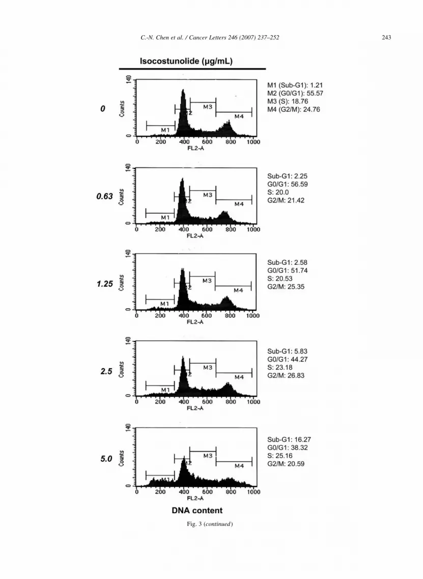
Fig. 3 (continued)
C.-N. Chen et al. / Cancer Letters 246 (2007) 237–252 243

Fig. 3 (continued)
C.-N. Chen et al. / Cancer Letters 246 (2007) 237–252244

C.-N. Chen et al. / Cancer Letters 246 (2007) 237–252 245
0, 2, 4, and 6 h, respectively. Our result indicated a
time-course dependent increase (0.52–11.47%) in
apoptotic cell population following isocostunolide
treatment. The induction of apoptosis was further
confirmed by the presence of the sub-G1 cell population
in flow cytometry. The DNA content of A2058 cells,
treated with isocostunolide at various concentrations (0,
0.63, 1.25, 2.5, and 5.0 mg/mL) for 48 h (Fig. 3b), or
treated with a fixed concentration of isocostunolide
(7.5 mg/mL) for 0, 3, 6, 12, and 24 h, was analyzed by
flow cytometry as shown in Fig. 3c. The respective
percentages of apoptotic cells observed after treatment
with 0, 0.63, 1.25, 2.5, and 5.0 mg/mL of isocostunolide
for 48 h were as follows: 1.21, 2.25, 2.58, 5.83 and
16.27%. The time course induction of apoptosis in
A2058 cells by isocostunolide at a fixed concentration
of 7.5 mg/mL was also shown (Fig. 3c). These results
show that isocostunolide at a concentration of 5.0 mg/
mL for 48 h or treated with isocostunolide at 7.5 mg/mL
for 12 h was sufficient to induce apoptosis in A2058
cells.
Fig. 4. Induction of apoptosis via caspase-dependent pathway and clearage
various concentrations of isocostunolide (1.25–7.5 mg/mL) for 48 h, or treat
were prepared and subjected to SDS-PAGE. Pro-caspase-3 and PARP (a),
respective specific antibodies. (d) Cellular morphology of A2058 cells with o
or a specific caspase-3 inhibitor (Z-DEVD-FMK) for 2 h, then treated with
contrast microscope. (e) Activation of caspase-3 by isocostunolide. A2058 ce
treatment, caspase activities were measured.
3.4. Isocostunolide induces apoptosis via activation of
caspase-dependent pathway
Initiator caspases (including 8, 9, 10, and 12) are
closely coupled to pro-apoptotic signals. Once
activated, these caspases cleave and activate down-
stream effector caspases (including 3, 6, and 7),
which in turn cleave cytoskeletal and nuclear
proteins, such as PARP and lamin A, and finally
induce apoptosis. These pro-apoptotic stimuli include
FasL, TNF-a, DNA damage, and ER stress. We first
evaluated whether caspase-dependent signal pathways
were involved in the apoptotic cell death induced by
isocostunolde in A2058 cells. Treatment of A2058
cells with 7.5 mg/mL for 3, 6, 12, and 24 h or treat
ment with various concentrations of isocostunolide
for 24 h resulted in dramatic cleavage of PARP and
markedly decreased pro-caspase-3 protein levels
(Fig. 4a). Because caspase-8 and Fas play an
important role both in the mitochondria dependent
and independent pathway in apoptosis, we thus
of caspase-8, caspase-3, and PARP. A2058 cells were treated with
ed with isocostunolide (7.5 mg/mL) for 3, 6, 12, and 24 h. Cell lysates
pro-caspase-8 (b), Fas (c) were determined by immunoblotting using
r without treatment 50 mM of general caspase inhibitor (Z-VAD-FMK)
isocostunolide (3.75 mg/mL) for 24 h. Cells were analysed by phase
lls were treated with 7.5 mg/mL isocostunolide for 2, 4, 6, and 8 h, after

0 2 4 6 8
Incubation time (hours)
0
1
2
3
4
5
Cas
pase
-3 a
ctiv
ity (
fold
)
(d)
(e)
Fig. 4 (continued)
C.-N. Chen et al. / Cancer Letters 246 (2007) 237–252246
further evaluated whether caspase-8 might be acti-
vated during the induction of apoptosis by isocostu-
nolide, as shown in Fig. 4b. Our data indicated that
isocostunolide 1.25–5.0 mg/mL markedly increased
Fas protein expression (Fig. 4c). All these results
indicate the PARP, pro-caspase-8, and pro-caspase-3
expression fell markedly under treatment with
isocostunolide at a concentration of 5.0–7.5 mg/mL.
However, the level of Fas was increased markedly in
a dose-dependent manner. To ask whether a particular
caspase plays the crucial role in isocostunolide-
induced apoptosis, two caspase inhibitors were used
to address this question. A2058 cells were pre-treated
with general caspase inhibitor (Z-VAD-FMK) or a

C.-N. Chen et al. / Cancer Letters 246 (2007) 237–252 247
specific caspase-3 inhibitor (Z-DEVD-FMK), then
treated with isocostunolide (3.75 mg/mL) for 24 h.
Our data indicate that not only the general caspase
inhibitor markedly inhibits isocostunolide-induced
cell death but also caspase-3 inhibitor as shown in
Fig. 4d. Activation of caspase-3 has been known to
play an important role in the induction of apoptosis
by various stimuli. Upon treatment of A2058 cells
with 7.5 mg/mL isocostunolide, the activity of
caspase-3 increased significantly within 4 h after the
start of treatment (Fig. 4e). The results obtained from
these experiments suggest that caspase-3 is involved
in isocostunolide-induced apoptosis of A2058 cells.
3.5. Mitochondria-dependent pathway was avtivated
during isocostunolide-induced apoptosis
Cytosolic Bid is cleaved by caspase-8 and myr-
istoylated before translation into mitochondria during
mitochondria-dependent pathway of apoptosis. To
determine if this pathway is activated by isocostunolide
treatment, we next examined the Bid protein level after
isocostunolide (7.5 mg/mL) treatment with various
concentrations (1.25, 2.5, 5.0, and 7.5 mg/mL) for
24 h (Fig. 5a) or treatment with 3, 6, 12, and 24 h
(Fig. 5b) in A2058 cells. The results indicate that Bid
was increased transiently at 6 h after treatment with
isocostunolide and cleaved markedly later on 12–24 h.
Another Bcl-2 family protein, Bcl-2 was significantly
Fig. 5. Induction of apoptosis via mitochondria-dependent pathway and activ
into cytosol. A2058 cells were treated with various concentrations of isoc
(7.5 mg/mL) for 3, 6, 12, and 24 h. Cell lysates were prepared and subjected to
Dose-dependent changes of Bid (a), time course-dependent change of Bid (b)
cytochrome c from mitochondria (d).
suppressed protein expression by treated with isocos-
tunolide (7.5 mg/mL) in a time-course manner (Fig. 5c).
Our data also indicated that cytochrome c was
markedly released from mitochondria into the cyto-
plasm in a dose-dependent manner (Fig. 5d). Taken
together, these results suggest convincingly that the
isocostunolide-induced apoptosis of A2058 cells may
be through the mitochondria-dependent pathway, as
evidenced by the activation of caspase-8, Bid, and
caspase-3.
3.6. Mitochondrial membrane depolarization by
isocostunolide
To further demonstrate the induction of apoptosis by
isocostunolide, the mitochondrial membrane depolar-
ization was examined by using DiOC6. The A2058 cells
were treated with isocostunolide at a concentration
(7.5 mg/mL) for 0.5, 1.0, 2.0, 4.0, and 6.0 h (Fig. 6a).
Our results indicate that isocostunolide induced
mitochondrial membrane depolarization in a time
course-dependent manner (14.1–55.8%). Fig. 6b
shows the dose-dependency of the induction of
mitochondrial membrane depolarization effects in
A2058 cells treatment with isocostunolide concen-
trations of 0.63–7.5 mg/mL. These results clearly show
that isocostunolide is an efficient inducer of A2058
cells mitochondrial membrane depolarization.
ation of Bid, and induction of cytochrome c release from mitochondria
ostunolide (1.25–7.5 mg/mL) for 48 h, or treated with isocostunolide
SDS-PAGE and immunoblotting using respective specific antibodies.
, time course-dependent change of Bcl-2 (c), dose-dependent release of

C.-N. Chen et al. / Cancer Letters 246 (2007) 237–252248
3.7. Mitochondrial membrane depolarization by iso-
costunolide treatment was not accompanied by the
production of intracellular reactive oxygen species
(ROS)
Many reports have implicated that intracellular ROS
are involved in the signal transduction pathways of
apoptosis. In this study, we evaluated whether the
Fig. 6. Analysis of mitochondrial membrane depolarization in A2058 cel
manner. (a) Percentage of cells (of 10,000 analyzed) with depolarized DJm
1.0, 2.0, 4.0, and 6.0 h. (b) The same conditions prevailed for measuring the
cells were treated with various concentrations of isocostunolide (0.63–7.5 m
analysis of the mitochondrial membrane depolarization was used. Detail
representative experiment of three repeats.
induction of mitochondrial membrane depolarization
induced by isocostunolide occurred via modulated
production ROS. We measured the production of
ROS after treatment with various concentrations of
isocostunolide (0, 2.5, 5.0, and 7.5 mg/mL) or of
hydrogen peroxide (30 mM, as a positive control) for
15 min. The oxidative burst was detected by a
FACScan flow cytometry as shown in Fig. 7. Our
ls treated with isocostunolide in a time course- and dose-dependent
was measured after treatment with isocostunolide (7.5 mg/mL) for 0.5,
membrane depolarization assays in a dose-dependent manner. A2058
g/mL) for 24 h. The fluorescence dye (DiOC6) in the flow cytometric
procedures were as described in Section 2. Results were from the

Fig. 6 (continued)
C.-N. Chen et al. / Cancer Letters 246 (2007) 237–252 249
results indicate that isocostunolide did not elicit the
production of intracellular ROS during membrane
depolarization.
4. Discussion
In this study, we evaluated the inhibitory effect of
isocostunolide on the proliferation of embryonic
fibroblast and cancer cells. We show that isocostuno-
lide, a sesquiterpene lactone isolated from I. helenium,
exerted a more marked cytotoxic effect at 2.5–5.0
mg/mL or less against three different cancer cell lines
(A2058, HT-29, and HepG2) than embryonic fibroblast
cells (data not shown). Our result suggests that
isocostunolide’s anti-proliferative effect is mediated
through apoptosis that involves caspase activation. No
biological activities of this compound have ever been
reported. Traditionally, many plants contain high levels
of sesquiterpene lactones have been used as folk
medicines because of their exerted many pharmaco-
logic properties. For example, its ingredient partheno-
lide was reported to be capable of inhibiting DNA

Fig. 7. Isocostunolide does not induce production of intracellular reactive oxygen species (ROS) in A2058. Suspension of A2058 cells were
incubated with 10 mM 5-(and-6)-chloromethyl-2 0,7 0-dichlorodihydrofluorescein diacetate, acetyl ester (Molecular Probes) for 15 min followed by
the addition of various concentrations of isocostunolide (0, 2.5, 5.0, and 7.5 mg/mL) or hydrogen peroxide (30 mM). The incubation was continued
for 20 min at 37 8C. The oxidative burst (hydrogen peroxide) was detected using a FACScan flow cytometer with excitation and emission settings of
488 and 530 nm, respectively.
C.-N. Chen et al. / Cancer Letters 246 (2007) 237–252250
binding of transcription factors NF-kB and STAT-3 and
to reduce MAP kinase activity and the reactive oxygen
species (ROS) generation [19–21]. Artemisinin, a
sesquiterpene lactone with an endoperoxide group,
has been used as an anti-malarial drug [22]. Costuno-
lide, a naturally occurring sesquiterpene lactone, was
reported to strongly inhibit human breast cancer MCF-7
cell growth [23]. Another report demonstrated that
costunolide suppresses gene expression of hepatitis B
virus surface antigen in human hepatoma cells [11].
Two major apoptotic pathways have been identified:
(1) ‘intrinsic or mitochondrial’ and (2) ‘extrinsic or
death receptor-related’ [24,25]. The intrinsic pathway
involves the cell oxidative stress that triggers the
mitochondria-dependent pathway, resulting in induc-
tion of cytochrome c release from mitochondria into the
cytosol and activation of caspase-9. The extrinsic
pathway is triggered by the binding of ligands such as
FasL and TNF with their receptors and the recruitment
of adaptor proteins such as FADD, followed by
activation of caspase-8. In this study, we show that
isocostunolide-induced apoptosis in A2058 cells might
have up-regulating Fas expression and activation of
both caspase-8 and Bid. These results suggest that
isocostunolide-induced apoptosis in A2058 cells might
occur via the cooperative effects of both an ‘extrinsic’
and an ‘intrinsic’ pathway.
Mitochondria are important in energy production,
cellular calcium homeostasis, generation of ROS and
capacity to release apoptogenic proteins (members of
the Bcl-2 family) [26,27]. Several factors can stimulate
mitochondria-mediated apoptosis, these factors include
DNA damaging agents, UV, activation of tumor
suppressors, and chemotherapeutic agents [28–30].
The Bcl-2 family of proteins, as the pivotal regulators
of the mitochondrial apoptotic pathway can either
induce or inhibit the change of mitochondrial
membrane potential and the release of cytochrome c
from mitochondria to cytosol [31,32]. Our results
indicate that isocostunolide can decrease mitochondrial
membrane potential, and this decrease may be
modulated via activation of Bid and down-regulation
of Bcl-2 protein expression.
ROS have been implicated as second messengers in
multiple signaling pathways [33]. In our study, we have
observed no increasing the level of ROS due to
isocostunolide exposure in A2058 cells. Many natural
products, such as tea polyphenol EGCg [34], which can
induce apoptosis of cancer cells via production of ROS
and induction of mitochondrial membrane depolariz-
ation. However, in our studies, isocostunolide did not
seem to induce a mitochondrial membrane depolariz-
ation effect through the generation of ROS. Taken
together, our data suggest that isocostunolide initiates

C.-N. Chen et al. / Cancer Letters 246 (2007) 237–252 251
cell death through caspase- and mitochondria-depen-
dent pathways in A2058 cells. Modulation of Bid or
Bcl-2 lead to changes in the pores of mitochondrial
membrane to facilitate the release of cytochrome c from
mitochondria to cytosol, where it triggers the formation
of apoptosome, a multimeric molecular complex
containing Apaf-1 (apoptotic protease activating
factor-1), ATP, and cytochrome c. The apoptosome in
mammalian cells initiates the recruitment and activate
caspase-9, which in turn targets and activates caspase-
3, and finally triggering the apoptotic signaling cascade.
Acknowledgements
This work was supported by a research grant from
the National Health Research Institutes (NHRI) of
Taiwan.
References
[1] T. Okuda (Ed.), Encyclopedia of Natural Medicine, vol. 1,
Hirokawa, Tokyo, 1986, p. 64.
[2] Jiangsu New Medical College (Ed.), Dictionary of Traditional
Chinese Medicines, Shanghai Scientific and Technological
Publishing House, Shanghai, 1986, p. 80.
[3] F. Bohlmann, C. Zedero, Naturally occurring terpene deriva-
tives. Part 104. New sesquiterpene lactones and thymol
derivatives from Inula species, Phytochemistry 16 (1997)
1243–1245.
[4] J.K. Kang, Y.D. Yoon, K.H. Lee, S.K. Park, H.M. Kim,
Costunolide inhibits interleukin-1b expression by down-regu-
lation of AP-1 and MAPK activity in LPS-stimulated RAW
264.7 cells, Biophys. Res. Commun. 313 (2004) 171–177.
[5] T. Konishi, Y. Shimada, T. Nagao, H. Okabe, T. Konoshima,
Antiproliferative sesquiterpene lactones from the roots of inula
helenium, Biol. Pharm. Bull. 25 (2002) 1370–1372.
[6] Y.K. Won, C.N. Ong, X. Shi, H.M. Shen, Chemopreventive
activity of parthenolide against UVB-induced skin cancer and its
mechanisms, Carcinogenesis 25 (2004) 1449–1458.
[7] Indian J. Chem., 9 (1971) 608.
[8] J. Wen, K.R. You, S.Y. Lee, C.H. Song, D.G. Kim, Oxidative
stress-mediated apoptosis, the anticancer effect of the sesqui-
terpene lactone parthenolide, J. Biol. Chem. 277 (2002)
38954–38964.
[9] P.M. Bork, M.L. Schmitz, M. Kuhnt, C. Escher, M. Heinrich,
Sesquiterpene lactone containing mexican india medicinal
plants as potent inhibitors of transcription factor NF-kB, Fed.
Eur. Biochem. Soc. Lett. 402 (1997) 85–90.
[10] A.F. Barrero, J.E. Oltra, M. Alvarez, D.S. Raslan, D.A. Saude,
M. Akssira, New sources and antifungal activity of sesquiter-
pene lactones, Fitoterapia 71 (2000) 60–64.
[11] H.C. Chen, C.K. Chou, S.D. Lee, J.C. Wang, S.F. Yeh, Active
compounds from saussurea lappa clarks that suppress hepatitis B
virus surface antigen gene expression in human hepatoma cells,
Antiviral Res. 27 (1995) 99–109.
[12] B.M. Fraga, Natural sesquiterpenoids, Nat. Prod. Rep. 18 (2001)
650–673.
[13] B.M. Fraga, Natural sesquiterpenoids, Nat. Prod. Rep. 19 (2002)
650–672.
[14] A.H. Wyllie, Apoptosis: an overiew, Br. Med. Bull. 53 (1997)
451–465.
[15] A.S. Don, P.J. Hogg, Mitochondria as cancer drug targets,
Trends Mol. Med. 10 (2004) 372–378.
[16] J.M. Grad, E. Cepero, L.H. Boise, Mitochondria as targets for
established and novel anti-cancer agents, Drug Resist. Updat. 4
(2001) 85–91.
[17] D.R. Green, J.C. Reed, Mitochondria and apoptosis, Science 281
(1998) 1309–1312.
[18] C. Fleury, B. Mignotte, J.L. Vayssiere, Mitochondrial reactive
oxygen species in cell death signaling, Biochimie 84 (2002)
131–141.
[19] J.M. Woynarowski, J. Konopa, Inhibition of DNA biosynthesis
in heLa cells by cytotoxic and antitumor sesquiterpene lactones,
Mol. Pharmacol. 19 (1981) 97–102.
[20] N.M. Patel, S. Nozaki, N.H. Shortle, P. Bhat-Nakshatri,
T.R. Newton, S. Rice, et al., Paclitaxel sensitivity of breast cancer
cells with constitutively active NF-kB is enhanced by IkBa
super-repressor and parthenolide, Oncogene 19 (2000) 4159–4169.
[21] S.P. Hehner, T.G. Hofmann, W. Droge, M.L. Schmitz, The anti-
inflammatory sesquiterpene lactone parthenolide inhibits NF-kB
by targeting the I kappa B kinase complex, J. Immunol. 163
(1999) 5617–5623.
[22] M.Z. Abdin, M. Israr, R.U. Rehman, S.K. Jain, Artemisinin, a
novel antimalarial drug: biochemical and molecular approaches
for enhanced production, Planta Med. 69: 289–99.
[23] C. Bocca, L. Gabriel, F. Bozzo, A. Miglietta, A sesquiterpene
lactone, costunolide, interacts with microtubule protein and
inhibits the growth of MCF-7 cells, Chem. Biol. Interact. 147
(2004) 79–86.
[24] T.R. Singh, S. Shanker, X. Chen, M. Asim, R.K. Srivastava,
Synergistic interactions of chemotherapeutic drugs and tumor
necrosis factor-related apoptosis-including ligand/apo-2 ligand
on apoptosis and on regression of breast carcinoma in vivo,
Cancer Res. 63 (2003) 5390–5400.
[25] A. Basu, A. Miura, Differential regulation of extrinsic and
intrinsic cell death pathways by protein kinase C, Int J. Mol.
Med. 10 (2002) 541–545.
[26] P.S. Brookes, Y. Yoon, J.L. Robotham, M.W. Anders,
S.S. Sheu, Calcium, ATP, and ROS: a mitochondrial love-heat
triangle, Am J. Physiol. Cell. Physiol. 287 (2004) 817–833.
[27] P.S. Brookes, Mitochondrial proton leak and superoxide
generation: an hypothesis, Biochem. Soc. Trans. 26 (1998) 331.
[28] S. Cai, Y. Xu, R.J. Cooper, M.J. Ferkowicz, J.R. Hartwell,
K.E. Pollok, M.R. Kelley, Mitochondrial targeting of human
O6-methylguanine DNA methyltransferase protects against cell
killing by chemotherapeutic alkylating agents, Cancer Res. 65
(2005) 3319–3327.
[29] M.Y. Chin, K.C. Ng, G. Li, The novel tumor suppressor
p33ING2 enhances UVB-induced apoptosis in human mela-
noma cells, Exp. Cell. Res. 304 (2005) 531–543.
[30] R. Rotem, A. Heyfets, O. Fingrut, D. Blickstein, M. Shaklai,
E. Flescher, Jasmonates: novel anticancer agents acting directly
and selectively on human cancer cell mitochondria, Cancer Res.
65 (2005) 1984–1993.
[31] L.L. Zhou, L.Y. Zhou, K.Q. Luo, D.C. Chang, Smac/DIABLO and
cytochrome c are released from mitochondria through a similar
mechanism during UV-induced, Apoptosis 10 (2005) 289–299.
[32] M. Haraguchi, S. Torii, S. Matsuzawa, Z. Xie, S. Kitada,
S. Krajewski, et al., Apoptotic protease activating factor 1 (apaf-

C.-N. Chen et al. / Cancer Letters 246 (2007) 237–252252
1)-independent cell death suppression by Bcl-2, J. Exp. Med.
191 (2000) 1709–1720.
[33] K. Apel, H. Hirt, Reactive oxygen species: metabolism,
oxidative stress, and signal transduction, Annu. Rev. Plant
Biol. 55 (2004) 373–399.
[34] S. Qanungo, M. Das, S. Haldar, A. Basu, Epigallocatechin-3-
gallate induces mitochondrial membrane depolarization and
caspase-dependent apoptosis in pancreatic cancer cells, Car-
cinogenesis 26 (2005) 958–967.
[35] H. Matsuda, T. Kageura, Y. Inoue, T. Morikawa, M. Yoshikawa,
Absolute stereostructures and syntheses of saussureamines a, b,
c, d and e, amino acid-sesquiterpene conjugates with gastro-
protective effect, from the roots of saussurea lappa, Tetrahedron
56 (2000) 7763–7777.