is thrombin a key player in the 'coagulation-atherogenesis' maze?
TRANSCRIPT
Review
Is thrombin a key player in the ‘coagulation-atherogenesis’ maze?
Julian Ilcheff Borissoff1, Henri M.H. Spronk1, Sylvia Heeneman2, and Hugo ten Cate1*
1Laboratory for Clinical Thrombosis and Hemostasis, Department of Internal Medicine, Cardiovascular Research InstituteMaastricht (CARIM), Maastricht University Medical Centerþ (MUMCþ), Universiteitsingel 50, PO Box 616, Box 8, 6200 MDMaastricht, The Netherlands; and 2Department of Pathology, Cardiovascular Research Institute Maastricht (CARIM),Maastricht University Medical Centerþ (MUMCþ), Maastricht, The Netherlands
Received 17 September 2008; revised 7 February 2009; accepted 17 February 2009; online publish-ahead-of-print 19 February 2009
Time for primary review: 28 days
In addition to its established roles in the haemostatic system, thrombin is an intriguing coagulation pro-tease demonstrating an array of effects on endothelial cells, vascular smooth muscle cells (VSMC),monocytes, and platelets, all of which are involved in the pathophysiology of atherosclerosis. Thereis mounting evidence that thrombin acts as a powerful modulator of many processes like regulationof vascular tone, permeability, migration and proliferation of VSMC, recruitment of monocytes intothe atherosclerotic lesions, induction of diverse pro-inflammatory markers, and all of these arerelated to the progression of cardiovascular disease. Recent studies in transgenic mice models indicatethat the deletion of the natural thrombin inhibitor heparin cofactor II promotes an accelerated athero-genic state. Moreover, the reduction of thrombin activity levels in apolipoprotein E-deficient mice,because of the administration of the direct thrombin inhibitor melagatran, attenuates plaque pro-gression and promotes stability in advanced atherosclerotic lesions. The combined evidence points tothrombin as a pivotal contributor to vascular pathophysiology. Considering the clinical developmentof selective anticoagulants including direct thrombin inhibitors, it is a relevant moment to reviewthe different thrombin-induced mechanisms that contribute to the initiation, formation, progression,and destabilization of atherosclerotic plaques.
KEYWORDSThrombin;
FIIa;
Coagulation;
Atherogenesis;
Atherosclerosis
1. Introduction
There is abundant evidence for a close interaction betweeninflammation and coagulation systems and a bidirectionalcooperation between these mechanisms has been pro-posed.1,2 Although the important contribution of bloodcells involved in coagulation, particularly platelets and leu-cocytes, to atherothrombosis is beyond dispute, the proper-ties of several coagulation proteins and their expression inatherosclerotic lesions suggest that they might also contrib-ute to the pathogenesis of cardiovascular disease (CVD).
With the current development of highly specific antith-rombotic agents including thrombin inhibitors aimed forlong-term use in patients with CVD it seemed appropriateto focus on the pleiotropic actions of thrombin, in order tobetter appreciate possible long-term sequelae related tothrombin inhibition. This is even more important consideringa number of physiological functions of thrombin (anticoagu-lant, vasodilating properties) that are of importance in ahealthy vascular system. Taking physiology as a starting
point for this review we next focus on the different mechan-isms by which thrombin may modulate the formation of theatherosclerotic lesion and the course of atherogenesis.
2. Thrombin’s functional roles in physiology
In the coagulation cascade, thrombin is one of the keyplayers. It is a central enzyme generated upon the exposureof tissue factor (TF) which binds and activates circulatingfactor VII and subsequently enters into the formation of acomplex with factor X. The formed prothrombinasecomplex of factor Xa, factor Va, calcium (Ca2þ) cleaves pro-thrombin into thrombin. Thus the coagulation pathways areamplified by thrombin feedback activation of the cofactors Vand factor VIII and the activation of the factor XI zymogen.Hence, generated thrombin leads to the conversion offibrinogen into fibrin and ultimately to the formation of afibrin clot.
Thrombin activates a subfamily of G protein-coupledreceptors named protease-activated receptors (PARs)—1,3, and 4, affecting processes such as vasomotor regulation.Thrombin depicts a two-faceted role at the level of vascularreactivity, showing diverse vasoactive features, not only
* Corresponding author. Tel: þ31 43 3884262; fax: þ31 43 3884159.E-mail address: [email protected]
Published on behalf of the European Society of Cardiology. All rights reserved. & The Author 2009.For permissions please email: [email protected].
Cardiovascular Research (2009) 82, 392–403doi:10.1093/cvr/cvp066
at Ernst M
ayr Library of the M
useum C
omp Z
oology, Harvard U
niversity on Decem
ber 13, 2012http://cardiovascres.oxfordjournals.org/
Dow
nloaded from
with regard to the type of vascular bed but also to the phys-iological condition of the vessel—whether healthy or dis-eased one. Several reports indicate that thrombinpredominantly causes endothelium-dependent vasorelaxa-tion in different species in vitro.3–5 In addition, recent pub-lished data show that thrombin induces PAR-1-mediatedforearm arterial vasodilatation in humans in vivo.6 Theseendothelium-dependent dilating effects are generallyattributed to a PAR-1-mediated production of various vaso-protective factors such as prostacyclin (PGI2), endothelium-derived hyperpolarizing factor, and mainly nitric oxide(NO).7
Similarly to its contrasting functional effects on vasoreac-tivity, thrombin demonstrates antagonizing actions in hae-mostasis also, e.g. the procoagulant action of convertingfibrinogen into fibrin vs. the anticoagulant action of activat-ing protein C (APC) after binding of thrombin to thrombomo-dulin (TM).8 Moreover, systemically generated thrombin, notcaptured by receptors is rapidly inactivated by inhibitorssuch as antithrombin (AT), APC, or heparin-cofactor II (HCII).
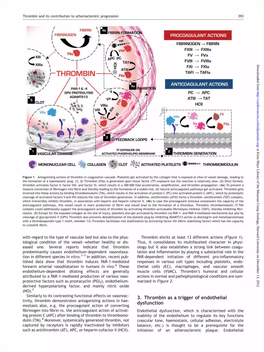
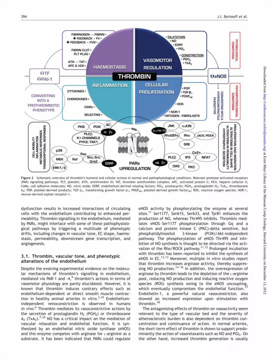
Thrombin elicits at least 13 different actions (Figure 1).Thus, it consolidates its multifaceted character in physi-ology but it also establishes a strong link between coagu-lation and inflammation by playing a substantial role in thePAR-dependent initiation of different pro-inflammatoryresponses in various cell types including platelets, endo-thelial cells (EC), macrophages, and vascular smoothmuscle cells (VSMC). Thrombin’s humoral and cellularactions in normal and pathophysiological conditions are sum-marized in Figure 2.
3. Thrombin as a trigger of endothelialdysfunction
Endothelial dysfunction, which is characterized with theinability of the endothelium to regulate its key functions(vascular tone, haemostasis, cellular adhesion, electrolytebalance, etc.) is thought to be a prerequisite for theinitiation of an atherosclerotic plaque. Endothelial
Figure 1 Antagonizing actions of thrombin in coagulation cascade. Platelets get activated by the collagen that is exposed at sites of vessel damage, leading tothe formation of a haemostatic plug. (1, 2) Thrombin (FIIa) is generated upon tissue factor (TF) exposure but the reaction is relatively slow. (3) Once formed,thrombin activates factor V, factor VIII, and factor XI, which results in a 300 000-fold acceleration, amplification, and thrombin propagation. (4a) To prevent amassive conversion of fibrinogen into fibrin and thereby leading to the formation of a stable clot, all natural anticoagulant pathways get activated. Thrombin getsinvolved into these actions by binding thrombomodulin (TM), which results in the activation of protein C (PC) into activated protein C (APC), which by proteolyticcleavage of activated factors V and VIII reduces the rate of thrombin generation. In addition, antithrombin (ATIII) forms a thrombin–antithrombin (TAT) complex,which irreversibly inhibits thrombin, in association with heparin and heparin cofactor II. (4b) In case the procoagulant stimulus overpowers the capacity of theanticoagulant pathways, this would result in more production of fibrin and would lead to the formation of a thrombus. Thrombin–thrombomodulin (T-TM)complex could additionally support the procoagulant actions of thrombin by activating thrombin-activatable fibrinolysis inhibitor (TAFI), thereby inhibiting fibri-nolysis. (5) Except for the exposed collagen at the site of injury, platelets also get activated by thrombin via PAR-1- and PAR-4-mediated mechanisms but also bycleavage of glycoprotein V (GPV).Thrombin also prevents destabilization of the platelet plug by inhibiting ADAMTS13 action (a disintegrin and metalloproteinasewith a thrombospondin type 1 motif, member 13).Thrombin facilitates clot stabilization by activating factor XIII (fibrin stabilizing factor) which has the capacityto crosslink fibrin.
Thrombin and its contribution to atherosclerotic progression 393
at Ernst M
ayr Library of the M
useum C
omp Z
oology, Harvard U
niversity on Decem
ber 13, 2012http://cardiovascres.oxfordjournals.org/
Dow
nloaded from
dysfunction results in increased interactions of circulatingcells with the endothelium contributing to enhanced per-meability. Thrombin signalling in the endothelium, mediatedby PARs, might interlace with some of these pathophysiolo-gical pathways by triggering a multitude of phenotypicdrifts, including changes in vascular tone, EC shape, haemo-stasis, permeability, downstream gene transcription, andangiogenesis.
3.1. Thrombin, vascular tone, and phenotypicalterations of the endothelium
Despite the evolving experimental evidence on the molecu-lar mechanisms of thrombin’s signalling in endothelium,mediated via PAR-1 and -4,9 thrombin’s actions in terms ofvasomotor physiology are partly elucidated. However, it isknown that thrombin induces contrary effects such asendothelium-dependent or direct smooth muscle contrac-tion in healthy animal arteries in vitro.3,10 Endothelium-independent venoconstriction is observed in humansin vivo.6 Thrombin mediates its vasoconstrictive actions bythe secretion of prostaglandin H2 (PGH2) or thromboxaneA2 (TxA2).
7,10 NO has a critical impact on the mediation ofvascular relaxation and endothelial function. It is syn-thesized by an endothelial nitric oxide synthase (eNOS)and this enzyme competes with arginase for L-arginine as asubstrate. It has been indicated that PARs could regulate
eNOS activity by phosphorylating the enzyme at severalsites.11 Ser1177, Ser615, Ser633, and Tyr81 enhances theproduction of NO, whereas Thr495 inhibits. Thrombin med-iates eNOS–Ser1177 phosphorylation through Gq and acalcium and protein kinase C (PKC)-delta sensitive, butphosphatidylinositol 3-kinase (PI3K)/Akt-independentpathway. The phosphorylation of eNOS–Thr495 and inhi-bition of NO synthesis is thought to be directed via the acti-vation of the Rho/ROCK pathway.11,12 Prolonged incubationwith thrombin has been reported to inhibit the synthesis ofeNOS in EC.12,13 Moreover, multiple in vitro studies reportthat thrombin increases arginase activity, thereby suppres-sing NO production.14–16 In addition, the overexpression ofarginase by thrombin leads to the depletion of the L-argininepool, reducing NO production and inducing reactive oxygenspecies (ROS) synthesis owing to the eNOS uncoupling,which eventually compromises the endothelial function.17
Endothelin-1, a powerful natural vasoconstrictor, alsoshowed an increased expression upon stimulation withthrombin.18
The antagonizing effects of thrombin on vasoactivity seemrelevant to the type of vascular bed and the severity ofatherosclerotic burden is also dependent on thrombin con-centration and continuance of action. In normal arteries,the short-term effect of thrombin is shown to support predo-minantly the action of vasorelaxants such as NO and PGI2. Onthe other hand, increased thrombin generation is usually
Figure 2 Schematic overview of thrombin’s humoral and cellular actions in normal and pathophysiological conditions. Relevant protease-activated receptors(PAR) signalling pathways. PLT, platelet; ATIII, antithrombin III; TAT, thrombin–antithrombin complex; APC, activated protein C; HCII, heparin cofactor II;CAMs, cell adhesive molecules; NO, nitric oxide; EDRF, endothelium-derived relaxing factors; PGI2, prostacyclin; PGH2, prostaglandin H2; TxA2, thromboxaneA2; PDP, platelet-derived products; TGF-b1, transforming growth factor-b1; PDGFAB, platelet-derived growth factorAB; ROS, reactive oxygen species; NOR-1,neuron-derived orphan receptor-1.
J.I. Borissoff et al.394
at Ernst M
ayr Library of the M
useum C
omp Z
oology, Harvard U
niversity on Decem
ber 13, 2012http://cardiovascres.oxfordjournals.org/
Dow
nloaded from
concentrated at the sites of vascular injury or within formedthrombus in vivo, 19 but also in patients with advanced CVDor suffering acute coronary syndromes.20 In vascular lesionsthrombin promotes a pro-inflammatory response, character-ized by increased production of diverse chemokines andcytokines, cell adhesion molecules (CAMs), enhancedvascular permeability, VSMC migration and proliferation,wall thickening and vasoconstriction.7 This might be aresult of the combination of a diminished TM and endothelialprotein C receptor (EPCR) capacity coupled to an overex-pression of PAR-1 and PAR-2 receptors in vascularlesions.21–23 Various mechanisms have been reportedlinked to PARs upregulation. First, thrombin-induced acti-vation of PAR-1 in cultured human EC in vitro upregulatesPAR-1 gene expression by signalling via Gi1/2 coupled toSrc and PI-3K, thus inducing the downstream Ras/MAPKpathway.24 Selective augmentation of PAR-2 and -4 geneexpression is indicated upon treatment with inflammatorystimuli such as interleukin (IL)-1a, (IL)-1b, tumour necrosisfactor (TNF)-a, and lipopolysaccharide (LPS).25,26 Finally,high shear stress, also characterized by reduced expressionof various atherogenesis-related genes, inhibits PAR-1expression in human EC in vitro.27 Thus, the alterations inthe vascular tone and the degree of expression of PARs inthe vessel wall might have additional impact on thepotency of thrombin’s cell signalling activity and the pro-gression of atherosclerotic disease.
3.2. Impairing the barrier function and otherthrombin-mediated effects on the endothelium
Rabiet et al.28 proposed a mechanism in which thrombinstimulates the intracellular accumulation of Ca2þ, consecu-tively activating the PKC pathway, and causing eventual dis-ruption of (VE)–cadherin–catenin complexes at the EC-celljunctions. Further in vitro studies consolidated the partici-pation of PKC in this pathophysiological process.29 Moreover,Nobe et al.30 suggested that thrombin-induced endothelialbarrier impairment is a biphasic process in which the Rho/Rho kinase pathway is also involved leading to rearrange-ment of actin stress fibres. A recent study elicits a newmechanism which gives input to a better comprehension ofthe thrombin-induced endothelial gap formation and per-meability. It was proposed that thrombin activates metallo-protease ADAM10, which mediates VE–cadherin proteolysisby specifically cleaving its ectodomain.31
Thrombin could also promote the generation of endothelialmicroparticles (MPs) via ROCK-II activation.32 Increase levelsof endothelial MPs have been correlated with the morphologyand severity of stenosis in patients with CVD.33
3.3. Thrombin-induced oxidative stress
Aside from the induction of pro-inflammatory responses,elevated ROS levels are presumably associated with the pro-motion of endothelial dysfunction, combined most likelywith diminished NO bioavailability. The majority of riskfactors of atherosclerosis positively correlate with anenhanced ROS synthesis, which tends to initiate multiplepro-atherogenic effects.34 ROS are implicated in cellular sig-nalling mechanisms, such as gene expression, proliferation,migration or apoptosis. Several reports indicate the poten-tiating effect of thrombin on ROS production in humanVSMCs35,36 and platelets.37 Different enzymatic systems
take part in the production of ROS in the vasculature, suchas xanthine oxidase, nicotinamide adenine dinucleotidephosphate (NADPH) oxidases, and NOS. Nevertheless,NADPH oxidases have been indicated as a major source ofsuperoxide in vascular cells and myocytes.38 The importanceof NADPH oxidases in thrombin-induced ROS synthesis wasstudied by the depletion of p22phox subunit, which sup-pressed ROS formation in VSMCs.36,39 Thrombin also triggersthe activation of p38 mitogen-activated protein kinases(MAPK) in a NADPH oxidase-dependent manner,35,40 whichestablishes a link between thrombin and the MAPK/ERKpathway, suggesting that it is also indirectly involved in pro-cesses like cell differentiation, cell survival, and apoptosis.Djordjevic et al.41 demonstrated that thrombin induceselevated ROS production in EC in vitro by activating p38MAPK and PI3K/Akt, inducing enhanced proliferation.
Intriguingly, thrombin induces its PAR-1 de novore-expression via Src-dependent mechanism, including Gproteins, PI3K, p38 MAPK, suggesting that redox pathwaysare also implicated in the regulation of PAR-1 expression.24
The latter was consolidated by two reports indicating thattreating VSMCs with either flavin inhibitor diphenyleneiodo-nium or antioxidants prevents PAR-1 upregulation uponstimulation by cyclic strain or oxidative agents.42,43
Hawkins et al.44 indicated a thrombin-induced mechanism,causing the production of mitochondrial-derived superoxide(mROS), which is an outcome of a Ca2þ mobilization via ino-sitol (1,4,5)-trisphosphate receptor (InsP3R), leading to asubsequent mitochondrial uptake of Ca2þ, triggering mROSexpression and nuclear factor-kappa B (NF-kB) pathway sig-nalling, which strongly promotes the overexpression ofintercellular cell adhesion molecule (ICAM)-1 and theadhesion of leucocytes to the vascular endothelium.
4. Thrombin in the early stage ofatherosclerotic plaque formation
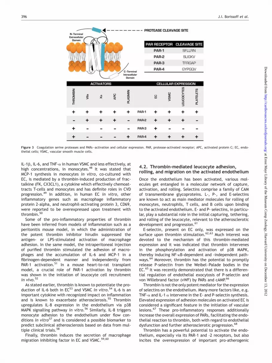
Although several more coagulation serine proteases couldfunction as activators of PARs by cleaving the N-terminalextracellular domain (Figure 3) abundant in vitro exper-imental data suggest that thrombin is a critical mediatorin the coagulation, inflammation, vessel wall crosstalk.Thrombin enhances ROS production in the arterial vesselwall facilitating lipid peroxidation and apoptotic processes.Thrombin also induces a plethora of pro-inflammatorymediators, causing alterations in gene transcription ofIL-6, IL-8, monocyte chemoattractant protein 1 (MCP-1,CCL2), vascular cell adhesion molecule (VCAM)-1, andICAM-1, etc., facilitating the recruitment of blood circulat-ing monocytes into the arterial vessel wall and encouragesearly plaque formation. Its signalling mechanisms with apro-atherogenic impact on the arterial vessel wall aremostly established via PARs.45
4.1. Thrombin-induced pro-inflammatoryresponses in blood and vascular wall
Thrombin participates in the selective recruitment of mono-cytes and T-cells into the vessel wall by inducing the syn-thesis of MCP-1 in EC and monocytes.46 MCP-1 is awell-characterized chemokine which is abundant in humanmacrophage-rich atherosclerotic plaques.47 Thrombin hasbeen shown to augment mRNA levels encoding for MCP-1,
Thrombin and its contribution to atherosclerotic progression 395
at Ernst M
ayr Library of the M
useum C
omp Z
oology, Harvard U
niversity on Decem
ber 13, 2012http://cardiovascres.oxfordjournals.org/
Dow
nloaded from
IL-1b, IL-6, and TNF-a in human VSMC and less effectively, athigh concentrations, in monocytes.48 It was stated thatMCP-1 synthesis in monocytes in vitro, co-cultured withEC, is mediated by a thrombin-induced production of frac-talkine (FK, CX3CL1), a cytokine which effectively chemoat-tracts T-cells and monocytes and has definite roles in CVDprogression.49 In addition, in human EC in vitro, otherinflammatory genes such as macrophage inflammatoryprotein 2-alpha, and neutrophil-activating protein 3, CD69,were reported to be overexpressed upon treatment withthrombin.50
Some of the pro-inflammatory properties of thrombinhave been inferred from models of inflammation such as aperitonitis mouse model, in which the administration ofthe potent thrombin inhibitor hirudin suppressed theantigen- or LPS-stimulated activation of macrophageadhesion. In the same model, the intraperitoneal injectionof purified thrombin stimulated the adhesion of macro-phages and the accumulation of IL-6 and MCP-1 in afibrinogen-dependent manner and independently fromPAR-1 activation.51 In a mouse heart-to-rat transplantmodel, a crucial role of PAR-1 activation by thrombinwas shown in the initiation of leucocyte cell recruitmentin vivo.52
As stated earlier, thrombin is known to potentiate the pro-duction of IL-6 both in EC53 and VSMC in vitro.54 IL-6 is animportant cytokine with recognized impact on inflammationand is known to exacerbate atherosclerosis.55 Thrombinupregulates IL-8 expression in the endothelium via p38MAPK signalling pathway in vitro.56 Similarly, IL-8 triggersmonocyte adhesion to the endothelium under flow con-ditions in vitro57 and is considered a possible biomarker topredict subclinical atherosclerosis based on data from mul-tiple clinical trials.58
Finally, thrombin induces the secretion of macrophagemigration inhibiting factor in EC and VSMC.59,60
4.2. Thrombin-mediated leucocyte adhesion,rolling, and migration on the activated endothelium
Once the endothelium has been activated, various mol-ecules get entangled in a molecular network of capture,activation, and rolling. Selectins comprise a family of CAMof transmembrane glycoproteins. L-, P-, and E-selectinsare known to act as main mediator molecules for rolling ofmonocytes, neutrophils, T cells, and B cells upon bindingto the activated endothelium. E- and P- selectins, in particu-lar, play a substantial role in the initial capturing, tethering,and rolling of the leucocyte, relevant to the atheroscleroticdevelopment and progression.61
E-selectin, present on EC only, was expressed on thesurface upon thrombin stimulation.62,63 Much interest wasdevoted to the mechanism of this thrombin-mediatedexpression and it was indicated that thrombin intervenesin the phosphorylation and activation of p38 MAPK,thereby inducing NF-kB-dependent and -independent path-ways.64 Moreover, thrombin has the potential to promptlyrelease P-selectin from the Weibel–Palade bodies in theEC.65 It was recently demonstrated that there is a differen-tial regulation of endothelial exocytosis of P-selectin andvon Willebrand factor (vWF) by PARs and cAMP.66
Thrombin is not the only potent mediator for the expressionof selectins on the endothelium. Many more factors like, e.g.TNF-a and IL-1 a intervene in the E- and P-selectin synthesis.Elevated expression of adhesion molecules on activated EC isconsidered a significant feature in the initiation of vascularlesions.67 These pro-inflammatory responses additionallyincrease the overall expression of PARs, facilitating the endo-thelial reaction to thrombin, both with regard to endothelialdysfunction and further atherosclerotic progression.68
Thrombin has a powerful potential to activate the endo-thelium, especially via its PAR-1 and -2 receptors, but alsoincites the overexpression of important pro-atherogenic
Figure 3 Coagulation serine proteases and PARs—activation and cellular expression. PAR, protease-activated receptor; APC, activated protein C; EC, endo-thelial cells; VSMC, vascular smooth muscle cells.
J.I. Borissoff et al.396
at Ernst M
ayr Library of the M
useum C
omp Z
oology, Harvard U
niversity on Decem
ber 13, 2012http://cardiovascres.oxfordjournals.org/
Dow
nloaded from
immunoglobulin superfamily molecules such as ICAM-1 andVCAM-1.50,69,70 Rolling activated leucocytes are exposed tothe influence of various chemoattractants, mediated bydiverse integrins, and captured to cell adhesion glyco-proteins. This eventually leads to the so called ‘leucocytearrest’.
Thrombin enhances VCAM- and ICAM-1 synthesis in culturedhuman EC. NF-kB- and GATA-dependency was observed withregard to VCAM-1 expression.71 Other in vitro studies indi-cated that PKC-d and RhoA/ROCK activation independentlylead to thrombin-induced NF-kB-dependent ICAM-1 upregula-tion.72,73 Moreover, the inhibition of both c-Jun N-terminalkinase (JNK) and NF-kB pathways showed additive inhibitoryeffect on ICAM-1 expression on the endothelium and high-lighted a significant role for JNK signalling.74
The actual process of transmigration of leucocytes usuallyoccurs on activated endothelial regions thus facilitating theleucocytes to pass through. Thrombin seems to interlace byincreasing the release of Ca2þ from the intracellularstores,75,76 favouring the ligation of ICAM-1, activating Rhofamily GTPases,77,78 which increases the myosin contracti-lity of EC impairing the inter-endothelial junctions by dis-rupting VE–cadherin complexes.79
4.3. Thrombin and monocytes/macrophagesin atherosclerosis
The effects of thrombin on monocytes and monocyte-derived macrophages during atherosclerotic progressionremain less elucidated compared with other blood cellssuch as platelets. Initially, it was indicated that VSMC maybe more sensitive to thrombin activation than monocytesand macrophages in vitro, the latter needing much higherconcentrations of thrombin to achieve increased IL-6,IL-1b, MCP-1, or TNF-a mRNA expression.48 Human mono-cytes, macrophages, and dendritic cells in vitro expressPARs. PAR-1 was expressed in all cell types, whereas PAR-3mRNA was less detected in monocytes and macrophages.PAR-1, -2, and -3 levels were upregulated upon thrombintreatment subsequently inducing MCP-1 expression. IL-4downregulated PAR-1, -2, and -3 expression in dendriticcells derived from monocytes by granulocyte–macrophagecolony-stimulating factor (GM-CSF).80 Li et al. found PAR-4protein expression on monocytes, though they failed todetect PAR-4 transcripts. They also showed that IL-6 wasreleased upon treatment with agonist peptides of PAR-1and PAR-4, but not of PAR-3 which was associated withPAR-3 incapability of mediating transmembrane signalling.81
Finally, there are multiple pro-inflammatory effects ofthrombin on other cell types which indirectly inducepro-atherogenic reactions in monocytes (as discussed inthe text).
5. Thrombin in the advanced stageof atherosclerosis
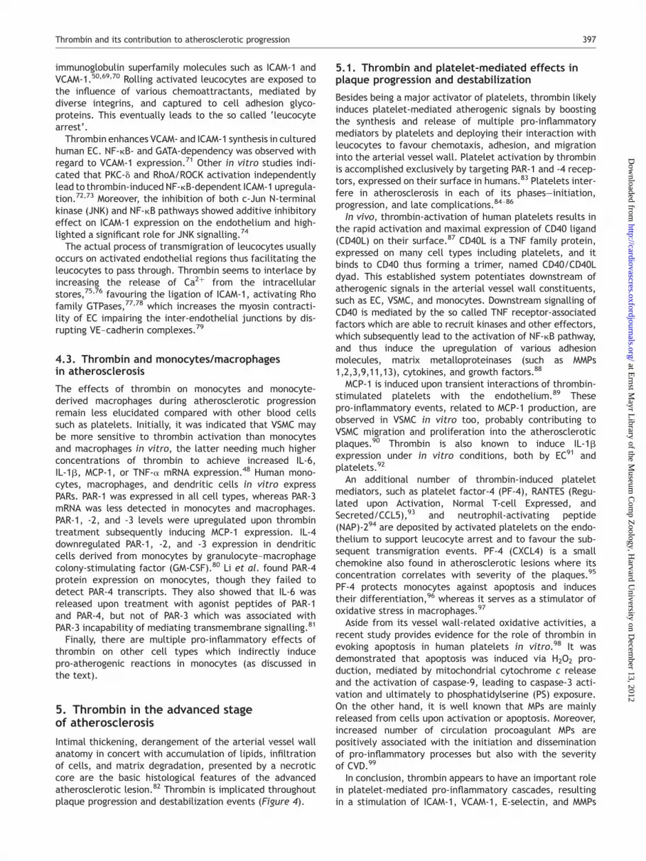
Intimal thickening, derangement of the arterial vessel wallanatomy in concert with accumulation of lipids, infiltrationof cells, and matrix degradation, presented by a necroticcore are the basic histological features of the advancedatherosclerotic lesion.82 Thrombin is implicated throughoutplaque progression and destabilization events (Figure 4).
5.1. Thrombin and platelet-mediated effects inplaque progression and destabilization
Besides being a major activator of platelets, thrombin likelyinduces platelet-mediated atherogenic signals by boostingthe synthesis and release of multiple pro-inflammatorymediators by platelets and deploying their interaction withleucocytes to favour chemotaxis, adhesion, and migrationinto the arterial vessel wall. Platelet activation by thrombinis accomplished exclusively by targeting PAR-1 and -4 recep-tors, expressed on their surface in humans.83 Platelets inter-fere in atherosclerosis in each of its phases—initiation,progression, and late complications.84–86
In vivo, thrombin-activation of human platelets results inthe rapid activation and maximal expression of CD40 ligand(CD40L) on their surface.87 CD40L is a TNF family protein,expressed on many cell types including platelets, and itbinds to CD40 thus forming a trimer, named CD40/CD40Ldyad. This established system potentiates downstream ofatherogenic signals in the arterial vessel wall constituents,such as EC, VSMC, and monocytes. Downstream signalling ofCD40 is mediated by the so called TNF receptor-associatedfactors which are able to recruit kinases and other effectors,which subsequently lead to the activation of NF-kB pathway,and thus induce the upregulation of various adhesionmolecules, matrix metalloproteinases (such as MMPs1,2,3,9,11,13), cytokines, and growth factors.88
MCP-1 is induced upon transient interactions of thrombin-stimulated platelets with the endothelium.89 Thesepro-inflammatory events, related to MCP-1 production, areobserved in VSMC in vitro too, probably contributing toVSMC migration and proliferation into the atheroscleroticplaques.90 Thrombin is also known to induce IL-1bexpression under in vitro conditions, both by EC91 andplatelets.92
An additional number of thrombin-induced plateletmediators, such as platelet factor-4 (PF-4), RANTES (Regu-lated upon Activation, Normal T-cell Expressed, andSecreted/CCL5),93 and neutrophil-activating peptide(NAP)-294 are deposited by activated platelets on the endo-thelium to support leucocyte arrest and to favour the sub-sequent transmigration events. PF-4 (CXCL4) is a smallchemokine also found in atherosclerotic lesions where itsconcentration correlates with severity of the plaques.95
PF-4 protects monocytes against apoptosis and inducestheir differentiation,96 whereas it serves as a stimulator ofoxidative stress in macrophages.97
Aside from its vessel wall-related oxidative activities, arecent study provides evidence for the role of thrombin inevoking apoptosis in human platelets in vitro.98 It wasdemonstrated that apoptosis was induced via H2O2 pro-duction, mediated by mitochondrial cytochrome c releaseand the activation of caspase-9, leading to caspase-3 acti-vation and ultimately to phosphatidylserine (PS) exposure.On the other hand, it is well known that MPs are mainlyreleased from cells upon activation or apoptosis. Moreover,increased number of circulation procoagulant MPs arepositively associated with the initiation and disseminationof pro-inflammatory processes but also with the severityof CVD.99
In conclusion, thrombin appears to have an important rolein platelet-mediated pro-inflammatory cascades, resultingin a stimulation of ICAM-1, VCAM-1, E-selectin, and MMPs
Thrombin and its contribution to atherosclerotic progression 397
at Ernst M
ayr Library of the M
useum C
omp Z
oology, Harvard U
niversity on Decem
ber 13, 2012http://cardiovascres.oxfordjournals.org/
Dow
nloaded from
production, all processes that contribute to plaque pro-gression, subsequent destabilization, and rupture.88,100–102
5.2. Thrombin and VSMC migration andproliferation
Besides its functions in the regulation of vascular tone,thrombin mediates migration, proliferation, and hypertro-phy of VSMC. VSMC are known to express PAR-1, -2, and -4thus potentiating the effect of thrombin in the activationof VSMC proliferation and migration.103 Multiple studiesreport on situations associated with changes in theexpression of PARs in VSMC. We have to take into consider-ation that the upregulation of these receptors might be ascrucial as the direct effect of thrombin alone, because ofthe fact that they are the main mediators for its furtheractions. Hence, an upregulation of PAR-1 in human and rat
VSMC in vivo is demonstrated upon the release of multipleplatelet-derived products (PDP) such as transforminggrowth factor (TGF)-b1, platelet-derived growth factorAB
(PDGFAB) and to a lesser extent, serotonin.104 Thus a long-term generation of new thrombin receptors at sites of vascu-lar injury might consolidate that thrombin amplifies itspro-atherogenic actions throughout the development of avascular lesion. Moreover, PAR-1 expression seems respon-sive to physical stress in both human and rat aortic VSMCsin vitro—being enhanced when cyclic strain is applied43
and being inhibited upon stimulation with high shearstress.105 This substantiates the idea that VSMC requiresphysical stimulation (flow or strain) in order to maintainvessel wall homeostasis, and perturbation of this processmay be involved in atherosclerosis where an overexpressionof PAR-1 and PAR-2 receptors has been demonstrated.21–23
Figure 4 Proposed mechanism for thrombin-induced atherogenesis. All known thrombin-induced pro-atherogenic actions are depicted in a consecutive way,showing its impact throughout the different stages of atherosclerotic development. Square with inverted ‘V’ indicates activation; encircled plus symbol indicatesinduction; upward arrow indicates elevated levels; MCP-1, monocyte chemoattractant protein-1; PDGF, platelet-derived growth factor; EDN-1, endothelin-1gene; ECE-1, endothelin converting enzyme-1 gene; COX-2, cyclooxygenase-2; MIF, migration inhibiting factor; ADAM10, A Disintegrin And Metalloproteinaseprotein-10; ROS, reactive oxygen species; mROS, mitochondrial-derived reactive oxygen species; IL, interleukin; TNF-a, tumour necrosis factor-a; Mo, monocyte;NO, nitric oxide; ICAM-1, intercellular cell adhesion molecule-1; VCAM-1, vascular cell adhesion molecule-1; MPs, microparticles; CD40L, CD40 Ligand; MMP,matrix metalloproteinases; PF-4, platelet factor-4; RANTES, Regulated upon Activation, Normal T-Cell Expressed, and Secreted; NAP-2, neutrophil-activatingpeptide-2; NOR-1, neuron-derived orphan receptor-1; VEGF, vascular endothelial growth factor; PARs, protease-activated receptors; TF, tissue factor; PAI-1, plas-minogen activator inhibitor-1.
J.I. Borissoff et al.398
at Ernst M
ayr Library of the M
useum C
omp Z
oology, Harvard U
niversity on Decem
ber 13, 2012http://cardiovascres.oxfordjournals.org/
Dow
nloaded from

Wang et al. studied thrombin-induced VSMC migration incultured VSMC and demonstrated that the process isp38-MAPK-mediated upon the generation of ROS. Maruyamaet al.106 indicated that thrombin-induced proliferation incultured human VSMC is regulated by NF-kB. VSMC prolifer-ation appears to be regulated by neuron-derived orphanreceptor-1 (NOR-1), a transcription factor overexpressed inhuman atherosclerotic plaques upon stimulation withthrombin.107
Finally, the regulation of PDGF in the endothelium alsoappears to be linked to thrombin. PDGF is related to ather-osclerosis for its properties to stimulate VSMC migration andproliferation. PDGF levels rise upon treatment with throm-bin of human umbilical vein EC, together with monocytetransmigration and E-selectin expression.108
5.3. Thrombin and its pro-angiogenic responses
Neoangiogenesis is closely associated with plaque pro-gression. Intraplaque haemorrhage is currently considereda critical factor for plaque destabilization and is predomi-nantly attributed to the neovascularization of the intimaand media by disorganized and immature ‘leaky’ microves-sels.109 Thrombin promotes angiogenesis both in vitro andin vivo.110 It is indicated that it reduces the ability of ECto affix to their anchorage on the basement membrane,thereby promoting early angiogenic events.111 Furthermore,it has been stated that thrombin increases the mRNA andprotein levels of anb3-integrin in a concentration-dependentmanner in EC.112 anb3-integrin is a known angiogenic markerin vascular tissue and it directly interacts with thrombin,thereby facilitating EC attachment, migration, and survival.anb3-integrin also mediates progelatinase A (MMP-2) acti-vation. Stimulation with thrombin has shown the inductionof MMP-2 release in both human EC113 and rat aorta in adose-dependent mode in vitro.114 In addition, thrombin aug-ments the expression of vascular endothelial growth factor(VEGF) and angiopoietin-2 via PAR-1-mediated mechan-ism.115,116 Finally, various studies indicate a relevant rolefor hypoxia-inducible factor-1a signalling pathway in thethrombin-induced VEGF gene expression and angiogenesis.
6. Thrombin and atherosclerosis—in vivoanimal studies
Despite the wealth of existing data on thrombin’spro-atherogenic actions in vitro, we should point out thatmany of these studies have been carried out with cell cul-tures and purified thrombin, in the absence of receptorsand inhibitors, such that the relevance of any of these out-comes may be debated. However, the critical role of throm-bin in atherogenesis is supported by recent in vivo studies.
Indirect evidence shows that heterozygous tissue factorpathway inhibitor (TFPI)-deficient ApoE2/2 mice exhibiteda significantly greater atherosclerotic burden comparedwith TFPI wild-type genotype.117 TFPI is a potent inhibitorof TF-mediated thrombin generation.
Direct evidence for the involvement of thrombin comesfrom experiments in which the administration of the directthrombin inhibitor melagatran to ApoE2/2 mice reducedlesion progression in brachiocephalic arteries. Total lesionarea was significantly decreased in melagatran-treatedanimals. Thrombin inhibition also contributed to plaque
stability (significant increase of immunohistochemical stain-ing against VSMC a-actin), characterized by thicker fibrouscaps, increased media thickness, smaller necrotic cores,and a significant decrease of staining against MMP-9.118
MMP-9 is considered an important catalyser of plaquerupture.
Finally, in a study employing transgenic double knock-outmice, deficient for HCII, a natural thrombin inhibitor, on aApoE2/2 background, HCII deficiency was associated withapproximately 64% larger total plaque area and increasedneointimal formation than in wild-type mice. In support ofthese findings, the administration of dermatan sulfate,which potentiates the inhibitory function of HCII about10 000-fold, showed a HCII-dependent antiproliferativeeffect in wild-type animals.119
7. Clinical studies
Thrombin’s impact on atherosclerotic development is a rela-tively novel topic to investigate and no specific clinical trialshave been conducted yet. However, several reportsindirectly demonstrate its importance with regard to CVDprogression.
Aihara et al.120 found a negative correlation betweenplasma HCII activity and ultrasound imaged plaque thicknessof the carotid arteries in 306 elderly Japanese patients andsuggested that HCII inhibits atherogenesis, thereby alsoshowing a possible indirect link between higher thrombingeneration and atherosclerosis progression.
Moreover, various thrombotic markers measured upon pro-gressive CVD, indicate an indirect link for thrombin andatherosclerosis. The Cardiovascular Health Study (CHS)showed that prothrombin fragments F1-2 (F1-2) and fibrino-peptide A measured in 5201 individuals (399 free of CVD),which are markers for thrombin generation in vivo, corre-lated with various CVD risk factors such as triglycerides, C-reactive protein, low ankle-brachial pressure index (ABPI),etc.121 F1-2 plasma levels were also independently associ-ated with carotid intima-media thickness in a populationof 181 middle-aged adults, free of clinically overt athero-sclerosis.122 Moreover, Nylaende et al. studied the relation-ship of prothrombotic activity and the severity of peripheralarterial occlusive disease (PAD). Multiple haemostaticmarkers such as vWF, soluble TM, soluble TF, TAT complex,and D-dimer were determined in a cross-sectional study of127 patients, diagnosed with PAD. Plasma levels ofD-dimer, TAT complex, and fibrinogen significantly corre-lated with the severity of atherosclerotic burden, evaluatedby maximum treadmill walking distance and ABPI.123 Arecent meta-analysis of 191 studies, investigating sevencommon haemostatic gene polymorphisms in CVD, indicatedthat the 1691A variant of the factor V gene and 20210Avariant of the prothrombin gene, both of which promotethrombin generation in blood, might be associated withthe risk of CAD.124 Moreover, it was recently shown thatlong after acute myocardial infarction, patients generatehigher, earlier, and faster thrombin in comparison withchronic CAD patients.125 This strengthens the concept of vul-nerable atherosclerotic plaques contributing to the propa-gation of thrombin generation, thereby leading toaggravation of CVD.
Several more indirect cross-relations might be of interestin this context. Numerous clinical trials postulate that
Thrombin and its contribution to atherosclerotic progression 399
at Ernst M
ayr Library of the M
useum C
omp Z
oology, Harvard U
niversity on Decem
ber 13, 2012http://cardiovascres.oxfordjournals.org/
Dow
nloaded from
haemostatic factors such as fibrinogen, C-reactive protein,plasminogen activator inhibitor-1 (PAI-1) are risk factorsfor CVD progression.126 A recent study associated the pro-gression of symptomatic intracranial large artery athero-sclerosis with a pro-inflammatory state and impairedfibrinolysis, characterized with elevated concentrations ofthe endogenous fibrinolysis inhibitor PAI-1.127 Despite thefact that thrombin is not a sole mediator of PAI-1 itinduces its expression together with TF128,129 in ECin vitro. TF and PAI-1 are already recognized for theirpro-inflammatory features. In addition, many studiesdemonstrate a relationship between elevated PAI-1 levelsand the development of atherosclerosis, not only systemi-cally but also locally.130
Leucocytosis, and high neutrophil count in particular, mayrepresent another intriguing mechanism for enhancingchronic atherosclerosis via maintaining a hypercoagulablestate in CVD patients.131 Neutrophils are a pivotal linkbetween inflammation and coagulation. They produce mul-tiple procoagulant factors and are able to release diversematrix-destabilizing enzymes (elastase, cathepsin G),which easily activate the coagulation system.132 They con-tribute to the liberation of TF-laden MPs into the bloodstream upon stimulation with cytokines and consequentplatelet adhesion via P-selectin.132 This seems anotherpotential mechanism for a continuous thrombin generationin vivo, facilitating the amplification of thrombin’spro-atherogenic features.
8. Summary and Perspectives
From histological studies an intense interaction betweencoagulation, inflammation, and the complex process ofatherosclerosis has emerged.133 Advanced atheroscleroticlesions show evidence of the presence of active coagulationproducts including fibrin and fibrin cleavage products.Hence, the presence of an active coagulation cascadewithin the arterial vessel wall seems likely and our recentimmunohistochemical data show that essentially all coagu-lation proteins are detectable in the atheroscleroticlesion.134 In the coagulation cascade we and others considerthe generation of thrombin as one of the key regulatingevents. In vivo, thrombin is thought to be continuously gen-erated as indicated by measurable quantities of F1-2 andTAT complexes in the plasma of normal individuals. Physio-logically, the generation of thrombin is the product of syn-thesis under influence of TF and inhibition by severalinhibitors including AT and HCII. The net amount of thrombinwill be determined by the rate of synthesis and inactivation,the localization (free or bound to surfaces), and its associ-ated binding to receptors including PARs and TM. Upon pro-gressive atherosclerosis, there is a diminution in the level ofTM at the endothelium,1 which impairs the anticoagulantaction of thrombin and the increased production of thrombinbecause of TF exposure allows interactions of thrombin withcomponents of the arterial vessel wall, including dysfunc-tional EC on both initial and advanced lesions and othercell types in ruptured (thrombotic) plaques.
The continuous generation of mostly procoagulant throm-bin may contribute to a vicious circle in the thrombin-induced atherogenesis process. As discussed, thrombinacts mostly via PARs, inducing multiple vascular pro-inflammatory reactions. The authors are aware that also
other coagulation proteases including factor VIIa, factorXa, and APC contain PAR-activation properties that mayinterfere with or add to the actions of thrombin. Therehas indeed been a public debate on the preference ofthrombin vis-a-vis APC in their binding to PAR-1 and thisdebate has not yet been settled.135 Atherosclerotic altera-tions in the vessel wall are known to increase the level ofexpressed PARs on the surface of most vessel wall constitu-ents.21–23 Thrombin-mediated pro-inflammatory events area powerful trigger for more thrombin formation, whichmay eventually amplify its contribution to further athero-sclerotic progression.
Finally, from a clinical perspective the introduction of anumber of selective oral anticoagulants that will also beaimed for long-term administration makes it of actualimportance to consider the effects and possible side-effectsof thrombin inhibition on the extent and nature of athero-sclerosis. Hopefully, thrombin inhibition is, as predictedfrom animal experiments, associated with a favourablechange in atherosclerosis phenotype. However, the typicalJanus face of many clotting proteases should warn againstovert enthusiasm and calls for prospective clinical studies.
Acknowledgements
S.H. participates in the European Vascular Genomics Network(http://www.evgn.org), a Network of Excellence supported by theEuropean Community’s Sixth Framework Program for Research Pri-ority 1 (Life Sciences, Genomics, and Biotechnology for Health; con-tract LSHM-CT-2003-503254).
Funding
EU Marie Curie Fellowship (MEST-CT-2005-020706 to J.I.B.).
Conflict of interest: none declared.
References1. Esmon CT. The interactions between inflammation and coagulation. Br J
Haematol 2005;131:417–430.2. Levi M, van der Poll T. Two-way interactions between inflammation and
coagulation. Trends Cardiovasc Med 2005;15:254–259.3. Ku DD, Zaleski JK. Receptor mechanism of thrombin-induced
endothelium-dependent and endothelium-independent coronary vascu-lar effects in dogs. J Cardiovasc Pharmacol 1993;22:609–616.
4. Mizuno O, Hirano K, Nishimura J, Kubo C, Kanaide H. Mechanism ofendothelium-dependent relaxation induced by thrombin in the pig cor-onary artery. Eur J Pharmacol 1998;351:67–77.
5. Hamilton JR, Cocks TM. Heterogeneous mechanisms of endothelium-dependent relaxation for thrombin and peptide activators ofprotease-activated receptor-1 in porcine isolated coronary artery. Br JPharmacol 2000;130:181–188.
6. Gudmundsdottir IJ, Lang NN, Boon NA, Ludlam CA, Webb DJ, Fox KAet al. Role of the endothelium in the vascular effects of the thrombinreceptor (protease-activated receptor type 1) in humans. J Am CollCardiol 2008;51:1749–1756.
7. Hirano K. The roles of proteinase-activated receptors in the vascularphysiology and pathophysiology. Arterioscler Thromb Vasc Biol 2007;27:27–36.
8. Lane DA, Philippou H, Huntington JA. Directing thrombin. Blood 2005;106:2605–2612.
9. Kataoka H, Hamilton JR, McKemy DD, Camerer E, Zheng YW, Cheng Aet al. Protease-activated receptors 1 and 4 mediate thrombin signalingin endothelial cells. Blood 2003;102:3224–3231.
10. Derkach DN, Ihara E, Hirano K, Nishimura J, Takahashi S, Kanaide H.Thrombin causes endothelium-dependent biphasic regulation of
J.I. Borissoff et al.400
at Ernst M
ayr Library of the M
useum C
omp Z
oology, Harvard U
niversity on Decem
ber 13, 2012http://cardiovascres.oxfordjournals.org/
Dow
nloaded from
vascular tone in the porcine renal interlobar artery. Br J Pharmacol2000;131:1635–1642.
11. Watts VL, Motley ED. Role of protease-activated receptor-1 in endo-thelial nitric oxide synthase-Thr495 phosphorylation. Exp Biol Med(Maywood) 2009;234:132–139.
12. Ming XF, Viswambharan H, Barandier C, Ruffieux J, Kaibuchi K, Rusconi Set al. Rho GTPase/Rho kinase negatively regulates endothelial nitricoxide synthase phosphorylation through the inhibition of proteinkinase B/Akt in human endothelial cells. Mol Cell Biol 2002;22:8467–8477.
13. Eto M, Barandier C, Rathgeb L, Kozai T, Joch H, Yang Z et al. Thrombinsuppresses endothelial nitric oxide synthase and upregulatesendothelin-converting enzyme-1 expression by distinct pathways: roleof Rho/ROCK and mitogen-activated protein kinase. Circ Res 2001;89:583–590.
14. Ming XF, Barandier C, Viswambharan H, Kwak BR, Mach F, Mazzolai Let al. Thrombin stimulates human endothelial arginase enzymaticactivity via RhoA/ROCK pathway: implications for atheroscleroticendothelial dysfunction. Circulation 2004;110:3708–3714.
15. Yang L, Lewis CM, Chandrasekharan UM, Kinney CM, Dicorleto PE,Kashyap VS. Arginase activity is increased by thrombin: a mechanismfor endothelial dysfunction in arterial thrombosis. J Am Coll Surg2006;203:817–826.
16. Lewis C, Zhu W, Pavkov ML, Kinney CM, Dicorleto PE, Kashyap VS. Argi-nase blockade lessens endothelial dysfunction after thrombosis. J VascSurg 2008;48:441–446.
17. Zhang C, Hein TW, Wang W, Miller MW, Fossum TW, McDonald MM et al.Upregulation of vascular arginase in hypertension decreases nitricoxide-mediated dilation of coronary arterioles. Hypertension 2004;44:935–943.
18. Delerive P, Martin-Nizard F, Chinetti G, Trottein F, Fruchart JC, Najib Jet al. Peroxisome proliferator-activated receptor activators inhibitthrombin-induced endothelin-1 production in human vascular endo-thelial cells by inhibiting the activator protein-1 signaling pathway.Circ Res 1999;85:394–402.
19. Hatton MW, Moar SL, Richardson M. Deendothelialization in vivoinitiates a thrombogenic reaction at the rabbit aorta surface. Corre-lation of uptake of fibrinogen and antithrombin III with thrombin gener-ation by the exposed subendothelium. Am J Pathol 1989;135:499–508.
20. Merlini PA, Bauer KA, Oltrona L, Ardissino D, Cattaneo M, Belli C et al.Persistent activation of coagulation mechanism in unstable angina andmyocardial infarction. Circulation 1994;90:61–68.
21. Nelken NA, Soifer SJ, O’Keefe J, Vu TK, Charo IF, Coughlin SR. Thrombinreceptor expression in normal and atherosclerotic human arteries.J Clin Invest 1992;90:1614–1621.
22. Wilcox JN, Rodriguez J, Subramanian R, Ollerenshaw J, Zhong C,Hayzer DJ et al. Characterization of thrombin receptor expressionduring vascular lesion formation. Circ Res 1994;75:1029–1038.
23. Ku DD, Dai J. Expression of thrombin receptors in human atheroscleroticcoronary arteries leads to an exaggerated vasoconstrictory response invitro. J Cardiovasc Pharmacol 1997;30:649–657.
24. Ellis CA, Malik AB, Gilchrist A, Hamm H, Sandoval R,Voyno-Yasenetskaya T et al. Thrombin induces proteinase-activatedreceptor-1 gene expression in endothelial cells via activation ofGi-linked Ras/mitogen-activated protein kinase pathway. J Biol Chem1999;274:13718–13727.
25. Nystedt S, Ramakrishnan V, Sundelin J. The proteinase-activated recep-tor 2 is induced by inflammatory mediators in human endothelial cells.Comparison with the thrombin receptor. J Biol Chem 1996;271:14910–14915.
26. Hamilton JR, Frauman AG, Cocks TM. Increased expression ofprotease-activated receptor-2 (PAR2) and PAR4 in human coronaryartery by inflammatory stimuli unveils endothelium-dependent relax-ations to PAR2 and PAR4 agonists. Circ Res 2001;89:92–98.
27. Nguyen KT, Eskin SG, Patterson C, Runge MS, McIntire LV. Shear stressreduces protease activated receptor-1 expression in human endothelialcells. Ann Biomed Eng 2001;29:145–152.
28. Rabiet MJ, Plantier JL, Rival Y, Genoux Y, Lampugnani MG, Dejana E.Thrombin-induced increase in endothelial permeability is associatedwith changes in cell-to-cell junction organization. Arterioscler ThrombVasc Biol 1996;16:488–496.
29. Vuong PT, Malik AB, Nagpala PG, Lum H. Protein kinase C beta modulatesthrombin-induced Ca2þ signaling and endothelial permeabilityincrease. J Cell Physiol 1998;175:379–387.
30. Nobe K, Sone T, Paul RJ, Honda K. Thrombin-induced force developmentin vascular endothelial cells: contribution to alteration of permeability
mediated by calcium-dependent and -independent pathways.J Pharmacol Sci 2005;99:252–263.
31. Schulz B, Pruessmeyer J, Maretzky T, Ludwig A, Blobel CP, Saftig P et al.ADAM10 regulates endothelial permeability and T-Cell transmigration byproteolysis of vascular endothelial cadherin. Circ Res 2008;102:1192–1201.
32. Sapet C, Simoncini S, Loriod B, Puthier D, Sampol J, Nguyen C et al.Thrombin-induced endothelial microparticle generation: identificationof a novel pathway involving ROCK-II activation by caspase-2. Blood2006;108:1868–1876.
33. Bernal-Mizrachi L, Jy W, Fierro C, Macdonough R, Velazques HA, Purow Jet al. Endothelial microparticles correlate with high-risk angiographiclesions in acute coronary syndromes. Int J Cardiol 2004;97:439–446.
34. Harrison D, Griendling KK, Landmesser U, Hornig B, Drexler H. Role ofoxidative stress in atherosclerosis. Am J Cardiol 2003;91:7A–11A.
35. Brandes RP, Viedt C, Nguyen K, Beer S, Kreuzer J, Busse R et al.Thrombin-induced MCP-1 expression involves activation of the p22phox-containing NADPH oxidase in human vascular smooth muscle cells.Thromb Haemost 2001;85:1104–1110.
36. Gorlach A, Diebold I, Schini-Kerth VB, Berchner-Pfannschmidt U, Roth U,Brandes RP et al. Thrombin activates the hypoxia-inducible factor-1 sig-naling pathway in vascular smooth muscle cells: Role of the p22(phox)-containing NADPH oxidase. Circ Res 2001;89:47–54.
37. Wachowicz B, Olas B, Zbikowska HM, Buczynski A. Generation of reac-tive oxygen species in blood platelets. Platelets 2002;13:175–182.
38. Griendling KK, Sorescu D, Ushio-Fukai M. NAD(P)H oxidase: role in cardi-ovascular biology and disease. Circ Res 2000;86:494–501.
39. Herkert O, Diebold I, Brandes RP, Hess J, Busse R, Gorlach A. NADPHoxidase mediates tissue factor-dependent surface procoagulant activityby thrombin in human vascular smooth muscle cells. Circulation 2002;105:2030–2036.
40. Kanda Y, Mizuno K, Kuroki Y, Watanabe Y. Thrombin-induced p38mitogen-activated protein kinase activation is mediated by epidermalgrowth factor receptor transactivation pathway. Br J Pharmacol 2001;132:1657–1664.
41. Djordjevic T, Pogrebniak A, BelAiba RS, Bonello S, Wotzlaw C, Acker Het al. The expression of the NADPH oxidase subunit p22phox is regulatedby a redox-sensitive pathway in endothelial cells. Free Radic Biol Med2005;38:616–630.
42. Li F, Baykal D, Horaist C, Yan CN, Carr BN, Rao GN et al. Cloning andidentification of regulatory sequences of the human thrombin receptorgene. J Biol Chem 1996;271:26320–26328.
43. Nguyen KT, Frye SR, Eskin SG, Patterson C, Runge MS, McIntire LV. Cyclicstrain increases protease-activated receptor-1 expression in vascularsmooth muscle cells. Hypertension 2001;38:1038–1043.
44. Hawkins BJ, Solt LA, Chowdhury I, Kazi AS, Abid MR, Aird WC et al. Gprotein-coupled receptor Ca2þ-linked mitochondrial reactive oxygenspecies are essential for endothelial/leukocyte adherence. Mol CellBiol 2007;27:7582–7593.
45. Martorell L, Martinez-Gonzalez J, Rodriguez C, Gentile M, Calvayrac O,Badimon L. Thrombin and protease-activated receptors (PARs) in ather-othrombosis. Thromb Haemost 2008;99:305–315.
46. Colotta F, Sciacca FL, Sironi M, Luini W, Rabiet MJ, Mantovani A.Expression of monocyte chemotactic protein-1 by monocytes and endo-thelial cells exposed to thrombin. Am J Pathol 1994;144:975–985.
47. Nelken NA, Coughlin SR, Gordon D, Wilcox JN. Monocyte chemoattrac-tant protein-1 in human atheromatous plaques. J Clin Invest 1991;88:1121–1127.
48. Kranzhofer R, Clinton SK, Ishii K, Coughlin SR, Fenton JW II, Libby P.Thrombin potently stimulates cytokine production in human vascularsmooth muscle cells but not in mononuclear phagocytes. Circ Res1996;79:286–294.
49. Popovic M, Laumonnier Y, Burysek L, Syrovets T, Simmet T. Thrombin-induced expression of endothelial CX3CL1 potentiates monocyte CCL2production and transendothelial migration. J Leukoc Biol 2008;84:215–223.
50. Okada M, Suzuki K, Takada K, Nakashima M, Nakanishi T, Shinohara T.Detection of up-regulated genes in thrombin-stimulated human umbili-cal vein endothelial cells. Thromb Res 2006;118:715–721.
51. Szaba FM, Smiley ST. Roles for thrombin and fibrin(ogen) in cytokine/chemokine production and macrophage adhesion in vivo. Blood 2002;99:1053–1059.
52. Chen D, Carpenter A, Abrahams J, Chambers RC, Lechler RI, McVey JHet al. Protease-activated receptor 1 activation is necessary for mono-cyte chemoattractant protein 1-dependent leukocyte recruitment invivo. J Exp Med 2008;205:1739–1746.
Thrombin and its contribution to atherosclerotic progression 401
at Ernst M
ayr Library of the M
useum C
omp Z
oology, Harvard U
niversity on Decem
ber 13, 2012http://cardiovascres.oxfordjournals.org/
Dow
nloaded from
53. Marin V, Montero-Julian FA, Gres S, Boulay V, Bongrand P, Farnarier Cet al. The IL-6-soluble IL-6Ralpha autocrine loop of endothelial acti-vation as an intermediate between acute and chronic inflammation:an experimental model involving thrombin. J Immunol 2001;167:3435–3442.
54. Tokunou T, Ichiki T, Takeda K, Funakoshi Y, Iino N, Shimokawa H et al.Thrombin induces interleukin-6 expression through the cAMP responseelement in vascular smooth muscle cells. Arterioscler Thromb VascBiol 2001;21:1759–1763.
55. Huber SA, Sakkinen P, Conze D, Hardin N, Tracy R. Interleukin-6 exacer-bates early atherosclerosis in mice. Arterioscler Thromb Vasc Biol 1999;19:2364–2367.
56. Marin V, Farnarier C, Gres S, Kaplanski S, Su MS, Dinarello CA et al. Thep38 mitogen-activated protein kinase pathway plays a critical role inthrombin-induced endothelial chemokine production and leukocyterecruitment. Blood 2001;98:667–673.
57. Gerszten RE, Garcia-Zepeda EA, Lim YC, Yoshida M, Ding HA,Gimbrone MA Jr et al. MCP-1 and IL-8 trigger firm adhesion of monocytesto vascular endothelium under flow conditions. Nature 1999;398:718–723.
58. Aukrust P, Halvorsen B, Yndestad A, Ueland T, Oie E, Otterdal K et al.Chemokines and cardiovascular risk. Arterioscler Thromb Vasc Biol2008;28:1909–1919.
59. Shimizu T, Nishihira J, Watanabe H, Abe R, Honda A, Ishibashi T et al.Macrophage migration inhibitory factor is induced by thrombin andfactor Xa in endothelial cells. J Biol Chem 2004;279:13729–13737.
60. Bernhagen J, Krohn R, Lue H, Gregory JL, Zernecke A, Koenen RR et al.MIF is a noncognate ligand of CXC chemokine receptors in inflammatoryand atherogenic cell recruitment. Nat Med 2007;13:587–596.
61. Dong ZM, Chapman SM, Brown AA, Frenette PS, Hynes RO, Wagner DD.The combined role of P- and E-selectins in atherosclerosis. J ClinInvest 1998;102:145–152.
62. Kaplanski G, Fabrigoule M, Boulay V, Dinarello CA, Bongrand P,Kaplanski S et al. Thrombin induces endothelial type II activationin vitro: IL-1 and TNF-alpha-independent IL-8 secretion and E-selectinexpression. J Immunol 1997;158:5435–5441.
63. Ostrovsky L, Carvalho-Tavares J, Woodman RC, Kubes P. Translationalinhibition of E-selectin expression stimulates P-selectin-dependentneutrophil recruitment. Am J Physiol Heart Circ Physiol 2000;278:H1225–H1232.
64. Kaur J, Woodman RC, Kubes P. P38 MAPK: critical molecule inthrombin-induced NF-kappa B-dependent leukocyte recruitment. Am JPhysiol Heart Circ Physiol 2003;284:H1095–H1103.
65. Sugama Y, Malik AB. Thrombin receptor 14-amino acid peptide mediatesendothelial hyperadhesivity and neutrophil adhesion by P-selectin-dependent mechanism. Circ Res 1992;71:1015–1019.
66. Cleator JH, Zhu WQ, Vaughan DE, Hamm HE. Differential regulation ofendothelial exocytosis of P-selectin and von Willebrand factor byprotease-activated receptors and cAMP. Blood 2006;107:2736–2744.
67. Davies MJ, Gordon JL, Gearing AJ, Pigott R, Woolf N, Katz D et al. Theexpression of the adhesion molecules ICAM-1, VCAM-1, PECAM, and E-selectin in human atherosclerosis. J Pathol 1993;171:223–229.
68. Braunersreuther V, Mach F. Leukocyte recruitment in atherosclerosis:potential targets for therapeutic approaches? Cell Mol Life Sci 2006;63:2079–2088.
69. Kaplanski G, Marin V, Fabrigoule M, Boulay V, Benoliel AM, Bongrand Pet al. Thrombin-activated human endothelial cells support monocyteadhesion in vitro following expression of intercellular adhesionmolecule-1 (ICAM-1; CD54) and vascular cell adhesion molecule-1(VCAM-1; CD106). Blood 1998;92:1259–1267.
70. Minami T, Sugiyama A, Wu SQ, Abid R, Kodama T, Aird WC. Thrombin andphenotypic modulation of the endothelium. Arterioscler Thromb VascBiol 2004;24:41–53.
71. Minami T, Abid MR, Zhang J, King G, Kodama T, Aird WC. Thrombinstimulation of vascular adhesion molecule-1 in endothelial cells ismediated by protein kinase C (PKC)-delta-NF-kappa B andPKC-zeta-GATA signaling pathways. J Biol Chem 2003;278:6976–6984.
72. Anwar KN, Fazal F, Malik AB, Rahman A. RhoA/Rho-associated kinasepathway selectively regulates thrombin-induced intercellular adhesionmolecule-1 expression in endothelial cells via activation of I kappa Bkinase beta and phosphorylation of RelA/p65. J Immunol 2004;173:6965–6972.
73. Bijli KM, Fazal F, Minhajuddin M, Rahman A. Activation of Syk by PKCdelta regulates thrombin-induced ICAM-1 expression in endothelialcells via tyrosine phosphorylation of RelA/p65. J Biol Chem 2008;283:14674–14684.
74. Miho N, Ishida T, Kuwaba N, Ishida M, Shimote-Abe K, Tabuchi K et al.Role of the JNK pathway in thrombin-induced ICAM-1 expression inendothelial cells. Cardiovasc Res 2005;68:289–298.
75. Sandoval R, Malik AB, Minshall RD, Kouklis P, Ellis CA, Tiruppathi C.Ca(2þ) signalling and PKCalpha activate increased endothelial per-meability by disassembly of VE-cadherin junctions. J Physiol 2001;533:433–445.
76. Vanhauwe JF, Thomas TO, Minshall RD, Tiruppathi C, Li A, Gilchrist Aet al. Thrombin receptors activate G(o) proteins in endothelial cellsto regulate intracellular calcium and cell shape changes. J Biol Chem2002;277:34143–34149.
77. Vouret-Craviari V, Bourcier C, Boulter E, van Obberghen-Schilling E. Dis-tinct signals via Rho GTPases and Src drive shape changes by thrombinand sphingosine-1-phosphate in endothelial cells. J Cell Sci 2002;115:2475–2484.
78. van Nieuw Amerongen GP, Beckers CM, Achekar ID, Zeeman S,Musters RJ, van Hinsbergh VW. Involvement of Rho kinase in endothelialbarrier maintenance. Arterioscler Thromb Vasc Biol 2007;27:2332–2339.
79. van Nieuw Amerongen GP, Musters RJ, Eringa EC, Sipkema P, vanHinsbergh VW. Thrombin-induced endothelial barrier disruption inintact microvessels. Am J Physiol Cell Physiol 2008;294:C1234–C1234.
80. Colognato R, Slupsky JR, Jendrach M, Burysek L, Syrovets T, Simmet T.Differential expression and regulation of protease-activated receptorsin human peripheral monocytes and monocyte-derived antigen-presenting cells. Blood 2003;102:2645–2652.
81. Li T, Wang H, He S. Induction of interleukin-6 release from monocytes byserine proteinases and its potential mechanisms. Scand J Immunol 2006;64:10–16.
82. Virmani R, Kolodgie FD, Burke AP, Farb A, Schwartz SM. Lessons fromsudden coronary death: a comprehensive morphological classificationscheme for atherosclerotic lesions. Arterioscler Thromb Vasc Biol2000;20:1262–1275.
83. Kahn ML, Zheng YW, Huang W, Bigornia V, Zeng D, Moff S et al. A dualthrombin receptor system for platelet activation. Nature 1998;394:690–694.
84. Massberg S, Brand K, Gruner S, Page S, Muller E, Muller I et al. A criticalrole of platelet adhesion in the initiation of atherosclerotic lesion for-mation. J Exp Med 2002;196:887–896.
85. Gawaz M. Platelets in the onset of atherosclerosis. Blood Cells Mol Dis2006;36:206–210.
86. May AE, Seizer P, Gawaz M. Platelets: inflammatory firebugs of vascularwalls. Arterioscler Thromb Vasc Biol 2008;28:s5–s10.
87. Henn V, Slupsky JR, Grafe M, Anagnostopoulos I, Forster R,Muller-Berghaus G et al. CD40 ligand on activated platelets triggers aninflammatory reaction of endothelial cells. Nature 1998;391:591–594.
88. Lutgens E, Lievens D, Beckers L, Donners M, Daemen M. CD40 and itsligand in atherosclerosis. Trends Cardiovasc Med 2007;17:118–123.
89. Dickfeld T, Lengyel E, May AE, Massberg S, Brand K, Page S et al. Tran-sient interaction of activated platelets with endothelial cells inducesexpression of monocyte-chemoattractant protein-1 via a p38 mitogen-activated protein kinase mediated pathway. Implications for atherogen-esis. Cardiovasc Res 2001;49:189–199.
90. Massberg S, Vogt F, Dickfeld T, Brand K, Page S, Gawaz M. Activatedplatelets trigger an inflammatory response and enhance migration ofaortic smooth muscle cells. Thromb Res 2003;110:187–194.
91. Gawaz M, Brand K, Dickfeld T, Pogatsa-Murray G, Page S, Bogner C et al.Platelets induce alterations of chemotactic and adhesive properties ofendothelial cells mediated through an interleukin-1-dependent mech-anism. Implications for atherogenesis. Atherosclerosis 2000;148:75–85.
92. Lindemann S, Tolley ND, Dixon DA, McIntyre TM, Prescott SM,Zimmerman GA et al. Activated platelets mediate inflammatory signal-ing by regulated interleukin 1beta synthesis. J Cell Biol 2001;154:485–490.
93. von Hundelshausen P, Weber KS, Huo Y, Proudfoot AE, Nelson PJ, Ley Ket al. RANTES deposition by platelets triggers monocyte arrest oninflamed and atherosclerotic endothelium. Circulation 2001;103:1772–1777.
94. Piccardoni P, Evangelista V, Piccoli A, de Gaetano G, Walz A, Cerletti C.Thrombin-activated human platelets release two NAP-2 variants thatstimulate polymorphonuclear leukocytes. Thromb Haemost 1996;76:780–785.
95. Pitsilos S, Hunt J, Mohler ER, Prabhakar AM, Poncz M, Dawicki J et al.Platelet factor 4 localization in carotid atherosclerotic plaques: corre-lation with clinical parameters. Thromb Haemost 2003;90:1112–1120.
J.I. Borissoff et al.402
at Ernst M
ayr Library of the M
useum C
omp Z
oology, Harvard U
niversity on Decem
ber 13, 2012http://cardiovascres.oxfordjournals.org/
Dow
nloaded from

96. Scheuerer B, Ernst M, Durrbaum-Landmann I, Fleischer J,Grage-Griebenow E, Brandt E et al. The CXC-chemokine plateletfactor 4 promotes monocyte survival and induces monocyte differen-tiation into macrophages. Blood 2000;95:1158–1166.
97. Pervushina O, Scheuerer B, Reiling N, Behnke L, Schroder JM, Kasper Bet al. Platelet factor 4/CXCL4 induces phagocytosis and the generationof reactive oxygen metabolites in mononuclear phagocytes indepen-dently of Gi protein activation or intracellular calcium transients.J Immunol 2004;173:2060–2067.
98. Lopez JJ, Salido GM, Gomez-Arteta E, Rosado JA, Pariente JA. Thrombininduces apoptotic events through the generation of reactive oxygenspecies in human platelets. J Thromb Haemost 2007;5:1283–1291.
99. George FD. Microparticles in vascular diseases. Thromb Res 2008;122(Suppl. 1):S55–S59.
100. Gawaz M, Neumann FJ, Dickfeld T, Koch W, Laugwitz KL, Adelsberger Het al. Activated platelets induce monocyte chemotactic protein-1secretion and surface expression of intercellular adhesion molecule-1on endothelial cells. Circulation 1998;98:1164–1171.
101. Schonbeck U, Libby P. The CD40/CD154 receptor/ligand dyad. Cell MolLife Sci 2001;58:4–43.
102. Yu G, Rux AH, Ma P, Bdeir K, Sachais BS. Endothelial expression of E-selectin is induced by the platelet-specific chemokine platelet factor4 through LRP in an NF-kappaB-dependent manner. Blood 2005;105:3545–3551.
103. McNamara CA, Sarembock IJ, Gimple LW, Fenton JW II, Coughlin SR,Owens GK. Thrombin stimulates proliferation of cultured rat aorticsmooth muscle cells by a proteolytically activated receptor. J ClinInvest 1993;91:94–98.
104. Schini-Kerth VB, Bassus S, Fisslthaler B, Kirchmaier CM, Busse R. Aggre-gating human platelets stimulate the expression of thrombin receptorsin cultured vascular smooth muscle cells via the release of transforminggrowth factor-beta1 and platelet-derived growth factorAB. Circulation1997;96:3888–3896.
105. Papadaki M, Ruef J, Nguyen KT, Li F, Patterson C, Eskin SG et al. Differ-ential regulation of protease activated receptor-1 and tissue plasmino-gen activator expression by shear stress in vascular smooth muscle cells.Circ Res 1998;83:1027–1034.
106. Maruyama I, Shigeta K, Miyahara H, Nakajima T, Shin H, Ide S et al.Thrombin activates NF-kappa B through thrombin receptor and resultsin proliferation of vascular smooth muscle cells: role of thrombin inatherosclerosis and restenosis. Ann NY Acad Sci 1997;811:429–436.
107. Martorell L, Rodriguez C, Calvayrac O, Gentile M, Badimon L,Martinez-Gonzalez J. Vascular effects of thrombin: involvement ofNOR-1 in thrombin-induced mitogenic stimulus in vascular cells. FrontBiosci 2008;13:2909–2915.
108. Shankar R, de la Motte CA, Poptic EJ, DiCorleto PE. Thrombin receptor-activating peptides differentially stimulate platelet-derived growthfactor production, monocytic cell adhesion, and E-selectin expressionin human umbilical vein endothelial cells. J Biol Chem 1994;269:13936–13941.
109. Virmani R, Kolodgie FD, Burke AP, Finn AV, Gold HK, Tulenko TN et al.Atherosclerotic plaque progression and vulnerability to rupture: angio-genesis as a source of intraplaque hemorrhage. Arterioscler ThrombVasc Biol 2005;25:2054–2061.
110. Haralabopoulos GC, Grant DS, Kleinman HK, Maragoudakis ME. Thrombinpromotes endothelial cell alignment in Matrigel in vitro and angiogen-esis in vivo. Am J Physiol 1997;273:C239–C245.
111. Tsopanoglou NE, Maragoudakis ME. On the mechanism ofthrombin-induced angiogenesis: inhibition of attachment of endothelialcells on basement membrane components. Angiogenesis 1998;1:192–200.
112. Tsopanoglou NE, Andriopoulou P, Maragoudakis ME. On the mechanism ofthrombin-induced angiogenesis: involvement of alphavbeta3-integrin.Am J Physiol Cell Physiol 2002;283:C1501–C1510.
113. Maragoudakis ME, Kraniti N, Giannopoulou E, Alexopoulos K,Matsoukas J. Modulation of angiogenesis and progelatinase a by throm-bin receptor mimetics and antagonists. Endothelium 2001;8:195–205.
114. Fernandez-Patron C, Zhang Y, Radomski MW, Hollenberg MD, Davidge ST.Rapid release of matrix metalloproteinase (MMP)-2 by thrombin in the
rat aorta: modulation by protein tyrosine kinase/phosphatase. ThrombHaemost 1999;82:1353–1357.
115. Caunt M, Huang YQ, Brooks PC, Karpatkin S. Thrombin induces neoan-giogenesis in the chick chorioallantoic membrane. J Thromb Haemost2003;1:2097–2102.
116. Wang Z, Castresana MR, Newman WH. Reactive oxygen species-sensitivep38 MAPK controls thrombin-induced migration of vascular smoothmuscle cells. J Mol Cell Cardiol 2004;36:49–56.
117. Westrick RJ, Bodary PF, Xu Z, Shen YC, Broze GJ, Eitzman DT. Deficiencyof tissue factor pathway inhibitor promotes atherosclerosis and throm-bosis in mice. Circulation 2001;103:3044–3046.
118. Bea F, Kreuzer J, Preusch M, Schaab S, Isermann B, Rosenfeld ME et al.Melagatran reduces advanced atherosclerotic lesion size and maypromote plaque stability in apolipoprotein E-deficient mice. Arterios-cler Thromb Vasc Biol 2006;26:2787–2792.
119. Vicente CP, He L, Tollefsen DM. Accelerated atherogenesis and neoin-tima formation in heparin cofactor II deficient mice. Blood 2007;110:4261–4267.
120. Aihara K, Azuma H, Takamori N, Kanagawa Y, Akaike M, Fujimura Met al. Heparin cofactor II is a novel protective factor against carotidatherosclerosis in elderly individuals. Circulation 2004;109:2761–2765.
121. Cushman M, Psaty BM, Macy E, Bovill EG, Cornell ES, Kuller LH et al. Cor-relates of thrombin markers in an elderly cohort free of clinical cardio-vascular disease. Arterioscler Thromb Vasc Biol 1996;16:1163–1169.
122. Paramo JA, Orbe J, Beloqui O, Benito A, Colina I, Martinez-Vila E et al.Prothrombin fragment 1þ2 is associated with carotid intima-mediathickness in subjects free of clinical cardiovascular disease. Stroke2004;35:1085–1089.
123. Nylaende M, Kroese A, Stranden E, Morken B, Sandbaek G, Lindahl AKet al. Prothrombotic activity is associated with the anatomical as wellas the functional severity of peripheral arterial occlusive disease.Thromb Haemost 2006;95:702–707.
124. Ye Z, Liu EH, Higgins JP, Keavney BD, Lowe GD, Collins R et al. Sevenhaemostatic gene polymorphisms in coronary disease: meta-analysis of66,155 cases and 91,307 controls. Lancet 2006;367:651–658.
125. Orbe J, Zudaire M, Serrano R, Coma-Canella I, Martinez de Sizarrondo S,Rodriguez JA et al. Increased thrombin generation after acute versuschronic coronary disease as assessed by the thrombin generation test.Thromb Haemost 2008;99:382–387.
126. Kannel WB. Overview of hemostatic factors involved in atheroscleroticcardiovascular disease. Lipids 2005;40:1215–1220.
127. Arenillas JF, Alvarez-Sabin J, Molina CA, Chacon P, Fernandez-Cadenas I,Ribo M et al. Progression of symptomatic intracranial large artery ather-osclerosis is associated with a proinflammatory state and impaired fibri-nolysis. Stroke 2008;39:1456–1463.
128. Eto M, Kozai T, Cosentino F, Joch H, Luscher TF. Statin prevents tissuefactor expression in human endothelial cells: role of Rho/Rho-kinaseand Akt pathways. Circulation 2002;105:1756–1759.
129. Takeya H, Gabazza EC, Aoki S, Ueno H, Suzuki K. Synergistic effect ofsphingosine 1-phosphate on thrombin-induced tissue factor expressionin endothelial cells. Blood 2003;102:1693–1700.
130. Aso Y. Plasminogen activator inhibitor (PAI)-1 in vascular inflammationand thrombosis. Front Biosci 2007;12:2957–2966.
131. Coller BS. Leukocytosis and ischemic vascular disease morbidity andmortality: is it time to intervene? Arterioscler Thromb Vasc Biol 2005;25:658–670.
132. Afshar-Kharghan V, Thiagarajan P. Leukocyte adhesion and thrombosis.Curr Opin Hematol 2006;13:34–39.
133. Spronk HM, van der Voort D, Ten Cate H. Blood coagulation and the riskof atherothrombosis: a complex relationship. Thromb J 2004;2:12.
134. Borissoff JI, Spronk HMH, Heeneman S, van der Voort D, Kassak P,Govers-Riemslag JW et al. Localization of coagulation proteins withinthe arterial vessel wall in relation to thrombogenicity and progressionof atherosclerotic lesions (Abstract Book). Fifteenth International Vas-cular Biology Meeting, Sydney, Australia, 2008.
135. Esmon CT. Is APC activation of endothelial cell PAR1 important in severesepsis?: No. J Thromb Haemost 2005;3:1910–1911.
Thrombin and its contribution to atherosclerotic progression 403
at Ernst M
ayr Library of the M
useum C
omp Z
oology, Harvard U
niversity on Decem
ber 13, 2012http://cardiovascres.oxfordjournals.org/
Dow
nloaded from