iridaea cordata (gigartinales, rhodophyta): responses to artificial uvb radiation
TRANSCRIPT
Iridaea cordata (Gigartinales, Rhodophyta): responsesto artificial UVB radiation
Nelso P. Navarro & Andrés Mansilla & Estela M. Plastino
Received: 14 July 2009 /Revised and accepted: 24 August 2009 /Published online: 16 September 2009# Springer Science + Business Media B.V. 2009
Abstract The effects of UVB radiation on the differentdevelopmental stages of the carrageenan-producing red algaIridaea cordata were evaluated considering: (1) carposporeand discoid germling mortality; (2) growth rates andmorphology of young tetrasporophytes; and (3) growthrates and pigment content of field-collected plant frag-ments. Unialgal cultures were submitted to 0.17, 0.5, or0.83 Wm−2 of UVB radiation for 3 h per day. The generalculture conditions were as follows: 12 h light/12 h darkcycles; irradiance of 55µmol photon.per square meter persecond; temperature of 9±1°C; and seawater enriched withProvasoli solution. All UVB irradiation treatments wereharmful to carpospores (0:17 Wm�2 ¼ 40:9� 6:9%,0:5 Wm�2 ¼ 59:8� 13:4%, 0:83 Wm�2 ¼ 49� 17:4%mortality in 3 days). Even though the mortality of alldiscoid germlings exposed to UVB radiation was un-changed when compared to the control, those germlingsexposed to 0.5 and 0.83 Wm−2 treatments became paler andhad smaller diameters than those cultivated under controltreatment. Decreases in growth rates were observed inyoung tetrasporophytes, mainly in 0.5 and 0.83 Wm−2
treatments. Similar effects were only observed in fragmentsof adult plants cultivated at 0.83 Wm−2. Additionally, UVB
radiation caused morphological changes in fragments ofadult plants in the first week, while the young individualsonly displayed this pattern during the third week. Theverified morphological alterations in I. cordata could beinterpreted as a defense against UVB by reducing the areaexposed to radiation. However, a high level of radiationappears to produce irreparable damage, especially under long-term exposure. Our results suggest that the sensitivity toultraviolet radiation decreases with increased algal age andthat the various developmental stages have different responseswhen exposed to the same doses of UVB radiation.
Keywords Carpospores . Germlings .
Morphological alteration .Mortality . UVB effects
Introduction
Artificial UVB radiation (UVBR) affects marine macro-algae in several ways, including effects on photosynthesis,nitrogen metabolism, growth, and DNA damage, as shownin numerous publications reviewed by Franklin and Forster(1997), Xue et al. (2005), and Bischof et al. (2006). Most ofthese studies focused on the adult stages. Studies on spores,gametes, and the young developmental stages have recentlybeen undertaken (reviewed by Roleda et al. 2007). Earlystages are more vulnerable to environmental stresses whencompared with juvenile and adult macrothalli, as has beenshown for zoospores and germlings of various brown algae(Dring et al. 1996; Wiencke et al. 2000, 2006; Altamiranoet al. 2003; Roleda et al. 2007), unicells of Ulvales(Cordi et al. 2001), and early life stages of Gigartinales(Roleda et al. 2004a; Navarro et al. 2008). Recruitment ofspecies depends on the survival of spores and plantlets, andconclusions regarding macroscopic stages should not beextrapolated to microscopic stages (Altamirano et al. 2003).
N. P. Navarro :A. MansillaFacultad de Ciencias, Universidad de Magallanes,Casilla 113-D,Punta Arenas, Chile
N. P. Navarro : E. M. Plastino (*)Instituto de Biociências, Universidade de São Paulo,Rua do Matão 277, Cidade Universitária, 05508-090,São Paulo, São Paulo, Brazile-mail: [email protected]
A. MansillaInstitute of Ecology and Biodiversity,Casilla 653,Santiago, Chile
J Appl Phycol (2010) 22:385–394DOI 10.1007/s10811-009-9470-5
However, information on this subject regarding macroalgae,especially red algae, is still limited.
UV radiation effects on macroalgae have primarily beenassessed by studying growth rates (GR; Wood 1989;Friedlander and Ben-Amotz 1991; Grobe and Murphy1998; van de Poll et al. 2001; Roleda et al. 2004b; Mansillaet al. 2006). In contrast to growth, morphology has not beenfrequently addressed as a parameter influenced by radiationeffects (Franklin and Forster 1997), although it canintegrate stress effects at several levels. Morphologicaleffects have been studied mainly in higher plants, in whichspecific receptors for UVBR are activated, producing asubtle alteration in morphological characteristics (e.g., size,number of leaves, degree of bleaching, and stem elonga-tion) (Barnes et al. 1988, 1996).
Iridaea cordata (Turner) Bory de Saint-Vincent isdistributed along the Antarctic and sub-Antarctic coasts(Wiencke 1990; Cormaci et al. 1992). This alga is animportant carrageenan-producing red alga (Craigie 1990),and it has been harvested in the south of Chile, togetherwith other carrageenan algae.
The aim of this study was to evaluate the effects ofdifferent irradiances of artificial UVBR on differentdevelopmental stages of I. cordata. We assessed (1)carpospore and discoid germling mortality, (2) GR ofyoung tetrasporophytes, and (3) GR and pigment contentof fragments from field-collected plants.
Materials and methods
Algal material and general culture conditions
Infertile and cystocarpic plants of I. cordata were collectedfrom the intertidal zone of Posesión Bay (52°13′ S,69°17′ W), Strait of Magellan (Chile), in January of 2001.They were transported to the laboratory and unialgal cultureswere established from fragments, as described by Oliveira etal. (1995). Cultures were maintained in Provasoli’s enrichedseawater (20 mL L−1; 31 psu salinity), which was preparedwithout Tris phosphate (Ursi et al. 2008), in a temperature-controlled room at 9±1°C and 55 μmol photons m−2 s−1 PARprovided by Philips TLT 20 W/54 daylight fluorescent tubes,on a 12 h light/12 h dark cycle. The medium was renewedweekly. Voucher specimens were housed in the phycologicalherbarium of the University of São Paulo and cryptogamieherbarium of the University of Magallanes.
Experimental irradiance conditions
Four treatments were performed (control and three UVBRlevels). PAR treatment (control) was similar to general cultureconditions, whereas in other treatments, three irradiances of
UVBR exposure were provided for 3 h per day in the middleof the light period: 0.17, 0.5, and 0.83 Wm−2 (hereafterUVBR1, UVBR2, and UVBR3, respectively). These irradi-ances were supplied by three artificial UVB tubes (TL 20 W/12RS, Philips) with peak output at 312 nm. UVC light wasfiltered with cellulose diacetate foil (0.075-mm thick), whichdisplayed 0% transmission below 286 nm. The UVBRvalues were achieved by varying the distance between theexperimental unit (uncovered Petri dishes) and the overheadlight source. UVBR and PAR were measured using aphotometer/radiometer (Solar Light Company, PMA2200)connected to UVBR and PAR detectors (Solar LightPMA2101 and PMA2132, respectively). Daily doses for alltreatments are shown in Table 1.
Carpospore and discoid germling mortality
Carpospores were obtained from field-collected cystocarpicplants. Release of carpospores was induced by stress due todehydration of the fronds. Carpospores were carefully collectedand counted by Pasteur pipettes (under stereoscopy micro-scope) before transfer to Petri dishes with sterilized seawater.Three hundred carpospores were cultivated in triplicate in eachexperimental irradiance condition. Carpospore mortality wascalculated by percentage of algae alive and settled after 3 days.The live cells were easily distinguishable (pigmented anddividing cells) from the dead cells (no pigment and sometimesnot settled). The percentage of dead carpospores for UVBRtreatments was calculated using the average number of deadcells as a control value, as described by Wiencke et al. (2000):
Dead spores %ð Þ ¼ 100 Sdead � DCð ÞL�1C ;
where Sdead is the number of dead carpospores under UVBRexposure, and DC and LC are the number of dead and livingcarpospores, respectively, under control treatment.
The total number of survivals at third day was consideredas the reference value (100%) to calculate the mortalitypercentage of young discoid germlings after 4 days. At thispoint (7 days after release), the diameter of 30 discoidgermlings was recorded in triplicate with photographs. Thesephotographs were taken using the stereoscopic microscopeand analyzed with Image Pro Plus 4.0 software.
Growth rates of young tetrasporophytes
Young tetrasporophytes were obtained from carposporescultivated under control conditions in the absence ofUVBR for 2 months. After that, 30 tetrasporophytes werecultivated in triplicate in each experimental irradiancecondition for GR determination. The surface area wasassessed weekly for 28 days, whereas the degree ofcurling of tetrasporophytes was recorded at the end of theexperiment using photographs which were taken using a
386 J Appl Phycol (2010) 22:385–394
stereoscopic microscope and analyzed by Image Pro Plus4.1 software. GRs were estimated from the followingequation: GR % day�1ð Þ ¼ ln Af � ln Aið Þt�1ð Þ100 whereAi is the initial and Af is the final area of the youngtetrasporophytes after t days of culture under the differenttreatments.
Growth rates of fragments from field-collected plants
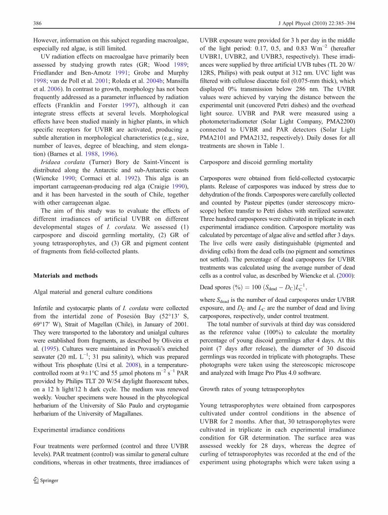
Because the apical sections of I. cordata are very irregular,1-cm2 fragments were selected from subapical infertilefield-collected plants (Fig. 1) to standardize the biomassand area. Afterwards, these fragments were cultivated for2 weeks under general culture conditions for acclimatiza-tion. Next, four fragments were cultivated in triplicate ineach experimental irradiance condition for GR determina-tion. The fresh biomass was recorded weekly for a period of35 days, and the GRs were estimated with the equationused for young tetrasporophytes, replacing area withbiomass.
Pigment analysis
The pigment analysis was carried out on fragments fromsubapical infertile field-collected plants cultivated for35 days in each experimental irradiance condition. Bothphycobiliprotein and chlorophyll-a (Chl-a) were analyzedfrom the same sample by spectrophotometry using anHP8452 A spectrophotometer. Pigment extractions werecarried out at 4°C, according to Kursar et al. (1983) asmodified by Plastino and Guimarães (2001). Briefly,250 mg of tissue from fragments cultivated for GRdetermination were disrupted by grinding with liquidnitrogen and 50 mmol L−1 phosphate buffer, pH5.5. Crudeextracts were centrifuged at 36,000×g for 25 min to obtainthe phycobiliproteins. Chl-a was extracted after dissolvingthe pellet in 90% acetone and centrifuged at 12,000×g for15 min. Pigment concentration was calculated according toKursar et al. (1983) for phycobiliproteins [phycoerythrin(PE), phycocyanin (PC), and alophycocyanin (APC)] andaccording to Jeffrey and Humphrey (1975) for Chl-a. Allpigment extractions were performed in triplicate.
Data analysis
Data were tested for homogeneity of variances and normality(Kolmogorov–Smirnov test). Corresponding transformationswere performed for data that were heteroskedastic (unequalvariances) and non-normal. The arcsine transformation wasapplied to percentage data of carpospore mortality. Data(UVB irradiance treatment) were compared by one-wayanalysis of variance (ANOVA). Time series measurementson the GR of the young tetrasporophytes and adult segmentswere subjected to repeated measures ANOVA to determinethe effects of treatments across the sampling days. In all cases,a posteriori Newman–Keuls test was used to establishstatistical differences. Statistical analyses were done usingthe Statistica 7 program.
Results
Under control conditions Iridaea cordata carpospores devel-oped into small red multicellular basal disks, with a diameter
Table 1 UVBR and PAR doses during the course of the experiments
Intensity (Wm−2) Doses (kJm−2)
1day 3days 1week 2weeks 3weeks 4weeks 5weeks
PAR (control) 11.95 516 1,549 3,097 7,227 10,841 14,455 18,068
UVBR1 0.17 1.8 5.51 11.0 25.7 38.6 51.4 64.3
UVBR2 0.5 5.4 16.2 32.4 75.6 113.4 151.2 189
UVBR3 0.83 9.0 26.9 53.8 125.5 188.2 251.1 313.7
Fig. 1 Iridaea cordata collectedfrom intertidal zone of PosesiónBay, Strait of Magallanes(Chile). White squares indicatethe subapical region wherefragments were excised. Scalebar=1 cm
J Appl Phycol (2010) 22:385–394 387
of 0.089±0.005 mm at 2 weeks of culture. At this time, asmall primary erect cylindrical axis was initiated from thecentral area of the disk. The erect frond continued toelongate (2 mm at 2 months) and formed a foliose thallus.
Carpospore and discoid germling mortality
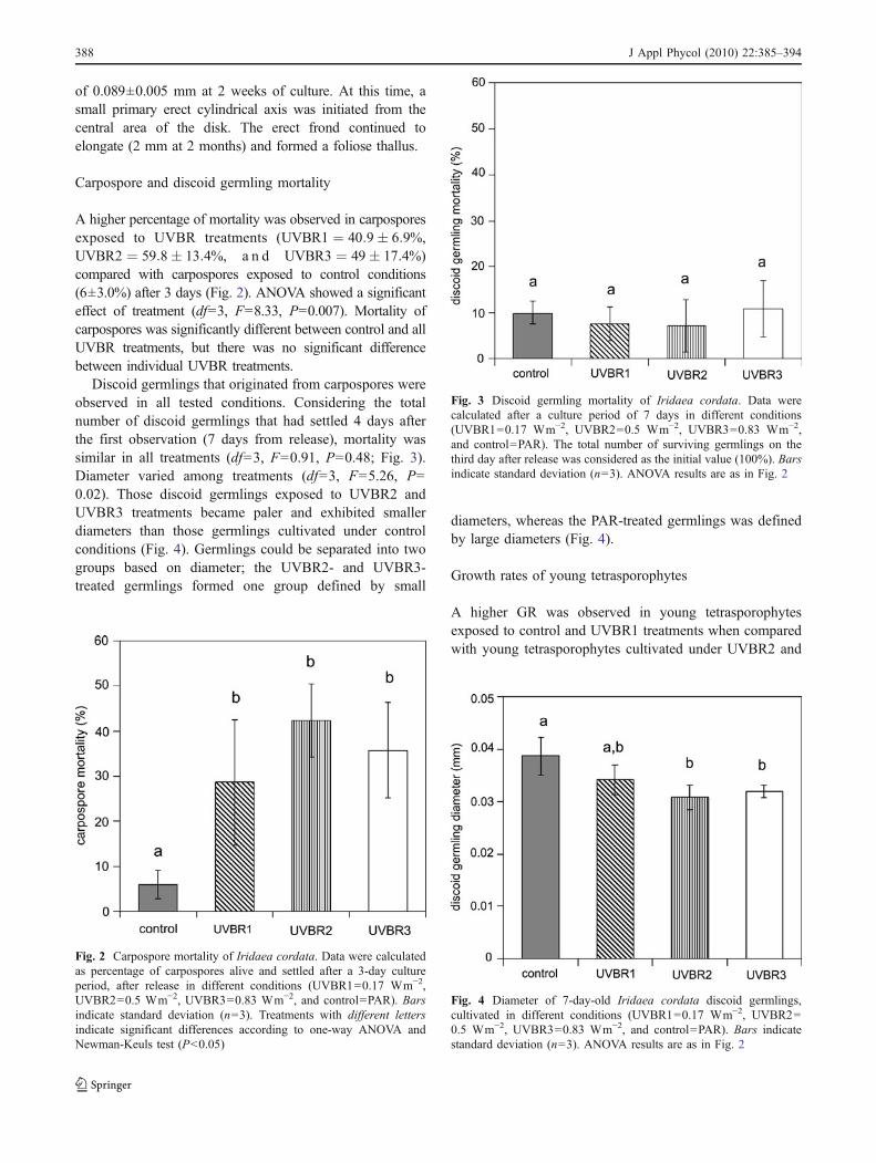
A higher percentage of mortality was observed in carposporesexposed to UVBR treatments (UVBR1 ¼ 40:9� 6:9%,UVBR2 ¼ 59:8� 13:4%, a n d UVBR3 ¼ 49� 17:4%)compared with carpospores exposed to control conditions(6±3.0%) after 3 days (Fig. 2). ANOVA showed a significanteffect of treatment (df=3, F=8.33, P=0.007). Mortality ofcarpospores was significantly different between control and allUVBR treatments, but there was no significant differencebetween individual UVBR treatments.
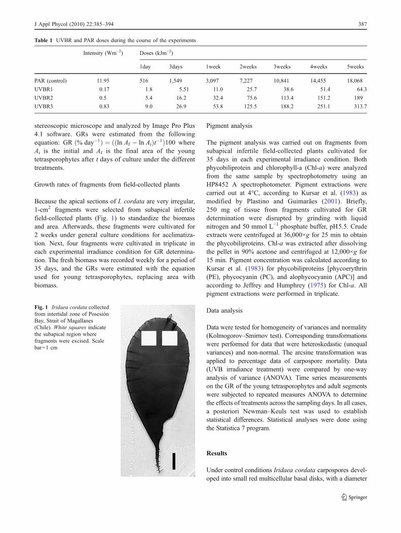
Discoid germlings that originated from carpospores wereobserved in all tested conditions. Considering the totalnumber of discoid germlings that had settled 4 days afterthe first observation (7 days from release), mortality wassimilar in all treatments (df=3, F=0.91, P=0.48; Fig. 3).Diameter varied among treatments (df=3, F=5.26, P=0.02). Those discoid germlings exposed to UVBR2 andUVBR3 treatments became paler and exhibited smallerdiameters than those germlings cultivated under controlconditions (Fig. 4). Germlings could be separated into twogroups based on diameter; the UVBR2- and UVBR3-treated germlings formed one group defined by small
diameters, whereas the PAR-treated germlings was definedby large diameters (Fig. 4).
Growth rates of young tetrasporophytes
A higher GR was observed in young tetrasporophytesexposed to control and UVBR1 treatments when comparedwith young tetrasporophytes cultivated under UVBR2 and
Fig. 3 Discoid germling mortality of Iridaea cordata. Data werecalculated after a culture period of 7 days in different conditions(UVBR1=0.17 Wm−2, UVBR2=0.5 Wm−2, UVBR3=0.83 Wm−2,and control=PAR). The total number of surviving germlings on thethird day after release was considered as the initial value (100%). Barsindicate standard deviation (n=3). ANOVA results are as in Fig. 2
Fig. 4 Diameter of 7-day-old Iridaea cordata discoid germlings,cultivated in different conditions (UVBR1=0.17 Wm−2, UVBR2=0.5 Wm−2, UVBR3=0.83 Wm−2, and control=PAR). Bars indicatestandard deviation (n=3). ANOVA results are as in Fig. 2
Fig. 2 Carpospore mortality of Iridaea cordata. Data were calculatedas percentage of carpospores alive and settled after a 3-day cultureperiod, after release in different conditions (UVBR1=0.17 Wm−2,UVBR2=0.5 Wm−2, UVBR3=0.83 Wm−2, and control=PAR). Barsindicate standard deviation (n=3). Treatments with different lettersindicate significant differences according to one-way ANOVA andNewman-Keuls test (P<0.05)
388 J Appl Phycol (2010) 22:385–394
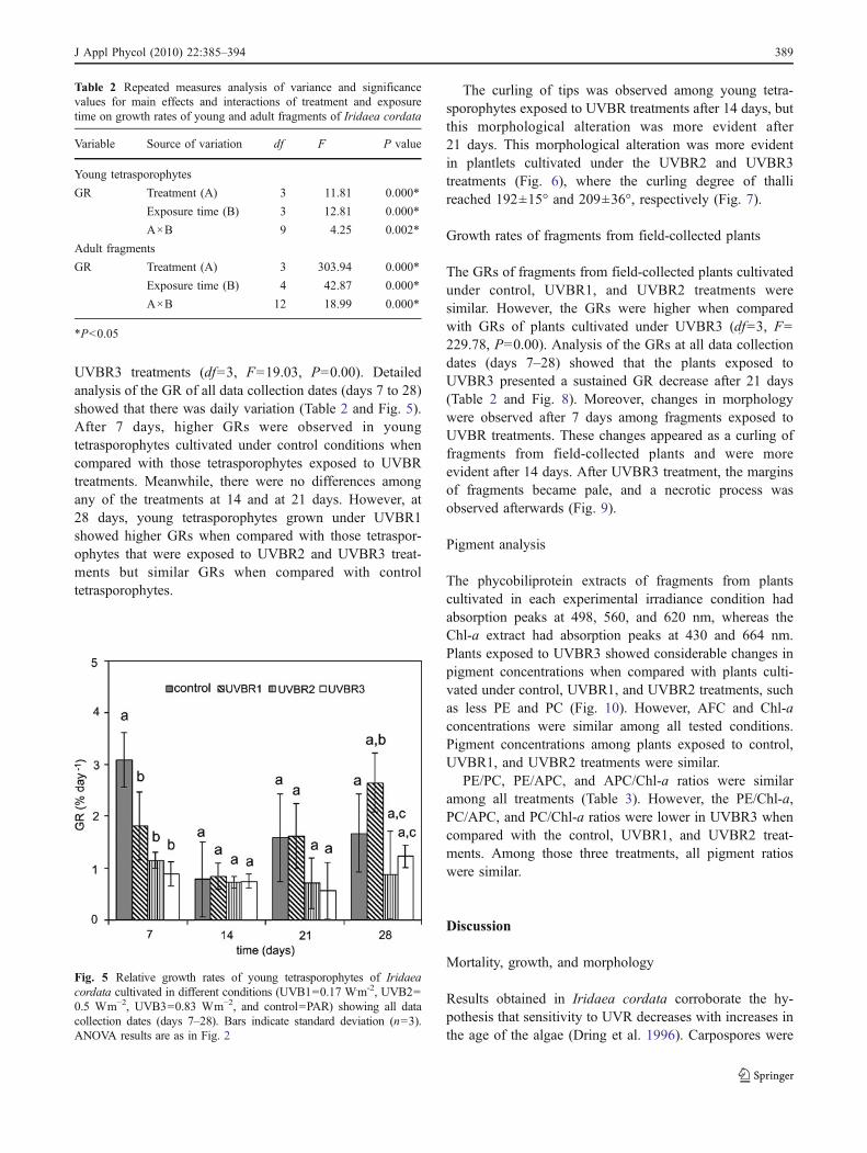
UVBR3 treatments (df=3, F=19.03, P=0.00). Detailedanalysis of the GR of all data collection dates (days 7 to 28)showed that there was daily variation (Table 2 and Fig. 5).After 7 days, higher GRs were observed in youngtetrasporophytes cultivated under control conditions whencompared with those tetrasporophytes exposed to UVBRtreatments. Meanwhile, there were no differences amongany of the treatments at 14 and at 21 days. However, at28 days, young tetrasporophytes grown under UVBR1showed higher GRs when compared with those tetraspor-ophytes that were exposed to UVBR2 and UVBR3 treat-ments but similar GRs when compared with controltetrasporophytes.
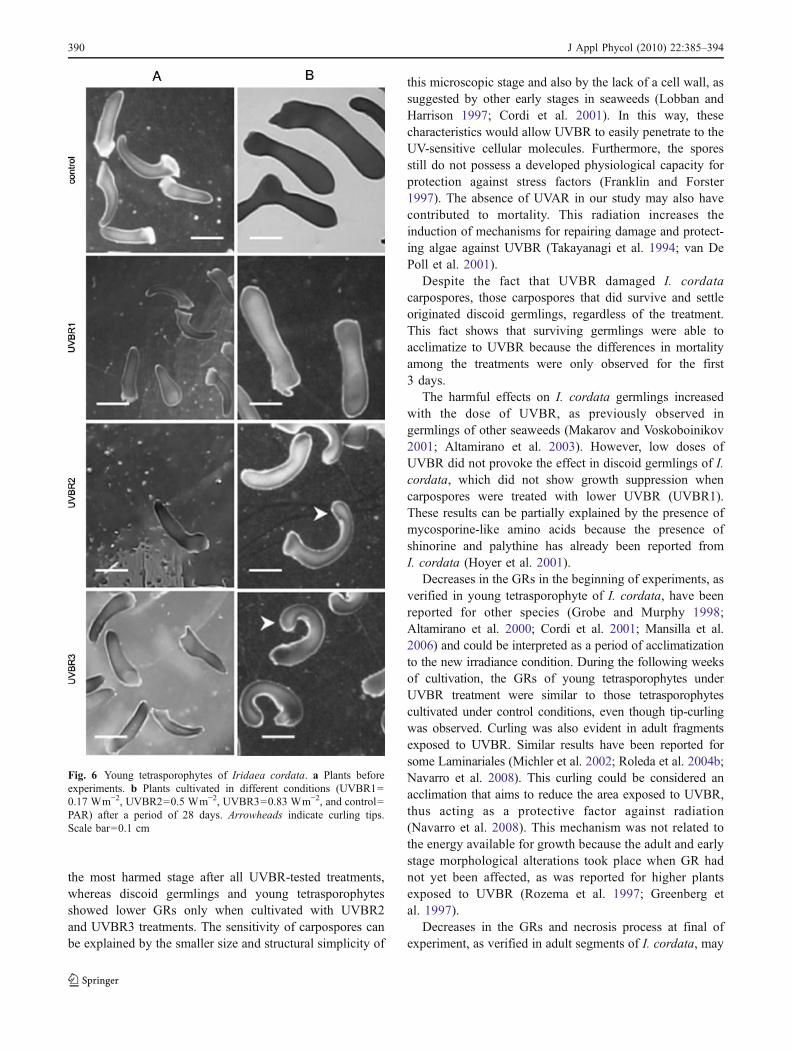
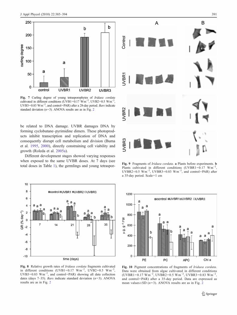
The curling of tips was observed among young tetra-sporophytes exposed to UVBR treatments after 14 days, butthis morphological alteration was more evident after21 days. This morphological alteration was more evidentin plantlets cultivated under the UVBR2 and UVBR3treatments (Fig. 6), where the curling degree of thallireached 192±15° and 209±36°, respectively (Fig. 7).
Growth rates of fragments from field-collected plants
The GRs of fragments from field-collected plants cultivatedunder control, UVBR1, and UVBR2 treatments weresimilar. However, the GRs were higher when comparedwith GRs of plants cultivated under UVBR3 (df=3, F=229.78, P=0.00). Analysis of the GRs at all data collectiondates (days 7–28) showed that the plants exposed toUVBR3 presented a sustained GR decrease after 21 days(Table 2 and Fig. 8). Moreover, changes in morphologywere observed after 7 days among fragments exposed toUVBR treatments. These changes appeared as a curling offragments from field-collected plants and were moreevident after 14 days. After UVBR3 treatment, the marginsof fragments became pale, and a necrotic process wasobserved afterwards (Fig. 9).
Pigment analysis
The phycobiliprotein extracts of fragments from plantscultivated in each experimental irradiance condition hadabsorption peaks at 498, 560, and 620 nm, whereas theChl-a extract had absorption peaks at 430 and 664 nm.Plants exposed to UVBR3 showed considerable changes inpigment concentrations when compared with plants culti-vated under control, UVBR1, and UVBR2 treatments, suchas less PE and PC (Fig. 10). However, AFC and Chl-aconcentrations were similar among all tested conditions.Pigment concentrations among plants exposed to control,UVBR1, and UVBR2 treatments were similar.
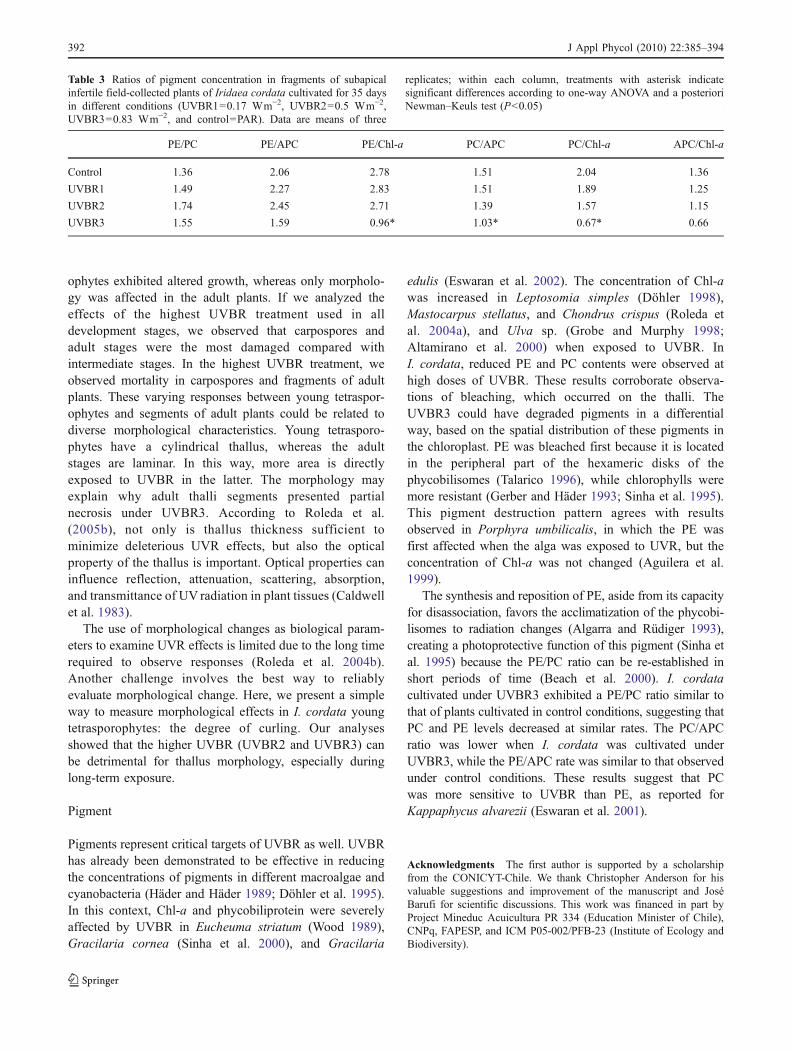
PE/PC, PE/APC, and APC/Chl-a ratios were similaramong all treatments (Table 3). However, the PE/Chl-a,PC/APC, and PC/Chl-a ratios were lower in UVBR3 whencompared with the control, UVBR1, and UVBR2 treat-ments. Among those three treatments, all pigment ratioswere similar.
Discussion
Mortality, growth, and morphology
Results obtained in Iridaea cordata corroborate the hy-pothesis that sensitivity to UVR decreases with increases inthe age of the algae (Dring et al. 1996). Carpospores were
Fig. 5 Relative growth rates of young tetrasporophytes of Iridaeacordata cultivated in different conditions (UVB1=0.17 W.m-2, UVB2=0.5 Wm−2, UVB3=0.83 Wm−2, and control=PAR) showing all datacollection dates (days 7–28). Bars indicate standard deviation (n=3).ANOVA results are as in Fig. 2
Table 2 Repeated measures analysis of variance and significancevalues for main effects and interactions of treatment and exposuretime on growth rates of young and adult fragments of Iridaea cordata
Variable Source of variation df F P value
Young tetrasporophytes
GR Treatment (A) 3 11.81 0.000*
Exposure time (B) 3 12.81 0.000*
A×B 9 4.25 0.002*
Adult fragments
GR Treatment (A) 3 303.94 0.000*
Exposure time (B) 4 42.87 0.000*
A×B 12 18.99 0.000*
*P<0.05
J Appl Phycol (2010) 22:385–394 389
the most harmed stage after all UVBR-tested treatments,whereas discoid germlings and young tetrasporophytesshowed lower GRs only when cultivated with UVBR2and UVBR3 treatments. The sensitivity of carpospores canbe explained by the smaller size and structural simplicity of
this microscopic stage and also by the lack of a cell wall, assuggested by other early stages in seaweeds (Lobban andHarrison 1997; Cordi et al. 2001). In this way, thesecharacteristics would allow UVBR to easily penetrate to theUV-sensitive cellular molecules. Furthermore, the sporesstill do not possess a developed physiological capacity forprotection against stress factors (Franklin and Forster1997). The absence of UVAR in our study may also havecontributed to mortality. This radiation increases theinduction of mechanisms for repairing damage and protect-ing algae against UVBR (Takayanagi et al. 1994; van DePoll et al. 2001).
Despite the fact that UVBR damaged I. cordatacarpospores, those carpospores that did survive and settleoriginated discoid germlings, regardless of the treatment.This fact shows that surviving germlings were able toacclimatize to UVBR because the differences in mortalityamong the treatments were only observed for the first3 days.
The harmful effects on I. cordata germlings increasedwith the dose of UVBR, as previously observed ingermlings of other seaweeds (Makarov and Voskoboinikov2001; Altamirano et al. 2003). However, low doses ofUVBR did not provoke the effect in discoid germlings of I.cordata, which did not show growth suppression whencarpospores were treated with lower UVBR (UVBR1).These results can be partially explained by the presence ofmycosporine-like amino acids because the presence ofshinorine and palythine has already been reported fromI. cordata (Hoyer et al. 2001).
Decreases in the GRs in the beginning of experiments, asverified in young tetrasporophyte of I. cordata, have beenreported for other species (Grobe and Murphy 1998;Altamirano et al. 2000; Cordi et al. 2001; Mansilla et al.2006) and could be interpreted as a period of acclimatizationto the new irradiance condition. During the following weeksof cultivation, the GRs of young tetrasporophytes underUVBR treatment were similar to those tetrasporophytescultivated under control conditions, even though tip-curlingwas observed. Curling was also evident in adult fragmentsexposed to UVBR. Similar results have been reported forsome Laminariales (Michler et al. 2002; Roleda et al. 2004b;Navarro et al. 2008). This curling could be considered anacclimation that aims to reduce the area exposed to UVBR,thus acting as a protective factor against radiation(Navarro et al. 2008). This mechanism was not related tothe energy available for growth because the adult and earlystage morphological alterations took place when GR hadnot yet been affected, as was reported for higher plantsexposed to UVBR (Rozema et al. 1997; Greenberg etal. 1997).
Decreases in the GRs and necrosis process at final ofexperiment, as verified in adult segments of I. cordata, may
Fig. 6 Young tetrasporophytes of Iridaea cordata. a Plants beforeexperiments. b Plants cultivated in different conditions (UVBR1=0.17 Wm−2, UVBR2=0.5 Wm−2, UVBR3=0.83 Wm−2, and control=PAR) after a period of 28 days. Arrowheads indicate curling tips.Scale bar=0.1 cm
390 J Appl Phycol (2010) 22:385–394
be related to DNA damage. UVBR damages DNA byforming cyclobutane–pyrimidine dimers. These photoprod-ucts inhibit transcription and replication of DNA andconsequently disrupt cell metabolism and division (Bumaet al. 1995, 2000), directly constraining cell viability andgrowth (Roleda et al. 2005a).
Different development stages showed varying responseswhen exposed to the same UVBR doses. At 7 days (seetotal doses in Table 1), the germlings and young tetraspor-
Fig. 9 Fragments of Iridaea cordata. a Plants before experiments. bPlants cultivated in different conditions (UVBR1=0.17 Wm−2,UVBR2=0.5 Wm−2, UVBR3=0.83 Wm−2, and control=PAR) aftera 35-day period. Scale=1 cm
Fig. 10 Pigment concentrations of fragments of Iridaea cordata.Data were obtained from algae cultivated in different conditions(UVBR1=0.17 Wm−2, UVBR2=0.5 Wm−2, UVBR3=0.83 Wm−2,and control=PAR) after a 35-day period. Data are expressed asmean values±SD (n=3). ANOVA results are as in Fig. 2
Fig. 8 Relative growth rates of Iridaea cordata fragments cultivatedin different conditions (UVB1=0.17 Wm−2, UVB2=0.5 Wm−2,UVB3=0.83 Wm−2, and control=PAR) showing all data collectiondates (days 7–35). Bars indicate standard deviation (n=3). ANOVAresults are as in Fig. 2
Fig. 7 Curling degree of young tetrasporophytes of Iridaea cordatacultivated in different conditions (UVB1=0.17 Wm−2, UVB2=0.5 Wm−2,UVB3=0.83 Wm−2, and control=PAR) after a 28-day period. Bars indicatestandard deviation (n=3). ANOVA results are as in Fig. 2
J Appl Phycol (2010) 22:385–394 391
ophytes exhibited altered growth, whereas only morpholo-gy was affected in the adult plants. If we analyzed theeffects of the highest UVBR treatment used in alldevelopment stages, we observed that carpospores andadult stages were the most damaged compared withintermediate stages. In the highest UVBR treatment, weobserved mortality in carpospores and fragments of adultplants. These varying responses between young tetraspor-ophytes and segments of adult plants could be related todiverse morphological characteristics. Young tetrasporo-phytes have a cylindrical thallus, whereas the adultstages are laminar. In this way, more area is directlyexposed to UVBR in the latter. The morphology mayexplain why adult thalli segments presented partialnecrosis under UVBR3. According to Roleda et al.(2005b), not only is thallus thickness sufficient tominimize deleterious UVR effects, but also the opticalproperty of the thallus is important. Optical properties caninfluence reflection, attenuation, scattering, absorption,and transmittance of UV radiation in plant tissues (Caldwellet al. 1983).
The use of morphological changes as biological param-eters to examine UVR effects is limited due to the long timerequired to observe responses (Roleda et al. 2004b).Another challenge involves the best way to reliablyevaluate morphological change. Here, we present a simpleway to measure morphological effects in I. cordata youngtetrasporophytes: the degree of curling. Our analysesshowed that the higher UVBR (UVBR2 and UVBR3) canbe detrimental for thallus morphology, especially duringlong-term exposure.
Pigment
Pigments represent critical targets of UVBR as well. UVBRhas already been demonstrated to be effective in reducingthe concentrations of pigments in different macroalgae andcyanobacteria (Häder and Häder 1989; Döhler et al. 1995).In this context, Chl-a and phycobiliprotein were severelyaffected by UVBR in Eucheuma striatum (Wood 1989),Gracilaria cornea (Sinha et al. 2000), and Gracilaria
edulis (Eswaran et al. 2002). The concentration of Chl-awas increased in Leptosomia simples (Döhler 1998),Mastocarpus stellatus, and Chondrus crispus (Roleda etal. 2004a), and Ulva sp. (Grobe and Murphy 1998;Altamirano et al. 2000) when exposed to UVBR. InI. cordata, reduced PE and PC contents were observed athigh doses of UVBR. These results corroborate observa-tions of bleaching, which occurred on the thalli. TheUVBR3 could have degraded pigments in a differentialway, based on the spatial distribution of these pigments inthe chloroplast. PE was bleached first because it is locatedin the peripheral part of the hexameric disks of thephycobilisomes (Talarico 1996), while chlorophylls weremore resistant (Gerber and Häder 1993; Sinha et al. 1995).This pigment destruction pattern agrees with resultsobserved in Porphyra umbilicalis, in which the PE wasfirst affected when the alga was exposed to UVR, but theconcentration of Chl-a was not changed (Aguilera et al.1999).
The synthesis and reposition of PE, aside from its capacityfor disassociation, favors the acclimatization of the phycobi-lisomes to radiation changes (Algarra and Rüdiger 1993),creating a photoprotective function of this pigment (Sinha etal. 1995) because the PE/PC ratio can be re-established inshort periods of time (Beach et al. 2000). I. cordatacultivated under UVBR3 exhibited a PE/PC ratio similar tothat of plants cultivated in control conditions, suggesting thatPC and PE levels decreased at similar rates. The PC/APCratio was lower when I. cordata was cultivated underUVBR3, while the PE/APC rate was similar to that observedunder control conditions. These results suggest that PCwas more sensitive to UVBR than PE, as reported forKappaphycus alvarezii (Eswaran et al. 2001).
Acknowledgments The first author is supported by a scholarshipfrom the CONICYT-Chile. We thank Christopher Anderson for hisvaluable suggestions and improvement of the manuscript and JoséBarufi for scientific discussions. This work was financed in part byProject Mineduc Acuicultura PR 334 (Education Minister of Chile),CNPq, FAPESP, and ICM P05-002/PFB-23 (Institute of Ecology andBiodiversity).
Table 3 Ratios of pigment concentration in fragments of subapical infertile field-collected plants of Iridaea cordata cultivated for 35 days indifferent conditions (UVBR1=0.17 Wm−2, UVBR2=0.5 Wm−2, UVBR3=0.83 Wm−2, and control=PAR). Data are means of three replicates;within each column, treatments with asterisk indicate significant differences according to one-way ANOVA and a posteriori Newman–Keuls test(P<0.05)
PE/PC PE/APC PE/Chl-a PC/APC PC/Chl-a APC/Chl-a
Control 1.36 2.06 2.78 1.51 2.04 1.36
UVBR1 1.49 2.27 2.83 1.51 1.89 1.25
UVBR2 1.74 2.45 2.71 1.39 1.57 1.15
UVBR3 1.55 1.59 0.96* 1.03* 0.67* 0.66
Table 3 Ratios of pigment concentration in fragments of subapicalinfertile field-collected plants of Iridaea cordata cultivated for 35 daysin different conditions (UVBR1=0.17 Wm−2, UVBR2=0.5 Wm−2,UVBR3=0.83 Wm−2, and control=PAR). Data are means of three
replicates; within each column, treatments with asterisk indicatesignificant differences according to one-way ANOVA and a posterioriNewman–Keuls test (P<0.05)
392 J Appl Phycol (2010) 22:385–394
References
Aguilera J, Jiménez C, Figueroa F, Lebert M, Häder D (1999) Effectof ultraviolet radiation on thallus absorption and photosyntheticpigments in red alga Porphyra umbilicalis. J Photochem Photo-biol B 48:75–82
Algarra P, Rüdiger W (1993) Acclimatation processes in the lightharvesting complex of the red alga Porphyridium purpureum(Bory) Drew et Ross, according to irradiance and nutrientavailability. Plant Cell Environ 16:149–159
Altamirano M, Flores-Moya A, Figueroa F (2000) Long-term effectsof natural sunlight under various ultraviolet radiation conditionson growth and photosynthesis of intertidal Ulva rigida (Chlor-ophyceae) cultivated in situ. Bot Mar 43:119–126
Altamirano M, Flores-Moya A, Figueroa F (2003) Effects of theradiation and temperature on growth of germling of three speciesof Fucus (Phaeophyta). Aquat Bot 75:9–20
Barnes PW, Jordan PW, Gold WC, Flint SD, Caldwell M (1988)Competition, morphology and canopy structure in wheat (Triti-cum aestivum L.) and wild oat (Avena fatua L.) exposed toenhanced ultraviolet-B radiation. Funct Ecol 2:319–330
Barnes PW, Ballare CL, Caldwell MM (1996) Photomorphogeneticeffects of UV-B radiation on plants: consequences for lightcompetition. J Plant Physiol 148:15–20
Beach KS, Smith CM, Okano R (2000) Experimental analysis ofrhodophyte photoacclimation to PAR and UV-radiation using invivo absorvance spectroscopy. Bot Mar 43:525–536
Bischof K, Gómez I, Molis M, Hanelt D, Karsten U, Lüder U, RoledaMY, Zacher K, Wiencke C (2006) Ultraviolet radiation shapesseaweed communities. Rev Environ Sci Biotechnol 5:141–166
Buma AG, van Hannen EJ, Roza L, Veldhuis MJ, Gieskes WW(1995) Monitoring ultraviolet-B-induced DNA damage in indi-vidual diatom cells by immunofluorescent thymine dimerdetection. J Phycol 31:314–321
Buma AG, van Oijen T, van de Poll WH, Veldhuis MJ, Gieskes WW(2000) The sensitivity of Emiliania huxleyi (Prymnesiophyceae)to ultraviolet-B radiation. J Phycol 36:296–303
Caldwell MM, Robberecht R, Flint SD (1983) Internal filters: prospectsfor UV-acclimation in higher plants. Physiol Plant 58:445–450
Cordi B, Donkin ME, Peloquin J, Price DN, Depledge MH (2001)The influence of UV-B radiation on the reproductive cells of theintertidal macroalga, Enteromorpha intestinalis. Aquat Toxicol56:1–11
Cormaci M, Furnari G, Scamacca B (1992) The benthic algal flora ofTerra Nova Bay (Ross Sea, Antarctica). Bot Mar 35:541–552
Craigie J (1990) Cell wall. In: Cole KM, Sheath RG (eds) Biology of thered algae. Cambridge University Press, New York, pp 221–257
Döhler G (1998) Effect of UV radiation on pigment of the Antarcticmacroalga Leptosomia simplex L. Photosynthetica 35:473–476
Döhler G, Hagmeier E, David C (1995) Effects of solar and artificial UVirradiance on pigments and assimilation of 15N ammonium and 15Nnitrate by macroalgae. J Photochem Photobiol, B Biol 30:179–187
Dring M, Makarov V, Schoschina E, Lorenz M, Lüning K (1996)Influence of ultraviolet-radiation on chlorophyll fluorescence andgrowth in different life-history stages of three species ofLaminaria (Phaeophyta). Mar Biol 126:183–191
Eswaran K, Suba Rao PV, Mairh OP (2001) Impact of ultraviolet-Bradiation on a marine red alga Kappaphycus alvarezii. Indian JMar Sci 30:105–107
Eswaran K, Mairh OP, Subba Rao PV (2002) Inhibition of pigmentand phycocolloid in a marine red alga Gracilaria edulis byultraviolet-B radiation. Biol Plant 45(1):157–159
Franklin L, Forster R (1997) The changing irradiance environment:consequences for marine macrophyte physiology, productivityand ecology. Eur J Phycol 32:207–237
Friedlander M, Ben-Amotz A (1991) The effect of out-door cultureconditions on growth and epiphytes of Gracilaria conferta.Aquat Bot 39:315–333
Gerber S, Häder D (1993) Effect of solar irradiation on motility andpigmentation of three species of phytoplankton. Environ Exp Bot33:515–521
Greenberg BM, Wilson MI, Huang X, Duxbury CL, Gerhardt KE,Gensemer RW (1997) The effect of ultraviolet-B radiation onhigher plants. In: Wang W, Gorsuch JW, Hughes JS (eds) Plantsfor environmental studies. Lewis, Boca Raton, pp 1–35
Grobe C, Murphy T (1998) Solar ultraviolet-B radiation effects ongrowth and pigment composition of the intertidal alga Ulvaexpansa (Setch.) S. & G. (Chlorophyta). J Exp Mar Biol Ecol225:39–51
Häder DP, Häder M (1989) Effects of solar and artificial radiation onmotility and pigmentation in Cyanophora paradoxa. ArchMicrobiol 152:453–457
Hoyer K, Karsten U, Sawall T, Wiencke C (2001) Photoprotectivesubstances in Antarctic macroalgae and their variation withrespect to depth distribution, different tissues and developmentalstages. Mar Ecol Prog Ser 211:117–129
Jeffrey SW, Humphrey GF (1975) New spectrophotometric equationsfor determining chlorophylls a, b, c1 and c2 in higher plant, algaeand natural phytoplankton. Biochem Physiol Pflanzen 167:191–194
Kursar TA, Van Der Meer J, Alberte RS (1983) Light-harvestingsystem of the red alga Gracilaria tikvahiae. I. Biochemicalanalyses of pigment mutations. Plant Physiol 73:353–360
Lobban C, Harrison P (1997) Seaweed ecology and physiology.Cambridge University Press, Cambridge
Makarov MV, Voskoboinikov GM (2001) The influence of ultraviolet-B radiation on spores release and growth of kelp Laminariasaccharina. Bot Mar 44:89–94
Mansilla A, Werlinger C, Palacios M, Navarro NP, Cuadra P (2006)Effects of UVB radiation on the initial stages of growth ofGigartina skottsbergii, Sarcothalia crispata and Mazzaellalaminarioides (Gigartinales, Rhodophyta). J Appl Phycol18:451–459
Michler T, Aguilera J, Hanelt D, Bischof K, Wiencke C (2002) Longterm effects of ultraviolet radiation on growth and photosyntheticperformance of polar and cold-temperate macroalgae. Mar Biol140:1117–1127
Navarro NP, Mansilla A, Palacios M (2008) UVB effects on earlydevelopmental stages of commercially important macroalgae insouthern Chile. J Appl Phycol 20(5):897–906
Oliveira EC, Paula EJ, Plastino EM, Petti R (1995) Metodologías para elcultivo no axenico de macroalgas marinas in vitro. In: Alveal K,Ferrario M, Oliveira E, Sar E (eds) Manual de métodos ficológicos.Universidad de Concepción, Concepción-Chile, pp 429–447
Plastino EM, Guimarães M (2001) Diversidad intraespecífica. In: AlvealK, Antezana T (eds) Sustentabilidad de la biodiversidad, unproblema actual. Bases científico-técnicas, teorización y proyec-ciones. Universidad de Concepción, Concepción-Chile, pp 19–27
Roleda M, van de Poll W, Hanelt D, Wiencke C (2004a) PAR and UVBReffects on photosynthesis, viability, growth andDNA in different lifestages of two coexisting Gigartinales: implications for recruitmentand zonation pattern. Mar Ecol Prog Ser 281:37–50
Roleda M, Hanelt D, Krabs G, Wiencke C (2004b) Morphology,growth, photosynthesis and pigment in Laminaria ochroleuca(Laminariales, Phaeophyta) under ultraviolet radiation. Phycolo-gia 43:603–613
Roleda M, Wiencke C, Hanelt D, van de Poll W, Gruber A (2005a)Sensitivity of Laminariales zoospores from Helgoland (NorthSea) to ultraviolet and photosynthetically active radiation:implications for depth distribution and seasonal reproduction.Plant Cell Environ 28:466–479
J Appl Phycol (2010) 22:385–394 393
Roleda MY, Hanelt D, Wiencke C (2005b) Growth kinetics related tophysiological parameters in young Saccorhiza dermatodea andAlaria esculenta sporophytes exposed to UV radiation. Polar Biol28:539–549
Roleda MY, Wiencke C, Hanelt D, Bischof K (2007) Sensitivity of thearly life stages of macroalgae from the northern hemisphere toultraviolet radiation. Photochem Photobiol 83:1–12
Rozema J, van de Staaij J, Björn LO, Caldwell M (1997) UV-B as anenvironmental factor in plant life: stress and regulation. TrendsEcol Evol 12:22–28
Sinha RP, Lebert M, Kumar A, Kumar HD, Häder DP (1995)Spectroscopic and biochemical analyses of UV effects on phycobili-proteins of Anabaena sp. and Nostoc carmium. Bot Acta 108:87–92
Sinha RP, Klisch M, Gröniger A, Häder DP (2000) Mycosporine-likeamino acids in the marine red alga Gracilaria cornea—effects ofUV and heat. Environ Exp Bot 43:33–43
Takayanagi S, Trunk J, Sutherland JC, Sutherland BM (1994) Alfalfagrown outdoor are more resistant to UV-induced DNA damagethan plants grown in a UV-free environmental chamber. Photo-chem Photobiol 60:363–367
Talarico L (1996) Phycobiliproteins and phycobilisomes in red algae:adaptive responses to light. Sci Mar 60:205–222
Ursi S, Guimarães M, Plastino EM (2008) Deleterious effect of TRISbuffer on growth rates and pigment contents of Gracilaria
birdiae Plastino & E.C. Oliveira (Gracilariales, Rhodophyta).Acta Bot Bras 22:891–896
van De Poll W, Eggert A, Buma A, Breeman A (2001) Effects of UV-induced DNA damage and photoinhibition on growth oftemperature marine red macrophytes: habitat - related differencesin UV-B tolerance. J Phycol 37:30–37
Wiencke C (1990) Seasonality of red and green mcroalgae fromAntarctica. A long-term culture study under fluctuating antarcticdaylengths. Polar Biol 10:601–607
Wiencke C, Gómez I, Pakker H, Flores-Moya A, Altamirano M,Hanelt D, Bischof K, Figueroa FL (2000) Impact of UV-radiationon viability, photosynthetic characteristics and DNA of brownalgal zoospores: implications for depth zonation. Mar Ecol ProgSer 197:217–229
Wiencke C, Roleda MY, Gruber A, Clayton MN, Bischof K (2006)Susceptibility of zoospores to UV radiation determines upperdepth distribution limit of Arctic kelps: evidence through fieldexperiments. J Ecol 94:455–463
Wood W (1989) Photoadaptative responses of the tropical red algaEucheuma strictum Schmitz (Gigartinales) to ultraviolet radia-tion. Aquat Bot 33:41–51
Xue L, Zhang Y, Zhang T, An L, Wang X (2005) Effects of enhancedultraviolet-B radiation on algae and cianobacteria. Crit RevMicrobiol 31:79–89
394 J Appl Phycol (2010) 22:385–394