inward rectifier potassium (kir) current in dopaminergic periglomerular neurons of the mouse...
TRANSCRIPT

ORIGINAL RESEARCH ARTICLEpublished: 08 August 2014
doi: 10.3389/fncel.2014.00223CELLULAR NEUROSCIENCE
Inward rectifier potassium (Kir) current in dopaminergicperiglomerular neurons of the mouse olfactory bulbMirta Borin†, Alex Fogli Iseppe†, Angela Pignatelli and Ottorino Belluzzi*
Department of Life Sciences and Biotechnology, University of Ferrara, Ferrara, Italy
Edited by:
Arianna Maffei, SUNY Stony Brook,USA
Reviewed by:
Alasdair James Gibb, UniversityCollege London, UKArianna Maffei, SUNY Stony Brook,USA
*Correspondence:
Ottorino Belluzzi, Department of LifeSciences and Biotechnology,University of Ferrara, Via Borsari 46,44121 Ferrara, Italye-mail: [email protected]
†These authors have contributedequally to this work.
Dopaminergic (DA) periglomerular (PG) neurons are critically placed at the entry of thebulbar circuitry, directly in contact with both the terminals of olfactory sensory neuronsand the apical dendrites of projection neurons; they are autorhythmic and are the targetof numerous terminals releasing a variety of neurotransmitters. Despite the centrality oftheir position, suggesting a critical role in the sensory processing, their properties -andconsequently their function- remain elusive. The current mediated by inward rectifierpotassium (Kir) channels in DA-PG cells was recorded by adopting the perforated-patchconfiguration in thin slices; IKir could be distinguished from the hyperpolarization-activatedcurrent (I ) by showing full activation in 10 ms, no inactivation, suppression by Ba2
h < + in atypical voltage-dependent manner (IC50 208 μM) and reversal potential nearly coincidentwith EK. Ba2+ (2 mM) induces a large depolarization of DA-PG cells, paralleled by anincrease of the input resistance, leading to a block of the spontaneous activity, but theKir current is not an essential component of the pacemaker machinery. The Kir currentis negatively modulated by intracellular cAMP, as shown by a decrease of its amplitudeinduced by forskolin or 8Br-cAMP. We have also tested the neuromodulatory effects of theactivation of several metabotropic receptors known to be present on these cells, showingthat the current can be modulated by a multiplicity of pathways, whose activation in somecase increases the amplitude of the current, as can be observed with agonists of D2,muscarinic, and GABAA receptors, whereas in other cases has the opposite effect, as itcan be observed with agonists of α1 noradrenergic, 5-HT and histamine receptors. Thesecharacteristics of the Kir currents provide the basis for an unexpected plasticity of DA-PGcell function, making them potentially capable to reconfigure the bulbar network to allowa better flexibility.
Keywords: olfactory bulb, dopaminergic neurons, periglomerular cell, Kir channels, patch-clamp techniques
INTRODUCTIONThe background potassium conductance mediated by inwardrectifying potassium channels impacts on many physiologicalprocesses, from the excitability profile of nerve and muscle cellsto hormone release. A voltage-dependent block of the channelpore by polyamines and intracellular magnesium is thought to beresponsible for the inward rectification of these channels (Lopatinet al., 1995); for a review see Hibino et al. (2010) which, open-ing at potentials close to EK, tend to maintain the membrane in ahyperpolarized state.
Dopaminergic (DA) neurons represent an estimated 10–16%of the neurons residing in the most external (glomerular) layerof the main olfactory bulb (MOB) (Halász et al., 1977; McLeanand Shipley, 1988). The glomerular layer (GL) region is occupiedby three main types of interneurons, periglomerular (PG) cells,short-axon cells and external tufted (ET) cells - sometimes collec-tively referred to as juxtaglomerular cells (Kratskin and Belluzzi,2003; Panzanelli et al., 2007). Dopaminergic neurons in the GLinclude PG cells (Kosaka et al., 1985; Gall et al., 1987) and afraction of ET cells (Halász, 1990).
Analyzing the excitability profile of DA-PG cells, we observedthat Ba2+ (300 μM, a blocker of the Kir channels), induceda large depolarization in bulbar DA neurons, large enoughto lead to complete blockage of spontaneous firing of thesecells. We then examined the problem, finding that actuallythere is a Kir current in these cells which escaped a pre-vious investigation of ours (Pignatelli et al., 2005) for itsrelatively small amplitude in standard saline. The currentcan be better evidenced with ionic manipulations causinga depolarizing shift of the potassium equilibrium potential,but even under physiological conditions, for the elevatedinput resistance of these cells, the Kir current is sufficientlylarge to exert a relevant influence on the cell excitabilityprofile.
Being selective for potassium ions, the channels of theinwardly rectifying family conduct currents which are inwardat potentials negative to the K+ equilibrium potential (EK)and outward at potentials positive to EK, in so doing con-tributing to the resting membrane potential (Hibino et al.,2010).
Frontiers in Cellular Neuroscience www.frontiersin.org August 2014 | Volume 8 | Article 223 | 1

Borin et al. IKir in dopaminergic periglomerular cells
MATERIALS AND METHODSETHIC STATEMENTA total of 123 mice have been used. The experimental processwas designed so as to minimize animal number and suffering ofthe animals used. The protocols adopted were designed accord-ing to European Council Directives (609/1986 and 63/2010) andItalian laws (DL 116/92) on the protection of animals used forscientific purposes. The experimental procedures were approvedby the Ethical Committee for Animal Experiments of the FerraraUniversity (CEASA), by the Directorate-General for AnimalHealth of the Ministry of Health, and supervised by the CampusVeterinarian of the University of Ferrara.
ANIMALS AND SURGICAL PROCEDURESFor these experiments we used a transgenic mice strain (TH-GFP/21–31), carrying the eGFP transgene under the control ofthe TH promoter (Sawamoto et al., 2001; Matsushita et al., 2002).The TH-GFP strain was maintained as heterozygous by breedingwith C57BL/6J inbred mice.
RECORDING CONDITIONSThe temperature of the 1-ml recording chamber was con-trolled using Peltier devices (RS Components, Milan, Italy)and measured with a high-precision, type K thermocouple (RSComponents).
For current and voltage recordings an Axopatch 200B ampli-fier (Molecular Devices, Sunnyvale, CA) was used, and the signalswere digitized and acquired with a Digidata 1440A (MolecularDevices) 16 bit A/D–D/A converter; correction for the junc-tion potential was calculated using the related function of theacquisition software (pClamp 10, Molecular Devices).
Patch pipettes were built from borosilicate glass capillaries(1.5 o.d., 0.87 i.d., with filament; Hilgenberg, Malsfeld, Germany)with a Zeitz-DMZ puller (Martinsried, Germany), and showed aresistance of 4–5 M� when filled with standard intracellular solu-tion (see below); the seal formation was assisted by a MCPU-3air pressure controller (MPI, Lorenz Meβgerätebau, Katlenburg-Lindau, Germany); the seal resistance obtained was always greaterthan 3 G�.
SOLUTIONSThe solutions used had the following composition (mM):
• EC0, standard ACSF extracellular (EC) solution: 125 NaCl, 2.5KCl, 26 NaHCO3, 1.25 NaH2PO4, 2 CaCl2, 1 MgCl2, and 15glucose.
• EC1, high K EC solution: 95 NaCl, 32.5 KCl, 26 NaHCO3,1.25 NaH2PO4, 2 CaCl2, 1 MgCl2, and 15 glucose.
• EC2, K-TEA EC solution: 100 NaCl, 2.5 KCl, 26 NaHCO3,1.25 NaH2PO4, 2 CaCl2, 1 MgCl2, 20 TEA, and 10 glucose.
• EC3, high K-TEA EC solution: 70 NaCl, 32.5 KCl, 26 NaHCO3,1.25 NaH2PO4, 2 CaCl2, 1 MgCl2, 20 TEA, and glucose.
All EC solutions were continuously bubbled with 95% O2 and5% CO2, and the osmolarity was corrected to 305 mOsm withglucose.
Standard pipette-filling intracellular (IC) solution:120 KCl, 10 NaCl, 2 MgCl2, 0.5 CaCl2, 5 EGTA, 10 HEPES,2 Na-ATP, 10 glucose; in this solution, the free ICcalcium concentration was calculated to be 16 nM(http://www.stanford.edu/∼cpatton/downloads.htm).
For perforated patches, 200 μg/ml amphotericin B was addedto the IC solution (plus 300 μg/ml pluronic F-127). EGTA wasomitted and CaCl2 concentration was increased to 3 mM inthe electrode filling solution to control of the integrity of theperforated patch, as in case of unexpected rupture, the mas-sive entry of calcium from the pipette would cause a rapid celldeath. Data were collected after the series resistance droppedbelow 50 M�.
In all IC solutions the osmolarity was finely tuned to 295mOsm with glucose, and the pH to 7.2 with KOH.
Except where indicated, when recording from slices, the ECsolutions included two mixtures of blockers:
- BL1, for ligand-gated channels (1 mM kynurenic acid and10 μM bicuculline).
- BL2, for voltage-dependent channels (TTX 0.6 μM, Cd2+100 μM and ivabradine 10 μM).
ANALYSIS OF CURRENT RECORDINGSIKir amplitude was measured as instantaneous current at thebeginning (Iinst) and at the end of test voltage pulses as steady-state current (Iss).
The temperature-dependence of activation and deactivationrate constants were calculated as:
Q10 =(
rate (T2)
rate (T1)
) 10T2−T1
(1)
where Q10 is the fold-change as a consequence of increasing thetemperature by 10◦C, calculated between the two temperaturesT1 and T2.
DATA ANALYSISTo evoke the Kir current, a series of hyperpolarizing voltage stepsin −10 mV increments were imposed from the holding potentialof −40 to −130 mV at 10 s intervals. Unless otherwise indi-cated, the current amplitudes were measured at the end of thehyperpolarizing step (steady-state current).
When box charts are used to represent data ensembles, the cen-tral square represents the mean, the central line the median, therange of the boxes represent the S.E, and the whiskers define the10–90% range of data samples.
Offline analysis was performed using version 10.3 of pClamp(Molecular Devices) and version 8.1 of OriginPro (OriginLabCorporation, Northampton, MA).
Unless otherwise indicated, data are presented as means ±s.e.m.; for the statistical analysis we used the software Prism 5(GraphPad, La Jolla, CA). The statistical significance was assessedwith Two-Way analysis of variance (ANOVA), or Student’s t-testfor paired samples as indicated; in Two-Way ANOVA multi-ple comparisons post-tests were performed using the Bonferronimethod.
Frontiers in Cellular Neuroscience www.frontiersin.org August 2014 | Volume 8 | Article 223 | 2

Borin et al. IKir in dopaminergic periglomerular cells
P value of < 0.05 was considered significant; in figures, 1 to 4asterisks represent differences significant at the 0.05, 0.01, 0.001,0.0001 level, respectively.
RESULTSThe data are based on recordings from 285 TH+ PG neuronsfrom the glomerular layer. Most OB DA cells are small, PGinterneurons (about 5–8 μm in diameter), but there is also a cer-tain number of external tufted (ET) cells (about 10–15 μm indiameter) (Baker et al., 1993; Kosaka and Kosaka, 2009, 2011).In this study, we restricted the analysis to PG cells; these wereselected on the basis of their location around the glomerularborder, dendritic arborization extending within the glomeru-lar neuropil, membrane capacitance (8.0 ± 0.2 pF; n = 297)and input resistance (979.4 ± 33.4 M�; n = 276). In additionto the evident differences in dimension (Kosaka and Kosaka,2008), membrane capacity and input resistance (Pignatelli et al.,2005), DA-PG cells show a regular firing pattern, whereas DA-ET cells show burst pattern activity (Hayar et al., 2004). Finally,short-axon cells, which have membrane capacitance and inputresistance very similar to PG cells, can be usually recognized inslice for their fusiform shape, position amid different glomeruli,and dendrites extending between neighboring glomeruli (Shipleyand Ennis, 1996).
IDENTIFICATION AND BASIC PROPERTIES OF THE CURRENTIn a first series of experiments, carried out using perforatedpatch recordings in slice at 34◦C, hyperpolarizing steps were
delivered from a holding potential of −40 mV, which is close tothe predicted K+ equilibrium potential in our experimental con-ditions (EK = −37.7 mV for [K+]o = 32.5 mM), to potentialsranging from −60 to −130 mV (10 mV increments, 10 s interval).The inward current obtained in response is shown in Figure 1A; afraction of this current could be suppressed by two organic com-pounds known as selective HCN channels blockers, i.e., ZD728830 μM (BoSmith et al., 1993) and S-16257, a.k.a. ivabradine,10 μM; (Bois et al., 1996; Bucchi et al., 2002), Figure 1B; theh-current in DA-PG cells has been the object of another study(Pignatelli et al., 2013), and will be not further discussed in thispaper.
The current activated by hyperpolarization remainingafter suppression of the h-current, was suppressed by Ba2+(Figure 1C), a classical blocker of Kir channels (Hagiwara et al.,1978; French and Shoukimas, 1985); for its potassium andvoltage-dependence, reversal potential and sensitivity to Ba2+this component was identified as potassium inward rectifier(Kir) current (Hibino et al., 2010). The I/V relationship of thecurrent evoked by hyperpolarization in a group of 81 cells in thepresence of 0.6 μM TTX, 100 μM Cd2+ and 10 μM ivabradine isshown in Figure 1D; here, and in the following experiments, forthe inherent difficulties, the leakage component of the currentevoked by hyperpolarization was not subtracted.
Barium sensitivityThe Ba2+ dependent block of IKir was evaluated from the decreaseof steady-state current amplitude at −120 mV in the presence
FIGURE 1 | General properties. (A–C) Representative currents obtained inresponse to hyperpolarizing pulses in 32.5 mM external K+ solution: (A) EC1saline with 1 mM kynurenic acid, 10 μM bicuculline (BL1 mix), 0.6 μM TTX,100 μM Cd2+; (B) same solution as in A plus 10 μM ivabradine; (C) samesolution as in B plus 2 mM Ba2+. Voltage steps from a holding potential of−40 mV with hyperpolarizing steps ranging from −60 to −130 mV in 10 mVincrements. (D): I–V relationship of peak (green dots) and steady-state (bluedots) current; mean current amplitude of 81 cell recordings. Vertical error bars
represent standard error; EC2 saline, with BL1 and BL2 mixes of blockers. (E)
Instantaneous I/V curve during application of a 220 mV/s ramp protocol (from−180 to +40 mV, 0.23 V/s) in a DA PG cell perfused with the solutiondescribed in (A), after subtraction of the ohmic leak; the red dot (−41.3 mV)marks the observed reversal potential, the green dot the Nernstianequilibrium potential in the experimental conditions used ([K+]o = 32.5 mM).All the experiments shown in this figure were performed in slice, perforatedpatch configuration, at 34◦C.
Frontiers in Cellular Neuroscience www.frontiersin.org August 2014 | Volume 8 | Article 223 | 3

Borin et al. IKir in dopaminergic periglomerular cells
of increasing external Ba2+ concentrations. In Figure 2A isrepresented the percentage of the current inhibition as a functionof external Ba2+ concentrations ranging from 1 μM to 10 mM.The data could be interpolated by a logistic equation with theform:
y = Imax/(
1 + ([Ba2+]
o /Kd)H
)(2)
where Imax is the asymptotic value of the current block, Kd theexternal Ba2+ concentration causing 50% block, and H is theslope of the dose-response curve (Hill coefficient). The fit of theBa2+ block of peak IKir gave a Kd of 0.21 ± 0.10 mM and a Hvalue of 0.69 ± 0.23 (n = 5, −120 mV).
Voltage-dependence of the steady-state block by Ba2+ and Cs+Ba2+ and Cs+ have been found to block the Kir channel throughan interaction which is thought to occur via a deep binding site,located approximately half-way along the channel (Standen andStanfield, 1978; Shieh et al., 1998; Alagem et al., 2001). As nor-mally occurs for deep-site blockers, Ba2+ and Cs+ block is highlyvoltage dependent (Hagiwara et al., 1978; Harvey and Ten Eick,1989; Alagem et al., 2001). The effect induced by 1 mM Ba2+ inDA-PG cells is shown in Figures 2B,C. The time required for theblocking reaction to reach steady state was calculated by fittingthe exponential decay of the currents to the function:
I = A exp( − t/tblk) + C (3)
where A is the current amplitude, t is the independent variable,C is the current amplitude at the steady-state, and τblk is theblocking time constant, whose voltage dependence is shown inFigure 2F.
We also tested the effects of 1 mM Cs+, another classicalblocker of this channel for which the approach to steady-stateblock following a voltage step is much faster than for Ba2+(Hagiwara et al., 1976, 1978; Shioya et al., 1993). The results,shown in Figures 2D–F, are in good agreement with thosereported in literature (Hagiwara et al., 1976, 1978).
Reversal potentialThe Kir channels are selective for K+ ions, and consequentlythe reversal potentials of the inward rectifying current for dif-ferent extracellular K+ concentrations should always follow theNernstian equilibrium potential for potassium (Figures 1E, 3A).When the [K+]o was changed from 2.5 to 10 and 32.5 mM,the reversal potentials progressively shifted toward more positivepotentials (−105.12 ± 3.67 mV, n = 15, for 2.5 mM; −56.67 ±9.78 mV, n = 9, for 10 mM; −36.78 mV ± 1.89, n = 27, for32.5 mM); the reversal potentials in the different experimentalconditions are represented in Figure 3B, where they are comparedto the theoretical Nernstian equilibrium potentials for K+ ions(black triangles). The plot of the reversal potentials against thelogarithm of [K+]o gives a linear relationship (r2 = 0.93) witha slope of −61.9 mV, close to the theoretical value of −61.0 mVpredicted by the Nernst equation (Figure 3C).
K+ and voltage dependence of the IKir
Besides the selectivity to K+ ions, another typical characteristicof this current is a voltage-dependence of the Kir conductance(gKir) on the K+ reversal potential; then, in DA-PG cells we fur-ther examined the dependence of gKir from membrane potentialfor different external K+ concentrations.
The conductance-voltage relationship showed the typical sig-moidal profile, increasing at negative potentials and with a point
FIGURE 2 | Barium sensitivity. (A) Percentage inhibition of steady-state Kir
current vs. [Ba2+]o. [K+]o = 2.5 mM, 32◦C. The data (green dots) were fittedwith a logistic function (see text), giving a concentration at half-block (Kd) of0.21 ± 0.10 mM, and a slope (Hill coefficient) of 0.69 ± 0.23 (n = 5,−120 mV). (B,C): sample tracings obtained in a single cells in response to
hyperpolarizing steps from −40 to −120 mV in standard saline plus BL1 andBL2 (B), and in the presence of 1 mM Ba2+ (C). (D,E): same experimentalconditions and protocol as above; blocking effect of 1 mM Cs+. (F) Voltagedependence of the blocking time constant; the data points were obtained inthe presence of 1 mM Ba2+ (yellow dots) or Cs+ (orange dots).
Frontiers in Cellular Neuroscience www.frontiersin.org August 2014 | Volume 8 | Article 223 | 4

Borin et al. IKir in dopaminergic periglomerular cells
FIGURE 3 | Potassium sensitivity. (A) Effect of changing [K+]o onmembrane current. Average currents (n = 8) at the indicated externalpotassium concentrations in response to voltage ramps from −170 to +20 mVfrom a holding potential of −40 mV, 0.22 V/s; perforated patches; the bathingsolution included Bl1 and Bl2. (B) Box charts showing the reversal potentialsat different [K+]o [same color code as in (A)]; black arrow heads to the right ofeach box mark the expected reversal potentials predicted by the Nernstequation. In the box charts, here and in the following, the square in the centerof the box represents the mean value, the horizontal line crossing the boxindicates the median, the range of the box represents standard error and the
whiskers define the 10–90% range of data sample. (C) Plot of the reversalpotential for the inwardly rectifying current against the logarithm of [K+]o. Thelinear regression fit (black dash line) has a slope of −61.9 mV, close to thetheoretical value of −61 mV predicted by the Nernst equation (red line). (D) K+-and voltage-dependence of chord conductance (gKir); the chord conductancewas calculated using the equation gKir = IKir/(Vm − EK), where IKir = steadystate current. gKir plotted as a function of voltage-clamp test potentials at 2.5,10, and 32.5 mM [K+]o. (E) Data in (D) replotted as a function of the drivingforce. Data points were fitted by Boltzmann curve using a least-squaresmethod; n for 2.5, 10, and 32.5 mM was 7, 5, and 12, respectively.
of half-activation approximately centered at EK (Figure 3D).Plotting the conductance for different [K+]o as a function of thedriving force (Vm − EK, Figure 3E), the midpoints were approx-imately aligned at the zero of the abscissa axis, with minima andmaxima at the same voltage levels. This confirms that Kir conduc-tance in DA-PG cells has a voltage-dependence which is functionof EK, in analogy to what has been found for IKir in several otherpreparations (Hestrin, 1981; Leech and Stanfield, 1981; Harveyand Ten Eick, 1988).
Effect of IKir on membrane potential and input resistanceIf the IKir is active at rest, then it should be expected that theblock of the current with Ba2+ should influence both input resis-tance and resting potential; in effect, Ba2+ (2 mM) induces a rapidand strong depolarization of DA-PG cells (Figures 4A,C), par-alleled by an increase of the firing frequency before its block indepolarization (Figures 4A,B).
The Kir current is not essential to the pacemaker process, as theinjection of hyperpolarizing current (40 pA at the time marked
Frontiers in Cellular Neuroscience www.frontiersin.org August 2014 | Volume 8 | Article 223 | 5

Borin et al. IKir in dopaminergic periglomerular cells
FIGURE 4 | Effects of Barium on DA-PG cells. (A) Effect of Ba2+ onmembrane potential. Perforated patch recording in standard saline (EC1solution). The blue bar indicates the time of application of 2 mM Ba2+ into thebath; starting at the time indicated by the downward arrow, a 40 pAhyperpolarizing current was injected; (further explanation in the text). (B)
Frequency analysis of action potentials (SPS, spike per second) for theexperiment shown in panel A; the dashed line marks the time at which Ba2+has been applied and the yellow point after the x-axis interruption is ameasure of the activity after the injection of a hyperpolarizing current, at thetime marked by a yellow point in (A). (C) Illustration of the method used forthe calculation of the prevailing membrane potential (further explanation in
the text): 10 s frequency count histograms of the membrane potential wererealized at 10 s intervals, and the distributions were fitted by an exponentiallymodified Gaussian function (equation 3 in the text); the point marked by thered dot indicates the prevailing membrane potential (xc in equation 3). (D)
Depolarization induced by 2 mM Ba2+ in the experiment shown in (A) usingthe analysis of the prevailing membrane potential (blue dots); the dashed linemarks the time at which Ba2+ has been applied and the yellow point after thex-axis interruption is a measure of the membrane potential at the timemarked by a yellow point in (A). (E) Depolarization induced by two differentconcentrations of [Ba2+]o: 13. 3 ± 2.2 mV for 300 μM (n = 14), and38.1 ± 6.0 mV for 2 mM (n = 7).
by a downward arrow in the representative experiment shown inFigure 4A) resumes completely the activity.
To find a parameter accounting for the “resting” membranepotential in a cell characterized by autorhythmicity, we havecalculated the potential at which the cell was staying most ofthe time, that we have defined “prevailing membrane poten-tial,” using the method illustrated in Figure 4C: frequency count
histograms of the digitized membrane potential were obtained at10 s intervals, and the distributions were fitted by an exponen-tially modified Gaussian function (Kalambet et al., 2011) with theform:
f (x) = y0 + A
t0e
12
(wt0
)z− x−xct0
∫ z
−∞1√2π
e− y2
2 dy (4)
Frontiers in Cellular Neuroscience www.frontiersin.org August 2014 | Volume 8 | Article 223 | 6

Borin et al. IKir in dopaminergic periglomerular cells
where
z = x − xc
w− w
t0
and y0 is the offset, A is the amplitude, xc is the center of the peak(i.e., the prevailing potential, red dot in Figure 4C), w is the widthof the peak and t0 is the modification factor (skewness, t0 > 0).
Using this method, we measured the variation of the prevail-ing membrane potential for two different external Ba2+ con-centrations (0.3 and 2 mM). In a group of cells, we measureda depolarization from −59.1 ± 4.1 to −45.94 ± 4.0 mV with0.3 mM Ba2+ (Figure 4E; n = 14, p = 0.000025, t-test for paireddata), and from −52.3 ± 3.7 to −16.2 ± 4.9 mV with 2 mM Ba2+(Figure 4E; n = 7, p = 0.0006, t-test for paired data).
Next, we tested the variations of the input resistance inresponse to hyperpolarizing current pulses in presence of 0.3 and2 mM Ba2+ (Figures 5A,B). In these conditions, for both concen-trations we observed an increase of the membrane impedance(Figures 5D,E). In Ba2+ 0.3 mM the membrane impedancechanges from 1079.6 ± 163.9 to 1260.0 ± 186.5 M� (Figure 5C;n = 12, p = 0.00033, t-test for paired data), and in Ba2+ 2 mMthe mean value changes from 1061.6 ± 202.0 M� to 1621.2 ±284.2 M� (n = 10; Figure 5C; p = 0.0018, t-test for paired data).
Effect of temperatureAs for the other K currents, also the Kir kinetics is sensitiveto thermic conditions. The temperature at which electrophysi-ological recordings are obtained influence the current kinetics(Figure 6A), and therefore in this study all recordings were madein controlled temperature conditions.
Q10 at the different voltages, measured using Equation 1, issubstantially stable, with a mean value of 1.22 ± 0.008 (n = 9;Figure 6B), a value which is typical for inward rectifying K-conductances (Leech and Stanfield, 1981; Mitsuiye et al., 1997;Paajanen and Vornanen, 2003).
PHARMACOLOGYBlockersAlthough the involvement of Kir channels has been demonstratedin numerous common disorders, including hypertension, cardiacarrhythmias and pain, their pharmacology is virtually limited toBa2+, Cs+, and few poorly selective cardiovascular and neuroac-tive drugs with off-target activity toward these channels (Bhaveet al., 2010; Hibino et al., 2010; Lüscher and Slesinger, 2010).
Tertiapin. Tertiapin, a toxin from the honey bee (Apis mellif-era), is a remarkable exception, as it is a rather selective blockerof Kir1.1 and Kir3.1 – 3.4 channels (Jin and Lu, 1998; Dobrev
FIGURE 5 | Effect of different concentrations of Ba2+ on input resistance.
(A,B) Sample tracings showing the response to the injection of 40 pA incurrent-clamp conditions for the indicated external Ba2+ concentrations. (C)
Increase of input resistance at the indicated external Ba2+ concentrations:+17.8 ± 3.2%, n = 12, and 58.7 ± 14.2%, n = 10 in 0.3 and 2 mM external[Ba2+]o with respect to controls. ∗∗ and ∗∗∗ indicate significance levels of 0.01
and 0.001, respectively. (D) Family of tracings obtained in response tohyperpolarizing current pulses as indicated in (A); green and blue traces aretaken at the beginning and at the end of a 5′ test; Ba2+ was applied after 2′.(E) Time course of the variation of input resistance for the experiment shownin (D); the dashed line marks the time of application of Ba2+ 2 mM; greenand blue dots mark the resistance of the traces with the same color in (D).
Frontiers in Cellular Neuroscience www.frontiersin.org August 2014 | Volume 8 | Article 223 | 7

Borin et al. IKir in dopaminergic periglomerular cells
FIGURE 6 | Effect of temperature. (A) Comparison of Kir current amplitude (Iss) at 27 and 37◦C; EC2 solution with BL1 and BL2; n = 8. (B) Q10 at thedifferent voltages; the mean value is 1.22 ± 0.003; n = 8.
et al., 2002; Ramu et al., 2008). The former, renal outer medullarypotassium channels, are of no interest in our case, but the lat-ter (G protein-coupled Kir, a.k.a. GIRK, channels) are present inthe periglomerular layer of the MOB (Karschin et al., 1996), andtherefore it was of some importance to test the efficacy of the drugin our cells.
The oxidation-resistant form of the drug, tertiapin-Q, wasineffective when tested alone at concentrations ranging from100 nM to 3 μM (not shown). However, GIRK channels becomeactivated only following the binding of ligands to their cognateG protein-coupled receptors, which causes the dissociation ofthe βγ subunits of a pertussis toxin-sensitive G protein whichsubsequently bind to and activate the GIRK channel (Walsh,2011). Therefore, we tested the effect of tertiapin after activa-tion of Kir current with oxotremorine, a metabotropic cholin-ergic receptor activator (see also below). In these conditions,tertiapin completely abolished the current increment promotedby the muscarinic receptor activation (Figure 7A), suggestingthat functional GIRK channels are actually present in DA-PGcells.
Quinacrine. Quinacrine is a molecule developed in the 1920sas anti-malarial agent, based upon the aminoacridine ringstructure; more recently, it has been shown to inhibit differ-ent ionic currents, like the IA (Kehl, 1991), the L-type Ca2+current (Nagano et al., 1996) and the inward rectifier K+current (Evans and Surprenant, 1993; López-Izquierdo et al.,2011). We then tested quinacrine (100 μM), which suppressesa significant fraction of the hyperpolarization-activated cur-rent in DA-PG cells (Figure 7B): for voltage commands to -100 mV, the amplitude of the inward current was reduced from−17.16 ± 3.00 pA/pF (CTL) to −12.19 ± 2.98 pA/pF (p < 0.001;n = 9; Two-Way ANOVA). With the 2.x channels blocked byquinacrine (Figure 7C, orange dots), oxotremorine was stillcapable of increasing the hyperpolarization-activated current(Figure 7C, yellow dots), increase that could be blocked by ter-tiapin (Figure 7C, green dots), in agreement with the selectivityof the drug for GIRK channels.
Quinacrine 100 μM was applied in current-clamp recordingsto verify its capacity to reproduce the barium effect on membrane
potential. Quinacrine, which -unlike barium- blocks the Kir
current with a voltage-independent mechanism, causes a largedepolarization leading to a complete suppression of firing activity(Figure 7D). However, following the injection of hyperpolarizingcurrent bringing the membrane potential back to resting values,the spontaneous activity was resumed (Figure 7D, right), a resultconfirming that the Kir current exerts a tonic control of the rest-ing potential, but is not an essential component of the pacemakermechanism.
Quinacrine has been reported to have a psychotic side effect(Lindenmayer and Vargas, 1981), via inhibition of PLA2 andincrease of DA release (Reid et al., 2002), but we can reason-ably exclude this mechanism in our case as DA increases the Kir
amplitude (see below).
Kir modulation by cAMPThe inward rectifier potassium current can be modulated bycAMP, which has been found to either inhibit (Ito et al., 1997;Jakob and Krieglstein, 1997; Xu et al., 2002; Podda et al., 2010) orenhance the current (Park et al., 2005; Bolton and Butt, 2006) indifferent preparations.
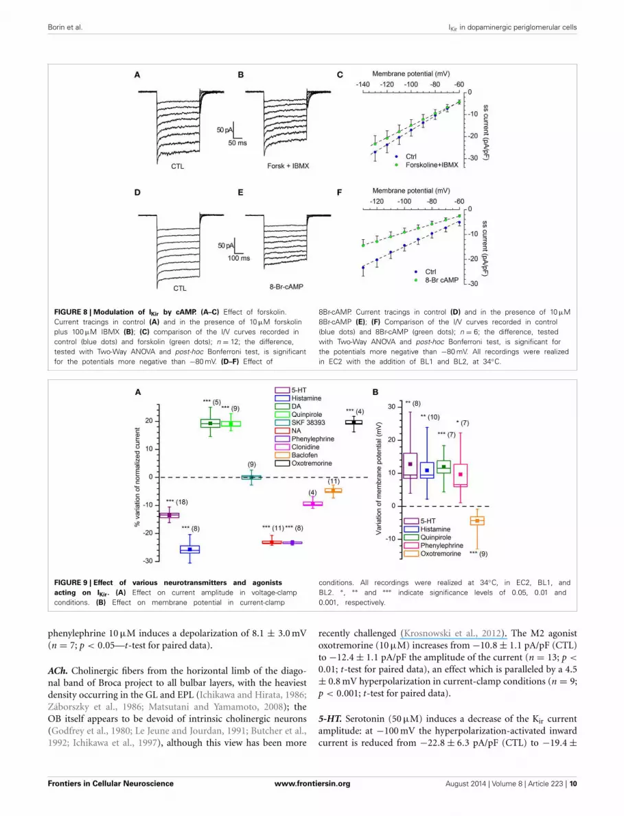
Under voltage-clamp conditions, the addition to the extracel-lular solution of 10 μM forskolin, a classical activator of adenylylcyclase (Seamon and Daly, 1981) and 0.1 mM IBMX, a phos-phodiesterase inhibitor, induces a decrease of the Kir current(Figures 8A–C): the stimulation of the cAMP synthesis reducesthe IKir amplitude of 12.3 ± 0.22 % in the range from −80to −130 mV (n = 12; p < 0.01).
The experiment was repeated in the same testing conditions,but using 10 μM 8Br-cAMP. The effect was more marked, witha 36.9 ± 0.15 % reduction of current amplitude (Figures 8D–F;n = 6; p < 0.001). In both cases, the difference among controland test was significant in the range of potentials more negativethan −80 mV (Two-Way ANOVA).
Kir modulation by neurotransmittersDopaminergic cells in the MOB are the target of numerous affer-ents releasing a variety of neurotransmitters potentially capable ofa modulation of the Kir-current, including some which are knownto affect the cAMP pathway.
Frontiers in Cellular Neuroscience www.frontiersin.org August 2014 | Volume 8 | Article 223 | 8

Borin et al. IKir in dopaminergic periglomerular cells
FIGURE 7 | Organic blockers of the Kir channels. (A) with a GIRK currentactivated by a cholinergic muscarinic agonist (oxotremorine 10 μM), the GIRKchannels blocker tertiapin-Q (1 μM) completely suppresses the current(n = 11; see text for explanation); tertiapin-Q alone does not change theamplitude of hyperpolarization-activated (not shown; n = 13). In this as in thefollowing panels, the steady-state (ss) current is calculated in relationto membrane capacity. (B) Quinacrine (100 μM) inhibition ofhyperpolarization-activated current (n = 15). (C) With the KIR2.x channelsblocked by quinacrine, a muscarinic cholinergic agonist (oxotremorine 10 μM)
can activate a GIRK current (yellow dots), and this fraction can be completelysuppressed by tertiapin (green dots; n = 7). ∗, ∗∗ and ∗∗∗ indicate significancelevels of 0.05, 0.01 and 0.001, respectively. (D) Effect of quinacrine onmembrane potential. Perforated patch recording in standard saline (EC1solution). The blue bar indicates the time of application of 100 μM into thebath; to the right, a sequence recorded after injection of 35 pAhyperpolarizing current; (further explanation in the text). All recordings wererealized at 34◦C, in EC2, BL1, and BL2; statistical analysis performed withTwo-Way ANOVA and post-hoc Bonferroni test.
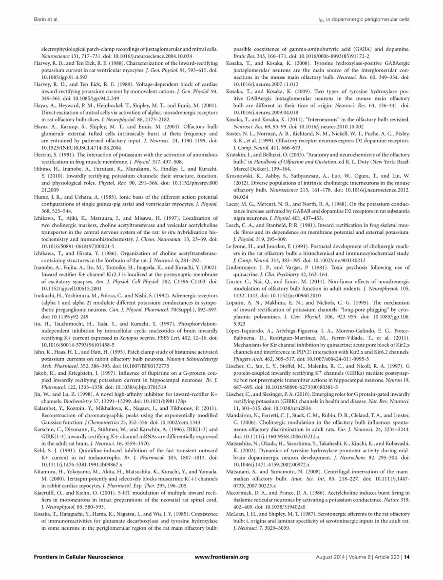
We tested the effects on the Kir-current amplitude of 5–10 minapplications of 5-HT (50 μM), dopamine (100 μM, + 1 mMascorbic acid), quinpirole (D2 agonist, 30 μM), SKF 38393 (D1agonist, 15 μM) noradrenaline (NA; 100 μM, + 1 mM ascorbicacid), phenylephrine (α1 agonist, 10 μM), clonidine (α2 agonist,10 μM), histamine (10 μM), oxotremorine (muscarinic agonist,10 μM) and baclofen (GABAB agonist, 10 μM); the results, illus-trated in Figure 9.
NA. The MOB receives a rich noradrenergic projection from thelocus cœruleus (LC): approximately 40% of LC neurons (an esti-mated 400–600 out of 1600 cells) project to the rat OB (Shipleyet al., 1985).
NA, acting via α1 receptors, has been reported to inhibitrectifying and non-rectifying leak potassium currents (Inokuchiet al., 1992; Vaughan et al., 1996; Hayar et al., 2001; Nai et al.,2010). We tested the NA (100 μM) on DA-PG cells in slice at34◦C observing a 24.6% reduction in the amplitude of the current
activated by hyperpolarization: the current evoked at −100 mVdecreased from −17.51 ± 1.62 pA/pF in control conditions to−13.20 ± 1.23 pA/pF in the presence of NA (n = 11; p < 0.001;Two-Way ANOVA; Figure 9A). Next, we tried to further charac-terize this effect identifying the subtype of α-receptor involved.Clonidine (α2 agonist, 10 μM) was ineffective (from −15.1 ± 1.4pA/pF in control conditions to −14.1 ± 1.7 pA/pF with cloni-dine; n = 4; p = 0.4—Two-Way ANOVA; Figure 9A), whereasphenylephrine (α1 agonist, 10 μM) induced a 24.1% inhibition(from −12.9 ± 1.3 pA/pF in control conditions to −9.8 ± 0.92pA/pF with phenylephrine; n = 8; p < 0.05—Two-Way ANOVA;Figure 9A), an inhibition almost identical to that of NA.
If the Kir current deeply influences the resting potential, thenwe should expect that any modification of the amplitude of thecurrent is paralleled by a variation of the membrane potential. Inparticular, in this case, a reduction of a hyperpolarizing currentshould be reflected in a depolarization of the cell in current-clampexperiments; this is exactly what can be observed (Figure 9B):
Frontiers in Cellular Neuroscience www.frontiersin.org August 2014 | Volume 8 | Article 223 | 9
Borin et al. IKir in dopaminergic periglomerular cells
FIGURE 8 | Modulation of IKir by cAMP. (A–C) Effect of forskolin.Current tracings in control (A) and in the presence of 10 μM forskolinplus 100 μM IBMX (B); (C) comparison of the I/V curves recorded incontrol (blue dots) and forskolin (green dots); n = 12; the difference,tested with Two-Way ANOVA and post-hoc Bonferroni test, is significantfor the potentials more negative than −80 mV. (D–F) Effect of
8Br-cAMP. Current tracings in control (D) and in the presence of 10 μM8Br-cAMP (E); (F) Comparison of the I/V curves recorded in control(blue dots) and 8Br-cAMP (green dots); n = 6; the difference, testedwith Two-Way ANOVA and post-hoc Bonferroni test, is significant forthe potentials more negative than −80 mV. All recordings were realizedin EC2 with the addition of BL1 and BL2, at 34◦C.
FIGURE 9 | Effect of various neurotransmitters and agonists
acting on IKir. (A) Effect on current amplitude in voltage-clampconditions. (B) Effect on membrane potential in current-clamp
conditions. All recordings were realized at 34◦C, in EC2, BL1, andBL2. ∗, ∗∗ and ∗∗∗ indicate significance levels of 0.05, 0.01 and0.001, respectively.
phenylephrine 10 μM induces a depolarization of 8.1 ± 3.0 mV(n = 7; p < 0.05—t-test for paired data).
ACh. Cholinergic fibers from the horizontal limb of the diago-nal band of Broca project to all bulbar layers, with the heaviestdensity occurring in the GL and EPL (Ichikawa and Hirata, 1986;Záborszky et al., 1986; Matsutani and Yamamoto, 2008); theOB itself appears to be devoid of intrinsic cholinergic neurons(Godfrey et al., 1980; Le Jeune and Jourdan, 1991; Butcher et al.,1992; Ichikawa et al., 1997), although this view has been more
recently challenged (Krosnowski et al., 2012). The M2 agonistoxotremorine (10 μM) increases from −10.8 ± 1.1 pA/pF (CTL)to −12.4 ± 1.1 pA/pF the amplitude of the current (n = 13; p <
0.01; t-test for paired data), an effect which is paralleled by a 4.5± 0.8 mV hyperpolarization in current-clamp conditions (n = 9;p < 0.001; t-test for paired data).
5-HT. Serotonin (50 μM) induces a decrease of the Kir currentamplitude: at −100 mV the hyperpolarization-activated inwardcurrent is reduced from −22.8 ± 6.3 pA/pF (CTL) to −19.4 ±
Frontiers in Cellular Neuroscience www.frontiersin.org August 2014 | Volume 8 | Article 223 | 10

Borin et al. IKir in dopaminergic periglomerular cells
5.3 pA/pF (n = 18, p < 0.01; Two-Way ANOVA), to which corre-spond a depolarization of 12.8 ± 3.2 mV (n = 8; p < 0.001; t-testfor paired data) in current-clamp conditions.
Histamine. In voltage-clamp conditions, histamine (10 μM)induces a significant reduction of the Kir current amplitude,which at −100 mV decreases from −19.0 ± 2.0 pA/pF (CTL) to−14.1 ± 1.8 pA/pF (test; p < 0.05, Two-Way ANOVA n = 8),an effect which is paralleled by a 10.9 ± 2.4 mV depolarization(p = 0.0013, n = 10; t-test for coupled data).
DA. The presence of autoreceptors is an hallmark of dopamin-ergic neurons, and therefore it was of interest to verify if theiractivation could modify the IKir. Dopamine (100 μM) inducesan increase of the Kir current: in slice, at 34◦C there is a nearly17% increase of the current amplitude, from −16.9 ± 2.9 pA/pF(CTL) to −19.9 ± 2.3 pA/pF (current measured in response to astep to −100 mV; n = 5; p < 0.01; Two-Way ANOVA).
The effect is exactly mimicked by the D2 agonist quinpi-role: 30 μM promotes an average increase 17%, from −15.5 ±0.8 pA/pF (CTL) to −18.3 ± 1.2 pA/pF (current measured inresponse to a step to −100 mV; n = 9; p < 0.05; Two-WayANOVA); on the contrary, the D1 agonist SKF 38393 (Sibley et al.,1982) remains ineffective (15 μM, n = 4; Figure 9A).
GABA. Kir3 channel family (GIRK) has been shown to be func-tionally regulated by GABAB receptors in numerous systems(Sodickson and Bean, 1996; Lüscher et al., 1997; Tabata et al.,2005; David et al., 2006), including dopaminergic neurons (Laceyet al., 1988). We therefore tested the GABAB agonist baclofen(Bowery et al., 1980) 10 μM on the Kir current, without observ-ing any effect (from −30.5 ± 4.6 pA/pF to −29.5 ± 5.0 pA/pF,n = 11; p > 0.5; Two-Way ANOVA; not shown).
DISCUSSIONTwo hyperpolarization-activated currents with inward rectifyingproperties are present in TH-GFP+ neurons.
The first is an h-current (Ih, or If in cardiac tissue), a mixedcation current with a reversal potential substantially positive to EK
(Hibino et al., 2010). Ih has a relatively slow activation kinetics, isinsensitive to Ba2+, can be selectively blocked by drugs like ivabra-dine or ZD728, and does not show a voltage sensitivity dependenton [K+]o (Biel et al., 2009). This current has been the object ofa previous study (Pignatelli et al., 2013), and will not be furtherdiscussed here.
A second type of hyperpolarization-activated current is char-acterized by fast kinetics, is permeable primarily to K+, is blockedby extracellular Ba2+ and Cs+, has a voltage-dependence itselfdependent on extracellular K+ concentration, and is identifiedas a classical inward rectifier potassium current (Kir). Sensitivityto Ba2+, insensitivity to selective h-current blockers, fast kinet-ics of activation and reversal potential, all suggest that the secondhyperpolarization-activated current observed in TH-GFP+ neu-rons and described in this study belongs to this class.
Under physiological conditions, Kir channels generate a largeK+ conductance at potentials negative to EK, but permit a smallcurrent flow also at potentials positive to EK (Hibino et al.,
2010); as a result, the Kir conductance has a tonic hyperpolarizinginfluence on the resting membrane potential (Vrest), controllingexcitability and affecting the repolarizing phase of the actionpotentials in excitable cells (Constanti and Galvan, 1983; Humeand Uehara, 1985; Day et al., 2005). In this study, we show thatthe Kir current plays a key role in controlling Vrest in DA-PG cells,neurons that -due to their strategic positioning at the entry of thebulbar circuitry and for direct connection with both the sensoryinput and projection neurons- are pivotal elements in the opera-tion of glomerular circuits, and we show that the IKir in these cellsis finely tuned by a variety of neurotransmitters.
WHICH POPULATION OF Kir CHANNELS?Of the seven main types of Kir channels, at least two (KIR2.xand 3.x) are present in the MOB. Of the 2.x family, KIR2.1 ishighly expressed in periglomerular cells (Prüss et al., 2005), aswell as KIR2.2 (a.k.a. IRK2/KCNJ12; (Karschin et al., 1996); alsoKIR2.3 is weakly expressed in the glomerular layer (Inanobe et al.,2002; Allen Brain Atlas, 2013). Quinacrine, which differentiallyinhibits the Kir channels (KIR2.3 > KIR2.1 � KIR6.2; (López-Izquierdo et al., 2011), suppresses a large (46%) fraction ofhyperpolarization-activated inward current. However, the pres-ence of KIR6.x (a.k.a. KATP) channels can probably be excluded:these channels are thought to be octomers composed of fourpore-forming Kir subunits, and four auxiliary proteins, the sul-fonylurea receptors (SURx) believed to be responsible for thechannel (Hibino et al., 2010). SURx proteins are not detectedin the MOB (Allen Brain Atlas, 2013), and therefore the morelikely target of the action of quinacrine are 2.x Kir channels, whosepresence in the MOB would be confirmed by our data.
The presence of KIR3.x channels (G protein-coupled Kir, a.k.a.GIRK, channels) has been reported in the periglomerular layerof the MOB (Karschin et al., 1996); the sensitivity of a fractionof the hyperpolarization-activated inward current to tertiapin, arather selective blocker of KIR3.1–3.4 channels (Jin and Lu, 1998;Kitamura et al., 2000; Ramu et al., 2008), would confirm thisfinding.
In conclusion, in control conditions, DA-PG cells display aninward rectifying current at hyperpolarizing potentials aroundEK. A first component is sustained by Ba2+-sensitive KIR2.x chan-nels, which are constitutively active and which are well known tocontribute to the resting K+ conductance in many cells (Hibinoet al., 2010). On the other hand, this background activity couldreceive the contribution also of KIR3.x channels opening inresponse to G-protein activation by different neuromodulators,as discussed below.
PHARMACOLOGYMany neuromodulators such as NA, ACh, and 5-HT, play impor-tant functions in many sensory systems. As it occurs to other brainfunctions, sensory perception must be finely tuned accordingto task demands, qualities of sensory stimuli -such as strengthor signal-to-noise ratio- and global physiological state. In thiscontext, it is rather interesting that IKir, a current playing suchan important role in the resting membrane potential of cellsstrategically placed at the entry of the bulbar circuitry, can bemodulated in both directions by a variety of neurotransmitters,
Frontiers in Cellular Neuroscience www.frontiersin.org August 2014 | Volume 8 | Article 223 | 11

Borin et al. IKir in dopaminergic periglomerular cells
all released in the region where DA-PG cells reside. The responsesinduced by neurotransmitters shown and discussed in this workare due to the direct activation of receptors on bulbar DA-PGcells, since all recordings were made in conditions of block ofsynaptic transmission.
NAIn this work, we show that in mouse DA-PG cells, NA and the α1agonist phenylephrine significantly reduce the IKir and depolarizethe cell.
Although the role of NA in olfactory function is one of thebest studied in the OB (Trombley, 1994; Ciombor et al., 1999;Devore and Linster, 2012; Zimnik et al., 2013) to name a few,its effects at cellular, network and behavioral levels are some-what discordant (Ennis and Hayar, 2008); it is worth noting thatthese inconsistencies have been ascribed at least in part to theabsence of information pertaining to glomerular modulation byNA (Linster et al., 2011), a gap to fill which this work provides afirst contribution.
The locus cœruleus neurons have been proposed to influenceexternal signal processing so that salient stimuli are enhanced andthe activity more related to tonic, vegetative functions is sup-pressed (Aston-Jones et al., 2000). In the OB, the NA release inresponse to LC activation should bring the DA-PG neurons to amore excited state for the inhibition of the Kir conductance. This,for the known presynaptic inhibitory effect of DA (Koster et al.,1999; Ennis et al., 2001), would reduce the release of glutamatefrom the olfactory nerve terminals, improving the signal-to-noiseratio of the information coming from the olfactory epithelium,an effect that would be in line with the postulated general role ofthe LC on sensory stimuli perception (Aston-Jones et al., 2000).
An additional role of NA on dopaminergic cells might be ofsome interest: a significant fraction of the interneurons added inadulthood to the glomerular layer of the MOB are dopaminer-gic (Pignatelli et al., 2009), and noradrenaline signaling enhancesnewborn cell survival (Bauer et al., 2003; Bovetti et al., 2011).
AChIn the MOB, ACh, acting on both nicotinic and muscarinic recep-tors, has a complex effect (for a review see Devore and Linster,2012). Overall, the resulting effect of cholinergic modulation isexcitatory (Elaagouby and Gervais, 1992) and the multiple actionof ACh seems to be orchestrated toward an enhancement of speci-ficity and temporal precision of mitral cell responses to odors(Elaagouby and Gervais, 1992; Mandairon et al., 2006; Tsunoet al., 2008; D’Souza and Vijayaraghavan, 2012).
In this work we show that in DA-PG cells the activationof M2 muscarinic receptors increases the amplitude of a Kir
current, an effect which is paralleled by a 4.5 mV hyperpolar-ization in current-clamp conditions. A similar effect has beenreported in a variety of preparations, ranging from mammal atrialmyocytes (Sakmann et al., 1983), to thalamic reticular neurons(Mccormick and Prince, 1986), spinal motoneurons (Chevallieret al., 2008), interneurons of striatum (Calabresi et al., 1998), neo-cortex (Xiang et al., 1998), and hippocampus (McQuiston andMadison, 1999).
M2-type muscarinic receptors are described in the glomeru-lar layer associated to PG-DA neurons (Crespo et al., 2000; Allen
Brain Atlas, 2013). In a previous paper (Pignatelli and Belluzzi,2008), we showed that the activation of M2 metabotropic cholin-ergic receptor in PG-DA neurons induced a hyperpolarizationmediated by a K-conductance, which in the present work is nowidentified as a Kir.
5-HTProjections from the dorsal and median raphe nuclei -one of themost prominent neuromodulatory systems in the brain- providea dense serotonergic innervation of the MOB, and in particular ofthe glomerular region (Araneda et al., 1980; McLean and Shipley,1987). Earlier studies have shown that 5-HT2 receptor mRNAand protein are heavily expressed in the glomerular layer (Morilaket al., 1993) and activation of 5-HT(2C) receptors mediates exci-tation in about one third of glomerular neurons, not betteridentified (Hardy et al., 2005); this study further develops thisobservations showing that serotonin produces excitatory mod-ulation of DA-PG cells by reducing the IKir amplitude, therebydepolarizing the cell for 12.8 ± 3.2 mV.
A similar action on IKir is described also in rat motoneurons(Kjaerulff and Kiehn, 2001).
HistamineThe MOB receives histaminergic inputs primarily from the cau-dal tuberal and postmammillary magnocellular hypothalamus(Auvinen and Panula, 1988; Panula et al., 1989) via the olfactorypeduncle (Brunjes, 2013), and previous studies have shown thatin an unidentified fraction of periglomerular cells, H1-receptoractivation causes a block of a potassium current (Jahn et al.,1995).Here we show that the IKir in DA-PG cells is reduced andthat this action results in a depolarization of the cells.
DADopamine (100 μM) induces an increase of the Kir current viaD2R; the D2R agonist quinpirole (30 μM) perfectly replicates theeffect of dopamine. Further experiments using receptor protec-tion with D1R selective antagonists might exclude more defini-tively a contribution from D1R, although a direct activation ofD1R with SKF 38393 (15 μM) was completely ineffective. We didnot investigate the pathways involved.
CONCLUDING OBSERVATIONSIn the present study we have shown that (i) DA-PG cells containa large inward rectifier current whose block produces significantdepolarizations, nominating this conductance as one of the mainplayers controlling the resting membrane potential (and conse-quently excitability) in these cells, and that (ii) this current issubject to a complex modulation.
Bulbar DA-PG cells, the largest and one of the most conservedpopulations of DA neurons in the CNS, are pivotal neurons in theoperation of glomerular circuits, the site where odor informationis initially processed. It is therefore of some interest that theirexcitability is profoundly dependent upon the Kir current, andthat this -in turn- is target of numerous neurotransmitters thatcan finely modulate its amplitude, a process that ultimatelyimpacts all subsequent odor processing in the olfactory system.
In this context, it is increasingly evident that in the bulbas a whole there is an enormous and still largely hidden layer
Frontiers in Cellular Neuroscience www.frontiersin.org August 2014 | Volume 8 | Article 223 | 12

Borin et al. IKir in dopaminergic periglomerular cells
of “molecular computation” (Bhalla, 2014), which multipliestremendously the degrees of freedom of the bulbar network insignal processing.
ACKNOWLEDGMENTSWe thank the Allen Brain Institute, Seattle (WA), for use oftheir online materials, available from http://mouse.brain-map.
org/. We wish to thank Dr. Faride Pighin for his help and supportin the experimental phase.
REFERENCESAlagem, N., Dvir, M., and Reuveny, E. (2001). Mechanism of Ba(2+) block of a
mouse inwardly rectifying K+ channel: differential contribution by two discreteresidues. J. Physiol. 534, 381–393. doi: 10.1111/j.1469-7793.2001.00381.x
Allen Brain Atlas. (2013). Available online at: http://www.brain-map.org.Araneda, S., Bobillier, P., Buda, M., and Pujol, J. F. (1980). Retrograde axonal trans-
port following injection of [3H]serotonin in the olfactory bulb. I. Biochemicalstudy. Brain Res. 196, 405–415. doi: 10.1016/0006-8993(80)90404-7
Aston-Jones, G., Rajkowski, J., and Cohen, J. (2000). Locus coeruleus and regula-tion of behavioral flexibility and attention. Prog. Brain Res. 126, 165–182. doi:10.1016/S0079-6123(00)26013-5
Auvinen, S., and Panula, P. (1988). Development of histamine-immunoreactiveneurons in the rat brain. J. Comp. Neurol. 276, 289–303. doi:10.1002/cne.902760211
Baker, H., Morel, K., Stone, D. M., and Maruniak, J. A. (1993). Adult naris closureprofoundly reduces tyrosine hydroxylase expression in mouse olfactory bulb.Brain Res. 614, 109–116. doi: 10.1016/0006-8993(93)91023-L
Bauer, S., Moyse, E., Jourdan, F., Colpaert, F., Martel, J. C., and Marien, M. (2003).Effects of the alpha 2-adrenoreceptor antagonist dexefaroxan on neurogenesis inthe olfactory bulb of the adult rat in vivo: selective protection against neuronaldeath. Neuroscience 117, 281–291. doi: 10.1016/S0306-4522(02)00757-1
Bhalla, U. S. (2014). Molecular computation in neurons: a modeling perspective.Curr. Opin. Neurobiol. 25, 31–37. doi: 10.1016/j.conb.2013.11.006
Bhave, G., Lonergan, D., Chauder, B. A., and Denton, J. S. (2010). Small-moleculemodulators of inward rectifier K channels: recent advances and future possibil-ities. Future Med. Chem. 2, 757–774. doi: 10.4155/fmc.10.179
Biel, M., Wahl-Schott, C., Michalakis, S., and Zong, X. (2009). Hyperpolarization-activated cation channels: from genes to function. Physiol. Rev. 89, 847–885. doi:10.1152/physrev.00029.2008
Bois, P., Bescond, J., Renaudon, B., and Lenfant, J. (1996). Mode of action of brady-cardic agent, S 16257, on ionic currents of rabbit sinoatrial node cells. Br. J.Pharmacol. 118, 1051–1057. doi: 10.1111/j.1476-5381.1996.tb15505.x
Bolton, S., and Butt, A. M. (2006). Cyclic AMP-mediated regulation of the rest-ing membrane potential in myelin-forming oligodendrocytes in the isolatedintact rat optic nerve. Exp. Neurol. 202, 36–43. doi: 10.1016/j.expneurol.2006.05.009
BoSmith, R. E., Briggs, I., and Sturgess, N. C. (1993). Inhibitory actions ofZENECA ZD7288 on whole-cell hyperpolarization activated inward current (If)in guinea-pig dissociated sinoatrial node cells. Br. J. Pharmacol. 110, 343–349.doi: 10.1111/j.1476-5381.1993.tb13815.x
Bovetti, S., Gribaudo, S., Puche, A. C., De Marchis, S., and Fasolo, A.(2011). From progenitors to integrated neurons: role of neurotransmit-ters in adult olfactory neurogenesis. J. Chem. Neuroanat. 42, 304–316. doi:10.1016/j.jchemneu.2011.05.006
Bowery, N. G., Hill, D. R., Hudson, A. L., Doble, A., Middlemiss, D. N., Shaw,J., et al. (1980). (-)Baclofen decreases neurotransmitter release in the mam-malian CNS by an action at a novel GABA receptor. Nature 283, 92–94. doi:10.1038/283092a0
Brunjes, P. C. (2013). The mouse olfactory peduncle. 2.The anterior limb of theanterior commissure. Front. Neuroanat. 6:51. doi: 10.3389/fnana.2012.00051
Bucchi, A., Baruscotti, M., and DiFrancesco, D. (2002). Current-dependent blockof rabbit sino-atrial node I(f) channels by ivabradine. J. Gen. Physiol. 120, 1–13.doi: 10.1085/jgp.20028593
Butcher, L. L., Oh, J. D., Woolf, N. J., Edwards, R. H., and Roghani, A. (1992).Organization of central cholinergic neurons revealed by combined in situhybridization histochemistry and choline-O-acetyltransferase immunocyto-chemistry. Neurochem. Int. 21, 429–445. doi: 10.1016/0197-0186(92)90195-W
Calabresi, P., Centonze, D., Pisani, A., Sancesario, G., North, R. A., and Bernardi,G. (1998). Muscarinic IPSPs in rat striatal cholinergic interneurones. J. Physiol.510, 421–427. doi: 10.1111/j.1469-7793.1998.421bk.x
Chevallier, S., Nagy, F., and Cabelguen, J. M. (2008). Muscarinic control of theexcitability of hindlimb motoneurons in chronic spinal-transected salamanders.Eur. J. Neurosci. 28, 2243–2253. doi: 10.1111/j.1460-9568.2008.06506.x
Ciombor, K. J., Ennis, M., and Shipley, M. T. (1999). Norepinephrine increases ratmitral cell excitatory responses to weak olfactory nerve input via alpha-1 recep-tors in vitro. Neuroscience 90, 595–606. doi: 10.1016/S0306-4522(98)00437-0
Constanti, A., and Galvan, M. (1983). Fast inward-rectifying current accounts foranomalous rectification in olfactory cortex neurones. J. Physiol. 335, 153–178.
Crespo, C., Blasco-Ibanez, J. M., Brinon, J. G., Alonso, J. R., Dominguez, M. I.,and Martinez-Guijarro, F. J. (2000). Subcellular localization of m2 muscarinicreceptors in GABAergic interneurons of the olfactory bulb. Eur. J. Neurosci. 12,3963–3974. doi: 10.1046/j.1460-9568.2000.00289.x
David, M., Richer, M., Mamarbachi, A. M., Villeneuve, L. R., Dupré, D. J.,and Hebert, T. E. (2006). Interactions between GABA-B1 receptors and Kir3 inwardly rectifying potassium channels. Cell. Signal. 18, 2172–2181. doi:10.1016/j.cellsig.2006.05.014
Day, M., Carr, D. B., Ulrich, S., Ilijic, E., Tkatch, T., and Surmeier, D. J. (2005).Dendritic excitability of mouse frontal cortex pyramidal neurons is shapedby the interaction among HCN, Kir2, and Kleak channels. J. Neurosci. 25,8776–8787. doi: 10.1523/JNEUROSCI.2650-05.2005
Devore, S., and Linster, C. (2012). Noradrenergic and cholinergic modula-tion of olfactory bulb sensory processing. Front. Behav. Neurosci. 6:52. doi:10.3389/fnbeh.2012.00052
Dobrev, D., Wettwer, E., Kortner, A., Knaut, M., Schüler, S., and Ravens, U. (2002).Human inward rectifier potassium channels in chronic and postoperative atrialfibrillation. Cardiovasc. Res. 54, 397–404. doi: 10.1016/S0008-6363(01)00555-7
D’Souza, R. D., and Vijayaraghavan, S. (2012). Nicotinic receptor-mediated filter-ing of mitral cell responses to olfactory nerve inputs involves the a3b4 subtype.J. Neurosci. 32, 3261–3266. doi: 10.1523/JNEUROSCI.5024-11.2012
Elaagouby, A., and Gervais, R. (1992). ACh-induced long-lasting enhancement inexcitability of the olfactory bulb. Neuroreport 3, 10–12. doi: 10.1097/00001756-199201000-00002
Ennis, M., and Hayar, A. (2008). “4.37 - Physiology of the main olfactory bulb,” inThe Senses: a Comprehensive Reference, Vol. 1, eds A. I. Basbaum, K. Akimichi,M. S. Gordon, W. Gerald, D. A. Thomas, H. M. Richard, et al. (New York, NY:Academic Press), 641–686.
Ennis, M., Zhou, F. M., Ciombor, K. J., Aroniadou-Anderjaska, V., Hayar, A.,Borrelli, E., et al. (2001). Dopamine D2 receptor-mediated presynaptic inhi-bition of olfactory nerve terminals. J. Neurophysiol. 86, 2986–2997.
Evans, R. J., and Surprenant, A. (1993). Effects of phospholipase A2 inhibitorson coupling of alpha 2-adrenoceptors to inwardly rectifying potassium cur-rents in guinea-pig submucosal neurones. Br. J. Pharmacol. 110, 591–596. doi:10.1111/j.1476-5381.1993.tb13851.x
French, R. J., and Shoukimas, J. J. (1985). An ion’s view of the potassium channel.The structure of the permeation pathway as sensed by a variety of blocking ions.J. Gen. Physiol. 85, 669–698. doi: 10.1085/jgp.85.5.669
Gall, C. M., Hendry, S. H., Seroogy, K. B., Jones, E. G., and Haycock, J. W.(1987). Evidence for coexistence of GABA and dopamine in neurons of therat olfactory bulb. J. Comp. Neurol. 266, 307–318. doi: 10.1002/cne.902660302
Godfrey, D. A., Ross, C. D., Herrmann, A. D., and Matschinsky, F. M. (1980).Distribution and derivation of cholinergic elements in the rat olfactory bulb.Neuroscience 5, 273–292. doi: 10.1016/0306-4522(80)90103-7
Hagiwara, S., Miyazaki, S., Moody, W., and Patlak, J. (1978). Blocking effectsof barium and hydrogen ions on the potassium current during anomalousrectification in the starfish egg. J. Physiol. 279, 167–185.
Hagiwara, S., Miyazaki, S., and Rosenthal, N. P. (1976). Potassium current and theeffect of cesium on this current during anomalous rectification of the egg cellmembrane of a starfish. J. Gen. Physiol. 67, 621–638. doi: 10.1085/jgp.67.6.621
Halász, N. (1990). The Vertebrate Olfactory System: Chemical Neuroanatomy,Function and Development. Budapest: Académiai Kiadó.
Halász, N., Hökfelt, T., Ljungdahl, A., Johansson, O., and Goldstein, M. (1977).Dopamine neurons in the olfactory bulb. Adv. Biochem. Psychopharmacol. 16,169–177.
Hardy, A., Palouzier-Paulignan, B., Duchamp, A., Royet, J. P., and Duchamp-Viret, P. (2005). 5-hydroxytryptamine action in the rat olfactory bulb: in vitro
Frontiers in Cellular Neuroscience www.frontiersin.org August 2014 | Volume 8 | Article 223 | 13
Borin et al. IKir in dopaminergic periglomerular cells
electrophysiological patch-clamp recordings of juxtaglomerular and mitral cells.Neuroscience 131, 717–731. doi: 10.1016/j.neuroscience.2004.10.034
Harvey, R. D., and Ten Eick, R. E. (1988). Characterization of the inward-rectifyingpotassium current in cat ventricular myocytes. J. Gen. Physiol. 91, 593–615. doi:10.1085/jgp.91.4.593
Harvey, R. D., and Ten Eick, R. E. (1989). Voltage-dependent block of cardiacinward-rectifying potassium current by monovalent cations. J. Gen. Physiol. 94,349–361. doi: 10.1085/jgp.94.2.349
Hayar, A., Heyward, P. M., Heinbockel, T., Shipley, M. T., and Ennis, M. (2001).Direct excitation of mitral cells via activation of alpha1-noradrenergic receptorsin rat olfactory bulb slices. J. Neurophysiol. 86, 2173–2182.
Hayar, A., Karnup, S., Shipley, M. T., and Ennis, M. (2004). Olfactory bulbglomeruli: external tufted cells intrinsically burst at theta frequency andare entrained by patterned olfactory input. J. Neurosci. 24, 1190–1199. doi:10.1523/JNEUROSCI.4714-03.2004
Hestrin, S. (1981). The interaction of potassium with the activation of anomalousrectification in frog muscle membrane. J. Physiol. 317, 497–508.
Hibino, H., Inanobe, A., Furutani, K., Murakami, S., Findlay, I., and Kurachi,Y. (2010). Inwardly rectifying potassium channels: their structure, function,and physiological roles. Physiol. Rev. 90, 291–366. doi: 10.1152/physrev.00021.2009
Hume, J. R., and Uehara, A. (1985). Ionic basis of the different action potentialconfigurations of single guinea-pig atrial and ventricular myocytes. J. Physiol.368, 525–544.
Ichikawa, T., Ajiki, K., Matsuura, J., and Misawa, H. (1997). Localization oftwo cholinergic markers, choline acetyltransferase and vesicular acetylcholinetransporter in the central nervous system of the rat: in situ hybridization his-tochemistry and immunohistochemistry. J. Chem. Neuroanat. 13, 23–39. doi:10.1016/S0891-0618(97)00021-5
Ichikawa, T., and Hirata, Y. (1986). Organization of choline acetyltransferase-containing structures in the forebrain of the rat. J. Neurosci. 6, 281–292.
Inanobe, A., Fujita, A., Ito, M., Tomoike, H., Inageda, K., and Kurachi, Y. (2002).Inward rectifier K+ channel Kir2.3 is localized at the postsynaptic membraneof excitatory synapses. Am. J. Physiol. Cell Physiol. 282, C1396–C1403. doi:10.1152/ajpcell.00615.2001
Inokuchi, H., Yoshimura, M., Polosa, C., and Nishi, S. (1992). Adrenergic receptors(alpha 1 and alpha 2) modulate different potassium conductances in sympa-thetic preganglionic neurons. Can. J. Physiol. Pharmacol. 70(Suppl.), S92–S97.doi: 10.1139/y92-249
Ito, H., Tsuchimochi, H., Tada, Y., and Kurachi, Y. (1997). Phosphorylation-independent inhibition by intracellular cyclic nucleotides of brain inwardlyrectifying K+ current expressed in Xenopus oocytes. FEBS Lett. 402, 12–16. doi:10.1016/S0014-5793(96)01458-5
Jahn, K., Haas, H. L., and Hatt, H. (1995). Patch clamp study of histamine activatedpotassium currents on rabbit olfactory bulb neurons. Naunyn SchmiedebergsArch. Pharmacol. 352, 386–393. doi: 10.1007/BF00172775
Jakob, R., and Krieglstein, J. (1997). Influence of flupirtine on a G-protein cou-pled inwardly rectifying potassium current in hippocampal neurones. Br. J.Pharmacol. 122, 1333–1338. doi: 10.1038/sj.bjp.0701519
Jin, W., and Lu, Z. (1998). A novel high-affinity inhibitor for inward-rectifier K+channels. Biochemistry 37, 13291–13299. doi: 10.1021/bi981178p
Kalambet, Y., Kozmin, Y., Mikhailova, K., Nagaev, I., and Tikhonov, P. (2011).Reconstruction of chromatographic peaks using the exponentially modifiedGaussian function. J Chemometrics 25, 352–356. doi: 10.1002/cem.1343
Karschin, C., Dissmann, E., Stühmer, W., and Karschin, A. (1996). IRK(1-3) andGIRK(1-4) inwardly rectifying K+ channel mRNAs are differentially expressedin the adult rat brain. J. Neurosci. 16, 3559–3570.
Kehl, S. J. (1991). Quinidine-induced inhibition of the fast transient outwardK+ current in rat melanotrophs. Br. J. Pharmacol. 103, 1807–1813. doi:10.1111/j.1476-5381.1991.tb09867.x
Kitamura, H., Yokoyama, M., Akita, H., Matsushita, K., Kurachi, Y., and Yamada,M. (2000). Tertiapin potently and selectively blocks muscarinic K(+) channelsin rabbit cardiac myocytes. J. Pharmacol. Exp. Ther. 293, 196–205.
Kjaerulff, O., and Kiehn, O. (2001). 5-HT modulation of multiple inward recti-fiers in motoneurons in intact preparations of the neonatal rat spinal cord.J. Neurophysiol. 85, 580–593.
Kosaka, T., Hataguchi, Y., Hama, K., Nagatsu, I., and Wu, J. Y. (1985). Coexistenceof immunoreactivities for glutamate decarboxylase and tyrosine hydroxylasein some neurons in the periglomerular region of the rat main olfactory bulb:
possible coexistence of gamma-aminobutyric acid (GABA) and dopamine.Brain Res. 343, 166–171. doi: 10.1016/0006-8993(85)91172-2
Kosaka, T., and Kosaka, K. (2008). Tyrosine hydroxylase-positive GABAergicjuxtaglomerular neurons are the main source of the interglomerular con-nections in the mouse main olfactory bulb. Neurosci. Res. 60, 349–354. doi:10.1016/j.neures.2007.11.012
Kosaka, T., and Kosaka, K. (2009). Two types of tyrosine hydroxylase pos-itive GABAergic juxtaglomerular neurons in the mouse main olfactorybulb are different in their time of origin. Neurosci. Res. 64, 436–441. doi:10.1016/j.neures.2009.04.018
Kosaka, T., and Kosaka, K. (2011). “Interneurons” in the olfactory bulb revisited.Neurosci. Res. 69, 93–99. doi: 10.1016/j.neures.2010.10.002
Koster, N. L., Norman, A. B., Richtand, N. M., Nickell, W. T., Puche, A. C., Pixley,S. K., et al. (1999). Olfactory receptor neurons express D2 dopamine receptors.J. Comp. Neurol. 411, 666–673.
Kratskin, I., and Belluzzi, O. (2003). “Anatomy and neurochemistry of the olfactorybulb,” in Handbook of Olfaction and Gustation, ed R. L. Doty (New York; Basel:Marcel Dekker), 139–164.
Krosnowski, K., Ashby, S., Sathyanesan, A., Luo, W., Ogura, T., and Lin, W.(2012). Diverse populations of intrinsic cholinergic interneurons in the mouseolfactory bulb. Neuroscience 213, 161–178. doi: 10.1016/j.neuroscience.2012.04.024
Lacey, M. G., Mercuri, N. B., and North, R. A. (1988). On the potassium conduc-tance increase activated by GABAB and dopamine D2 receptors in rat substantianigra neurones. J. Physiol. 401, 437–453.
Leech, C. A., and Stanfield, P. R. (1981). Inward rectification in frog skeletal mus-cle fibres and its dependence on membrane potential and external potassium.J. Physiol. 319, 295–309.
Le Jeune, H., and Jourdan, F. (1991). Postnatal development of cholinergic mark-ers in the rat olfactory bulb: a histochemical and immunocytochemical study.J. Comp. Neurol. 314, 383–395. doi: 10.1002/cne.903140212
Lindenmayer, J. P., and Vargas, P. (1981). Toxic psychosis following use ofquinacrine. J. Clin. Psychiatry 42, 162–164.
Linster, C., Nai, Q., and Ennis, M. (2011). Non-linear effects of noradrenergicmodulation of olfactory bulb function in adult rodents. J. Neurophysiol. 105,1432–1443. doi: 10.1152/jn.00960.2010
Lopatin, A. N., Makhina, E. N., and Nichols, C. G. (1995). The mechanismof inward rectification of potassium channels: “long-pore plugging” by cyto-plasmic polyamines. J. Gen. Physiol. 106, 923–955. doi: 10.1085/jgp.106.5.923
López-Izquierdo, A., Aréchiga-Figueroa, I. A., Moreno-Galindo, E. G., Ponce-Balbuena, D., Rodríguez-Martínez, M., Ferrer-Villada, T., et al. (2011).Mechanisms for Kir channel inhibition by quinacrine: acute pore block of Kir2.xchannels and interference in PIP(2) interaction with Kir2.x and Kir6.2 channels.Pflugers Arch. 462, 505–517. doi: 10.1007/s00424-011-0995-5
Lüscher, C., Jan, L. Y., Stoffel, M., Malenka, R. C., and Nicoll, R. A. (1997). Gprotein-coupled inwardly rectifying K+ channels (GIRKs) mediate postsynap-tic but not presynaptic transmitter actions in hippocampal neurons. Neuron 19,687–695. doi: 10.1016/S0896-6273(00)80381-5
Lüscher, C., and Slesinger, P. A. (2010). Emerging roles for G protein-gated inwardlyrectifying potassium (GIRK) channels in health and disease. Nat. Rev. Neurosci.11, 301–315. doi: 10.1038/nrn2834
Mandairon, N., Ferretti, C. J., Stack, C. M., Rubin, D. B., Cleland, T. A., and Linster,C. (2006). Cholinergic modulation in the olfactory bulb influences sponta-neous olfactory discrimination in adult rats. Eur. J. Neurosci. 24, 3234–3244.doi: 10.1111/j.1460-9568.2006.05212.x
Matsushita, N., Okada, H., Yasoshima, Y., Takahashi, K., Kiuchi, K., and Kobayashi,K. (2002). Dynamics of tyrosine hydroxylase promoter activity during mid-brain dopaminergic neuron development. J. Neurochem. 82, 295–304. doi:10.1046/j.1471-4159.2002.00972.x
Matsutani, S., and Yamamoto, N. (2008). Centrifugal innervation of the mam-malian olfactory bulb. Anat. Sci. Int. 83, 218–227. doi: 10.1111/j.1447-073X.2007.00223.x
Mccormick, D. A., and Prince, D. A. (1986). Acetylcholine induces burst firing inthalamic reticular neurones by activating a potassium conductance. Nature 319,402–405. doi: 10.1038/319402a0
McLean, J. H., and Shipley, M. T. (1987). Serotonergic afferents to the rat olfactorybulb: i. origins and laminar specificity of serotoninergic inputs in the adult rat.J. Neurosci. 7, 3029–3039.
Frontiers in Cellular Neuroscience www.frontiersin.org August 2014 | Volume 8 | Article 223 | 14
Borin et al. IKir in dopaminergic periglomerular cells
McLean, J. H., and Shipley, M. T. (1988). Postmitotic, postmigrational expressionof tyrosine hydroxylase in olfactory bulb dopaminergic neurons. J. Neurosci. 8,3658–3669.
McQuiston, A. R., and Madison, D. V. (1999). Muscarinic receptor activity hasmultiple effects on the resting membrane potentials of CA1 hippocampalinterneurons. J. Neurosci. 19, 5693–5702.
Mitsuiye, T., Shinagawa, Y., and Noma, A. (1997). Temperature dependence of theinward rectifier K+ channel gating in guinea-pig ventricular cells. Jpn. J. Physiol.47, 73–79. doi: 10.2170/jjphysiol.47.73
Morilak, D. A., Garlow, S. J., and Ciaranello, R. D. (1993). Immunocytochemicallocalization and description of neurons expressing serotonin2 receptorsin the rat brain. Neuroscience 54, 701–717. doi: 10.1016/0306-4522(93)90241-7
Nagano, N., Imaizumi, Y., and Watanabe, M. (1996). Novel blockade of Ca2+ cur-rent by quinacrine in smooth muscle cells of the guinea pig. Jpn. J. Pharmacol.71, 51–60. doi: 10.1254/jjp.71.51
Nai, Q., Dong, H. W., Linster, C., and Ennis, M. (2010). Activation of alpha1and alpha2 noradrenergic receptors exert opposing effects on excitabil-ity of main olfactory bulb granule cells. Neuroscience 169, 882–892. doi:10.1016/j.neuroscience.2010.05.010
Paajanen, V., and Vornanen, M. (2003). Regulation of action potential dura-tion under acute heat stress by I(K,ATP) and I(K1) in fish cardiacmyocytes. Am. J. Physiol. Regul. Integr. Comp. Physiol. 286, R405–R415. doi:10.1152/ajpregu.00500.2003
Panula, P., Pirvola, U., Auvinen, S., and Airaksinen, M. S. (1989). Histamine-immunoreactive nerve fibers in the rat brain. Neuroscience 28, 585–610. doi:10.1016/0306-4522(89)90007-9
Panzanelli, P., Fritschy, J. M., Yanagawa, Y., Obata, K., and Sassoè-Pognetto, M.(2007). GABAergic phenotype of periglomerular cells in the rodent olfactorybulb. J. Comp. Neurol. 502, 990–1002. doi: 10.1002/cne.21356
Park, W. S., Han, J., Kim, N., Ko, J. H., Kim, S. J., and Earm, Y. E. (2005).Activation of inward rectifier K+ channels by hypoxia in rabbit coronary arte-rial smooth muscle cells. Am. J. Physiol. Heart Circ. Physiol. 289, H2461–H2467.doi: 10.1152/ajpheart.00331.2005
Pignatelli, A., Ackman, J. B., Vigetti, D., Beltrami, A. P., Zucchini, S., and Belluzzi,O. (2009). A potential reservoir of immature dopaminergic replacement neu-rons in the adult mammalian olfactory bulb. Pflugers Arch. 457, 899–915. doi:10.1007/s00424-008-0535-0
Pignatelli, A., and Belluzzi, O. (2008). Cholinergic modulation of dopaminer-gic neurons in the mouse olfactory bulb. Chem. Senses 33, 331–338. doi:10.1093/chemse/bjm091
Pignatelli, A., Borin, M., Fogli Iseppe, A., Gambardella, C., and Belluzzi, O. (2013).The h-current in periglomerular dopaminergic neurons of the mouse olfactorybulb. PLoS ONE 8:571. doi: 10.1371/journal.pone.0056571
Pignatelli, A., Kobayashi, K., Okano, H., and Belluzzi, O. (2005). Functional prop-erties of dopaminergic neurones in the mouse olfactory bulb. J. Physiol. 564,501–514. doi: 10.1113/jphysiol.2005.084632
Podda, M. V., Riccardi, E., D’Ascenzo, M., Azzena, G. B., and Grassi, C.(2010). Dopamine D1-like receptor activation depolarizes medium spinyneurons of the mouse nucleus accumbens by inhibiting inwardly rectify-ing K+ currents through a cAMP-dependent protein kinase A-independentmechanism. Neuroscience 167, 678–690. doi: 10.1016/j.neuroscience.2010.02.075
Prüss, H., Derst, C., Lommel, R., and Veh, R. W. (2005). Differential distribu-tion of individual subunits of strongly inwardly rectifying potassium chan-nels (Kir2 family) in rat brain. Brain Res. Mol. Brain Res. 139, 63–79. doi:10.1016/j.molbrainres.2005.05.006
Ramu, Y., Xu, Y., and Lu, Z. (2008). Engineered specific and high-affinity inhibitorfor a subtype of inward-rectifier K+ channels. Proc. Natl. Acad. Sci. U.S.A. 105,10774–10778. doi: 10.1073/pnas.0802850105
Reid, M. S., Ho, L. B., Hsu, K., Fox, L., Tolliver, B. K., Adams, J. U., et al.(2002). Evidence for the involvement of cyclooxygenase activity in the devel-opment of cocaine sensitization. Pharmacol. Biochem. Behav. 71, 37–54. doi:10.1016/S0091-3057(01)00614-1
Sakmann, B., Noma, A., and Trautwein, W. (1983). Acetylcholine activation of sin-gle muscarinic K+ channels in isolated pacemaker cells of the mammalian heart.Nature 303, 250–253. doi: 10.1038/303250a0
Sawamoto, K., Nakao, N., Kakishita, K., Ogawa, Y., Toyama, Y., Yamamoto, A.,et al. (2001). Generation of dopaminergic neurons in the adult brain from
mesencephalic precursor cells labeled with a nestin-GFP transgene. J. Neurosci.21, 3895–3903.
Seamon, K. B., and Daly, J. W. (1981). Forskolin: a unique diterpene activator ofcyclic AMP-generating systems. J. Cyclic Nucleotide Res. 7, 201–224.
Shieh, R. C., Chang, J. C., and Arreola, J. (1998). Interaction of Ba2+ withthe pores of the cloned inward rectifier K+ channels Kir2.1 expressed inXenopus oocytes. Biophys. J. 75, 2313–2322. doi: 10.1016/S0006-3495(98)77675-1
Shioya, T., Matsuda, H., and Noma, A. (1993). Fast and slow blockades of theinward-rectifier K+ channel by external divalent cations in guinea-pig cardiacmyocytes. Pflugers Arch. 422, 427–435. doi: 10.1007/BF00375067
Shipley, M. T., and Ennis, M. (1996). Functional organization of olfactory system.J. Neurobiol. 30, 123–176.
Shipley, M. T., Halloran, F. J., and De La Torre, J. (1985). Surprisingly rich projec-tion from locus coeruleus to the olfactory bulb. Brain Res. 329, 294–299. doi:10.1016/0006-8993(85)90537-2
Sibley, D. R., Leff, S. E., and Creese, I. (1982). Interactions of novel dopaminer-gic ligands with D-1 and D-2 dopamine receptors. Life Sci. 31, 637–645. doi:10.1016/0024-3205(82)90764-0
Sodickson, D. L., and Bean, B. P. (1996). GABAB receptor-activated inwardly recti-fying potassium current in dissociated hippocampal CA3 neurons. J. Neurosci.16, 6374–6385.
Standen, N. B., and Stanfield, P. R. (1978). A potential- and time-dependentblockade of inward rectification in frog skeletal muscle fibres by barium andstrontium ions. J. Physiol. 280, 169–191.
Tabata, T., Haruki, S., Nakayama, H., and Kano, M. (2005). GABAergic activation ofan inwardly rectifying K+ current in mouse cerebellar Purkinje cells. J. Physiol.563, 443–457. doi: 10.1113/jphysiol.2004.081000
Trombley, P. Q. (1994). Noradrenergic modulation of synaptic transmissionbetween olfactory bulb neurons in culture: implications to olfactory learning.Brain Res. Bull. 35, 473–484. doi: 10.1016/0361-9230(94)90161-9
Tsuno, Y., Kashiwadani, H., and Mori, K. (2008). Behavioral state regulationof dendrodendritic synaptic inhibition in the olfactory bulb. J. Neurosci. 28,9227–9238. doi: 10.1523/JNEUROSCI.1576-08.2008
Vaughan, C. W., Bandler, R., and Christie, M. J. (1996). Differential responsesof lateral and ventrolateral rat periaqueductal grey neurones to noradrenalinein vitro. J. Physiol. 490, 373–381.
Walsh, K. B. (2011). Targeting GIRK channels for the development of new thera-peutic agents. Front. Pharmacol. 2:64. doi: 10.3389/fphar.2011.00064
Xiang, Z., Huguenard, J. R., and Prince, D. A. (1998). Cholinergic switchingwithin neocortical inhibitory networks. Science 281, 985–988. doi: 10.1126/sci-ence.281.5379.985
Xu, R., Zhao, Y., and Chen, C. (2002). Growth hormone-releasing peptide-2reduces inward rectifying K+ currents via a PKA-cAMP-mediated signallingpathway in ovine somatotropes. J. Physiol. 545, 421–433. doi: 10.1113/jphys-iol.2002.030916
Záborszky, L., Carlsen, J., Brashear, H. R., and Heimer, L. (1986). Cholinergic andGABAergic afferents to the olfactory bulb in the rat with special emphasis on theprojection neurons in the nucleus of the horizontal limb of the diagonal band.J. Comp. Neurol. 243, 488–509. doi: 10.1002/cne.902430405
Zimnik, N. C., Treadway, T., Smith, R. S., and Araneda, R. C. (2013). a1A-Adrenergic regulation of inhibition in the olfactory bulb. J. Physiol. 591,1631–1643. doi: 10.1113/jphysiol.2012.248591
Conflict of Interest Statement: The authors declare that the research was con-ducted in the absence of any commercial or financial relationships that could beconstrued as a potential conflict of interest.
Received: 27 April 2014; accepted: 21 July 2014; published online: 08 August 2014.Citation: Borin M, Fogli Iseppe A, Pignatelli A and Belluzzi O (2014) Inward rec-tifier potassium (Kir) current in dopaminergic periglomerular neurons of the mouseolfactory bulb. Front. Cell. Neurosci. 8:223. doi: 10.3389/fncel.2014.00223This article was submitted to the journal Frontiers in Cellular Neuroscience.Copyright © 2014 Borin, Fogli Iseppe, Pignatelli and Belluzzi. This is an open-access article distributed under the terms of the Creative Commons Attribution License(CC BY). The use, distribution or reproduction in other forums is permitted, providedthe original author(s) or licensor are credited and that the original publication in thisjournal is cited, in accordance with accepted academic practice. No use, distribution orreproduction is permitted which does not comply with these terms.
Frontiers in Cellular Neuroscience www.frontiersin.org August 2014 | Volume 8 | Article 223 | 15