interpregnancy interval
TRANSCRIPT

HealthMEDVolume 6 / Number 11 / 2012 ISSN 1840-2291
Journal of Society for development in new net environment in B&H
Thomson Reuters ISI web of Science,Science Citation Index-Expanded, Scopus, Embase,
EBSCO Academic Search Premier, Index Copernicus, getCITED
HealthMED journal with impact factor indexed in: HealthMED
- Journal of Society for development of teaching and business processes in new
net environment in B
&H
- Volume 6 / N
umber 11 / 2012
design by Mirza Basic
EDITORIAL BOARD
Editor-in-chief Mensura Kudumovic
Execute Editor Mostafa Nejati
Associate Editor Azra Kudumovic
Technical Editor Mirza Basic
Eldin Huremovic
Cover design Mirza Basic Members Paul Andrew Bourne (Jamaica) Xiuxiang Liu (China) Nicolas Zdanowicz (Belgique) Farah Mustafa (Pakistan) Yann Meunier (USA) Suresh Vatsyayann (New Zealand) Maizirwan Mel (Malaysia) Budimka Novakovic (Serbia) Diaa Eldin Abdel Hameed Mohamad (Egypt) Zmago Turk (Slovenia) Chao Chen (Canada) Farid Ljuca (Bosnia & Herzegovina) Sukrija Zvizdic (Bosnia & Herzegovina) Damir Marjanovic (Bosnia & Herzegovina) Emina Nakas-Icindic (Bosnia & Herzegovina) Aida Hasanovic(Bosnia & Herzegovina) Bozo Banjanin (Bosnia & Herzegovina) Gordana Manic (Bosnia & Herzegovina) Address of the Sarajevo, Bolnicka BB Editorial Board phone/fax 00387 33 956 080
[email protected] http://www.healthmedjournal.com Published by DRUNPP, Sarajevo Volume 6 Number 11, 2012 ISSN 1840-2291
HealthMEDVolume 6 / Number 11 / 2012
Journal of Society for development in new net environment in B&H
Sadržaj / Table of Contents
HealthMED journal with impact factor indexed in:- Thomson Reuters ISI web of Science,- Science Citation Index-Expanded,- Scopus,- EBSCO Academic Search Premier,- EMBASE- Index Copernicus,- getCITED, and etc..
An elicited relaxation response study on phalange temperature in guided devoutness-based islamic prayer performed among female college students ............................................................... 3548Wang Jing, Muhanmmad Nubli Abdul Wahab, Gu Ming, Bai JingyaCharacterization of the motor performance in infants with a diagnosis of Cerebral Palsy in process of rehabilitation: the importance of the proactivity of caregivers ...................................... 3558Dafne Herrero, Carlos Bandeira de Mello Monteiro, Thais Massetti, Talita Dias da Silva, Aline Rita de Barros, Vitor E. Valenti, Luiz Carlos de AbreuNarratives of suicide ....................................................... 3565Modesto Leite Rolim Neto, Jose Cezario de Almeida, Alberto Olavo Advincula Reis, Luiz Carlos de AbreuThe association of state anxiety and factors that are related to increased anxiety before coronary angiography ................................................... 3571Homeyra Tahmasbi, Elieh Abasi, Mandana Zafari, Poorghiz Hasan, Hava Abdi A metabonomics study on Crohn’s Disease using Nuclear Magnetic Resonance spectroscopy ...... 3577Fariba Fathi, Anahita Kyani, Mohamad Rostami Nejad, Mostafa Rezaye-Tavirani, Nosratollah Naderi, Mohamad Reza Zali, Mohsen Tafazzoli, Afsaneh Arefi OskouieDoes internet is as a barrier or facilitation factor in academic life? ................................................. 3584Azita Bala Ghafari, Hasan Siamian, Ghasem Abedi, Ramzan HasanzadehIncreased hemoglobin and thrombocytocrit in non-alcoholic fatty liver disease .................................... 3591You-ming LiThe effect of early breastfeeding after cesarean section on the success of exclusive breastfeeding ................................................................... 3597Mohammad Mehdi Nasehi, Roya Farhadi, Vajihe Ghaffari, Mohammad Ghaffari-CharatiFactors effective on morbidity and mortality in rectal injuries caused by penetrating and blunt traumas: a civilian experience ............................ 3602Ibrahim Aliosmanoglu, Mesut Gul, Zulfu Arikanoglu, Fatih Taskesen, Omer Uslukaya, Musluh Hakseven
ELSEVIER

Sadržaj / Table of Contents
Yag laser capsulotomy rates of 50 years and older individuals after use of the Zaraccom Ultraflex intraocular lenses ........................................... 3608Mustafa Ilker Toker, Ayse Vural Ozec, Ayhan Dursun, Ismail Yurdakul, Haydar Erdogan, Aysen Topalkara, Mustafa Kemal Arici, The role of physiological scores for decision making in internal pre-hospital emergency situations ...................................................... 3612Abbasali Ebrahimian, Hamidreza Shabanikiya, Nader KhalesiPlasma inflammatory marker levels in the systolic heart failure patients with and without atrial fibrillation ............................................... 3616Ersan Tatli, Mehmet Akif Cakar, Mustafa Yilmaztepe, Ahmet BarutcuChanges of serum lipid patterns during anticonvulsive treatment in epileptic children ............ 3621Abbaskhanian Ali, Rezai Mohammad Sadegh, Vahidshahi Koorosh, Ravan Nima, Hosseini Amir SaeedThe analysis of violence against the nurses who are in employee status in Mugla State Hospital, Turkey ....................................... 3626Metin Picakciefe, Sema Akca, Ayse Elibol, Artuner Deveci, Nevin Yilmaz, Ugur Eser YilmazAn applicable pattern for energy optimization in Iranian hospitals ........................................................ 3638Hassani Seyed Abbas, Abolhallaje Masoud, Ramezanian Maryam, Rahimi Masoume, Pourmohammadi Kimia, Bastani PeivandFlexible bronchoscopy findings in lung amoebiasis: a case report ............................................... 3644Selvi Kelekci, Velat Sen, Tuba Tuncel, Hadice Selimoglu Sen, Muttalip Cicek, Duygu Erge, M. Fuat GurkanDementia type of Alzheimer’s disease due to β-amyloid was improved by Gallic acid in rats ...................................................................... 3648Zohre Valizadeh, Akram Eidi, Alireza Sarkaki, Yaghoob Farbood, Pejman MortazaviSand fly fever: the disease which must be introduced to doctors, health care workers and public now ............................................................... 3657A Mehrabi TavanaIs there an association between height of R wave or R/S ratio in ECG lead aVR and thelevel of consciousness in intubated comatose tricyclic antidepressant-poisoned patients? ................ 3660Behrooz Ghanbari, Hossein Sanaei-Zadeh, Farhad Shahmohammadi, Nasim Zamani, Babak MostafazadehFunctional endoscopic sinus surgery: indications and complications ....................................... 3665Sara Ehteshami, Seyyed Abbas Hashemi, Seyyed Abdollah Madani
Recurrent cerebral ischemic infarction in a patient with paroxysmal nocturnal hemoglobinuria ............................................................... 3671Gaoping Lin, Wanzhuo Xie, Xiaobiao LaiOrganization and implementation of first-aid unit in chinese navy hospital ship to carry out overseas humanitarian medical aid mission ......... 3675Ding Xinmin, Xu Qinzhi, Huang Yeli, Qian YangmingAge specific prevalence of metabolic syndrome in patients with schizophrenia treated with atypical antipsychotics and control group ................... 3680Hamidreza Ahmadkhaniha, Hamid Abdolmaleki Mostafavi, Marzieh Nojoomi, Bahman Parvizi-Emran The identification of female victims of domestic violence by emergency first aid health care professionals ................................................ 3685Serap Ozer, Seyda Dulgerler, Esra Engin, Melek Ardahan, Emel TekindorMalnutrition risk and associated factors among elderly people in Turkey.................................... 3694Nalan Hakime Nogay, Ayse Cil AkıncıThe relation between aspirin resistance and mean platelet volume in patients with significant coronary artery stenosis .............................. 3701Kemal Karaagac, Hakan Ucar, Esra Karaagac, Zeynel Abidin Yetgin, Yusuf AkturkExtended-spectrum β-lactamase-producing E.coli and Klebsiella pneumoniae isolated from urinary tract infections in Milad Hospital, Tehran, Iran ........................................ 3706Leila Arbabi, Mohammad Rahbar, Mosadegh Jabbari, Mona Mohammad-Zadeh, Leila Azimi, Amirmorteza Ebrahimzadeh Namvar, Abdolaziz Rastegar LariAssessment of 5556 applications submitted to the rights of patient unit of a university hospital ....................................................... 3711Yasemin Durduran, Berrin Okka, Said Bodur, Hamide DindasCluster analysis of skinfold thickness and body composition among chinese adults of Han nationality ........................................................... 3722He Ye, Bai Jingya, Hai Xiangjun, Wang Jing, He Jinquan, Wang Yutang, Xi HuanjiuQuality of life and mental health of elder stroke patients in a Shanghai urban community ........................................................... 3731Yafang Xu, Hong Jiang, Ying Huang, Ding DingThe mechanism of activity of ankaferd blood stopper in the control of arterial bleeding and in the process of wound healing ............. 3736Tanzer Korkmaz, Necla Gurbuz Sarikas, Ali Kılıcgun, Erdinc Serin, Cetin BoranSmoking induced atherosclerosis in cancers ............... 3744Mehmet Rami Helvaci, Yusuf Aydin, Mehmet Gundogdu
Sadržaj / Table of Contents
Selective mutation searching of exon 28 in the VWF gene in type 3 Von Willebrand disease patients from Southwest Iran .......................... 3750Nasiri Mahboobeh, Galehdari Hamid, Darbouy Mojtaba, Yavarian Majid, Keikhaie BijanThe seroprevalence of hepatitis B virus, hepatitis C virus, human immunodeficiency virus and syphilis in the South-West Region of Turkey ....................................... 3757Metin Picakciefe, Nevin Yilmaz, Gulsen Duzoz, Ugur Eser YilmazThe effectiveness of pediatric symptom checklist for psychosocial screening in low-income turkish children.......................................... 3768Meryem Ozturk Haney, Semra Erdogan, Aysun ArdıcIncreased hemoglobin and thrombocytocrit in non-alcoholic fatty liver disease .............................. 3777Shao-Hua Chen, Yu Zhang, Chen Xia, Li-Ping Yang, You-Ming LiKnowledge and attitudes towards childhood injury prevention: a study of parents in Shanghai, China ........................................... 3783Koustuv Dalal, Zhinqin Lao, Mervyn Gifford, Shu-Mei WangEvent related potentials after acute bouts of exercise at different intensities in female non athletes and their relationship with sex hormones ................................................................... 3790Vukadin M. Milankov, Otto Barak, Andrijana S. Sekulic, Vasja M. Milankov Psychological and behavioral mechanisms in temporomandibular disorders .................................. 3797Sasa Stankovic, Mirjana Boskovic, Jovanka Gasic, Dragan Mladenovic, Danimir Jevremovic, Slobodan Vlajkovic, Ivan RisticLeader-member exchange influence on organizational commitment among serbian hospital workers ............................................................. 3802Valentin Konja, Leposava Grubic-Nesic, Danijela LalicWhat makes nursing a profession: professionalization elements .......................................... 3815Andrej Starc, Majda Pahor, Branko IlicPhysical activity in ADHD children treatment ........... 3822Danijela Zivkovic, Nenad Zivanovic, Miroslava Zivkovic, Olga Milojkovic, Marija DjordjevicApplication of assistive technology in rehabilitation of persons with cognitive disabilities ....................................................... 3826Marina Radic Sestic, Biljana Milanovic Dobrota, Vesna Radovanovic, Jasmina Karic
Physical fitness adaptations to 9-week precompetitive training period in professional soccer team ................................................ 3834Zoran Milosevic, Dusko Bjelica, Dusica Rakic, Dejan Madic, Borislav Obradovic, Jelena Obradovic, Ilona Mihajlovic, Miroslav SmajicEnvironmentally induced health impacts among elderly with cardio-vascular disease ................ 3841Rok Fink, Ivan Erzen, Saso MedvedDistal movement of the first upper molars with pendulum appliance. Case report ........................ 3850Konstantinos Papadopoulos, TatjanaTanicEffectiveness of multi-component balance specific training on active community-dwelling elderly ............................................................................... 3856Darja Rugelj, Marija Tomsic, France SevsekInsufficient caloric intake worsens the outcome of Intensive Care Unit patients...................... 3866Vjollca Shpata, Albana Gjyzari, Irena Kito, Ilir OhriResults of bone tumor treatment in children ............. 3874Edo HasanbegovicCharacterization of acetylsalicylic acid with thin-layer chromatography and hot–stage microscopy depending to solvent system .................................................................. 3878Ekrem Pehlic, Mirza Nuhanovic, Aida Sapcanin, Bozo Banjanin, Husein Nanic, Safeta Redzic, Melita PoljakovicStaphylococcus epidermidis biofilms ........................... 3885Monia Avdic, Suad Habes, Elida AvdicInstructions for the authors ........................................... 3890
3548
HealthMED - Volume 6 / Number 11 / 2012
Journal of Society for development in new net environment in B&H
An elicited relaxation response study on phalange temperature in guided devoutness-based islamic prayer performed among female college studentsWang Jing1, Muhanmmad Nubli Abdul Wahab1, Gu Ming2, Bai Jingya3 1 Centre for Modern Languages and Human Science, University Malaysia Pahang, Malaysia2 Foreign Languages & Literature Faculty, Northwest University for Nationalities, China3 Faulty of Medicine, Northwest University for Nationalities, China
Abstract
Islamic prayer (Salah) is a practice of formal prayer in Islam. Salah is known to improve psycho-logical, musculoskeletal and cerebral effects and gain significant health benefits students. This stu-dy explored the experience of devoutness-based Salah training for female college students in Salah practitioners and compared them with respective controls. The index of Phalange temperature (PT) is also surveyed by approaches of performing Sa-lah, which is possible in using psychophysiolo-gical signals as strategies of relieving symptoms of stress. In this study, a group of subjects who practiced devoutness-based Salah for 22 days showed more pronounced improvement of PT on day 22 compared to day 1. The control group did not show important changes baseline and on day 22. PT was explored through the analysis of measured psychophysiological signals and for the analysis of procedures to performing Salah, which was explored. Psychophysiologically-driven in-struments and data displays for mitigating stress, anxiety and depression symptoms and promoting health situation purposes were built. The findings are as follows: (1) Participation in performing de-voutness-based Salah program can lead to signifi-cant reduction in levels of stress in female college students who suffered from stress disorders. (2) This study demonstrates that the protocol is an opportunity to spend a little time in performing devoutness-based Salah, makes psychophysiolo-gical benefits effectively by evaluating the PT.
Keywords: Phalange Temperature, Devoutne-ss-Based Salah, Female College Students
Introduction
There is no doubt that throughout the history, religious activities played pivotal role for human being psychology and physiology. In Islam, the Qur'an and prophetic traditions and sayings of Mohammad were religious, spiritual, and scienti-fic and influenced medical and anatomical texts. Researchers have published findings that Salah helped us relax and reduce stress and anxiety; however, it can also give us a lot more. These are some of the benefits that daily Salah can give us. Most of these published ones discussed using qu-alitative evaluation.
Saba R. A. and Bagheri E., (2009) provided a brief overview of Muslim college students and the issues they faced on campus. Specific practical su-ggestions are given on what student affairs profe-ssionals can do to combat hostility toward Islamic religious groups through the use of dialogue and to create safe spaces for Muslim students to engage in spiritual exploration. In (Steven V. M., 2009), at mass demonstrations, rallies with small groups of opposing forces, and other public events involving multiple actors, sights, sounds, and interactions, collaboration provides multiple perspectives in a given research moment that one researcher cannot, by definition, experience and observe alone. Ellen G. L., et al., (2009), compare differences in use of prayer between breast cancer survivors from diffe-rent ethnic groups and examine how use of prayer is related to mood and quality of life. Badsha H. and Paul P. T.,(2008), the patient with ankylosing spondylitis markedly increased his spinal mobility after just 1 month of intensive Islamic prayers in the month of Ramadan.
HealthMED - Volume 6 / Number 11 / 2012
Journal of Society for development in new net environment in B&H 3549
However, published researches are scarce and often lack meticulous description to indicate po-sitive effects of Salah using quantitative analysis. Quantitative analysis of behavior is the quantitati-ve form of the experimental analysis of behavior. This has become the dominant scientific approach to behavior analysis. It represents behavioral re-search using quantitative models of behavior. The parameters in the models hopefully have theore-tical meaning beyond being used to fit models to data. Furthermore, quantitative analysis can be done for a number of reasons such as measure-ment, performance evaluation or valuation of sensors, for example, biofeedback instruments. It can also be used to capture psychophysiological signals and measure variants inside their bodies.
Medical and healthcare research have been striving to find relationships between core body temperature at female genitals and certain health conditions, such as ovulation period. A study pre-sents some conclusions on the correlation between covert attentions and basal temperature changes during the menstrual cycle phase on 22 adult fe-males proves the importance of basal (intra-vagi-nal) temperature (J. Beaudoin. et al., 2005). In this study, traditional way was used for temperature measurement. However, automatic measurements and analysis of intra-vaginal temperature readings in an unobtrusive and efficient way are desirable. Another study uses a radio pill created for astro-naut use, to access internal body temperature on athletes, and take measures to cool them down, avoiding excessive fatigue (W. Jones, 2006). The AMON research team included a temperature sen-sor on their wearable system (AMON) (Anliker U., 2004) to study a possible correlation between the temperature readings taken at skin by the sen-sor and the core body temperature. They conclu-ded that skin temperature could be influenced by the environment conditions. Therefore, they could not show any correlation between skin temperatu-re and core body temperature. DuoFertility project (DuoFertility, 2009) created a system to predict women fertile period. This system bases its pre-diction on the measurement of skin temperature. During fertile period, the variation of women core body temperature occurs around 10-14 days of menstrual cycle. It only changes about 0.5 degree Celsius (L. Ngalamou, 2002).
The primary purpose of this study was to explo-re the experience (for 23 women) of undergoing Salah intervention and respective controls. To my knowledge, these are the first studies to explore the experience of Salah training for this particular population. This study provides interesting insight about the role of devoutness-based Salah in fema-le college students, when female college students are facing stress, explaining how it might contri-bute to mitigate negative emotions and improving health measured by the human psychophysiologi-cal signals.
Methods
Study DesignA repeated-measure, within-subject research
design was incorporated using a mixed-metho-dology approach. Each consenting participant met with this researcher four times per week for approximately 50 minutes of intervention training using instructions of researchers. The participants were also encouraged and reminded to indepen-dently rehearse five times Salah relaxation tech-niques throughout the whole day using instructi-ons of researchers. The goal of the intervention was to assist the participant in using the Salah re-laxation techniques at will without increasing stre-ss during daily students’ study and life activities. This treatment intervention aligns with Devout-ness-based Salah evaluation with several steps approach as a component of self-evaluation to Salah training. Primary research question, relating to performance and psychophysiological stability, were investigated using every sample repeatedly. Research questions 1 through 3 concentrated on the participants' perception of the Salah interven-tion and were explored quantitatively using bio-metric indicator of PT during the whole processes every two days.
Target Population Research with Salah and the emphasis of re-
laxation focus in mental and psychosocial stress as a component of biologically-driven psycho-physiological instruments has not been investiga-ted officially in literature review. There typically are links between religious practices and redu-ced onset of physical and mental illnesses, redu-
3550
HealthMED - Volume 6 / Number 11 / 2012
Journal of Society for development in new net environment in B&H
ced mortality, and likelihood of recovery from or adjustment to physical and mental illness (Linda K. 2000). The psychophysiological mechanisms underlying these relationships involve religion in increasing healthy behaviors, social support, and stressful situation reducing. However, their re-ports are a by-product of media interviews and not rigorous research.
The target population of participants was recru-ited through the Student union, advertisements on the website and recommendations from associa-ted professors in the University Malaysia Pahang. Respondents underwent a structured telephone interview and filled in forms to ensure they met the criteria of irregular Salah performances for fe-male Muslim and were then randomly assigned to an intervention or control group. The subject was asked about age, nationality and physical situa-tion. Questionnaires were administered to appli-cants who fulfilled in the standard perceived stress scale (PSS) form (Cohen, S. et al., 1988), which provides an idea of her general stress state.
This ongoing analysis continued with interview data thus organized into different sets of working charts. Two participant themes emerged while transcribing the recorded interviews and were ve-rified using researcher reflections and the indivi-dual and collective organizing charts. Through this process, it became apparent to the researcher that self-care and community constituted participant-emergent themes which participants appeared to find compelling. After the interview document was finalized and approved by the dissertation di-rector and prior to collecting data, approval to pro-ceed with the study was sought from the center of modern language and human science of proposed student and faculty research at the university whe-re the researcher was a graduate student. An over-view of the study, research questions, and all data collection documents, as described herein, were submitted to the center of modern language and human science for approval, which was granted before participants were contacted to participate and before all data collection.
Selection of Participants From a total of 98 female volunteers with over
25 scores on the PSS forms, 23 eligible subjects were included in the study. The samples consisted
of 23 healthy subjects, 16 female undergraduate students and 7 female graduate students (6 students with migration backgrounds, mean age 22.5 years (SD = 2.2); range 18–25), mean height = 156.0 cm (SD =5.2) and mean weight = 55.2 kg (SD =3.4). To eliminate any selection bias, the model let the computer randomly assign subjects to one of the two groups. People who pray for the prayer group were given just the names of the subjects and pre-liminary information on their physical conditions. Potential participants will call a research assistant who then screens them for eligibility as well as collecting basic demographic information. De-mographic characteristics will be collected during initial screening, including country of birth, lan-guage spoken in the campus, level of education, immigration background, regular menstrual cycle, praying frequency, if any Government pensions/benefits are received, living situation, and total time spent in a car as a driver or passenger each week. In addition, data on any medical conditions and medication use will be collected through the medical clearance forms participants fill out prior to commencing the study.
All subjects, belonging to either of the grou-ps, signed a consent form, informing them of the possibility that they might or might not get Salah training. So none of the patients knew whether she actually got Salah training and at the same time, every one stood an equal chance. Throughout, protecting the research participants’ identities and privacy was of utmost importance. Thus, each participant was also asked to choose a pseudonym to protect her anonymity when beginning the in-terview process and was then referred to by her chosen pseudonym during the interview process. Subsequently, the researcher chose a second set of pseudonyms because of potential breach in anonymity.
According to their reports, they have good he-althy conditions and no Salah performance at least 7 days before training of the first session. To meet study inclusion/exclusion criteria for participati-on, the participants had no history of heart disea-se and musculoskeletal disorders, and were asked to avoid hard physical activity the day before the day of measurement, and the subjects were trained by their job task which is keyboard typewriting practice programs (Erik Peper, et al., 2003). The
HealthMED - Volume 6 / Number 11 / 2012
Journal of Society for development in new net environment in B&H 3551
subjects were training in order to work up their typing speed to over fifty words a minute after the whole week training. Inclusion criteria were: age between 18 and 25 years and had the good physi-cal conditions, observed for at least 20 days. All subjects were informed of the study aim and the details of the academic and experimental exami-nations. Before participation, subjects gave their written informed consent. The study was appro-ved by center for modern languages and human science at University Malaysia Pahang.
Data Collection The investigated constructs in this research
included devoutness-based Salah relaxation per-formance, stressful, anxious and depressive situ-ations reducing benefits, and quantitative analysis for biological signals. The psychophysiological parameters were measured by the various biome-tric indicators interventions that were implemen-ted for all consenting participants over 20-day period. The intervention was defined as BioGraph Infiniti's PT with the incorporation of the devout-ness-based Salah evaluation as a component of se-lf-report. The mental health was evaluated by the level of stressful, anxious and depressive situation reduction using modified perceived stress scale. Relaxation performance was defined as the indi-vidual evaluating of modified PSS though various relaxed methods by each sample. These relaxati-ons training included the following: sitting in the comfortable armchair with paced breathing prac-ticing and devoutness-based Salah training. Salah training is considered a comprehensive method as the sample attempts to improve devoutness for the Quran and other contents inside Salah.
The effectiveness of devoutness-based Salah for individual analysis was conducted by compa-ring reference tests before the intervention (base-line setting), during the relaxation performance, after the intervention (post-recovery), the devout-ness-based stability performance in immediate term (post-baseline) and the devoutness-based stability performance in long term (follow-up session). This provided a cause and effect relati-onship between the Salah interventions and sitting relaxation performance. Stressful situation reduc-tion was determined by low, medium and high consistent in relaxation performance provided
by thought technology products as the fingers or upper trapezius muscles charted the participant's biological signals with capturing the biological si-gnals in the Salah intervention group and the con-trol group. Higher consistence in relaxation reflec-ted mindfulness and balance, which is the goal of relaxation training though the devoutness-based Salah practicing. Biological signals with the per-formance of relaxation were saved at four specific times during each session in two groups. The first three-minute data gathering occurred at the initia-tion of each session. This data provided a baseli-ne stressful situation reduction consistence in the performance of relaxation, reflecting shifts in au-tonomic nervous system balance and mental and psychosocial stress. Afterwards, the five-minute data gathering occurred at Salah practicing period. The second three-minute data gathering occurred at the completion of Salah intervention while the participant was practicing Salah self-evaluation with feedback. This data provided an independent post-recovery relaxation demonstrating the per-formance of devoutness-based Salah intervention. The third three-minute data gathering occurred at the concluding operation of each session. This data provided an independent post-baseline relaxation demonstrating the performance of devoutness-ba-sed stability performance in immediate term. After four-week experiments, data gathering in two gro-ups are occurred at the further stage relaxation af-ter the completion of the training session while the participant was practicing devoutness-based Salah and sitting armchair relaxation. This data provided the further relaxation effectiveness of devoutness-based stability performance in long term.
Protocol Description The study employed a single-blind design. The
experimenter performing the pre- and post-in-tervention measurements did not know to which group (intervention (11 samples) vs. control (12 samples)) the subject belonged. The intervention group participated in 2-day sessions of attenti-on Electromyography training led by a licensed psychologist. During Session 1 and 10, individual mindful Salah was assessed by having the subject salah at various activities. The control group took part in the protocol in Session 1 and 10, without any prescribed treatment in between. In order to
3552
HealthMED - Volume 6 / Number 11 / 2012
Journal of Society for development in new net environment in B&H
evaluate treatment efficacy regarding subjective and physiological outcomes, both groups took part in an extensive assessment protocol the week be-fore and after intervention.
Session 1 Definition of devoutness during Islamic activities; relevance to stress and health; introduction to assigned texts; in-class writing in which students describe their hopes for and con-cerns about the course; practice Salah; handouts for the next week’s homework: readings, journal, and practicing Salah using CD of the introduction to Salah.
Session 2 Present detailed procedures during performing Salah; conduct a detailed discussion about movements, postures and general meaning explanation of writings during Salah.
Session 3 Paying attention to the importance of intention and discuss the importance and healthy benefits of Salah; It is not necessary to say the in-tention with the tongue as long as a firm intention has been made in the heart. However, the expli-cit verbalization of this intention is not required, though it can be helpful. The person should think his prayer to be the Last Prayer so that he may perform the best he can. When making intention especially for Salah, one must state the Salah that one is making intention for. Muslims need to pay attention to the intentions for worship; introduce the evidence in the holy Quran and Hadith; intro-duce focus on pleasant moments; introduce some papers which present the healthy benefits when people adopt Salah; introduce focus on painful moments and discuss coping with both emotional and physical pain.
Session 4 Introduce the further meaning and background of verse in Quran and Hadith as well as the writings inside Salah the focus on these du-ring Salah for devoutness training.
Session 5 Introduce the standard way of Quran recitation and correct the basic faults during reci-ting. To be beneficial to relaxation with breathing, the standard reciting way is instructed to recite of Quran but alert the long tone having a compara-tively great duration in order to benefit for breat-hing.
Session 6 Present the gentle of movements and reciting writings.
Session 7 Do the exercise of breathing paralle-led with Quran and writings recitation.
Session 8 Introduce basically inspiring imagi-nations for the further meaning of recitation.
Session 9 Use interactive explanation for im-proving the performance in Salah.
Session 10 introduce and discuss interrelati-onship of stress and the immune systems; encou-rage them to performing devout Salah in every cycle during Salah.
Procedures This researcher/clinician was invited by the
Islamic coach to present the study to his female Muslims. First, an explanation of the Salah study using Phalange temperature and perceived stress scale evaluation were discussed. Second, the bi-ometric indicators devices were demonstrated on the coach so the samples could visualize biome-tric indicators on the PC screen. Subsequently, the coach was excused from the room. Third, a list of the exclusion criterion previously mentioned and a one-page consent form were administered to all 23 samples. The researcher proceeded to read both forms with the potential participants.
At the completion, the participants were given the opportunity to ask any questions. After all que-stions and concerns were addressed, the samples met individually with the researcher to avoid any peer pressure. If the samples desired to participate, she was requested to sign the consent form and a copy of the form was available to her at her first Salah training session. If any of the exclusion cri-teria applied to her, the participant simply stated that she was not able to participate without having to identify the reason. If she did not choose to par-ticipate, the cause for her decision was not inqui-red. Once the participants were identified and the consent forms signed, an appointment was made with each individual for her first session.
Ideally, two-day Salah sessions would be ad-ministered over the duration of 20 days. Howe-ver, considering the busy schedules of student, the protocol allowed for 10 Salah sessions within a 22-day period. Due to the particularity of Salah for female Muslim, the whole experimental du-ration is a recurring cycle (beginning at menarc-he and ending at menopause) within 29 days for

HealthMED - Volume 6 / Number 11 / 2012
Journal of Society for development in new net environment in B&H 3553
each sample. The menstruation of all participants ware requested to report individually. For samples performing Salah easily in the Salah intervention group, samples were trained by detailed procedu-res during performing Salah in devoutness-based Salah relaxation protocol met over a period of two days before the beginning of experiments, one time per day for one hour and 55 minutes in a group setting, as well as over a period of 22-day during experiments, one time after the one session for 15 minutes in a group setting.
Throughout the course, participants were gu-ided through standard and basic Salah procedure performing and devoutness training. In the first part, a detailed procedure of performing Salah was introduced including movements, postures and general meaning explanation of writings du-ring Salah. Afterwards, in the second part, the way of performing devout Salah is introduced by Isla-mic coach including the importance of intention, the importance of Salah, the further meaning and background of verse in Quran and Hadith as well as the writings inside Salah, the standard way of Quran recitation, the gentle of movement and reci-ting writings, the exercise of breathing paralleled with Quran and writings recitation and inspiring imaginations for the further meaning of recitation.
In detail, participants were taught standard Salah procedure which is 45-minute exercise in which participants focus attention on different movements and postures by themselves and ob-serve the sensations in those parts of the body whi-le they were trying to understand general meaning explanation of writings during performing Salah. The importance of intention is also a focal point of devoutness-based Salah training, which refers to the firm intent of the heart. Participants were instructed to understand the further meaning and background of verse in Quran and Hadith as well as the writings, and focus on the meaning inside Salah. When a person begins thinking about so-mething other than the contents, she is asked to gently bring herself back to the meaning inside Salah. Participants were instructed to recite of Quran by the standard way but alert the long tone having a comparatively great duration in order to benefit for breathing. When a person recites the holy Quran and writings in the fast ways other than the gentle and standard methods, she is asked
to gently recite and exercise breathing way pa-ralleled with Quran and writings recitation due to the standard recitation way producing the natural way for breathing training.
Finally, participants were guided through inspi-ring imaginations for the further meaning of re-citation which emphasize gentle paying attention to the literal meaning and then in-depth interpre-tation and allow participants to focus on the sen-sations in the body while performing Salah. The instructor encouraged participants to eventually wean themselves off the compact disc recordings and guideline books through continued practice. Participants were asked to practice outside of gro-up sessions for 5 times salah per day in the whole week. In the group meetings, participants had the opportunity to discuss their personal practice of devoutness-based Salah.
The stress tests were done during the experi-ment. The order of the test was randomized among the subjects to minimize crossover effects. The participants were asked to avoid hard physical ac-tivities the day before the day of measurement. For measuring PT, during the lunch time but before the second time Salah (Zuhur), subjects were trained by their task which is keyboard typewriting prac-tice programs. The subjects were enrolled, if they worked up her typing speed to over seventy words a minute after the whole week training. Before the keyboard typewriting practice test started, the su-bject was told that if he finished the test within a certain amount of time, he would get a reward. If he would not manage to finish within this amount of time, he would be punished. After finishing the present-winning test, the subject was told that he had completed the task well enough to receive the present.
During the subjects being tested, a sitting static trial was collected prior to the test conditions for later determination of periods of muscle activati-on. They were instructed to keep their stomach in, chest out, shoulder back and head out, in the same time, keep their eyes wide open with a fixed gaze on the screen. This test functioned as a reference test to induce mental and psychosocial stress. The test was done under time pressure. The subject had 20 min to complete 1000 words length keyboard typewriting programs. When an error was made, a red screen appeared, a buzzer sounded and the

3554
HealthMED - Volume 6 / Number 11 / 2012
Journal of Society for development in new net environment in B&H
subject had to typewrite again. A countdown timer was running while the subject was performing the test. The color of the timer bar faded from yellow to red. Beeps sounded at 10:00, 15:00 and 18:00 min. When time was almost up, the program star-ted beeping every two seconds from 19:00 to 19:40 min and it beeped every second during the last 20 seconds. Afterwards, continuous PT recor-dings were made using BioGraph Infiniti Softwa-re. For Salah intervention group, the experiments are designated by four periods of baseline, Salah performing, post-recovery and post-baseline each as measuring periods. The periods were defined as follows: baseline (3 min into the baseline period), performing nufl Salah with two cycles (within 5 min, preparing ablution before stress tests), post-recovery (sitting on a comfortable armchair with 3 min after performing Salah) and post-baseline (3 min after post-recovery). For the control group, the samples are instructed by instructor for 14 min relaxation with sitting on a comfortable armcha-ir with paced breathing. In this group, biometric indicator is measured by the same periods as the Salah intervention group. The fellow-up session is designed by measuring the long-term effects of two groups. The measures were administered four weeks after the last treatment session.
The experimental groups completed the stress tests. The modified PSS forms are filled out after each stress test and stress test in the follow-up se-ssion. Therefore, the follow-up measures functio-ned as a pretest as well for these participants. We calculated mean GSR for each measuring period for compare the effectiveness of two groups. To be suitable for the designation model, the modi-fied PSS forms are used for evaluating the stress
of samples every two days. Perceived stress que-stionnaires had to be filled out before the baseline period. The answers in modified PSS had to be gi-ven on a five-point scale with scores ranging from zero (not at all) to four (very much).
Data analysis The values, which were used in the analysis,
were based on mean values measured during the whole period of experiments. Differences for the level of stressful situation reduction using modifi-ed PSS, as well as the performance of mental and psychosocial relaxation (dependent variables), before and after the devoutness-based Salah in-tervention and paced breathing relaxation of qu-antitative analysis in two groups, were analyzed the comparison of any differences between the two groups and variations over time for biological signals. A value of p<0.05 was considered to be significant, and tests of one-sided hypotheses were deemed significant if a two sided p-value was less than 0.1. All biological signals were recorded by ProComp Infiniti systems using database storage to document each participant‘s experimental data, which provided a verifiable data trail and thus trustworthiness for both the participant and rese-archer. Primary analysis will be via intention-to-treat with all participants included regardless of dropout or level of adherence. Missing data will be imputed according to the maximum likelihood expectation algorithm via the Statistical Package for the Social Sciences (IBM©, SPSS Version 19.0). Data will be presented as the mean ± stan-dard deviation or median and range, as appropri-ate. Confidence intervals will be used to express group differences.
Salah intervention group Control groupBefore salah After salah Follow-up Before
trainingAfter
training Follow-up
PSS scores Sample population
≥20 0 4 5 0 0 021-25 5 4 3 6 7 526-30 4 3 3 4 4 4≤30 2 0 0 2 1 3
Mean(SD) 25.35(3.9) 20.55(5.7) 19.41(6.9) 26.77(4.1) 25.19(4.7) 26.21(5.0)P value 0.07 0.75
Table 1. Comparison of different levels of PSS scores in experimental groups

HealthMED - Volume 6 / Number 11 / 2012
Journal of Society for development in new net environment in B&H 3555
In this study, we examined effects of Salah on perceived PSS in women referred to Salah clinic. Symptoms of stress in both groups were asse-ssed and compared pre and post intervention and between the experimental and control groups. As the study was done in women’s Salah clinic, all subjects were female. Experimental and control groups consisted of 11 and 12 women respecti-vely. Comparison of educational states in both groups did not show any statistically significant difference between the groups. Mean PSS scores in the experimental group before Salah was 25.35. This decreased to 20.55 after Salah intervention. Furthermore, this decrease was statistically signi-ficant (P =0.07) (Table 1). Mean PSS scores in the control group was 26.77 at the beginning and 25.19 after training sessions, but these were also not significantly different (P value=0.25).
From the whole training, this decrease in PT was essentially maintained throughout the 22-days Salah intervention and the variant trend maintai-ned sable in follow-up in comparison with last two session. Moreover, the follow-up session shown that in baseline and post-baseline of the control group, the quantitative values of PT of conserva-tively were unvarying compared with the baseli-ne. When comparing the average prevalence of stress between the experimental and control gro-ups, Salah intervention group showed significant difference (P value=0.05). Therefore, Salah inter-vention group essentially reduced throughout the subsequent sessions of intervention and follow-up in stress scores prevalence at the beginning of the study. From Table 2, mean PT score before Salah was 36.06 in the experimental group. This decreased to 35.28 post Salah interventions in the baseline setting, and Mean PT score before Salah was 35.55 in the experimental group. This decreased to 34.75 post Salah interventions in the post-baseline setting, which represented a
statistically significant difference (P value 0.03 and 0.01 respectively). Mean PT score in con-trol group was 35.43 at the beginning and 35.03 after training in the baseline setting, and mean PT score in control group was 35.06 at the begin-ning and 35.11 after training in the post-baseline setting, representing no significant difference (P value 0.95 and 0.91, respectively).
Discussions
The essence of psychophysiology is to infer psychological processes from measured physiolo-gical signals. Rendering such inference plausible rests on assumptions about how these signals are generated, albeit in many instances without a for-mal specification. A small study in which the same approach to devoutness-based Salah was compa-red to relaxation demonstrated that while those who prayed in “mental silence” manifested skin temperature reduction. Interestingly, the degree of skin temperature reduction in the Salah inter-vention group correlated highly with worshipers’ self-reported devout scale of Salah (Ramesh M. et al., 2010). The skin temperature changes suggest that a potentially unique fractionation of the re-laxation response occurs in association with the mental health. Peripheral vascular beds are major sites of vasoconstrictor activity and are important for circulatory regulation. Measuring peripheral skin temperature may be useful as an indicator of sympathetic stress reactions (Musante et al., 1994; Lindblad et al., 2006) measured peripheral vasoconstriction in 6th and 9th graders during an extracurricular test. The researchers distinguished between “the worry group” and the “no worry gro-up” and found that the “worry group” displayed higher levels of peripheral vasoconstriction than the “no worry group”. Peripheral vasoconstricti-on for the “worry group” increased continuously
Salah intervention group Control group
Mean(SD) Before Salah
After Salah
P value
Before training
After training
P value
PT (baseline) 36.06(0.74) 35.28(0.39) 0.03 35.43(0.59) 35.03(0.53) 0.95PT (post-
baseline) 35.55 (0.32) 34.75(0.48) 0.01 35.06(0.64) 35.11(0.84) 0.91
Table 2. Baseline and post-baseline Comparison of PT in experimental groups
3556
HealthMED - Volume 6 / Number 11 / 2012
Journal of Society for development in new net environment in B&H
during the test and continued to increase until 45 minutes after the test completion. This suggests that peripheral vasoconstriction reflects one of the body’s responses to sympathetic activation (Lind-blad et al. 2006). This study suggests that mea-suring peripheral skin temperature is a valid and objective method of measuring anxiety and worry in subjects. Parallel results also have demonstra-ted that subjects voluntarily can learn how to increase or decrease their peripheral skin tempe-ratures with training (Zaichkowsky, 1984; Violani & Lombardo, 2003). This implies that the mental silence-orientated conceptualization of devoutne-ss-based Salah is associated with specific physio-logical changes of reducing the peripheral tempe-rature. These changes are responsible for the spe-cific effects observed in this study. Future studies of this approach to devoutness-based Salah should therefore correlate clinical and behavioral changes with convention measures of arousal.
Salah - which is recitation of the Quran in diffe-rent postures including deep bowing and prostra-tion - is designed in such a way that gives myriad benefits in all the three dimensions of life namely spiritual, mental and physical. The wisdom of Sa-lah in Islam is firstly to create a spiritual connecti-on between man and God and secondly to relieve the person’s mind completely from life’s stress or issues by taking a mental and physical break. The benefit of Salah is felt right from the outset when one gets refreshed as he performs ablution by was-hing his face, hands and feet to rid himself from his physical impurities to begin his commune with his creator. Once in Salah, when he begins the re-citation of the words of God focusing his mind on the sublime meaning of the divine words, he feels a spiritual elevation and a heightened and lasting sense of God conscience. The protocol confirms the usefulness of these short-timely breaks - the average time of each Salah is 15 minutes - to com-pletely turn the attention to focus away from the mental and physical patterns of stress and move towards something of neutral or positive value and the passive disregard for the normal thought that would arise. This helps in breaking the stress response of the body and the body begins to relax. Adding to this is the different postures in Salah which further help the body and mind to relax as it gives an outlet for the energy produced by the
body in the stress response.The one common fea-ture of all Salah practices examined in this review is the apparent ability to practice Salah without adopting a specific system of spiritual or religious belief. However, the extent to which spirituality and belief are part of any given Salah practice is poorly described. Furthermore, if the Taoist me-taphysical assumptions of Qi Gong are crucial to successfully understand, visualize, and guide qi, then at least this practice requires adopting a spe-cific belief system. The extent to which spirituality or belief play a role in any Salah practice appears to depend in large part on the individual practitio-ner. Though the traditional practices were develo-ped within specific spiritual or religious contexts (Vipassana, Zen Buddhist meditation, Yoga, Tai Chi, Qi Gong), and therefore have spiritual or re-ligious aspects, this does not mean that a practitio-ner must adopt the belief systems upon which they were based. In addition, some practices developed for purposes other than spiritual enlightenment; for example, Tai Chi and Qi Gong were develo-ped within a system martial exercise and Tradi-tional Chinese Medicine, respectively. Though Yoga, too, has spiritual and religious components, it is often considered more properly a system of metaphysics and psychology, especially when the ethical instructions are ignored. In summary, it appears that all Salah practices can be perfor-med, to some degree, without adopting a specific system of spirituality or belief.
Reference1. Anliker U.,. Ward J. A, Lukowicz P., G.Troster, F. Do-
lveck, M. Baer, F. Keita, E. B. Schenker, F. Catarsi, L. Coluccini, A. Belardinelli, D. Shklarski, M. Alon, E. Hirt, R. Schmid, and M. Vuskovic,(2004): AMON: a wearable multiparameter medical monitoring and alert system, in IEEE Transactions on Information Technology in Biomedicine vol. 8, Issue 4, pp. 415 - 427,
2. Badsha H. and Paul P. T.,(2008). Can Islamic prayers benefit spondyloarthritides? Case report of a pati-ent with ankylosing spondylitis and increased spinal mobility after an intensive regimen of Islamic prayer. Rheumatology International, 28(10):1057-1059.
3. Beaudoin J. and R. Marrocco, (2005).Attentional va-lidity effect across the human menstrual cycle varies with basal temperature changes. In Behavioural brain research. vol. 158, pp. 23-29,
HealthMED - Volume 6 / Number 11 / 2012
Journal of Society for development in new net environment in B&H 3557
4. Cohen, S. and Williamson, G. (1998) Perceived Stress in a Probability Sample of the United States. Spaca-pan, S. and Oskamp, S. (Eds.) The Social Psychology of Health. Newbury Park, CA: Sage,.
5. DuoFertility,(2009)“http://www.duofertility.com”, accessed in Feb.
6. Ellen G. L., Caryn A., Grace Y., Cheryl E. and Alfred A., (2009). The benefits of prayer on mood and well-being of breast cancer survivors. Supportive Care in Cancer,17(3):295-306.
7. Erik Peper, Vietta S. Wilson, Katherine H. Gibney,Kate Huber, Richard Harvey, and Dianne M. Shumay. (2003). The Integration of Electromyography (SEMG) at the Workstation: Assessment, Treatment, and Pre-vention of Repetitive Strain Injury (RSI). Applied Psychophysiology and Biofeedback, 28(2):167-182
8. Jones W. (2006). Taking Body Temperature, Inside Out. IEEE Spectrum online, pp. 13-15.
9. Linda K. George, David B. Larson, Harold G. Koe-nig, and Michael E. McCullough (2000). Spirituality and Health: What We Know, What We Need to Know. Journal of Social and Clinical Psychology: Vol. 19, No. 1, pp. 102-116.
10. Lindblad, F., Lindhal, M., Theorell, T., & Von Schee-le, B. (2006). Physiological stress reactions in 6th and 9th graders during test performance. Stress and Health: Journal of the International Society for the Investigation of Stress 22, 189-195.
11. Musante, L., Raunikar, R. A., Treiber, F., Davis, H., Dysart, J., Levy, M.,& Strong, W. B. (1994)!. Consi-stency of children’s hemodynamic responses to la-boratory stressors. International Journal of Psycho-physiology, 7, 65–71.
12. Ngalamou L. and D. Rose,(2002) “Fertility infor-mation appliance,” in Proceedings of the 15th IEEE Symposium on Computer-Based Medical Systems (CBMS 2002). pp. 335- 338.
13. Saba R. A. and Bagheri E., (2009). Practical su-ggestions to accommodate the needs of Muslim stu-dents on campus. New Directions for Student Servi-ces,125:47–54.
14. Steven V. M., (2009). Prayer, Contentious Politics, and the Women of the Wall: The Benefits of Collabo-ration in Participant Observation at Intense, Multi-focal Events. Field Methods,15(1): 25-50.
15. Violani C., Lombardo C. (2003) Peripheral tempe-rature changes during rest and gender differences in thermal biofeedback J. Psychosom. Res. 54(4):391.
16. Zaichkowsky Judith Lynne (1984), “Conceptuali-zing and Measuring the Involvement Construct in Marketing,” unpublished doctoral dissertation, Uni-versity of California, Los Angeles.
Corresponding AuthorGu Ming,Foreign Languages & Literature Faculty, Northwest University for Nationalities, Lanzhou, China, E-mail: [email protected]
3558
HealthMED - Volume 6 / Number 11 / 2012
Journal of Society for development in new net environment in B&H
Characterization of the motor performance in infants with a diagnosis of Cerebral Palsy in process of rehabilitation: the importance of the proactivity of caregiversDafne Herrero1, Carlos Bandeira de Mello Monteiro2, Thais Massetti2, Talita Dias da Silva2, Aline Rita de Barros3, Vitor E. Valenti4, Luiz Carlos de Abreu1,3
1 Programa de Pos-graduacao em Saude Publica. Area de concentracao: Saude, Ciclos de Vida e Sociedade da Faculdade de Saude Publica da USP, Sao Paulo, SP, Brasil2 Grupo de estudo e pesquisa em capacidades e habilidades motoras da Universidade de Sao Paulo- EACH / USP, Sao Paulo, SP, Brasil3 Laboratorio de Escrita Cientifica do Departamento de Morfologia e FisiologiaFaculdade de Medicina do ABC, Santo Andre, SP, Brasil4 Departamento de Fonoaudiologia, Faculdade de Ciencias e Filosofia, Universidade Estadual Paulista, UNESP, Marilia, SP, Brasil
Abstract
Introduction: The progress in technology, associated to the high survival rate in premature newborn infants in neonatal intensive care units, causes an increase in morbidity. Individuals with CP present complex motor alterations, with pri-mary deficits of abnormal muscle tone affecting posture and voluntary movement, alteration of balance and coordination, decrease of force, and loss of selective motor control with secondary problems of contractures and bone deformities.
Objective: The aim of this work is to descri-be the spontaneous movement and strategies that lead infants with cerebral palsy to move.
Methods: Seven infants used to receive assi-stance at the Essential Stimulation Center of CIAM (Israeli Center for Multidisciplinary Support - Phi-lanthropic Institution), with ages ranging betwe-en six and 18 months with diagnosis of Cerebral Palsy (CP) were assessed.
Results: The results show the difficulty presen-ted by the infants with respect to the spontaneous motor functions and the necessity of help from the caregiver in order to perform the functional acti-vity (mobility). Prematurity prevails as the major risk factor among the complications.
Conclusion: The child development can be un-derstood as a product of the dynamic interactions involving the infant, the family, and the context. Thus, the social interactions and family envi-ronment in which the infant live may encourage
or limit both the acquisition of skills and the func-tional independence.
Keywords: Movement, Cerebral Palsy, Infant, Musculoskeletal Development.
Introduction
The progress in technology, associated to the high survival rate in premature newborn infants in neonatal intensive care units, causes an increase in morbidity1-6. Any condition leading to a brain injury due to severe respiratory or hemorrhagic alteration, for instance, may be the cause of ce-rebral palsy7,8. CP is defined by Rosembaum et al., (2007)9 as a permanent group of disorders in the development of posture and movement that causes limitations in activities and is attributed to non-progressive disorders that occur in the fetal brain in development or in infancy. It is characteri-zed by epilepsy, physical disability and disturban-ces which affect sensory, perception, cognition, communication and behavior, in addition to se-condary musculoskeletal problems9,10. Individuals with CP present complex motor alterations, with primary deficits of abnormal muscle tone affecting posture and voluntary movement, alteration of ba-lance and coordination, decrease of force, and loss of selective motor control with secondary pro-blems of contractures and bone deformities9,11,12. The health area responsible for providing services to this demand is the maternal and infant one.

HealthMED - Volume 6 / Number 11 / 2012
Journal of Society for development in new net environment in B&H 3559
According to the Brazilian Institute of Geo-graphy and Statistics, in 1996, the maternal and infant health area includes the population of wo-men of childbearing age who, in practical terms, are between 15 and 49 years old, children, and adolescents. The importance given to this group is due mainly to its effective demand presented to health services and to the assault by factors which almost always can be prevented8,13,14. The incre-ase in morbidity rate, the events that can occur during labor and postpartum leading to an injury in the CNS, and intrauterine infections can lead to CP11,15. The brain injury is not progressive and causes variable impairment in the coordination of the muscular action. Consequently, the child is not able to remain in postures and perform coordina-ted movements11,15. An age presenting accelerated motor development is between six and 18 months of age. In this period, the motor development pre-sents a rhythm of changes that culminate in mo-bility functions, with the acquisition of crawling and independent walking, respectively at the age of nine and 12 months of age16.
There may be delay in the developmental mi-lestones in the child with CP8,17. Therefore, it is important to develop works to verify the impact of cerebral palsy on the motor development in this range of age. This development, which involves postural changes, acquisition and maintenance of posture, independent mobility, and motor action becomes more exciting and essentially playful when we use games as strategies. Babies start to learn about the world surrounding them through what they hear, see, touch, and taste with their mothers interacting with them and providing help only when necessary.
In reality, games offer any infants the possibi-lity to develop intellectual, emotional and commu-nication abilities as well as gross and motor skills. However, due to the physical, and sometimes sen-sory, difficulties of the child with CP, progress in development can be slow18.
Thus, the aim is to describe the spontaneous movement and strategies that lead infants with ce-rebral palsy to move, as well as the relationship of the mother as a proactive agent in the rehabilitati-on process of children with cerebral palsy.
Method
Seven infants that used to receive assistance at the Essential Stimulation Center of CIAM (Israeli Center for Multidisciplinary Support - Philanthro-pic Institution), with ages ranging between six and 18 months, born singly, and with diagnosis of Ce-rebral Palsy (CP) were assessed.
The work was approved by the Ethics Com-mittee of the Universidade Cidade de São Paulo (UNICID), in accordance with the authorizations under the legislation of the National Health Co-uncil 196/96. Data were collected only after the Informed Consent Form was signed by those res-ponsible. The non-participation in the study would not interfere in the full assistance of the infant.
The instrument used for assessing the sponta-neous movement was the Alberta Infant Motor Scale, developed by Piper and Darrah (1994)19
and it evaluates the gross motor development of infants since their birth until they are 18 months old. Some of its of objectives are:
- to identify restrictions of the neuromotor development of infants;
- to inform parents about the motor activities that the baby performs, the activities that the infant does not perform, and the activities that are being developed;
- to analyze the motor development in a certain period of time or before and after hospitalization;
- measure the very small changes in the motor development which cannot be identified through more traditional methods;
- act as a research instrument in order to identify the effectiveness in stimulation programs for infants with motor disorders. It should be noted that the Canadian authors found an interobserver reliability of 0.99 and in the test-retest, the reliability was of 0.99.
Studies were carried out in Brazil in order to compare the score of the Canadian infants with those from Rio Grande do Sul20. The result was that, although the scale is widely used both for re-search practice and at clinics, it has certain limita-tions in terms of behavioral differentiation in the first two months of life and after fifteen months

3560
HealthMED - Volume 6 / Number 11 / 2012
Journal of Society for development in new net environment in B&H
of age. This reduced the sensibility at the extre-mes of the age range for the assessment in Brazil20. However, it did not cause any significant limitati-on for the present study, as there were no infants under two months of age and the infants with fif-teen months of age had not reached the postures corresponding to such range of age. The focus of the scale is to observe the sequencial development of the postural control regarding the supine, prone, sitting, and standing positions. The assessment is observational, with minimal handling and an esti-mated period of 20 minutes, approximately. In this study, the environment was organized with little visual stimulation (in order to increase infant’s concentration on the performance and maintenan-ce of postures). Only the hospital companion and the evaluator physical therapist were present and a few toys, already known by the infants, were used. The collection of mobility data and assistance of the caregiver for effectively performing the acti-vities was made through P.E.D.I. (Pediatric Eva-luation of Disability Inventory), a structured que-stionnaire that registers the functional profile of children between six months and seven years and six months of age. This functional profile provi-des information on the child’s ability performance (Part I), on the independence or quantity of help provided by caregiver (Part II), and on the modifi-cations of the domestic physical environment used in the child’s daily routine (Part III). Each part of the test provides information on the functional areas: self-care, mobility, and social function11,21.
The questionnaire is filled out according to the information provided by the caregiver. In this case, it was applied in the same day of the evalua-tion through Alberta Scale and it took approxima-tely 20-30 minutes.
Results
Out of the seven infants assessed the number of girls was higher than the number of boys, four to three. The youngest infant was 10 months old (female) and the older infants were 18 months old (three girls and one boy). The gestational age presented variations. Four infants were classified as pre-term, that is, they were born before the 37 weeks22; and three infants were classified and full term newborns. They were born after 37 weeks and six days, and before the 42 weeks.
Prematurity prevails as the major risk factor among the complications. It is followed by hydro-cephalus, periventricular leukomalacia (defined by Silveira et al., (2008)4, as a white matter injury with diffuse components) and intracranial hemorr-hage (explained by Farage and Assis23 as the most prevalent disease of the central nervous system of the premature newborn infants). Table 1 shows perinatal complication. The types of complicati-ons repeat in infants and one single baby can have more than one complication. For this reason, the “no” seems higher than seven (total number of infants) in the table. This information was collec-ted by the social worker in the first interview with the mother or the person responsible for the child, when the data of the discharge from the maternity hospital are recorded.
About more than 50% of the children are loo-ked after by the mother, followed by the aunts and then fathers. Even having the mother as the main caregiver, most infants did not have exclusive bre-astfeeding. Such fact is due to the time most of these infants spent at the neonatal intensive care unit and/or the sucking difficulties and probable incoordination of movement. According to the in-formation obtained through the questionnaire as to the place where the infant remains at home, two of them used to stay on the assistive positioning tool (calça de posicionamento - a device developed by Brazilian researchers comprised of a pair of blue jeans filled with foam with ends sewn to facilita-
Table 1. Perinatal complications (Essential Stimulati-on Center, 2007, São Paulo, Brazil).
Type of complication no* %**Bronchopulmonary Dysplasia 1 14.2Schizencephaly 1 14.2Interatrial Communication 1 14.2Hyaline Membrane Disease 1 14.2Hydrocephalus 2 28.5Congenital Clubfoot 1 14.2Prematurity 4 57.14Meningitis 1 14.2Low Weight 1 14.2Diabetic Hand 1 14.2Periventricular Leukomalacia 2 28.5Intracranial Hemorrhage 2 28.5Patent Ductus Arteriosus 1 14.2
*no number; **% percent
HealthMED - Volume 6 / Number 11 / 2012
Journal of Society for development in new net environment in B&H 3561
te the maintenance of postures) for long periods. Some other infants used to be at places such as, the cradle, sofa, and baby stroller, which provide little movement.
The assessed patients had much difficulty in obtaining score in the Mobility area, as their motor impairment is more intense. It is known that the lower the score in this area in the part of Functio-nal Abilities, the lower the score in Assistance by Caregiver, that is, the infant depends more on the help to perform daily life tasks (table 2).
In the normative score, the infants are compa-red to those of the same age. The ideal score is between 30 and 70. Only two of the infants pre-sented ideal score. The rest is below the expecta-tions for the age. Thus, the results of the P.E.D.I. show that the functional movements (table 3) are performed but the help from the caregiver is nece-ssary for such performance (table 4).
Meeting these items, which are expected for
the age of six months, was due to the motor condi-tions presented by the infants. 85% of them can sit with a support and can roll, pivot, drag, or crawl on the floor. Some of them can walk with the help from an adult. As to the other items, scores appear isolatedly.
According to the score obtained through Al-berta Scale, infants should present percentile >10 in order to demonstrate that they have motor de-velopment proper for the age and all abilities to explore their own body and environment where they live. The motor age of the infants, when asse-ssed by the Alberta Scale (scale for assessing the spontaneous motor development), corresponds to those of three to six months old babies, accor-ding to the table made by the authors with age and score variables. According to the reference of the scale19, at this age, babies can roll unilaterally or bilaterally in supine, raise their head, bring toys towards the mouth and drop, align trunk and head
Table 2. Distribution of infants, according to normative scores obtained through the P.E.D.I. in the area of mo-bility in parts I and II, Functional Abilities, and Assistance by Caregiver, respectively, (Essential Stimulation Center, 2007, São Paulo, Brazil).
Age in months I. Functional Abilities II. Assistance by Caregiver
Mobility Expected for the age10 30.5 32.8 30 - 7012 16.5 <10 30 - 7017 16.5 <10 30 - 7018 <10 <10 30 - 7018 <10 <10 30 - 7018 <10 <10 30 - 7018 41.3 24.3 30 - 70
Table 3. Items met in the area of Mobility in Functional Abilities (Essential Stimulation Center, 2007, São Pau-lo, Brazil).
P.E.D.I. assessment item no. of infants who met this item
% of infants who met this item
Sits if supported by equipment or an adult 6 85Rolls, pivots, drags, or crawls 6 85Walks, but supporting themselves on furniture, wall, an adult or uses a device for support 3 50
Changes place intentionally 3 50Climb up and down their own bed 1 16.6Moves from a lying position to a sitting position 1 16.6Moves around, but with difficulty 1 16.6Moves simultaneously with objects on the floor 1 16.6Carries small objects that fit in one hand 1 16.6Sits on a chair or bench without support 1 16.6
3562
HealthMED - Volume 6 / Number 11 / 2012
Journal of Society for development in new net environment in B&H
Table 4. Total Score and Percentile presented by the infants in the Alberta Infant Motor Scale (Essential Stimu-lation Center, 2007, São Paulo, Brazil).
Age of infants in months Total ScorePercentile
Obtained by the infant Expected for the age
10 10 <5 10 - 9012 14 <5 10 - 9017 25 <5 10 - 9018 19 <5 10 - 9018 26 <5 10 - 9018 19 <5 10 - 9018 20 <5 10 - 90
and kick alternately. In prone position, they raise their heads at 90°, support themselves on the fore-arm, pivot, release upper limbs, and have “swim-ming” movements with active extensor standard. In sitting position, arms remain in front of the body. In bipedal position, they align head, pelvis and move lower limbs with the help from an adult.
As to the real age of the infants, average of 14 months, they should roll bilaterally, have proper balance in the positions, stand on four-point posi-tion, sit, move to bipedal position, hold an object in the hands when walking, handle objects and have good stability of the pelvis. This does not occur due to the motor impairment and delay in the development of the gross motor skills as a con-sequence of the limited exploration of the body and environment, which is evidenced by the result of the Alberta Scale.
Discussion
The results show the difficulty presented by the infants with respect to the spontaneous motor functions and the necessity of help from the care-giver in order to perform the functional activity (mobility). The normative score and the percentile presented as result of the scales prove that accor-ding to the interval expected for the age, both indi-cate a delay in this group of infants. In the P.E.D.I., most part of the score of infants is <10; and in the AIMS, the percentile is <5. It is known that mo-vement provides different sensations to children (motor, organic, sensory, affective). They percei-ve, reproduce, revive, and experience24,25. If the infant has difficulty in exploring, he/she should be assisted in order to give opportunity to their expe-riences and movements. That explains the impor-
tance of the proper handling at home and during the therapy, because when the activity is made with help, the infant experiences with the touch and sensation. Thus, the environment where the individual lives and the possibilities offered by the caregivers influence directly in the development of the child with CP12,26.
Caregivers are a reference for the infant and their importance is due to the fact that they offer guidance with proper verbal command, the han-dling is firm and at the same time offering the po-ssibility for the infant to participate, and the gui-dance provided by the professionals may be heard and performed by the same person, which reduces the probability of improper handling and postu-res26. Although they appear as the main caregivers, mothers in most cases, there was no exclusive bre-astfeeding for more than half of the infants. It is known that the period of Exclusive Breastfeeding is a facilitatory practice for the neuromotor de-velopment due to the hand-to-hand, hand-breast contact (tactile and proprioceptive stimulation), provision of proper quantity and quality of nutri-ents with respect to temperature and consistency, besides the close contact with the mother and the transposition of antibodies (which reduces the res-piratory and intestinal infections and the time of hospitalization of the infant)27.
Factors related to the prematurity of this group, such as the immaturity for the sucking movement and the time spent at the intensive care unit may be considered the cause of early weaning.
The movement during mothering such as brea-stfeeding, diaper change, the position for sleeping, playing, eating or changing clothes can evidence motor difficulties. During the performance and completion of tasks or in the exploration of the

HealthMED - Volume 6 / Number 11 / 2012
Journal of Society for development in new net environment in B&H 3563
environment or the body itself, a feeling of fru-stration may appear and inhibit a new attempt of movement28. Encouraging and helping the perfor-mance of the task can increase the interest and in-tention in participating. This interest is essential for the infant to perform and repeat the tasks in an efficient way and help in the motor development11. It is important that, during these practices, the in-fant makes functional movements through game strategies and situations that make them feel in-terest and call their attention. Only this way there will be participation, exploration, and stimulation for the intellectual development, which will help in the full motor development.
Games related to the age and nursery rhymes, preferably those of the infants (according to infor-mation provided by caregivers for the score to be reached after the best performance of the infant) were used as strategy to stimulate the spontaneo-us movement during the application of the AIMS. Their presence was also noted during the appli-cation of the P.E.D.I., when the mother or caregi-ver justified that the infant managed to perform a functional activity when a certain song was sung or if they went around the house when a certain toy was moved. In the application of the P.E.D.I. (Mobility area) and the AIMS (motor development without intervention), infants are below the score expected for the age. Factors such as limitation of movements, tonic alteration, decrease in the expe-riences and explorations of the age probably ju-stify the insufficient score.
Moreover, the child development can be un-derstood as a product of the dynamic interactions involving the infant, the family, and the context. Studies investigated that the relationship betwe-en the family and the infant performance revea-led that parents are the modulating agents of their son’s experiences29. The action of these caregivers is influenced by factors of the social and cultural context of the family12,17,30. Thus, the social inte-ractions and family environment in which the in-fant live may encourage or limit both the acquisiti-on of skills and the functional independence.
Our study present relevant clinical information, since impairments during childhood is factor to in-fluence quality of life later in life.
References 1. Tecklin JS. Fisioterapia Pediátrica. Porto Alegre: Ar-
tmed, 570 p, 2002.2. Silveira MF, Santos IS, Matijasevich A, Malta DC,
Duarte EC. Nascimentos pré-termo no Brasil entre 1994 e 2005 conforme o Sistema de Informações sobre Nascidos Vivos (SINASC). Cad Saúde Pública. 2009; 25(6): 1267-1275.
3. Tavares LSH, Siqueira BJM. Estudo retrospectivo de crianças pré-termo no Ambulatório de Especia-lidades Jardim Peri-Peri. Arq Bras Oftalmol. 2009; 72(3): 360-364.
4. Silveira RC, Procianoy RS, Dill JC, Costa CS. Sep-se neonatal como fator de risco para leucomalácia periventricular em pré-termos de muito baixo peso. J Pediatr. 2008; 84(3): 211-216.
5. Vieira MEB, Linhares MBM. Desenvolvimento e qu-alidade de vida em crianças nascidas pré-termo em idades pré-escolar e escolar. J Pediatr. 2011; 87(4): 281-291.
6. UNICEF. Net, SP. 2003. Desenvolvimento motor. Dis-ponível em <www.unicef.br>. Acesso em 10 jan 2012.
7. Leite LMRS, Prado GF. Paralisia cerebral Aspectos Fisioterapêuticos e Clínicos. Revista Neurociências. 2004; 12(1): 41-45
8. Brasileiro IC, Moreira TMM, Jorge MSB, Queiroz MVO, Mont’Alverne DG B. Atividades e participação de crianças com Paralisia Cerebral conforme a Classificação Internacional de Funcionalidade, In-capacidade e Saúde. Rev Bras Enferm. 2009; 62(4): 503-511.
9. Rosembaum P, Paneth N, Levinton A, Goldstein M, Bax M. A Report: The Definition and classification of Cerebral Palsy. Dev Med Child Neurol. 2007;109: 8-14.
10. Han T, Gray N, Vasquez MM, Zou LP, Shen K, Dun-can B. Comparison of the GMFM-66 and the PEDI Functional Skills Mobility domain in a group of Chinese children with cerebral palsy. Child Care Health Dev. 2011;37(3):398-403.
11. Monteiro CBM, Jakabi CM, Palma GCS, Torriani-Pasin C, Meira-Junior CM. Aprendizagem motora em crianças com paralisia cerebral. Rev Bras Cresc e Desenv Hum. 2010; 20(2): 250-262.
12. Ribeiro MFM, Barbosa MA, PortoCC. Paralisia ce-rebral e síndrome de Down: nível de conhecimento e informação dos pais. Ciênc saúde coletiva. 2011; 16(4): 2099-2106.
13. Mancini MC, Megale L, Brandão MB, Melo APP; Sampaio RF. Efeito moderador do risco social na relação entre risco biológico e desempenho funci-onal infantil. Rev Bras Saúde Mater Infant. 2004; 4(1):25-34.
14. Rouquayrol MZ. Epidemiologia e Saúde. Rio de Ja-neiro: MEDSI, 2003. 527p.
3564
HealthMED - Volume 6 / Number 11 / 2012
Journal of Society for development in new net environment in B&H
15. Bobath. Apostila do curso do Centro Bobath. Lon-dres, 1993.
16. Mancini MC, Paixão ML, Gontijo APB, Ferreira AP. A Perfil do desenvolvimento neuromotor do bebê de alto risco no primeiro ano de vida. Temas sobre o desenvolvimento. 1992; 8:3-8.
17. Willrich A, Azevedo CCF, Fernandes JO. Desenvol-vimento motor na infância: influência dos fatores de risco e programas de intervenção. Rev Neurocienc. 2009; 17(1): 51-56.
18. Finnie NR. O manuseio em casa da criança com pa-ralisia cerebral. 3ª ed. São Paulo: Manole, 2000.
19. Piper MC, Darrah J. Alberta. Infant Motor Scale. Philadelphia: WB Sauders; 1994.
20. Valentini NC, Saccani R. Brazilian validati-on of the Alberta Infant Motor Scale. Phys Ther. 2012;92(3):440-7.
21. Mancini MC. Inventário de Avaliação Pediátrica de Incapacidade (PEDI), Manual da Versão Brasilei-ra Adaptada. Minas Gerais: Editora UFMG, 2005. 193p.
22. OMS. Disponível em <http://www. www.oms.org>. Acesso em 16 jan 2012.
23. Farage L, Assis MC. Achados ultra-sonográficos da hemorragia intracraniana em recém-nascidos pre-maturos. Arq Neuro-Psiquiatr. 2005; 63(3b): 814-816.
24. Hunsinger MMBY. O bebê e a coordenação motora. 2ª Ed. São Paulo: Summus Editorial, 1994.
25. Mancini MC, Teixeira S, Araújo LG, Paixão ML, Magalhães LC, Coelho ZA, et al. Estudo do dese-nvolvimento da função motora aos 8 e 12 meses de idade em crianças pré-termo e a termo. Arq Neuro-Psiquiatria. 2002; 60(4): 974-980.
26. Moura MLS, Ribas RCJ, Picinini CA, Bastos ACS, Magalhães CMC, Vieira ML, et al. Conhecimento sobre o desenvolvimento infantil em mães primípa-ras de diferentes centros urbanos do país. Estudos de Psicologia. 2004; 9(3):421-429.
27. Bernardi JR, Gama CM, Vitolo MR. Impacto de um programa de atualização em alimentação infantil em unidades de saúde na prática do aleitamento materno e na ocorrência de morbidade. Cad. Saúde Pública. 2011; 27(6): 1213-1222.
28. Herrero D, Monteiro CBM. Verificação das Habi-lidades Funcionais e Necessidades de Auxílio do Cuidador em Crianças com Paralisia Cerebral nos Primeiros Meses de Vida. Rev Bras Crescimento De-senvol Hum. 2008; 18(2): 163-169.
29. Oliveira ACT, Ballarino H, Monteiro MR, Pinto NA, Pires ELSR. Análise da visão e forma de colocação de órtese tornozelo-pé pelos pais ou cuidadores de pacientes com paralisia cerebral. Rev Bras Clin Med. 2010;8(6): 490-494.
30. Kellegrew DH. Constructing daily routines: a qua-litative examination of mothers with young children with disabilities. Am J Occup Ther. 2000; 54(3): 252-259.
31. Drezzet J, Pedroso D, Vertamatti MAF, Macedo JR H, Blake MT, Gebrim LH, Valenti VE, Abreu LC. Pregnancy resulting from sexual abuse: reasons alleged by Brazilian women for carrying out the abortion. HealthMED J. 2012; 6: 819-825.
32. Torigoshi MF, Valenti VE, Siqueira AAF, Reis AOA, Leone C, Abreu LC. Outcomes of newborns admitted in the intensive care unit at a public hospital. He-althMED J. 2011; 5:295-300.
33. Abreu LC, Valenti VE, Oliveira AG, Leone C, Sique-ira AAF, Gallo PR, Fonseca FLA, Nascimento VG, Saldiva PHN. Effects of physiotherapy on hemodyna-mic variables in newborns with acute respiratory distress syndrome. HealthMED J. 2011; 5:528-534.
34. de Moraes SH, Espiridião S, Abreu LC, Valenti VE, do Souto RP. Collection time of thyroid hormones and TSH in preterm newborns. HealthMED J. 2011; 5: 627-634.
35. Abreu LC, Valenti VE, Moura Filho OF, Vanderlei LCM, Carvalho TD, Vertamatti MAF, Oliveira AG, Moreno IL, Gonçalves ACCR, Siqueira AAF. Chest associated to motor physiotherapy acutely improves oxygen saturation, heart rate and respiratory rate in premature newborns with periventricular-intraven-tricular hemorrhage. HealthMED J. 2011; 5:1381-1388.
Corresponding AuthorDafne HerreroPrograma de Pos-graduacao em Saude Publica. Area de concentracao: Saude, Ciclos de Vida e Sociedade da Faculdade de SaudePublica da USP, Sao Paulo, SP, Brasil.E-mail: [email protected]

HealthMED - Volume 6 / Number 11 / 2012
Journal of Society for development in new net environment in B&H 3565
Narratives of suicideModesto Leite Rolim Neto1, Jose Cezario de Almeida2, Alberto Olavo Advincula Reis3, Luiz Carlos de Abreu3
1 Faculty of Medicine, Ceara Federal University - UFC, Barbalha, CE, Brazil2 Faculty of Medicine, Campina Grande University – UFCG, Cajazeiras, PB, Brazil3 Faculty of Public Health , Sao Paulo University - USP, Sao Paulo, SP, Brazil
Abstract
Clinical observations indicate that suffering and psychic pain, by means of the repertory of languages established by depression, are asso-ciated to subjective experiences of commitment to well-being and the social recognition of this experience. The objective of this qualitative stu-dy is to understand how people interpret the me-anings associated with depression and probable consequences of suicidal tendencies. 150 people, including adolescents and adults of both sexes, were investigated. Narrative interview and liste-ning were the instruments adopted to understand the problem. Besides the patients, statements from family members and the medical team were analyzed. The target population consisted of all individuals in states of depression, with suicidal manifestations, or with a history of attempted su-icide. After classifying the data, an analysis was performed to bring all types of information to the final report.
Keywords: Depression. Suicide. Society.
Introduction
We undergo in many stages of our daily life, experiences where we search for the meaning of words. In this context, we confront personal and collective difficulties, dualities, and expectations in the different situations in which words allow for dialogues on examining conflicts, chance, compla-ints and descriptions which guide individuals in their pain and psychic suffering. Alert to messages which seek to transmit and explain the harshne-ss of secret areas, regions of non-listening, spaces of need, recurrences and narratives, we have been tracing during the course of our anxieties, reflec-tions on the spaces of listening, for a determinate field of application: depression. In the words of Peres[1], what to say about pain which cannot be said? Pain of nothingness, simply of the emptine-
ss of being, indescribable, incommensurable, and because of this, call the word in vain? In this sen-se, words enter the field of listening lacking forms of recognition in that which bears the possible and the necessary, familiarity and its absence, repetiti-on and contempt when pain and psychic suffering transform themselves into disease. Authors such as Helman[2] believe that a person is defined as being ill if there is agreement between his own perceptions of commitment to well-being and the perceptions of people around him becoming ill is a social process, which involves other individuals besides the patient.
The objective is not to force truths, meanings which these words may hold, but, faced with cir-cumstances which arise, to reveal fragilities and anxieties, the movements of its elaboration, upon the emergence of voices, in the articulation of the gaps with the experienced. Kanaan[3], concluded that it is the gaps which confer ‘truth’ to the story, the narrative which would open space for another/Another, for a sensitive listening. This discussion is interesting when we think about the concept of order and disorder, as well as the relation betwe-en both, on repositioning the personal and cultural meanings in the context of the language of suffe-ring itself, which makes its presence in the bridge between the subjective experiences of commi-tment to well-being and the social recognition of this experiences[2].
Our interest was in defining it as the place for responding to bad experiences, in the dialogical interaction with its interpretations, effects and so-lutions in people’s lives. The process, however, of defining someone as ill contains words on self comprehension, as in the perceptions of others, that which is represented by suffering or in the in-teraction of both. Perhaps it is possible, at this mo-ment, to recognize the role of listening as an appeal to what exists and seeks to communicate, normally implying a series of subjective experiences.
3566
HealthMED - Volume 6 / Number 11 / 2012
Journal of Society for development in new net environment in B&H
As Psychologists, we have been selecting, through clinical observations, the difficulties that people face on manifesting their pain and psychic suffering, when emptiness, loss or need offer wor-ds on encountering mediation between personal experience and the meaning that these offered to their own selves. It is from this experience that will arise the particular interest for the language of suffering and its repercussions in becoming ill. Fundamental in this process, was perceiving that falling ill involves subjective experiences of physical and/or emotional changes and to what degree this provokes order and disorder in the conduct of individuals when mobilizing words to express helplessness. Kanaan[3], suggests that to support the emergency of the strange is to live an experience of deterritorialization with another, and of resistance created by the forces which act in this field, in this intersubjective relationship, we seek another territory, another option in life.
Thus, we attempt to comprehend the existing relation between being depressive and the level of contempt contained in the individuals’ narratives, on recognizing their subjectivities, as well as the suffering encountered in the treatment of under-privileged patients at Primary Health Care Clinics. The narratives, according to Becker[4], represent cultural documents which emerge in moments of unexpected rupture in the flow of daily life.
The results of this study are revealing linear forms of translating the personal and cultural me-anings concealed in the treatment of the depressi-ve, that is, on decodifying the words of commi-tment with the real nature of the suffering and its dimensions, characterizing as “passive” the pain and suffering which reside in that which wants to be translated.
In studies developed in the mental health area at Auta Alves Ferreira Health Clinic (founded in 1971, associated with SUS (Brazilian Public He-alth System), with Clinical, Psychological and Odontological services), in the city of Aparecida, in the hinterland of the state of Paraiba, we en-countered a population of adolescents and adults from both sexes, derived from the rural and urban zone, whose principal economic activities were handcrafts (hammocks, quilts, cushions, embro-ideries) and subsistence agriculture (corn, beans, and rice) as well as a small production of cotton,
having a per capita income averaging $35.00 per month.
In an attempt to better understand the experi-ence of meeting these individuals, treatments for depression were discussed during their visits to the Health Clinic. In this case, it can be concluded that the narrative would be exercising basic forms of organizing the experience with that of depre-ssion, allowing for the search of meaning in that which affects and mobilizes feelings. In the words of kannan[3] the subjects of this relation arrive in-vested with a series of experiences, bearing marks which were acquired over their lifetime. All are bearers-proponents of an enigma. Of a‘voice’.
At this same time, we received an invitation to act in the Clinical Psychology area at the Santa Cruz Municipal Hospital, (with 30 beds, functi-oning with an ambulatory, minor surgeries and a psychological treatment service), located in the Paraiba hinterland. With patients from the rural and urban zones, we verified that the Health Se-cretary is concerned with establishing standard forms of becoming ill, known as “disease of the nerves”, considering the factors involved in depre-ssion. We concentrated our activity in the afore-mentioned Hospital, in the dimension of listening to the real meaning of the symptoms associated with the idea of “nerves”. We noticed that the concept of nerves is associated to the forms of explaining pain and psychic suffering, in 46% of the subjects examined, in a population of 150 in-dividuals. “Nerves”, however, puts the question of distance and approximation of the communication forms established in depression in the exposition of the narratives, assuming, however, a unique manner of explaining traumatic experiences, asso-ciated to the symptoms.
It was from this experience that arose the inte-rest for another clinical manifestation such as su-icide, obliging us to better reflect on this question and the interchange with depression. It should be pointed out that Finkler[5] alerts us that doctors frequently objectify the disorder and separate it from the life of the patient, in whom the disorder is imbedded they concentrate on the pathology possibly not perceiving the real significance of the problem. It is in this professional and acade-mic direction that depression and suicide acquire the stature of a more elaborate research to reve-
HealthMED - Volume 6 / Number 11 / 2012
Journal of Society for development in new net environment in B&H 3567
al the listening, the familiar and the tragic. It is worthwhile, at this point, to mention that the de-mands observed in the voices which needed in-terpretation by means of the authentication of the disease, come to assume the character that Matu-rana[6] denominates as world configuration, un-derstood by Rey[7], as defining personality as a form of organizing individual subjectivity.
Thus, listening comes to constitute a decoder of what is obscure, confused and silent, to bring out the “sub” meaning from the conscience (that which is experienced, postulated, intentional zed as concealed[2,7] that is, connected to the spaces of pain and suffering from which the individual seeks to communicate through the psychic.
Understanding how individuals interpret de-pression and suicide and how they respond to them, constitutes the objective of our investigati-on. In this context, the study interprets the voices used by people in organizing this experience, prin-cipally that which confers them meaning. Such an interest was articulated about the words which the subjects employed in their visits to the Auta Alves Ferreira Health Clinic in the city of Aparecida, and Santa Cruz Municipal Hospital, in the city of Santa Cruz, both located in the hinterland of the state of Paraiba, about the components of their suffering and psychic pain. As Schnitman[8] writes, such an encounter was linked to investigations. It was an invitation for reflection, curiosity, and search; not to certainty, but to multiple voices, to polyphony.
Methods
We assembled, by means of cartography of symptoms associated with depression, the expe-rience of the voices that interpret. This idea can be much more understood if we refer to the notion of cartography of Cortesão e Stoer[9], in the sen-se of providing a theoretical and political reading – concepts, absences, practices and effects, new meanings an interesting model for interpretation.
In addition, we reconstructed the events linked to depression and to suicide, from the perspective of the patient, following the ideas of Schutze[10]. The narratives, however, were the focus of atten-tion of the study, marking and searching for channels of communication established between the individuals’ pain and psychic suffering , reve-
aling their experiences when expressing individual and social existence[11], analyzing Schutze’s[10] proposal defined it as a systematic proposal of cre-ating narratives with finalities for social research. We follow, therefore, six steps for its analysis.
1º)Detailed transcription of verbal expressions from the subjects of the study;
2º)Division of the text into indexed and non-indexed subject matter. The indexed statements have a concrete reference to “who did what, when, where and why”, whereas the non-indexed statements go beyond the experiences and express values, judgments and all manner of a generalized “knowledge of life”;
3º)Use of indexed subject matter from the text, in order to analyze the order of each individual’s experiences, which Schutze[10] calls “trajectories”;
4º) Investigation of the non-indexed dimensions of the text, such as “knowledge analysis”;
5º)Understanding of the grouping and comparison among individual trajectories;
6º)Comparison of cases and individual trajectories, within the context, and determination of similarities. This process allows for the identification of collective trajectories.
Universe and sampleThe study universe included 324 individuals 10
years of age or greater, who sought treatment in the referred study areas for the following disor-ders: depression, suicidal tendencies or attempted suicides. Initially the following were performed: data gathering on the subjects during medical and psychological appointments, related to principal complaints linked to pain and psychic suffering; the selection of subjects was elaborated from re-gistered cases of depression and from a history of attempted suicides, in the medical records of the previously-mentioned Primary Health Care locati-ons. With this information, we began mapping our sample considering:
- Number of visits to the Primary Health Care Service, when the complaint was the non-improvement of previously reported symptoms;

3568
HealthMED - Volume 6 / Number 11 / 2012
Journal of Society for development in new net environment in B&H
- Prolonged psychotropic drug treatment, with repercussions in the present;
- History of two or more recurrences in the same year;
- History of suicidal tendencies;- History of attempted suicide;- Prolonged psychological treatment.
We constituted from this mapping, 03 inclusi-on criteria for the sample, taking into account the following indicators:
- Being depressive;- Having suicidal tendencies;- Having a history of attempted suicide.
150 individuals, who satisfied the inclusion criteria, were selected, including adolescents and adults from both sexes, distributed as follows.
At a subsequent moment, we recovered the wor-ds themselves and their meanings in that which is learned, experienced and shared in the dynamics of health services, pertaining to psychic disease. Interviews were conducted with a medical and psychological team as well as with family mem-bers, in order to better understand the formation of events in the flow of pain and psychic suffering.
Results and discussion
The perspective of individuals about their symptoms generally reveals keywords, used to explain what the subject feels, interpret the ori-gin and importance of suffering, as well as the effects on his behavior, functioning as conceptual remnants. In these remnants, the symptoms repre-sent specific forms for elaborating feelings about the vicissitudes resulting from pain and/or psychic suffering, placing them in a pattern which is reco-gnizable in the language of depression. These help to provoke and legitimize the narrative, as well as the emotional response, in trying to explain the cultural, psychic and biological context into which they are inserted.
Concerning this question, the words are inser-ted at the center of the reflections, as conductors of feeling, which implies perceiving them in their course through listening, in the reference involved between the person, in the exchange of conversa-tions and reactions established in diagnostic eva-
luations, as well as in the treatment determined for the individual. Following this reasoning, Matura-na6 alerts us that words are only the gestures, so-unds, behavior or body postures which participate as consensual elements, in the flow of consensual coordination of behavior which constitutes lan-guage. Words are, however, forms of consensual coordination of behavior. In this perspective, de-pression and suicide share a set of responses which denounce and contribute in creating a context, an ecology of ideas which energize themes, inquiries and metaphors[12,13,14,15].
We are led, however, to a reactivation of “re-lationally thinking”, as Bordeau[16], would say, the network or configuration of objective relations between positions which releases the depressive process in that which guides the narrative of the symptom. In the words of Boaventura Santos[17] we are accentuating the things instituted, their ma-teriality, their forms of self-organization where resistance and perverse effects, neutralization and obstruction, autonomy and creativity are genera-ted. Our focus was on defining how these themes, inquiries and metaphors guide the narratives. This aspect seems very interesting to us, especially if we consider the cartography for conception of di-sease, in the context that it produces an intertextu-ality between concurrent concepts and discourses, to show that knowledge and action are interrela-ted[9], in the spaces which are defined as suffering or in the reactions to psychic pain.
The conception of depression as an disease brings with it fragments of daily life. These likely constitute “maps” characteristic of transformati-ons identified over the course of suffering or of the reactions to psychic pain, influencing the manner in which they express their complaints. They be-come useful representations, at the same time as they seek to form the images necessary for them to accurately describe/guide their vulnerabilities.
Thus, the area of suffering and psychic pain was interpreted from a repertory of languages establis-hed by order and disorder, as a structure based on their meanings. This is in accordance with Schni-tman[8], If reality is not natural and self-evident, more structured, it can also be destroyed, investi-gated, questioned. Peres[18], calls attention to the fact that suffering is a sensation which endures only while we feel it. It is man himself who is
HealthMED - Volume 6 / Number 11 / 2012
Journal of Society for development in new net environment in B&H 3569
responsible for creating situations which result in the greatest source of negative feelings. Following this direction, translating the words, while ma-nifestation of areas unique for each person, that which surrounds order/disorder, has characterized “baggage-words”, which Morin[19] contextuali-zes, faced with areas of pain and suffering, into its dualities and oppositions, description and com-partments of psychic life, frequently concealed in private places.
This implies a tematha when promoting themes which contain impulses and existential options, linked to the search for knowledge Morin[20], on rethinking the presence/absence of interlocutors, when depression and suicide cause a crossing of in-stabilities and fragmentations in the development of words and their meanings. We call attention to the thematas used in understanding the complaints which contribute to depression at the moment that words seek to explain the impulse points of the meanings, giving them directions over the disease.
It is necessary to reorganize the words which enable us to think about how and why a person became depressed or attempted suicide, which enables us to think about the language repertory derived from the culture in which this suffe-ring occurred, expressed in a culturally specific manner[4,21]. This repertory can be understo-od as a hypomnemata – “solid memory of things read, heard or thought”[22] to the re-reading of the words, within the possibilities of contact with their meaning and significance. Individuals, prin-cipally through their complaints, employ words which include not only personal experience, but also the meaning that these acquire in relation to one another. Following this course, the metaphors of depression operated simultaneously with things read, heard and principally thought.
The words encounter a substitute to create me-aning, when faced with the discovery of experi-encing the disease. The idea that they represent is of something that destroys, inside or beyond the mind, incorporating the intensity of their suffe-ring and pain within the vulnerabilities to which they are exposed, principally when the deleterious effects of pain and/or suffering are mediated by social conditions and unfavorable contexts. In this course of private places, depression and suicide prevail as a sequence of referential systems while
sharing pain and suffering. Individuals and their feelings enter into areas of order and disorder, vio-lation and diversity, absence of self and of others- and learn that, cumulatitively, they challenge their own uncertainties, by which the value of the word and of listening is put up for discussion. Accor-ding to Capra[23], life, at all its levels, is inextri-cably interconnected by complex networks. The strategy, as Schnitman8 would say, would be “a scenario of action which can be modified in func-tion of the information, happenings, and the unfo-reseen which occur during the course of the acti-on.” Depression, however, acquires diverse points of interpretation concerning the daily experience of the disease, reorganizing it into the very words of experienced reality, represented as a disease of the nerves, of fear, of the soul, of death, of the he-art, of negative things. In accordance with Figue-iredo[24], discourse responds to a clearly-heard virtuality, or in other words, the understanding of interpretation as a realization of meaning.
Paraphrasing Augé[25] we would be entering into the local context, moving between the univer-sal and the particular, the individual and the soci-al without forgetting the words and the meanings which stimulate them. This presupposes, however, a particular relation with the equilibrium of the in-terlocutors, for the other meanings which will ari-se through the medication process, invested by the desire for improvement.
Conclusion
Individuals possess their own repertories of suffering interconnected to the use of medication, which determines meanings with respect to subjec-tive experiences of changes to the physiological and/or emotional level, as well as recognizing the very dependence to which they are exposed. The resulting pattern of this experience delimits the connection between the time and space which lead to risk and its repercussions on psychism. Consi-dering Barros’[26] reflection, It is a fundamental problem for human society, involving a practical challenge, since solutions and theories must be fo-und, for we need to explain what occurred, how it originated and what its history is. Thus, it leads to a search for meanings, which, in order to be reco-gnized, must be interpreted.

3570
HealthMED - Volume 6 / Number 11 / 2012
Journal of Society for development in new net environment in B&H
Considering, however, society as the fabric for individual interrelations Schnitman[8], we empha-size a position founded on working the constructi-on/destruction (in the context of words), bringing to light the influence of cultural formation in that which Helman[2] translates as beliefs, behaviors, perceptions, emotions, language, concepts of time and space and attitudes towards the disease, the pain and other forms of misfortune- all of which may have important implications for health and for its care.
Acknowledgements
This study was supported by grants the Ceará Federal University – UFC.
References
1. Peres UT (2003). Depressão e melancolia. Jorge Za-har Editores, Rio de Janeiro.
2. Helman CG 2003. Cultura, saúde e sociedade. Ar-tmed, Porto Alegre.
3. Kanaan DAB (2002). Escuta e subjetivação: a escri-tura de pertencimento de Clarice Lispector. Casa do Psicólogo, São Paulo.
4. Becker G (1997). Disrupted lives. University of Cali-fornia Press, Berkeley.
5. Finkler K (1991). Physicians at work, people in pain. Westview Press.
6. Maturana H. (1997). A ontologia da realidade. Edito-ra UFMG, Belo Horizonte.
7. Rey FG (2003). Sujeito e subjetividade: uma aproximação histórico-cultural. Pioneira Thomsom Learning, São Paulo.
8. Shinitman DF (1996). Novos paradigmas, cultura e subjetividade. Artes Médicas, São Paulo.
9. Cortesão L, Stoer SR (2002). Cartografando a transnacionalização do campo educativo: o caso português. SS Boaventura (org.) A globalização e as ciências sociais. Cortez, São Paulo, 377-416.
10. Schütze, F. (1977) Die Technik des narrativen Inter-views in Interaktionsfeldstudien –dargestellt an ei-nem Projekt zur Erforschiung von kommunikativen Machtstrukturen. Arbeitsberichte und Forschun-gsmaterialien No. 1. Universität Bielefeld, Depar-tment of Sociology.
11. Jovchelovitch S, Bauer MW (2002). Entrevista narrativa. MW Bauer, G Gaskell. Pesquisa qualita-tiva com texto, imagem e som: um manual prático. Vozes, Petrópolis, 90-113.
12. Bateson G (1973). Steps to an ecology of mind. Ba-llantine Books, Nova Iorque.
13. Guattari F (1989). Las tres ecologies. Pré-Textos, Valência.
14. Hayles NK (1991). Chaos and order. The University of Chicago Press, Chicago/Londres.
15. Morin E, Bocchi G, Geruti M 1991. Um nouveau commencement. Edtions du Seuil, Paris.
16. Bordeau P, Wacquant LJD (1992). Réponses : pour une antropologie réflexive. Éd. du Seuil, Paris.
17. Santos BS (1998). Uma cartografia simbólica das representações sociais: o caso do direito. Revista Crítica de Ciências Sociais, 24: 139-172.
18. Peres, U. T. (1996). Melancolia. São Paulo: Escuta
19. Morin E, Moigne JL (2000). A inteligência da com-plexidade. Fundação Peirópolis, São Paulo.
20. Morin E (1998). O método IV: habitat, vida, costu-mes, organização. Sulinas, Porto Alegre.
21. Kleinman A (1988). The illness naratives. Basic Bo-oks, New York.
22. Foucault M (2002). O que é um autor? Veja, Lisboa.
23. Capra F (2002). As conexões ocultas : ciência para uma vida sustentável.Cultrix, São Paulo.
24. Figueiredo LCM (1994). Escutar, recordar, dizer: encontros heideggerianos com a clínica psicanalíti-ca. Escuta/Educ, São Paulo.
25. Augé M. (1986). Ordine biologico, ordine sociale. La malattia, forma elementare dell’avvenimento. M Augé, C Herzlich (orgs.) II senso del male: antrop-pologia storia e sociologia della malattia. II Saggia-tore, Milano, 33-85.
26. Barros DD. (1998). Itinerários de uma dor emi-ssária: loucura em territórios Dogon (Oeste da Áfri-ca). Tese de Doutorado. Universidade de São Paulo, Departamento de Sociologia, São Paulo.
27. Barthes R, Havas R (1987). Escuta. Enciclopédia Einaudi. Oral/escrito. Argumentação11. Imprensa Nacional, Casa da Moeda, Lisboa, 138-140.
Corresponding AuthorModesto Leite Rolim Neto,Faculty of Medicine, Ceara Federal University-UFC,Barbalha, CE, Brazil,E-mail: [email protected]
HealthMED - Volume 6 / Number 11 / 2012
Journal of Society for development in new net environment in B&H 3571
The association of state anxiety and factors that are related to increased anxiety before coronary angiography Homeyra Tahmasbi1, Elieh Abasi2, Mandana Zafari2, Poorghiz Hasan3, Hava Abdi1 1 Faculty of Medicine, Islamic Azad University, Sari Branch, Sari, Iran2 Department of Midwifery, Islamic Azad University, Sari Branch, Sari, Iran3 Islamic Azad University Ghochan Branch, Sari, Iran4 Islamic Azad University Medical Tehran Branch, Sari, Iran
Abstract
Background and Objective: Anxiety among patients experiencing coronary angiography increases within invasive studies including angio-graphy. Anxiety as an intensifier at cardiovascular reactions can endanger patients in angiography clinics.
Materials and Method: This research is a prospective and descriptive study performed on a group of 180patients hospitalized in Sari Fatemeh-Zahra Hospital for coronary angiography who were randomly selected. Demographic data and variables sheets and Spielberger state-trait anxiety inventory were distributed among the two grou-ps before and after aromatherapy. The data were analyzed using SPSS-16 as well as statistical T-test and F test, Kendon coefficient ,Pearson corre-lation coefficient and chi-square test.
Results: The results also showed that in the 60-90 years old group, illiterate persons as well as patients who were smokers, obese people, patients who had high trait anxiety level with waiting peri-od of more than 7 days, as well as patients who did not enjoy exercising habits, patients who had fa-mily background of heart disease, patients experi-encing their first angiography, patients who had no history of angiography in their family and patients who had no awareness on angiography all revea-led high levels of state anxiety and, statistically, significant relationship was observed between sta-te anxiety and the mentioned variables .(p<0.05)
Conclusion: Increase age, low illiterate, smo-ke, obese, had no exercising, family history of he-art disease, lack of awareness about angiography, experiencing first angiography and family history angiography, waiting period (>6days) and trail
anxiety are factors which must be focused and ma-naged. In general, high anxiety in these patients makes clear the necessity of paying more attention by nursing and medical personel to reduction of anxiety and application of appropriate interventi-ons in order to reduce such problems and this issue will be possible when the factors causing anxiety are identifie
Keywords: Anxiety, State anxiety, Angio-graphy, Coronary Angiography
Introduction
Coronary angiography is an invasive investi-gation and is used in diagnosis of known or sus-pected coronary artery disease1. According to the latest information presented by American Heart Association, one million patients in the United States undergo cardiac invasive and diagnostic te-sts annually 2.
In Iran, about 16 to 18 thousand cases of angio-graphy are performed annually3. In most cases, in-vasive diagnostic tests create stress and anxiety for the patient 4. Most hospitalized patients have some degree of anxiety the most common of which is anxiety before coronary angiography 5. Anxiety as the enhancing agent of cardiovascular reactions has an impact on patient’s physiological responses such as respiration rate, heart rate, blood pressu-re, myocardial oxygen consumption and plasma concentration of epinephrine and norepinephrine and puts the patients at risk in angiography room 6-7. Also it causes irregularity of heart rate due to an increase in sympathetic nerves activity and also along with the increase in reactivity of blood ve-ssels as well as increase in heart rate and blood pressure can lead to inter-tissue damages and pla-

3572
HealthMED - Volume 6 / Number 11 / 2012
Journal of Society for development in new net environment in B&H
telets aggregation9. The studies indicate that over 72% of patients face anxiety before coronary an-giography 4. In this regard, studies conducted by Uzun10 also indicate that 74 percent of patients experienced anxiety before angiography 10. One of the main functions of nursing is to reduce pain and discomfort of patients because the level of anxiety is one of the main determinants of com-fort and convenience of the patient. Nurses should incorporate anxiety reduction measures in patient care plans. When designing nursing interventions, information about the level of anxiety and effecti-ve factors on this level of anxiety would be useful 9-10.when planning the nursing interventions would be helpful to know the level of anxiety and those factors that influence this level to anxiety. It is no-teworthy that state and trait anxiety are two diffe-rent types of anxiety, in such a way that trait anxi-ety is indicative of the personality anxiety of indi-vidual. Some people are potentially more suscepti-ble to trait anxiety than others, while state anxiety occurs in special situations and the individual has a feeling of tension and conflict against it and has no self-control (12). Many studies have addressed state and trait anxiety before angiography but few studied the anxiety level characteristic. Therefore, in this study the factors influencing levels of anxi-ety before angiography are examined.
Research method
The study is designed as a prospective and des-criptive study.
The statistical population of this study includes all the patients undergoing coronary angiography who were exclusively hospitalized for angio-graphy in the Angiography Ward of Training and Medical Center of Sari Fatemeh-Zahra Hospital. The research sample consisted of the persons who experienced only coronary angiography for the first time, without right heart catheterization. Before the angiography, the patients should not have undergone other invasive procedures such as echocardiography through esophagus, have not suffered from valvular heart disease and all subjects should have complete consciousness and should not have a disease with critical condition. This study was conducted on 195eligible patients admitted to the Hospital, but 9 patients refused
to complete the questions (due to hearing loss) and 6 emergency patients were transferred to the catheterism department and could not complete the questionnaire. Therefore, the study population consisted of 180 patients. In this study, Fatemeh-Zahra Hospital- affiliated to Mazandaran Uni-versity of Medical Sciences- which is the angio-graphy and other invasive interventions center for cardiovascular patients of Mazandaran Province, was selected as the research environment. In this study, two following forms were used in order to determine anxiety of patients before angiography and to collect data: 1) Demographic data questi-onnaire and the data related to disease included, and 2) Spielberger standard questionnaire to me-asure anxiety. This test consisted of two separate covert and overt parts. The first part of this que-stionnaire contained 20 expressions to determine overt anxiety and the second part also contained 20 expressions to determine covert anxiety. Overt anxiety means the feeling of individual at that specific moment, and covert anxiety means the common feeling of individual at most cases 4. The questions were measured from 1 to 4 based on Li-kert 4-degree scale. The total scores of both covert and overt anxiety scale were in the range of 20 to 80. State and trait anxiety of subjects were stu-died based on the scores obtained in three levels of slight (20-39), moderate (40-59) and high anxi-ety (60-80). This questionnaire (STAI) is widely used by psychologists, experts and researchers and the validity and reliability of Persian transla-tion of Spielburger test have been reviewed and approved by the faculty members of Shahid Behe-shti University as well as the faculty members of Tehran Psychiatric Institute13. In this study, same description was given to people were engaged in the study with personal satisfaction. Demographic questionnaire was distributed among the subjects who were hospitalized for coronary angiography and after that, Spielburger questionnaire was filled out by nurse for people and in the same day before dispatching to angiography ward.
Data collection
This study was carried out under control and observation of the cardiologist who was the pro-ject partner. In this study, at first, the nurse briefly
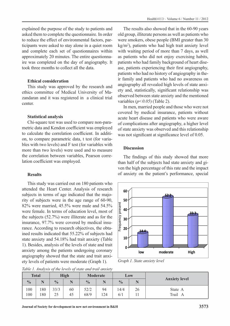
HealthMED - Volume 6 / Number 11 / 2012
Journal of Society for development in new net environment in B&H 3573
explained the purpose of the study to patients and asked them to complete the questionnaire. In order to reduce the effect of environmental factors, par-ticipants were asked to stay alone in a quiet room and complete each set of questionnaires within approximately 20 minutes. The entire questionna-ire was completed on the day of angiography. It took three months to collect all the data.
Ethical considerationThis study was approved by the research and
ethics committee of Medical University of Ma-zandaran and it was registered in a clinical trial center.
Statistical analysisChi-square test was used to compare non-para-
metric data and Kendon coefficient was employed to calculate the correlation coefficient. In additi-on, to compare parametric data, t test (for varia-bles with two levels) and F test (for variables with more than two levels) were used and to measure the correlation between variables, Pearson corre-lation coefficient was employed.
Results
This study was carried out on 180 patients who attended the Heart Center. Analysis of research subjects in terms of age indicated that the majo-rity of subjects were in the age range of 60-90, 82% were married, 45.5% were male and 54.5% were female. In terms of education level, most of the subjects (52.7%) were illiterate and as for the insurance, 97.7% were covered by medical insu-rance. According to research objectives, the obta-ined results indicated that 55.22% of subjects had state anxiety and 54.18% had trait anxiety (Table 1). Besides, analysis of the levels of state and trait anxiety among the patients undergoing coronary angiography showed that the state and trait anxi-ety levels of patients were moderate (Graph 1).Table 1. Analysis of the levels of state and trail anxiety
Total High Moderate LowAnxiety level
% N % N % N % N
100100
180180
33/325
6045
52/268/9
94124
14/46/1
2611
State ATrail A
The results also showed that in the 60-90 years old group, illiterate persons as well as patients who were smokers, obese people (BMI greater than 30 kg/m2), patients who had high trait anxiety level with waiting period of more than 7 days, as well as patients who did not enjoy exercising habits, patients who had family background of heart dise-ase, patients experiencing their first angiography, patients who had no history of angiography in the-ir family and patients who had no awareness on angiography all revealed high levels of state anxi-ety and, statistically, significant relationship was observed between state anxiety and the mentioned variables (p<0.05) (Table 2).
In men, married people and those who were not covered by medical insurance, patients without acute heart disease and patients who were aware of complications after angiography, a higher level of state anxiety was observed and this relationship was not significant at significance level of 0.05.
Discussion
The findings of this study showed that more than half of the subjects had state anxiety and gi-ven the high percentage of this rate and the impact of anxiety on the patient’s performance, special
14.4
52.2
33.3
0
10
20
30
40
50
60
low moderate High
Freq
uenc
y pr
esen
t
Graph 1. State anxiety level

3574
HealthMED - Volume 6 / Number 11 / 2012
Journal of Society for development in new net environment in B&H
P N (mean ) Variables
0/00 75 ( 52 / 13 )105 (57 / 43 )
Age30-6060-90
0/00 82 (55 / 96 )98 ( 54 / 60 )
Gendermale
female
0/0/6
90 ( 57 / 47 )39 ( 53 / 23 )32 ( 53 / 28 )19 ( 51 /95 )
Education attainmentilliterateprimary
high schooluniversity education
0/538 148 (55 / 45 )32 ( 44 / 55 )
Martital statusmarried
un married
0/291 4 (60 / 25 )176 (55 /11 )
Medical insurancenoyes
0/004 54 ( 58 / 37 ) 126 ( 53 /87 )
Smoking statusSmoker
nonsmoker
0/00 30/01 Obes BMI>30kg/m2
0/011 98 ( 53 /60 )82 ( 57 / 16 )
Exercising habitsyesno
0/00 5/ 53 Waiting period (days)
0/00 33 ( 46 / 27 )146 (54 / 55 )
Family history of heat diseaseyesno
0/960 113 ( 55 / 19 )67 ( 55 / 27 )
Previous diagnosis of heart diseaseyesno
0/001 33 ( 59 / 97 )147 ( 54 / 16 )
Aware of angioyesno
0/594 10 ( 56 / 80 ) 170 ( 55 /13 )
Aware of complications after angioyesno
0/000 33 ( 46 / 27 ) 147 ( 57 / 23 )
History of angioyesno
0/00 33 ( 65 / 15 )147 ( 52 / 99 0
Family history of angioyesno
0/00 180(54/18) Trail anxiety
Table 2. Evaluation of factors associated with state anxiety
attention should be paid to this issue and there should be attempts to reduce such anxiety throu-gh examining the effective factors and appropriate solutions. This study also indicated that most of
the subjects had a moderate level of state anxiety. Similarly, the study of Harkness9 and Jamshidi14 showed that most of the subjects had a moderate level of state anxiety.
HealthMED - Volume 6 / Number 11 / 2012
Journal of Society for development in new net environment in B&H 3575
This study shows that state anxiety depends on factors like age and it increases with an increase in age. In addition, regarding illiterates and those who have no awareness on angiography procedure as well as patients without history of angiography in their family, there is a significant statistical rela-tionship between state anxiety and the mentioned variables. Conducted surveys indicate that aware-ness of angiography is very influential in reducing state anxiety in patients; because patient is faced with an unknown surgery which, in turn, causes anxiety. In this regard, it can be stated that pri-or experience of this surgery will also help calm the patient during angiography. The researches of Harkness9 and Bazatlo13 also suggests that lack of awareness about invasive procedures such as an-giography can increase anxiety in patients, there-fore the best and most effective approach in this respect is to enhance the knowledge and awarene-ss of patients through training before angiography 9-14. The results indicated that patients who smoke have more state anxiety and there is a significant statistical relationship between the two variables (p<0.05). Patlon states: “There is a correlation between the symptoms of anxiety and smoking and this issue should be considered in order to raise the level of health15. Obesity (BMI>30) and lack of exercise are also factors that influence state anxiety. Tala and colleagues pointed out this issue and showed that obesity and exercise affect anxi-ety16. This study also showed that the level of state anxiety before angiography is associated with tra-it anxiety. In other words, trait anxiety cannot be directly observed but appears as situational anxi-ety when experiencing stress. Uzan’s studies also point out this issue10. Another influential factor on state anxiety is the waiting duration. Buzatlo’s research suggests that waiting time has an effect on anxiety of patients before angiography14. The-refore, it is recommended that in order to reduce the level of state anxiety one consider the level of trait anxiety before angiography and the waiting time for angiography should be less than 6 days, especially for patients who have higher levels of trait anxiety
Conclusion
Understanding the levels of stress and anxiety of candidate patients for coronary angiography helps nurses and other members of care team to decrease patients’ problems timely and effectively using pharmacological and non-pharmacological methods (behavior therapy, training, music the-rapy, muscle relaxation, aromatherapy, etc.). In addition, more attention should be paid to can-didates for angiography with low awareness and those who have no family background of angio-graphy, smokers, obese people and those who do not exercise as well as those with more than 6 days of waiting time and particularly the people who have trait anxiety.
In general, high anxiety in these patients ma-kes clear the necessity of paying more attention by nursing and medical personnel to reduction of anxiety and application of appropriate interventi-ons in order to reduce such problems and this issue will be possible when the factors causing anxiety are identified.
References
1. Hanser S, Mandel S. The effects of music therapy in cardio health- care. Heart Lung 2004; 33: 237-248.
2. Wood S, Sivarajon S, et al. Cardiovascular nursing. Philadelphia: Lippincott; 2005.
3. Sadephy M. [The relation complications post coro-nary artery graft with long time intubation] Persian. 15th National congress on cardiovascular update. Te-hran: Razi conferences Hall; 2007.
4. Mageadi A. [Effect of sound of the Koran to anxiety patients post coronary angiography] Persian. Mare-fat 2000; 60(4): 5-10.
5. Haneafy N, Ahmadi F, Memariyan R, et al. [Effects of relaxation Benson method on conditions hemodyna-mic in patients undergoing coronary angiography] Persian. Yazd J Res Med Sci 2004; 12(4): 78-85.
6. Mandegar M, Soltani M. [Knowledge’s cardiac] Per-sian. 3rd ed. Tehran: Bayan Press; 2002.
7. Shinia Y, Funabashi N, Lee K, et al. Relaxation effects of lavender aromatherapy improve coronary flow velocity reserve in healthy men evaluated by Transt-horacic doppler echocardiography. Int J cardialogy 2007; 53: 640-7.

3576
HealthMED - Volume 6 / Number 11 / 2012
Journal of Society for development in new net environment in B&H
8. Haneafy N, Ahmadi F, Memariyan R, et al. [Effects of relaxation Benson method on conditions hemodyna-mic in patients undergoing coronary angiography] Persian. Yazd J Res Med Sci 2004; 12(4): 78-85.
9. Harkness K,Morrow L,Smith K.The effect of early education on patient anxiety while waitiny for elective cardiac catheterization.Europan Journal of Cardio-vascular nursing 2(2003)113-121
10. Uzun S, Rural H, Uzun M. State and trait anxiety le-vels before coronary angiography. J Clin Nurs 2008; 12: 602- 607.
11. Wood S, Sivarajon S, et al. Cardiovascular nursing. Philadelphia: Lippincott; 2005
12. Sedaphati P,Ghalaji H,Kozehchian H,Arjmand A.Dose Regular Walking effect onstate and tra-te anxiety in pregnant women,Olampic Jour-nal,2008:16(2)20-26
13. Lorie M, Force M, Sebold M. Aromatherapy and reducing preprocedural anxiety. Gastroenterol Nurs 2006; 29(6): 466-741.
14. Jamshidi N, Abaszade A, Najafi M. Stress, Anxiety and Depression of patients befor coronary Angiography. zahedan J Res Med sci (ZIRMS)2012:13(SUPP1):29.
15. Buzzatto L, Viskizanei S, Viskizanei S. Patient’s anxiety before cardiac catheterization . einstein (2010);8(4 pt 1):83-7
16. Patton G,Carlin JB.Goffy C,Wolfe R,et al.Depression,anxiety amd smoking initiation;a prospective study over 3years.AMERICAN Journal of public Health;1998,vol.88,no.10.pp1518-1522.
17. Strine T, Mokdd D, Balluz l. The association of de-pression and anxiety with obesity and unhealthy be-haviors among community-dwelling us adults.
Corresponding AuthorAlieh Abasi,Department of Midwifery, Islamic Azad University, Sari Branch, Sari, Iran,E-mail:[email protected]
HealthMED - Volume 6 / Number 11 / 2012
Journal of Society for development in new net environment in B&H 3577
A metabonomics study on Crohn’s Disease using Nuclear Magnetic Resonance spectroscopyFariba Fathi1, Anahita Kyani2, Mohamad Rostami Nejad3, Mostafa Rezaye-Tavirani4, Nosratollah Naderi3, Mohamad Reza Zali3, Mohsen Tafazzoli1, Afsaneh Arefi Oskouie4
1 Department of Chemistry, Sharif University of Technology, Tehran, Iran2 Institute of Biochemistry and Biophysics,University of Tehran, Tehran, Iran 3 Research center for gastroenterology and liver disease, Shahid Beheshti University of Medical Sciences, Tehran, Iran4 Department of Basic Science Faculty of Paramedical, Shahid Beheshti University of Medical Sciences, Tehran, Iran
Abstract
Objective: Crohn’s disease (CD) is one the im-portant illnesses can affect any part of the gastro-intestinal tract. CD is not easily diagnosed using the clinical tests. Thus, the discovery of proper methods would be a major step towards CD dia-gnosis. The aim of this study was to seek the meta-bolic biomarkers causes of CD compare to control group.
Materials and Methods: In present study, we employed metabolic profiling using proton nucle-ar magnetic resonance spectroscopy (1HNMR) to find metabolites in serum which are helpful for the diagnosis of CD. Classification of CD and healthy subject was done using classification and regre-ssion trees (CART). The metabolites that caused changes in people with CD were identified using CART.
Results: According to CART diagram, we concluded that just using one descriptor CD and control groups could be classified separately. The obtained classification model showed a 89% correct classification of CD and healthy subject for the external test set.
Conclusions: The findings of the present stu-dy reveal new differentiating metabolites for CD. This metabolite may provide diagnostic biomar-kers and/or monitoring tools as well as insight into potential targets for prevention and disease therapy.
Keywords: Nuclear magnetic resonance spectroscopy, Crohn’s disease, Classification and regression tree, metabonomics.
Introduction
Crohn’s disease (CD) is an inflammatory bowel disease (IBD). Inflammation of the digestive or gastrointestinal (GI) tract can be considered as its result(1-3). Among different parts of digestive system, the small intestine and/or colon would be affected .Although the exact cause of CD is un-known, but it is known that both the host genotype and environmental factors are important factors. In areas with high rates of CD, such as northern Europe and North America, the incidence and pre-valence of CD are almost stable, whereas in low-incidence areas such as southern Europe, Asia, and the developing world they continue to rise (4).
Reach the correct diagnosis CD in some cases may be difficult because CD often mimics other symptoms (5). For accurate diagnosis, a combi-nation of information from the patient’s history and physical tests including X-rays, endoscopy, colonoscopy and pathology are needed. Methods for diagnosing CD are expensive, time consuming and can cause pain to the patients. With respect to these problems, metabonomics is an important technique for detection of biomarkers for diseases diagnosis (6).
Metabonomics is the study of metabolites and their role in diverse physiological conditions (7). This quantitative study can provide significant results only if the metabolic and biological va-riations in target group is meaningfully different from those in control group (8). There are several available techniques for metabolite profiling, such as proton nuclear magnetic resonance (1H NMR). NMR has the power to measure numerous meta-
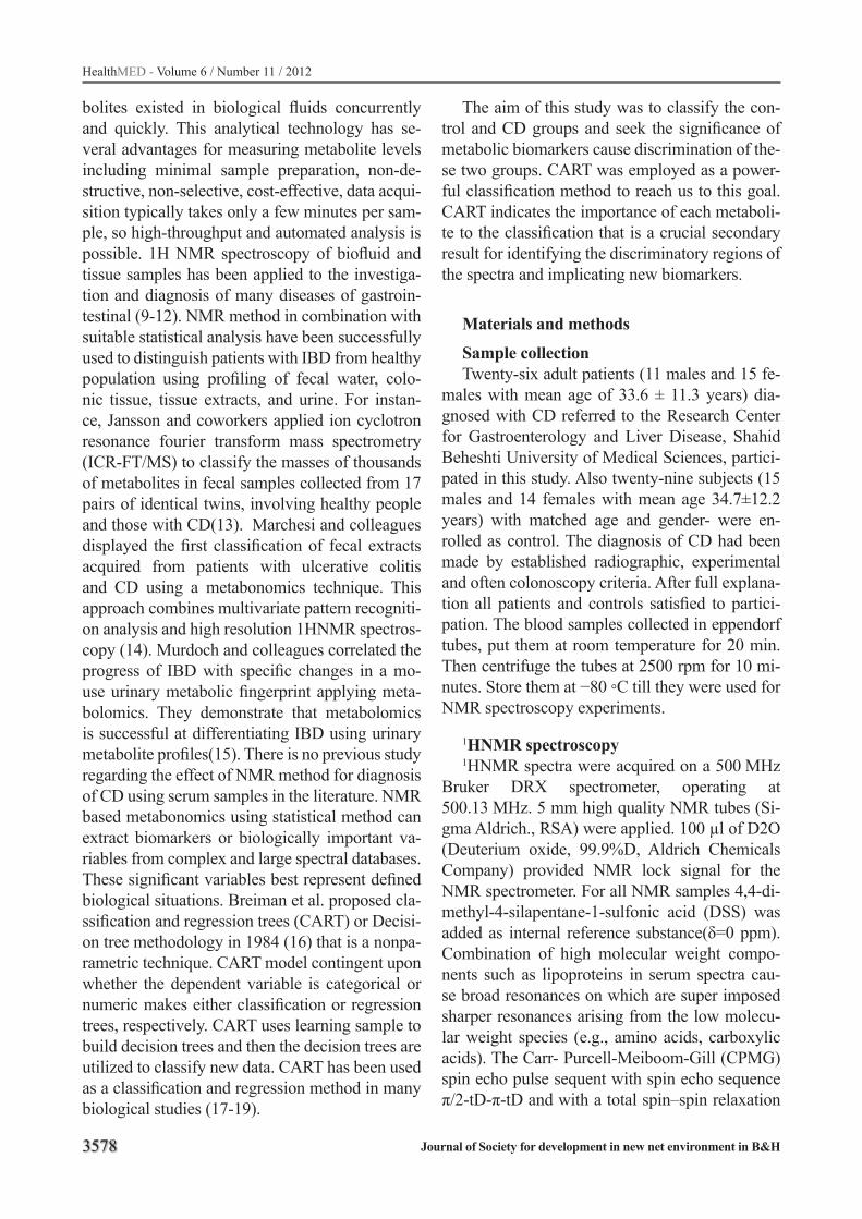
3578
HealthMED - Volume 6 / Number 11 / 2012
Journal of Society for development in new net environment in B&H
bolites existed in biological fluids concurrently and quickly. This analytical technology has se-veral advantages for measuring metabolite levels including minimal sample preparation, non-de-structive, non-selective, cost-effective, data acqui-sition typically takes only a few minutes per sam-ple, so high-throughput and automated analysis is possible. 1H NMR spectroscopy of biofluid and tissue samples has been applied to the investiga-tion and diagnosis of many diseases of gastroin-testinal (9-12). NMR method in combination with suitable statistical analysis have been successfully used to distinguish patients with IBD from healthy population using profiling of fecal water, colo-nic tissue, tissue extracts, and urine. For instan-ce, Jansson and coworkers applied ion cyclotron resonance fourier transform mass spectrometry (ICR-FT/MS) to classify the masses of thousands of metabolites in fecal samples collected from 17 pairs of identical twins, involving healthy people and those with CD(13). Marchesi and colleagues displayed the first classification of fecal extracts acquired from patients with ulcerative colitis and CD using a metabonomics technique. This approach combines multivariate pattern recogniti-on analysis and high resolution 1HNMR spectros-copy (14). Murdoch and colleagues correlated the progress of IBD with specific changes in a mo-use urinary metabolic fingerprint applying meta-bolomics. They demonstrate that metabolomics is successful at differentiating IBD using urinary metabolite profiles(15). There is no previous study regarding the effect of NMR method for diagnosis of CD using serum samples in the literature. NMR based metabonomics using statistical method can extract biomarkers or biologically important va-riables from complex and large spectral databases. These significant variables best represent defined biological situations. Breiman et al. proposed cla-ssification and regression trees (CART) or Decisi-on tree methodology in 1984 (16) that is a nonpa-rametric technique. CART model contingent upon whether the dependent variable is categorical or numeric makes either classification or regression trees, respectively. CART uses learning sample to build decision trees and then the decision trees are utilized to classify new data. CART has been used as a classification and regression method in many biological studies (17-19).
The aim of this study was to classify the con-trol and CD groups and seek the significance of metabolic biomarkers cause discrimination of the-se two groups. CART was employed as a power-ful classification method to reach us to this goal. CART indicates the importance of each metaboli-te to the classification that is a crucial secondary result for identifying the discriminatory regions of the spectra and implicating new biomarkers.
Materials and methods
Sample collectionTwenty-six adult patients (11 males and 15 fe-
males with mean age of 33.6 ± 11.3 years) dia-gnosed with CD referred to the Research Center for Gastroenterology and Liver Disease, Shahid Beheshti University of Medical Sciences, partici-pated in this study. Also twenty-nine subjects (15 males and 14 females with mean age 34.7±12.2 years) with matched age and gender- were en-rolled as control. The diagnosis of CD had been made by established radiographic, experimental and often colonoscopy criteria. After full explana-tion all patients and controls satisfied to partici-pation. The blood samples collected in eppendorf tubes, put them at room temperature for 20 min. Then centrifuge the tubes at 2500 rpm for 10 mi-nutes. Store them at −80 C till they were used for NMR spectroscopy experiments.
1HNMR spectroscopy1HNMR spectra were acquired on a 500 MHz
Bruker DRX spectrometer, operating at 500.13 MHz. 5 mm high quality NMR tubes (Si-gma Aldrich., RSA) were applied. 100 µl of D2O (Deuterium oxide, 99.9%D, Aldrich Chemicals Company) provided NMR lock signal for the NMR spectrometer. For all NMR samples 4,4-di-methyl-4-silapentane-1-sulfonic acid (DSS) was added as internal reference substance(δ=0 ppm). Combination of high molecular weight compo-nents such as lipoproteins in serum spectra cau-se broad resonances on which are super imposed sharper resonances arising from the low molecu-lar weight species (e.g., amino acids, carboxylic acids). The Carr- Purcell-Meiboom-Gill (CPMG) spin echo pulse sequent with spin echo sequence π/2-tD-π-tD and with a total spin–spin relaxation
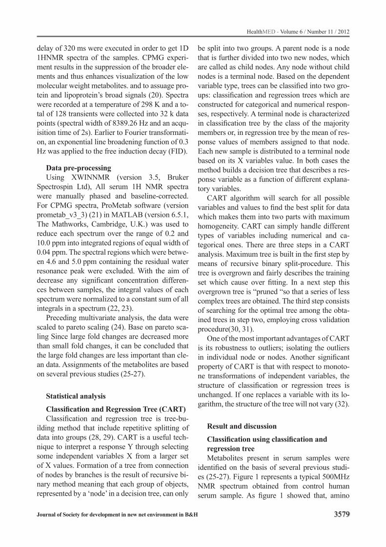
HealthMED - Volume 6 / Number 11 / 2012
Journal of Society for development in new net environment in B&H 3579
delay of 320 ms were executed in order to get 1D 1HNMR spectra of the samples. CPMG experi-ment results in the suppression of the broader ele-ments and thus enhances visualization of the low molecular weight metabolites. and to assuage pro-tein and lipoprotein’s broad signals (20). Spectra were recorded at a temperature of 298 K and a to-tal of 128 transients were collected into 32 k data points (spectral width of 8389.26 Hz and an acqu-isition time of 2s). Earlier to Fourier transformati-on, an exponential line broadening function of 0.3 Hz was applied to the free induction decay (FID).
Data pre-processingUsing XWINNMR (version 3.5, Bruker
Spectrospin Ltd), All serum 1H NMR spectra were manually phased and baseline-corrected. For CPMG spectra, ProMetab software (version prometab_v3_3) (21) in MATLAB (version 6.5.1, The Mathworks, Cambridge, U.K.) was used to reduce each spectrum over the range of 0.2 and 10.0 ppm into integrated regions of equal width of 0.04 ppm. The spectral regions which were betwe-en 4.6 and 5.0 ppm containing the residual water resonance peak were excluded. With the aim of decrease any significant concentration differen-ces between samples, the integral values of each spectrum were normalized to a constant sum of all integrals in a spectrum (22, 23).
Preceding multivariate analysis, the data were scaled to pareto scaling (24). Base on pareto sca-ling Since large fold changes are decreased more than small fold changes, it can be concluded that the large fold changes are less important than cle-an data. Assignments of the metabolites are based on several previous studies (25-27).
Statistical analysis
Classification and Regression Tree (CART)Classification and regression tree is tree-bu-
ilding method that include repetitive splitting of data into groups (28, 29). CART is a useful tech-nique to interpret a response Y through selecting some independent variables X from a larger set of X values. Formation of a tree from connection of nodes by branches is the result of recursive bi-nary method meaning that each group of objects, represented by a ‘node’ in a decision tree, can only
be split into two groups. A parent node is a node that is further divided into two new nodes, which are called as child nodes. Any node without child nodes is a terminal node. Based on the dependent variable type, trees can be classified into two gro-ups: classification and regression trees which are constructed for categorical and numerical respon-ses, respectively. A terminal node is characterized in classification tree by the class of the majority members or, in regression tree by the mean of res-ponse values of members assigned to that node. Each new sample is distributed to a terminal node based on its X variables value. In both cases the method builds a decision tree that describes a res-ponse variable as a function of different explana-tory variables.
CART algorithm will search for all possible variables and values to find the best split for data which makes them into two parts with maximum homogeneity. CART can simply handle different types of variables including numerical and ca-tegorical ones. There are three steps in a CART analysis. Maximum tree is built in the first step by means of recursive binary split-procedure. This tree is overgrown and fairly describes the training set which cause over fitting. In a next step this overgrown tree is “pruned “so that a series of less complex trees are obtained. The third step consists of searching for the optimal tree among the obta-ined trees in step two, employing cross validation procedure(30, 31).
One of the most important advantages of CART is its robustness to outliers; isolating the outliers in individual node or nodes. Another significant property of CART is that with respect to monoto-ne transformations of independent variables, the structure of classification or regression trees is unchanged. If one replaces a variable with its lo-garithm, the structure of the tree will not vary (32).
Result and discussion
Classification using classification and regression treeMetabolites present in serum samples were
identified on the basis of several previous studi-es (25-27). Figure 1 represents a typical 500MHz NMR spectrum obtained from control human serum sample. As figure 1 showed that, amino
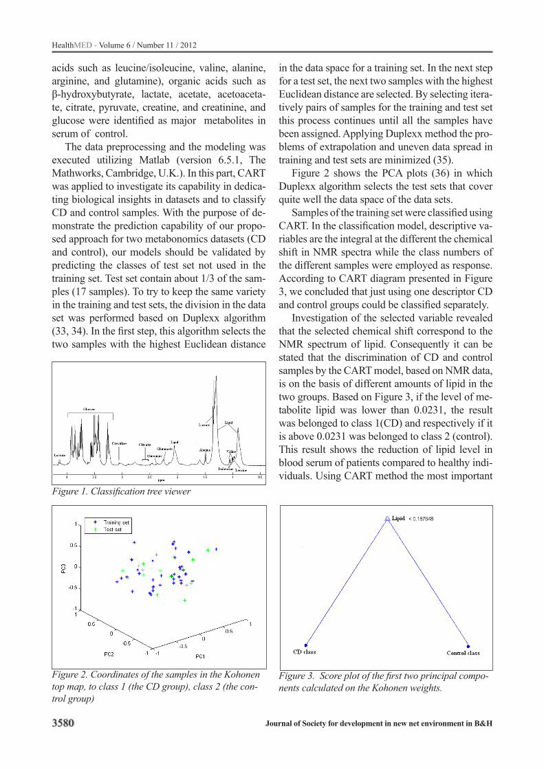
3580
HealthMED - Volume 6 / Number 11 / 2012
Journal of Society for development in new net environment in B&H
acids such as leucine/isoleucine, valine, alanine, arginine, and glutamine), organic acids such as β-hydroxybutyrate, lactate, acetate, acetoaceta-te, citrate, pyruvate, creatine, and creatinine, and glucose were identified as major metabolites in serum of control.
The data preprocessing and the modeling was executed utilizing Matlab (version 6.5.1, The Mathworks, Cambridge, U.K.). In this part, CART was applied to investigate its capability in dedica-ting biological insights in datasets and to classify CD and control samples. With the purpose of de-monstrate the prediction capability of our propo-sed approach for two metabonomics datasets (CD and control), our models should be validated by predicting the classes of test set not used in the training set. Test set contain about 1/3 of the sam-ples (17 samples). To try to keep the same variety in the training and test sets, the division in the data set was performed based on Duplexx algorithm (33, 34). In the first step, this algorithm selects the two samples with the highest Euclidean distance
in the data space for a training set. In the next step for a test set, the next two samples with the highest Euclidean distance are selected. By selecting itera-tively pairs of samples for the training and test set this process continues until all the samples have been assigned. Applying Duplexx method the pro-blems of extrapolation and uneven data spread in training and test sets are minimized (35).
Figure 2 shows the PCA plots (36) in which Duplexx algorithm selects the test sets that cover quite well the data space of the data sets.
Samples of the training set were classified using CART. In the classification model, descriptive va-riables are the integral at the different the chemical shift in NMR spectra while the class numbers of the different samples were employed as response. According to CART diagram presented in Figure 3, we concluded that just using one descriptor CD and control groups could be classified separately.
Investigation of the selected variable revealed that the selected chemical shift correspond to the NMR spectrum of lipid. Consequently it can be stated that the discrimination of CD and control samples by the CART model, based on NMR data, is on the basis of different amounts of lipid in the two groups. Based on Figure 3, if the level of me-tabolite lipid was lower than 0.0231, the result was belonged to class 1(CD) and respectively if it is above 0.0231 was belonged to class 2 (control). This result shows the reduction of lipid level in blood serum of patients compared to healthy indi-viduals. Using CART method the most important
Figure 1. Classification tree viewer
Figure 2. Coordinates of the samples in the Kohonen top map, to class 1 (the CD group), class 2 (the con-trol group)
Figure 3. Score plot of the first two principal compo-nents calculated on the Kohonen weights.
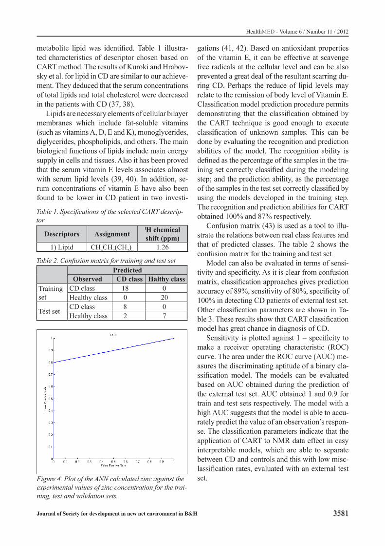
HealthMED - Volume 6 / Number 11 / 2012
Journal of Society for development in new net environment in B&H 3581
metabolite lipid was identified. Table 1 illustra-ted characteristics of descriptor chosen based on CART method. The results of Kuroki and Hrabov-sky et al. for lipid in CD are similar to our achieve-ment. They deduced that the serum concentrations of total lipids and total cholesterol were decreased in the patients with CD (37, 38).
Lipids are necessary elements of cellular bilayer membranes which include fat-soluble vitamins (such as vitamins A, D, E and K), monoglycerides, diglycerides, phospholipids, and others. The main biological functions of lipids include main energy supply in cells and tissues. Also it has been proved that the serum vitamin E levels associates almost with serum lipid levels (39, 40). In addition, se-rum concentrations of vitamin E have also been found to be lower in CD patient in two investi-
gations (41, 42). Based on antioxidant properties of the vitamin E, it can be effective at scavenge free radicals at the cellular level and can be also prevented a great deal of the resultant scarring du-ring CD. Perhaps the reduce of lipid levels may relate to the remission of body level of Vitamin E. Classification model prediction procedure permits demonstrating that the classification obtained by the CART technique is good enough to execute classification of unknown samples. This can be done by evaluating the recognition and prediction abilities of the model. The recognition ability is defined as the percentage of the samples in the tra-ining set correctly classified during the modeling step; and the prediction ability, as the percentage of the samples in the test set correctly classified by using the models developed in the training step. The recognition and prediction abilities for CART obtained 100% and 87% respectively.
Confusion matrix (43) is used as a tool to illu-strate the relations between real class features and that of predicted classes. The table 2 shows the confusion matrix for the training and test set
Model can also be evaluated in terms of sensi-tivity and specificity. As it is clear from confusion matrix, classification approaches gives prediction accuracy of 89%, sensitivity of 80%, specificity of 100% in detecting CD patients of external test set. Other classification parameters are shown in Ta-ble 3. These results show that CART classification model has great chance in diagnosis of CD.
Sensitivity is plotted against 1 – specificity to make a receiver operating characteristic (ROC) curve. The area under the ROC curve (AUC) me-asures the discriminating aptitude of a binary cla-ssification model. The models can be evaluated based on AUC obtained during the prediction of the external test set. AUC obtained 1 and 0.9 for train and test sets respectively. The model with a high AUC suggests that the model is able to accu-rately predict the value of an observation’s respon-se. The classification parameters indicate that the application of CART to NMR data effect in easy interpretable models, which are able to separate between CD and controls and this with low misc-lassification rates, evaluated with an external test set.
Table 1. Specifications of the selected CART descrip-tor
Descriptors AssignmentlH chemical shift (ppm)
1) Lipid CH3CH2(CH2)n 1.26
Table 2. Confusion matrix for training and test setPredicted
Observed CD class Halthy classTraining set
CD class 18 0Healthy class 0 20
Test set CD class 8 0 Healthy class 2 7
Figure 4. Plot of the ANN calculated zinc against the experimental values of zinc concentration for the trai-ning, test and validation sets.
3582
HealthMED - Volume 6 / Number 11 / 2012
Journal of Society for development in new net environment in B&H
Conclusion
In this paper we evaluated the use of Classifica-tion and Regression Trees (CART) to build an ea-sily interpretable predictive model to distinguish between CD and healthy controls and confirmed that the CART model is able to distinguish betwe-en cases and controls.
In the present work, a 1H NMR based meta-bonomics approach was gave an evidence for the existence of clear metabolic differentiation betwe-en two groups (CD and control group). Conside-ring these findings Gall et al. found that the me-tabonomics based on NMR data of aqueous fecal extraction was able to predict IBD and control group membership with good sensitivity and spe-cificity (12). The result of classification on serum of both healthy and CD patients in our study was better than Gall et al. study. Since 1H NMR based metabonomics is effective to monitor the progre-ssion of disease, and helpful to discover biomar-kers of crohn’s disease, we can suggest that, NMR based on metabonomics can provide the possibi-lity for assisting in early of CD. Therefore, further investigations are required to establish its real use-fulness in clinical practice.
Reference
1. Baumgart D, Carding S. Inflammatory bowel di-sease: cause and immunobiology. The Lancet 2007;369:1627–40.
2. Baumgart D, Sandborn W. Inflammatory bowel di-sease: clinical aspects and established and evolving therapies. The Lancet 2007;369:1641–57.
3. Xavier R, Podolsky D. Unravelling the pathogenesis of inflammatory bowel disease. Nature. 2007;448:427–34.
4. Loftus EVJ. Clinical epidemiology of inflamma-tory bowel disease: Incidence, prevalence, and environmental influences. Gastroenterology. 2004;126:1504-17.
5. Stephen BH, Sandborn W. Management of Crohn’s di-sease in adults. Am J Gastroenterol 2001;96:635–43.
6. German JB, Hammock BD, Watkins SM. Metabolo-mics: Building on a century of biochemistry to guide human health. Metabolomics. 2005;1:3-9.
7. Nicholson J, Lindon J, Holmes. E. Metabonomics’: understanding the metabolic responses of living systems to pathophysiological stimuli via multivariate statistical analysis of biological NMR spectroscopic data. Xenobiotica. 1999;29:1181-9.
8. Nicholson J, Connelly J, Lindon J, Holmes E. Meta-bonomics: a platform for studying drug toxicity and gene function. Nat Rev Drug Discov. 2002:153-61.
9. Hong Y-S, Hong KS, Park M-H, Ahn Y-T, Lee J-H, Huh C-S, et al. Metabonomic Understanding of Pro-biotic Effects in Humans With Irritable Bowel Syndro-me. J Clin Gastroenterol 2011(45):415–25.
10. Dryland PA, Love DR, Walker MF, Dommels Y, Butts C, Rowan D, et al. Allantoin as A Biomarker of In-flammation in an Inflammatory Bowel Disease Mou-se Model: NMR Analysis of Urine. The Open Bioac-tive Compounds Journal. 2008;1:1-6.
11. Bezabeh T, Somorjai RL, Smith ICP. MR metabo-lomics of fecal extracts:applications in the study of bowel diseases. Magn Reson Chem. 2009;47:54-61.
12. Gall GL, Noor SO, Ridgway K, Scovell L, Jamieson C, Johnson IT, et al. Metabolomics of Fecal Extracts Detects Altered Metabolic Activity of Gut Microbio-ta in Ulcerative Colitis and Irritable Bowel Syndro-me. J Proteome Res. 2011;10:4208–18.
13. Jansson JK, Willing B, Lucio M, Fekete A, Dick-sved J, Halfvarson J, et al. Metabolomics reveals metabolic biomarkers of Crohn’s disease. Plos one. 2009;4:1-10.
14. Marchesi JR, Holmes E, Khan F, Kochhar S, Scan-lan P, Shanahan F, et al. Rapid and Noninvasive Me-tabonomic Characterization of Inflammatory Bowel Disease. J Proteome Res 2007;6:546–51.
15. Murdoch TB, Fu H, MacFarlane S, Sydora BC, Fe-dorak RN, Slupsky CM. Urinary metabolic profiles of inflammatory bowel disease in interleukin-10 ge-ne-deficient mice. Anal Chem. 2008 80:5524-31.
16. Breiman L, Friedman J, Olshen R, Stone C. Cla-ssification and Regression Trees. Chapman &Hall (Wadsworth I, editor. New York; 1984.
17. Cao D-S, Zeng M-M, Yi L-Z, Wang B, X Q-S, Hud Q-N, et al. A novel kernel Fisher discriminant analysis: Constructing informative kernel by deci-sion tree ensemble for metabolomics data analysis. AnalChemActa. 2011;706:97-104.
18. Aymerich FX, Alonso J, Cabañas ME, Comabe-lla M, Sobrevilla P, Rovira A. Decision tree based fuzzy classifier of 1H magnetic resonance spectra from cerebrospinal fluid samples. FuzzySetSyst. 2011;170:43–63.
19. Deconinck E, Sacré PY, Coomans D, Beer JD. Cla-ssification trees based on infrared spectroscopic data to discriminate between genuine and counterfeit me-dicines. J Pharmaceut Biomed Anal. 2012;57:68-75.
20. Zhang G, Hirasaki G. CPMG relaxation by diffusion with constant magnetic field gradient in a restricted geometry: numerical simulation and application. J Magn Reson 2003;163:81–91.
21. Viant MR. Improved methods for the acquisition and interpretation of NMR metabolomic data. Biochem Biophys Res Commun. 2003;310:943–8.
HealthMED - Volume 6 / Number 11 / 2012
Journal of Society for development in new net environment in B&H 3583
22. Holmes E, Nicholis AW, Lindon JC, Ramos S, Spraul M, Neidig P, et al. Development of a model for cla-ssification of toxin-induced lesions using 1H NMR spectroscopy of urine combined with pattern reco-gnition. NMR Biomed. 1998;11:235–44.
23. Waters NJ, Holmes E, Williams A, Waterfield CJ, Farrant RD, Nicholson JK. NMR and pattern reco-gnition studies on the time-related metabolic effects of alpha-naphthylisothiocyanate on liver, urine, and plasma in the rat: an integrative metabonomic approach. Chem Res Toxicol. 2001;14:1401–12.
24. Eriksson L, Johansson E, Kettaneh-Wold N, Wold S. Introduction to multi and megavariate data analysis using projection methods, PCA and PLS. UME-TRICS. 1999:213-25.
25. Nicholson JK, Foxall PJ, Spraul M, Farrant RD, Lindon JC. 750 MHz 1H and 1H-13C NMR spectros-copy of human blood plasma. Anal Chem. 1995;67 793-811.
26. Fan M. Metabolite profiling by one- and two-dimen-sional NMR analysis of complex mixtures. Prog Nucl Magn Reson Spectrosc. 1996;28:161-219.
27. 27. Lindon JC. NMR Spectroscopy on biofluids. Annu Rep NMR Spectrosc. 1999;38:1-88.
28. Zhang MH, Xu QS, Daeyaert F, Lewi PJ, Massart DL. Application of boosting to classification problems in chemometrics. Anal Chim Acta. 2005;544:167–76.
29. Breiman L, Friedman JH, Olshen RA, Stone CJ, , , . Classification and Regression Trees. Group WI, editor.: Belmont; 1984.
30. Deconinck E, Hancock T, Coomans D, Massart DL, Heyden YV. Classification of drugs in absor-ption classes using Classification And Regression Trees (CART)-methodology. J Pharm Biomed Anal. 2005;39: 91-103.
31. Deconinck E, Zhang MH, Coomans D, Heyden YV. Classification tree models for the prediction of blo-od-brain barrier passage of drugs. J Chem InfMo-del. 2006;46:1410-9.
32. De’ath G, Fabricius K. Classification and regressi-on trees: a powerful yet simple technique for ecolo-gical data analysis. Ecology,. 2000;81:3178–92.
33. Snee RD. Validation of regression models: methods and examples. Technometrics. 1977;19:415-28.
34. Deconinck E, Sacré PY, Coomans D, DeBeer J. Cla-ssification trees based on infrared spectroscopic data to discriminate between genuine and counterfeit me-dicines. J Pharm Biomed Anal. 2012; 57:68–75.
35. Jones A, Young D, Taylor J, Kell DB, Rowland JJ. quantification of microbial productivity via multi-angle light scattering and supervised learning. Bio-technol Bioeng 1998;59:131-43.
36. Massart DL, Vandeginste BGM, Buydens LMC, Jong SD, Lewi PJ, Smeyers-Verbeke J. Handbook of Chemometrics and Qualimetrics. Science E, editor. Amsterdam; 1997.
37. Hrabovsky V, Zadak Z, Blaha V, Hyspler R, Ticha A, Karlik T. LIPID METABOLISM IN ACTIVE CROHN’S DISEASE: PRE-RESULTS. Biomed Pap Med Fac Univ Palacky Olomouc Czech Repub. 2006;150:363–6.
38. Kuroki F, Iida M, Tominaga M, Matsumoto T, Ka-namoto K, Fujishima M. Is vitamin E depleted in Crohn’s disease at initial diagnosis? DigDis. 1994;12:248-54.
39. Rubinstein HM, Dietz AA, Srinivasan R. Relati-on of vitaminE and serumlipids. Clin Chim Acta. 1969;23:1–6.
40. Horwitt MK, Harvey CC, Dahm CHJ, Searcy MT. Relationship between tocopherol and serum lipid le-vels for determination of nutritional adequacy. Ann N Y Acad Sci. 1972;203:223-36.
41. Fernandez-Banares F, Abad-Lacruz A, Xiol X, Gine JJ, Dolz C, Cabre E, et al. Vitamin status in patients with inflammatory bowel disease. Scand J Gastroen-terol 1989;40:744-8.
42. Kuroki F, Iida M, Tominaga M, Matsumoto T, Hira-kawa K, Sugiyama S, et al. Multiple vitamin status in Crohn’s disease. Correlation with disease activity. DigDisSci. 1993;38:1614-8.
43. Kohavi R, Provost F. Glossary of Terms. Machine Learning. 1998;30:271-4.
Corresponding AuthorMohsen Tafazzoli, Department of Chemistry, Sharif University of Technology, Tehran, Iran,E-mail: [email protected]
3584
HealthMED - Volume 6 / Number 11 / 2012
Journal of Society for development in new net environment in B&H
Does internet is as a barrier or facilitation factor in academic life? Azita Bala Ghafari1, Hasan Siamian2, Ghasem Abedi3, Ramzan Hasanzadeh4
1 Health Information Management Department, Mazandaran University ofMedical Sciences, Sari, Mazandaran, Iran2 Health Information Management Department, Mazandaran University ofMedical Sciences, Sari, Mazandaran, Iran3 Health Sciences Center, Mazandaran University of Medical Sciences, Sari, Mazandaran, Iran4 Department of Psychology, Islamic Azad University, Sari- Branch, Mazandaran, Iran
Abstract
Background and Purpose: The purpose of this study is to evaluate internet as fact in stu-dents life and the demographic variables related to the rate of Internet addiction in students. With this increasing number of internet users, the problem of internet addiction has attracted high attention from psychiatrists, psychologists, educators, and the public . The use of the internet has increased considerably over the last few years. The purpose of this research is a study of internet addiction and mental health among students as new challenge in health psychology.
Methods: Research method is descriptive. Sta-tistical population is all the students of Islamic Azad University- Sari- Branch. Statistical sam-ple is 261 college students who were selected by through the random sampling method. The sample size is estimated by using Kerejci and Morgan’s table. In this research in order to gather data, a questionnaire was used. In the present study, six hypotheses were tested. To analyze data, the co-efficient correlation and the t-test have been used.
Results: The results of the study showed that: University Students suffer from internet addicti-on (17.6%).University Students use internet for different reasons (14 reasons).There is significant difference between internet addiction in male and female students (t=4.267, p<0.05).There is signi-ficant difference between internet addiction in stu-dents of university with different major (F=12.187, p<0.05). There isn’t any significant difference between internet addiction in students of univer-sity with different term (F=1.339, p<0.05).
Conclusion: Based on the results of the rese-arch, the Internet addiction is currently becoming a serious mental health problem among college students. Internet addiction, as a new challenge in
the health psychology, needed to be prevented and cured considerably.
Keywords: Internet Addiction, Mental Health, College Students, Health Psychology
Introduction
Anecdotal reports indicated that some on-line users were becoming addicted to the Internet in much the same way that others became addicted to drugs or alcohol which resulted in academic, soci-al, and occupational impairment. However, resear-ch among sociologists, psychologists, or psychia-trists has not formally identified addictive use of the Internet as a problematic behavior (Wang 2001). This study investigated the existence of In-ternet addiction and the extent of problems caused by such potential misuse. Of all the diagnoses re-ferenced in the Diagnostic and Statistical Manual of Mental Disorders—Fourth Edition, Pathologi-cal Gambling was viewed as most similar to the pathological nature of Internet use. By using Pat-hological Gambling as a model, addictive Internet use can be defined as an impulse-control disorder that does not involve an intoxicant. Therefore, this study developed a brief eight-item questionnaire referred to as a Diagnostic Questionnaire (DQ), which modified criteria for pathological gambling to provide a screening instrument for classification of participants (Park and Kim 2011). One of the most important impacts of information techno-logy on students’ lives is the ever-increasing use of the Internet. Although there exist many reports in the media regarding the unhealthy Internet use among students, research is still limited and has mainly relied upon on-line self-selected reports on Internet dependency or “Internet addiction”(Wang 2001).

HealthMED - Volume 6 / Number 11 / 2012
Journal of Society for development in new net environment in B&H 3585
The Internet was originally designed to facilita-te communication and research activities. Howe-ver, the dramatic increase in the use of the Internet in recent years has led to pathological use (Internet addiction) (Alavi, Maracy et al. 2011). The com-puter and the Internet are becoming major effects in the lives of adolescents, progressing beyond the level of recreational activity(Kim, Ryu et al. 2006). Adolescent addiction to the Internet is be-coming a serious problem. Extensive Internet use may create a heightened level of psychological arousal, resulting in little sleep, failure to eat for long periods, and limited physical activity (Wang 2001) (Young, 1998), possibly resulting in the user experiencing physical and mental health problems such as depression, loneliness, low self-esteem, and anxiety. The work of Young (1998) focused on the factors leading to Internet addiction, which were compared to pathological gambling (Canan, Ataoglu et al. 2011).
The concept of addiction, though traditionally used to describe a physical dependence on a su-bstance has been applied to excessive use of the Internet (Du, Liu et al. 2011).
The change to an Internet-based society can be viewed both optimistically and pessimistically. A positive view-point acknowledges that individua-lism and spontaneity are important, and that chan-ges in the forms of communication through the existing mass media allows for a healthy diver-sification in society. Here, it is believed that new concepts such as broadband access to network, powerlessness, and devaluation, which are based on an amalgamation of the computer and commu-nications technology and their incorporation into the digitalization and multi-medialization of our society, will change our lives for the better(Wang 2001). In contrast, the negative viewpoint is that the new information and communication techno-logies simply represent new tools for perpetuating capitalism. This viewpoint includes forecasts that these technologies will isolate individuals from real-life experiences, and eventually from life itself(Krajewska-Kulak, Kulak et al. 2011).
The use of the Internet on college campuses has increased dramatically in recent years, leading to pathological use, or Internet addiction, for some students. Internet addiction is defined as a psycho-logical dependence on the Internet and is characte-
rized by (a) an increasing investment of resources on Internet-related activities, (b) unpleasant fee-lings (e.g., anxiety, depression, emptiness) when offline, (c) an increasing tolerance to the effects of being online, and (d) the denial of the problematic behaviors. Individuals exhibiting such symptoms often are dealing with underlying psychological issues. College students are particularly vulnerable to pathological Internet use due to several factors. These factors include (a) the psychological and developmental characteristics of late adolescence/young adulthood, (b) ready access to the Internet, and (c) an expectation of computer/Internet use. The nature of the computer medium and the sense of control experienced when engaged in compu-ter activities can also contribute to the potentiality for problematic computer/Internet use. Research on Internet addiction is in its infancy stage. The need for greater understanding of Internet addicti-on and its treatment is noted (Canan, Ataoglu et al. 2011)(6). Yen et al. (2007) suggested that Internet addiction is associated with symptoms of ADHD and depressive disorders. However, hostility was associated with Internet addiction only in males. Effective evaluation and treatment for ADHD and depressive disorders are required for adolescents with Internet addiction. More attention should be paid to male adolescents with high hostility in intervention of Internet addiction (Lin, Ko et al. 2011).
Research conducted in the field of Internet addiction has reported contradictory results. Tre-uer, Cao et al. Yellowlees et al and Li & Chung noted that the excessive and pathological use of the Internet has reduced the students’ mental health(Li and Chung 2006; Cao, Su et al. 2007; Yellowlees and Marks 2007; Su, Fang et al. 2011). The researchers found that students with pat-hological Internet use, compared with students who do not have the same experience, show higher pathology and mental health problems. Indeed, the increased levels of experien-ce on working with the Internet are asso-ciated with lower levels of mental health. Kim et al considered gender as one of the factors related to Internet addiction. Inter-net addiction directly and indirectly affects a wide range of people (Xie, Zhou et al. 2010). Evaluating the number of people who directly
3586
HealthMED - Volume 6 / Number 11 / 2012
Journal of Society for development in new net environment in B&H
or indirectly affected by this phenomenon ma-kes it possible to understand the extent of this issue. Internet addiction is a global phenome-non which has different ranges of use from 5 to 25 percent among American, Chinese, Korean, British, Australian, Taiwanese, Japanese, ea-stern and western European countries students. Some studies showed that women have shown a twice as much Internet addicti-on as that of men (Griffiths 2003; Yen, Ko et al. 2007; Canan, Ataoglu et al. 2010). Amichai Hamburger and Ben Artizi showed that students with extreme dependence on the Inter-net are feeling lonely in terms of their mental health(Amichai-Hamburger and Ben-Artzi 2003). Studies indicate that university students with Inter-net addiction do not have the required social skills for building relationship (Anderson 2001; Bloch 2002). The researchers found that students with Internet addiction have higher degrees of vulnerability and have poor mental health.
Brenner and Kaplan in their studies reached the fact that students with internet addiction, compa-red with those who don’t have such experience, have lower mental health and social skills (Caplan 2002; Song, Zheng et al. 2010)
Cao et al, Kim, Lee et al and Yen et al com-pared the psychological, personality and so-cial characteristics of the students who had excessive use of the Internet, with respect to students who had normal use of the Internet. Their results showed that rates of depression, anxi-ety, thoughts of suicide, hyperactivity, anxiety, social anxiety, aggression and violence and anti-social behavior in students with Internet addiction are higher (Chou and Hsiao 2000; Kim, Ryu et al. 2006; Li and Chung 2006; Yen, Ko et al. 2007).
Fung, Gerifets, Jones and Madden, Krau et al in their studies showed that intenet addiction among students will cause the low individual, fa-milial and social relationship; they will also enjoy a low personal and social identity. The researchers believe that the students’ entrance into the univer-sity is a a contradictory issue because when they enter universities, they are faced with the modern technology and have more opportunity to use the internet, but they are also more likely to be suffe-ring from internet addiction (Fung 2002 ; Griffiths 2003; Jones 2008; Kesici and Sahin 2009).
In the present study, the following hypotheses are being evaluated:
- Internet addiction is widespread among university students.
- Internet addiction is different in male and female students.
- Internet addiction is different in students with different ages.
- Internet addiction is different in students with different Internet use.
Materials and Methods
This research is descriptive research to evalu-ate the present status of the issue. The populati-on of the study is all students attending the Sari Islamic Azad University. Based on the latest Sta-tistics from the Computer and Information Stati-stics department, there are 7558 students who are currently studying at this university. The sample for this study consists of 261 who were selected through stratified random sampling. The sample size determination was made possible through Krejcie and Morgan table and population size. In addition to the library study for data collecti-on and review of literature and background rese-arches, a questionnaire was used. Data collection tool was the questionnaire.
Internet Addiction Test (IAT)Internet Addiction Test (IAT) first 20 questions
and Young’s Internet Addiction Test (IAT)(4) were adopted to evaluate the respondents’ level of In-ternet addiction. The IAT has 20 items associated with Internet use, including psychological depen-dence, compulsive use and withdrawal, as well as related problems of school, sleep, family, and time management. For each item, a graded response can be selected (1 = “not at all” to 5 = “always”). The minimum score is 20 while the maximum is 100; the higher the score, the greater the level of Internet addiction.
As suggested by Young, cut-off scores for the IAT were used to classify Internet users based on the severity of their addictive behavior. In current study, the same cut-off scores were used: Minimal
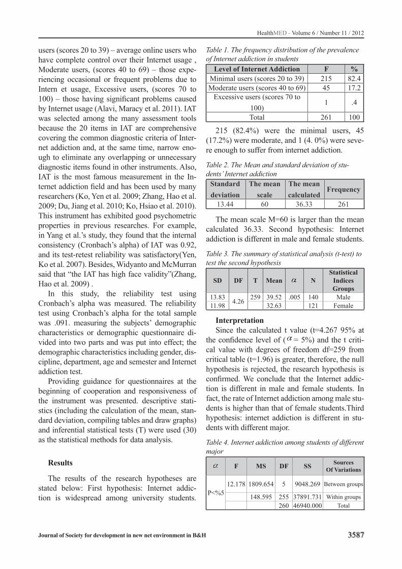
HealthMED - Volume 6 / Number 11 / 2012
Journal of Society for development in new net environment in B&H 3587
users (scores 20 to 39) – average online users who have complete control over their Internet usage , Moderate users, (scores 40 to 69) – those expe-riencing occasional or frequent problems due to Intern et usage, Excessive users, (scores 70 to 100) – those having significant problems caused by Internet usage (Alavi, Maracy et al. 2011). IAT was selected among the many assessment tools because the 20 items in IAT are comprehensive covering the common diagnostic criteria of Inter-net addiction and, at the same time, narrow eno-ugh to eliminate any overlapping or unnecessary diagnostic items found in other instruments. Also, IAT is the most famous measurement in the In-ternet addiction field and has been used by many researchers (Ko, Yen et al. 2009; Zhang, Hao et al. 2009; Du, Jiang et al. 2010; Ko, Hsiao et al. 2010). This instrument has exhibited good psychometric properties in previous researches. For example, in Yang et al.’s study, they found that the internal consistency (Cronbach’s alpha) of IAT was 0.92, and its test-retest reliability was satisfactory(Yen, Ko et al. 2007). Besides, Widyanto and McMurran said that “the IAT has high face validity”(Zhang, Hao et al. 2009) .
In this study, the reliability test using Cronbach’s alpha was measured. The reliability test using Cronbach’s alpha for the total sample was .091. measuring the subjects’ demographic characteristics or demographic questionnaire di-vided into two parts and was put into effect; the demographic characteristics including gender, dis-cipline, department, age and semester and Internet addiction test.
Providing guidance for questionnaires at the beginning of cooperation and responsiveness of the instrument was presented. descriptive stati-stics (including the calculation of the mean, stan-dard deviation, compiling tables and draw graphs) and inferential statistical tests (T) were used (30) as the statistical methods for data analysis.
Results
The results of the research hypotheses are stated below: First hypothesis: Internet addic-tion is widespread among university students.
Table 1. The frequency distribution of the prevalence of Internet addiction in students
Level of Internet Addiction F %Minimal users (scores 20 to 39) 215 82.4Moderate users (scores 40 to 69) 45 17.2
Excessive users (scores 70 to 100)
1 .4
Total 261 100
215 (82.4%) were the minimal users, 45 (17.2%) were moderate, and 1 (4. 0%) were seve-re enough to suffer from internet addiction.
Table 2. The Mean and standard deviation of stu-dents’ Internet addiction
Standard deviation
The mean scale
The mean calculated
Frequency
13.44 60 36.33 261
The mean scale M=60 is larger than the mean calculated 36.33. Second hypothesis: Internet addiction is different in male and female students.
Table 3. The summary of statistical analysis (t-test) to test the second hypothesis
SD DF T Mean NStatistical
IndicesGroups
13.83 4.26 259 39.52 .005 140 Male11.98 32.63 121 Female
InterpretationSince the calculated t value (t=4.267 95% at
the confidence level of ( = 5%) and the t criti-cal value with degrees of freedom df=259 from critical table (t=1.96) is greater, therefore, the null hypothesis is rejected, the research hypothesis is confirmed. We conclude that the Internet addic-tion is different in male and female students. In fact, the rate of Internet addiction among male stu-dents is higher than that of female students.Third hypothesis: internet addiction is different in stu-dents with different major.
Table 4. Internet addiction among students of different major
F MS DF SS SourcesOf Variations
P<%512.178 1809.654 5 9048.269 Between groups
148.595 255 37891.731 Within groups260 46940.000 Total
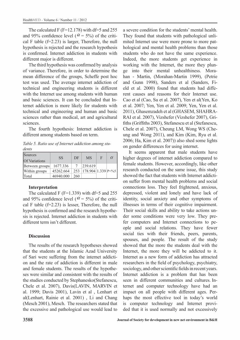
3588
HealthMED - Volume 6 / Number 11 / 2012
Journal of Society for development in new net environment in B&H
The calculated F (F=12.178) with df=5 and 255 and 95% confidence level ( = 5%) of the criti-cal F table (f=2.23) is larger, Therefore, the null hypothesis is rejected and the research hypothesis is confirmed. Internet addiction in students with different major is different.
The third hypothesis was confirmed by analysis of variance. Therefore, in order to determine the mean difference of the groups, Scheffe post hoc test was used. The average internet addiction of technical and engineering students is different with the Internet use among students with human and basic sciences. It can be concluded that In-ternet addiction is more likely for students with technical and engineering and human and basic sciences rather than medical, art and agricultural sciences.
The fourth hypothesis: Internet addiction is different among students based on term.
Table 5. Ratio use of Internet addiction among stu-dentsSourcesOf Variations
SS DF MS F
Between groups 1677.336 7 239.6191.339 P<%5Within groups 45262.664 253 178.904
Total 46940.000 260
InterpretationThe calculated F (F=1.339) with df=5 and 255
and 95% confidence level ( = 5%) of the criti-cal F table (f=2.23) is lesser, Therefore, the null hypothesis is confirmed and the research hypothe-sis is rejected. Internet addiction in students with different term isn’t different.
Discussion
The results of the research hypotheses showed that the students at the Islamic Azad University of Sari were suffering from the internet addicti-on and the rate of addiction is different in male and female students. The results of the hypothe-ses were similar and consistent with the results of the studies conducted by Stephanesko(Stefanescu, Chele et al. 2007), Davis(LAVIN, MARVIN et al. 1999; Davis 2001), Lavin et al , Lenhart et al(Lenhart, Rainie et al. 2001) , Li and Chang (Mesch 2001), Mesch. The researchers stated that the excessive and pathological use would lead to
a severe condition for the students’ mental health. They found that students with pathological unli-mited Internet use were more prone to more pat-hological and mental health problems than those students who do not have the same experience. Indeed, the more students get experience in working with the Internet, the more they plun-ge into their mental unhealthiness. Mora-han - Martin, (Morahan-Martin 1999), (Petrie and Gunn 1998), Sanders et al (Sanders, Fi-eld et al. 2000) found that students had diffe-rent causes and reasons for their Internet use. Cao et al (Cao, Su et al. 2007), Yen et al(Yen, Ko et al. 2007; Yen, Yen et al. 2009; Yen, Yen et al. 2011), Ghasemzadeh et al (GHASEM, SHAHRA-RAI et al. 2007), Vizshefer (Vizshefer 2007), Gri-fiths (Griffiths 2003), Stefanescu et al (Stefanescu, Chele et al. 2007), Cheung LM, Wong WS (Che-ung and Wong 2011), and Kim (Kim, Ryu et al. 2006; Ha, Kim et al. 2007)) also shed some lights on gender differences for using internet.
It seems apparent that male students have higher degrees of internet addiction compared to female students. However, accordingly, like other research conducted on the same issue, this study showed the fact that students with Internet addicti-on suffer from mental health problems and social connections loss. They feel frightened, anxious, depressed, violent and lonely and have lack of identity, social anxiety and other symptoms of illnesses in terms of their cognitive impairment. Their social skills and ability to take actions un-der some conditions were very low. They pre-fer computers and Internet connections to pe-ople and social relations. They have fewer social ties with their friends, peers, parents, spouses, and people. The result of the study showed that the more the students deal with the Internet, the more they will be addicted to it. Internet as a new form of addiction has attracted researchers in the field of psychology, psychiatry, sociology, and other scientific fields in recent years. Internet addiction is a problem that has been seen in different communities and cultures. In-ternet and computer technology have had an impact on all people with different ages. Per-haps the most effective tool in today’s world is computer technology and Internet provi-ded that it is used normally and not excessively
HealthMED - Volume 6 / Number 11 / 2012
Journal of Society for development in new net environment in B&H 3589
which could cause physical and mental health. The rate of Internet addiction in students is below average and it is lower than expected (Table 2). It can be concluded that Internet addiction is different in male and female students. In fact, the rate of Internet addiction among male students is higher than that of female students. (Table 3) Internet addiction in students at different ages is not different (Table 4), Therefore, the null hypothesis is rejected and the study confirmed the hypothesis that Internet addic-tion is different in students with different Internet use. The fifth hypothesis was confirmed by the variance analysis. Therefore, in order to determi-ne the mean difference of the groups, the Scheffe post hoc test was used (Table 5) in which with the increase in Internet use more Internet addiction is likely to involve people. (Table 6)
References
1. Alavi, S. S., M. R. Maracy, et al. (2011). “The effect of psychiatric symptoms on the internet addiction disor-der in Isfahan’s University students.” J Res Med Sci 16(6): 793-800.
2. Amichai-Hamburger, Y. and E. Ben-Artzi (2003). “Loneliness and Internet use.” Computers in Human Behavior 19(1): 71-80.
3. Anderson, K. J. (2001). “Internet use among college students: An exploratory study.” Journal of American College Health 50(1): 21-26.
4. Bloch, J. (2002). “Student/teacher interaction via email: The social context of Internet discourse.” Journal of Second Language Writing 11(2): 117-134.
5. Canan, F., A. Ataoglu, et al. (2010). “Evaluation of psychometric properties of the internet addiction scale in a sample of Turkish high school students.” Cyberpsychol Behav Soc Netw 13(3): 317-320.
6. Canan, F., A. Ataoglu, et al. (2011). “The association between Internet addiction and dissociation among Turkish college students.” Compr Psychiatry.
7. Cao, F., L. Su, et al. (2007). “The relationship betwe-en impulsivity and Internet addiction in a sample of Chinese adolescents.” European Psychiatry 22(7): 466-471.
8. Caplan, S. E. (2002). “Problematic Internet use and psychosocial well-being: Development of a theory-ba-sed cognitive-behavioral measurement instrument.” Computers in Human Behavior 18(5): 553-575.
9. Chou, C. and M. C. Hsiao (2000). “Internet addicti-on, usage, gratification, and pleasure experience: the Taiwan college students’ case.” Computers & Educa-tion 35(1): 65-80.
10. Du, W., J. Liu, et al. (2011). “[Functional magnetic resonance imaging of brain of college students with internet addiction].” Zhong Nan Da Xue Xue Bao Yi Xue Ban 36(8): 744-749.
11. Du, Y. S., W. Jiang, et al. (2010). “Longer term effect of randomized, controlled group cognitive beha-vioural therapy for Internet addiction in adolescent students in Shanghai.” Aust N Z J Psychiatry 44(2): 129-134.
12. Fung, T.-W. (2002 ). “Fung, Identity, Emotional Re-gulation and Interpersonal Relationship between Highly and Midly
13. Internet-addictive Groups, Formosa “ Journal of Mental Health 15: 65-76.
14. Griffiths, M. (2003). “Internet Abuse in the Workpla-ce: Issues and Concerns for Employers and Em-ployment Counselors.” Journal of Employment Co-unseling 40(2): 87-96.
15. Jones, S. (2008). Internet Goes to College: How Stu-dents are Living in the Future with Today s Techno-logy, DIANE Publishing.
16. Kesici, S. and I. Sahin (2009). “A comparative study of uses of the Internet among college students with and without Internet addiction.” Psychol Rep 105(3 Pt 2): 1103-1112.
17. Kim, K., E. Ryu, et al. (2006). “Internet addiction in Korean adolescents and its relation to depressi-on and suicidal ideation: A questionnaire survey.” International journal of nursing studies 43(2): 185-192.
18. Ko, C. H., S. Hsiao, et al. (2010). “The characteri-stics of decision making, potential to take risks, and personality of college students with Internet addicti-on.” Psychiatry Res 175(1-2): 121-125.
19. Ko, C. H., J. Y. Yen, et al. (2009). “Proposed diagno-stic criteria and the screening and diagnosing tool of Internet addiction in college students.” Compr Psychiatry 50(4): 378-384.
20. Krajewska-Kulak, E., W. Kulak, et al. (2011). “In-ternet addiction among students of the medical uni-versity of bialystok.” Comput Inform Nurs 29(11): 657-661.
21. Li, S. M. and T. M. Chung (2006). “Internet function and Internet addictive behavior.” Computers in Hu-man Behavior 22(6): 1067-1071.
22. Lin, M. P., H. C. Ko, et al. (2011). “Prevalence and psychosocial risk factors associated with internet addiction in a nationally representative sample of college students in taiwan.” Cyberpsychol Behav Soc Netw 14(12): 741-746.
23. Park, G. R. and H. S. Kim (2011). “[Effects of a Group Counseling Integration Program on Self-de-termination and Internet Addiction in High School Students with Tendency to Internet Addiction].” J Korean Acad Nurs 41(5): 694-703.

3590
HealthMED - Volume 6 / Number 11 / 2012
Journal of Society for development in new net environment in B&H
24. Song, X. Q., L. Zheng, et al. (2010). “[Status of ‘internet addiction disorder’ (IAD) and its risk fac-tors among first-grade junior students in Wuhan.].” Zhonghua Liu Xing Bing Xue Za Zhi 31(1): 14-17.
25. Su, W., X. Fang, et al. (2011). “Internet-based in-tervention for the treatment of online addiction for college students in China: a pilot study of the He-althy Online Self-helping Center.” Cyberpsychol Be-hav Soc Netw 14(9): 497-503.
26. Wang, W. (2001). “Internet dependency and psycho-social maturity among college students.” Interna-tional Journal of Human-Computer Studies 55(6): 919-938.
27. Xie, Y. B., P. Zhou, et al. (2010). “[Prevalence of internet addiction and the related factors in middle school students in Guangzhou].” Nan Fang Yi Ke Da Xue Xue Bao 30(8): 1801-1804.
28. Yellowlees, P. M. and S. Marks (2007). “Problematic Internet use or Internet addiction?” Computers in Human Behavior 23(3): 1447-1453.
29. Yen, J. Y., C. H. Ko, et al. (2007). “The comorbid psychiatric symptoms of Internet addiction: atten-tion deficit and hyperactivity disorder (ADHD), depression, social phobia, and hostility.” J Adolesc Health 41(1): 93-98.
30. Zhang, Z. H., J. H. Hao, et al. (2009). “[The relati-onship between emotional, physical abuse and In-ternet addiction disorder among middle school stu-dents].” Zhonghua Liu Xing Bing Xue Za Zhi 30(2): 115-118.
Corresponding Author Ramzan Hasanzadeh,Department of Psychology, Islamic Azad University- Sari- Branch, Sari, Mazandaran, Iran.E-mail: [email protected]
HealthMED - Volume 6 / Number 11 / 2012
Journal of Society for development in new net environment in B&H 3591
Increased hemoglobin and thrombocytocrit in non-alcoholic fatty liver diseaseYou-ming Li
The First Affiliated Hospital, College of Medicine, Zhejiang University, Hangzhou city, China
Abstract
Objective: Complete blood count (CBC) test is a simple, universally used, inexpensive mar-ker used in clinical practices, however, few stu-dy focused on the relationship between CBC and non-alcoholic fatty liver disease (NAFLD). The aim of the present study was to assess the relati-onship between them.
Materials and methods: Four hundred and forty subjects (223 men and 217 women) par-ticipated in a routine medical checkup and were chosen to be included in this study. Measurements such as weight, height, BMI, and blood pressu-re were recorded. Serum biochemical indicators were measured, and an ultrasound of the liver was obtained. CBC test was performed, including me-asurements of white cell count, red cell count, pla-telet count and hemoglobin.
Results: Measurements of weight, BMI and blood pressure were significantly higher in the NAFLD patients compared to that of controls. The level of alanine aminotransferase, aspartate aminotransferase, acetylcholinesterase, gamma glutamil transferase, triglyceride, total cholesterol, fasting plasma glucose and uric acid was signifi-cantly higher in NAFLD patients than that of he-althy controls. The level of HDL- cholesterol was lower in NAFLD patients compared to healthy controls. The level of hemoglobin and the percent of the thrombocytocrit were significantly higher in NAFLD patients compared to controls. However, differences in white blood cell count, red cell co-unt and blood platelet count were not significant between NAFLD and healthy controls.
Conclusions: Increased hemoglobin and thrombocytocrit level were found in NAFLD pa-tients. Clinicians should be aware of this kind of alterations in complete blood count.
Keywords: fatty liver; blood cell count; hemo-globin; thrombocytocrit
Introduction
Nonalcoholic fatty liver disease (NAFLD) has emerged as one of the most common chronic li-ver diseases around the world. NAFLD consists of a spectrum of liver diseases, ranging from sim-ple steatosis to steatohepatitis, fibrosis and cirr-hosis. And NAFLD is also recognized as a cause of hepatocellular carcinoma. The pathogenesis of NAFLD is related to insulin resistance and it is frequently found in individuals who have central obesity, diabetes, glycemia and hyperlipidemia or metabolic syndromes. Hypertriglyceridemia, hypercholesterolemia and hyperglycemia is the most common abnormalities from the serum bio-chemistry tests in NAFLD patients. Complete blo-od count (CBC) test is a simple, universally used, inexpensive marker used in clinical practices to aid in the diagnosis of disorders such as anemia, infection, and many other diseases(1). Many pa-tients will have baseline CBC tests to help deter-mine their general health status. However, only a few studies in the field of NAFLD research have investigated the relationship of NAFLD with blo-od cell count. Importantly, CBC has been sugge-sted to be associated with factors that contribute to NAFLD. Elevated peripheral blood WBC count and monocyte fraction was found in nonalcoholic fatty liver disease(2,3). Therefore, the aim of this study was to further investigate the association of CBC with NAFLD.
Subjects and methods
Subjects Four hundred and forty subjects (223 men and
217 women) were selected for this study due to their participation in a routine medical checkup at the First Affiliated Hospital, College of Medicine, Zhejiang University. All subjects ranging from 30 to 60 years of age were enrolled in this study between September 2009 and January 2010. And
3592
HealthMED - Volume 6 / Number 11 / 2012
Journal of Society for development in new net environment in B&H
the patients with diabetes, alcoholic liver disease, hematological disease were excluded. Informed consent was obtained from all subjects, and the study protocol for the research project has been approved by a suitably constituted Ethics Com-mittee of our institution and that it conforms to the provisions of the Declaration of Helsinki (revised in Edinburgh 2000).
NAFLD patients were diagnosed by abdominal ultrasonography based on the guidelines for the diagnosis and treatment of nonalcoholic fatty li-ver diseases created by the Chinese National Con-sensus Workshop on Nonalcoholic Fatty Liver Disease(4). The guidelines used were as follows: (1) NAFLD was defined by an “echogenic ” or “bright ” liver as found by abdominal ultrasono-graphy; (2) intake of less than 140 g (male) or 70 g (female) of ethanol per week; and (3) appropriate exclusion of other liver diseases, such as alcoholic liver disease, viral hepatitis (such as HBV, HCV), autoimmune hepatitis, drug-induced liver dise-ase, primary sclerosing cholangitis, primary bi-liary cirrhosis, biliary obstruction, and metabolic liver diseases. The controls were also according to the above criteria excluding an “echogenic ” or “bright ” liver by abdominal ultrasonography.
Methods
Subject anthropometry data including age, height, weight, systolic blood pressure (SBP) diastolic blood pressure (DBP), and drinking hi-story were recorded. Body mass index (BMI) was defined as weight / height 2 (kg/m2). Blood samples were collected under fasting conditions of at least 12 hours. Total protein, albumin, glo-bulin, alanine aminotransferase (ALT), aspartate
aminotransferase (AST), alkaline phosphatase (AKP), acetylcholinesterase (ACHE), total bile acid, total bilirubin, direct bilirubin, gamma gluta-mil transferase(GGT), uric acid, triglyceride, total cholesterol, HDL- cholesterol and fasting plasma glucose (FPG) were determined with an autoa-nalyzer. Complete blood count including white cell count, red cell count, platelet count and hemo-globin was performed by an automated analyzer.
Statistical analysisData were analyzed by SPSS17.0 statistical
software (SPSS Inc., Chicago, IL, USA). Data are presented as mean ± standard deviation (SD). Differences in continuous variables between gro-ups of subjects were tested with a t-test when the distribution of variables approached a normal distribution. The differences were considered sta-tistically significant at p < 0.05. The association between NAFLD and clinical data was analyzed by multifactor logistic regression, in which the backward method was used to select factors that significantly affect the NAFLD. Backward se-lection starts using all variables with significance lower than 0.1. This selection then drops variables one at a time in the order of worst to best accor-ding to criteria selected. Limits were set to a signi-ficance of > 0.15 using this model.
Results
Clinical characteristics of NAFLD and healthy controlsThe clinical characteristics of the subjects are
presented in Table 1. In this study, data from 440 subjects ranging from 30 to 60 years of age were reviewed. Out of this sample, 223 patients with
Table 1. Clinical characteristics of NAFLD patients and healthy controlsNAFLD
8n=223)
CONTROL
(n=217)t value p value
Gender (F/M) 107/117 110/106Age (years) 48.59±7.376 47.37±7.201 1.753 0.080Height (cm) 163.767±8.3327 163.292±7.5334 0.626 0.531Weight (kg) 71.014±10.230 61.993±8.8854 9.861 <0.001BMI (kg/m2) 26.68±3.13 22.64±3.11 11.519 <0.001SBP (mmHg) 138.10±17.805 128.46±17.031 5.797 <0.001DBP (mmHg) 83.36±10.818 76.43±10.798 6.718 <0.001
BMI: body mass index; SBP :systolic blood pressure; DBP: diastolic blood pressure.
HealthMED - Volume 6 / Number 11 / 2012
Journal of Society for development in new net environment in B&H 3593
Table 2. Comparison of biochemical markers between NAFLD patients and healthy controls (mean±SD)
IndexReference
Ranges
NAFLD
(n=223)
Control
(n=217)t value p value
Total protein (g/L) 60-83 74.110±7.089 74.84±5.115 1.230 0.219Albumin (g/L) 35-55 47.595±3.321 47.713±3.121 0.385 0.701Globulin (g/L) 20-35 26.800±4.033 27.13±3.822 0.873 0.383
ALT (U/L) 3-50 31.330±19.414 21.190±17.957 5.695 <0.001AST (U/L) 3-40 24.210±1.189 21.950±11.462 2.277 0.023AKP (U/L) 30-115 60.410±17.369 57.550±16.712 1.750 0.079
ACHE (U/L) 4500-13000 9839.48±1630.177 8681.63±1643.649 7.195 <0.001Total bile acid
(μmol/L)1-12 3.240±2.953 3.560±3.986 0.945 0.345
Total bilirubin
(μmol/L)1-22 14.370±6.076 15.100±5.839 1.294 0.196
Direct bilirubin
(μmol/L)1-7 3.750±1.545 4.000±1.583 1.727 0.083
GGT (U/L) 0-54 44.890±35.436 30.520±34.931 4.276 <0.001Uric acid (μmol/L) 90-420 335.940±79.583 301.73±78.460 4.340 <0.001
Triglyceride
(mmol/L)0.35-1.70 2.133±2.085 1.388±1.046 4.754 <0.001
Totalcholesterol
(mmol/L)3.10-5.70 4.892±0.902 4.572±1.018 3.489 <0.001
HDL-cholesterol
(mmol/L)0.78-1.81 1.202±0.278 1.275±.0322 6.039 <0.001
FPG (mmol/L) 3.92-6.16 5.205±1.384 4.669±0.455 5.488 <0.001ALT: alanine aminotransferase; AST :aspartate aminotransferase; AKP: alkaline phosphatase; ACHE:acetylcholinesterase; GGT:gamma glutamil transferase; HDL: High-density lipoprotein ; FPG: fasting plasma glucose
NAFLD and 217controls were identified. There was no significant difference in the distribution of gender and age between the NAFLD group and healthy controls. Weight, BMI, and blood pressure were significantly higher in the NAFLD patients as compared to that of controls (Table 1).
Biochemical features of NAFLD patients and healthy controlsThe level of ALT, AST, ACHE, and GGT was
significantly higher in NAFLD patients compared to that of healthy controls(Table 2). The triglyce-ride, total cholesterol, FPG and uric acid level was also higher in NAFLD patients compared to healthy controls (p≤0.01). The level of HDL- cho-lesterol was lower in NAFLD patients than that of healthy controls (p≤0.01).
Complete blood count of NAFLD and he-althy controlsThe level of hemoglobin and the percent of
the thrombocytocrit were significantly higher in NAFLD patients than those of healthy controls (p≤0.05). The difference of white blood cell count, red cell count and blood platelet count were not significant between NAFLD and healthy controls (p≥0.05). (Table 3)
Multifactor logistic regressionThe multifactor logistic regression analysis
results showed that NALFD was correlated with BMI, DBP, ALT, AST, total cholesterol, HDL-C, FPG and thrombocytocrit. Table 4 showed the results of selection of candidate predicators at backward logistic regression.
3594
HealthMED - Volume 6 / Number 11 / 2012
Journal of Society for development in new net environment in B&H
Table 3. Comparison of complete blood count of NAFLD patients and healthy controls (mean±SD)
IndexReference
Ranges
NAFLD
(n=223)
Control
(n=217)t value p value
White blood cell count (10E9/L) 4.0-10.0 6.231±1.6308 6.023±1.8592 1.249 0.2120Neutrophil count (10E9/L) 2.0-7.0 3.488±1.2163 3.575±1.5367 0.655 0.513
Lymphocyte count (10E9/L) 0.8-4.0 2.129±0.5929 1.850±0.5609 5.043 <0.001Monocyte count(10E9/L) 0.1-0.6 0.441±0.1466 0.428±0.1540 0.904 0.366
Eosinophile granulocyte count (10E9/L) 0.05-0.5 0.1551±0.1316 0.1389±0.1264 1.313 0.190Basophilic granulocyte (10E9/L) 0.00-0.10 0.0163±0.02533 0.0158±0.0264 0.203 0.839
Red cell count (10E12/L) 3.5-5.0 4.7531±0.5157 4.8092±3.0089 0.274 0.784Hematocrit(%) 35.0-45.0 42.784±4.3192 41.892±5.8154 1.824 0.069
Red cell distribution width(%) 11.5-14.5 13.265±1.0241 14.562±16.0179 1.207 0.228Mean corpuscular volume(fl) 79.0-101.0 89.864±4.8054 90.169±6.6671 0.551 0.582
Hemoglobin(g/L) 110-150 144.93±15.618 140.91±18.853 2.44 0.015Mean hemoglobin weigh(pg) 26.0-35.0 30.462±1.9256 32.192±2.1503 1.197 0.232
Mean hemoglobin concentration(g/L) 310-370 338.97±11.679 337.06±30.904 0.860 0.390Platelet count(10E9/L) 100-300 218.81±55.140 209.81±52.565 1.751 0.081
Mean platelet volume(fl) 7.4-12.5 10.069±1.6712 9.886±1.6699 1.132 0.258Thrombocytocrit(%) 0.108-0.282 0.2191±0.05715 0.2049±0.05350 2.639 0.009
Discussion
Over the past few decades, the prevalence of nonalcoholic fatty liver disease (NAFLD) has increased dramatically to 20-35% in the general population (5,6). NAFLD is considered a hepatic manifestation of metabolic syndrome, and is stron-gly associated with obesity, type 2 diabetes, hyper-tension and hyperdyslipidemia(7,8). From the re-sults of our study, weight, BMI and blood pressure was significantly increased in NAFLD patients as compared to that of healthy controls(p<0.001). Our results were consistent with other relevant stu-dies in the literature (7,9, 10,11,12). The levels of ALT, AST, ACHE, GGT, triglyceride, total chole-sterol, FPG and uric acid were significantly higher in NAFLD patients compared to healthy controls
Table 4. Results of multifactor logistic regressionRegression
coefficientWald χ2 p OR
95%CI
Lower UpperConstant -13.743 43.585 .000 .000
BMI .294 30.502 .000 1.342 1.209 1.490DBP .027 4.526 .033 1.027 1.002 1.053ALT .020 2.918 .088 1.020 .997 1.044AST -.046 2.890 .089 .955 .905 1.007
Total cholesterol .344 5.116 .024 1.410 1.047 1.900HDL-C -1.262 6.770 .009 .283 .109 .732
FPG .486 5.123 .024 1.627 1.067 2.479Thrombocytocrit 0.735 3.907 .048 1.911 1.041 2.415
OR:odds ratio; CI:confidence interval
(p<0.05). Levels of HDL-cholesterol were found to be lower in NAFLD patients compared to their healthy control counterparts.
A complete blood count is broad screening test requested by doctors to check for the general he-alth status and disorders such as anemia, infection, and many other diseases (1). Blood cell counts are amongst the most commonly performed blood te-sts in medicine, as they can provide an overview of a patient’s general health status. The cells cir-culating in the bloodstream are generally divided into three types: red blood cells, white blood cells and platelets. Abnormally high or low counts of any of these three types may indicate the presen-ce of varying forms of disease. White blood cell count is a simple marker of inflammation and has become a useful predictor of specific diseases

HealthMED - Volume 6 / Number 11 / 2012
Journal of Society for development in new net environment in B&H 3595
such as cardiovascular disease and mortality in middle-aged and older populations (13,14). In a recent study, white blood cell count was found to be independently associated with the presence of NAFLD (2). And in our study, paradoxical results were found and there was no significant difference in the white blood cell count between the NAFLD patients and healthy controls. Platelets play a fun-damental role in hemostasis, and are a natural source of growth factors. They circulate in the blo-od of mammals and help in wound healing. It was found that stage of fibrosis was correlated with platelet count (p = 0.009) (15).Moreover, platelet count was found to be an independent predictor of nonalcoholic fatty liver cirrhosis (16). However, there was no significant difference in platelet co-unt between NAFLD patients and controls in our study.
Mean platelet volume (MPV) is an indicator of platelet activation. It was reported that NAFLD pa-tients had lower platelet counts and higher MPVs compared to controls (17,18). In a logistic regressi-on analysis, NAFLD was found to be the indepen-dent predictor of MPV (Odds Ratio (OR):21.98) [95% confidence interval (CI): 2.404-201.048; p=0.006] (17). In this study, the difference in the level of MPV between the NAFLD patients and healthy controls was not significant.
Thrombocytocrit is the volume of packed blo-od platelets in a given quantity of blood. A throm-bocytocrit value (Tct) is the product of the platelet mean volume (fl) times platelet count(*109/L), expressed as a percentage. In this study, the thrombocytocrit value in NAFLD patients was significantly higher than that of healthy controls (p=0.009). Likewise, multifactor logistic regre-ssion analysis showed that NAFLD was correla-ted with the level of thrombocytocrit (OR=1.911, 95%CI:1.041-2.415).
Hemoglobin is the iron-containing oxygen-transport metalloprotein found in red blood cells. Hemoglobin is responsible for the transport of oxygen from the lungs to the rest of the body, re-leasing it for use in cells. Trak-Smayra et al. fo-und that serum free hemoglobin subunits correla-ted positively with the severity of liver lesions in NAFLD patients, and may serve as a biomarker for this disease (19). Xu L et al. found that the pre-valence of NAFLD increased with elevated levels
of hemoglobin (20). In our study, the same trend was found, with hemoglobin levels of NAFLD pa-tients higher than that of controls (p=0.015).
Though significant results were found between NAFLD and controls, there were some inherent limitations to our study. Although liver biopsy is currently the gold standard for diagnosis, there is a need for less invasive methods. Therefore it was not performed due to its invasive and expensive nature. The potential risks associated with liver bi-opsies outweighed the need in this study, therefore histological data as such were not measured. Alt-hough ultrasound is probably not the most relia-ble imaging method, it has many advantages and gives a high degree of certainty of the diagnosis if positive depending on the prevalence of fatty liver in the population being studied.
From the above study, increased hemoglobin and thrombocytocrit were found in non-alcoholic fatty liver disease. And further prospective and experimental research is needed to better under-stand the association between blood cell count and the histological indications of NAFLD. Ul-timately, future studies will involve investigating the pathophysiological role thrombocytocrit mea-surements may have in the diagnosis of the deve-lopment of NAFLD.
Acknowledgement
The authors would like to thank Mr. Fan He for his help in the statistical analyses. The authors also thank the grant support from the National Key Technology R & D Program of China (No. 2008BAI52B03), two grants from national natu-ral science foundation of China (No. 30871154 & No.30700354) and the Traditional Chinese Medicine Program of Zhejiang Province (No. 2010ZA053)
References
1. http://www.labtestsonline.org/understanding/analytes/cbc/test.html.
2. Lee YJ, Lee HR, Shim JY, Moon BS, Lee JH, Kim JK. Relationship between white blood cell count and nonalcoholic fatty liver disease. Dig Liver Dis. 2010;42:888-894.
3596
HealthMED - Volume 6 / Number 11 / 2012
Journal of Society for development in new net environment in B&H
3. Kim HL, Chung GE, Park IY, et,al.Elevated periphe-ral blood monocyte fraction in nonalcoholic fatty li-ver disease. Tohoku J Exp Med. 2011;223(3):227-33.
4. Farrell GC, Chitturi S, Lau GK, Sollano JD. Guideli-nes for the assessment and management of non-alco-holic fatty liver disease in the Asia-Pacific region: executive summary. J Gastroenterol Hepatol 2007; 22:775-777
5. Cheung O, Sanyal AJ. Recent advances in nonalcoho-lic fatty liver disease. Curr Opin Gastroenterol 2010; 26:202-208
6. Moore JB. Non-alcoholic fatty liver disease: the he-patic consequence of obesity and the metabolic syn-drome. Proc Nutr Soc 2010; 69:211-220
7. Lewis JR, Mohanty SR. Nonalcoholic fatty liver dise-ase: a review and update. Dig Dis Sci 2010; 55:560-578
8. Vanni E, Bugianesi E, Kotronen A, De Minicis S, Yki-Jarvinen H, Svegliati-Baroni G. From the metabolic syndrome to NAFLD or vice versa? Dig Liver Dis 2010; 42:320-330
9. Torres DM, Harrison SA. Diagnosis and therapy of nonalcoholic steatohepatitis. Gastroenterology 2008; 134:1682-1698
10. Palekar NA, Naus R, Larson SP, Ward J, Harrison SA. Clinical model for distinguishing nonalcoho-lic steatohepatitis from simple steatosis in patients with nonalcoholic fatty liver disease. Liver Int 2006; 26:151-156
11. Pantsari MW, Harrison SA. Nonalcoholic fatty liver disease presenting with an isolated elevated alkaline phosphatase. J Clin Gastroenterol 2006; 40:633-635
12. Poynard T, Ratziu V, Naveau S, Thabut D, Charlotte F, Messous D, et al. The diagnostic value of biomar-kers (SteatoTest) for the prediction of liver steatosis. Comp Hepatol 2005; 4:10
13. Lee CD, Folsom AR, Nieto FJ, Chambless LE, Sha-har E, Wolfe DA. White blood cell count and inci-dence of coronary heart disease and ischemic stroke and mortality from cardiovascular disease in Afri-can-American and White men and women: atheros-clerosis risk in communities study. Am J Epidemiol 2001; 154:758-764
14. Park KS, Lee YS, Park HW, et al. Factors associated or related to with pathological severity of nonalco-holic fatty liver disease. Korean J Intern Med 2004; 19:19-26
15. Willems JM, Trompet S, Blauw GJ, Westendorp RG, de Craen AJ. White blood cell count and C-reactive protein are independent predictors of mortality in the oldest old. J Gerontol A Biol Sci Med Sci 2004; 65:764-768
16. Kaneda H, Hashimoto E, Yatsuji S, Tokushige K, Shiratori K. Hyaluronic acid levels can predict seve-re fibrosis and platelet counts can predict cirrhosis in patients with nonalcoholic fatty liver disease. J Gastroenterol Hepatol 2006; 21:1459-1465
17. Ozhan H, Aydin M, Yazici M, et al. Mean platelet volume in patients with non-alcoholic fatty liver di-sease. Platelets 2010; 21:29-32
18. Frith J, Day CP, Henderson E, Burt AD, Newton JL. Non-alcoholic fatty liver disease in older people. Gerontology 2009; 55:607-613
19. Trak-Smayra V, Dargere D, Noun R, et al. Serum proteomic profiling of obese patients: correlation with liver pathology and evolution after bariatric surgery. Gut 2009; 58:825-832
20. Xu L, Xu CF, Yu CH, Miao M, Li YM. Haemoglo-bin and non-alcoholic fatty liver disease: further evidence from a population-based study. Gut 2009; 58:1706-1707
Corresponding Author You-ming LI, The First Affiliated Hospital, College of Medicine, Zhejiang University, Hangzhou city,P.R.China,E-mail: [email protected]
HealthMED - Volume 6 / Number 11 / 2012
Journal of Society for development in new net environment in B&H 3597
The effect of early breastfeeding after cesarean section on the success of exclusive breastfeedingMohammad Mehdi Nasehi1, Roya Farhadi1, Vajihe Ghaffari1, Mohammad Ghaffari-Charati2
1 Pediatrics department, Faculty of Medicine, Mazandaran University of Medical Sciences, Sari, Iran2 Pediatrics resident, Faculty of medicine, Mazandaran University of Medical Sciences, Sari, Iran
Abstract
Introduction and aim: Tthis study was con-ducted to assess the effects of early breastfeeding on the success of exclusive breastfeeding.
Methods and materials: This was a double blinded interventional study that was conducted on 110 women with term pregnancy (54 in the in-tervention group and 56 in the control group) who referred to an academic referral hospital for their first delivery and underwent cesarean with general anesthesia. Mothers were randomly allocated into the intervention group (the newborns were breast-fed by their mother and with the help of a midwife, in their first two hours of life) and control group (no interventions). Those with previous history of medical diseases, mental disorders, aged below 18, substance use, 5th minute APGAR score be-low 7, gestational age below 37 weeks, low birth weight, newborns who needed resuscitation and infants who couldn’t have a skin contact with the-ir mother in order to be breastfed were excluded from the study. Nutritional status of all the infants was evaluated three months after delivery.
Results: Demoghrafic variables were not si-gnificant difference between intervention group and control group. In the follow up period, 45 (83.3%) of the intervention group and 42 (75%) of the control group had exclusive breastfeeding and this difference wasn’t significant (P =0.351).
Conclusion: The results of this study showed that the rate of exclusive breastfeeding wasn’t significantly higher in patients who started early breastfeeding after a cesarean delivery than pati-ents without early breastfeeding or skin contact.
Keywords: Exclusive breastfeeding, cesarean, embrace care
Introduction
It has been proved that breastfeeding has be-nefits for the mother, child and society (1). Many studies have shown the importance of breastfee-ding as a proper nutrition and a way of preventing various diseases. Many factors including the type of delivery and mother’s training are effective in starting and continuing breastfeeding (2). Accor-ding to a national survey in Iran, the total preva-lence of breastfeeding was 57% to 90%. The ratio of exclusive breastfeeding in the first 4 to 6 month was 29% to 58% in rural areas and 27% to 56% in urban areas (3). Many researchers have studied the effects of cesarean delivery on breastfeeding. Some of them reported that cesarean decreases the successful rate and others concluded that there is no relationship between cesarean and the success rate of breastfeeding (2).
Recently, the number of cesarean deliveries has increased (4). In comparison to normal vagi-nal delivery, children born with cesarean delivery are apart from their mother for a longer time in the first hours of their lives. Kangaroo care (skin to skin contact) shortly after birth has positive effects on the amount of milk production and other mother - infant related conditions (5, 6). A study conducted by Moore and colleagues, showed that a close skin contact between the mother and ne-onate, in the first two hours after birth, increases the success rate of breastfeeding after delivery. However, no significant difference was seen after one month (7). Although, the most important time of starting breastfeeding is at the first two hours after birth (7), children born by cesarean delivery aren’t usually breastfed in that specific time (due to post surgical conditions), and since the rate of cesarean surgeries is high in our country, and li-mited studies have been conducted in this regard,

3598
HealthMED - Volume 6 / Number 11 / 2012
Journal of Society for development in new net environment in B&H
we designed this study to evaluate the effects of skin contact and early breastfeeding on successful exclusive breastfeeding.
Methods and materials
This was a double blinded interventional stu-dy conducted on all nullipara women with term pregnancies who referred to Emam Khomeini hospital for cesarean delivery and underwent ge-neral anesthesia. The sample size was calculated by considering a 75% success rate in one gro-up and 50% success rate in another group and α =0.05 and β =0.2 and 55 participants were needed in each group (total of 110 participants). All the patients provided informed consents. After the end of cesarean and after they were transferred to the recovery room, mothers were randomly allocated into two groups (by using predefined and closed envelopes). In the intervention group (case group), with the help of a midwife, newborns had a close skin contact with their mother and were breastfed in the first two hours. In the control group, usu-al post cesarean breastfeeding was done after the mother’s full recovery which lasted for more than two hours. Those with previous history of medical diseases, mental illnesses, age below 18, substan-ce use, 5th minute APGAR score below 7, gestati-onal age below 37 weeks, congenital anomalies, neonates with respiratory distress, syndrome, low birth weight, newborns who needed resuscitation and newborns who couldn’t contact their mother’s skin in order to be breastfed were excluded from the study. Previous diseases of the mother inclu-ded any medical illness such as malignancy re-lated chemotherapy, surgeries (i.e. mastectomy) or infectious diseases such as herpes that made breastfeeding contraindicated. Regarding previo-us mental disorders, a history of mental disorder such as schizophrenia or major depressive syndro-me that resulted in consuming anti psychotics and was confirmed by a psychiatrist and reported by the companions of the patients. To eliminate the factor of age, patients were divided into two age groups of 18 to 25 years and older than 25 years. All the patients were followed up after 3 months to evaluate their nutritional status. Patients were asked about exclusive breastfeeding and if the child was breastfed whenever needed and if the
child had any food other than breast milk and mul-tivitamin and any other prescribed drugs. Informa-tion was analyzed by SPSS statistics software and chi square, Ficsher’s exact test and t test. A p -va-lue less than 0.05 was considered as significant.
Results
In this study, 54 patients were allocated into the intervention group and 56 patients were allocated into the control group. As seen in table 1, the mean age of mothers of the groups wasn’t significantly different with each other. Also, in the interventi-on group, 15 patients (27.8%) were 25 years old or younger while 39 patients (72.2%) were ol-der than 25 years. In the other group, 14 patients (25%) were younger than 25 years and 42 patients (75%) were older than 25. The age distribution of participants of the two groups wasn’t statistically different (P =0.83). Also, 24 mothers (44.4%) of the intervention group and 19 (33.9%) of the con-trol group lived in urban areas (P =0.329). The differences between the educational level of the participants of each group wasn’t significant; 46 patients (85.2%) of the intervention group and 52 patients (92.9%) of the control group had low educational levels. Mean gestational age and birth weight of the two groups wasn’t significantly different. The first minute APGAR score of 51 newborns (94.4%) of the intervention group and 51 newborns (91.1%) of the control group was 9 and this difference wasn’t significant. The 5th mi-nute APGAR score of all the neonates was 10. The
Figure 1. Distribution of the infants' nutritional status after the cesarean delivery in each group (P 0.351)
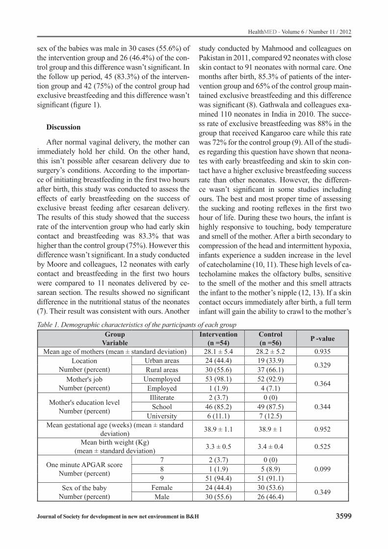
HealthMED - Volume 6 / Number 11 / 2012
Journal of Society for development in new net environment in B&H 3599
Table 1. Demographic characteristics of the participants of each groupGroup
VariableIntervention
(n =54)Control (n =56) P -value
Mean age of mothers (mean ± standard deviation) 28.1 ± 5.4 28.2 ± 5.2 0.935Location
Number (percent)Urban areas 24 (44.4) 19 (33.9) 0.329Rural areas 30 (55.6) 37 (66.1)
Mother's job Number (percent)
Unemployed 53 (98.1) 52 (92.9) 0.364Employed 1 (1.9) 4 (7.1)
Mother's education level Number (percent)
Illiterate 2 (3.7) 0 (0)0.344School 46 (85.2) 49 (87.5)
University 6 (11.1) 7 (12.5)Mean gestational age (weeks) (mean ± standard
deviation) 38.9 ± 1.1 38.9 ± 1 0.952
Mean birth weight (Kg)(mean ± standard deviation) 3.3 ± 0.5 3.4 ± 0.4 0.525
One minute APGAR scoreNumber (percent)
7 2 (3.7) 0 (0)0.0998 1 (1.9) 5 (8.9)
9 51 (94.4) 51 (91.1)Sex of the baby
Number (percent)Female 24 (44.4) 30 (53.6) 0.349Male 30 (55.6) 26 (46.4)
sex of the babies was male in 30 cases (55.6%) of the intervention group and 26 (46.4%) of the con-trol group and this difference wasn’t significant. In the follow up period, 45 (83.3%) of the interven-tion group and 42 (75%) of the control group had exclusive breastfeeding and this difference wasn’t significant (figure 1).
Discussion
After normal vaginal delivery, the mother can immediately hold her child. On the other hand, this isn’t possible after cesarean delivery due to surgery’s conditions. According to the importan-ce of initiating breastfeeding in the first two hours after birth, this study was conducted to assess the effects of early breastfeeding on the success of exclusive breast feeding after cesarean delivery. The results of this study showed that the success rate of the intervention group who had early skin contact and breastfeeding was 83.3% that was higher than the control group (75%). However this difference wasn’t significant. In a study conducted by Moore and colleagues, 12 neonates with early contact and breastfeeding in the first two hours were compared to 11 neonates delivered by ce-sarean section. The results showed no significant difference in the nutritional status of the neonates (7). Their result was consistent with ours. Another
study conducted by Mahmood and colleagues on Pakistan in 2011, compared 92 neonates with close skin contact to 91 neonates with normal care. One months after birth, 85.3% of patients of the inter-vention group and 65% of the control group main-tained exclusive breastfeeding and this difference was significant (8). Gathwala and colleagues exa-mined 110 neonates in India in 2010. The succe-ss rate of exclusive breastfeeding was 88% in the group that received Kangaroo care while this rate was 72% for the control group (9). All of the studi-es regarding this question have shown that neona-tes with early breastfeeding and skin to skin con-tact have a higher exclusive breastfeeding success rate than other neonates. However, the differen-ce wasn’t significant in some studies including ours. The best and most proper time of assessing the sucking and rooting reflexes in the first two hour of life. During these two hours, the infant is highly responsive to touching, body temperature and smell of the mother. After a birth secondary to compression of the head and intermittent hypoxia, infants experience a sudden increase in the level of catecholamine (10, 11). These high levels of ca-techolamine makes the olfactory bulbs, sensitive to the smell of the mother and this smell attracts the infant to the mother’s nipple (12, 13). If a skin contact occurs immediately after birth, a full term infant will gain the ability to crawl to the mother’s
3600
HealthMED - Volume 6 / Number 11 / 2012
Journal of Society for development in new net environment in B&H
nipple to clutch it in the first 60 minutes (14, 15). After these two hours, the level of catecholamine decreases and makes the infant sleepy (16). One possible explanation for the differences of the results of our study with Moore’s results may be the fact that many other factors have effect on the nutritional status. A study conducted by Qiu and colleagues in China that evaluated the prevalen-ce of exclusive breastfeeding in urban, semi ur-ban and rural areas, showed that location, cesa-rean delivery, time of breastfeeding initiation and using supplementary products are risk factors of its failure (17). Theofilogiannakou and colleagues studied the factors related to the start and length of breastfeeding in state and private hospitals of Greece. Their study showed that academic educa-tion was related to early initiation of breastfeeding in the first hour of life (18). However, in our study, variables such as location, level of education and employment status had no significant difference between the intervention and study group.
Conclusion
The results of our study showed that early brea-stfeeding didn’t have any significant impact on the success rate of exclusive breastfeeding of infants born with cesarean section.
Acknowledgements
We would like to give special thanks to Mrs. Mostashragh who helped us during the interven-tion.
References
1. Kazemi H, Ranjkesh F. Evaluating the problems of mothers in exclusive breastfeeding and educational intervention for improving nutrition status in Iran. Healthmed 2011; 5(6): 1517-21.
2. Cakmak H, Kuguoglu S. Comparison of the breastfee-ding patterns of mothers who delivered their babies per vagina and via cesarean section: an observati-onal study using the LATCH breastfeeding charting system. Int J Nurs Stud 2007; 44(7): 1128-37.
3. Olang B, Farivar K, Heidarzadeh A, Strandvik B, Yn-gve A. Breastfeeding in Iran: prevalence, duration and current recommendations. Int Breastfeed J 2009;4:8.
4. Moini A, Riazi K, Ebrahimi A, Ostovan N. Caesarean section rates in teaching hospitals of Tehran 1999-2003. East Mediterr Health J 2007; 13(2): 457-60.
5. Feldman R, Eidelman AI, Sirota L, Weller A. Compa-rison of skin- to-skin (kangaroo) and traditional care: parenting outcomes and pre-term infant development. Pediatrics 2002; 110 (1 Pt 1): 16-26.
6. Walters MW, Boggs KM, Ludington-Hoe S, Price KM, Morrison B. Kangaroo care at birth for full term in-fants: a pilot study. MCN Am J Matern Child Nurs 2007; 32(6): 375-81.
7. Moore ER, Anderson GC. Randomized controlled trial of very early mother-infant skin-to-skin contact and breastfeeding status. J Midwifery Womens Health 2007; 52(2): 116-25.
8. Mahmood I, Jamal M, Khan N. Effect of mother-in-fant early skin-to-skin contact on breastfeeding sta-tus: a randomized controlled trial. J Coll Physicians Surg Pak 2011; 21(10): 601-5.
9. Gathwala G, Singh B, Singh J. Effect of Kangaroo Mother Care on physical growth, breastfeeding and its acceptability. Trop Doct 2010; 40(4): 199-202.
10. Lagercrantz H, Slotkin TA. The “stress” of being born. Sci Am 1986; 254(4): 100-7.
11. Lagercrantz H. Stress, arousal and gene activation at birth. News Physiol Sci 1996;11(5):214–8
12. Porter RH, Winberg J. Unique salience of maternal breast odors for newborn infants. Neurosci Biobe-hav Rev 1999; 23(3):439-49.
13. Porter RH. The biological significance of skin-to-skin contact and maternal odours. Acta Paediatr 2004; 93(12): 1560-2.
14. Righard L, Alade MO. Effect of delivery room rou-tines on success of first breast-feed. Lancet 1990; 336(8723): 1105-7.
15. Widstrom AM, Ransjo-Arvidson AB, Christensson K, Matthiesen AS, Winberg J, Uvnas-Moberg K. Ga-stric suction in healthy newborn infants: Effects on circulation and developing feeding behavior. Acta Paediatr Scand 1987;76(4):566 –72.
16. Britton JR. The transition to extrauterine life and disorders of transition. Clin Perinatol 1998; 25(2): 271–94.
17. Qiu L, Zhao Y, Binns CW, Lee AH, Xie X. Initiation of breastfeeding and prevalence of exclusive brea-stfeeding at hospital discharge in urban, suburban and rural areas of Zhejiang China. Int Breastfeed J 2009; 28;4:1.

HealthMED - Volume 6 / Number 11 / 2012
Journal of Society for development in new net environment in B&H 3601
18. Theofilogiannakou M, Skouroliakou M, Gounaris A, Panagiotakos D, Markantonis SL. Breast-feeding in Athens, Greece: factors associated with its initiation and duration. J Pediatr Gastroenterol Nutr 2006; 43(3): 379-84.
Corresponding Author Mohammad Mehdi Nasehi, Assistant professor, Pediatrician Faculty of Medicine, Mazandaran University of Medical Sciences,Sari, IranE-mail: [email protected]
3602
HealthMED - Volume 6 / Number 11 / 2012
Journal of Society for development in new net environment in B&H
Factors effective on morbidity and mortality in rectal injuries caused by penetrating and blunt traumas: a civilian experienceIbrahim Aliosmanoglu, Mesut Gul, Zulfu Arikanoglu, Fatih Taskesen, Omer Uslukaya, Musluh Hakseven
Department of General Surgery, Medical Faculty, Dicle University, Diyarbakır, Turkey
Abstract
Purpose: Our objective in this study is to esta-blish the factors effecting morbidity and mortality in rectal injuries which occurred in civilian inju-ries.
Methods: Fifty-two patients who had been hospitalized for rectal injuries are evaluated. Pa-tients are arranged in two groups according to the etiology: Group I rectal injuries caused by pene-trating traumas; Group II rectal injuries caused by blunt traumas. To determine the risk factors, de-mographic data of the patients are compared.
Results: Group I consisted of 35 male and 2 female patients and average age was 27,5±7,5, while Group II consisted of 12 male and 3 female patients, and average age was 32,8±12,4 (p>0.05). Mean average of ISS was 10.1±10.1 in Group I and 19.4±12.1 in Group II (p=0.014). However, mean average of TRISS was 98.5±1.4 in Group I, and 96.4±3.5 in Group II; and the difference was statistically significant (p=0.011). Mortality num-bers for the groups were 1 patient in Group I (2.7 %), 3 patients in Group II (20%), (p=0.034).
Conclusions: Although rectal injuries are ra-rely seen in clinic, morbidity and mortality rates are high. So, being aware of the risk factors and developing a therapy plan, considering the patient is important for the success of therapy.
KeyWords: traumatic rectal injuries; morbi-dity; mortality.
Introduction
Rectal injuries are seen rarely due to anatomi-cal reasons and are mostly caused by penetrating traumas. Less than 1% of total trauma cases are rectal injuries (1- 3). 80-85 % of rectal injury cases are caused by firearms. Other causes include iatro-
genic injuries as a result of surgical procedures, foreign bodies and sexual misadventures. Blunt traumas are responsible for 5-10 % of cases and mostly as a result of pelvic fractures; rectal injury is seen in 2 % of these fractures (4). Digital rectal examination and rigid proctoscopy are important for the diagnosis of rectal injuries and have high diagnostic values. Because false negative results may be reported, further investigation must be carried out in case of suspicion (4). In this case, cysto-urethrograms, abdominal x-rays, water-so-luble contrast studies, and computed tomography (CT) scanning are the diagnostic tools to be consi-dered (5). Treatment of rectal injuries differ due to extent and grade of injury. Abdomen, genital area, perinea and pelvis of the patients with rectal inju-ries must be examined for concomitant injuries (5). Treatment has 4 components: fecal diversion, presacral drainage (PDS), distal rectal washout, if possible, repair of the injury (1). According to the condition of the injury, different combinati-on of these components may be carried out (5). Localization of rectum in pelvis and settlement of the 1/3 bottom part (of 12-15 cm. length) in the extraperitoneal area makes it harder to access the injury and repair (4). Today, treatment approaches differ for injuries caused by penetrating traumas and blunt traumas. Cleary et al. suggested a tre-atment algorithm for penetrating trauma cases (5). In this algorithm, if the rectal injury site cannot be identified in destructive extraperitoneal rectal injuries, or cannot be treated with the primary re-pair, the proximal colon diversion and PSD are suggested to be suitable treatments. On the other hand, in cases without serious concomitant inju-ries or with no accompanying medical diseases, the primary repair without proximal colon diver-sion is more suitable (6). Rectal injuries caused by blunt trauma are associated with pelvic blood

HealthMED - Volume 6 / Number 11 / 2012
Journal of Society for development in new net environment in B&H 3603
vessel, bladder and ureter injuries; therefore, mor-tality and morbidity rates are high. In cases of se-vere pelvic, peritoneal area, pelvic vascular and pelvic bone injuries, multidisciplinary treatments are required. In blunt rectal traumas, delayed per-forations may be found a few days after trauma, and if severe fecal contamination occurs during the operation and septic conditions, resection and diverting colostomy are chosen over primary re-pair (7). Traumatic rectal injuries are rare but have high morbidity and mortality rates. Our objective in this study is to emphasize the significance of this clinical condition and to investigate if there are differences in morbidity and mortality due to the etiology of injury.
Materials and methods
In this study, fifty-two patients who had been treated inpatient for rectal injuries due to blunt and penetrating traumas, at Dicle University Medici-ne Faculty, General Surgery Department between 2000 and 2011 are included. Blunt trauma injuries were caused by traffic accidents and falling from heights, while penetrating trauma injuries were caused by firearms and cutters. Patients are arran-ged in two groups according to the etiology: Group I (n=37) injuries caused by penetrating traumas; Group II (n=15) injuries caused by blunt traumas. Demographic data of patients are taken from their files retrospectively. Age and gender of patients, factors causing injury, trauma-treatment interval (TTI), concomitant organ injuries, Injury Seve-rity Score (ISS), Revised Trauma Score (RTS), Trauma Injury Severity Score (TRISS), treatment methods, morbidity and mortality rates, length of hospital stay are evaluated.
Results
The study group consisted of 47 male (90.4 %) and 5 female patients (9.6 %). Average age was 29.1±9.3 (ranging from 16 to 55). Cause of injury was firearms in 40.4 %, cutters in 30.8 %, traffic accidents in 11.5 % and falling from heights in 17.3 % of the patients. 32 patients (61.5 %) had concomitant organ injuries. 36 patients (69.2 %) had extraperitoneal, 10 patients (19.3 %) had in-traperitoneal and 6 patients (11.5 %) had intra-
extraperitoneal injuries. Mean trauma-treatment interval was 11.1±15.2 hours. As treatment met-hod, ostomy is applied to 39 patients (75 %), pri-mary repair to 12 patients (23.1%), and 1 patient (1.9 %) is followed medically. 19 patients (36.5%) had complications, while 4 patients (7.7%) had mortality. Average length of hospital stay was 13.4±10.8 days (Table 1). Group I consisted of 35 male and 2 female patients and average age was 27.5±7.5, while Group II consisted of 12 male and 3 female patients, and average age was 32.8±12.4 (p>0.05). Localization of injury; in Group I, was extraperitoneal in 25 patients (67.6%), intraperito-neal in 7 patients (18.9 %), intra-extraperitoneal in
Table 1. General characteristics of the patientsn
(=patient) %
Cause of trauma
Gunshot wound Stab wound Traffic accident Falling from height
211669
40,430,811,517,4
Concomitant organ injury Present Absent
3220
61,538,5
Grade of rectal injuries I II III IV
118321
1,934,661,51,9
Site of injury Ekstraperitoneal İntraperitoneal Ekstra+intraperitoneal
36106
69,219,311,5
Therapy Medical Primary repair Ostomy
11239
1,923,175
Causes of morbidity Vesicorectal fistula Wound site infection Ano-gluteal fistula Necrotizing fasciitis Rektovajinal fistül Morbidity
21032219
3,819,25,83,83,836,5
Mortality 4 7,7
Mean±SD Min-Max
Trauma-treatment interval (saat)Hastanede yatış süresi (gün)
11,1±15,213,4±10,8
4-961-51
SD: Standard deviation, Min: Minimum, Max: Maximum.
3604
HealthMED - Volume 6 / Number 11 / 2012
Journal of Society for development in new net environment in B&H
Table 2. Concomitant organ injuries in Groups I and II
OrganGroup I(n=37)n (%)
Group II (n=15)n (%)
Small Bowell 7 (18,9) 1 (6,7)Bladder 7 (18,9) 3 (20)Vascular 4 (18,8) 4 (26,7)
Pelvic bone 2 (5,4) 4(26,7)Sigmoid colon 2 (5,4) 0 (0)
Femur 1 (2,7) 1 (6,7)Uretra 2 (5,4) 2 (13,3)
Table 3. Comparison of the characteristics of the patients in Group I and Group IIGroup I (n=37)
n (%) and Mean±SDGroup II (n=15)
n (%9) and Mean±SD P
Age (year) 27,5±7,5 32,8±12,4 0.141Gender 0.106 Male Female
35 (94,6)2 (5,4)
12 (80)3 (20)
Concomitant organ injury 0.081 Present Absent
20 (54)17 (46)
12 (80)3 (20)
Site of injury 0.782 Extraperitoneal İntraperitoneal İntra-extraperitoneal
25 (67,6)7 (18,9)5 (13,5)
11 (73,3)3 (20)1 (6,7)
Grade of injury 0.211 I-II III-IV
12 (32,4)25 (67,6)
7 (46,7)8 (53,3)
TTI 0.649 ≥8 <8
27 (73)10 (27)
10 (66,7)5 (33,3)
ISS 10,1±10,1 19,4±12,1 0.014RTS 7,7±0,2 7,5±0,4 0.182TRISS 98,5±1,4 96,4±3,5 0.011Morbidity 13 (35,1) 6 (40) 0.741Mortality 1 (2,7) 3 (20) 0.034Lenght of hospital stay 14,9±11,2 9,8±9,0 0.066
TTI: Trauma-treatment interval, ISS: Injury severity score, RTS: Revised trauma score, TRISS: Trauma injury severity sco-re, SD: Standard deviation.
5 patients (13.5 %); while in Group II, was extra-peritoneal in 11 patients (73.3 %), intraperitoneal in 3 patients (20 %) and intra-extraperitoneal in 1 patient (6.7 %) (p=0.782). In Group I, 20 pati-ents (54 %) had 25 concomitant organ injuries, for Group II, it was 12 patients (80 %) and 15 conco-mitant organ injuries (p=0.081) (Table 2). TTI was before 8 hours in 25 patients in Group I and in 10 patients in Group II (p=0.649). There were no si-gnificant differences between two groups in terms
of treatment methods and length of hospital stay (p>0.05). Mean average of ISS was 10.1±10.1 in Group I, and 4±12.1 in Group II (p=0.014). In Group I, average TRISS was 98.5±1.4, while in Group II it was 96.4±3.5 and the difference was statistically significant (p=0.011). In Group I, mor-bidity was seen in 13 patients (35.1 %) and it was seen in 6 patients (40 %) in Group II (p=0.741). Mortality numbers for the groups were 1 patient in Group I (2.7 %), and 3 patients in Group II (20 %), (p=0.034) (Table 3). Excess of concomitant organ injuries, grade and site of injury, TTI and ISS had significant effect on morbidity (p<0.05) (Table 4). In terms of mortality, effective factors were, exce-ss of concomitant organ injuries, grade of injury, TTI, ISS, RTS and TRISS (p<0.05) (Table 5).
Discussion
Experiences on rectal injury treatment are mostly gained during wartime. Present therapy techniques are questioned if to be applied to rectal

HealthMED - Volume 6 / Number 11 / 2012
Journal of Society for development in new net environment in B&H 3605
Table 4. Morbidity impact of patient characteristicsMorbidityMean±SD p
Age 0.188 27,7±8,5 Absent 31,3±10,5 Present ISS 0.030 10,2±10,6 Absent 17,2±11,6 Present RTS 0,130 7,7±0,1 Absent 7,6±0,4 Present TRISS 0.054 98,4±1,6 Absent 97,0±3,2 Present
n (%) Gender 0.610 Male 17 (36,2) Female 2 (40)Concomittan organ injury 0.011 Absent 3 (15) Present 16 (50)Grade of injury I-II 2 (10,5) 0.020 III-IV 17 (51,5)Site of injury 0.040 Extraperitoneal 12 (33,3) İntraperitoneal 1 (10)TTI <0.001 <8 7 (18,9) ≥8 12 (80)
TTI: Trauma-treatment interval, ISS: Injury severity score, RTS: Revised trauma score, TRISS: Trauma injury severity score. SD: Standard deviation.
Table 5. Mortality impact of patient characteristics Mean ±SD Mortality p
Age 0.054 28,3±8,7 Absent 37,7±13,8 Present ISS 0.020 11,2±10,4 Absent 31,2±4,2 Present RTS <0,001 7,8±0,1 Absent 6,8±0,1 Present TRISS 0.001 98,4±1,4 Absent 91,7±3,4 Present
n (%) Gender 0.341 Male 3 (6,4) Female 1 (20)Concomittan organ injury 0.043 Absent 0(0) Present 4 (12,5)Grade of injury I-II 0 (0) 0.022 III-IV 4 (12,1)Site of injury 0.084 Extraperitoneal 2 (5,6) İntraperitoneal 0 (0)TTI 0.005 <8 0 (0) ≥8 4 (26,7)
TTI: Trauma-treatment interval, ISS: Injury severity score, RTS: Revised trauma score, TRISS: Trauma injury severity score, SD: Standard deviation.
injuries in civil life. To have extent knowledge of civil rectal injury cases’ characteristics, factors effective on morbidity and mortality will help to improve treatment approaches. Before World War I, surgical treatment wasn’t applied to rectal inju-ries and mortality rates were around 90 %. Du-ring war, primary suture was used and it fell to 67 %. During World War II, following application of fecal diversion and presacral drainage, morta-lity dropped to 30 %. Primary repair was applied together with distal rectal irrigation during Viet-nam War and mortality was reduced around 15 % (5). In a recent study 175 cases of military rectal injuries during Iraqi War and treated at 31st Com-
bat Support Hospital, mortality rate was found to be 18 % (8). Better resuscitation techniques and antibiotics contributed to the reduction in morta-lity. During Vietnam War, by adding distal rectal irrigation to treatment, morbidity of rectal injuries dropped from 72 % to 10 % (4). In our series of patients, mortality rate was 7.7 %. Treatment of rectal injury differs according to grade and extent of injury. Papadopoulos et al. (9) suggested pri-mary repair for penetrating and intraperitoneal injuries at first, and diversion for extraperitone-al injuries which couldn’t be repaired and anal sphincter injuries. Abdomen, genital area, perinea and pelvis of the patients with rectal injuries must

3606
HealthMED - Volume 6 / Number 11 / 2012
Journal of Society for development in new net environment in B&H
be examined for concomitant injuries (5). In our series of patients, we decided on surgical method according to grade of injury, fecal contamination risk and site of injury and we applied ostomy to patients with high risk of fecal contamination and who has extraperitoneal rectum injury. A patient (1.9 %) who had only seros injury was followed medically while 23.1 % of patients were applied primary repair and ostomy was performed on 75 % of patients. In series of civilian rectal injuries, morbidity rates are notified as 6-42 % and morta-lity rates as 0-10 % (10). In a recent study conduc-ted by Shatnawi&Bani-Hani (11) reported morbi-dity rate was 47.8%, and mortality rate 13 %, in 19 extraperitoneal and 4 extra+intraperitoneal civili-an rectal injury cases. In our study, for all cases, we established morbidity rate as 36.5 % and mor-tality rate as 7.7 %. Comparing groups, morbidity rate for Group I was 35.1 % and 40 % for Group II (p=0.741). Mortality rates for the groups were; 2.7 % and 20 % respectively, and the difference was statistically significant (p=0.034). We suggest as the most important reasons of high mortality rate in Group II, are more concomitant organ injuries and more vessel bleedings due to pelvic fractures. TTI to be more than 8 hours for all 4 patients who had mortality in our study shows the effect of time till treatment. Diverting colostomy is accepted as standard treatment of rectal injury by most of the authors (12, 13). Demirbaş et al., (14) preferred diverting colostomy, distal rectal irrigation and presacral drainage as treatment of ano-rectal inju-ries caused by firearms. In our study, ostomy was applied to 39 patients and primary repair for 12 pa-tients. It has been reported that presacral drainage has an effect of reducing morbidity and mortality (15). Armstrong et al. showed that pelvic abscess rate was reduced from 36 % to 25 % with the addi-tion of presacral drainage, on the other hand, the-re are studies showing no benefit from presacral drainage on extraperitoneal rectal injuries (16). Gonzales et al. conducted a randomized study and found that in civilian rectal traumas presacral dra-inage without distal rectal washout did not reduce infectious complications (17). Distal rectal irriga-tion is a contradictory issue. The study conducted by Shannon et al. showed statistical significance in favor of distal rectal washout (18). Their results established a difference in nonwashout versus
washout groups for pelvic abscess (46% and. 8%), rectal fistulae (23% and. 8%), and sepsis (15% and 8%). In our study, we applied presacral drainage to 4 patients and distal irrigation to 3 patients. It has been reported that only in half of extraperito-neal injury cases, injury site can be localized and suggested to apply primary repair if possible (15). Primary repair is reported to be more successful in civilian series than military series of patients (8). Bostick et al., (15) applied primary repair 32.1 % of 28 extraperitoneal rectal injuries. Burch et al., (10) reported that colostomy and drainage were to be successful in treatment of civil extraperitoneal rectal injuries and additional procedures like di-verting colostomy, rectal injury repair and rectum irrigation has very little effect on mortality and morbidity. We applied ostomy to 81 % of extra-peritoneal injury cases. For both groups, preferred treatment technique didn’t have a significant diffe-rence on morbidity and mortality (p<0.05). Mean average of ISS was more in Group II (p=0.014), and mean average of TRISS was less (p=0.011). We commented that this difference was due to excess of concomitant organ injuries. In terms of length of hospital stay, there was no statistically significant difference (p>0.05). In our study, exce-ss of concomitant organ injuries, high grade of injury, presence of isolated extraperitoneal injury, high average of ISS and TTI being longer than 8 hours had significant effect on morbidity (p<0.05). In terms of mortality, higher average of ISS, lower average of RTS and TRISS, excess of concomitant organ injuries, grade of injury, TTI being longer than 8 hours had significant effect on mortality (p<0.05).
Conclusion
We conclude that the most important factors effective on morbidity and mortality in penetrating and blunt rectal injuries are increased risk of fecal contamination due to long TTI period and possible complications due to delayed treatment of conco-mitant organ injuries. To be able to diagnose and treat rectal injuries, awareness has to be created among patients and doctors. We think that primary repair is sufficient for cases with low grade, low fecal contamination, no accompanying perianal ti-ssue defect and sphincter injuries, no concomitant
HealthMED - Volume 6 / Number 11 / 2012
Journal of Society for development in new net environment in B&H 3607
organ and system injuries, who applied to hospital within 8 hours. For the other patients who don’t have these characteristics, ostomy has to be added to treatment.
References
1. Carrillo EH, Somberg LB, Ceballos CE, Martini MA Jr, Ginzburg E, Sosa JL, Martin LC (1996) Blunt trau-matic injuries to the colon and rectum. J Am Coll Surg 183:548-552.
2. Steel M, Danne P, Jones I (2002) Colon trauma: Royal Melbourne Hospital experience. ANZ J Surg 72: 357-359.
3. Beard D, Henry JM, Grant PT (2000) National audit of the management of injured patients in 20 Scottish hospitals. Health Bull 58: 118-126.
4. Demetriades D, Salim A (2007) Colon and rectal tra-uma and rectal foreign bodies. In: Wolff BG, Flesh-man JW, Beck DE, et al. editors. The ASCRS Textbook of Colon and Rectal Surgery. New York, NY, Springer Science & Business Media, 322-334.
5. Cleary RK, Pomerantz RA, Lampman RM (2006) Co-lon and rectal injuries. Dis Colon Rectum 49:1203-1222.
6. Choi WJ (2011) Management of colorectal trauma. J Korean Soc Coloproctol 27:166-72.
7. Stewart RM, Rosenthal D (2005) Colorectal trauma. In: Corman ML, editor. Colon and rectal surgery. Phi-ladelphia, PA: Lippincott Williams & Wilkins, 427-449.
8. Steele SR, Wolcott KE, Mullenix PS, Martin MJ, Se-besta JA, Azarow KS, Beekley AC (2007) Colon and rectal injuries during Operation Iraqi Freedom: are there any changing trends in management or outco-me? Dis Colon Rectum 50:870-877.
9. Papadopoulos VN, Michalopoulos A, Apostolidis S, Paramythiotis D, Ioannidis A, Mekras A, Panidis S, Stavrou G, Basdanis G (2011) Surgical management of colorectal injuries: colostomy or primary repair? Tech Coloproctol 15:63-66.
10. Burch JM, Feliciano DV, Mattox KL (1989) Colo-stomy and drainage for civilian rectal injuries: is that all? Ann Surg 209:600-611.
11. Shatnawi NJ, Bani-Hani KE (2006) Management of civilian extraperitoneal rectal injuries. Asian J Surg 29:11-16.
12. Navsaria PH, Graham R, Nicol A (2001) A new approach to extraperitoneal rectal injuries: laparos-copy and diverting loop sigmoid colostomy. J Trau-ma 51:532-535.
13. Navsaria PH, Shaw JM, Zellweger R, Nicol AJ, Kahn D (2004) Diagnostic laparoscopy and diver-ting sigmoid loop colostomy in the management of civilian extraperitoneal rectal gunshot injuries. Br J Surg 91:460-464.
14. Demirbaş S, Yildiz M, Uluutku AH, Kalemoğlu M, Kurt Y, Erenoğlu C, Akin ML, Celenk T (2004) Sur-gical treatment of ano-rectal gunshot injuries cau-sed by low-velocity bullets. Ulus Travma Acil Cerra-hi Derg 10:17-21.
15. Bostick PJ, Johnson DA, Heard JF, Islas JT, Sims EH, Fleming AW, Sterling-Scott RP (1993) Manage-ment of extraperitoneal rectal injuries. J Natl Med Assoc 85:460-463.
16. Navsaria PH, Edu S, Nicol AJ (2007) Civilian extra-peritoneal rectal gunshot wounds: surgical manage-ment made simpler. World J Surg 31:1345-1351.
17. Gonzalez RP, Falimirski ME, Holevar MR (1998) The role of presacral drainage in the management of penetrating rectal injuries. J Trauma 45:656-561.
18. Shannon FL, Moore EE, Moore FA, McCroskey BL (1988) Value of distal colon washout in civilian rectal trauma--reducing gut bacterial translocation. J Trauma 28:989-994.
Corresponding AuthorIbrahim Aliosmanoglu,Dicle University Medical Faculty, Department of General Surgery,Diyarbakır, Turkey,Email: [email protected]
3608
HealthMED - Volume 6 / Number 11 / 2012
Journal of Society for development in new net environment in B&H
Yag laser capsulotomy rates of 50 years and older individuals after use of the Zaraccom Ultraflex intraocular lensesMustafa Ilker Toker, Ayse Vural Ozec, Ayhan Dursun, Ismail Yurdakul, Haydar Erdogan, Aysen Topalkara, Mustafa Kemal Arici,
Department of Ophthalmology, Cumhuriyet University School of Medicine, Sivas, Turkey
Abstract
Aim: to evaluate the rate of symptomatic po-sterior capsule opacification requiring Nd: YAG capsulotomy in patients who underwent cataract extraction and implantation of zaraccom ultraflex lenses (one-piece hydrophobic acrylic square ed-ged intraocular lens).
Methods: 142 eyes of 103 patients who had cataract extraction and implantation of zaraccom ultraflex lenses (IOLs) were evaluated for rate of nd: yag capsulotomy from april 2008 to august 2009. The cases that received Nd: YAG capsulo-tomies were evaluated for visual acuity, lens po-sition and centralization and the frequency of nd: yag laser determined.
Results: the mean preoperative best corrected visual acuity (BCVA) was 0.31 ± 0.20 And the final mean BCVA was 0.96 ± 0.06 With snellen chart. The rate of nd: yag capsulotomy was 0.7%. The mean time to Nd: YAG capsulotomy was 24.34 Months (range: 12 to 30 months).
Conclusion: posterior capsule opacificati-on was significantly less for the patients 50 years and over with the Zaraccom Ultraflex IOLs at 2 years.
Keywords: cataract surgery; intra ocular lens; nd: yag laser capsulotomy, posterior capsular opa-cification
Introduction
Cataract surgery is one of the most common surgical procedures performed worldwide. Impro-vements in surgical technique have made cataract surgery an increasingly safe procedure; however, posterior capsular opacification (PCO) remains the most common complication of cataract extrac-tion and intraocular lens (IOL) implantation.
It is well known that PCO is caused by mi-gration of the residual lens epithelial cells cen-trally toward the visual axis. The cause of PCO following cataract extraction and IOL implantati-on is multifactorial and is postulated to depend on the IOL optic material, IOL design, and surgical technique.1,3
PCO can be treated readily with Neodynium: Yttrium Aluminium Garnet (Nd: YAG) laser cap-sulotomy, which involves clearing the visual axis by creating a central opening in the opacified po-sterior capsule. Although this procedure is easy and quick, there are vision-related complications and puts significant financial burdens on the he-alth care system. Thus, one of the major concerns regarding current cataract surgery is how to decre-ase the PCO rate.4,5
Zaraccom Ultraflex lens (Anatolia Medicine Technologies Co., Sivas, Turkey) is the newer version of Zaraccom F260 single piece foldable hydrophobic acrylic square edged IOL. It has 6 mm optic diameter and approximately 800 µ cen-tral thicknesses. Overall length is 12.5 mm. Its re-fractive index is 1.51. Its angle of water contact is 82°. Its photo polymerization manufacturing technique is different from the other traditional foldable hydrophobic acrylic intraocular lenses manufactured by lathe cutting. The photopolyme-rization technique is a cast-molding method. It is known that cast-molded contact lenses are asso-ciated with apparently ‘stickier’ surfaces.
Advances in surgical techniques, intraocular lens materials, and designs have reduced the PCO rate, but it is still a significant problem. Intraocu-lar lenses made with novel materials and various types of edges have been developed in recent ye-ars and considerable research has focused on the PCO-inhibiting effects of these new IOLs.6,7 The aim of this study was to evaluate the nd: yag laser

HealthMED - Volume 6 / Number 11 / 2012
Journal of Society for development in new net environment in B&H 3609
posterior capsulotomy rate of Zaraccom Ultraflex (hydrophobic acrylic, one-piece intraocular lens) on cataract treatment.
Material and methods
This study was performed with adhering to the tenets of the declaration of helsinki. After obtaining institutional review board approval, a retrospective review was performed of eyes that had phacoemulsification with posterior chamber Zaraccom Ultraflex IOL implantation between april 2008 and august 2009. For each surgery, the patient’s chart was reviewed for the following data: age at surgery, sex of patient, right or left eye, past ocular history, preoperative and postoperati-ve best corrected vision, date of cataract extracti-on, date of Nd:YAG capsulotomy (if performed), pre-Nd:YAG and post-Nd:YAG capsulotomy best corrected vision, and date of last examination.
All surgeries were performed by one surgeon. Patients with any of the following conditions were excluded: capsule tear, vitreous loss, incomplete cortical cleanup, IOL not fixated in capsular bag, PCO noted at time of surgery, congenital cataract, history of trauma, history of preoperative uveitis, or postoperative follow up less than 5 months. The number of eyes needing nd: yag capsulotomy, and the time from surgery to follow up were de-termined. A total of 142 eyes of 103 patients (age 69.10±11.23 (50 To 90) years [mean ± standard deviation]) scheduled to undergo cataract surgery were included in this study. The principle inclusi-on criterion was the presence of senile cataract in an otherwise healthy eye in patients older than 50 years of age. The same well-experienced surge-on performed all cataract surgeries with the same technique. All patients were operated under topi-cal anesthesia.
All cataract operations comprised phacoemul-sification and IOL implantation. A continuous cu-rvilinear capsulorrhexis (ccc) approximately 5.0 Mm in diameter was created using a needle cysto-tome and a capsulorhexis forceps. After hydro-dissection, phacoemulsification of the nucleus and cortical aspiration were performed. After the an-terior chamber had been filled with a viscoelastic agent (Healon; Pharmacia, Uppsala, Sweden), Za-raccom Ultraflex was inserted in the capsular bag.
The viscoelastic agent was then washed out. Pati-ents were examined on the first day, six months, one year and two years after surgery. During each examination (under maximum pupil dilation) the lens capsules were evaluated using a slit lamp.
Nd:YAG capsulotomy was performed when an eye lost 2 or more decimal lines of visual acuity or when the patient complained of blurred vision. The presence of PCO also detected on dilated slit lamp examination. Patients had the appropriate pupil dilated and the position of the IOL was exa-mined using the TOPCON SL-3C slit lamp.
Statistical evaluation was done using frequency analysis with spps version 14.0.
Results
The mean age of patients was 69.10±11.23 (50 - 90 Years). The mean postoperative follow-up time was 24.34±4.79 (12-30) months. The mean preoperative best corrected visual acuity (bcva) was 0.31 ± 0.20. The final mean bcva was 0.96 ± 0.06 with snellen chart. The percentage of patients who had laser Nd:YAG capsulotomy was 0.7% with Zaraccom Ultraflex after 2 years from the surgeries. The all lenses were centralized.
Discussion
This study describes the demand over time for nd: yag laser capsulotomy during a mean follow up period of 2 years after phacoemulsification ca-taract surgery. In our setting the Nd:YAG laser capsulotomy rate of Zaraccom Ultraflex (foldable hydrophobic acrylic intraocular lens with square edge optic design) was 0.7%, which is lower than the rate reported in recent literature. The reported incidence of Nd:YAG laser capsulotomy rate vari-es widely among studies and these differences are based on varying lengths of follow-up and diffe-rent surgical techniques, intraocular lens (IOL) designs, and methods of IOL implantation.2
Studies have been showed that hydrophobic acrylic IOLs with a sharp optic edge are associa-ted with less PCO than other materials.2,8,9 Lens geometry plays an important role in the occurren-ce of PCO and a higher PCO inhibitory effect has been observed with IOLs that provide a mechani-cal barrier effect on the posterior lens capsule.10
3610
HealthMED - Volume 6 / Number 11 / 2012
Journal of Society for development in new net environment in B&H
Nishi et al3 first showed the existence of an enhan-ced barrier effect afforded by a square edge optic design and it appears that PCO is less frequent in patients given square-edged lenses than in those with curved edges.11 It has been also showed that sharp-optic edge is the major inhibitory factor of lens epithelial cell (LEC) migration behind the IOL optic regardless of the material of the IOL.2,12,13
Comparison of hydrophobic and hydrophilic materials showed that PCO is more frequent with hydrophilic acrylic lenses than with hydrophobic acrylic lenses.14,15 Vasavada et al16 compared the posterior capsule opacification of hydrophobic acrylic and hydrophilic acrylic intraocular lenses and found significantly less posterior capsule opa-cification with hydrophobic acrylic IOL at 3 years.
Multiple studies comparing PCO incidence when using one piece versus three piece acrylic IOLs have showed that three-piece and one-piece acrylic hydrophobic IOLs were very similar for visual acuity and PCO development.17,18
Acrylic lens size may influence the rate of po-sterior capsule opacification (PCO) and need for nd: yag capsulotomy, although controversy re-mains as to this assertion.19 Several studies have found that PCO is reduced when the anterior cap-sulorhexis is in complete contact with the anteri-or IOL surface, which is easier to achieve with a larger optic.20,21,22 On the other hand larger IOL size has a negative effect on PCO formation. Za-raccom ultraflex has a diameter of 12.5 Mm and differs with this feature from the most similar lens on the market which has a 13 mm diameter. In a recent study of us, we determined that tension cau-sed by lens implantation forms a permanent cavity between optic-haptic junction of the IOL and po-sterior capsule that is affecting the IOL adhesion. In that study we saw that, when the diameter of IOL is decreased, the cavity between optic-haptic junction of the IOL and the posterior capsule is decreased and IOL is adhering to posterior capsule better when the diameter is 12.5 Mm (not yet pu-blished). One proposed explanation for the redu-ced occurrence of PCO with use of the zaraccom ultraflex IOL is the size.
The limitations of this study include its retros-pective design and the use of nd: yag capsulotomy as an indirect measure of PCO. In this study nd: yag capsulotomy was performed when an eye
lost 2 or more decimal lines of visual acuity or when the patient complained of blurred vision. This inclusion criterion may affect the founded Nd:YAG laser capsulotomy rate.
Fifty year and older patients included to this study and the mean age of patients were 69.1. Age plays a crucial role in the rate of PCO de-velopment, and PCO rate of this study probably found lower due to high mean age of patients.
Research laboratories worldwide attempting to eliminate the problem of PCO development are focusing on several strategies, including im-proving surgical techniques, IOL materials, IOL designs, use of therapeutic agents, and combina-tion therapy. IOL design and improved surgical methods have led to a reduced level of PCO over the past decade, but have not eradicated the pro-blem. The results of this study showed that folda-ble hydrophobic acrylic intraocular lens zaraccom ultraflex has very low nd: yag laser posterior cap-sulotomy rate in cataract patients in long term.
References
1. Awasthi n, guo s, wagner bj. Posterior capsular opa-cification: a problem reduced but not yet eradicated. Arch ophthalmol. 2009;127(4):555-562.
2. Cheng jw, wei rl, cai jp, xi gl, zhu h, li y, ma xy. Effi-cacy of different intraocular lens materials and op-tic edge designs in preventing posterior capsular opacification: a meta-analysis. Am j ophthalmol. 2007;143(3):428-36.
3. Nishi o, nishi k, wickstrom k. Preventing lens epithe-lial cell migration using intraocular lenses with sharp rectangular edges. J cataract refract surg. 2000;26(10):1543-1549.
4. Aslam tm, devlin h, dhillon b. Use of nd: yag laser capsulotomy. Surv ophthalmol. 2003;48(6):594-612.
5. Apple dj, peng q, visessook n, werner l, pandey sk, escobar-gomez m, ram j, auffarth gu. Eradication of posterior capsule opacification: documentation of a marked decrease in nd: yag laser posterior capsulo-tomy rates noted in an analysis of 5416 pseudophakic human eyes obtained postmortem. Ophthalmology. 2001;108(3):505-518.
6. Findl o, buehl w, bauer p, sycha t. Interventions for preventing posterior capsule opacification. Cochrane database syst rev. 2010;(3):1-81.
HealthMED - Volume 6 / Number 11 / 2012
Journal of Society for development in new net environment in B&H 3611
7. Pandey sk, apple dj, werner l, maloof aj, milverton ej. Posterior capsule opacification: a review of the aetiopathogenesis, experimental and clinical studi-es and factors for prevention. Indian j ophthalmol. 2004;52(2):99-112.
8. Iwase t, nishi y, oveson bc, jo yj.Hydrophobic versus double-square-edged hydrophilic foldable acrylic in-traocular lens: effect on posterior capsule opacifica-tion. J cataract refract surg. 2011;37(6):1060-1068.
9. Schmidbauer jm, vargas lg, apple dj, escobar-gomez m, izak a, arthur sn, golescu a, peng q. Evaluation of neodymium: yttrium-aluminum-garnet capsulotomi-es in eyes implanted with acrysof intraocular lenses. Ophthalmology. 2002;109(8):1421–1426.
10. Hayashi k, hayashi h. Posterior capsule opacifica-tion in the presence of an intraocular lens with a sharp versus rounded optic edge. Ophthalmology. 2005;112(9):1550–1556.
11. Cleary g, spalton dj, koch dd. Effect of square-edged intraocular lenses on neodymium:yag laser capsu-lotomy rates in the united states. J cataract refract surg. 2007;33(11):1899-1906.
12. Nishi o, nishi k, osakabe y. Effect of intraocular lenses on preventing posterior capsule opacificati-on: design versus material. J cataract refract surg. 2004;30(10):2170 –2176.
13. Findl o, menapace r, sacu s, buehl w, rainer g. Effect of material on posterior capsule opacification in intraocular lenses with sharp-edge optics. Ophthal-mology. 2005;112(1): 67–72.
14. Gauthier l, lafuma a, laurendeau c, berdeaux g. Neodymium:yag laser rates after bilateral im-plantation of hydrophobic or hydrophilic multi-focal intraocular lenses: twenty-four month re-trospective comparative study. J cataract refract surg. 2010;36(7):1195-200.
15. Boureau c, lafuma a, jeanbat v, berdeaux g, smith af. Incidence of nd:yag laser capsulotomies after cataract surgery: comparison of 3 square-edged lenses of different composition. Can j ophthalmol. 2009;44(2):165-170.
16. Vasavada ar, raj sm, shah a, shah g, vasavada v, va-savada v. Comparison of posterior capsule opa-cification with hydrophobic acrylic and hydrophi-lic acrylic intraocular lenses. J cataract refract surg. 2011;37(6):1050-1059.
17. Zemaitiene r, jasinskas v, auffarth gu. Influence of three-piece and singlepiece designs of two sharp-ed-ge optic hydrophobic acrylic intraocular lenses on the prevention of posterior capsule opacification: a prospective, randomised, long-term clinical trial. Br j ophthalmol. 2007;91(5):644-648.
18. Leydolt c, davidovic s, sacu s, menapace r, neu-mayer t, prinz a, buehl w, findl o. Long-term effect of 1-piece and 3-piece hydrophobic acrylic intraocular lens on posterior capsule opacification: a randomi-zed trial. Ophthalmology. 2007;114(9):1663-1669.
19. Vasavada ar, raj sm. Anterior capsule relationship of the acrysof intraocular lens optic and posterior capsule opacification: a prospective randomized cli-nical trial. Ophthalmology. 2004;111(5):886–894.
20. Hayashi k, hayashi h. Influence on posterior capsule opacification and visual function of intraocular lens optic material. Am j ophthalmol. 2007;144(2):195-202.
21. Wejde g, kugelberg m, zetterstrom c. Position of ante-rior capsulorhexis and posterior capsule opacifica-tion. Acta ophthalmol scand. 2004;82(5): 531–534.
22. Wren sm, spalton dj, jose r, boyce j, heatley cj. Fac-tors that influence the development of posterior capsule opacification with a polyacrylic intraocular lens. Am j ophthalmol. 2005;139(4): 691–695.
Corresponding AuthorDr. Ayse Vural Ozec,Cumhuriyet Universitesiu, Tıp Fakultesi, Sivas,TurkeyE–mail: [email protected]

3612
HealthMED - Volume 6 / Number 11 / 2012
Journal of Society for development in new net environment in B&H
The role of physiological scores for decision making in internal pre-hospital emergency situationsAbbasali Ebrahimian1, 2, Hamidreza Shabanikiya2, Nader Khalesi 3
1 Department of Medical Emergencies, School of Sorkheh Allied Health, Semnan University of Medical Sciences, Semnan, Iran2 School of Health Management and Information Sciences, Tehran University of Medical Sciences, Tehran, Iran3 Health Services Management Department, School of Health Management and Information Sciences, Tehran University of Medical Sciences, Tehran, Iran
Abstract
Introduction: Pre-hospital emergency services need appropriate means to identify the actual criti-cal necessities of internalpatients. The purpose of this study is to assess the physiological efficiency scoring system in order to manage the transferen-ce of internal patients across Iran.
Methods: This paper is a cross sectional stu-dy. The research community is the patients tran-sferred by pre-hospital emergency of Tehran hos-pital. Physiological scoring system is used as the data collection device. Data were analyzed by the use of descriptive and analytical census (Pearson correlation coefficient) and by SPSS19 software.
Findings: The median physiological scores of the research subjects equals to 1.54 ± 2.38. More than half of patients, who were sent to hospital, need no emergency treatment, besides there were a meaningful statistical relationship between the physiological scores and number of hospitalized patients.
Conclusion: The use of physiological scoring system seems to be a suitable method to make de-cision about whether to transfer an internal patient or not. But extensive studies in this area are essen-tial in order to make a definitive statement about the usage of these measurements.
Keywords: Internal patients, Physiological Scoring, Pre-hospital emergency, Transference management
Introduction
In Iran the pre-hospital emergency is respon-sible for free transference of patients with inter-nal problems and those injured of traffic and non-
traffic accidents. Tehran is a city in which every day more than 1,100 missions’ are implemented by pre-hospital emergency personnel. About 85-80% of the cases are subject to consideration and transference of non-traumatic and internal patients (1). On the other hand, the average rate of pre-hos-pital emergency missions’growth equals to 16% (2). This means that every 6 years, the number of pre-hospital emergency missions is doubled. Therefore, every six years the fleet of pre-hospital emergency of Iran must be doubled. It should be considered that depreciation ofexistingambulan-ces and their equipment and exhaustion of the cu-rrent staff will be a heavy cost for the Healthcare System of Iran. There are some efforts to reduce the pre-hospital emergency missions causing no harm to people or without reduction of service quality. Since traumas are considered to be the mainreason of disability and economic damages caused by healthcare affairs (3); therein to inve-stigate health condition of patients suffering from traumas; various scales, standards and systems are developed and widely used around the world (4). Despite the fact that the major part of pre-hospital emergency mission deals with care and transfe-rence of non-traumaticpatients, fewstandards have been allocated to this subject and few studies have been done. Physiological scoring system designed by Subbe et al, is one of the standards to determine the fatality of internal patients’ condition. Some-rating and classification systems based on physi-ological criteria have been implemented in some studies outside Iran (5-7). In Iran there are no stu-dies to provide standards for determining the fata-lity of internal patients’ condition at pre-hospital emergency phases; thereby transference is based
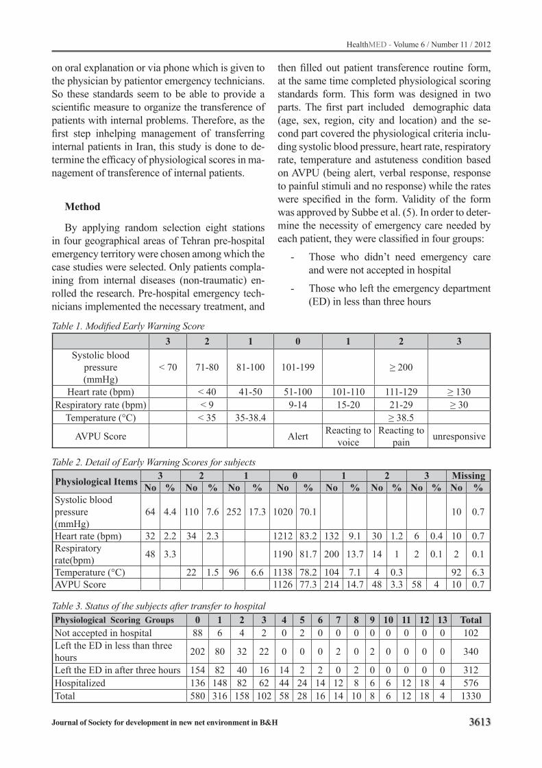
HealthMED - Volume 6 / Number 11 / 2012
Journal of Society for development in new net environment in B&H 3613
on oral explanation or via phone which is given to the physician by patientor emergency technicians. So these standards seem to be able to provide a scientific measure to organize the transference of patients with internal problems. Therefore, as the first step inhelping management of transferring internal patients in Iran, this study is done to de-termine the efficacy of physiological scores in ma-nagement of transference of internal patients.
Method
By applying random selection eight stations in four geographical areas of Tehran pre-hospital emergency territory were chosen among which the case studies were selected. Only patients compla-ining from internal diseases (non-traumatic) en-rolled the research. Pre-hospital emergency tech-nicians implemented the necessary treatment, and
Table 1. Modified Early Warning Score3 2 1 0 1 2 3
Systolic blood pressure(mmHg)
< 70 71-80 81-100 101-199 ≥ 200
Heart rate (bpm) < 40 41-50 51-100 101-110 111-129 ≥ 130Respiratory rate (bpm) < 9 9-14 15-20 21-29 ≥ 30
Temperature (°C) < 35 35-38.4 ≥ 38.5
AVPU Score Alert Reacting tovoice
Reacting topain unresponsive
then filled out patient transference routine form, at the same time completed physiological scoring standards form. This form was designed in two parts. The first part included demographic data (age, sex, region, city and location) and the se-cond part covered the physiological criteria inclu-ding systolic blood pressure, heart rate, respiratory rate, temperature and astuteness condition based on AVPU (being alert, verbal response, response to painful stimuli and no response) while the rates were specified in the form. Validity of the form was approved by Subbe et al. (5). In order to deter-mine the necessity of emergency care needed by each patient, they were classified in four groups:
- Those who didn’t need emergency care and were not accepted in hospital
- Those who left the emergency department (ED) in less than three hours
Table 2. Detail of Early Warning Scores for subjects
Physiological Items 3 2 1 0 1 2 3 MissingNo % No % No % No % No % No % No % No %
Systolic blood pressure(mmHg)
64 4.4 110 7.6 252 17.3 1020 70.1 10 0.7
Heart rate (bpm) 32 2.2 34 2.3 1212 83.2 132 9.1 30 1.2 6 0.4 10 0.7Respiratory rate(bpm) 48 3.3 1190 81.7 200 13.7 14 1 2 0.1 2 0.1
Temperature (°C) 22 1.5 96 6.6 1138 78.2 104 7.1 4 0.3 92 6.3AVPU Score 1126 77.3 214 14.7 48 3.3 58 4 10 0.7
Table 3. Status of the subjects after transfer to hospitalPhysiological Scoring Groups 0 1 2 3 4 5 6 7 8 9 10 11 12 13 TotalNot accepted in hospital 88 6 4 2 0 2 0 0 0 0 0 0 0 0 102Left the ED in less than three hours 202 80 32 22 0 0 0 2 0 2 0 0 0 0 340
Left the ED in after three hours 154 82 40 16 14 2 2 0 2 0 0 0 0 0 312Hospitalized 136 148 82 62 44 24 14 12 8 6 6 12 18 4 576Total 580 316 158 102 58 28 16 14 10 8 6 12 18 4 1330

3614
HealthMED - Volume 6 / Number 11 / 2012
Journal of Society for development in new net environment in B&H
- Those who left the emergency department after three hours
- Those who were hospitalizedThen the scores provided by physiological stan-
dards form and demographic data and information about patient’s conditions after being transferred to hospital were analyzed by Pearson correlation coefficient and SPSS19.
Results
A total of 1602 samples were collected. 1454 forms which were properly completed provided input data needed by the software to present sta-tistical analysis. The average age of the patients equaled to 51.08 ± 22.11, maximum and mini-mum age considered as 100 and 12 years of age, respectively. 58.2% of participants were men and 41.8% of them were women. Median of physiolo-gical score of transferred patients equaled to 1.54 ± 2.38. For other information about the case studi-es see Table 2 and 3 and Figure 1.
Discussion
Average age of the subjects was 51.08 years. This range of age is because the samples of this study were patients with internal problems and in-ternal problems are more prevalent among older ages. Similar studies represent the median age of patients who were transferred to hospital by pre-hospital emergency personnel as 54.9 to 62 years (9,8,5). This means that whether the age of pati-ents suffering from internal problems or the age of those calling pre-hospital emergency has decre-ased. The results showed that in comparison with the number of women, a larger number of men called the pre-hospital emergency for help and were transferred to hospital. Other studies confir-med this result (5). Perhaps because men have got a variety of responsibilities within family and so-ciety, so they bear a lot of pressures and stresses. Another reason that may explain this issue is that health is much more important to women. Usually women, more than men, care for their health and before the appearance of weakening symptoms, they turn to physicians. Or maybe when women need medical services, men take them to hospitals
by their own cars instead of calling pre-hospital emergency center.
Results show a meaningful statistic relation between the physiological scores and the chance of being hospitalized; it means the higher physi-ological score is, the higher probability of being hospitalized would be, so that almost all patients who were scored more than five were hospitali-zed. It is because the vital signs of most patients suffering from serious internal disorders began to worsen since several hours earlier (10). Other studies in this field have also shown that higher scores gathered via physiological measurement forms will also increases the chance of being hos-pitalized (5,6,11). But the important finding of this study which is also very disturbing is that a significant number of patients were transferred to hospital while having a zero physiological stan-dard score. This shows a dramatic discrepancy with other studies in which a small percentage of transferred patients were zero-rated (5, 6). Perhaps this happens because of freeness of pre-hospital emergency services in Iran or technicians’ anxi-ety of chastisement in case of no transference or absence of suitable standards to determine the se-riousness of patient’s condition. There may also be other factors as the technicians’ inability to detect seriousness of patients’ condition; fast tran-sportation of ambulances through heavy traffics of Tehran, lonely life of some patients especially el-derlies, probability of sudden death, better accep-tance of transferred patients by hospital personnel, social and psychological needs which cause the spread of this phenomenon. It is also shown that about 7% of transferred patients did not need to
Figure 1. The frequency of subjects according to physiological score
HealthMED - Volume 6 / Number 11 / 2012
Journal of Society for development in new net environment in B&H 3615
be sent to hospitals while seriousness of 44.8% patients’ disease was not too much to be sent to hospital, immediately. That is more than half of patient transference have not been essential and must be prohibited. Financial burden, depreciati-on of equipment and exhaustion of manpower re-sulted from this burdensome process confirms the necessity of further studies in this field.
Conclusion
Although there is the possibility of wrong use of physiological standards in emergency occasi-ons (12), these standards can help doctors to cla-rify patients with fatal conditions (13). Therefore physiological scoring system seems to be useful in determining the seriousness of patients’ condition and to decide if it is necessary to transfer a patient to hospital or not. This would be also commodious and optimized especially when there is only one ambulance to carry out several requests. To make certain statements in this regard, further meticulo-us studies are required.
Acknowledgement
We wish to thank of department manager of Health Services Management for comments on an early draft of the manuscript. This study was supported by School of Health Management and Information Sciences, Tehran University of Medi-cal Sciences (TUMS/SHMIS).
References
1. Availablefrom:URL: http//ems115.behdasht.gov.ir/index.aspx?siteid=105&siteid=105&pageid=35204.
2. Halstead J. Management briefing: Emergency Admi-ssions. Electronic Library for Health 2005.
3. Leigh J, Maczskill P, Kuosma E, K M. Global burden of disease and injury due to occupational factors. Epi-demiology. 1999;10(5):626-31.
4. Jana B.A, MacLeod, Olive Kobusingye, Chris Frost, Ron Lett, Fred Kirya, et al. A Comparison of the Kam-pala Trauma Score (KTS) with the Revised Trauma Score (RTS), Injury Severity Score (ISS) and the TRI-SS Method in a Ugandan Trauma Registry. European Journal of Trauma. 2003;29:392-8.
5. Subbe C.P, Kruger M, Rutherford P, L G. Validation of a modified Early Warning Score in medical admissi-ons. Q J Med 2001;94(10):521-26.
6. Kirsty Challen, Walter D. Physiological Scoring: An Aid to Emergency Medical Services Transport Decisions? Prehospital and Disaster Medicine. 2009;2(4):320-24.
7. Gray J, A W. AMPDS categories: are they an appro-priate method to select cases for extended role am-bulance practitioners? Emerg Medical Journal. 2008;25(9):601-3.
8. James N. Fullerton, Charlotte L. Price, Natalie E. Silvey, Samantha J. Brace, Gavin D. Perkins. Is the Modified Early Warning Score (MEWS) superior to clinician judgment in detecting critical illness in the pre-hospital environment? Resuscitation. 2012; 83: 557– 562.
9. Tobias M Merz, Reto Etter, Ludger Mende, Daniel Barthelmes, Jan Wiegand, Luca Martinolli, Jukka Takala. Risk assessment in the first fifteen minutes: a prospective cohort study of a simple physiological scoring system in the emergency department. Critical Care. 2011; 15:R25.
10. Panchagnula U, Thomas A. N, Rhodes S. Associa-tion between outcome and modified early warning scores: relationship with age and medical discipline. European Journal of Anesthesiology. 2007; 25: 72-85.
11. Larry L.Y. Lee, K.L. Yeung, Wendy Y.L. Lo, Yvonne S.C. Lau, Simon Y.H. Tang, Jimmy T.S. Chan. Evalu-ation of a simplified therapeutic intervention scoring system (TISS-28) and the modified early warning score (MEWS) in predicting physiological deterio-ration during inter-facility transport. Resuscitation. 2008. 76, 47-51.
12. Alison Day, Carol Oldroyd. The Use of Early War-ning Score in The Emergency Department. Clinical Nurses Forum. 2010; 36(2): 154-5.
13. Giles N. Cattermole, S.K. Paulina Mak, C.H. Eliza-beth Liow, Man Fung Ho, Kin Yee Grace Hung, Kai Man Keung, Hoi Man Li , Colin A. Graham, Timothy H. Rainer. Resuscitation. 2009; 1000–1005.
Corresponding AuthorHamidreza Shabanikiya,School of Health Management and Information Sciences,Tehran University of Medical Sciences, Tehran, Iran,E-mail: [email protected]

3616
HealthMED - Volume 6 / Number 11 / 2012
Journal of Society for development in new net environment in B&H
Plasma inflammatory marker levels in the systolic heart failure patients with and without atrial fibrillationErsan Tatli1, Mehmet Akif Cakar2, Mustafa Yilmaztepe1, Ahmet Barutcu1
1 Department of Cardiology, Ada Tıp Hospital, Sakarya, Turkey2 Sakarya Education and Research Hospital, Cardiology Department, Sakarya, Turkey
Abstract
Objective: Heart Failure (HF) imposes the greatest risk for the occurrence of atrial fibrillation(AF). Occurrence of AF adversely affects morbidity and mortality in patients with HF. To investigate the association between levels of inflammatory markers and clinical and echocar-diographic parameters in the population of systo-lic heart failure (SHF) patients with or without AF.
Methods and results: The study included 128 patients with SHF. The patients were divided into two groups. Group I (n=98) included patients with sinus rhythm . Group II (n=30) consisted of patients with AF. In all patients, plasma high se-snsitive C-reactive protein (C-RP) and cytokines levels, echocardiographic paramaters were mea-sured. Clinical variables were recorded. Plasma C-RP levels in group II (11.7±4.8mg/l) was si-gnificantly higher than in group I (8.8±3.1 mg/l) (p=0.03). The number of patients with New york heart association (NYHA) < 2 in group II were significantly higher than in group I (p=0.002). Pla-sma cytokines levels and clinical variables were not significant statistically in the between groups.
Conclusion: This study results indicated that plasma C-RP levels are associated with AF in pa-tients with SHF. This finding may have important implications for the development of new therape-utic and preventive approaches of AF in the setting of SHF.
Keywords: C-reactive protein, heart failure, atrial fibrillation.
Introduction
Atrial fibrillation affects approximately 2 milli-on people in the United States and is a common comorbidity among patients with heart failure
(HF)1. Furthermore, 13% to 27% of atrial fibri-llation (AF) patients have chronic heart failure and left ventricular dysfunction 2,3. The mor-tality and morbidity of AF is mainly related to thromboembolic consequences4,5. The mortality rate is approximately doubled in patients with AF compared with patients in normal sinus rhythm and is linked to the severity of heart disease6.Atrial fibrillation is also associated with loss of atrial contractile functioning and may result in hemodynamic deterioration. This hemodynamic deterioration may result in exacerbation of chro-nic heart failure symptoms and impaired exercise tolerance4,5. Numerous reports indicate that there is a relationship between AF and markers of syste-mic inflammation such as high sensitive C-reac-tive protein (C-RP), tumour necrosis factor alpha (TNF-α), interleukin -1 (IL-1), and interleukin-6 (IL-6)7,8. Plasma levels of inflammatory markers are also elevated in HF9. However, there aren’t sufficiently reports concerning the relationship between concentrations of inflammatory markers and occurrence of AF in patients with HF. In this study, we aimed to assess the interrelationships of inflammatory markers, echocardiographic pa-ramaters, and occurence of AF in patients with systolic HF (SHF).
Material and methods
Patients who admitted to the department of car-diology of our institution with a diagnosis of SHF were screened and 128 of them were included in the study. The patients were divided into two groups. Group I (n=98) included patients with si-nus rhythm. Group II (n=30) consisted of patients with AF. The inclusion criteria were the presence of New york heart association (NYHA) class II-IV heart failure, left ventricular ejection fraction
HealthMED - Volume 6 / Number 11 / 2012
Journal of Society for development in new net environment in B&H 3617
less than 40%, and the current treatment for heart failure with standard therapy including: diuretics, angiotensin-converting enzyme inhibitors, beta blockers and digoxin at a stable dosage for at le-ast six weeks. Criteria for exclusion from the stu-dy were: chronic obstructive pulmonary disease, acute or chronic infections at baseline, significant valvular heart disease, thyrotoxicosis, hypothyro-idism, chronic kidney and liver diseases, mali-gnancy, anemia (Hb<9gr/dl), systolic blood pre-ssure lower than 90 mmHg, heart rate lower than 50/min, and first- or second- degree heart block. Patients receiving antiarrhythmic drugs, patients with a permanent pacemaker and psychiatric pro-blems were also excluded from the study.
Detailed medical history, physical examinati-on, and routine biochemical testing were perfor-med . Left ventricular size, ejection fraction, and left atrial diameter (LAD) were evaluated by tran-sthoracic echocardiography. The protocol of this study was approved by our hospital’s ethics com-mittee and each patient gave written consent.
EchocardiographyAll patients underwent echocardiography
using GE Vingmed System FIVE and a 2.5-5.0 MHz standard probe. The following echocardio-graphic parameters were assessed: LAD, left ven-tricular end diastolic diameter and left ventricu-lar ejection fraction (LVEF). Left atrial diameter was measured by M-mode in the long axis of the parasternal view. The largest atrial diameter was measured during maximum anterior movement of the posterior aortic wall. Left ventricular ejection fraction was calculated with the modified Simp-son method.
Laboratory testsBlood was collected up to 24 hours after admi-
ssion in the begining of study. C-RP levels were measured using an immunoturbidimetric method involving C-RP agglutination with latex beads co-ated with anti-human C-RP monoclonal antibodi-es. Measurements were performed with a Immage 800 (Beckman-Coulter, USA). To measure TNF-α, IL-2, and IL-6 concentrations, 15 mL of blood was drawn from an antecubital vein and collected into prechilled evacuated tubes contain ing ethylene-
diaminetetraacetic acid. Plasma was sepa rated by centrifugation at 2,500 rpm for 12 minutes wit-hin 15 minutes of collection. Samples were sto-red at –70 °C. Measurements of TNF-α, IL-2, and IL-6 were performed in undiluted plasma with a commer cially available, enzyme-linked agent for immunoassay (Immulite, Diagnostic Products Corporation; Los An geles, Calif).
Statistical analysisQuantitative values are expressed as mean ±
SD and were compared using unpaired Student t test. The X2 test, Fisher’s exact test and Mann Whitney U test were used to analyze categorical data. For all tests, p > 0.05 was designated nonsi-gnificant, and a value of p < 0.05 was considered statistically significant. The Statistical Package for Social Sciences (SPSS) statistical software packa-ge (version 10.0, Inc., Chicago, USA) was used to perform all statistical calculations.
Results
The study included 128 patients (104 males and 24 females at mean age of 58.4±11.3years ) with SHF. Baseline characteristics of all patients were summarized in Table 1. Group I (n=98) included patients with sinus rhythm. Group II (n=30) consisted of patients with AF. There were no significant differences between two groups in terms of hypertension , smoking , diabetes mellitus hyperlipidemia, systolic and diastolic blood pre-ssure, body mass index, levels of IL-2, IL-6 and TNF-α (p>0.05). Left ventricular ejection fracti-on, left ventricular end- diastolic diamaters and left atrial diamater were not significant between in two groups (p>0.05). Plasma C-RP levels in group II (11.7±4.8mg/l) was significantly higher than in group I (8.8±3.1 mg/l) (p=0.03) (Figure 1). The number of patients with NYHA < 2 (93%) in group II were significantly higher than in group I (p=0.002) (Figure 2). Clinical variables and in-flammatory markers were summarized in Table 2.
Discussion
The results showed that plasma C-RP levels and the number of patients with NYHA < 2 in
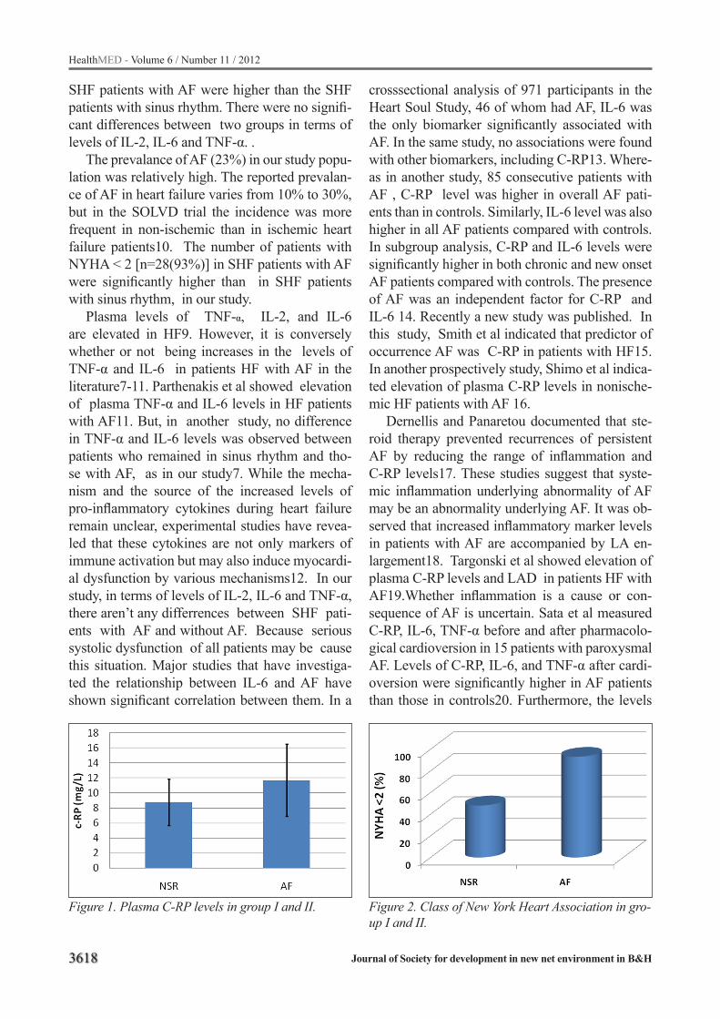
3618
HealthMED - Volume 6 / Number 11 / 2012
Journal of Society for development in new net environment in B&H
Figure 1. Plasma C-RP levels in group I and II. Figure 2. Class of New York Heart Association in gro-up I and II.
SHF patients with AF were higher than the SHF patients with sinus rhythm. There were no signifi-cant differences between two groups in terms of levels of IL-2, IL-6 and TNF-α. .
The prevalance of AF (23%) in our study popu-lation was relatively high. The reported prevalan-ce of AF in heart failure varies from 10% to 30%, but in the SOLVD trial the incidence was more frequent in non-ischemic than in ischemic heart failure patients10. The number of patients with NYHA < 2 [n=28(93%)] in SHF patients with AF were significantly higher than in SHF patients with sinus rhythm, in our study.
Plasma levels of TNF-α, IL-2, and IL-6 are elevated in HF9. However, it is conversely whether or not being increases in the levels of TNF-α and IL-6 in patients HF with AF in the literature7-11. Parthenakis et al showed elevation of plasma TNF-α and IL-6 levels in HF patients with AF11. But, in another study, no difference in TNF-α and IL-6 levels was observed between patients who remained in sinus rhythm and tho-se with AF, as in our study7. While the mecha-nism and the source of the increased levels of pro-inflammatory cytokines during heart failure remain unclear, experimental studies have revea-led that these cytokines are not only markers of immune activation but may also induce myocardi-al dysfunction by various mechanisms12. In our study, in terms of levels of IL-2, IL-6 and TNF-α, there aren’t any differrences between SHF pati-ents with AF and without AF. Because serious systolic dysfunction of all patients may be cause this situation. Major studies that have investiga-ted the relationship between IL-6 and AF have shown significant correlation between them. In a
crosssectional analysis of 971 participants in the Heart Soul Study, 46 of whom had AF, IL-6 was the only biomarker significantly associated with AF. In the same study, no associations were found with other biomarkers, including C-RP13. Where-as in another study, 85 consecutive patients with AF , C-RP level was higher in overall AF pati-ents than in controls. Similarly, IL-6 level was also higher in all AF patients compared with controls. In subgroup analysis, C-RP and IL-6 levels were significantly higher in both chronic and new onset AF patients compared with controls. The presence of AF was an independent factor for C-RP and IL-6 14. Recently a new study was published. In this study, Smith et al indicated that predictor of occurrence AF was C-RP in patients with HF15. In another prospectively study, Shimo et al indica-ted elevation of plasma C-RP levels in nonische-mic HF patients with AF 16.
Dernellis and Panaretou documented that ste-roid therapy prevented recurrences of persistent AF by reducing the range of inflammation and C-RP levels17. These studies suggest that syste-mic inflammation underlying abnormality of AF may be an abnormality underlying AF. It was ob-served that increased inflammatory marker levels in patients with AF are accompanied by LA en-largement18. Targonski et al showed elevation of plasma C-RP levels and LAD in patients HF with AF19.Whether inflammation is a cause or con-sequence of AF is uncertain. Sata et al measured C-RP, IL-6, TNF-α before and after pharmacolo-gical cardioversion in 15 patients with paroxysmal AF. Levels of C-RP, IL-6, and TNF-α after cardi-oversion were significantly higher in AF patients than those in controls20. Furthermore, the levels
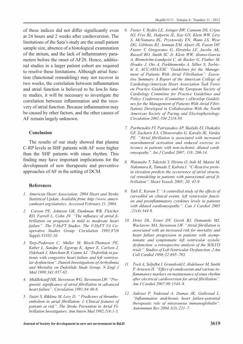
HealthMED - Volume 6 / Number 11 / 2012
Journal of Society for development in new net environment in B&H 3619
of these indices did not differ significantly even at 24 hours and 2 weeks after cardioversion. The limitations of the Sata’s study are the small patient sample size, absence of a histological examination of the atrium, and the lack of inflammatory para-meters before the onset of AF20. Hence, additio-nal studies in a larger patient cohort are required to resolve these limitations. Although atrial func-tion (functional remodeling) may not recover in two weeks, the correlation between inflammation and atrial function is believed to be low.In futu-re studies, it will be necessary to investigate the correlation between inflammation and the reco-very of atrial function. Because inflammation may be caused by other factors, and the other causes of AF remain largely unknown.
Conclusion
The results of our study showed that plasma C-RP levels in SHF patients with AF were higher than the SHF patients with sinus rhythm. This finding may have important implications for the development of new therapeutic and preventive approaches of AF in the setting of DCM.
References
1. American Heart Association. 2004 Heart and Stroke Statistical Update. Available from: http://www. ameri-canheart.org/statistics. Accessed February 11, 2004.
2. Carson PE, Johnson GR, Dunkman WB, Fletcher RD, Farrell L, Cohn JN. “The influence of atrial fi-brillation on prognosis in mild to moderate heart failure”. The V-HeFT Studies. The V-HeFT VA Co-operative Studies Group. Circulation 1993;87(6 Suppl):VI102-10.
3. Torp-Pedersen C, Møller M, Bloch-Thomsen PE, Køber L, Sandøe E, Egstrup K, Agner E, Carlsen J, Videbaek J, Marchant B, Camm AJ. “Dofetilide in pa-tients with congestive heart failure and left ventricu-lar dysfunction”. Danish Investigations of Arrhythmia and Mortality on Dofetilide Study Group. N Engl J Med 1999;341:857-65.
4. Middlekauff HR, Stevenson WG, Stevenson LW. “Pro-gnostic significance of atrial fibrillation in advanced heart failure”. Circulation 1991;84:40-8.
5. Vaziri S, Bikkina M, Levy D. “ Predictors of thrombo-embolism in atrial fibrillation: I. Clinical features of patients at risk”. The Stroke Prevention in Atrial Fi-brillation Investigators. Ann Intern Med 1992;116:1-5.
6. Fuster V, Rydén LE, Asinger RW, Cannom DS, Crijns HJ, Frye RL, Halperin JL, Kay GN, Klein WW, Lévy S, McNamara RL, Prystowsky EN, Wann LS, Wyse DG, Gibbons RJ, Antman EM, Alpert JS, Faxon DP, Fuster V, Gregoratos G, Hiratzka LF, Jacobs AK, Russell RO, Smith SC Jr, Klein WW, Alonso-Garcia A, Blomström-Lundqvist C, de Backer G, Flather M, Hradec J, Oto A, Parkhomenko A, Silber S, Torbic-ki A; ACC/AHA/ESC “Guidelines for the Manage-ment of Patients With Atrial Fibrillation”: Execu-tive Summary A Report of the American College of Cardiology/American Heart Association Task Force on Practice Guidelines and the European Society of Cardiology Committee for Practice Guidelines and Policy Conferences (Committee t oDevelop Guideli-nes for the Management of Patients With Atrial Fibri-llation) Developed in Collaboration With the North American Society of Pacing and Electrophysiology. Circulation 2001;104:2118-50.
7. Parthenakis FI, Patrianakos AP, Skalidis EI, Diakakis GF, Zacharis EA, Chlouverakis G, Karalis IK, Vardas PE. “Atrial fibrillation is associated with increased neurohumoral activation and reduced exercise to-lerance in patients with non-ischemic dilated cardi-omyopathy”. Int J Cardiol 2007; 118: 206-14.
8. Watanabe T, Takeishi Y, Hirono O, Itoh M, Matsui M, Nakamura K, Tamada Y, Kubota I. “C-Reactive prote-in elevation predicts the occurrence of atrial structu-ral remodeling in patients with paroxysmal atrial fi-brillation”. Heart Vessels 2005; 20: 45-9.
9. Tatli E, Kurum T. “A controlled study of the effects of carvedilol on clinical events, left ventricular functi-on and proinflammatory cytokines levels in patients with dilated cardiomyopathy”. Can J Cardiol 2005 ;21(4):344-8.
10. Dries DL, Exner DV, Gersh BJ, Domanski MJ, Waclawiw MA, Stevenson LW. “Atrial fibrillation is associated with an increased risk for mortality and heart failure progression in patients with asymp-tomatic and symptomatic left ventricular systolic dysfunction: a retrospective analysis of the SOLVD trials”. Studies of Left Ventricular Dysfunction. J Am Coll Cardiol 1998;32:695–703.
11. Tveit A, Seljeflot I, Grundvold I, Abdelnoor M, Smith P, Arnesen H. “Effect of candesartan and various in-flammatory markers on maintenance of sinus rhythm after electrical cardioversion for atrial fibrillation”. Am J Cardiol 2007;99:1544–8.
12. Aukrust P, Yndestad A, Damas JK, Gullestad L. “Inflammation andchronic heart failure-potential therapeutic role of intravenous immunoglobulin”. Autoimmun Rev 2004;3(3):221–7.
3620
HealthMED - Volume 6 / Number 11 / 2012
Journal of Society for development in new net environment in B&H
13. Marcus GM, Whooley MA, Glidden DV, Pawli-kowska L, Zaroff JG, Olgin JE. “Interleukin-6 and atrial fibrillation in patients with coronary artery disease: data from the Heart and Soul Study”. Am Heart J 2008;155(2):303–309.
14. Dudley SC Jr, Hoch NE, McCann LA, Honeycutt C, Diamandopoulos L, Fukai T, Harrison DG, Dikalov SI, Langberg J.“Atrial fibrillation increasesproduc-tion of superoxide by the left atrium and left atrial appendage:role of the NADPH and xanthine oxida-ses”. Circulation 2005;112:1266–1273.
15. Smith JG, Newton-Cheh C, Almgren P, Struck J, Morgenthaler NG, Bergmann A, Platonov PG, Hed-blad B, Engström G, Wang TJ, Melander. “Asse-ssment of conventional cardiovascular risk factors and multiple biomarkers for the prediction of inci-dent heart failure and atrial fibrillation”. Am Coll Cardiol 2010 Nov 16;56(21):1712-9.
16. Dai S, Zhang S, Guo Y, Chu J, Hua W, Wang F. “C-reactive protein and atrial fibrillation in idio-pathic dilated cardiomyopathy”. Clin Cardiol 2009 Sep;32(9):E45-50.
17. Dernellis J, Panaretou M.” Relationship between C-reactive protein concentrations during glucocor-ticoid therapy and recurrent atrial fibrillation”. Eur Heart J 2004; 25: 1100-7.
18. Psychari SN, Apostolou TS, Sinos L, Hamodraka E, Liakos G, Kremastinos DT. “Relation of elevated C reactive protein and interleukin-6 levels to left atrial size and duration of episodes in patients with atrial fibrillation”. Am J Cardiol 2005; 95: 764-7.
19. Targoński R, Salczyńska D, Sadowski J, Cichowski L. “Relationship between inflammatory markers and clinical patterns of atrial fibrillation in patients with congestive heart failure”. Kardiol Pol 2008 Jul;66(7):729-36
20. Sata N, Hamada N, Horinouchi T, Amitani S, Yamas-hita T, Moriyama Y, Miyahara K. “C-reactive prote-in and atrial fibrillation: is inflammation a consequ-ence or a cause of atrial fibrillation?” Jpn Heart J. 2004;45:441–445.
Corresponding AuthorErsan Tatli,Department of Cardiology, Ada Tıp Hospital, Sakarya,Turkey E-mail: [email protected]

HealthMED - Volume 6 / Number 11 / 2012
Journal of Society for development in new net environment in B&H 3621
Changes of serum lipid patterns during anticonvulsive treatment in epileptic childrenAbbaskhanian Ali1, Rezai Mohammad Sadegh2, Vahidshahi Koorosh3, Ravan Nima4, Hosseini Amir Saeed5
1 Booali Sina Hospital, Mazandaran university of Medical Scineces, Mazandaran, Iran2 Mazandaran university of Medical Scineces, Mazandaran, Iran3 Booali Sina Hospital, Mazandaran university of Medical Scineces, Mazandaran, Iran4 Booali Sina Hospital, Mazandaran university of Medical Scineces, Mazandaran, Iran5 Education Development Center, Mazandaran university of Medical Scineces, Mazandaran, Iran
Abstract
Background: The goal of this study was to evaluate the effect of Carbamazepine, Phenobar-bital and Valproic acid consumption on serum li-pid profile in epileptic children who were referred to pediatric center in north of Iran.
Methods: This study was designed as a ‘be-fore- after’ interventional research. Statistical po-pulation consisted of 150 children with seizure. Seizure diagnosis was based on clinical history, electroencephalogram (EEG) and other paraclini-cal findings by a neurologist. Lipid profiles were checked before treatment and 6 months after.
Results: total cholesterol, high density lipopro-tein (HDL) and triglyceride (TG) levels in Carba-mazepine group increased significantly but low density lipoprotein (LDL) level showed no signifi-cant difference before and after treatment. In Phe-nobarbital group, TG, HDL and LDL levels were significantly increased but no significant differen-ce in total cholesterol level was seen. TG and total cholesterol levels in valproic acid group increased significantly but changes of LDL and HDL levels were not significant.
Conclusion: The result of this study suggests that the use of carbamazepine and phenobarbital is associated with a change in lipid status. By con-sidering the atherogenic effect of these drugs and their cardiovascular complications, it is important to monitor serum lipid levels during the anti-epi-leptic treatment.
Keywords: Phenobarbital, Carbamazepine, Valproic acid, seizure, lipid profile
Introduction
It is estimated that 10.5 million children un-der 15 years have active epilepsy. This represents about 25%of the global epilepsy population (1). Seizure can present partial or generalized and is classified to tonic, clonic, tonic-clonic and absen-ce According to dominant motor pattern (1). Epi-lepsy is defined as more than two seizure attacks which is not stimulated by other diseases. Epi-lepsy frequency in different countries is about 0.5 to 1 percent. (2). Different drugs are used to treat epilepsy according to seizure type. Drug compli-cations can be divided into 2 categories: Dose-de-pendent reactions and idiosyncratic reactions (3). Most of anti-epileptic complications are known (neurologic, hematologic and hepatic complicati-ons) (3) but recently some studies were performed to evaluate their effect on serum lipids and these relationships were explained by the effect of these drugs on liver enzymes (4, 5).
By considering the atherogenic effect of li-pids and the cardiovascular complications of Hyperlipidemia, it is important to consider these complications during treatment period (6). Some epidemiologic studies show that cardiovascular disorders and brain strokes are more frequent in epileptic patients (7). Different studies showed different results of anti-epileptic effects on serum lipid profile.
By considering these controversies and high importance of atherogenic complications of anti-epileptic drugs due to long term treatments, this study tried to evaluate the effect of Carbamazepi-ne, Phenobarbital and Valproic acid on serum lipid profile in epileptic children referred to pediatric center in north of Iran.
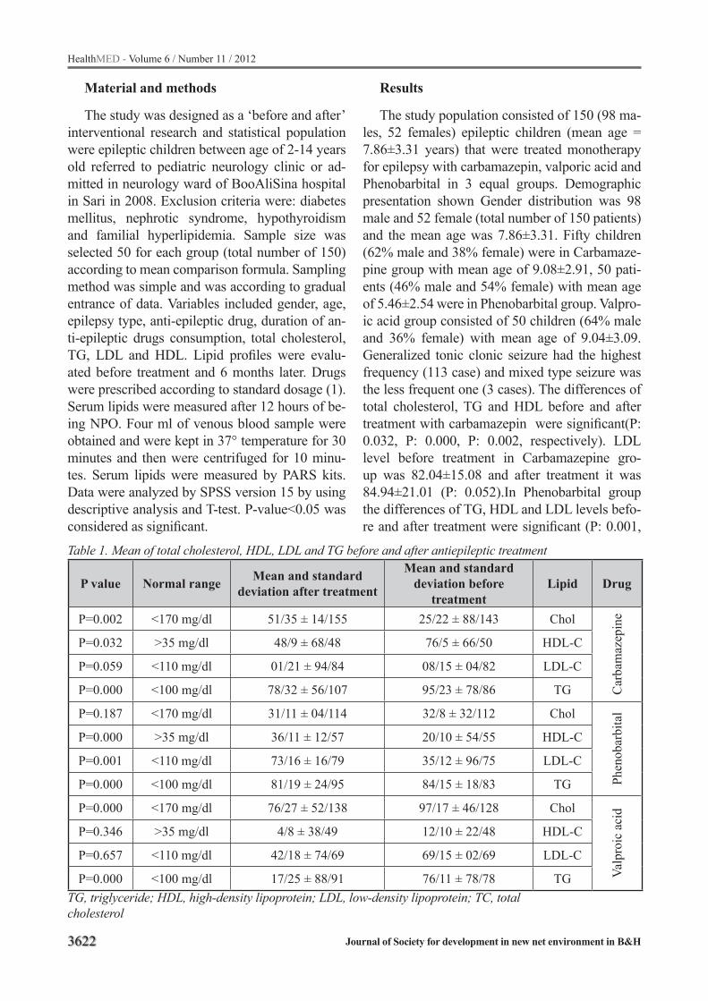
3622
HealthMED - Volume 6 / Number 11 / 2012
Journal of Society for development in new net environment in B&H
Material and methods
The study was designed as a ‘before and after’ interventional research and statistical population were epileptic children between age of 2-14 years old referred to pediatric neurology clinic or ad-mitted in neurology ward of BooAliSina hospital in Sari in 2008. Exclusion criteria were: diabetes mellitus, nephrotic syndrome, hypothyroidism and familial hyperlipidemia. Sample size was selected 50 for each group (total number of 150) according to mean comparison formula. Sampling method was simple and was according to gradual entrance of data. Variables included gender, age, epilepsy type, anti-epileptic drug, duration of an-ti-epileptic drugs consumption, total cholesterol, TG, LDL and HDL. Lipid profiles were evalu-ated before treatment and 6 months later. Drugs were prescribed according to standard dosage (1). Serum lipids were measured after 12 hours of be-ing NPO. Four ml of venous blood sample were obtained and were kept in 37° temperature for 30 minutes and then were centrifuged for 10 minu-tes. Serum lipids were measured by PARS kits. Data were analyzed by SPSS version 15 by using descriptive analysis and T-test. P-value<0.05 was considered as significant.
Results
The study population consisted of 150 (98 ma-les, 52 females) epileptic children (mean age = 7.86±3.31 years) that were treated monotherapy for epilepsy with carbamazepin, valporic acid and Phenobarbital in 3 equal groups. Demographic presentation shown Gender distribution was 98 male and 52 female (total number of 150 patients) and the mean age was 7.86±3.31. Fifty children (62% male and 38% female) were in Carbamaze-pine group with mean age of 9.08±2.91, 50 pati-ents (46% male and 54% female) with mean age of 5.46±2.54 were in Phenobarbital group. Valpro-ic acid group consisted of 50 children (64% male and 36% female) with mean age of 9.04±3.09.Generalized tonic clonic seizure had the highest frequency (113 case) and mixed type seizure was the less frequent one (3 cases). The differences of total cholesterol, TG and HDL before and after treatment with carbamazepin were significant(P: 0.032, P: 0.000, P: 0.002, respectively). LDL level before treatment in Carbamazepine gro-up was 82.04±15.08 and after treatment it was 84.94±21.01 (P: 0.052).In Phenobarbital group the differences of TG, HDL and LDL levels befo-re and after treatment were significant (P: 0.001,
Table 1. Mean of total cholesterol, HDL, LDL and TG before and after antiepileptic treatment
P value Normal range Mean and standard deviation after treatment
Mean and standard deviation before
treatmentLipid Drug
P=0.002 <170 mg/dl 51/35 ± 14/155 25/22 ± 88/143 CholC
arba
maz
epin
e
P=0.032 >35 mg/dl 48/9 ± 68/48 76/5 ± 66/50 HDL-C
P=0.059 <110 mg/dl 01/21 ± 94/84 08/15 ± 04/82 LDL-C
P=0.000 <100 mg/dl 78/32 ± 56/107 95/23 ± 78/86 TG
P=0.187 <170 mg/dl 31/11 ± 04/114 32/8 ± 32/112 Chol
Phen
obar
bita
l
P=0.000 >35 mg/dl 36/11 ± 12/57 20/10 ± 54/55 HDL-C
P=0.001 <110 mg/dl 73/16 ± 16/79 35/12 ± 96/75 LDL-C
P=0.000 <100 mg/dl 81/19 ± 24/95 84/15 ± 18/83 TG
P=0.000 <170 mg/dl 76/27 ± 52/138 97/17 ± 46/128 Chol
Valp
roic
aci
d
P=0.346 >35 mg/dl 4/8 ± 38/49 12/10 ± 22/48 HDL-C
P=0.657 <110 mg/dl 42/18 ± 74/69 69/15 ± 02/69 LDL-C
P=0.000 <100 mg/dl 17/25 ± 88/91 76/11 ± 78/78 TGTG, triglyceride; HDL, high-density lipoprotein; LDL, low-density lipoprotein; TC, totalcholesterol
HealthMED - Volume 6 / Number 11 / 2012
Journal of Society for development in new net environment in B&H 3623
P: 0.000, P: 0.000, respectively) but the changes of cholesterol level was not significant (P: 0.187). In valproic acid group the difference of total cho-lesterol and TG levels were significant (P: 0.000 and P: 0.000) but no significant differences in LDL and HDL levels were detected (P: 0.346 and P: 0.657, respectively). Mean and standard devia-tion of total cholesterol, TG, LDL and HDL level changes are shown in table1.
Discussion
The widely prescribed anticonvulsant drugs are potent inducers of cytochrome P450 enzymes, which are involved in cholesterol synthesis. Epi-demiological studies suggest that patients with epilepsy have a greater prevalence of cardiovas-cular and cerebrovascular disease than is seen in the general population. (6, 7) In our study, total cholesterol, HDL and TG levels in Carbamaze-pine group were increases significantly but LDL level showed no significant difference before and after treatment. Changes of total cholesterol in our study were similar to Eiris (8), Verrotti (9), Niko-laos (10), Aggarwal (11), Yilmaz (12), Demirci-oglu (13) and Sozuer (14) studies but in Kumar (7), Aynaci (15) and Sonmez (16) researches this difference was not significant. HDL level was si-gnificantly increased by Carbamazepine in some studies (8, 9, 10, 11, 12) but no significant changes were detected in some other researches (11, 13, 15). TG level in Kumar (7), Verrotti (9), Aggarwal (11), Yilmaz (12) and Verrotti (17) studies was si-gnificantly increases which was compatible with our study but in some other studies the differences were not significant (8, 10, 13, 15, 16, 18). LDL serum changes with Carbamazepine in Aynaci (15) and Sonmez (16) studies were not significant as in our study but in some other studies it was significantly increased (7, 8, 9, 10, 11, 12, 13, 14, 17, and 18). In Phenobarbital group, TG, HDL and LDL levels were significantly increased but no significant difference in total cholesterol level was detected in our study. In some studies total cholesterol significantly elevated in Phenobar-bital group (8, 9, 12, and 16) but in Aynaci (15) and Nikolaos (10) studies it was not significantly changed as in our study. LDL level in Phenobarbi-tal group was significantly increased in Eiris (8),
Verrotti (9), Yilmaz (12) and Somnez (16) studies as in our study but the difference was not signifi-cant in Aynaci (15) and Nikolaos (10) researches. HDL level was also significantly increased in Phe-nobarbital group in Yilmaz (12) and Aynaci (15) studies which was compatible with our results but in some other studies this difference was not signi-ficant (8, 9, 10, 16). TG level in Phenobarbital gro-up showed significant increase in Yilmaz (12) and Somnez (16) studies which was compatible with our study but in some other researches the diffe-rence was not significant (8, 10, 15) and in Verrotti (19) study it was significantly decreased. Carba-mazepin and Phenobarbital are first-generation anti-epileptic drugs which are metabolized in liver and they can lead to lipid metabolism changes by some interactions in P450 isoenzymes (19). The most accepted idea is the increase of serum lipids by these interactions of anti-epileptic drugs on li-ver isoenzymes (14, 20) but some studies did not show significant increase in lipid levels which can be because of low sample size or short treatment period duration (14, 15). Evaluation of other fac-tors which affect on serum lipids (like diets and physical activity) may help to better explanations and it is mentioned in some studies that the effect of low fat diets on lipid metabolism is reversible at the end of treatment period (21). In our study, TG and total cholesterol levels in valproic acid group increased significantly but changes of LDL and HDL levels were not significant. Total cholesterol level in valproic acid group was significantly de-creased in Eiris (8) and Nikolaos (10) studies but this difference was not compatible with other re-searches (12, 13, 14, 15, 16, and 22). LDL level in Eiris(8), Verrotti (9) and Nikolaos(10) studies was also decreased significantly which was not com-patible with our study and also showed no signi-ficant changes in other studies (12, 13, 14, 15, 16, 22). HDL level in valproic acid group in Verrotti (9) study was significantly increased but in some other studies it showed no significant changes (8, 10, 12, 13, 14, 15, 16, 22) which was compatible to our results. TG level in Verrotti (9) and Niko-laos (10) studies were significantly decreased in valproic acid group but in some other studies it showed no significant changes (8, 12, 13, 14, 15, 16, and 22). These results were not compati-ble to our study. Valproic acid is an anti-epileptic
3624
HealthMED - Volume 6 / Number 11 / 2012
Journal of Society for development in new net environment in B&H
drug which inhibits P450 isoenzymes. It is more accepted that this drug decreases serum lipids le-vel or does not cause any significant changes (14, 20). Some studies tried to explain Valproic acid effect on lipid profile but the mechanism is still unknown (21). In our study the drug’s serum le-vel at the same time was not measured so the re-lationship between lipid level changes and drugs’ serum level was not evaluated which is one of the limitations of our study. By considering the effect of lipid levels on cardiovascular system, long term follow up of lipid changes in patients who receive anti-epileptic drugs is recommended.
Conclusion
The result of this study suggests that the use of carbamazepine and phenobarbital is associated with a change in lipid status. By considering the atherogenic effect of these drugs and their cardio-vascular complications, it is important to monitor serum lipid levels during the anti-epileptic tre-atment.
Acknowledgment
This study was a pediatric residency thesis of Dr Nima Ravan and was supported by a grant from Mazandaran University of Medical Sciences to Ali Abbaskhanian.
References
1. Forsgren L. Incidence and prevalence. In: Wallace SJ, Farrell K, editors. Epilepsy in children. London: Arnold; 2004. p. 21–5.
2. Annegers JF, Dubinsky S, Coan SP, Newmark SM, Roht L. The incidence of epilepsy and unprovoked se-izures in multiethnic, urban health maintenance orga-nizations. Epilepsia. 2002;40:502-506.
3. Glauser TA, Pippenger CE. Controversies in blood-level monitoring: reexamining its role in the treatment of epilepsy. Epilepsia. 2000; 41(8):6-15.
4. Luoma PV, Pelkonen RO, Myllyla V, Sotaneimi EA. Relationship between serum lipid levels and indices of drug metabolism in epileptic on anticonvulsant. Clini-cal Pharmacol Therp. 2004; 25:235-237.
5. Smith DB, Delgade ES, Cueta AV, Carmer JA, Maltson RH. Historical prospective on the choice of antiepi-leptic drug for the treatment of seizures in adults. Ne-urology. 2000; 33:2-4.
6. Mantel-Teeuwisse AK, Kloosterman JM, Zee AH, Klungel OH, Porsius AJ, Boer A. Drug-induced lipid changes: A review of the unintended effects of some commonly used drugs on serum lipid levels. Drug Safty. 2001; 24(6):443-456.
7. Kumar P, Tyagi M, Kumar Tyagi Y, Kumar A, Kumar Rai Y. Effect of anticonvulsant drug on lipid profi-le in epileptic patients. Jurnal of Neurology. 2004: 3(1):202-210.
8. Eiris J, Novo-Rodriguez MI, Del Rio M, Meseguer P, Del Rio MC, Castro-Gago M. The effects on lipid and apolipoprotein serum levels of long-term carba-mazepine, valproic acid and phenobarbital therapy in children with epilepsy. Epilepsy Res. 2000; 41(1):1-7.
9. Verrotti A, Domizio S, Angelozzi B, Sabatino G, Mor-gese G, Chiarelli F.Changes in serum lipids and lipo-proteins in epileptic children treated with anticonvul-sants. J Paediatr Child Health. 1997 Jun;33(3):242-5.
10. Nikolaos T, Stylianos G, Chryssoula N, Irini P, Chri-stos M, Dimitrios T, Konstantinos P, Antonis T. The effect of long-term antiepileptic treatment on serum cholesterol (TC, HDL, LDL) and triglyceride levels in adult epileptic patients on monotherapy. Med Sci Monit. 2004 Apr; 10(4):MT50-2.
11. Aggarwal A, Kumar M, Faridi MM. Effect of car-bamazepine on serum lipids and liver function tests. Indian Pediatr. 2005; 42(9):913-918.
12. Yilmaz E, Dosan Y, Gurgoze MK, Gungor S. Serum lipid changes during anticonvulsive treatment serum lipids in epileptic children. Acta Neurol Belg. 2001; 101(4):217-220.
13. Demircioğlu S, Soylu A, Dirik E. Carbamazepine and valproic acid: effects on the serum lipids and li-ver functions in children. Pediatr Neurol. 2000 Aug; 23(2):142-6.
14. Sozuer DT, Atakil D, Dogu O, Baybas S, Arpaci B. Serum lipids in epileptic children treated with car-bamazepine and valproate. Eur J Pediatr. 1997; 156(7):565-567.
15. Aynaci FM, Orhan F, Orem A, Yildirmis S, Gedik Y. Effect of Antiepileptic drugs on plasma lipoprotein (a) and other lipid levels in childhood. J Child Neu-rol. 2001 May; 16(5):367-9.
16. Sonmes FM, Demir E, Orem A, Yildirmis S, Orhan F, Aslan A, et al. Effect of antiepileptic drugs on pla-sma lipids, lipoprotein(a) and liver enzymes. J Child Neurol. 2006; 21(1):70-74.

HealthMED - Volume 6 / Number 11 / 2012
Journal of Society for development in new net environment in B&H 3625
17. Verrotti A, Basciani F, Domizio S, Sabatino G, Mor-gese G, Chiarelli F. Serum lipids and lipoprotein in patients treated with antiepileptic drugs. Pediatr Ne-urol. 2000; 19:364-367.
18. Aggarwal A, Singh V, Batra S, Faridi MM, Sharma S. Effect of carbamazepine therapy on serum lipids in children with partial epilepsy. Pediatr Neurol. 2009 Feb;40(2):94-7.
19. Laura S Cheng, Asuri N Prasad, Michael J Rie-der. Relationship between Antiepileptic Drugs and Biological Markers Affecting Long-Term Cardio-vascular Function in Children and Adolescents. Can J Clin Pharmacol.2010; 17 (1):e5-e46; January 4, 2010.
20. Voudris KA, Attilakos A, Katsarou E, Drakatos A, Dimou S, Mastroyianni S, Skardoutsou A, Prassouli A, Garoufi A. Early and persistent increase in serum lipoprotein (a) concentrations in epileptic children treated with carbamazepine and sodium valproate monotherapy. Epilepsy Res. 2006 Aug;70(2-3):211-7. Epub 2006 Jun 15.
21. Jakubus T, Michalska-Jakubus M, Lukawski K, Ja-nowska A, Czuczwar SJ. Atherosclerotic risk among children taking antiepileptic drugs. Pharmacol Rep. 2009 May-Jun;61(3):411-23.
Corresponding AuthorAmir Saeed Hosseini,Education Development Center, Mazandaran university of Medical Scineces, Mazandaran, IranE-mail: AShosseini@ muzums.ac.ir
3626
HealthMED - Volume 6 / Number 11 / 2012
Journal of Society for development in new net environment in B&H
The analysis of violence against the nurses who are in employee status in Mugla State Hospital, TurkeyMetin Picakciefe1, Sema Akca2, Ayse Elibol2, Artuner Deveci3, Nevin Yilmaz4, Ugur Eser Yilmaz5
1 Department of Public Health, Faculty of Medicine, Mugla University, Turkey2 School of Health Sciences, University of Mugla, Turkey3 Department of Psychiatry, Faculty of Medicine, Celal Bayar University, Turkey4 Department of Internal Medicine, Faculty of Medicine, Mugla University, Turkey5 Biomedical Engineering and Premed program, VCU, USA
Abstract
Objektive: Workplace violence in the co untry, especially in the health system remains obscure. The search of the relationship using an intersecti-onal, analytical survey between the working con-ditions and violence exposure of the nurses who work in Muğla State Hospital was aimed in the study.
Materials and methods: Of the 310, 268 nur-ses participated, yielding a response rate of 86.5%. The data was examined by using Fisher’s Exact Test, Pearson Chi-Square Test and Logistic Re-gression. In result, it was found that 85.8% of the nurses were exposed to violence, with 70.4% of the violence coming from the relatives of patients.
Results: 77.2% of the nurses were exposed to verbal abuse while 71.4% were physically as-saulted. Unfortunately, 98.4% of the abused did not report the physical abuse. 91.3% of the nur-ses always felt violence anxiety, while 92.9% of them thought that their instutions do not make an effort for the security systems. According to logi-stic regression analysis, having worked 21 years and over, night work and having felt a violent en-counter in the workplace, significantly increases the frequency of meeting with violence of nurses.
Conclusion:In conclusion we found that the nurses were exposed to multiple forms of violen-ce. This was correlated with their monthly average income, marrital status, parental status, work sta-tus, night shifts, overtime, rotational shifts, total and daily work hours.
Keywords: Nurse, State hospital, Workplace, Violence, Turkey
Introduction
Violence is a phenomenon that shows itself in very different ways, which can be met frequently in both an individual and a social dimension. The violence shown in the workplace changes in form from the threat of murder to attempt at physical harm. Workplace violence is defined as “violent behaviors including physical attacks and attack threats for employees, “by National Institute for Occupational Safety and Health (NIOSH) [1]. Today, the number of violence cases which em-ployees face every day in workplaces is increasing [2]. This situation has been reported in a number of research studies. It was shown that in South Africa, Britain, France and Japan, employees face an increasing frequency of violence [3]. American Occupational Health and Safety Authority repor-ted that “The attacks in health and social services are more than in any other industry”. The same report included that violence is being taken into account as a part of the profession in the health in-dustry” [4].The latest data shows hospital workers are at a higher risk of experiencing violence in the United States. The Bureau of Labor Statistics esti-mates that 2,637 non-fatal attacks occured against the hospital staff in a year, this ratio is 8.3% in 10 thousand employees [1]. According to the Univer-sity of Iowa, violence is directed towards health professionals at the highest rate. For example; the ratio of violence is more than 100 events on 100 workers in some departments of psychiatry in a year. According to a study conducted by the Emer-gency Medical System in Virginia, the violence related with health care is getting more dangerous today in all kinds of health organizations. In addi-tion, it has been defined in the study conducted
HealthMED - Volume 6 / Number 11 / 2012
Journal of Society for development in new net environment in B&H 3627
in Virginia, that the medical staff certainly has the highest ratio of attack in comparison with all other business sectors [4]. The nurses and assistants that are in direct contact with patients have the highest risks to be a victim of violence, despite all hos-pital workers possibly being victims of violence. Increased risk of violence includes other hospital staff, emergency personnel, hospital safety offi-cers [1]. Doctors, pharmacists, interns, assistants, nurses, aides, therapists, technicians, home health care workers, social / support workers, all of emer-gency medical personnel are under the high risk of violence by patients or friends and relatives of patients, but mostly nurses encounter violence [4]. One of the studies shows that exposure to violence is frequent in nurses [5]. It is strongly believed that reasons such as; heavy load of emotion generated by the relatives of the patient, shift work, job stre-ss, inadequate staff, lack of security, intense pace of work, and work being mostly conducted by wo-men are among the factors that make encounters with violence a risk for nurses [6, 7].Nurses, be-ing health workers, are the first who meet with the victims of increasing violence. In addition, nurses suffer damage from social tolerance of violence. In many cases, The legal system does not accept to pay compensation to nurses in many instances. Nurses see themselves as a “legal target” and feel that “violence is a part of the profession” [8].The number of trauma and violence towards workers are increasing every year. Health is a unity in the working life and the trauma is a factor that dis-rupts this integrity. Violence as one of the causes of trauma plays an important role due to cau-sing biological and psychological destruction on employee’s health [9]. Prevention of violence has priority among the public health issues [10]. Well-being of nurses is very important for public health as they are a part of public health and are resposi-ble to provide public service.The aim of this study is to examine socio-demographic characteristics and working conditions of nurses in Mugla State Hospital with relationship between the status of exposure to violence.
Materials and methods
This study is a cross-sectional, analytical type of study. The study contacted all 310 nurses wor-
king in the hospital. Of the 310, 268 nurses parti-cipated, yielding a response rate of 86.5%. 22 nu-rses did not want to attend the study due to work intensity. The remaining 20 nurses who were not contacted were either taking their yearly leave or permitted to take a leave.
The questionnaire of the sociodemographic characteristics and working conditions A questionnaire was developed by the re-
searchers consists of 25 questions concerning workplace violence, sociodemographic characte-ristics and working conditions. Self-filling met-hod was used; application of a questionnaire took approximately 10-15 minutes. The questionnaire was carried out between February 26, 2009 and January 9, 2009.
Study variablesDependent variablesThe dependent variable is encounter with vi-
olence in the workplace situation. The encounter with workplace violence are discussed under the sub-headings of; violence (yes, no), physical vi-olence (yes, no), verbal violence (yes, no), sexu-al violence (yes, no), psychological (mobbing) violence (yes, no), and violence exposure with written document (textual violence) (yes, no).
Independent variablesIndependent variables were discussed under
socio-demographic and working conditions rela-ted variables sub-titles: Socio-demographic va-riables are gender, age, educational level, marital status, the presence of income other than salary, and children situation. Working condition varia-bles are total working time (year), daily working time (hours), overtime work in the last one year, shift work in the last one year, night work in the last one year, work units, the fear of encountering with violence, protection measures taken by the institution.
Statistical analysesSPSS 15.0 package program was used to
analyze the data. The data was examined by using Fisher’s Exact Test, Pearson Chi-Square Test and
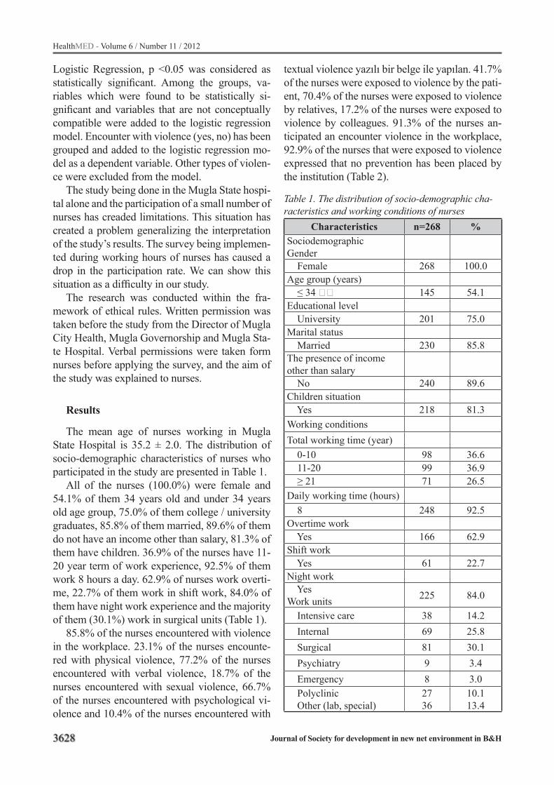
3628
HealthMED - Volume 6 / Number 11 / 2012
Journal of Society for development in new net environment in B&H
Logistic Regression, p <0.05 was considered as statistically significant. Among the groups, va-riables which were found to be statistically si-gnificant and variables that are not conceptually compatible were added to the logistic regression model. Encounter with violence (yes, no) has been grouped and added to the logistic regression mo-del as a dependent variable. Other types of violen-ce were excluded from the model.
The study being done in the Mugla State hospi-tal alone and the participation of a small number of nurses has creaded limitations. This situation has created a problem generalizing the interpretation of the study’s results. The survey being implemen-ted during working hours of nurses has caused a drop in the participation rate. We can show this situation as a difficulty in our study.
The research was conducted within the fra-mework of ethical rules. Written permission was taken before the study from the Director of Mugla City Health, Mugla Governorship and Mugla Sta-te Hospital. Verbal permissions were taken form nurses before applying the survey, and the aim of the study was explained to nurses.
Results
The mean age of nurses working in Mugla State Hospital is 35.2 ± 2.0. The distribution of socio-demographic characteristics of nurses who participated in the study are presented in Table 1.
All of the nurses (100.0%) were female and 54.1% of them 34 years old and under 34 years old age group, 75.0% of them college / university graduates, 85.8% of them married, 89.6% of them do not have an income other than salary, 81.3% of them have children. 36.9% of the nurses have 11-20 year term of work experience, 92.5% of them work 8 hours a day. 62.9% of nurses work overti-me, 22.7% of them work in shift work, 84.0% of them have night work experience and the majority of them (30.1%) work in surgical units (Table 1).
85.8% of the nurses encountered with violence in the workplace. 23.1% of the nurses encounte-red with physical violence, 77.2% of the nurses encountered with verbal violence, 18.7% of the nurses encountered with sexual violence, 66.7% of the nurses encountered with psychological vi-olence and 10.4% of the nurses encountered with
textual violence yazılı bir belge ile yapılan. 41.7% of the nurses were exposed to violence by the pati-ent, 70.4% of the nurses were exposed to violence by relatives, 17.2% of the nurses were exposed to violence by colleagues. 91.3% of the nurses an-ticipated an encounter violence in the workplace, 92.9% of the nurses that were exposed to violence expressed that no prevention has been placed by the institution (Table 2).
Table 1. The distribution of socio-demographic cha-racteristics and working conditions of nurses
Characteristics n=268 %SociodemographicGender Female 268 100.0Age group (years) ≤ 34 145 54.1Educational level University 201 75.0Marital status Married 230 85.8The presence of income other than salary No 240 89.6Children situation Yes 218 81.3Working conditionsTotal working time (year) 0-10 98 36.6 11-20 99 36.9 ≥ 21 71 26.5Daily working time (hours) 8 248 92.5Overtime work Yes 166 62.9Shift work Yes 61 22.7Night work YesWork units 225 84.0
Intensive care 38 14.2 Internal 69 25.8 Surgical 81 30.1 Psychiatry 9 3.4 Emergency 8 3.0 Polyclinic Other (lab, special)
2736
10.113.4
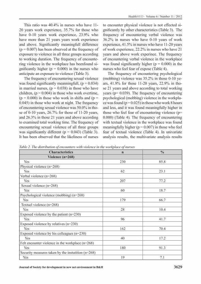
HealthMED - Volume 6 / Number 11 / 2012
Journal of Society for development in new net environment in B&H 3629
This ratio was 40.4% in nurses who have 11-20 years work experience, 35.7% for those who have 0-10 years work experience, 23.9% who have more than 21 years years work experience and above. Significantly meaningfull difference (p = 0.007) has been observed at the frequency of exposure to violence in all three groups according to working duration. The frequency of encounte-ring violence in the workplace has beenfound si-gnificantly higher (p = 0.000) in the nurses who anticipate an exposure to violence (Table 3).
The frequency of encountering sexual violence was found significantly meanningfull, (p = 0.034) in married nurses, (p = 0.038) in those who have children, (p = 0.004) in those who work overtime, (p = 0.000) in those who work in shifts and (p = 0.045) in those who work at night. The frequency of encountering sexual violence was 50.0% in tho-se of 0-10 years, 26.7% for those of 11-20 years, and 26.3% in those 21 years and above according to examined total working time. The frequency of encountering sexual violence of all three groups was significantly different (p = 0.043) (Table 3). It has been observed that the likeliness of nurses
to encounter physical violence is not effected si-gnificantly by other characteristics (Table 3). The frequency of encountering verbal violence was 36.2% in nurses who have 0-10 years of work experience, 41.5% in nurses who have 11-20 years of work experience, 22.2% in nurses who have 21 years and above work experince. The frequency of encountering verbal violence in the workplace was found significantly higher (p = 0.000) in the nurses who feel fear of expose (Table 4).
The frequency of encountering psychological (mobbing) violence was 35.2% in those 0-10 ye-ars, 41.9% for those 11-20 years, 22.9% in tho-se 21 years and above according to total working years (p= 0.039). The frequency of encountering psychological (mobbing) violence in the workpla-ce was found (p = 0.025) in those who work 8 hours and less, and it was found meaningfully higher in those who feel fear of encountering violence (p= 0.000) (Table 4). The frequency of encountering with textual violence in the workplace was found meaningfully higher (p = 0.007) in those who feel fear of textual violence (Table 4). In univariate analysis results, the multivariate analysis results
Table 2. The distribution of encounters with violence in the workplace of nursesCharacteristics n %
Violence (n=268) Yes 230 85.8Physical violence (n=268) Yes 62 23.1Verbal violence (n=268) Yes 207 77.2 Sexual violence (n=268) Yes 60 18.7Psychological violence (mobbing) (n=268) Yes 179 66.7 Textuel violence (n=268) Yes 28 10.4Exposed violence by the patient (n=230) Yes 96 41.7Exposed violence by relatives (n=230) Yes 162 70.4Exposed violence by his colleagues (n=230) Yes 40 17.2Felt encounter violence in the workplace (n=268) Yes 180 91.3Security measures taken by the instutition (n=268) Yes 19 7.1
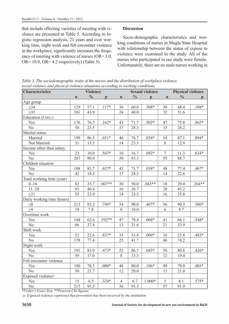
3630
HealthMED - Volume 6 / Number 11 / 2012
Journal of Society for development in new net environment in B&H
Table 3. The sociodemographic traits of the nurses and the distribution of workplace violence, sexial violence and physical violence situations according to working conditionsCharacteristics Violence Sexual violence Physical violence n % p n % p n % p Age group
≤34 129 57.1 .117* 36 60.0 .308* 30 48.4 .188*≥35 101 43.9 24 40.0 32 51.6
Education (Univ.)Yes 176 76.5 .162* 43 71.7 .502* 47 75.8 .865*No 54 23.5 17 28.3 15 24.2
Marital statusMarried 199 86.5 .451* 46 76.7 .034* 54 87.1 .894*Not Married 31 13.5 14 23.3 8 12.9
Income other than salary Yes 23 10.0 .567* 10 16.7 .092* 7 11.3 .634*No 207 90.0 50 83.3 55 88.7
Children situation Yes 188 81.7 .657* 43 71.7 .038* 48 77.4 .467*No 42 18.3 17 28.3 14 22.6
Total working time (year)0–10 82 35.7 .007** 30 50.0 .043** 18 29.0 .264**11–20 93 40.4 16 26.7 28 45.2≥21 55 23.9 14 23.3 16 25.8
Daily working time (hours) ≤8 212 92.2 .749* 54 90.0 .407* 56 90.3 .580*≥9 18 7.8 6 10.0 6 9.7
Overtime workYes 144 62.6 .592** 47 78.4 .004* 41 66.1 .548*No 86 37.4 13 21.6 21 33.9
Shift work Yes 52 22.6 .837* 35 53.8 .000* 16 25.8 .483*No 178 77.4 25 41.7 46 74.2
Night workYes 191 83.0 .473* 52 86.7 .045* 50 80.6 .426*No 39 17.0 8 13.3 12 19.4
Felt encounter violenceYes 180 78.3 .000* 48 80.0 .106* 49 79.0 .403*No 50 21.7 12 20.0 13 21.0
Exposed violencea
Yes 15 6.5 .324* 4 6.7 1.000* 5 8.1 .575*No 215 93.5 56 93.3 57 91.9
*Fisher’s Exact Test, **Pearson Chi-Square a: Exposed violence expressed that prevention has been received by the institution
that include effecting varieties of meeting with vi-olence are presented in Table 5. According to lo-gistic regression analysis, 21 years and over wor-king time, night work and felt encounter violence in the workplace, significantly increases the frequ-ency of meeting with violence of nurses (OR= 3.0, OR= 10.9, OR= 4.2 respectively) (Table 5).
Discussion
Socio-demographic characteristics and wor-king conditions of nurses in Mugla State Hospital with relationship between the status of expose to violence were examined in the study. All of the nurses who participated in our study were female. Unfortunately, there are no male nurses working in
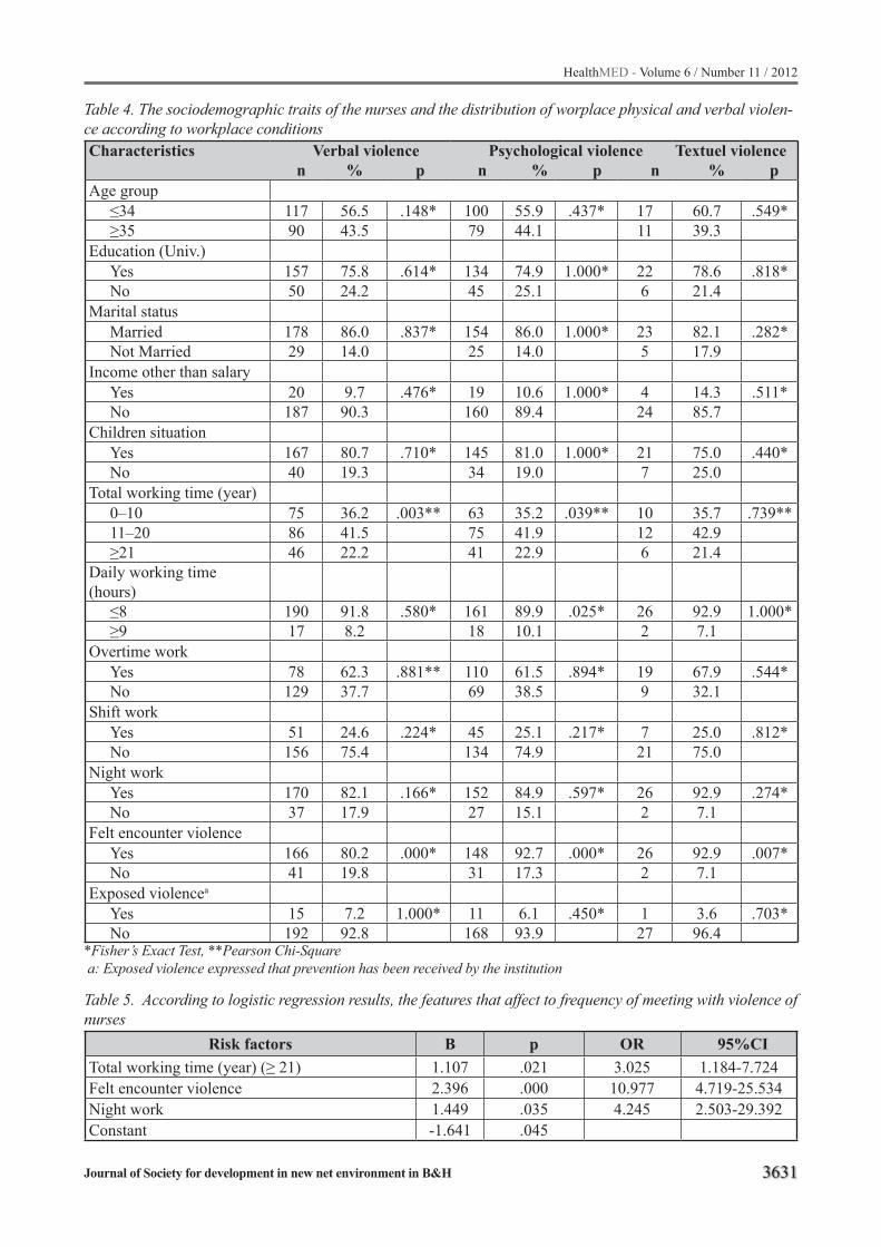
HealthMED - Volume 6 / Number 11 / 2012
Journal of Society for development in new net environment in B&H 3631
Table 4. The sociodemographic traits of the nurses and the distribution of worplace physical and verbal violen-ce according to workplace conditionsCharacteristics Verbal violence Psychological violence Textuel violence n % p n % p n % p Age group
≤34 117 56.5 .148* 100 55.9 .437* 17 60.7 .549*≥35 90 43.5 79 44.1 11 39.3
Education (Univ.)Yes 157 75.8 .614* 134 74.9 1.000* 22 78.6 .818*No 50 24.2 45 25.1 6 21.4
Marital statusMarried 178 86.0 .837* 154 86.0 1.000* 23 82.1 .282*Not Married 29 14.0 25 14.0 5 17.9
Income other than salary Yes 20 9.7 .476* 19 10.6 1.000* 4 14.3 .511*No 187 90.3 160 89.4 24 85.7
Children situation Yes 167 80.7 .710* 145 81.0 1.000* 21 75.0 .440*No 40 19.3 34 19.0 7 25.0
Total working time (year)0–10 75 36.2 .003** 63 35.2 .039** 10 35.7 .739**11–20 86 41.5 75 41.9 12 42.9≥21 46 22.2 41 22.9 6 21.4
Daily working time (hours)
≤8 190 91.8 .580* 161 89.9 .025* 26 92.9 1.000*≥9 17 8.2 18 10.1 2 7.1
Overtime workYes 78 62.3 .881** 110 61.5 .894* 19 67.9 .544*No 129 37.7 69 38.5 9 32.1
Shift work Yes 51 24.6 .224* 45 25.1 .217* 7 25.0 .812*No 156 75.4 134 74.9 21 75.0
Night workYes 170 82.1 .166* 152 84.9 .597* 26 92.9 .274*No 37 17.9 27 15.1 2 7.1
Felt encounter violenceYes 166 80.2 .000* 148 92.7 .000* 26 92.9 .007*No 41 19.8 31 17.3 2 7.1
Exposed violencea
Yes 15 7.2 1.000* 11 6.1 .450* 1 3.6 .703*No 192 92.8 168 93.9 27 96.4
*Fisher’s Exact Test, **Pearson Chi-Square a: Exposed violence expressed that prevention has been received by the institution
Table 5. According to logistic regression results, the features that affect to frequency of meeting with violence of nurses
Risk factors B p OR 95%CITotal working time (year) (≥ 21) 1.107 .021 3.025 1.184-7.724Felt encounter violence 2.396 .000 10.977 4.719-25.534Night work 1.449 .035 4.245 2.503-29.392Constant -1.641 .045
3632
HealthMED - Volume 6 / Number 11 / 2012
Journal of Society for development in new net environment in B&H
Mugla State Hospital. 95% of nurses in the world are female [5]. It is accepted that nursing professi-on is a profession for women in our country, hen-ce all nurses in the study are women. However, with the new legal arregments in the law men are allowed to do the profession of nursing [11].
It has been observed that 78.8% of nurses in Kocaeli are in the 22-30 age group [12 ], 36.2% of nurses in Adana are in the 24-28 age group [13], 46.7% of nurses in Duzce are in the 29 and un-der 29 age group [2], 73.5% of nurses in Diyarba-kir are in the 19-25 age group [14]. Other studies support our work. We see that most of the nurses in our study (54.1%) are in young age group.
78.8% of the nurses in Kocaeli, 67.7% of the nurses in Adana are college / university gradua-tes [12, 13]. 75.0% of the nurses in our study are college / university graduates. It can be evaluated that nurses complete college / university level edu-cation to get a better education and benefit from the increased salary during the seniority pension.
According to the 2010 Population Census, 79.8% of the population is married in Turkey [15]. Percentage of married nurses (85.8%) is higher than the general population. This situation can be explained as young age women who get a profe-ssion can be married in in their young ages in our country.
The majority of nurses (89.6%) do not have an income out of the monthly fee. 81.3% of the nur-ses have children and 46.6% of them have two and more children. Low wages, to have more children and to not have any income other than a monthly salary show that the nurses are in lack of liveliho-od.
12.6% of nurses in Diyarbakir, 24.1% of nurses in Adana, 21.4% of nurses in Eskisehir, 10.6% of nurses in Kocaeli have 11-15 working years expe-rience [14, 13, 2, 12]. The reasons of having high amount of work years in our study according to other studies are that our hospital is in the tourism territory, and therefore to be preferred. In additi-on, nurses are experienced in their profession. The number of beginners is less. This may be due to not recruiting new nurse staff.
It has been observed that 62.9% of the nurses worked overtime in our study. According to the Civil Servants Act No. 657, official working hours is 8 hours a day [16]. Frequently and in a conti-
nuous manner overtime work will null the legal regulations related to the normal working hours and employees, and it will lead to detrimental conditions to emerge of the working hours [17]. Nurses are working more hours than allowed by the law to work. The reason of this situation is that nurses have to serve with a smaller number of em-ployees.
More than three-quarters of nurses worked in shift work (77.3%) and night work (84.0%). It was stated in a research conducted in Izmir, 20.8% of the health care workers worked in shift work and 56.7% of them worked in the night work. The night work forms are duty work (41.6%), on call (4.5%), and come to night (10.6%) [18]. Making the shift work is a method that has been used qu-ite widely and for a long time to organize wor-king time and it is a classic example of a dilemma. The social disadvantages against economic ad-vantages. There are economic benefits in case of implementation, because it allows the use of full production capacity. However, it includes social disadvantages due to breaking the normal order of life of employees [17]. The shift work is a source of stress alone. Lack of compliance by the body during the day and night, unsufficient daytime sleep, REM sleep interruptions cause weakness and fatigue [19].
More than 12 hours work for emergency ser-vice work shifts are not proper [20]. 63.4% of Iz-mir physicians stated that shift work is a source of stress [21]. It was found that absent-mindedne-ss, depression, anger, fatigue and anxiety scores were high, vigor scores, systolic blood pressure, heart rate, plasma ACTH and cortisol levels were lower on the nurses who complete night shift as opposed to nurses who complete normal work in a study which measured the effects of the night shift on psychological status, cardio-vascular and nerve-hormonal system in a hospital in Japan [22]. It has been observed that nurses are working in shifts and night time with high ratio. Work shift and night work have negative effecst on general health status of nurses.
Violence against women in the workplace is increasing each passing day. It has been reported that especially female workers who work in ho-usehold professions and in the entertainment bu-siness encounter with workplace violence in the
HealthMED - Volume 6 / Number 11 / 2012
Journal of Society for development in new net environment in B&H 3633
Philippines and an average of 20 workers were killed and 18,000 were attacked in a week in the U.S [3]. Of the 68% of emergency service wor-kers faced with violence in Canada, 60% of them reported there is a significant increase of violen-ce [23]. Studies show that nurses more frequently encounter with violence as opposed to other he-althcare professionals [24, 25]. The violent enco-unter prevalence of nurses varies. More than 30% of nurses in Colorado [26], more than one-third of nurses in the United States [27], 60.3% of nurses in Duzce experienced violence [28].
According to the literature workplace violen-ce encounter rates of nurses are high, however we have found them to be slightly higher in our stu-dy (85.8%). The reason for this situation may be difference on perception of violence, and different cultural characteristics.
60% of emergency room nurses in the United States have suffered violence by the patient [29]. It is reported that the source of the side applying physical violence to nurses are 95% of the time the patients in emergency departments, 100% of the time the patients in psychiatric wards in Ca-nada [30]. 51% of attackers are patients in Kuwa-itte [31]. It was determined that 47.0% of nurses have suffered violence by the patients in Ireland [26]. This rate was found less (41.7%) in our stu-dy. Most nurses met violence by patients’ relatives (70.4%), and least by their colleagues (17.2%).
The results of a research conducted in Erzurum supports our study; nurses met violence by pati-ents’ relatives 59.8% of the, 22.7% of the time by patients, 13.1% of the time by colleagues [32]. As seen there are differences on the sides who apply violence to nurses. The most important reason for this difference is they may have different cultural characteristics. More studies are needed on this subject. However, there is a known fact that nurses are constantly in communication with patients and their relatives. The risk of violence increases when the interactions increase with the patient according to a study conducted in the United States [33].
Physical violence in the workplace is one of the crucial questions of the working life. According to the report for the year of 2000 (European Founda-tion, 2000), 9 million workers are facing physical violence in the European Union countries. Accor-ding to the results of the study conducted in 15 EU
member countries and participated by 15,800 pe-ople, 4% of workers (6 million employees) were exposed to physical violence [34]. The incidence of physical violence towards the health care indu-stry is increasing every day. It has been reported in the Synthesis report 2002 that the frequency of physical violence was 6% in Brazil, 9% in Bulga-ria, 6% in Lebanon, 17% in South Africa and 10% in Thailand in the healthcare business [35]. Nurses in the healthcare sector are often faced with physi-cal violence. 23.1% of nurses have faced with physical violence in our study.
Other studies support our results. It was re-ported in a study conducted in Adana that 16% of nurses in Adana [13], 30.7% nurses in Bursa experienced physical violence [36]. It has been reported at the study conducted by the American Nurses Association 17% of nurses were exposed to physical violence [27]. This ratio was found to be 13.2% on 100 nurses per year in the Minneso-ta/USA [37]. The frequency of physical violence encounter of nurses is high in the workplace, but also other health professionals, especially emer-gency employees and 112 employees are exposed to physical violence in the workplace. It was re-ported that 19.6% of 112 emergency and hospital emergency room workers in Samsun and, 16.8% of 112 emergency workers in Izmir encountered with physical violence [38, 39].
It has been stated that 57% of emergency ser-vice workers were exposed to physical assault in Canada [23]. 20.5% of doctors in primary he-althcare centers in Izmir and 15.3% of other em-ployees encountered physical violence [40].
Violence is common among health professio-nals in health sectors in all countries. More than half of healthcare workers were affected in types of violence such as verbal violence. According to Synthesis Report 2002, verbal violence is more prominent than physical violence in the EU coun-tries health sector [35]. It has been reported that 98.5% of nurses working in emergency depar-tments in Izmir, 91.1% of the nurses in Kocaeli [41], 47.8% of nurses in Adana have faced verbal violence [13]. It has been reported that more than half of nurses (57%) were threatened or suffered verbal violence in a survey conducted by Ameri-can Nurses Association [27]. 48% of nurses suffe-red verbal violence in the last 6 months in Kuwait

3634
HealthMED - Volume 6 / Number 11 / 2012
Journal of Society for development in new net environment in B&H
[42]. It was found that 95% of nurses met with verbal violence in the last 12 months in Austra-lia [43], more than half of the nurses working in the emergency room suffered verbal and physi-cal violence in Ireland [44]. According to the re-sults of the study conducted in three hospitals in Erzurum; 87.6% of the nurses reported that they encountered verbal violence in the past one year, 60.6% of them in the previous six months [32]. The frequency of encounter with verbal violence of nurses (77.2%) in our study is consistent with other studies. The causes of the high frequency of verbal violence in the workplace can be that the application of verbal violence is easier compared to other types of violence, more difficult to prove the presence of and resulting in a lighter criminal sanction.
It is known that women more frequently face sexual assaults. It was reported in a study conduc-ted in Canada that 50% of women faced physical and sexual assault, 18% of assaults resulted with physical wounds [3]. 2% of workers in the EU (3 million employees) were exposed to sexual hara-ssment [34]. Many women, especially working in the home and entertainment professions, often face violence in the Philippines. It has been repor-ted by the Occupational Health and Safety Autho-rity in Germany that 93% of women were exposed to sexual harassment in the workplace throughout the their professional lives [3]. 75% of nurses were exposed to sexual violence in the workplace in Warsaw [5]. 57% of nurses in Canada, 79.0% in Adana faced with sexual violence [45, 13]. The frequency of sexual violence in nurses who participated to our study was lower (18.7%) than others. The reason for this situation can be explai-ned by our country’s customs, traditions and tradi-tional social structure of women that can include hiding cases of sexual violence.
World Health Organization particular stressed the importance of psychosocial aspects of work and in work-related psychosocial factors while determining the strategies of “Working People’s Health” [46]. Psychological violence (mobbing) is one of the psychosocial factors that employees often faced. It has been found that 54% of UK employees, 11% of German employees, and 8% of EU employees were exposed to mobbing [34]. 8.3% of employees in Denmark, 28% of em-
ployees in Spain were exposed to mobbing [47, 48]. According to the National Institute for Occu-pational Safety and Health at Work in Germany, 11.3% of all employees were victims of mobbing in their working lifetime, women are under risk 75% more than men [49]. Mobbing more often is seen in Health employees than other employees. According to Synthesis Report 2002; mobbing / bullying is seen as often as, 30% in Bulgaria, 22% in Lebanon, 21% in South Africa, 15% in Brazil, 11% in Thailand, 10% in Australia in the health sector [35]. The frequency of mobbing was 28.8% at the two public service hospitals in Kütahya [50]. It has been found that 55% of employees of health and education organization sectors are exposed to mobbing in Bursa, health care workers were un-der the most risk [51]. It has been reported that 19.7% of nurses were exposed to mobbing in Tha-iland [52]. It has been found that 31.1% of primary health care workers were exposed to mobbing in Mugla [25]. While the annual rate of physical vi-olence was 13.2% per 100 nurses, annual rate of mobbing was 38.8% in the United States [53]. It has been found in our study that the frequency of encounter with psychological violence (mobbing) for nurses (66.7%) is higher than in other studies. All of the nurses were women in our study, this situation may have increased the ratio of violen-ce in our study. In addition, generally the causes of higher frequency of encounter with mobbing according to all health workers are that all staff nurses are women, being in close relationship with patients and their relatives, and more time alloca-tion to patients.
There has not been found any data related with encountering textual violence of nurses in workplace in the literature. Thenfore this subject has not been discussed. More studies are needed on this subject. 22.9% of Izmir municipal police offi-cers were exposed to textual violence [54]. 10.4% of nurses in our study reported that they were exposed to textual violence. The causes of the low frequency of textual violence in the workplace can be that textual violence has a high quality of evi-dence and more criminal enforcement.81% of 112 workers reported that they anticipate an encounter with violence in Izmir [39]. This study supports our work (91.3%). This high ratio of concern may affect nurses’ mental health.
HealthMED - Volume 6 / Number 11 / 2012
Journal of Society for development in new net environment in B&H 3635
Almost all of the nurses reported that the em-ployers did not take any protection measures in the workplace against violence. The number of people who think protection measures were taken aga-inst violence was relatively low in the workplace (7.1%). The taken protection measures were; se-curity officers, police support, medical support, and in the form of an oral agreement. Obligation to take measures of protection belongs to the em-ployer.
Conclusion
In conclusion, the nurses who have underta-ken an important task for public health face a high ratio of violence in the workplace. Nurses’ enco-unters with violence should be avoided. Firstly, nurses should take sufficient amount of and con-tinuous in-service training prior to coping with issues. The relevant state departments should be informed about the risk factors of the working environment of nurses. Nurses should be monito-red continuously and regularly in terms of health outcomes. Follow up structures should be deve-loped. In the long run, training such as; defense training, judicial and administrative reporting, and training related to the legal process (to obtain the rights and protection of possible legal problems) should be given to nurses. Counseling, moral, mental aspects of protection and treatment should be provided to nurses. The cooperation among the institutions (municipalities, police forces, univer-sities, hospitals, etc.) should be establised and co-ordinated efforts (meetings, seminars, etc.) shold be performed.
Acknowledgments
The authors thank all retired nurse who partici-pated in this study. Thanks to all those who coo-perated in this research, including the Provencial Director of Mugla City Health, and Mugla Gover-norship, and Mugla State Hospital.
References
1. Occupational hazards in hospitals. Violence. National Institute for Occupational Safety and Healt (NIOSH Publication No.02-101), 2002.
2. Ayrancı Ü, Yenilmez Ç, Günay Y, Kaptanoğlu C. Pre-valence of various health institutions and health pro-fessions groups exposed to violence. Anatolian Jour-nal of Psychiatry 2002;3:147-154.
3. New model prepared by chappell and di martino, ba-sed on poyner and warne: preveting violence to staff. Healt and Safety Execute. London,1998.
4. American Bar Association Commission on Domestic Violence. A guide for employees: domestic violence in the workplace. Washington DC, 1999.
5. Warshaw LJ, Messite J. Workplace violence: preventi-ve and interventive strategies. J Occup Environ Med. 1996 Oct;38:993-1006.
6. Droplerman GP. Anger in nurses; don’t lose it, us it. AJN 96;(4):26-31.
7. Libbus MK, Bowman KG. Sexuel harrasment of fe-male registirid nurses in hospital. J Nurs Adm 24; 6:26-31.
8. Violence: AWorld-wide epidemic.http://www.ncbi.nlm.nih.gov/sites/entrez.at Accessed [December 25, 2010].
9. Encylopedia of Occupational Health and Safety. 4nd ed. Geneva. ILO,1998.
10. Word Health Organization. Word report on violence and health. Geneva: WH0, 2002.
11. Nursing Law. May 2, 2007. Official Gazette No: 26510.
12. Aslan Ö, Lofçalı A, Uğur Ş, Tuğlu A. Nurses’ approach to violent case scenarios in emergency de-partment. Gulhane Medical Journal 2005;47:18-25.
13. Öztunc G. The investigation of nurses who confron-ted with verbal and physical harassment at various hospitals during working hours in Adana Province. Cukurova University School of Nursing Journal 2001;5:1-9.
14. Goz F, Kizil A. The violent content behaviors en-countered by clinical nurses. Journal of Sendrom 2006;3:100-103.
15. Turkey Statistical Information Services.http://tui-kapp.tuik.gov.tr/adnksdagitapp/adnks. zul.at Acce-ssed [August 24, 2009].
16. Pinar İ. State officials law and related regulations. 7nd ed. Ankara. Seçkin Pres, 1999.
17. Clerk JM. Introduction to working conditions and environment. 2nd ed. Geneva: İnternational Labor Office,1989.
18. Kiran S. The evaluation of occupational factors le-vels and relation with expose disease in health wor-kers. Izmir: Dokuz Eylul Univ., 2003.
3636
HealthMED - Volume 6 / Number 11 / 2012
Journal of Society for development in new net environment in B&H
19. Houry DE, Shockley LW, Markovchick V. Welness ıssues and the emergency medicine resident. Ann Emerg Med 2000;35:394-397.
20. Thomas HJR, Schwart E, Whiteheat DC. Eigt-versus 12-hours shifts: implication for emergency physici-ans. Ann Emerg Med 1994;23:1096-1100.
21. Serinkan M.The Burnout syndrome in the emergency department. Izmir: Dokuz Eylul Univ., 2002.
22. Munakata M, Ichi S, Nunokawa T et al. Influence of night shift work on psychologic state and cardi-ovascular and neuroendocrine responses in healthy nurses. Hypertens Res Jan 2001;24(1):25-31.
23. Fernandes CM, Bouthillette F, Raboud JM, Bullock L, Moore CF, Christenson JM, Grafstein E, Rae S, Ouellet L, Gillrie C, Way M. Violence in the eme-gency department: A survey of health care workers. CMAJ 1999;10:1245–1248.
24. Ayranci U. Violence towards healthcare workers emergency departments in west Turkey. The Journal of Emergency Medicine 2005;28(3):361-363.
25. Picakciefe M, Acar G, Colak Z, Kilic İ. An investiga-tion of “mobbing” that health employees, who work at prımary care, face wıth in Muğla. 6th National Congress of Primary Healt Care. Books of Abstracts İzmir. October 15-17, 2009;64.
26. Slattery M. The epidemic hazards of nursing. Am J Nurs 1998;11:50–53.
27. Distasio C.A. Protecting yourself from violence in the workplace. Nursing Jun 2002;32:58-63.
28. Arcelik A, Deniz F, Yeşildal N, Mayda AS, Şerifi BA. The evaluation of healthproblems and living habits of nurses working in AİBÜ Medical Faculty Hospi-tal. TAF Preventive Medicine Bulletin 2005;4:55-66.
29. Early MR, Williams A.Emergency nurses’ experince wirh violence: Does it affect nursing care of battered women?. J Emerg Nurs 2002;28:199-204.
30. Hesketh KL, Duncan SM, Estabrooks CA, Reimer MA, Giovannetti P, Hyndman K et al.,Workplace vi-olence in Alberta and British Columbia Hospitals.Helth Policy 2003;63:311-312.
31. Salim M, Adip S, Al-Shatti AK, Kemal S, El-Gerges N, Al-Raquem M.Violence against nurses in he-althcare facilities in Kuwait. İnternational Journal of Nursing Studies 2002;39:469-478.
32. Uzun Ö. Perception and experiences of nurses in Turkey about verbal abuse in clinial Setting. Journal of Nursing Scholarship 2003;1:81-85.
33. Findoff MJ, McGovern PM, Wall M, Gerberich SG, Alexander B.Risk factor for work related violen-ce in a health care organization. Injury Prevention 2004;10:296-302.
34. Chappell D, Martino V. This paper appeared as an article in the Asian-Pacific Newsletter on Occupati-onal Health and Safety April 1999;6:1.
35. Framework guidelines for addressing workplace violence in the health sector. The training manual. ICN, ILO, WHO. 1nd ed. Geneva, Switzerland, 2005.
36. Pala K, Engindeniz K. The Evaluation of violence against nurses working in Uludag University Me-dical Faculty. Hacettepe University. 9th National Congress of Public Health. Books of Abstracts An-kara.3-6 November, 2004;311.
37. The European Foundation for the Improvement of Living and Working Conditionshttp://www.eurofo-und.europa.eu/eiro/2004/02/feature/it0402104f.htm. at Accessed [October 8, 2010].
38. Canbaz S, Dundar C, Dabak S ve ark. The evalua-tion of expose to violence and anxiety levels of wor-kers who work in hospital emergency services and 112 emergency in Samsun city center. Hacettepe University. IX. Congress on Public Health. Ankara. Books of Abstracts November 3-6, 2004;310.
39. Olmezoğlu ZB, Vatansever K, Ergor A.The eva-luation of expose to violence of 112 workers in Iz-mir metropolitan area. Community and Physician 1999;4:420-425.
40. Ergor A, Kılıc B, Demiral Y at al. Business risks in primary care centers. Journal of Occupational Health and Safety October-November-December 2003;44-51.
41. Cam O, Pektas I.The reflection of the violence on nursing. Journal of Forensic Psychiatry 2005;2:3-8.
42. Adib SM, Al-Shatti AK, Kamal S, El-Gerges N, Al-Raqem M. Violence Against Nurses in Healthcare Facilities in Kuwait. Int J Nurs Stud 2002;39:469-78.
43. O’Connell B, Young J, Brooks J, Hutchings J, Loft-house J. Nurses perceptions of the nature and frequ-ency of aggression in general ward settings and high dependency areas. Clin Nurs 2000;9:602-610.
44. Rose M. Asurvey of violence toward nursing staff in one irish accident and emergency department. Jour-nal of Emergency Nursing 1997;23:214-19.
HealthMED - Volume 6 / Number 11 / 2012
Journal of Society for development in new net environment in B&H 3637
45. Williams MF.Violence and sexual harresment impact on registered nurses in the workplace. AAOHNJ 1996;44:73-77.
46. Occupational health in the 1990s. A framework for change. European Occupational Health Series No 1. WHO Regional Office for Europe, 1990.
47. Ortega A, Hogh A, Pejtersen JH. Prevalence of workplace bullying and risk groups: a representative population study. Int. Arch. Occup Environ Health 2009;82:417-426.
48. Pedro MM, Sanchez MISS, Navarro MCS, Izquier-do MC. Workplace mobbing and effects on worker’s health. The Spanish Journal of Psychology 2008;11:219-227.
49. Pasquale VD. Study examines “mobbing” at the workplace. Baden Eaden Germany. http://eurofo-und.europea.eu/eiro/2002/08/feature/de0208203f.htm.at Accessed [April 12, 2010].
50. Yener A.The investigation of mobbing in health per-sonnel. Istanbul. Marmara University, 2008.
51. Kirel C. The supporting and risk reduction re-commendations on mobbing management of the or-ganizations. Anadolu University Journal of Social Sciences 2007;7: 317-334.
52. Callaghan P, Palmstierena T, Hijman H, Oud N. Vi-olence in clinical psychiatry. Proceeding of The 5th Europen Congress on Violence in Clinical Psychia-try. Kavanan, Amsterdam, 2007.
53. Nachreiner N, Gerberich S, Ryan A, Mcgovern P. Minnesota nurses study: Perceptions of violen-ce and the work environment. Industrial Health 2007;45:672-678.
54. Picakciefe M, Ergor A, Kilic B, Yemez B. The state of municipal police facing with violence in working life in Izmir. 28th International Congress on Occupatio-nal Health. Renewing a century of commitment to a healthy, safe and productive working life. Books of Abstracts. Milan, Italy June 11-16, 2006;226.
Corresponding AuthorMetin Picakciefe,Department of Public Health,Faculty of Medicine,Mugla University Mugla,Turkey E-mail: [email protected] [email protected]
3638
HealthMED - Volume 6 / Number 11 / 2012
Journal of Society for development in new net environment in B&H
An applicable pattern for energy optimization in Iranian hospitals Hassani Seyed Abbas1, Abolhallaje Masoud2, Ramezanian Maryam3, Rahimi Masoume4, Pourmohammadi Kimia5, Bastani Peivand6
1 Tehran University of Medical Science, Tehran, Iran2 School of Management and Information Sciences, Tehran University of Medical Sciences, Tehran, Iran3 Research Center for Health Services Management, University of Medical Sciences, Kerman, Iran4 Business Management, Ministery of Health ,Tehran, Iran5 Health service management, Shiraz university of medical sciences, Shiraz, Iran6 Health service management , Tehran University of Medical Sciences, Tehran, Iran
Abstract
Introduction: In spite of spending lots of cost in hospitals, again there is a great difference between accessible resources and required ones Therefore it is really necessary to evaluate resour-ce consumption rate and also energy wasting in hospitals accompanied with submission a suitable pattern and applicable guidelines.
Methods: This paper is a descriptive , cross-sectional study through which we could calculate the break down list of costs and also break down list of 2nd chapter of budget costs plus 2nd chapter of governmental hospitals costs throughout the co-untry. Then it was possible to make a comparison between the share of energy carriers costs to the cost of health section in 2009 (before targeting of subsides) & 2010 (after targeting of subsides).
Results: According to the studies, the share of energy carriers costs to the cost of health in 2009 was equal to 1.8 and in 2010 it was 2.4 (free from applying new rates of energy carriers). Also the rate of water, electricity, gas, telephone, fuel and communications costs against 2nd chapter of he-alth section in 2009 was equal to 3.8 and in 2010 was 4.7.
Conclusion: In order to increase economy po-tential in energy consumption at hospitals, it is ne-cessary to follow up national economy regulations for all constructions and implementation a suitable energy management law for upgrading any usage of specialists for applying new guidelines in the field of energy consumption reduction and impro-vement of economy culture.
Keywords: energy consumption, optimization, health care, cost
Introduction
Daily increase of services in organizations is one of the most important problems in most un-der-developing countries. The real reason is the increase of demands for receiving more services in compliance with suitable social standards and criteria. Some of the reasons for serious tendency towards optimization of consumption and impro-vement of profitability of services sector are the increase of volume of this sector and upgrading the expectation level of people for increasing the efficiency and effectiveness from one side and huge costs of public sector along with budget shortage of government on the other. Meanwhile today we have Health Section as the most impor-tant sectors and even one of the special indexes of development & social welfare. Hospitals are certa-inly the most fundamental and applicable parts of this section. Therefore there is a serious increase in costs of this sector within last years while about %60 of total costs of health section belongs to hos-pitals(1-2). Furthermore, quick increase of health costs in comparison with incomes in developing countries accompanied with economic crisis and lack of budget, made all hospitals face with a lot of problems and tolerating heavy pressures for con-trolling and reduction of costs. Health section has %5 of domestic gross production in developing countries with %5-10 of governmental costs. But in most of countries we have serious bottlenecks for Health sector. As a result any optimization of energy consumption and improvement of effici-ency of this section has a special importance.
In spite of great amounts which may be appli-ed for hospitals, again there is a great difference between growth of accessible & required resources
HealthMED - Volume 6 / Number 11 / 2012
Journal of Society for development in new net environment in B&H 3639
in this part(3). Regarding economic infra structu-res and serious damages in case of facing with any fluctuations in monetary & merchandise markets, it seems that these problems would be twice upon targeting subsides. In addition besides increasing demand for receiving of goods and health servi-ces but the hospitals are always facing with limita-tions of resources. In a way that it is impossible to meet all customers’ needs in this section.
Some of other challenges of health system are wasting of resources and lack of efficiency in be-nefiting from these resources especially at hospi-tals. Therefore the most important and applicable guideline is the efficiency and optimization of energy consumption for further evaluation and measurement of health systems like hospitals. As a result, all managers and economist had pay attention to have a good evaluation of energy op-timization in different parts of this system within recent decades. (4)
For the purpose of finding a solution for energy wasting and further evaluation of this part, not only it is necessary to consider real expenses of hospital and its processes but also we should make managerial policies and functions as well. Therefore energy management will provide a fra-mework for these assumptions. Any analysis of costs & production services of hospital related to allocation of resources may specify the efficiency of hospital functions and designing of profitable policies for it.(5)
There are a lot of studies in the field of costs & consumption of resources and energy at hospitals from 1986. Studies show a wide range of damages to this sector while according to the estimation of World Health Organization about %40 of total re-sources are wasting in health section.(6) On the other hand the mentioned resources are so much important due to the volume and type of hospital functions which may be maintained by upgrading the efficiency. For instance, a study at Malawi showed that a non-applicable operational manage-ment may cause %40 economy in non-personnel costs of hospital.
Regarding the important of this item, followin-gs are different reasons for energy consumption growth at all state’s hospitals:
- Increasing the number of hospital beds
- Entrance of new tools & equipment for energy consumption specially electrical tools at par clinical departments
- Increasing the social welfare level(7)- Changing in hotel necessities of hospitals - Differentiation of climatic condition &
increasing the needs to electrical heating/cooling systems
- Cheapness of energy rates and tariffs prior to subsides targeting
- Low level of outputs & profitability of electrical tools throughout the country
- Lack of respecting correct energy consumption pattern by personnel and patients
- High level of wastes in energy at building & hospital constructions(8)
Therefore, it seems that we should evaluate any consumption and estimation of energy wasting at State’s hospitals accompanied with submission a suitable pattern and applicable guidelines as men-tioned in present study.
Materials and methods
This paper is a descriptive , cross- sectional study through which we could calculate the bre-ak down list of costs and also break down list of 2nd chapter of budget costs plus 2nd chapter of governmental hospitals costs throughout the co-untry. Then it was possible to make a comparison between the share of energy carriers costs to the-rapeutic section costs in 2009 (before targeting of subsides) & 2010 (after targeting of subsides).
This is an applicable research because all fin-dings and results of it could be useful for persons in charge of hospitals and generally all policy ma-kers and health managers in health section.
FindingsUnfortunately there is not a fundamental func-
tion for manner of energy consumption at different buildings of Health Centers and reducing ways of it. But after globalization of economy & trade, consumption energy price will reach to internati-onal price levels. Also upon targeting the subsides and increasing of energy & fuel costs, and also li-

3640
HealthMED - Volume 6 / Number 11 / 2012
Journal of Society for development in new net environment in B&H
mitless consumption of it we will witness of great damages to national capitals and economic chain of country with serious challenges for profitabi-lity of hospitals and other health centers. As a re-sult any finding of economic ways at therapeutic centers and study of Health Economy for further controlling of costs and analysis of costs operati-on and applying of suitable policies will be led to increasing of efficiency and reduction of energy wastes and also better allocation of resources and finding good processes with a clear image of re-sources consumption behavior at different organi-zations.
For this purpose, there are different operators for providing of Financial Health at public secti-on as follows: Central & provincial government, Ministry of Health and Medical sciences Univer-sities as the affiliate of Ministry of Health, Armed forces, other central governmental organizations, Municipality, health Services Organization, Soci-al Security Organization, Broadcasting, Institutes & Companies, Banks, Oil Co., Complementary Insurance Co. Also there are different operators at private sector as follows: Families, Non-pro-fit institutes for servicing of families and foreign assists. Then such multi-trustees situation could be considered a threat for this part. According to the previous studies on National Health Accounts of Iran, total average growth rate of health costs in 1995 to 2007 was about 6.1 from a negative mini-mum 4.4 up to maximum 15 through the mentio-ned years. In the same study we have fixed price index of 1997 against 2007 equal to a sum of Rls. 336397 with a growth of 1.5 in comparison with 1995 (with a fixed price of Rls. 205491). Then we have different factors for mentioned per capi-ta increase like changing of gender pyramid and disease model and cultural changes of people for more benefits from health services and supplier of induce demand for recognition services. In spite of all efforts of government to cover any costs of Health & Therapeutic costs, but people are under the pressure of such a financial load which may cause expensive costs for a family. It is in a way that %58 of total health & therapeutic costs has been paid by people in 2007 (Direct payments out of pocket).
Regarding the results of any energy wastes for government and especially people, we have
supplying of suitable services and betterment of hospital efficiency as the first priority and appli-cable by consumption management of resources and energy. In order to have a good consumption management, we should apply smart investment through benefiting from modern & audited tech-nologies as well. Regarding today discussions about targeting of energy subsides, energy has a special role in costs basket than before, Therefore by targeting energy price we may increase its sha-re in price of products and also inevitable optimi-zation of energy consumption.
According to the statistics, the severity of energy consumption in Iran is about “15” times more than Japan and “2” times more than China and “2.5” times more than average rate of the world, Unfortunately it is a serious statistics and may cause a worrying situation as well. We do not have any exact information and statistics about energy consumption at hospitals. But according to some case studies, it is obvious that fossil fuel consumption index was 3000 Mj/m2 before opti-mization and then 1800 Mk/m2 after optimizati-on with electricity index of 130 kWh/m2 before optimization and then 100 kWh/m2 after that for which the economy potential is more than %20.
According to the latest statistics, Iran has the highest rate of energy consumption among all ma-jor supplying & consuming countries in a way that energy consumption of Homes & Public section (hospitals & therapeutic centers include in this rate) in 2008 was more than 410 million crude oil barrels and/or $35 of total energy consumption of country (as the greatest supplier). On the other hand and according to all studies, there is an esti-mation of energy consumption reduction of %49 to %50. Then with a simple calculation it is under-standable that financial value of such a reduction will be an annual rate of $10 billion. According to a study made by Energy group of Danesh Paj-ouhan Foundation, energy consumption of hospi-tals is about two times more than residential com-plexes and administrative apartments and hotels. Hospitals have complex systems and in case of a small change it will be effective on all other parts.
In another study by Budget & Measuring Cen-ter of Ministry of Health and Medical Education, the share of total costs and 2nd chapter costs to total 2nd chapter costs have been measured in a
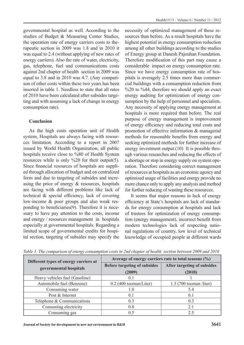
HealthMED - Volume 6 / Number 11 / 2012
Journal of Society for development in new net environment in B&H 3641
governmental hospital as well. According to the studies of Budget & Measuring Center Studies, the operation rate of energy carriers costs to the-rapeutic section in 2009 was 1.8 and in 2010 it was equal to 2.4 (without applying of new rates of energy carriers). Also the rate of water, electricity, gas, telephone, fuel and communications costs against 2nd chapter of health section in 2009 was equal to 3.8 and in 2010 was 4.7. (Any compari-son of other costs within these two years has been inserted in table 1. Needless to state that all rates of 2010 have been calculated after subsides targe-ting and with assuming a lack of change in energy consumption rate).
Conclusion
As the high costs operation unit of Health system, Hospitals are always facing with resour-ces limitation. According to a report in 2007 issued by World Health Organization, all public hospitals receive close to %80 of Health System resources while is only %20 for their output(5). Since financial resources of hospitals are suppli-ed through allocation of budget and on centralized form and due to targeting of subsides and incre-asing the price of energy & resources, hospitals are facing with different problems like lack of technical & special efficiency, lack of covering low-income & poor groups and also weak res-ponding to beneficiaries(9). Therefore it is nece-ssary to have pay attention to the costs, income and energy / resources management in hospitals especially at governmental hospitals. Regarding a limited scope of governmental credits for hospi-tal section, targeting of subsides may specify the
Table 1. The comparison of energy consumption costs in 2nd chapter of health section between 2009 and 2010
Different types of energy carriers at governmental hospitals
Average of energy carriers rate to total seasons (%)Before targeting of subsides
(2009)After targeting of subsides
(2010)Heavy vehicles fuel (Gasoline) 0.1 1
Automobile fuel (Benzene) 0.2 (400 tooman/Liter) 1.3 (700 tooman /liter)Consuming water 1.8 5.4Post & Internet 0.1 0.1
Telephone & Communications 0.3 0.3Consuming electricity 0.8 2.1
Consuming gas 0.5 2.5
necessity of optimized management of these re-sources than before. As a result hospitals have the highest potential in energy consumption reduction among all other buildings according to the studies of Energy group at Danesh Pajouhan Foundation. Therefore modification of this part may cause a considerable impact on energy consumption rate. Since we have energy consumption rate of hos-pitals is averagely 2.5 times more than commer-cial buildings with a consumption reduction from %20 to %44, therefore we should apply an exact energy auditing for optimization of energy con-sumption by the help of personnel and specialists. Any necessity of applying energy management at hospitals is more required than before. The real purpose of energy management is improvement of energy efficiency and reducing total costs and promotion of effective information & managerial methods for reasonable benefits from energy and seeking optimized methods for further increase of energy investment output.(10) It is possible thro-ugh various researches and reducing the effects of a shortage or stop in energy supply on system ope-ration. Therefore considering correct management of resources at hospitals as an economic agency and optimized usage of facilities and energy provide no more chance only to apply any analysis and method for further reducing of wasting these resources.
It seems that major reasons in lack of energy efficiency at State’s hospitals are lack of standar-ds for energy consumption at hospitals and lack of trustees for optimization of energy consump-tion (energy management), incorrect benefit from modern technologies lack of respecting natio-nal regulations of country, low level of technical knowledge of occupied people at different wards
3642
HealthMED - Volume 6 / Number 11 / 2012
Journal of Society for development in new net environment in B&H
of hospitals, lack of enough knowledge of techno-logy users in the field of optimization of energy consumption and also high level of energy con-sumption.
Jabbari in his study about hospital building and energy profitability, believes that the first job for managing energy consumption is energy audi-ting and feasibility studies as a standard method in the world. Also it is possible to benefit from tens of companies in the field of energy services. Any applying of national rules and regulations of building especially Article 19 by supervisor en-gineer has a great role in optimization of energy consumption as well.(11). In addition and with regard to a study by Energy Consumption Optimi-zation Organization, any application of consump-tion optimization guidelines in building & engine house may cause an economy potential in electri-city & fuel consumption respectively %30 & %21 as well. Benefiting from vehicles is applicable in transportation sector of hospitals and as an effecti-ve factor in energy consumption pattern. As a su-bstitute fuel, natural gas may cause a considerable reduction in fuel consumption costs of hospitals and ambulances.
In another study by Hatam, it was possible to estimate economy potential in 2004. He has esti-mated the same equal to %50 in a study on 25 hospitals. He has stated various functions for op-timization of consumption including continuous evaluation of functions, qualitative & quantitative improvement of services and further programming for better usage of resources Hatam in 2004with regard to the volume of hospital operations.(12) In another study by Rey in England, he has estimated energy costs equal to %3 of total operation bud-get of the hospitals with a %3-%10 of economy potential as well. Meanwhile energy consumption rate is more in our country with more possibilities in energy consumption reduction.(13) One of the economic ways in energy consumption at Health Section, as stated in Lancet Magazine, is benefi-ting from different energies with lower damages to environment including solar energy instead of fossil ones. (14) In 2010, Armstrong introduces ar-chitecture & engineering of hospitals as an impor-tant factor in reducing energy wastes. Of course he believes that special architecture & engineering should be applied for hospitals in accordance with
the relevant environment.(15) According to the Hospitals Structure & Desi-
gning Magazine published at U.S.A, energy con-sumption management at hospitals depends upon the type, size, place and structure of it. It is assumed that energy management programs need a daily measurement of functions and specifying the duti-es of all managerial levels in relation with energy wastes reduction. The above-mentioned magazi-ne introduces any daily evaluation and control of energy consumption as a success key in reducing of energy consumption and increasing the effici-ency accordingly.(16) From theoretical viewpoint, generally it seems that applying of suitable & sci-entific economic methods like measuring of time / job for compliance usage from resources with re-gard to the volume of functions and different met-hods of estimation could be more applicable as a tool for further policy making like random terri-tory analysis & data coverage analysis as well. It is only for better management of resources and re-ducing of costs which is higher than a research in itself. The major motivation of applying scientific & applicable methods is optimized usage of physi-cal, technological and current energy resources.
Then according to a report issued by Ministry of Health in 2002 and a study on %91 of total hos-pitals under the coverage of the mentioned mini-stry, about %14 of hospital beds are out of usage. Therefore it may naturally cause overhead costs and high level of energy wastes resulted from lack of management and indifference position against benefiting from energy consumption resources at hospitals. Followings are practical & applicable aspects for optimization of energy consumption at hospitals:
- Heating insulation of walls, ceiling and floors
- Two –layers windows with suitable sealing - Double rows of entrance doors & automatic
open/close facility- Replacement of lamps & benefiting from
low-consumption lighting - Installation of sensor at rooms & corridors- Replacing of fuel- Applying of Alert systems
HealthMED - Volume 6 / Number 11 / 2012
Journal of Society for development in new net environment in B&H 3643
Furthermore and in order to increase economy potential in energy consumption at hospitals, it is necessary to follow up national economy regula-tions for all constructions and approving/imple-mentation a suitable energy management law for upgrading any usage of specialists for applying new guidelines in the field of energy consumption reduction and improvement of economy culture.
Acknowledgment
The study was funded by an institutional grant from Planning and Budget office of Health Admi-nistration, Iran.
The authors wish to thank all the colleges who were involved in this study.
Resources
1. Pourmohamadi K. Assessing technical efficiency in Social Security Hospitals between 2005-2008 applying DEA and SFA, Thesis for Master degree of health service management, Shiraz University of Me-dical Sciences, 2008:82-3 [In Persian]
2. Barati Marnani A, SadeghifarJ, PourmohammadiK, MostafaieD, AbolhalajM, BastaniP, Performance assessment indicators: How DEA and Pabon La-sso describe Iranian hospitals` performance, He-althMED, 2012;6(3):791-6
3. Emami Meibodi A. Efficancy and production princi-pals (Scientifis Applied), 2nd ed, Scientific and Rese-arch institution of trade press, Tehran, 2001; 20-80, 103-4[In Persian]
4. Scott, R.W, The organization of medical care ser-vices: Toward an integrated model. Med.CareRev., 2004;50(3):271-301
5. Milka L, Hakinnen U. Determinants of Cost efficiency of Finnish Hospitals:A Comparison of DEA and SFA National Research and Development Centre for Wel-fare and Health Institute of Pharmaco-Economics., 2001;44:45-56
6. World Health Organization, The world health report: a safer future: global public health security in the 21st century. World Health Organization press ,2007
7. Rowena Jacobs, Peter C. Smith, Andrew Street. Mea-suring efficiency in health care”,. Cambridge univer-sity press, first published ,2006:109-120.
8. Ferrier,G.D.Lovell,C.A.K. Measuring cost effici-encyin banking econometrics and linear program-ming evidence”. Journal of econometrics, 1990; 46:229-45.
9. Niccie L.MC. Kay, Marry E. deily. Cost inefficiency and hospital health outcome, journal of health econo-mics , 2008;17(7) :833-848.
10. Kwakye, E, Relative efficiency of some selected hos-pital in the ACCRA TERM METROPOLIS”, insti-tute of statistical , social and economics research (ISSER), University of Gana,2003
11. Chirikas, T.N. Sear, A.M. measuring Hospital Effici-ency: A comparison of two approaches, Health Ser-vice Research, 2000; 34(6): 1389-1408.
12. Kirigia, JM. Sambo , LG. Scheel, H. Technical Effi-ciency and Productivity of Public Sector Hospitals in three south Africa Provinces. South African Jour-nal of Economics,2001;69:336-358.
13. Rey. D.Healthcare. Executives Place Higher Pri-ority on Energy Efficiency Than Others, Research Shows, Energy Weekly News, 2008; 4,: 29-38
14. Cutting Carbon, Improving Health, The Lancet jour-nal, 374(5), 2009. http://download.thelancet.com/pdfs/journals/lancet/
15. Armstrong. “Series EHU-700 Electronic, Steam Hu-midifier. “Retrieved August 2010, from http://www.armstronginternationl.com/files/common/hvacoluti-onsource/ehu700.pdf
16. AIA, FGI. Guidelines for Design & Construction of Health Care Facilities. Washington D.C., American Institute of Architects, 2006
Corresponding Author Ramezanian Maryam,Center for Budgeting and performance Monitoring,Mohme,Tehran, Iran,E-mail: [email protected]
3644
HealthMED - Volume 6 / Number 11 / 2012
Journal of Society for development in new net environment in B&H
Flexible bronchoscopy findings in lung amoebiasis: a case reportSelvi Kelekci1, Velat Sen1, Tuba Tuncel1, Hadice Selimoglu Sen2, Muttalip Cicek3, Duygu Erge4, M. Fuat Gurkan1
1 Dicle Univercity Medicine School, Department of Pediatrics Pulmonology, Diyarbakır, Turkey 2 Dicle Univercity Medicine School, Department of Chest Disease, Diyarbakır, Turkey 3 Dicle Univercity Medicine School, Department of Microbiology, Diyarbakır, Turkey 4 Inonu Univercity Medicine School, Department of Pediatrics Allergy and Immunology, Malatya, Turkey
Abstract
The protozoon Entamoeba histolytica is an agent of human amoebiasis. Amoebiasis is common around the world, especially in tropical and subtropical regions. About 90% of infections are asymptomatic, while the remaining 10% are characterized by dysentery and abscesses obser-vable in the liver or other organs. The liver is the organ most commonly affected by extraintestinal amoebiasis. Pulmonary and invasive amoebiasis is seen in 2-3% of patients, but isolated pulmo-nary amoebiasis is rarely seen in the pediatric age group. In this study, a 14 year old male patient diagnosed with isolated pulmonary amoebiasis is presented. Diagnosis was based on the detecti-on of trophozoites through direct examination of bronchoalveolar lavage fluid obtained by flexible bronchoscopy, and the presence of amoebic IgG in the blood. Three weeks of metronidazole combi-ned with antimicrobial treatment significantly im-proved the patient’s clinical and radiologic findin-gs. The aim of this report was to present a rare case of childhood pulmonary amoebiasis without liver or other organ involvement and to demonstrate the efficacy of flexible bronchoscopy for diagnosis.
Keywords: Children; amoeba; lung; bronchos-copy
Introduction
Entamoeba histolytica is a protozoon belon-ging to the family Amoebozoa. E. histolytica has two forms: trophozoites and cysts; the cysts are re-sistant to stomach acid. Contagion usually occurs through contaminated food and only rarely thro-ugh contaminated water. Contagion through con-taminated water is not often seen as nutrients.(1) Zoonotic transmission has not been reported.
Amoebiasis is the second most common pa-rasitic disease in the world and the third most common cause of death, after malaria and schisto-somiasis. Amoebiasis occurs primarily in Central America, South America and India and is endemic to all tropical and temperate regions.(2) Pulmo-nary amoebiasis is often related to amoebic liver abscesses and affects 2-3% of patients with inva-sive amoebiasis. Isolated pulmonary amoebiasis without liver involvement is generally thought to be caused by hematogenous spread.(3) A diagno-sis of E. histolytica can be determined via micros-copy, serology, stool antigen detection, culture, and molecular biological methods. We aimed to compare the clinical and radiological findings of our isolated pulmonary amoebiasis case with the literature.
Case
A 14 year old male patient with no history of pulmonary complaints presented to a pediatrici-an complaining of cough, fatigue, and anorexia. He was thought to have atypical pneumonia, and the pediatrician prescribed 10 days of oral clarit-hromycin therapy. A few days post-treatment, the patient complained of brown sputum, and an an-terior-posterior chest X-ray was taken. The pati-ent was referred to our pediatric chest clinic due to a mass in his lung. Chest radiography revea-led an air-fluid level in the lower lobe of the right lung, and the patient was admitted to our center for further evaluation and treatment. Neither the patient nor his family had any related medical hi-story. At his physical examination, the patient’s temperature was 36°C, respiratory rate was 20 breaths/min, blood pressure was 90/60 mm/Hg, and the middle and lower zones of the right lung
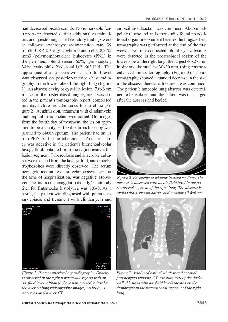
HealthMED - Volume 6 / Number 11 / 2012
Journal of Society for development in new net environment in B&H 3645
had decreased breath sounds. No remarkable fea-tures were detected during additional examinati-ons and questioning. The laboratory findings were as follows: erythrocyte sedimentation rate, 39 mm/h; CRP, 0.3 mg/L; white blood cells, 8,870/mm3 (polymorphonuclear leukocytes (PNL) in the peripheral blood smear, 60%; lymphocytes, 38%; eosinophils, 2%); total IgE, 503 IU/L. The appearance of an abscess with an air-fluid level was observed on posterior-anterior chest radio-graphy in the lower lobe of the right lung (Figure 1). An abscess cavity or cyst-like lesion, 7.6x6 cm in size, in the posterobasal lung segment was no-ted in the patient’s tomography report, completed one day before his admittance to our clinic (Fi-gure 2). At admission, treatment with clindamycin and ampicillin-sulbactam was started. On images from the fourth day of treatment, the lesion appe-ared to be a cavity, so flexible bronchoscopy was planned to obtain sputum. The patient had an 18 mm PPD test but no tuberculosis. Acid resistan-ce was negative in the patient’s bronchoalveolar lavage fluid, obtained from the region nearest the lesion segment. Tuberculosis and anaerobic cultu-res were seeded from the lavage fluid, and amoeba trophozoites were directly observed. The serum hemagglutination test for echinococcis, sent at the time of hospitalization, was negative. Howe-ver, the indirect hemagglutination IgG antibody titer for Entamoeba histolytica was 1:640. As a result, the patient was diagnosed with pulmonary amoebiasis and treatment with clindamycin and
ampicillin-sulbactam was continued. Abdominal-pelvic ultrasound and other audits found no addi-tional organ involvement besides the lungs. Chest tomography was performed at the end of the first week. Two interconnected plural cystic lesions were detected in the posterobasal region of the lower lobe of the right lung, the largest 40x27 mm in size and the smallest 30x30 mm, using contrast-enhanced thorax tomography (Figure 3). Thorax tomography showed a marked decrease in the size of the abscess, therefore, treatment was continued. The patient’s amoebic lung abscess was determi-ned to be isolated, and the patient was discharged after the abscess had healed.
Figure 1. Posteroanterior lung radiography. Opacity is observed in the right paracardiac region with an air-fluid level. Although the lesion seemed to involve the liver on lung radiographic images, no lesion is observed on the liver CT.
Figure 2. Parenchyma window in axial sections. The abscess is observed with an air-fluid level in the po-sterobasal segment of the right lung. The abscess is ovoid with a smooth border and measures 7.6x6 cm.
Figure 3. Axial mediastinal window and coronal parenchyma window. CT investigations of the thick-walled lesions with air-fluid levels located on the diaphragm in the posterobasal segment of the right lung.
3646
HealthMED - Volume 6 / Number 11 / 2012
Journal of Society for development in new net environment in B&H
Discussion
Isolated pulmonary amoebiasis without liver involvement is rarely seen, even in regions ende-mic for amoebiasis. Amoebiasis can spread to the lungs directly from a liver abscess, or can spread via the transdiaphragmatic lymphatic transport, the hepatic veins, the ductus thoracicus, or the he-morrhodal veins. Our patient had no predisposing factors except living in a subtropical region.
The liver is often involved in amoebiasis, and lung involvement is seen in 15-20% of patients with liver involvement.(1,2) Complaints often include coughing and brown sputum when ope-ning the bronchia;(4) the patient in our study had a history of both. Pulmonary amoebiasis often appe-ars on chest radiographs in the form of an abscess in the lower lobe of the right lung. One abscess, 76x60 mm in size and located in the posteroba-sal region of the right lower lobe, was observed in the patient’s first tomographic images. After the first week of treatment, tomography showed the abscess in two pieces, 40x20 mm and 30x30mm in size, opening to a bronchus. The abscess was observed to be severely diminished on thorax tomographic images. Leukocytosis, anemia and eosinophilia may occur during the course of the disease, in which case, patients receive positi-ve serology results.(5-7) In our case, the patient experienced moderate anemia, mild eosinophilia and high serum IgE. The serum hemagglutination IgG titer for Entamoeba histolytica was positive (1:640). A definitive diagnosis is made by demon-strating the presence of E. histolytica trophozoites in the sputum or abscess, using fine needle aspira-tion, as well as, in stool samples or pleural maid. Trophozoites are most commonly observed in the abscess material. We also identified trophozoites by directly examining the bronchoalveolar lavage fluid from our patient. Flexible bronchoscopy can be considered for patients with abscesses unres-ponsive to antibiotic treatment, with atypical cli-nical findings, and with abscesses which appear to have developed malign cavity, tumor and foreign bodies following aspiration.
Tumor biopsy, removal of foreign bodies, and draining the abscess all provide material which can be tested for microorganisms.(8) We initially interpreted the first tomography findings in favor
of cavitary tuberculosis, and the patient’s tubercu-lin test was positive, so we decided to use flexi-ble bronchoscopy. Bronchoalveolar lavage fluid obtained during bronchoscopy excluded pulmo-nary tuberculosis and demonstrated the presence of trophozoites, supporting the positive results for serum Entamoeba antibodies.
Treatment for pulmonary amoebiasis includes the improvement of hygiene, chest physiotherapy, and antibiotics to facilitate drainage of the abscess opening into a bronchus. The duration of therapy is determined by clinical and radiological findings and by the patient`s response to treatment. Patients should be treated at least 2-3 weeks until fever and bad-smelling sputum disappear and the fluid level in the abscess no longer appears on radiological imaging. Medical therapy improves 44% of lung abscesses by the second week. In our case, the le-sions had decreased in size by the end of the first week and had completely disappeared by the end of the third week. It should be noted that relap-ses often occur in patients whose therapy was not sufficiently well-managed; for example, disease etiology was not determined or treatment-resistant microorganisms were not identified.(9)
Conclusion
In conclusion, amoebic abscesses should be considered, especially in endemic regions, in the differential diagnosis of pulmonary abscesses du-ring childhood, and diagnostic factors should be sought in all obtainable body fluids. Furthermore, flexible bronchoscopy might be required to eluci-date the etiology and best determine the treatment management of pulmonary abscesses in pediatric patients.
References
1. Neghina R, Neghina AM, Merkler C, Marincu I, Iaco-biciu I. A case report of pulmonary amoebiasis with Entamoeba histolytica diagnosed in western Roma-nia. J Infect Developing Countries 2008;2:400-2.
2. Samie A, Obi LC, Bessong PO, Stroup S, Houpt E, Guerrant RL. Prevalence and species distributi-on of E. Histolytica and E. Dispar in the Venda re-gion, Limpopo, South Africa. Am J Trop Med Hyg. 2006;75(3):565-71.
HealthMED - Volume 6 / Number 11 / 2012
Journal of Society for development in new net environment in B&H 3647
3. Gupta KB, Manchanda M, Chaudhary U, Verma M. Superior Vena cava syndrome caused by pulmo-nary amoebic abscess. Indian J Chest Dis Allied Sci 2006;48:275-7.
4. Quah BS, Indudharan R, Hashim I, Simpson H.Lung abscess: an unusual presentation of congenital tracheoesophageal fistula without atresia. J Pediatr Surg. 1998;33(12):1817-9.
5. Seaton A, Seaton D,Leitch AG.Crofton and Douglas’s Respiratory,Blackwell Scientific publication,London.Fourth Edition,1989;476-479.
6. Fraser RG,Pare JAP,Pare D, Fraser RS,Genereux GP.Diagnosis of Disease of Chest.WB Saunders OF Company,Philedelphia,Third Edition, 1991;1082-1083.
7. Baret-Connorr E et All. Parasitic Pulmonary Disea-se.Am Rev Respir Dis 1982;126:558-563.
8. Finegold SM, Fishman JA. Empyema and lung absce-ss. In: Fishman AP (ed): Fishman’s Pulmonary Di-seases and Disorders. McGraw Hill; New York, 1998;2021-2034
9. Erasmus JJ, Mc Adams HP, Rossi J, Kelley MJ. Percutaneous management of intrapulmonary air and fluid collections. Radiol. Clin North America 2000;38,385-393.
Corresponding AuthorSelvi Kelekci,Dicle Univercity Medicine School, Department of Pediatrics Pulmonology, Diyarbakır, Turkey, E-mail: [email protected],
3648
HealthMED - Volume 6 / Number 11 / 2012
Journal of Society for development in new net environment in B&H
Dementia type of Alzheimer’s disease due to β-amyloid was improved by Gallic acid in rats Zohre Valizadeh1, Akram Eidi1, Alireza Sarkaki2, Yaghoob Farbood3, Pejman Mortazavi4
1 Department of Biology, Science and Research Branch, Islamic Azad University, Tehran, Iran 2 Physiology Research Center and Medicinal Plants Research Center, Ahvaz Jundishapur University of Medical Sciences, Ahvaz, Iran 3 Dept. of Physiology and Physiology Research Center, Ahvaz Jundishapur University of Medical Sciences, Ahvaz, Iran 4 Department of Pathology, Faculty of Specialized Veterinary Science, Science& Research Branch, Islamic Azad University, Tehran, Iran
Abstract
Background and objective: The aim of this study was evaluation the effect of oral admini-stration of different doses of gallic acid (GA) on dementia type of Alzheimer’s disease (AD) indu-ced by intrahippocampal injection of beta amyloid (Aβ) in rats.
Methods: Forty eight adult male Wistar rats (300±20g) were used in this study. The animals divided randomly into six groups of 8 in each. 1) Control; 2) AD (received 1μg/μl Aβ into hippocam-pal bilaterally), 3) Sham (received 1μl normal sali-ne into hippocampal bilaterally), 4) AD+GA50, 5) AD+GA100, 6) AD+GA200,(mg/kg for ten days), 7) AD+Veh, 8) Positive control (cont+GA100). Animals were anesthetized with intraperitoneal injection (i.p.) of ketamine HCl and xylazine. Aβ (1μg/μl in each site) was infused into hippocampal areas bilaterally at a rate of 1μl/5min using a 10μl Hamilton syringe connected to an infusion pump. Seven days after the infusion of Aβ animals were traind in Morris water maze to evaluate the spatial learning and memory.
Results: spatial learning and memory in rats with AD induced with Aβ have decreased signifi-cantly (P<0.05). Rats with AD that received Gallic acid for 10 days show significant improvement of spatial learning and memory (P<0.05). GA con-sumption can improve spatial learning and me-mory impairment induced by Aβ injection.
Conclusion: findings indicated that GA redu-ces neural damage with respect to age. polyphe-nolic compounds such as GA could reduce brain amyloid neuropathology and improve cognitive function by promoting non amyloidogenic-secre-tase activity and inhibit oligomerization of Aβ.
Keywords: Alzheimer’s disease; beta-amylo-id; spatial memory; Morris water maze; rat
Introduction
Alzheimer’s disease (AD) is a neurodegenera-tive disorder characterized by cognitive dysfunc-tion. Beta-Amyloid peptide (Aβ) is known to be involved in the neuropathogenesis of AD. Aβ is a small protein which consists of 39–43 amino acid residues. It can exist in different forms, including monomers, oligomers, protofibrils, and fibrils (Nomura et al., 2005, Frozza et al., 2009).
A hallmark of AD pathology is deposition of amyloid neuritic plaques comprised primarily of Aβ peptides in the brain. Accumulation of high molecular weight (HMW) soluble oligomeric Aβ species is hypothesized to initiate a cascade of cel-lular events resulting in synaptic failure, neuronal injury, apoptotic neuronal death, and ultimately, cognitive and functional decline including deficits in spatial memory (Cleary et al., 2005, Ferruzzi et al., 2009). The disease process involves the dege-neration of synapses and neurons in brain regions that play fundamental roles in learning and me-mory including hippocampus, entorhinal cortex, basal forebrain, amygdale, frontal cortex, and in-ferior parietal cortex (Mattson and Chan, 2003, Frozza et al., 2009).
Substantial evidence indicates that excess of Aβ which aggregates into toxic fibrillar deposits, plays a central role in the etiology of Alzheimer’s disease. In support of this hypothesis, numerous in vitro and in vivo studies have reported on the neurotoxic effects of Aβ-related fragments (e.g. Aβ1-40 and Aβ1-42) in neurons derived from re-

HealthMED - Volume 6 / Number 11 / 2012
Journal of Society for development in new net environment in B&H 3649
gions severely affected in AD. Although the preci-se mechanisms mediating the toxic properties of Aβ have yet to be fully established. It has been proposed that they are associated with an increa-sed production of reactive oxygen species (ROS) and apoptosis (Bastianetto et al., 2006).
Recent investigations suggest that Aβ may play a direct role in oxidative damage, suggested lipo-protein oxidization is an important factor in AD, so that oxidative stress involves in the mechanism of Aβ-induced neurotoxicity and AD pathogene-sis (McDaid et al., 2005). Synaptic pathology is considered as a major and early contributor to the cognitive deficits and reduced cerebral activity of Alzheimer’s disease (Kim et al., 2001).
Oxidative stress (OS) and inflammation are thought to be major factors in brain aging and in age-related neurodegenerative disease (Engelhart et al., 2002, Shukitt-Hale et al., 2006). Humans and animals show increased motor and cognitive declines with aging , that are thought to be due to increased susceptibility to the long term effects of OS and inflammation (Joseph et al., 2005). The-refore, it would seem that age-related deleterious effects on behavior and brain function could be retarded or even reversed by increasing antioxi-dant and/or anti-inflammatory levels, and that the synergistic effects of combinations of antioxidants and/or anti-inflammatories, particularly phytoche-micals (polyphenols), might be effective in this re-gard because polyphenols possess potent antioxi-dant properties (Shukitt-Hale et al., 2006). Foods containing high levels of antioxidants may also slow the progression of AD, possibly by preven-ting or neutralizing the damaging effects of free radicals (Kostrzewa and Segura-Aguilar, 2003, Hartman et al., 2006).
Memory as one of the basic cognitive functions ( Sladjana et al.,2012 ). Extensive research into the potential therapeutic effects of antioxidants in the treatment of AD has produced promising results. Antioxidants have been found to improve cogni-tive function in aged rat, and to prevent learning and memory deficits following intracerebroventri-cular (ICV) infusion of Aβ (McDaid et al., 2005).
Previous findings have suggested that reversals in age related cognition declines might be accom-plished by increasing the dietary intake of fruits and vegetables, especially those identified as being
high in antioxidant activity. These results suggest that polyphenolics in antioxidant-rich foods might be effective in forestalling functional age-related deficits. As has been seen with respect to red wine. Concord grape juice is a rich source of flavonoids that include gallic acid, catechins, epicatechins, quercetins, anthocyanins, and proanthocyanidins are potent antioxidants (Shukitt-Hale et al., 2006).
Gallic acid (GA) is an endogenous product fo-und in plants (Shahrzad et al., 2001). It is widely distributed throughout the plant kingdom, where it is present either in free form or, more commonly, as a constituent of tannins, namely gallotannin (Niemetz and Gross, 2005). Strawberries, pi-neapples, bananas, lemons, red and white wines, gallnuts, sumac, witch hazel, tea leaves, oak bark and apple peels are some of the natural products which are rich in GA (Chu et al., 2002, Wolfe et al., 2003). Regarding its biological activity, GA exerts anti-bacterial, anti-viral, anti-inflammatory and antioxidant effects and antimelanogenic acti-vity via the inhibition of tyrosinase activity (Wol-fe et al., 2003, Kim et al., 2006). It also inhibits high fat diet-induced dyslipidaemia, anti-prolife-rative, pro-apoptotic and anti-tumorigenic effects against prostate carcinoma xenograft growth in nude mice (Kaur et al., 2009). GA binds to pro-teins and key minerals such as iron, zinc and cal-cium, and affects their bioavailability by forming insoluble complexes (Niho et al., 2001). Oxidative stress induced by reactive oxygen species (ROS) is strongly associated with the pathogenesis of various neurodegenerative disorders, including Alzheimer’s disease (Kyung et al., 2009). In the case of Alzheimer’s disease detected anthocyanins in rat brains after supplementation with berry and grape extracts (Casadesus et al., 2004).
So this study aimed to investigate the effects of oral administration of GA on spatial cognition de-ficit in rat model of AD by intrahipocampal injec-tion of Aβ.
Materials and methods
Animals Forty eight adult male Wistar rats (300±20g)
were used in this study were obtained from central animal house of Ahvaz Jundishapur University of Medical Sciences (AJUMS). They were housed

3650
HealthMED - Volume 6 / Number 11 / 2012
Journal of Society for development in new net environment in B&H
individually in standard cages under controlled room temperature (20±2°C), humidity (50-55%) and light exposure conditions 12:12h light/dark cycle with free access to food and water ad libi-tum. All experiments carried out during the light phase of the cycle (8:00am to 6:00 pm). Animal handling and experimental procedures performed under observance of the University and Institu-tional legislation, controlled by the Local Ethics Committee for the Purpose of Control and Super-vision of Experiments on Laboratory Animals.
The animals divided randomly into six grou-ps of 8 in each included: 1) Control; aged healthy rats, 2) AD (received 1μg/μl Aβ into hippocam-pal CA1 bilaterally), 3) Sham operated rats (re-ceived 1μl normal saline into hippocampal CA1 bilaterally), 4) AD+GA50 (received 1μg/μl Aβ into hippocampal CA1 bilaterally and 50mg/kg GA by oral gavages for ten days), 5) AD+GA100 (received 1μg/μl Aβ into hippocampal CA1 bila-terally and 100mg/kg GA by oral gavages for ten days), 6) AD+GA200 (received 1μg/μl Aβ into hippocampal CA1 bilaterally and 200mg/kg GA by oral gavages for ten days), 7) AD+Veh (recei-ved 1μg/μl Aβ into hippocampal CA1 bilaterally and normal saline by oral gavages for ten days), 8) Positive control (cont+GA100, healthy rats recei-ved most effective dose of GA (100mg/kg) by oral gavages for ten days.
Beta-amyloid peptide 1–42 (Aβ1–42) Beta- amyloid powder purchased from Sigma–
Aldrich Co., USA. It was solved in normal sali-ne (pH=7.2) (Kim et al., 2001, Goryacheva et al., 2010), at the concentration of 1μg/μl and solution was incubated at 370C for one week before use. The drug (2μg/2μl) was injected into hippocampal CA1 areas of rat’s brain bilaterally to induce the early stage of AD.
Preparation of Gallic acid Gallic acid (Sigma–Aldrich Company, USA)
was solved in normal saline. The Gallic acid (50,100,200 mg/kg) was adminisrered to rats orally for 10 days after AD induction (Ferruzzi et al., 2009).
Neurosurgery and drug administrationAnimals were anesthetized with intraperito-
neal injection (i.p.) of 60 mg/kg ketamine HCl (Alfasan, Woerden–Holland) and 10 mg/kg xyla-zine (Alfasan, Woerden–Holland). In brief, after anesthesia, animal head was fixed in a stereotaxic apparatus (Narishige, Tokyo, Japan) and Aβ (1μg/μl in each site) was infused into hippocampal CA1 areas bilaterally at a rate of 1μl/5min using a 10μl Hamilton syringe connected to an infusion pump (WPI 101i, USA). Infusion was done with coordi-nations of AP; −4.8 from the bregma, ML; ±3.5, DV; −4 from dura mater, according to the Paxinos and Watson (Paxino and Wastene, 2006). After in-fusion, the cannula left in place to additional 5min to allow the diffusion of drug completely. All ani-mals allowed to recovery period (7–10 days) be-fore testing (Ferihan and Sibel, 2006).
The spatial cognition test Training apparatus A black circular pool (120 cm in diameter and
80 cm in height) filled with tap water (27±2 C) with a depth 60 cm was used (Widy-Tyszkiewicz et al., 2002). The maze divided geographically into four equal size quadrants and release points designed in each quadrant as north (N), east (E), south (S) and west (W). A hidden circular esca-pe platform (12cm in diameter) was emerged 2 cm below the water level and was located in the center of the northeast quadrant. Some fixed vi-sual cues including computer, desk, shelves, po-sters and illumination lights placed on the walls around the pool. A camera positioned above the center of the pool that connected to a computer to record the animal motions. An automated tracking system (Radiab ver. 2, Tehran, Iran) used to me-asure the escape latency, swimming distance and speed (Doulah et al., 2009).
Habituation Twenty-four hours prior to the start of training,
rats were habituated to the pool by allowing them to perform a 60 s swimming without the platform.
Training procedure Training, took place during the light phase of
the cycle between 8:00 am and 5:00 pm. Seven days after the infusion of Aβ animals were su-

HealthMED - Volume 6 / Number 11 / 2012
Journal of Society for development in new net environment in B&H 3651
bjected to the training procedure of one session of four trials (block) daily for four consecutive days in the water maze. In each trial, the animals were allowed 60 s to find the platform, then they were allowed to remain there for 30 s, if they did not find the platform within 60 s, animals were gently guided to the platform. After the completion of a trial, animals were returned to a holding cage for an inter-trial interval of 60 s. After 24 h of the last trial, platform was removed and the rats were re-leased from southwest as a probe trial (consisted of a 60 s free swim period) and the time spent in the target quadrant was recorded (Doulah et al., 2009).
StatisticsData were expressed as mean±SEM and pro-
cessed by SPSS ver.17. Data were analyzed with
Figure 1. Comparison of path length between gro-ups: (A) there are no significant difference betwe-en control and sham groups. (B) Path length was increased significantly in AD group during sessi-ons 1–4 (*P < 0.05, **P < 0.01, ***P < 0.001) when compared to sham group (D, E). Path len-gth was decreased significantly in AD+GA groups during sessions 1– 2 (*P < 0.05, **P<0.01, and ***P < 0.001 for AD+ GA vs. AD Veh).
repeated measures two-way analysis of variance (ANOVA) followed by LSD post-hoc analysis. P-value less than 0.05 was considered to be statisti-cally significant.
Results
Path length: Comparing the swimming path length between the control and sham groups did not indicate any differences during 4 days spatial training in water maze. Figure 1-A shows path len-gth of AD group in each session was longer than sham group significantly (* p<.05, **p < 0.01, and ***p < 0.001for AD vs. sham). On the other hand path length was not different between AD and AD+Veh groups, but reduced in AD+GA group (100, 200) significantly during first and second se-ssions of training (***p < 0.001 for session 1 and *p < 0.05 for sessions 2).
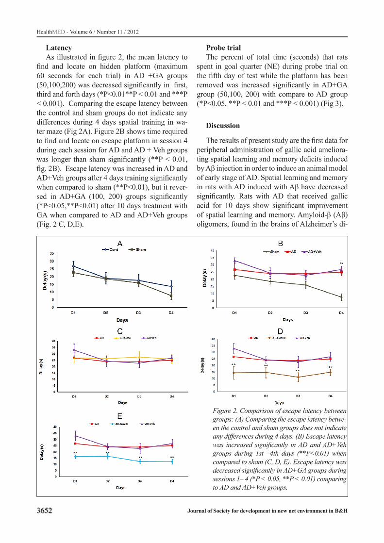
3652
HealthMED - Volume 6 / Number 11 / 2012
Journal of Society for development in new net environment in B&H
Latency As illustrated in figure 2, the mean latency to
find and locate on hidden platform (maximum 60 seconds for each trial) in AD +GA groups (50,100,200) was decreased significantly in first, third and forth days (*P<0.01**P < 0.01 and ***P < 0.001). Comparing the escape latency between the control and sham groups do not indicate any differences during 4 days spatial training in wa-ter maze (Fig 2A). Figure 2B shows time required to find and locate on escape platform in session 4 during each session for AD and AD + Veh groups was longer than sham significantly (**P < 0.01, fig. 2B). Escape latency was increased in AD and AD+Veh groups after 4 days training significantly when compared to sham (**P<0.01), but it rever-sed in AD+GA (100, 200) groups significantly (*P<0.05,**P<0.01) after 10 days treatment with GA when compared to AD and AD+Veh groups (Fig. 2 C, D,E).
Probe trialThe percent of total time (seconds) that rats
spent in goal quarter (NE) during probe trial on the fifth day of test while the platform has been removed was increased significantly in AD+GA group (50,100, 200) with compare to AD group (*P<0.05, **P < 0.01 and ***P < 0.001) (Fig 3).
Discussion
The results of present study are the first data for peripheral administration of gallic acid ameliora-ting spatial learning and memory deficits induced by Aβ injection in order to induce an animal model of early stage of AD. Spatial learning and memory in rats with AD induced with Aβ have decreased significantly. Rats with AD that received gallic acid for 10 days show significant improvement of spatial learning and memory. Amyloid-β (Aβ) oligomers, found in the brains of Alzheimer’s di-
Figure 2. Comparison of escape latency between groups: (A) Comparing the escape latency betwe-en the control and sham groups does not indicate any differences during 4 days. (B) Escape latency was increased significantly in AD and AD+Veh groups during 1st –4th days (**P<0.01) when compared to sham (C, D, E). Escape latency was decreased significantly in AD+GA groups during sessions 1– 4 (*P < 0.05, **P < 0.01) comparing to AD and AD+Veh groups.

HealthMED - Volume 6 / Number 11 / 2012
Journal of Society for development in new net environment in B&H 3653
Figure 3. Percent of time spend in goal quarter during probe trial. A) It was decreased significantly in AD com-paring to control group (***P < 0.001). B-C) It increased significantly in AD +GAgroups after 10 days treating with GA when compared to AD + Veh group (*P<0.05, **P < 0.01 and ***P < 0.001 vs. AD + Veh group).
sease (AD) patients and transgenic mouse models of AD, cause synaptic toxicity and memory impa-irment (Liu et al., 2011).
Recent evidence from experimental AD mou-se models indicates that accumulation of soluble high molecular weight (HMW) oligomeric Aβ species in the brain, rather than deposition of ne-uritic plaque per se, may be specifically related to spatial memory reference deficits because it may disrupt hippocampal long-term potentiation and synaptic plasticity, and induce synaptic deficits. Evidence has also demonstrated that neutralizati-on of soluble HMW Aβ oligomeric species in the brain causally improves spatial memory functions in a mouse model of AD (Balu et al., 2005).
Several lines of studies have reported on the neurotoxic effects of Aβ-related fragments (e.g. Aβ1-40 and Aβ1-42) in neurons derived from regions severely affected in AD such as the hip-pocampus. Although the precise mechanisms me-diating the toxic properties of Aβ have yet to be
fully established, it has been proposed that they are associated with an increased production of re-active oxygen species (ROS) and apoptosis (Ba-stianetto et al., 2006, Liu et al., 2011).
Experimental findings resulted from both in vi-tro and in vivo studies indicate that different mo-lecular forms of Aβ affect a wide array of neuronal and glial functions, thereby leading to neural cell death. Frozza and et al (2009) have shown that Aβ (1-42) or Aβ (25-35) peptides induced similar cell death pattern after 48 h exposure. In the other hand, Sa´ez-Valeroet and his collegeuses (2002) and Stepanichev et al (2006) with using intrace-rebroventricular administration of Aβ had shown changes in molecular distribution of acetylcholine esterase in rat cortex and cerebrospinal fluid, as well as pathomorphological changes in the hippo-campus and impaired of spatial memory (Saez-Va-lero et al., 2002, Stepanichev et al., 2006, Frozza et al., 2009). Beyond of necrotic cell death, there is new strong evidence that neurotoxic effect of
3654
HealthMED - Volume 6 / Number 11 / 2012
Journal of Society for development in new net environment in B&H
Aβ peptides is mediated through activation of cas-pases. Neuronal apoptosis is also seen in human brain with AD. Activation of the apoptotic cascade induced by Aβ could also explain many of the fe-atures of the disease and its progression (Frozza et al., 2009). In other hand some studies suggested that Aβ toxicity is mediated by the intracellular accumulation of ROS and apoptosis (Bastianetto et al., 2006, Liu et al., 2011).
Phenolic acids and flavonoids, although not essential for survival, may over the long term provide protection against a number of chronic diseases. The phenolic acids potentially invol-ved in these beneficial effects include gallic acid, (Joseph et al., 2003). The main flavonoids of in-terest are anthocyanins being able to inhibit lipid peroxidation and protect low-density lipoproteins against oxidation. They can also reduce platelet aggregation and enhance vasodilation. However, the protective effects of these compounds may not be due exclusively to their antioxidant properties and other mechanisms may also operate (Mullen et al., 2007).
Our findings show that GA consumption can improve spatial learning and memory impairment induced by Aβ injection is consistent with other researches.
Extensive research on potential therapeutic effects of antioxidants in the treatment of AD has produced promising results. The antioxidants have been shown to protect neurons against Aβ-induced cell death, lipid peroxidation in cell culture, impro-ving cognitive function in aged rats and to prevent learning and memory deficits following intracere-broventricular (ICV) infusion of Aβ (Shukitt-Hale et al., 2006).
Mario G. Ferruzzi and his colleagues (2009) explored the bioavailability and brain deposition of a grape seed polyphenolic extract (GSPE) atte-nuate cognitive deterioration in a mouse model of AD. Additionally, 4-methylgallic acid (4-OMe GA) were identified as circulating metabolite of GSPE phenolic constituents. They suggest that brain deposition of GA is affected by repeated do-sing of GSPE. These findings suggest that lower concentrations of GSPE may be sufficient to alter cognitive performance (Ferruzzi et al., 2009).
Previous studies have suggested that some polyphenolic compounds could reduce brain
amyloid neuropathology and improve cognitive function by promoting non amyloidogenic-secre-tase activity. Moreover, studies suggest that gra-pe-derived polyphenolic compounds may inhibit oligomerization of Aβ (Porat et al., 2006). Such inhibition would be highly significant, because accumulation of soluble extracellular high-mole-cular-weight (HMW) oligomeric Aβ species in the brain currently is considered as a major risk factor for the onset and progression of cognitive deterio-ration in AD. Thus, pharmacological strategies for the prevention of Aβ oligomerization in the bra-in might result in improved cognitive function in AD (Wang et al., 2008). Inflammation is another hallmark of AD. There is mounting evidence that chronic inflammatory processes play a fundamen-tal role in the progression of neuropathological changes in the AD brain (Kim et al., 2011).
Balue and his colleagues (2005) showed that memory impairment in old rats, improved with use of grape seed extract (GSE) and were attribu-ted to antioxidant properties polyphenols such as gallic acid (GA) in GSE. These antioxidative su-bstances in brain tissues are factors in prevention and treatment of disorders that induced by oxida-tive damages (Balu et al., 2005). It is probably GA as a potent antioxidant and major compounds of GSE can able to improve learning and memory.
In conclusion; our findings indicated that GA reduces neural damage with respect to age. Thus, peripheral administration of GA for 10 days can improve significantly cognitive deficiency due to intra-hippocampal injection of Aβ. The present study has potentially great value in public health although animal experiments need to confirm in human studies.
Acknowledgments
This article extracted as a part of Miss Zoreh Valizadeh’s Ph.D thesis. It has done in Ahvaz Jundishapur University of Medical Sciences, Physiology Research Center (PRC-73). All con-suming materials expenses (but not behavioral and electrophysiological instruments), were paid by herself. Authors thanks to central animal house staff.
HealthMED - Volume 6 / Number 11 / 2012
Journal of Society for development in new net environment in B&H 3655
References
1. Balu M, Sangeetha P, Murali G, Panneerselvam C. Age-related oxidative protein damages in central ner-vous system of rats: modulatory role of grape seed extract. Int J Dev Neurosci 23:501-507.2005
2. Bastianetto S, Yao ZX, Papadopoulos V, Quirion R . Neuroprotective effects of green and black teas and their catechin gallate esters against beta-amyloid-in-duced toxicity. Eur J Neurosci 23:55-64.2006.
3. Casadesus G, Shukitt-Hale B, Stellwagen HM, Zhu X, Lee HG, Smith MA, Joseph JA (Modulation of hip-pocampal plasticity and cognitive behavior by short-term blueberry supplementation in aged rats. Nutr Neurosci 7:309-316.2004.
4. Chu YF, Sun J, Wu X, Liu RH. Antioxidant and anti-proliferative activities of common vegetables. J Agric Food Chem 50:6910-6916.2002.
5. Cleary JP, Walsh DM, Hofmeister JJ, Shankar GM, Kuskowski MA, Selkoe DJ, Ashe KH. Natural oligo-mers of the amyloid-beta protein specifically disrupt cognitive function. Nat Neurosci 8:79-84.2005.
6. Doulah AH, Rohani AH, Khaksari Haddad M, Mo-tamedi F, Farbood Y, Badavi M, Malek M, Sarkaki A. The effect of peripheral administration of growth hormone on AD-like cognitive deficiency in NBM-le-sioned rats. Neurosci Lett 466:47-51.2009.
7. Engelhart MJ, Geerlings MI, Ruitenberg A, van Swi-eten JC, Hofman A, Witteman JC, Breteler MM. Die-tary intake of antioxidants and risk of Alzheimer dise-ase. JAMA 287:3223-3229.2002.
8. Ferihan C, Sibel D. The effect of interahippocam-pal beta amyloid(1-42) peptide injection on oxidant and antioxdidant status in rat brain. AnnNYAcad Sci 1100:8.2006.
9. Ferruzzi MG, Lobo JK, Janle EM, Cooper B, Simon JE, Wu QL, Welch C, Ho L, Weaver C, Pasinetti GM. Bioavailability of gallic acid and catechins from grape seed polyphenol extract is improved by repeated do-sing in rats: implications for treatment in Alzheimer’s disease. J Alzheimers Dis 18:113-124.2009.
10. Frozza RL, Horn AP, Hoppe JB, Simao F, Gerhardt D, Comiran RA, Salbego CG. A comparative study of beta-amyloid peptides Abeta1-42 and Abeta25-35 toxicity in organotypic hippocampal slice cultures. Neurochem Res 34:295-303.2009.
11. Goryacheva AV, Kruglov SV, Pshennikova MG, Smirin BV, Malyshev IY, Barskov IV, Viktorov IV, Downey HF, Manukhina EB. Adaptation to inter-mittent hypoxia restricts nitric oxide overproduction and prevents beta-amyloid toxicity in rat brain. Ni-tric Oxide 23:289-299.2010.
12. Hartman RE, Shah A, Fagan AM, Schwetye KE, Parsadanian M, Schulman RN, Finn MB, Holtzman DM. Pomegranate juice decreases amyloid load and improves behavior in a mouse model of Alzheimer’s disease. Neurobiol Dis 24:506-515.2006.
13. Joseph JA, Denisova NA, Arendash G, Gordon M, Diamond D, Shukitt-Hale B, Morgan D. Blueberry supplementation enhances signaling and prevents behavioral deficits in an Alzheimer disease model. Nutr Neurosci 6:153-162.2003.
14. Joseph JA, Shukitt-Hale B, Casadesus G. Reversing the deleterious effects of aging on neuronal commu-nication and behavior: beneficial properties of fruit polyphenolic compounds. Am J Clin Nutr 81:313S-316S.2005.
15. Kaur M, Velmurugan B, Rajamanickam S, Agarwal R, Agarwal C. Gallic acid, an active constituent of grape seed extract, exhibits anti-proliferative, pro-apoptotic and anti-tumorigenic effects against pro-state carcinoma xenograft growth in nude mice. Pharm Res 26:2133-2140.2009.
16. Kim JH, Anwyl R, Suh YH, Djamgoz MB, Rowan MJ. Use-dependent effects of amyloidogenic fragments of (beta)-amyloid precursor protein on synaptic plasticity in rat hippocampus in vivo. J Neurosci 21:1327-1333.2001.
17. Kim MJ, Seong AR, Yoo JY, Jin CH, Lee YH, Kim YJ, Lee J, Jun WJ, Yoon HG. Gallic acid, a histone acetyltransferase inhibitor, suppresses beta-amylo-id neurotoxicity by inhibiting microglial-mediated neuroinflammation. Mol Nutr Food Res 55:1798-1808.2011.
18. Kim SH, Jun CD, Suk K, Choi BJ, Lim H, Park S, Lee SH, Shin HY, Kim DK, Shin TY. Gallic acid in-hibits histamine release and pro-inflammatory cyto-kine production in mast cells. Toxicol Sci 91:123-131.2006.
19. Kostrzewa RM, Segura-Aguilar J. Novel mechani-sms and approaches in the study of neurodegene-ration and neuroprotection. a review. Neurotox Res 5:375-383.2003.
20. Kyung K, Joo K, Jin Y, Won L, Joo L. Gallic acid induces neuronal cell death through activation of c-Jun N-terminal kinase and down regulation of Bcl-2.,. Academi of Sciences 1171:7.2009.
21. Liu P, Kemper LJ, Wang J, Zahs KR, Ashe KH, Pa-sinetti GM. Grape seed polyphenolic extract speci-fically decreases abeta*56 in the brains of Tg2576 mice. J Alzheimers Dis 26:657-666.2011.
22. Mattson MP, Chan SL. Neuronal and glial calci-um signaling in Alzheimer’s disease. Cell Calcium 34:385-397.2003.
3656
HealthMED - Volume 6 / Number 11 / 2012
Journal of Society for development in new net environment in B&H
23. McDaid DG, Kim EM, Reid RE, Leslie JC, Cleary J, O’Hare E. Parenteral antioxidant treatment pre-serves temporal discrimination following intrahip-pocampal aggregated Abeta(1-42) injections. Behav Pharmacol 16:237-242.2005.
24. Mullen W, Marks SC, Crozier A. Evaluation of phe-nolic compounds in commercial fruit juices and fruit drinks. J Agric Food Chem 55:3148-3157.2007.
25. Niemetz R, Gross GG. Enzymology of gallotannin and ellagitannin biosynthesis. Phytochemistry 66:2001-2011.2005.
26. Niho N, Shibutani M, Tamura T, Toyoda K, Uneya-ma C, Takahashi N, Hirose M. Subchronic toxicity study of gallic acid by oral administration in F344 rats. Food Chem Toxicol 39:1063-1070.2001.
27. Nomura I, Kato N, Kita T, Takechi H. Mechanism of impairment of long-term potentiation by amyloid beta is independent of NMDA receptors or voltage-dependent calcium channels in hippocampal CA1 pyramidal neurons. Neurosci Lett 391:1-6.2005.
28. Paxino G, Wastene V (2006) The rat brain stereo-taxic coordinates.
29. Porat Y, Abramowitz A, Gazit E. Inhibition of amylo-id fibril formation by polyphenols: structural simila-rity and aromatic interactions as a common inhibiti-on mechanism. Chem Biol Drug Des 67:27-37.2006.
30. Saez-Valero J, de Ceballos ML, Small DH, de Feli-pe C. Changes in molecular isoform distribution of acetylcholinesterase in rat cortex and cerebrospinal fluid after intracerebroventricular administration of amyloid beta-peptide. Neurosci Lett 325:199-202.2002.
31. Shahrzad S, Aoyagi K, Winter A, Koyama A, Bitsch I. Pharmacokinetics of gallic acid and its relative bioavailability from tea in healthy humans. J Nutr 131:1207-1210.2001.
32. Shukitt-Hale B, Carey A, Simon L, Mark DA, Joseph JA (Effects of Concord grape juice on cognitive and motor deficits in aging. Nutrition 22:295-302.2006).
33. Sladjana A, Fadilj E, Ivona S, Slavisa Jan K, Mile D. The Role of Executive Functions at Dyscalculia.HealthMED.;6(1):314-318. 2012.
34. Stepanichev MY, Zdobnova IM, Zarubenko, II, Laza-reva NA, Gulyaeva NV. Studies of the effects of cen-tral administration of beta-amyloid peptide (25-35): pathomorphological changes in the Hippocampus and impairment of spatial memory. Neurosci Behav Physiol 36:101-106.2006.
35. Wang J, Ho L, Zhao W, Ono K, Rosensweig C, Chen L, Humala N, Teplow DB, Pasinetti GM. Grape-de-rived polyphenolics prevent Abeta oligomerization and attenuate cognitive deterioration in a mouse model of Alzheimer’s disease. J Neurosci 28:6388-6392.2008.
36. Widy-Tyszkiewicz E, Piechal A, Gajkowska B, Smi-alek M. Tellurium-induced cognitive deficits in rats are related to neuropathological changes in the cen-tral nervous system. Toxicol Lett 131:203-214.2002.
37. Wolfe K, Wu X, Liu RH. Antioxidant activity of apple peels. J Agric Food Chem 51:609-614.2003.
Corresponding Author Zohre Valizadeh, Department of Biology, Science and Research Branch, Islamic Azad University,Tehran,Iran, E-mail: [email protected]
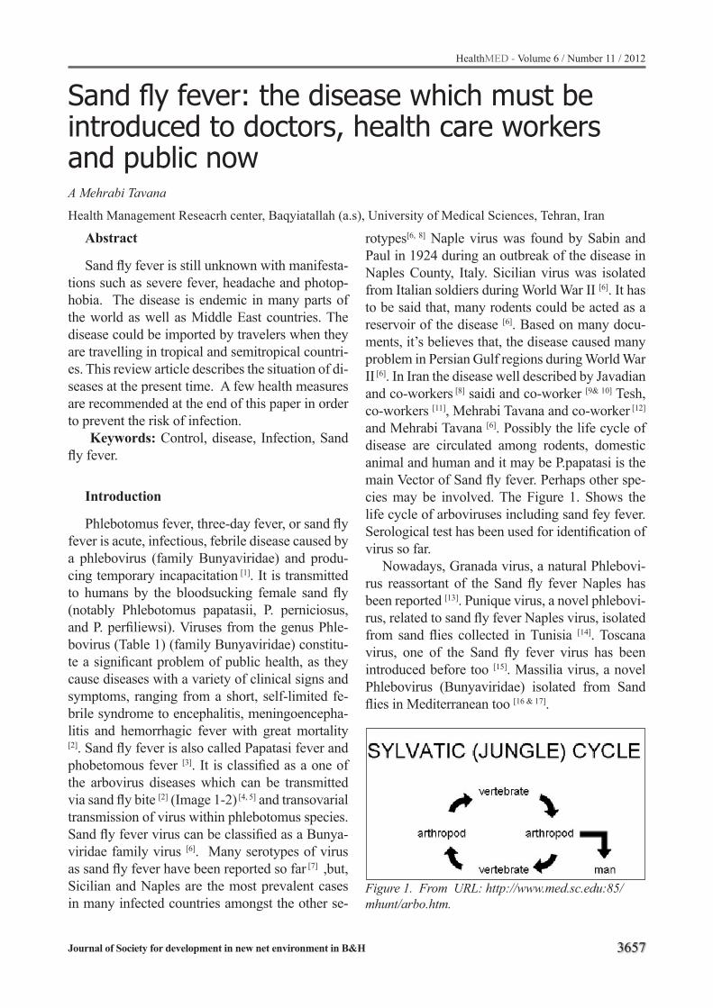
HealthMED - Volume 6 / Number 11 / 2012
Journal of Society for development in new net environment in B&H 3657
Sand fly fever: the disease which must be introduced to doctors, health care workers and public now A Mehrabi Tavana
Health Management Reseacrh center, Baqyiatallah (a.s), University of Medical Sciences, Tehran, Iran
Abstract
Sand fly fever is still unknown with manifesta-tions such as severe fever, headache and photop-hobia. The disease is endemic in many parts of the world as well as Middle East countries. The disease could be imported by travelers when they are travelling in tropical and semitropical countri-es. This review article describes the situation of di-seases at the present time. A few health measures are recommended at the end of this paper in order to prevent the risk of infection.
Keywords: Control, disease, Infection, Sand fly fever.
Introduction
Phlebotomus fever, three-day fever, or sand fly fever is acute, infectious, febrile disease caused by a phlebovirus (family Bunyaviridae) and produ-cing temporary incapacitation [1]. It is transmitted to humans by the bloodsucking female sand fly (notably Phlebotomus papatasii, P. perniciosus, and P. perfiliewsi). Viruses from the genus Phle-bovirus (Table 1) (family Bunyaviridae) constitu-te a significant problem of public health, as they cause diseases with a variety of clinical signs and symptoms, ranging from a short, self-limited fe-brile syndrome to encephalitis, meningoencepha-litis and hemorrhagic fever with great mortality [2]. Sand fly fever is also called Papatasi fever and phobetomous fever [3]. It is classified as a one of the arbovirus diseases which can be transmitted via sand fly bite [2] (Image 1-2) [4, 5] and transovarial transmission of virus within phlebotomus species. Sand fly fever virus can be classified as a Bunya-viridae family virus [6]. Many serotypes of virus as sand fly fever have been reported so far [7] ,but, Sicilian and Naples are the most prevalent cases in many infected countries amongst the other se-
rotypes[6, 8] Naple virus was found by Sabin and Paul in 1924 during an outbreak of the disease in Naples County, Italy. Sicilian virus was isolated from Italian soldiers during World War II [6]. It has to be said that, many rodents could be acted as a reservoir of the disease [6]. Based on many docu-ments, it’s believes that, the disease caused many problem in Persian Gulf regions during World War II [6]. In Iran the disease well described by Javadian and co-workers [8] saidi and co-worker [9& 10] Tesh, co-workers [11], Mehrabi Tavana and co-worker [12]
and Mehrabi Tavana [6]. Possibly the life cycle of disease are circulated among rodents, domestic animal and human and it may be P.papatasi is the main Vector of Sand fly fever. Perhaps other spe-cies may be involved. The Figure 1. Shows the life cycle of arboviruses including sand fey fever. Serological test has been used for identification of virus so far.
Nowadays, Granada virus, a natural Phlebovi-rus reassortant of the Sand fly fever Naples has been reported [13]. Punique virus, a novel phlebovi-rus, related to sand fly fever Naples virus, isolated from sand flies collected in Tunisia [14]. Toscana virus, one of the Sand fly fever virus has been introduced before too [15]. Massilia virus, a novel Phlebovirus (Bunyaviridae) isolated from Sand flies in Mediterranean too [16 & 17].
Figure 1. From URL: http://www.med.sc.edu:85/mhunt/arbo.htm.
3658
HealthMED - Volume 6 / Number 11 / 2012
Journal of Society for development in new net environment in B&H
Discussion
Still little is known regarding sand fly fever in particular it’s epidemiological clinical diagno-sis and control aspects of infection. It has to be said the disease causes problem in many part of the world in particular in tropical and semitropical regions including Europe, Asia, Africa and other part of the world [18]. Different serotype of viruses has been described in the last decades. However in spite of present of vector P.papatasi in different continents no report has been published yet from different part of world. It supposed the disease may be present in other continent too. Non immu-ne person such as travelers highly susceptible to get the infection [6]. It should be noted that the di-sease may be present in other part of the world such as Afghanistan [19] Middle East countries [13] because of same vector with coetaneous and Vis-ceral leishmaniasis present in the area of infection. In addition, sometime the disease could be seen with it’s complication similar the infection come from a Toscana virus one of the different serotype of sand fly fever which is present in Italy [20] . The disease is endemic in different part of the world [21] very recently the disease has been reported in different part of Europe [22]. Perhaps the infection may not completely clear during travel [23] or the clinical symptoms could not be seen all the time completely, however the IgG present in the se-rum of patient and volunteer blood donors too [24]. The patients with fever, Phetophobia and headac-he should be visited by their doctor. The disease more prevalent among travelers who are travelling to endemic area [25 -31].
It should be noted that the febrile illness may be identified by molecular techniques [16 & 29]. No vaccine available for preventing the infection, precaution measures including health education, personnel hygiene must be taken and repellents should be used overnight in endemic area for peo-ple who are living in tropical or semi tropical co-untries.
References
1. Butenko AM, Klimenko IS, Mosquito fevers. Classifi-cation of viruses, epidemiology, prevention, Med Pa-razitol (Mosk). 2010;(3):54-7.
2. Colacicco-Mayhugh MG, Masuoka PM, Grieco JP, Ecological niche model of Phlebotomus alexandri and P. papatasi (Diptera: Psychodidae) in the Middle East, Int J Health Geogr. 2010; 21;9:2.
3. Cross ER and Hyams KC. The potential effect of glo-bal warming on the geographic and seasonal distri-bution of Phlebotomus papatasi in southwest Asia. Environ Health Perspect, 1996; 104(7):724-7.
4. Cross ER, Newcomb WW, Tucker CJ. Use of weather data and remote sensing to predict the geographic and seasonal distribution of Phlebotomus papatasi in southwest Asia. American Journal of Tropical Medi-cine and Hygiene. 1996;54(5):530–536.
5. Tesh RB, Chaniotis BN Transovarial transmission of viruses by Phlebotomine sandflies. Ann N Y Acad Sci, 1975; 266:125-34.
6. Mehrabi Tavana A. The Seroepidemiological Studies of Sand fly fever in Iran During Imposed War Iranian J. Publ. Health, 2001; 30, Nos. 3-4, 145-146.
7. Gabriel M, Resch C, Günther S, Schmidt-Chanasit J, Toscana virus infection imported from Elba into Switzerland, Emerg Infect Dis. 2010;16(6):1034-6.
8. Javadian E, Tesh R, Saidi S, Nadim A. Studies on the epidemiology of sand fly fever in Iran. III. Host-feeding patterns of Phlebotomus papatasi in an en-demic area of the disease. Am J Trop Med Hyg. 1977;26:294–298.
9. Saidi S, Tesh R, Javadian E, Sahabi Z, Nadim A. Stu-dies on the epidemiology of sand fly fever in Iran. II: the Prevalence of human and animal infection with five phlebotomus fever virus serotypes in Isfahan pro-vince. Am Soc Trop Med Hyg, .1977; 26:288-93.
10. Tesh R, Saidi S, Javadian E, Nadim A. Studies on the epidemiology of sand fly fever in Iran. I. Virus isolates obtained from Phlebotomus. Am J Trop Med Hyg. 1977;26:282–287.
11. Tesh R, Saidi S, Javadian, E, Nadim A. and Seedi-Rashti M.A. The distribution and prevalence of hu-man infection with phlebotomus fever group viruses in Iran. Iranian J Publ Hlth, 1976;5(1) : 1-7
12. Mehrabi Tavana A, Javadian E, . The seroepidemi-ological studies of sand fly fever during the imposed war, 1980-88. Hakim Research Journal (In Persi-an) 1999; 1(2): 14-7.
13. Collao X, Palacios G, de Ory F, Sanbonmatsu S, Pérez-Ruiz M, Navarro JM, Molina R, Hutchison SK, Lipkin WI, Tenorio A, Sánchez-Seco MP Granada vi-rus: a natural phlebovirus reassortant of the sandfly fever Naples serocomplex with low seroprevalen-ce in humans. [Journal Article, Research Support, N.I.H., Extramural, Research Support, Non-U.S. Gov’t, Research Support, U.S. Gov’t, Non-P.H.S.] Am J Trop Med Hyg 2010 Oct; 83(4):760-5
HealthMED - Volume 6 / Number 11 / 2012
Journal of Society for development in new net environment in B&H 3659
14. Elyes Zhioua, Grégory Moureau, Ifhem Chelbi, Laetitia Ninove, Laurence Bichaud, Mohamed Der-bali, Mylène Champs, Saifeddine Cherni, Nicolas Salez, Shelley Cook, Xavier de Lamballerie, and Remi N. Charrel. . Punique virus, a novel phlebo-virus, related to sandfly fever Naples virus, isolated from sandflies collected in Tunisia. J Gen Virol May 2010;. 91; 5; 1275-1283.
15. Zehender G, Bernini F, Delogu M, Cusi MG, Rezza G, Galli M, Ciccozzi M Bayesian skyline plot infe-rence of the Toscana virus epidemic: a decline in the effective number of infections over the last 30 years. Infect Genet Evol, 2009;9(4):562-6.
16. Charrel RN, Izri A, Temmam S, Delaunay P, Toga I, Dumon H, Marty P, de Lamballerie X, Parola P. Co-circulation of 2 genotypes of Toscana virus, southe-astern France. Emerg Infect Dis. 2007; 13(3):465-8.
17. Charrel RN, Moureau G, Temmam S, et al, Massilia virus, a novel Phlebovirus (Bunyaviridae) isolated from sandflies in the Mediterranean, Vector Borne Zoonotic Dis. 2009;9(5):519-30.
18. Bryan JP, Iqbal M, Ksiazek TG, Ahmed A, Duncan JF, Awan B, Krieg RE, Riaz M, Leduc JW, Nabi S, Qureshi MS, Malik IA and Legters LJ Prevalence of sand fly fever, West Nile, Crimean-Congo hemorrha-gic fever, and leptospirosis antibodies in Pakistani military personnel. Mil Med, 1996; 161(3):149-53.
19. Myles, O, Wortmann, G, Two cases of visceral leis-hmaniasis in U.S. military personnel--Afghanistan, 2002-2004. MMWR Morb Mortal Wkly Rep. 2004; 2; 53(12):265-8.
20. Beersma MF, Grimbergen YA, Kroon FP, Veldkamp PJ. [Meningitis caused by Toscana virus during a summer stay in Italy] Ned Tijdschr Geneeskd. 2004; 7; 148(6):286-8.
21. Nicoletti L, Ciufolini MG, Fortuna C, Magurano F, Fiorentini C, Marchi A, Benedetti E, Bucci P. Arbo-viruses in Italy, 2008;50(1-2):109-11. Parassitolo-gia.
22. Depaquit J, Grandadam M, Fouque F, Andry1 PE, Peyrefitte5et C. Arthropod-borne viruses tran-smitted by Phlebotomine sandflies in Europe: a re-view, Euro Surveill. 2010 Mar 11;15(10):19507.
23. Hill DR. The burden of illness in international trave-lers. N Engl J Med 2006; 12; 354(2):115-7.
24. De Lamballerie X, Tolou H, Durand JP, Charrel RN. Prevalence of Toscana virus antibodies in and patients with central nervous system infections in southeastern France. Vector Borne Zoonotic Dis. 2007 Summer;7(2):275-7.
25. Brett-Major DM, Claborn DM, Sand fly fever: what have we learned in one hundred years? Mil Med. 2009 ;174(4):426-31.
26. Davis RF, Johnston GA, Sladden MJ, Recognition and management of common ectoparasitic diseases in travelers. Am J Clin Dermatol. 2009;10(1):1-8.
27. Mehrabi Tavana , A. Minireview on Sand Fly Fe-ver. Journal of Entomology,2007; 4: 401-403.DOI: 10.3923/je.2007.401.403.
28. Verani P, Ciufolini MG, Nicoletti L. Arbovirus surveillance in Italy. Parassitologia. 1995 ; 37(2-3):105-8.
29. Sanchez-Seco, M.P. Echevarria, J.M Hernandez, L. et al. Detection and identification of Toscana and other phleboviruses by RT-nested-PCR assays with degenerated primers, J Med Virol; 2003;, 71pp. 140–149.
30. Papa, A. Konstantinou, G. Pavlidou V.and Antonia-dis, A. Sandfly fever virus outbreak in Cyprus, Clin Microbiol Infect 2006, 12;. 192–194.
31. Niklasson B. and Eitrem, R. Sandfly fever among Swedish UN troops in Cyprus, Lancet 1;1985, 1212.
Corresponding AuthorA Mehrabi Tavana,Health Management Reseacrh center,Baqyiatallah,University of Medical Sciences,Tehran,Iran.E-mail: [email protected].
3660
HealthMED - Volume 6 / Number 11 / 2012
Journal of Society for development in new net environment in B&H
Is there an association between height of R wave or R/S ratio in ECG lead aVR and the level of consciousness in intubated comatose tricyclic antidepressant-poisoned patients?Behrooz Ghanbari1, Hossein Sanaei-Zadeh2, Farhad Shahmohammadi3, Nasim Zamani4, Babak Mostafazadeh5
1 Hazrat Rasoul Akram Hospital, Tehran University of Medical Sciences, Tehran, Iran2 Emergency Room/Division of Medical Toxicology, Hazrat Ali-Asghar (p) Hospital, Shiraz University of Medical Sciences, Shiraz, Iran3 Toxicology ICU, Family Hospital, AJA University of Medical Sciences, Tehran, Iran4 Department of Clinical Toxicology, Loghman-Hakim Hospital, Shahid Beheshti University of Medical Sciences, Tehran, Iran5 Department of Forensic Medicine and Toxicology, Loghman-Hakim Hospital, Shahid Beheshti University of Medical Sciences, Tehran, Iran
Abstract
Aim: This study was designed to evaluate the previously suggested association between the de-crease in the height of R wave and R/S ratio in lead aVR and increase in the level of consciou-sness according to Glasgow coma score (GCS) in intubated tricyclic antidepressant (TCA)-poisoned patients.
Methods and material: In this retrospective study, the medical charts of all survived patients diagnosed and treated for TCA toxicity, hospita-lized in Loghman-Hakim Poison Hospital in Te-hran, Iran between March 2009 and March 2010 were evaluated. The patients who had presented with deep coma (GCS≤8) and had been intubated before or after hospital presentation, had an initial 12-lead ECG performed after hospital presentati-on and another ECG after regaining of the con-sciousness (GCS≥10), and had positive urine or plasma screen test for TCAs were included. Pa-tients with multiple drug ingestion, head trauma, and underlying heart diseases and those on con-tinuous sedative infusion after intubation were excluded. Statistical analysis used: application of paired sample t test, logistic regression, K-stati-stic, and receiver operating characteristic (ROC) curve.
Results: A total of 22 patients met the inclusion criteria and were entered into the study. A statisti-cally significant difference was found between pa-rameters of the ECGs (in GCS≤8) on presentation
and the ECGs after regaining of the consciousness and extubation (in GCS≥10) in terms of ECG rate, QRS interval, QTc, T40-ms, frontal plane QRS axis and height of R wave and R/S ratio in lead aVR. Using forward logistic regression analysis, ECG rate and QRS duration were determined as the predictive parameters of GCS recovery from toxicity (P=0.003 and P=0.003, respectively). Op-timal sensitivity (80%) and specificity (69%) for the differentiation of these two limits of GCS (≤8 and ≥10) occurred at a QRS duration of 0.08 se-cond. Conclusions: The role of height of R wave or R/S ratio in prediction of increment of GCS le-vel can not be proposed without attention to other ECG parameters in TCA-poisoned patients.
Keywords: tricyclic antidepressants, overdose, height of R wave in lead aVR, R/S ratio, level of the consciousness, Glasgow coma score, electro-cardiography
Introduction
The most toxic effects of the tricyclic antide-pressants (TCAs) involve the central nervous and cardiovascular systems. Neurologic toxicity inclu-des altered level of consciousness, delirium and seizures. This toxicity is known to be the result of pharmacological effects such as the blockade of monoamine reuptake and anticholinergic acti-on.[1, 2] Cardiovascular toxicity includes changes of electrocardiographic (ECG) parameters and dysrhythmias.[3, 4] Tricyclic antidepressants pro-

HealthMED - Volume 6 / Number 11 / 2012
Journal of Society for development in new net environment in B&H 3661
long phases 0 and 2 of the action potential and also have alpha receptor blocking effects.[3, 5] It has been shown that ECG abnormalities, with the exception of the infrequent premature ven-tricular contractions and sinus tachycardia were associated with a severely depressed sensorium and disappeared with neurologic improvement.[6]
Additionally, it has been demonstrated that clini-cal improvement occurred both before and during changes of QRS duration and terminal 40-milli-second (T40-ms) frontal plane QRS axis.[1] This is while it has been suggested that the decrease in the height of R wave and R/S ratio in lead aVR may be related to the level of consciousness and be informative in predicting recovery from toxi-city following TCA overdose. [7] This study was designed to evaluate the association between the decrease in the height of R wave and R/S ratio in lead aVR and increase in the level of consciou-sness according to Glasgow coma score (GCS) in intubated TCA-poisoned patients.
Materials and methods
In this retrospective case series study, the me-dical charts of all survived patients diagnosed and treated for TCA toxicity, hospitalized in Loghman-Hakim Poison Hospital in Tehran, Iran betwe-en March 2009 and March 2010 were identified by the computerized discharge diagnosis (ICD-10 codes) and evaluated. The cases with all the following criteria were included: 1- The patients with a documented diagnosis of TCA poisoning; Diagnosis was based on a positive history of TCA ingestion and clinical manifestations in all patients with a positive urine or plasma drug screen test for TCAs. The positive history of TCA ingestion was defined as giving the name of the consumed medication by the patient him/herself before ente-ring the comatose state (to his/her relatives or the personnel of emergency medical service) or after complete recovery from coma and identifying and retaining any evidence of TCA pills (such as the bottle or the packet of the medication) in the bedroom or household, workplace, etc. 2-Tho-se who had presented with deep coma (GCS≤8) and had been intubated (with or without need for mechanical ventilation) before presentation to the hospital (by the personnel of emergency medical
service) or after that due to hypoventilation, loss of protective airway reflexes, seizures, and he-modynamically significant arrhythmias. The pati-ents with inadequate oxygenation and ventilation despite supplemental oxygen had mechanically been ventilated; and 3- The cases whose first 12-lead ECG performed after hospital presentation were present in their charts and also, had another 12-lead ECG after regaining of the consciousne-ss (GCS≥10). These last ECGs were selected and evaluated regarding their time and date record and the physician’s note in the patients’ charts. The ca-ses with multiple drug ingestion, head trauma, un-derlying heart diseases, and those on an infusion of sedatives after intubation were excluded from the study. The treatment modality consisted of routine supportive care and administration of sodi-um bicarbonate (NaHCO3). Hypertonic NaHCO3 had been given (1 mEq/kg) as an intravenous bo-lus and then, the patient was put on a continuous infusion on the condition that serum pH did not exceed 7.55. Information including age, gender, type of drug ingested (documented by urine and/or serum drug screen test, pill ID, or patient’s hi-story), and time interval between the initial ECGs and the ECGs taken after increasing the GCS to 10 were extracted and recorded in the standardized abstraction forms. [8] The data was double-extrac-ted. The standardized abstraction forms were au-dited by the project manager (head of the resear-chers) and the data extractors were trained before the start of the research project.
They were both medical doctors with adequate skills in obtaining data from the medical charts. [8] The ECG parameters were manually measured and characteristics including rate, PR interval, maximal limb-lead QRS duration, corrected QT interval (QTc), terminal 40-millisecond (T40-ms) frontal plane QRS axis, and the height of R wave and R/S ratio in the lead aVR were recorded. Electrocardiographic interpretation was supervi-sed by two cardiologists blind to the topic of the study. Statistical analysis was done using SPSS software (version 17, Chicago, Ill, USA), and application of paired sample t test, logistic regre-ssion, and receiver operating characteristic (ROC) curve. Power of paired sample t test for each ECG parameter was calculated. [9] An inter-rater relia-bility analysis (K-statistic) was done to determine
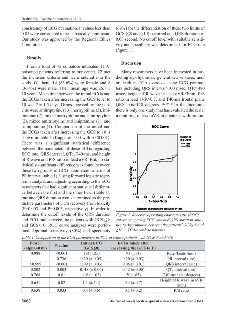
3662
HealthMED - Volume 6 / Number 11 / 2012
Journal of Society for development in new net environment in B&H
consistency of ECG evaluation. P values less than 0.05 were considered to be statistically significant. Our study was approved by the Regional Ethics Committee.
Results
From a total of 72 comatose intubated TCA-poisoned patients referring to our center, 22 met the inclusion criteria and were entered into the study. Of them, 14 (63.6%) were female and 8 (36.4%) were male. Their mean age was 26.7 ± 10 years. Mean time between the initial ECGs and the ECGs taken after increasing the GCS level to 10 was 2 ± 1.5 days. Drugs ingested by the pati-ents were amitriptyline (13), nortryptiline (3), imi-pramine (2), mixed nortryptiline and amitriptyline (2), mixed amitriptyline and imipramine (1), and trimipramine (1). Comparison of the initial and the ECGs taken after increasing the GCS to 10 is shown in table 1 (Kappa of 1.00 with p <0.001). There was a significant statistical difference between the parameters of these ECGs regarding ECG rate, QRS interval, QTc, T40-ms, and height of R wave and R/S ratio in lead aVR. But, no sta-tistically significant difference was found between these two groups of ECG parameters in terms of PR interval (table 1). Using forward logistic regre-ssion analysis and adjusting according to the ECG parameters that had significant statistical differen-ce between the first and the other ECG (table 1), rate and QRS duration were determined as the pre-dictive parameters of GCS recovery from toxicity (P=0.003 and P=0.003, respectively). In order to determine the cutoff levels of the QRS duration and ECG rate between the patients with GCS ≤ 8 and GCS≥10, ROC curve analyses were perfor-med. Optimal sensitivity (80%) and specificity
(69%) for the differentiation of these two limits of GCS (≤8 and ≥10) occurred at a QRS duration of 0.08 second. No cutoff level with suitable sensiti-vity and specificity was determined for ECG rate (figure 1).
Discussion
Many researchers have been interested in pre-dicting dysrhythmias, generalized seizures, and/or death in TCA overdose using ECG parame-ters including QRS interval>100 msec, QTc>480 msec, height of R wave in lead aVR>3mm, R/S ratio in lead aVR>0.7, and T40-ms frontal plane QRS axis>120 degrees. [1, 10-14] In the literature, there is only one study that has evaluated the serial monitoring of lead aVR in a patient with prolon-
Figure 1. Receiver operating characteristic (ROC) curves comparing ECG rate and QRS duration abili-ties to discriminate between the patients' GCS≤ 8 and ≥10 in TCA-overdose patients
Table 1. Comparison of the ECG parameters in TCA-overdose patients with GCS≤8 and ≥10Power
(alpha=0.05) P value Initial ECG (GCS≤8)
ECGs taken after increasing the GCS to 10
0.988 <0.001 114 (±25) 93 (±14) Rate (beats /min)- 0.736 0.20 (± 0.05) 0.20 (± 0.03) PR interval (sec)
>0.999 <0.001 0.09 (± 0.03) 0.06 (± 0.01) QRS interval (sec)0.802 0.003 0. 48 (± 0.06) 0.42 (± 0.06) QTc interval (sec)0.768 0.01 118 (±103) 50 (±85) T40-ms axis (degrees)
0.641 0.02 1.1 (± 1.4) 0.4 (± 0.7) Height of R wave in aVR (mm)
0.638 0.033 0.4 (± 0.6) 0.1 (± 0.2) R/S ratio
HealthMED - Volume 6 / Number 11 / 2012
Journal of Society for development in new net environment in B&H 3663
ged unconsciousness due to TCA overdose. This study showed that the changes of amplitude of R wave and R/S ratio in lead aVR tended to decline in proportion to improvements in GCS scores. [7] Our study shows that the height of R wave in lead aVR in our patients had increased after increasing GCS [1.1 (± 1.4) mm versus 0.4 (± 0.7) mm]. Also, R/S ratio had decreased from 0.4 (± 0.6) to 0.1 (± 0.2) second in these patients after increasing the level of GCS from less than 8 to more than 10 (table 1). Although these findings confirm the previous suggestions in this regard, [7] their role in prediction of increment of GCS level can not be proposed without attention to other ECG para-meters as in the present study, it has been shown that using logistic regression model R wave and R/S ratio in lead aVR could not be established as independent predictors of GCS recovery in TCA-poisoned patients, as well.
Regarding other ECG parameters and their association with the level of consciousness in the TCA-poisoned patients, only few studies exist in the literature. Olgun and colleagues showed that in amitryptiline poisoning in children, QRS dura-tion of 100 ms or longer predicted coma with a high positive predictive value. [15] However, despi-te their study, the mean QRS interval was 0.09 ± 0.03 and 0.06 ± 0.01 second in our patients with the GCS level of ≤ 8 and ≥10, respectively (table 1). This shows that QRS interval may be within normal limits in intubated, comatose, TCA-poi-soned patients. Also, the mean T40-ms axis was 118±103 degrees in the patients with GCS level of 8 or less and 50±85 degrees in those with GCS level of 10 or more. These findings are in accor-dance with the previous study conducted by Lie-belt et al [16] that had shown clinical improvement occurred both before and during QRS interval and T40-msec axis deviation changes (table 1).
Hypertonic NaHCO3 has been shown to have dramatic benefits in the treatment of TCA-poiso-ned patients and therefore, had been administered to all of our patients. [17-20] With this treatment mo-dality, the only ECG parameter that could discri-minate between the GCS level of less than 8 and more than 10 was QRS duration of ≤0.08 second (P<0.001) with a sensitivity of 80% and specifi-city of 69% (figure 1). In other words, in an in-tubated, comatose, TCA-poisoned patient- who is
not on sedative infusion and has received hyper-tonic NaHCO3- with initial QRS interval of more than 0.08 second, the patient definitely shows an increasing trend of GCS when this interval decre-ases to 0.08 second or less. However, it cannot be determined if the decrease in QRS interval is re-ally correlated to increase in the level of GCS or is due to the administration of hypertonic NaHCO3.
Like any other retrospective study, [8] there are some limitations in our study. Limitations of fin-ding charts of ICD code and chart reviews are of them. Additionally, errors including a false negati-ve or positive sample for inclusion or exclusion of our patients should be borne in mind which may be another limitation of our study.
Although previously suggested, the role of height of R wave or R/S ratio in prediction of increment of GCS level can not be proposed wit-hout attention to other ECG parameters in TCA-poisoned patients. The only ECG parameter that could discriminate between GCS level of less than 8 and more than 10 was QRS duration (sensitivity, 80%; specificity, 69%). It can then be concluded that in intubated, comatose, TCA-poisoned pa-tients, while QRS interval is being decreased to 0.08 second and less, the GCS level is definitely increasing.
Acknowledgements
We would like to thank Dr Seyed Javad Haj-Mir-Esmaeil and Dr Hashem Sezavar, the cardi-ologists in the Cardiology Department of Hazrat Rasoul Akram Hospital, Tehran, Iran for their kind cooperation in supervising the interpretation of the ECGs.
References1. Liebelt EL, Francis PD, Woolf AD. ECG lead aVR
versus QRS interval in predicting seizures and arrhythmias in acute tricyclic antidepressant toxicity. Ann Emerg Med 1995;26:195-201.
2. Güloglu C, Orak M, Ustündag M, Altunci YA. Analysis of amitriptyline overdose in emergency medicine. Emerg Med J 2011;28:296-9.
3. Glassman AH. Cardiovascular effects of tricyclic an-tidepressants. Annu Rev Med 1984;35:503-11.
4. Niemann JT, Bessen HA, Rothstein RJ, Laks MM. Electrocardiographic criteria for tricyclic antidepres-sant cardiotoxicity. Am J Cardiol 1986;57:1154-9.

3664
HealthMED - Volume 6 / Number 11 / 2012
Journal of Society for development in new net environment in B&H
5. Frommer DA, Kulig KW, Marx JA, Rumack B. Tricyclic antidepressant overdose. A review. JAMA 1987;257:521-6.
6. Fasoli RA, Glauser FL. Cardiac arrhythmias and ECG abnormalities in tricyclic antidepressant over-dose. Clin Toxicol 1981;18:155-163.
7. Choi KH, Lee KU. Serial Monitoring of Lead aVR in Patients with Prolonged Unconsciousness Following Tricyclic Antidepressant Overdose. Psychiatry Inve-stig 2008;5:247-50.
8. Gilbert EH, Lowenstein SR, Koziol-McLain J, Barta DC, Steiner J. Chart reviews in emergency medicine research: Where are the methods? Ann Emerg Med 1996;27:305-8.
9. Statistical power calculators; retrieved from: http://power.phs.wfubmc.edu/index.cfm?calc=pm
10. Boehnert MT, Lovejoy FH Jr. Value of the QRS du-ration versus the serum drug level in predicting se-izures and ventricular arrhythmias after an acute overdose of tricyclic antidepressants. N Engl J Med 1985;313:474-9.
11. Caravati EM, Bossart PJ. Demographic and electrocardiographic factors associated with severe tricyclic antidepressant toxicity. J Toxicol Clin Toxi-col 1991;29:31-43.
12. Harrigan RA, Brady WJ. ECG abnormalities in tricyclic antidepressant ingestion. Am J Emerg Med 1999;17:387-93.
13. Singh N, Singh HK, Khan IA. Serial electrocardi-ographic changes as a predictor of cardiovascular toxicity in acute tricyclic antidepressant overdose. Am J Ther 2002;9:75-9.
14. Bailey B, Buckley NA, Amre DK. A meta-analysis of prognostic indicators to predict seizures, arrhythmias or death after tricyclic antidepressant overdose. J Toxicol Clin Toxicol 2004; 42:877-88.
15. Olgun H, Yildirim ZK, Karacan M, Ceviz N. Clini-cal, electrocardiographic, and laboratory findings in children with amitriptyline intoxication. Pediatr Emerg Care 2009;25:170-3.
16. Liebelt EL, Ulrich A, Francis PD, Woolf A. Serial electrocardiogram changes in acute tricyclic antide-pressant overdoses. Crit Care Med 1997;25:1721-26.
17. Bradberry SM, Thanacoody HK, Watt BE, Tho-mas SH, Vale JA. Management of the cardiovas-cular complications of tricyclic antidepressant po-isoning: role of sodium bicarbonate. Toxicol Rev 2005;24:195-204.
18. Chan CY, Waring WS. Images in cardiovascular me-dicine. Tricyclic cardiotoxicity treated with sodium bicarbonate. Circulation 2007;115:e63-4.
19. Thanacoody HK, Waring WS. Toxins that affect the cardiovascular system. Clin Med 2008;8:92-5.
20. Sanaei-Zadeh H, Ghassemi Toussi A. Resolution of wide complex tachycardia after administration of hypertonic sodium bicarbonate in a patient with se-vere tricyclic antidepressant poisoning. Resuscitati-on 2011;82:792-3.
Corresponding AuthorHossein Sanaei-Zadeh, Emergency Room/Division of Medical Toxicology, Hazrat Ali-Asghar (p) Hospital, Shiraz University of Medical Sciences, Shiraz, Iran E-mail: [email protected]
HealthMED - Volume 6 / Number 11 / 2012
Journal of Society for development in new net environment in B&H 3665
Functional endoscopic sinus surgery: indications and complicationsSara Ehteshami1, Seyyed Abbas Hashemi2, Seyyed Abdollah Madani3
1 Education development center, Mazandaran University of medical sciences, Sari, Iran2 Faculty of medicine, student research committee, Mazandaran University of medical sciences, Sari, Iran3 Department of Otorhinolaryngology, Head and Neck Surgery, Mazandaran university of medical sciences, Sari, Iran
Abstract
During past decades, surgical intervention for sinus diseases involved external procedures, often requiring an incision on the face. Since the early development of functional endoscopic sinus sur-gery (FESS), this minimally invasive procedure has gained increasing popularity among otorhino-laryngologists. Many studies have showed symp-tomatic improvement of sinus disorders by FESS in 76% to 87.5% of patients after the surgery. But there were different reports about the complicati-ons of this technique. In this article we reviewed the previous published articles in this relation and summarized the most important complications.
Keywords: Functional Endoscopic Sinus Sur-gery, FESS, Indications, Complications
Introduction
Prior to 1985, surgical intervention for sinus disorders included external procedures, often needing an incision on the face. To approach the maxillary sinus, a Caldwell–Luc incision (made in the gingivobuccal space) was used to make a window in the anterior wall of the maxillary sinus. A Lynch incision was made on the face just me-dial to the medial brow to access the ethmoid and frontal sinuses. More major frontal sinus disorders often included a bicoronal incision, which exten-ded across the scalp in the coronal plane from ear to ear. These external procedures were unsightly and invasive, and therefore, they were a last-resort approach to recalcitrant sinus disorders (1).
Since the early development of functional en-doscopic sinus surgery (FESS), this minimally invasive technique has obtained increasing po-pularity among otorhinolaryngologists (2,3). The main limiting factors for endoscopic procedures to
paranasal sinuses and the skull base are its com-plex anatomy and the high vascularity (2,3). Du-ring past years fess has been used for treatment of paranasal sinus diseases (4-6). Many investigati-ons have reported symptomatic and radiographic achievement of sinus disorders by FESS in 76% to 87.5% of patients observed 1 to 3 years after the surgery (6,7,8). Even recent researches reve-aled asthma improvement after FESS in children (9,10,11,12,13).
Indications for sinus surgery FESS is so named because it is designed to re-
store physiologic sinus ventilation and drainage. FESS and endoscopic operation were undertaken for a wide range of approaches. These involved FESS with middle meatal antrostomies and,or sphenoidotomy, and,or frontal recess surgery. The-se procedures were established for nasal polyposis, fungal rhinosinusitis, mucoceles and peri-orbital abscesses. Endoscopic operation was also appli-ed for limited inverted papilloma extending into the ethmoids,medial antrum,sphenoid with lateral rhinotomy for more extensive diseases.
Lateral rhinotomy was performed for extensi-ve anterior septal papilloma with split skin graf-ting. Endoscopic sphenopalatine artery ligation for refractory epistaxis and endoscopic dacro-cystorhinostomy. Septorhinoplasty to involve augmentation, reduction, revision and functional cases plus external procedure for septal perfora-tion repair using auricular cartilage interposition grafting. Exclusions would be major sinus mali-gnancy referred to regional centre (Oxford) for craniofacial resection or maxillectomy, juvenile angiofibroma, etc (14). Benign sinonasal tumors and limited malignancies are commonly remo-ved endoscopically. For instance, juvenile nasop-haryngeal angiomas and inverting papillomas are

3666
HealthMED - Volume 6 / Number 11 / 2012
Journal of Society for development in new net environment in B&H
removed safely with the same or lower recurren-ce and complication rates as external procedures (15,16). Repairing the skull base, the orbit, and the lacrimal duct. Cerebrospinal fluid leaks can be repaired endoscopically since long-term improve-ment rates are reported at greater than 90% (17). Orbital decompressions for Graves’ ophthalmo-pathy and Repairing of the nasolacrimal duct (18). It provided direct visualization of the location and severity of nasolacrimal duct pathology.
Although most complications from ethmoid and frontal sinusitis could be managed endosco-pically, many situations benefit from a combined methods (19,20). Bleeding presents a formidable obstacle in some of these active infections; in these patients, an external method is better. Patients with intracranial abscess needs neurosurgical interven-tion, an external approach to the frontal sinus is important. Due to large size and lateral location, some mucoceles located in the frontal sinus might need a bicoronal procedure for definitive therapy. In his regard, Major complications are blindness, intracranial injury, orbital hematoma, stroke, and cerebrospinal fluid leak.
These rates can be reduced by high tech optics. Another technological advance used in FESS is image guidance approaches. These systems help the surgeon by providing real-time intra-operative anatomical location (1).
Absolute indications for FESS in pediatrics include the following (21):
1. Complete nasal obstruction in Cystic fibrosis caused by massive polyposis or caused by medialization of the lateral nasal wall;
2. Orbital abscess;3. Intracranial complications;4. Antrochoanal polyp;5. Mucocoeles or mucopyocoeles; 6. Fungal rhinosinusitis.
Possible indications involve chronic rhinosinu-sitis with frequent exacerbations persisting despite optimal medical treatment and after exclusion of any systemic disorders. FESS should be followed by medical therapy to control mucosal inflamma-tion, or symptoms will return without change (22). This is nearly right for surgical polypectomy; polyps usually reaccumulate within a few years without medical maintenance treatment (22,23).
FESS is indicated in cases of Allergic fungal rhino-sinusitis to (1) bring back sinus ostial patency and ventilation, (2) confirm the diagnosis, and (3) re-move inspissated allergic mucin. Another indicati-on for surgery is bony erosion, given that it signi-fies extension of disease beyond the sinus spaces.
List of complications
Hemorrhagic complicationsThere are several reports about the hemorrha-
ge or bleeding during FESS (primary or secon-dary) (24-33). Venkatachalam et al (24) found 12% bleeding in patients underwent the FESS in their study. Severe bleeding or bleeding needing packing or readmission was noted by three rese-arches (34-36). Blood transfusion was observed in five investigations (34, 36-39). These studies (25,28,29,32,35,40,41,42) reported eye ecchymo-ses , periorbital bruising, periorbital ecchymoses and orbital hematomas in the patients underwent FESS. But in The randomized trial which was done by Venkatachalam et al (24) noted no orbital hematomas in the FESS group.
Sphenopalatine hemorrhage was reported by Harkness et al (25) with the rate of 0.09%.Five researches revealed epistaxis in their evaluation (40,38,39,43,44) and ranged from 0 to 2.4% with a median of 0.6%.These studies (26,27,43,44) no-ted preseptal hemorrhage ,palpebral hematoma , septal hematoma, which ranged from 0.2 to 2%.
Infection complicationsWound infection, meningitis, orbital cellulitis
were reported in three studies (24,45,46).H Mehrzad et al (14) showed in their study ten
patients had post FESS infection or facial swelling from 461 procedures over the 5-year period. No major complications like CSF rhinorrhea, orbital damage or meningitis were observed. This was comparable to a reported rate of 1.41% (47) from an investigation undertaken by The Royal Colle-ge of Surgeons of England in 1994, and a Dutch study (48) on endoscopic sinus surgery noting ra-tes of 4.2% for minor complications and 0.3% for serious complications. A French study (49) revea-led rates of 2.2% for major and 13.4% for minor complications.
HealthMED - Volume 6 / Number 11 / 2012
Journal of Society for development in new net environment in B&H 3667
Intranasal complicationsMiddle meatal stenosis,closure,partial closu-
re, Middle turbinate lateralization was revealed in different studies (25,26,35,44,50).Wigand et al (45) observed Obstruction of the lacrimal duct in 0.5% of 600 patients in their study .Venkatachalam et al (24) reported Lamina papyracea,perforation of lamina papyracea in 4% of the patients and 20% synechiae formation. Loss of olfactory sen-sation was discovered in three percent of patients underwent FESS by Wigand et al (45).
Orbital and ocular complicationsPeriorbital, orbital fat exposure was reported in
several studies (25,26,28,37, ,32,42,38).Harkness et al (25) noted orbital emphysema in 0.09% of pa-tients underwent FESS . Diplopia was observed in five investigations (27,30,37,38,44) ranged betwe-en 0 and 1.3% . Two studies by Jianget al (38) and Park et al (30) showed nasolacriminal duct injury. One investigation reported orbital penetration (34) and epiphora was noted in three researches (29, 37, 44). The orbital complications of FESS can be divided into minor and major group. Minor com-plications include; periorbital ecchymosis, orbital emphysema, transient diplopia, edema and forma-tion of lipogranuloma (51).
Major complications include; Extra-ocular muscle injury, persistent diplopia, nasolacrimal duct injury, orbital hemorrhage or hematoma, orbital foreign body, optic nerve injury, blindne-ss, subperiosteal abscess, abscess of the orbital tissue, orbital cellulitis, cavernous sinus throm-bosis, enophthalmos, injuries to the vascular and nervous structures of the orbit (52,53) and orbital emphysema leading to blindness (54). Open glo-be injury to the eye during endoscopic surgery, probably secondary to perforation of the lamina papyracea, and entrance into the orbit and the glo-be by the electrocautery tip has been noted by Ca-stellarin et al (55).
Pharyngeal and mouth complicationsHypesthesis and numbness of teeth or lip or
cheek was reported 3% in the FESS group by Penttilä et al (56) and 0.3% in Weber et al (36) study.
Intracranial complicationsTwelve researches (37,27,45,29,30,43,41,44,4
2,46,38,36) noted CFS leak in their examination. Jiang et al (38) reported dural exposure in 0.2% of procedures where applied. Damage to internal carotid artery was observed in 0.3% of the patients reported by Weber et al (36).
Systemic complicationsMild bronchospasm, cardiac ischemia and
cardiac arrest was reported in the four studies (40,57,44,30)
Other complicationsPostoperative headache (58), cheek pain, ten-
derness (56), pain (38) an unremoved nasal pack or sponge (34), cheek edema (25), atrophic rhini-tis (38) and soft tissue infiltration (34) were other complications which were reported.
Outcomes of sinus surgery The outcomes of FESS have been reported
in some researches (59,60).One of the best rese-arches which reported the outcomes of 120 con-secutive patients with a mean follow-up of 18 months was done by Kennedy DW (59). Nearly all subjects (98%) reported improvement in the-ir CRS symptoms at the time of final follow-up visit (85%, 13%, and 2% were markedly, mildly, and not improved, respectively). But, 45% of the sinus cavities undergoing surgeries were endosco-pically abnormal at the end of the investigation. The phenotype of chronic rhinosinusitis seemed to affect the operation outcome because patients with advanced polypoid changes preoperatively had a much higher rate of recurrence of disease and relapse after operation. In a subsequent sur-vey of 72 patients from the original cohort with an average follow-up of 7.8 years postoperatively, 98% of the patients reported sustained subjective improvement (60).
There is some evidences that medical treatment cause improved long-term results comparable with those originated from FESS(61) also repor-ted that the combination of FESS, careful posto-perative care, and medical treatment will result in improvement in favorable long-term impacts on both CRS and asthma (62). A systematic review by Dalziel K et al (63) showed that most part of

3668
HealthMED - Volume 6 / Number 11 / 2012
Journal of Society for development in new net environment in B&H
studies reported that symptoms improve following FESS with relatively few complications.
Acknowledgment
The authors would like to thank Mr. Seyyed Hamed Hashemi for his scientific editorial assi-stance in writing the manuscript.
References
1. Luong A , F. Marple B. Sinus Surgery;Indications and Techniques. Clinical Reviews in Allergy & Immuno-logy.2006(30),217-222
2. Stammberger H, Kopp W. Functional Endoscopic Sinus Surgery: The Messerklinger Technique. Phila-delphia, PA: BC Decker; 1991.
3. Stammberger H, Posawetz W. Functional endoscopic sinus surgery: concepts, indications, and results of the Messerklinger technique. Eur Arch Otorhinolaryngol. 1990;247(2):63-76.
4. Vining EM, Kennedy DW. Surgical management in adults. Immunol Allergy Clin North Am. 1994;14:97-111.
5. Bucca C, Rolla G, Scappaticci E, et al. Extrathoracic and intrathoracic airway responsiveness in sinusitis. J Allergy Clin Immunol. 1995;95(pt 1):52-59.
6. Kennedy DW. Prognostic factors, outcomes, and staging in ethmoid sinus surgery. Laryngoscope. 1992;102(suppl 57):1-18.
7. Rice DH. Endoscopic sinus surgery: results at 2-year follow-up. Otolaryngol Head Neck Surg. 1989;101:476-479.
8. Levine HL. Functional endoscopic sinus surgery: evaluation, surgery, and follow- up of 250 patients. Laryngoscope. 1990;100:79-84.
9. Lund VJ, Holmstrom M, Scadding GK. Functional en-doscopic sinus surgery in the management of chronic rhinosinusitis: an objective assessment. J Laryngol Otol. 1991;105:832-835.
10. Lazar R, Younis R, Gross C. Pediatric functional endonasal sinus surgery: review of 210 cases. Head Neck. 1992;14:92-98.
11. Manning SC, Wasserman RL, Silver R, Phillips DL. Results of endoscopic sinus surgery in pediatric pa-tients with chronic sinusitis and asthma. Arch Oto-laryngol Head Neck Surg. 1994;120:1142-1145.
12. Parsons D, Phillips D. Functional endoscopic sur-gery in children: a retrospective analysis of results. Laryngoscope. 1993;103:889-903.
13. Pransky S. Surgical outcomes in preschool children undergoing functional endoscopic sinus surgery. Pa-per presented at: Annual Meeting of the American Society of Pediatric Otolaryngology; April 1993; Los Angeles, Calif.
14. H Mehrzad, M Irvine, S Kundu, N Bleach. A 5-Year Audit of Rhinology Procedures Carried Out in a Dis-trict General Hospital. Ann R Coll Surg Engl. 2007 November; 89(8): 804–807.
15. Pryor, S.G., Moore, E.J., and Kasperbauer, J. (2005), Laryngoscope 115(7), 1201–1212.
16. Han, J.K., Smith, T.L., Loehrl, T., et al (2001), Laryn-goscope 111(8), 1395–1400.
17. McMains, K.C., Gross, C.W., and Kountakis, S.E. (2004), Laryngoscope 114(10),1833–1837.
18. Vaseghi, M., Tarin, T.T., Levin, P.S., and Terris, D.J. (2003), Ann Otol Rhinol Laryngol 112(1), 57–62.
19. Deutsch, E., Hevron, I., and Eilon, A. (2000), Rhino-logy 38(4), 177–180.
20. Bhargava, D., Sankhla, D., Ganesan, A., and Chand, P. (2001), Rhinology 39(3). 151–155.
21. Fokkens W, Lund V, Mullol J, European Position Paper on Rhinosinusitis and Nasal Polyps group. European position paper on rhinosinusitis and nasal polyps 2007. Rhinol Suppl 2007;20:1-136.
22. Gosepath J, Pogodsky T, Mann WJ. Characteristics of recurrent chronic rhinosinusitis after previous surgical therapy. Acta Otolaryngol 2008;128:778-84.
23. Li Y, Zhang GH, Liu X, Ye J, Zeng ZP. Clinical pro-gnostic factors of chronic rhinosinusitis after endos-copic sinus surgery. ORL J Otorhinolaryngol Relat Spec 2008;70:113-7.
24. Venkatachalam VP, Bhat A. Comparative evaluation of endoscopic and conventional surgical techniques in the management of nasal polyposis. JK Pract 1998;5:295–9.
25. Harkness P, Brown P, Fowler S, Topham J. A natio-nal audit of sinus surgery. Results of the Royal Colle-ge of Surgeons of England comparative audit of ENT surgery. Clin Otolaryngol 1997;22:147–51.
26. Friedman M, Landsberg R, Schults RA, Tanyeri H, Caldarelli DD. Frontal sinus surgery: endosco-pic technique and preliminary results. Am J Rhinol 2000;14:393–403.
27. 27. Friedman M, Landsberg R, Tanyeri H. Middle turbinate medialization and preservation in endos-copic sinus surgery. Otolaryngol Head Neck Surg 2000;123(1 Pt 1):76–80.
28. Jacobs JB. 100 years of frontal sinus surgery. Laryn-goscope 1997;107(11 Pt 2):1–36.
HealthMED - Volume 6 / Number 11 / 2012
Journal of Society for development in new net environment in B&H 3669
29. Lawson W. The intranasal ethmoidectomy – an experience with 1,077 procedures. Laryngoscope 1991;101:367–71.
30. Park AH, Lau J, Stankiewicz J, Chow J . The role of functional endoscopic sinus surgery in asthmatic patients. J Otolaryngol 1998;27:275–80.
31. Danielsen A, Olofsson J. Endoscopic endonasal si-nus surgery. A long-term follow-up study. Acta Oto-laryngol 1996;116(4):611–19.
32. Jakobsen J, Svendstrup F. Functional endoscopic sinus surgery in chronic sinusitis – a series of 237 consecutively operated patients. Acta Otolaryngo Suppl 2000;543:158–61.
33. Venkatachalam VP, Bhat A. Functional endoscopic sinus surgery – a newer surgical concept in the ma-nagement of chronic sinusitis. Indian J Otolaryngol Head Neck Surg 1999;52:13–16.
34. Stammberger H, Posawetz W. Functional endosco-pic sinus surgery. Concept, indications and results of the Messerklinger technique. Eur Arch Otorhino-laryngol 1990;247:63–76.
35. Levine HL. Functional endoscopic sinus surgery: evaluation, surgery, and follow-up of 250 patients. Laryngoscope 1990;100:79–84.
36. Weber R, Draf W, Keerl R, Schick B, Saha A. Endo-nasal microendoscopic pansinusoperation in chro-nic sinusitis. II. Results and complications. Am J Otolaryngol 1997;18:247–53.
37. Davis WE, Templer JW, Lamear WR, Davis WE, Craig SB. Middle meatus anstrostomy: patency ra-tes and risk factors. Otolaryngol Head Neck Surg 1991;104:467–72.
38. Jiang RS, Hsu CY. Endoscopic sinus surgery for the treatment of chronic sinusitis in geriatric patients. Ear Nose Throat J 2001;80:230–2.
39. Klossek JM, Peloquin L, Friedman WH, Ferrier JC, Fontanel JP. Diffuse nasal polyposis: postope-rative long-term results after endoscopic sinus sur-gery and frontal irrigation. Otolaryngol Head Neck Surg1997;117:355–61.
40. Katsantonis GP, Friedman WH, Bruns M. Intranasal sphenoethmoidectomy: an evolution of technique. Otolaryngol Head Neck Surg 1994; 111:781–6.
41. Lund VJ, MacKay IS. Outcome assessment of endos-copic sinus surgery. J R Soc Med 1994; 87:70–2.
42. Wolf G, Greistorfer K, Jebeles JA. The endoscopic endonasal surgical technique in the treatment of chronic recurring sinusitis in children. Rhinology 1995;33:97–103.
43. Schaefer SD. An anatomic approach to endoscopic intranasal ethmoidectomy. Laryngoscope 1998; 108(11 Pt 1):1628–34.
44. Massegur H, Adema JM, Lluansi J, Fabra JM, Montserrat JM. Endoscopic sinus surgery in sinusi-tis. Rhinology 1995;33:89–92.
45. Wigand ME, Hosemann W. Microsurgical treatment of recurrent nasal polyposis. Rhinol Suppl 1989; 825–9.
46. Sobol SE, Wright ED, Frenkiel S. One-year outcome analysis of functional endoscopic sinus surgery for chronic sinusitis. J Otolaryngol 1998;27:252–7.
47. Harkness P, Brown P, Fowler S, Topham J. A na-tional audit of sinus surgery. Results of The Royal College of Surgeons of England comparative audit of ENT surgery. Clin Otolaryngol. 1997;22:147–51.
48. Vleming M, Middelweed RJ, de Vries N. Complica-tions of endoscopic sinus surgery. Arch Otolaryngol Head Neck Surg. 1992;118:617–23.
49. Castillo L, Verschuur HP, Poissonnet G, Vaille G, Santini J. Complications of endoscopically guided sinus surgery. Rhinology. 1996;34:215–8.
50. Nishioka GJ, Cook PR, Davis WE, McKinsey JP. Immunotherapy in patients undergoing functional endoscopic sinus surgery. Otolaryngol Head Neck Surg 1994;110:406–12.
51. Rosner M, Kurtz S, Shelah M, Rosen N. Orbital li-pogranuloma after sinus surgery. Eur J Ophthalmol 2000. Apr-Jun;10(2):183-186.
52. Dutton JJ. Orbital complications of paranasal sinus surgery. Ophthal Plast Reconstr Surg 1986;2(3):119-127. doi: 10.1097/00002341-198601060-00001.
53. Buus DR, Tse DT, Farris BK. Ophthalmic com-plications of sinus surgery. Ophthalmology 1990. May;97(5):612-619.
54. Rubinstein A, Riddell CE, Akram I, Ahmado A, Be-njamin L. Orbital emphysema leading to blindness following routine functional endoscopic sinus sur-gery. Arch Ophthalmol 2005. Oct;123(10):1452. doi: 10.1001/archopht.123.10.1452.
55. Castellarin A, Lipskey S, Sternberg P., Jr Iatroge-nic open globe eye injury following sinus surgery. Am J Ophthalmol 2004. Jan;137(1):175-176. doi: 10.1016/S0002-9394(03)00715-3.
56. Penttilä M, Rautiainen M, Pukander J, Kataja M. Functional vs. radical maxillary surgery. Failures after functional endoscopic sinus surgery. Acta Oto-laryngol Suppl 1997;(529):173–6.
57. Kennedy DW. Prognostic factors, outcomes and staging in ethmoid sinus surgery. Laryngoscope 1992;102(12 Pt 2 Suppl 57):1–18.
58. Jankowski R, Pigret D, Decroocq F. Comparison of functional results after ethmoidectomy and nasali-zation for diffuse and severe nasal polyposis. Acta Otolaryngol 1997;117:601–8.
3670
HealthMED - Volume 6 / Number 11 / 2012
Journal of Society for development in new net environment in B&H
59. Kennedy DW. Prognostic factors, outcomes and staging in ethmoid sinus surgery. Laryngoscope 1992;102(suppl 57):1-18.
60. Senior BA, Kennedy DW, Tanabodee J, Kroger H, Hassab M, Lanza D. Long-term results of func-tional endoscopic sinus surgery. Laryngoscope 1998;108:151-7.
61. Ragab SM, Lund VJ, Scadding G. Evaluation of the medical and surgical treatment of chronic rhinosi-nusitis: a prospective, randomised, controlled trial. Laryngoscope 2004;114:923-30.
62. Senior BA, Kennedy DW, Tanabodee J, Kroger H, Hassab M, Lanza DC. Longterm impact of functio-nal endoscopic sinus surgery on asthma. Otolaryn-gol Head Neck Surg 1999;121:66-8.
63. Dalziel K, Stein K, Round A, Garside R, Royle P. Systematic review of endoscopic sinus surgery for nasal polyps. Health Technol Assess 2003; 7(17).
Corresponding Author Seyyed Abbas Hashemi,Department of Otorhinolaryngology, Head and Neck Surgery, Mazandaran university of medical sciences, Sari, Iran.E-mail: [email protected]
HealthMED - Volume 6 / Number 11 / 2012
Journal of Society for development in new net environment in B&H 3671
Recurrent cerebral ischemic infarction in a patient with paroxysmal nocturnal hemoglobinuriaGaoping Lin1, Wanzhuo Xie2, Xiaobiao Lai3
1 Department of neurology, Zhejiang provincial people’s hospital, Hangzhou, China 2 Bone Marrow Transplantation Center, The First Affiliated Hospital, Zhejiang University School of Medicine, Hangzhou, China 3 Department of neurology, Zhejiang provincial people’s hospital, Hangzhou, China
Abstract
Paroxysmal nocturnal hemoglobinuria (PNH) is a rare, acquired stem cell disorder, leading to a deficient biosynthesis of GPI-linked proteins in hematopoetic cells. We report a 50-year-old fema-le patient who presented with intravascular hemo-lysis, cytopenias, and recurrent thrombosis. This case documents a rare neurologic presentation of PNH with recurrent ischemic cerebral infarction.
Keywords: paroxysmal nocturnal hemoglobi-nuria, stem cell disorder, infarction.
Introduction
Paroxysmal nocturnal hemoglobinuria (PNH) is a rare, acquired stem cell disorder characterized by the triad of intravascular hemolysis, venous thrombosis and cytopenias (1). Mutations of the PIG-A gene lead to the absence of glycosylphos-phatidylinositol (GPI) anchor. As a result, PNH cells are deficient in all GPI-linked proteins inclu-ding complement regulatory proteins such as CD55 and CD59(2,3), and there for vulnerable to complement mediated lysis and thrombosis (4). Recurrent cerebral ischemic infarction caused by paroxysmal nocturnal hemoglobinuria has seldom been reported. In this study, we describe a pati-ent with PNH who developed recurrent arterial is-chemic stroke and possible cerebral venous sinus thrombosis.
Case presentation
On June 2009, a 50-year-old female patient was admitted to another hospital with progressive fatigue and jaundice. A routine blood test revealed anemia (hemoglobin 6.4g/dl, reticulocyte 0.15)
and leucopenia (1.6×10⁹/L), while the platelet co-unt was normal (100×109/L). Blood biochemistry results indicated total bilirubin of 31.3μmol/L, direct bilirubin of 22.8μmol/L, lactate dehydro-genase of 1457U/L. Routine urine test indicated absence of urobilinogen. The plasma hemoglobin level was 24mg/dl (0―10mg/dl). A bone marrow biopsy specimen showed erythroid hyperplasia. The direct and indirect Coombs tests were nega-tive. Flow cytometry established the diagnosis of PNH with deficiency of CD55 and CD59 in eryt-hrocytes and granulocytes. She was treated with iron supplementation and dexamethasone.
On the 10th hospital day she developed mild monoplegia of the right upper extremity (4/5 strength on manual muscle test). Magnetic reso-nance imaging (MRI) revealed acute infarct of the left centrum semiovale, and antiplatelet treatment with aspirin was added.
On July 22 2009, slurred speech and memory decline were observed, and new infarcts of left he-misphere were found on MRI (figure 1). The anti-platelet drug was changed to clopidogrel, and the fluency of speech improved gradually.
On August 11, 2009, the patient developed right hemiparesis and global aphasia. MRI reve-aled infarcts larger than they were 20 days before (figure 2). Treatment with clopidogrel continued.
She was admitted to our hospital with weakne-ss of left extremities on April 30 2010. Diffusion-weighted imaging showed acute infarcts of the frontal and parietal lobes of the right hemisphere (figure 3). Coagulation test showed normal prot-hrombin time (10.3 s), D-dimer (67 ng/ml) and fibrinogen (2.92 g/L). Lower leg ultrasonography excluded deep venous thrombosis. Transesopha-geal cardiac ultrasonography revealed no signs of
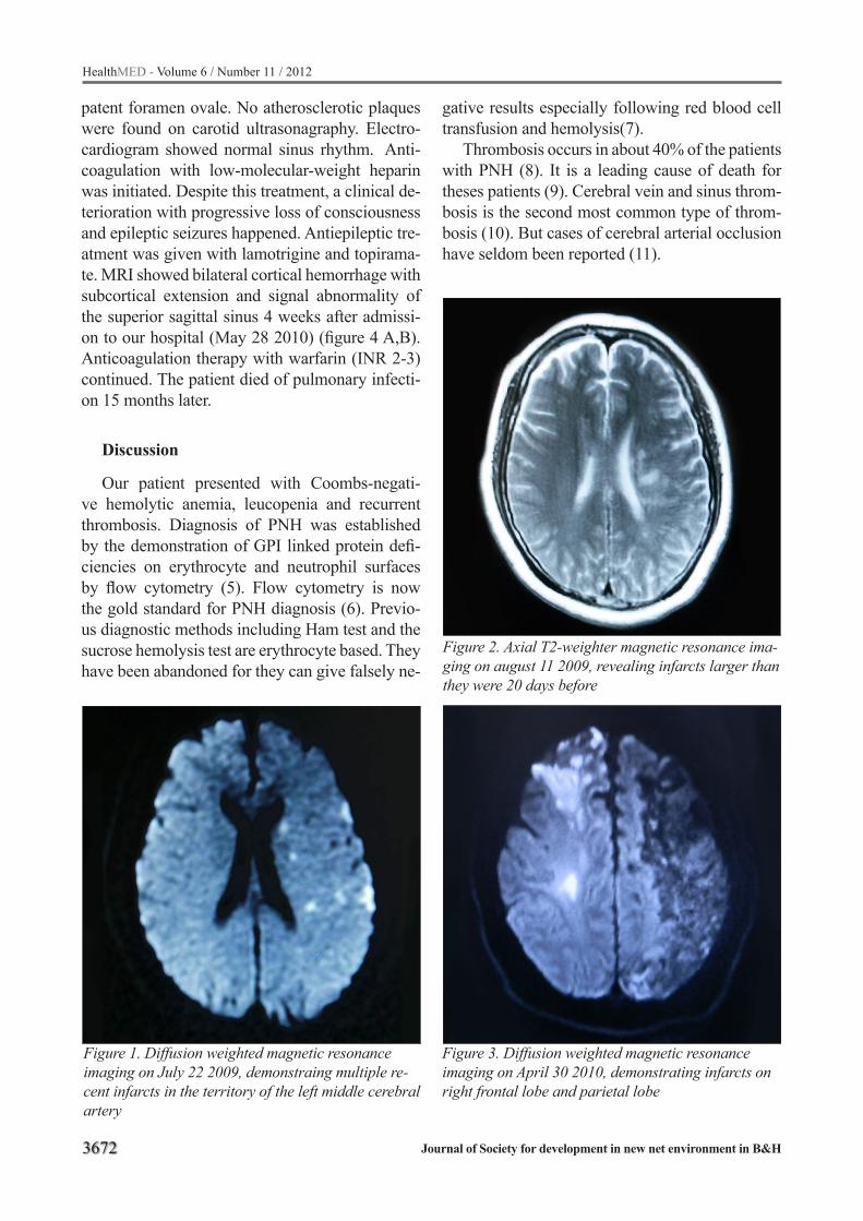
3672
HealthMED - Volume 6 / Number 11 / 2012
Journal of Society for development in new net environment in B&H
Figure 1. Diffusion weighted magnetic resonance imaging on July 22 2009, demonstraing multiple re-cent infarcts in the territory of the left middle cerebral artery
Figure 2. Axial T2-weighter magnetic resonance ima-ging on august 11 2009, revealing infarcts larger than they were 20 days before
Figure 3. Diffusion weighted magnetic resonance imaging on April 30 2010, demonstrating infarcts on right frontal lobe and parietal lobe
patent foramen ovale. No atherosclerotic plaques were found on carotid ultrasonagraphy. Electro-cardiogram showed normal sinus rhythm. Anti-coagulation with low-molecular-weight heparin was initiated. Despite this treatment, a clinical de-terioration with progressive loss of consciousness and epileptic seizures happened. Antiepileptic tre-atment was given with lamotrigine and topirama-te. MRI showed bilateral cortical hemorrhage with subcortical extension and signal abnormality of the superior sagittal sinus 4 weeks after admissi-on to our hospital (May 28 2010) (figure 4 A,B). Anticoagulation therapy with warfarin (INR 2-3) continued. The patient died of pulmonary infecti-on 15 months later.
Discussion
Our patient presented with Coombs-negati-ve hemolytic anemia, leucopenia and recurrent thrombosis. Diagnosis of PNH was established by the demonstration of GPI linked protein defi-ciencies on erythrocyte and neutrophil surfaces by flow cytometry (5). Flow cytometry is now the gold standard for PNH diagnosis (6). Previo-us diagnostic methods including Ham test and the sucrose hemolysis test are erythrocyte based. They have been abandoned for they can give falsely ne-
gative results especially following red blood cell transfusion and hemolysis(7).
Thrombosis occurs in about 40% of the patients with PNH (8). It is a leading cause of death for theses patients (9). Cerebral vein and sinus throm-bosis is the second most common type of throm-bosis (10). But cases of cerebral arterial occlusion have seldom been reported (11).
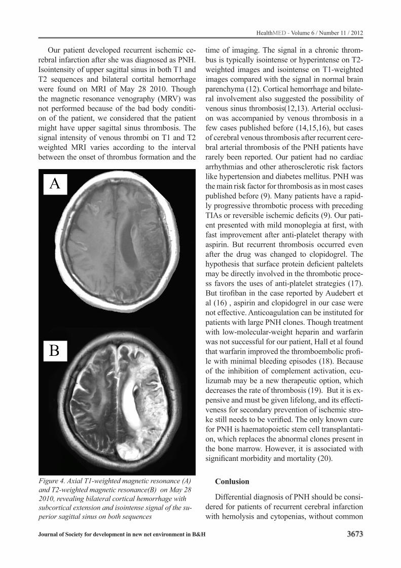
HealthMED - Volume 6 / Number 11 / 2012
Journal of Society for development in new net environment in B&H 3673
Figure 4. Axial T1-weighted magnetic resonance (A) and T2-weighted magnetic resonance(B) on May 28 2010, revealing bilateral cortical hemorrhage with subcortical extension and isointense signal of the su-perior sagittal sinus on both sequences
Our patient developed recurrent ischemic ce-rebral infarction after she was diagnosed as PNH. Isointensity of upper sagittal sinus in both T1 and T2 sequences and bilateral cortital hemorrhage were found on MRI of May 28 2010. Though the magnetic resonance venography (MRV) was not performed because of the bad body conditi-on of the patient, we considered that the patient might have upper sagittal sinus thrombosis. The signal intensity of venous thrombi on T1 and T2 weighted MRI varies according to the interval between the onset of thrombus formation and the
time of imaging. The signal in a chronic throm-bus is typically isointense or hyperintense on T2-weighted images and isointense on T1-weighted images compared with the signal in normal brain parenchyma (12). Cortical hemorrhage and bilate-ral involvement also suggested the possibility of venous sinus thrombosis(12,13). Arterial occlusi-on was accompanied by venous thrombosis in a few cases published before (14,15,16), but cases of cerebral venous thrombosis after recurrent cere-bral arterial thrombosis of the PNH patients have rarely been reported. Our patient had no cardiac arrhythmias and other atherosclerotic risk factors like hypertension and diabetes mellitus. PNH was the main risk factor for thrombosis as in most cases published before (9). Many patients have a rapid-ly progressive thrombotic process with preceding TIAs or reversible ischemic deficits (9). Our pati-ent presented with mild monoplegia at first, with fast improvement after anti-platelet therapy with aspirin. But recurrent thrombosis occurred even after the drug was changed to clopidogrel. The hypothesis that surface protein deficient paltelets may be directly involved in the thrombotic proce-ss favors the uses of anti-platelet strategies (17). But tirofiban in the case reported by Audebert et al (16) , aspirin and clopidogrel in our case were not effective. Anticoagulation can be instituted for patients with large PNH clones. Though treatment with low-molecular-weight heparin and warfarin was not successful for our patient, Hall et al found that warfarin improved the thromboembolic profi-le with minimal bleeding episodes (18). Because of the inhibition of complement activation, ecu-lizumab may be a new therapeutic option, which decreases the rate of thrombosis (19). But it is ex-pensive and must be given lifelong, and its effecti-veness for secondary prevention of ischemic stro-ke still needs to be verified. The only known cure for PNH is haematopoietic stem cell transplantati-on, which replaces the abnormal clones present in the bone marrow. However, it is associated with significant morbidity and mortality (20).
Conlusion
Differential diagnosis of PNH should be consi-dered for patients of recurrent cerebral infarction with hemolysis and cytopenias, without common
3674
HealthMED - Volume 6 / Number 11 / 2012
Journal of Society for development in new net environment in B&H
risk factors for cerebra-vascular diseases. And flow cytometry should be performed to confirm the diagnosis. Cerebral venous sinus thrombosis may develop after recurrent arterial infarction, and MRV should be performed to confirm the diagno-sis.
Reference
1. Lucio Luzzatto, Antonio Maria Risitano, Rosario No-taro. paroxysmal nocturnal hemoglobinuria and ecu-lizumab. Haematologica 2010; 95(4):523-6.
2. Kinoshita T, Ohishi K, Takeda J. GPI-anchor synthe-sis in mammalian cells: genes, their products, and a deficiency. J Biochem 1997;122:251–7.
3. Hillmen R, Lewis SM, Bessler M, Luzzatto L, Dacie JV: Natural history of paroxysmal nocturnal hemo-globinuria. N Engl J Med 1995; 333: 1253–8.
4. Hugel B, Socie G, Vu T, Toti F, Gluckman E, Freyssinet JM, et al. Elevated levels of circulating procoagulant microparticles in patients with paroxysmal nocturnal hemoglobinuria and aplastic anemia. Blood 1999; 93: 3451-6.
5. Samadder NJ, Casaubon L, Silver F, Cavalcan-ti R..Neurological complications of paroxysmal nocturnal hemoglobinuria. Can J Neurol Sci 2007 Aug;34(3):368-71.
6. Rachidi S, Musallam KM, Taher AT. A closer look at paroxysmal nocturnal hemoglobinuria. Eur J Intern Med 2010 Aug;21(4):260-7.
7. Brodsky RA. How I treat paroxysmal nocturnal hemo-globinuria. Blood 2009;113:6522–7.
8. Poulou LS, Xila V, Rokas GI, Karianakis G, Bartzou-dis D, Ziakas PD. Temporal trends in mortality rates from visceral vein thrombosis in paroxysmal noctur-nal haemoglobinuria: An optimistic view. Thromb Haemost 2008; 99: 642-5.
9. Von Stuckrad-Barre S, Berkefeld J, Steckel D, Sitzer M: Cerebral arterial thrombosis in paroxysmal nocturnal hemoglobinuria. J Neurol 2003; 250:756–7.
10. Ziakas PD, Poulou LS, Pomoni A. Thrombosis in paroxysmal nocturnal hemoglobinuria at a glan-ce: a clinical review. Curr Vasc Pharmacol 2008 Oct;6(4):347-53.
11. Yamazaki T, Suzuki K, Sumi M, Yanaka K, Kojima H, Matsumura A. Cerebral embolism as a complication of paroxysmal nocturnal hemoglobinuria. Eur Neu-rol 2005;53(4):217-20.
12. Leach JL, Fortuna RB, Jones BV, Gaskill-Shipley MF. Imaging of cerebral venous thrombosis: current techniques, spectrum of findings, and diagnostic pitfalls. Radiographics 2006 Oct;26 Suppl 1:S19-41; discussion S42-3
13. Agostoni E, Aliprandi A, Longoni M. Cerebral venous thrombosis. Expert Rev Neurother 2009 Apr;9(4):553-64.
14. al-Samman MB, Cuetter AC, Guerra LG, Ho H. Cerebral arterial thrombosis as a complication of paroxysmal nocturnal hemoglobinuria. South Med J 1994;87:765–7.
15. Perkins HA, Boulware JM, Feichtmeir TV, Thayer WW, Spaet TH. Paroxysmal nocturnal hemoglobi-nuria: report of four cases,with observations on tre-atment with 3,3’-methylenebis (4-hydroxycoumarin) (dicumarol). Ann Intern Med 1955; 43: 1218–29.
16. Audebert HJ, Planck J, Eisenburg M, et al. Cere-bral ischemic infarction in paroxysmal nocturnal hemoglobinuria report of 2 cases and updated re-view of 7 previously published patients. Neurol 2005 Nov;252(11):1379-86.
17. Wiedmer T, Hall SE, Ortel TL, etal. Complement-in-duced vesiculation and exposure of membrane prot-hrombinase sites in platelets of paroxysmal noctur-nal hemoglobinuria. Blood 1993;82:1192–6.
18. Hall C, Richards S, Hillmen P. Primary prophylaxiswith warfarin prevents thrombosis in pa-roxysmal nocturnal hemoglobinuria (PNH). Blood 2003;102:3587–91.
19. Hillmen P, Young NS, Schubert J, Brodsky RA, Socie G, Muus P, et al. The complement inhibitor eculizu-mab in paroxysmal nocturnal hemoglobinuria. N Engl J Med 2006;355:1233–43.
20. Brodsky RA. Stem cell transplantation for pa-roxysmal nocturnal hemoglobinuria. Haematologi-ca 2010 Jun;95(6):855-6.
Corresponding AuthorXiaobiao Lai,Department of neurology,Zhejiang provincial people’s hospital,Hangzhou, P. R. China. E-mail: [email protected]
HealthMED - Volume 6 / Number 11 / 2012
Journal of Society for development in new net environment in B&H 3675
Organization and implementation of first-aid unit in chinese navy hospital ship to carry out overseas humanitarian medical aid missionDing Xinmin, Xu Qinzhi, Huang Yeli, Qian Yangming
Department of Respiratory Medicine, Navy General Hospital of PLA, Beijing City, China
Abstract
The Chinese navy hospital ship (Peace Ark) has performed a good number of overseas huma-nitarian medical aid missions since it was fielded. The first-aid unit has been sent out by the hospital ship to temporary medical service site at the host country territory and performs humanitarian me-dical aid mission, which expands the hospital ship space and avails the hospital ship to implement accompanying medical support tasks in a modu-lar, multi-point and multi-faceted way whether it is peacetime and wartime. The first-aid unit can independently fulfill diagnosis and treatment of common diseases, but can not do surgery. The first-aid unit has non-medical institution support and medical institution support two models to carry out medical assistance mission. The first-aid unit should make full preparations after organiza-tion, and its service process should be flexible to adapt to the needs of medical assistant mission.
Keywords: Hospital ship, first-aid unit, Huma-nitarian medical aid mission, Overseas
Introduction
Chinese navy hospital ship (Peace Ark) has un-dertaken many large missions since it was fielded, such as “Coastal Journey of Medical Service Trip of Peace Vessel”, “Harmonious Mission 2010” and “Harmonious Mission 2011”. For now, Chine-se navy hospital ship carries out non-war military missions in following forms: to provide medical aid in case of international disaster; deliver me-dical aid in case of domestic disaster; provide medical guarantee for naval ship, island safegu-arding force and residents on islands; provide hu-manitarian medical aid in overseas countries.1As the naval forces engage in more missions and tasks, it will become more frequently that hospi-
tal ship fulfills various kinds of non-war military missions. Chinese “Harmonious Missions” refers to provide humanitarian medical aid in other co-untries. Based on the experience of the hospital ship in carrying out “Harmonious Mission 2010”, “Harmonious Mission 2011” and other tasks, this Article is expected to summarize the organizati-on and implementation of the first-aid unit when Chinese navy hospital ship carries out overseas humanitarian medical aid missions.
Necessity of setting up first-aid unitAs the hospital ship has relatively limited spa-
ce and medical service couldn’t be immediately reached out to the land, it’s necessary to establish the first-aid unit which selects medical staff from the hospital ship, is equipped with various medi-cal devices and drugs to provide first-aid service at designated site, which avails the hospital ship to implement accompanying medical support tasks in a modular, multi-point and multi-faceted way whether it is peacetime and wartime, representing a beneficial supplement to the hospital ship in va-rious missions.
Target and composition of first-aid unitThe goal of setting up first-aid unit is: to have
complete divisions, including such departments as internal medicine, general surgery, orthope-dics, otorhinolaryngology, ophthalmology, gyna-ecology, stomatology, Dermatology, paediatrics, auxiliary inspection section (able to provide ul-trasonic testing and electrocardiographic exami-nation), laboratory section (able to provide blood routine, urine routine, serum biochemical test, etc) and pharmacy which can independently finish the diagnosis and treatment of various common dise-ases and frequently-occurring diseases except sur-gery. Major tasks of the first-aid units include: (1)
3676
HealthMED - Volume 6 / Number 11 / 2012
Journal of Society for development in new net environment in B&H
the diagnosis and treatment of various common diseases and frequently-occurring diseases except surgery, (2) health education at local medical si-tes. (3) selecting patients needing in-hospital sur-gery and examination by large medical equipment in the hospital ship and then sending them to the hospital ship. (4) completing other tasks assigned by the command group. For that matter, the pre-liminary composition of the first-aid unit is 25 to 30 staffs, including health service experts, medical staffs and logistic guarantee staffs.
Operating mode of first-aid unitThe first-aid unit has non-medical institution
support and medical institution support two mo-dels to carry out medical assistance mission. By having medical institution support, we mean that the first-aid unit has the aid of the space of local medical place and even can borrow the equipment and device of local medical institutions to finish medical work. By having non-medical institution support, we mean that the host country provides gym, church, school and other open spaces where the first-aid unit needs tent and other facilities to set up temporary medical site. “Harmonious Mi-ssion 2010” fulfilled medical aid task in Kenya, Tanzania, Seychelles and Bangladesh; “Harmo-nious Mission 2011” provided medical assistance to Jamaica, Trinidad and Tobago and Costa Rica. During the process of implementing the Harmo-nious Mission, the first-aid unit employed the mode of having medical institution support except for the mission in Kenya where Mombasa Red Cross Society provided KOBLENZ HALL rapid-ly to set up temporary medical establishment and independently to provide humanitarian medical service. The medical site of the first-aid unit shall be set up one day before starting the medical ser-vice.
Medical activities procedure of first-aid unitMedical activities were launched by the first-
aid unit include three phases: preparation before providing medical service, medical service (inclu-de patient organization and delivery to hospital ship), and withdrawal. The first stage also covers: health intelligence investigation on first-aid medi-cal site, reporting to the command group about the
investigation, and human and materials readjus-tment. The specific work flows are: health intelli-gence investigation, coordination meeting with the command group, re-selection of staff, re-appli-cation and obtainment of materials and medicine, gathering of personnel, materials and medicine at port, delivering staff and materials to destination by vehicles, medical service (including patients organizing and evacuation), withdrawal, coordi-nation meeting and work summary inside the first-aid unit, coordination meeting with the command group as required.
The health intelligence investigation before carrying out the medical work is of great impor-tance. Relevant leader of the command group, chief and head nurse of the first-aid unit and 1-2 other medical staffers, 1 health service expert and 1 translator will be take part in the health intelli-gence investigation. The contents of health intelli-gence investigation includes: (1) getting to know the safety facility and safeguarding of the medical site, specifying that the counter-party is responsi-ble for keeping the order of medical activities. (2) getting to the know the operating mode, having medical institution support or not. (3) if medical institution support is available, getting to know whether first-unit jointly carried out medical ser-vice with local hospital, or independently carried out. and to know the medical site provided by the counter-party whether or not can meet medical service and whether needs to be adjusted. (4) if no medical institution support, first-aid unit pro-vides drawings of provisional medical site and ne-cessary conditions for the work to be carried out, while embassy or local government is responsible for providing tent and other partition materials to set up separate consulting room and for ensuring electricity and water supply. (5) getting to know common diseases and frequently-occurring dise-ases and identifying the medical demand of the counter-party. (6) identifying which party is res-ponsible for disposal of household garbage and medical waste. (7) identifying the way of orga-nizing patients of the host country, either random patients, or organizing patients of certain disease, or both of them. (8) other relevant matters. etc.
After completing the health intelligence in-vestigation, the command group hosts meeting according to the need which is often held on the
HealthMED - Volume 6 / Number 11 / 2012
Journal of Society for development in new net environment in B&H 3677
evening of the day before starting the medical work and participated by leader of the command group, medical service expert and first-aid unit members. The meeting involves such contents: reporting on the health intelligence investigation, discussing on plans for the medical service on the second day, and the plan for preparing staff, ma-terials and medicine; confirming medical staffs of the second day. And the command group will notify the hospital ship to prepare food and wa-ter for the second day. The original first-aid unit members may be slightly adjusted according to the defined plan which shall be notified to relevant staff, and deficient materials and medicine shall be re-supplied.
Medical service processOn the day when medical service is provided,
all members and materials are gathered at the port and will go to the first-aid medical site by special vehicle to provide medical service work. The pro-cesses are as follows: patient waiting for medical service-triage and register of patient information-patient receiving doctors’ office visiting at corres-ponding department- patient according to the need to take medicine- patient leaving the medical site. In addition, patient needs to finish necessary auxi-liary examination and then return back to corres-ponding department. If necessary, consultation and treatment among relevant departments may be executed. If a patient is suitable for receiving in-hospital surgery and examination by large medical device at the hospital ship, first contact doctor of this patient is responsible for filling in the evacu-ation application form. After the lead of the first-aid unit approves, the patient will be evacuated to hospital ship by evacuation group.
Removal stageWhen the medical service is finished, the lead
of first-aid unit will issue a removal order. Whether the materials and medicine needs to be taken back to the hospital ship will be decided based on the security condition of the first-aid medical site. If medical institution support is available, the first-aid unit may take back the box to replenish mate-rials and medicine according to the consumption condition. If local medical institution can ensure
security, other boxes may be stored at the medi-cal service site. A summary meeting will be held on the way back or after the dinner. Every mem-ber needs to sum up the medical work of the day, provide service plan and offer improvements to be made on the second day. After the overall medical service is finished, all materials and medicine will be removed back to the hospital ship.
Discussion
Function and composition of first-aid unitThe function and composition of first-aid unit
in Chinese navy hospital ship is different from that in the U.S. Navy hospital ship. 2-4 The first-aid unit in Chinese navy hospital ship is composed of dozens of staffs to provide medical service, but it covers a basically complete set of medical speci-alties and can meet the demand of patients in the local community. But the first-aid in USA hospi-tal ship has more staffs than that in Chinese navy hospital ship. For instance, at the stop of Colum-bia when implementing the “Continuing Promi-se 2009” mission, the first-aid unit of the USNS Comfort (T-AH20), a U.S. Navy hospital ship, has a good number of members. The USNS Comfort dispatched 200 medical staff to Tumart’s shore-based rescue site every day and was responsible for providing medical assistance to local residents. Helicopter is the only tool to transport patients to the USNS Comfort from the shore-based rescue site. Patients who need operation and were tran-sported to hospital ship, other patients were tre-ated at the shore-based rescue site. After surgical recovery, the patient will be transported to the sho-re-based rescue site. The U.S. Navy hospital ship has no patients if no patients have been evacuated by the first-aid unit.4 But in Chinese case, so long as the hospital ship stops at the port and conditi-ons permit, patients can walk to the hospital ship. Both the hospital ship and the first-aid unit provide out-patient service and physical checkup service. So even without patients to be evacuated by the first-aid unit, the hospital ship can also have pa-tients requiring surgery, resulting in overlapping work for both the hospital ship and the first-aid unit. The first-aid unit represents an expansion of the hospital ship in space and a beneficial supple-ment to the hospital ship to carry out various mi-
3678
HealthMED - Volume 6 / Number 11 / 2012
Journal of Society for development in new net environment in B&H
ssions, and avails the hospital ship to implement accompanying medical support tasks in a modu-lar, multi-point and multi-faceted way whether it is peacetime and wartime, meeting the demand of humanitarian medical aid for assisting citizens in the host country in a maximum way. The first-aid unit directly reaches out to the citizens of the host country and faces media organizations of several countries. As their words and deeds are totally laid bare before their overseas counterparts, they are fulfilling the mission of military diplomacy while providing complicated medical service; they carry forward the humanitarian spirit and spread out the idea of harmonious world and harmonious ocean.
When selecting medical staff in Chinese navy hospital ship, we need to take into account that different from the work carried out on the hospital ship, the first-aid unit needs to provide humanitari-an aid within the territory of the host country, and each doctor in the most cases needs to complete disease diagnosis and treatment independently. So the members shall be politically-sensitive with strong competence and communication skills. The unit is composed of health service expert, medical staff and logistic guarantee staff. Medical staffs who have intermediary certificate and compre-hensive professional knowledge will be preferred. The director who can be a medical expert is res-ponsible for the daily management and vocational study of the unit. When implementing the first-aid mission, the health service expert will serve as the lead, responsible for controlling the medical acti-vities of the entire unit and the director will assist the lead to finish the medical service work. Wit-hout special conditions, the director, head nurse and key medical staff are relatively regular which will be beneficial for members to get familiar with the medical service procedures and to ensure me-dical service quality.
Advantages and disadvantages of first-aid operating modeOne can imagine the convenience of having
medical institution support, but the work scale and consulting room are subject to the size of space provided by the local authority. If without support, it appears more complicated to establish provisi-onal medical institute, but the advantage is that
the medical institution can be set up according to our own intention. As the first-aid unit provides humanitarian medical aid, although it has medical institution support, it doesn’t occupy medical re-sources of the host country except for space, such as using the medical device and equipment of the host country.
Optimization of service procedures of first-aid unitThe abovementioned service procedures aren’t
changeless which shall be flexible according to real conditions when providing service. Various kinds of materials and medicine shall be well-prepared before providing service. Competent members shall be selected according to realities of the mission and training shall be made available to them, including (1) foreign language scenario simulation training, preparing commonly used medical English words based on profession, and diagnosis and treatment procedure, organizing role reversal simulation training; (2) professional medical training, starting professional learning on common diseases and frequently-occurring dise-ases and multiple infectious diseases of the host country; (3) preparation and study of contingency plan of the first-aid unit, etc.5
What has happened in our real practice told us that the role of triage is very critical among vario-us links of first-aid medical service.6-9 A correct triage can shorten the patients’ time in receiving diagnosis and treatment and maintain sound or-der of medical service. In the medical activity of “Harmonious Mission - 2011”, we asked doctors who have sound communication skills in foreign language and comprehensive medical knowledge to do triage who write down the chief complaint and demand of patients on the medical visit card, which significantly reduced the error score of tri-age, and shortened patients’ time in receiving in receiving diagnosis and treatment, and increased doctors’ working efficiency.
In a word, the target of the first-aid unit of Chinese navy hospital ship is different from that of the United States. Our first-aid unit is defined by small yet proficient functional unit, involves com-plete departments and its work mode can flexibly adapt to the conditions provided by the local aut-
HealthMED - Volume 6 / Number 11 / 2012
Journal of Society for development in new net environment in B&H 3679
hority so as to satisfy the demand of the host co-untry for humanitarian medical aid in a maximum fashion.
References
1. Hu Jia-qing, LiuYong, Wang Meng, et al. Thoughts on Medical Support of Hospital Ship in Non-war Mili-tary Operations. Hospital Administration Journal of Chinese People’s Liberation Army 2010; 17(5):454-455. Article in Chinese.
2. Negus TL, Brown CJ, Konoske P. Determining medi-cal staffing requirements for humanitarian assistance missions. Mil Med 2010; 175(1):1-6.
3. Skyrme L. Pacific Partnership 2010: humanitarian civic assistance on a U.S. Navy hospital ship. J R Nav Med Serv 2010; 96(3):169-74.
4. Wang Hai-wei, Sun tao, Wen min, et al. The U.S. mi-litary hospital executive “Continuing Promise 2009” international humanitarian relief operations and to our enlightenment. China Journal of Emergency Re-suscitation and Disaster Medicine 2010; 5(3):209-211. Article in Chinese.
5. Qian Yang-ming, Wang Hai-wei, Chen Yong-pen, et al. Problems and Suggestions on the Medical Flow in the New-type Hospital Ship. Hospital Administration Journal of Chinese People’s Liberation Army 2010; 17(7):628-629. Article in Chinese.
6. Etienne M, Powell C and Amundson D. Healthcare ethics: the experience after the Haitian earthquake. Am J Disaster Med 2010;5(3):141-7.
7. Deshmukh N. Mass casualty aboard USS Kitty Hawk (CV 63) January 2005--lessons learned. Mil Med 2006 ;171(1):ii.
8. Carter PA. The mass casualty situation affecting the USS Kitty Hawk. Mil Med 2006;171(7):ix-x.
9. Ganley L and Gloster AS. An overview of triage in the emergency department. Nurs Stand 2011; 26(12):49-56.
Corresponding AuthorQian Yangming,Department of Respiratory Medicine,Navy General Hospital of PLA,Beijing City, China, E-mail: [email protected]
3680
HealthMED - Volume 6 / Number 11 / 2012
Journal of Society for development in new net environment in B&H
Age specific prevalence of metabolic syndrome in patients with schizophrenia treated with atypical antipsychotics and control groupHamidreza Ahmadkhaniha1, Hamid Abdolmaleki Mostafavi1, Marzieh Nojoomi2, Bahman Parvizi-Emran1 1 Department of Psychiatry, Tehran Psychiatric Institute and Mental Health Research Center, Tehran University of Medical Sciences, Tehran, Iran 2 Department of Community Medicine, Tehran University of Medical Sciences, Tehran, Iran
Abstract
Objective: Metabolic syndrome has high pre-valence in patients with Schizophrenia in compa-rison with normal population. According to high prevalence of metabolic syndrome in subject with higher than 40 years, so the aim of this study was to comparison of metabolic syndrome in patients with Schizophrenia treated with atypical antip-sychotics and control group.
Methods: This cross-sectional study was done in 100 patients with Schizophrenia treated with atypical antipsychotics and 135 healthy subjects. The prevalence of metabolic syndrome and indi-vidual metabolic syndrome factors were evaluated in less and more than 40 years.
Results: In patients less than 40 years, the prevalence of metabolic syndrome was 18.6% in patients with Schizophrenia treated with atypi-cal antipsychotics and 7% in healthy subjects. In patients more than 40 years, the prevalence of metabolic syndrome was 10% in patients with Schizophrenia treated with atypical antipsychotics and 32.8% in healthy subjects. The prevalence of metabolic syndrome in patients with schizophre-nia was 3 times (OR = 3, CI95%: 1.01-8.9) more than control group in patients less than 40 years, and 0.2-fold (OR=0.2, CI95%: 0.06-0.83) in pati-ents with more than 40 years.
Conclusion: Metabolic syndrome had high prevalence in schizophrenia patient with less than 40 years old, however patients with schizophrenia had been screened for metabolic syndrome in yo-unger age and this concern in patients with older than 40 years had minor importance.
Keywords: Atypical Antipsychotics, Metabo-lic Syndrome, Schizophrenia,
Introduction
Schizophrenia is a psychiatric condition with involvement of thought, emotion, movement and behavior (1). The life time prevalence of schizophrenia is less than 1% (2). It usually be-gins before the age of 25 years (3). Antipsychotics (typical or atypical) have a key role in the treatment of this disorder (4). Atypical neuroleptics such as clozapine, olanzapine, quetiapine, and risperido-ne have demonstrated efficacy in the treatment of schizophrenia with fewer extrapyramidal side effects than other neuroleptics, but the use of these drug has been associated with weight gain (5).
Patients with schizophrenia are at increased risk for developing the metabolic syndrome or its individual components due to their lifestyle, suspected genetic predisposition, and exposure to antipsychotic medications that can cause weight gain and other metabolic side effects (6). A high prevalence of metabolic syndrome has been repor-ted among patients with schizophrenia (7-9) and has been related to an increased risk for cardiovas-cular diseases (10,11), diabetes (12) and mortality (13). Metabolic syndrome is characterized by the coincidence of hypertension, abdominal obesity, abnormal lipid level (blood triglycerides, chole-sterol) and/or impaired blood glucose regulation (14). The association of metabolic syndrome and treated and untreated schizophrenic patients has been reported previously (15-17) but the effect of age in this association did not investigated pre-viously completely, so the aim of this study was to comparison of pprevalence of metabolic Syn-drome in patients with schizophrenia treated with atypical antipsychotics and healthy group in less and older than 40 years.
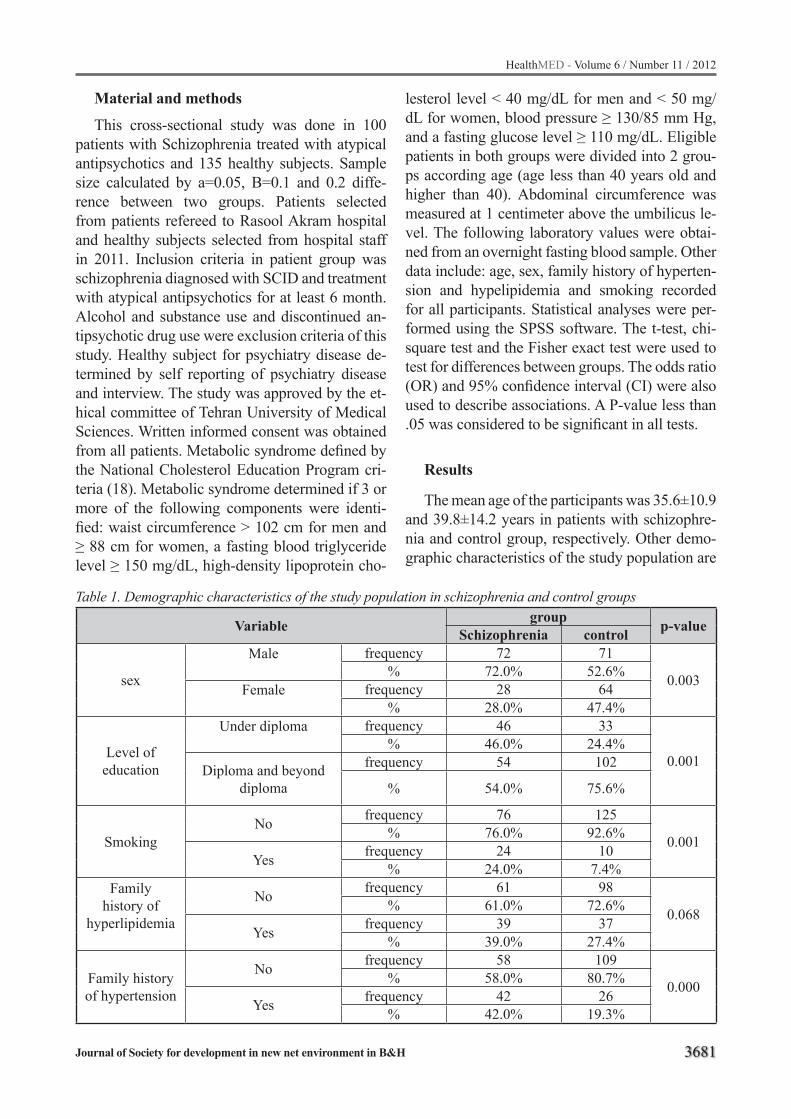
HealthMED - Volume 6 / Number 11 / 2012
Journal of Society for development in new net environment in B&H 3681
Material and methods
This cross-sectional study was done in 100 patients with Schizophrenia treated with atypical antipsychotics and 135 healthy subjects. Sample size calculated by a=0.05, B=0.1 and 0.2 diffe-rence between two groups. Patients selected from patients refereed to Rasool Akram hospital and healthy subjects selected from hospital staff in 2011. Inclusion criteria in patient group was schizophrenia diagnosed with SCID and treatment with atypical antipsychotics for at least 6 month. Alcohol and substance use and discontinued an-tipsychotic drug use were exclusion criteria of this study. Healthy subject for psychiatry disease de-termined by self reporting of psychiatry disease and interview. The study was approved by the et-hical committee of Tehran University of Medical Sciences. Written informed consent was obtained from all patients. Metabolic syndrome defined by the National Cholesterol Education Program cri-teria (18). Metabolic syndrome determined if 3 or more of the following components were identi-fied: waist circumference > 102 cm for men and ≥ 88 cm for women, a fasting blood triglyceride level ≥ 150 mg/dL, high-density lipoprotein cho-
lesterol level < 40 mg/dL for men and < 50 mg/dL for women, blood pressure ≥ 130/85 mm Hg, and a fasting glucose level ≥ 110 mg/dL. Eligible patients in both groups were divided into 2 grou-ps according age (age less than 40 years old and higher than 40). Abdominal circumference was measured at 1 centimeter above the umbilicus le-vel. The following laboratory values were obtai-ned from an overnight fasting blood sample. Other data include: age, sex, family history of hyperten-sion and hypelipidemia and smoking recorded for all participants. Statistical analyses were per-formed using the SPSS software. The t-test, chi-square test and the Fisher exact test were used to test for differences between groups. The odds ratio (OR) and 95% confidence interval (CI) were also used to describe associations. A P-value less than .05 was considered to be significant in all tests.
Results
The mean age of the participants was 35.6±10.9 and 39.8±14.2 years in patients with schizophre-nia and control group, respectively. Other demo-graphic characteristics of the study population are
Table 1. Demographic characteristics of the study population in schizophrenia and control groups
Variable group p-value Schizophrenia control
sex
Male
frequency 72 71
0.003% 72.0% 52.6%Female
frequency 28 64
% 28.0% 47.4%
Level of education
Under diploma frequency 46 33
0.001% 46.0% 24.4%
Diploma and beyond diploma
frequency 54 102
% 54.0% 75.6%
SmokingNo frequency 76 125
0.001% 76.0% 92.6%
Yes frequency 24 10% 24.0% 7.4%
Family history of
hyperlipidemia
No frequency 61 98
0.068% 61.0% 72.6%
Yes frequency 39 37% 39.0% 27.4%
Family history of hypertension
No frequency 58 109
0.000% 58.0% 80.7%
Yes frequency 42 26% 42.0% 19.3%
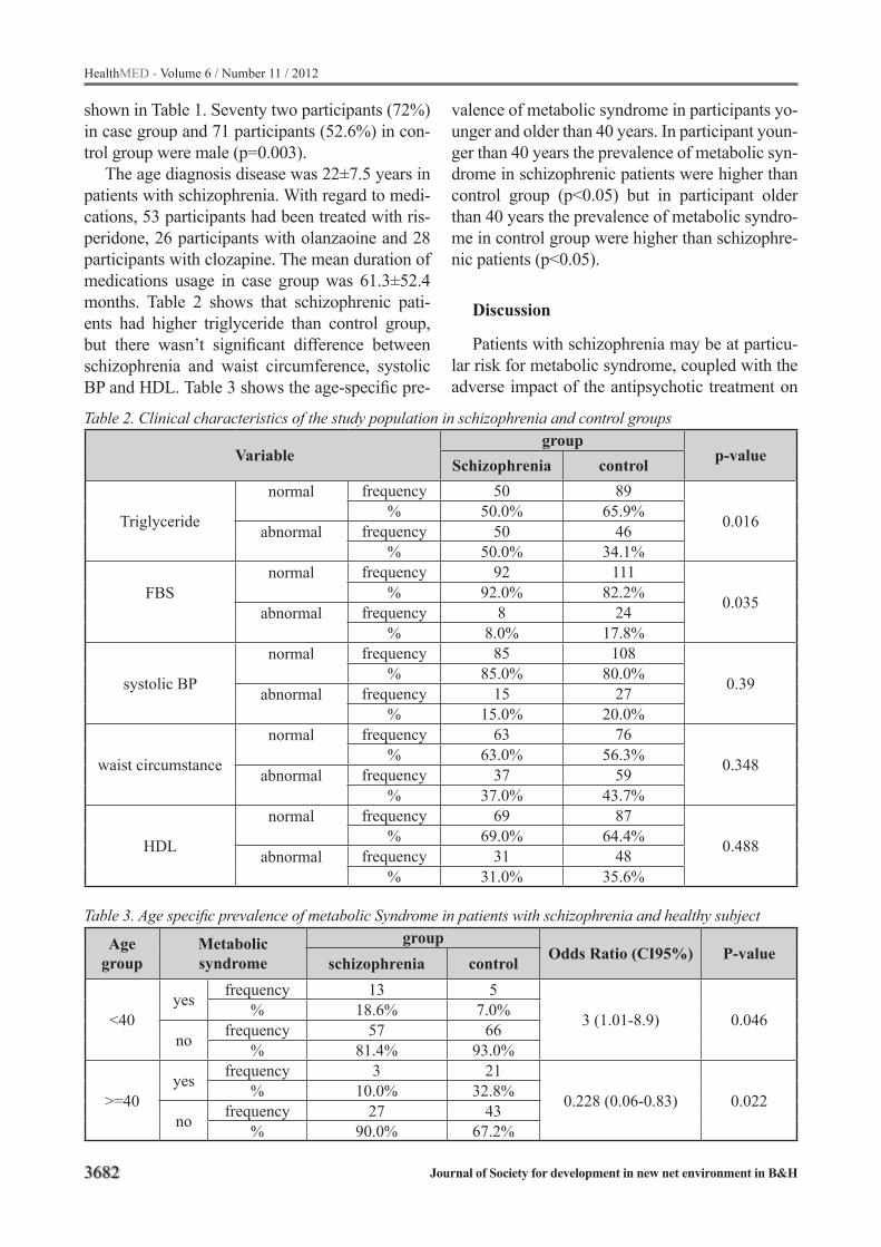
3682
HealthMED - Volume 6 / Number 11 / 2012
Journal of Society for development in new net environment in B&H
Table 2. Clinical characteristics of the study population in schizophrenia and control groups
Variablegroup
p-valueSchizophrenia control
Triglyceride
normal
frequency 50 89
0.016% 50.0% 65.9%abnormal
frequency 50 46
% 50.0% 34.1%
FBS normal
frequency 92 111
0.035% 92.0% 82.2%abnormal
frequency 8 24
% 8.0% 17.8%
systolic BP
normal
frequency 85 108
0.39% 85.0% 80.0%abnormal
frequency 15 27
% 15.0% 20.0%
waist circumstance
normal
frequency 63 76
0.348% 63.0% 56.3%abnormal
frequency 37 59
% 37.0% 43.7%
HDL
normal
frequency 69 87
0.488% 69.0% 64.4%abnormal
frequency 31 48
% 31.0% 35.6%
Table 3. Age specific prevalence of metabolic Syndrome in patients with schizophrenia and healthy subject
Age group
Metabolic syndrome
groupOdds Ratio (CI95%) P-valueschizophrenia control
<40 yes frequency 13 5
3 (1.01-8.9) 0.046% 18.6% 7.0%
no frequency 57 66% 81.4% 93.0%
>=40yes frequency 3 21
0.228 (0.06-0.83) 0.022% 10.0% 32.8%
no frequency 27 43% 90.0% 67.2%
shown in Table 1. Seventy two participants (72%) in case group and 71 participants (52.6%) in con-trol group were male (p=0.003).
The age diagnosis disease was 22±7.5 years in patients with schizophrenia. With regard to medi-cations, 53 participants had been treated with ris-peridone, 26 participants with olanzaoine and 28 participants with clozapine. The mean duration of medications usage in case group was 61.3±52.4 months. Table 2 shows that schizophrenic pati-ents had higher triglyceride than control group, but there wasn’t significant difference between schizophrenia and waist circumference, systolic BP and HDL. Table 3 shows the age-specific pre-
valence of metabolic syndrome in participants yo-unger and older than 40 years. In participant youn-ger than 40 years the prevalence of metabolic syn-drome in schizophrenic patients were higher than control group (p<0.05) but in participant older than 40 years the prevalence of metabolic syndro-me in control group were higher than schizophre-nic patients (p<0.05).
Discussion
Patients with schizophrenia may be at particu-lar risk for metabolic syndrome, coupled with the adverse impact of the antipsychotic treatment on
HealthMED - Volume 6 / Number 11 / 2012
Journal of Society for development in new net environment in B&H 3683
serum lipids and metabolic syndrome (19). Addi-tionally, it has been suggested that ethnicity may play a moderating effect in determining those who may be at increased risk for developing lipid ab-normalities and other metabolic disturbances du-ring antipsychotic treatment (20). The result of present study shown that the prevalence of me-tabolic syndrome in schizophrenic patients were higher than control group (p<0.05), in participant younger than 40 years, but in participant older than 40 years the prevalence of metabolic syndrome in control group were higher than schizophrenic pa-tients (p<0.05). So, it seems that the association of metabolic syndrome and antipsychotic exposure in schizophrenic patients is remarkable in patients younger than 40 years and patients and there was little worry for development of metabolic syn-drome in older patients. Sernyak et al, studied the association of diabetes mellitus with use of atypi-cal neuroleptics in the treatment of schizophrenia and shown that the strongest effect between dia-betes mellitus and use of atypical neuroleptics in patients less than 40 years old (5). In Huang et al study, in Taiwan, the difference of MS prevalen-ce between schizophrenic patients and the general population was marked in male patients under 40 years of age and in female patients under 50 years old (21). Sugawara et al, in Japan, reported that patients with schizophrenia or schizoaffective dis-order had high prevalence of metabolic syndrome compared to the general population, and was most apparent for those under 60 years of age (22). Re-sult of our study in comparison to other reported suggests that patients with schizophrenia in youn-ger age need more attention for screening of me-tabolic syndrome and encouragement for change in life style.
In limitation of present study, the prevalence rate of metabolic syndrome for our study derived from the cross-sectional design, a prospective, longitudinal or trial study with adjustments for age would have performed in this particular populati-on over time.
Acknowledgements
The authors of this paper present a special thanks to Dr. Alizadeh.
Reference
1. Hosseini SH, Karkhaneh Yousefi M. Correlation between quality of life and global functioning in schizophrenia. Iran J Psychiatry Behav Sci 2011; 5(2):120-5.
2. Stilo SA, Murray RM. The epidemiology of schizophre-nia: replacing dogma with knowledge. Dialogues Clin Neurosci. 2010;12(3):305-15.
3. Mueser KT, McGurk SR. Schizophrenia. Lancet 2004; 363: 2063-72.
4. Loga-Zec S, Fisekovic S, Loga S. The usage of the antipsychotic drugs in the specific different centers in Federation of Bosnia and Herzegovina. Healthmed 2011; 5(3): 660-4.
5. Sernyak MJ, Leslie DL, . Alarcon RD, Losonczy MF, Rosenheck R. Association of diabetes mellitus with use of atypical neuroleptics in the treatment of schizophrenia. Am J Psychiatry 2002;159:561-566.
6. Hasnain M, Fredrickson SK, Vieweg WV, Pandurangi AK. Metabolic syndrome associated with schizophre-nia and atypical antipsychotics. Curr Diab Rep. 2010; 10(3):209-16.
7. Cohn T, Prud’homme D, Streiner D, Kameh H, Re-mington G. Characterizing coronary heart disease risk in chronic schizophrenia: high prevalence of the metabolic syndrome. Can J Psychiatry. 2004;49:753–760.
8. Meyer JM, Nasrallah HA, McEvoy JP, Goff DC, Da-vis SM, Chakos M, Patel JK, Keefe RS, Stroup TS, Lieberman JA. The Clinical Antipsychotic Trials Of Intervention Effectiveness (CATIE) Schizophrenia Trial: clinical comparison of subgroups with and without the metabolic syndrome. Schizophr Res. 2005;80:9–18.
9. Bobes J, Arango C, Aranda P, Carmena R, Garcia-Garcia M, Rejas J. CLAMORS Study Collaborative Group. Cardiovascular and metabolic risk in outpa-tients with schizophrenia treated with antipsycho-tics: results of the CLAMORS study. Schizophr Res. 2007;90:162–173.
10. Isomaa B, Almgren P, Tuomi T, Forsen B, Lahti K, Nissen M, Taskinen MR, Groop L. Cardiovascular morbidity and mortality associated with the metabo-lic syndrome. Diabetes Care. 2001;24:683–689.
11. Lakka HM, Laaksonen DE, Lakka TA, Niskanen LK, Kumpusalo E, Tuomilehto J, Salonen JT. The metabolic syndrome and total and cardiovascu-lar disease mortality in middle-aged men. JAMA. 2002;288:2709–2716.
3684
HealthMED - Volume 6 / Number 11 / 2012
Journal of Society for development in new net environment in B&H
12. Wilson PW, D’Agostino RB, Parise H, Sullivan L, Meigs JB. Metabolic syndrome as a precursor of cardiovascular disease and type 2 diabetes mellitus. Circulation. 2005;112:3066–3072.
13. Expert Panel on Detection, Evaluation and Tre-atmentof High Blood Cholesterol in Adults. Execu-tive Summary of The Third Report of The National Cholesterol Education Program (NCEP) Expert Pa-nel on Detection, Evaluation, And Treatment of High Blood Cholesterol In Adults (Adult Treatment Panel III) JAMA. 2001;285:2486–2497.
14. Grundy SM, Cleeman JI, Daniels SR, Donato KA, Eckel RH, Franklin BA, et al. Diagnosis and ma-nagement of the metabolic syndrome: an American Heart Association/National Heart, Lung, and Blo-od Institute Scientific Statement. Circulation 2005, 112:2735-2752.
15. Gianfrancesco F, White R, Wang RH, Nasrallah HA: Antipsychotic-induced type 2 diabetes: evidence from a large health plan database. J Clin Psychop-harmacol 2003, 23:328-335.
16. Casey DE: Dyslipidemia and atypical antipsychotic drugs. J Clin Psych 2004, 65(Suppl 18):27-35.
17. van Winkel R, Moons T, Peerbooms O, Rutten B, Pe-uskens J, Claes S, van Os J, De Hert M: MTHFR genotype and differential evolution of metabolic pa-rameters after initiation of a second generation an-tipsychotic: an observational study. Int Clin Psycho-pharmacol 2010, 25(5):270-276.
18. National Cholesterol Education Program (NCEP) Expert Panel on Detection, Evaluation, and Tre-atment of High Blood Cholesterol in Adults (Adult Treatment Panel III) Third Report of the National Cholesterol Education Program (NCEP) Expert Pa-nel on Detection, Evaluation, and Treatment of High Blood Cholesterol in Adults (Adult Treatment Panel III) final report. Circulation. 2002; 106(25):3143–3421.
19. Meyer JM, Koro CE. The effects of antipsychotic therapy on serum lipids: a comprehensive review. Schizophr Res. 2004;70(1):1–17.
20. Meyer JM, Rosenblatt LC, Kim E, et al. The mode-rating impact of ethnicity on metabolic outcomes during treatment with olanzapine and aripiprazole in patients with schizophrenia. J Clin Psychiatry. 2009;70(3):318–325.
21. Huang MC, Lu ML, Tsai CJ, Chen PY, Chiu CC, Jian DL, et al. Prevalence of metabolic syndrome among patients with schizophrenia or schizoaffecti-ve disorder in Taiwan. Acta Psychiatr Scand. 2009; 120(4):274-80.
22. Sugawara N, Yasui-Furukori N, Sato Y, Umeda T, Kishida I, Yamashita H, et al. Prevalence of metabo-lic syndrome among patients with schizophrenia in Japan. Schizophr Res. 2010; 123(2-3):244-50.
Corresponding Author Bahman Parvizi-Emran, Department of Psychiatry, Tehran Psychiatric Institute and Mental Health Research Center, Tehran University of Medical Sciences, Tehran, Iran.E-mail: [email protected]
HealthMED - Volume 6 / Number 11 / 2012
Journal of Society for development in new net environment in B&H 3685
The identification of female victims of domestic violence by emergency first aid health care professionalsSerap Ozer1, Seyda Dulgerler1, Esra Engin1, Melek Ardahan2, Emel Tekindor3
1 Ege University Faculty of Nursing, Department of Psychiatric Nursing, Bornova, Izmir, Turkey2 Ege University School of Nursing, Department of Public Health Nursing, Bornova, Izmir, Turkey3 Izmir Health Group Unit, 112 Emergency Service, Izmir, Turkey
Abstract
Aim: This research examined the ability of he-alth care professionals working in emergency first aid stations to identify female victims of domestic violence.
Methods: The data were collected by means of a Descriptive Information Form and a Scale Form. The total population of the research consisted of a hundred health care professionals working at emergency first aid stations in Izmir (n=100). The breakdown of the study group was as follows; 60% nurses, 17% health officers, 20% obstetricians and 3% paramedics. The response rate was 100%.
Results: During their professional lives 90% of the health care professionals in the study had attended a woman who had been subjected to do-mestic violence. From the scale used in this study it was found that the average ability of the group of health care professionals to identify female vic-tims of domestic violence was 21.50 ±4.25. Of the personnel participating in this research 75% were found to have partially adequate knowledge, 15% had inadequate knowledge, and only 10% were fo-und to have adequate knowledge to identify those women who had experienced domestic violence.
Conclusions: Since a large percentage of the health care professionals in this study lacked the information to accurately identify female victims of domestic violence it is concluded that it is very important to introduce a new in-service training program on the topic of “Female victims of dome-stic violence”.
Keywords: Violence, Domestic Violence, Wo-man, Female Victims, Emergency, First Aid, He-alth Care Professionals, Nurse, Izmir, Turkey
Introduction
Violence towards women is the most common but the least described violation of human rights worldwide (Atman 2003; Garcia-Moreno et al. 2005). Today, half of the women in the world are subjected to violence by their husbands (Altinto-prak et al. 2006; Erbek et al. 2004; Deveci & Acik 2002; Demir & Ozkan 2000). In Turkey, the Prime Ministry Family Research Institution (1995) anno-unced that there is physical violence in 34% of fa-milies and there is oral violence in 53% of them and 46% of children have been subjected to physi-cal violence (Vahip & Doganavsargil 2006; Unal & Bilge 2004). In research carried out by Mor Çatı between 1990-1996 among 1,259 women it was shown that 88.2% of the women lived in a violent environment, and 68% of them had been hit by their husbands. After the study conducted in 2000 on 1,070 women concerning family violence by Woman Status and Problems Head Office, 21.2% of the women declared that their husbands had been violent towards them (Gültekin et al. 2004).
Today, the abuse of women is a problem still seen not only in Turkey but in every country all over the world today. Health care professionals have the responsibility of diagnosing, treating the abused women and keeping legal records. 112 emergency service health care professionals are in a very special position in terms of identifying and preventing violence against woman. These health care workers are able to observe families in their home environment, often they are present in the environment where the event has just happened, and they are responsible for informing the hospital personnel about the violence which can then lead to legal procedures. However, it can very difficult to determine whether a woman has been abused
3686
HealthMED - Volume 6 / Number 11 / 2012
Journal of Society for development in new net environment in B&H
this can be because the female victim will not con-firm the abuse thus the health professional asse-sses the physical injury symptoms as accidental or the health professional perceived the abuse as a domestic problem related to family relationships rather than a health problem (Garcia-Moreno et al. 2005; Henderson et al. 2004; Heise et al. 1999).
Baysan (2003) determined that 32.5% of nu-rses and midwives were unable to diagnose the symptoms of domestic violence inflicted on wo-men, 31.2% were unable to diagnose the emotio-nal symptoms and 50.0% were unable to diagnose physical symptoms. Aksan & Aksu (2007) carried out a study of doctors and nurses in hospital acci-dent and emergency (A & E) units and found that most of the nurses and doctors can diagnose the signs of sexual abuse of women and they are awa-re of the situation. There have been a few studies on the diagnostic capabilities of A&E personnel in Turkey; however, no research has been carried out into the identification of the symptoms of do-mestic violence in women by 112 emergency first aid professions.
Background In the majority of countries in the world there is
an emergency call system, in the UK the number is 999, in the USA it is 911. In Turkey, in keeping with the European standard the number to call is 112 for emergency first aid and rescue services. In Izmir the Emergency health services were initially provided by Atatürk Teaching Hospital, Karşıyaka Public Hospital and Buca Municipality depended on the center established in Eşrefpaşa Municipality Hospital. In 1986 there was 1 center, 3 ambulance stations and 6 ambulances. In 1994, The Turkish Ministry of Health started a 112 Emergency Aid and Rescue Services project in, Istanbul, Ankara and Izmir with the aim of developing pre hospital health services. Currently there are a total of 41 Emergency First Aid stations 21 in the Izmir me-tropolitan area and 20 stations on the periphery of the city (Ertugrul 2006).
The Izmir 112 Emergency Health Services are directed from the office of the Health Manage-ment of the Province. The command control center where the calls are taken assesses the emergency call and dispatches an ambulance to location of the incident from the nearest emergency Aid Stati-
on. All dialogues are recorded on computer media. A minimum of 15 personnel including 5 doctors, 5 health personnel and 5 drivers work at each 112 station in Izmir. These emergency centers provide service for the situations of every kind of injury from traffic and home accidents, heart attacks and serious illnesses, attempted suicide and mental ill-ness. In a study in Samsun in 2004 it was found that 12.3% of the 112 calls came from those with mental health problems. The most frequent reason for calling the emergency services was for synco-pe (48.1%), and the most frequent diagnosis after examination was conversion disorder (62.6%). The most frequent early diagnosis for women was conversion disorder with 73.6% and suicide attempts constituted 9.1% of the calls (Ertugrul 2006).
Research question: Do health care professionals working in emer-
gency first aid stations have sufficient knowledge to identify female victims of domestic violence?
The Study
Aim This research examined the ability of emer-
gency first aid station’ health care professionals to identify female victims of domestic violence.
Design and SampleThe total population of the research consisted
of one hundred health care professionals working at 21 emergency first aid stations in the city of Iz-mir, Turkey (n=100). The breakdown of the study group was as follows; 60% nurses, 17% health officers, 20% obstetricians and 3% paramedics. The response rate was 100% and all participation was voluntary. The data collecting instrument was administered face to face with the 112 emergency first aid professions by the researchers. The nece-ssary permission was taken from the institution to carry out the research. Before the questionnaire was applied, the research purpose and procedu-re was explained to the participants and their in-formed consent was obtained. The research was implemented within a framework of ethical rules and in accordance with the ethical principles of the Declaration of Helsinki Principles.
HealthMED - Volume 6 / Number 11 / 2012
Journal of Society for development in new net environment in B&H 3687
MeasurementsTwo forms were used as the data collecting in-
strument, the Introductory Information form for health care professionals and a Scale Form related to the identification of female victims of domestic violence by health care professionals.
Introductory Information Form for Health Care Professionals The form developed by the researchers consi-
sted of 7 questions prepared to determine aspects such as, education, qualifications, status of in ser-vice training and experience about women victims of domestic violence.
Scale Form Related to the Identification by Health Care Professionals:This scale developed by Baysan (2003) for nu-
rses and midwives consists of 33 statements requ-iring true or false responses. The Cronbach Alpha internal consistency coefficient was found to be 0.76 in the validity and reliability study carried out on nurses and midwives. In this Scale Form, in order to evaluate the answers of health personnel, the responses are given 1 point for true and 0 for false in positive expressions, and 0 point for true and 1 point for false in negative expressions. The answers were converted into numerical values and the total scale point averages were obtained. The
Table 1. Distribution of the introductory information of 112 emergency service health personnel participants (n=100)Introductory Features Number Percentage
Age Group17-25 1 126-35 75 7536-45 24 24
ProfessionNurse 60 60Health officer 17 17Midwife 20 20Other (Paramedic) 3 3
EducationVocational school of health 31 31Associate degree (diploma) 43 43Graduate 23 23Postgraduate 3 3
Years of service in profession0-5 years 7 76-10 years 17 1711-15 years 45 4516 years and more 31 31
Years of working in 112 service institution0-12 months 13 131-5 Years 48 486-10 Years 33 3311-20 Years 6 6
Encountered case of domestic violence against a womanYes 90 90No 10 10
Received in service trainingYes 13 13No 87 87TOTAL 100 100
Distribution of 112 emergency service health care professionals according to their introductory features ( age group, professi-on, education, years of service in profession, years of working in 112 service institution, encountered case of violence against female, received in service training).
3688
HealthMED - Volume 6 / Number 11 / 2012
Journal of Society for development in new net environment in B&H
total scale point was determined as 33. According to Baysan’s (2003) research The knowledge of he-alth care professionals answering true to 80% or more of the statements of scale about diagnosing the symptoms of women who had experienced domestic violence was assessed as adequate, the knowledge of those answering true to 51-79% of the statements was considered to partially adequ-ate and those answering true to 50% or less of the statements were assessed as inadequate.
In this study, the Cronbach Alpha internal con-sistency coefficient was found to be 0.82 therefore it was accepted as valid and reliable.
Data AnalysisThe data obtained was evaluated with an SPSS
13.0 package computer program. The number and percentage dispersion of the findings about intro-ductory features of the health care professionals was carried out; an ANOVA and independent sam-ple t test were used to examine the relationship between the total scale point averages of health professions in relation to the ability to diagnosis of domestic violence having been inflicted on wo-men and independent differentials.
Results
The introductory features of the participants are shown in Table 1. 75% of the 112 health care professionals were aged 26-35, 60% were nurses, 17% were health officers, 20% were obstetricians and 3% were in the other vocational groups (pa-ramedics). 43% of the health care professionals had graduated with an associate degree (diplo-ma). 45% of the participants had been working for 11-15 years, and 48% had been working for 1-5 years in a 112 institution. It was also found that during their professional career 90% of the health care professionals had attended a woman who had been subjected to violence however, 87%
had received no in-service training concerning the diagnosis and treatment of women victims of do-mestic violence.
Examining the scale concerning the ability of health care professionals to diagnose that a wo-man had experienced domestic violence accor-ding to the total scale point average, the total sca-le point average was determined as 21.50±4.25. The health professionals had minimum of 10 and a maximum 31 points on the scale. According to the scores obtained from the scale in terms of the skills of health care professionals in diagnosing female victims of domestic violence, 10% were found to be sufficient, 75% were found to be parti-ally adequate and 15% were found to be inadequ-ate (see Table 2).
The distribution of total scale point averages of the health care professionals according to de-mographic features can be seen in Table 3. There was statistically reasonable difference between the total scale point averages according to the vocatio-nal groups (F=1.391, P=0.250), years of professio-nal service (F= 1.664, P=0.180), and whether they had attended a woman who had been subjected to domestic violence (t=0.390, P=0.244). There was no statistically reasonable difference between the total scale point averages according to the ages of the health care professionals (F=0.485, P=0.617), their education level (F=0.457, P=0.713), the num-ber of years that they had worked in a 112 insti-tution (F=0.540, P=0.983), and whether they had received in service training (t=l.156. P=0.757).
Discussion
In this study, in their professional capacity ne-arly all of the participants had encountered a wo-man who had been subjected to violence during their professional lives. In the study by Baysan (2003) it was determined that 67.5% of the nur-se and midwifes treated a woman who had been
Table 2. Distribution of adequate status according to the scores of health care professionals (n=100) Adequate status The scores obtained from the scale
Number PercentageAdequate 10 10
Partially adequate 75 75Inadequate 15 15
TOTAL 100 00.0
HealthMED - Volume 6 / Number 11 / 2012
Journal of Society for development in new net environment in B&H 3689
Table 3. Dispersion of total scale point average of health personnel related to the recognition of the symptoms of domestic violence in women according to introductory features (n=100)
Introductory Features X SD F/t PAge Group 0.485 0.617
17-25 0.7526-35 0.64 0.1236-45 0.66 0.14
Profession 1.391 0.250Nurse 0.65 0.13Health officer 0.66 0.11Midwife 0.65 0.11Other (Paramedic) 0.50 0.11
Education Level 0.457 0.713Vocational school of health 0.65 0.12Associate degree (diploma) 0.65 0.13Bachelor degree 0.65 0.12Master’s degree 0.56 0.09
Years of service in profession 1.664 0.1800-5 years 0.55 0.136-10 years 0.68 0.1211-15 years 0.64 0.1116 years + 0.65 0.14
Years working in 112 service institution 0.540 0.9830-12 months 0.64 0.161-5 Years 0.65 0.136-10 Years 0.64 0.1111-20 Years 0.67 0.11
Encountered a case of domestic violence 0.390 0.244Yes 0.64 0.13No 0.66 0.11
Received in service training 1.156 0.757Yes 0.61 0.14No 0.65 0.12
_
a victim of domestic violence. Aksan & Aksu (2007) found that health professionals working in university hospital emergency service encounte-red abuse cases in the rate of 76-90%. Elliot et al. (2002) found that in their study for doctors’ wor-king primary and secondary medical treatment, the incidence of coming across cases of domestic violence against women was between 86% and 90%. Compared to the study by Baysan (2003), the rate of encountered female victims of dome-stic violence in 112 emergency services is higher than the primary care institutions. This finding supports the fact that it is crucially important for the health care professionals to identify the vio-lence and keep accurate records about these inci-
dents. However, many of these cases are recorded as physical or psychological disturbances not as violence or abuse. It seems that in Turkey the lack of recognition that a woman has been the victim of domestic violence is a sign of a cultural and socie-tal structure which internalizes violence and has a tendency to accept it as “normal” even by educa-ted health care professionals.
For centuries, in Turkey, a man hitting his wife or daughter was accepted as the right and duty of man. This situation has created a taboo which un-til recently makes it shameful to talk about dome-stic violence and this was reflected in the media proverbs educational books in the Turkish legal system (Polat 2001). It is a fact that in Turkish so-

3690
HealthMED - Volume 6 / Number 11 / 2012
Journal of Society for development in new net environment in B&H
ciety inflicting physical violence on women is not considered to be a crime. Proverbs and expressi-ons about this subject show the way of thinking; “Beating comes from heaven”, “Spare the rod and spoil the child”, “Man has the right of both loving and beating”, “The one who does not obey the instructions deserves beating”, “A bear shows its love for its baby by hitting it”, “The one that died is the criminal not the killer”. In Turkey soci-ety, education, employment opportunities, cultural and legal reforms and health services for women are not at the required level. Furthermore, it is also a striking reality that the general reaction of the population of Turkey towards changing attitudes towards domestic violence inflicted on women is noticeably slow (Baysan 2003).
Data about domestic violence against women has only been collected for about 20 to 25 years and even today the true dimensions of this violen-ce are not known (Vahip & Doganavsargil 2006; Witting et al., 2006; Ramsay et al., 2002). In many countries although legally domestic violence is punishable and in fact in Turkey it is a criminal offence, privately many men consider it their right to beat female members of the family for disobe-dience or other “bad” behavior. A further factor which contributes to the hidden nature of domestic violence is that it occurs within the family in pri-vate (Kocacik 2004; Polat 2001).
In Turkey most of the studies carried out on this issue have been based on the complaints made by women in health care centers, psychiatric cli-nics and other non emergency treatment centers (Aksan & Aksu 2007; Giray et al., 2005; Unal & Bilge 2004; Ayranci et al., 2002; Akyüz et al., 2002). However, it is the emergency first aid pro-fessionals who are the first to deal with women who have experienced serious domestic violen-ce. From the evidence in this current studies and other research, 112 health care professionals state that they frequently see cases of women victims of domestic violence and this does not tally with the number of events that appear in medical re-cords and hospital reports (Witting et al., 2006; Yanikkerem & Arikan 2006; Akyüz et al., 2002). Therefore, it is important that the 112 health care professionals should be able to accurately record the occurrence of domestic violence, offer imme-diate counseling and ensure that the women are re-
ferred to appropriate professional help. However, in this study, it appears that majority of the health care professionals of the 112 emergency services have received no training regarding the treatment and care of women who have been subjected to domestic violence.
In this study, it has been determined that more than half of the 112 emergency health care profe-ssionals were “Partially Adequate” in their ability to determine the indicators of the violence against woman. However, in comparison with Baysan’s (2003) study of nurses and midwives working in First Step Health Care Institutions, the health care professionals working in the 112 emergency servi-ces have a higher level of ability to identify the in-dicators of domestic violence against woman than those nurses and midwives. In this study, the total scale point average of identifying the indicators of violence against woman was a little higher than intermediate level among the 112 emergency ser-vice health care professionals who participated in this research. When comparing these results with Baysan’s (2003) findings, it could be possible that the emergency health care professionals have a higher point average because of their high frequ-ency of attending domestic violence cases against women. In this study, when their frequency of fa-cing with these violence cases against woman is examined with the total scale point average distri-bution, no statistically significant difference was found. In Baysan’s (2003) study, the total scale point averages of the nurses and midwives who had faced more cases of domestic violence were higher than the nurses and midwives who had wit-nessed less of these kinds of cases. On the other hand, in a study by Uysal (1998) dealing with child negligence or abuse cases had no effect on the information point averages of the nurses and midwives. These findings seem to imply that just facing with the case is not enough by itself to increase the awareness. What is needed is specific training in identifying the indicators of domestic violence against women.
In this study, the total scale points of the 17-25 age group, vocational school of health graduates, personnel who have been working for 11-20 years in the 112 emergency services were found to be higher than other groups but this difference is seen to be meaningless in the statistical sense. Ramsay
HealthMED - Volume 6 / Number 11 / 2012
Journal of Society for development in new net environment in B&H 3691
et al., (2002) stated that the awareness and reacti-on level of experienced health care professionals is higher than the other professions. Rodriquez et al., (1999) found that male doctors were less able to define domestic violence against women cases than female doctors (Ramsay et al., 2002; Rodriquez et al., 1999). In the study by Baysan (2003) the total scale point averages of the older (aged 46 to54) nurses and midwives were higher than the participants in the study. On the other hand, in Uysal’s (1998) research the age of nur-ses had no effect on the total scale point averages of the identification of domestic violence against women cases. When the research findings are compared with those of the other studies, different results can be seen, this seems to mean that per-sonal awareness is more important than the socio demographic characteristics of the health care pro-fessionals in the ability to identify the indicators of domestic violence against women.
In this study, when the total scale point ave-rages of the health care professionals according to their training regarding the treatment and care of women who have been subjected to domestic violence were examined, there was no meaningful difference, statistically, between their educational background and the total scale point averages. Similarly, studies by Baysan (2003) and Uysal (1998) have shown that the in-service education of the nurses and midwives had no effect on the total scale point averages. When the topics of the in-service education programs of 112 institutions were examined, it was noticeable that there was no topic related to the management of cases where a woman had been the subject of domestic violence. The training for health care professions working at the 112 emergency service were focused on physical first aid applications, this is supported by Ertugrul (2006) who states in her study, that 79% of the health care professionals working at the 112 emergency services had received cardio-pulmo-nary resuscitation training but had no training con-cerning the female victims of domestic violence. Ellis (1999) emphasizes that education concerning violence and abuse against woman should also include detailed information on support systems and legal issues and also that there is a need for cooperation between the various disciplines in the health service. In addition Weiss et al., (2000)
supported the need for this type of training for me-dical technicians.
According to the American National Violence against Woman Survey, each year approxima-tely 1.5 million women face violence from their partners, however, only between 2% to 12% of these cases were recorded as domestic violence. Similarly, there is inadequate data on the number of cases of domestic violence against women in Turkey. There is no data from the records that 112 emergency health care professionals kept about their feelings at the moment they faced in each case of domestic violence. Health care professio-nals, especially those working for 112 emergency services health care professionals have a unique opportunity and the obligation to identify treat and offer support to women victims of domestic vio-lence.
LimitationsThis research could have been enhanced by
accumulating data about the attitudes of the he-alth care professionals towards women victims of domestic violence and their feelings about the particular cases that they faced. Moreover, there is no data of the records that 112 emergency health care professionals kept about their feelings at the moment they faced each case of domestic violen-ce. Since only one hundred health care professi-onals were included in the research, the research results can only give an indication of the situation but cannot be generalized for all health care profe-ssionals in Turkey.
Conclusions
112 emergency service health care professio-nals are often the first medical staff to witness the injuries women receive and they appear to face more cases of domestic violence than other he-alth care professionals. In order to bring this ma-jor public health issue to public and government attention it is necessary for the 112 health care professionals to correctly identify cases of dome-stic violence and accurately record the injuries sustained by the women and the circumstances in which this occurred. This information can also be used in legal cases against the perpetrators of do-
3692
HealthMED - Volume 6 / Number 11 / 2012
Journal of Society for development in new net environment in B&H
mestic violence. Furthermore, the 112 health care personnel should be able to respond appropriately to the women victims, not only by offering imme-diate physical medical care but also by ensuring that these women are directed to psychological and other support services including temporary accommodation. These women cannot go back to the men who abuse them.
For the 112 health care professionals to take on this responsibility it is necessary to organize in-service education programs covering the medical and legal aspects of the care of women victims of domestic violence. Following the inception of this training research should be undertaken to assess the impact of the program the ability of the medi-cal staff to identify, treat and accurately record the cases where women are injured in domestic vio-lence. Finally there should be an extension of this type of research to cover ER medical personnel and other emergency treatment centers.
References
1. Aksan, H.A.D. & Aksu, F. (2007) The training needs of Turkish emergency department personnel regar-ding intimate partner violence. BMC Public Health, 7, 350.
2. Akyüz, G., Kugu, N. & Dogan, O. (2002) Family vio-lence, marriage problems, appliance complaints and psychiatric diagnosis in the married woman who have applied to attended a psychiatric clinic. New Sympo-sium, 40, 41-48.
3. Altintoprak, E., Davas, A.A. & Aktas, E.O. (2006) Approach towards the family violence matters, a handbook for occupational groups.(First edn). Grafmat Publishing and Ad Corp.: Izmir. Atman, U.C. ( 2003) Violence against woman: sexual abuse/rape. STED, 12(9), 333-335.
4. Ayranci, U., Günay, Y. & Unlüoglu, I. (2002) Violence in the family during pregnancy: a research among the woman who have applied to a first step medical clinic. Journal of Anatolian Psychiatry, 3, 75-87.
5. Baysan, L. (2003) Development of a scale about the identification of the indicators of the violence against woman for the nurses and obstetricians. Master The-sis. Ege University Health Sciences Institute: Izmir. Demir, U. & Ozkan A. (2000) Abuse of the Woman. 1. National Family Services Symposium Book. (First edn). Can Advertisement Agency Publishing Offset Publishers: Ankara, 280-285.
6. Deveci, S.E. & Acik Y. (2002) Physical violence si-tuation of the primary school students’ mothers who are faced with violence from their fathers. Journal of Firat Medicine, 7(4), 847-851.
7. Eliot, L., Nerney, M., Jones ,T. & Friedmann, P.D.(2002) Barriers to screening for domestic violen-ce. J.Gen. Int. Med., 17, 112-116.
8. Ellis, J.M. (1999) Barriers to effective screening for domestic violence by registered nurses in the emer-gency department. Crit. Care Nurs.Q., 22(1), 27-41.
9. Erbek, E., Eradamlar, N., Bestepe, E., Akar, H. & Alpkan L. (2004) Violence against woman both physi-cally and sexually: a comparison study applied to three groups of married couples. Journal of Thinking Man, 17(4), 196-204.
10. Ertugrul, P. (2006) Examination of the information levels of the 112 emergency service health personnel about the cardio pulmonary resuscitation. Unpublis-hed License Thesis, Ege University School of Nur-sing: Izmir.
11. García-Moreno, C., Jansen, A.F., Ellsberg, M., Hei-se, L. & Watts, C. (2005).WHO Multi-country Study on Women’s Health and Domestic Violence against Women: Initial results on prevalence, health outco-mes and women’s responses. WHO Pres: Geneva.
12. Giray, H., Keskinoglu, P.& Sonmez, Y. (2005) The elements affecting family violence in pregnancy. STED,14(10),217-220.
13. Gültekin, G., Tunali, G. & Akduman B. (2004) Vio-lence in the family and legal regulations. Journal of Law Psychiatry, 1(4),17-30.
14. Heise, L., Ellsberg, M. & Gottemoeller M. (1999) Ending violence against women. In Population Re-ports. Series L, No. 11. Population Information Pro-gram, Johns Hopkins University School of Public Health. Maryland: Baltimore .
15. Henderson, T.J., Hodson, Scholle, S., Weisman, C.S. & Anderson, T.R. (2004) The role of physici-an gender in the evaluation of the national centers of excellence in women’s health: test of an alternate hypothesis. Women’s Health Issues, 14,130-9.
16. Kocacik, E. (2004) Violence against woman in fami-lial relationships. January. No: 93. Republic Univer-sity Publications: Sivas.
17. Polat, O. (2001) Approach of the health care per-sonnel towards violence subjects based on gender. Child and Violence. Der Publishing: Istanbul, 504- 522.
18. Ramsay, J., Richardson, J., Carter, Y.H. & Feder, G. (2002) Should health professionals screen women for domestic violence? BMJ, 325 (7359), 314.
HealthMED - Volume 6 / Number 11 / 2012
Journal of Society for development in new net environment in B&H 3693
19. Rodriguez, M.A., Bauer, H.M., McLoughlin, E. & Grumbach, K. (1999) Screening and intervention for intimate partner abuse: practices and attitudes of primary care physicians. JAMA, 282(5), 468-74.
20. Unal, G. & Bilge, A. (2004) The role of the E.R. nu-rse in preventing the domestic violence. Journal of Ege University School of Nursing, 20(1),127-135.
21. Uysal, A. (1998) Determination of the information levels of the nurses and obstetricians in the identi-fication of child abuse and negligence indicators. Master Thesis, Ege University Health Sciences In-stitute: Izmir.
22. Vahip, I. & Doganavsargil, O. (2006) Physical vio-lence in the family and our woman patients, Journal of Turkish Psychiatry, 17(2),107-114.
23. Weiss, J.J., Ernst, A.A., Branton, D., Sewell, D. & Nick, T.G. (2000) EMT domestic violence knowledge and the results of an educational intervention. Am. J. Emerg. Med. , 18 (2),168-71.
24. Witting, M.D., Hirshon, J.M., Krugman, S.D. & Périssé, A.R.S. (2006) Support for emergency de-partment screening for intimate partner violence de-pends on perceived risk. J. Int. Viol., 21(5),585-596.
25. Yanikkerem, E. & Arikan A. (2006) Effects of violen-ce in the family on the child health. First National Family Services Symposium Book. (First edition), Can Advertisement Agency Publishing Offset Pu-blishers: Ankara, 286-291.
Corresponding AuthorSeyda Dulgerler,Ege University Faculty of Nursing,Department of Psychiatric Nursing,Bornova,Izmir,Turkey,E- mail: [email protected] , [email protected]

3694
HealthMED - Volume 6 / Number 11 / 2012
Journal of Society for development in new net environment in B&H
Malnutrition risk and associated factors among elderly people in TurkeyNalan Hakime Nogay1, Ayse Cil Akıncı2
1 Kırklareli University, Health School, Department of Nutrition and Dietetics, Kirklareli, Turkey2 Kırklareli University, Health School, Department of Nursing, Kirklareli, Turkey
Abstract
Background and purpose: Factors such as oral and dental health problems, gastrointestinal and urinary system diseases, sensory problems regarding taste and smell, having difficulty in re-aching food and dependency may cause malnutri-tion among the elderly. Malnutrition leads to poor outcomes such as functional decline, frailty, the decline of quality of life and higher mortality ra-tes. The aim of this study was to determine the nu-tritional status and factors affecting the nutritional status among the elderly.
Methods: This study was conducted on 473 elderly people who applied to the Family Health Centers in Kırklareli, Turkey and who voluntee-red to participate. The data was gathered using the Mini Nutritional Assessment (MNA), the Sa-tisfaction with Life Scale (SWLS), Activities of Daily Living Scale (ADL), and the Instrumental Activities of Daily Living Scale (IADL).
Results: According to the MNA, the rates of the elderly who were at malnutrition risk and who had malnutrition were 35.7% and 7.8%, respecti-vely. The BMI, ADL, IADL, and SWLS scores of participants who are at malnutrition risk and who had malnutrition were significantly lower than pe-ople who had a normal nutritional status (p<0.05). In addition, their mean age was significantly higher compared to the healthy controls (p<0.01). The MNA scores of the elderly who had worse economic status, who lived in retirement homes or with a professional caregiver, and who had chro-nic diseases were significantly lower (p<0.05).
Conclusion: Our study indicates that the risk of malnutrition is highly prevalent among the el-derly people. The nutritional status of the elderly was negatively affected by factors including age, poor economic status, loneliness, increased levels of dependency on the IADL and ADL, and having a chronic disease. This leads to a decrease in life
satisfaction. The nutritional status of the elderly who live in retirement homes or consult health centers should be carefully evaluated and those who have malnutrition or at malnutrition risk sho-uld be treated.
Keywords: Mini nutritional assessment, mal-nutrition, elderly
Introduction
It has been predicted that the elderly population throughout the world would reach two billion in year 2050.1 In 2008, it has been reported that 7% of Turkey’s population consisted of old people. This percentage is 6% in urban areas and 10% in rural areas. A 201% increase in the old population is expected to occur between years 2008-2040 in Turkey.2 Old age has been associated with a decli-ne in adapting to environmental factors and vario-us health problems. In addition to these factors, the elderly experience certain physiological changes. Insufficient nutrition, smoking, alcohol consump-tion, and stress accelerate the aging process. These factors also contribute to the frequency of various diseases including obesity, diabetes, hypertension, coronary heart diseases, cerebrovascular events, musculoskeletal problems, dementia, and depre-ssion.3 Insufficient nutrition is a common problem among the elderly.4 Changes occurring during the ageing process affect nutritional status. Among the elderly persons, lean body mass, total body water and basal metabolic rate decreases whereas the adipose tissue increases. Thus, the elderly persons need lower energy intake but the need for protein, calcium, vitamin D, vitamin B12, and micronutri-ents such as folate increases. Factors such as oral and dental health problems, gastrointestinal and urinary system diseases, sensory problems regar-ding taste and smell, having difficulty in reaching food and dependency may cause malnutrition among the elderly.5
HealthMED - Volume 6 / Number 11 / 2012
Journal of Society for development in new net environment in B&H 3695
It has been found that more than 50% of the elderly in hospitals and institutions were malnou-rished. Malnutrition leads to poor outcomes such as functional decline, frailty, the decline of quality of life and higher mortality rates. Various factors have been associated with malnutrition, including demographic, physical and psychosocial factors such as age, living alone, eating and oral pro-blems, low functional capacity, and depression.6
These problems also are in a vicious circle with malnutrition. In addition, the literature review did not yield any results regarding the nutritional sta-tus and associated factors among the elderly per-sons living in Kırklareli, Turkey.
The aim of this study was to describe the nutri-tional status in elderly persons living in Kırklareli and to investigate factors which affect nutritional status in this group.
Methods
Design, Setting and SampleThis study was carried on 473 elderly people of
age 65 and older who applied to the Family Health Centers in Kırklareli and agreed to participate in the study. Family Health Centers provide health counseling to all population who haven’t major health problems. The inclusion criteria for our stu-dy were a stable medical condition and cognitive function to answer questions.
MeasurementsData were collected using a questionnaire for
socio-demographic characteristic, Mini Nutritio-nal Assessment (MNA), the Satisfaction with Life Scale (SWLS), Activities of Daily Living Scale (ADL), and the Instrumental Activities of Daily Living Scale (IADL).
MNAThe Mini-Nutritional Assessment (MNA) was
used to evaluate the risk of malnutrition. The full MNA includes 18 items grouped in 4 rubrics: ant-hropometric assessment (BMI calculated from weight and height, weight loss, and arm and calf circumferences); general assessment (lifestyle, medication, mobility and presence of signs of de-pression or dementia); short dietary assessment (number of meals, food and fluid intake, and auto-
nomy of feeding); and subjective assessment (self perception of health and nutrition). MNA can be completed in less than 15 minutes. Each answer has a numerical value and contributes to the final score, which has a maximum of 30. With thres-hold values of ≥ 24 for well-nourished, 17-23.5 for at risk of malnutrition, and <17 for malnou-rished, the sensitivity, specificity and positive pre-dictive values according to the clinical status were 96%, 98% and 97%.7
SWLSThis scale was created to assess a person’s glo-
bal judgment of life satisfaction. SWLS contains 5 items. Each item is scored from 1 to 7, so the possible scores on the questionnaire is range from 5 (low satisfaction) to 35 (high satisfaction).8
ADLThis scale was developed by Katz, Ford, Mo-
skowitz, Jackson, & Jaffe 9 in order to evaluate basic activities of daily living. The scale consists of 6 questions; namely bathing, dressing, toileting, transfer, continence, and feeding. Each activity scored from 1 (dependent) to 3 (independent), so the possible scores on the ADL is range from 6 to 18.
IADLThis scale was developed by Lawton and
Brody10 in order to evaluate instrumental activities of daily living. The scale consists of 8 questions regarding ability to use telephone, food prepara-tion, shopping, housekeeping, laundry, mode of transportation, responsibility for own medications, and ability to handle finances. Each activity sco-red from 1 (dependent) to 3 (independent), so the possible scores on the ADL is range from 8 to 24.
Ethical IssuesWe received permission from the Kırklareli
Health Board to do research on the elderly peo-ple and we ran the study according to the Helsinki Declaration.11
Statistical AnalysisData were assessed by using the SPSS 15.0
program.12 Descriptive (mean, SD [standard de-viation], range and frequency), comparative (the ANOVA test and the t test for independent groups)

3696
HealthMED - Volume 6 / Number 11 / 2012
Journal of Society for development in new net environment in B&H
and correlational (Pearson correlation) statistics were used to analyze the data.
ResultsThe mean age of the participants was 72.9±6.7.
Among the 473 participants, 52.4% were female, 72.1% had moderate income, and 12.9% had low income. Mean BMI was 27.4±5.2 kg/m2. It has been found that 54.5% of the elderly had at least one chronic disease, 92.4% lived in their own ho-uses, 57.5% lived with a spouse, and 17.8% lived alone (Table 1). Among the participants, mean ADL, IADL, and SWLS scores were 16.7±3.1, 20.6±4.9, and 22.2±7.1, respectively. According to the MNA, 35.7% of the elderly were at malnu-trition risk, whereas 7.8% already had malnutriti-on (Table 1).
Participants who had malnutrition and who
were at malnutrition risk had significantly lower BMI, ADL, IADL, and SWLS scores and were also significantly older than their well nourished counterparts (p<0.05) (Table 2). There was a ne-gative and statistically significant relationship between age and MNA scores (p<0.05). It has been observed that the MNA scores decreased with an increase in age. A positive and statisti-cally significant relationship between the BMI and MNA scores has been found (p<0.05). The MNA scores increased with an increase in the BMI. In addition, MNA scores were positively and signifi-cantly correlated with the ADL, IADL, and SWLS scores (p<0.05). The MNA scores of the partici-pants increased with an increase in life satisfacti-on, daily activities, and instrumental daily activity scores (Table 3).
The relationships between MNA scores and
Table 1. The characteristics of the elderlyVariables Mean+SD Range n %
Age (year) 72.9+6.7 65-92Gender Female 248 52.4 Male 225 47.6Economical situation Good 71 15.0 Avarage 341 72.1 Poor 61 12.9Chronic diseases Yes 258 54.5 No 215 45.5Living place Their own house 437 92.4 Relatives home 22 4.7 Retirement home 14 3.0Person living with Spouse 272 57.5 Children 103 21.8 Alone 84 17.8 Caregiver 14 3.0
Mean+SD Range n %Body Mass Index (BMI) (kg/m2) 27.4+5.2 14.6-49.3MNA Well-nourishedRisk of malnutritionMalnourished
26.4±1.521.1±1.813.0±3.0
24-3017-23.53.5-16
26716937
56.435.77.8
ADL score 16.7+3.1 6-18IADL score 20.6+4.9 8-27SWLS score 22.2+7.1 5-35

HealthMED - Volume 6 / Number 11 / 2012
Journal of Society for development in new net environment in B&H 3697
socio-demographic variables are shown in Table 4. The MNA scores differed according to gender. Men’s MNA scores were significantly higher than those of women (p<0.05). The MNA scores diffe-red according to economic status. The MNA sco-res of participants who had worse economic status were significantly lower than those who had good or moderate economic status (p< 0.05). The MNA scores differed according to the type of residence. The MNA scores of participants who lived in their own houses were the highest, whereas participants who stayed at retirement homes had the lowest MNA scores (p<0.05). The MNA scores of par-ticipants who lived with a professional caregiver were significantly lower than those who lived with their spouses or lived alone (p<0.05). The MNA scores of participants who did not have a chronic disease were significantly higher than those have a chronic disease (p<0.05) (Table 4).
Discussion
In a study screening the nutritional status of el-derly persons who consulted a geriatrics clinic in Turkey with the Mini Nutritional Assessment Test, it has been reported that 13% of the participants had malnutrition and 31% were at malnutrition risk.13 In two cross sectional studies conducted at a large retirement home in Turkey between years 2009-2010, it has been shown that the malnutri-tion rate among the elderly was 9.8% whereas 22.8% of the participants were at malnutrition risk in 2009. In addition, the results of these studies showed that the malnutrition rate was 13.5% whe-reas 33.5% of the participants were at malnutrition risk in 2010.5 A Japanese study investigating the nutritional status of 130 elderly persons who were healthy indicated that the risk of malnutrition was 12%.6 Another study evaluating the nutritional sta-
tus of elderly persons living in a retirement home in Brazil reported that 8.3% of the participants had malnutrition, whereas 55.6% of them were at malnutrition risk.14 In our study, it has been found that 7.8% of the elderly persons had malnutrition and 35.7% of them were at malnutrition risk. Our
Table 2. Comparison of certain parameters according to nutritional status
Variables Well-nourished Mean+SD
Risk of malnutrition
Mean+SD
Malnourished Mean+SD p
Age(year) 71.5+ 6.2 74.1+ 6.6 76.5+8.4 0.000BMI (kg/m2) 28.1+5.1 26.8 +5.0 24.4+5.3 0.000ADL score 17.6+1.5 16.4+3.1 11.1+4.5 0.000IADL score 22.3+3.0 19.5+5.2 12.8+5.5 0.000SWLS score 24.1+6.0 20.8+7.0 14.5+8.1 0.000
p<0.05 Anova
Table 3. The relationships between MNA and certain parameters
Variables MNAr p
Age (year) - 0.285 0.000BMI 0.215 0.000
ADL score 0.560 0.000IADL score 0.561 0.000
SWLS 0.397 0.000*p<0.01Table 4. The relationships between MNA and socio-demographic variables
Variables Mean+SD p
GenderFemaleMale
23.0+ 4.224.0+ 4.3
p=0.009
Economical situationGoodAvaragePoor
23.7+4.523.9+4.021.1+5.1
p =0.000
Chronic DiseasesYesNo
22.8+ 4.324.3 + 4.2
p=0.000
Living placeTheir own houseRelatives homeRetirement home
23.6+4.122.8+5.019.3+5.9
p=0.001
Person living withSpouseChildrenAloneCaregiver
24.2+3.922.1+4.723.3+4.319.8+5.1
p=0.000
t-test, Anova
3698
HealthMED - Volume 6 / Number 11 / 2012
Journal of Society for development in new net environment in B&H
results are similar to the findings of other studies conducted in Turkey.
The MNA scores differ according to gender. Studies have shown that men score higher on the MNA compared to women.15,16 Our results are consistent with these findings; men had signifi-cantly higher MNA scores than women (p<0.05). Chronic illnesses such as diabetes, hypertension, congestive heart failure, and coronary artery dise-ase are treated with dietary restrictions and with medication that affects food intake, which may cause malnutrition.17 According to our results, the MNA scores of participants who did not have a chronic disease were significantly higher than tho-se have a chronic disease (p<0.05). Other studies seem to support this finding. It has been reported that the MNA scores of older persons with diabe-tes mellitus were significantly lower than non-dia-betic persons.18,19 In our study, it has been found that the mean age of participants who were mal-nourished according to the MNA was significantly higher than those of the malnutrition risk group and the healthy group (p<0.05). In addition BMI was significantly lower in the malnutiriton group (p<0.05). Other studies seem to support these fin-dings and report that malnourished persons were older and had lower BMI.20,21 Loneliness, isolati-on from society and lack of care by their children are important problems for the majority of elderly persons. These problems also affect nutritional status in the elderly. One study reported that el-derly persons who lived alone had fewer meals and decreased appetite which causes malnutrition risk, compared to those who lived with their fami-lies.22 Our results indicated that the MNA scores of participants who lived with their spouses were the highest (24.2+3.9), whereas participants who lived with a professional caregiver had the lowest MNA scores (19.8+5.1). The MNA scores of par-ticipants who lived with a professional caregiver were significantly lower than those who lived alo-ne or lived with their spouses (p<0.05). In conclu-sion, it can be assumed that living with a spouse positively affects nutritional status among the el-derly, whereas living with a professional caregiver affects the nutritional status negatively. It should be noted that elderly persons who live with a pro-fessional caregiver are dependent individuals.
Factors such as lower economic status and the
type of residence affect the nutritional status of the elderly and may lead to an increased risk of mal-nutrition. In a study, it has been shown that lower economic status is related to malnutrition among the elderly.23 According to our results, the MNA scores of participants who had worse economic status were significantly lower than those who had good or moderate economic status (p< 0.05). In addition, the MNA scores of participants who li-ved in their own houses were the highest, whereas participants who stayed at retirement homes had the lowest MNA scores (p<0.05).
Functional status of the elderly can be evalua-ted using the ADL and IADL.3 Malnutrition cau-ses a decline in functionality among the elderly.24
Certain studies indicated that instrumental daily activities and daily activities are associated with the risk of malnutrition.25,26 In contrast to our re-sults, in a study it has been reported that there is no relationship between IADL and nutritional sta-tus.6 In our study, it has been found that the MNA scores of the elderly significantly increased with an increase in IADL and ADL scores (p<0.01). Participants who had malnutrition according to the MNA had significantly lower ADL and IADL scores compared to those who were at malnutri-tion risk and who had a normal nutritional status (p<0.05).
The prevention of malnutrition increases the quality of life in the elderly. The quality of life includes certain dimensions such as life satisfac-tion and physical and mental well-being.27 Feeling satisfied with life and lower risk of under nutrition were two important predictors of perceived good health.28 The number of studies investigating the relationship between the MNA and life satisfac-tion is limited. In one of these studies, a positive correlation between MNA scores and life satisfac-tion scores has been found.29 Our study yielded si-milar results and showed that the life satisfaction of malnourished participants was lower compared to the persons who were at malnutrition risk or were well nourished (p<0.05).
In conclusion, malnutrition and the risk of mal-nutrition are common problems among the elderly. Malnutrition has negative effects on the functional status, life satisfaction and general health status of elderly persons. The nutritional status of the elderly is negatively affected by various factors
HealthMED - Volume 6 / Number 11 / 2012
Journal of Society for development in new net environment in B&H 3699
including old age, poor economic status, loneline-ss, increased dependency on the IADL and ADL, and chronic diseases. These factors lead to lower life satisfaction and increased rates of mortality. Therefore, the nutritional status of elderly persons, especially those who live in retirement homes and consult health centers, should be screened. The screening of nutritional status should be integrated with standard procedures.
References
1. WHO. 10 facts on aging and the life course. Retrieved April 12, 2011, from the WHO website: http://www.who.int/features/factfiles/ageing/en/index.html.
2. Şahin S, Cankurtaran M. Geriatrics syndromes. Ege Journal of Medicine 2010;49(3):31-37.
3. Döventaş A. Multifaceted geriatric assessment, geria-tric patients and their problems, Beger T, Erdinçler DS, Altıparmak MR, İ.Ü. Cerrahpaşa Faculty of Me-dicine Commission on Continuing Medical Educati-on, Istanbul, Turkey, 2011;pp 9-21.
4. Cuervo M, Ansorena D, Martínez-González MA, García A, Astiasarán I, Martínez JA. Impact of glo-bal and subjective mini nutritional assessment (MNA) questions on the evaluation of the nutritional status: The role of gender and age. Archives of Gerontology and Geriatrics 2009;49(1):69-73.
5. Saka B. Malnutrition in elderly patients, geriatric patients and their problems, Beger T, Erdinçler DS, Altıparmak MR, İ.Ü. Cerrahpaşa Faculty of Medicine Commission on Continuing Medical Education, Ista-nbul, Turkey, 2011;pp 147-165.
6. Lizaka S, Tadaka E, Sanada H. Comprehensive asse-ssment of nutritional status and associated factors in the healthy, community-dwelling elderly. Geriatr Ge-rontol Int 2008; 8: 24–31.
7. Guigoz Y. The Mini-Nutritional Assessment (MNA®) Review of the Literature – What does it tell us? J Nutr Health Aging 2006; 10: 466-487.
8. Diener, E., Emmons, R.A., Larsen, R.J., & Griffin, S. The Satisfaction With Life Scale. Journal of Persona-lity Assessment 1985;49:71-75.
9. Katz S, Ford AB, Moskowitz RW, Jackson BA, Jaffe MW. Studies of illness in the aged: The Index of ADL: A standardized measure of biological and psychosoci-al function. JAMA 1963;185:914-919.
10. Lawton, M.P., & Brody, E.M. Assessment of older people: self-maintaining and instrumental activities of daily living. Gerontologist 1969;9:179-86.
11. World Medical Association Declaration of Helsinki
Ethical Principles For Medical Research Involving Human Subjects Avaible from URL: http://www.wma.net/en/30publications/10policies/b3/17c.pdf, accessed 12 June 2011.
12. SPSS Inc. SPSS for windows-release 15. Chicago: SPSS Inc, 2008.
13. Saka B, Kaya O, Ozturk GB, Erten N, Karan MA. Malnutrition in the elderly and its relati-onship with other geriatric syndromes. Clin Nutr 2010;29(6):745-8.
14. Pereira Machado RS, Santa Cruz Coelho MA. Risk of malnutrition among Brazilian institutionalized elderly: a study with the Mini Nutritional Asse-ssment (MNA) questionnaire. J Nutr Health Aging 2011;15(7):532-5.
15. Jiménez Sanz M, Sola Villafranca JM, Pérez Ruiz C, Turienzo Llata MJ, Larrañaga Lavin G, Mancebo Santamaría MA. Study of the nutritional status of elders in Cantabria. Nutr Hosp 2011;26(2):345-54.
16. Chang CC, Roberts BL. Malnutrition and feeding difficulty in Taiwanese older with dementia. J Clin Nurs 2011;20(15-16):2153-61.
17. Evans C. Malnutrition in the Elderly: A Multifac-torial Failure to Thrive. The Permanente Journal 2005;9 (3):38-41.
18. Araki A, Ito H. Diabetes mellitus and geriatric syn-dromes. Geriatr Gerontol Int 2009; 9: 105–114.
19. Vischer UM, Perrenoud L, Genet C, Ardigo S, Re-giste-Rameau Y, Herrmann FR. The high prevalence of malnutrition in elderly diabetic patients: impli-cations for anti-diabetic drug treatments. Diabetic Medicine 2010;27(8):918-924.
20. Chevalier S, Saoud F, Gray-Donald K, Morais JA. The physical functional capacity of frail elderly per-sons undergoing ambulatory rehabilitation is rela-ted to their nutritional status. J Nutr Health Aging 2008;12(10):721-6.
21. Johansson Y, Bachrach-Lindström M, Carstensen J, Ek AC. Malnutrition in a home-living older popula-tion: prevalence, incidence and risk factors. A pros-pective study. J Clin Nurs 2009;18(9):1354-1364.
22. Ramic E, Pranjic N, Batic-Mujanovic O, Ka-ric E, Alibasic E, Alic A. The effect of loneline-ss on malnutrition in elderly population. Med Arh 2011;65(2):92-5.
23. Timpini A, Facchi E, Cossi S, Ghisla MK, Roma-nelli G, Marengoni A. Self-reported socio-econo-mic status, social, physical and leisure activities and risk for malnutrition in late life: a cross-secti-onal population-based study. J Nutr Health Aging 2011;15(3):233-8.
24. Kaiser MJ, Bauer JM, Rämsch C, Uter W, Guigoz
3700
HealthMED - Volume 6 / Number 11 / 2012
Journal of Society for development in new net environment in B&H
Y, Cederholm T et al. Frequency of malnutrition in older adults: a multinational perspective using the mini nutritional assessment. J Am Geriatr Soc 2010;58:1734-1738.
25. Ulger Z, Halil M, Kalan I, Yavuz BB, Cankurtaran M, Güngör E, Arıoğul S. Comprhensive assessment of malnutrition risk and related factors in a large group of community-dwelling older adults. Clin Nutr 2010; 29:507-511.
26. Lee LC, Tsai AC. Mini-Nutritional-Assessment (MNA) without Body Mass Index (BMI) predicts functional disability in elderly Taiwanese .Archives of Gerontology and Geriatrics 2012 Jan 2.
27. Amarantos E, Martinez A, Dwyer J. Nutrition and Quality of Life in Older Adults. J Gerontol A Biol Sci Med Sci 2001;56(2): 54-64.
28. Söderhamn U, Flateland S, Jessen L, Söderhamn O. Perceived health and risk of undernutrition: a com-parison of different nutritional screening results in older patients. J Clin Nurs 2011;20(15-16):2162-71.
29. Tsai AC, Yang SF, Wang JY. Validation of popula-tion-specific Mini-Nutritional Assessment with its long-term mortality-predicting ability: results of a population-based longitudinal 4-year study in Taiwan. Br J Nutr 2010;104(1):93-9.
Corresponding AuthorNalan Hakime Nogay,Kırklareli University, Health School, Department of Nutrition and Dietetics, Kırklareli,Turkey,E-mail: [email protected]
HealthMED - Volume 6 / Number 11 / 2012
Journal of Society for development in new net environment in B&H 3701
The relation between aspirin resistance and mean platelet volume in patients with significant coronary artery stenosisKemal Karaagac1, Hakan Ucar2, Esra Karaagac3, Zeynel Abidin Yetgin2, Yusuf Akturk4
1 Bursa Postgraduate Hospital, Department of Cardiology, Bursa, Turkey 2 Bursa Sevket Yilmaz Postgraduate Hospital, Department of Cardiology, Bursa, Turkey 3 Bursa Bahar Hospital, Department of Biochemistry, Bursa, Turkey 4 Bursa Acibadem Hospital, Department of Cardiology, Bursa, Turkey
Abstract
Objective: Aspirin is the basis of antiplatelet treatment in patients with coronary artery disea-se, but aspirin resistance is seen in a wide range (0.4%- 83.3%) in these patients. Mean platelet vo-lume (MPV) is a parameter of platelet activation. In this study, we aimed to investigate the relation between MPV and aspirin resistance in patients ta-king aspirin regularly (100 mg or 300 mg per day) and who were found to have significant coronary artery stenosis angiographically.
Study Design: 140 patients with angiographi-cally significant coronary stenosis (lesions > %50) were enrolled to study (77 men, 63 women, mean age: 51.9±6.9). Before coronary angiography ve-nous blood samples of patients were taken for MPV measurements. The patients were divided to two groups accordig to their MPV values: group 1, patients with MPV >10,5 fL and group 2, patients with MPV ≤10,5 fL. The patients’ answers to aspi-rin was determined by PFA-100 (platelet function anayliser) method. In this system patients whose collagen/epinephrine closing time were under 165 seconds were accepted as resistant to aspirin.
Results: 36 patients (25.7%) were found to be resistant to aspirin. When patients were investi-gated according to their MPV values, there was statistically significant difference between aspirin resistance of two groups (35 % and 13%; p:0,002). When aspirin resistant and and aspirin sensitive patients were investigated according to their MPV values there was significant difference between the groups (in group A MPV was 9,08±1,34 while MPV was 10,3±1,89, in group B, p:0,03). There was also statistically significant relation between two groups in correlation analyses (r:0,24, p:0.01).
Conclusion: In our study we found out that as-pirin resistance rates were higher in patients with significant coronary artery disease who had higher MPV values. We think that we can determine aspi-rin resistance in patients with sigfinicant coronary artery disease and higher MPV values by PFA-100 device in a short time and so we can give antiag-gregan treatments more effectively
Keywords: Mean Platelet Volume, Aspirin Re-sistance, Coronary Artery Disease
Introduction
Platelets have an important role in the initiation of atherosclerotic lesions and subsequent compli-cations1. Increased platelet activity is associated with increased platelet volume. Large platelets that contain more dense granules are metabolically and enzymatically more active than small platelets and higher thrombotic potential2. Mean platelet volume (MPV) has been shown to be an indicator of platelet activation3. Despite the development of newer antiplatelet drugs in the last decade, aspi-rin is still the most widely used antiplatelet agent across the world to prevent cardiovascular disea-ses4-5. Long-term aspirin administration in patients at high risk of occlusive vascular events reduced up to 34% of nonfatal myocardial infarction (MI), 25% of nonfatal stroke, and 18% of all-cause mor-tality5. Ever since, several patients have reported developing adverse vascular events despite aspi-rin intake, an observation that was later coined the term “aspirin resistance” (AR)6. Nowadays, the term has been employed to express the occurrence of cardiovascular events in spite of regular intake of aspirin at recommended doses7-8. In this study we aimed to investigate the relation between aspi-
3702
HealthMED - Volume 6 / Number 11 / 2012
Journal of Society for development in new net environment in B&H
rin resistance and mean platelet volume in patients with significant coronary artery disease who takes aspirin regularly.
Patients and methods
The study was reviewed and approved by the institutional ethics committee and all patients gave informed consent before participating. A total of 140 consecutive volunteer patients with stable co-ronary artery disease (CAD) was enrolled to the study from the outpatient clinic. Patients were on regular aspirin therapy (80–300 mg/day) for at least 1 month. Patients were questioned care-fully for their compliance with aspirin use. Platelet function assays were performed at least 1 month after the cessation of drugs that might affect in vitro platelet function tests (eg, non-steroidal an-ti-inflammatory drugs, dipyridamole, heparin, low-molecularweight heparins, clopidogrel, ticlo-pidine, warfarin and glycoprotein antagonists). Patients of all ages with >50% diameter stenosis in 1 or more of the 3 major coronary arteries on angiography were enrolled in the study. Exclusion criteria were thrombocytopenia (<100,000/mm3) or thrombocytosis (>400,000/mm3), anemia (he-moglobin <10 g/dl), polycythemia (hematocrit >50%), end-stage renal disease, hematologic dise-ases and malignancies. Hypertension was defined according to the ‘The Seventh Report of the Jo-int National Committee on Prevention, Detection, Evaluation, and Treatment of High Blood Pressu-re9. Diabetes mellitus was defined according to the American Diabetes Association criteria10.
Angiographic dataLeft heart catheterization was performed using
the Standard Judkins technique. Angiographic images were obtained in standard views using right and left, and cranial and caudal angulations. Patients of all ages with >50% diameter stenosis in 1 or more of the 3 major coronary arteries on angiography were enrolled in the study.
Blood sampling and laboratory determina-tionsBlood samples were drawn from each subject
in the morning between 7 and 10 AM (2-4 hours
after aspirin intake) in the fasting state. They were withdrawn by antecubital venipuncture and the first several mL of blood were discarded to avo-id spontaneous platelet activation. Citrated blood (0.129 M trisodium citrate in dilution 1:10) was used for analysis by PFA-100® (Platelet Function Analyzer, Dade Behring, Germany) and 4.5 mL of blood was collected in ethylene diamine tetra ace-tic acid (EDTA) tubes for platelet counts and he-matocrit. Mean platelet volume (MPV) was mea-sured using a Abbott cell dyne 3700. Normal range of MPV is 6-10.5 fL (fentolitre).All analyses were performed within a range of 1-2 hours after blood collection. Total cholesterol, HDL cholesterol and triglyceride levels were measured enzymatically by an autoanalyzer Abbott Architect c 16000. LDL cholesterol levels were determined using the Friedewald formula.
PFA-100® systemPlatelet reactivity was measured with the PFA-
100 system (Dade normal range of collagen/epi-nephrine (CEPI) closure time is between 95 and 164 seconds. In this study, the mean calculated in-terassay coefficient of variation was 0.6% (range 0.0% to 1.1%), and incomplete inhibition of pla-telets determined by the PFA-100 was defined as a normal collagen/epinephrine (CT/EPI) closure time despite aspirin treatment (<165 sec). . If the CT/EPI was > 300 seconds, the result was reported as 300 seconds For all patients, PFA-100 system measurements were performed in duplicate. The mean percent error for each patient was < 4%.
Statistical analysisStatistics were obtained using the ready-to-
use program of SPSS version 13.0. Two indepen-dent variables were compared by means of the Student’s t-test. If normality assumption was vi-olated non-parametric Man-Whitney U was used for continuous variables. The categorical data were analysed using the chi-square test, Biva-riate correlations between two continuous varia-bles were evaluated using the Pearson correlation coefficient when indicated All of the values are expressed as mean±standard deviation; p<0.05 was accepted as statistically significant.
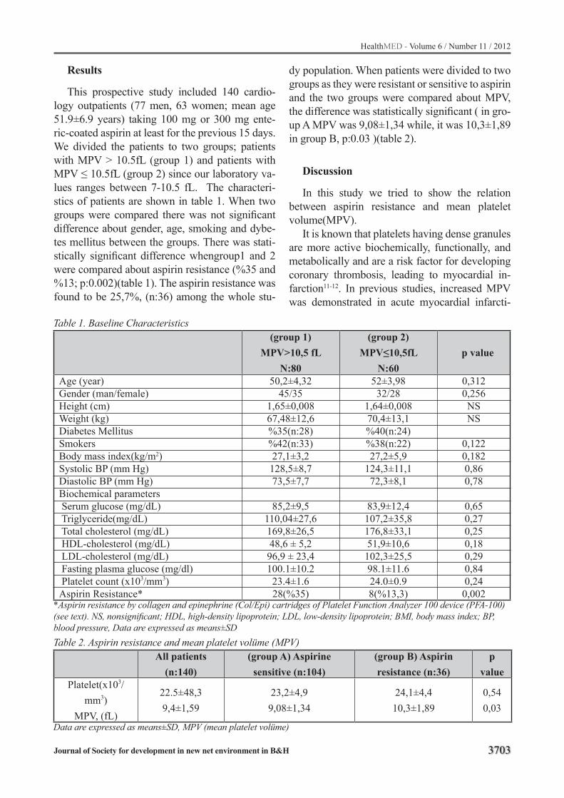
HealthMED - Volume 6 / Number 11 / 2012
Journal of Society for development in new net environment in B&H 3703
Results
This prospective study included 140 cardio-logy outpatients (77 men, 63 women; mean age 51.9±6.9 years) taking 100 mg or 300 mg ente-ric-coated aspirin at least for the previous 15 days. We divided the patients to two groups; patients with MPV > 10.5fL (group 1) and patients with MPV ≤ 10.5fL (group 2) since our laboratory va-lues ranges between 7-10.5 fL. The characteri-stics of patients are shown in table 1. When two groups were compared there was not significant difference about gender, age, smoking and dybe-tes mellitus between the groups. There was stati-stically significant difference whengroup1 and 2 were compared about aspirin resistance (%35 and %13; p:0.002)(table 1). The aspirin resistance was found to be 25,7%, (n:36) among the whole stu-
dy population. When patients were divided to two groups as they were resistant or sensitive to aspirin and the two groups were compared about MPV, the difference was statistically significant ( in gro-up A MPV was 9,08±1,34 while, it was 10,3±1,89 in group B, p:0.03 )(table 2).
Discussion
In this study we tried to show the relation between aspirin resistance and mean platelet volume(MPV).
It is known that platelets having dense granules are more active biochemically, functionally, and metabolically and are a risk factor for developing coronary thrombosis, leading to myocardial in-farction11-12. In previous studies, increased MPV was demonstrated in acute myocardial infarcti-
Table 1. Baseline Characteristics(group 1)
MPV>10,5 fLN:80
(group 2)MPV≤10,5fL
N:60 p value
Age (year)Gender (man/female)Height (cm)Weight (kg)Diabetes MellitusSmokersBody mass index(kg/m2)Systolic BP (mm Hg) Diastolic BP (mm Hg)Biochemical parameters Serum glucose (mg/dL) Triglyceride(mg/dL) Total cholesterol (mg/dL) HDL-cholesterol (mg/dL) LDL-cholesterol (mg/dL) Fasting plasma glucose (mg/dl) Platelet count (x103/mm3)Aspirin Resistance*
50,2±4,3245/35
1,65±0,00867,48±12,6%35(n:28)%42(n:33)27,1±3,2128,5±8,773,5±7,7
85,2±9,5110,04±27,6169,8±26,548,6 ± 5,296,9 ± 23,4100.1±10.223.4±1.628(%35)
52±3,9832/28
1,64±0,00870,4±13,1%40(n:24)%38(n:22)27,2±5,9
124,3±11,172,3±8,1
83,9±12,4107,2±35,8176,8±33,151,9±10,6102,3±25,598.1±11.624.0±0.98(%13,3)
0,3120,256NSNS
0,1220,1820,860,78
0,650,270,250,180,290,840,240,002
*Aspirin resistance by collagen and epinephrine (Col/Epi) cartridges of Platelet Function Analyzer 100 device (PFA-100) (see text). NS, nonsignificant; HDL, high-density lipoprotein; LDL, low-density lipoprotein; BMI, body mass index; BP, blood pressure, Data are expressed as means±SD Table 2. Aspirin resistance and mean platelet volüme (MPV)
All patients (n:140)
(group A) Aspirine sensitive (n:104)
(group B) Aspirin resistance (n:36)
p value
Platelet(x103/mm3)
MPV, (fL)
22.5±48,39,4±1,59
23,2±4,99,08±1,34
24,1±4,410,3±1,89
0,540,03
Data are expressed as means±SD, MPV (mean platelet volüme)
3704
HealthMED - Volume 6 / Number 11 / 2012
Journal of Society for development in new net environment in B&H
on12, unstable angina pectoris13, congestive he-art failure14 and coronary artery ectasia15. In acute coronary syndromes, platelet activation plays a considerable role16. Larger platelets secrete high levels of prothrombogenic thromboxane A2, se-rotonin, betathromboglobulin, and procoagulant membrane proteins like P-selectin and glycopro-tein IIIa12-13. In addition, they are less sensitive to inhibitory effects of prostacyclin on aggregation and secretion than small platelets12-16. Some inve-stigator examined platelet activation in subjects with no known cardiovascular disease in order to show the role of platelet in early stage and also progression of atherosclerosis. Fusegawa et al.17
showed increased platelet agreeability in hyper-tensive patients with carotid artery plaque and free of cardiovascular and schemic heart disease or stroke. Kurrelmeyer et al18. demonstrated increa-sed platelet activity in asymptomatic individuals with family histories of premature coronary artery disease (CAD).
Despite the development of newer antiplatelet drugs in the last decade, aspirin is still the most widely used antiplatelet agent across the world to prevent cardiovascular diseases4-5. Long-term aspirin administration in patients at high risk of occlusive vascular events reduced up to 34% of nonfatal myocardial infarction (MI), 25% of non-fatal stroke, and 18% of all-cause mortality5. Cli-nically, aspirin resistance is known as repetition of atherothrombotic events despite of sufficient anti platelet treatment. Aspirin resistance rates differs between %0.4 and %83.3 according to different studies containing diferent populations of patients and using different methods. Gum et al reported aspirin resistance rate as 31% in 326 patients with stable coronary artery disease using PFA-100 met-hod. Coma-Canella et al reported resistance rate as 32% using the same method in patients with stable coronary artery disease. In our country, using PFA-100 method Pamukçu et al found as-pirin resistance rate as 23.4% in 505 patients with coronary artery disease while Akay et al found this rate as 27.5% in 280 healhty subjects. In our study we found aspirin resistance as 25.7% in 140 pati-ents with significant coronary artery disease using PFA-100 method. Narvaez et al reported aspirin resistance as 16% in a similar population using the same method.
As a result aspirin resistance is an important problem in patients with coronary artery disease and MPV is a parameter showing platelet activa-tion. In our study we found out that aspirin resi-stance rate was higher in patients with significant coronary artery disease whose MPV values were higher. We believe that aspirin resistance can be determined in a short period of time using PFA 100 method in patients with coronary artery disea-se and high MPV values so that we can administer antiaggregan treatments more effectively. Thus this will contribute to improvement of mortality and morbidity of these patients. But it is necessary to support this study with further prospective stu-dies in larger populations of patients.
References
1. Massberg S, Schulz C, Gawaz M. Role of platelets in the pathophysiology of acute coronary syndrome. Se-min Vasc Med 2003;3(2):147-62
2. Thombson CB, Eaton K, Princiotta SM, Rushin CA, Valeri CR. Size dependent platelet subpopulati-ons: relationship of platelet volume to ultrastructu-re, enzymatic activity, and function. Br J Haematol 1982;50(3):509-19
3. Tsiara S, Elisaf M, Jagroop IA, Mikhailidis DP. Plate-lets as predictors of vascular risk: is there a practical index of platelet activity? Clin Appl Thromb Hemost 2003;9(3):177-90
4. Baigent C, Collins R, Appleby P, Parish S, Sleight P, Peto R. ISIS-2: 10 year survival among patients with suspected acute myocardial infarction in randomised comparison of intravenous streptokinase, oral aspi-rin, both, or neither. The ISIS-2 (Second International Study of Infarct Survival) Collaborative Group. BMJ. 1998 ;316(7141):1337-43
5. C. Baigent, C. Sudlow, R. Collins, and R. Peto, “Co-llaborative meta-analysis of randomised trials of an-tiplatelet therapy for prevention of death, myocardial infarction, and stroke in high risk patients,” BMJ. 2002;324(7329):71-86.
6. C. Patrono, L. A. Garcia Rodriguez, R. Landolfi, and C.Baigent, “Low-dose aspirin for the prevention of atherothrombosis,”The New England Journal of Me-dicine, 2005;353(22):2373-83.
7. A. D.Michelson, M. Cattaneo, J.W. Eikelboom, et al. “Aspirin resistance: position paper of the Working Group on Aspirin Resistance,” Journal of Thrombosis and Haemostasis, 2005;3(6): 1309–11

HealthMED - Volume 6 / Number 11 / 2012
Journal of Society for development in new net environment in B&H 3705
8. Michos ED, Ardehali R, Blumenthal RS, Lange RA, Ardehali H, “Aspirin and clopidogrel resistance,” Mayo Clinic Proc.2006;81(4):518-26
9. Chobanian AV, Bakris GL, Black HR, Cushman WC, Green LA, Izzo JL Jr, et al. The Seventh Report of the Joint National Committee on Prevention, Detection, Evaluation, and Treatment of High Blood Pressure: The JNC 7 report. JAMA 2003; 289(19): 2560– 72
10. American Diabetes Association. Diagnosis and cla-ssification of diabetes mellitus. Diabetes Care 2007; 30 Suppl1: S42– 7
11. Senaran H, Ileri M, Altinbas A, Kosar A, Yetkin E, Ozturk M, et al. Thrombopoietin and mean plate-let volume in coronary artery disease. Clin Cardiol 2001; 24(5):405-8
12. Endler G, Klimesch A, Sunder-Plassmann H, Schillinger M, Exner M, Mannhalter C, et al. Mean platelet volume is an independent risk factor for myocardial infarction but not for coronary artery di-sease. Br J Haematol 2002; 117(2):399-404.
13. Martin JF, Bath PM, Burr ML. Influence of platelet size on outcome after myocardial infarction. Lancet 1991;338(8780):1409-11.
14. Erne P, Wardle J, Sanders K, Lewis SM, Maseri A. Mean platelet volume and size distribution and their sensitivity to agonists in patients with coronary ar-tery disease and congestive heart failure. Thromb Haemost 1988;59(2):259-63.
15. Bitigen A, Tanalp AC, Elonu OH, Karavelioglu Y, Ozdemir N. Mean platelet volume in patients with isolated coronary artery ectasia. J Thromb Throm-bolysis 2007;24(2):99-103
16. Yilmaz MB, Cihan G, Guray Y, Guray U, Kisacik HL, Sasmaz H, et al. Role of mean platelet volume in triagging acute coronary syndromes. J Thromb Thrombolysis 2008;26(1):49-54.
17. Fusegawa Y, Hashizume H, Okumura T, Deguchi Y, Shina Y, Ikari Y, et al. Hypertensive patients with ca-rotid artery plaque exhibit increased platelet aggre-gability Thromb Res 2006;117(6):615-22.
18. Kurrelmeyer K, Becker L, Becker D, Yanek L, Gol-dschmidt-Clermont P, Bray PF. Platelet hyperreacti-vity in women from families with premature atheros-clerosis. J Am Med Womens Assoc 2003;58(4):272-7.
19. Sahin DY, Koç M, Caylı M, Uysal OK, Karaarslan O, Kanadaşı M, at all, The frequency of aspirin re-sistance by a modified thrombelastography method and its relationship with clinical and laboratory parameters in patients with stable coronary artery disease, Arch Turk Soc Cardiol. 2012;40(1):33-40.
20. Lordkipanidzé M, Pharand C, Schampaert E, Tur-geon J, Palisaitis DA, Diodati JG. A comparison of six major platelet function tests to determine the prevalence of aspirin resistance in patients with stable coronary artery disease. Eur Heart J 2007(14);28:1702-8
21. Gum PA, Kottke-Marchant K, Welsh PA, White J, Topol EJ. A prospective, blinded determination of the natural history of aspirin resistance among sta-ble patients with cardiovascular disease. J Am Coll Cardiol 2003;41(6):961-5
22. Coma-Canella I, Velasco A, Castano S. Prevalence of aspirin resistance measured by PFA-100. Int J Cardiol 2005;101(1):71-6
23. Pamukcu B, Oflaz H, Onur İ, Öncul A, Umman B, Koylan N, et al. Aspirin-resistant platelet aggrega-tion in a cohort of patients with coronary heart di-sease. Blood Coagul Fibrinolysis 2007;18(5):461-5
24. Akay OM, Cantürk Z, Akın E, Bal C, Gülbas Z. As-pirinresistance frequency: a prospective study in 280 healthy Turkish volunteers. Clin Appl Thromb He-most 2009;15(1):98-102
25. Narvaez I, Sagastagoitia JD, Vacas M, Saez Y, Lafita M, Monica S, et al. Prevalence and biologic profile of aspirin resistance in patients with angiographi-cally proven coronary artery disease. Thromb Res 2007;120(5):671-7
Corresponding AuthorKemal Karaagac,Kardiyoloji Klinigi, Bursa Yüksek İhtisas Eğitim ve Araştırma Hastanesi,Yıldırım, Bursa,Turkey ,E-mail: [email protected]

3706
HealthMED - Volume 6 / Number 11 / 2012
Journal of Society for development in new net environment in B&H
Extended-spectrum β-lactamase-producing E.coli and Klebsiella pneumoniae isolated from urinary tract infections in Milad Hospital, Tehran, IranLeila Arbabi1, Mohammad Rahbar1,2, Mosadegh Jabbari3, Mona Mohammad-Zadeh2, Leila Azimi1, Amirmorteza Ebrahimzadeh Namvar1, Abdolaziz Rastegar Lari1
1 Antimicrobial Resistance Research center, Tehran University of Medical sciences, Tehran, Iran2 Department of Microbiology, Iranian Reference Health laboratory. Tehran Iran and Department of Microbiology, Milad Hospital, Tehran, Iran 3 Department of Nephrology, Rasoul Akram Hospital, Tehran University of Medical Sciences, Tehran, Iran
Abstract
Aim: The aim of this study was To determine the prevalence of extended spectrum beta-lacta-mase (ESBL) producing Escherichia coli and Klebsiella pneumoniae strains isolated from uri-nary tract infections in Miald Hospital in Tehran.
Materials and methods: A total of 885 E.coli and 110 Klebsiella pneumoniae strains isolated from 16530 urine cultures, which were processed between March to June 2011in Milad hospital, Te-hran, Iran, were included to the study. Identifica-tion of bacteria and antibiotic susceptibility tests were performed by disk diffusion. The double-disk test was used for ESBL detection.
Results: The rate of resistance to Ampicillin is the highest and the rate of susceptibility to Imipe-nem is the lowest in both E. coli and K. pneumo-nia. 33% of E. coli and 30% of K. pneumoniae identified as an ESBL producer isolates.
Conclusion: The Spreading of ESBL produ-cing strains are a major concern, because it causes limitations to the antimicrobial drugs for optimal treatment of patients. The most reliable and effec-tive antimicrobial treatment for infections caused by ESBL microorganism is imipenem. Strict anti-biotic policies adopted in hospitals limit the indis-criminate use of antimicrobial agents, in order to steps towards decreasing this resistance.
Keywords: ESBL detection, E. coli, K. pneu-moniae, antimicrobial susceptibility
Introduction
Urinary tract infection is identified as a serio-us health problem worldwide that affects millions of humans every year (1). Species of the family Enterobacteriaceae such as Escherichia coli and Klebsiella pneumoniae are responsible for the majority of urinary tract infection (2). The family Enterobacteriaceae mainly by Escherichia coli, Klebsiella pneumoniae and Klebsiella oxyto-ca produces extended spectrum β-lactamases (ESBLs) enzymes (3). The heavy used of exten-ded-spectrum cephalosporins or β-lactams and monobactams have led to create of ESBL produ-cer isolates.
The produce of extended-spectrum β-lactamase (ESBL) enzymes is the main mechanism of resi-stance to broad-spectrum cephalosporins. Most ESBL enzymes are encoded on plasmid and can be easily transmitted to other bacteria. Exten-ded-spectrum β-lactamases are enzymes that can hydrolyze broad-spectrum cephalosporins, parti-cularly cefotaxime and ceftazidime (4,5, 21).
Increasing resistance to extended-spectrum cephalosporins among the Enterobacteriaceae fa-mily restricts the role of beta-lactam antibiotics in the empirical treatment of urinary tract infec-tion (6). Recently, Infections by microorganisms producing ESBL pose challenging infection con-trol problems in many hospitals. The prevalence and spreading of ESBL producing bacteria such as Klebsiella pneumoniae and E. coli have been varied in many hospitals (7,8, 20). Delay in repor-ting of ESBL production by E. coli and Klebsiella
HealthMED - Volume 6 / Number 11 / 2012
Journal of Society for development in new net environment in B&H 3707
pneumoniae is associated with a prolonged hos-pital stay, increase health care costs, morbidity and motility (9). Fluoroquinolones, especially le-vofloxacin and ciprofloxacin, are valuable in the treatment of urinary tract infections. Ciprofloxacin has been demonstrated to be more effective than aminoglycosides and trimethoprim-sulfamethoxa-zole for the clinical treatment of urinary tract in-fections (10).
Resistance to the expanded-spectrum cephalos-porins has been observed in species of the family Enterobacteriaceae such as E. coli and Klebsiella pneumoniae. The various global report of high in-cidence rates and outbreaks of ESBL-producing bacteria emphasizes the importance of searching and detecting their occurrence in all hospitals (11, 12). However, all these characteristics make ESBL isolates a serious infection control problem in the health care setting. Therefore, this study was performed to determine the prevalence of exten-ded spectrum β-lactamases among clinical isolates of K. pneumoniae and E. coli recovered from uri-nary tract infections in a Milad hospital in Tehran.
Materials and methods
This present study was carried out investigating ESBL detection and susceptibility pattern of clini-cal isolates of E. coli and Klebsiella pneumoniae, collected during the 3-month, between March and June, a total of 16530 urine samples were proce-ssed for significant bacteruria in clinical Microbi-ology Laboratory at Antimicrobial Resistance Re-search center. The mid-stream urine samples were initially cultured in EMB medium, then specific biochemical tests such as SIM, TSI, Citrate, and Urea has been performed. This study was perfor-med on 16530urine gram-negative bacilli isolates including 885 strains of E. coli and 110 strains of K. pneumoniae. Susceptibility pattern of Cepha-lothin (30 μg), Ceftizoxime (30 μg), ceftriaxone (30 μg), Cefotaxime (30 μg), Ceftazidime (30 μg), Carbenicillin (100 μg), ), Ampicillin (10 μg), Aztreonam (30 μg), Imipenem (10 μg), Genta-micin (10 μg), Amikacin (30 μg), Tobramycin (10 μg), Ciprofloxacin (5 μg), ofloxacin (5 μg), Nalidixic acid (30 μg) , Nitrofurantoin (300 μg), Tetracycline (30 μg), Trimethoprimsulfamethoxa-zole (1.25/23.75 μg) were determined according
to Clinical Laboratory Standards Institute (CLSI) guideline (CLSI,2006). All the mentioned disks were used by MAST Company in U.K.
Phenotypic Detection of ESBLCeftazidime resistant isolates were examined
for producing ESBL. Double disk method was conducted for detection of this enzyme. This met-hod explained previously. Briefly, a disc of Cefta-zidim (30μg) alone and in combination with Cla-vulonicAcid (30μg/10μg) and Cefotaxime (30 μg ) alone and in combination with ClavulonicAcid (30μg/10μg) were placed at the distance of 20mm center to center onto Mueller Hinton agar plate inoculated with a bacterial concentration of 1:100 suspension of 0.5 Mc Farland turbidity standards and incubated overnight at 37°c. A positive test result was defined as a ≥ 5mm increase in inhibiti-on hallo compared with a disk without clavulanic acid.
The quality control check for routine suscepti-bility testing was done once weekly using the refe-rence strains E. coli ATCC 25922, Staphylococcus aureus ATCC 29212 and Pseudomonas aerugino-sa ATCC 27853. ESBLs positive E. coli ATCC 35218 and k. pneumoniae ATCC 700603 was used to check the results combination disk method that used for detecting ESBL. All data were analyzed using SPSS 18, and statistical analysis was per-formed.
Results
273 patients were isolated ESBL producing bacteria, that were 81% females and 19% males and 211 were outpatients, and 62 were hospita-lized. The age of patients included in this study ranged from 0 and 82 years. A total of 16530 urine samples was studied. During this study, a total of 885 E. coli and 110 K. pneumoniae were isolated from a range of clinical specimens of patients hos-pitalised in Milad Hospital, Tehran, Iran. Results of antibiotic susceptibility of E. coli and Klebsi-ella pneumoniae have determined based on CLSI standard (Table.1 and 2 respectively). The ESBL phenotype based on positive combination disk test was detected in 332 (89.5%) of the isolates of 33% of E. coli species and 30% of Klebsiella pneumo-
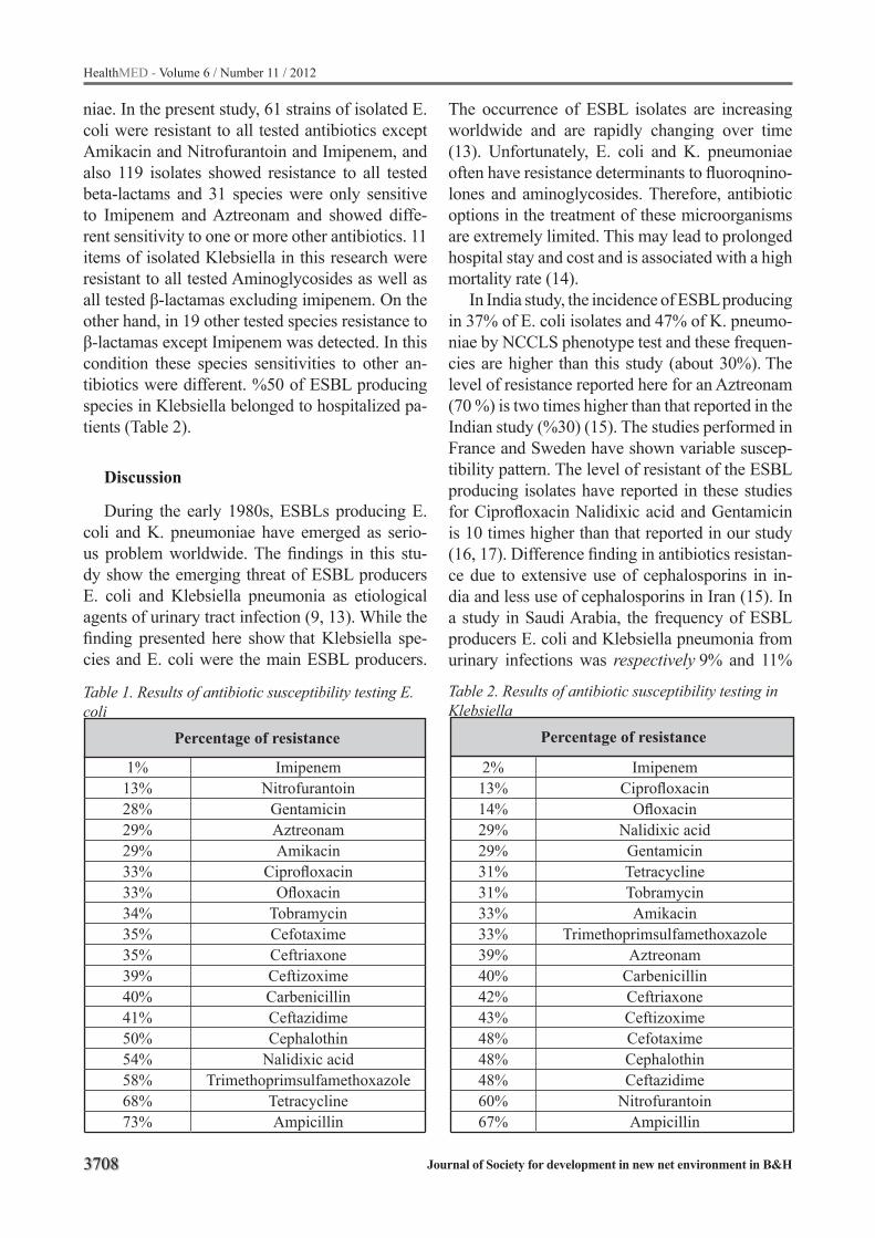
3708
HealthMED - Volume 6 / Number 11 / 2012
Journal of Society for development in new net environment in B&H
Table 1. Results of antibiotic susceptibility testing E. coli
ImipenemNitrofurantoin
GentamicinAztreonamAmikacin
CiprofloxacinOfloxacin
TobramycinCefotaximeCeftriaxoneCeftizoximeCarbenicillinCeftazidimeCephalothin
Nalidixic acidTrimethoprimsulfamethoxazole
TetracyclineAmpicillin
1%13%28%29%29%33%33%34%35%35%39%40%41%50%54%58%68%73%
Percentage of resistance
Table 2. Results of antibiotic susceptibility testing in Klebsiella
ImipenemCiprofloxacin
OfloxacinNalidixic acidGentamicinTetracyclineTobramycinAmikacin
TrimethoprimsulfamethoxazoleAztreonam
CarbenicillinCeftriaxoneCeftizoximeCefotaximeCephalothinCeftazidime
NitrofurantoinAmpicillin
2%13%14%29%29%31%31%33%33%39%40%42%43%48%48%48%60%67%
Percentage of resistance
niae. In the present study, 61 strains of isolated E. coli were resistant to all tested antibiotics except Amikacin and Nitrofurantoin and Imipenem, and also 119 isolates showed resistance to all tested beta-lactams and 31 species were only sensitive to Imipenem and Aztreonam and showed diffe-rent sensitivity to one or more other antibiotics. 11 items of isolated Klebsiella in this research were resistant to all tested Aminoglycosides as well as all tested β-lactamas excluding imipenem. On the other hand, in 19 other tested species resistance to β-lactamas except Imipenem was detected. In this condition these species sensitivities to other an-tibiotics were different. %50 of ESBL producing species in Klebsiella belonged to hospitalized pa-tients (Table 2).
Discussion
During the early 1980s, ESBLs producing E. coli and K. pneumoniae have emerged as serio-us problem worldwide. The findings in this stu-dy show the emerging threat of ESBL producers E. coli and Klebsiella pneumonia as etiological agents of urinary tract infection (9, 13). While the finding presented here show that Klebsiella spe-cies and E. coli were the main ESBL producers.
The occurrence of ESBL isolates are increasing worldwide and are rapidly changing over time (13). Unfortunately, E. coli and K. pneumoniae often have resistance determinants to fluoroqnino-lones and aminoglycosides. Therefore, antibiotic options in the treatment of these microorganisms are extremely limited. This may lead to prolonged hospital stay and cost and is associated with a high mortality rate (14).
In India study, the incidence of ESBL producing in 37% of E. coli isolates and 47% of K. pneumo-niae by NCCLS phenotype test and these frequen-cies are higher than this study (about 30%). The level of resistance reported here for an Aztreonam (70 %) is two times higher than that reported in the Indian study (%30) (15). The studies performed in France and Sweden have shown variable suscep-tibility pattern. The level of resistant of the ESBL producing isolates have reported in these studies for Ciprofloxacin Nalidixic acid and Gentamicin is 10 times higher than that reported in our study (16, 17). Difference finding in antibiotics resistan-ce due to extensive use of cephalosporins in in-dia and less use of cephalosporins in Iran (15). In a study in Saudi Arabia, the frequency of ESBL producers E. coli and Klebsiella pneumonia from urinary infections was respectively 9% and 11%
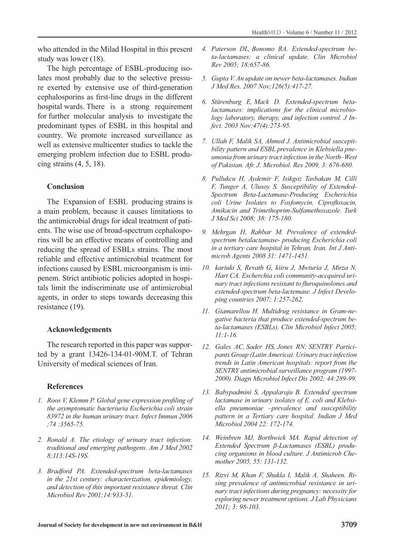
HealthMED - Volume 6 / Number 11 / 2012
Journal of Society for development in new net environment in B&H 3709
who attended in the Milad Hospital in this present study was lower (18).
The high percentage of ESBL-producing iso-lates most probably due to the selective pressu-re exerted by extensive use of third-generation cephalosporins as first-line drugs in the different hospital wards. There is a strong requirement for further molecular analysis to investigate the predominant types of ESBL in this hospital and country. We promote increased surveillance as well as extensive multicenter studies to tackle the emerging problem infection due to ESBL produ-cing strains (4, 5, 18).
Conclusion
The Expansion of ESBL producing strains is a main problem, because it causes limitations to the antimicrobial drugs for ideal treatment of pati-ents. The wise use of broad-spectrum cephalospo-rins will be an effective means of controlling and reducing the spread of ESBLs strains. The most reliable and effective antimicrobial treatment for infections caused by ESBL microorganism is imi-penem. Strict antibiotic policies adopted in hospi-tals limit the indiscriminate use of antimicrobial agents, in order to steps towards decreasing this resistance (19).
Acknowledgements
The research reported in this paper was suppor-ted by a grant 13426-134-01-90M.T. of Tehran University of medical sciences of Iran.
References
1. Roos V, Klemm P. Global gene expression profiling of the asymptomatic bacteriuria Escherichia coli strain 83972 in the human urinary tract. Infect Immun 2006 ;74 :3565-75.
2. Ronald A. The etiology of urinary tract infection: traditional and emerging pathogens. Am J Med 2002 8;113:14S-19S.
3. Bradford PA. Extended-spectrum beta-lactamases in the 21st century: characterization, epidemiology, and detection of this important resistance threat. Clin Microbiol Rev 2001;14:933-51.
4. Paterson DL, Bonomo RA. Extended-spectrum be-ta-lactamases: a clinical update. Clin Microbiol Rev 2005; 18:657-86.
5. Gupta V. An update on newer beta-lactamases. Indian J Med Res. 2007 Nov;126(5):417-27.
6. Stürenburg E, Mack D. Extended-spectrum beta-lactamases: implications for the clinical microbio-logy laboratory, therapy, and infection control. J In-fect. 2003 Nov;47(4):273-95.
7. Ullah F, Malik SA, Ahmed J. Antimicrobial suscepti-bility pattern and ESBL prevalence in Klebsiella pne-umonia from urinary tract infection in the North–West of Pakistan. Afr. J. Microbiol. Res 2009, 3: 676-680.
8. Pullukcu H, Aydemir F, Isikgoz Tasbakan M, Cilli F, Tunger A, Ulusoy S. Susceptibility of Extended-Spectrum Beta-Lactamase-Producing Escherichia coli Urine Isolates to Fosfomycin, Ciprofloxacin, Amikacin and Trimethoprim-Sulfamethoxazole. Turk J Med Sci 2008; 38: 175-180.
9. Mehrgan H, Rahbar M. Prevalence of extended-spectrum betalactamase- producing Escherichia coli in a tertiary care hospital in Tehran, Iran. Int J Anti-microb Agents 2008 31: 1471-1451.
10. kariuki S, Revath G, kiiru J, Mwturia J, Mirza N, Hart CA. Escherchia coli community-accquired uri-nary tract infections resistant to fluroquinolones and extended-spectrum beta-lactemase. J Infect Develo-ping countries 2007; 1:257-262.
11. Giamarellou H. Multidrug resistance in Gram-ne-gative bacteria that produce extended-spectrum be-ta-lactamases (ESBLs). Clin Microbiol Infect 2005; 11:1-16.
12. Gales AC, Sader HS, Jones RN; SENTRY Partici-pants Group (Latin America). Urinary tract infection trends in Latin American hospitals: report from the SENTRY antimicrobial surveillance program (1997-2000). Diagn Microbiol Infect Dis 2002; 44:289-99.
13. Babypadmini S, Appalaraju B. Extended spectrum lactamase in urinary isolates of E. coli and Klebsi-ella pneumoniae –prevalence and susceptibility pattern in a Tertiary care hospital. Indian J Med Microbiol 2004 22: 172-174.
14. Weinbren MJ, Borthwick MA. Rapid detection of Extended Spectrum β-Lactamases (ESBL) produ-cing organisms in blood culture. J Antimicrob Che-mother 2005, 55: 131-132.
15. Rizvi M, Khan F, Shukla I, Malik A, Shaheen. Ri-sing prevalence of antimicrobial resistance in uri-nary tract infections during pregnancy: necessity for exploring newer treatment options. J Lab Physicians 2011; 3: 98-103.
3710
HealthMED - Volume 6 / Number 11 / 2012
Journal of Society for development in new net environment in B&H
16. Goldestin FW. Antibiotic susceptibility of bacterial strains isolated from patients with community-acqu-ired urinary tract infections in France. Multicentre study Group. Eur J Clin Microbiol 2000; 19:112-7.
17. Kahlmeter G. An international survey of the anti-microbial susceptibility of pathogens from uncom-plicated urinary tract infections: the ECO.SENS Project. . J. Antimicrob. Chemother 2003; 51:69-76.
18. Kader AA, Angamuthu K. Extended-spectrum beta-lactamases in urinary isolates of Escherichia coli, Klebsiella pneumoniaee and other gram-negative bacteria in a hospital in Eastern province, Saudi Arabia. Saudi Med J 2005; 26:956-9
19. Ghafourian S, Bin Sekawi Z, Sadeghifard N, Mohebi R, Kumari Neela V, Maleki A, Hematian A, Rhabar M, Raftari M, Ranjbar R. The Prevalence of ESBLs Producing Klebsiella pneumoniae Isolates in Some Major Hospitals, Iran. Open Microbiol J 2011; 5:91-5.
20. Meltem IŞIKGOZ TAŞBAKAN, Husnu PULLUK-CU, Oğuz Reşat SİPAHİ, Tansu YAMAZHAN, Bilgin ARDA, Sercan ULUSOY. A pooled analysis of the re-sistance patterns of Escherichia coli strains isolated from urine cultures in Turkey: a comparison of the periods 1997-2001 and 2002-2007. Turk J Med Sci 2011; 41: 557-564.
21. Özlem KURT AZAP, Hande ARSLAN, Sedef Ö. KA-RAMAN, Turhan TOGAN. Risk Factors for Fecal Carriage of Extended-Spectrum Beta-Lactamase Producing Escherichia coli and Klebsiella spp. in the Community. Turk J Med Sci 2007; 37: 31-38.
Corresponding AuthorAbdolaziz Rastegar Lari,Antimicrobial Resistance Research Center,Tehran University of Medical Sciences,Tehran, Iran,E-mail: [email protected]
HealthMED - Volume 6 / Number 11 / 2012
Journal of Society for development in new net environment in B&H 3711
Assessment of 5556 applications submitted to the rights of patient unit of a university hospitalYasemin Durduran1, Berrin Okka2, Said Bodur1, Hamide Dindas3
1 Selcuk University, Meram Medical School, Dept of Public Health, Konya, Turkey 2 Selcuk University, Meram Medical School, Dept of Medical History and Ethics, Konya, Turkey3 Selcuk University, Meram Medical School, Rights of Patient Unit, Konya, Turkey
Abstract
Objective: The first step in assessing applica-tions and the practice of the rights of patient in hospitals is the rights of patient unit. The practi-ces the patients are not satisfied with have to be determined in great detail in order to increase the patient satisfaction. This study was carried out to explicate the applications of the patients or their relatives to the rights of patient unit.
Methods: Study design: This descriptive arc-hive study was carried out in Konya (Turkey) in April-June 2011. Setting: In the study, the appli-cations made to the Rights of Patient Unit of Me-ram Medical Faculty of Selcuk University were examined. Participants: 5556 applications made in January-2006 and March-2011 were examined. Intervention(s): Register books the applications were registered in, complaint box forms, letters of applications, and the contents of the files compri-sing online applications were evaluated. The data were transcribed to verse forms prepared for this purpose. Main Outcome Measure(s): Independent variables of the study were demographic featu-res; and dependent variables were the department/person to whom the complaints were brought, the complaints, and the way they were responded. Statistics: The data were coded and assessed on electronic environment. The applications were expressed in a number of ways; however, they were summed up under 10 main topics. In descrip-tions the percentages were used, and in compari-sons chi-square was used.
Results: The number of applications submitted to the rights of patient unit tended to increase. 51% of the application forms 31% of which were submitted by women were made via complaint boxes and 31% directly. 59% of the applicants
were from the metropolis center, and the remaining were from long-distance. 85% of the applications were complaints, 15% were suggestions, deman-ds, and thanks. The first three of the application cases consisted of the communication problems by 19% rate, hitch in the service by 15% rate, and disapproval of physical conditions by 13%. 41% of the applications were about persons and 59% about institutions and service procedures. The personnel complained about the most were the physicians by 41% (P<0.001). The rate of the per-sonal applications increased every year (P<0,001). While the rate of suggestions and thanks decrea-sed, the rate of applications about the hitch in the service increased.
Conclusion: According to the findings of this study, the level of expectations of the patients and their relatives from hospitals is increasing. It can be concluded that the patients with similar pro-blems behave more self-confidently. Increasing the communication skills and acquainting the pa-tients with the contents of the patient rights may decrease the complaints.
Keywords: Rights of patients, the rights of pa-tient unit, complaint, suggestions.
Introduction
Rights of the patient is considered to be a sign of reflection of the human rights and values on medical care in developed societies, and it takes place among the issues of first priority of medical care institutions (1).
“Declaration of Helsinki,” one of the docu-ments concerning the rights of patient concept, which has completed several phases so far, was issued in 1964 (2). In early 1973, the American Hospital Association issued a patients bill of rights
3712
HealthMED - Volume 6 / Number 11 / 2012
Journal of Society for development in new net environment in B&H
(3). “Declaration of Lisbon on the Rights of Pati-ent,” adopted by the World Medical Assembly in 1981, is the first international document approved in this field (4). “Manifesto of Developing the Rights of Patient in Europe” was prepared in a congress held by the Regional Office for Europe of World Health Organization in 1984 in Amster-dam. In 1995 Declaration of Lisbon was reconsi-dered and updated, and its latest form was revised at the Council Session, Santiago, Chile, October 2005 (5, 6). As in the developed countries, the ne-cessary attempts to draw up a protocol have also been made in the direction of these manifestos in order to bear modern responsibilities concerning the rights of patients in accordance with our own conditions through regulations and instructions issued in our country (7, 8).
With the practice of patient rights that started in the direction of “Guidelines Related with the Practice of the Rights of the Patient in Health Fa-cilities” issued by the Ministry of Health in 2003, it was aimed that the patients and their relatives would be able to get use of their rights determined with the regulations, should be informed in every phase, protected from degradation, and be able to use legal ways of protection when needed. The practice of the rights of patient that had started in some hospitals was generalized all over the coun-try in 2004 (9). Published in 2005, ‘Patient Rights Enforcement Directive’ laid out the duties and working principles of both the ‘hospital patient rights committee’ and also the ‘rights of the pati-ent unit’ and defined the tasks of their employees with duties at the board and units (10).
Rights of Patients is a part of right to live he-althy , which provides them with having equal power to the personnel equipped with power that arises from their medical knowledge, and they are also the rules the institutions and personnel must obey. This gives the patients the right to claim right (11, 12).
It is important for the patients to know how to claim their rights. There are several ways of clai-ming rights one of which is to apply to the rights of patient unit (10). The Rights of Patient Unit is the first step in assessing the applications and practice of the rights of patient. In order to incre-ase the satisfaction, it is needed to determine, in detail, the practices the patients are not satisfied
with. It is the task of the rights of patients unit, in order to prevent abuse of the rights of patient, to accept the applications, to find solutions for ur-gent problems, to inform superiors, and to educate the personnel and the patients with respect to the rights of patient (9, 10). The main objective here is to enable the patient to apply to the unit and, if possible, to find solutions for the problems the patients have, or to take a written application form if not possible. The applications are processed and put in practice according to “Patient Rights Enfor-cement Directive” and they are submitted to the council that convenes at scheduled times. The de-cision of the council is issued to the applicant and the relevant medical personnel. The proposals are presented to the hospital management (13).
Evaluating the content of the applications submitted to the rights of patient unit will provi-de valuable information regarding the precautions and attitudes taken by the hospital management. In line with positive changes in the level of commu-nity awareness for patient rights, there would be inevitable changes with regard to both the beha-vior of staff assigned and also the claims of the patients for their issues.
This study was carried out to examine the applications submitted to the rights of patient unit by the patients and their relatives.
Methods
Study designThis descriptive archive study was carried out
in Konya (Turkey) in April-June 2011. Konya is an important metropolitan city in the Central Ana-tolia. It has a worldwide fame for its history and culture and thus it is famous as an international tourist city.
SettingThe applications submitted to the Rights of Pa-
tient Unit of a university hospital (Meram Medi-cal Faculty Hospital of Selcuk University) were examined carefully. It was the only hospital that served to more than three million people during the years this study was carried out. In the direc-tion of the filed archive data, in 2005 informatory seminaries on the rights of patient were conduc-
HealthMED - Volume 6 / Number 11 / 2012
Journal of Society for development in new net environment in B&H 3713
ted for the personnel, “the Rights of Patient Unit” started to serve in the end of 2005, and the patients were asked to put the papers on which they wrote their suggestions and complaints into the compla-int boxes placed at certain points in the hospitals. Later the BIMER (The Communication Centre of the Turkish Prime Ministry) complaints were re-ceived through telephone or e-mail. It was remar-kable that, while the files being scrutinized, some data had not been written down, so some descripti-ve data were missing. It was also detected that the complaint letters and the other correspondences were stored in the same files, although it decreased in time. Furthermore, it was detected that the form of report keeping changed in years.
PermissionsA written permission was taken from the Et-
hical Committee of Meram Medical Faculty of Selcuk University and Chief Physician of Meram Medical Faculty.
ParticipantsAll of 5556 applications submitted by the pa-
tients or their relatives between 01 January 2006 and 31 March 2011 were included. Owing to the fact that there were very few applications to the unit, the records kept in 2005 were not included.
Data sourcesJust like the applications submitted to the
Rights of Patient Unit in various ways, the records and the way they are kept varied. Only a few of the applications were written down in record bo-oks; the written complaints and BIMER compla-ints received via internet were kept in a different file. The complaints put into the complaint boxes were filed in different files without being recor-ded in the record books. Only was the information related with the records submitted in person and resolved in hospitals kept in the record books.
ProcedureFirst, information about the working system of
the Rights of Patient Unit was collected. The con-tents of all the record books in which the personal applications resolved in hospitals were recorded,
the complaint box forms, letters of application and the application received via internet were scruti-nized. The data were transcribed onto the infor-mation forms prepared specially. The form com-prised manner of applying to the rights of patient, the date of application, the gender, educational background, profession, place, relationship of the applicant with the patient, (if available) distinct characteristics of the patient, the unit/person com-plained about, the complaint and topic and how the complaint was responded.
Main outcome measure(s)The independent variables of the study were
the date of the application; the age, place, educati-onal background, profession of the applicant and his/her relation with the patient; and the way the application made. The dependent variables of the study were the department/person about whom the complaint was made, the complaint, and how they were responded.
Statistical analysis After the data being encoded, they were asse-
ssed on electronic environment. Although the applications were expressed in different ways, they were summarized under ten main topics con-sidering the frequency of the complaints. The per-centages were utilized in descriptions. Since some information about the patient and their relatives was missing, the percentages were taken from the present data independently from the way the applications made. Ki-square test was utilized in comparisons and p<0.05 was regarded as signifi-cance.
Results
Demographic findings5556 applications were submitted to the rights of
patients unit within the period of 5 years and three months. 69 % of the applicants were males, and 57% were under 45 years of age. While 57% of the applicants were the patients themselves, 43 % were the relatives of the patients. 59% of the applicants came from Konya city center. The civil servants took precedence (28%) when the applicants were classified according to their professions (Table 1).
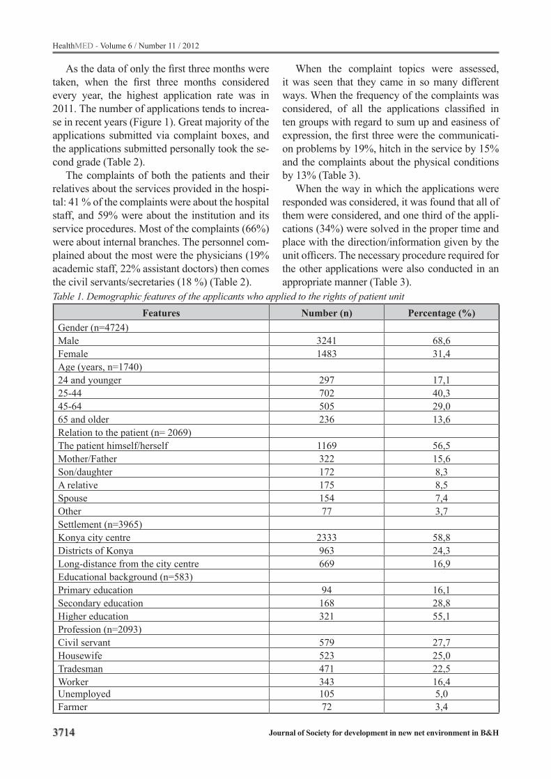
3714
HealthMED - Volume 6 / Number 11 / 2012
Journal of Society for development in new net environment in B&H
As the data of only the first three months were taken, when the first three months considered every year, the highest application rate was in 2011. The number of applications tends to increa-se in recent years (Figure 1). Great majority of the applications submitted via complaint boxes, and the applications submitted personally took the se-cond grade (Table 2).
The complaints of both the patients and their relatives about the services provided in the hospi-tal: 41 % of the complaints were about the hospital staff, and 59% were about the institution and its service procedures. Most of the complaints (66%) were about internal branches. The personnel com-plained about the most were the physicians (19% academic staff, 22% assistant doctors) then comes the civil servants/secretaries (18 %) (Table 2). Table 1. Demographic features of the applicants who applied to the rights of patient unit
Features Number (n) Percentage (%)Gender (n=4724)Male 3241 68,6Female 1483 31,4Age (years, n=1740)24 and younger 297 17,125-44 702 40,345-64 505 29,065 and older 236 13,6Relation to the patient (n= 2069)The patient himself/herself 1169 56,5Mother/Father 322 15,6Son/daughter 172 8,3A relative 175 8,5Spouse 154 7,4Other 77 3,7Settlement (n=3965)Konya city centre 2333 58,8Districts of Konya 963 24,3Long-distance from the city centre 669 16,9Educational background (n=583)Primary education 94 16,1Secondary education 168 28,8Higher education 321 55,1Profession (n=2093)Civil servant 579 27,7Housewife 523 25,0Tradesman 471 22,5Worker 343 16,4Unemployed 105 5,0Farmer 72 3,4
When the complaint topics were assessed, it was seen that they came in so many different ways. When the frequency of the complaints was considered, of all the applications classified in ten groups with regard to sum up and easiness of expression, the first three were the communicati-on problems by 19%, hitch in the service by 15% and the complaints about the physical conditions by 13% (Table 3).
When the way in which the applications were responded was considered, it was found that all of them were considered, and one third of the appli-cations (34%) were solved in the proper time and place with the direction/information given by the unit officers. The necessary procedure required for the other applications were also conducted in an appropriate manner (Table 3).

HealthMED - Volume 6 / Number 11 / 2012
Journal of Society for development in new net environment in B&H 3715
Table 2. Distribution of the application submitted to the rights of patient unit according to the years and way of application
Years (n=5556) Number (n) Percentage (%)2006 1122 20,22007 824 14,82008 647 11,62009 754 13,62010 1566 28,22011 (fırst three month) 643 11,6Way of application (n=5556)Complaint box 2817 50,7Personal application to the unit 1744 31,4Written application 707 12,7Via e-mail 288 5,2The Persons complained about (n=1667)Physician 682 40,9Civil servant/secretary 299 17,9Personnel of the service 282 16,9Nurse 233 14,0Technician / Laboratory assistant 153 9,2Other 18 1,1
Table 3. The reasons and result of the application to the rights of patient unitReasons of complaint/application (n=5556) Number (n) Percentage (%)
Communication problems 1045 18,8Hitch in the service 836 15,0Disapproval of physical conditions, insufficiency 740 13,3Complaints about the appointment system 671 12,1Statement of idea (suggestion-request) 626 11,3Not feeling comfortable in service 495 8,9Difficulties in procedures 483 8,7Not being informed, not being able to get information 373 6,7Thanks for the service they received 221 4,0Not being allowed to accept visitors, hours of visit 66 1,2Results of the application (n=5556)Treated without feedback 2728 49,1Informed 989 17,8Amended/directed given by the unit officers 901 16,2Administrative procedure 724 13,0Not worth for administrative procedure 214 3,9
Requests, ideas, and suggestions of the patients and their relatives:
11% of the applications were suggestions and 4 % thanks for the service they received (Table 3). The patients and their relatives made right and appropriate suggestions and requests some of which were given in Table 4 as examples.
Assessment of the applications with regard to the years:
When some characteristics of the applications were scrutinized, it was found that the number of the applications submitted in the first three months of the year was quite high when compared to other years (Figure 1). While half of the applications were submitted by the patients and the other half by their relatives in the first years, the rate of the applications made by the patients personally rose to 80% in recent years (p<0,001). Of all the 583
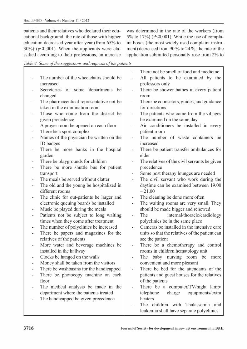
3716
HealthMED - Volume 6 / Number 11 / 2012
Journal of Society for development in new net environment in B&H
Table 4. Some of the suggestions and requests of the patients
- The number of the wheelchairs should be increased
- Secretaries of some departments be changed
- The pharmaceutical representative not be taken in the examination room
- Those who come from the district be given precedence
- A prayer room be opened on each floor- There be a sport complex- Names of the physician be written on the
ID badges- There be more banks in the hospital
garden - There be playgrounds for children- There be more shuttle bus for patient
transport - The meals be served without clatter- The old and the young be hospitalized in
different rooms- The clinic for out-patients be larger and
electronic queuing boards be installed- Music be played during the meals- Patients not be subject to long waiting
times when they come after treatment - The number of polyclinics be increased- There be papers and magazines for the
relatives of the patients- More water and beverage machines be
installed in the hallway- Clocks be hanged on the walls- Money shall be taken from the visitors - There be washbasins for the handicapped- There be photocopy machine on each
floor- The medical analysis be made in the
department where the patients treated- The handicapped be given precedence
- There not be smell of food and medicine- All patients to be examined by the
professors only - There be shower bathes in every patient
room- There be counselors, guides, and guidance
for directions- The patients who come from the villages
be examined on the same day- Air conditioners be installed in every
patient room- The number of waste containers be
increased- There be patient transfer ambulances for
elder- The relatives of the civil servants be given
precedence- Some post therapy lounges are needed - The civil servant who work during the
daytime can be examined between 19.00 – 21.00
- The cleaning be done more often- The waiting rooms are very small. They
should be made bigger and renewed- The internal/thoracic/cardiology
polyclinics be in the same place- Cameras be installed in the intensive care
units so that the relatives of the patient can see the patient
- There be a chemotherapy and control rooms in children hematology unit
- The baby nursing room be more convenient and more pleasant
- There be bed for the attendants of the patients and guest houses for the relatives of the patients
- There be a computer/TV/night lamp/telephone charge equipments/extra heaters
- The children with Thalassemia and leukemia shall have separate polyclinics
patients and their relatives who declared their edu-cational background, the rate of those with higher education decreased year after year (from 65% to 30%) (p<0,001). When the applicants were cla-ssified according to their professions, an increase
was determined in the rate of the workers (from 5% to 17%) (P<0,001). While the use of compla-int boxes (the most widely used complaint instru-ment) decreased from 90 % to 24 %, the rate of the application submitted personally rose from 2% to

HealthMED - Volume 6 / Number 11 / 2012
Journal of Society for development in new net environment in B&H 3717
62% (P<0,001). As the goal rate of the complaint about institution/hospital staff the rate 55% and 45% altered against hospital staff year after year (P<0,001). In the last two years, there was a signi-ficant increase in the rate of the complaints about the academic staff (from 10% to 29%) (P<0,001). Furthermore, while the rate of the suggestions-thanks was decreasing (from 19% to 11%), the rate of the applications that do not need transac-tion was increasing (from 1% to 11%) year after year (P<0,05).
Discussion
The findings of this study, carried out in order to scrutinize the applications submitted to the rights of patient unit by the patient and their relatives, is limited with the data recorded in the records of the unit. The missing information from the recor-ds complicated the study. Some basic information was missing from the complaint forms; therefo-re, the percentages were taken from the present data, and they were deemed to reflect all the data. However, the differences reflecting on the records year after year and the fact that all of the same information was not available was another diffi-culty encountered during the study. As the diffe-rent forms utilized in the ways of complaints did not include the same basic information, or even if they included, the information deficiency was explicitly seen owing to the fact that the filling of the complaint forms were not requisite. The fact that the in-house correspondence concerning the institution and the applications submitted to the unit were filed together was one of the difficulties encountered during the study; however, the fact that all the information found in the registers were utilized reflects the authenticity of the study.
Figure 1. The number of applications submitted in the first 3 months of each year
According to the data of the Turkish Statistical Institution issued in 2010, while females have he-alth problems more often and use more medical service (14), according to results of our study, two third of those who applied to the rights of pati-ents unit (69%) were males. Although females had more health problems, males applied to the rights of patient unit more, and this can be explained with the traditional interfamilial role share. In this family structure, the spokespersons are usually the men in the families.
The studies showed that the rate of the females applied to the rights of patient unit was higher than males, however there was no explanation given for this (15, 16). More than half of the applicants were the patients themselves. This is an expected and a good situation. The fact that it was the pati-ents themselves that submitted the complaints was highly important because it helped to take the com-plaints from the first persons. It is not customary in our society to complain personally. Therefore, this is very good and should be encouraged. The studies carried out for this purpose showed that the age and health of the patient played an impor-tant role, that complaints were mostly submitted by the relatives of children and old patients, and that the complaints were submitted by the patients personally when they could cooperate (17). The information can be acquired while the iron is hot without the interference of second persons by deli-vering the patients envelopes to fill in the services.
The fact that more than half of the applications were from the city center is normal because most of the patients were from the city center. In a pe-riod during which urbanization tended to increase all over the world, since the big regional and state hospitals or mediocre hospitals were chosen in the studies carried out on this subject due to the fact that data collection was easier, it happened that most of the applications were from city centers (18). The reason why one fifth of the applications were civil servants and they took the precedence can be explained with their being more courageo-us in self expression owing to their higher edu-cational background. The rise in the rate of the workers’ complaints year by year can be explained with the fact that the claim of rights became ea-sier and the awareness of the society increased. A study carried out in the United States of America
3718
HealthMED - Volume 6 / Number 11 / 2012
Journal of Society for development in new net environment in B&H
showed that the patients with regular monthly in-come claim rights more than the unemployed (19).
In spite of the amendments to the replies to the earlier complaints, the rate of the applications for the rights of patient is increasing. This can be explained with the increase in the awareness of the society about the topic. Furthermore, the increase in the personal applications is favorable with re-gard to the direct communication between the in-stitution and the people to whom the health servi-ce was introduced. This shows that the process of demanding rights is getting better in the direction of democratization in Turkey. It is important that increasing the level of knowledge and awareness of the society of right claim and encouraging them to take on more responsibilities and to take part in the decision process be among the policies of the ministry (20). Besides, the rights of consumer take place on the agenda more, and the rights of pati-ents may be its reflection. The studies show that the complaints are increasing all over the world, which may be based on the fact that the patients are thought to be customers whose satisfaction, takes the precedence (21, 22). When it is taken into consideration with regard to the institution/hospital staff, the gradual tends in the applications towards hospital staff can be comprehended as the increase in the concretization and clarification in the complaint topics. It is a good development that the society should have a say by participating the process. On the other hand, owing to the recovery in the substructure and service procedures of the institution with the onion skin impact, the compla-ints may be directed towards the persons. It can be comprehended as a productive and easier way to complain about the hospital staff instead of insti-tutions. Another reason why the complaints about the hospital staff increased may be the result of the increase in the expectations (23).
Most of the complaints were about the inter-nal branches, so it was not surprising that two third of the applications were made to the inter-nal branches. In a study carried out in Canada, it was stated that the rate of the complaints about the branches with more patients was higher than the branches with fewer patients (24).
On the other hand, because of the fact that the chain of patient transfer cannot be completely for-med, the patients who can be treated in the lower
grade hospitals without applying to the university hospitals directly apply to the university hospitals might have increased the rate of the complaints about the internal branches.
The fact that nearly half of the complaints are about the physicians may be the result of the fact that each patient meets the physicians and has high expectations from them because the people who meet the physicians is feeling sorrow and pain, and they come to the physicians with the identity of a patient rather than their real identities. The main point to specify here is that the rate of the complaints about the academic staff increased in the last two years. This may be the result of the fact that the physicians do not only examine the patients because they work in a research and trai-ning hospital. Since the physicians have more wor-kload, the patients may have to wait longer, which may cause the patients to complain more about them. The civil servants and the secretaries take the second rank among those who are complained about. A study carried out by Bark et al confirms this view. In that study the physicians take the first rank, the registrars the second, and the nurses the third rank among those who are complained about (25). It was stated in the same study that the attitu-des of the medical personnel took the precedence as the most significant complaint. This shows that our study is congruent with common expectations. The complaints started first about the hospitals, then the physicians and other medical personnel, and a situation in which the complaints are expre-ssed more clearly and in greater detail will take place in future. Therefore, new forms comprising of the details which objectifies the incidents with more information about which point and aspect of the incident is complained about should be made.
The communication problems take the prece-dence among the complaints, and the hitch of ser-vice comes after it, and it is followed by the com-plaints about physical conditions. In a study carri-ed on by Taylor et al on the topics of the compla-ints of the patients who applied to the emergency services, they found that the complaints that took the precedence by 33.4 % was about the treatment, second was about communication by 31.6%, and third was about the delay in treatment(26).
It is startling that the biggest problem is the
HealthMED - Volume 6 / Number 11 / 2012
Journal of Society for development in new net environment in B&H 3719
communication. It varies in every country, but as we are a susceptible nation, we may get easily irritated and give a sudden rebuke. The commu-nication problems may cause defensive medical practices, which may affect the motivation of the medical personnel (27, 28).
Communication is an important procedure to be followed both by the patient and medical per-sonnel. It should be considered that everybody has their own cultural characteristics, and they may act selfishly when they get ill, so they may expect the service as they wish. It may be a solution for some of the communication problems to make those who expect service accustomed to coming in time for the appointments and to observe the fixed time. The service should be offered in a reasonable and professional way. Both those who serve and also the ones who take the service should be made con-scious of this issue (29, 30).
Inadequacy of physical places may also cause communication problems. For example, if there are fewer seats than the number of the patients in the lounge, it makes it difficult for the patients to wait, and may cause the patients who has grown inpatient in the lounge to have problems with the personnel. These kinds of complaints are related with logistic problems and physical adequacy, but it may vary in different countries or different re-gions in the same country. In a study on the emer-gency services, these kinds of complaints were not assessed while significant rates of the complaints about communication problems were considered worth assessment (31).
According to the types of the solutions for pro-blems cited in the applications, one third of the problems were solved in the convenient time and place thanks to notification and guidance of the officers in the unit. This suggests that the infor-mation deficiency in the applications may cause problems, so it is thought that the rate of patient satisfaction will increase and the rate of applica-tions submitted to the rights of patients unit will decrease thanks to the satisfactory information provided for the patient (32).
The fact that the patients’ personal applications rose to 80% in recent years can be the reflection of the patients’ acceptation of the rights of pati-ent units and the proportional increase in the level of awareness of the rights of patients. The results
stated is congruent with other studies carried out in developed countries. It may broadly indicate that the dissatisfaction rose because the level of expectations of the patients have risen; the stan-dard of life is rising; and we have older population owing to fact that the life span has been lengthe-ned. The rate of thanks and suggestions has decre-ased every year, and this result is congruent with the literature (33, 34).
A significant data we got during the study is that 49.1% of the problems conveyed to the rights of patients unit were solved in the place of the problem, and only 13% of the applications were conveyed to other pertinent institutions for admi-nistrative actions. This detected rate resembles the literature (35).
Conclusion
As a result, the patient’s being able to claim their rights may contribute to the developments in medical service because the law entails to res-pond to all of the applications. The applications submitted to the rights of patient unit must both be accepted, and periodically assessed with regard to essence. The number of the complaints about the same topics can be decreased by some measures taken with respect to the content assessment.
The assessments will be reliable if the forms contain the same information and all the infor-mation is written down in the form. Besides, it will be good to write what has been done to solve the problem in the result part of the form. Accor-ding to the data obtained in this study, the level of expectations of the patients and their relatives rise in due course. Likewise, it can be asserted that the patients are getting more self-reliant. Developing the communication skill in the society and provi-ding contextual information about the rights of pa-tients may decrease the number of the complaints.
As there are multifarious hitches in the service, emphasizing total quality, strengthening the sub-structure, integration of the appointment system among the institutions may decrease the number of the complaints. Besides, making the society acquainted with the rights of patients may contri-bute to decrease the number of unnecessary appli-cations.
3720
HealthMED - Volume 6 / Number 11 / 2012
Journal of Society for development in new net environment in B&H
Acknowledgments
This study’s data are presented in 14th Natio-nal Public Health Congress (04-07 October 2011, Trabzon, Turkey) as preliminary study. This rese-arch received no specific grant from any funding agency. We thank Dr.Ozlem Tekin, Dr.Umit Yil-maz, Dr. M. Hilmi Hoke, Dr.Erdogan Suguler, Dr.Necmi Kilinc, Dr.Beyza Demirbas, Dr.Songul Cati, Dr.Fahri Sensoy and Dr.Esra Baykan for the-ir excellent assistance with archive scanning and also for transferring the values into an electronic database.
References
1. Gable L. The patient protection and affordable care act, public health and the elusive target of human rights. The Journal of Law, Medicine and Ethics. 2011;39:340-353
2. World Medical Association declaration of Helsinki. Recommendations guiding physicians in biomedical research involving human subjects. JAMA. 1997 Mar 19; 277(11):922-3
3. Annas GJ. A national bill of patients’ rights. N Eng. J Med. 1998 Mar 5; 338(10); 695-9
4. Winton R. The Declaration of Lisbon: Patients have rights, too. Med J Aust 1982 Feb.6; 1(3):101
5. Dreezen I, Nys H. Human rights: Implications for patients and staff. EDTNA ERCA J.2003 Apr-Jun;29 (2): 93-5
6. Erdemir DA, Elçioğlu SÖ. Tıp Etiği Işığında Hasta ve Hekim Hakları. Türkiye Klinikleri. Ankara 2000; 36-91
7. Önal GV, C Murat. Hasta Hakları Uygulama Yöner-gesi: Türkiye’de hasta haklarının yaşama geçirilmesi için bir adım. Türkiye Klinikleri Tıp Etiği-Hukuku-Tarihi Dergisi, 2005, 13: 203-208
8. Kıdak LB, Keskinoğlu P. İzmir İlindeki Devlet Hastanelerine Hasta Hakları Başvurularının Değerlendirilmesi. Tepecik Egit Hast Derg 2008: 18 (3): 140-146
9. T.C. Sağlık Bakanlığı Tedavi Hizmetleri Genel Müdürlüğü. Hasta Hakları Uygulama Yönergesi. Yönerge no: 2005/74. http://www.saglik.gov.tr/TR/dosya/1-16521/h/200574.pdf. (Accessed: 16.06.2011).
10. T.C. Sağlık Bakanlığı Tedavi Hizmetleri Genel Müdürlüğü. Hasta Hakları Uygulama Yönerge-si.Yönergeno:3077.http://www.saglik.gov.tr/TR/dosya/116101/h/hastahaklariuygulamayonergesi.pdf (accessed:16.06.2011)
11. Özlü T. Hasta Hakları. İstanbul: Timaş Yayınları, 2005;p:14-15
12. Aydın E. Tıp Etiği. Ankara: Güneş Kitabevi, 2006; p:48-57
13. Uludağ A. Hastane hasta hakları kurullarının ile-tişim sorunu içerikli başvurulara bakışı: Konya ha-staneleri örneği. Türkiye Klinikleri J Med Sci, 2011; 31(3): 653-63
14. Turkish Statistical Institution http://www.tuik.gov.tr/PreHaberBultenleri.do?id=8407 (accessed: 01/03/2012)
15. Schwartz LR, Overton DT. Emergency department complaints: a one-year analysis. Ann Emerg Med 1987 ;16 : 857-861
16. Daniel AE, Burn RJ, Horarik S. Patients’ compla-ints about medical practice. Med J Aust 1999; 170: 598-602
17. Bismark MM, Brennan TA, Paterson RJ, at al. Rela-tionship between complaints and quality care in New Zealand: a descriptive analysis of complainants and non-complainants following adverse event. Qual Saf Health Care 2006; 15:17-22.
18. Sjetne IS, Veenstra M, Stavem K. The effect of hos-pital size and teaching status on patient experiences with hospital care: a multilevel analysis. Med Care. 2007 Mar; 45(3):252-8.
19. Alexander AA. Complaints, grievances, and claims against physicians: does tort reform make a diffe-rence? J Healthc Risk Manag. 2010; 30(1):32-42.
20. Resmî Gazete. Sağlık bakanlığı ve bağlı kuruluşlarının teşkilat ve görevleri hakkında kanun hükmünde kararname. Sayı: 28103, 2011
21. Doig G. Responding to formal complaints about the emergency department: Lessons from the service marketing literature. Emerg Med Australasia. 2004; 16: 353-360
22. Palmer A, Beggs R, Keown-McMullan C. Equity and repurchase intention following service failure. J. Services Marketing. 2000; 14: 513-28.
23. Maxwell S. Biased attributions of a price increase: effects of culture and gender. J. Consumer Marke-ting. 1999; 16: 9-23.
24. Kline TJ, Willness C, Ghali WA. Predicting patient complaints in hospital setting. Qual Saf Health Care 2008; 17: 346-350.
25. Bark P, Vincent C, Jones A. Savory J. Clinical com-plaints: a means of improving quality of care. Qual Health Care. 1994 Sep; 3(3):123-32.
26. Taylor DM, Wolfe R, Cameron PA. Complaints from emergency department patients largely result from treatment and communication problems. Emergency Med (Fremantle). 2002 Mar; 14 (1): 43-9.
HealthMED - Volume 6 / Number 11 / 2012
Journal of Society for development in new net environment in B&H 3721
27. Jarvis J, Frizelle F. Patients’ complaints about doc-tors in surgical training. N Z Med J. 2006; 119(1236).
28. Veldhuis M. Defensive behavior of Dutch family physicians: Widening the concept. Fam Med 1994; 226: 27-9.
29. Teutsch C. Patient-doctor communication. Med Clin North Am. 2003 Sep; 87( 5): 1115-45
30. Rosenstein AH, O’Daniel M. A survey of the impact of disruptive behaviors and communication defects on patient safety. Jt Comm J Qual Patient Saf. 2008 Aug; 34 (8), 464-71
31. Wong LL, Ooi SB, Goh LG. Patients’ complaints in a hospital emergency department in Singapore. Singa-pore Med J. 2007 Nov; 48(11):990-995.
32. Kleefstra SM, Kool RB, Veldkamp CMA, et al. A core questionnaire for the assessment of patient satisfac-tion in academic hospitals in the Netherlands: deve-lopment and first results in a nationwide study. Qual Saf Health Care 2010; 19:1-6.
33. Christiaans-Dingelhoff I, Smits M, Zwaan L, et al. To what extent are adverse events found in patient records reported by patients and healthcare pro-fessionals via complaints, claims and incident re-ports? BMC Health Services Research 2011; 11:49.
34. Wessel M, Lynøe N, Juth N, Helgesson G. The tip of an iceberg? A cross-sectional study of the general public’s experiences of reporting healthcare compla-ints in Stockholm, Sweden. BMJ Open. 2012 Jan 26; 2(1):e000489. Print 2012
35. Paterson R. Protecting Patient’s Rights in New Zea-land. Med Law 2005; 24: 51-60.
Corresponding AuthorBerrin Okka,Selcuk University,Meram Medical School,Dept of Medical History and Ethics,Meram Tip Fakultesi, Akyokus,Konya, Turkey,E-mail: [email protected]
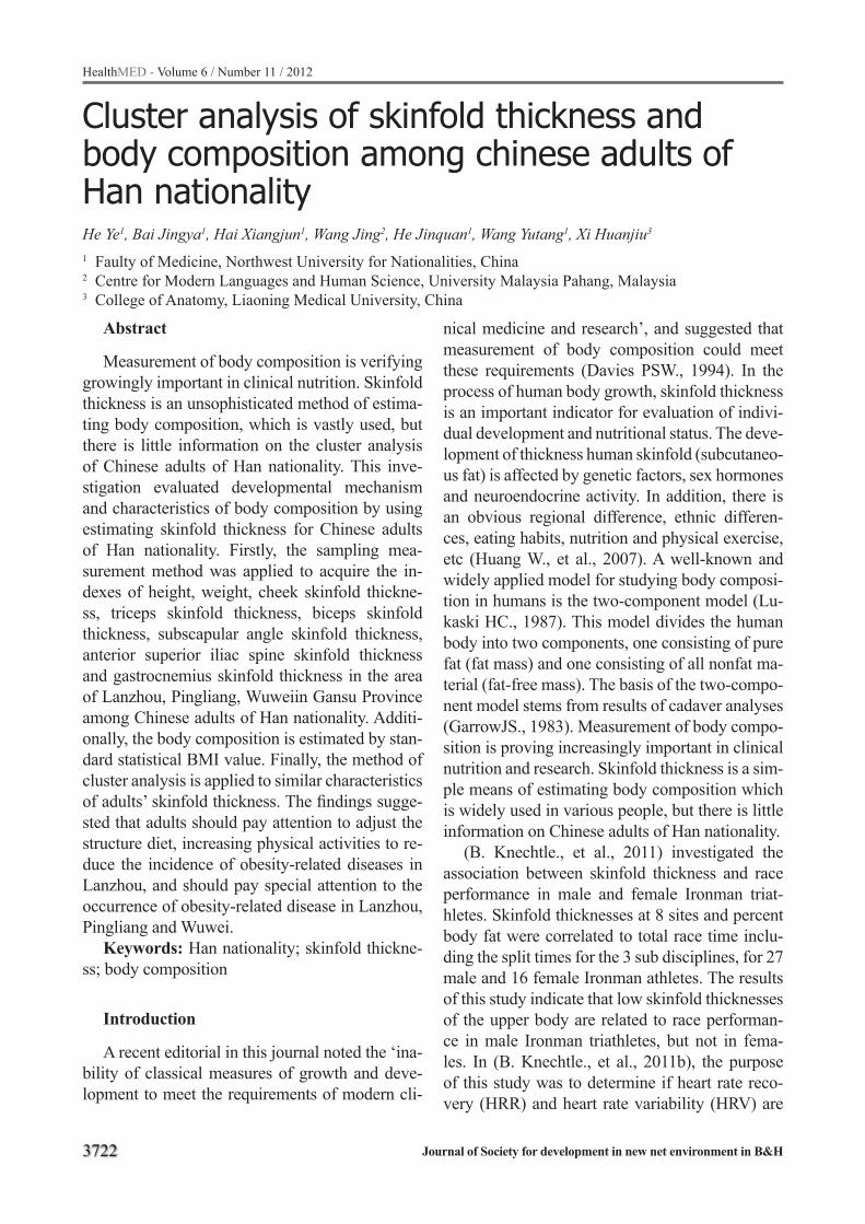
3722
HealthMED - Volume 6 / Number 11 / 2012
Journal of Society for development in new net environment in B&H
Cluster analysis of skinfold thickness and body composition among chinese adults of Han nationalityHe Ye1, Bai Jingya1, Hai Xiangjun1, Wang Jing2, He Jinquan1, Wang Yutang1, Xi Huanjiu3
1 Faulty of Medicine, Northwest University for Nationalities, China2 Centre for Modern Languages and Human Science, University Malaysia Pahang, Malaysia3 College of Anatomy, Liaoning Medical University, China
Abstract
Measurement of body composition is verifying growingly important in clinical nutrition. Skinfold thickness is an unsophisticated method of estima-ting body composition, which is vastly used, but there is little information on the cluster analysis of Chinese adults of Han nationality. This inve-stigation evaluated developmental mechanism and characteristics of body composition by using estimating skinfold thickness for Chinese adults of Han nationality. Firstly, the sampling mea-surement method was applied to acquire the in-dexes of height, weight, cheek skinfold thickne-ss, triceps skinfold thickness, biceps skinfold thickness, subscapular angle skinfold thickness, anterior superior iliac spine skinfold thickness and gastrocnemius skinfold thickness in the area of Lanzhou, Pingliang, Wuweiin Gansu Province among Chinese adults of Han nationality. Additi-onally, the body composition is estimated by stan-dard statistical BMI value. Finally, the method of cluster analysis is applied to similar characteristics of adults’ skinfold thickness. The findings sugge-sted that adults should pay attention to adjust the structure diet, increasing physical activities to re-duce the incidence of obesity-related diseases in Lanzhou, and should pay special attention to the occurrence of obesity-related disease in Lanzhou, Pingliang and Wuwei.
Keywords: Han nationality; skinfold thickne-ss; body composition
Introduction
A recent editorial in this journal noted the ‘ina-bility of classical measures of growth and deve-lopment to meet the requirements of modern cli-
nical medicine and research’, and suggested that measurement of body composition could meet these requirements (Davies PSW., 1994). In the process of human body growth, skinfold thickness is an important indicator for evaluation of indivi-dual development and nutritional status. The deve-lopment of thickness human skinfold (subcutaneo-us fat) is affected by genetic factors, sex hormones and neuroendocrine activity. In addition, there is an obvious regional difference, ethnic differen-ces, eating habits, nutrition and physical exercise, etc (Huang W., et al., 2007). A well-known and widely applied model for studying body composi-tion in humans is the two-component model (Lu-kaski HC., 1987). This model divides the human body into two components, one consisting of pure fat (fat mass) and one consisting of all nonfat ma-terial (fat-free mass). The basis of the two-compo-nent model stems from results of cadaver analyses (GarrowJS., 1983). Measurement of body compo-sition is proving increasingly important in clinical nutrition and research. Skinfold thickness is a sim-ple means of estimating body composition which is widely used in various people, but there is little information on Chinese adults of Han nationality.
(B. Knechtle., et al., 2011) investigated the association between skinfold thickness and race performance in male and female Ironman triat-hletes. Skinfold thicknesses at 8 sites and percent body fat were correlated to total race time inclu-ding the split times for the 3 sub disciplines, for 27 male and 16 female Ironman athletes. The results of this study indicate that low skinfold thicknesses of the upper body are related to race performan-ce in male Ironman triathletes, but not in fema-les. In (B. Knechtle., et al., 2011b), the purpose of this study was to determine if heart rate reco-very (HRR) and heart rate variability (HRV) are
HealthMED - Volume 6 / Number 11 / 2012
Journal of Society for development in new net environment in B&H 3723
related to maximal aerobic fitness and selected body composition measurements. The results of this study suggest that cardiovascular autonomic modulation is significantly related to maximal ae-robic fitness and body composition. In (Esco, M R., et al., 2011a), the purpose of this study was to determine if resting HRV is related to maximal aerobic fitness and the selected body compositi-on measurements as follows: body mass index (BMI), waist circumference (WC) and the sum of skinfold thickness (SUMSF). This study showed the importance of the simple technique of mea-suring skinfold thickness in relation to cardiovas-cular autonomic control of heart rate. The HRV parameters analyzed in this study (i.e., HFnu and LF:HF) were significantly related to SUMSF, but not [latin capital V with dot above]o2max, WC, or BMI. The skinfold technique is a commonly used field measure for predicting body fat percentage. Therefore, exercise training should perhaps indu-ce an improve body fat mass to appreciably au-gment cardiovascular autonomic modulation.
China is a multi-ethnic country, and the Han Nationality is the main body nationality in China. According to the sixth national census in 2010 Data Bulletin (No. 1), until November in 2010, Han Nationality population reaches more than 1,200,000,000, accounts for 91.51% in the popu-lation of China. At present, in China the resear-ches on children, youth, minorities, and skinfold thickness are more fruitful (Xiao Y., et al., 2009; Xu F., et al 2000; Liu X., et al., 2009; Zheng L., et al 2003; Zheng L., et al 2004; Suo L., et al 2005).
However, actually studies are few (Wang Z., et al 2011), while the population Han Nationality is majority in China. Therefore, this article has con-ducted the research skinfold thickness in Han Na-tionality adults.
Methods
The survey and sampling methodsThis study focused on the Han nationality in
Gansu Province. Gansu is one of the traditional settlements and birthplaces of the Han nationality in China. According to the sixth national census data Gazette of Gansu Province in 2010, the resi-dent population was 25,575,254 in Gansu provin-ce. Among them, the population of Han nationa-
lity was 23,164,756, accounted for 90.57%. In this study, the research subjects were Chinese adults of Han nationality in Lanzhou, Pingliang, and Wuwei of Gansu Province. The random cluster sampling method was used for the target popula-tion of participants. 1500 potential participants, above 20 years old, called a research assistant who then screens them for eligibility as well as collec-ting basic demographic information. Demographic characteristics will be collected during initial scre-ening, including country of birth, language spo-ken, level of education, regular menstrual cycle, if any Government pensions/benefits are received, living situation, and total time spent in a car as a driver or passenger each week. In addition, data on any medical conditions and medication use will be collected through the medical clearance forms participants fill out prior to commencing the stu-dy. Potential participants were residents for a long time. The excluded participants had incomplete material and data, and also patients with some problems of the physical development. In the end, there are validly sample of 1407 people, including 700 men and 707 women. This ongoing analysis continued with interview data thus organized into different sets of working charts. Two participant themes emerged while transcribing the recorded interviews and were verified using researcher re-flections and the individual and collective organi-zing charts. Through this process, it became appa-rent to the researcher that self-care and community constituted participant-emergent themes which participants appeared to find compelling.
The survey content and methodsMeasurement standards are in accordance with
national standards of the People’s Republic of China (1985). The methods of measurement and controlling quality are in accordance with “anthro-pometry” of (Xi H., 2010). The measurers have been carried out strictly training, and if the test of small-sample pre-survey was failed, they will lose the chance to the formal investigation. Measure-ment data including height, weight, skinfold of the cheek, right arm, triceps skinfold, arm brachial bi-ceps bit skinfold, subscapular angle skinfold skin-fold anterior superior iliac spine, gastrocnemius skinfold. All the data of measurements were ave-

3724
HealthMED - Volume 6 / Number 11 / 2012
Journal of Society for development in new net environment in B&H
rage in continuous measurements three times, and body mass index (BMI) and body fat (BF %) for evaluation of medical conditions were calculated.
Height: standing height and sitting height me-ter was used as the instrument to measure with marking off in centimeters. Subjects were asked to take off his shoes and hats, stood on the flo-or at attention, hang naturally down hands, bring heels closer together, stands apart toes to about 45 degrees apart, as well as the three points of heels, hips and shoulders were asked to close up the column, the trunk is were asked to naturally hold themselves upright, the two eyes were asked to level with the front. Moreover, the examiner was asked to stand in the right side of the measu-ring board, and gently move the slide until reach the head point. The measurement error shall not exceed 0.5 cm.
Weight: the instrument of the leverage sca-le was used for the weight measurement in ki-lograms. The subjects were asked to deplete the urine and underwear, and were asked to wear un-derwear, stand over in the middle of the weighing platform with bared feet. As well as the hands did not touch other objects, and the counterweight was adjusted until the balance of leverage and the data was recorded to the smallest scale. And the test error is not more than 0.1 kilograms.
Skinfold thickness measurements: the Imi-tation Japanese Rong Yan type modified skinfold thickness gauge was used as skinfold thickness meter. The measured instrument was strictly ca-librated before measuring skinfold thickness. Sur-veyors were disciplined by a professional trainer in order for controlling strictly quality. And all of the testing errors should not exceed 0.5 mm.
Skinfold measurement methods are as follows:
1. Skinfold thickness in the cheek: the thumb was fixed on the corners of the mouth outside of subjects, and the index finger was asked to point at the ear lobe. The distance of the two fingers is about 3cm. and the two fingers were asked to pinch the skin and subcutaneous tissue.
2. Skinfold thickness in the three-headed muscle of the right arm: Taking the midpoint of the right upper arm acromion points and olecranon, and the direction of
the skinfold paralleling with the direction of the long axis of the arm;
3. Skinfold thickness in the biceps of the right arm: in the biceps of the right upper arm, the medial side of the upper arm, the horizontal position of the midpoint of the acromion and the radial, the direction of the skinfold paralleling with the long axis of upper arm;
4. Skinfold thickness in subscapular angle: Taking the bottom of the subscapular angle, the skinfold direction is downward biased outside the 45 ° angle;
5. Skinfold thickness in anterosuperior iliac spine: Taking the top of the anterosuperior iliac spine, the skinfold direction is downward biased inside the 45 ° angle;
6. Skinfold thickness in gastrocnemius: taking the medial side of the maximum horizontal circumferences in the short leg, and the direction of the skinfold paralleling with the direction of the long axis of the short leg;
Data ProcessingEvery 10-year of age is considered as an age
group. The samples were divided into five age groups such as 20-29, 30-39, 40-49, 50-59, above 60. The formula, used for measuring BMI and BF % (Brozek J., 1963), were as follows.
BMI = body weight (kg) / height (m) (1)
BF% = [(4.570 / D) -4.142] * 100% (2)where D is the body density (Brozek J., 1963).
It is calculated by the regression equation, which is proposed by Japanese scholars Changling. Normally, D=1.0913-0.00116 • X for male, and D=1.0897-0.00133 • X for female. (X = scapular angle, skin fold thickness (mm) + triceps skin fold thickness (mm)) (Chen M., 1993 ).
Results
Skinfold thickness analysis among Chinese adults of Han nationality in Gansu. Primary analysis will be via intention-to-treat
with all samples included regardless of dropout or level of adherence. Missing data will be imputed according to the maximum likelihood expectation
HealthMED - Volume 6 / Number 11 / 2012
Journal of Society for development in new net environment in B&H 3725
algorithm via the Statistical Package for the Social Sciences (IBM©, SPSS Version 19.0 Excel 2003). Data will be presented as the mean ± standard de-viation or median and range, as appropriate.
Analysis for male: compared with six index of skinfold thickness in Table 1, the values of subsca-pular skinfold thickness are the highest in Pingli-ang and Lanzhou. In addition, the anterior superi-
Table 1. the results of body composition and six index of skinfold thickness of Han nationality in Gansu ((x± S) mm) Age Samples
cheeks skinfold
biceps skinfold
triceps skinfold
Subscapularisskinfold
anterior skinfold
gastrocnemius skinfold BMI BF%
Male in Lanzhou
20~ 40 6.44±2.45 5.54±3.36 8.84±4.34 14.80±6.58 13.40±7.46 8.94±3.25 22.54±3.27 15.42±5.04
30~ 40 8.57±4.01 5.49±3.29 8.66±4.48 16.98±8.67 12.72±6.44 7.45±3.27 24.09±3.14 16.38±5.99
40~ 40 9.86±5.02 6.22±3.99 10.19±5.32 18.27±8.65 14.19±8.03 8.02±4.08 24.47±4.12 17.72±6.41
50~ 40 10.37±3.69 6.28±2.59 10.34±4.30 19.54±7.50 13.73±6.08 8.27±6.07 24.81±3.00 18.36±5.14
60~ 40 9.96±3.11 5.45±2.08 9.40±3.41 16.60±5.76 12.18±5.36 6.97±3.48 24.54±3.14 16.50±3.78
Total 200 9.04±3.99 5.79±3.12 9.49±4.42 17.24±7.61 13.24±6.71 7.93±4.18 24.09±3.42 16.88±5.40
Female in Lanzhou
20~ 40 11.71±3.09 6.90±2.37 13.85±4.64 17.37±7.63 13.60±6.94 12.00±3.70 20.48±2.35 21.87±5.93
30~ 40 13.28±3.63 8.78±4.16 17.97±6.32 22.25±8.87 18.45±8.94 12.71±5.59 24.80±4.03 26.98±8.34
40~ 40 13.75±3.48 8.76±4.38 19.36±6.36 24.33±8.62 18.54±8.01 13.67±4.58 24.59±2.78 28.95±8.07
50~ 45 14.31±3.75 10.81±3.95 19.84±5.97 25.46±7.65 22.02±7.42 13.45±5.35 24.90±3.47 29.84±7.22
60~ 41 13.85±3.94 9.70±4.24 17.42±4.79 25.55±7.59 21.05±7.48 12.67±4.46 26.01±3.24 28.49±6.56
Total 206 13.40±3.67 9.04±4.07 17.74±6.00 23.06±8.56 18.82±8.24 12.91±4.79 24.18±3.72 27.29±7.74
Male in Pingliang
20~ 50 6.94±3.61 4.82±2.80 9.22±5.57 14.75±7.03 11.82±6.82 8.58±4.27 22.58±3.03 15.58±5.53
30~ 50 7.72±3.83 4.65±2.35 8.81±4.95 14.79±7.54 11.65±7.61 7.73±4.48 23.02±3.26 15.41±5.76
40~ 50 7.65±3.41 4.40±2.36 8.84±4.75 13.87±6.89 9.66±5.81 6.42±3.54 23.21±3.31 14.99±5.31
50~ 50 7.85±3.80 3.92±2.02 7.53±3.86 13.94±8.07 8.38±5.32 5.72±3.29 22.40±3.95 14.41±5.47
60~ 50 9.85±4.56 4.74±3.02 8.55±4.81 14.14±7.08 8.59±4.98 5.92±2.33 22.42±3.04 14.98±5.33
Total 250 8.00±3.95 4.51±2.54 8.59±4.81 14.30±7.29 10.02±6.31 6.89±3.80 22.73±3.32 15.08±5.45
Female in Pingliang
20~ 50 11.49±3.72 6.67±2.76 17.03±5.56 17.11±6.00 13.19±6.17 12.51±4.87 22.71±3.00 23.49±5.98
30~ 50 12.36±3.59 7.83±3.58 17.67±5.65 20.24±7.31 13.86±7.13 13.29±4.15 22.87±2.90 25.62±6.35
40~ 50 11.54±4.26 7.67±2.93 17.41±5.20 21.12±6.97 15.04±7.12 12.52±3.96 24.24±3.03 25.97±6.28
50~ 50 12.30±3.78 8.48±4.45 18.01±6.19 21.70±8.43 17.14±8.38 12.29±5.30 24.36±3.71 26.68±7.70
60~ 52 13.05±5.19 9.20±5.24 18.22±7.09 20.67±10.06 14.78±9.39 13.22±6.99 24.81±4.24 26.27±9.49
Total 252 12.15±4.17 7.98±3.98 17.67±5.95 20.17±8.00 14.80±7.79 12.77±5.16 23.61±3.58 25.61±7.32
Male in Wuwei
20~ 50 6.34±2.41 3.78±1.66 8.15±3.39 10.62±5.27 5.72±2.24 6.31±2.21 20.20±2.55 13.12±3.58
30~ 48 7.81±2.98 4.96±2.25 8.97±4.05 14.21±5.85 9.61±4.57 6.67±3.40 23.68±2.83 15.19±4.43
40~ 50 8.64±4.42 4.68±1.96 9.42±4.98 15.48±6.99 8.51±5.67 5.81±3.01 23.81±2.94 16.01±5.22
50~ 50 7.60±3.65 4.40±2.17 7.78±4.23 13.53±5.61 7.11±3.36 5.29±2.25 23.13±3.11 14.31±4.28
60~ 50 7.43±3.31 4.12±1.61 7.20±3.28 13.20±6.37 6.76±3.29 5.11±2.53 22.99±2.82 13.89±4.24
Total 250 7.56±3.47 4.39±1.98 8.31±4.08 13.41±6.21 7.54±4.20 5.84±2.76 22.76±3.13 14.51±4.46
Female in Wuwei
20~ 50 13.17±3.44 7.50±2.77 15.50±4.89 16.07±5.52 10.78±3.95 13.08±4.38 20.67±2.42 22.06±5.50
30~ 50 12.99±6.65 7.97±3.61 16.59±6.42 18.59±8.19 12.75±6.45 11.88±5.73 23.11±3.52 24.13±7.76
40~ 50 11.88±4.15 9.06±4.00 17.24±5.19 21.79±6.35 15.20±6.08 12.42±6.08 24.97±3.03 26.23±5.85
50~ 50 12.69±4.20 9.13±3.97 18.40±5.52 23.52±7.94 15.43±7.61 12.11±5.39 24.75±3.06 27.91±7.00
60~ 49 12.82±4.53 8.81±3.46 17.65±6.38 21.56±8.01 14.19±6.53 11.87±6.15 24.58±3.23 26.38±7.48
Total 249 12.53±4.13 8.49±3.62 17.07±5.75 23.30±7.69 13.67±6.43 12.27±5.55 23.61±3.45 25.34±7.02
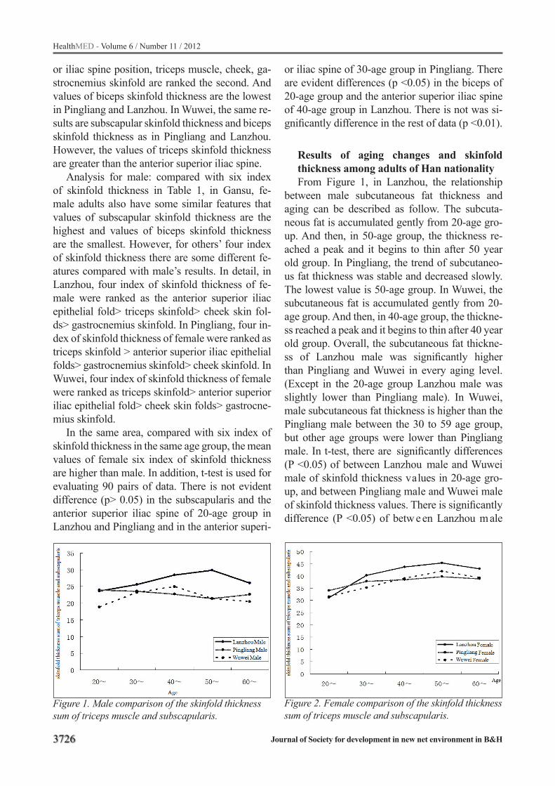
3726
HealthMED - Volume 6 / Number 11 / 2012
Journal of Society for development in new net environment in B&H
or iliac spine position, triceps muscle, cheek, ga-strocnemius skinfold are ranked the second. And values of biceps skinfold thickness are the lowest in Pingliang and Lanzhou. In Wuwei, the same re-sults are subscapular skinfold thickness and biceps skinfold thickness as in Pingliang and Lanzhou. However, the values of triceps skinfold thickness are greater than the anterior superior iliac spine.
Analysis for male: compared with six index of skinfold thickness in Table 1, in Gansu, fe-male adults also have some similar features that values of subscapular skinfold thickness are the highest and values of biceps skinfold thickness are the smallest. However, for others’ four index of skinfold thickness there are some different fe-atures compared with male’s results. In detail, in Lanzhou, four index of skinfold thickness of fe-male were ranked as the anterior superior iliac epithelial fold> triceps skinfold> cheek skin fol-ds> gastrocnemius skinfold. In Pingliang, four in-dex of skinfold thickness of female were ranked as triceps skinfold > anterior superior iliac epithelial folds> gastrocnemius skinfold> cheek skinfold. In Wuwei, four index of skinfold thickness of female were ranked as triceps skinfold> anterior superior iliac epithelial fold> cheek skin folds> gastrocne-mius skinfold.
In the same area, compared with six index of skinfold thickness in the same age group, the mean values of female six index of skinfold thickness are higher than male. In addition, t-test is used for evaluating 90 pairs of data. There is not evident difference (p> 0.05) in the subscapularis and the anterior superior iliac spine of 20-age group in Lanzhou and Pingliang and in the anterior superi-
Figure 1. Male comparison of the skinfold thickness sum of triceps muscle and subscapularis.
Figure 2. Female comparison of the skinfold thickness sum of triceps muscle and subscapularis.
or iliac spine of 30-age group in Pingliang. There are evident differences (p <0.05) in the biceps of 20-age group and the anterior superior iliac spine of 40-age group in Lanzhou. There is not was si-gnificantly difference in the rest of data (p <0.01).
Results of aging changes and skinfold thickness among adults of Han nationalityFrom Figure 1, in Lanzhou, the relationship
between male subcutaneous fat thickness and aging can be described as follow. The subcuta-neous fat is accumulated gently from 20-age gro-up. And then, in 50-age group, the thickness re-ached a peak and it begins to thin after 50 year old group. In Pingliang, the trend of subcutaneo-us fat thickness was stable and decreased slowly. The lowest value is 50-age group. In Wuwei, the subcutaneous fat is accumulated gently from 20-age group. And then, in 40-age group, the thickne-ss reached a peak and it begins to thin after 40 year old group. Overall, the subcutaneous fat thickne-ss of Lanzhou male was significantly higher than Pingliang and Wuwei in every aging level. (Except in the 20-age group Lanzhou male was slightly lower than Pingliang male). In Wuwei, male subcutaneous fat thickness is higher than the Pingliang male between the 30 to 59 age group, but other age groups were lower than Pingliang male. In t-test, there are significantly differences (P <0.05) of between Lanzhou male and Wuwei male of skinfold thickness values in 20-age gro-up, and between Pingliang male and Wuwei male of skinfold thickness values. There is significantly difference (P <0.05) of betw e en Lanzhou m ale
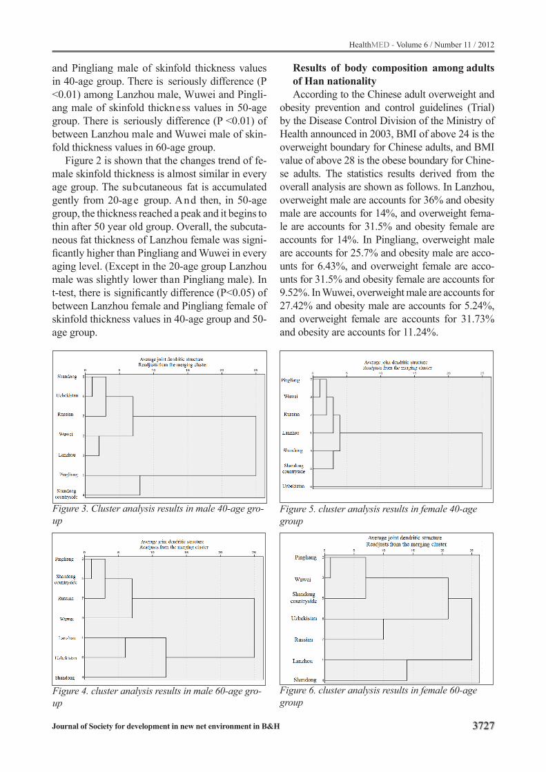
HealthMED - Volume 6 / Number 11 / 2012
Journal of Society for development in new net environment in B&H 3727
and Pingliang male of skinfold thickness values in 40-age group. There is seriously difference (P <0.01) among Lanzhou male, Wuwei and Pingli-ang male of skinfold thickness values in 50-age group. There is seriously difference (P <0.01) of between Lanzhou male and Wuwei male of skin-fold thickness values in 60-age group.
Figure 2 is shown that the changes trend of fe-male skinfold thickness is almost similar in every age group. The subcutaneous fat is accumulated gently from 20-age group. And then, in 50-age group, the thickness reached a peak and it begins to thin after 50 year old group. Overall, the subcuta-neous fat thickness of Lanzhou female was signi-ficantly higher than Pingliang and Wuwei in every aging level. (Except in the 20-age group Lanzhou male was slightly lower than Pingliang male). In t-test, there is significantly difference (P<0.05) of between Lanzhou female and Pingliang female of skinfold thickness values in 40-age group and 50-age group.
Results of body composition among adults of Han nationalityAccording to the Chinese adult overweight and
obesity prevention and control guidelines (Trial) by the Disease Control Division of the Ministry of Health announced in 2003, BMI of above 24 is the overweight boundary for Chinese adults, and BMI value of above 28 is the obese boundary for Chine-se adults. The statistics results derived from the overall analysis are shown as follows. In Lanzhou, overweight male are accounts for 36% and obesity male are accounts for 14%, and overweight fema-le are accounts for 31.5% and obesity female are accounts for 14%. In Pingliang, overweight male are accounts for 25.7% and obesity male are acco-unts for 6.43%, and overweight female are acco-unts for 31.5% and obesity female are accounts for 9.52%. In Wuwei, overweight male are accounts for 27.42% and obesity male are accounts for 5.24%, and overweight female are accounts for 31.73% and obesity are accounts for 11.24%.
Figure 3. Cluster analysis results in male 40-age gro-up
Figure 4. cluster analysis results in male 60-age gro-up
Figure 5. cluster analysis results in female 40-age group
Figure 6. cluster analysis results in female 60-age group
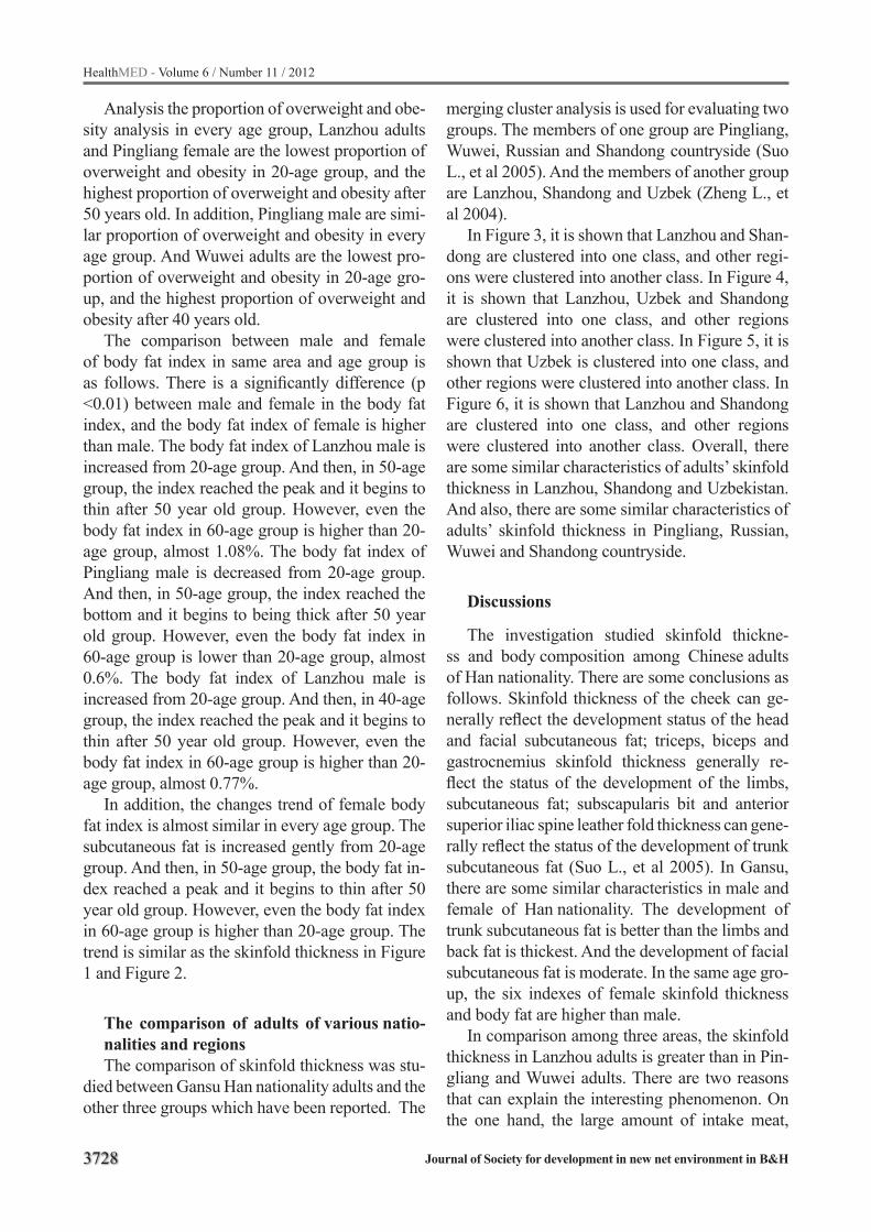
3728
HealthMED - Volume 6 / Number 11 / 2012
Journal of Society for development in new net environment in B&H
Analysis the proportion of overweight and obe-sity analysis in every age group, Lanzhou adults and Pingliang female are the lowest proportion of overweight and obesity in 20-age group, and the highest proportion of overweight and obesity after 50 years old. In addition, Pingliang male are simi-lar proportion of overweight and obesity in every age group. And Wuwei adults are the lowest pro-portion of overweight and obesity in 20-age gro-up, and the highest proportion of overweight and obesity after 40 years old.
The comparison between male and female of body fat index in same area and age group is as follows. There is a significantly difference (p <0.01) between male and female in the body fat index, and the body fat index of female is higher than male. The body fat index of Lanzhou male is increased from 20-age group. And then, in 50-age group, the index reached the peak and it begins to thin after 50 year old group. However, even the body fat index in 60-age group is higher than 20-age group, almost 1.08%. The body fat index of Pingliang male is decreased from 20-age group. And then, in 50-age group, the index reached the bottom and it begins to being thick after 50 year old group. However, even the body fat index in 60-age group is lower than 20-age group, almost 0.6%. The body fat index of Lanzhou male is increased from 20-age group. And then, in 40-age group, the index reached the peak and it begins to thin after 50 year old group. However, even the body fat index in 60-age group is higher than 20-age group, almost 0.77%.
In addition, the changes trend of female body fat index is almost similar in every age group. The subcutaneous fat is increased gently from 20-age group. And then, in 50-age group, the body fat in-dex reached a peak and it begins to thin after 50 year old group. However, even the body fat index in 60-age group is higher than 20-age group. The trend is similar as the skinfold thickness in Figure 1 and Figure 2.
The comparison of adults of various natio-nalities and regions The comparison of skinfold thickness was stu-
died between Gansu Han nationality adults and the other three groups which have been reported. The
merging cluster analysis is used for evaluating two groups. The members of one group are Pingliang, Wuwei, Russian and Shandong countryside (Suo L., et al 2005). And the members of another group are Lanzhou, Shandong and Uzbek (Zheng L., et al 2004).
In Figure 3, it is shown that Lanzhou and Shan-dong are clustered into one class, and other regi-ons were clustered into another class. In Figure 4, it is shown that Lanzhou, Uzbek and Shandong are clustered into one class, and other regions were clustered into another class. In Figure 5, it is shown that Uzbek is clustered into one class, and other regions were clustered into another class. In Figure 6, it is shown that Lanzhou and Shandong are clustered into one class, and other regions were clustered into another class. Overall, there are some similar characteristics of adults’ skinfold thickness in Lanzhou, Shandong and Uzbekistan. And also, there are some similar characteristics of adults’ skinfold thickness in Pingliang, Russian, Wuwei and Shandong countryside.
Discussions
The investigation studied skinfold thickne-ss and body composition among Chinese adults of Han nationality. There are some conclusions as follows. Skinfold thickness of the cheek can ge-nerally reflect the development status of the head and facial subcutaneous fat; triceps, biceps and gastrocnemius skinfold thickness generally re-flect the status of the development of the limbs, subcutaneous fat; subscapularis bit and anterior superior iliac spine leather fold thickness can gene-rally reflect the status of the development of trunk subcutaneous fat (Suo L., et al 2005). In Gansu, there are some similar characteristics in male and female of Han nationality. The development of trunk subcutaneous fat is better than the limbs and back fat is thickest. And the development of facial subcutaneous fat is moderate. In the same age gro-up, the six indexes of female skinfold thickness and body fat are higher than male.
In comparison among three areas, the skinfold thickness in Lanzhou adults is greater than in Pin-gliang and Wuwei adults. There are two reasons that can explain the interesting phenomenon. On the one hand, the large amount of intake meat,

HealthMED - Volume 6 / Number 11 / 2012
Journal of Society for development in new net environment in B&H 3729
eggs, milk and high-fat high-protein food is in the diet of Lanzhou residents, and intake these food is frequent. On another hand, the reason may be because of the modernization of urban life, and convenient transportation, and the doing exercise is relatively little. The Pingliang residents worked in agriculture-based activities, physical exertion and low living standard. And male is major labor in the most family and has engaged in heavy ma-nual labor usually. Therefore, it is shown that there are little changes in skinfold thickness of Pingli-ang male with aging, and sometimes, the trend has shown downward. became thinner and thinner.Wuwei is the convergence zone of the Qinghai-Tibet Plateau, the Loess Plateau and Inner Mon-golia Plateau. And the elevation is between 2040-4874 meters (Lanzhou city center of elevation is 1520 m). The territory of climate is cold plateau climate. In Wuwei, Han nationality has the si-milar diet as Tibetan residents. The large amou-nt of intake meat, dairy products, roasted barley flour, butter tea and highland barley wine is in the diet of Wuwei residents. The reducing of skin-fold thickness accompanied with the rising of the elevation (Wang W., et al 2001) and the ambient temperature (Wang Z., et al 2001). The skinfold thickness of Gansu Han female has the similar trends with age. The reason is that the age of the female skinfold thickness trends was related to the female hormone secretion. In the adolescence, the female ovarian improved the abilities to secre-ting estrogen, and inhibited growth hormone, and enhanced insulin action, and promoted the cyto-plasmic volume increased, and then the fat began to accumulate in large numbers. After the meno-pause (about 45-50 years old), the abilities of the female hormone secretion drastically reduced, the abilities of the accumulation of fat was diminis-hed, and the fat layer became thin (Suo L., et al 2005).
It can be seen that the proportion of Lanzhou male and female overweight and obesity rates were higher than in Pingliang and Wuwei male and female. The reasons are the favorable conditi-ons of nutrition in Lanzhou and usual office-based work, but the large amount farm-based of physi-cal labor. In addition, the overweight proportion of Lanzhou male are higher than women, and the overweight and obesity proportion of Pingliang
and Wuwei female are higher than male. There is some reason of working pressure increasing for female in Lanzhou.
In the cluster comparison, Lanzhou, Uzbek and Shandong are clustered into one class, and other regions were clustered into another class. These phenomena reflected that the skinfold thickness is affected by many factors, and where the differen-ces in living standard and genetic factors played a major role (Wang Z., et al 2011).
Recommendation
The investigation found that the proportion of overweight and obesity of Lanzhou is better than Pingliang and Wuwei. It prompted that the risk of the Lanzhou adults suffering from obesity-rela-ted diseases is higher than Pingliang and Wuwei, where male is more obvious than female. The Re-commendation is that the Lanzhou adults of Han nationality should prevent obesity-related disea-ses by proceed from the adjusting diet and doing exercises. And the same suggestion gave a cue to Wuwei adults in 40-age group, Lanzhou adults in 50-age group and Pingliang female 50-age group.
Acknowledgement
National Natural Science Funds (30830062)
References
1. Knechtle B, Knechtle P, Rosemann T (2011). Upper Body Skinfold Thickness is related to Race Perfor-mance in Male Ironman Triathletes. Int J Sports Med. 32(1): 20-27.
2. Brozek J (1963). Densitometric analysis of body com-position: Revision of some quantitative assumptions. Ann.New York Aead Sci.104:110-113.
3. Chen M (1993). Anthropometric Science. Beijing: Peking University Health Science Center Press and Peking Union Medical College Press.
4. Davies PSW (1994). Body composition assessment. Arch. Dis. Child. 69: 337-338.
5. Esco MR, Olson MS, Williford HN, Russell AR, Ga-ston K (2011)a. Skinfold Thickness is Related to Cardiovascular Autonomic Control as Assessed By Resting Heart Rate Variability. Journal of Stren-gth & Conditioning Research. doi: 10.1097/01.JSC.0000395741.73051.cf.
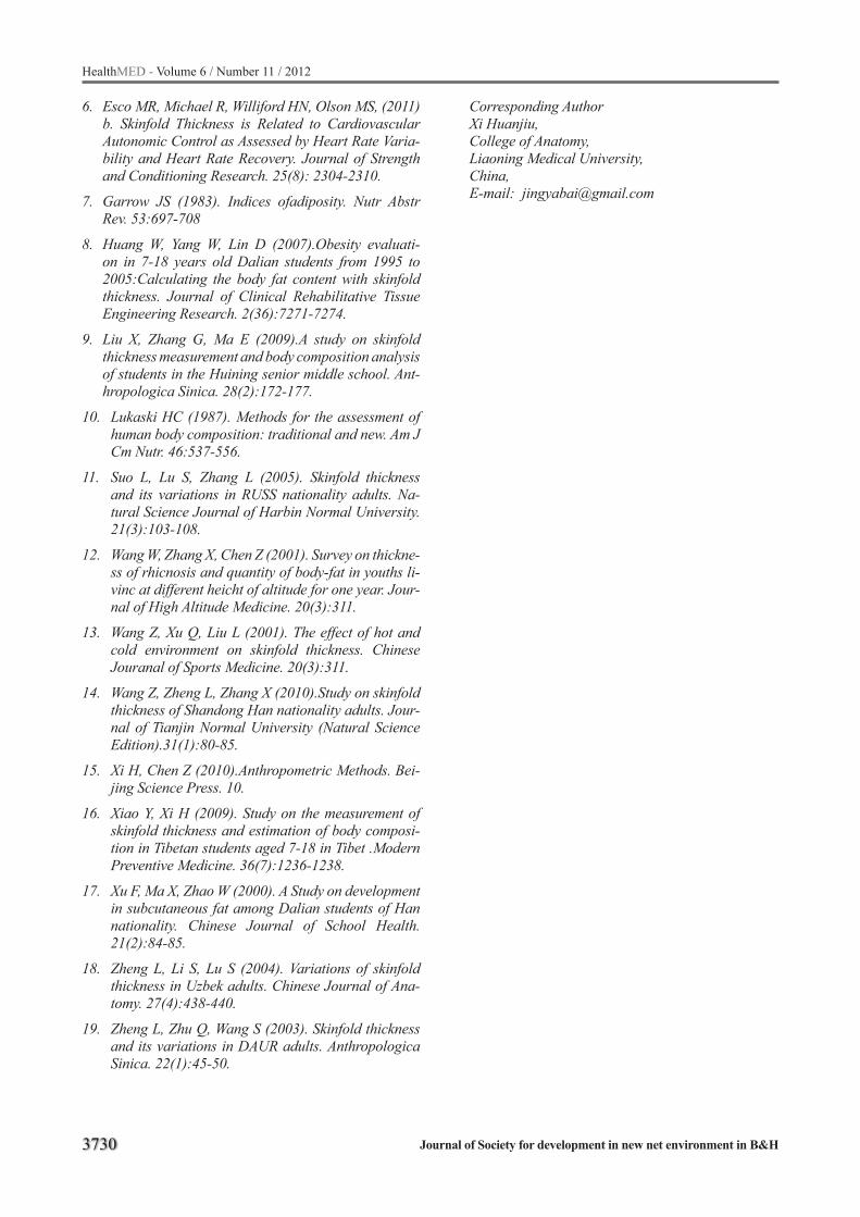
3730
HealthMED - Volume 6 / Number 11 / 2012
Journal of Society for development in new net environment in B&H
6. Esco MR, Michael R, Williford HN, Olson MS, (2011)b. Skinfold Thickness is Related to Cardiovascular Autonomic Control as Assessed by Heart Rate Varia-bility and Heart Rate Recovery. Journal of Strength and Conditioning Research. 25(8): 2304-2310.
7. Garrow JS (1983). Indices ofadiposity. Nutr Abstr Rev. 53:697-708
8. Huang W, Yang W, Lin D (2007).Obesity evaluati-on in 7-18 years old Dalian students from 1995 to 2005:Calculating the body fat content with skinfold thickness. Journal of Clinical Rehabilitative Tissue Engineering Research. 2(36):7271-7274.
9. Liu X, Zhang G, Ma E (2009).A study on skinfold thickness measurement and body composition analysis of students in the Huining senior middle school. Ant-hropologica Sinica. 28(2):172-177.
10. Lukaski HC (1987). Methods for the assessment of human body composition: traditional and new. Am J Cm Nutr. 46:537-556.
11. Suo L, Lu S, Zhang L (2005). Skinfold thickness and its variations in RUSS nationality adults. Na-tural Science Journal of Harbin Normal University. 21(3):103-108.
12. Wang W, Zhang X, Chen Z (2001). Survey on thickne-ss of rhicnosis and quantity of body-fat in youths li-vinc at different heicht of altitude for one year. Jour-nal of High Altitude Medicine. 20(3):311.
13. Wang Z, Xu Q, Liu L (2001). The effect of hot and cold environment on skinfold thickness. Chinese Jouranal of Sports Medicine. 20(3):311.
14. Wang Z, Zheng L, Zhang X (2010).Study on skinfold thickness of Shandong Han nationality adults. Jour-nal of Tianjin Normal University (Natural Science Edition).31(1):80-85.
15. Xi H, Chen Z (2010).Anthropometric Methods. Bei-jing Science Press. 10.
16. Xiao Y, Xi H (2009). Study on the measurement of skinfold thickness and estimation of body composi-tion in Tibetan students aged 7-18 in Tibet .Modern Preventive Medicine. 36(7):1236-1238.
17. Xu F, Ma X, Zhao W (2000). A Study on development in subcutaneous fat among Dalian students of Han nationality. Chinese Journal of School Health. 21(2):84-85.
18. Zheng L, Li S, Lu S (2004). Variations of skinfold thickness in Uzbek adults. Chinese Journal of Ana-tomy. 27(4):438-440.
19. Zheng L, Zhu Q, Wang S (2003). Skinfold thickness and its variations in DAUR adults. Anthropologica Sinica. 22(1):45-50.
Corresponding AuthorXi Huanjiu,College of Anatomy, Liaoning Medical University, China,E-mail: [email protected]
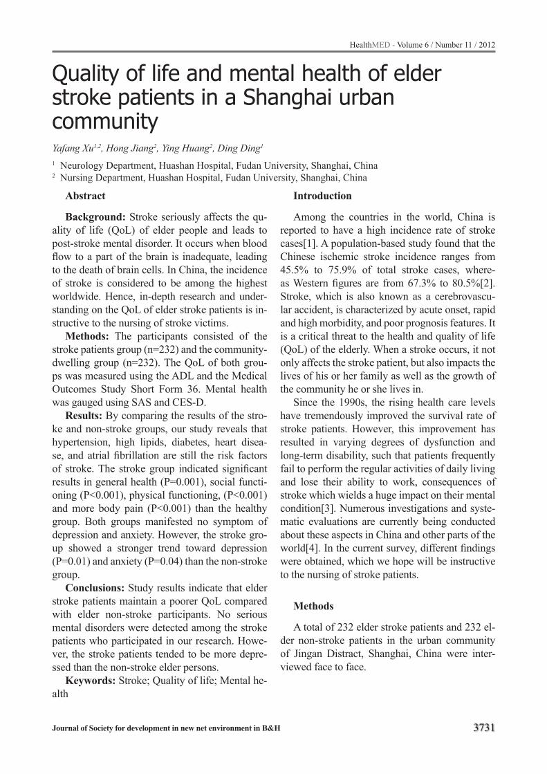
HealthMED - Volume 6 / Number 11 / 2012
Journal of Society for development in new net environment in B&H 3731
Quality of life and mental health of elder stroke patients in a Shanghai urban communityYafang Xu1,2, Hong Jiang2, Ying Huang2, Ding Ding1
1 Neurology Department, Huashan Hospital, Fudan University, Shanghai, China2 Nursing Department, Huashan Hospital, Fudan University, Shanghai, China
Abstract
Background: Stroke seriously affects the qu-ality of life (QoL) of elder people and leads to post-stroke mental disorder. It occurs when blood flow to a part of the brain is inadequate, leading to the death of brain cells. In China, the incidence of stroke is considered to be among the highest worldwide. Hence, in-depth research and under-standing on the QoL of elder stroke patients is in-structive to the nursing of stroke victims.
Methods: The participants consisted of the stroke patients group (n=232) and the community-dwelling group (n=232). The QoL of both grou-ps was measured using the ADL and the Medical Outcomes Study Short Form 36. Mental health was gauged using SAS and CES-D.
Results: By comparing the results of the stro-ke and non-stroke groups, our study reveals that hypertension, high lipids, diabetes, heart disea-se, and atrial fibrillation are still the risk factors of stroke. The stroke group indicated significant results in general health (P=0.001), social functi-oning (P<0.001), physical functioning, (P<0.001) and more body pain (P<0.001) than the healthy group. Both groups manifested no symptom of depression and anxiety. However, the stroke gro-up showed a stronger trend toward depression (P=0.01) and anxiety (P=0.04) than the non-stroke group.
Conclusions: Study results indicate that elder stroke patients maintain a poorer QoL compared with elder non-stroke participants. No serious mental disorders were detected among the stroke patients who participated in our research. Howe-ver, the stroke patients tended to be more depre-ssed than the non-stroke elder persons.
Keywords: Stroke; Quality of life; Mental he-alth
Introduction
Among the countries in the world, China is reported to have a high incidence rate of stroke cases[1]. A population-based study found that the Chinese ischemic stroke incidence ranges from 45.5% to 75.9% of total stroke cases, where-as Western figures are from 67.3% to 80.5%[2]. Stroke, which is also known as a cerebrovascu-lar accident, is characterized by acute onset, rapid and high morbidity, and poor prognosis features. It is a critical threat to the health and quality of life (QoL) of the elderly. When a stroke occurs, it not only affects the stroke patient, but also impacts the lives of his or her family as well as the growth of the community he or she lives in.
Since the 1990s, the rising health care levels have tremendously improved the survival rate of stroke patients. However, this improvement has resulted in varying degrees of dysfunction and long-term disability, such that patients frequently fail to perform the regular activities of daily living and lose their ability to work, consequences of stroke which wields a huge impact on their mental condition[3]. Numerous investigations and syste-matic evaluations are currently being conducted about these aspects in China and other parts of the world[4]. In the current survey, different findings were obtained, which we hope will be instructive to the nursing of stroke patients.
Methods
A total of 232 elder stroke patients and 232 el-der non-stroke patients in the urban community of Jingan Distract, Shanghai, China were inter-viewed face to face.
3732
HealthMED - Volume 6 / Number 11 / 2012
Journal of Society for development in new net environment in B&H
Inclusion criteria of subjectThe inclusion criteria for participants in the
stroke group consisted of the following: (1) community residents who have been diagnosed as stroke patients and (2) patients who are more than 60 years of age. Excluded in this study gro-up were the patients suffering from disturbance of consciousness and aphasia. The inclusion criteria for participants in the non-stroke group consisted of (1) community residents who have not been diagnosed as stroke patients and (2) community elders who are more than 60 years of age. The-re were no exclusion criteria for this study group. Basic demographic information of the two study groups such as age, gender, and education level were included in the analysis.
Protection of human subjectsThe studies were conducted following the gu-
ideline set by the International Conference on Har-monization Notes for Guidance on Good Clinical Practice and the Declaration of Helsinki. An inde-pendent ethics committee or institutional review board at each study center approved the relevant study protocol. All participants from both groups provided written informed consents and all other concerned parties signed a consent form.
InstrumentsThe Medical Outcomes Study Short Form-36The Medical Outcomes Study Short (SF-36) is
one of the most appropriate instruments used to measure life activities within the ICF framework. The SF-36 comprises eight subscales, namely, physical functioning (PF), social functioning (SF), mental health (MH), role limitations due to physi-cal problems (RP), role limitations due to emotio-nal problems (RE), bodily pain (BP), general he-alth (GH), and vitality (VT)[5].
Zung SAS The Zung SAS[6] was purposely designed to
quantify the level of anxiety for patients experi-encing anxiety-related symptoms. This self-admi-nistered test consists of 20 items, and each item is rated on a scale of 1 to 4 (1 = none or a little of the time, 2 = some of the time, 3 = good part of the time, and 4 = most of the time). The score ranges
from 25 to 100. Scores from 25 to 44 indicate the normal range of anxiety, 45 to 59 imply mild-to-moderate anxiety levels, 60 to 74 denote marked-to-severe anxiety levels, and 75 and above suggest extreme anxiety levels. The Chinese version of the Zung questionnaire was validated for use in China[7–9].
The Center of Epidemiologic Studies Depre-ssion Scale In this study, we used the Center of Epidemi-
ologic Studies Depression Scale (CES-D) to me-asure the performance of depression. The CES-D is a 20-item, self-report, and depressive symptom-based scale for depression[10]. A score of 16 in this scale has normally been used to categorize significant depressive symptoms in the general population. However, this finding has been criti-cized as producing a high false positive rate and poor specificity in medical outpatients and inpa-tients[11,12]. Due to these shortcomings, the cut-score of 20 was recommended for use.
Data analysisSPSS 13.0 was used to analyze the data. Demo-
graphic and clinical data were presented through descriptive statistics. Mann-Whitney U test was utilized to identify the differences between the stroke group and the non-stroke group. A P value less than 0.05 was considered to be statistically si-gnificant.
Results
Characteristics of participantsWe measured the basic characteristics of all
the participants, including age, gender, education level, body mass index, smoking, drinking, and complicated diseases such as hypertension, heart disease, diabetes, high lipid, and atrial fibrilla-tion. Basic information such as age, gender, and education level revealed no significant difference between the two groups of participants (p>0.05). Statistics showed that stroke patients were more likely to have hypertension (74.6%), high lipid (44%), heart disease (23.7%), diabetes (15%), and atrial fibrillation (10.3%) than the non-stroke par-ticipants. These figures were significantly higher

HealthMED - Volume 6 / Number 11 / 2012
Journal of Society for development in new net environment in B&H 3733
than those of the non-stroke group (p<0.05). Alt-hough these factors were considered as the causes of stroke[13,14], they did not affect our research (Table1).
Comparison of QoL for the stroke group and the non-stroke groupThe QoL of the stroke patients, as measured
on the SF-36, was compared with that of the non-stroke patients. The details are listed in Table 2. The stroke group reported significantly lower sco-res in their BP, GH, SF, and PF compared with the non-stroke group. On average, the scores of the stroke group were in the middle level, excluding that for GH.
Comparison of mental health for the stroke group and the healthy groupTable 3 presents the comparison of mental he-
alth based on the anxiety and depression levels of the two study groups. Both the stroke and non-stroke groups indicated low scores in the SAS and CES-D tests. These results mean both groups did not manifest any depression or anxiety symptoms. However, significant differences were revealed between the stroke group and the non-stroke group (depression, p=0.004; anxiety, p=0.01). The stroke group showed a stronger trend toward developing mental problems.
Table 1. Demographic Characteristics of the Stroke and Healthy Groups
Characteristicsn (%) or Mean (SD)
P valueStroke(N%) Mean Healthy
(N%) Mean
Age (y) 75.2 hor 75.3 hor M 0.995Gender (M) 99 (42.7%) 99 (42.7%) 1.0Education 0.061. Illiteracy 18 (7.8%) 10 (4.3%)2. Primary school 44 (19.0%) 34 (14.7%)3. Middle school 41 (17.7%) 43 (18.5%)4. High school 69 (29.7%) 59 (25.4%)5. University 60 (25.9%) 86 (37.1%)Solitary 25 (10.8%) 23 (9.9%) 0.879Hypertension 173 (74.6%) 137 (59.1%) 0.001Heart Disease 55 (23.7%) 36 (15.5%) 0.035Diabetes 54 (15.5%) 36 (23.3%) 0.046High Lipid 102 (44.0%) 74 (31.9%) 0.01Atrial Fibrillation 24 (10.3%) 9 (3.9%) 0.01BMI 24.7 4.24 23.9 3.22 0.134Smoke 19 (48.7%) 20 (51.3%) 0.869Drink 17 (7.3%) 18 (7.8%) 0.863
Table 2. Comparison of QoL Between the Stroke and Healthy Groups
Variables Mean (SD) P ValueStroke (N=232) Healthy (N=232)SF-36
BP 63.2±24.6 73.0±25.1 0.000GH 12.9±8.2 15.3±7.3 0.001VT 50.4±19.5 51.1±25.5 0.733SF 54.0±39.0 70.5±33.6 0.000RE 36.4±46.2 22.7±37.4 0.001MH 64.9±15.0 67.3±14.4 0.085PF 59.3±26.6 71.8±20.8 0.000RP 48.6±48.1 32.4±44.3 0.000

3734
HealthMED - Volume 6 / Number 11 / 2012
Journal of Society for development in new net environment in B&H
Discussion
Stroke is a chronic and important health pro-blem affecting all aspects of the life of an individu-al. The neurological impairment that results from stroke leads to functional disability in patients. Numerous studies exist in literature regarding the relationship between stroke and QoL as well as the occurrence of depression and anxiety[15,16]. The analysis in the present research shows signi-ficantly reduced scores for SF-36 forms in the stroke and non-stroke groups, and trends toward lower mental health. These findings were similar those of other research.
However, our research found several different results. The QoL among stroke patients was not as poor as seemingly indicated by the SF-36 score. Except for GH, the score was in the middle level, indicating that the QoL of our participants was not very poor. Hence, they do not have serious mental health problems.
After analyzing the information of the research participants, we established that only a very small percentage (1%) of the participants live alone. In China, the immediate family has the primary responsibility of taking care of their elderly. The-refore, we believe that being in a nursing home, staying with family, and providing elder stroke pa-tients with greater care are effective methods for making their lives better and are helpful in pre-venting post-stroke depression and anxiety[17]. In addition, only 0.08% of stroke subjects smoked and only 0.06% drank liquor. According to studies on the relation between the factors and depression or anxiety, smoking and drinking are risk factors of depression, hence, the implications may be si-milar in stroke patients[18,19].
Although the stroke patients who participated in our research do not have any mental disorder, they still presented the tendency to be depressed. Therefore, measures have to be implemented to prevent such occurrence. The elders can take ac-tive part in social activities, and obtain stronger support from their family and the society.
Based on our investigation, family care is cru-cial in order to improve the QoL of elder pati-ents[20]. In China, the family has the primary res-ponsibility of taking care of their elderly. Howe-ver, in the gradual progress of our society, it will eventually be given the primary responsibility of taking care of the elderly. There is increasing con-cern that this development will not present any benefit to the rehabilitation of stroke patients. Mo-reover, changes in the unhealthy lifestyle, such as lessening or eliminating the vices of smoking and drinking, may be helpful to the rehabilitation of stroke patients.
Despite its potential, the present study has se-veral limitations. First, this survey was carried out in an urban community in Shanghai, which does not represent the general rule. Second, we conduc-ted the survey only at a point in time. As a result, we were not able to assess the dynamic changes in the physical and mental QoL of the stroke pa-tients. To address these limitations, we can con-duct more research on the QoL and mental he-alth over time. In addition, further studies can be done on appropriate intervention measures. Apart from these limitations, there was a problem in the number of available research articles. To date, no objective scale about the QoL, level of anxiety, and depression exists. In our study, these scales were assessed based on the subjective feelings of the sample. However, we cannot improve in this respect at present.
Conclusion
To sum up, the elder stroke patients in this study have poorer QoL than the elder non-stroke patients. In addition, no serious mental disorders were detected in the stroke patients who participa-ted in our research. Although most of the subjects were cared for by their families, they still tended to suffer from depression more compared with the non-stroke participants of the study. Measures should thus be taken to prevent mental health dis-orders among elder patients.
Table 3. Comparison of anxiety and depression between stroke and healthy groups
Variables Mean (SD) P ValueStroke (N=232) Healthy (N=232)SAS total 28.1 8.1 26. 0.5.4 0.01
CES-D total 6.5 .5- 4.5 .5- 0.004

HealthMED - Volume 6 / Number 11 / 2012
Journal of Society for development in new net environment in B&H 3735
Reference
1. Ming Liu, Bo Wu, Wen-Zhi Wang, Li-Ming, Shi-Hong Zhang, Ling-Zhi Kong Stroke in China: epidemiology, prevention, and management strategies The Lancet neurology. 2007;6(5):456–464.
2. Yang Shengli, Li Zhenglong, LvDongxin. Every city the burden of population aging on the impact of pen-sion. Northwest population, 2010, 31 (5):61–66.
3. Opara JA, Jaracz K. Quality of life of post-stroke patients and their caregivers. J Med Life. 2010 Jul–Sep;3(3):216–20.
4. Dayapoglu N, Tan M. Quality of life in stroke pati-ents. Neurol India. 2010;58(5):697–701.
5. Ware JE Jr., Snow KK, Kosinski M, and Gandek B (1993) SF-36 Health Survey. Manual and Interpreta-tion Guide. The Health institute, New England Medi-cal Center, Boston.
6. Zung WW. A rating instrument for anxiety disorders. Psychosomatics. 1971;12:371–379.
7. LIU Yun-lin, LIU Jun, XU Lin. The effect of non-lan-guage psychological treatment on patients with cere-bral ischemia. Chinese journal of behavior medical sciences[J], 2005,14(10):890–891.
8. YE Pei-hua. Investigation and analysis on psycho-logical health of family caregivers of patients with cirrhosis. Journal of Chinese Modern Nursing(J), 2005,2(13):1153–1154.
9. Li Qi, Cut Guimei. Investigating the Psychological Status of Medical College Students and Counter-measures in Guangzhou. China Journal of Health Psychology[J], 2008,16(2):131–132.
10. Radloff LS: The CES-D Scale: A self-report depre-ssion scale for research in the general population. Applied Psychological Measurement1:385–401, 1977
11. Schein R, Koenig H. The Center for Epidemiological Studies–Depression(CES-D) Scale: assessment of depression in the medically ill elderly. Int J Geriatr Psychiatry 1997;12:436–446.
12. Schulberg HC, Saul M, McClelland M, Ganguli M, Christy W, Frank R. Assessing depression in pri-mary medical and psychiatric practices. Arch Gen Psychiatry 1985;42:1164–1170.
13. Mandić M, Rancić N. Risk factors for stroke. Med Pregl. 2011;64(11–12):600–5.
14. Diener HC, Weber R, Lip GY, Hohnloser SH. Stroke prevention in atrial fibrillation: do we still need war-farin? Curr Opin Neurol. 2012 Feb;25(1):27–35.
15. Gunaydin R, Karatepe AG, Kaya T, Ulutas O. De-terminants of quality of life (QoL) in elderly stro-ke patients: a short-term follow-up study. Arch Ge-rontol Geriatr. 2011;53(1):19–23. Epub 2010 Jul 2.
16. Kwok T, Pan JH, Lo R, Song X. The influence of par-ticipation on health-related quality of life in stroke patients. Disabil Rehabil. 2011;33(21–22):1990–6.
17. Markle-Reid M, Orridge C, Weir R, Browne G, Gaf-ni A, Lewis M, Walsh M, Levy C, Daub S, Brien H, Roberts J, Thabane L. Interprofessional stroke rehabilitation for stroke survivors using home care. Can J Neurol Sci. 2011;38(2):317–34.
18. Vogl M, Wenig CM, Leidl R, Pokhrel S. Smoking and health-related quality of life in English general po-pulation: implications for economic evaluations.
19. BMC Public Health. 2012;12(1):203. [Epub ahead of print]
20. Satre DD, Chi FW, Eisendrath S, Weisner C. Subdi-agnostic alcohol use by depressed men and women seeking outpatient psychiatric services: consumpti-on patterns and motivation to reduce drinking. Alco-hol Clin Exp Res. 2011; 35(4):695–702.
21. [20] Braun SM, van Haastregt JC, Beurskens AJ, Gielen AI, Wade DT, Schols JM.
22. Feasibility of a mental practice intervention in stro-ke patients in nursing homes: a process evaluation. BMC Neurol. 2010;24;10:74.
Corresponding AuthorYing Huang,Nursing Department,Ding Ding,Neurology Department,Huashan Hospital,Fudan University,Shanghai,China,Email: [email protected]
3736
HealthMED - Volume 6 / Number 11 / 2012
Journal of Society for development in new net environment in B&H
The mechanism of activity of ankaferd blood stopper in the control of arterial bleeding and in the process of wound healingTanzer Korkmaz1, Necla Gurbuz Sarikas2, Ali Kılıcgun3, Erdinc Serin4, Cetin Boran5
1 Abant Izzet Baysal University, Medicine of Faculty, Department of Emergency, Bolu, Turkey2 Abant Izzet Baysal University, Medicine of Faculty, Department of Pediatric Surgery, Bolu, Turkey3 Abant Izzet Baysal University, Medicine of Faculty, Department of Thoracic Surgery, Bolu, Turkey4 Abant Izzet Baysal University, Medicine of Faculty, Department of Biochemistry, Bolu, Turkey5 Abant Izzet Baysal University, Medicine of Faculty, Department of Pathology, Bolu, Turkey
Abstract
Objectives: Ankaferd Blood Stopper (ABS) carries a historical role in the Traditional Turkish Medicine as a topical haemostatic agent. We ai-med to study the effect of the ABS on the control of arterial bleeding in addition to investigate its impact on cytokine and platelet levels and vascu-lar histopathology
Methods: Blood samples were collected from all the rats (21 rats) before and after the study for platelet, interleukin-1, and tumor necrosis factor-alfa measurements. Following the anterior incision of the femoral artery, the rats were divided into 3 groups, the first to be a sterile tomponade applied on the incised artery, the second ABS spray and the third ABS embedded sponge and bleeding time was recorded. After seven days the animals were sacri-ficed and the previously incised femoral arteries were excised for histopathological evaluations.
Results: The bleeding time of the control group was longer (209.28 seconds) than the ABS spray group (53.57 seconds) and ABS sponge group (56.42 seconds). There was no statistical significant difference regarding PLT (platelet) levels. Additio-nally the analysis of tumor necrosis factor-alfa and interleukin-1, reveald no significant difference both within (p=0.218, p=0,759 respectively) and among the groups day0 and day7 (p=O.441, p=0.846 res-pectively).
Conclusion: The present study suggests that the ABS can be easily used and efficiently control the bleeding. However, the results of the present study showed no major effect of the ABS on PLT valu-es, and pro-inflammatory cytokines. Further studi-es remain to be done for illuminating the efficient mechanisms that ABS uses for stopping bleeding.
Keywords: Ankaferd Blood Stopper, homeo-stasis, arterial hemorrhage, cytokines
Introduction
Approximately 35% of the deaths due to trau-ma are attributable to exsanguinations, bleeding to death [1]. Over the years, diverse pharmacological agents have been developed and used to control the bleeding. These agents exert their effect in sto-pping bleeding through inducing vasoconstriction, clot formation, and forming a scaffold for coagu-lation. One of these pharmacological agents is An-kaferd Blood Stopper (ABS), a mixture of herbal extract obtained from 5 different plants: Thymus vulgaris, Glycyrrhiza glabra, Vitis vinifera, Alpi-nia officinarum and Urtica dioica [2].
The ABS has been used in traditional Turkish medicine as topical haemostatic agent, particu-larly in exterior wounds and dental operations. The ABS is formulated from a standardized mixture of extract pulled out from 5 different plants mentio-ned above [2]. The extract of each plant on its on is effective on endothelial cells, blood cells, angi-ogenesis, cellular proliferation, vascular dynamics and mediators [2]. Furthermore, the ABS has been shown to promote the formation of an encapsulated protein mesh which acts as an anchor for erythro-cyte aggregation without significantly interfering with individual coagulation factors [2, 3]. The use of ABS has been approved by Ministry of Health in Turkey for stopping hemorrhages during the dental surgery and the control of external hemorrhages. The tests have verified its safety, efficacy, sterility, and non-toxicity (www.Ankaferd.com) [2].
Cytokines are signaling molecules that main-tain cellular communication among the various
HealthMED - Volume 6 / Number 11 / 2012
Journal of Society for development in new net environment in B&H 3737
number of cells such as T cells, monocytes, ma-crophages, and other cells during the immune res-ponse, inflammation, hematopoiesis, and systemic reaction in response to traumas. Cytokines are proteins, peptides, or glycoproteins [4]. In additi-on, tumor necrosis factor-alfa (TNF-α) and interle-ukin-1 (IL-1) are cytokines that work collectively and play a significant role in the innate immunity. TNF-α is a main modulator for acute inflamma-tory reactions that develop against gram negative and other infectious bacteria. The expression of adhesion molecules on vascular endothelial cells augments IL-1 secretion from macrophages. In contrast to TNF-α, IL-1 is also synthesized by en-dothelial cells, epithelial cells, and neutrophils [4].
Although the dependability of the ABS as blo-od stopper has been clinically proofed, the mecha-nisms for its haemostatic effect have not been fully elucidated yet. Therefore, in the present study we intended to investigate the effect of two different forms of ABS (ABS spray & ABS sponge) on the control of arterial bleeding and the process of wo-und healing in rats and its relations of citokines. We hoped to increase more effective use of the ABS and shed light on the relevant studies in the future.
Methods
Study design The experimental design of the present study
was approved by Abant İzzet Baysal University Medical School Ethics Committee and was carri-ed out in accordance with the approved design. In addition, all procedures were in full compliance with the Helsinki Declaration of World Medical Association recommendations on animal studies. All animals received humane care according to guidelines that complied with the Principles of Laboratory Animal Care of the Use of Laboratory Animals formulated by the National Academy of Sceinces. We preferred to use rats for the current experiment since (1) vascular tissue is very similar to that of human, (2) there have been several vas-cular wound models studied previously, (3) wound healing in the rats is shown to be faster compared to other animal species.
Twenty one male Wistar albino rats (240±20gr) were used in this study. Animals were fasted 48
hours before the surgical procedure. The animals were housed in a climate-controlled facility. Food and water was available ad libitum and the rats were fed with pellet food formulated for experi-mental animals. The animals were allocated into three groups. Blood samples of 1.5cc were collec-ted from all the rats both prior to the study using the tail vein and at the end of the study through withdrawing the blood from the heart after sca-rifying the animals. The blood samples were kept at fridge until use for measuring the values of pla-telet (PLT), IL-1, and TNF-α.
Preparation of the animals for the surgeryThe rats were anesthetized using a combinati-
on of xylazine hydrochloride (10mg/kg, Rompun, Bayer, Toronto, Canada) and ketamine hydro-chloride (60mg mg/kg, Ketalar, Parke Davis-Eczacıbaşı, İstanbul, Türkiye), which were ad-ministered through intramuscular injection. After the set of the anesthesia, the rats were placed in a dorsal recumbent position, cleaned with povido-ne-iodinesolution and shaved for aseptic surgery. At the end of the study, the rats were sacrificed via blood withdrawal from their heart under sedation.
Study Protocol After the set of the anesthesia, the rats were
placed in a dorsal recumbent position, and their front and hind legs were fixed on the operation ta-ble. The skin was cleaned with povidone-iodine-solution and shaved for aseptic surgery. The skin over the femoral artery was incised to expose the artery. After isolating the artery, a notch consti-tuting the 1/3 of the arterial diameter was placed on the femoral arteries of the rats in all 3 groups using the lancet (21 gauge). In Group 1 (control group), only a sterile tampon was applied over the notch on the femoral arteries while applying a normal pressure on the tampon; in Group 2 (ABS spray group), the notches were treated with the 4 puffs (1ml/puff x 4) of ABS (Ankaferd Health Products Ltd., İstanbul, Türkiye) spray simply; and in Group 3 (ABS sponge) the notches were treated with the ABS sponges and sterile tampon dressing material (5x7cm) while applying a nor-mal pressure over the excision area. The bleeding time (BT) was checked every 20 seconds intervals
3738
HealthMED - Volume 6 / Number 11 / 2012
Journal of Society for development in new net environment in B&H
and the bleeding times (the time it takes for blee-ding to stop), whether bleeding was controlled or reoccurred were recorded by the same person for standardization. Hemostasis was defined as the absence of visually detectable bleeding from the injury area. After the cessation of the bleeding, the incision site was repaired and sutured using the suture, PDS II, size 7-0, a monofilament synthetic absorbable material. The animals were kept alive for 7 days.
Biochemical and histopathological protocols The rats were sacrificed 7 day after the surgery
through withdrawing blood from the heart under ketamin (60-90mg /kg, im.) sedation and the blood samples were collected for the comparison of the parameters. Biochemical measurements showed that the level of PLT was ranged between 142-424 K/uL. The samples were pasted on EDTA coated lams and were assessed with the method of electri-cal optic impedance using Abbott Cell Dyne3700 device. The tests were performed in the biochemi-stry laboratory at the Department of biochemistry.
Enzyme-linked immunosorbent assay (ELISA) kits (Invitrogen, CA, USA) were used to determi-ne IL-1β and TNF-α levels in the serum samples. The kits were used according to the manufacturer’s instructions. Overall, the assays were based on the quantitative sandwich enzyme immunoassay principle, using two monoclonal antibodies from mouse, directed against two different epitopes of cytokines. Both antibodies recognize epitopes that are essential for receptor binding. These enabled the specific determination of biologically active cytokines in the current assay system. During the first incubation step, cytokines in standard and samples were simultaneously bound by the bio-tin-labeled antibody and peroxidase-conjugated detection antibody forming a complex that bound by biotin-labeled antibody to the streptavidin-co-ated surface of the microtiter plate. Subsequent to the washing step, the peroxidase bound in the complex was developed by tetramethylbenzidine as a substrate and concentrations determined pho-tometrically. The developed color was proportio-nal to the concentration of cytokine. Standards of defined concentrations were run for each assay, allowing the construction of a calibration curve
by blotting absorption versus concentration. The cytokine concentrations of samples were then cal-culated from this calibration curve. The measuring range of this test system for IL-1 beta and TNF-alpha has been shown to be between 0-2000 pg/ml and 0-750 pg/ml, respectively.
Moreover, at the end of the 7th day after the excision of the femoral arteries, the animals were sacrificed and the notches (the cut sites on the ve-ssel) with their surrounding sites were removed and saved for histopathological evaluations. The tissues were fixed in 10% formaldehyde for the analyses of the alterations in the vessel wall and the presence of thrombus in the vessel lumen. All the sections were stained with H&E and were exa-mined using light microscopy (Olympus BX, Ja-pan) at 40X, 100X, and 400X magnifications. The sections obtained from each group were assessed for deterioration in the vessel wall and the forma-tion of the thrombus (presence or absence) in the vessel. The deterioration in the vessel wall was scaled as follows: Stage 1: normal vessel wall, Stage 2: ruptures in the internal elastic lamina, Stage 3: diffused deteriorations (hyalinization and fibrosis) at the vessel wall.
Statistical AnalysisThe data obtained from the current experiment
were analyzed using the SPSS 16.0 for Windows (SPSS Inc., Chicago, IL). All descriptive data are presented as mean ± standard deviation. TNF-α, IL1, and PLT were analyzed using a two-way ANOVA with groups (control, ABS spray, ABS sponge) as a between subjects factor, and the time of blood sampling (day 0 and day 7) as a within subjects factor. Bleeding time was analyzed using a one way ANOVA with groups as the between subjects factor. A Scheffe analysis was used for post-hoc comparisons. p <0.05 was defined as the level of statistical significance.
Results
Bleeding of the femoral artery was repeated in only one rat in Group 2, thus, the time passed for complete stopping of the bleeding was defined as the BT for this rat as well. When we evaluated the bleeding time (BT) using the injured femoral ar-

HealthMED - Volume 6 / Number 11 / 2012
Journal of Society for development in new net environment in B&H 3739
tery model in rats, we found that the BT of the control group was longer than that of the ABS spray group and ABS sponge group (p<0.001) (Table I). Advanced analyses demonstrated that the spray, sponge and control groups differed from each other significantly (control-spray p<0.001, control-sponge p=0.002, spray-sponge p=0.997).
The blood PLT values of the rats included in the present study are given in Table II. In this stu-dy, PLT levels did not differ significantly between the groups (p=0.105). Analyses revealed that on Days 1 and 7, the increase in PLT levels was si-gnificant in each group (p<0.001). However, when PLT levels before and after the application of ABS were evaluated for each group, no statistically si-gnificant difference was found to exist (p=0.539).
The results of the analyses showed that the use of ABS had no influence on PLT levels.
We were unable to perform statistical com-parisons for the histopathological changes in the vessel wall and the occurrence of the thrombus inside the vessel due to insufficient number of the rats used for the present study. In addition to its ability to function as a blood stopper, the ABS has been shown to possess anti- inflammatory effect (4). In the present study we tried to examine the relationship between the anti-inflammatory effect of ABS and cytokine production. The distribution of TNF-α and IL1 levels for the different groups are presented in Table III. Analyses for TNF-α re-vealed that these values did not vary significantly either within (p=0.218) or between the groups on
Table 1. The times passed for stopping the bleeding among the groupsGroups Bleeding Duration (Second)
Mean Minimum-maximum pGroup 1 (Control) 209,28±107,75 120,00- 400,00
<0.001Group 2 (Spray) 53,57±18,64 35,00-85,00Group 3 (Sponge) 56,42± 31,05 30,00-120,00
Table 2. Comparisons of thrombocyte (PLT) levels among the groups Groups n Mean Minimum MaximumGroup 1(Control)
6 PLT day0 218,83±142,28 78,00 420,006 PLT day7 698,16±86,22 607,00 809,00
Group 2(Spray)
7 PLT day1 374,42±164,85 87,00 619,007 PLT day7 793,85±205,05 460,00 1040,00
Group 3(Sponge)
7 PLT day0 317,57±135,84 132,00 493,007 PLT day7 650,57±179,58 400,00 873,00
PLT day0: present in the blood samples collected on day 0 (before beginning of the study) PLT day7: present in the blood samples collected at 7th days after the surgery
Table 3. IL-1 and TNF-α Values
Groups IL-1 and TNF-α n Mean(Pg/ml)
SD*±
Minimum(Pg/ml)
Maximum(Pg/ml)
Group 1 (Control)
TNF-α (Day0) 6 21,59 3,31 17,08 36,99TNF-α (Day7) 6 27,09 12,49 16,75 50,73
IL-1 (Day0) 6 24,92 16,08 11,13 61,06IL-1 (Day7) 6 26,89 15,54 11,54 45,75
Group 2 (Spray)
TNF-α (Day0) 7 28,26 9,21 16,09 41,00TNF-α (Day7) 7 46,56 42,30 19,06 140,88
IL-1 (Day0) 7 29,39 30,09 10,63 91,75IL-1 (Day7) 7 22,49 9,09 11,90 32,09
Group 3 (Sponge)
TNF-α (Day0) 7 32,30 6,46 24,80 40,67TNF-α (Day7) 7 31,90 6,59 23,36 40,33
IL-1 (Day0) 7 21,58 14,65 13,40 52,74IL-1 (Day7) 7 20,60 9,96 11,03 36,20
*SD: Standard deviation
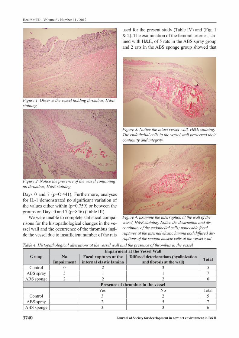
3740
HealthMED - Volume 6 / Number 11 / 2012
Journal of Society for development in new net environment in B&H
Days 0 and 7 (p=O.441). Furthermore, analyses for IL-1 demonstrated no significant variation of the values either within (p=0.759) or between the groups on Days 0 and 7 (p=846) (Table III).
We were unable to complete statistical compa-risons for the histopathological changes in the ve-ssel wall and the occurrence of the thrombus insi-de the vessel due to insufficient number of the rats
used for the present study (Table IV) and (Fig. 1 & 2). The examination of the femoral arteries, sta-ined with H&E, of 5 rats in the ABS spray group and 2 rats in the ABS sponge group showed that
Table 4. Histopathological alterations at the vessel wall and the presence of thrombus in the vessel
GroupImpairment at the Vessel Wall
No Impairment
Focal ruptures at the internal elastic lamina
Diffused deteriorations (hyalinization and fibrosis at the wall) Total
Control 0 2 3 5ABS spray 5 1 1 7
ABS sponge 2 2 2 6Presence of thrombus in the vessel
Yes No TotalControl 3 2 5
ABS spray 2 5 7ABS sponge 3 3 6
Figure 1. Observe the vessel holding thrombus, H&E staining.
Figure 2. Notice the presence of the vessel containing no thrombus, H&E staining.
Figure 3. Notice the intact vessel wall, H&E staining. The endothelial cells in the vessel wall preserved their continuity and integrity.
Figure 4. Examine the interruption at the wall of the vessel, H&E staining. Notice the destruction and dis-continuity of the endothelial cells; noticeable focal ruptures at the internal elastic lamina and diffused dis-ruptions of the smooth muscle cells at the vessel wall
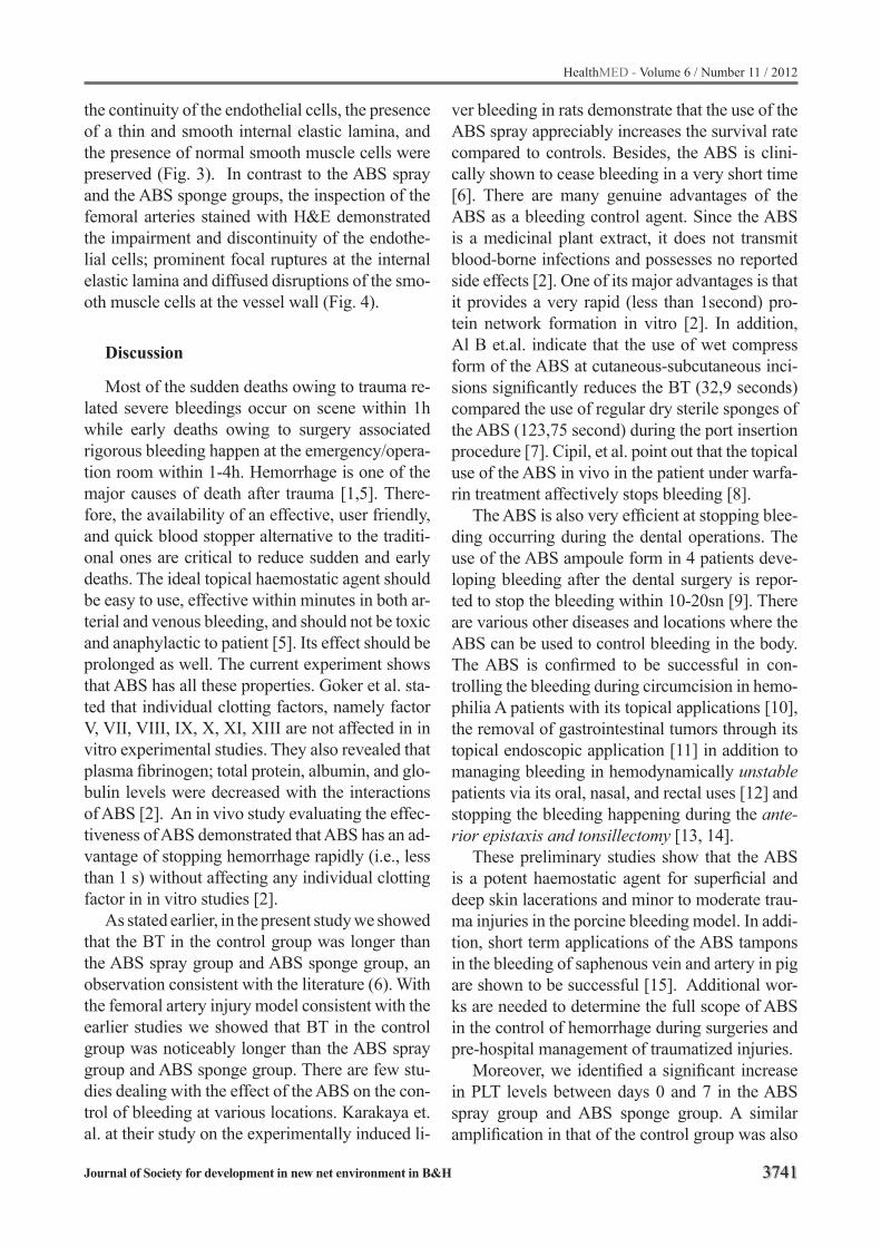
HealthMED - Volume 6 / Number 11 / 2012
Journal of Society for development in new net environment in B&H 3741
the continuity of the endothelial cells, the presence of a thin and smooth internal elastic lamina, and the presence of normal smooth muscle cells were preserved (Fig. 3). In contrast to the ABS spray and the ABS sponge groups, the inspection of the femoral arteries stained with H&E demonstrated the impairment and discontinuity of the endothe-lial cells; prominent focal ruptures at the internal elastic lamina and diffused disruptions of the smo-oth muscle cells at the vessel wall (Fig. 4).
Discussion
Most of the sudden deaths owing to trauma re-lated severe bleedings occur on scene within 1h while early deaths owing to surgery associated rigorous bleeding happen at the emergency/opera-tion room within 1-4h. Hemorrhage is one of the major causes of death after trauma [1,5]. There-fore, the availability of an effective, user friendly, and quick blood stopper alternative to the traditi-onal ones are critical to reduce sudden and early deaths. The ideal topical haemostatic agent should be easy to use, effective within minutes in both ar-terial and venous bleeding, and should not be toxic and anaphylactic to patient [5]. Its effect should be prolonged as well. The current experiment shows that ABS has all these properties. Goker et al. sta-ted that individual clotting factors, namely factor V, VII, VIII, IX, X, XI, XIII are not affected in in vitro experimental studies. They also revealed that plasma fibrinogen; total protein, albumin, and glo-bulin levels were decreased with the interactions of ABS [2]. An in vivo study evaluating the effec-tiveness of ABS demonstrated that ABS has an ad-vantage of stopping hemorrhage rapidly (i.e., less than 1 s) without affecting any individual clotting factor in in vitro studies [2].
As stated earlier, in the present study we showed that the BT in the control group was longer than the ABS spray group and ABS sponge group, an observation consistent with the literature (6). With the femoral artery injury model consistent with the earlier studies we showed that BT in the control group was noticeably longer than the ABS spray group and ABS sponge group. There are few stu-dies dealing with the effect of the ABS on the con-trol of bleeding at various locations. Karakaya et. al. at their study on the experimentally induced li-
ver bleeding in rats demonstrate that the use of the ABS spray appreciably increases the survival rate compared to controls. Besides, the ABS is clini-cally shown to cease bleeding in a very short time [6]. There are many genuine advantages of the ABS as a bleeding control agent. Since the ABS is a medicinal plant extract, it does not transmit blood-borne infections and possesses no reported side effects [2]. One of its major advantages is that it provides a very rapid (less than 1second) pro-tein network formation in vitro [2]. In addition, Al B et.al. indicate that the use of wet compress form of the ABS at cutaneous-subcutaneous inci-sions significantly reduces the BT (32,9 seconds) compared the use of regular dry sterile sponges of the ABS (123,75 second) during the port insertion procedure [7]. Cipil, et al. point out that the topical use of the ABS in vivo in the patient under warfa-rin treatment affectively stops bleeding [8].
The ABS is also very efficient at stopping blee-ding occurring during the dental operations. The use of the ABS ampoule form in 4 patients deve-loping bleeding after the dental surgery is repor-ted to stop the bleeding within 10-20sn [9]. There are various other diseases and locations where the ABS can be used to control bleeding in the body. The ABS is confirmed to be successful in con-trolling the bleeding during circumcision in hemo-philia A patients with its topical applications [10], the removal of gastrointestinal tumors through its topical endoscopic application [11] in addition to managing bleeding in hemodynamically unstable patients via its oral, nasal, and rectal uses [12] and stopping the bleeding happening during the ante-rior epistaxis and tonsillectomy [13, 14].
These preliminary studies show that the ABS is a potent haemostatic agent for superficial and deep skin lacerations and minor to moderate trau-ma injuries in the porcine bleeding model. In addi-tion, short term applications of the ABS tampons in the bleeding of saphenous vein and artery in pig are shown to be successful [15]. Additional wor-ks are needed to determine the full scope of ABS in the control of hemorrhage during surgeries and pre-hospital management of traumatized injuries.
Moreover, we identified a significant increase in PLT levels between days 0 and 7 in the ABS spray group and ABS sponge group. A similar amplification in that of the control group was also

3742
HealthMED - Volume 6 / Number 11 / 2012
Journal of Society for development in new net environment in B&H
measured suggesting no meaningful effect of the ABS on the release of PLT (Table II). Likewise, a recent work studied by Bilgili et.al. disclose no si-gnificant impact of the ABS on the release of PLT [16]. Trauma, major surgery, acute bleeding may due to acute and transient thrombocytosis (17, 18). Then ABS and thrombocystosis relations may not causal. Further studies remain to be done for reve-aling the mechanisms of the ABS.
The control of the production and release of cytokines release is extremely complex and firmly modulated in response to traumatic insult or infec-tion, capable of producing different effects depen-ding on the body’s regional composition. Infecti-on or trauma induces an immediate, system-wide proinflammatory release, unleashing cytokines that help to recruit neutrophils, B cells, T cells, platelets, and coagulation factors to the site of da-mage [19, 20]. At the present study we assessed pro-inflammatory release of IL-1 and TNF-α and found no significant difference of these cytokines between the 0 day and 7th day of the study (Ta-ble III). This observation suggests that the anti-in-flammatory activity of the ABS is not regulated through the generation of cytokines. Our literature review turned out no paper dealing with the effect of the ABS use on the cytokine production; the-refore, the finding of the current study will shed light on the relevant studies in the future.
In one study it was also shown that ABS help pancreatic fluid to solidify in vitro and it is pro-moted to be used in post operative complications like pancreatic fistulas [21]. In summary, the pre-sent study indicated that the use of ABS preserved the continuity of the endothelial cells, the presen-ce of a typical internal elastic lamina and normal smooth muscle cells, suggesting that the ABS can be easily used and efficiently control and stop the bleeding in a short time. However, the results of the present study showed no major effect of the ABS on PLT values, and pro-inflammatory cyto-kines. Further studies remain to be done for illu-minating the efficient mechanisms that ABS uses for stopping bleeding.
Limitations
We have identified a couple of limitations to our study. We kept the rats alive for 7 days for the
present study. The rats could be kept alive longer for better histopathological assessment of the de-velopment of the infection and tissue recuperati-on. Another limitation to the present study was the lack of statistical analyses due to inadequate num-ber of the animals used in the experiment.
Conclusions
The findings of the present study indicate that the ABS can be effectively used at the site of the accidents and emergency rooms for stopping and controlling the bleeding that is responsible for a significant part of the deaths owing to traumas. However, the results of the present study showed no significant effect of the ABS on PLT values and pro-inflammatory cytokines. Further investigati-ons remain to be done for elucidating the effective mechanisms that ABS uses for stopping bleeding.
References
1. Pfeifer R, Tarkin IS, Rocos B, Pape HC. Patterns of mortality and causes of death in polytrauma pa-tients—has anything changed ? Injury. Int. J. Care Injured. 2009;40: 907–11.
2. Goker H, Haznedaroglu IC, Ercetin S, Kirazli S, Akman U, Ozturk Y, at al. Haemostatic actions of the folkloric medicinal plant extract Ankaferd Blood Sto-pper. J İnt Med Res. 2008; 36: 163–70.
3. Ozaslan E, Purnak T, Yildiz A, Haznedaroglu IC. A new practical alternative for tumoural gastrointesti-nal bleeding: Ankaferd blood stopper. Dig Liver Dis 2010; 42; 594–5.
4. Opal SM, De Polo VA. Antiinflammatory cytokines. Chest. 2000; 117: 1162–72
5. Mannucci PM: Hemostatic drugs. N Engl J. Med 1998; 339: 245–53.
6. Karakaya M, Ucan HB, Tascılar O, Emre AU, Ca-kmak GK, Ankarali H, at al. Evaluation of a New Hemostatic Agent Ankaferd Blood Stopper in Expe-rimental Liver Laceration. Journal of Investigative Surgery. 2009;22.201–6.
7. Al B, Yildirim C, Cavdar M, Zengin S, Buyukaslan H, Kalender ME. Effectiveness of Ankaferd blood stopper in the topical control of active bleeding due to cutaneous-subcutaneous incisions. Saudi Med J. 2009;30,1520–5.
8. Cipil HS, Kosar A, Kaya A, Uz B, Haznedaroglu IC, Goker H, at al. In vivo hemostatic effect of the me-dicinal plant extract Ankaferd Blood Stopper in rats pretreated with warfarin. Clin Appl Thromb Hemost. 2009;15.270–6.
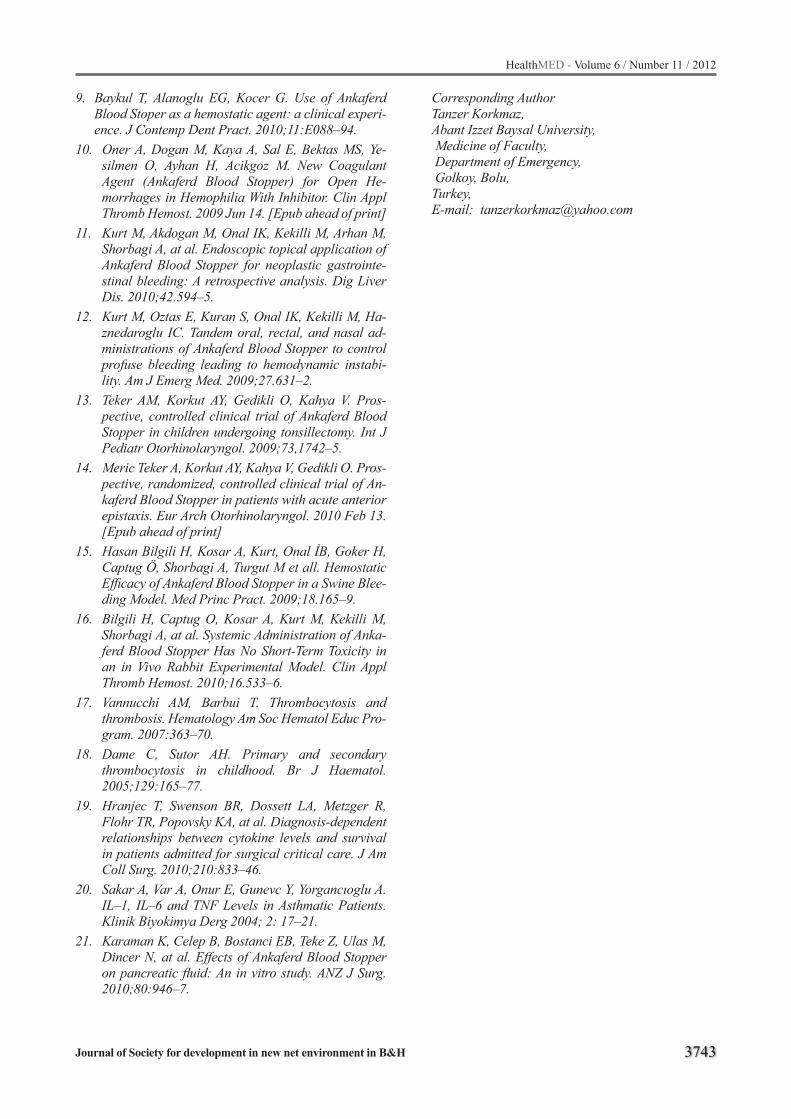
HealthMED - Volume 6 / Number 11 / 2012
Journal of Society for development in new net environment in B&H 3743
9. Baykul T, Alanoglu EG, Kocer G. Use of Ankaferd Blood Stoper as a hemostatic agent: a clinical experi-ence. J Contemp Dent Pract. 2010;11:E088–94.
10. Oner A, Dogan M, Kaya A, Sal E, Bektas MS, Ye-silmen O, Ayhan H, Acikgoz M. New Coagulant Agent (Ankaferd Blood Stopper) for Open He-morrhages in Hemophilia With Inhibitor. Clin Appl Thromb Hemost. 2009 Jun 14. [Epub ahead of print]
11. Kurt M, Akdogan M, Onal IK, Kekilli M, Arhan M, Shorbagi A, at al. Endoscopic topical application of Ankaferd Blood Stopper for neoplastic gastrointe-stinal bleeding: A retrospective analysis. Dig Liver Dis. 2010;42.594–5.
12. Kurt M, Oztas E, Kuran S, Onal IK, Kekilli M, Ha-znedaroglu IC. Tandem oral, rectal, and nasal ad-ministrations of Ankaferd Blood Stopper to control profuse bleeding leading to hemodynamic instabi-lity. Am J Emerg Med. 2009;27.631–2.
13. Teker AM, Korkut AY, Gedikli O, Kahya V. Pros-pective, controlled clinical trial of Ankaferd Blood Stopper in children undergoing tonsillectomy. Int J Pediatr Otorhinolaryngol. 2009;73,1742–5.
14. Meric Teker A, Korkut AY, Kahya V, Gedikli O. Pros-pective, randomized, controlled clinical trial of An-kaferd Blood Stopper in patients with acute anterior epistaxis. Eur Arch Otorhinolaryngol. 2010 Feb 13. [Epub ahead of print]
15. Hasan Bilgili H, Kosar A, Kurt, Onal İB, Goker H, Captug Ö, Shorbagi A, Turgut M et all. Hemostatic Efficacy of Ankaferd Blood Stopper in a Swine Blee-ding Model. Med Princ Pract. 2009;18.165–9.
16. Bilgili H, Captug O, Kosar A, Kurt M, Kekilli M, Shorbagi A, at al. Systemic Administration of Anka-ferd Blood Stopper Has No Short-Term Toxicity in an in Vivo Rabbit Experimental Model. Clin Appl Thromb Hemost. 2010;16.533–6.
17. Vannucchi AM, Barbui T. Thrombocytosis and thrombosis. Hematology Am Soc Hematol Educ Pro-gram. 2007:363–70.
18. Dame C, Sutor AH. Primary and secondary thrombocytosis in childhood. Br J Haematol. 2005;129:165–77.
19. Hranjec T, Swenson BR, Dossett LA, Metzger R, Flohr TR, Popovsky KA, at al. Diagnosis-dependent relationships between cytokine levels and survival in patients admitted for surgical critical care. J Am Coll Surg. 2010;210:833–46.
20. Sakar A, Var A, Onur E, Gunevc Y, Yorgancıoglu A. IL–1, IL–6 and TNF Levels in Asthmatic Patients. Klinik Biyokimya Derg 2004; 2: 17–21.
21. Karaman K, Celep B, Bostanci EB, Teke Z, Ulas M, Dincer N, at al. Effects of Ankaferd Blood Stopper on pancreatic fluid: An in vitro study. ANZ J Surg. 2010;80:946–7.
Corresponding AuthorTanzer Korkmaz,Abant Izzet Baysal University, Medicine of Faculty, Department of Emergency, Golkoy, Bolu,Turkey,E-mail: [email protected]
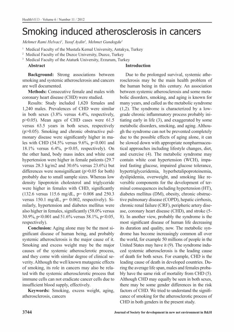
3744
HealthMED - Volume 6 / Number 11 / 2012
Journal of Society for development in new net environment in B&H
Smoking induced atherosclerosis in cancersMehmet Rami Helvaci1, Yusuf Aydin2, Mehmet Gundogdu3
1 Medical Faculty of the Mustafa Kemal University, Antakya, Turkey2 Medical Faculty of the Duzce University, Duzce, Turkey3 Medical Faculty of the Ataturk University, Erzurum, Turkey
Abstract
Background: Strong associations between smoking and systemic atherosclerosis and cancers are well documented.
Methods: Consecutive female and males with coronary heart disease (CHD) were studied.
Results: Study included 1,620 females and 1,240 males. Prevalences of CHD were similar in both sexes (3.8% versus 4.4%, respectively, p>0.05). Mean ages of CHD cases were 61.5 versus 63.5 years in both sexes, respectively (p>0.05). Smoking and chronic obstructive pul-monary disease were significantly higher in ma-les with CHD (54.5% versus 9.6%, p<0.001 and 18.1% versus 6.4%, p<0.05, respectively). On the other hand, body mass index and white coat hypertension were higher in female patients (29.7 versus 28.3 kg/m2 and 30.6% versus 23.6%) but differences were nonsignificant (p>0.05 for both) probably due to small sample sizes. Whereas low density lipoprotein cholesterol and triglyceride were higher in females with CHD, significantly (132.6 versus 115.6 mg/dL, p= 0.008 and 250.3 versus 150.1 mg/dL, p= 0.002, respectively). Si-milarly, hypertension and diabetes mellitus were also higher in females, significantly (58.0% versus 30.9%, p<0.001 and 51.6% versus 38.1%, p<0.05, respectively).
Conclusion: Aging alone may be the most si-gnificant disease of human being, and probably systemic atherosclerosis is the major cause of it. Smoking and excess weight may be the major causes of the systemic atherosclerotic process, and they come with similar degree of clinical se-verity. Although the well known mutagenic effects of smoking, its role in cancers may also be rela-ted with the systemic atherosclerotic process that immune cells can not eradicate cancer cells due to insufficient blood supply, effectively.
Keywords: Smoking, excess weight, aging, atherosclerosis, cancers
Introduction
Due to the prolonged survival, systemic athe-rosclerosis may be the main health problem of the human being in this century. An association between systemic atherosclerosis and some meta-bolic disorders, smoking, and aging is known for many years, and called as the metabolic syndrome (1,2). The syndrome is characterized by a low-grade chronic inflammatory process probably ini-tiating early in life (3), and exaggerated by some metabolic disorders, smoking, and aging. Althou-gh the syndrome can not be prevented completely due to the possible effects of aging alone, it can be slowed down with appropriate nonpharmaceu-tical approaches including lifestyle changes, diet, and exercise (4). The metabolic syndrome may contain white coat hypertension (WCH), impa-ired fasting glucose, impaired glucose tolerance, hypertriglyceridemia, hyperbetalipoproteinemia, dyslipidemia, overweight, and smoking like re-versible components for the development of ter-minal consequences including hypertension (HT), diabetes mellitus (DM), obesity, chronic obstruc-tive pulmonary disease (COPD), hepatic cirrhosis, chronic renal failure (CRF), peripheric artery dise-ase, coronary heart disease (CHD), and stroke (5-8). In another view, probably the syndrome is the most significant disease of human life decreasing its duration and quality, now. The metabolic syn-drome has become increasingly common all over the world, for example 50 millions of people in the United States may have it (9). The syndrome indu-ced systemic atherosclerosis is the leading cause of death for both sexes. For example, CHD is the leading cause of death in developed countries. Du-ring the average life span, males and females proba-bly have the same risk of mortality from CHD (5). Although CHD may equally be seen in both sexes, there may be some gender differences in the risk factors of CHD. We tried to understand the signifi-cance of smoking for the atherosclerotic process of CHD in both genders in the present study.
HealthMED - Volume 6 / Number 11 / 2012
Journal of Society for development in new net environment in B&H 3745
Material and methods
The study was performed in the Internal Me-dicine Polyclinic of the Dumlupinar University between August 2005 and March 2007. We took consecutive patients applying for any reason at and above the age of 15 years. Their medical hi-stories including smoking habit were learnt, and a routine check up procedure including fasting pla-sma glucose (FPG), low density lipoprotein chole-sterol (LDL-C), triglyceride (TG ), and an electro-cardiography was performed. Current smokers with six pack-months and cases with a history of five pack-years were accepted as smokers. COPD was diagnosed via the pulmonary function tests in suspected cases in which the ratio of forced expiratory volume in the first second of expiration to forced vital capacity is lower than 70%. Body mass index (BMI) of each case was calculated by the measurements of the Same Physician in-stead of verbal expressions. Weight in kilograms is divided by height in meters squared (10). Ca-ses with an overnight FPG level of 126 mg/dL or greater on two occasions or already using antidia-betic medications were defined as diabetics. An oral glucose tolerance test with 75-gram glucose was performed in cases with a FPG level between 110 and 126 mg/dL, and diagnosis of cases with a two-hour plasma glucose level of 200 mg/dL is DM. An office blood pressure (OBP) was chec-ked after a 5-minute of rest in seated position with a mercury sphygmomanometer on three visits. A 10-day twice daily measurement of blood pressure at home (HBP) was obtained in all cases, even in normotensives in the office due to the risk of ma-sked HT after an education about proper BP mea-surement techniques (11).
A 24-hour ambulatory blood pressure monito-ring was not required due to its equal effectivene-ss with HBP measurements (6). Eventually, HT is defined as a mean HBP value of 135/85 mmHg or greater, and WCH as an OBP of 140/90 mmHg or greater together with a mean HBP value of lower than 135/85 mmHg (11). A stress electrocardio-graphy was performed in cases with an abnormal electrocardiography and/or history of angina pec-toris. A coronary angiography was obtained just for the stress electrocardiography positive cases. So CHD was diagnosed either angiographically
or with a history of coronary artery stenting and/or bypass graft surgery. Eventually, all cases with CHD were divided into two groups according to gender distribution, and the mean age, weight, BMI, LDL-C, and TG values and prevalences of smoking, COPD, WCH, HT, and DM were com-pared in between. Mann-Whitney U test, Inde-pendent-Samples T test, and comparison of pro-portions were used as the methods of statistical analyses.
Results
The study included 1,620 females and 1,240 males (Table 1). Mean ages of them were 41.7 and 40.8 years, respectively (p>0.05). Prevalence of the CHD was similar in the females and males (3.8% versus 4.4%, respectively, p>0.05). Mean ages of the CHD cases were 61.5 versus 63.5 years, respectively (p>0.05). Prevalence of smo-king was significantly higher in male cases with CHD (54.5% versus 9.6%, p<0.001). Parallel to the higher prevalence of smoking, prevalence of COPD was also higher in males, significantly (18.1% versus 6.4%, p<0.05). On the other hand, although the mean weight of male cases with CHD was significantly higher (79.1 versus 74.4 kg, p= 0.027), the females had a higher mean BMI value (29.7 versus 28.3 kg/m2, p>0.05), but the difference was statistically nonsignificant proba-bly due to the small sample sizes of the groups. Similarly, both of the mean LDL-C and TG va-lues were significantly higher in the females, too (132.6 versus 115.6 mg/dL, p= 0.008 and 250.3 versus 150.1 mg/dL, p= 0.002, respectively). Alt-hough prevalence of WCH was also higher in females, the difference was nonsignificant proba-bly due to the above reason again (30.6% versus 23.6%, p>0.05). On the other hand, prevalences of HT and DM were significantly higher in females (58.0% versus 30.9%, p<0.001 and 51.6% versus 38.1%, p<0.05, respectively).
Discussion
Atherosclerosis is a systemic disease in which various substances such as cholesterol, cellular waste, and calcium are deposited along the lining of arterial wall. These plaques narrow vascular
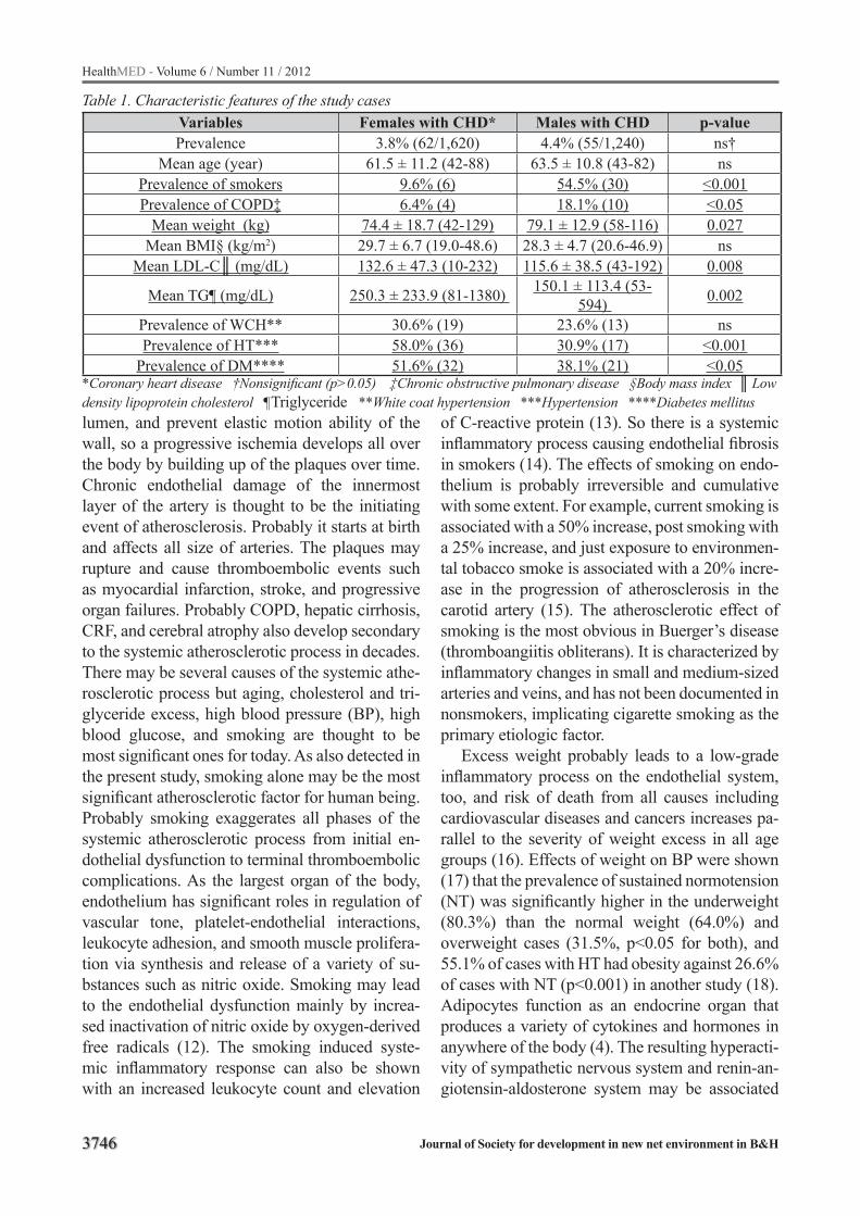
3746
HealthMED - Volume 6 / Number 11 / 2012
Journal of Society for development in new net environment in B&H
Table 1. Characteristic features of the study casesVariables Females with CHD* Males with CHD p-valuePrevalence 3.8% (62/1,620) 4.4% (55/1,240) ns†
Mean age (year) 61.5 ± 11.2 (42-88) 63.5 ± 10.8 (43-82) nsPrevalence of smokers 9.6% (6) 54.5% (30) <0.001Prevalence of COPD‡ 6.4% (4) 18.1% (10) <0.05
Mean weight (kg) 74.4 ± 18.7 (42-129) 79.1 ± 12.9 (58-116) 0.027Mean BMI§ (kg/m2) 29.7 ± 6.7 (19.0-48.6) 28.3 ± 4.7 (20.6-46.9) ns
Mean LDL-C (mg/dL) 132.6 ± 47.3 (10-232) 115.6 ± 38.5 (43-192) 0.008
Mean TG¶ (mg/dL) 250.3 ± 233.9 (81-1380) 150.1 ± 113.4 (53-594) 0.002
Prevalence of WCH** 30.6% (19) 23.6% (13) nsPrevalence of HT*** 58.0% (36) 30.9% (17) <0.001
Prevalence of DM**** 51.6% (32) 38.1% (21) <0.05*Coronary heart disease †Nonsignificant (p>0.05) ‡Chronic obstructive pulmonary disease §Body mass index Low density lipoprotein cholesterol ¶Triglyceride **White coat hypertension ***Hypertension ****Diabetes mellituslumen, and prevent elastic motion ability of the wall, so a progressive ischemia develops all over the body by building up of the plaques over time. Chronic endothelial damage of the innermost layer of the artery is thought to be the initiating event of atherosclerosis. Probably it starts at birth and affects all size of arteries. The plaques may rupture and cause thromboembolic events such as myocardial infarction, stroke, and progressive organ failures. Probably COPD, hepatic cirrhosis, CRF, and cerebral atrophy also develop secondary to the systemic atherosclerotic process in decades. There may be several causes of the systemic athe-rosclerotic process but aging, cholesterol and tri-glyceride excess, high blood pressure (BP), high blood glucose, and smoking are thought to be most significant ones for today. As also detected in the present study, smoking alone may be the most significant atherosclerotic factor for human being. Probably smoking exaggerates all phases of the systemic atherosclerotic process from initial en-dothelial dysfunction to terminal thromboembolic complications. As the largest organ of the body, endothelium has significant roles in regulation of vascular tone, platelet-endothelial interactions, leukocyte adhesion, and smooth muscle prolifera-tion via synthesis and release of a variety of su-bstances such as nitric oxide. Smoking may lead to the endothelial dysfunction mainly by increa-sed inactivation of nitric oxide by oxygen-derived free radicals (12). The smoking induced syste-mic inflammatory response can also be shown with an increased leukocyte count and elevation
of C-reactive protein (13). So there is a systemic inflammatory process causing endothelial fibrosis in smokers (14). The effects of smoking on endo-thelium is probably irreversible and cumulative with some extent. For example, current smoking is associated with a 50% increase, post smoking with a 25% increase, and just exposure to environmen-tal tobacco smoke is associated with a 20% incre-ase in the progression of atherosclerosis in the carotid artery (15). The atherosclerotic effect of smoking is the most obvious in Buerger’s disease (thromboangiitis obliterans). It is characterized by inflammatory changes in small and medium-sized arteries and veins, and has not been documented in nonsmokers, implicating cigarette smoking as the primary etiologic factor.
Excess weight probably leads to a low-grade inflammatory process on the endothelial system, too, and risk of death from all causes including cardiovascular diseases and cancers increases pa-rallel to the severity of weight excess in all age groups (16). Effects of weight on BP were shown (17) that the prevalence of sustained normotension (NT) was significantly higher in the underweight (80.3%) than the normal weight (64.0%) and overweight cases (31.5%, p<0.05 for both), and 55.1% of cases with HT had obesity against 26.6% of cases with NT (p<0.001) in another study (18). Adipocytes function as an endocrine organ that produces a variety of cytokines and hormones in anywhere of the body (4). The resulting hyperacti-vity of sympathetic nervous system and renin-an-giotensin-aldosterone system may be associated
HealthMED - Volume 6 / Number 11 / 2012
Journal of Society for development in new net environment in B&H 3747
with a chronic endothelial inflammation, insulin resistance, and an elevated BP. Similarly, Adult Treatment Panel III reported that excess weight predisposes to dyslipidemia, HT, DM, CHD, and stroke (10).
Although the obvious atherosclerotic effects of smoking, some studies reported that smoking in humans and nicotine administration in animals are associated with a decreased body weight (19). Evidence revealed an increased energy expenditu-re while smoking both on rest and light physical activity (20), and nicotine supplied by patch after smoking cessation decreased caloric intake in a dose-related manner (21). According to an animal study, nicotine may lengthen intermeal time, and simultaneously decreases amount of meal eaten (22). Additionally, body weight seems to be the highest in former, the lowest in the current and medium in never smokers (23). Similarly, althou-gh the CHD were detected with similar prevalen-ces in both sexes in the present study, prevalences of smoking and COPD were significantly higher in males against the higher prevalences of BMI, WCH, LDL-C, TG, HT, and DM in females as the other atherosclerotic risk factors. This result may indicate both the strong atherosclerotic and weight decreasing roles of smoking. Similarly, the incidence of myocardial infarction is increa-sed six-fold in women and three-fold in men who smoke at least 20 cigarettes per day compared to the never smoked cases (24). In another word, smoking is more harmful for women about CHD probably due to the associated higher BMI and its consequences in women. So smoking is probably a powerful athersclerotic risk factor with some suppressor effects on appetite. On the other hand, smoking, as a pleasure in life, may also show the weakness of volition to control eating, so it comes with additional weight and its complications alt-hough some inhibitory effects on appetite. Simi-larly, prevalences of HT, DM, and smoking were the highest in the highest TG having group as a significant component of metabolic syndrome in another study (25).
Smoking is the single greatest avoidable risk factor for cancer. It is the cause of 28% of all de-aths from cancers (26). Approximately half of regular smokers will die from the habit, and half of them in the middle age (27). Smoking is the
major risk factor for lung cancer in the current or previous smokers, and it causes 88% of male and 84% of female deaths from lung cancer in the UK (26). Even exposure to environmental tobacco smoke is risky for cancers (28). The risk of lung cancer increases with both duration and intensity of smoking (29). Smoking causes cancers in more organ sites than any other human carcinogens. It is an established risk factor for myeloid leukemia and cancers of esophagus, larynx, pharynx, oral cavity, pancreas, urinary bladder, nasal cavity and sinuses, stomach, liver, kidney, bowel, ovaries (mucinous), cervix, and breast (12,30,31). Multi-ple factors seem to intervene on carcinogenesis in smokers. There are over 5,000 chemical compo-unds identified in tobacco, and 62 of them have been evaluated as showing ‘sufficient evidence for carcinogenicity’ either in animal or human studies (12,30,31). Smoking contributes to oxida-tively induced DNA damage, and DNA mutations are increased in smokers (32). Smoking cessation appears to reduce levels of specific damage mar-kers between 30-50% in short term (32). The most important mutations may occur on tumor suppre-ssor genes such as p53 (33). On the other hand, it is evident that evading apoptosis plays a critical role in cigarette smoke-induced carcinogenesis and chemoresistance (33). Failure of apoptosis may lead to undesirable cell survival and unchec-ked cell growths. Resistance to apoptosis is often seen in cancers due to mutations. Nicotine, a major component of cigarette smoke, is known to play an important role in carcinogenesis, and it inhibits apoptosis induced by tumor necrosis factor, ultra-violet radiation, or chemotherapeutics (34). Pro-liferation of mesothelioma cells is also enhanced by nicotine, that may also be seen in endothelial cells of smokers (35). The carcinogenic effects of smoking may also be irreversible and cumulative. For instance, risk of cancers of upper aerodigesti-ve tract in ex-smokers becomes lower than that of current smokers within five years, although the risk is still higher than someone who has never smoked, 20 or more years after cessation (36). The risk of bladder cancer is also higher than in never-smokers 20 years after cessation (37,38).
Smoking, excess weight, and aging associated increased risk of cancers may either be related to the chronic inflammatory process on endothelium
3748
HealthMED - Volume 6 / Number 11 / 2012
Journal of Society for development in new net environment in B&H
or the systemic atherosclerotic process. The low-grade chronic inflammation may cause genetic changes on epithelial cells of the organs over time. For example, about 80% of cancers develop over the age of 55 years (39), and most of the cancers are carcinomas which arise from the epithelial cells that are prone to the genetic changes due to the external stresses. On the other hand, smoking, excess weight, and aging induced systemic athe-rosclerotic process may also decrease clearance of malignant cells by the immune system, effecti-vely. Normally, a number of malignant cells deve-lop, and are destroyed effectively by the immune system in human body everyday. Whereas due to the strong atherosclerotic effects of smoking, the clearance mechanism can not work, properly, and the malignant cells divide and spread all over the body. The same atherosclerotic process may also take role in the COPD, in which repair mechani-sms of the damaged airways can not work properly due to the narrowed capillary vasculature. The same etiologic mechanism may also be important in hepatic cirrhosis, CRF, and cerebral atrophy like end-organ failures in case of smoking, excess weight, and/or aging.
As a conclusion, aging alone may be the most significant disease of human being, and probably systemic atherosclerosis is the major cause of it. Smoking and excess weight may be the major causes of the systemic atherosclerotic process, and they come with similar degree of clinical seve-rity in front. Although the well known mutagenic effects of smoking, its role in cancers may also be related to the systemic atherosclerotic process that immune cells can not eradicate cancer cells due to insufficient blood supply, effectively.
References
1. Eckel RH, Grundy SM, Zimmet PZ. The metabolic syndrome. Lancet 2005; 365: 1415-1428.
2. Gundy SM, Brewer HB Jr, Cleeman JI, Smith SC Jr, Lenfant C. Definition of metabolic syndrome: Report of the National Heart, Lung, and Blood Institute/American Heart Association conference on scientific issues related to definition. Circulation 2004; 109: 433-438.
3. Tonkin AM. The metabolic syndrome(s)? Curr Athe-roscler Rep 2004; 6: 165-166.
4. Franklin SS, Barboza MG, Pio JR, Wong ND. Blood pressure categories, hypertensive subtypes, and the metabolic syndrome. J Hypertens 2006; 24: 2009-2016.
5. Helvaci MR, Kaya H, Gundogdu M. Gender differen-ces in coronary heart disease. Pak J Med Sci 2012; 28: 40-44.
6. Helvaci MR, Seyhanli M. What a high prevalence of white coat hypertension in society! Intern Med 2006; 45: 671-674.
7. Helvaci MR, Kaya H, Sevinc A, Camci C. Body weight and white coat hypertension. Pak J Med Sci 2009; 25: 6: 916-921.
8. Helvaci MR, Kaya H, Seyhanli M, Yalcin A. White coat hypertension in definition of metabolic syndro-me. Int Heart J 2008; 49: 449-457.
9. Clark LT, El-Atat F. Metabolic Syndrome in African Americans: implications for preventing coronary he-art disease. Clin Cardiol 2007; 30: 161-164.
10. Third Report of the National Cholesterol Education Program (NCEP) Expert Panel on Detection, Eva-luation, and Treatment of High Blood Cholesterol in Adults (Adult Treatment Panel III) final report. Cir-culation 2002; 17:106: 3143-3421.
11. O’Brien E, Asmar R, Beilin L, Imai Y, Mallion JM, Mancia G, et al. European Society of Hypertensi-on recommendations for conventional, ambulatory and home blood pressure measurement. J Hypertens 2003; 21: 821-848.
12. Heitzer T, Meinertz T. Prevention of coronary heart disease: smoking. Z Kardiol 2005; 94: 30-42. Ger-man.
13. Nanri A, Moore MA, Kono S. Impact of C-reactive protein on disease risk and its relation to dietary factors. Asian Pac J Cancer Prev 2007; 8: 167-177.
14. Howard G, Wagenknecht LE, Burke GL, Diez-Roux A, Evans GW, McGovern P, et al. Cigarette smoking and progression of atherosclerosis: The Atheros-clerosis Risk in Communities (ARIC) Study. JAMA 1998; 279: 119-124.
15. Ambrose JA, Barua RS. The pathophysiology of cigarette smoking and cardiovascular disease: an update. J Am Coll Cardiol 2004; 43: 1731-1737.
16. Calle EE, Thun MJ, Petrelli JM, Rodriguez C, Heath CW Jr. Body-mass index and mortality in a prospec-tive cohort of U.S. adults. N Engl J Med 1999; 341: 1097-1105.
HealthMED - Volume 6 / Number 11 / 2012
Journal of Society for development in new net environment in B&H 3749
17. Helvaci MR, Ozcura F, Kaya H, Yalcin A. Fundusco-pic examination has limited benefit for management of hypertension. Int Heart J 2007; 48: 187-194.
18. Helvaci MR, Kaya H, Yalcin A, Kuvandik G. Preva-lence of white coat hypertension in underweight and overweight subjects. Int Heart J 2007; 48: 605-613.
19. Grunberg NE, Greenwood MR, Collins F, Epste-in LH, Hatsukami D, Niaura R, et al. National working conference on smoking and body weight. Task Force 1: Mechanisms relevant to the relations between cigarette smoking and body weight. Health Psychol 1992; 11: 4-9.
20. Walker JF, Collins LC, Rowell PP, Goldsmith LJ, Moffatt RJ, Stamford BA. The effect of smoking on energy expenditure and plasma catecholamine and nicotine levels during light physical activity. Nicoti-ne Tob Res 1999; 1: 365-370.
21. Hughes JR, Hatsukami DK. Effects of three doses of transdermal nicotine on post-cessation eating, hun-ger and weight. J Subst Abuse 1997; 9: 151-159.
22. Miyata G, Meguid MM, Varma M, Fetissov SO, Kim HJ. Nicotine alters the usual reciprocity betwe-en meal size and meal number in female rat. Physiol Behav 2001; 74: 169-176.
23. Laaksonen M, Rahkonen O, Prattala R. Smoking status and relative weight by educational level in Finland, 1978-1995. Prev Med 1998; 27: 431-437.
24. Prescott E, Hippe M, Schnohr P, Hein HO, Vestbo J. Smoking and risk of myocardial infarction in women and men: longitudinal population study. BMJ 1998; 316: 1043-1047.
25. Helvaci MR, Kaya H, Gundogdu M. Association of increased triglyceride levels in metabolic syndrome with coronary artery disease. Pak J Med Sci 2010; 26: 667-672.
26. Peto R, Lopez AD, Boreham J, Thun M, Heath C Jr, Doll R. Mortality from smoking worldwide. Br Med Bull 1996; 52: 12-21.
27. Peto R. Smoking and death: the past 40 years and the next 40. BMJ 1994; 309: 937-939.
28. Taylor R, Najafi F, Dobson A. Meta-analysis of stu-dies of passive smoking and lung cancer: effects of study type and continent. Int J Epidemiol 2007; 36: 1048-1059.
29. Rylander R, Axelsson G, Andersson L, Liljequist T, Bergman B. Lung cancer, smoking and diet among Swedish men. Lung Cancer 1996; 14: 75-83.
30. Parkin DM. Tobacco-attributable cancer burden in the UK in 2010. Br J Cancer 2011; 105: 6-13
31. Anand P, Kunnumakkara AB, Sundaram C, Hari-kumar KB, Tharakan ST, Lai OS, et al. Cancer is a preventable disease that requires major lifestyle changes. Pharm Res 2008; 25: 2097-2116.
32. Box HC, O’Connor RJ, Patrzyc HB, Iijima H, Dawidzik JB, Freund HG, et al. Reduction in oxi-datively generated DNA damage following smoking cessation. Tob Induc Dis 2011; 9: 5.
33. Chen RJ, Chang LW, Lin P, Wang YJ. Epigenetic effects and molecular mechanisms of tumorigenesis induced by cigarette smoke: an overview. J Oncol 2011; 2011: 654931.
34. Xu J, Huang H, Pan C, Zhang B, Liu X, Zhang L. Nicotine inhibits apoptosis induced by cisplatin in human oral cancer cells. Int J Oral Maxillofac Surg 2007; 36: 739-744.
35. Trombino S, Cesario A, Margaritora S, Granone P, Motta G, Falugi C, et al. Alpha7-nicotinic acetylcho-line receptors affect growth regulation of human me-sothelioma cells: role of mitogen-activated protein kinase pathway. Cancer Res 2004; 64: 135-145.
36. Bosetti C, Gallus S, Peto R, Negri E, Talamini R, Tavani A, et al. Tobacco smoking, smoking cessati-on, and cumulative risk of upper aerodigestive tract cancers. Am J Epidemiol 2008; 167: 468-473.
37. Brennan P, Bogillot O, Cordier S, Greiser E, Schill W, Vineis P, et al. Cigarette smoking and bladder cancer in men: a pooled analysis of 11 case-control studies. Int J Cancer 2000; 86: 289-294.
38. Brennan P, Bogillot O, Greiser E, Chang-Claude J, Wahrendorf J, Cordier S, et al. The contribution of cigarette smoking to bladder cancer in women (po-oled European data). Cancer Causes Control 2001; 12: 411-417.
39. Jemal A, Siegel R, Ward E, Hao Y, Xu J, Murray T, et al. Cancer statistics, 2008. CA Cancer J Clin 2008; 58: 71-96.
Corresponding AuthorMehmet Rami Helvaci, Medical Faculty of the Mustafa Kemal University,Serinyol, Antakya, Hatay, TurkeyE-mail: [email protected]
3750
HealthMED - Volume 6 / Number 11 / 2012
Journal of Society for development in new net environment in B&H
Selective mutation searching of exon 28 in the VWF gene in type 3 Von Willebrand disease patients from Southwest IranNasiri Mahboobeh1, Galehdari Hamid2, Darbouy Mojtaba1, Yavarian Majid3, Keikhaie Bijan2
1 Department of Genetics, Science and Research Branch, Islamic Azad University, Fars, Iran2 Research Centre for Thalassemia and Hemoglobinopathies of Ahvaz Jundishapur University of Medical Sciences Ahvaz, Iran 3 Haematology Research Centre, Shiraz University of Medical Science, Shiraz, Iran
Abstract
Von Willebrand disease (VWD) is a common bleeding disorder and type 3 belongs to the most registered type in Iranian VWD patients. The di-sease is resulted from quantitative defects in Von Willebrand factor (VWF), a key glycoprotein with central role in haemostasis. Type 3 VWD is characterized by low or absent of VWF, enco-ded by the large VWF gene on chromosome 12 (12p13.3). Inheritance of two null alleles cause type 3 VWD. Different mutations have been ob-served in type 3 VWD, including deletions, in-sertions, missense and nonsense mutations, and nucleotide changes in the intron-exon boundaries causing alternative splicing. We selectively sear-ched for mutations in exon 28, the largest exon of the VWA gene, in 33 individuals suffering from type 3 VWD from Southwest Iran. Molecular analysis was performed by direct sequencing of PCR products, resulting from amplification of en-tire exon 28. The technique PCR-RFLP was also used to verifying nucleotide changes. We obser-ved nonsense mutations in 28 patients (87.87%) as follow: One individual (3.03%) was homozygous for the nonsense mutation Q1311X. Twenty three patients (69.69%) were compound heterozygous for two nonsense mutations Q1311X and R1659X and five additional patients (15.15%) were hete-rozygous for the Q1311X mutation. Four samples were negative for any relevant changes in exon 28. Our results indicate that exon 28 of the VWF gene might be the first choice for molecular testing of a large portion of type 3 VWD individuals, at least from southwest Iran.
Keywords: Von Willebrand disease, Von Wille-brand factor, Exon 28, Mutation, South-west Iran.
Introduction
Von Willebrand disease (VWD) is the most common bleeding disorder with an incidence of 1-2% in the general populations (1, 2). The disea-se is characterized by deficiency or dysfunction of Von Willebrand factor (VWF), a large multimeric glycoprotein that plays essential roles in the hemo-stasis by carrying and stabilizing the factor VIII in the plasma and mediating platelet adhesion at vas-cular injury sites (3, 4). The VWD is classified in three main types on the basis of clinical phenotypes and genetic disorders (5). Type 1, as the most pre-valent form, is a mild to moderate quantitative de-ficiency of VWF (6). Type 2 is characterized by a qualitative abnormality of VWF with autosomal dominant pattern of inheritance (7). Type 3 VWD is an autosomal recessive disorder that is charac-terized by very low or undetectable VWF level in plasma and platelets, causing severe bleeding diathesis including mucosal tract haemorrhages, excessive and prolonged bleeding after surgery or during menstruation and delivery in affected wo-men (8, 9). The prevalence of type 3 VWD varies from 1/1000000 to 1/500000, but higher in Arab nations or in individuals with Arabian backgro-und, such as some people in southwest Iran (10).
Due to several variable parameters such as blo-od group, age and gender that influence the plasma levels of the VWF, a clinical diagnosis is generally very difficult (11, 12).
The VWF is encoded by a large gene spanning 178 kb length on the chromosome 12, consisting of 52 exons, whereby exon 28 is the largest exon that spans 1.4 kb in length. Besides this, there is a partial unprocessed pseudogene, locating on chro-mosome 22 with a length of 21-29 kb. Its sequen-ce is equivalent to exons 23-34 of the active VWF
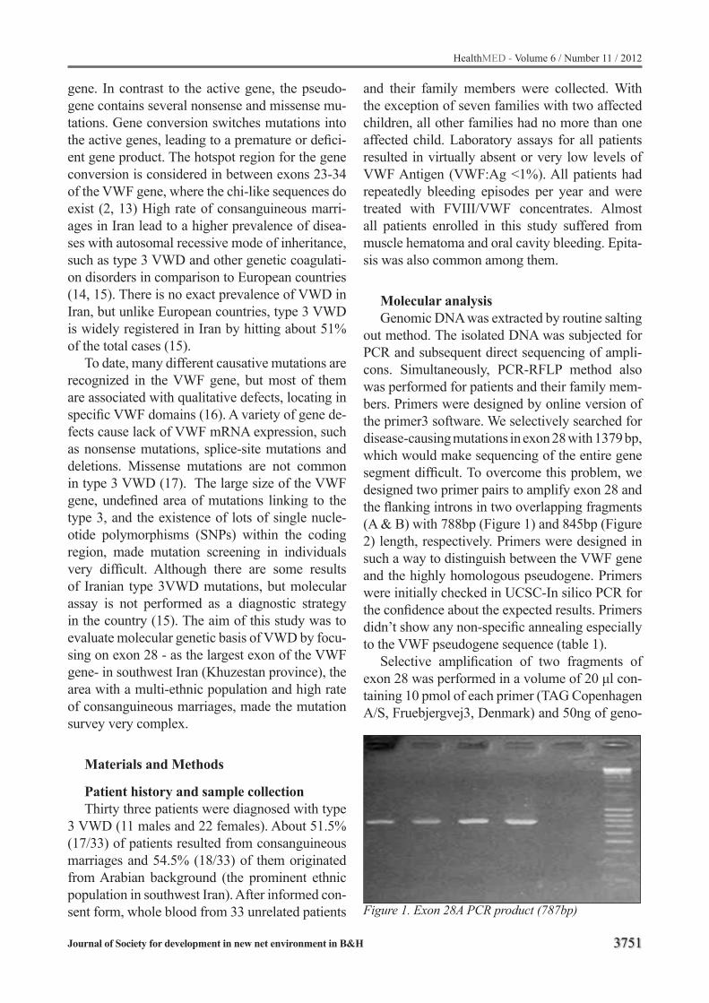
HealthMED - Volume 6 / Number 11 / 2012
Journal of Society for development in new net environment in B&H 3751
gene. In contrast to the active gene, the pseudo-gene contains several nonsense and missense mu-tations. Gene conversion switches mutations into the active genes, leading to a premature or defici-ent gene product. The hotspot region for the gene conversion is considered in between exons 23-34 of the VWF gene, where the chi-like sequences do exist (2, 13) High rate of consanguineous marri-ages in Iran lead to a higher prevalence of disea-ses with autosomal recessive mode of inheritance, such as type 3 VWD and other genetic coagulati-on disorders in comparison to European countries (14, 15). There is no exact prevalence of VWD in Iran, but unlike European countries, type 3 VWD is widely registered in Iran by hitting about 51% of the total cases (15).
To date, many different causative mutations are recognized in the VWF gene, but most of them are associated with qualitative defects, locating in specific VWF domains (16). A variety of gene de-fects cause lack of VWF mRNA expression, such as nonsense mutations, splice-site mutations and deletions. Missense mutations are not common in type 3 VWD (17). The large size of the VWF gene, undefined area of mutations linking to the type 3, and the existence of lots of single nucle-otide polymorphisms (SNPs) within the coding region, made mutation screening in individuals very difficult. Although there are some results of Iranian type 3VWD mutations, but molecular assay is not performed as a diagnostic strategy in the country (15). The aim of this study was to evaluate molecular genetic basis of VWD by focu-sing on exon 28 - as the largest exon of the VWF gene- in southwest Iran (Khuzestan province), the area with a multi-ethnic population and high rate of consanguineous marriages, made the mutation survey very complex.
Materials and Methods
Patient history and sample collection Thirty three patients were diagnosed with type
3 VWD (11 males and 22 females). About 51.5% (17/33) of patients resulted from consanguineous marriages and 54.5% (18/33) of them originated from Arabian background (the prominent ethnic population in southwest Iran). After informed con-sent form, whole blood from 33 unrelated patients
and their family members were collected. With the exception of seven families with two affected children, all other families had no more than one affected child. Laboratory assays for all patients resulted in virtually absent or very low levels of VWF Antigen (VWF:Ag <1%). All patients had repeatedly bleeding episodes per year and were treated with FVIII/VWF concentrates. Almost all patients enrolled in this study suffered from muscle hematoma and oral cavity bleeding. Epita-sis was also common among them.
Molecular analysisGenomic DNA was extracted by routine salting
out method. The isolated DNA was subjected for PCR and subsequent direct sequencing of ampli-cons. Simultaneously, PCR-RFLP method also was performed for patients and their family mem-bers. Primers were designed by online version of the primer3 software. We selectively searched for disease-causing mutations in exon 28 with 1379 bp, which would make sequencing of the entire gene segment difficult. To overcome this problem, we designed two primer pairs to amplify exon 28 and the flanking introns in two overlapping fragments (A & B) with 788bp (Figure 1) and 845bp (Figure 2) length, respectively. Primers were designed in such a way to distinguish between the VWF gene and the highly homologous pseudogene. Primers were initially checked in UCSC-In silico PCR for the confidence about the expected results. Primers didn’t show any non-specific annealing especially to the VWF pseudogene sequence (table 1).
Selective amplification of two fragments of exon 28 was performed in a volume of 20 μl con-taining 10 pmol of each primer (TAG Copenhagen A/S, Fruebjergvej3, Denmark) and 50ng of geno-
Figure 1. Exon 28A PCR product (787bp)
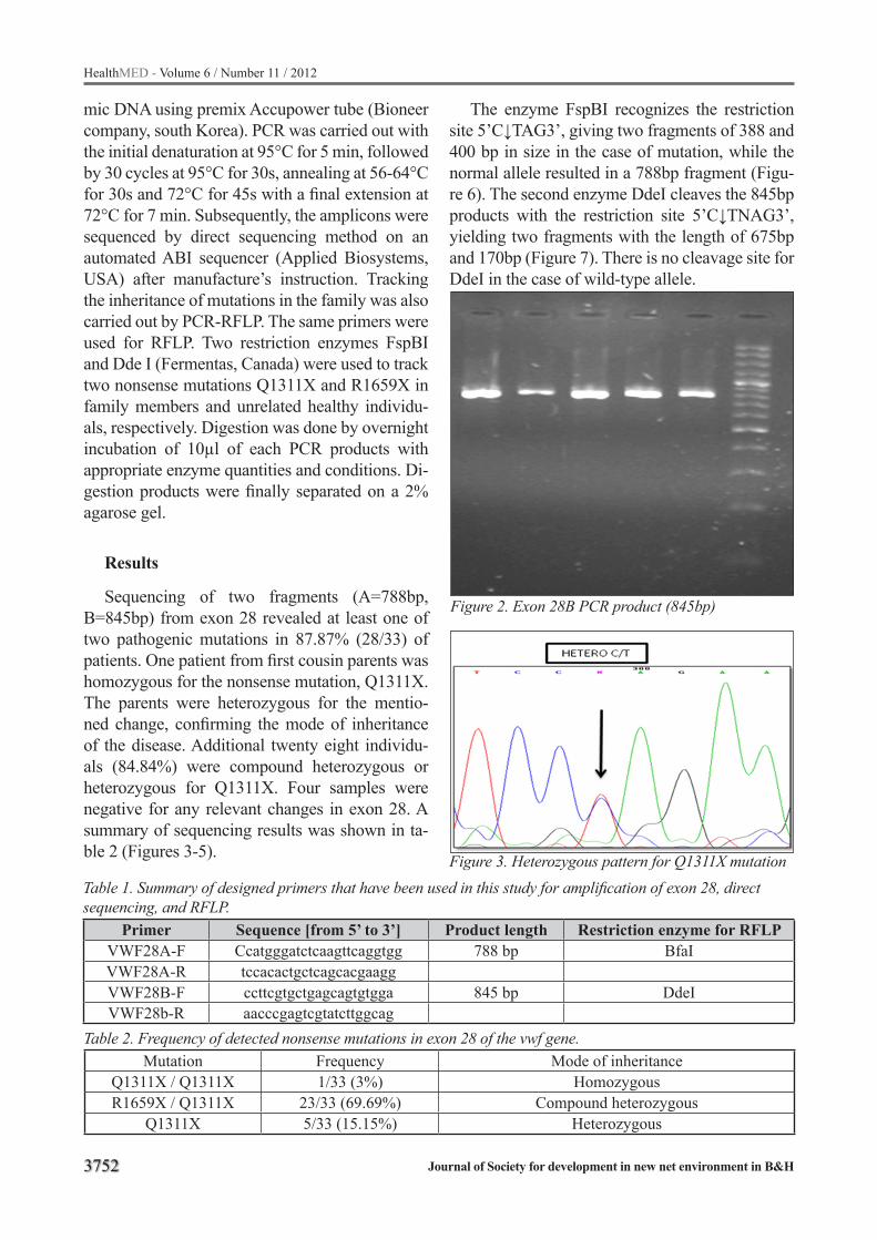
3752
HealthMED - Volume 6 / Number 11 / 2012
Journal of Society for development in new net environment in B&H
Table 1. Summary of designed primers that have been used in this study for amplification of exon 28, direct sequencing, and RFLP.
Primer Sequence [from 5’ to 3’] Product length Restriction enzyme for RFLPVWF28A-F Ccatgggatctcaagttcaggtgg 788 bp BfaI VWF28A-R tccacactgctcagcacgaaggVWF28B-F ccttcgtgctgagcagtgtgga 845 bp DdeIVWF28b-R aacccgagtcgtatcttggcag
Table 2. Frequency of detected nonsense mutations in exon 28 of the vwf gene.Mutation Frequency Mode of inheritance
Q1311X / Q1311X 1/33 (3%) HomozygousR1659X / Q1311X 23/33 (69.69%) Compound heterozygous
Q1311X 5/33 (15.15%) Heterozygous
mic DNA using premix Accupower tube (Bioneer company, south Korea). PCR was carried out with the initial denaturation at 95°C for 5 min, followed by 30 cycles at 95°C for 30s, annealing at 56-64°C for 30s and 72°C for 45s with a final extension at 72°C for 7 min. Subsequently, the amplicons were sequenced by direct sequencing method on an automated ABI sequencer (Applied Biosystems, USA) after manufacture’s instruction. Tracking the inheritance of mutations in the family was also carried out by PCR-RFLP. The same primers were used for RFLP. Two restriction enzymes FspBI and Dde I (Fermentas, Canada) were used to track two nonsense mutations Q1311X and R1659X in family members and unrelated healthy individu-als, respectively. Digestion was done by overnight incubation of 10µl of each PCR products with appropriate enzyme quantities and conditions. Di-gestion products were finally separated on a 2% agarose gel.
Results
Sequencing of two fragments (A=788bp, B=845bp) from exon 28 revealed at least one of two pathogenic mutations in 87.87% (28/33) of patients. One patient from first cousin parents was homozygous for the nonsense mutation, Q1311X. The parents were heterozygous for the mentio-ned change, confirming the mode of inheritance of the disease. Additional twenty eight individu-als (84.84%) were compound heterozygous or heterozygous for Q1311X. Four samples were negative for any relevant changes in exon 28. A summary of sequencing results was shown in ta-ble 2 (Figures 3-5).
The enzyme FspBI recognizes the restriction site 5’C↓TAG3’, giving two fragments of 388 and 400 bp in size in the case of mutation, while the normal allele resulted in a 788bp fragment (Figu-re 6). The second enzyme DdeI cleaves the 845bp products with the restriction site 5’C↓TNAG3’, yielding two fragments with the length of 675bp and 170bp (Figure 7). There is no cleavage site for DdeI in the case of wild-type allele.
Figure 2. Exon 28B PCR product (845bp)
Figure 3. Heterozygous pattern for Q1311X mutation

HealthMED - Volume 6 / Number 11 / 2012
Journal of Society for development in new net environment in B&H 3753
Table 3. Summary of identified SNPs in exon 28 of the VWF gene. Status Nucleotide No. in cDNA Alleles Amino acid No. Report status No.
Heterozygous 3685 T/G V/G1229 Novel 1 Heterozygous 3692 A/C N/H1231 Novel 2 Heterozygous 3731 T/C V/G1244 Novel 3 Heterozygous 3735 G/A V1245 Novel 4 Heterozygous 3789 G/A S1263 Known 5 Heterozygous 3951 C/T A1317 Known 6 Heterozygous 4163 A/G Q/R1388 novel 7 Homozygous 4141 A/G T/A1381 Known 8 Heterozygous 4230 T/C I1410 novel 9 Heterozygous 4240 G/A V/M1414 novel 10 Heterozygous 4242 G/C V1414 novel 11 Heterozygous 4284 C/T I1428 novel 12 Homozygous 4414 G/C D/H1472 Known 13 Homozygous 4453 G/C V/L1485 novel 14 Homozygous 4500 G/A A1500 Known 15 Heterozygous 4503 C/T F1501 Known 16 Homozygous 4508 T/C F1503 novel 17 Heterozygous 4517 C/T S/L1506 Known 18 Homozygous 4641 T/C T1547 Known 19 Homozygous 4716 C/T G1572 Known 20 Homozygous 4748 G/T R/L1583 novel 21 Homozygous 4754 T/C L/P1585 Novel 22 Homozygous 4758 T/C S1586 novel 23 Homozygous 4799 C/T A/V1600 Novel 24 Homozygous 4814 A/C T/S1605 novel 25 Homozygous 4825 G/C G/R1609 novel 26 Homozygous 4843 G/A E/L1615 novel 27 Homozygous 4855 C/T L1619 Known 28 Homozygous 4926 T/C I1642 Known 29 Homozygous 4944 T/C P1648 Known 30 Homozygous 5049 A/C A1683 novel 31
Figure 4. Homozygous pattern for Q1311X mutation Figure 5. Heterozygous pattern for R1659X mutation
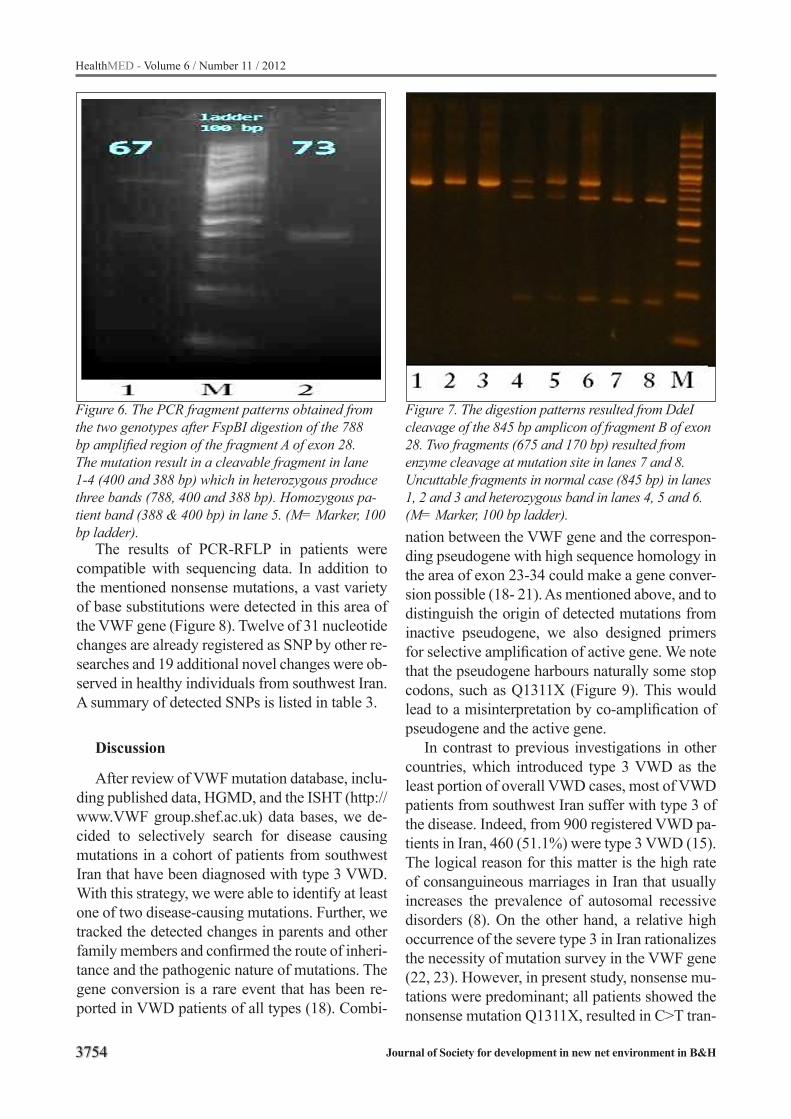
3754
HealthMED - Volume 6 / Number 11 / 2012
Journal of Society for development in new net environment in B&H
The results of PCR-RFLP in patients were compatible with sequencing data. In addition to the mentioned nonsense mutations, a vast variety of base substitutions were detected in this area of the VWF gene (Figure 8). Twelve of 31 nucleotide changes are already registered as SNP by other re-searches and 19 additional novel changes were ob-served in healthy individuals from southwest Iran. A summary of detected SNPs is listed in table 3.
Discussion
After review of VWF mutation database, inclu-ding published data, HGMD, and the ISHT (http://www.VWF group.shef.ac.uk) data bases, we de-cided to selectively search for disease causing mutations in a cohort of patients from southwest Iran that have been diagnosed with type 3 VWD. With this strategy, we were able to identify at least one of two disease-causing mutations. Further, we tracked the detected changes in parents and other family members and confirmed the route of inheri-tance and the pathogenic nature of mutations. The gene conversion is a rare event that has been re-ported in VWD patients of all types (18). Combi-
Figure 6. The PCR fragment patterns obtained from the two genotypes after FspBI digestion of the 788 bp amplified region of the fragment A of exon 28. The mutation result in a cleavable fragment in lane 1-4 (400 and 388 bp) which in heterozygous produce three bands (788, 400 and 388 bp). Homozygous pa-tient band (388 & 400 bp) in lane 5. (M= Marker, 100 bp ladder).
Figure 7. The digestion patterns resulted from DdeI cleavage of the 845 bp amplicon of fragment B of exon 28. Two fragments (675 and 170 bp) resulted from enzyme cleavage at mutation site in lanes 7 and 8. Uncuttable fragments in normal case (845 bp) in lanes 1, 2 and 3 and heterozygous band in lanes 4, 5 and 6. (M= Marker, 100 bp ladder).nation between the VWF gene and the correspon-ding pseudogene with high sequence homology in the area of exon 23-34 could make a gene conver-sion possible (18- 21). As mentioned above, and to distinguish the origin of detected mutations from inactive pseudogene, we also designed primers for selective amplification of active gene. We note that the pseudogene harbours naturally some stop codons, such as Q1311X (Figure 9). This would lead to a misinterpretation by co-amplification of pseudogene and the active gene.
In contrast to previous investigations in other countries, which introduced type 3 VWD as the least portion of overall VWD cases, most of VWD patients from southwest Iran suffer with type 3 of the disease. Indeed, from 900 registered VWD pa-tients in Iran, 460 (51.1%) were type 3 VWD (15). The logical reason for this matter is the high rate of consanguineous marriages in Iran that usually increases the prevalence of autosomal recessive disorders (8). On the other hand, a relative high occurrence of the severe type 3 in Iran rationalizes the necessity of mutation survey in the VWF gene (22, 23). However, in present study, nonsense mu-tations were predominant; all patients showed the nonsense mutation Q1311X, resulted in C>T tran-
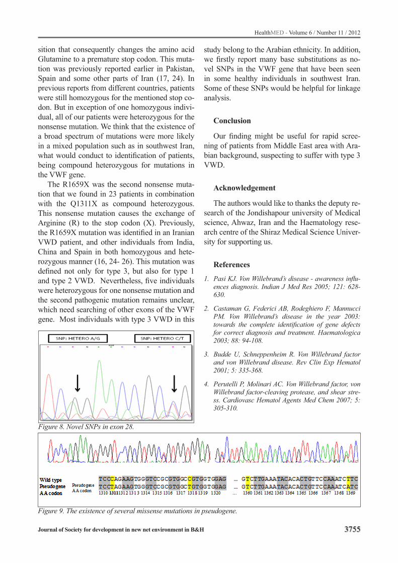
HealthMED - Volume 6 / Number 11 / 2012
Journal of Society for development in new net environment in B&H 3755
sition that consequently changes the amino acid Glutamine to a premature stop codon. This muta-tion was previously reported earlier in Pakistan, Spain and some other parts of Iran (17, 24). In previous reports from different countries, patients were still homozygous for the mentioned stop co-don. But in exception of one homozygous indivi-dual, all of our patients were heterozygous for the nonsense mutation. We think that the existence of a broad spectrum of mutations were more likely in a mixed population such as in southwest Iran, what would conduct to identification of patients, being compound heterozygous for mutations in the VWF gene.
The R1659X was the second nonsense muta-tion that we found in 23 patients in combination with the Q1311X as compound heterozygous. This nonsense mutation causes the exchange of Arginine (R) to the stop codon (X). Previously, the R1659X mutation was identified in an Iranian VWD patient, and other individuals from India, China and Spain in both homozygous and hete-rozygous manner (16, 24- 26). This mutation was defined not only for type 3, but also for type 1 and type 2 VWD. Nevertheless, five individuals were heterozygous for one nonsense mutation and the second pathogenic mutation remains unclear, which need searching of other exons of the VWF gene. Most individuals with type 3 VWD in this
Figure 8. Novel SNPs in exon 28.
Figure 9. The existence of several missense mutations in pseudogene.
study belong to the Arabian ethnicity. In addition, we firstly report many base substitutions as no-vel SNPs in the VWF gene that have been seen in some healthy individuals in southwest Iran. Some of these SNPs would be helpful for linkage analysis.
Conclusion
Our finding might be useful for rapid scree-ning of patients from Middle East area with Ara-bian background, suspecting to suffer with type 3 VWD.
Acknowledgement
The authors would like to thanks the deputy re-search of the Jondishapour university of Medical science, Ahwaz, Iran and the Haematology rese-arch centre of the Shiraz Medical Science Univer-sity for supporting us.
References
1. Pasi KJ. Von Willebrand’s disease - awareness influ-ences diagnosis. Indian J Med Res 2005; 121: 628-630.
2. Castaman G, Federici AB, Rodeghiero F, Mannucci PM. Von Willebrand’s disease in the year 2003: towards the complete identification of gene defects for correct diagnosis and treatment. Haematologica 2003; 88: 94-108.
3. Budde U, Schneppenheim R. Von Willebrand factor and von Willebrand disease. Rev Clin Exp Hematol 2001; 5: 335-368.
4. Perutelli P, Molinari AC. Von Willebrand factor, von Willebrand factor-cleaving protease, and shear stre-ss. Cardiovasc Hematol Agents Med Chem 2007; 5: 305-310.

3756
HealthMED - Volume 6 / Number 11 / 2012
Journal of Society for development in new net environment in B&H
5. Sadler JE, Budde U, Eikenboom JC, Favaloro EJ, Hill FG, Holmberg L, et al. Update on the pathophysi-ology and classification of von Willebrand disease: a report of the Subcommittee on von Willebrand Factor. J Thromb Haemost 2006; 4: 2103-2114.
6. Wilde JT. Von Willebrand disease. Clin Med 2007; 7: 629-632.
7. Mannucci PM. Treatment of Von Willebrand’s disea-se. N Engl J Med 2004; 351: 683.
8. Mothabeng MS. Sequencing of exon 28 of Von Wille-brand factor in five patients with type 2 Von Wille-brand disease. Free State University. 2009.
9. Lake M, Peyvandi F, Mannucci PM. Clinical manife-stations and complications of childbirth and replace-ment therapy in 385 Iranian patients with type 3 von Willebrand disease. Br J Haematol 2000; 111: 1236-9.
10. Srivastava A. Von Willebrand disease in the develo-ping world. Semin Hematol 2005; 42: 36-41.
11. Gill JC, Endres-Brooks J, Bauer PJ, Bauer PJ, Mar-ks WJ, Montgomery RR. The effect of ABO blood group on the diagnosis of Von Willebrand disease. Blood 1987; 69: 1691-5.
12. Nichols WC, Cooney KA, Mohlke KL, Ballew JD, Yang A, Bruck ME, et al. Von Willebrand disease in the RIIIS/J mouse is caused by a defect outside of the von Willebrand factor gene. Blood 1994; 83: 3225-31.
13. Mancuso DJ, Tuley EA, Westfield LA, Worrall NK, Shelton-Inloes BB, Sorace JM, et al. Structure of the gene for human von Willebrand factor. J Biol Chem 1989; 264: 19514-19527.
14. Shahbazi S, Mahdian R, Ala FA, Lavergne JM, Denis CV, Christophe OD. Molecular characterization of Iranian patients with type 3 von Willebrand disease. Haemophilia 2009; 15: 1058-1064.
15. Cohan N, Karimi M. Diagnosis and Management of von Willebrand Disease in Iran. Semin Thromb He-most 2011; 37: 602–606.
16. Casana P, Martinez F, Haya S, Tavares A, Aznar JA. New mutations in exon 28 of the von Willebrand fac-tor gene detected in patients with different types of von Willebrand’s disease. Haematologica 2001; 86: 414-419.
17. Baronciani L, Cozzi G, Canciani M.T, Peyvandi F, Srivastava A, Federici AB, et al. Molecular defects in type 3 von Willebrand disease: updated results from 40 multiethnic patients. Blood Cells Mol Dis 2003; 30: 264–270.
18. Surdhar GK, Enayat MS, Lawson S, Williams MD, Hill FGH. Homozygous gene conversion in von willebrand factor gene as a cause of type 3 von willebrand disease and predisposition to inhibitor development, Blood 2001; 98(1): 248-250.
19. Holmberg L, Dent J.A, Schneppenheim R, Budde U, Ware J, Ruggeri Z.M. Von Willebrand factor mu-tation enhancing interaction with platelets in pati-ents with normal multimeric structure. J Clin Invest 1993; 91: 2169–2177.
20. Eikenboom JCJ, Vink T, Briet E, Sixma JJ, Reitsma PH. Multiple substitutions in the von Willebrand fac-tor that mimic the pseudogene sequence. Proceedin-gs of the National Academy of Sciences of the United States of America 1994; 91: 2221–2224.
21. Kanaji T, Okamura T, Kuroiwa M, Murakawa M, Kamura T, Niho Y. A sequence analysis of von Wille-brand factor mRNA, gene, and pseudogene in two patients with von Willebrand disease type 2B, and an investigation of gene conversion in its gene. Intl J Hematol 1996; 64: 53–59.
22. Karimi M, Ravanbod S, Cohan N, Ala F. How to deal with medical and social aspects of bleeding disor-ders (preparing women and the family in developing countries). Haemophilia 2011; 17(1): 42–44.
23. Lillicrap D. Von Willebrand disease-phenotype ver-sus genotype: deficiency versus disease. Thromb Res 2007; 120(1): S11-S16.
24. Corrales I, Ramirez L, Altisent C, Parra R, Vidal F. Rapid molecular diagnosis of Von Willebrand dise-ase by direct sequencing. Detection of 12 putative mutations in VWF gene. Thromb Haemost 2009; 101(3): 570-6.
25. Gupta PK, Saxena R, Adamtziki E, Budde U, Oyen F, Obser T, et al. Genetic defect in Von willebrand disease type 3 in Indian and Greek patients. Blood Cells Mol Dis 2008; 41: 219-222.
26. Zhang ZP, Falk G, Blomback M, Egberg N, Anvret M. Identification of a new nonsense mutation in the Von Willebrand factor gene in patients with Von willebrand disease type III. Hum.Mol.Genet 1992; 1: 61-62.
Corresponding Author Mahboobeh Nasiri, Department of Genetics,Science and Research Branch,Islamic Azad University, Fars,Iran,E-mail: [email protected]

HealthMED - Volume 6 / Number 11 / 2012
Journal of Society for development in new net environment in B&H 3757
The seroprevalence of hepatitis B virus, hepatitis C virus, human immunodeficiency virus and syphilis in the South-West Region of TurkeyMetin Picakciefe1, Nevin Yilmaz2, Gulsen Duzoz3, Ugur Eser Yilmaz4
1 Department of Public Health, Faculty of Medicine, Mugla University, The Governance of Ministry of Health-Mugla University Teaching Hospital, Mugla, Turkey 2 Department of Gastroenterology-Internal Medicine, Faculty of Medicine, Mugla University, The Governance of Ministry of Health-Mugla University Teaching Hospital, Mugla, Turkey 3 Department of Nursing, School of Health Sciences, Mugla University, Mugla, Turkey4 Royal College of Surgeons in Ireland, Medical School, Dublin, Ireland
Abstract
Objektive: There is no certain data regarding the prevalence of hepatitis in the South-West part of our country. As based on province of Mugla, the blood donors admitted to the blood center, He-patitis B, Hepatitis C, HIV and syphilis seropre-valence, the sociodemographic characteristics of the positive cases and the blood-making situations and the relationships between them were exami-ned in this study.
Materials and methods: A retrospective analysis of consecutive blood donors’ records co-vering the period between January 2009 and Ja-nuary 2010 was conducted at the governance of Ministry of Health-Mugla University Teaching Hospital. Blood samples (about 10 mL) from 7289 donors were tested for antibodies against HCV and HIV, and hepatitis B surface antigen (HBsAg) and syphilis. The data was evaluated by SPSS 15.0 package program. Data collected by counting were analysed using Chi-Square test and Logistic regression, P < 0.05 was considered as statistically significant.
Results: 96.7% of the donors were male, half of the donors (50.5%) were at the age of 34 and under, 34.6% of them were primary school gradu-ates, 66.2% of the group were married and 74.6% of them were living in the tourism zone. 36.2% of donors’ blood group were A Rh+ and 70.4% of the donors donated blood for their relatives. HBsAg was positive 0.6% of donors, anti-HCV was 0.4% of donors and syphilis seropositive was detected on 0.2% of donors. Anti-HIV 1/2 seropositivity
has not been found on any of the donor blood. While HBsAg seropositiveness was found signifi-cantly higher (P = 0.028) in males, anti-HCV was significantly higher (P = 0.011) in residents who were living in the tourism zone.
Conclusion: The seroprevalence of hepatitis B, anti-HIV and syphilis was found to be within the range of national averages, and lower than interna-tional averages. The seroprevalence of hepatitis C was found to be within the average ranges both na-tionally and internationally. The most significant findings of this study were that the composition of most of the detected hepatitis C cases were by donors in the tourism area, and the discovery of a meaningful relationship between gender and he-patitis B and syphilis.
Keywords: Hepatitis B virus; Hepatitis C vi-rus; Human immunodeficiency virus; Syphilis; Blood donors
Introduction
Hepatitis B virus (HBV), hepatitis C virus (HCV), Human immunodeficiency virus (HIV) and syphilis are viri that can be transmitted sexu-ally and through blood. Transmission could also be through contact with the bodily fluids, muco-sa and damaged skin of an infected person. The prognosis of hepatitis B and hepatitis C could be dangerous and serious. AIDS caused by HIV is a disease that is impossible to treat completely and is generally lethal when untreated. Complications develop more in patients that are chronically infec-ted[1-3]. In developed countries the primary way
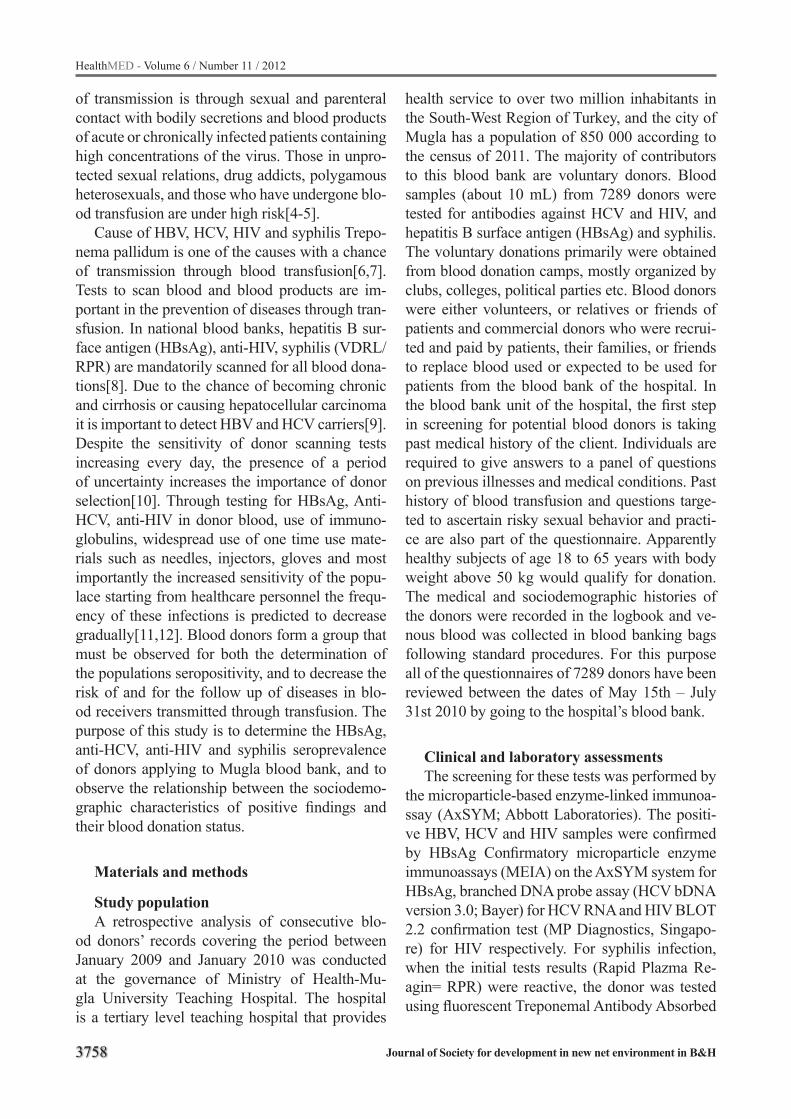
3758
HealthMED - Volume 6 / Number 11 / 2012
Journal of Society for development in new net environment in B&H
of transmission is through sexual and parenteral contact with bodily secretions and blood products of acute or chronically infected patients containing high concentrations of the virus. Those in unpro-tected sexual relations, drug addicts, polygamous heterosexuals, and those who have undergone blo-od transfusion are under high risk[4-5].
Cause of HBV, HCV, HIV and syphilis Trepo-nema pallidum is one of the causes with a chance of transmission through blood transfusion[6,7]. Tests to scan blood and blood products are im-portant in the prevention of diseases through tran-sfusion. In national blood banks, hepatitis B sur-face antigen (HBsAg), anti-HIV, syphilis (VDRL/RPR) are mandatorily scanned for all blood dona-tions[8]. Due to the chance of becoming chronic and cirrhosis or causing hepatocellular carcinoma it is important to detect HBV and HCV carriers[9]. Despite the sensitivity of donor scanning tests increasing every day, the presence of a period of uncertainty increases the importance of donor selection[10]. Through testing for HBsAg, Anti-HCV, anti-HIV in donor blood, use of immuno-globulins, widespread use of one time use mate-rials such as needles, injectors, gloves and most importantly the increased sensitivity of the popu-lace starting from healthcare personnel the frequ-ency of these infections is predicted to decrease gradually[11,12]. Blood donors form a group that must be observed for both the determination of the populations seropositivity, and to decrease the risk of and for the follow up of diseases in blo-od receivers transmitted through transfusion. The purpose of this study is to determine the HBsAg, anti-HCV, anti-HIV and syphilis seroprevalence of donors applying to Mugla blood bank, and to observe the relationship between the sociodemo-graphic characteristics of positive findings and their blood donation status.
Materials and methods
Study populationA retrospective analysis of consecutive blo-
od donors’ records covering the period between January 2009 and January 2010 was conducted at the governance of Ministry of Health-Mu-gla University Teaching Hospital. The hospital is a tertiary level teaching hospital that provides
health service to over two million inhabitants in the South-West Region of Turkey, and the city of Mugla has a population of 850 000 according to the census of 2011. The majority of contributors to this blood bank are voluntary donors. Blood samples (about 10 mL) from 7289 donors were tested for antibodies against HCV and HIV, and hepatitis B surface antigen (HBsAg) and syphilis. The voluntary donations primarily were obtained from blood donation camps, mostly organized by clubs, colleges, political parties etc. Blood donors were either volunteers, or relatives or friends of patients and commercial donors who were recrui-ted and paid by patients, their families, or friends to replace blood used or expected to be used for patients from the blood bank of the hospital. In the blood bank unit of the hospital, the first step in screening for potential blood donors is taking past medical history of the client. Individuals are required to give answers to a panel of questions on previous illnesses and medical conditions. Past history of blood transfusion and questions targe-ted to ascertain risky sexual behavior and practi-ce are also part of the questionnaire. Apparently healthy subjects of age 18 to 65 years with body weight above 50 kg would qualify for donation. The medical and sociodemographic histories of the donors were recorded in the logbook and ve-nous blood was collected in blood banking bags following standard procedures. For this purpose all of the questionnaires of 7289 donors have been reviewed between the dates of May 15th – July 31st 2010 by going to the hospital’s blood bank.
Clinical and laboratory assessmentsThe screening for these tests was performed by
the microparticle-based enzyme-linked immunoa-ssay (AxSYM; Abbott Laboratories). The positi-ve HBV, HCV and HIV samples were confirmed by HBsAg Confirmatory microparticle enzyme immunoassays (MEIA) on the AxSYM system for HBsAg, branched DNA probe assay (HCV bDNA version 3.0; Bayer) for HCV RNA and HIV BLOT 2.2 confirmation test (MP Diagnostics, Singapo-re) for HIV respectively. For syphilis infection, when the initial tests results (Rapid Plazma Re-agin= RPR) were reactive, the donor was tested using fluorescent Treponemal Antibody Absorbed

HealthMED - Volume 6 / Number 11 / 2012
Journal of Society for development in new net environment in B&H 3759
(FTA-ABS), a confirmatory Treponemal-based assay. ABO and Rh blood groups determinations were carried out on a slide using monoclonal blo-od grouping antisera; anti A, anti-B, anti-AB, anti-D (Micromed S.p.A, Italy)
Study variables Dependent variablesDependent variables are the status of donors
as seropositive. The seropositive status; being HBsAg, anti-HCV, anti-HIV 1/2 and syphilis po-sitive has been examined in subtopics.
Independent variablesIndependent variables have been examined
under the subtopics of sociodemographics, blo-od, physical examination and variables related to laboratory: Sociodemographic variables are; gender, age, education level, marital status, and location. Blood related variables are; blood type, reason for blood donation, blood taken, and the acceptance status of blood. Physical examination and laboratory related variables are; weight (kg), systolic and diastolic pressure (mmHg), hemoglo-bin (g/dL) and hematocrit (%).
Statistical analysesThe data was evaluated by SPSS 15.0 package
program. Data collected by counting were analysed using Chi-Square test and logistic regression, P < 0.05 was considered as statistically significant. The variables for which a statistically significant difference has been found among the groups, the variables which are not compatible meaningwi-se and those variables that have been considered useful additions to the model have been included in the logistic regression model. HbsAg (positive, negative), anti-HCV (positive, negative), anti-HIV 1/2 (positive, negative), and syphilis (positive, negative) have been grouped and included in the logistic regression model as dependent variables. Only a single dependent variable has been used in the different models created. As independent va-riables; gender (male, female), age (34 and under, 35 and over), education level (university graduate, not a university graduate), marital status (married, not married), location (tourism area, non-tourism area), reason for blood donation (charity, for their
patients) have been included in the model to create the logistic regression model
Ethical considerationsThe research was conducted within the fra-
mework of ethical rules. Written permission was taken before the study from the Director of Mugla City Health, Mugla Governorship and the gover-nance of Ministry of Health-Mugla University Te-aching Hospital.
Results
The average age of the donors applying to the Mugla blood bank is 35.50 ± 10.50. The sociode-mographic and blood properties of the donors in-volved in the study are shown on Table 1.
96.7% of donors are male, 50.5% are in the age group of 34 and under, 34.6% are elementary school graduates, 66.2% are married, 74.6% are living in the tourism area. When looking at the properties of the blood, 36.2% of donors have blo-od type A Rh+, 70.4% have charity as reason for donation, properties and erythrocyte suspension of blood from 97.8%, and blood from 98% has been accepted (Table 1).
Looking at the distribution of donors based on serologic properties HBsAg has been found on 0.6%, anti-HCV on 0.4%, syphilis seropositive on 0.2%. Anti-HIV 1/2 seropositivity has not been fo-und on any of the donor blood (Table 2).
Averages of physical examination and labora-tory properties have been found as; weight (kg) 76.64 ± 19.87, systolic pressure (mmHg) 119.00 ± 18.89, diastolic pressure (mmHg) 76.35 ± 12.35, hemoglobin (g/dL) 14.90 ± 1.43, hematocrit (%) 44.83 ± 3.15 (Table 3).
The prevalence of HBsAg, anti-HCV, anti-HIV, and syphilis in blood donors in Turkey are shown on Table 4. The international prevalence of HBsAg, anti-HCV, anti-HIV, and syphilis in blo-od donors in various parts of word are shown on Table 5. No significant difference was found on consideration of HBsAg seropositiveness of donor blood on age, education level, marital status, loca-tion, and blood donation reason. HBsAg seropo-stive frequency in donors has been found signifi-cantly higher in men (P = 0.028). It has been found that anti-HCV frequency of donors is unaffected

3760
HealthMED - Volume 6 / Number 11 / 2012
Journal of Society for development in new net environment in B&H
Table 1. Distribution of donors based on sociodemo-graphic and blood properties
Features n %SociodemographicGender (n=7289) Female Male
2407049
3.396.7
Age group (n= 7281) ≤34 3676 50.5 ≥35 3605 49.5 Education (n= 6631) Elementary school 2295 34.6 Middle school 918 13.8 High school 1818 27.4 University 1600 24.2Marital Status (n= 6560) Married 4341 66.2 Not married 2219 33.8Place of residence (n=7289)Tourism region 5441 74.6Blood Blood group (Rh) (n= 7289)A+
A-
B+
B-
AB+
AB-
0+
0-
2638275109910645044
2410267
36.23.815.11.56.20.633.13.7
The reason for blood making (n= 7252) For donation 2079 28.6 For patients 5170 70.4Received blood (n= 7289) Whole blood 159 2.2 Erythrocyte suspension 7130 97.8Acceptance of the blood (n= 7289) Acceptance 7146 98.0
Rejection 143 2.0
Table 2. Prevalence of HBV, HIV, HCV, and syphilis among blood donors
Features n %HBsAg (n=7289) Pozitif 43 0.6 Negatif 7246 99.4Anti-HCV (n=7289) Pozitif 30 0.4 Negatif 7259 99.6Anti-HIV (n=7289) Pozitif Negatif
07289
0.0100.0
Syphilis (n=7289) Pozitif Negatif
167273
0.299.8
Table 3. Distribution of donors based on physical examination and laboratory propertiesFeatures N Mean Median Std. DEviation Minimum Maximum
Weight (kg) 7285 76.64 79.00 19.87 46 140
Systolic pressure (mmHg) 7289 119.00 120.00 18.89 90 180
Diastolic pressure (mmHg) 7289 76.35 80.00 12.35 50 110
Hemoglobin (g/dL) 7275 14.90 15.00 1.43 11.6 25.0
Hematocrit (%) 7274 44.83 45.00 3.15 14.4 59.0
by gender, age, education level, marital status and reason for blood donation. On those living in the tourism area anti-HCV seropositive frequency has been found significantly higher (P = 0.011). It has been found that syphilis seropositiveness of do-nors is unaffected by age, education level, marital status, location, and blood donation reason. Syphi-lis seropositive frequency has been found signifi-cantly higher in men (P = 0.015) (Table 6). Based on the logistic regression analysis, the likelihood of males being HBsAg seropostive is 3.08 times higher and the likelihood of being syphilis sero-positive is 6.72 times higher than that of females (P = .035, P = .004). The likelihood of donors to be anti-HCV seropostive is 2.70 times higher for those patients living in the tourism area than those patients who are not. (P = .008) (Tablo 7).
Discussion
Scanning tests done on blood and blood pro-ducts are important in the prevention of tran-sfusionally transmitted diseases. HBsAg, anti-HCV, anti-HIV 1/2, VDRL/RPR are mandatory scanning tests in national blood banks. Transmi-ssion of infectious agents through blood has been
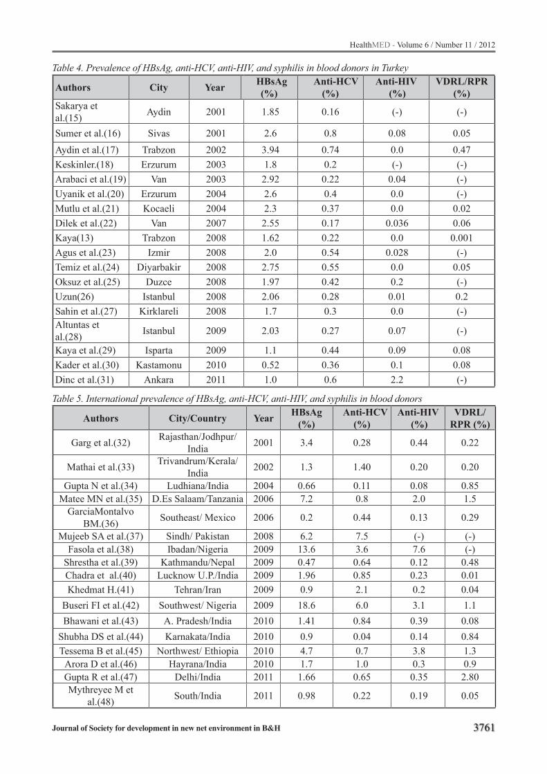
HealthMED - Volume 6 / Number 11 / 2012
Journal of Society for development in new net environment in B&H 3761
Table 4. Prevalence of HBsAg, anti-HCV, anti-HIV, and syphilis in blood donors in Turkey
Authors City Year HBsAg (%)
Anti-HCV (%)
Anti-HIV (%)
VDRL/RPR (%)
Sakarya et al.(15) Aydin 2001 1.85 0.16 (-) (-)
Sumer et al.(16) Sivas 2001 2.6 0.8 0.08 0.05Aydin et al.(17) Trabzon 2002 3.94 0.74 0.0 0.47Keskinler.(18) Erzurum 2003 1.8 0.2 (-) (-)Arabaci et al.(19) Van 2003 2.92 0.22 0.04 (-)Uyanik et al.(20) Erzurum 2004 2.6 0.4 0.0 (-)Mutlu et al.(21) Kocaeli 2004 2.3 0.37 0.0 0.02Dilek et al.(22) Van 2007 2.55 0.17 0.036 0.06Kaya(13) Trabzon 2008 1.62 0.22 0.0 0.001Agus et al.(23) Izmir 2008 2.0 0.54 0.028 (-)Temiz et al.(24) Diyarbakir 2008 2.75 0.55 0.0 0.05Oksuz et al.(25) Duzce 2008 1.97 0.42 0.2 (-)Uzun(26) Istanbul 2008 2.06 0.28 0.01 0.2Sahin et al.(27) Kirklareli 2008 1.7 0.3 0.0 (-)Altuntas et al.(28) Istanbul 2009 2.03 0.27 0.07 (-)
Kaya et al.(29) Isparta 2009 1.1 0.44 0.09 0.08Kader et al.(30) Kastamonu 2010 0.52 0.36 0.1 0.08Dinc et al.(31) Ankara 2011 1.0 0.6 2.2 (-)
Table 5. International prevalence of HBsAg, anti-HCV, anti-HIV, and syphilis in blood donors
Authors City/Country Year HBsAg (%)
Anti-HCV (%)
Anti-HIV (%)
VDRL/RPR (%)
Garg et al.(32) Rajasthan/Jodhpur/India 2001 3.4 0.28 0.44 0.22
Mathai et al.(33) Trivandrum/Kerala/India 2002 1.3 1.40 0.20 0.20
Gupta N et al.(34) Ludhiana/India 2004 0.66 0.11 0.08 0.85Matee MN et al.(35) D.Es Salaam/Tanzania 2006 7.2 0.8 2.0 1.5
GarciaMontalvo BM.(36) Southeast/ Mexico 2006 0.2 0.44 0.13 0.29
Mujeeb SA et al.(37) Sindh/ Pakistan 2008 6.2 7.5 (-) (-)Fasola et al.(38) Ibadan/Nigeria 2009 13.6 3.6 7.6 (-)
Shrestha et al.(39) Kathmandu/Nepal 2009 0.47 0.64 0.12 0.48Chadra et al.(40) Lucknow U.P./India 2009 1.96 0.85 0.23 0.01Khedmat H.(41) Tehran/Iran 2009 0.9 2.1 0.2 0.04
Buseri FI et al.(42) Southwest/ Nigeria 2009 18.6 6.0 3.1 1.1Bhawani et al.(43) A. Pradesh/India 2010 1.41 0.84 0.39 0.08
Shubha DS et al.(44) Karnakata/India 2010 0.9 0.04 0.14 0.84Tessema B et al.(45) Northwest/ Ethiopia 2010 4.7 0.7 3.8 1.3
Arora D et al.(46) Hayrana/India 2010 1.7 1.0 0.3 0.9Gupta R et al.(47) Delhi/India 2011 1.66 0.65 0.35 2.80Mythreyee M et
al.(48) South/India 2011 0.98 0.22 0.19 0.05
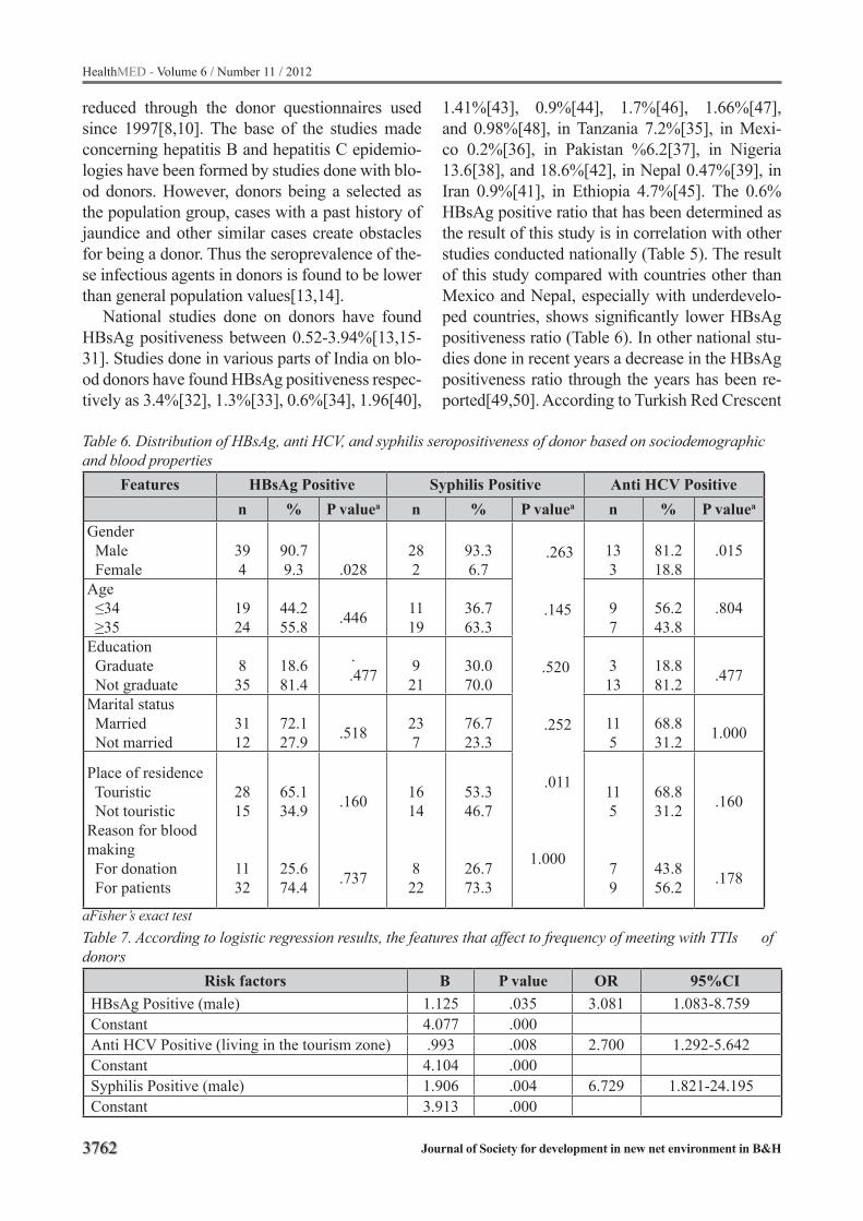
3762
HealthMED - Volume 6 / Number 11 / 2012
Journal of Society for development in new net environment in B&H
reduced through the donor questionnaires used since 1997[8,10]. The base of the studies made concerning hepatitis B and hepatitis C epidemio-logies have been formed by studies done with blo-od donors. However, donors being a selected as the population group, cases with a past history of jaundice and other similar cases create obstacles for being a donor. Thus the seroprevalence of the-se infectious agents in donors is found to be lower than general population values[13,14].
National studies done on donors have found HBsAg positiveness between 0.52-3.94%[13,15-31]. Studies done in various parts of India on blo-od donors have found HBsAg positiveness respec-tively as 3.4%[32], 1.3%[33], 0.6%[34], 1.96[40],
1.41%[43], 0.9%[44], 1.7%[46], 1.66%[47], and 0.98%[48], in Tanzania 7.2%[35], in Mexi-co 0.2%[36], in Pakistan %6.2[37], in Nigeria 13.6[38], and 18.6%[42], in Nepal 0.47%[39], in Iran 0.9%[41], in Ethiopia 4.7%[45]. The 0.6% HBsAg positive ratio that has been determined as the result of this study is in correlation with other studies conducted nationally (Table 5). The result of this study compared with countries other than Mexico and Nepal, especially with underdevelo-ped countries, shows significantly lower HBsAg positiveness ratio (Table 6). In other national stu-dies done in recent years a decrease in the HBsAg positiveness ratio through the years has been re-ported[49,50]. According to Turkish Red Crescent
Table 6. Distribution of HBsAg, anti HCV, and syphilis seropositiveness of donor based on sociodemographic and blood properties
Features HBsAg Positive Syphilis Positive Anti HCV Positiven % P valuea n % P valuea n % P valuea
Gender Male Female
394
90.79.3 .028
282
93.36.7
.263
.145
.520
.252
.011
1.000
133
81.218.8
.015
Age ≤34 ≥35
1924
44.255.8 .446 11
1936.763.3
97
56.243.8
.804
Education Graduate Not graduate
835
18.681.4
. .477 9
21 30.070.0
313
18.881.2 .477
Marital status Married Not married
3112
72.127.9 .518 23
776.723.3
115
68.831.2 1.000
Place of residence Touristic Not touristicReason for blood making For donation For patients
2815
1132
65.134.9
25.674.4
.160
.737
1614
822
53.346.7
26.773.3
115
79
68.831.2
43.856.2
.160
.178
aFisher’s exact testTable 7. According to logistic regression results, the features that affect to frequency of meeting with TTIs of donors
Risk factors B P value OR 95%CIHBsAg Positive (male) 1.125 .035 3.081 1.083-8.759Constant 4.077 .000Anti HCV Positive (living in the tourism zone) .993 .008 2.700 1.292-5.642Constant 4.104 .000Syphilis Positive (male) 1.906 .004 6.729 1.821-24.195Constant 3.913 .000
HealthMED - Volume 6 / Number 11 / 2012
Journal of Society for development in new net environment in B&H 3763
data HBsAg positiveness in Turkey was 2.12% in 2004 while decreasing to 1.21% in 2009[51]. The reasons for low HbsAg ratio in donor blood have been listed as having more volunteering donors and patient relatives giving blood donations, the conduct of a detailed questionnaire in the blood bank along with examinations, and widespread hepatitis B vaccinations.
In this study the anti-HCV positiveness of blo-od donors has been found as 0.4%. Studies con-ducted nationally show anti-HCV positiveness in blood donors within 0.16-0.8%[13,20,15-31,49]. Studies done in various parts of India on blood do-nors have found HBsAg positiveness 0.28%[32], 1.40%[33], 0.11%[34], 0.85%[40], 0.84%[43], 0.04%[44], 1.0%[46], 0.65%[47], and 0.22%[48], in Tanzania 0.8%[35], in Mexico 0.44%[36], in Pakistan 7.5%[37], in Nigeria 3.6%[38], and 6.0 %[42], in Nepal 0.64%[39], in Iran 2.1%[41], in Ethiopia 0.7%[45]. As seen on table 6 anti-HCV positiveness in these countries is within 0.04-7.5%. The anti-HCV positiveness ratio determi-ned as a result of this study is in correlation with results of studies conducted nationally and inter-nationally.
According to the data of the Ministry of He-alth 5224 HIV-positive findings were discovered in Turkey between October 1st 1985 – Decem-ber 31th 2011[52]. National anti-HIV positive-ness in blood donors have been reported between 0-2.2%[13,24,15-31]. In this study no anti-HIV positiveness has been discovered. Studies done in various parts of India on blood donors have fo-und anti-HIV positiveness 0.44%[32], 0.20%[33], 0.08%[34], 0.23%[40], 0.39%[43], 0.14%[44], 0.3%[46], 0.35%[47], and 0.19%[48], in Tanza-nia 2.0%[35], in Mexico 0.13%[36], in Nigeria 7.6%[38], and 3.1%[42], in Nepal 0.12%[39], in Iran 0.2%[41], in Ethiopia 3.8%[45]. As seen on table 6 international anti-HIV positiveness is wit-hin 0.08-7.6%. Despite the lack of presence of any discovered anti-HIV positiveness in this study this is seen to be in correlation with ratios reported na-tionally (Table 5).
Compared to international studies this could be said to be lower. Syphilis, being another in-fection able to be transmitted through transfusi-on has a prevalence of 0.001-0.47% nationally[13,16,17,21,22,24,26,29,30]. In this study
syphilis seropositiveness has been found to be 0.02%. Studies done in various parts of India on blood donors have found syphilis positiveness 0.22%[32], 0.20%[33], 0.85%[34], 0.01%[40], 0.08%[43], 0.84%[44], 0.9%[46], 2.80%[47], and 0.05%[48], in Tanzania 1.5%[35], in Mexi-co 0.29%[36], in Nigeria 1.1%[42], in Ne-pal 0.48%[39], in Iran 0.04%[41], in Ethiopia 1.3%[45]. As seen on table 6 syphilis positiveness internationally is within 0.01-2.8%. Syphilis po-sitiveness found as a result of this study is within nationally reported ranges (Table 5). This is lower than the results of studies conducted internatio-nally.
In this study it has been found that the male gender increases the HBsAg and syphilis sero-positive frequency significantly. A study done in Andhra Pradesh India supports this study. In this study HBsAg and syphilis seropositive frequency has been found to be significantly higher in male donors than in females[43]. In a study done in northwest Ethiopia HBsAg and syphilis seroposi-tive frequency in men have been found higher than in women; however, the difference between the two genders has not been found significant[45]. Studies done in Izmir, Kastamonu, and Erzurum have not found a relation between gender and HBsAg and syphilis positiveness[23,20,30]. In this study it has been found that female donors apply to the blood bank in a lower frequency com-pared to males. Many studies conducted natio-nally support this result. Women apply to blood banks in frequencies of 2.6% in Izmir[23], 10.8% in Erzurum[20], 5.4% in Kastamonu[30], 4.7% in Adiyaman[53]. Studies conducted internationally correlate with this study as well as other studies done nationally. Studies done in India report fe-male blood donor frequencies as 10.5% and 3.8 %[43,46], 15.1% in Nepal[39], 12.1% in Ethio-pia[45], 10.9% in Tanzania[35].
This shows that women donate blood less frequ-ently than men. This prevents the seropostiveness frequency of Transfussion Transmitted Infections from being revealed. There is a need for more stu-dies establishing relationships between gender and blood donors. In this study anti-HCV seropositive frequency has been found significantly higher in those living in the tourism area. Those living in the tourism area mostly have tourism related pro-

3764
HealthMED - Volume 6 / Number 11 / 2012
Journal of Society for development in new net environment in B&H
fessions. A study done in Egypt has found that tho-se having tourism related professions have higher frequency of anti-HCV seropositive[54]. Hepati-tis C virus infection is among the risk factors for those travelling[55,56,57]. Travellers are at risk if they practise unsafe behaviour involving the use of contaminated needles or syringes for injection, acupuncture, piercing or tattooing. An accident or medical emergency requiring blood transfusion may result in infection if the blood has not been screened for hepatitis C virus. Travellers engaged in humanitarian relief activities may be exposed to infected blood or other body fluids in health care settings[58]. Hepatitis C virus is infrequently tran-smitted through sexual contact[59]. Relationships with tourists coming from endemic countries have been reported to increase anti-HCV seroprevalen-ce[60].
In conclusion, the national seroprevalence of hepatitis B, anti-HIV, and syphilis in the south-west area of Turkey is found to be within national averages, and lower than other undeveloped or de-veloping countries’ averages. Hepatitis C seropre-valence has been found within national averages as well as those of other undeveloped or develo-ping countries. The discovered hepatitis C findin-gs mostly consisting of donors in the tourism area, and the significant relationship found between gender and hepatitis B and syphilis are the most notable results of this study. Despite the advance-ments in testing procedures for the prevention of blood transfusion transmitted infections there is always a risk. The determination of areal spread of transfusion transmitted infections, detection of carriers and their treatment, active use of donor questionnaires in blood banks, immunoprophlaxis and population informing activities may reduce these risks.
Acknowledgments
The authors thank all retired donors who parti-cipated in this study. Thanks to all those who co-operated in this research, including the Provincial Director of Mugla City Health, and Mugla Gover-norship and the governance of Ministry of Health-Mugla University Teaching Hospital.
References
1. Lai CL, Ratziu V, Yuen MF, Poynard T. Viral hepatitis B. The Lancet 2003; 362: 2089-94
2. Lee WM. Hepatitis B virus infection. N Engl J Med 1997; 337: 1733-45
3. Marco M. The hepatitis report. Epidemiology, modes of transmission and risk factors for hepatitis C virus. The Body 2000 [updated 2012 February 20]; Avai-lable from: http://www.thebody.com/content/art1696.html
4. Kahalili M. Coinfection with Hepatitis Viruses and HIV. HIV In Site Knowledge Base Chapter; 2003
5. Brau N. Update On Chronic Hepatitis C In HIV/HCV Coinfected Patients: Viral Interactions and Therapy. AIDS, 2003: 2279-90
6. Sepkowitz KA. Nosocomial Hepatitis and Other Infec-tions Transmitted By Blood and Blood Products. In: Mandell GL, Bennet JE, Douglas RG, editors. Princi-ples and Practice of Infectious Diseases. 4nd ed. New York: Churcill Livingstone, 1995: 3039- 3052
7. Kohn WG, Collins AS, Cleveland JL, Harte JA, Eklund KJ, Malvitz DM. Centers for Disease Control and Prevention (CDC). Guidelines for infection control in dental health-care settings-2003. MMWR Recomm Rep 2003; 52 (RR-17); 1-61
8. Aksoy A. The problem of viral hepatitis and the blood bank, The Turkish Red Crescent Blood Centers data, The problems. IX. Viral Hepatitis National Congress (3-6 April 2008, Antalya) Book. Ankara: Viral Hepa-titis Prevention Society, 2008: 50-1
9. Straisser SI, McDonald GB. Hepatitis viruses and hematopoietic cell transplantation: a guide to patient and donor management. Blood 1999; 93(4): 1127-36
10. Uluhan R. Safe blood. Ankem Derg 2007; 21(Suppl. 2): 142-145
11. Lavanchy D. Hepatitis B virus epidemiology, disea-se burden, treatment and current and emerging pre-vention and control measures. J Viral Hepatol 2004; 11: 97-107
12. McKnight CA, Des Jarlais DC, Perlis T, Eigo K. Syring exchange programs United States, 2005. MMWF. Morb Mortal Wkly Rep 2007; 56:1164-1167
13. Kaya S. Seroprevalance of hepatitis B virus, hepati-tis C virus, human immunodeficiency virus infection and syphilis in blood donors. Klimik Journal 2008; 21(2): 65-8
HealthMED - Volume 6 / Number 11 / 2012
Journal of Society for development in new net environment in B&H 3765
14. Van der Bij AK, Coutinho RA, Van der Poel CL. Sur-veillance of risk profi les among new and repeat blo-od donors with transfusion-transmissible infections from 1995 through 2003 in the Netherlands. Tran-sfusion 2006; 46(10): 1729-36
15. Sakarya S, Tuncer G, Yasa H, Cicek C. Kadikoylu G, Yukselen V. Seroprevalence of HBsAg and anti-HCV and their correlation to age and gender in blood do-nors in the province of Aydın. Klimik Journal 2001 ;14(1): 22-4
16. Sumer Z, Sumer H, Bakici MZ, Koc S. The Evolation of HBsAg, anti-HCV, anti-HIV and syphilis seropo-sitivity in blood donors at Cumhuriyet University Medical Faculty Hospital Blood Bank. Viral Hepatit Journal 2001; 7(2): 330-2
17. Aydin F, Cubukcu K, Yetiskul S, Yazici Y, Kaklikkaya N. Retrospective evaluation of HBsAg, anti-HCV, anti-HIV and syphilis reaginic antibody seropositi-vities in blood donors of Trabzon Farabi Hospital. Bulletin of Microbiology 2002; 36(1): 85-90
18. Keskinler DU. Serologic evaluation for HBV and HCV of blood donors applying to red- crescent blood bank in Erzurum. Journal of Inonu University Medical Faculty 2003; 10(4): 195-8
19. Arabaci F, Sahin HA, Sahin I, Kartal S. Seropositive-ness of HBV, HCV, HIV and VDRL in blood donors. Klimik Journal 2003; 16(1): 18-20.
20. Uyanik MH, Kuzucu Malcok H, Aktas O. Seropreva-lence of hepatitis B, hepatitis C and HIV-1/2 in blo-od donors. The Eurasian Journal of Medicine 2004; 36(2): 35-8
21. Mutlu B, Meric M, Willke A. Seroprevalence of he-patitis B and hepatitis C, human immunodeficiency virus and syphilis in blood donors. Bulletin of Micro-biology 2004; 38(4): 445-8
22. Dilek I, Demir C, Bay A, Akdeniz H, Oner AF. Sero-positivity rates of HBsAg, anti-HCV, anti-HIV and VDRL in blood donors in Eastern Turkey. Turk J He-matol 2007; 24(1):4-7
23. Agus N, Ozkalay YN, Cengiz A, Sanal E, Sert H. Se-roprevalence of HBsAg, anti-HCV, anti-HIV in blo-od donors. Ankem Derg 2008; 22(1): 7-9
24. Temiz H, Gul K. The Evaluation of HBsAg, Anti-HCV, anti-HIV and VDRL test results in blood do-nors. Turkish Journal of Infection 2008; 22(2): 79-82
25. Oksuz S, Yildirim M, Ozaydin C, Sahin I, Sencan I. Seropositivity rates of HBsAg, anti-HCV and anti-HIV in blood donors in Düzce Area. Viral Hepatit Derg 2008; 13(1): 27-30
26. Uzun C. Evaluation of HbsAg, anti-HCV, anti-HIV and RPR test results in blood donors.Turk Mikro-biyol Cem Derg 2008; 38(3-4): 143-6
27. Sahin D, Sahin I, Sozeri F, Onder K. HBV, HCV and HIV seroprevalance in applicant donors of Kirklare-li State Hospital Blood Center: a retrospective study. Viral Hepatit Derg 2008; 13: 31-5
28. Altuntas AO, Kumbasar KH, Kökrek A, Isik M E, Nazlican O. Seroprevalences of HBsAg, anti-HCV and anti-HIV among blood donors in Istanbul. Viral Hepatit Derg 2009; 14(2): 69-73
29. Kaya S, Alanoglu G, Polat M, Sipahi T. Evaluation of serologic scanning tests results in blood donors in Suleyman Demirel University, Hospital of Me-dical Faculty, Blood Center between 2000-2007. Medical Journal of Suleyman Demirel University 2009; 16(2): 13-5.
30. Kader C, Erbay A, Birengel B, Gurbuz M. Seropre-valance of hepatitis B virus, hepatitis C virus, human immunodeficiency virus infection and syphilis in blo-od donors. Klimik Journal 2010; 23(3): 95-9
31. Dinc B, Karabiber N, Yagci S, Arca EA, Gurbuz A, Tolunay EA. Serological profile of the blood donors in Turkey High Specialization Training and Rese-arch Hospital in Ankara.Turk Hij Den Biyol Derg 2011; 68(1): 17-22
32. Garg S, Mathur DR, Garg DK. Comparison of se-ropositivity of HIV, HBV, HCV and syphilis in repla-cement and voluntary blood donors in western In-dia. Indian Journal of Pathology and Microbiology 2001; 44(4): 409-412
33. Mathai PVJ, Sulochana S, Satyabhama PK, Nair R, Sivakumar S. Profile of transfusion transmissible infections and associated risk factors among blood donors of Kerala. Indian Journal of Pathology and Microbiology 2002;45(3): 319-322
34. Gupta N, Kumar V, Kaur A. Seroprevalence of HIV, HBV, HCV and syphilis in voluntary blood donors. Indian J Med Sci 2004; 58:255-7
35. Matee MN, Magesa MP, Lyamuya EF. Seropreva-lence of human immunodeficiency virus, hepatitis B and C viruses and syphilis infections among blood donors at the Muhimbili National Hospital in Dar Es Salaam, Tanzania. BMC Public Health 06; 6: 21
36. Garcia-Montalvo BM. Seropositivity of HIV, HBV, HCV and Treponema pallidum in blood donors in southeast Mexico. Rev Invest Clin 2006; 58(6): 567-72.
37. Mujeeb SA, Pearce MS. Temporal trends in hepatitis B and C infection in family blood donors from interi-or Sindh, Pakistan. BMC Infect Dis 2008; 8:43

3766
HealthMED - Volume 6 / Number 11 / 2012
Journal of Society for development in new net environment in B&H
38. Fasola FA, Kotila TR, Akinyemi J. Trends in tran-sfusion transmitted viral infections from 2001–2006, Ibadan, Nigeria. Virology Journal 2009; 13(6): 21
39. Shrestha AC, Ghimire P, Tiwari BE, Rajkarnikar M. Transfusion transmissible infections among blo-od donors in Kathmandu, Nepal. Journal of Infecti-ons in Developing Countries 2009; 7(4): 293-9
40. Chandra T, Kumar A, Gupta A. Prevalence of tran-sfusion transmitted infections in blood donors: an Indian experience. Transfusion 2009; 49(10): 2214-20
41. Khedmat H, Alavian SM, Miri SM, Amini M, Abolghasemi H, Hajibeigi B, Alaeddini F, Fallahian F. Trends in seroprevalence of hepatitis B, hepati-tis C, HIV, and syphilis infections in Iranian blood donors from 2003 to 2005. Hepatitis Monthly 2009; 9(1): 24-28
42. Buseri FI, Muhibi MA, Jeremiah ZA. Sero-epidemio-logy of transfusion-transmissible infectious diseases among blood donors in Osogbo, south-west Nigeria. Blood Transfus 2009; 7(4): 293-9
43. Bhawani Y, Rao PR, V Sudhakar. Biology and Medi-cine 2010; 2(4): 45-48
44. Shubha DS, Naryanmurthy C, Farheen F. Sero-prevalence of transfussion transmitted infections (TTIs): Humanimmunodefficiency virus (HIV), he-patitis B virus (HBV), hepatitis C virus (HCV) and syphilis in blood donors. Global Journal of Medical Research 2010; 3: 9-11
45. Tessema B, Yismaw G, Kassu A, Amsalu A, Mulu A, Emmrich F, Sack U. Seroprevalence of HIV, HBV, HCV and syphilis infections among blood donors at Gondar University Teaching Hospital, Northwest Ethiopia: declining trends over a period of five ye-ars. BMC Infect Dis 2010 May 10; 10: 111
46. Arora D, Arora B, Khetarpal A. Seroprevalence of HIV, HBV, HCV and syphilis in blood donors in Southern Haryana. Indian J Pathol Microbiol 2010; 53: 308-9
47. Gupta R, Singh B, Singh DK, Chugh M. Prevalence and trends of transfusion transmitted infections in a regional blood transfusion centre. Asian J Transfus Sci 2011; 5: 177-8
48. Mythreyee M, Jayachandran C, Amudhan M, Sivas-hankar M, Mythily N, Ramalingam Sekar R. Low prevalence of transfusion-transmissible infections among voluntary blood donors in South India. J In-fect Dev Ctries 2011; 5(5): 410-412
49. 49. Kocak N, Hepgul S, Ozbayburtlu S, Altunay H, Ozsoy MF, Kosan E. Trends in major transfusion-transmissible infections among blood donors over 17 years in Istanbul, Turkey. J Int Med Res 2004; 32(6): 671-5
50. Emekdas G, Cavuslu S, Oncul O, Artuk C, Aksoy A. Trends in hepatitis B and hepatitis C virus among blood donors over 16 years in Turkey. Eur J Epide-miol 2006; 21(4): 299-305
51. Tasli O. The role of the Turkish Red Crescent in the reconstruction of blood banking in Turkey. İstanbul: Association of Blood Centers and Transfusion [updated 2010 June 21]; Available from: http://www.kmtd. org.tr/pdf/omer_tasli.pdf
52. Tumer A. HIV/AIDS in Turkey. AIDS Treatment and Research Center. Hacettepe University, An-kara; 2011. [updated 2012 February 21]; Ava-ilable from: http://www.hatam.hacettepe.edu.tr/AIDSweb2011_240212.pdf
53. Demiraslan H, Aksoz S. The Evaluation of HBsAg and anti-HCV seroprevalences of blood donors in Adiyaman. Viral Hepatit Derg 2008; 13(1): 23-26
54. El-Sayed M, Gomatos P J, Rodier G R, Wierzba T F, Darwish A, Khashaba S. Seroprevalence survey of Egyptian tourism workers for hepatitis B virus, hepatitis C virus, human immunodeficiency virus, and Treponema pallidum infections: association of hepatitis C virus infections with specific regions of Egypt. The American Journal of Tropical Medicine and Hygiene 1996; 55(2): 179-84
55. Chen L.H, Wilson M.E, Davis X, Loutan L, Schwartz E, Keystone J, Hale D, Poh Lian Lim P L, McCarthy A, Klotsas EG, Schlagenhauf P. Illness in long-term travelers visiting Geosentinel Clinics. Emerging In-fectious Diseases November 2009; 15(11): 1773-82
56. The National Travel Health Network and Centre (NaTHNaC) Travellers. Hepatitis C. September 2011 [updated 2012 January 04]; Available from: http://www.nathnac.org/travel/factsheets/hep_c.htm
57. International Association for Medical Assistance to Travellers (IAMAT). Hepatitis C. 2012 [updated 2012 January 04]; Available from: http://www.ia-mat.org/disease_details.cfm?id=99
58. World Health Organization. International Travel and Health 2011. Chapter 5. Infectious diseases of potential risk for travellers. p. 64-65. 2011 [updated 2012 January 01]; Available from: http://www.who.int/ith/ITH2009Chapter5.pdf
HealthMED - Volume 6 / Number 11 / 2012
Journal of Society for development in new net environment in B&H 3767
59. Holmberg S. Infectious Diseases Related To Travel. Hepatitis C. In: Gary W. Brunette editors. CDC He-alth Information for International Traval New York: Oxford University Press; 2012 [updated 2012 Janu-ary 04]; Available from: http://wwwnc.cdc.gov/tra-vel/yellowbook/2012/chapter-3-infectious-diseases-related-to- travel/hepatitis-c.htm
60. Carballo M, Cody R, O’Reilly E. Migration, Hepa-titis B and Hepatitis C. International Centre for Mi-gration, Health and Development, 2010: 15
Corresponding Author Metin Picakciefe, Department of Public Health, Faculty of Medicine,Mugla University, The Governance of Ministry of Health-Mugla University Teaching Hospital, Mugla , Turkey, E-mail: [email protected], [email protected]
3768
HealthMED - Volume 6 / Number 11 / 2012
Journal of Society for development in new net environment in B&H
The effectiveness of pediatric symptom checklist for psychosocial screening in low-income turkish childrenMeryem Ozturk Haney1, Semra Erdogan2, Aysun Ardıc3
1 Dokuz Eylul University Nursing Faculty, Izmir, Turkey2 Istanbul University Nursing Faculty, Istanbul, Turkey3 Istanbul University Nursing Faculty, Istanbul, Turkey
Abstract
Background: Using a brief and valid scree-ning tool to enhance early detection and treatment of psychosocial problems can lead to timely seve-re impairment.
Objective: To evaluate the effectiveness of Turkish version of Pediatric Symptom Checklist (PSC) for screening of psychosocial dysfunction in a low income school sample.
Design: Survey.Method: We included 808 children aged 6-16
attending to elementary school in Istanbul, Tur-key. Parents completed Turkish version of PSC and CBCL which was used as criteria for the vali-dity of the PSC during the year 2011. Concurrent, discriminant validity and internal consistency, sta-bility, sensitivity, specificity, diagnostic index and Youden’s index were analyzed and reported.
Results: The PSC successfully discriminated among the normal, borderline and clinical range groups. The area under curve, using the CBCL as a gold standard, was 0.932. The optimal cut-off score was 20 for early detection of the psychosoci-al dysfunction (sensitivity: 90.9; specificity: 82.6). Internal consistency was found 0.90; test-retest re-liability was found 0.53. The male children were higher risk of impairment than female children.
Conclusion: The findings supported the use of Turkish version of the PSC with the recommended cut-off score of 20 in low income, school children for psychosocial problems screening.
Keywords: Low income children, Pediatric Symptom Checklist, psychosocial problems, re-liability, Turkey, validity.
Introducton
Mental health is an important component of children healthy development. Psychosocial pro-blems, such as behavioral and emotional pro-blems, are highly prevalent among children and early detection and treatment may prevent more severe psychosocial impairment of these children [1, 2]. Although one of five children has a diagno-sable mental health disorder in the world, 75 % to 80 % of children in need of mental health ser-vices do not receive them. The failure to identify the children who display initially minor psycho-social problems may pose a greater risk as it may contribute to the development of more serious and persistent psychosocial disorders. It may lead to negative treatment outcomes, higher health-care use rates, and poorer adherence to medical re-commendations [3, 4, 5].
So, early diagnosis and treatment of psychoso-cial problems is very important for the health of children and their families [6, 7, 8]. Systematic psychosocial screening should be implemented for routine use in primary health care services and schools with the use of standardized instruments [5, 9]. But, it is not routinely done in most health centres since the interviewing process is time-consuming, requires special training, fee charging [10, 11]. Although there are several available scre-ening measures that are psychometrically valid and reliable to identify psychosocial problems in primary health care setting, but using brief scre-ening questionnaires can facilitate the screening of psychosocial dysfunction among children . One measure designed to be used as a broad screening tool in pediatric settings is the Pediatric Symp-tom Checklist (PSC). The Pediatric Symptom Checklist (PSC) was developed to allow for rapid
HealthMED - Volume 6 / Number 11 / 2012
Journal of Society for development in new net environment in B&H 3769
screening of children in primary care settings and schools who would benefit most from further ser-vices to evaluate and treat of the emotional and behavioural problems. The PSC reflects parent’s impressions of their 6-16 years old children’s psychosocial functioning on 35 items [12].
The PSC has been used in many various pe-diatric settings including children with gastrointe-stinal disorders, Down syndrome, HIV infection, prenatal drug exposure and children with chronic diseases such as diabetes mellitus and sickle cell anemia [13-18]. And it has been translated into other languages including Japanese, Spanish, German, and Dutch [6, 19-21]. The researchers recommended different optimal cut off scores for translated versions in different ethnic groups, for example, cut-off score is 17 for Japanese version, [19], cut-off score is 22 for the Dutch version [6], cut-off score is 12 for Spanish version [20], and cut off score is 20 for Setswana version [15].
Despite the nationwide studies about childho-od psychosocial problems are limited in Turkey, there is only one study that using the short form of PSC (PSC-17) in Turkey [22]. In other studies, the Child Behaviour Checklist (CBCL), the Teacher’s Report Form (TRF) and the Youth Self-Report Form (YSR) were used [23, 24].
The aim of this study was to assess the properti-es of the Turkish version of the PSC among school age children. The CBCL was used as a standard measure of psychosocial problems against which we evaluated the validity of the PSC. This com-parison allowed us to calculate the sensitivity and specificity of the PSC in Turkish children. In addition, we used receiver operator characteristic (ROC) curves to investigate whether there was an optimal cut off score, with high sensitivity and specificity, for the PSC in this low-income, Tur-kish children aged 6-16.
Methods
Participants This is a methodological research conducted
on 808 families with low socio-economical status (SES) whose children was attended at the elemen-tary school in Istanbul, Turkey. Data were collec-ted from three schools in different areas during the year 2011.
ProcedureThe study was conducted after ethical and ad-
ministrative approvals of directorate of national education in the city. Researchers informed the fa-milies about the study details and invited them to participate the study by mail. Data was withdrawn from the families who were agreed to participa-te and they completed the surveys in the school settings. The families were encouraged to comple-te the survey unaided and in private.
MeasurementsThe survey consisted of the Demographic
Questionnaire (DQ), The Pediatric Symptom Checklist (PSC) and Child Behaviour Checklist (CBCL). Demographic Questionnaire obtained age and sex of the child, mother education, father education, parent’s employment status, health in-surance status, parental status of households, and the family size.
Pediatric Symptom Checklist-35 (PSC): The PSC is a parent–completed questionnaire with 35 items that allow for rapid identification of the school-aged children between ages of 6 and 16 years who are in need of a detailed mental health evaluation in the primary care setting. Each item describe a behaviour that parents rate each symp-tom as occurring “often” (2 points), “sometimes” (1 points), or “never” (0 points) respectively. Item scores are summed and the total score indicates psychosocial impairment [12]. It takes 10 minu-tes. The cut-off score of original scale is 28 for children aged 6 to 6 years. Previous studies have tested the translated versions in the acceptable samples with different cut-off scores providing effectiveness in different settings [6, 12, 20]. The cut-off scores of 25, 17 and 12 are recommended for Dutch, Japanese and low income Mexican American populations, respectively.
Child Behaviour Checklist (CBCL/6-18): The CBCL is a parent-completed diagnostic tool that reports the children’s problems over the preceding six months. It has been used extensively in both clinical and research settings to identify psycho-pathologic disorders in children [25]. The CBCL has 113 items that describe behavioural and emo-tional problems. Parents rate items for how true each behaviour using the 3-point scale; 0, not true;

3770
HealthMED - Volume 6 / Number 11 / 2012
Journal of Society for development in new net environment in B&H
1, somewhat or sometimes true and 2, very true or often true. The sum of all problem item scores gives total problems score. There are 8 syndrome scales and 2 broad-based scales. A total T-score of 63 or greater was considered indicative of clinical range problems.
Translation processThe PSC was translated following the stan-
dard forward–backward procedure. The Turkish translated and original forms of the scale were compared for dialectical and cultural conforma-tion and modifications of the Turkish version were made if necessary. The scale was presented an expert panel to rate the relevance of each item using a four-point rating scale ranging from 1 (not relevant) to 4 (very relevant and succinct). The each item was reviewed for content validity index (CVI) and it was accepted the CVI should be at least 80 % [26]. The Turkish PSC total CVI score was calculated to be 96.8 %, which indicated sa-tisfactory agreement among the experts. Finally, translated version was piloted among 20 parents and was seen to be efficient.
Statistical analysisThe descriptive statistics including the number,
percentage, mean and standard deviation were used to describe subjects and the scores of the PSC and CBCL. The reliability of the scale was evalu-ated with Cronbach’s alpha coefficient and test-re-test correlation. Anova test was used to test discri-minant validity on the differences among the nor-mal, borderline and clinical ranged children using the PSC-35 scores. The normal, borderline and clinical ranged children were grouped according to their CBCL scores. Spearman’s correlation was calculated to test concurrent validity on the relati-onships between PSC and CBCL Total, Internali-zing and Externalizing scores. Independent sam-ples T test was used to examine the gender diffe-rences on the PSC and CBCL total and subscales’ scores. The ROC analysis was used to calculate the sensitivity, specificity, diagnostic index (DI) and youden’s index ( γ ) as a reference for the suitability of the PSC cut off point using CBCL as “gold standard”. Probability levels of 0.05 were considered significant. Statistical analyses were carried out using SPSS for windows 15.0.
Table 1. Socio-demographic characteristics of participants Characteristics No.(%) of ChildrenAge Mean ± SD(Range), y 9.14 ± 2.06 (6-16)
Gender Female Male
371 (45.9)437 (54.1)
Parental status of households (N=802) Single-parent Two parent
42 (5.2)760 (94.8)
Household size M (S.D.) 4.71 ± 1.24 (2-11)Maternal education (N=801) Low than fifth years Secondary school and high school Graduated from high school
564 (70.5)207 (25.8)30 (3.7)
Paternal education (N=800) Low than fifth years Secondary school and high school Graduated from high school
412 (51.5)332 (41.5)56 (7.0)
Employment status of parents (N=803) No parent with paid employment One parent with paid employment Two parent with paid employment
59 (7.3)671 (83.6)73 (9.1)
Health insurance status of family (N=780) No insurance Public or private
169 (21.7)611 (78.3)
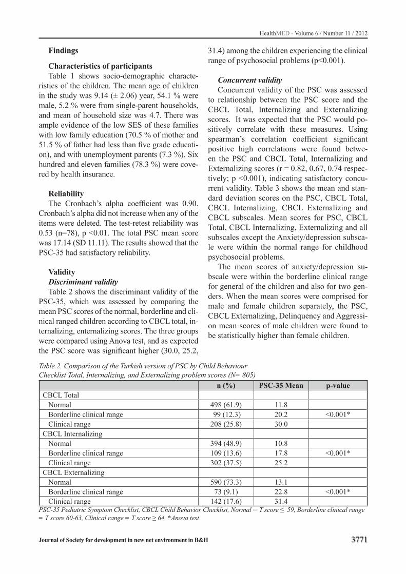
HealthMED - Volume 6 / Number 11 / 2012
Journal of Society for development in new net environment in B&H 3771
Findings
Characteristics of participantsTable 1 shows socio-demographic characte-
ristics of the children. The mean age of children in the study was 9.14 (± 2.06) year, 54.1 % were male, 5.2 % were from single-parent households, and mean of household size was 4.7. There was ample evidence of the low SES of these families with low family education (70.5 % of mother and 51.5 % of father had less than five grade educati-on), and with unemployment parents (7.3 %). Six hundred and eleven families (78.3 %) were cove-red by health insurance.
ReliabilityThe Cronbach’s alpha coefficient was 0.90.
Cronbach’s alpha did not increase when any of the items were deleted. The test-retest reliability was 0.53 (n=78), p <0.01. The total PSC mean score was 17.14 (SD 11.11). The results showed that the PSC-35 had satisfactory reliability.
ValidityDiscriminant validityTable 2 shows the discriminant validity of the
PSC-35, which was assessed by comparing the mean PSC scores of the normal, borderline and cli-nical ranged children according to CBCL total, in-ternalizing, enternalizing scores. The three groups were compared using Anova test, and as expected the PSC score was significant higher (30.0, 25.2,
31.4) among the children experiencing the clinical range of psychosocial problems (p<0.001).
Concurrent validityConcurrent validity of the PSC was assessed
to relationship between the PSC score and the CBCL Total, Internalizing and Externalizing scores. It was expected that the PSC would po-sitively correlate with these measures. Using spearman’s correlation coefficient significant positive high correlations were found betwe-en the PSC and CBCL Total, Internalizing and Externalizing scores (r = 0.82, 0.67, 0.74 respec-tively; p <0.001), indicating satisfactory concu-rrent validity. Table 3 shows the mean and stan-dard deviation scores on the PSC, CBCL Total, CBCL Internalizing, CBCL Externalizing and CBCL subscales. Mean scores for PSC, CBCL Total, CBCL Internalizing, Externalizing and all subscales except the Anxiety/depression subsca-le were within the normal range for childhood psychosocial problems.
The mean scores of anxiety/depression su-bscale were within the borderline clinical range for general of the children and also for two gen-ders. When the mean scores were comprised for male and female children separately, the PSC, CBCL Externalizing, Delinquency and Aggressi-on mean scores of male children were found to be statistically higher than female children.
Table 2. Comparison of the Turkish version of PSC by Child Behaviour Checklist Total, Internalizing, and Externalizing problem scores (N= 805)
n (%) PSC-35 Mean p-valueCBCL Total Normal 498 (61.9) 11.8 Borderline clinical range 99 (12.3) 20.2 <0.001* Clinical range 208 (25.8) 30.0CBCL Internalizing Normal 394 (48.9) 10.8 Borderline clinical range 109 (13.6) 17.8 <0.001* Clinical range 302 (37.5) 25.2CBCL Externalizing Normal 590 (73.3) 13.1 Borderline clinical range 73 (9.1) 22.8 <0.001* Clinical range 142 (17.6) 31.4
PSC-35 Pediatric Symptom Checklist, CBCL Child Behavior Checklist, Normal = T score ≤ 59, Borderline clinical range = T score 60-63, Clinical range = T score ≥ 64, *Anova test
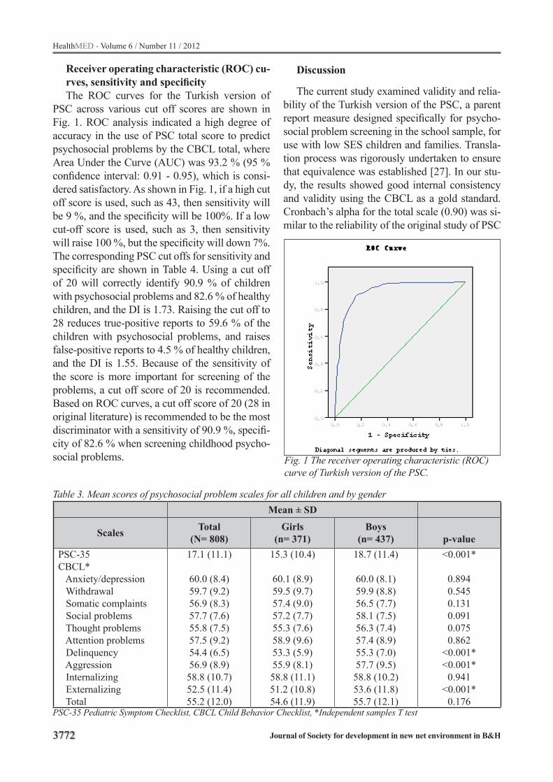
3772
HealthMED - Volume 6 / Number 11 / 2012
Journal of Society for development in new net environment in B&H
Table 3. Mean scores of psychosocial problem scales for all children and by genderMean ± SD
Scales Total (N= 808)
Girls(n= 371)
Boys(n= 437) p-value
PSC-35CBCL* Anxiety/depression Withdrawal Somatic complaints Social problems Thought problems Attention problems Delinquency Aggression Internalizing Externalizing Total
17.1 (11.1)
60.0 (8.4)59.7 (9.2)56.9 (8.3)57.7 (7.6)55.8 (7.5)57.5 (9.2)54.4 (6.5)56.9 (8.9)58.8 (10.7)52.5 (11.4)55.2 (12.0)
15.3 (10.4)
60.1 (8.9)59.5 (9.7)57.4 (9.0)57.2 (7.7)55.3 (7.6)58.9 (9.6)53.3 (5.9)55.9 (8.1)58.8 (11.1)51.2 (10.8)54.6 (11.9)
18.7 (11.4)
60.0 (8.1)59.9 (8.8)56.5 (7.7)58.1 (7.5)56.3 (7.4)57.4 (8.9)55.3 (7.0)57.7 (9.5)58.8 (10.2)53.6 (11.8)55.7 (12.1)
<0.001*
0.8940.5450.1310.0910.0750.862
<0.001*<0.001*
0.941<0.001*
0.176PSC-35 Pediatric Symptom Checklist, CBCL Child Behavior Checklist, *Independent samples T test
Receiver operating characteristic (ROC) cu-rves, sensitivity and specificityThe ROC curves for the Turkish version of
PSC across various cut off scores are shown in Fig. 1. ROC analysis indicated a high degree of accuracy in the use of PSC total score to predict psychosocial problems by the CBCL total, where Area Under the Curve (AUC) was 93.2 % (95 % confidence interval: 0.91 - 0.95), which is consi-dered satisfactory. As shown in Fig. 1, if a high cut off score is used, such as 43, then sensitivity will be 9 %, and the specificity will be 100%. If a low cut-off score is used, such as 3, then sensitivity will raise 100 %, but the specificity will down 7%. The corresponding PSC cut offs for sensitivity and specificity are shown in Table 4. Using a cut off of 20 will correctly identify 90.9 % of children with psychosocial problems and 82.6 % of healthy children, and the DI is 1.73. Raising the cut off to 28 reduces true-positive reports to 59.6 % of the children with psychosocial problems, and raises false-positive reports to 4.5 % of healthy children, and the DI is 1.55. Because of the sensitivity of the score is more important for screening of the problems, a cut off score of 20 is recommended. Based on ROC curves, a cut off score of 20 (28 in original literature) is recommended to be the most discriminator with a sensitivity of 90.9 %, specifi-city of 82.6 % when screening childhood psycho-social problems.
Discussion
The current study examined validity and relia-bility of the Turkish version of the PSC, a parent report measure designed specifically for psycho-social problem screening in the school sample, for use with low SES children and families. Transla-tion process was rigorously undertaken to ensure that equivalence was established [27]. In our stu-dy, the results showed good internal consistency and validity using the CBCL as a gold standard. Cronbach’s alpha for the total scale (0.90) was si-milar to the reliability of the original study of PSC
Fig. 1 The receiver operating characteristic (ROC) curve of Turkish version of the PSC.
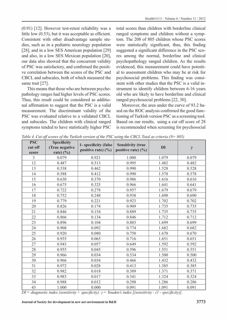
HealthMED - Volume 6 / Number 11 / 2012
Journal of Society for development in new net environment in B&H 3773
Table 4. Cut off scores of the Turkish version of the PSC using the CBCL Total as criteria (N= 805)PSC
cut off score
Specificity(True negative
rate) (%)
1- specificity (false positive rate) (%)
Sensitivity (true positive rate) (%) DI γ
3 0.079 0.921 1.000 1.079 0.07912 0.487 0.513 0.995 1.482 0.48213 0.538 0.462 0.990 1.528 0.52814 0.588 0.412 0.990 1.578 0.57815 0.630 0.370 0.986 1.616 0.61616 0.675 0.325 0.966 1.641 0.64117 0.722 0.278 0.957 1.679 0.67918 0.752 0.248 0.938 1.690 0.69019 0.779 0.221 0.923 1.702 0.70220 0.826 0.174 0.909 1.735 0.73521 0.846 0.154 0.889 1.735 0.73522 0.866 0.134 0.846 1.712 0.71223 0.896 0.104 0.803 1.699 0.69924 0.908 0.092 0.774 1.682 0.68225 0.920 0.080 0.750 1.670 0.67026 0.935 0.065 0.716 1.651 0.65127 0.943 0.057 0.649 1.592 0.59228 0.955 0.045 0.596 1.551 0.55129 0.966 0.034 0.534 1.500 0.50030 0.966 0.034 0.466 1.432 0.43231 0.972 0.028 0.413 1.385 0.38532 0.982 0.018 0.389 1.371 0.37133 0.983 0.017 0.341 1.324 0.32434 0.988 0.012 0.298 1.286 0.28643 1.000 0.000 0.091 1.091 0.091
DI = diagnostic index (sensitivity + specificity). γ = Youden’s index [(sensitivity - (1 - specificity)].
(0.91) [12]. However test-retest reliability was a little low (0.53), but it was acceptable as efficient.Consistent with other disadvantage sample stu-dies, such as in a pediatric neurology population [28], and in a low SES American population [29] and also, in a low SES Mexican population [20], our data also showed that the concurrent validity of PSC was satisfactory, and confirmed the positi-ve correlation between the scores of the PSC and CBCL and subscales, both of which measured the same trait [27].
This means that those who are between psycho-pathology ranges had higher levels of PSC scores. Thus, this result could be considered as additio-nal affirmation to suggest that the PSC is a valid measurement. The discriminant validity of the PSC was evaluated relative to a validated CBCL and subscales. The children with clinical ranged symptoms tended to have statistically higher PSC
total scores than children with borderline clinical ranged symptoms and children without a symp-tom. The 208 of 805 children whose PSC scores were statistically significant, thus, this finding suggested a significant difference in the PSC sco-res among the normal, borderline and clinical psychopathology ranged children. As the results evidenced, this measurement could have potenti-al to assessment children who may be at risk for psychosocial problems. This finding was consi-stent with other studies that the PSC is a valid in-strument to identify children between 6-16 years old who are likely to have borderline and clinical ranged psychosocial problems [22, 30].
Moreover, the area under the curve of 93.2 ba-sed on the ROC analysis confirmed the good func-tioning of Turkish version PSC as a screening tool. Based on our results, using a cut off score of 28 is recommended when screening for psychosocial
3774
HealthMED - Volume 6 / Number 11 / 2012
Journal of Society for development in new net environment in B&H
dysfunction was primarily lower sensitivity (59.6 % true-positive report) and we optimized to sli-ghtly lower cut off score (≥20; 90.9% true-positive report ) since it has high sensitivity and acceptable specificity. This finding was comparatively lower than the cut off value of 22 among Dutch scho-ol aged children [6] and 28 among USA children in original study [12]. However, our results were similar with the cut off score of 20 among Mexi-can well-children [31] and HIV infected Batswa-na children [15]. Conversely this results, another some studies recommended lower cut off scores. For instance, in a low income Mexican sample, Jutte et al. [20] optimizated to a cut off score of 12 and in a recent Thai study, ROC analysis confir-med the optimal cut off score of 16 for early detec-tion of psychosocial dysfunction and the same aut-hors recommended a cut off of 20 for referral and further psychiatric investigation [32]. This results evidenced the lower cut off score is likely related to the exclusive focus on disadvantaged children.
Although previous studies [20, 29, 31] reported insignificant results in terms of gender differences, this study demonstrated that male children were more likely to have higher PSC, CBCL Externali-zing, Delinquency and Aggression scores than fe-male children. These results were consistent with other epidemiological studies [6, 12, 21] that were found boys have higher prevalence of psychoso-cial problems than girls for school aged year old children.
Our results indicated that (a) the PSC was si-gnificantly correlated with the used extensively settings, well validated and more time spending CBCL and subscales, (b) the children with clini-cal ranged symptoms received higher PSC total scores than children with borderline clinical ran-ged symptoms and children without a symptom, (c) male children received higher PSC total and CBCL Externalizing (Delinquency and Aggressi-on) scores than female children (d) using the esta-blished cut off score of 28 with low SES children resulted in a 59.6 % true-positive report, and (e) modification of the cut off score from 28 to 20 optimized true-positive report (90.9 %) .
This research has limitations, since it was based on the subjects from low income SES; it cannot represent the general Turkish school aged children population. Moreover, when compared with the
actual diagnoses to be made by psychiatrists, the PSC might exaggerate the number of children cla-ssified as having psychosocial problems.
Conclusion
This study assessed the effectiveness of the Turkish version of the PSC for early detection of psychosocial problems among low income school children aged 6-16. Results showed the PSC has good reliability and validity using the CBCL as gold standard. Moreover, cut off score of 20 have to be used for screening of the psychosocial pro-blems because of the sensitivity is more important in disadvantage groups.
References
1. Prince M, Patel V, Saxena S, Maj M, Maselko J, Phillips MR, Rahman A. No health without mental he-alth. Lancet. 2007; 370: 859–877.
2. Theunissen MHC, Vogels AGC, Reijneveld SA. Early detection of psychosocial problems in children aged 5 to 6 years by preventive child healthcare: Has It Im-proved?. Journal of Pediatrics. (2012); 160:500-504.
3. Lavigne JV, Gibbons RD, Arend R, Rosenbaum D, Binns HJ, Christoffel KK. Rational service planning in pediatric primary care: continuity and change in psychopathology among children enrolled in pedia-tric practices. Journal of Pediatric Psychology. 1999; 24: 393– 403.
4. Riekert KA, Stancin T, Palermo TM, Drotar D. A psychological behavioral screening service: use, fea-sibility, and impact in a primary care setting. Journal of Pediatric Psychology. 1999; 24: 405–414.
5. Polaha J, Dalton WT, Allen S. The Prevalence of emotional and behavior problems in pediatric pri-mary care serving rural children. Journal of Pediatric Psychology. 2011; 36(6): 652-660.
6. Reijneveld SA, Vogels AGC, Hoekstra F, Crone MR. Use of the pediatric symptom checklist for the detec-tion of psychosocial problems in preventive child he-althcare. BMC Public Health. 2006; 6: 197-204.
7. Essex MJ, Kraemer HC, Slattery MJ, Burk LR, Boyce WT, Woodward HR, Kupfer DJ. Screening for child-hood mental health problems: outcomes and early identification. Journal of Child Psychology and Psychiatry. 2009; 50: 562–570.
8. Honeyman C. Recognising mental health problems in children and young people. Paediatric Nursing. 2007; 19: 38-44.
HealthMED - Volume 6 / Number 11 / 2012
Journal of Society for development in new net environment in B&H 3775
9. Gardner W, Murphy JM, Childs G, Kelleher K, Pa-gano M, Jellinek M, McInerny TK, Wasserman RC, Nutting P, Chiappetta L. The PSC-17: a brief pedia-tric symptom checklist with psychosocial problem su-bscales. A report from PROS and ASPN. Ambulatory Child Health. 1999; 5: 225–236.
10. Glascoe FP. Early detection of developmental and behavioral problems. Pediatrics in Review. 2000; 21 (8): 272-279.
11. Brown JD, Riley AW, Wissow LS. Identification of youth psychosocial problems during pediatric pri-mary care visits. Administration and Policy in Men-tal Health and Mental Health Services Research. 2007; 34:269-81.
12. Jellinek MS, Murphy JM, Little M, Pagano ME, Co-mer DM, Kelleher KJ. Use of the pediatric symptom checklist to screen for psychosocial problems in pe-diatric primary care: a national feasibility study. Ar-chives of Pediatrics & Adolescent Medicine. 1999; 153(3): 254-260.
13. Reed-Knight B, Hayutin LG, Lewis JD, Blount RL. Factor structure of the pediatric symptom checklist with a gastroenterology sample. Journal of Clinical Psychology in Medical Settings. 2011; 18: 299–306 DOI 10.1007/s10880-011-9242-7
14. Norizan A, Shamsuddin A. Predictors of parenting stress among Malaysian mothers of children with down syndrome. Journal of Intellectual Disability Research. 2010; 54: 992-1003 DOI: 10.1111/j.1365-2788.2010.01324.x
15. Lowenthal E, Lawler K, Harari N, Moamogwe L, Masunge J, Masedi M, Matome B, Seloilwe E, Jelli-nek M, Murphy M, Gross R. Validation of the pe-diatric symptom checklist in HIV-infected Batswana. Journal of Child and Adolescent Mental Health. 2011; 23(1): 17–28.
16. Whitaker TM, Bada HS, Bann CM, Shankaran S, LaGasse L, Lester BM, Bauer CR, Hammond J, Higgins R. Serial pediatric symptom checklist screeening in children with prenatal drug exposure. Journal of Developmental & Behavioral Pediatrics. 2011; 32 (3): 206-215.
17. Stoppelbein L, Greening L, Jordan SS, Elkin TD, Moll G, Pullen J. Factor analysis of the Pediatric Symptom Checklist with a chronically ill pediatric population. Journal of & Developmental Behavioral Pediatrics. 2005; 26(5): 349-55.
18. Jordan SS, Hilker KA, Stoppelbein L, Elkin TD, Applegate H, Iyer R. Nocturnal enuresis and psycho-social problems in pediatric sickle cell disease and sibling controls. Journal of Developmental & Beha-vioral Pediatrics. 2005; 26 (6): 404-411.
19. Ishizaki Y, Fukai Y, Kobayashi Y, Ozawa K. “Valida-tion and cutoff score of the Japanese version of the Pediatric Symptom Checklist: Screening of school-aged children with psychosocial and psychosomatic disorders (in Japanese).” The Journal of the Japan Pediatric Society. 2000; 104: 831-840.
20. Jutte DP, Burgos A, Mendoza F, Ford CB & Huffman LC. Use of the pediatric symptom checklist in a low- income, Mexican American population. Archives of Pediatrics & Adolescent Medicine. 2003; 157: 1169-1176.
21. Thun-Hohenstein L , Herzog S. The predictive va-lue of the pediatric symptom checklist in 5-year-old Austrian children. European Journal of Pediatrics. 2008; 167:323–329.
22. Erdogan S, Ozturk M. Psychometric evaluation of the Turkish version of the pediatric symptom checklist-17 for detecting psychosocial problems in low-income children. Journal of Clinical Nursing. 2011; 20(17-18): 2591-2599.
23. Erol N, Simsek Z. Mental health of Turkish children, behavioral and emotional problems reported by pa-rents, teachers, and adolescents. In International perspectives on child and adolescent mental health (Singh NN, Leung JP & Singh AN eds) Elsevier, Lon-don.2000; 223-247.
24. Simsek Z, Erol N, Öztop D, Özcan ÖÖ. Kurum bakımındaki çocuk ve ergenlerde davranış ve duygu-sal sorunların epidemiyolojisi; ulusal örneklemde karşılaştırmalı bir araştırma [Epidemiology of emo-tional and behavioral problems in children and ado-lescents reared in orphanages: a national compara-tive study]. Türk Psikiyatri Dergisi [Turkish Journal of Psychiatry].2008; 19: 235-246.
25. Berube RL, Achenbach TM. Bibliography of publis-hed studies using ASEBA instruments: 2004 edition. Burlington: University of Vermont, Research Center for Children, Youth, and Families. 2004.
26. Pierce AG. Measurement. In Principles and Practi-ce of Nursing Research (Talbot LA eds.). Mosby, St Louis. 1995; 265–291.
27. Burns N, Grove SK. The practice of nursing resear-ch: appraisal, synthesis and generation of evidence. Saunders, USA. 2009.
28. Anderson DL, Spratt EG, Macias MM, Jellinek MS, Murphy JM, Pagano M, Griesemer DA, Holden KR, Barbosa E. Use of the pediatric symptom checklist in the pediatric neurology population. Pediatric Ne-urology. 1999; 20 (2): 116-120.
3776
HealthMED - Volume 6 / Number 11 / 2012
Journal of Society for development in new net environment in B&H
29. Simonian SJ, Tarnowski KJ. Utility of the pediatric symptom checklist for behavioural screening of di-sadvantaged children. Child Psychiatry and Human Development. 2001; 31: 269-278.
30. Boothroyd RA, Armstrong M. An examination of the psychometric properties of the pediatric symptom checklist with enrolled in medicaid. Journal of Emo-tional and Behavioral Disorders. (2010); 18 (2): 113-126.
31. Leiner MA, Balcazar H, Straus DV, Shirsat P, Han-dal G. Screening Mexicans for psychosocial and behavioral problems during pediatric consultation. Revista de Investigacion Clinica. 2007; 59 (2): 116-123.
32. Pliibrukam R, Amloy K, Kanjanapatanakul W. The Effectiveness of brief psychosocial screening in identifying behavioral problems in children. Journal of the Medical Association of Thailand. 2011; 94: Suppl. 3 (145-151).
Corresponding Author Meryem Ozturk Haney,Dokuz Eylul University, Hemşirelik Fakültesi,Izmır,Turkey,E-mail: [email protected]
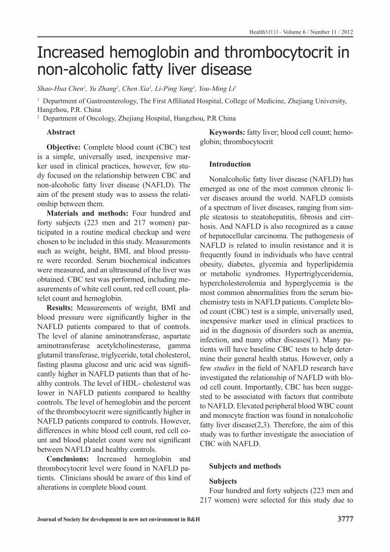
HealthMED - Volume 6 / Number 11 / 2012
Journal of Society for development in new net environment in B&H 3777
Increased hemoglobin and thrombocytocrit in non-alcoholic fatty liver disease Shao-Hua Chen1, Yu Zhang2, Chen Xia1, Li-Ping Yang1, You-Ming Li1
1 Department of Gastroenterology, The First Affiliated Hospital, College of Medicine, Zhejiang University, Hangzhou, P.R. China2 Department of Oncology, Zhejiang Hospital, Hangzhou, P.R China
Abstract
Objective: Complete blood count (CBC) test is a simple, universally used, inexpensive mar-ker used in clinical practices, however, few stu-dy focused on the relationship between CBC and non-alcoholic fatty liver disease (NAFLD). The aim of the present study was to assess the relati-onship between them.
Materials and methods: Four hundred and forty subjects (223 men and 217 women) par-ticipated in a routine medical checkup and were chosen to be included in this study. Measurements such as weight, height, BMI, and blood pressu-re were recorded. Serum biochemical indicators were measured, and an ultrasound of the liver was obtained. CBC test was performed, including me-asurements of white cell count, red cell count, pla-telet count and hemoglobin.
Results: Measurements of weight, BMI and blood pressure were significantly higher in the NAFLD patients compared to that of controls. The level of alanine aminotransferase, aspartate aminotransferase acetylcholinesterase, gamma glutamil transferase, triglyceride, total cholesterol, fasting plasma glucose and uric acid was signifi-cantly higher in NAFLD patients than that of he-althy controls. The level of HDL- cholesterol was lower in NAFLD patients compared to healthy controls. The level of hemoglobin and the percent of the thrombocytocrit were significantly higher in NAFLD patients compared to controls. However, differences in white blood cell count, red cell co-unt and blood platelet count were not significant between NAFLD and healthy controls.
Conclusions: Increased hemoglobin and thrombocytocrit level were found in NAFLD pa-tients. Clinicians should be aware of this kind of alterations in complete blood count.
Keywords: fatty liver; blood cell count; hemo-globin; thrombocytocrit
Introduction
Nonalcoholic fatty liver disease (NAFLD) has emerged as one of the most common chronic li-ver diseases around the world. NAFLD consists of a spectrum of liver diseases, ranging from sim-ple steatosis to steatohepatitis, fibrosis and cirr-hosis. And NAFLD is also recognized as a cause of hepatocellular carcinoma. The pathogenesis of NAFLD is related to insulin resistance and it is frequently found in individuals who have central obesity, diabetes, glycemia and hyperlipidemia or metabolic syndromes. Hypertriglyceridemia, hypercholesterolemia and hyperglycemia is the most common abnormalities from the serum bio-chemistry tests in NAFLD patients. Complete blo-od count (CBC) test is a simple, universally used, inexpensive marker used in clinical practices to aid in the diagnosis of disorders such as anemia, infection, and many other diseases(1). Many pa-tients will have baseline CBC tests to help deter-mine their general health status. However, only a few studies in the field of NAFLD research have investigated the relationship of NAFLD with blo-od cell count. Importantly, CBC has been sugge-sted to be associated with factors that contribute to NAFLD. Elevated peripheral blood WBC count and monocyte fraction was found in nonalcoholic fatty liver disease(2,3). Therefore, the aim of this study was to further investigate the association of CBC with NAFLD.
Subjects and methods
Subjects Four hundred and forty subjects (223 men and
217 women) were selected for this study due to

3778
HealthMED - Volume 6 / Number 11 / 2012
Journal of Society for development in new net environment in B&H
their participation in a routine medical checkup at the First Affiliated Hospital, College of Medicine, Zhejiang University. All subjects ranging from 30 to 60 years of age were enrolled in this study between September 2009 and January 2010. And the patients with diabetes, alcoholic liver disease, hematological disease were excluded. Informed consent was obtained from all subjects, and the study protocol for the research project has been approved by a suitably constituted Ethics Com-mittee of our institution and that it conforms to the provisions of the Declaration of Helsinki (revised in Edinburgh 2000).
NAFLD patients were diagnosed by abdominal ultrasonography based on the guidelines for the diagnosis and treatment of nonalcoholic fatty li-ver diseases created by the Chinese National Con-sensus Workshop on Nonalcoholic Fatty Liver Disease(4). The guidelines used were as follows: (1) NAFLD was defined by an “echogenic ” or “bright ” liver as found by abdominal ultrasono-graphy; (2) intake of less than 140 g (male) or 70 g (female) of ethanol per week; and (3) appropriate exclusion of other liver diseases, such as alcoholic liver disease, viral hepatitis (such as HBV, HCV), autoimmune hepatitis, drug-induced liver dise-ase, primary sclerosing cholangitis, primary bi-liary cirrhosis, biliary obstruction, and metabolic liver diseases. The controls were also according to the above criteria excluding an “echogenic ” or “bright ” liver by abdominal ultrasonography.
Methods
Subject anthropometry data including age, height, weight, systolic blood pressure (SBP) diastolic blood pressure (DBP), and drinking hi-story were recorded. Body mass index (BMI) was defined as weight / height 2 (kg/m2). Blood samples were collected under fasting conditions of at least 12 hours. Total protein, albumin, glo-bulin, alanine aminotransferase (ALT), aspartate aminotransferase (AST), alkaline phosphatase (AKP), acetylcholinesterase (ACHE), total bile acid, total bilirubin, direct bilirubin, gamma gluta-mil transferase(GGT), uric acid, triglyceride, total cholesterol, HDL- cholesterol and fasting plasma glucose (FPG) were determined with an autoa-nalyzer. Complete blood count including white
cell count, red cell count, platelet count and hemo-globin was performed by an automated analyzer.
Statistical analysisData were analyzed by SPSS17.0 statistical
software (SPSS Inc., Chicago, IL, USA). Data are presented as mean ± standard deviation (SD). Differences in continuous variables between gro-ups of subjects were tested with a t-test when the distribution of variables approached a normal distribution. The differences were considered sta-tistically significant at p < 0.05. The association between NAFLD and clinical data was analyzed by multifactor logistic regression, in which the backward method was used to select factors that significantly affect the NAFLD. Backward se-lection starts using all variables with significance lower than 0.1. This selection then drops variables one at a time in the order of worst to best accor-ding to criteria selected. Limits were set to a signi-ficance of > 0.15 using this model.
Results
Clinical characteristics of NAFLD and he-althy controlsThe clinical characteristics of the subjects are
presented in Table 1. In this study, data from 440 subjects ranging from 30 to 60 years of age were reviewed. Out of this sample, 223 patients with NAFLD and 217controls were identified. There was no significant difference in the distribution of gender and age between the NAFLD group and healthy controls. Weight, BMI, and blood pressure were significantly higher in the NAFLD patients as compared to that of controls(Table 1).
Biochemical features of NAFLD patients and healthy controlsThe level of ALT, AST, ACHE, and GGT was
significantly higher in NAFLD patients compared to that of healthy controls(Table 2). The triglyce-ride, total cholesterol, FPG and uric acid level was also higher in NAFLD patients compared to healthy controls (p≤0.01). The level of HDL- cho-lesterol was lower in NAFLD patients than that of healthy controls(p≤0.01).
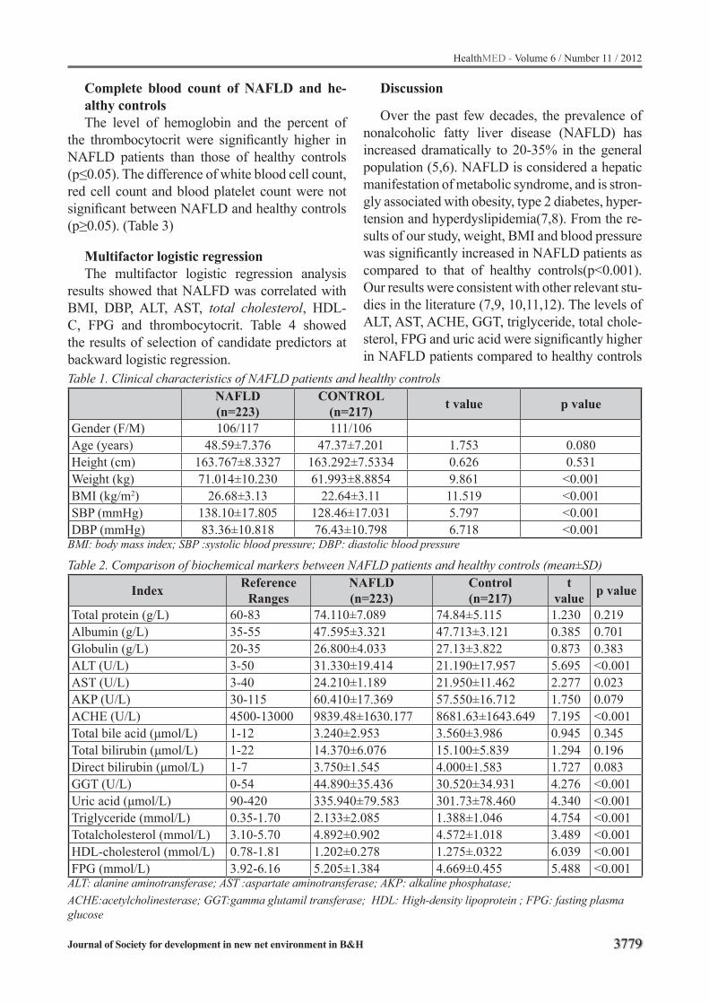
HealthMED - Volume 6 / Number 11 / 2012
Journal of Society for development in new net environment in B&H 3779
Table 1. Clinical characteristics of NAFLD patients and healthy controlsNAFLD (n=223)
CONTROL(n=217) t value p value
Gender (F/M) 106/117 111/106Age (years) 48.59±7.376 47.37±7.201 1.753 0.080Height (cm) 163.767±8.3327 163.292±7.5334 0.626 0.531Weight (kg) 71.014±10.230 61.993±8.8854 9.861 <0.001BMI (kg/m2) 26.68±3.13 22.64±3.11 11.519 <0.001SBP (mmHg) 138.10±17.805 128.46±17.031 5.797 <0.001DBP (mmHg) 83.36±10.818 76.43±10.798 6.718 <0.001
BMI: body mass index; SBP :systolic blood pressure; DBP: diastolic blood pressure
Table 2. Comparison of biochemical markers between NAFLD patients and healthy controls (mean±SD)
Index Reference Ranges
NAFLD(n=223)
Control(n=217)
t value p value
Total protein (g/L) 60-83 74.110±7.089 74.84±5.115 1.230 0.219Albumin (g/L) 35-55 47.595±3.321 47.713±3.121 0.385 0.701Globulin (g/L) 20-35 26.800±4.033 27.13±3.822 0.873 0.383ALT (U/L) 3-50 31.330±19.414 21.190±17.957 5.695 <0.001AST (U/L) 3-40 24.210±1.189 21.950±11.462 2.277 0.023AKP (U/L) 30-115 60.410±17.369 57.550±16.712 1.750 0.079ACHE (U/L) 4500-13000 9839.48±1630.177 8681.63±1643.649 7.195 <0.001Total bile acid (μmol/L) 1-12 3.240±2.953 3.560±3.986 0.945 0.345Total bilirubin (μmol/L) 1-22 14.370±6.076 15.100±5.839 1.294 0.196Direct bilirubin (μmol/L) 1-7 3.750±1.545 4.000±1.583 1.727 0.083GGT (U/L) 0-54 44.890±35.436 30.520±34.931 4.276 <0.001Uric acid (μmol/L) 90-420 335.940±79.583 301.73±78.460 4.340 <0.001Triglyceride (mmol/L) 0.35-1.70 2.133±2.085 1.388±1.046 4.754 <0.001Totalcholesterol (mmol/L) 3.10-5.70 4.892±0.902 4.572±1.018 3.489 <0.001HDL-cholesterol (mmol/L) 0.78-1.81 1.202±0.278 1.275±.0322 6.039 <0.001FPG (mmol/L) 3.92-6.16 5.205±1.384 4.669±0.455 5.488 <0.001
ALT: alanine aminotransferase; AST :aspartate aminotransferase; AKP: alkaline phosphatase; ACHE:acetylcholinesterase; GGT:gamma glutamil transferase; HDL: High-density lipoprotein ; FPG: fasting plasma glucose
Complete blood count of NAFLD and he-althy controlsThe level of hemoglobin and the percent of
the thrombocytocrit were significantly higher in NAFLD patients than those of healthy controls (p≤0.05). The difference of white blood cell count, red cell count and blood platelet count were not significant between NAFLD and healthy controls (p≥0.05). (Table 3)
Multifactor logistic regressionThe multifactor logistic regression analysis
results showed that NALFD was correlated with BMI, DBP, ALT, AST, total cholesterol, HDL-C, FPG and thrombocytocrit. Table 4 showed the results of selection of candidate predictors at backward logistic regression.
Discussion
Over the past few decades, the prevalence of nonalcoholic fatty liver disease (NAFLD) has increased dramatically to 20-35% in the general population (5,6). NAFLD is considered a hepatic manifestation of metabolic syndrome, and is stron-gly associated with obesity, type 2 diabetes, hyper-tension and hyperdyslipidemia(7,8). From the re-sults of our study, weight, BMI and blood pressure was significantly increased in NAFLD patients as compared to that of healthy controls(p<0.001). Our results were consistent with other relevant stu-dies in the literature (7,9, 10,11,12). The levels of ALT, AST, ACHE, GGT, triglyceride, total chole-sterol, FPG and uric acid were significantly higher in NAFLD patients compared to healthy controls
3780
HealthMED - Volume 6 / Number 11 / 2012
Journal of Society for development in new net environment in B&H
Table 3. Comparison of complete blood count of NAFLD patients and healthy controls (mean±SD)
Index Reference Ranges
NAFLD(n=223)
Control(n=217)
t value
p value
White blood cell count (10E9/L) 4.0-10.0 6.231±1.6308 6.023±1.8592 1.249 0.2120Neutrophil count (10E9/L) 2.0-7.0 3.488±1.2163 3.575±1.5367 0.655 0.513Lymphocyte count (10E9/L) 0.8-4.0 2.129±0.5929 1.850±0.5609 5.043 <0.001Monocyte count(10E9/L) 0.1-0.6 0.441±0.1466 0.428±0.1540 0.904 0.366Eosinophile granulocyte count (10E9/L) 0.05-0.5 0.1551±0.1316 0.1389±0.1264 1.313 0.190
Basophilic granulocyte (10E9/L) 0.00-0.10 0.0163±0.02533 0.0158±0.0264 0.203 0.839Red cell count (10E12/L) 3.5-5.0 4.7531±0.5157 4.8092±3.0089 0.274 0.784Hematocrit(%) 35.0-45.0 42.784±4.3192 41.892±5.8154 1.824 0.069Red cell distribution width(%) 11.5-14.5 13.265±1.0241 14.562±16.0179 1.207 0.228Mean corpuscular volume(fl9 79.0-101.0 89.864±4.8054 90.169±6.6671 0.551 0.582Hemoglobin(g/L) 110-150 144.93±15.618 140.91±18.853 2.44 0.015Mean hemoglobin weigh(pg) 26.0-35.0 30.462±1.9256 32.192±2.1503 1.197 0.232Mean hemoglobin concentration(g/L) 310-370 338.97±11.679 337.06±30.904 0.860 0.390
Platelet count(10E9/L) 100-300 218.81±55.140 209.81±52.565 1.751 0.081Mean platelet volume(fl) 7.4-12.5 10.069±1.6712 9.886±1.6699 1.132 0.258Thrombocytocrit(%) 0.108-0.282 0.2191±0.05715 0.2049±0.05350 2.639 0.009
Table 4. Results of multifactor logistic regressionRegression coefficient Wald χ2 p OR 95%CI
Lower UpperConstant -13.743 43.585 .000 .000BMI .294 30.502 .000 1.342 1.209 1.490DBP .027 4.526 .033 1.027 1.002 1.053ALT .020 2.918 .088 1.020 .997 1.044AST -.046 2.890 .089 .955 .905 1.007Total cholesterol .344 5.116 .024 1.410 1.047 1.900HDL-C -1.262 6.770 .009 .283 .109 .732FPG .486 5.123 .024 1.627 1.067 2.479Thrombocytocrit 0.735 3.907 .048 1.911 1.041 2.415
OR:odds ratio; CI:confidence interval
(p<0.05). Levels of HDL-cholesterol were found to be lower in NAFLD patients compared to their healthy control counterparts.
A complete blood count is broad screening test requested by doctors to check for the general he-alth status and disorders such as anemia, infection, and many other diseases (1). Blood cell counts are amongst the most commonly performed blood te-sts in medicine, as they can provide an overview of a patient’s general health status. The cells cir-culating in the bloodstream are generally divided into three types: red blood cells, white blood cells and platelets. Abnormally high or low counts of any of these three types may indicate the presence of varying forms of disease.
White blood cell count is a simple marker of inflammation and has become a useful predictor of specific diseases such as cardiovascular disease and mortality in middle-aged and older popula-tions (13,14). In a recent study, white blood cell count was found to be independently associated with the presence of NAFLD (2). And in our stu-dy, paradoxical results were found and there was no significant difference in the white blood cell count between the NAFLD patients and healthy controls.
Platelets play a fundamental role in hemostasis, and are a natural source of growth factors. They circulate in the blood of mammals and help in wound healing. It was found that stage of fibro-sis was correlated with platelet count (p = 0.009)
HealthMED - Volume 6 / Number 11 / 2012
Journal of Society for development in new net environment in B&H 3781
(15).Moreover, platelet count was found to be an independent predictor of nonalcoholic fatty liver cirrhosis (16). However, there was no significant difference in platelet count between NAFLD pati-ents and controls in our study.
Mean platelet volume (MPV) is an indicator of platelet activation. It was reported that NAFLD pa-tients had lower platelet counts and higher MPVs compared to controls (17,18). In a logistic regressi-on analysis, NAFLD was found to be the indepen-dent predictor of MPV (Odds Ratio (OR):21.98) [95% confidence interval (CI): 2.404-201.048; p=0.006] (17). In this study, the difference in the level of MPV between the NAFLD patients and healthy controls was not significant.
Thrombocytocrit is the volume of packed blo-od platelets in a given quantity of blood. A throm-bocytocrit value (Tct) is the product of the platelet mean volume (fl) times platelet count(*109/L), expressed as a percentage. In this study, the thrombocytocrit value in NAFLD patients was significantly higher than that of healthy controls (p=0.009). Likewise, multifactor logistic regre-ssion analysis showed that NAFLD was correla-ted with the level of thrombocytocrit(OR=1.911, 95%CI:1.041-2.415).
Hemoglobin is the iron-containing oxygen-transport metalloprotein found in red blood cells. Hemoglobin is responsible for the transport of oxygen from the lungs to the rest of the body, re-leasing it for use in cells. Trak-Smayra et al. fo-und that serum free hemoglobin subunits correla-ted positively with the severity of liver lesions in NAFLD patients, and may serve as a biomarker for this disease (19). Xu L et al. found that the pre-valence of NAFLD increased with elevated levels of hemoglobin (20). In our study, the same trend was found, with hemoglobin levels of NAFLD pa-tients higher than that of controls (p=0.015).
Though significant results were found between NAFLD and controls, there were some inherent limitations to our study. Although liver biopsy is currently the gold standard for diagnosis, there is a need for less invasive methods. Therefore it was not performed due to its invasive and expensive nature. The potential risks associated with liver bi-opsies outweighed the need in this study, therefore histological data as such were not measured. Alt-hough ultrasound is probably not the most relia-
ble imaging method, it has many advantages and gives a high degree of certainty of the diagnosis if positive depending on the prevalence of fatty liver in the population being studied.
From the above study, increased hemoglobin and thrombocytocrit were found in non-alcoholic fatty liver disease. And further prospective and experimental research is needed to better under-stand the association between blood cell count and the histological indications of NAFLD. Ul-timately, future studies will involve investigating the pathophysiological role thrombocytocrit mea-surements may have in the diagnosis of the deve-lopment of NAFLD.
Acknowledgement
The authors would like to thank Mr. Fan He for his help in the statistical analyses. The authors also thank the grant support from the National Key Technology R & D Program of China (No. 2008BAI52B03), two grants from national natu-ral science foundation of China (No. 30871154 & No.30700354) and the Traditional Chinese Medicine Program of Zhejiang Province (No. 2010ZA053)
References
1. http://www.labtestsonline.org/understanding/analytes/cbc/test.html.
2. Lee YJ, Lee HR, Shim JY, Moon BS, Lee JH, Kim JK. Relationship between white blood cell count and nonalcoholic fatty liver disease. Dig Liver Dis. 2010;42:888-894.
3. Kim HL, Chung GE, Park IY, et,al.Elevated periphe-ral blood monocyte fraction in nonalcoholic fatty li-ver disease. Tohoku J Exp Med. 2011;223(3):227-33.
4. Farrell GC, Chitturi S, Lau GK, Sollano JD. Guideli-nes for the assessment and management of non-alco-holic fatty liver disease in the Asia-Pacific region: executive summary. J Gastroenterol Hepatol 2007; 22:775-777
5. Cheung O, Sanyal AJ. Recent advances in nonalcoho-lic fatty liver disease. Curr Opin Gastroenterol 2010; 26:202-208
6. Moore JB. Non-alcoholic fatty liver disease: the he-patic consequence of obesity and the metabolic syn-drome. Proc Nutr Soc 2010; 69:211-220

3782
HealthMED - Volume 6 / Number 11 / 2012
Journal of Society for development in new net environment in B&H
7. Lewis JR, Mohanty SR. Nonalcoholic fatty liver dise-ase: a review and update. Dig Dis Sci 2010; 55:560-578
8. Vanni E, Bugianesi E, Kotronen A, De Minicis S, Yki-Jarvinen H, Svegliati-Baroni G. From the meta-bolic syndrome to NAFLD or vice versa? Dig Liver Dis 2010; 42:320-330
9. Torres DM, Harrison SA. Diagnosis and therapy of nonalcoholic steatohepatitis. Gastroenterology 2008; 134:1682-1698
10. Palekar NA, Naus R, Larson SP, Ward J, Harrison SA. Clinical model for distinguishing nonalcoho-lic steatohepatitis from simple steatosis in patients with nonalcoholic fatty liver disease. Liver Int 2006; 26:151-156
11. Pantsari MW, Harrison SA. Nonalcoholic fatty liver disease presenting with an isolated elevated alkaline phosphatase. J Clin Gastroenterol 2006; 40:633-635
12. Poynard T, Ratziu V, Naveau S, Thabut D, Charlotte F, Messous D, et al. The diagnostic value of biomar-kers (SteatoTest) for the prediction of liver steatosis. Comp Hepatol 2005; 4:10
13. Lee CD, Folsom AR, Nieto FJ, Chambless LE, Sha-har E, Wolfe DA. White blood cell count and inci-dence of coronary heart disease and ischemic stroke and mortality from cardiovascular disease in Afri-can-American and White men and women: atheros-clerosis risk in communities study. Am J Epidemiol 2001; 154:758-764
14. Park KS, Lee YS, Park HW, et al. Factors associated or related to with pathological severity of nonalco-holic fatty liver disease. Korean J Intern Med 2004; 19:19-26
15. Willems JM, Trompet S, Blauw GJ, Westendorp RG, de Craen AJ. White blood cell count and C-reactive protein are independent predictors of mortality in the oldest old. J Gerontol A Biol Sci Med Sci 2004; 65:764-768
16. Kaneda H, Hashimoto E, Yatsuji S, Tokushige K, Shiratori K. Hyaluronic acid levels can predict seve-re fibrosis and platelet counts can predict cirrhosis in patients with nonalcoholic fatty liver disease. J Gastroenterol Hepatol 2006; 21:1459-1465
17. Ozhan H, Aydin M, Yazici M, et al. Mean platelet volume in patients with non-alcoholic fatty liver di-sease. Platelets 2010; 21:29-32
18. Frith J, Day CP, Henderson E, Burt AD, Newton JL. Non-alcoholic fatty liver disease in older people. Gerontology 2009; 55:607-613
19. Trak-Smayra V, Dargere D, Noun R, et al. Serum proteomic profiling of obese patients: correlation with liver pathology and evolution after bariatric surgery. Gut 2009; 58:825-832
20. Xu L, Xu CF, Yu CH, Miao M, Li YM. Haemoglo-bin and non-alcoholic fatty liver disease: further evidence from a population-based study. Gut 2009; 58:1706-1707
Corresponding Author You-ming Li,The First Affiliated Hospital,College of Medicine, Zhejiang University. Hangzhou city, P.R.ChinaE-mail: [email protected]
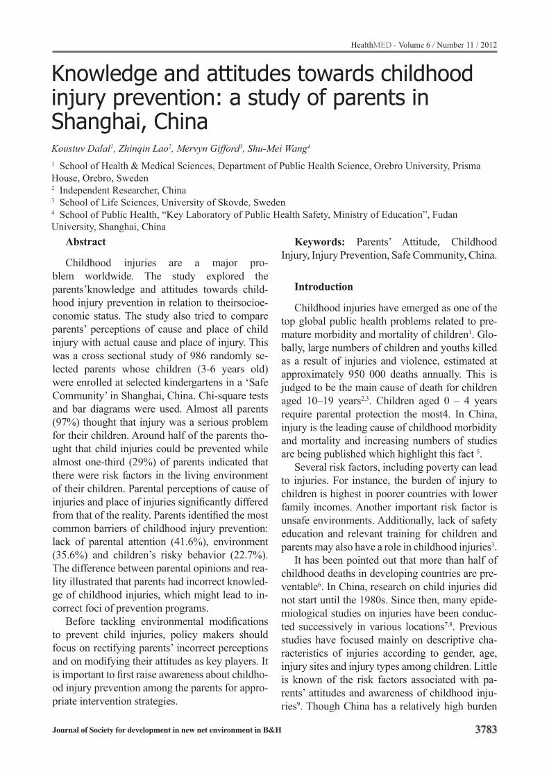
HealthMED - Volume 6 / Number 11 / 2012
Journal of Society for development in new net environment in B&H 3783
Knowledge and attitudes towards childhood injury prevention: a study of parents in Shanghai, ChinaKoustuv Dalal1, Zhinqin Lao2, Mervyn Gifford3, Shu-Mei Wang4
1 School of Health & Medical Sciences, Department of Public Health Science, Orebro University, Prisma House, Orebro, Sweden2 Independent Researcher, China3 School of Life Sciences, University of Skovde, Sweden4 School of Public Health, “Key Laboratory of Public Health Safety, Ministry of Education”, Fudan University, Shanghai, China
Abstract
Childhood injuries are a major pro-blem worldwide. The study explored the parents’knowledge and attitudes towards child-hood injury prevention in relation to theirsocioe-conomic status. The study also tried to compare parents’ perceptions of cause and place of child injury with actual cause and place of injury. This was a cross sectional study of 986 randomly se-lected parents whose children (3-6 years old) were enrolled at selected kindergartens in a ‘Safe Community’ in Shanghai, China. Chi-square tests and bar diagrams were used. Almost all parents (97%) thought that injury was a serious problem for their children. Around half of the parents tho-ught that child injuries could be prevented while almost one-third (29%) of parents indicated that there were risk factors in the living environment of their children. Parental perceptions of cause of injuries and place of injuries significantly differed from that of the reality. Parents identified the most common barriers of childhood injury prevention: lack of parental attention (41.6%), environment (35.6%) and children’s risky behavior (22.7%). The difference between parental opinions and rea-lity illustrated that parents had incorrect knowled-ge of childhood injuries, which might lead to in-correct foci of prevention programs.
Before tackling environmental modifications to prevent child injuries, policy makers should focus on rectifying parents’ incorrect perceptions and on modifying their attitudes as key players. It is important to first raise awareness about childho-od injury prevention among the parents for appro-priate intervention strategies.
Keywords: Parents’ Attitude, Childhood Injury, Injury Prevention, Safe Community, China.
Introduction
Childhood injuries have emerged as one of the top global public health problems related to pre-mature morbidity and mortality of children1. Glo-bally, large numbers of children and youths killed as a result of injuries and violence, estimated at approximately 950 000 deaths annually. This is judged to be the main cause of death for children aged 10–19 years2,3. Children aged 0 – 4 years require parental protection the most4. In China, injury is the leading cause of childhood morbidity and mortality and increasing numbers of studies are being published which highlight this fact 5.
Several risk factors, including poverty can lead to injuries. For instance, the burden of injury to children is highest in poorer countries with lower family incomes. Another important risk factor is unsafe environments. Additionally, lack of safety education and relevant training for children and parents may also have a role in childhood injuries3.
It has been pointed out that more than half of childhood deaths in developing countries are pre-ventable6. In China, research on child injuries did not start until the 1980s. Since then, many epide-miological studies on injuries have been conduc-ted successively in various locations7,8. Previous studies have focused mainly on descriptive cha-racteristics of injuries according to gender, age, injury sites and injury types among children. Little is known of the risk factors associated with pa-rents’ attitudes and awareness of childhood inju-ries9. Though China has a relatively high burden

3784
HealthMED - Volume 6 / Number 11 / 2012
Journal of Society for development in new net environment in B&H
of childhood injuries, information about parental attitudes and awareness to such issues is lacking in China5. Parental safety beliefs are related to the underlying parenting processes or attributes, such as conscientiousness of being parents, good or-ganization, and intention to have high standards. These in fact predict children’s injuries instead of specifying the parenting supervisory behaviors 10. It is therefore imperative to identify the risk fac-tors related to increased risk of injuries so that effective intervention program strategies can be constructed to aid the prevention and reduction of childhood injuries.
The objectives of this study are to discover pa-rental knowledge and attitudes towards childhood injury and prevention, and to explore the relati-onships between parental awareness of childhood injuries with their (parents) income/educational levels and occupations. The study also attempts to compare parents’ perception and actual incidence of injury.
Methods
This study was a cross sectional study, conduc-ted in Shanghai, during 2008. Experienced public health researchers designed the questionnaire in compliance with the literature and appropriate sci-entific methods. The questionnaire was tested in a pilot study. The local Health Administration of a designated Safe Community in Shanghai, China was one of the key stakeholders in promoting and evaluating the child injury prevention project. The community is very active in injury prevention and safety promotion and it has been recognized by the World Health Organization initiative of Community Safety Promotion through its Safe Community movement. All the kindergartens in the community were selected. The main subjects were parents of children aged 3 to 6 years in these selected kindergartens.
Students in a public health department were trained to collect the information. The pre-tested ‘Parents’ questionnaire of child injury prevention’ consisted mainly of bi-/multiple-choices closed ended questions and took approximately 20 minu-tes to administer. The parents of each child in the selected kindergarten were invited to take part in the study. In total, 986 children were selected from
the kindergartens of the target area. For each child either the mother or the father was selected for the interview.
Variables of interestThe parents were asked the following three
questions: Do you think that injury is a serious problem for children? Do you think that childhood injury can be prevented? Do you think that there is a potential risk factor of injury in your child’s living environment? The questions were closed ended, with response options of yes or no. The-se three questions were used as target variables to assess parental knowledge and attitudes towards child injuries.
Parental demographic factors considered were housing type, family income, fathers’ education and occupation, mothers’ education and occupati-on. There were five categories of types of housing: villa, apartment, public housing facilities, old pu-blic housing and other housing facilities. There were six categories of monthly family income: less than 800 CNY (China Yuan Renminbi), 8001 – 2000 CNY, 2001 – 3000 CNY, 3001 – 5000 CNY, 5001 – 8000 CNY and above 8001 CNY (1 CNY = 0.15 USD). Occupation was classified into one of six categories: manager, professional, tech-nologist, service, production/manufacturing and others. Education was classified into four catego-ries: Uneducated, primary education, secondary education and higher education.
Respondents were asked to identify major po-tential causes of childhood injuries and to iden-tify major potential locations where injuries co-uld occur. They were also asked about whether the child had sustained any injuries during the last year. If injuries had occurred, they were then asked to describe the cause of injury and the place of injury. In cases where more than one injury was sustained, respondents were asked to describe the circumstances surrounding the major one. Thro-ugh analysis of the questionnaires we compared the parent’s knowledge of factors that can lead to injury and actual incidence of child injuries.
Statistical analysisWe employed cross tables to identify potential
relationships between target variables and paren-
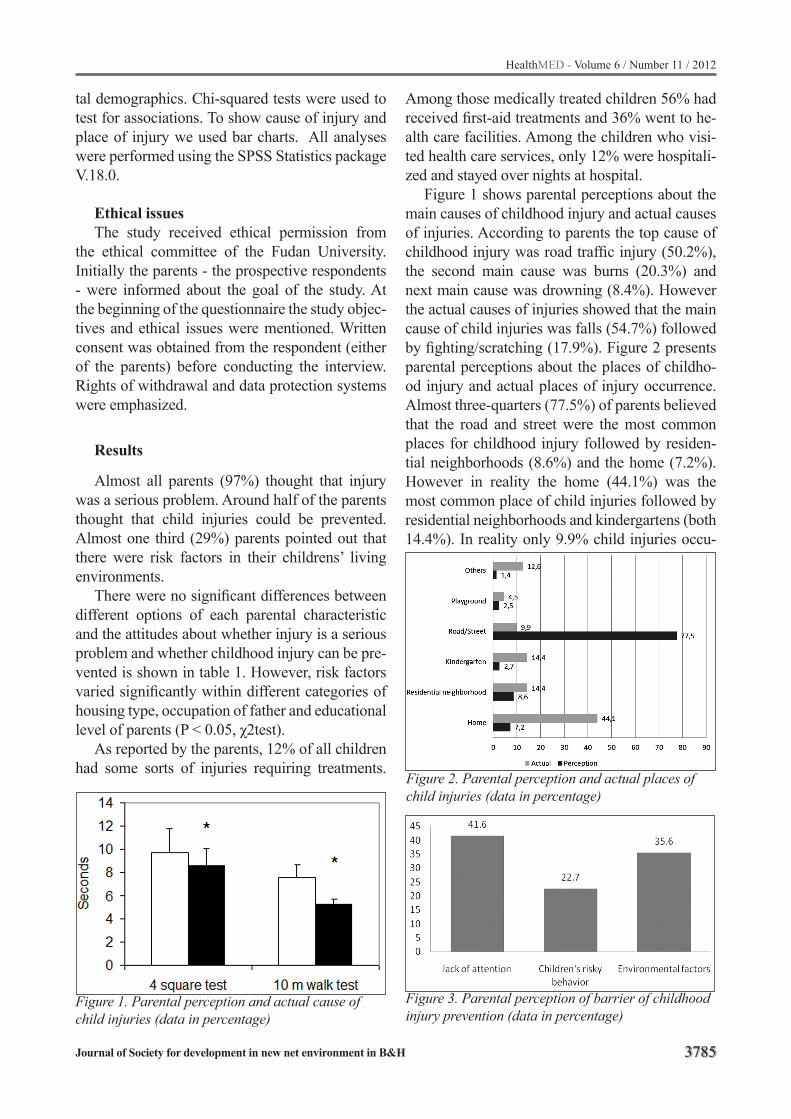
HealthMED - Volume 6 / Number 11 / 2012
Journal of Society for development in new net environment in B&H 3785
tal demographics. Chi-squared tests were used to test for associations. To show cause of injury and place of injury we used bar charts. All analyses were performed using the SPSS Statistics package V.18.0.
Ethical issuesThe study received ethical permission from
the ethical committee of the Fudan University. Initially the parents - the prospective respondents - were informed about the goal of the study. At the beginning of the questionnaire the study objec-tives and ethical issues were mentioned. Written consent was obtained from the respondent (either of the parents) before conducting the interview. Rights of withdrawal and data protection systems were emphasized.
Results
Almost all parents (97%) thought that injury was a serious problem. Around half of the parents thought that child injuries could be prevented. Almost one third (29%) parents pointed out that there were risk factors in their childrens’ living environments.
There were no significant differences between different options of each parental characteristic and the attitudes about whether injury is a serious problem and whether childhood injury can be pre-vented is shown in table 1. However, risk factors varied significantly within different categories of housing type, occupation of father and educational level of parents (P < 0.05, χ2test).
As reported by the parents, 12% of all children had some sorts of injuries requiring treatments.
Among those medically treated children 56% had received first-aid treatments and 36% went to he-alth care facilities. Among the children who visi-ted health care services, only 12% were hospitali-zed and stayed over nights at hospital.
Figure 1 shows parental perceptions about the main causes of childhood injury and actual causes of injuries. According to parents the top cause of childhood injury was road traffic injury (50.2%), the second main cause was burns (20.3%) and next main cause was drowning (8.4%). However the actual causes of injuries showed that the main cause of child injuries was falls (54.7%) followed by fighting/scratching (17.9%). Figure 2 presents parental perceptions about the places of childho-od injury and actual places of injury occurrence. Almost three-quarters (77.5%) of parents believed that the road and street were the most common places for childhood injury followed by residen-tial neighborhoods (8.6%) and the home (7.2%). However in reality the home (44.1%) was the most common place of child injuries followed by residential neighborhoods and kindergartens (both 14.4%). In reality only 9.9% child injuries occu-
Figure 1. Parental perception and actual cause of child injuries (data in percentage)
Figure 3. Parental perception of barrier of childhood injury prevention (data in percentage)
Figure 2. Parental perception and actual places of child injuries (data in percentage)
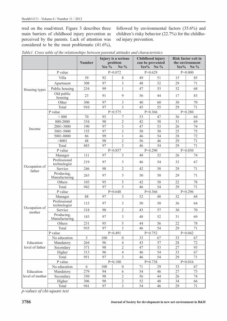
3786
HealthMED - Volume 6 / Number 11 / 2012
Journal of Society for development in new net environment in B&H
Table1. Cross table of the relationships between parental attitudes and characteristics
NumberInjury is a serious
problemYes % No %
Childhood injury can be prevented Yes% No %
Risk factor exit in the environment Yes% No %
Housing types
P value P=0.072 P=0.629 P=0.000Villa 39 92 8 49 51 15 85
Apartment 308 97 3 48 52 29 71Public housing 234 99 1 47 53 32 68
Old public housing 23 91 9 56 44 17 83
Other 306 97 3 40 60 30 70Total 910 97 3 45 55 29 71
Income
P value P=0.575 P=0.366 P=0.280< 800 70 93 7 53 47 36 64
800-2000 334 98 2 42 58 31 692001-3000 190 97 3 47 53 26 743001-5000 155 97 3 50 50 25 755001-8000 86 99 1 46 54 28 72
>8001 48 98 2 56 46 29 71Total 883 97 3 46 54 29 71
Occupation of father
P value P=0.857 P=0.290 P=0.030Manager 111 97 3 48 52 26 74
Professional technologist 219 97 3 46 54 33 67
Service 246 98 2 42 58 29 71Producing,
Manufacturing 263 97 3 50 50 29 71
Others 103 95 5 42 58 22 78Total 942 97 3 46 54 29 71
Occupation of mother
P value P=0.648 P=0.366 P=0.296Manager 88 97 3 52 48 32 68
Professional technologist 115 97 3 50 50 36 64
Service 318 98 2 43 57 30 70Producing,
Manufacturing 183 97 3 48 52 31 69
Others 231 95 5 44 56 22 78Total 935 97 3 46 54 29 71
Education level of father
P value P=0.491 P=0.752 P=0.042No education 3 100 0 33 67 33 67Mandatory 264 96 4 43 57 28 72Secondary 371 98 2 47 53 27 93
Higher 313 96 4 46 54 33 67Total 951 97 3 46 54 29 71
Education level of mother
P value P=0.180 P=0.738 P=0.016No education 6 100 0 71 29 33 67Mandatory 279 94 6 54 46 27 73Secondary 350 98 2 56 44 26 74
Higher 306 98 2 52 48 34 66Total 941 97 3 54 46 29 71
p-values of chi-square test
rred on the road/street. Figure 3 describes three main barriers of childhood injury prevention as perceived by the parents. Lack of attention was considered to be the most problematic (41.6%),
followed by environmental factors (35.6%) and children’s risky behavior (22.7%) for the childho-od injury prevention.
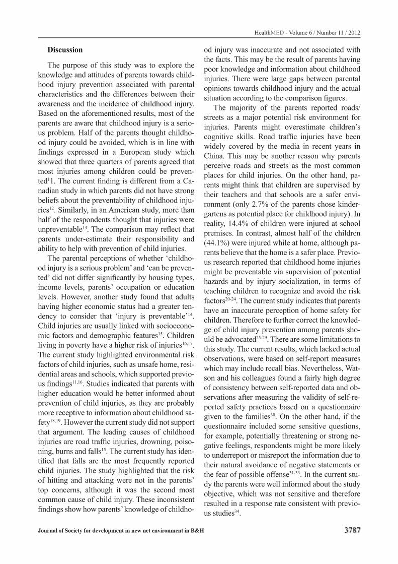
HealthMED - Volume 6 / Number 11 / 2012
Journal of Society for development in new net environment in B&H 3787
Discussion
The purpose of this study was to explore the knowledge and attitudes of parents towards child-hood injury prevention associated with parental characteristics and the differences between their awareness and the incidence of childhood injury. Based on the aforementioned results, most of the parents are aware that childhood injury is a serio-us problem. Half of the parents thought childho-od injury could be avoided, which is in line with findings expressed in a European study which showed that three quarters of parents agreed that most injuries among children could be preven-ted11. The current finding is different from a Ca-nadian study in which parents did not have strong beliefs about the preventability of childhood inju-ries12. Similarly, in an American study, more than half of the respondents thought that injuries were unpreventable13. The comparison may reflect that parents under-estimate their responsibility and ability to help with prevention of child injuries.
The parental perceptions of whether ‘childho-od injury is a serious problem’ and ‘can be preven-ted’ did not differ significantly by housing types, income levels, parents’ occupation or education levels. However, another study found that adults having higher economic status had a greater ten-dency to consider that ‘injury is preventable’14. Child injuries are usually linked with socioecono-mic factors and demographic features15. Children living in poverty have a higher risk of injuries16,17. The current study highlighted environmental risk factors of child injuries, such as unsafe home, resi-dential areas and schools, which supported previo-us findings11,16. Studies indicated that parents with higher education would be better informed about prevention of child injuries, as they are probably more receptive to information about childhood sa-fety18,19. However the current study did not support that argument. The leading causes of childhood injuries are road traffic injuries, drowning, poiso-ning, burns and falls15. The current study has iden-tified that falls are the most frequently reported child injuries. The study highlighted that the risk of hitting and attacking were not in the parents’ top concerns, although it was the second most common cause of child injury. These inconsistent findings show how parents’ knowledge of childho-
od injury was inaccurate and not associated with the facts. This may be the result of parents having poor knowledge and information about childhood injuries. There were large gaps between parental opinions towards childhood injury and the actual situation according to the comparison figures.
The majority of the parents reported roads/streets as a major potential risk environment for injuries. Parents might overestimate children’s cognitive skills. Road traffic injuries have been widely covered by the media in recent years in China. This may be another reason why parents perceive roads and streets as the most common places for child injuries. On the other hand, pa-rents might think that children are supervised by their teachers and that schools are a safer envi-ronment (only 2.7% of the parents chose kinder-gartens as potential place for childhood injury). In reality, 14.4% of children were injured at school premises. In contrast, almost half of the children (44.1%) were injured while at home, although pa-rents believe that the home is a safer place. Previo-us research reported that childhood home injuries might be preventable via supervision of potential hazards and by injury socialization, in terms of teaching children to recognize and avoid the risk factors20-24. The current study indicates that parents have an inaccurate perception of home safety for children. Therefore to further correct the knowled-ge of child injury prevention among parents sho-uld be advocated25-29. There are some limitations to this study. The current results, which lacked actual observations, were based on self-report measures which may include recall bias. Nevertheless, Wat-son and his colleagues found a fairly high degree of consistency between self-reported data and ob-servations after measuring the validity of self-re-ported safety practices based on a questionnaire given to the families30. On the other hand, if the questionnaire included some sensitive questions, for example, potentially threatening or strong ne-gative feelings, respondents might be more likely to underreport or misreport the information due to their natural avoidance of negative statements or the fear of possible offense31-33. In the current stu-dy the parents were well informed about the study objective, which was not sensitive and therefore resulted in a response rate consistent with previo-us studies34.
3788
HealthMED - Volume 6 / Number 11 / 2012
Journal of Society for development in new net environment in B&H
Overall, the present study describes the general knowledge and attitudes of parents towards child-hood injury. The large difference between parental opinions and reality illustrates the fact that parents have inaccurate knowledge of childhood injuries and this, therefore, may be problematic in relati-on to preventive strategies. Before tackling envi-ronmental modifications to prevent child injuries, policy makers should focus on rectifying parents’ incorrect perceptions and on modifying their atti-tudes as key players. It is important to raise paren-tal awareness about injury prevention among chil-dren so that more effective intervention strategies may be devised. The Safe Community programs can also target the parents for awareness deve-lopment towards in relation to injury prevention and safety promotion within its community safety promotion activities35. Furthermore, broad edu-cation campaigns combined with informed public policies and the creation of safer environments could create a better culture of childhood safety in China.
References
1. Sleet DA, Ballesteros MF, Borse NN. A review of unin-tentional injuries in adolescents, Annu.Rev. Public Health 2010; 31: 195-212.
2. World Health Organization: Global Burden of Disea-se: 2004 update. Geneva; 2008.
3. Peden M, et al. (Ed), World Report on Child Injury Prevention. Unicef & WHO; 2008.
4. Valent F, Little D, Tamburini G, et al. Burden of dise-ase attributable to selected environmental factors and injury among children and adolescents in Europe. Lancet 2004; 363: 2032 – 2039.
5. UNICEF. About UNICEF in China. http://www.unicef.org/china/about_643.html (accessed Feb12, 20012).
6. Linnan M, Giersing M, Cox R, et al. Child mortality and injury in Asia: an overview. Florence, Italy: UNI-CEF Innocenti Research Centre, 2007:1e26.
7. Xiong Z, Liu X. Injuries of children aged 0–14 years in Hubei province during 2000–2001. Chin J Child Health Care 2003; 11: 17–9.
8. Zhang P, Cheng RH, Deng JY. Comparison on the sta-tus of child injury in urban and rural area of Jiangsu province, China. Chin J Dis Control Prev 2004; 8: 503–5.
9. Doll LS (eds): Handbook of injury and violence pre-vention., Atlanta, GA, Springer: 2007.
10. Morrongiello BA. Caregiver supervision and child-injury risk: I. Issues in defining and measuring super-vision; II. Findings and directions for future research. J Pediatr Psychol 2005; 30(7): 536–552.
11. Vincenten JA. European Child Safety Alliance. Priori-ties for Child Safety in the European Union: Agenda for Action. (Amsterdam: ECOSA): 2004.
12. Morrongiello BA, Dayler L. A community-based study of parents’ knowledge, attitudes and beliefs related to childhood injuries. Canadian Journal of Public He-alth 1996; 87: 383 – 388.
13. Bennet LM. Adolescent mothers’ beliefs about paren-ting and injury prevention: results of a focus group. Journal of Pediatric Health Care 2001; 15: 194 – 199.
14. Girasek, D.C. Public beliefs about the preventability of unintentional injury deaths.Accident Analysis and Prevention 2001; 33: 455 – 465.
15. Braddock M, Lapidus G, Gregorio D, et al. Popula-tion, income and ecological correlates of child pede-strian injury. Pediatrics 1991; 88 (6): 1242–1247.
16. UNICEF. A League Table of Child Deaths by Injury in Rich Nations. Innocenti Report Card. (Florence: UNICEF): 2001.
17. Durkin MS, Davidson LL, Kuhn L, et al, Low Inco-me Neighbourhoods and the Risk of Severe Pediatric Injury: A Small Area Analysis in Northern Manhattan American Journal of Public Health 1994; 84: 587–592.
18. Tsoumakes K, Mavridi F, Matziou V, et al. Parents’ Knowledge and Attitudes About Preventing Injuries in Motor Vehicle Accidents in Children in Greece. Traffi-cInjury Prevention 2008; 9: 129–134.
19. Chaudhari N. Adolescent injuries. Indian Pediatr 1990; 27: 1261-1267.
20. Robertson LS: Injury epidemiology. New York: Oxford University Press, 1992.
21. Morrongiello BA, Midgett C, Shields R. Don’t run with scissors: Young children’s knowledge of home safety rules. J Pediatr Psychol 2001; 26(2): 105–115.
22. Morrongiello BA, House K. Measuring parent attri-butes and supervision behaviors relevant to child injury risk: Examining the usefulness of questionnaire measures. Injury Prevention 2004; 10: 114–118.
HealthMED - Volume 6 / Number 11 / 2012
Journal of Society for development in new net environment in B&H 3789
23. Morrongiello BA, Corbett M, McCourt M, Johnston N. Understanding unintentional injury risk in young children II. The contribution of caregiver supervisi-on, child attributes, and parent attributes. J Pediatr Psychol 2006; 31: 540–551.
24. Peterson L, Stern, BL. Family processes and child risk for injury. Behav Res Ther 1997; 35(3): 179–190.
25. Rivara FP, Howard D. Parental knowledge of child development and injury risks. Journal of Developmen-tal and Behavioral Pediatrics 1982; 3: 103 – 105.
26. Liller KD, Kent E, Mcdermott RJ. Postpartum pa-tients’ knowledge, risk perceptions, and behaviors pertaining to childhood injuries. Journal of Nurse Midwifery 1991; 36: 355 – 360.
27. Hu X, Wesson D, Parkin P, Rootman I. Pediatric inju-ries: parental knowledge, attitudes and needs. Cana-dian Journal of Public Health 1996; 87: 101 – 105.
28. Vincenten JA, Sector MJ, Rogmans W, et al. Parents’ perceptions, attitudes and behaviours towards child safety: a study in 14 European countries. Internati-onal Journal of Injury Control and Safety Promotion 2005; 12 ( 3): 183 – 189.
29. Ehrlich PF, Longhi J, Vaughan R. Correlation Between Parental Perception and Actual Childhood Patterns of Bicycle Helmet Use and Riding Practices: Implications for Designing Injury Prevention Strate-gies. J. Pediatric Surgery 2001; 36: 763–766.
30. Watson M, Kendrick D, Coupland C. Validation of a home safety questionnaire used in a randomised con-trolled trial. Injury Prevention 2003; 9: 180 – 183.
31. Bradburn NM, Sudman S. Improving interview met-hod and questionnaire design: response effects to threatening questions in survey research. San Fran-cisco: Jossey-Bass, 1979.
32. Schuman H, Presser S. Questions and answers in atti-tude surveys. New York: Academic Press, 1981.
33. Tourangeau R, Rasinski K. Cognitive processes un-derlying context effects in attitude measurement. Psychol Bull 1988; 103: 299–314.
34. Scheidt CP, Brenner AR, Possi WM. et al. Parental attitudes regarding interviews about injuries to their children. Injury Prevention 2000; 6: 51 – 55.
35. Lindqvist K, Dalal K. The impact of child safety promotion on different social strata in a WHO Safe Community. Journal of Injury and Violence Research 2011. doi: 10.5249/jivr.v4i1.83.
Corresponding AuthorShu-Mei Wang, School of Public Health,“Key Laboratory of Public Health Safety, Ministry of Education”, Fudan University, Shanghai, China, E-mail: [email protected]
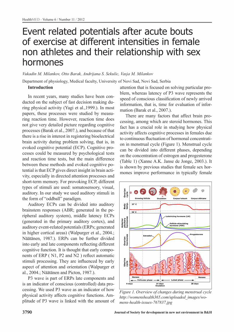
3790
HealthMED - Volume 6 / Number 11 / 2012
Journal of Society for development in new net environment in B&H
Event related potentials after acute bouts of exercise at different intensities in female non athletes and their relationship with sex hormonesVukadin M. Milankov, Otto Barak, Andrijana S. Sekulic, Vasja M. Milankov
Department of physiology, Medical faculty, University of Novi Sad, Novi Sad, SerbiaIntroduction
In recent years, many studies have been con-ducted on the subject of fast decision making du-ring physical activity (Yagi et al.,1999.). In most papers, these processes were studied by measu-ring reaction time. However, reaction time does not give very detailed picture regarding cognitive processes (Barak et al., 2007.), and because of that there is a rise in interest in registering bioelectrical brain activity during problem solving, that is, in evoked cognitive potential (ECP). Cognitive pro-cesses could be measured by psychological tests and reaction time tests, but the main difference between these methods and evoked cognitive po-tential is that ECP gives direct insight in brain acti-vity, especially in directed attention processes and short-term memory. For provoking ECP, different types of stimuli are used: somatosensory, visual, auditory. In our study we used auditory stimuli in the form of “oddball” paradigm.
Auditory ECPs can be divided into auditory brainstem responses (ABR; generated in the pe-ripheral auditory system), middle latency ECPs (generated in the primary auditory cortex), and auditory event-related potentials (ERPs; generated in higher cortical areas) (Walpurger et al., 2004.; Näätänen, 1987.). ERPs can be further divided into early and late components reflecting different cognitive function. It is thought that early compo-nents of ERP ( N1, P2 and N2 ) reflect automatic stimuli processing. They are influenced by early aspect of attention and orientation (Walpurger et al., 2004.; Näätänen and Picton, 1987.).
P3 wave is part of ERPs late components and is an indicator of conscious (controlled) data pro-cessing. We used P3 wave as an indicator of how physical activity affects cognitive functions. Am-plitude of P3 wave is linked with the amount of
attention that is focused on solving particular pro-blem, whereas latency of P3 wave represents the speed of conscious classification of newly arrived information, that is, time for evaluation of infor-mation (Barak et al., 2007.).
There are many factors that affect brain pro-cessing, among which are steroid hormones. This fact has a crucial role in studying how physical activity affects cognitive processes in females due to continuous fluctuation of hormonal concentrati-on in menstrual cycle (Figure 1). Menstrual cycle can be divided into different phases, depending on the concentration of estrogen and progesterone (Table 1) (Xanne A.K. Janse de Jonge, 2003.). It is shown by previous studies that female sex hor-mones improve performance in typically female
Figure 1. Overview of changes during menstrual cyclehttp://womenshealth365.com/uploaded_images/wo-mens-health-issues-767837.jpg
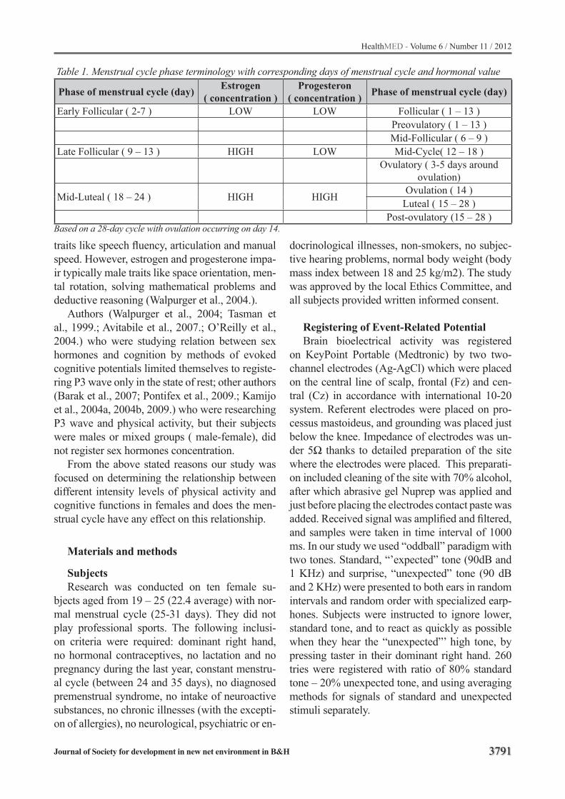
HealthMED - Volume 6 / Number 11 / 2012
Journal of Society for development in new net environment in B&H 3791
traits like speech fluency, articulation and manual speed. However, estrogen and progesterone impa-ir typically male traits like space orientation, men-tal rotation, solving mathematical problems and deductive reasoning (Walpurger et al., 2004.).
Authors (Walpurger et al., 2004; Tasman et al., 1999.; Avitabile et al., 2007.; O’Reilly et al., 2004.) who were studying relation between sex hormones and cognition by methods of evoked cognitive potentials limited themselves to registe-ring P3 wave only in the state of rest; other authors (Barak et al., 2007; Pontifex et al., 2009.; Kamijo et al., 2004a, 2004b, 2009.) who were researching P3 wave and physical activity, but their subjects were males or mixed groups ( male-female), did not register sex hormones concentration.
From the above stated reasons our study was focused on determining the relationship between different intensity levels of physical activity and cognitive functions in females and does the men-strual cycle have any effect on this relationship.
Materials and methods
SubjectsResearch was conducted on ten female su-
bjects aged from 19 – 25 (22.4 average) with nor-mal menstrual cycle (25-31 days). They did not play professional sports. The following inclusi-on criteria were required: dominant right hand, no hormonal contraceptives, no lactation and no pregnancy during the last year, constant menstru-al cycle (between 24 and 35 days), no diagnosed premenstrual syndrome, no intake of neuroactive substances, no chronic illnesses (with the excepti-on of allergies), no neurological, psychiatric or en-
docrinological illnesses, non-smokers, no subjec-tive hearing problems, normal body weight (body mass index between 18 and 25 kg/m2). The study was approved by the local Ethics Committee, and all subjects provided written informed consent.
Registering of Event-Related PotentialBrain bioelectrical activity was registered
on KeyPoint Portable (Medtronic) by two two-channel electrodes (Ag-AgCl) which were placed on the central line of scalp, frontal (Fz) and cen-tral (Cz) in accordance with international 10-20 system. Referent electrodes were placed on pro-cessus mastoideus, and grounding was placed just below the knee. Impedance of electrodes was un-der 5Ω thanks to detailed preparation of the site where the electrodes were placed. This preparati-on included cleaning of the site with 70% alcohol, after which abrasive gel Nuprep was applied and just before placing the electrodes contact paste was added. Received signal was amplified and filtered, and samples were taken in time interval of 1000 ms. In our study we used “oddball” paradigm with two tones. Standard, “’expected” tone (90dB and 1 KHz) and surprise, “unexpected” tone (90 dB and 2 KHz) were presented to both ears in random intervals and random order with specialized earp-hones. Subjects were instructed to ignore lower, standard tone, and to react as quickly as possible when they hear the “unexpected”’ high tone, by pressing taster in their dominant right hand. 260 tries were registered with ratio of 80% standard tone – 20% unexpected tone, and using averaging methods for signals of standard and unexpected stimuli separately.
Table 1. Menstrual cycle phase terminology with corresponding days of menstrual cycle and hormonal value
Phase of menstrual cycle (day) Estrogen ( concentration )
Progesteron ( concentration ) Phase of menstrual cycle (day)
Early Follicular ( 2-7 ) LOW LOW Follicular ( 1 – 13 ) Preovulatory ( 1 – 13 ) Mid-Follicular ( 6 – 9 )Late Follicular ( 9 – 13 ) HIGH LOW Mid-Cycle( 12 – 18 )
Ovulatory ( 3-5 days around ovulation)
Mid-Luteal ( 18 – 24 ) HIGH HIGH Ovulation ( 14 )Luteal ( 15 – 28 )
Post-ovulatory (15 – 28 )Based on a 28-day cycle with ovulation occurring on day 14.

3792
HealthMED - Volume 6 / Number 11 / 2012
Journal of Society for development in new net environment in B&H
Reading of event-related potential implied in-dividual identification of the most positive wave in the window of 220-450 ms for every electrode and determining amplitude and latency of P3 po-tential for “unexpected” signal. To rule out ocu-lar artifact, ERP were measured in a totally dark room. Participants were blindfolded with a slee-ping mask, and they were asked to restrain from eye movement and mimic. Software that was pro-vided by manufacturer was enabledto filter out ocular artifacts.
Hormone measurementForty five minutes before measuring P3 wave,
10ml of blood has been drawn from the subject’s cubital vein and taken for hormone analysis (estrogen and progesterone). Biologically most active estrogen is 17β-estradiol, whose concentra-tion we analyzed. For assessment of 17β-estradiol concentration we used electrochemiluminescen-ce immunoassay ‘ECLIA’ which was done using Elecsys 1010/2010 instrument. Elecsys Estradiol II test was employed. It is based on the principle of competitive measuring of polyclonal antibodies specific to 17β-estradiol. Endogen estradiol was released from sample with mesterol and it compe-tes with estradiol derivate marked with ruthenium complex (Tris(2,2’-bipyridyl) ruthenium(II)-com-plex (Ru(bpy)⅔*)) for binding on biotin marked antibodies.
Similar method was used for determining the concentration of progesterone. Elecsys Progeste-rone II test was employed.
Analytical sensitivity of the method was 18.4 pmol/L (5,0 pg/mL) for estradiol, and 0.095 nmol/L (0.03 ng/mL) for progesterone. Variance coefficient for both measurements was < 20%.
Protocol of ResearchThe research was done in two phases depending
on menstrual cycle (Fig. 2). First time P3 wave was registered in mid-follicular phase (betwe-en 7th–10th day of menstrual cycle) and the se-cond time was done in mid-luteal phase (between 22nd–25th day of menstrual cycle). Identification of phase of menstrual cycle was done by counting days from first day of the last menstruation, and was confirmed by measuring concentration of sex hormones.
P3 wave was registered between 16h and 19h on the Department of Physiology, Medical Faculty of Novi Sad, Serbia.
45 minutes before subjected were introduced to physical activity, blood was draw from there cu-bital vein for measuring sex hormone concentra-tion (estrogen and progesterone). Blood drawing was done before the exercise to avoid error ridin-gs in ERP because of the effects of fear of blood drawing and the direct effect of physical activity on hormonal status.
After registering ERP’s in the state of rest, su-bjects were exposed to controlled resistance on bicycle ergometer. The duration of one ride was ten minutes, with gradual increase of resistance until reaching the desired value of percentage of maximal pulse and sustaining that value for at le-ast six minutes. Pulse was measured with Polar pulse meter, and the resistance in Watt was con-trolled on bicycle ergometer depending on 60%, 75%, and 90% of maximal pulse. After each stage of resistance, ERP’s were measured. Pause betwe-en stages were twenty minutes which subjected had used for active rest and muscle stretching.
The results were statistically analyzed using the software package Statistica for Windows. We used descriptive statistics to determine average value, standard deviation and coefficient of varia-tion. Importance of statistical differences between parameters of the intervention were determined by three way factorial (intensity x phase x electrode ) analysis of variance (ANOVA).
Results
Follicular Phase of Menstrual CycleIn Fig 3. are shown the values of P3 wave am-
plitude and their relationship with different inten-sities of physical activity in follicular phase of menstrual cycle. Analyzing all the values, we have not found any statistically significant difference.
In Fig 4. are shown values of P3 wave la-tency and their relationship with different inten-sity levels of physical activity in follicular phase of menstrual cycle. After analysis, we found that there is statistically significant effect of resistance of 75% of maximal pulse on latency of P3 wave, We used Bonferroni correction student t test that showed statistically significant increase in latency

HealthMED - Volume 6 / Number 11 / 2012
Journal of Society for development in new net environment in B&H 3793
Figure 2. Test days. On figure 2 is show how test days were chosen depending on day of menstrual cycle and hormone concentration
Figure 3. Amplitude of P3 wave above Fz and Cz electrode in follicular phase
Figure 4. Latency of P3 wave above Fz and Cz electrode in follicular phaze
value above Fz electrode on resistance of 75% of maximal pulse (335.3ms [33.13]) in comparison with state of rest (329,00ms [33.01]). (F=7,211; p<0,01; ɛ=0.663)
Luteal Phase of Menstrual CycleIn Fig 5. are shown the values of P3 wave am-
plitude and their relationship with different inten-sity levels of physical activity in luteal phase of menstrual cycle. Our results show that there is a statistically significant effect of intensity at re-sistance of 90% of maximal pulse on amplitude of P3 wave. By using Bonferroni correction stu-dent t test results show that there is drop in value (Fz t=2.231, p<0.05; CZ t=2.395, p<0.05) of P3 wave amplitude at the resistance of 90% of max pulse (Fz 9.35µV [6.02], CZ 11.83µV [5.12]) in
Figure 5. Amplitude of P3 wave Fz and Cz electrode in luteal phase
Figure 6. Latency of P3 wave above Fz i Cz electrode in luteal phase
Figure 7. Concentrations of progesterone and estro-gen in follicular and luteal phase of menstrual cycle
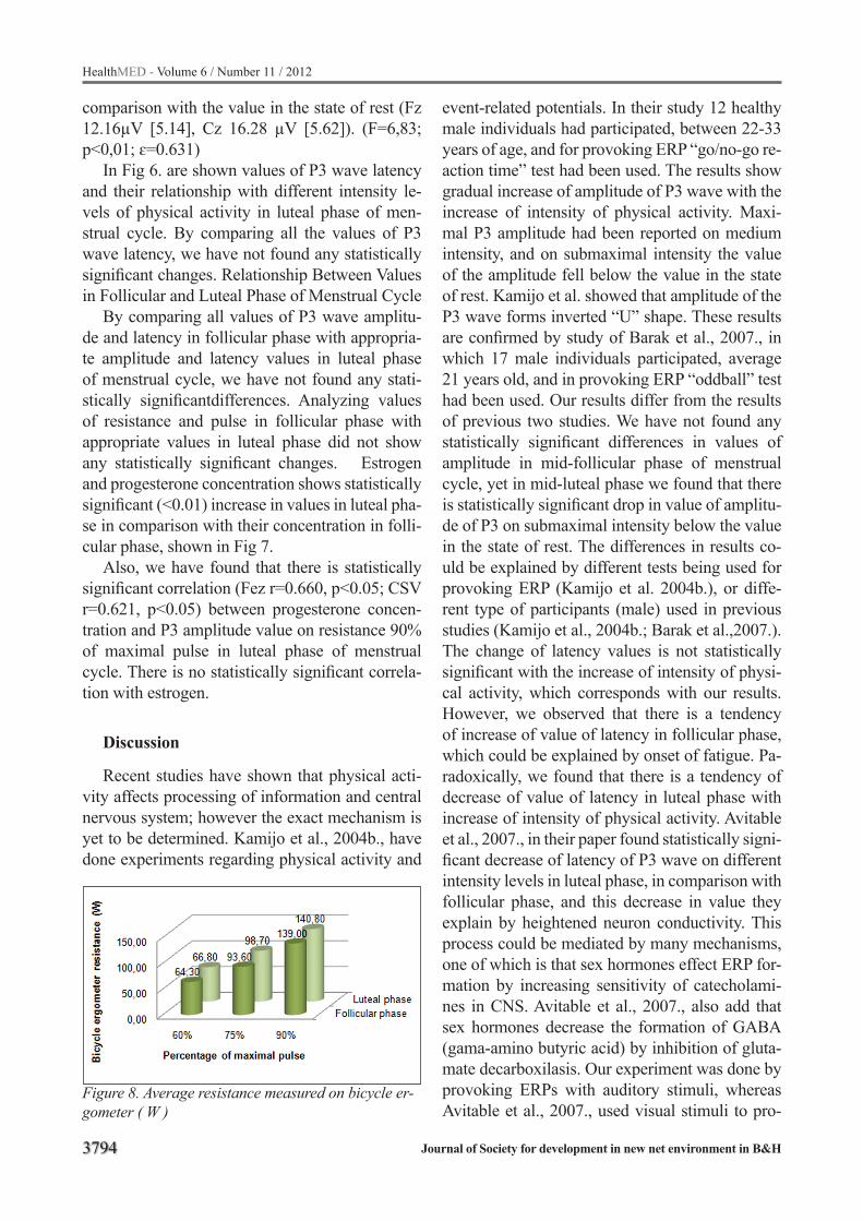
3794
HealthMED - Volume 6 / Number 11 / 2012
Journal of Society for development in new net environment in B&H
comparison with the value in the state of rest (Fz 12.16µV [5.14], Cz 16.28 µV [5.62]). (F=6,83; p<0,01; ɛ=0.631)
In Fig 6. are shown values of P3 wave latency and their relationship with different intensity le-vels of physical activity in luteal phase of men-strual cycle. By comparing all the values of P3 wave latency, we have not found any statistically significant changes. Relationship Between Values in Follicular and Luteal Phase of Menstrual Cycle
By comparing all values of P3 wave amplitu-de and latency in follicular phase with appropria-te amplitude and latency values in luteal phase of menstrual cycle, we have not found any stati-stically significantdifferences. Analyzing values of resistance and pulse in follicular phase with appropriate values in luteal phase did not show any statistically significant changes. Estrogen and progesterone concentration shows statistically significant (<0.01) increase in values in luteal pha-se in comparison with their concentration in folli-cular phase, shown in Fig 7.
Also, we have found that there is statistically significant correlation (Fez r=0.660, p<0.05; CSV r=0.621, p<0.05) between progesterone concen-tration and P3 amplitude value on resistance 90% of maximal pulse in luteal phase of menstrual cycle. There is no statistically significant correla-tion with estrogen.
Discussion
Recent studies have shown that physical acti-vity affects processing of information and central nervous system; however the exact mechanism is yet to be determined. Kamijo et al., 2004b., have done experiments regarding physical activity and
Figure 8. Average resistance measured on bicycle er-gometer ( W )
event-related potentials. In their study 12 healthy male individuals had participated, between 22-33 years of age, and for provoking ERP “go/no-go re-action time” test had been used. The results show gradual increase of amplitude of P3 wave with the increase of intensity of physical activity. Maxi-mal P3 amplitude had been reported on medium intensity, and on submaximal intensity the value of the amplitude fell below the value in the state of rest. Kamijo et al. showed that amplitude of the P3 wave forms inverted “U” shape. These results are confirmed by study of Barak et al., 2007., in which 17 male individuals participated, average 21 years old, and in provoking ERP “oddball” test had been used. Our results differ from the results of previous two studies. We have not found any statistically significant differences in values of amplitude in mid-follicular phase of menstrual cycle, yet in mid-luteal phase we found that there is statistically significant drop in value of amplitu-de of P3 on submaximal intensity below the value in the state of rest. The differences in results co-uld be explained by different tests being used for provoking ERP (Kamijo et al. 2004b.), or diffe-rent type of participants (male) used in previous studies (Kamijo et al., 2004b.; Barak et al.,2007.). The change of latency values is not statistically significant with the increase of intensity of physi-cal activity, which corresponds with our results. However, we observed that there is a tendency of increase of value of latency in follicular phase, which could be explained by onset of fatigue. Pa-radoxically, we found that there is a tendency of decrease of value of latency in luteal phase with increase of intensity of physical activity. Avitable et al., 2007., in their paper found statistically signi-ficant decrease of latency of P3 wave on different intensity levels in luteal phase, in comparison with follicular phase, and this decrease in value they explain by heightened neuron conductivity. This process could be mediated by many mechanisms, one of which is that sex hormones effect ERP for-mation by increasing sensitivity of catecholami-nes in CNS. Avitable et al., 2007., also add that sex hormones decrease the formation of GABA (gama-amino butyric acid) by inhibition of gluta-mate decarboxilasis. Our experiment was done by provoking ERPs with auditory stimuli, whereas Avitable et al., 2007., used visual stimuli to pro-
HealthMED - Volume 6 / Number 11 / 2012
Journal of Society for development in new net environment in B&H 3795
voke P3 wave, which could explain the difference of results between our two studies. There is also the possibility that our number of subjects is too small, so by increasing the number of subjects our study could confirm the results of Avitable et al., 2007.
Walpurger et al, 2004.,had measured P3 wave in three different phases of menstrual cycle (during menses, follicular phase and luteal phase), using auditory stimuli in the form of ‘’oddball’’ paradi-gm, only in the state of rest. After analyzing values of amplitude and latency, they had not found any statistically significant difference. The only excep-tion is that during ovulation the value of latency of P3 wave increases. The same results are observed by Tasman et al.7. O’Reilly et al., 2004.,compared the menses phase with the ovulation phase, using visual stimuli (word test). They found that there is statistically significant increase in the value of am-plitude of P3 wave during menses. The difference in results in previous three studies could be attri-buted to different tests used for evoking P3 wave. Our results partially correspond with Walpurgeret al., 2004.,and Tasman et al., 1999.,, because they show there is no statistically significant difference in values of amplitude and latency, in the state of rest. Observed increase in the value of latency du-ring ovulation we could not confirm due to diffe-rence in methodology. However, our results show statistically significant drop in the value of ampli-tude of P3 wave on submaximal intensity during luteal phase, and that this statistically significant value correlates with the increase in progestero-ne concentration. One of possible causes for this drop in amplitude value could be changes in basal temperature during luteal phase, because P3 wave is sensitive to temperature changes, both in exter-nal and internal environment. Temperature in the testing room was continuous 24°C, hence it could be concluded that for the drop in amplitude are res-ponsible changes in internal temperature. Recent studies confirm that during mid-luteal phase there is an increase in basal temperature and increase in body temperature during workout; in this period accumulated heat during physical exercise is libe-rated more slowly and because of that there is big-ger strain on cardiovascular system, and quicker onset of fatigue. It is believed that progesterone is responsible for this internal temperature change
by pushing thermoregulatory receptor threshold higher, though the exact mechanism is not well understood (Janse de Jonge XA et al., 2003.). Ta-sman et al., 1999.,and O’Reilly et al.,2004., also acknowledge the relationship between tempera-ture and evoked cognitive potentials. O’Reilly et al., 2004.,state that progesterone could directly decrease brain excitability with it sedative pro-perties. Research of Wingen et al., 2007.,indicate that progesterone could have direct influence on some structures of CNS by modulating functions without affecting state of consciousness. Because of varying results future research should be aimed at better understating of the relationship between progesterone, temperature, physical activity (stre-ss) and cognition.
Conclusion
In conclusion we emphasize that changes of amplitude and latency of P3 wave registered abo-ve certain regions of brain (Fz, Cz) in luteal pha-se of menstrual cycle are the result of complex effects of progesterone. These effects consist of lowering efficiency of GABA transmission, throu-gh GABAA receptors, by inhibiting glutamate de-carboxylases. With this effect activating influence of reticular formation on amygdala is enhanced, which results in promotion of memory processes in limbic system. By its direct influence on thermo receptors in hypothalamus, progesterone affects thermoregulation, and increases body temperatu-re, decreases liberation of accumulated heat, and puts more strain on cardiovascular system which quickens the onset of fatigue. Accumulation of water in the body during luteal phase, also by the action of progesterone, could, depending of its amount, increase excitability of the brain.
Within complex neurohumoral regulation of mental functions, we bring to consideration not only direct and indirect effects of progesterone and estrogen individually, but also their mutual re-lationship, in the framework of dynamics of men-strual cycle and pregnancy.
Based on the results and the experience of the previous studies, we have concluded that physical activity affects cognitive processes in the brain. In addition, menstrual cycle influences cognitive functions in luteal phase on submaximal intensity
3796
HealthMED - Volume 6 / Number 11 / 2012
Journal of Society for development in new net environment in B&H
of physical activity, through progesterone. This study draws attention to the necessity that during research of physical activity and evoked cognitive potentials, the studied subjects should be grouped by gender.
References
1. AvitabileT., Antonio L., Salvatore C., Gagliano C., Amato R., Scollo D., Lopes R., Pulvirenti L., Toto L., Torrisi B., Agnello C., 2007. Changes in Visual Evo-ked Potentials During the Menstrual Cycle in Young Women. Curr Eye Res, 32: 999–1003.
2. Barak O., Ivetic V., Filipovic D., Naumovic N., Lukac D, Drapsin M., Karaba-Jakovljevic D., Popadic-Ga-cesa j., Klasnja A., Grujic N., 2007. Event-related po-tentials following exercise bouts of different intensity, Med Pregl 11-12: 531-535.
3. KamijoKeita, Yoshiaki Nishihira, ArihiroHatta, Ta-keshi Kaneda, Tetsuo Kidac, TakuroHigashiura, Ka-zuo Kuroiwa, 2004a. Changes in arousal level by differential exercise intensity. ClinNeurophysiol 115: 2693–2698.
4. KamijoKeita, Yoshiaki Nishihira, ArihiroHatta, Ta-keshi Kaneda, Toshiaki Wasaka, Tetsuo Kida, Kazuo Kuroiwa, 2004b. Differential influences of exercise intensity on information processing in the central ner-vous system. Eur J ApplPhysiol 92: 305–311.
5. Kamijo. K., Hayashi. Y.. Sakai. T .•Yahiro. T .• Tanaka. K., Nishihira.Y. , 2009. Acute effects of aerobic exer-cise on cognitive function in older adult. J Gerontol B PsycholSciSocSci, 64B(3): 356-363.1.
6. Näätänen R., 1987. Implications of ERP data for psychological theories of attention. Biol. Psychol. 26, 117–163.
7. Näätänen R., Picton T., 1987. The N1 wave of the human electric and magnetic response to sound: a review and an analysis of the component structure. Psychophysiology 24: 375–425.
8. O’ReillyM., Cunningham C., Brian L., Walsh C., Rowan M., 2004. The effect of the menstrual cycle on electrophysiological and behavioral measures of me-mory and mood.Psychophysiology 41: 592–603.
9. PontifexM., Hillman C., Polich J., 2009. Age, physi-cal fitness, and attention: P3a and P3b. Psychophysi-ology, 46: 379–387.
10. Tasman A., Hahn T., and Maiste A., 1999. Menstrual Cycle Synchronized Changes in Brain Stem Auditory Evoked Potentials and Visual Evoked Potentials. Biol psychiatry 45:1516–1519.
11. WingenG., Broekhoven F., Jan Verkes R., Magnus Petersson K., Backstrӧm T., Buitelaar J., Fernandez G., 2007. How progesterone impairs memory for bi-ologically salient stimuli in healthy young women. J Neurosci 27(42):11416 –11423.
12. WalpurgerV., Pietrowsky R., Kirschbaum C., Wolf O., 2004. Effects of the menstrual cycle on auditory event-related potentials.HormBehav 46: 600– 606.
13. Xanne A.K. Janse de Jonge, 2003. Effects of the Menstrual Cycle on Exercise Performance. Sports Med 33 (11): 833-851.
14. YagiYasuo, Coburn K., Estes K., Arrunda J., 1999. Effects of aerobic exercise and gender on visual and auditory P300, reaction time, and accuracy. Eur J ApplPhysiol 80: 402-408.
Corresponding AuthorVukadin M. Milankov, Department of physiology, Medical faculty, University of Novi Sad, Novi Sad, Serbia.E-mail: [email protected]
HealthMED - Volume 6 / Number 11 / 2012
Journal of Society for development in new net environment in B&H 3797
Psychological and behavioral mechanisms in temporomandibular disordersSasa Stankovic1, Mirjana Boskovic2, Jovanka Gasic3, Dragan Mladenovic1, Danimir Jevremovic4, Slobodan Vlajkovic5, Ivan Ristic1
1 Medical faculty University of Nis, Department of Prostetic Dentistry, Nis, Serbia2 Medical faculty University of Nis, Doctoral study student, Nis, Serbia3 Medical faculty University of Nis, Dental Clinic, Department of Restorative Dentistry and Endodontics4 Dental faculty Pancevo, Department of Prostetic Dentistry, Nis, Serbia5 Medical faculty University of Niš, Institute of Anatomy, Nis, Serbia
Abstract
The role of psychological and behavioral fac-tors is acknowledged by most clinicians and by published guideliness for the diagnosis and ma-nagement of temporomandibular disorders. Much confusion remains in literature regarding the psychological and behavioral aspects of temporo-mandibular disorders.
Aim of study addresses the behaviors and emotional states which may predispose, initiate, perpetuate or result from temporomandibular dis-orders. This article explains how factors such as oral habits, anxiety, depression, chronic pain and patients beliefs are related to temporomandibular disorders.
It is important to recognize that various emoti-onal conditions may also occur as a result of acu-te or chronic pain. Conditions such as anger and hostility are not uncommon among temporoman-dibular disorders patients who have experienced repeated treatment failures. These conditions may increase the patient suffering and concern.
Key words: temporomandibular disorders, psyhological mechanisms, etiological factors
Introduction
The role of psychological and behavioral fac-tors is acknowledged by most clinicians and by pu-blished guideliness for the diagnosis and manage-ment of temporomandibular disorders(TMD)1. Much confusion remains in literature regarding the psychological and behavioral aspects of TMD. Although there are many answered questions, the clinician is faced with patients who must be assessed and managed today. The authors believe that an understanding of behavioral, cognitive and
emotional aspects of the various TM disorders is critical for the approporiate management2. Thus, in addition to the TMD literature, we have drawn on knowledge gained with other musculoscele-tal disorders and from our clinical experiences to provide a clinically useful framework for viewing psychological and behavioral aspects of TMD3.
Aim of studyAim of study addresses the behaviors, cogniti-
ons and emotional states which may predispose, initiate, perpetuate or result from temporomandi-bular disorders. This article explains how factors such as oral habits, anxiety, depression, secondary gain, coping skills, chronic pain and patients be-liefs are related to TMD.
Psychological factors and TMDPsychological factors in TMD and other pain
conditions are commonly discussed under three major headings: behavioral, cognitive and affec-tive. Under each of these broad categories are se-veral specific concepts. For example, the category of „behavioral“ includes oral habits, chewing, muscular tension problems and pain behaviora. In general, this category includes observable be-haviors which people do or have done to them. The next category, „cognitive“, deals with what people think, believe or perceive. The belief that a symptom indicates a life-threathening illness, for example, fall under this category. The final cate-gory, „affective“ deals with how people feel. This category includes emotional conditions such as anxiety, depression and fear. These three factors unteract with each other, with the environment and with the patients physical condition to determine the unique pain experience for each individual.
3798
HealthMED - Volume 6 / Number 11 / 2012
Journal of Society for development in new net environment in B&H
To understand how behaviors, cognitions and emotions might be involved in a TMD, it is helpful to consider the sequence of events which is presumed to occur before the patient seeks care in the dental office. The individual first develops a muscle or joint sign. The individual must next become aware of the joint or muscle sign. Aware-ness of a symptom, however, does not necessarily result in seeking care. The patient must be suffici-ently concerned about the symptom to seek care. As described below, behavioral, cognitive and emotional factors play a role at each step of this process. It should be noted that patients may exhi-bit paun behaviors, such as seeking care, with or without having pain or illness. A distinction must be made between illness and illness behaviors. A patient may have both or either independently. For example, a patient may have a non-symptomatic tumor or caries lesion and thus have an illness wit-hout illness behaviors (complain, take medications and visit the doctor) without an identifiable illness.
It is important to note that a large percentage (80 – 90%) of the adult population develops a sign of TMD at some time during their life; however, only about 5% seek care. Survey data provided by Park et al.4 suggest that only about 30% individu-als are aware of TMF signs. Of this group, only 5% seek care. The majority of people with signs of TMD do not seek care. They may be unaware of the sign or they may be aware but not concerned. Stankovic et al.5 reported that the most common reason given by patients for not seekeng care, was that the symptom did not „bother“ them and they „can live with it“. Behavioral, emotional and co-gnitive factors are important determinations in a patients decision to seek care.
Behavioral factors in TMDIndividuals may engage in a number of beha-
viors which may increase the probability of acqu-iring or aggravating a joint or muscle sign. These activities include such behaviors as oral habits, jaw posturing, playing musical instruments or traumatic accidents. Trauma includes signifficant blows to the face as well as prolonged microtra-uma which may occur with unfavorable loading during function, oral habits or posturing. A den-tal exam or dental tretament may result in a TMD
sign through prolonged opening of the mouth. The dental exam may also increase the chances that a patient will notice and be concerned about the symptom. Many patients, for example, are unawa-re of jaw clicking and/or minor muscular pains un-til these signs are called to their attention duri ng a TMD exam.
The literature generally supports the conclusi-on that behaviors or habits which result in muscle overuse or strain can be an etilogical factor in a subgroup of TMF patients. Some of these beha-viors such as a nocturnal bruxism appear to be stress-related6. Other muscle overuse problems may be occupational such as those related to use of musical instruments7. Chronic forward mandi-bular posturing in a rethrognatic patient may be a problem for some patients.
There is little doubt that muscle overuse can result in acute pain symptoms. However, it is not clear how may patients have muscle overuse as a primary etiological factor. Although many pati-ents report bruxism and oral habits related to their TMD, self-report measures may overstate the re-lationship. It is clear that bruxism and other oral behaviors occur in a large percentage of the popu-lation without being associated with TMD signs or symptoms, thus one must be cautious about attri-buting etiological significance.
Although muscle overuse may result in acute muscular pain problems, prolonged tonic muscle hyperactivity has been questioned as a mecha-nism producing chronic pain problems. Navez et al.8 have argued that pain reduces agonist muscle activity and only slightly increase the antagonist activity. This slight increase in muscle activity is part of the normal protective adaptation rather than a cause of chronic pain. Thus, although, a bout of bruxism may cause muscular pain, this theory would predict tht the pain condition would not be maintained by a vicious cycle of pain and muscular hyperactivity. Indeed most patients pain conditions do not become chronic but rather are most commonly self-limiting. With few excepti-ons, our experience with self-injurious behaviors such as bruxism is that the pain is not chronic bur rather reccurent3.This reccurent and self-limiting character suggests periodic re-injury rather than chronic pain. Our clinical observations and rese-arch on bruxism are consistent with the adaptive
HealthMED - Volume 6 / Number 11 / 2012
Journal of Society for development in new net environment in B&H 3799
model and do not support the „vicious pain cycle“ hypothesis of chronic pain. Although we focus on the etiological roles of oral behaviors, it is also crucial to recognize that oral behaviors, it is also crucial to recognize that oral behaviors and ha-bits may result from TMD. For example, a habit of unusual jaw posturing may result from a pati-ent attempting to avoid palcing pressure on tissue that is inflamed. Jaw movement may be signifi-cantly altered by pain. Bragdon et al.9 ,for exam-ple, showed that the velocity and amplitude of jaw opening movements are decreased by experimen-tal pain in the masseter muscle. The interactive nature of oral behaviors and the signs and symp-toms of TMD are clearly very complex and must be carefully considered on a case by case basis10.
It is important to note that our efforts in this review are intended to differentiate among acqui-ring a sign, noticing a symptom, being concerned and finally, seeking care. We feel that recognition of these specific steps is critical as it helps explain why only a small portion of patients with signs of a TMD seek care. Behaviors such as oral habits and jaw posturing may in some case cause a TMD. In other cases the habit may simply increase the chances of the patient noticing a previously unno-ticed symptom. The habit may involve movement which places pressure on tissue that may be tender.
Certain pain behaviors are elicited and mainata-ined by enviromental contingencies such as liga-tion and other forms or reinforcement. This is the concept of secondary gain. Certain pain behaviors have obvios payoffs in terms of money, sympathy and/or avoidance of selected activities. An under-standing of secondary gain provides insight as to why certain pain complaints appear inconsistent with the observed physical pathology. The concept of secondary gain helps explain why certain pati-ents continue to use the occlusal appliance long after signs and symptoms have been controled. Continued use of the appliance may provide evi-dence that thay are „still sick“ and require special consideration from spouse, children or employer.
Cognitive factorsOne of the most perplexing problems in TMD
is the observation that although 80 to 90% of the population have a sign or symptom of TMD, only
a small percentage, abot 5%, of patients seek care. It is recognized that the decision to seek care is not based solely upon the magnitude of the symp-tom11. The patient must first become aware of the symptom, then be sufficiently concerned about the symptom to seek care. There are several cognitive considerations which help determine the patients responses to a symptom and decision to seek care. These include beliefs about the origin and signi-ficance of the symptom in terms of its long-term consequences. Beliefs about perceived control and coping ability are also important.
Emotional factorsEmotional factors such as anxiety and depre-
ssion influence TMD as well as other pain con-ditions in multiple ways. The importance of re-cognizing emotional factors was recently shown in a treatment outcome study by Woda et al12. These investigators divided a group of TMD pa-tients who had concurrent depression into three treatment groups: a splint-only group, an antide-pressant group and one group who received both splint and antidepressant therapy. The combined splint and antidepressant treatment relieved both the pain and the depression. The single modality treatments were only partly successful in relieving the pain condition. Recent studies13,14 suggest that problems are more to be found in patients with myofascial pain disorders than those with prima-rily internal derangements.
It is important to recognize that various emoti-onal conditions may also occur as a result of acute chronic pain15. Conditions such as anger and ho-stility are not uncommon among TMD patients who have experienced repeated treatment failures. These conditions may increase the patient suffe-ring and concern. Frustration and impatience over vague diagnosis and poorly defined treatment go-als may add to the problem.
Prevalence and screening for depression and anxietyReports of the prevalence of anxiety and depre-
ssion in TMD vary. Oakley et al.16 reported finding depression in 28% of patients and anxiety in 24%. Twenty- six percent of 164 TMD patients studi-ed by Fricton et al.17 were reported to be anxious

3800
HealthMED - Volume 6 / Number 11 / 2012
Journal of Society for development in new net environment in B&H
while 23% were found to be depressed. In a study of 368 chronic pain patients, Gerschman et al.18
reported 17% with severe anxiety and 18% with severe depression. Although prevalence figures vary, it is clear that a significant of patients with TMD have anxiety and/or depression. The preva-lence is sufficiently high to justify the routine use of a screening procedure and current guidelines support this recomendation.
Schmidt and Carlson19 have reported hat pati-ents can be initially screened for depression and anxiety by simple questions( do you characterize yourself as depressed, as being anxious or tense?). Litt et al.20 suggest that patients answering posi-tively to either question be further evaluated for psychological factors. The sensitivity and specifi-city of the questions for depression were 72 and 83 respectively. For trait anxiety, the sensitivity and specificity of the anxiety question were 83 and 47 respectively. The development and testing of a simple screening method, such as these two questions, increases the probability of the metod being incorporated into routine clinical practice.
Caution in dealing with psychological factorsIt is critical to recognize that behavioral, emo-
tional and cognitive factors usually work in a po-sitive manner to maintain a healthy system. Most pateints have positive attitudes, appropriate be-liefs and coping skills which are adaptive. Altho-ugh poorly studied, these adaptive behaviors and beliefs are responsible for keeping the majority of patients with TMD symptoms out of trouble.
Even behaviors such as bruxism may have a positive, adaptive, effect and joint adaptations related to moderate bruxism may work to stren-gthen the system. Like physical conditioning, cer-tain chronic oral habits may be adaptive. Even the resulting tooth wear may mofify tooth structures such that forces are more appropriately distribu-ted. A history of moderate bruxism may make the system better able to withstand bouts of otherwise pathological levels of bruxism. Evidence for the positive adaptive role of certain types of bruxism comes from article of Janal et al.21. These inve-stigators have recently presented evidence that balancing side contacts may protect the ipsila-teral TMJ from excessive loading. They report
less clicking as a function of age in patients with balancing side contacts. One common cause of balancing side contacts is wear on the working canine which is often attributed to bruxism22. Thus the tooth wear of bruxism may provide an occlusal condition which further strengthens and/or protects the masticatory system23. The effects of oral habits will also depend upon the stability of the occlusal structure. Very little is known at this time about how occlusal schemes distribure the forces of bruxism. An understanding of oral habits will require their study in the context of the unique occlusal conditions24. Behaviors which are pathological for one individual may be adaptive forces for another individual25.
Conclusion
This article has provided a brief review of the various ways in which psychological and behavio-ral factors may be involved. The intent was to pro-vide a conceptually useful model for the clinician to use when considering the role of psychological factors in TMD patients. Experience in te clinic and laboratory during the past 10 years suggests that grouping TMD patients into two or three jo-int-muscle clasifications is inappropriate patient management and questionable research strategy. Patient with TMD are often lumped into one or two groups and given tretaments without regard for the patients specific problem. Studies with these pa-tients show 70 to 80 % improvement regardless of what treatments are applied. The result is that clinical research regarding patient management has not progressed significantly during the past 20 years. One of the major purposes of this article is to emphasize the need for clinicians to attempt to identify the unique psychological and/or behavio-ral factors for the purpose of patient management and research. The same effort should be applied to the structural aspects of the disorders.
References
1. Okeson JP. Management of temporomandibular dis-orders and occlusion.Mosby St. Louis 3nd printing, 2003.
2. Stanković S, AleksovLj, Ajduković Z, Krunić N, Pe-trović D. Disfunkcijetemporomandibularnogzgloba. Monografija, Medicinskifakultet Niš,2004.
HealthMED - Volume 6 / Number 11 / 2012
Journal of Society for development in new net environment in B&H 3801
3. Stanković S, Krunić N. Effect of transcutaneous electrical neurostimulation in patients with myofasci-alcraniomandibulardysfunction.Stomatologiia (Mo-skva) 2007; 3: 69‐71.
4. Park JW, Clark GT, Kim YK, Chung JW. Analysis of thermal pain sensitivity and psychological profiles in different subgroups of TMD patients.Int J Oral Maxillofac Surg. 2010 Oct;39(10):968-74.
5. Stanković S, Bošković M, Ajduković Z, KesićLj, Alek-sovLj. Neurofiziološkakoncepcija bola u kranioman-dibularnimdisfunkcijama. Medicinskipregled 2008; 9‐10: 478‐482.
6. Cairns BE. Pathophysiology of TMD pa-in--basic mechanisms and their implicati-ons for pharmacotherapy.J Oral Rehabil. 2010 May;37(6):391-410
7. Howard JA.: Temporomandibular joint disorders, fa-cial pain and dental problems in performing artists.In: sataloff RT, Brandfonbrener A, Lederman R eds. Textbook of Performing Arts Medicine. New York: Ra-ven Press, Ltd, 1991: 111-69.
8. Navez M, Créac'h C, Koenig M, Cathébras P, Laurent B.Primary and atypical facial pain: diagnosis and treatment.Rev Med Interne. 2005 Sep;26(9):703-16
9. Bragdon EE, Light KC, Costello NL, Sigurdsson A, Bunting S, Bhalang K, Maixner W. Group differences in pain modulation: pain-free women compared to pain-free men and to women with TMD.Pain. 2002 Apr;96(3):227-37
10. Vlajković S, Vasović L, Daković‐Bjelaković M, Stanković S, PopovićJ,Cukuranović R. Human bony jugular foramen: some additionalmorphological and morphometric features. Med SciMonit 2010; 16(5):140‐6.
11. Clauw DJ.Fibromyalgia: an overview.Am J Med. 2009 Dec;122(12 Suppl):S3-S13.
12. Woda A, Pionchon P.Orofacial idiopathic pain: cli-nical signs, causes and mechanisms.Rev Neurol (Pa-ris). 2001 Mar;157(3):265-83
13. Riley JL 3rd, Robinson ME, Kvaal SA, Gremilli-on HA. Effects of physical and sexual abuse in facial pain: direct or mediated?Cranio. 1998 Oct;16(4):259-66.
14. Glaros AG. Awareness of physiological responding under stress and nonstress conditions in temporo-mandibular disorders.Biofeedback Self Regul. 1996 Sep;21(3):261-72.
15. Celić R, Braut V, Petricević N. Influence of depressi-on and somatization on acute and chronic orofacial pain in patients with single or multiple TMD diagno-ses.CollAntropol. 2011 Sep;35(3):709-13.
16. Oakley ME et al.: Dentists ability to detect psycholo-gical problems in patients with temporomandibular disorders and chronic pain. J Am Dent Assoc 1989; 118:727-30.
17. Fricton J et all.:Myofascial pain syndrome of the head and neck: a review of clinical characteristics of 164 patients. Oral Surg Oral Med Oral Pathol 1985; 60: 615-23.
18. Gerschman JA et all.: Comparisons of psychologi-cal and social factors in patients with chronic oro-facial pain and dental phobic disorders. Aust Dent J 1987; 32: 331-5.
19. Schmidt JE, Carlson CR.:A controlled comparison of emotional reactivity and physiological response in masticatory muscle pain patients.J Orofac Pain. 2009 Summer;23(3):230-42.
20. Litt MD, Shafer DM, Ibanez CR, Kreutzer DL, Tawfik-Yonkers Z.: Momentary pain and coping in temporomandibular disorder pain: exploring mechanisms of cognitive behavioral treatment for chronic pain.Pain. 2009 Sep;145(1-2):160-8
21. Janal MN, Raphael KG, Klausner J, Teaford M.: The role of tooth-grinding in the maintenance of myofascial face pain: a test of alternate models.Pain Med. 2007 Sep;8(6):486-96.
22. Clark GT.: Classification, causation and treatment of masticatory myogenous pain and dysfunc-tion.OralMaxillofacSurgClin North Am. 2008 May;20(2):145-57
23. Cahana A, Forster A.: Mechanisms by which acute orofacial pain becomes chronic. Rev StomatolChir-Maxillofac. 2006 Jun;107(3):156-60.
24. Türp JC, Schindler HJ.: Chronic temporomandibu-lar disorders.Schmerz. 2004 Apr;18(2):109-17.
25. Manfredini D, Landi N, Romagnoli M, Cantini E, Bosco M.: Etiopathogenesis of parafunctional ha-bits of the stomatognathic system.Minerva Stomatol. 2003 Jul-Aug;52(7-8):339-45
Corresponding AuthorSasa Stankovic,Medicinski fakultet Univerziteta u Nisu,Klinika za stomatologiju,Odeljenje za Stomatolosku protetiku,Nis,Serbia,E-mail: [email protected]
3802
HealthMED - Volume 6 / Number 11 / 2012
Journal of Society for development in new net environment in B&H
Leader-member exchange influence on organizational commitment among serbian hospital workersValentin Konja1, Leposava Grubic-Nesic1, Danijela Lalic1
1 Faculty of Technical Sciences, University of Novi Sad, Serbia
Abstract
The purpose of this study was to discover the relations between the quality of subordinates and leader-member exchanges and their organizatio-nal commitment level in a large Serbian hospital. Two questionnaires, LMX-7 and OCQ were used with a total of 359 returned questionnaires. For the OCQ, two subscales were created, one for measu-ring value commitment and another for measuring commitment to stay. Correlations were determi-ned between LMX and OCQ with the subscales. Employees who assessed their LMX as high also had high organizational commitment and value commitment levels, and a lower commitment to stay. Further, LMX and value commitment have a much stronger relationship than the relationship between LMX and commitment to stay. This stu-dy revealed that LMX and organizational commi-tment have positive connectivity in a non-western environment. Critical employee categories were also detected. Measures for improving their LMX and commitment are needed primarily for males, older employees, employees with longer tenure and employees with secondary education, bache-lors and master’s degrees.
Leaders have strong influence on the commi-tment of their employees, so these findings could have significant implications in the process of le-adership modernization in Serbian hospitals. This is the first study measuring the influence of LMX on organizational commitment in a Serbian orga-nization, and one among a few studies in a non-western environment. Further, the study investiga-tes the relationship between these two organizati-onally important variables without incorporating other variables, as in a number of similar studies, which sometimes overly complicate the research and the findings.
Keywords: leader-member exchange, LMX, organizational commitment, OCQ, value commi-tment, commitment to stay, healthcare sector.
Introduction
It has been a while since scholars and experts realized the importance of social exchange betwe-en leaders and their coworkers in organizations. Graen and his colleagues (1-8) were among the first in research and theorization of this organizati-onal variable, along with the influences that affects it and its outcomes. They developed what was first known as the Vertical Dyad Linkage theory (8), which has developed and grown to its today’s most well-known form, the Leader-Member Exchange (LMX) theory (9, 10). LMX has been linked to a wide variety of organizational varia-bles in the past, such as job satisfaction, employee performance, employee behavior and organizatio-nal commitment. Organizational commitment is a very important organizational variable, which has been investigated for half a decade, with intensi-fication in the 70’s, 80’s and 90’s (11, 12-17). The main components of commitment are “strong be-lief in and acceptance of the organization’s goals, willingness to exert considerable effort on behalf of the organization, and definite desire to maintain organizational membership” (11).
Strong leadership and organizational commi-tment are crucial for the working conditions in which healthcare workers save lives every day. As opposed to the large number of researches on com-mitment and leadership in developed countries, in Serbia there were just not enough researches and academic engagement to know at least the basic characteristics of these organizational variables in different working environments. The business and working environment, characteristics of the peo-ple, culture and values are somewhat specific in

HealthMED - Volume 6 / Number 11 / 2012
Journal of Society for development in new net environment in B&H 3803
Serbia, so that academia and other experts should not just rely on foreign theory and research results. Foreign researches have given some indications that the quality of LMX is associated with orga-nizational commitment, especially among (18, 19-22), but there was generally not enough researches and evidence even abroad to support this in va-rious work conditions. Researches are also needed to investigate in which ways LMX affects com-mitment and to what extent. For example, Web of Knowledge search for keywords LMX and orga-nizational commitment shows a small number of works related to these two variables.
The work presented in this paper departs from the general idea of strong leadership and commi-tment importance for employees in organizations, especially for those in the healthcare sector. For this purpose, a research that reflects the connecti-ons between LMX and organizational commitment has been designed. A large Serbian public general hospital with over one thousand employees was chosen as the subject for this research. The ma-nagers of the hospital agreed that it is crucial to conduct research which will show to what extent the employees are committed, to what extent the leaders/managers affect the commitment of em-ployees and also which groups of employees are critical and require immediate measures and chan-ges. They also agreed to use the results presented in this paper and recommendations based on them as a starting point for the forthcoming changes and improvement in the process of modernization and transformation of Serbian state hospitals.
Theoretical background
Leader-member exchange theoryLeader-member exchange theory (LMX) is
a relational, entity perspective approach to lea-dership and relationships between leaders and members with the area of interest and focus on the behaviors and properties of individuals in interac-tions with each other (23). At its center is the so-cial exchange which occurs between leaders and members (10). According to LMX, leadership is composed of “three primary components: the cha-racteristics of the leader; those of the follower; and the maturity of the leadership relationship” as it “occurs within the context of the leadership relati-
onship(9). Early LMX scholars (7, 8) argued that managers treat different subordinates differently, generally putting them into two groups, which are called ‘IN’ and ‘OUT’ groups. The members of the in-group form stronger and closer relationshi-ps with their leaders that are known as high quality exchange relationships, while members of the out-group form low quality exchange relationships with their leaders. High quality relationships are accompanied with high levels of mutual trust, res-pect and obligation which results in subordinate job execution beyond job descriptions and impro-ved performance, as opposed to low quality rela-tionships which are accompanied with low level of trust, respect and obligation and subordinate execution of only what is requested from them in job descriptions (6, 10). This is in part due to addi-tional support, attention and information which are given to subordinates from their leaders in higher quality relationships (1). In the newer lite-rature, “the central concept of LMX theory is that leadership occurs when leaders and followers are able to develop effective relationships that result in incremental influence and thus gain access to the many benefits these relationships bring” (23). These effective relationships are called partnershi-ps (10). Graen and Uhl-Bien (9) argue that these partnerships are built through three stages in the “life cycle” of leadership. In the first “stranger” stage the relationship and the exchange between the leader and the follower is entirely contractual with no incremental influence among them. Some relationships do not ever go far beyond this stage and they stay undeveloped and low quality. The second “acquaintance” stage is characterized by increased, equitable, but not absolute exchange between the leader and the follower in a form of a test for advancing to the final “mature” stage of the relationship, which brings very high quality relationships-partnerships followed with loyalty, support, long time span reciprocation exchanges, emotions and high incremental influence between the leader and the follower (9, 10).
The origins of LMX theory lies in the VDL – Vertical Dyad Linkage theory. With VDL, diffe-rentiated dyadic relations between leaders and followers were discovered (1, 8). In sociology, the term ‘Dyad’ is used for denoting a group of two pe-ople, the smallest possible social group. ‘Dyadic’
3804
HealthMED - Volume 6 / Number 11 / 2012
Journal of Society for development in new net environment in B&H
is a word that is used for denoting interaction and communication of the mentioned groups. VDL’s focus is on the reciprocal influence processes in-side differentiated vertical dyads of a superior and a subordinate. It means that managers are deve-loping differentiated relationships with direct re-ports, as opposed to earlier leadership approaches by which managers are using an average leadership style and develop same relationships with all of their followers (1, 8). The focus was generally on the leaders’ individual behaviors. In the next sta-ge of theory development, the focus was moved from individual behaviors to the relationships and its outcomes, which changed the focus from VDL to LMX – Leader-member exchange theory (3). Important findings from this stage are: leaders’ and followers’ characteristics and behavior influ-ence the development of the exchange relations taking place in the role creation process; high qua-lity exchange relations have considerable positive effects for leaders, followers, departments, groups, teams and the whole organization; results of high quality exchange relations development and ma-intenance are also very effective leadership proce-sses (10). The next phase of development was on a much higher level and the traditional distinction between leaders and followers have been abando-ned in favor of ‘partnerships’ between co-workers. Managers should make an offer to every subordi-nate to develop a mutual partnership among them, so every employee has equal opportunities for a high quality relationship with their manager, ma-king the whole process of leadership more equita-ble. It is important that managers are encouraged to make these offers (3, 5). The decision on whether subordinates accept or this offer not is on them, but an equal partnership offer is crucial, because employees are well aware that if their manager treats them differently, this affects their perception of fairness (24). Employees who accept the offer and build high quality relationships with their ma-nagers as a result have much higher performance than those subordinates who don’t accept the offer (3, 5). Also, higher quality relationships are related to lower turnover rates and higher perceptions of leader support (2). However, Dunegan, Uhl-Bien and Duchon (25) argue that LMX was in the past indeed connected to various organizational varia-bles, but the proof for the links between LMX and
turnover, and LMX and subordinate performance were inconsistent and required further investigati-on. The fourth, final phase in LMX development is turned towards “systems of interdependent dya-dic relationship” (10). A system level perspective was adopted to answer the “question of how diffe-rentiated dyadic relationships combine together to form larger systems of network assemblies” (10).
Organizational commitmentOrganizational commitment is a work related
attitude (11, 17). Because attitudes influence our behavior toward objects, situations, persons or groups, the most simple way to define organiza-tional commitment is to say it is an attitude that reflects the strength of the relation between an or-ganization and its employees (26), or the extent to which an employee is loyal to his/her organizati-on (27). Kanter (16) was one of the first do define commitment as the willingness of a social actor to give his/her energy and loyalty to a social system. In terms of organizational commitment, the term actor refers to employees and the term system re-fers to an organization. Porter, et al. (11) defines organizational commitment as “a strong belief in and acceptance of the organization’s goals, a willingness to exert considerable effort on behalf of the organization, and a definite desire to main-tain organizational membership” . Similarly, Bate-man and Strasser (28) discuss that commitment is defined as “multidimensional in nature, involving an employee’s loyalty to the organization, willin-gness to exert effort on behalf of the organization, degree of goal and value congruency with the or-ganization, and desire to maintain membership”. Rusbult and Farrel (29) discuss commitment as “the likelihood that an individual will stick with a job and feel psychologically attached to it, whether it is satisfying or not”. According to Agnew, et al. (30) commitment can be seen as intent to stay and endure in a relationship, including long-term navigation toward involvement and feelings of psychological attachment.
At the beginning of commitment theory and research development, it was considered as a one-dimensional variable, but later approaches re-cognized the need for the distinction between se-veral types of commitment. The most recognized

HealthMED - Volume 6 / Number 11 / 2012
Journal of Society for development in new net environment in B&H 3805
approach to commitment type distinction is that of Meyer and his colleagues (31, 32-34) which makes a distinction between three types of com-mitment: Affective, Continuance and Normative commitment in their Three-Component Model.
Meyer and Allen (35) argue that there can be a wide variety of factors which affect commitment development, but the strongest and most common factors are usually situational. Many researchers (15, 17, 35, 36, 37) investigated the influence of personal characteristics, mainly age, education, tenure and similar, on commitment and found connections between these two variables. Accor-ding to Coe, Zehnder and Kinlaw (38) there are four critical conditions in the mind of people for building commitment in an organization: clear visions about core values and performance goals; influence over the job; competence to perform the job; and appreciation for the demonstrated perfor-mance. According to Meyer, et al. (35) age and tenure have mostly weak correlations with com-mitment. Further, external locus of control negati-vely correlates, while task self-efficacy positively correlates with affective commitment. Moral and ethics have an important influence on commi-tment, and this is highly expressed in public sector employees, because they have strong ethics (39).
Job characteristics and work related experien-ces also have a strong influence on organizatio-nal commitment (17, 35, 37). Meyer, et al. (35) found strong correlations between work-related experiences and commitment, especially affective commitment. The investigated variables were role ambiguity, role conflict and perceived organizatio-nal support (35). Perceived organizational support is very important for building affective commi-tment, by producing a felt obligation that helps in achieving organizational goals (40), as well as organizational dependability and trust (17). Com-mitment can also be influenced by organizational changes, specifically affective commitment by changes in comfort related and competence rela-ted work experiences; continuance commitment by changes in the budget, job security and alter-natives; and normative commitment by changes in the perception of the investments that the orga-nization makes in its employees (41). Johns and Saks (26) discuss that during recessions, a typical scenario is that employees have to stay in an orga-
nization they hate, which is related to low affective and high continuance commitment. Job security is an especially important antecedent of continuan-ce commitment for employees in the public sector (39, 42). Continuance commitment is also often associated with antecedents like investments and alternatives. According to Meyer and Allen (36), there can be no continuance commitment if em-ployees don’t recognize the alternatives.
Different levels of commitment can have vario-us outcomes. Meyer, Allen and Topolnytsky (41) argue that “conditions that lead to changes in the nature of commitment can have important impli-cations for employee morale, motivation, perfor-mance and, ultimately, organizational success”. Angle and Perry (13) discuss that “a committed member’s definite desire to maintain organizatio-nal membership would have a clear relationship to the motivation to participate”. In their research, they also found strong evidence for the claim that there is an inverse relationship between organiza-tional commitment and employee turnover, which was supported and proved by numerous other re-searches (11, 35, 36, 37, 43). Low absenteeism is also an important commitment outcome (36, 37, 43), but only for affective commitment, because continuance and normative commitment lead to higher levels of absenteeism (35). For Steers (37), the most important outcomes of commitment are desire to remain, intent to remain, attendance, employee retention and high job performances. In their meta-analysis Meyer, et al. (35) found correlations between commitment and numero-us other work related variables-consequences of commitment: negative correlation between com-mitment and job turnover; negative correlation between affective commitment and absenteeism; positive correlations between affective and nor-mative commitment and job performance; nega-tive correlation between continuance commitment and job performance; positive correlations betwe-en affective and normative commitment and orga-nizational citizenship behavior; negative correlati-ons between affective commitment and stress and work–family conflict; and positive correlations between continuance commitment and stress and work–family conflict.
3806
HealthMED - Volume 6 / Number 11 / 2012
Journal of Society for development in new net environment in B&H
Method
Sample and procedureThe research was conducted in February and
March 2012 in a large Serbian hospital with over 1000 employees. A total number of 530 sets of questionnaires were distributed evenly in all de-partments of the hospital to all employees, without focusing on a specific type of employee except for leaders in the highest positions in hospital (management), since the research had a member focus, i.e. the interest was on the members’ per-ceptions about the quality of the leader-member exchange relationship and its impact on their com-mitment. A total number of 359 valid questionna-ires were returned. The return rate was 67.74%. This was a very decent return rate, considering the type of activity and employees, its importance and their high level of occupancy at work. The que-stionnaires were completely anonymous and on every department, one person was in charge for their collection in a specially intended box. The questionnaires were all put together after the com-pletion of the research and no distinctions were made among departments, because it was not the goal of the research.
Description of the sampleThe majority of the respondents were females
(77.20%); the males were in the minority (20.9%), while only 7 employees (1.9%) did not specify their sex. There were two significant age groups among respondents, first with the average age about 30 years and second with about 55 years as shown in figure 1 (1.67% did not answer this question). The arithmetic mean for age was 41.26 years (SD = 11.045). Regarding tenure with the organization, the most important group had 10 ye-ars of tenure (figure 2), with an average of 16.33 years (SD = 10.739) (3.34% left this item blank). Tenure was given only in full years, months were not taken into consideration, so 0 years in analyses refers to employees with less than a year of tenure. Since the research was conducted in the healthca-re sector, with specific types of activity compared to other types of organizations, there were eight types of education offered in the questionnaires, with the results: primary school (3.1%), secon-dary school (69.9%), higher education (9.7%),
faculty-bachelor’s degree (5.8%), master’s de-gree (0.8%), doctors of medicine (2.8%), specia-list doctors of medicine (7.2%) and other (0.3%). Only one employee (0.3%) did not provide the data for his education level. Considering that most respondents were with secondary school (nurses and administrative workers), it can be concluded that they were far more willing to complete the questionnaire than the others, since there is much higher percentage of medical doctors and specia-lists than the percentages in this research. In or-der for further analyses to be more concise and meaningful, respondents with higher education, faculty-bachelor’s degree and master’s degree were merged into a single group (with 16.3% par-ticipation in the whole sample) for further use in the analyses. Considering its irrelevance, the type “other” was excluded from further analyses.
Instruments
Two questionnaires were used for the research: The quality of leader-member exchange was me-asured with the concise LMX-7 questionnaire for members (4, 10) on a standard 5 item Likert Sca-le. Identical questionnaires were given to every employee included in the research, so no leader-member distinction was made among them, as the focus was on how employees as members evalua-te their exchange with their leaders generally and what impact it has on their commitment, so they were all viewed as members. The area of interest of this research was not on specific leader-mem-ber relationships. Employees were asked to assess the quality of the exchange relationship with all of their leaders generally. The LMX-7 is a one-di-mensional scale and includes seven items with the response anchors differing with each item. This questionnaire was validated in a great number of researches (4, 10, 44-46). It is the most accepted questionnaire for measuring LMX. Cronbach’s alpha for this questionnaire in the present study was very high (α = .93). The validity of the que-stionnaire was confirmed using principal compo-nents analysis, the statistics are relevant according to Guttman-Kaiser criterion. Considering the qu-antity of variance that the first component includes (70.653% of the total variance, Λ=4.946), the que-stionnaire is one-dimensional and homogeneous.
HealthMED - Volume 6 / Number 11 / 2012
Journal of Society for development in new net environment in B&H 3807
All component saturations were above .76. The representativeness of the items according to the KMO criterion was significant (.923).
Organizational commitment was measured with the 15-item Organizational Commitment Questionnaire - OCQ (11), also measured on a standard 5-point Likert Scale from “completely di-sagree” to “completely agree”. This questionnaire is the most widely used instrument for measuring organizational commitment (14, 35), with investi-gated and proven psychometric characteristics and used in measuring commitment in a wide range of job categories (12). It includes items concerning the employee’s perceptions about their loyalty to the organization, their willingness to highly en-gage in activities to achieve organizational aims and their acceptance of organizational values (11). Cronbach’s alpha for this questionnaire in the pre-sent study was satisfactory (α = .881). The repre-sentativeness of the items according to the KMO criterion was significant (.901). The validity of the questionnaire was confirmed using factor analysis, with principal components method. According to Guttman-Kaiser criterion results, two subscales were created, similar to the subscales of Angle and Perry (13). The first subscale (Cronbach’s alpha = .913, Λ=6.096, includes 40.639% of the total variance) refers to the respondents value commitment, which reflects their affective com-mitment and includes items 1, 2, 4, 5, 6, 8, 10, 13, and 14. The second subscale (Cronbach’s alpha = .718, Λ=1.883, includes 12.550% of the total va-riance) refers to the respondents commitment to stay, which reflects their continuance commitment and includes items 3, 9, 11, 12, and 15. The two subscales are negatively correlated (r = -.378), which reflects a weak relationship. Normative commitment was not measured in this research. In further analyses of the main scale, the items with reversed directions (items 3, 9, 11, 12 and 15) were recoded.
Data processing methodsThe data in this research was analyzed comple-
tely with the SPSS statistics software. Analyzes included descriptive statistics, instruments check (Cronbach’s alpha, Guttman-Kaiser, factor analyses, representativeness, validity), analysis of
the distribution of scores, descriptive statistics for scores (Mean, SD, Skewness, Kurtosis, Kolmo-gorov-Smirnov), correlations, ANOVA, Post-hoc test - least significant difference (LSD), Analysis of covariance (ANCOVA) and t-test.
Research questions and hypotheses
Four main research questions were created for this research:
- Does high quality exchange with leaders exist among employees of the hospital?
- Are employees of the hospital committed to the organization and its goals?
- Do personal characteristics of employees affect their LMX and commitment level?
- Does the quality of exchange with leaders influence the commitment of employees?
The hypotheses created on the basis of the rese-arch questions are:
- H1: Employees of the hospital have high quality leader-member exchange relations with their leaders.
- H2: Employees of the hospital are committed to the organization.
- H3: Personal characteristics of employees influence their LMX and commitment level.
- H3.1: Differences in gender don’t affect LMX and commitment level.
- H3.2: Older and employees with longer tenure and higher education have higher levels of LMX and commitment.
- H4: The quality of the employee’s LMX is correlated with their commitment levels.
- H4.1: Employees with high quality LMX are more committed to the organization’s values.
Findings
Descriptive indicators for scoresLMX-7 – Mean = 21.349, SD = 6.2412,
Skewness = -.102, Kolmogorov-Smirnov = .070; according to the results, the discriminability was not significantly disrupted;
OCQ – Mean = 48.91, SD = 10.804, Skewness = -.217, Kolmogorov-Smirnov = .052; the discri-minability was not disrupted significantly;

3808
HealthMED - Volume 6 / Number 11 / 2012
Journal of Society for development in new net environment in B&H
OCQ (value commitment subscale) – Mean = 30.24, SD = 8.169, Skewness = -.268, Kolmogo-rov-Smirnov = .063; the discriminability was not disrupted significantly;
OCQ (commitment to stay subscale) – Mean = 17.40, SD = 4.398, Skewness = .212, Kolmogo-rov-Smirnov = .073; the discriminability was not disrupted significantly.
t-test for differences between gendersThe tests showed that there is statistically si-
gnificant difference in organizational commitment on the whole (t=-2.131, p≤0.05) and in value com-mitment (t=-2.699, p≤0.01) depending on gender. Females had higher scores on the general OCQ and the value commitment subscale. The tests
Table 1. t-test for differences between gendersLevene’s test t test for independent samples
F p t df p group N M s
LMX-7 2.358 .126 -.608 342 .543 male 74 21.149 6.9216female 270 21.644 6.0031
OCQ .317 .574 -2.131 318 .034 male 70 46.50 11.362female 250 49.59 10.533
Value commitment subscale
.037 .848 -2.699 327 .007male 72 28.14 8.411
female 257 31.07 8.070Commitment to stay
subscale 2.168 .142 .400 332 .690 male 72 17.53 4.753female 262 17.30 4.203
Table 2 Spearman’s coefficients of correlationAge Tenure
LMX-7Spearman ρ -.134* -.190**
p (2-tailed) .012 .000N 346 340
OCQSpearman ρ -.044 -.119*
p (2-tailed) .428 .035N 322 317
Value commitment subscale
Spearman ρ .006 -.093p (2-tailed) .914 .095
N 332 326
Commitment to stay subscale
Spearman ρ .105 .085p (2-tailed) .054 .122
N 335 330
did not find significant differences for LMX. Ta-ble 1 shows the results of the t-test for differences between genders.
Age and tenureThe relationships of age and tenure with
LMX and commitment were determined with Spearman’s coefficient of correlation. Negative relationships were detected between LMX and age, as well as between LMX and tenure. Older respondents and respondents with longer tenure assess the quality of exchange as lower than yo-unger respondents and respondents with shorter tenure.
There is also negative relationship between commitment level and tenure. Employees with
Table 3. Pearson’s correlations between concepts
Organizational commitment
Value commitment
subscale
Commitment to stay subscale
LMXPearson Correlation .539** .553** -.311**
p (2-tailed) .000 .000 .000N 321 330 335
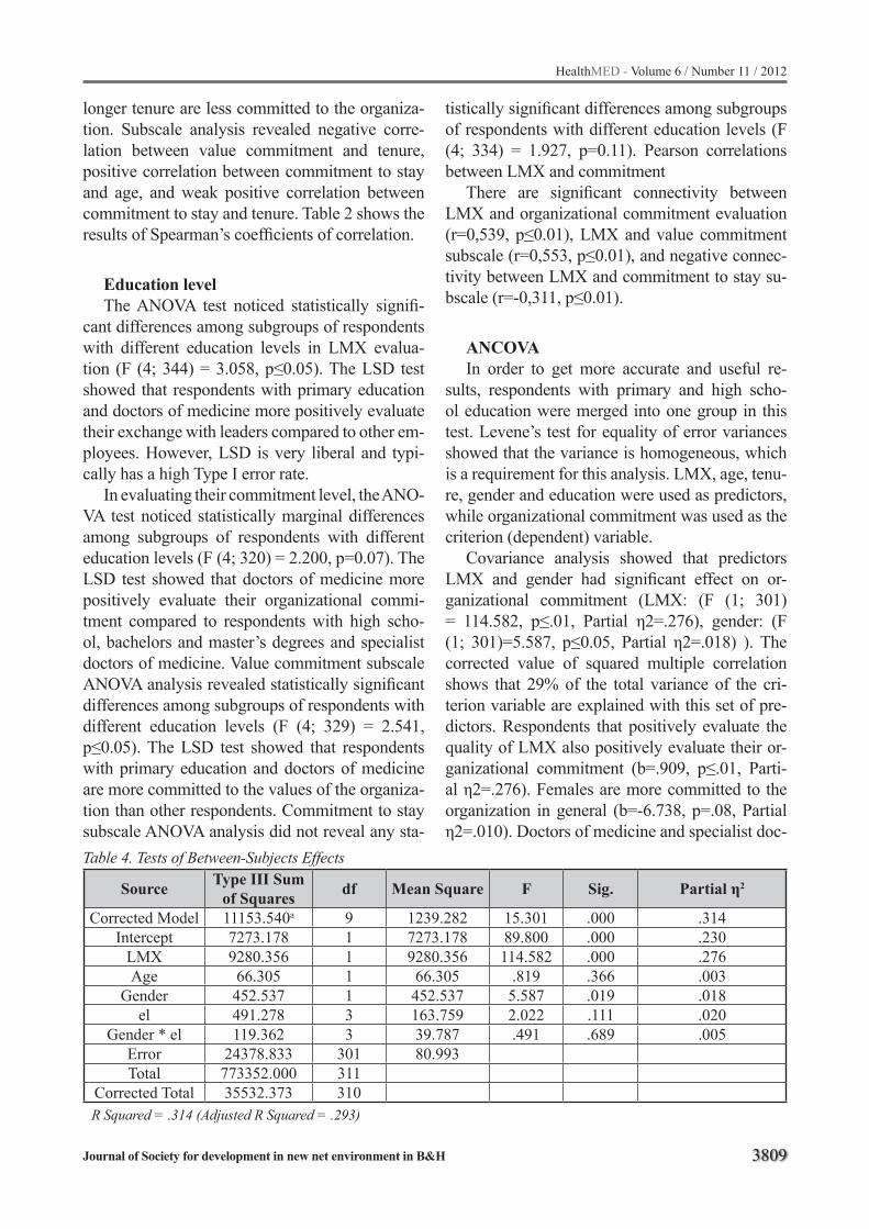
HealthMED - Volume 6 / Number 11 / 2012
Journal of Society for development in new net environment in B&H 3809
longer tenure are less committed to the organiza-tion. Subscale analysis revealed negative corre-lation between value commitment and tenure, positive correlation between commitment to stay and age, and weak positive correlation between commitment to stay and tenure. Table 2 shows the results of Spearman’s coefficients of correlation.
Education levelThe ANOVA test noticed statistically signifi-
cant differences among subgroups of respondents with different education levels in LMX evalua-tion (F (4; 344) = 3.058, p≤0.05). The LSD test showed that respondents with primary education and doctors of medicine more positively evaluate their exchange with leaders compared to other em-ployees. However, LSD is very liberal and typi-cally has a high Type I error rate.
In evaluating their commitment level, the ANO-VA test noticed statistically marginal differences among subgroups of respondents with different education levels (F (4; 320) = 2.200, p=0.07). The LSD test showed that doctors of medicine more positively evaluate their organizational commi-tment compared to respondents with high scho-ol, bachelors and master’s degrees and specialist doctors of medicine. Value commitment subscale ANOVA analysis revealed statistically significant differences among subgroups of respondents with different education levels (F (4; 329) = 2.541, p≤0.05). The LSD test showed that respondents with primary education and doctors of medicine are more committed to the values of the organiza-tion than other respondents. Commitment to stay subscale ANOVA analysis did not reveal any sta-
tistically significant differences among subgroups of respondents with different education levels (F (4; 334) = 1.927, p=0.11). Pearson correlations between LMX and commitment
There are significant connectivity between LMX and organizational commitment evaluation (r=0,539, p≤0.01), LMX and value commitment subscale (r=0,553, p≤0.01), and negative connec-tivity between LMX and commitment to stay su-bscale (r=-0,311, p≤0.01).
ANCOVAIn order to get more accurate and useful re-
sults, respondents with primary and high scho-ol education were merged into one group in this test. Levene’s test for equality of error variances showed that the variance is homogeneous, which is a requirement for this analysis. LMX, age, tenu-re, gender and education were used as predictors, while organizational commitment was used as the criterion (dependent) variable.
Covariance analysis showed that predictors LMX and gender had significant effect on or-ganizational commitment (LMX: (F (1; 301) = 114.582, p≤.01, Partial η2=.276), gender: (F (1; 301)=5.587, p≤0.05, Partial η2=.018) ). The corrected value of squared multiple correlation shows that 29% of the total variance of the cri-terion variable are explained with this set of pre-dictors. Respondents that positively evaluate the quality of LMX also positively evaluate their or-ganizational commitment (b=.909, p≤.01, Parti-al η2=.276). Females are more committed to the organization in general (b=-6.738, p=.08, Partial η2=.010). Doctors of medicine and specialist doc-
Table 4. Tests of Between-Subjects Effects
Source Type III Sum of Squares df Mean Square F Sig. Partial η2
Corrected Model 11153.540a 9 1239.282 15.301 .000 .314Intercept 7273.178 1 7273.178 89.800 .000 .230
LMX 9280.356 1 9280.356 114.582 .000 .276Age 66.305 1 66.305 .819 .366 .003
Gender 452.537 1 452.537 5.587 .019 .018el 491.278 3 163.759 2.022 .111 .020
Gender * el 119.362 3 39.787 .491 .689 .005Error 24378.833 301 80.993Total 773352.000 311
Corrected Total 35532.373 310R Squared = .314 (Adjusted R Squared = .293)
3810
HealthMED - Volume 6 / Number 11 / 2012
Journal of Society for development in new net environment in B&H
tors of medicine are also more committed to the organization compared to other respondents, alt-hough the effects of education do not have high significance.
Discussion
Some scholars already emphasized the impor-tant connections between LMX and organizational commitment (18, 19-22). However, most resear-ches do not reveal anything deeper than the simple notation that LMX correlates with organizational commitment. For example, Gerstner and Day (22) conclude that LMX is consistently correlated with commitment, but they don’t reveal anything signi-ficant about the nature of this relationship. Some of the researchers, who have previously conducted similar studies (19, 20) also noted that it is difficult to find research that incorporates LMX as an ante-cedent of commitment and that only a few studies deal with this issue. Joo (20) tried to go further and deeper in explaining the relationship betwe-en these two variables, however he measured only affective commitment and provided only the basic analysis and just discussed that LMX is indeed re-lated and has impact on commitment. Kang and his colleagues (19) made an interesting observati-on in favor of the necessity to conduct research on the impact of LMX on organizational commitment in various working environments that most of the researches incorporating LMX and commitment have been conducted in western developed eco-nomies. It also seems that in the majority of these researches LMX as an antecedent of organizati-onal commitment, analysis of their relationship and their importance are lost in the chaos between many other variables measured in these someti-mes over complicated studies. Another problem is that these studies often use different instruments for measuring both LMX and commitment, so the findings cannot be homogenous and completely comparable. In some researches, LMX is only used as a mediator variable (21). This study intended to contribute in solving at least two main issues no-ticed, the necessity to conduct researches outside western settings, and the unnecessary complication of studies incorporating LMX and commitment. It also had the intention to go further and deeper in explaining the relationships of these variables.
Discussion of the findingsH1, which proposes that employees have high
quality LMX with their leaders, was partially supported with descriptive statistics. H2, propo-sing that employees are committed to the organi-zation, also received partial support. The analyses showed that there is much empty space for impro-ving the employees LMX and commitment. Per-sonal characteristics have significant influence on both of the variables tested. Therefore, H3, sugge-sting that personal characteristics of employees influences their LMX and commitment level, re-ceived full support. Females are more committed to the organization and to its values. It is known that females are typically drawn to the healthcare sector because of their nurturing attitudes.
LMX was not affected by gender, so H3.1, proposing that differences in gender do not affect LMX and commitment level was supported only for LMX. Age, tenure and education analyses re-vealed very unexpected results. Regarding age differences and tenure, analysis showed that older respondents and those with longer tenure have lower LMX. Respondents with longer tenure are also less committed to the organization generally and to its values, while their commitment to stay increases with age and tenure. The presumption is that older and employees with longer tenure are more objective in evaluating their LMX and com-mitment. As time passes, they are more informed about the reality. The shock of facing the reality can have significant impact on people. The results can be perceived through the self-determination theory, which points on the autonomous behavi-or regulation style through the integration of life experience and personality disposition in a unique system of the self (47, 48).
They also realize that as they get older that finding another job will get harder, so the job se-curity granted to them as to most public service organization employees in Serbia leads to higher commitment to stay. Higher LMX and value com-mitment had respondents with primary education and doctors of medicine than others, while doctors of medicine are generally most committed to the organization. The ANCOVA test added specialist doctors of medicine to the list of generally most committed, but with small significance. High le-vels of LMX and commitment among employees
HealthMED - Volume 6 / Number 11 / 2012
Journal of Society for development in new net environment in B&H 3811
with only primary education was indeed a surpri-se. As age, tenure and education do not influence LMX and commitment in the expected way, H3.2 is not supported.
The most important analysis in this study is the correlation analysis between LMX and com-mitment. The tests confirmed the results of many earlier studies (18, 19-22) that LMX and com-mitment are significantly positively correlated. Further, analysis revealed that respondents with high quality LMX are more committed to the organization’s values (positive correlation) and have lower level of commitment to stay (negati-ve correlation). Employees with higher LMX are more likely to engage themselves in achieving the values of the organization, however they leave ea-sier from the organization, which reflects affective commitment towards the organization. The AN-COVA test also confirmed the connection between LMX and commitment and revealed that 29% of the total variance of organizational commitment is explained with predictors LMX and age. Thus, H4, proposing that LMX correlates commitment is supported with high certainty. H4.1, proposing that employees with high LMX have higher va-lue commitment, is also supported with great con-fidence.
Conclusions and implicationsDifferent economic and cultural conditions can
reveal different facts about the nature of impor-tant organizational variables, in this case about the exchange between leaders and members and organizational commitment. As mentioned befo-re, most of the studies conducted on both LMX and organizational commitment were in western environments. Thus, non-western environments had little influence on the theory and practice de-velopment. Therefore, sometimes the theory does not correspond to the characteristics of these cul-tural and business environments.
The main findings in this study revealed that LMX and organizational commitment have posi-tive connectivity in a non-western environment. It was also revealed that LMX and value commi-tment have much stronger relationship than the re-lationship between LMX and commitment to stay, which is a negative relationship. Critical employee
categories were also detected. Measures for im-proving their LMX and commitment are needed primarily for males, older employees, employees with longer tenure and employees with secondary education, bachelors and master’s degrees. The conclusion is that leaders and leadership have strong influence on the commitment of their em-ployees, so these findings could have significant implications in the process of leadership moder-nization in Serbian hospitals. Knowing how im-portant commitment can be among healthcare workers, greater attention needs to be given to le-adership in Serbian hospitals.
Recommendations for future researchFurther researches are needed in other he-
althcare facilities in Serbia and in the region and also in different private and public manufacturing and service organizations outside the healthca-re system. Data should be collected from many different business environments in the region to know more about the influence of LMX on orga-nizational commitment. According to Perry (39), moral and ethics have an important influence on commitment, and this is highly expressed among public sector employees, because they have strong ethics. It would be interesting to incorporate moral and ethics as moderator variables in further studi-es examining the influence of LMX on organizati-onal commitment.
References
1. Graen G, Cashman J. A role-making model of lea-dership in formal organizations: A developmental approach. In: Hunt JG, Larson LL, editors. Lea-dership Frontiers. Kent, OH: Kent State University Press; 1975. p. 143-66.
2. Graen G, Hoel W, Liden RC. Role of Leadership in the Employee Withdrawal Process. Journal of Appli-ed Psychology. 1982;67(6):868-72. PubMed PMID: 5111509.
3. Graen G, Novak MA, Sommerkamp P. The effects of leader—member exchange and job design on pro-ductivity and satisfaction: Testing a dual attachment model. Organizational Behavior and Human Per-formance. 1982;30(1):109-31. doi: 10.1016/0030-5073(82)90236-7.
3812
HealthMED - Volume 6 / Number 11 / 2012
Journal of Society for development in new net environment in B&H
4. Scandura TA, Graen GB. Moderating Effects of Ini-tial Leader-Member Exchange Status on the Effects of a Leadership. Journal of Applied Psychology. 1984;69(3):428-36. PubMed PMID: 6239343.
5. Graen G, Scandura TA, Graen MR. A Field Experi-mental Test of the Moderating Effects of Growth Need Strength on Productivity. Journal of Applied Psycho-logy. 1986;71(3):484-91. PubMed PMID: 5111651.
6. Zalesny MD, Graen G. Exchange theory in leadership research. In: Reber G, editor. Encyclopedia of Lea-dership. Linz: Linz University Press; 1986.
7. Graen G, Scandura TA. Toward a Psychology of Dya-dic Organizing. In: Staw BM, Cummings LL, editors. Research in Organizational Behavior. Greenwich, CT: JAI Press; 1987. p. 175-208.
8. Dansereau Jr F, Graen G, Haga WJ. A vertical dyad linkage approach to leadership within formal organi-zations: A longitudinal investigation of the role ma-king process. Organizational Behavior and Human Performance. 1975;13(1):46-78. doi: 10.1016/0030-5073(75)90005-7.
9. Graen G, Uhl-Bien M. The transformation of work group professionals into self-managing and parti-ally self-designing contributors: Toward a theory of leadership-making Journal of Management Systems. 1991;3(3):25-39.
10. Graen G, Uhl-Bien M. Relationship-based approach to leadership: Development of leader-member exchange (LMX) theory of leadership over 25 years: Applying a multi-level multi-domain perspective. The Leadership Quarterly. 1995;6(2):219-47. doi: 10.1016/1048-9843(95)90036-5.
11. Porter LW, Steers RM, Mowday RT, Boulian PV. Organizational commitment, job satisfaction, and turnover among psychiatric technicians. Journal of Applied Psychology. 1974;59(5):603-9. doi: 10.1037/h0037335.
12. Mowday RT, Steers RM, Porter LW. The measurement of organizational commitment. Journal of Vocational Behavior. 1979;14(2):224-47. doi: 10.1016/0001-8791(79)90072-1.
13. Angle HL, Perry JL. An Empirical Assessment of Organizational Commitment and Organizational Effectiveness. Administrative Science Quarterly. 1981;26(1):1-14. PubMed PMID: 4021472.
14. Meyer JP, Allen NJ. Testing the ‘Side-Bet Theory’ of Organizational Commitment: Some Methodologi-cal Considerations. Journal of Applied Psychology. 1984;69(3):372-8. PubMed PMID: 6239336.
15. Meyer JP, Allen NJ, Smith CA. Commitment to Or-ganizations and Occupations: Extension and Test of a Three-Component Conceptualization. Journal of Applied Psychology. 1993;78(4):538-51. PubMed PMID: 12359338.
16. Kanter RM. Commitment and Social Organizati-on: A Study of Commitment Mechanisms in Utopi-an Communities. American Sociological Review. 1968;33(4):499-517.
17. Buchanan B. Building Organizational Commitment: The Socialization of Managers in Work Organizations. Administrative Science Quarterly. 1974;19(4):533-46.
18. Yousaf A, Sanders K, Torka N, Ardts J. Having two bosses: considering the relationships between LMX, satisfaction with HR practices, and organizational commitment. International Journal of Human Re-source Management. 2011;22(15):3109-26. doi: 10.1080/09585192.2011.606124. PubMed PMID: WOS: 000299825700008.
19. Kang DS, Stewart J, Kim H. The effects of percei-ved external prestige, ethical organizational clima-te, and leader-member exchange (LMX) quality on employees’ commitments and their subsequent atti-tudes. Personnel Review. 2011;40(6):761-84. doi: 10.1108/00483481111169670. PubMed PMID: WOS: 000296599600006.
20. Joo BK. Organizational Commitment for Knowled-ge Workers: The Roles of Perceived Organizati-onal Learning Culture, Leader-Member Exchan-ge Quality, and Turnover Intention. Human Re-source Development Quarterly. 2010;21(1):69-85. doi: 10.1002/hrdq.20031. PubMed PMID: WOS:000286393900004.
21. Lee J. Effects of leadership and leader-member exchange on commitment. Leadership & Organi-zation Development Journal. 2005;26(7/8):655-72. PubMed PMID: 226915737.
22. Gerstner CR, Day DV. Meta-Analytic review of leader–member exchange theory: Correlates and construct issues. Journal of Applied Psycho-logy. 1997;82(6):827-44. doi: 10.1037/0021-9010.82.6.827.
23. Uhl-Bien M. Relational Leadership Theory: Explo-ring the social processes of leadership and orga-nizing. Leadership Quarterly. 2006;17(6):654-76. doi: 10.1016/j.leaqua.2006.10.007. PubMed PMID: WOS: 000243252700007.
HealthMED - Volume 6 / Number 11 / 2012
Journal of Society for development in new net environment in B&H 3813
24. Sias PM, Jablin FM. Differential Superior-Subordi-nate Relations, Perceptions of Fairness, and Cowor-ker Communication. Human Communication Resear-ch. 1995;22(1):5-38.
25. Dunegan KJ, Uhl-Bien M, Duchon D. LMX and su-bordinate performance: The moderating effects of task characteristics. Journal of Business and Psychology. 2002;17(2):275-85. doi: 10.1023/a:1019641700724. PubMed PMID: WOS: 000177936300008.
26. Johns G, Saks AM. Organizational behaviour: under-standing and managing life at work: Pearson Prentice Hall; 2005.
27. Schermerhorn JR, Hunt JG, Osborn RN, Uhl-Bien M. Organizational Behavior: John Wiley & Sons; 2010.
28. Bateman TS, Strasser S. A Longitudinal Analysis of the Antecedents of Organizational Commitment. The Academy of Management Journal. 1984;27(1):95-112.
29. Rusbult CE, Farrell D. A longitudinal test of the inves-tment model: The impact on job satisfaction, job com-mitment, and turnover of variations in rewards, costs, alternatives, and investments. Journal of Applied Psychology. 1983;68(3):429-38. doi: 10.1037/0021-9010.68.3.429.
30. Agnew CR, Van Lange PAM, Rusbult CE, Langston CA. Cognitive interdependence: Commitment and the mental representation of close relationships. Journal of Personality and Social Psychology. 1998;74(4):939-54. doi: 10.1037//0022-3514.74.4.939. PubMed PMID: WOS: 000073136200008.
31. Allen NJ, Meyer JP. The measurement and antece-dents of affective, continuance and normative com-mitment to the organization. Journal of Occupational Psychology. 1990;63:1-18.
32. Allen NJ, Meyer JP. Affective, continuance, and nor-mative commitment to the organization: An examinati-on of construct validity. Journal of Vocational Behavi-or. 1996;49(3):252-76. doi: 10.1006/jvbe.1996.0043. PubMed PMID: WOS:A1996VV02800003.
33. Meyer JP, Stanley DJ, Jackson TA, McInnis KJ, Maltin ER, Sheppard L. Affective, normative, and continuance commitment levels across cultures: A meta-analysis. Journal of Vocational Behavior. 2012;80(2):225-45. doi: 10.1016/j.jvb.2011.09.005.
34. Meyer JP, Allen NJ. A three-component conceptuali-zation of organizational commitment Human Resour-ce Management Review. 1991;1:61-89.
35. Meyer JP, Allen NJ. Commitment in the workplace: Theory, research, and application. Thousand Oaks, CA: SAGE; 1997.
36. Meyer JP, Stanley DJ, Herscovitch L, Topolnyt-sky L. Affective, continuance, and normative com-mitment to the organization: A meta-analysis of antecedents, correlates, and consequences. Jour-nal of Vocational Behavior. 2002;61(1):20-52. doi: 10.1006/jvbe.2001.1842. PubMed PMID: WOS:000176895600002.
37. Steers RM. Antecedents and Outcomes of Organizati-onal Commitment. Administrative Science Quarterly. 1977;22(1):46-56. PubMed PMID: 4021452.
38. Coe C, Zehnder A, Kinlaw DC. Coaching for Commi-tment: Achieving Superior Performance from Indivi-duals and Teams: John Wiley & Sons; 2008.
39. Perry JL. Antecedents of Public Service Motivation. Journal of Public Administration Research and The-ory. 1997;7(2):181-97.
40. Rhoades L, Eisenberger R. Perceived organizati-onal support: A review of the literature. Journal of Applied Psychology. 2002;87(4):698-714. doi: 10.1037//0021-9010.87.4.698. PubMed PMID: WOS: 000177332600008.
41. Meyer JP, Allen NJ, Topolnytsky L. Commitment in a changing world of work. Canadian Psycho-logy-Psychologie Canadienne. 1998;39(1-2):83-93. doi: 10.1037/h0086797. PubMed PMID: WOS: 000074410300009.
42. Perry JL, Brudney JL, Coursey D, Littlepage L. What drives morally committed citizens? A study of the antecedents of public service motivation. Public Administration Review. 2008;68(3):445-58. doi: 10.1111/j.1540-6210.2008.00881.x. PubMed PMID: WOS: 000254306800004.
43. Ostroff C. The Relationship Between Satisfacti-on, Attitudes, and Performance: An Organizatio-nal Level Analysis. Journal of Applied Psychology. 1992;77(6):963-74. PubMed PMID: 12428788.
44. Maslyn JM, Uhl-Bien M. Leader-Member Exchange and Its Dimension: Effects of Self-Effort and Other’s Effort on Relationship Quality. Journal of Applied Psychology. 2001;86(4):697-708. PubMed PMID: 12129154.
45. Greguras GJ, Ford JM. An examination of the multi-dimensionality of supervisor and subordinate percep-tions of leader-member exchange. Journal of Occupa-tional and Organizational Psychology. 2006;79:433-65. doi: 10.1348/096317905x53859. PubMed PMID: WOS:000240938000008.
3814
HealthMED - Volume 6 / Number 11 / 2012
Journal of Society for development in new net environment in B&H
46. Zhong JA, Lam W, Chen ZG. Relationship between leader-member exchange and organizational citi-zenship behaviors: Examining the moderating role of empowerment. Asia Pacific Journal of Management. 2011;28(3):609-26. doi: 10.1007/s10490-009-9163-2. PubMed PMID: WOS:000292971200009.
47. Deci EL, Ryan RM. Self-determination theory: A ma-crotheory of human motivation, development, and he-alth. Canadian Psychology/Psychologie canadienne. 2008;49(3):182-5. doi: 10.1037/a0012801.
48. Lam CF, Gurland ST. Self-determined work motivati-on predicts job outcomes, but what predicts self-deter-mined work motivation? 2008;42(4):1109-15.
Corresponding AuthorValentin Konja,Faculty of Technical Sciences, University of Novi Sad, Novi Sad,Serbia,E-mail: [email protected]
HealthMED - Volume 6 / Number 11 / 2012
Journal of Society for development in new net environment in B&H 3815
What makes nursing a profession: professionalization elementsAndrej Starc1, Majda Pahor1, Branko Ilic2
1 Faculty of Health Sciences, University of Ljubljana, Slovenia2 Faculty of Social Sciences, University of Ljubljana, Slovenia
Abstract
Background: Many endogenous and exoge-nous elements influence the process of nursing professionalization, forming the three core dimen-sions of professionalization: knowledge, power and ethics. Following theoretical starting-points, our leading thesis is that in the case of Slovenia nursing is developing gradually into profession. Deriving from cross-sectional survey which em-braced twenty health care organizations our goal is to explore key elements of nursing professiona-lization in Slovenia.
Methods: Survey data were analyzed using descriptive statistics, contingency analysis (chi-square test) and ordinary least squares multiva-riate linear regression with and without control variables (robustness test), both based on indexa-tion. For nursing professionals and members of professional management adjusted questionnaire was designed.
Results: Professional attributes in Slovenia are present in nursing professionals aged more than 51 years with more than 26 years of working experiences, employed on primary level of health care system, involved in ELP for at least 5 times per year and reading more professional nursing journals. They perceive that the acquisition of new specific knowledge means contribution for their HCO and raising their expert power as well.
The presence of LLL, professional autonomy and specific knowledge in nursing indicate rather clear and statistically significant positive impact, while ethics in nursing only marginal statistically significant positive impact on nursing professio-nalization.
Conclusions: Identification of professional elements in some segments of the nursing popula-tion in Slovenia allows further educational and po-licy planning as well as research into implications for nursing practice and quality of care in general.
Keywords: professionalization, nursing, nur-sing professional, health care.
Introduction
Professionalization can be defined as a strategy of occupational groups when trying to control the-ir own market position. In addition, professiona-lization should be understood as a special type of (collective) professional control in the market rather than just a list of characteristics that define immutable foundations of certain professions and professional. [1] Literature emphasizes key criti-cal dimensions of professionalization as knowled-ge, power and ethics. [2-3]
Considering these dimensions only few profe-ssional groups achieve a high level of professio-nalization, mainly because professionalized occu-pations take up resources and thus prevent access to them to other occupations. However, a lot of occupations failed on this path in order to enhance own power, prestige and influence. How far they succeed is essentially an empirical question. The literature mentions the concept of nursing as semi-profession, due to non-separation of basic scienti-fic knowledge and a greater tendency to emphasi-ze the skills, or inadequate theoretical knowledge and low power of self-regulation. [3-6]
Our research considered professions as a de-centralized form of social control and the action of professional groups members based on knowled-ge, power and ethical values . We focused on nu-rses professionals aiming to identify nursing pro-fessionalization elements in S lovenia. The basic thesis of our research is that in the case of Slovenia nursing is developing gradual l y into profession when achieving some significant professionaliza-tion attributes.
In HCO nursing professionals should be un-derstood as knowledge workers, characterized by combination of autonomy and responsibility for

3816
HealthMED - Volume 6 / Number 11 / 2012
Journal of Society for development in new net environment in B&H
quality and safe treatment of most complex pati-ents. [7] Nursing professionals in HCO represent the thinking human capital, since during educati-on and LLL their active knowledge increases and regularly recombines, thus making their exploi-tation and recombination of existing knowledge more effective. Note that following human capital theory short-run nursing education “costs” mean long-run investment outlays which increase the stock of human capital, thus raising average in-dividual working productivity and finally wages. When developing knowledge strategy we define nursing professionals as “bimodal learners”, i.e. simultaneous knowledge creators and knowledge exploiters respectively [8-19]
Results
As indicating in Table 1 there is relatively small proportional difference between active and sampled population educational structure of nur-sing employees.
The majority of respondents (44.9%, n = 284) are employed in the tertiary level of health care; the others belong to primary (30.6%, n = 194) and secondary health care level (22.9%, n = 145) .
Women prevail in the sample (93.7%, n = 546, N = 15,843); the prevalence of women is statisti-cally significant at all levels of health care (M = 1.06, SD = 0.244, p = 0.018, χ2 = 8.084), indica-ting strong nursing feminization.
The group aged from 41 to 50 years dominates significantly only at the primary health care level (n = 572, M = 2.37, SD = 0, 982, p = 0.000, χ2 = 37, 883), while at the secondary and tertiary level younger respondents prevail (from 31 to 40 years).
When taking into account working years, the group of more than 26 years of working experi-ences dominates significantly at the primary level
Table 1. Comparison between active population’s educational structure and the sample Education degree Active population* Sample
Postsecondary education N = 1329 25,12 % n = 127 22,20 %Diploma level education N = 3655 69,10 % n = 383 66,96 %
Degree level education, BSc N = 281 5,32 % n = 48 8,39 %Postgraduate education N = 25 0,47 % n = 14 2,45 %
Total N = 5290 100 % n = 572** 100 %Notes: *Data source: database of Nursing and Midwifery Chamber of Slovenia, 2nd January 2011. **61 questionnaires (of 633 delivered) were incomplete.
(n = 572, M = 3.66, SD = 1.845, p = 0.000, χ2 = 40.313). At all health care levels the distribution of respondents differs significantly with respect to working years: the group from 6 to 10 years pre-vails at the secondary, while the group from 0 to 5 of working years at the tertiary level. Concerning achieved professional degree the health care levels clearly differ, with diploma level education more present at the primary health care level. The cha-racteristics of working position/function in Slove-nian HCO is that team nurses dominate (43.5%, n = 250). The shares of others are smaller: ward nur-se, head of clinical/departmental nursing activities (head of one ward), head of clinical/departmental nursing activities (head of several wards) and head of all clinical/hospital activities (Head of nursing) (1.0%, n = 6) (n = 575, M = 1.87, SD = 0.859, p = 0.109, χ2 = 13.096). The distribution of nurse pro-fessions with respect to working position/function is rather different at all health care levels. Group of ward nurses prevails at primary and team nur-ses at tertiary level (see Table 2).
Participation in education and learning processParticipation in ELP from 1 to 2 times per year
is present at all levels of health care system (n = 575, M = 1.87, SD = 0.859, p = 0.074; χ2 = 11.513). More precisely, participation in ELP is varying from 3 to 4 times at the primary and from 1 to 2 times at the secondary as well as tertiary level of health care system (see Table 2).
Active partitipation in education and learning processThe active participation in ELP is significantly
most frequent from 1 - to 2-times at all levels of health care system (n = 580, M = 1.52, SD =
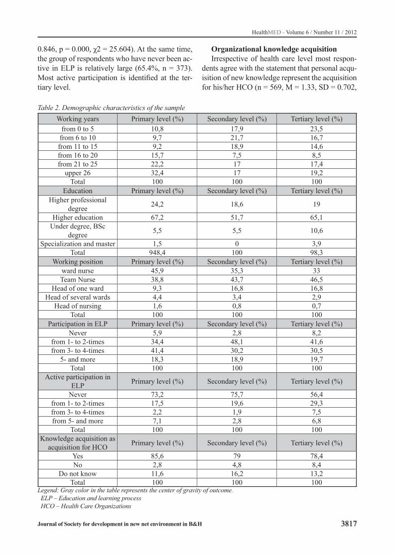
HealthMED - Volume 6 / Number 11 / 2012
Journal of Society for development in new net environment in B&H 3817
Table 2. Demographic characteristics of the sampleWorking years Primary level (%) Secondary level (%) Tertiary level (%)
from 0 to 5 10,8 17,9 23,5from 6 to 10 9,7 21,7 16,7from 11 to 15 9,2 18,9 14,6from 16 to 20 15,7 7,5 8,5from 21 to 25 22,2 17 17,4
upper 26 32,4 17 19,2Total 100 100 100
Education Primary level (%) Secondary level (%) Tertiary level (%)Higher professional
degree 24,2 18,6 19
Higher education 67,2 51,7 65,1Under degree, BSc
degree 5,5 5,5 10,6
Specialization and master 1,5 0 3,9Total 948,4 100 98,3
Working position Primary level (%) Secondary level (%) Tertiary level (%)ward nurse 45,9 35,3 33Team Nurse 38,8 43,7 46,5
Head of one ward 9,3 16,8 16,8Head of several wards 4,4 3,4 2,9
Head of nursing 1,6 0,8 0,7Total 100 100 100
Participation in ELP Primary level (%) Secondary level (%) Tertiary level (%)Never 5,9 2,8 8,2
from 1- to 2-times 34,4 48,1 41,6from 3- to 4-times 41,4 30,2 30,5
5- and more 18,3 18,9 19,7Total 100 100 100
Active participation in ELP Primary level (%) Secondary level (%) Tertiary level (%)
Never 73,2 75,7 56,4from 1- to 2-times 17,5 19,6 29,3from 3- to 4-times 2,2 1,9 7,5from 5- and more 7,1 2,8 6,8
Total 100 100 100Knowledge acquisition as
acquisition for HCO Primary level (%) Secondary level (%) Tertiary level (%)
Yes 85,6 79 78,4No 2,8 4,8 8,4
Do not know 11,6 16,2 13,2Total 100 100 100
Legend: Gray color in the table represents the center of gravity of outcome. ELP – Education and learning process HCO – Health Care Organizations
0.846, p = 0.000, χ2 = 25.604). At the same time, the group of respondents who have never been ac-tive in ELP is relatively large (65.4%, n = 373). Most active participation is identified at the ter-tiary level.
Organizational knowledge acquisitionIrrespective of health care level most respon-
dents agree with the statement that personal acqu-isition of new knowledge represent the acquisition for his/her HCO (n = 569, M = 1.33, SD = 0.702,
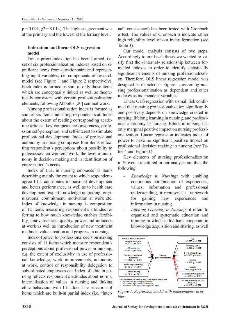
3818
HealthMED - Volume 6 / Number 11 / 2012
Journal of Society for development in new net environment in B&H
p = 0.091, χ2 = 8.014). The highest agreement was at the primary and the lowest at the tertiary level.
Indexation and linear OLS regression model First a-priori indexation has been formed, i.e.
set of six professionalization indexes based on si-gnificant items from questionnaire and represen-ting input variables, i.e. components of research model (see Figure 1 and Figure 2 respectively). Each index is formed as sum of only those items which are conceptually linked as well as theore-tically consistent with certain professionalization elements, following Abbott’s [20] seminal work.
Nursing professionalization index is formed as sum of six items indicating respondent’s attitudes about the extent of reading corresponding acade-mic articles, key competencies awareness, profe-ssion self-perception, and self-interest to stimulate professional development. Index of professional autonomy in nursing comprises four items reflec-ting respondent’s perceptions about possibility to judge/assess co-workers’ work, the level of auto-nomy in decision making and in identification of entire patient’s needs.
Index of LLL in nursing embraces 13 items describing mainly the extent to which respondents agree LLL contributes to personal development and better performance, as well as to health care development, expert knowledge upgrading, orga-nisational commitment, motivation at work etc. Index of knowledge in nursing is composition of 12 items, measuring respondent’s attitudes re-ferring to how much knowledge enables flexibi-lity, innovativeness, quality, power and influence at work as well as introduction of new treatment methods, value creation and progress in nursing.
Index of power for professional decision making consists of 11 items which measure respondent’s perceptions about professional power in nursing, e.g. the extent of exclusivity in use of professio-nal knowledge, work improvements, autonomy at work, control or responsibility delegation to subordinated employees etc. Index of ethic in nu-rsing reflects respondent’s attitudes about norms, internalisation of values in nursing and linking ethic behaviour with LLL too. The selection of items which are built-in partial index (i.e. “inter-
nal” consistency) has been tested with Cronbach α test. The values of Cronbach α indicate rather high reliability level of our index formation (see Table 3).
Our model analysis consists of two steps. Accordingly to our basic thesis we wanted to ve-rify first the »internal« relationship between for-matted indexes in order to identify statistically significant elements of nursing professionalizati-on. Therefore, OLS linear regression model was designed as depicted in Figure 1, assuming nur-sing professionalization as dependent and other indexes as independent variables.
Linear OLS regression with a small risk confir-med that nursing professionalization significantly and positively depends on knowledge created in nursing, lifelong learning in nursing, and professi-onal autonomy in nursing. Ethics in nursing has only marginal positive impact on nursing professi-onalization. Linear regression indicates index of power to have no significant positive impact on professional decision making in nursing (see Ta-ble 4 and Figure 1).
Key elements of nursing professionalization in Slovenia identified in our analysis are thus the following:
- Knowledge in Nursing: with enabling continuous combination of experiences, values, information and professional understanding, it represents a framework for gaining new experiences and information in nursing.
- Lifelong Learning in Nursing: it refers to organized and systematic education and training in which individuals cooperate in knowledge acquisition and sharing, as well
Figure 1. Regression model with independent varia-bles
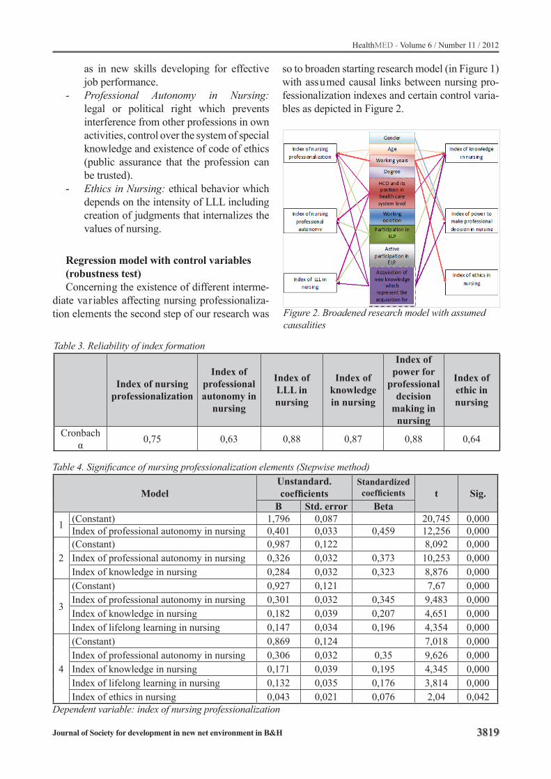
HealthMED - Volume 6 / Number 11 / 2012
Journal of Society for development in new net environment in B&H 3819
Table 4. Significance of nursing professionalization elements (Stepwise method)
ModelUnstandard.coefficients
Standardized coefficients t Sig.
B Std. error Beta
1 (Constant) 1,796 0,087 20,745 0,000Index of professional autonomy in nursing 0,401 0,033 0,459 12,256 0,000
2(Constant) 0,987 0,122 8,092 0,000Index of professional autonomy in nursing 0,326 0,032 0,373 10,253 0,000Index of knowledge in nursing 0,284 0,032 0,323 8,876 0,000
3
(Constant) 0,927 0,121 7,67 0,000Index of professional autonomy in nursing 0,301 0,032 0,345 9,483 0,000Index of knowledge in nursing 0,182 0,039 0,207 4,651 0,000Index of lifelong learning in nursing 0,147 0,034 0,196 4,354 0,000
4
(Constant) 0,869 0,124 7,018 0,000Index of professional autonomy in nursing 0,306 0,032 0,35 9,626 0,000Index of knowledge in nursing 0,171 0,039 0,195 4,345 0,000Index of lifelong learning in nursing 0,132 0,035 0,176 3,814 0,000Index of ethics in nursing 0,043 0,021 0,076 2,04 0,042
Dependent variable: index of nursing professionalization
Table 3. Reliability of index formation
Index of nursing professionalization
Index of professional autonomy in
nursing
Index of LLL in nursing
Index of knowledge in nursing
Index of power for
professional decision
making in nursing
Index of ethic in nursing
Cronbach α 0,75 0,63 0,88 0,87 0,88 0,64
as in new skills developing for effective job performance.
- Professional Autonomy in Nursing: legal or political right which prevents interference from other professions in own activities, control over the system of special knowledge and existence of code of ethics (public assurance that the profession can be trusted).
- Ethics in Nursing: ethical behavior which depends on the intensity of LLL including creation of judgments that internalizes the values of nursing.
Regression model with control variables (robustness test)Concerning the existence of different interme-
diate variables affecting nursing professionaliza-tion elements the second step of our research was
so to broaden starting research model (in Figure 1) with assumed causal links between nursing pro-fessionalization indexes and certain control varia-bles as depicted in Figure 2.
Figure 2. Broadened research model with assumed causalities

3820
HealthMED - Volume 6 / Number 11 / 2012
Journal of Society for development in new net environment in B&H
Table 5. Nursing professionalization elements in Slovenia (left) and the impact of control variables (right)Nursing professionalization elements
(independent variables)Control variables
(independent variables)Power to take nursing professional decision
t = 0,291; p = 0,988Age of respondents t = 3,233; p = 0,001
Lifelong learning in nursing t = 3,814; p = 0,000
Working years t = 3,214; p = 0,001
Professional autonomy in nursing t = 9,626; p = 0,000
Level in the system of health care t = -2,023; p = 0,044
Knowledge in nursing t = 4,345; p = 0,000
Gendert = 0,198; p = 0,843
Ethics in nursing t = 2,040; p = 0,042
Degreet = –0,71; p = 0,478Working position
t = –0,228; p = 0,820Investing in employees LLL from HCO
t = 1,057; p = 0,291
In order to verify the robustness of research model in multivariate linear regression we intro-duce control variables: gender, age, working ye-ars, degree, work position/function in health care organization, level of health care system and in-vestments of top management in employees (Ta-ble 5). Regression analysis indicates that only two control variables, i.e. age and working years have a significant positive impact on the level of health care and marginal significant impact on nursing professionalization. The analysis does not demon-strate significant positive impact of other varia-bles. There is significant positive and marginal im-pact of some control variables (age, working years and level of health care) on the index of nursing professionalization.
Age influences positively on participation in education and learning process (most in group between 41 and 50 years), reading professional nursing journals (most in group above 50 years), work performance self-assessment and field pro-motion (most in group from 51 and more years). Working experiences are in strong connection with the participation in ELP, and reading professional nursing journals too - the greater the former the stronger the latter. Therefore we can conclude that nursing professionalization seems to be significant more present in slightly older rather than younger nursing professionals. Note that more detailed in-terpretation of causalities between nursing profe-ssionalization and control variables above needs to be abstracted here due to exceeding the key
purpose of our study. However, this issues deserve anyway further theoretical and empirical research.
Discussion and conclusion
The key finding of our research is that primary level of health care gives obviously more potential and motivation for nurses to achieve higher level of professionalization.
Based on quantitative analysis we identified fundamental characteristics of nursing professio-nalization, i.e. knowledge, power and ethics go-ing hand in hand with our basic thesis. Further, we found that nursing professionalization can be understood as human capital upgrading. Most of respondents agreed that nurses’ knowledge acqu-isition represents an improvement also for HCO. Simultaneously, the majority of the respondents favors nursing professionalization, professional autonomy and LLL in nursing.
These findings are in concordance with litera-ture [1, 4, 20] so we might conclude that nurses in Slovenia are undergoing a similar process as has been reported in other countries. However, hie-rarchical structure of Slovenian health care and the central role of medical doctors is not entirely supportive to increased knowledge and power of the nursing workforce and their more autonomous role in relation to health care users. This tension might be resolved in the future with more equal collaboration between nurses and doctors, but also bring about possibilities of conflicts.

HealthMED - Volume 6 / Number 11 / 2012
Journal of Society for development in new net environment in B&H 3821
List of abbreviations:
ELP – education and learning processHCO – health care organizationLLL – lifelong learningOLS – ordinary least squares
References
1. Hampton D, Hampton G. Professionalism and the nu-rse-midwife practitioner: an exploratory study. J Am Acad Nurse Pract, 2000; 12(6): 218–25.
2. Turner B. Medical Power and Social Knowledge. 2nd Edition. London, Thousand Oaks, New Delhi: SAGE Publications; 1995.
3. Coburn D, in Willis E. The Medical Profession: Knowledge, Power, and Autonomy. In: Albrecht G, Fitzpatrick R, Scrimshaw S, eds. The Handbook of So-cial Studies in Health & Medicine. London, Thousand Oaks, New Delhi: SAGE, 2003; 377–393.
4. Hugman, R. Power in caring professions. Hound-mills, Basingstoke. London: Macmillan; 1991.
5. Giddey M. Institutions, individuals and professional power. In: Moon G, Gillespie R, eds. Society and Health. An Introduction to social science for health professionals. London, New York: Routledge; 1995, 127–40.
6. Gabe J, Bury M, Elston M. Key Concepts in Medical Sociology. London, Thousand Oaks, New Delhi: Sage Publications; 2004.
7. Starc A, Kos Grabnar E, Požun P. Predstavitev pod-lag specializacije v zdravstveni negi Slovenije. In: Dvoršak Majcen J, et al., eds. 7. Konres Zdravstvene in babiške nege Slovenije. Ljubljana: Zbornica zdrav-stvene in babiške nege Slovenije; 2009, 1–8.
8. Schultz, T. W. Resources of Higher Education: An Economist’s View. The Journal of Political Economy, Vol. 76, No. 3, May/June 1968: 327-347.
9. Simon H. Bounded rationality and organizational le-arning. Organ Sci 1991; 2(1): 125–34.
10. Civi E. Knowledge management as a competitive asset: A review. J Mark Int Plan 2000; 18(4): 166–174.
11. OECD. Labour market survey 1999. Paris: OECD; 2000a.
12. Bierly P. E., Daly P. Aligning Human Resource Management Prctices and Knowledge Strategies. In: Choo C. W., Bontis N., eds. The Strategic Ma-nagement of Intellectual Cpital and Organizational Knowledge Oxford: Oxford University Press, 2002: 277-295.
13. Wong K, Aspinwall E. Characterizing knowledge management in the small business environment. J Know Management 2004a; 8(3): 44-61.
14. Sitar AS. Oblike in razsežnosti znanja v organizaci-ji. In: Možina S, Kovač J, eds. Menedžment znanja: znanje kot temelj razvoja: na poti k učečemu se po-djetju. Maribor: Založba Pivec; 2006, 57-69.
15. Starc A, Ilič B. Pridobivanje in razvoj znanstvene-ga in strokovnega človeškega kapitala v zdravstveni negi: študija primera. Obzor Zdr N; 41 2007 (2/3): 61–9.
16. Starc A. Nursing Professionalism in Slovenia: Knowledge, Power, and Ethics. Nurs Sci Quart 2009; 22(4): 371–74.
17. Starc A. Identifikacija elementov profesionalizacije zdravstvena nege v Sloveniji [dissertation]. [Lju-bljana]: Univerza v Ljubljani; 2011.
18. Starc A, Ilič B. Human capital within health organi-zations. In Antončič B, eds. Conference proceedings. Koper: Edukator; 2010, 8.
19. Starc A. Izobraževanje za profesionalizacijo zdrav-stvene nege. In Bobnar A, eds. Kompetence medicin-skih sester v povezavi s profesionalizacijo: zbornik predavanj. Ljubljana: Zdravstvena fakulteta; 2010, 47-59.
20. Abbott A. D. The System of Professions: An Essay on the Division of Expert Labour. Chicago: University of Chicago Press; 1988
Corresponding AuthorAndrej Starc,University of LjubljanaFaculty of Health SciencesLjubljana, Slovenija,E-mail: [email protected]
3822
HealthMED - Volume 6 / Number 11 / 2012
Journal of Society for development in new net environment in B&H
Physical activity in ADHD children treatmentDanijela Zivkovic1, Nenad Zivanovic1, Miroslava Zivkovic2, Olga Milojkovic3, Marija Djordjevic1 1 University of Nis, Faculty of Sport and Physical Education, Serbia2 Department of Neurology, University Clinical Center Nis, Serbia3 Clinic of Mental Health, University Clinical Center Nis, Serbia
Abstract
Attention-deficit/hyperactivity disorder (ADHD) is a chronic condition that affects pre- or very young schoolchildren. It often persists into ado-lescence and adulthood - with some changes in symptoms. Basic ADHD symptoms include diffi-culty sustaining attention, impulsive behavior and hyperactivity. Our research problem was to deter-mine if physical exercising will exert any influen-ce on children’s psychological performance, more exactly if physical exercising can eliminate nega-tive effects of ADHD affecting children’s behavi-or and thus enhance their health. Research subject is psychological indicators of behavior (ten indi-cators according to Iowa Conners Rating Scale) of ADHD children observed prior, during and after the physical exercising treatment. Bearing in mind that up to now ADHD treatment ensued medicati-ons with numerous toxic side effects our research aim was to determine ways physical exercising can help young school children with ADHD so as to reduce or eliminate use of medications.
Keywords: ADHD, inattention, hyperactivity, physical activity
Introduction
Attention-deficit/hyperactivity disorder re-ferred to as ADHD is a chronic condition that affects pre- or very young schoolchildren. It of-ten persists into adolescence and adulthood - with some changes in symptoms. Basic ADHD symp-toms include difficulty sustaining attention, im-pulsive behavior and hyperactivity [3]. According to the research conducted by Cordest, ADHD prevails in 3 to 5% of preschoolers. ADHD is 4 to 9 times more often to diagnose in boys than in girls. The very start of ADHD dates back in early childhood and cannot be recognized before school attending. Also ADHD encompasses intellectual functions. When administered intelligence tests
ADHD children scored 7 to 15 points less sco-res compared to the control group, children wit-hout ADHD [5]. ADHD affects children’s deve-lopment manifolds and depending on children’s age can manifest itself in poor performance in school, troubled social relationships, struggle with low self-esteem, increased risk of injuries, alco-hol, smoking and drug abuse and other delinquent behavior in jobs or elsewhere. To diagnose ADHD on time is of paramount importance so as to start treatment as soon as possible and eliminate nega-tive effects of non-treated syndrome [8]. Althou-gh etymology and pathogenesis of ADHD is still obscure there is an extensive body of research to acclaim its neuro-biological and genetic causes. Patophysiological research and brain function re-cording point to dysfunction of the front-sub cor-tical pathways and the disorder of dopaminergic and noradrenergic system [4].
The aim of our research is to determine impact of physical activity to behavioral and psychologi-cal disorder among children with ADHD. Basic assumption of our research is that physical exer-cising is the best antistress program and fully de-vised set of exercises can significantly affect the final outcome of this state. One should treat such a serious disorder interdisciplinary and teamwi-se. Therapeutical team should encompass child psychologist, defectologist, kinezitherapist, nur-sery teachers and family.
Methods
SubjectsResearch was conducted on a sample of 26
schoolchildren (19 boys and 7 girls), aged 7 to 10 (мean age is 9.04) with confirmed ADHD after the examination of the neurologist and child psycho-logist (DSM IV-TR classification for ADHD dia-gnosis).
HealthMED - Volume 6 / Number 11 / 2012
Journal of Society for development in new net environment in B&H 3823
Precondition for group inclusion:- ADHD lasting at least for two years.Precondition for group exclusion:- verified organic substrate of syndrome
(verified tumor or brain stroke or posttraumatic or inflammatory encephalopatia),
- chronic organic psycho syndrome or oligofrenia, and
- cardiological insufficiency contradicting physical exercising.
Control group was composed of ten ADHD di-agnosed children non treated with physical exer-cise.
MeasurementsADHD was monitored by the Behavior Scale
in the form of the set questionnaire - Iowa Conners Rating Scale. Each indicator was marked by gra-des 0 to 3. Grade 0 meant total absence of the va-lue for the observed indicator while the highest grade 3 meant the biggest presence of the obser-ved indicator. Testing was performed prior to the exercising and at the end of each month in three months interval and the result was shown in scores of points. Rating scale at the beginning and in the end of exercising was done by child neuropsycho-logist or a psychologist.
Experimental and control treatmentsThe research was conducted in the Clinic of
Mental Health in Nis, in the period from March till May, 2010. After being diagnoses for ADHD by child neuropsychologist or a psychologist children started exercising and were regularly assessed for their results. Exercises were administered by ki-nezitherapists and graduates from the Faculty of sport and physical education, three times a week in duration of 30 minutes in three months interval. Evaluation scale was after each month filled in by the neuropsychiatrists or psychologists.
Two types of exercises were used, warm up and attention training exercises (orders obeying). Exercises were not implemented strictly in one place but according to the availability of faciliti-es, gyms and parks. Open spaces were also used (slides, swimming pools). Exercises were care-fully organized in their structures but some de-gree of freedom for corrections, improvisations
(following the children’s needs and their wishes) was granted. It was envisaged to perform a set of exercises as an introduction always in the same or-der (exercises of shaping) [9,10].
Statistical analysisEach parameter was evaluated on the ordinal
scale and there were three measurements. To eva-luate statistical significance of the change in each parameter Friedman non-parameter test for repe-ated measures was applied. Statistical data proce-ssing was performed by software package SSPS 13. Respecting the law of personal data protection of the patients subjects identity was hidden and all generalia were coded and displayed with numeric symbols. The subjects identity is revealed only to their allotted doctors.
Results
Results of the experimental group are shown in Table 1. Significant decrease in all of the mea-sured indicators in experimental group of children was noticed. Mean values of the measured indica-tors for three measurements in three months inter-val have shown statistically significant differences before and after the physical exercising treatment. It was found better cooperation, lower level of ar-guing, and excitement and physical restlessness. Children showed more focus and attention, less defiance and better performance of given tasks. Mean values of the measured indicators for three measurements in three months interval in control group have shown no statistically significant diffe-rences.
Discussion
Upon consulting available literature it is evi-dent that therapy strategy for this serious disorder was based mainly on the use of medications, but apart from sedative effect (mirroring the overall psychomotor activity of children). These medica-tions showed significant toxic side effects. Strict dosage of medications had influenced the quality of life of ADHD children [2]. Clinical and epide-miological studies show that ADHD children are more prone to psychiatric disorders and abuse of substances [2].

3824
HealthMED - Volume 6 / Number 11 / 2012
Journal of Society for development in new net environment in B&H
Table 1. Mean values of measured indicatiors for three measurements (experimental group)Parameter 1. measurement 2. measurement 3. measurement PFidgeting 2.52 2.12 1.37 <0.001Mumble 2.33 2.00 1.67 <0.001Excitable 2.83 1.79 1.38 <0.001Inattentive 2.65 1.98 1.37 <0.001Fail to attend to task 2.87 1.87 1.27 <0.001Ill-tempered 2.35 1.88 1.77 <0.001Showing off 2.58 1.79 1.63 <0.001Quick-tempered 2.87 1.87 1.27 <0.001Rebellious 2.73 1.88 1.38 <0.001 Does not cooperate 2.75 1.92 1.33 <0.001
Usual symptoms of ADHD children and ado-lescents are anxiousness, change of mood, ticks, disorder of coordination, bad learning practices, ill socializing and communicating[2]. Some stu-dies show that ADHD increases the risk of per-sonality disorders and if not treated it can com-plement with other disorders such as poor school functions, family conflicts, self injuries, antisocial behavior, accident inclinations. Epidemiological studies show that ADHD prolongs into adulthood as well [8]. Results of our research have shown that physical activity represents an important part of ADHD children treatment which was also men-tioned in the study of Barkley R.A. [1] which in-sisted on the use of physical exercising in ADHD children treatment.
Also our results are congruent with the study conducted by Knight L.A. et al. [6] where he insi-sted on behavioral school program as the primary treatment irrespectable of pharmacotherapy. Our research shows statistically significant decrease for each measured indicator (Table 1.). Control group of children did not show any improvement (Table 2.). It is necessary to note a decrease in in-dicators: “excitable”, “fail to attend to task”, “qu-ick-tempered”, explosive and unpredictable beha-vior”, and “non-cooperative”.
Physical activity cheers up, boosts remembe-ring skills, lowers anxiety, regulates dreaming and weight. Petrochemical basis of such a favorable influence of exercising is that it improves in chil-dren circulation levels of dopamine and serotonin, regulates stress hormones and antioxidance pro-duction [4]. Subjective estimation of parents was in accordance with the aptitude tests of children. All parents noticed better attention spans, lower
explosiveness and unpredictable behavior, better cooperation, and more willing task attendance. In conversation with school psychologist it was men-tioned that these children showed quiet sleeping and better appetite. On parents insisting these chil-dren continued exercising individually even after the elapse of the three months interval.
Conclusions
Physical exercising is the best antistress pro-gram and represents an important part of ADHD children treatment. Our results corroborate the ba-sic assumption that early diagnosing of the symp-toms and adequate multidisciplinary treatments with special emphasis on the physical exercising can immensely decrease harmful effects of ADHD on children and its environment.
Therapeutical team should encopass child psychologist, defectologist, kinesitherapist, nur-sery teachers and family.
Acknowledgement
Thanks are given to the Department of theo-retical-methodological sciences at the Faculty of Sport and Physical Education, University of Niš, for the help in carrying out of this research.
References
1. Barkley R.A. Adolescents With Attention Deficit/Hyperactivity Disorder: An Overview Of Empirically Based Tretmants, J.Psychiatr Prac., 2004; 10(1):39-56.
HealthMED - Volume 6 / Number 11 / 2012
Journal of Society for development in new net environment in B&H 3825
2. Biederman J. Attention-Deficit/Hyperactivity Dis-order; A Selective Overview. Biol Psychiatry. 2005; 57:1215-1220.
3. Biederman J., Faraone S. V. Attention Deficit Hype-ractivity Disorder. Lancet. 2005; 366:237-248.
4. Chamberlain S.R., Robbins T.W., Sahakian B.J. The Neurology Of Attention Deficit/Hyperactivity Disor-der. Biol Psychiatry, 2007; 61:1317-1319.
5. Cordest M., McLaughlin T.F. Attention Deficit Hype-ractivity Disorder And Rating Scales With A Brief Review Of The Connors Teacher Rating Scale. Inter-national Journal of Special Education. 2004; 19(2): 23-34.
6. Knight L.A., Rooney M., Chronis-Tuscano A. Psycho-social Treatments For Attention-Deficit/Hyperactivity Disorder. Curr Psychiatry Rep. 2008;10 (5):412-8.
7. Spencer T., Biederman J., Wilens T. Attention Deficit Hyperactivity Disorder And Comorbidity, PediatrClin North Am, 1999; 46:915-27.
8. Živanović N., Andrašić S., Ćirić M., Ćosić V. Analysis of the Encumbrence and Recouparation Utilyzing the Cooper Test on the Basis of Heart Rate. Fizička kultu-ra, Skopje, 2010; 37(1):351-354.
9. Živanović N., Marković S., Ćosić V., Palić R., Ćirić M., Andrašić S., Popović M. Effect of Acute Exercise on Xanthine Oxidase Activity. In: Sport, Stress, Adap-tation - part II. Sport & Nauka, supplement, National Sports Academy Sofia. 2010; 438-440.
Corresponding AuthorZivkovic Danijela,University of Nis,Faculty of Sport and Physical Education,Nis, Serbia,E-mail: [email protected]

3826
HealthMED - Volume 6 / Number 11 / 2012
Journal of Society for development in new net environment in B&H
Application of assistive technology in rehabilitation of persons with cognitive disabilitiesMarina Radic Sestic, Biljana Milanovic Dobrota, Vesna Radovanovic, Jasmina Karic
Faculty of Special Education and Rehabilitation, University of Belgrade, Serbia
Abstract
The main objective of this paper is to review the available literature to determine the extent to which assistive technology can help people with cognitive disabilities to independently participate in everyday activities at home, school, workplace, the wider community. Treatment of persons with cognitive disabilities requires time and patience of people who provide them with necessary assi-stance and support. However, the interpersonal dynamics of support can create problems for fa-mily members and professionals and as well for the person. Numerous studies suggest that use of modern technology has the potential to reduce the tension between the caregiver and the cared per-son and to increase the independence, confidence and reduce the costs of efficient care. Due to assi-stive technology the functioning of persons with cognitive impairments can be improved in several ways, from providing signals, sequential instruc-tions and reminders for tasks and activities to the development of interventions that restructure the requirements of tasks so that individuals can use the remaining abilities instead of those that are da-maged.
Keywords: assistive technology, cognitive di-sability
Introduction
Cognitive disabilities (cognitive disabilities, CD) refers to a wide spectrum of disorders cau-sed by different factors and conditions in which disorders occur. There are many aspects of cogni-tive disability that should be considered before it is diagnosed and thus attempts to formulate a ge-neral definition of cognitive disability did not give the result. Disorders that belong to this category are traumatic brain injury (1, 2), cerebrovascular
accident (3, 4), learning disabilities (5), multiple sclerosis (6), intellectual and multiple disability (7, 8), autism (9) and somewhat dementia (10, 11). Problems that occur in people with cognitive disa-bilities are reflected in: Significant limitations in learning and information processing. Individuals are limited to the retention of knowledge, learning skills, decision making and communication with others; Problems in behavior are the result of li-mited interpersonal, social and emotional functio-ning; Difficulties in development limit the ability to adapt to daily life activities in areas such as self-care, independence at home, school, work, enter-tainment and the wider community; Damages and limitations are persistent and long-term, and they are not exclusively related to IQ.
Due to its complexity, the concept of cognitive disabilities can be classified as clinical disability whose diagnosis can be useful for treatment, and as functional disability, which is useful for deve-loping rehabilitation measures aimed at individual independence to community life. Regardless of the classification, treatment of persons with cognitive disabilities requires time, patience and financial resources (12, 13). The care of these people le-ads closest family members, relatives and caregi-vers. Activities that are commonly performed are related to daily living routines such as dressing, mobility, personal hygiene, food preparation, ta-king medical therapy (14). But the dynamics of interpersonal assistance may create problems for both family members and caregivers (15) and for the person needing care and daily support (16). Today there is a considerable number of studies that support the use of assistive technology whose application has the potential to reduce the tension between the caregiver and the cared person (17), to increase independence, confidence and cost effec-tive care (18) and which can improve the quality
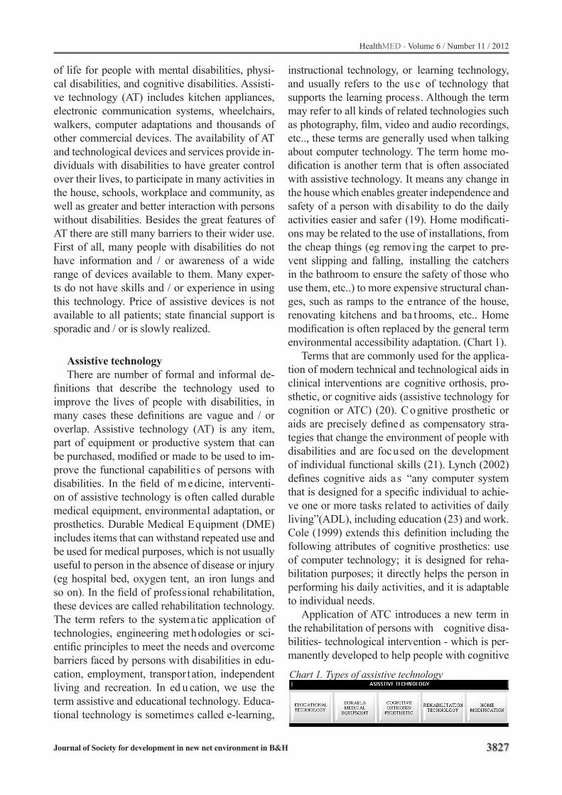
HealthMED - Volume 6 / Number 11 / 2012
Journal of Society for development in new net environment in B&H 3827
of life for people with mental disabilities, physi-cal disabilities, and cognitive disabilities. Assisti-ve technology (AT) includes kitchen appliances, electronic communication systems, wheelchairs, walkers, computer adaptations and thousands of other commercial devices. The availability of AT and technological devices and services provide in-dividuals with disabilities to have greater control over their lives, to participate in many activities in the house, schools, workplace and community, as well as greater and better interaction with persons without disabilities. Besides the great features of AT there are still many barriers to their wider use. First of all, many people with disabilities do not have information and / or awareness of a wide range of devices available to them. Many exper-ts do not have skills and / or experience in using this technology. Price of assistive devices is not available to all patients; state financial support is sporadic and / or is slowly realized.
Assistive technology There are number of formal and informal de-
finitions that describe the technology used to improve the lives of people with disabilities, in many cases these definitions are vague and / or overlap. Assistive technology (AT) is any item, part of equipment or productive system that can be purchased, modified or made to be used to im-prove the functional capabilities of persons with disabilities. In the field of m e dicine, interventi-on of assistive technology is often called durable medical equipment, environmental adaptation, or prosthetics. Durable Medical Equipment (DME) includes items that can withstand repeated use and be used for medical purposes, which is not usually useful to person in the absence of disease or injury (eg hospital bed, oxygen tent, an iron lungs and so on). In the field of professional rehabilitation, these devices are called rehabilitation technology. The term refers to the systematic application of technologies, engineering methodologies or sci-entific principles to meet the needs and overcome barriers faced by persons with disabilities in edu-cation, employment, transpor tation, independent living and recreation. In ed u cation, we use the term assistive and educational technology. Educa-tional technology is sometimes called e-learning,
instructional technology, or learning technology, and usually refers to the use of technology that supports the learning process. Although the term may refer to all kinds of related technologies such as photography, film, video and audio recordings, etc.., these terms are generally used when talking about computer technology. The term home mo-dification is another term that is often associated with assistive technology. It means any change in the house which enables greater independence and safety of a person with disability to do the daily activities easier and safer (19). Home modificati-ons may be related to the use of installations, from the cheap things (eg removing the carpet to pre-vent slipping and falling, installing the catchers in the bathroom to ensure the safety of those who use them, etc..) to more expensive structural chan-ges, such as ramps to the entrance of the house, renovating kitchens and ba t hrooms, etc.. Home modification is often replaced by the general term environmental accessibility adaptation. (Chart 1).
Terms that are commonly used for the applica-tion of modern technical and technological aids in clinical interventions are cognitive orthosis, pro-sthetic, or cognitive aids (assistive technology for cognition or ATC) (20). Cognitive prosthetic or aids are precisely defined as compensatory stra-tegies that change the environment of people with disabilities and are focused on the development of individual functional skills (21). Lynch (2002) defines cognitive aids as “any computer system that is designed for a specific individual to achie-ve one or more tasks related to activities of daily living”(ADL), including education (23) and work. Cole (1999) extends this definition including the following attributes of cognitive prosthetics: use of computer technology; it is designed for reha-bilitation purposes; it directly helps the person in performing his daily activities, and it is adaptable to individual needs.
Application of ATC introduces a new term in the rehabilitation of persons with cognitive disa-bilities- technological intervention - which is per-manently developed to help people with cognitive
Chart 1. Types of assistive technology
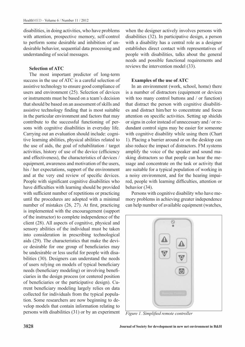
3828
HealthMED - Volume 6 / Number 11 / 2012
Journal of Society for development in new net environment in B&H
disabilities, in doing activities, who have problems with attention, prospective memory, self-control to perform some desirable and inhibition of un-desirable behavior, sequential data processing and understanding of social messages.
Selection of ATC The most important predictor of long-term
success in the use of ATC is a careful selection of assistive technology to ensure good compliance of users and environment (25). Selection of devices or instruments must be based on a team’s decision that should be based on an assessment of skills and assistive technology finding that is most suitable in the particular environment and factors that may contribute to the successful functioning of per-sons with cognitive disabilities in everyday life. Carrying out an evaluation should include: cogni-tive learning abilities, physical abilities related to the use of aids, the goal of rehabilitation / target activities, history of use of the device (efficiency and effectiveness), the characteristics of devices / equipment, awareness and motivation of the users, his / her expectations, support of the environment and at the very end review of specific devices. People with significant cognitive disabilities who have difficulties with learning should be provided with sufficient number of repetitions or practicing until the procedures are adopted with a minimal number of mistakes (26, 27). At first, practicing is implemented with the encouragement (support of the instructor) to complete independence of the client (28). All aspects of cognitive, physical and sensory abilities of the individual must be taken into consideration in prescribing technological aids (29). The characteristics that make the devi-ce desirable for one group of beneficiaries may be undesirable or less useful for people with disa-bilities (30). Designers can understand the needs of users relying on models of typical beneficiary needs (beneficiary modeling) or involving benefi-ciaries in the design process (or centered position of beneficiaries or the participative design). Cu-rrent beneficiary modeling largely relies on data collected for individuals from the typical popula-tion. Some researchers are now beginning to de-velop models that contain information relating to persons with disabilities (31) or by an experiment
when the designer actively involves persons with disabilities (32). In participative design, a person with a disability has a central role as a designer establishes direct contact with representatives of people with disabilities, talks about the general needs and possible functional requirements and reviews the intervention model (33).
Examples of the use of ATC In an environment (work, school, home) there
is a number of distractors (equipment or devices with too many control buttons and / or function) that distract the person with cognitive disabiliti-es and distract him/her to concentrate and focus attention on specific activities. Setting up shields or signs in color instead of unnecessary and / or re-dundant control signs may be easier for someone with cognitive disability while using them (Chart 1). Placing a barrier around or on the desktop can also reduce the impact of distractors. FM systems amplify the voice of the speaker and sound ma-sking distractors so that people can hear the me-ssage and concentrate on the task or activity that are suitable for a typical population of working in a noisy environment, and for the hearing impai-red, people with learning difficulties, attention or behavior (34).
Persons with cognitive disability who have me-mory problems in achieving greater independence can help number of available equipment (watches,
Figure 1. Simplified remote controller
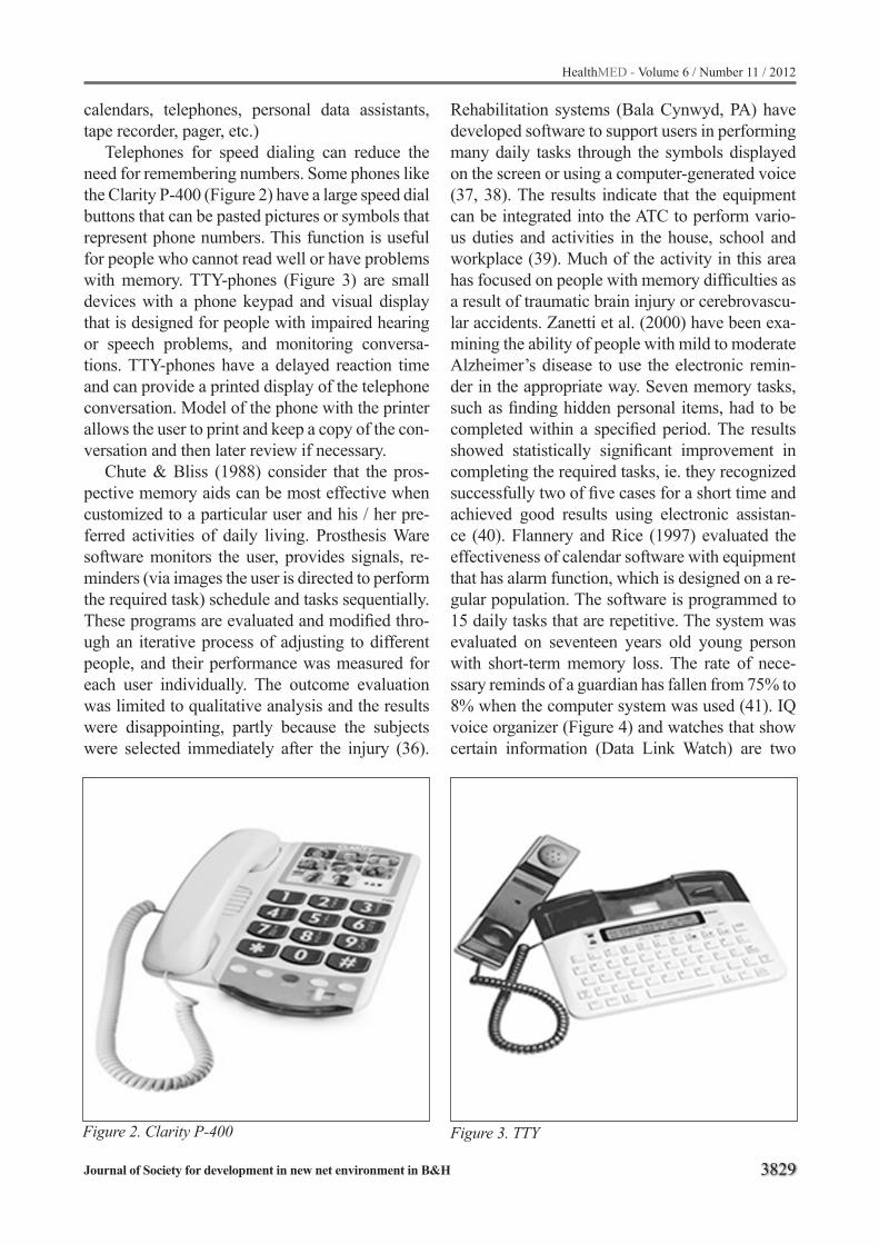
HealthMED - Volume 6 / Number 11 / 2012
Journal of Society for development in new net environment in B&H 3829
calendars, telephones, personal data assistants, tape recorder, pager, etc.)
Telephones for speed dialing can reduce the need for remembering numbers. Some phones like the Clarity P-400 (Figure 2) have a large speed dial buttons that can be pasted pictures or symbols that represent phone numbers. This function is useful for people who cannot read well or have problems with memory. TTY-phones (Figure 3) are small devices with a phone keypad and visual display that is designed for people with impaired hearing or speech problems, and monitoring conversa-tions. TTY-phones have a delayed reaction time and can provide a printed display of the telephone conversation. Model of the phone with the printer allows the user to print and keep a copy of the con-versation and then later review if necessary.
Chute & Bliss (1988) consider that the pros-pective memory aids can be most effective when customized to a particular user and his / her pre-ferred activities of daily living. Prosthesis Ware software monitors the user, provides signals, re-minders (via images the user is directed to perform the required task) schedule and tasks sequentially. These programs are evaluated and modified thro-ugh an iterative process of adjusting to different people, and their performance was measured for each user individually. The outcome evaluation was limited to qualitative analysis and the results were disappointing, partly because the subjects were selected immediately after the injury (36).
Figure 2. Clarity P-400 Figure 3. TTY
Rehabilitation systems (Bala Cynwyd, PA) have developed software to support users in performing many daily tasks through the symbols displayed on the screen or using a computer-generated voice (37, 38). The results indicate that the equipment can be integrated into the ATC to perform vario-us duties and activities in the house, school and workplace (39). Much of the activity in this area has focused on people with memory difficulties as a result of traumatic brain injury or cerebrovascu-lar accidents. Zanetti et al. (2000) have been exa-mining the ability of people with mild to moderate Alzheimer’s disease to use the electronic remin-der in the appropriate way. Seven memory tasks, such as finding hidden personal items, had to be completed within a specified period. The results showed statistically significant improvement in completing the required tasks, ie. they recognized successfully two of five cases for a short time and achieved good results using electronic assistan-ce (40). Flannery and Rice (1997) evaluated the effectiveness of calendar software with equipment that has alarm function, which is designed on a re-gular population. The software is programmed to 15 daily tasks that are repetitive. The system was evaluated on seventeen years old young person with short-term memory loss. The rate of nece-ssary reminds of a guardian has fallen from 75% to 8% when the computer system was used (41). IQ voice organizer (Figure 4) and watches that show certain information (Data Link Watch) are two
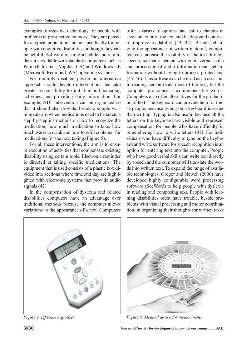
3830
HealthMED - Volume 6 / Number 11 / 2012
Journal of Society for development in new net environment in B&H
examples of assistive technology for people with problems in prospective memory. They are placed for a typical population and not specifically for pe-ople with cognitive disabilities, although they can be helpful. Software for time schedule and remin-ders are available with standard computers such as Palm (Palm Inc., Mipitas, CA) and Windows CE (Microsoft, Redmond, WA) operating systems.
For multiply disabled person an alternative approach should develop interventions that take greater responsibility for initiating and managing activities, and providing daily information. For example, ATC intervention can be organized so that it should also provide, beside a simple war-ning (alarm) when medications need to be taken, a step-by-step instructions on how to recognize the medication, how much medication to take, how much water to drink and how to refill container for medications for the next taking (Figure 5).
For all these interventions, the aim is to ensu-re execution of activities that compensate existing disability using certain tools. Electronic reminder is directed at taking specific medications. The equipment that is used consists of a plastic box di-vided into sections where time and day are highli-ghted with electronic systems that provide audio signals (42).
In the compensation of dyslexia and related disabilities computers have an advantage over traditional methods because the computer allows variations in the appearance of a text. Computers
offer a variety of options that lead to changes in size and color of the text and background contrast to improve readability (43, 44). Besides chan-ging the appearance of written material, compu-ters can increase the visibility of the text through speech, so that a person with good verbal skills and processing of audio information can get in-formation without having to process printed text (45, 46). This software can be used as an assistant in reading-person reads most of the text, but the computer pronounces incomprehensible words. Computers also offer alternatives for the producti-on of text. The keyboard can provide help for the-se people, because typing on a keyboard is easier than writing. Typing is also useful because all the letters on the keyboard are visible and represent compensation for people who have difficulty in remembering how to write letters (47). For indi-viduals who have difficulty to type on the keybo-ard and write software for speech recognition is an option for entering text into the computer. People who have good verbal skills can write text directly by speech and the computer will translate the wor-ds into written text. To expand the range of availa-ble technologies, Gregor and Newell (2000) have developed highly configurable word processing software (SeeWord) to help people with dyslexia in reading and composing text. People with lear-ning disabilities often have trouble, beside pro-blems with visual processing and motor coordina-tion, in organizing their thoughts for written tasks
Figure 4. IQ voice organizer Figure 5. Medical device for medicaments
HealthMED - Volume 6 / Number 11 / 2012
Journal of Society for development in new net environment in B&H 3831
(48). Computer software can help in the process of organizing through the instructions for creating concept maps. Concept mapping is the process of categorizing information in graphic form, known as concept maps or semantic networks. This visu-al information can be used to organize concepts and provide the basis for structuring the written text. Concept mapping makes the organization and detailed texts easier (49, 50). Some software (eg Inspiration Software) allows easy creating and modifying concept maps.
Conclusion
Numerous studies in recent decades support the use of assistive technology that may help pe-ople with cognitive disabilities in daily activities which otherwise would be unable to be perfor-med. Practical guidelines synthesized on the ba-sis of research indicate that “training in the use of technological aids should become standard practi-ce in the treatment” and rehabilitation of traumatic brain injury (51), cerebrovascular accidents, diffi-culties in learning, multiple sclerosis, intellectual and multiple disabilities, autism, and dementia.
References
1. Wilson, B.A., Evans, J.J., Emslie, H., Malineka, V. Evaluation of NeuroPage: a new memory aid. Jour-nal of Neurology Neurosurgery and Psychiatry. 1997; 63:113-115.
2. Dickov, A., Mitrovic Martinovic, S:, Vuckovic, N., Dickov, N:, Dragin, D., Budisa, D., Mitrovic, D., Se-kulic-Nad, I. Posttraumatic stress disorder (ptsd) after traffic accident. HealthMED. 2010; vol.4(4):1037-1044.
3. Wright, P., Rogers, N., Hall, C., Wilson, B., Evans, J., Emslie, H., & Bartram, C. Comparison of pocket-computer memory aids for people with brain injury. Brain Injury. 2001; 15(9):787–800.
4. Evans, J.J., Emslie, H.C. Wilson, B.A. (1998) Exter-nal cueing systems in the rehabilitation of executive impairments of action. Journal of the International Neuropsychological Society, 4, 399-408
5. MacArthur, C. A., Ferretti, R. P., Okolo, C. M., & Ca-valier, A. R. Technology applications for students with literacy problems: A critical review. Elementary Scho-ol Journal. 2001; 101(3):273–301
6. Allen, D. N., Goldstein, G., Heyman, R. A., & Rondi-nelli, T. Teaching memory strategies to persons with multiple sclerosis. Journal of Rehabilitation Research and Development, 1998; 35(4):405–410.
7. Kielhofner, G. Model of human occupation: Theory and application (4th ed.). Philadelphia: Lippincott, Williams & Wilkins. 2008.
8. Townsend E, Enabling Occupation – An Occupatio-nal Therapy Perspective. Ontario: Canadian Associa-tion of Occupational Therapists. 2002.
9. Strickland, D., Marcus, L., Mesibov, G.B., & Hogan, K. Brief report: Two case studies using virtual reality as a learning tool for autistic children. Journal of Au-tism and Developmental Disorders. 1996; 26(6):651–660.
10. Zanetti, O., Zanieri, G., Vreese, L. P. D., Frisoni, G., & Binetti, G. Utilizing an electronic memory aid with Alzheimer’s disease patients. A study of feasibility. Sixth International Stockholm/Springfield Symposi-um on Advances in Alzheimer Therapy, Stockholm, Sweden. 2000.
11. Gillespie, A., & Zittoun, T. Using resources: Con-ceptualizing the mediation and reflective use of tools and signs. Culture & Psychology. 2010; 16:37–62.
12. Pavolini, E. and Ranci, C. Restructuring the Welfare State: Reforms in Long-term Care in Western Euro-pean Countries, Journal of European Social Policy. 2008; 18 (3):246–59.
13. Wimo, A., Prince, M., The global economic impact of dementia. Alzheimer’s Disease International World Alzheimer Report 2010. http://www.alz.co.uk/research/files/WorldAlzheimerReport2010Executi-veSummary.pdf
14. Williams, B. C., Fries, B. E., Foley, W. J., Schneider, D., & Gavazzi, M. Activities of daily living and costs in nursing homes. Health Care Financing Review. 1994; 15(4):117-135.
15. Gillespie, A., Murphy, J., & Place, M. Divergences of perspective between people with aphasia and the-ir family caregivers. Aphasiology. 2010; 24:1559–1575
16. Proot, I.M., Crebolder, H.F.J.M., Huijer Abu-Saad H., Macor, T.H.G.M., ter Meulen, R.H.J. Facilitating and constraining factors on autonomy: the views of stroke patients on admission into nursing homes. Clin. Nurs. Res., in press. 2000.
17. de Joode, E., van Heugten, C., F., Verhey, & van Boxtel, M.. Efficacy and usability of assistive techno-logy for patients with cognitive deficits: Asystematic rewiev, Clinical rehabilitation. 2010; 24 (8):701-714
3832
HealthMED - Volume 6 / Number 11 / 2012
Journal of Society for development in new net environment in B&H
18. LoPresti, E. F., Mihailidis, A., & Kirsch, N. Assisti-ve technology for cognitive rehabilitation: State of the art, Neuropsychological rehabilitation. 2004; 14 (1/2):5–39
19. Pynoos, R., Rodrigues, N., Steinberg, A., Stuber, M., & Frederick, C. (1998). The UCLA PTSD Reaction Index for DSM IV (Rev. 1). Los Angeles: UCLA Tra-uma Psychiatric Program.
20. Kielhofner, G. Model of human occupation: Theory and application (4th ed.). Philadelphia: Lippincott, Williams & Wilkins. 2008.
21. Kirsch, N. L., Levine, S. P., Fallon-Kreuger, M., & Jaros, L. The microcomputer as an‘orthotic’ device for patients with cognitive deficits. Journal of Head Trauma Rehabilitation. 1987; 2(4):77–86.
22. Lynch, W. Historical review of computer-assisted cognitive retraining. Journal of Head Trauma Re-habilitation. 2002; 17(5) : 446-457.
23. Kudumovic, M., Kudumovic, Dž., Mesanovic, N., Huremovic, E. Modern information comunication technologies and educational technologies appli-ed to education of medicine. HealthMED. 2010; vol.4(1):158-162.
24. Cole, E. Cognitive prosthetics: An overview to a method of treatment. 1999; Neurorehabilitation. 12:39-51
25. Scherer, M., Jutai, J., Fuhrer, M., Demers, L., & De-Ruyter, F. A framework for modelling the selection of assistive technology devices (ATDs). Disability and Rehabilitation: Assistive Technology. 2007; 2(1):1–8.
26. Todd, M., & Barrow, C. Touch type: The acquisition of a useful complex perceptual-motor skill. Neurop-sychological Rehabilitation. 2008; 18(4):486–506.
27. Turkstra, L. S., & Bourgeois, M. S. Intervention for a modern day HM: Errorless learning of practical goals. Journal of Medical Speech Language Patho-logy. 2005; 13(3):205–212.
28. Ehlhardt, L., Sohlberg, M. M., Kennedy, M., Coelho, C., Ylvisaker, M., Turkstra, L., et al. Evidence-based practice guidelines for instructing individuals with neurogenic memory impairments: what have we le-arned in the past 20 years? Neuropsychological Re-habilitation. 2008; 18(3):300–342
29. LoPresti, E. F., & Willkomm, T. Comparison of commercially available electronic prospective me-mory aids. Proceedings of the Rehabilitation En-gineering Society of North America (RESNA) (pp. 523–525). Arlington, VA: RESNA Press. 1997
30. Cole, E., Ziegmann, M., Wu, Y., Yonker, V., Gustaf-son, C., & Cirwithen, S. Use of “therapist-friendly” tools in cognitive assistive technology and telere-habilitation. Proceedings of the Rehabilitation En-gineering Society of North America (RESNA) (pp. 31–33). Arlington, VA: RESNA Press. 2000
31. Keates, S., Clarkson, J., & Robinson, P. Investiga-ting the applicability of user models for motion-impaired users. Proceedings of Assets 2000: The Fourth International ACM Conference on Assistive Technologies, Washington, DC (pp. 129–136). New York: ACM Press. 2000.
32. Svensk, A. Empathic modeling (The Sober Version). Proceedings 4th European Conference for the Ad-vancement of Assistive Technology, (AAATE), The-ssaloniki, Greece. (pp. 432–435). Amsterdam: IOS Press. 1997.
33. Cole, E., Dehdashti, P., Petti, L., & Angert, M. De-sign and outcomes of computerbased cognitive pro-sthetics for brain injury: A field study of three su-bjects. NeuroRehabilitation. 1994; 4(3):174–186.
34. Westerveld, M., Flynn, M. Management of hearing loss in children with Down syndrom. New Zeland Down Syndrom News. 2001; 11:16-19.
35. Chute, D. L., & Bliss, M. E. Prosthesis ware: Perso-nal computer support for independent living. 1988; http://www.homemods.org/library/life-span/pro-sthesis.html
36. Chute, D. L., & Bliss, M. E. ProthesisWare. Experi-mental Aging Research. 1994; 20:229–238.
37. Bergman, M. Computer orthoses: Fostering self-sufficiency in people with cognitive challenges. Di-sability Today, 1996; Fall, 54–55.
38. Bergman, M. M. The benefits of a cognitive orthotic in brain injury rehabilitation. Journal of Head Trau-ma Rehabilitation. 2002; 17(5):432–445.
39. Bergman, M. M. People with cognitive challenges can enjoy rapid success in acquiring skills and ma-naging their lives: The exciting breakthrough of Co-gnitive Orthoses. California State University, Nort-hridge, Technology and Persons with Disabilities Conference (CSUN). Los Angeles, CA; 1997.
40. Zanetti, O., Zanieri, G., Vreese, L. P. D., Frisoni, G., & Binetti, G. Utilizing an electronic memory aid with Alzheimer’s disease patients. A study of feasibility. Sixth International Stockholm/Springfield Symposi-um on Advances in Alzheimer Therapy, Stockholm, Sweden. 2000.
HealthMED - Volume 6 / Number 11 / 2012
Journal of Society for development in new net environment in B&H 3833
41. Flannery, M., & Rice, D. Using available techno-logy for reminding. Proceedings of the Rehabilitati-on Engineering Society of North America (RESNA) (pp. 517–519). Arlington, VA: RESNA Press. 1997.
42. Fernie, G., & Fernie, B. The potential role of tech-nology to provide help at home for persons with Alzheimer’s disease. The 18th Annual Conference of the Alzheimer’s Society of Canada, Ottawa, ON. 1996.
43. Keates, A. Dyslexia and information and commu-nications technology: A guide for teachers and pa-rents. London: David Fulton Publishers. 2000.
44. Wilkins, A., & Lewis, E. Coloured overlays, text, and texture. Perception. 1999; 28:365–370.
45. Higgins, E. L., & Raskind, M. H. The compensatory effectiveness of optical character recognition/speech synthesis on reading comprehension of postsecon-dary students with learning disabilities. Learning Disabilities. 1997; 8(2):75–87.
46. Raskind, M. H., & Higgins, E. L. Effects of speech synthesis on the proofreading efficiency of postse-condary students with learning disabilities. Lear-ning Disability Quarterly. 1995; 18:141–158.
47. Gregor, P., & Newell, A. F. An empirical investigati-on of ways in which some of the problems encounte-red by some dyslexics may be alleviated using com-puter techniques. Proceedings of Assets 2000: The Fourth International ACM Conference on Assistive Technologies, Washington, DC (pp. 85–91). New York: ACM Press. 2000.
48. Newcomer, P., & Barenbaum, E. The written compo-sing problems of children with LD: A review. Journal of Learning Disabilities. 1991; 22:262–264.
49. Ruddell, R. B., & Boyle, O. F. A study of cognitive mapping as a means to improve comprehension of expository text. Reading Research and Instruction. 1989; 29(1):12–22.
50. Zipprich, M. A. (1995). Teaching web making as a guided planning tool to improve student narrative writing. Remedial & Special Education, 16(1), 3-16
51. Sohlberg, M. M., Kennedy, M. R. T., Avery, J., Co-elho, C., Turkstra, L., Ylvisaker, M., & Yorkston, K. Evidence based practice for the use of external aids as a memory rehabilitation technique. Journal of Medical Speech Pathology. 2007; 15(1): xv–li.
Corresponding AuthorBiljana Milanovic Dobrota,University of Belgrade,Faculty of Special Education and Rehabilitation, Belgrade;SerbiaE-mail: [email protected]
3834
HealthMED - Volume 6 / Number 11 / 2012
Journal of Society for development in new net environment in B&H
Physical fitness adaptations to 9-week precompetitive training period in professional soccer teamZoran Milosevic1, Dusko Bjelica2, Dusica Rakic3, Dejan Madic1, Borislav Obradovic1, Jelena Obradovic1, Ilona Mihajlovic1, Miroslav Smajic1
1 Faculty of Sport and Physical education, Novi Sad, Serbia2 Faculty for Sport and Physical education, Niksic, Serbia3 Faculty of Medicine, Novi Sad, Serbia
Abstract
The purpose of the present study was to inve-stigate adaptations of precompetitive training pro-gram period on several performance-related aero-bic and anaerobic indices in professional soccer players. Fifteen professional soccer players (age 22.2±3.27 years, height 180.53±5.50cm, weight 78.08±5.57kg; training experience 13.60±2.44 years; mean±SD), participated in the study. Su-bjects performed countermovement and vertical jump tests (CMJ and VJ) and incremental pseu-do-ramp test protocol with measured maximal oxygen consumption (VO2max) and anaerobic threshold (ANT). Modest but significant improve-ment (p<0.05) of aerobic performance was obser-ved after a 9-week precompetitive training period, with the 6,67% and 7.28% degree of change for maximal oxygen consumption and anaerobic thre-shold, respectively. Anaerobic power remained unchanged. These results suggest that preseason training can affect aerobic energy system. Howe-ver, implementation of additional high intensity intervals as well as greater volume of explosive training exercises may be required.
Keywords: Team sport, Countermovement jump, Aerobic fitness
Introduction
The physical demands of soccer game are su-bstantial, especially at the highest level (1). Du-ring a 90-minute game, elite-level players run about 10km at an average intensity close to the anaerobic threshold, which implies that aerobic metabolism dominate the game. In addition, game activity is saturated by repeated bouts of high in-tensity exercise interspersed with periods of low-
intensity activity, which further increase reliance on aerobic energy system. High aerobic fitness has been found to improve soccer performance by enhancing recovery from high intensity bouts during game and consequently enabling players to sustain a high work rate throughout a game (2). Aerobic fitness is determined by maximal oxygen consumption and anaerobic threshold. Within this endurance context,a number of important perfor-mance activities such as sprints, jumps, duels and kicks are largely anaerobic or explosive (3). On average, all-out sprint occur once every 90s and maximal or near maximal- intensity efforts every 30s of play. Those anaerobic power-type activities constitutes the crucial moments of the game and contributes directly to winning possession of the ball and to the scoring or conceding of goals (4). Therefore, to become successful, it seems that pre-requisite in the modern game for the elite soccer player is to have high level of both aerobic and anaerobic fitness. Indeed, a strong relationship between physical fitness and performance level has been reported in elite soccer (5). Moreover, it appears that the physical fitness of soccer players have become more and more important, conside-ring ever-increasing physical demands of game over the years (6).
Such prerequisite implies the need for well-designed sessions on a daily basis inducing high weekly training load during an entire season that ultimately maximize performance (3,7). Hovewer, precompetitive training period is considered to be of particular importance, as it has been stated that most of physical fitness improvements are obta-ined during this period, with seasonal training regimens aimed to maintain acquired level of fit-ness (8). Interestingly, while seasonal changes in

HealthMED - Volume 6 / Number 11 / 2012
Journal of Society for development in new net environment in B&H 3835
professional soccer players have been extensively investigated (9, 10, 11), there is evident paucity of research addressing changes in crucial performan-ce indices during precompetitive training period. To the author’s best knowledge, only one study targeting this specific period was recently publis-hed (12), with reported improvements in both ae-robic (Yo-Yo test distance covered) and anaerobic power (vertical jump height) after 8 week training period. However, subject in this study commen-ced preseason training period following 45 days of complete rest, which could largely influence ob-served improvements due to relatively deconditio-ned state of athletes at the start of training season. Moreover, the complete rest during off-season pe-riod is practice rarely seen nowadays in elite level teams (13). Therefore, the purpose of the present study was to investigate adaptations of precom-petitive training program period on several per-formance-related aerobic and anaerobic indices in professional soccer players. We hypothesized that preseason training would improve both aero-bic and anaerobic indices of professional soccer players.
Methods
SubjectsFifteen professional soccer players (age
22.2±3.27 years, height 180.53±5.50cm, weight 78.08±5.57kg; training experience 13.60±2.44 years; mean±SD) participated in the study. All su-bjects were members of the Serbian First League team consistently ranked among the first 4 teams in the League for several years.
All of the subjects gave their written informed consent and volunteered to participate in the stu-dy, which had the approval of the Local Ethical Advisory Commission. All participants were fully informed verbally and in writing about the nature and demands of the study and were informed that they could withdraw from the study at any time, even after giving their written consent. All mea-surements were obtained by the certified four-man squad. Subjects were familiarized with all testing procedures as part of their regular training process over the years. They were advised to avoid inten-sive exercise 24h prior to the data collection.
Experimental design All subjects were tested twice, at the start and
the end of precompetitive period (first week of June and last week of July, respectively).
Countermovement and Vertical jump (CMJ and VJ)
Before the testing, weight and height were me-asured for each subject. Body mass was measured using BC-554 body composition monitor (Tanita Corp., Tokyo, Japan) to the nearest 100g, and height was determined with portable stadiometer (SECA, Hamburg, Germany) to the nearest millimeter, with barefoot subjects wearing underwear only. All anthropometric measures were carried out by the same investigator. CMJ and VJ were estimated using a contact mat (Jump Mat, Axon, USA). Be-fore testing, the subjects were allowed to warm up on their own (e.g., jogging, calisthenics) but were requested not to engage in static stretching. Also, several preparation jumps were conducted at the end of warm-up period. CMJ requires the indivi-dual to begin in an upright posture with hands on hips. After a brief descending phase to approxima-tely semi squat position, subjects extended knees and hips into an upward jump, maintaining hands on the hips and landing with extended legs. VJ has basically the same kinematical pattern, with arms allowed to swing thus contributing to jump height as only difference. Three attempts for each jump type were allowed with the highest value included in further analysis. Jump height was calculated from the time the subject was off the mat by the computer which was connected to the mat.
Aerobic assessmentIncremental pseudo-ramp test protocol was con-
ducted on a computer controlled treadmill (13620 Treadmill, Vacumed, California, USA) with heart rate monitored continuously throughout the test session (Polar S-810, Polar Electro Oy, Finland). Expired air was collected and analyzed through a 2-way valve using automated gas analysis system (Cosmed, CPET, Rome, Italy). The gas analyzer and volume transducer were calibrated according to manufacturer’s specification. Following warm up (3min of running at 3.8mph), incremental pro-tocol, with speed increasing by 0.7mph every minute, was applied until volitional fatigue. The
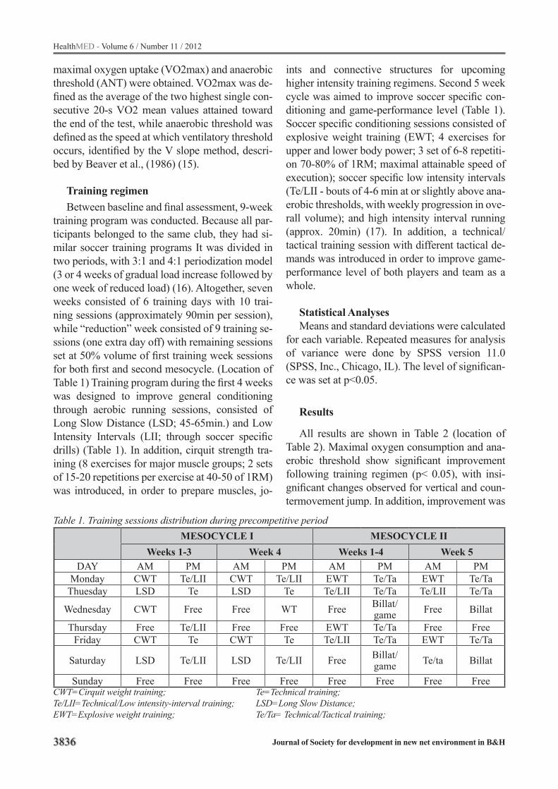
3836
HealthMED - Volume 6 / Number 11 / 2012
Journal of Society for development in new net environment in B&H
maximal oxygen uptake (VO2max) and anaerobic threshold (ANT) were obtained. VO2max was de-fined as the average of the two highest single con-secutive 20-s VO2 mean values attained toward the end of the test, while anaerobic threshold was defined as the speed at which ventilatory threshold occurs, identified by the V slope method, descri-bed by Beaver et al., (1986) (15).
Training regimenBetween baseline and final assessment, 9-week
training program was conducted. Because all par-ticipants belonged to the same club, they had si-milar soccer training programs It was divided in two periods, with 3:1 and 4:1 periodization model (3 or 4 weeks of gradual load increase followed by one week of reduced load) (16). Altogether, seven weeks consisted of 6 training days with 10 trai-ning sessions (approximately 90min per session), while “reduction” week consisted of 9 training se-ssions (one extra day off) with remaining sessions set at 50% volume of first training week sessions for both first and second mesocycle. (Location of Table 1) Training program during the first 4 weeks was designed to improve general conditioning through aerobic running sessions, consisted of Long Slow Distance (LSD; 45-65min.) and Low Intensity Intervals (LII; through soccer specific drills) (Table 1). In addition, cirquit strength tra-ining (8 exercises for major muscle groups; 2 sets of 15-20 repetitions per exercise at 40-50 of 1RM) was introduced, in order to prepare muscles, jo-
Table 1. Training sessions distribution during precompetitive periodMESOCYCLE I MESOCYCLE II
Weeks 1-3 Week 4 Weeks 1-4 Week 5DAY AM PM AM PM AM PM AM PM
Monday CWT Te/LII CWT Te/LII EWT Te/Ta EWT Te/TaThuesday LSD Te LSD Te Te/LII Te/Ta Te/LII Te/Ta
Wednesday CWT Free Free WT Free Billat/game Free Billat
Thursday Free Te/LII Free Free EWT Te/Ta Free FreeFriday CWT Te CWT Te Te/LII Te/Ta EWT Te/Ta
Saturday LSD Te/LII LSD Te/LII Free Billat/game Te/ta Billat
Sunday Free Free Free Free Free Free Free FreeCWT=Cirquit weight training; Te=Technical training;Te/LII=Technical/Low intensity-interval training; LSD=Long Slow Distance;EWT=Explosive weight training; Te/Ta= Technical/Tactical training;
ints and connective structures for upcoming higher intensity training regimens. Second 5 week cycle was aimed to improve soccer specific con-ditioning and game-performance level (Table 1). Soccer specific conditioning sessions consisted of explosive weight training (EWT; 4 exercises for upper and lower body power; 3 set of 6-8 repetiti-on 70-80% of 1RM; maximal attainable speed of execution); soccer specific low intensity intervals (Te/LII - bouts of 4-6 min at or slightly above ana-erobic thresholds, with weekly progression in ove-rall volume); and high intensity interval running (approx. 20min) (17). In addition, a technical/tactical training session with different tactical de-mands was introduced in order to improve game-performance level of both players and team as a whole.
Statistical AnalysesMeans and standard deviations were calculated
for each variable. Repeated measures for analysis of variance were done by SPSS version 11.0 (SPSS, Inc., Chicago, IL). The level of significan-ce was set at p<0.05.
Results
All results are shown in Table 2 (location of Table 2). Maximal oxygen consumption and ana-erobic threshold show significant improvement following training regimen (p< 0.05), with insi-gnificant changes observed for vertical and coun-termovement jump. In addition, improvement was

HealthMED - Volume 6 / Number 11 / 2012
Journal of Society for development in new net environment in B&H 3837
highest for anaerobic threshold (7,7%), followed by maximal oxygen consumption, countermove-ment and vertical jump (6,67%,3% and 0,98%, respectively).
Discussion
The competition performance of soccer players, among other things, depends on high le-vel of both aerobic and anaerobic abilities. High level of anaerobic and aerobic power as well as aerobic efficiency have been steadily reported as prerequisites for quality game performance. It co-uld be expected that main aim of precompetitive training regimens is to develop those qualities. The main finding of the present study represents the statisticaly significant improvement in maxi-mal oxygen consumption and anaerobic threshold (p< 0.05), but not in anaerobic power (represen-ted by vertical and countermovement jump) of the professional soccer players during precompetitive period.
Significant improvements in maximal oxygen consumption was observed during preseason pe-riod (from 54,39ml/kg/min-1 to 58,02ml/kg/min-1; 6,67%). The measured VO2max values (both pre and post training) are in accordance with some previous results (18, 5) but generaly fall under suboptimal level of aerobic fitness, as it has been suggested that a VO2max of 60ml/kg/min-1 is a minimal threshold for top level performance (2, 4). Moreover, Stolen et al. (19) suggested that this reference value likely needs upward adjustment, considering significantly higher values (over 70ml/kg/min-1) more frequently observed at top-level players. Sub-optimal level of aerobic capa-city imply slower recovery from high-intensity intermittent exercise, lactate removal and reduced PCr regeneration (20), which could compromise
execution of crucial performance-related activities such as short sprints in scoring area, jumps for sco-ring-headers, or powerfull goal-tending kicks. As, beyond genetic endowment, low values of maxi-mal oxygen consumption could be attributed to inadequate training stimulus (21), obtained results suggest a need for critical evaluation of training process. It could be noticed that, especially during first mesocycle, all aerobic training sessions con-sisted of low intensity training regimens, below or at anaerobic threshold. Althought such training regimens are traditionaly used at first stages of preseason period, its eficacy in developing aerobic fitness is highly questionable. According to Sto-len et al. (19), low-intensity training should not be programmed separately as soccer players will naturally perform such efforts during technical and tactical drills during regular soccer training. Moreover, it has been recognized that cardiac out-put limits VO2max and that there is no plateau in stroke volume in well-trained individuals (22, 23). Thus, to improve VO2max, one needs to incorpo-rate high intensity interval training regimens with active recovery in order to produce stroke volu-me-overload and consequent improvements in maximal oxygen consumption. Indeed, Helgerud et al. (2001) (2) reported that interval training of 4×4 minutes at 90–95% of Hrmax, separated by 3 minutes of active recovery at 60–70% of Hrmax, produced VO2max increment of 0,5% per session in elite junior soccer players. Similar results, with same or similar interval training intervention on various samples of soccer players, were extensi-vely presented in the literature (24, 25, 26).
The largest improvements of all variables was observed in anaerobic threshold, with speed at ana-erobic thresholds increment over 1 km/h or 7,7% (from 14,55km/h in the first test to 15,61km/h in
Table 2. Descriptive statistics and degree of change (%) for the selected variablesPre-training Post-training Change (%)
CMJ (cm) 37.47±3.08 38.63±2.56 3VJ (cm) 46.10±4.36 46.56±4.38 0,98
VO2max (ml/kg/min-1) 54.39±3.99 58.02±3.49* 6,67ANT(km/h) 14.55.4±0.85 15.61±0.98* 7,28
Note: Values are mean±SD.VO2max= maximal oxygen uptake; CMJ= Countermovement jump;VJ= Vertical jump; ANT= Anaerobic threshold*Indicates significant difference pre-versus post at p< 0.05;
3838
HealthMED - Volume 6 / Number 11 / 2012
Journal of Society for development in new net environment in B&H
the second) after 9-week period. This result is si-milar to those reported by Mcmillan et all. (12), but better than reported by Casujas (10) and re-cently presented for elite Greek soccer team (27). Generally, anaerobic threshold for soccer players is located somewhere between 13,5 and 15km/h (10). In that context, it could be argued that our players posses relatively high level of anaerobic threshold. Moreover, based on training schedule presented in Table 1, it is reasonable to assume that high volume of low intensity training, especially during first mesocycle of preparatory period, was largely responsible for observed improvements. Indeed, it is known that continuous running for over 30 minutes at an exercise intensity just below ANT leads to peripheral adaptations strongly asso-ciated with ANT (28). Anaerobic threshold is con-sidered important in soccer, as it has been shown that average intensity in soccer match is closely related to anaerobic threshold (19). Moreover, it has been recently presented that, in elite soccer players, velocity at anaerobic threshold was more strongly correlated with repeated sprint ability (an important fitness attribute for soccer performance) than maximal oxygen consumption (29).
Vertical jumping is an important feature of many sports, and increasing the height an athle-te can reach is likely beneficial to soccer perfor-mance (19). Indeed, close relationship between vertical jump height and performance in a soccer league has been reported (30), pointing out the need for high level of anaerobic power in profe-ssional soccer. The mean value of around 37cm and 46cm for countermovement and vertical jump heights respectively, reveal relatively low anae-robic power in our players. The jumping heights were markedly lower than previously reported in literature for adult soccer players (43.5-61.0cm for VJ; 19). Furthermore, we did not find any si-gnificant change in jumping performance in our subjects after 9-week training period,despite su-bstantional amount of explosive strength training during second mesocycle. Contrary to our results, it has been reported that jumping performance is likely to improve following high-resistance or explosive-type resistance training regimens, with the latter found more effective (31). Based on this, Hoff and Helgerud introduced specific maxi-mal strength training using high loads (85% + of
1RM), maximal concentric action-velocity and medium volume (four sets with five repetitions), and in a series of studies proved its effectiveness for both strength and power development in soccer players (32). Moreover, in study design similar to ours (specific strength training twice a week over 8 week preseason period), they reported that jum-ping height of elite soccer players increased from already respectable 57.2 to 60.2cm! Hence, it is hard to explain results considering effects of pre-competitive training period on jumping ability in our study. We could hypothesize that newly pre-sented specific strength training regimen might not be properly executed considering movement velocity-demands (intention to exert force as ra-pidly as possible). Consequently, such training regimens might solely affect maximal strength, which does not always result in anaerobic power enhancement (33).
It should be noted that the present study was conducted using a single squad of professional players, which is evident limitation. Another li-mitation of this study is the absence of maximal strength, speed and speed endurance evaluation, an important physical attributes for peak soccer performance. Therefore, future studies, examining additional fitness attributes on larger sample size, are warranted.
In conclusion, modest but significant improve-ment of aerobic performance was observed from the start of the preseason to the beginning of the competitive season. Anaerobic power remained unchanged during preseason period. Taken to-gether, those results suggest that preseason trai-ning prescription should be redesigned with im-plementation of additional high intensity intervals as well as greater volume of explosive training exercises. Such training regimens would likely in-duce preferable magnitude of adaptations to both aerobic and anaerobic fitness attributes in profe-ssional soccer players.
Acknowledgments
The authors acknowledge the cooperation of football club FC “Vojvodina”. The authors have no conflicts of interest that are directly relevant to the content of this manuscript

HealthMED - Volume 6 / Number 11 / 2012
Journal of Society for development in new net environment in B&H 3839
References
1. Reilly T. An Ergonomics model of the soccer training process. J. Sports Sci, 2005; 23(6): 561-572.
2. Helgerud J, Engen LC, Wisloff U, et al. Aerobic endu-rance training improvessoccer performance. Med Sci Sports Exerc 2001;33:1925–31.
3. Bangsbo J, Mohr M, Krustrup P. Physical and meta-bolic demands of training and match-play in the elite football player. J. Sports Sci, 2006; 24(7): 665–674.
4. Reilly T, Bangsbo J, and A. Franks. Anthropometric and physiological predispositions for elite soccer. J. Sports Sci, 2000; 18: 669–683.
5. Ostojic SM. Elite and Nonelite Soccer Players: Pre-seasonal Physical and Physiological Characteristics. Res Sports Med,2004; 12: 143–150.
6. Williams AM, Lee D, Reilly T. A quantitative analysis of matches played in the 1991—92 and 1997—98 sea-sons. London: The Football Association; 1999
7. Filaire E, Bernain X, Sagnol M, Lac G. Preliminary results on mood state, salivary testosterone:cortisol ratio and team performance in a professional soccer team. Eur J App Physiol, 2001; 86:179–184
8. Koutedakis Y. Season variation in fitness parameters in competi- tive athletes. Sports Med, 1995; 19: 373-392.
9. Caldwell BP, and Peters DM. Seasonal variation in physiolog- ical fitness of a semiprofessional soccer team. J Strength Cond Res, 2009; 23(5): 1370–1377.
10. Casajus, J. Seasonal variation in fitness variables in professional soccer players. J Sports Med Phys Fit-ness,2001; 41: 463–469.
11. Haritonidis K, Koutlianos N, Koudi E, Haritonidou M, and Deligiannis, A. Seasonal variation of aero-bic capacity in elite soccer, basketball and volleyball players. J Hum Mov Stud,2004; 16: 289–302.
12. McMillan K, Helgerud J, Grant SJ, Newell J, Wil-son J, Macdonald R and Hoff J. Lactate threshold responses to a season of professional British youth soccer. Br. J. Sports Med, 2005; 39:432-436.
13. Wong PL, Chaouachi A, Chamari K, Dellal A, and Wisloff U. Effect of preseason concurrent muscular strength and high intensity interval training in pro-fessional soccer players. J Strength Cond Res, 2010; 24(3): 653–660.
14. Reilly T. Fitness and the start of season. Insight FA Coaches Association Journal 6,69,2003.
15. Beaver WL, Wasserman K, and Whipp BJ. A new method for detecting anaerobic threshold bygas exchange. J App Physiol, 1986; 60:2020-2027.
16. Plisk SS, Stone MH Periodization Strategies. Stren-gth Cond, 2003; 25: (6), 19-37.
17. Billat VL, Flechet B, Petit B, Muriaux G, Koralsztein JP. Interval training at VO2max: effects on aerobic performance and overtraining markers. Med Sci Sports Exerc 1999; 3 (1): 156-163.
18. Aziz AR, Newton MJ, Tan HY, and Teh KC. Variation in fitness attributes of players during a competitive season in an Asian professional soccer league: a fi-eld-based investigation. Asi J Exe Sport Sci (2006), 3, 40-45.
19. Stølen T, Chamari K, Castagna C. Physiology of soccer: an update. Sports Med, 2005; 35: 501-36.
20. Tomlin DL, Wenger HA The relationship between aerobic fitness and recovery from high intensity in-termittent exercise. Sports Med 2001; 31 (1):1-11.
21. Ekblom B. Effect of physical training on oxygen transport system in man. Acta Physiol Scand, 1968; 328: 1-45.
22. Wagner PD. New ideas on limitations to VO2max. Exerc Sport Sci Rev ,2000; 28 (1): 10-4.
23. Wiebe CG, Gledhill N, Jamnik VK, et al. Exercise cardiacfunction in young through elderly endurance trained women. Med Sci Sports Exerc 1999, May; 31 (5): 684-91.
24. Hoff J, Wisløff U, Engen LC, Kemi OJ, Helgerud J. Soccer specific aerobic endurance training. Br J Sports Med 2002; 36 (3): 218-21.
25. Wisløff U, Helgerud J, Hoff J. Strength and endu-rance of elite soccer players. Med Sci Sports Exerc 1998; 30 (3): 462-7.
26. Platt D, Maxwell A, Horn R, Williams M, and Reilly T. Physiological and technical analysis 3 v 3 and 5 v 5 youth football matches. Insight FA Coaches Assoc J 2001; 4 (4): 23-4.
27. Kalapotharakos, VI, Ziogas, G, and Tokmakidis, SP. Seasonal aerobic performance variations in elite soccer players. J Strength Cond Res,2011; 25(6): 1502–1507.
28. Keith SP, Jacobs I, McLellan TM.Adaptations to training at the individual anaerobic threshold. Eur J Appl Physiol Occup Physiol. 1992;65(4):316-23.
29. da Silva JF, Guglielmo LGA, and Bishop, D. Relati-onship between different measures of aerobic fitness and repeated sprint ability in elite soccer players. J Strength Cond Res,2010; 24(8):2115–2121.
30. Arnason, A., Sigurdsson, S.B., Gudmundsson, A., Holme, I., Engebretsen, L. and Bahr, R. Physical fit-ness, injuries, and team performance in soccer. Med Sci Sport Exerc,2004; 36: 278-285.

3840
HealthMED - Volume 6 / Number 11 / 2012
Journal of Society for development in new net environment in B&H
31. Wilson GJ, Newton RU, Murphy AJ, and Humphries BJ. The optimal training load for the development of dynamic athletic performance. Med. Sci. Sports Exerc,1993; 25:1279–1286.
32. Hoff J and Helgerud J. Endurance and Strength Training for Soccer Players Physiological Conside-rations. Sports Med 2004; 34 (3): 165-180.
33. Bobbert MF, and Van Soest A. Effect of muscle strengthtening on vertical jump height: A simulation study. Med. Sci. Sports Exerc,1994; 26:1012–1020. 1994.
Corresponding Author Zoran Milosevic, Faculty of Sport and Physical educationNovi Sad, Serbia E-mail: [email protected]
HealthMED - Volume 6 / Number 11 / 2012
Journal of Society for development in new net environment in B&H 3841
Environmentally induced health impacts among elderly with cardio-vascular diseaseRok Fink1, Ivan Erzen2, Saso Medved3
1 University of Ljubljana, Faculty of Health Sciences, Ljubljana, Slovenia2 University of Ljubljana, Medical Faculty, Ljubljana, Slovenia3 University of Ljubljana Faculty of Mechanical Engineering, Ljubljana, Slovenia
Abstract
Background: Extreme environmental conditi-ons will affect most people in the coming decades and influence the health and lives of billions of pe-ople. Especially at greater risk will be the elderly with diagnosed chronic heart disease,who live in urban districts.
Aim: This study analyses the impacts of micro-climatic conditions and air quality on the health of the elderly living in nursing homes.
Methods: We analysed the impacts of tempe-rature, relative humidity, concentrations of outdo-or pollutants (particulate matter, sulphur dioxide, nitrogen dioxide, carbon monoxide and ozone) on heart rate, blood pressure and symptoms.
Results: The effects of apparent temperature on all physiological parameters and expressed symp-toms (p<0.001) were found. Heat burden appeared above 25 °C and increases with increasing tem-perature. Significant effects of air pollution were also noticed at low air quality. For air quality, we found statistically significant differences only for particles and ozone (p<0.05).
However, it has been demonstrated that inte-gral indicators as the Humidex Index and Air Qu-ality Stress Index have more obvious impacts on human health than single factor analysis.
Conclusions: Extreme environmental conditi-ons can cause adverse health effects among elderly people with cardio-vascular disease. Therefore, suitable conditions with regards to temperature, relative humidity and air quality in the nursing ho-mes for the elderly should be established. Medical staff should monitor physiological parameters and expressed symptoms to prevent heat related illne-sses.
Keywords: health impacts, microclimatic con-ditions, air quality, symptoms, heat illnesses, inte-gral indicators.
Introduction
Background Throughout the world, health in the twenty-first
century is being shaped by demographic and envi-ronmental forces. Traditional distinctions betwe-en health problems in developed and developing nations have become blurred. Demographic aging is now a universal trend, as is the globalisation of unhealthy environment. In 2003, the World Health Organisation reported that every year more than 160,000 people throughout the world die due to the global climate change; it is expected that this number could be doubled in the future (1). The-re are several mechanisms by which climate can affect health. Extremes of temperature, rainfall and infectious diseases have direct immediate effects on mortality and morbidity;furthermore,air pollutants can causeadverse health impacts (2).
Physiology of human thermoregulationThe ability to sense and regulate body tempe-
rature is a key feature of human survival. A de-viation of ± 3.5°C from the resting temperature of 37°C can result in physiological impairments and fatality (3). Heat transfer between the body and the external environment occurs through the processes of conduction, convection, radiation and evaporation. Skin blood flow in humans can increase substantially in response to thermal stre-ss: thermoregulatory vasodilation can increase skin blood flow to 6 to 8 L min-1 during severe hyperthermia, causing an increase of heart rate (HR) and a decrease in blood pressure (BP). Such responses in the skin circulation represent a vital aspect of normal thermoregulation in humans. Vasodilation and increased skin blood flow are essential to heat dissipation during heat exposure and exercise. During exposure to cold, vasocon-striction in the skin decreases heat loss from the
3842
HealthMED - Volume 6 / Number 11 / 2012
Journal of Society for development in new net environment in B&H
body and protects against hypothermia. Therefore, altered control of skin blood flow has important clinical implications and can substantially impair the ability to maintain normal body temperatures (4).
Risk factorsWith advancing age, our ability to thermo-re-
gulate tends to decrease. This is a multi-factori-al process involving many of our physiological systems with an emphasis on the cardiovascular system. Typically, on a population level these and other changes lead to reduced muscle strength, reduced work capacity, a reduced sweating capa-city, a reduced ability to transport heat from the body core to the skin, and a lower cardiovascular stability in the elderly. These effects will put el-derly people at a higher risk in extreme conditions, leading to an increase in morbidity and mortality (5). Gender can also be identified as the risk fac-tor. Some authors (6, 7, 8) reported that female su-bjects are more sensitive to extreme temperature than males. However, Whitman et al. (9) observed greater mortality among men than women in Chi-cago. Some evidence suggests that urban populati-ons are more exposed to extreme heat events (10). Increased temperatures in urban regions have ele-vated morality risk (11, 12). Most people affected by classic heat stroke are very young or elderly, poor, socially isolated and do not have access to air conditioning (13, 14, 15).
Thermal stressHeat illnesses may be induced by hyperther-
mia, dehydration, salt depletion, exercise, and hyperventilation, in any combination thereof. Heat syncope appears as a short period of unconsciou-sness due to a sudden drop of BP. Heat exhaustion and heatstroke are two extremes of acontinuum of disorders, and they probably share many common pathways and factors (16).Heat stroke is often fatal and is distinguished from the less severe condition of heat exhaustion, by tissue damage caused by more severe or prolonged increases in body tem-perature (17). Risk factorawareness, along with early recognition and treatment of milder forms of heat illness, may contribute to the prevention of heatstroke and associated fatalities (18).
Data on the incidence of heat stroke are im-precise because this illness is underdiagnosed and because the definition of heat-related death varies (19).
Air qualityThe influence of meteorology on air quality is
substantial and well established, giving rise to the expectation that changes in climate are likely to al-ter patterns of air pollution concentration. Higher temperatures hasten the chemical reactions that lead to ozone (O3) and secondary particle (PM) formation (20). Epidemiological research shows excess mortality related to the exposure to O3 and PM (21, 22, 23,). However, Smoyeret al. (10) fo-und that microclimatic conditions have more si-gnificant impact on human health than air pollu-tion.
Our society is faced with the problems of popu-lation aging and cardiovascular disease, the most common cause of death; climate change and the specific characteristics of urban areas directly and indirectly affect health. Therefore, the aim of the study was to analyse what impacts microclimatic conditions and air quality have on the health of elderly people with diagnosed cardio-vascular di-sease (CVD) who live in an urban district.
Methods
The subjects of this study were residents of a nursing home in an urban area of Ljubljana, the capital city of Slovenia. The elderly people inclu-ded in this research had to meet two criteria: age over 65 years and CVD diagnosed in accordan-ce with the International Classification of Disea-ses. The sample included 50 persons, both male (46 %) and female (54 %) with an average age of 84 years. The nursing home has no mechanical ventilation, nor air cooling or conditioning. Envi-ronmental factors were determined as microclima-tic conditionsand outdoor air pollution, while he-alth was defined as physiological cardio-vascular parameters and symptoms related to heat illnesses.
Microclimate conditionsThe microclimatic conditions, includingair
temperature and relative humidity (RH), were mo-
HealthMED - Volume 6 / Number 11 / 2012
Journal of Society for development in new net environment in B&H 3843
nitored in the indoor environment. Temperature and RH were measured by automatically recording the results of measurements with iButton® devices with an accuracy of 0.1 °C and 1% of RH. We also measured the surface temperature of constructions with a FLIR T300 infrared camera in the measu-ring range of -20 °C and 650 °C. We compared air temperature and mean radiant temperature for the calculation of apparent temperature (Tap).
We also calculated the Humidex index (HI), which is a measure of the perceived heat that re-sults from the combined effect of excessive hu-midity and high temperature.The index was well described by Conti et al. (24).
Outdoor air qualityData on hourly concentrations of pollutants
SO2, NO2, CO, PM10 and O3 for the nearest station (1000 meters away) were obtained on the Sloveni-an Environmental Agency (SEA). The approachin which data on concentrations of pollutants and/or meteorological variables to the nearest measuring station used to determine the relationship between risk factors and the resulting disease or mortality is used by several authors (25, 26, 27). For the assessment of the urban air environment, the air quality stres sindex (AQSI ) was calculated accor-ding to the formula of Theoharatoset al. (28).
Physiological parametersMeasurements of blood pressure (BP) and he-
art rate in rest (HR) were performed using an auto-matic electronic meter with margin of error of ± 3 mm Hg for BP and, ± 5% for HR. Measurements of BP and HR were carried out in accordance with guidelines issued by the American Heart Associa-tion (29). Measurements were carried out in a se-ated position using upper-arm cuffs. All parame-ters are measured twice between intervals of three minutes and are expressed as the average of two runs. The analysis included systolic blood pressu-re (SBP), diastolic blood pressure (DBP), and HR. Body core temperature (Tc) was measured using the ear thermometer with an margin of error of ± 0.2 °C and a range between 34.0 °C and 42.2 °C. Measuring the tympanic temperature is the best way to analyse the body overheating, as the tem-perature of eardrum is identical to the temperature
of the hypothalamus (30). Oxygen saturation in arterial blood (SaO2) was measured using an MD 300 C1 pulse oximeter with a range between 70% and 100% and 2% accuracy.
SymptomsIn this study, we monitor the physiological pa-
rameters in addition to the medical history data, i.e. symptoms that are related to CVD and heat illnesses. The research includes the occurrence and level of expressed symptoms used in clinical medicine in the process of diagnosing CVD. Nine symptoms, typical for heat illnesses and CVD were monitored: pain behind the breastbone, he-art rate dropping, nausea and fatigue, shortness of breath, cold hands and feet, swelling of the ankles, tinnitus, depletion in case of less physical effort, and overall health. All anamnesis parameters were evaluated with a five-step scale. Total frequency and intensity of symptoms expressed constitutes a symptom index (SI), ranging from 9 to 45 points.
Statistical analysis Statistical analyses were performed with SPSS
17.0 for Windows (SPSS Inc. Chicago, IL, USA).The changes among groups of gender, age and BMI (body mass index) were tested using one-way ANOVA. Linear regression models were used to assess the association between environmen-tal factors and health. Significance was set at an alpha level of 0.05. Ethic approval was provided by the National Medical Ethic Committee of the Republic of Slovenia. The aim of this study was to analyse impact of microclimatic conditions and air quality on the health status of elderly people with CVD. We hypothesise that integral indicators such as HI and AQSI demonstrate more obvious impacts on health than single parameter analyse-sof temperature, humidity or pollutants.
Results
The research was conducted for 80 days betwe-en the 17 June and 4 September 2011. In this time period, the outdoor temperature ranged between, 9 °C and 36 °C, while indoor air temperature was between 22°C and 33.6 °C. Two heat waves occu-rred: the first between 7 and 14 July and the other

3844
HealthMED - Volume 6 / Number 11 / 2012
Journal of Society for development in new net environment in B&H
between 17 and 26 August, which was even more pronounced. Results show significant effects of microclimatic conditions on cardio-vascular para-meters. In particular, Tap has significant impacts on physiological parameters (R²=0.82). In the tempe-rature interval between 22°C and 25°C, HR is re-latively constant, but increasing the Tap causes an obvious increase in HR (Figure 1). Consequently, a decrease in BP can be seen as temperature rises. We observed that the RH as single factor has no important impacts on physiological parameters. However, we found out that integral indicators of HI that combine temperature and relative humi-dity demonstrate an even more obvious impact on cardio-vascular parameters than the impact of sin-gle parameters (R²=0.92).
The analysis of the results shows no impacts of microclimatic conditions or air quality on Tc and SaO2. During the research,Tc ranged in the normal interval between 35.8 °C and 36.5 °C. A similar conclusion can be made for SaO2, where oxygen saturation ranged between 95 % and 100 %. In addition to temperature, air pollution can also mo-dify health. Among the selected parameters, only PM10 and O3 show health burdens. For PM10, an
HR SBP DBP
Tap
RH
HI
020406080
100120140
20 25 30 35
HR
(bpm
)
Tap (°C)
0
50
100
150
200
20 25 30 35
SBP
(mm
Hg)
Tap (°C)
0
20
40
60
80
100
20 25 30 35
DB
P(m
m H
g)
Tap (°C)
020406080
100120140
20 30 40 50 60 70
HR
(bpm
)
RH (%)
0
50
100
150
200
20 30 40 50 60 70
SBP
(mm
Hg)
RH (%)
0
20
40
60
80
100
20 30 40 50 60 70
DB
P(m
m H
g)
RH (%)
020406080
100120140
20 25 30 35 40
HR
(bpm
)
HI (/)
0
50
100
150
200
20 25 30 35 40
SBP
(mm
Hg)
HI (/)
0
20
40
60
80
100
20 25 30 35 40
DB
P(m
m H
g)
HI (/)
Figure 1. Impacts of microclimatic conditions on selected physiological parameters
increase in HR and a decrease of SBP and DBP can be detected. Even more expressed affects can be seen for O3. Similar effects can be observed in case of AQSI, with which individual air pollutants have an unclear impact on physiological parame-ters. The combined impact of AQSI shows more significant impact (Figure 2).
In addition to physiological parameters, the expressed symptoms can also indicate the health impacts of heat-related illnesses. Figure 3 shows relative constant SI in the temperature interval between 22 °C and 26°C. Above this threshold, an increase in symptoms can be seen. In comparison to physiological parameters, SI shows more expo-nential impacts of Tap and HI.
The results of the statistical analysis show stati-stically significant impacts of Tap, HI, O3 and AQSI on all selected physiological parameters and SI (p<0.001). We found statistically significant im-pacts of PM10 on HR, SBP and SI (p<0.05), no sta-tistically significant difference (p>0.05) for any of the selected parameters were found for SO2, NO2 and CO (Table 1). The analysis also included te-sting differences among gender, age and BMI. We found statistically significant differences among

HealthMED - Volume 6 / Number 11 / 2012
Journal of Society for development in new net environment in B&H 3845
HR SBP DBP PM10
SO2
NO2
020406080
100120140
0 20 40 60 80
HR
(bpm
)
PM10 (μg m-3)
0
50
100
150
200
0 20 40 60 80
SBP
(mm
Hg)
PM10 (μg m-3)
020406080
100
0 20 40 60 80
DB
P(m
m H
g)
PM10 (μg m-3)
020406080
100120140
0 2 4 6 8
HR
(bpm
)
SO2 (μg m-3)
0
50
100
150
200
0 2 4 6 8
SBP
(mm
Hg)
SO2 (μg m-3)
020406080
100
0 2 4 6 8
DB
P(m
m H
g)
SO2 (μg m-3)
020406080
100120140
0 10 20 30 40
HR
(bpm
)
NO2 (μg m-3)
0
50
100
150
200
0 10 20 30 40
SBP
(mm
Hg)
NO2 (μg m-3)
020406080
100
0 10 20 30 40
DB
P(m
m H
g)
NO2 (μg m-3) CO
O3
AQSI
020406080
100120140
0 0,1 0,2 0,3 0,4 0,5
HR
(bpm
)
CO (mg m-3)
0
50
100
150
200
0 0,1 0,2 0,3 0,4 0,5
SBP
(mm
Hg)
CO (mg m-3)
020406080
100
0 10 20 30 40
DB
P(m
m H
g)
CO (mg m-3)
020406080
100120140
0 50 100 150 200
HR
(bpm
)
O3 (μg m-3)
0
50
100
150
200
0 50 100 150 200
SBP
(mm
Hg)
O3 (μg m-3)
020406080
100
0 50 100 150 200
DB
P(m
m H
g)
O3 (μg m-3)
020406080
100120140
0 0,2 0,4 0,6 0,8
HR
(bpm
)
AQSI (/)
0
50
100
150
200
0 0,2 0,4 0,6 0,8
SBP
(mm
Hg)
AQSI (/)
020406080
100
0 0,2 0,4 0,6 0,8
DB
P(m
m H
g)
AQSI (/)
Figure 2. Impacts of air pollution on selected physiological parameters
male and female in DBP (p=0.02), but no diffe-rences were found for age or BMI (p>0.05). See Table 2.
Discussion
The fact that the physical, chemical and biolo-gical agents from the environment affect human

3846
HealthMED - Volume 6 / Number 11 / 2012
Journal of Society for development in new net environment in B&H
01020304050
20 25 30 35
SI (/
)
Tap (°C)
01020304050
20 30 40 50 60 70
SI (/
)
RH (%)
01020304050
20 25 30 35 40
SI (/
)
HI (/)
01020304050
0 20 40 60 80
SI (/
)
PM10 (μg m-3)
01020304050
0 2 4 6 8
SI (/
)
SO2 (μg m-3)
01020304050
0 10 20 30 40
SI (/
)
NO2 (μg m-3)
01020304050
0 0,1 0,2 0,3 0,4 0,5
SI (/
)
CO (mg m-3)
01020304050
0 50 100 150 200
SI (/
)
O3 (μg m-3)
01020304050
0 0,2 0,4 0,6 0,8
SI (/
)
AQSI (/)
Figure 3. Impacts of microclimatic conditions and air pollution on symptom index
Table 1. Statistical analysis of microclimatic conditions and air quality on selected health parametersHR SBP DBP SI
p-value R2 p-value R2 p-value R2 p-value R2
Tap 0.000** 0.820 0.000** 0.580 0.000** 0.518 0.000** 0.768RH 0.065 0.030 0.371 -0.002 0.649 -0.010 0.000** 0.222HI 0.000** 0.915 0.000** 0.691 0.000** 0.638 0.000** 0.665
PM10 0.013* 0.063 0.037* 0.042 0.077 0.027 0.005** 0.083SO2 0.420 -0.004 0.684 -0.010 0.639 -0.009 0.380 -0.007NO2 0.758 -0.013 0.861 -0.014 0.867 -0.014 0.058 0.038CO 0.876 -0.012 0.493 0.006 0.971 -0.012 0.454 -0.005O3 0.000** 0.374 0.000** 0.182 0.000** 0.250 0.000** 0.309
AQSI 0.000** 0.337 0.000** 0.165 0.000** 0.217 0.000** 0.375Legend: *significance: p<0.05; **strong significance: p<0.001.health hasbeen known since the time Hippocrates. However, today anthropogenic impacts on human health are becoming increasingly crucial; therefo-re, mutual interaction between human beings and the environment are all the more important. The developed environment in which increasing num-bers of people live represents specific conditions, both in terms of thermal response as well as the spread of pollutants (2).
Our research shows the significant impact of Tap on HR, SBP, DBP and SI (p<0.001). The incre-ase in HR and decrease in BP due to increasing temperature are physiological responses. Ther-mo-receptors increase skin blood flow and cause the secretion of sweat for evaporative cooling. To prevent a drop in BP, blood flow to the splanchnic
regions and to muscles is reduced, and stroke vo-lume and then HR are increased (31). Several aut-hors observed seasonal changes in BP and conclu-ded that BP has significant sinusoidal curve with high values in winter and low in summer (32, 33, 34). Schneider et al. (35) examined the influence of meteorological parameters on the occurrence of ischemia and arrhythmias in patients for reha-bilitation and found that equivalent temperature has a distinctive impact in comparison to the RH. We also found that RH has no significant impact on BP in CVD patients (p>0.05) and that Tap has a more significant impact (p<0.001). In contrast, negligible effects of air pollutants except for PM10 and O3 were found. Some researchers report on a positive association between PM exposure and
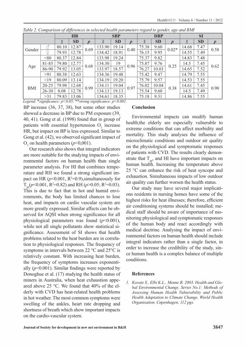
HealthMED - Volume 6 / Number 11 / 2012
Journal of Society for development in new net environment in B&H 3847
Table 2. Comparison of differences in selected health parameters regard to gender, age and BMIHR SBP DBP SISD p SD p SD p SD p
Gender 80.10 12.87 0.69 133.90 19.14 0.40 75.38 9.60 0.02* 14.68 7.47 0.58 79.93 12.78 134.42 18.91 76.15 9.95 14.55 7.49
Age
<80 80.37 12.84
0.68
133.98 19.24
0.96
75.37 9.82
0.25
14.83 7.48
0.6281-85 79.80 12.77 134.30 19 75.87 9.76 14.5 7.4586-90 79.92 13.05 133.97 18.57 76.27 10.03 14.65 7.52>91 80.38 12.63 134.36 19.48 75.42 9.47 14.79 7.55
BMI
<19 80.09 13.14
0.99
134.19 19.20
0.97
75.79 9.57
0.38
14.53 7.55
0.9020-25 79.98 12.68 134.11 19.04 76.02 10.04 14.61 7.4526-30 8.08 12.78 134.13 19.13 75.54 9.60 14.5 7.49>31 79.83 13.06 134.61 18.35 75.18 9.31 14.86 7.55
Legend: *significance: p<0.05; **strong significance: p<0.001.
x- x-x-x-
BP increase (36, 37, 38), but some other studies showed a decrease in BP due to PM exposure (39, 40, 41). Gong et al. (1998) found that in group of patients with essential hypertension O3 increase HR, but impact on BP is less expressed. Similar to Gong et al. (42), we observed significant impact of O3 on health parameters (p<0,001).
Our research also shows that integral indicators are more suitable for the studying impacts of envi-ronmental factors on human health than single parameter analysis. For HI that combines tempe-rature and RH we found a strong significant im-pact on HR (p<0.001, R2=0.9),simultaneously for Tap(p<0.001, R2=0.82) and RH (p>0.05; R2=0.03). This is due to fact that in hot and humid envi-ronments, the body has limited chances to lose heat, and impacts on cardio vascular system are more greatly expressed. Similar affects can be ob-served for AQSI when strong significance for all physiological parameters was found (p<0.001), while not all single pollutants show statistical si-gnificance. Assessment of SI shows that health problems related to the heat burden are in correla-tion to physiological responses. The frequency of symptoms in intervals between 22 °C and 25°C is relatively constant. With increasing heat burden, the frequency of symptoms increases exponenti-ally (p<0.001). Similar findings were reported by Donoghue et al. (17) studying the health status of miners in Australia, when heat exhaustion appe-ared above 25 °C. We found that 40% of the el-derly with CVD has heat-related health problems in hot weather. The most common symptoms were swelling of the ankles, heart rate dropping and shortness of breath which show important impacts on the cardio-vascular system.
Conclusion
Environmental impacts can modify human health;the elderly are especially vulnerable to extreme conditions that can affect morbidity and mortality. This study analyses the influence of microclimatic conditions and outdoor air quality on the physiological and symptomatic responses of patients with CVD. The results clearly demon-strate that Tap, and HI have important impacts on human health. Increasing the temperature above 25 °C can enhance the risk of heat syncope and exhaustion. Simultaneous impacts of low outdoor air quality can further worsen the health status.
Our study may have several major implicati-ons residents in nursing homes have some of the highest risks for heat illnesses; therefore, efficient air conditioning systems should be installed; me-dical staff should be aware of importance of mo-nitoring physiological and symptomatic responses of the human body and react accordingly with medical doctrine. Analysing the impact of envi-ronmental factors on human health should include integral indicators rather than a single factor, in order to increase the credibility of the study, sin-ce human health is a complex balance of multiple conditions.
References
1. Kovats S., Elbi K.L., Menne B. 2003. Health and Glo-bal Environmental Change. Series No.1: Methods of Assessing Human Health Vulnerability and Public Health Adaptation to Climate Change. World Health Organisation. Cøpenhagen, 112 pgs.
3848
HealthMED - Volume 6 / Number 11 / 2012
Journal of Society for development in new net environment in B&H
2. Haines A., Kovats R.S., Campbell-Lendrum D., Cor-valan C. 2006. Climate change and human health: Impacts, vulnerability, and mitigation. Lancet 367: 2101–2109.
3. Lim C.L. Byrne C., Lee J.K.W. Human thermoregula-tion and measurement of body temperature in exerci-se and clinical settings. Annals Academy of Medicine Singapore 2008;37:347–53.
4. Charkoudian N. Skin blood flow in adult human ther-moregulation: How it works, when it does not, and why. May Clinic Proceedings 2003; 78:603–612.
5. Havenith, G. 2005. Temperature Regulation, Heat Balance and Climatic Stress. V: Kirch W., Monne B., Bertollini R. Extreme Weather Events and Public Health Responses. Springer. Berlin Heidelberg New York. 69–80.
6. Dhainaut J.-F., Claessens Y.-E., Ginsburg C., Riou B. 2004. Unprecedented heat-related deaths during the 2003 heat wave in Paris: consequences on emergency departments. Critical Care 8:1–2.
7. Hansen A., Bi P., et al. The effect of heat waves on hos-pital admissions for renal disease in a temperate city in Australia. Int J Epidemiol 2008;37(6):1359–65.
8. D’Ippoliti D., Michelozzi P., et al. The impact of heat waves on mortality in 9 European cities: results from the EuroHEAT project. Environ Health 2010; 9:37.
9. Whitman S., Good G., Donoghue E.R., Benbow N., Shou W., Mou S., 1997. Mortality in Chicago attribu-ted to the July 1995 heat wave. American Journal of Public Health 87:1515–1518.
10. Smoyer K.E., Rainham D.G., Hewko J.N. 2000. He-at-stress related mortality in five cities in Southern Ontario: 1980–1996. Int J Biometeorol 44:190–197.
11. Arnfield A.J. 2003. Two decades of urban climate re-search: A review of turbulence, exchanges of energy and water, and the urban heat island. International Journal of Climatology 23:1–26.
12. Collier C.G. 2006. The impact of urban areas on weather. Quarterly Journal of the Royal Meteorolo-gical Society 132:1–25.
13. Semenza J.C., Rubin C.H., Falter K.H., et al. He-at-related deaths during the July 1995 heat wave in Chicago. N Engl J Med 1996;335:84–90.
14. Kilbourne E.M., Choi K., Jones T.S., Thacker S.B. Risk factors for heatstroke: a case-control study. JAMA 1982;247:3332–6.
15. Novikov I., Kalter-Leibovici O., Chetrit A., Stav N., Epstein Y. Weather condition and visits to the medi-cal wing of emergency rooms in a metropolitan area during the warm season in Israel: a predictive mode. International Journal of Biometeorology, 2011 (in press).
16. Gaffin S.L., Hubbard R.W. Pathophysiology of he-atstroke. In: Pandolf K.B., Burr R.E. (eds). Medical aspects of harsh environments. Vol.1. USA, Go-vernment Printing Office, 2001. 161–208.
17. Donoghue A.M., Sinclair M.J., Bates G.P. Heat exhaustion in deep underground metalliferous mine. Occupational Environmental Medicine 2000; 57: 165–174.
18. Coris E.E., Walz S.M., Duncanson R., Ramirez A.M. Heat Illness Symptom Index (HISI): A Novel Instru-ment for the Assessment of Heat Illness in Athletes. Southern Medical Journal 2006; 99 (4): 340–345.
19. Bouchama A., Knochel J.P. 2002. Heat stroke. The New England Journal of Medicine, 346, 25:1978–1988.
20. Kinney P.L., 2008. Climate Change, Air Quality, and Human Health. American Journal of Preventive Me-dicine 35(5): 459–467.
21. Fiala J., Cernikovsky L., de Leeuw F., Kurfuerst P. 2003. Air pollution by ozone in Europe in summer 2003. Overview of exceedances of EC ozone thres-hold values during the summer season April–August 2003 and comparisons with previous years. Cope-nhagen, Denmark: European Environment Agency and the European Topic Centre on Air and Climate Change.
22. Rainham D.G.C., Smoyer-Tomic K.E. 2003. The role of air pollution in the relationship between a heat stress index and human mortality in Toronto. Envi-ronmental Research 93(1):9–19.
23. Ordonez C., Mathis H., Furger M., Henne S., Huglin C., Staehelin J., et al. 2005. Changes of daily sur-face ozone maxima in Switzerland in all seasons from 1992 to 2002 and discussion of summer 2003. Atmospheric Chemistry and Physic 5:1187–203.
24. Conti S., Meli P., Minelli G., Solimini R., Toccaceli V., Vichi M., Beltrano C., Perini L. 2005. Epidemio-logic study of mortality during the summer 2003 heat wave in Italy. Environmental Research 98:390–399.
25. Ibald-Mulli A., Timoten K.L., Peters A., Heinrich J., Wölke G., Lanki T., Buzorius G., Kreyling W.G., de Hartog J., Hoek G., ten Brink H.M., Pekkanen J. Effects of particulate air pollution on blood pressure and heart rate in subjects with cardiovascular dise-ase: A multicenter approach. Environmental Health Perspectives 2004; 112;3; 259–377.
26. Whitsel E.A., Quibrera M., Christ S.L., Liao D., Pri-neas R.J., Andreson G.L., Heiss G. Heart rate va-riability, ambient particulate matter air pollution, and glucose homeostasis: The environment epide-miology of arrhythmogenesis in the women’s health initiative. American Journal of Epidemiology 2009; 169:693–703.
HealthMED - Volume 6 / Number 11 / 2012
Journal of Society for development in new net environment in B&H 3849
27. De’ Donato F.K., Stafoggia M., Rognoni M., Ponci-no S., Caranci N., Bisnati L., Demaria M., Forastie-re F., Michelozzi P., Pelosini R., Perucci C.A. Airport and city centre temperatures in the evaluation of the association between heat and mortality. Internatio-nal Journal of Biometeorology 2008; 52: 301–310.
28. Theoharatos G., Pantavou K., Mavrakis A., Spanou A., Katavoutas G., Efstathiou P., Mpekas P., Asima-kopoulos D. 2010. Heat waves observed in 2007 in Athens, Greece: Synoptic conditions, bioclima-tological assessment, air quality levels and health effects. Environmental Research 110: 152–161.
29. Pickering T.G. Hall, J.E., Appel, L.J., Falkner, B.E., Graves, J., Hill M.N., Jones D.W., Kurtz T., Sheps S.G., Rocella E. 2005. Recommendations for blood pressure measurement in humans and experimental animals: Part 1: Blood pressure measurement in humans: A public education of the American Heart Association council on high blood pressure resear-ch. Circulation 2005 111:697–716.
30. Klein D.G. Mitchell C., Petrinec A., Monroe M.K., Oblak M., Ross B., Youngblut J.M. A comparison of pulmonary artery, rectal, and tympanic membra-ne temperature measurements in the ICU Heart & Lung – The Journal of Acute and Critical Care 1993 22(5): 435–441.
31. Kenney W.L., Munce T.A. 2003. Physiology of Aging. Invited Review: Aging and human temperature re-gulation. Journal of Applied Physiology 95:2598–2603.
32. Kent S.T., Howard G., Crosson W.L., Prineas R.J., McClure L.A. The association of remotely sensed outdoor temperature with blood pressure levels in REGARDS: a cross-sectional study of a large, nati-onal cohort of African-American and white partici-pants. Environmental Health 2011; 10:7: 1–12.
33. Alpérovitch A., Lacombe J.M., Hanon O., Dartigues J.F., Ritchie K., Ducimetière P., Tzourion C. Relati-onship between blood pressure and outdoor tempe-rature in large sample of elderly individuals. Archi-ves of Internal Medicine 2009; 169(1)75–80.
34. Charach G., Rabinovich P.D., Weintraub M. Se-asonal changes in blood pressure and frequency of related complications in elderly Israeli patients with essential hypertension. Gerontology 2004; 50: 315–321.
35. Schneider A., Schuh A., Maetzel F.K., Rückerl R. Breitner S., Peters A. Weather-induced ischemia and arrhythmia in patients undergoing cardiac rehabi-litation: another difference between men and wo-men. International Journal of Biometeorology 2008; 52:535–547.
36. Brook R.D., Urch B., Dvonch J.T., Bard R.L., Speck M., Morishita M., Marsik F.J., Kamal A.S., Kaciroti N., Harkema J., Corey P., Silverman F., Gold D.R., Wellenius G., Mittlenman M.A., Rajagopalan S., Brook J.R. Air pollution and blood pressure: Insights into the mechanisms and mediators of the effects of air pollution exposure on blood pressure and vascu-lar function in healthy humans. Hypertension. 2009; 54: 659–667.
37. Harrabi I., Rondeau V., Dartigues J.F., Tessier J.F., Filleul L. Effects of particulate air pollution on systo-lic blood pressure: a population-based approach. Environmental Research 2006; 101:89–93.
38. Pope C.A. III, Verrier R.L., Lovett E.G., Larson A.C., Raizene M.E., Kanner R.E. Heart rate variability associated with particulate air pollution. American Heart Journal 1999; 138:890–899.
39. Gong H., Linn W.S., Terrell S., Anderson K., Clark A., Terrell L. Controlled exposure of healthy and asthmatic volunteers to concentrated ambient fine particles in Los Angeles. Inhalation Toxicology 2002; 15(4): 305–325.
40. Peters A., Perz S., Döring A., Stieber J., Joenig W., Wichmann H.E. Increases in heart rate during air pollution episode. American Journal of Epidemio-logy 1999; 150 (10):1094-1098.
41. Gold D.R., Litonujua A., Schwartz J., Lovett E., Lar-son A., Nearing B. Ambient pollution and heart rate variability. Circulation 2000; 101:1267–1273.
42. Gong H. Jr., Wong R., Sarma R.J., Linn W.S., Sulli-van E.D., Shamoo D.A., Anderson K.R., Prasad S.B. Cardiovascular effects of ozone exposure in human volunteers. American Journal of Respiratory Criti-cal Care and Medicine 1998; 158: 538–546.
Corresponding AuthorRok Fink,Universityof Ljubljana,FacultyofHealthSciences,Ljubljana,Slovenia,E-mail: [email protected]

3850
HealthMED - Volume 6 / Number 11 / 2012
Journal of Society for development in new net environment in B&H
Distal movement of the first upper molars with pendulum appliance. Case reportKonstantinos Papadopoulos1, TatjanaTanic2
1 Specialistic orthodontic practice, Katerini, Greece 2 Department of Orthodontics, Faculty of Medicine, University of Nis, Serbia
Abstract
By this work it has been shown the applicati-on of Pendulum appliance for the upper first mo-lars distal movement and the way this appliance works. The biggest advantage of this appliance is that there is no need for the patient’s cooperati-on, in contrast to the traditional method with the use of extraoral Headgear-appliances. With a re-gular activation a successful distal movement of the upper first molars can be succed. If there are second upper permanent molars present, their dis-talization can be achieved too. The action level of Pendulum-appliance is primarily in the maxillary dentition. However, it can have a minor skeletal effect-increase of anterior lower facial height.
Keywords: Class II malocclusion, Pendulum appliance
Introduction
One of the therapeutic concepts in Class II dentoalveolar therapy is a creation of space in maxillary arch by the upper first molars distaliza-tion, canines and incisors until Class I relationship is established. A traditional therapeutic procedu-re is the use of an extraoral headgear-appliance [1,2,3]. However, clinical experiences have indi-cated that the use of those appliances mentioned, has been very often proved to be unsuccessful [4]. The intraoral removable appliances which were introduced since 1980 [5], had also a limitted success because of a frequent lack of compliance with a patient. In later years, for the distalization of the upper molars, magnets were applied [6]. Other researchers, have been recommending the use of a continuous and constant forces produced from the use of superelastic coils [7,8]. However, according to the biomechanical aspects the above mentioned appliances had a lot of negative effects [9,7,10,11].
Hilgers described Pendulum appliance in 1991 [12,13] for the first time that was a turning point, because with this appliance an expansion of the upper jaw and at the same time a rotation and a distal movement of the first upper molars, could have been performed.
The aim of this case’s report was to follow the efficiency of pendulum appliance therapy in a 13 year old child.
Description of appliance
Parts of Pendulum applianceA passive part indicating a modification of
Nance appliance, has been bilateraly connected to the first upper and second premolars. The acrylate part has to be remote about 5 mm from the teeth in order to keep a dental hygiene.
An active part consisting of two TMA springs which has a distal position (0,32” wires, Ormco Corp. Glendora, Galif) going into an acrylate part. A free end has to be curved so that it could fit into the palatal sheaths of the upper first molars tubes. There is also a spiral loop, a little horizontal cur-ve for adaptation, and a free end inserted into the acrylate. The springs must be placed as close to the centre of the acrylate part to have an increa-sing range of action, having an easier position for insertion on the palatal sheaths of tubes and force reducing to the biologically accepted level. The activation of springs is more successful before the appliance is placed in the mouth. If there has been a need for a distalization of a greater degree for the upper first molars, then the springs have to be activated according to the palatal sutura under the angle of 45o. By using the appliance in this way we could manage 60 % of a coil’s activation after an adaptation, for there has been a loss of 1/3 of applicated force on the palatal tube.
As to Byloff and Darendeliler (1997) [14,15] the springs are to be activated for 45o.
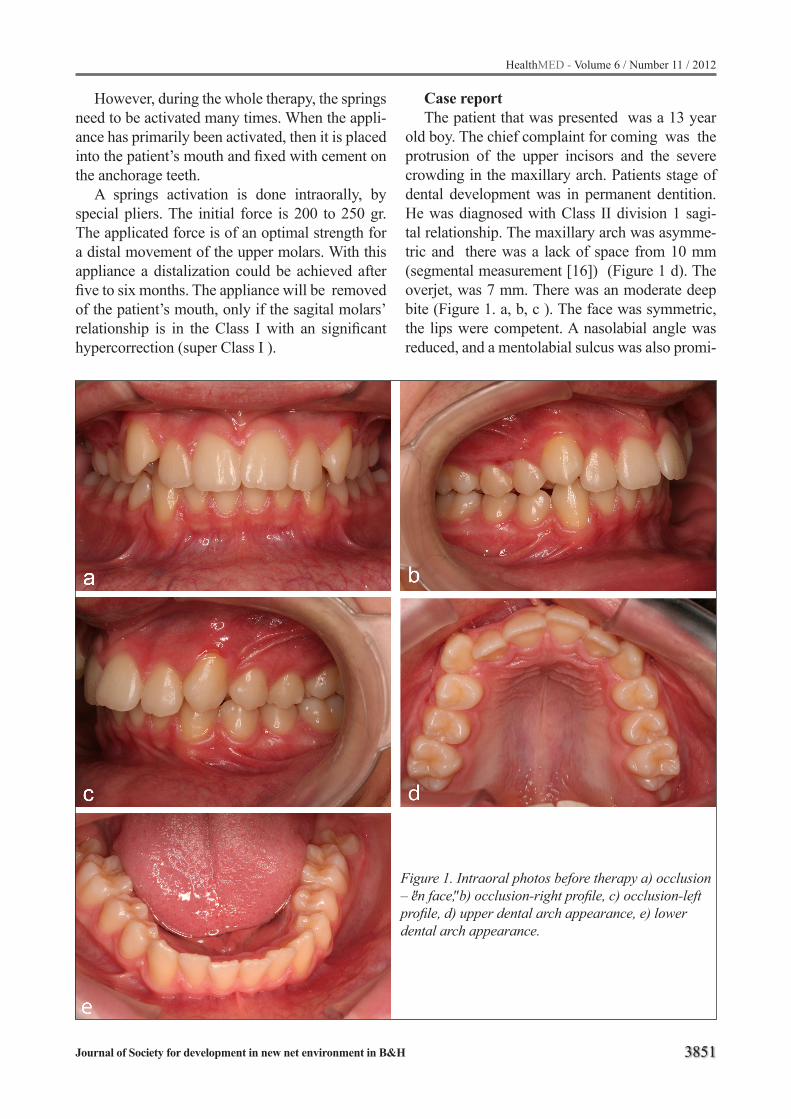
HealthMED - Volume 6 / Number 11 / 2012
Journal of Society for development in new net environment in B&H 3851
However, during the whole therapy, the springs need to be activated many times. When the appli-ance has primarily been activated, then it is placed into the patient’s mouth and fixed with cement on the anchorage teeth.
A springs activation is done intraorally, by special pliers. The initial force is 200 to 250 gr. The applicated force is of an optimal strength for a distal movement of the upper molars. With this appliance a distalization could be achieved after five to six months. The appliance will be removed of the patient’s mouth, only if the sagital molars’ relationship is in the Class I with an significant hypercorrection (super Class I ).
Case report The patient that was presented was a 13 year
old boy. The chief complaint for coming was the protrusion of the upper incisors and the severe crowding in the maxillary arch. Patients stage of dental development was in permanent dentition. He was diagnosed with Class II division 1 sagi-tal relationship. The maxillary arch was asymme-tric and there was a lack of space from 10 mm (segmental measurement [16]) (Figure 1 d). The overjet, was 7 mm. There was an moderate deep bite (Figure 1. a, b, c ). The face was symmetric, the lips were competent. A nasolabial angle was reduced, and a mentolabial sulcus was also promi-
Figure 1. Intraoral photos before therapy a) occlusion – "en face", b) occlusion-right profile, c) occlusion-left profile, d) upper dental arch appearance, e) lower dental arch appearance.

3852
HealthMED - Volume 6 / Number 11 / 2012
Journal of Society for development in new net environment in B&H
nent.The patient’s soft tissue profile was convex with a reduction of the lower anterior face height (Figure 2. a, b). By analysing the cephalometric roentgengram (Figure 2. c, d), the following valu-es were established (Table 1):
- There was the skeletal Class II (angle ANB 6o), maxillary prognathism (angle SNA 84o, Angle FH-NA 92o (angle Lande) and the mandibular retrognathizm (angles SNB 78o, FH-NPog 87o); counterclockwise rotation of the mandibule (angles FH-MP 20o, SN-MP, 28o), a labial inclination of the upper incisors (angles U1-FH 114o, U1-PP 114o U1-Apog 35o), labial inclination of the lower incisors (angles L1-FH 54o, L1-MP 105o, L1-Apog 27o).
- The interincisal angle was reduced (120o)- The lower anterior face height was also
reduced.- The following tretment goals were set:- Correction of dental sagital relationship
from Class II into Class I, - Non extraction treatment therapy. - The over bite’s and overjet’s correction.
- Optimal coordinated of the upper and lower dental arch forms, with ideal torque and inclination,
- Improvement of facial aesthetics with a pleasing smile.
As it has already been mentioned, by pendu-lum appliance a distalization of the upper molars has been performed with forming a space betwe-en these teeth and premolars or primary molars if they are present. Upon fabrication in the labo-ratory, the appliance was placed in the patient’s mouth. The retentional wire elements were bon-ding on the occlusal surfaces of the upper first and second premolars by light cured composite resin. The bands are bondind on the upper molars with a glass-ionomer cement. The springs for distalizati-on were activated to 45o, every month.
The correction of the molars’ sagital relati-onship, was established after six months. At the same time there was a space between molars and premolars (about 6-6,5 mm) bilateral which sho-uld be used for the retraction of premolars and ca-nines. Soon after pendulum appliance had been removed, a Nance holding arch was placed, as an anchorage to prevent the mesial relaps of the upper molars. Then a fixed appliance in the upper jaw was placed, by which a retraction of the se-cond premolars began then of first premolars and canines with the lacebacks.
The incisors retraction was done with a gable-bend on the 0,16 x 0,22 stainless steel archwire. At the same time a fixed appliance was placed in the lower jaw. The active treatment time was 26 months. After having been removed fixed applian-ces, the fixed retainer was used (from 33-43), and in the upper jaw there was clear plastic retainer.
The following results were achieved (Figure 3. a, b, c, d, e):
- correction of sagital relations to Class I both in molars and canines;
- correction of the severe crowding and the upper dental arch correction;
- reduction of the overjet from 7 mm to 2mm;
- optimally leveled dental arches:- a satisfactory face aesthetics was achieved
(Figure 4. a,b,c).Values of cephalometric analysis were shown
(Table 1):
Figure 2. Patient's facial appearance before therapy a) "en face", b) profile, c) lateral head radiograph befo-re therapy, d) cephalometric tracing before therapy.
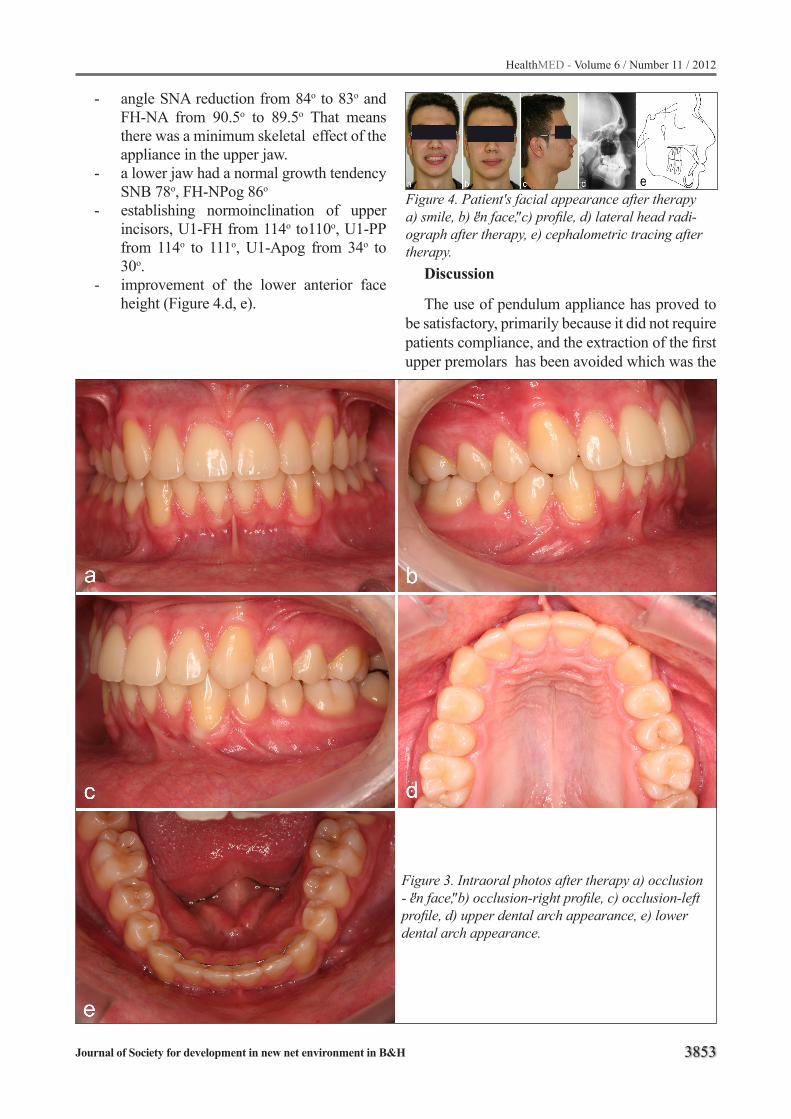
HealthMED - Volume 6 / Number 11 / 2012
Journal of Society for development in new net environment in B&H 3853
- angle SNA reduction from 84o to 83o and FH-NA from 90.5o to 89.5o That means there was a minimum skeletal effect of the appliance in the upper jaw.
- a lower jaw had a normal growth tendency SNB 78o, FH-NPog 86o
- establishing normoinclination of upper incisors, U1-FH from 114o to110o, U1-PP from 114o to 111o, U1-Apog from 34o to 30o.
- improvement of the lower anterior face height (Figure 4.d, e).
Figure 3. Intraoral photos after therapy a) occlusion - "en face", b) occlusion-right profile, c) occlusion-left profile, d) upper dental arch appearance, e) lower dental arch appearance.
Discussion
The use of pendulum appliance has proved to be satisfactory, primarily because it did not require patients compliance, and the extraction of the first upper premolars has been avoided which was the
Figure 4. Patient's facial appearance after therapy a) smile, b) "en face", c) profile, d) lateral head radi-ograph after therapy, e) cephalometric tracing after therapy.

3854
HealthMED - Volume 6 / Number 11 / 2012
Journal of Society for development in new net environment in B&H
alternative treatment plan for a Class II division 1 therapy. With a distalization of the upper first mo-lars, Class I molar relationship has been achieved, so a dentoalveolar compensation has been avoi-ded which was present with the traditional concept of extractional therapy. All results achieved were on the dentoalveolar level, that was confirmed by a cephalometric analysis. Hilgers [12,13], Gho-sh i Nanda [17] , Byllof i Darendeliler [14,15], Bussick i Mc Namara [18], Angelieri et al. [19], came to this conclusion too. A skeletal effect was established only in terms of minor increase in the lower anterior face height.
Conclusion
Pendulum is one of the most effective and relia-ble appliances which can be used for solving Cla-ss II sagital malocclussions or /and the crowding cases with which, the extraction of teeth hasn’t been included in the course of therapy.
The results can be achieved quickly enough and without patients compliance. The way the appliance works is mainly dentoalveolar and only in maxillary dentition, but it could have a minor skeletal effect mostly on the lower third of the face.
Table 1. Cephalometric analysis results
Dimensions Mean Min. Max. Before therapy
After therapy
Sagital skeletal relationsNSBa
FH – SNFH – NA
FH – NpogSNASNBANB
131˚6˚
88 ˚87,8˚80˚78˚2,8˚
4˚
82˚76,2˚75˚0,5˚
8˚
95˚83,8˚81˚5,1˚
128˚7˚90˚86˚84˚78˚6˚
128˚6˚89˚86˚83˚78˚5˚
Vertical skeletal relationsFH – MPSN – MPSN – PPNSGn
Y – AXISUpper facial heightLower facial height
23˚32˚8,5˚68˚
59,4˚44%56%
17˚30˚7˚63˚53˚44%55%
28˚34˚10˚72˚
66,2˚45%56%
20˚28˚8˚65˚58˚49%51%
21˚27˚7.5˚65˚59˚47%53%
Dental relationsAB – FOPFOP – PPU1 – FHU1 – PP
U1 – APogDist1 – APog
L1 – FHL1 – MPL1 – FOPL1 – APog
Dist L1 – APogU1 – L1
90,1˚11,3˚110˚
110,2˚22˚
2,7 mm65˚
91,4˚72,3˚23˚
0 mm135,4˚
80,75˚9,6˚105˚105˚19˚
-1 mm60˚
-8,5˚68,6˚20˚
-2 mm139˚
96˚13,8˚115˚115˚25˚
+5 mm70˚+7˚
76,7˚26˚
+3 mm150˚
92˚9˚
115˚114˚34˚
9mm54˚105˚62˚27˚
2 mm120˚
96˚8˚
110˚111˚30˚
6mm59˚99˚58˚29˚
3 mm123˚
Soft-tissue relationsDist UL – EPDist LL – EP
-2 mm-1 mm
-3 mm-2 mm
-1 mm0 mm
-1 mm2 mm
-2 mm2 mm
HealthMED - Volume 6 / Number 11 / 2012
Journal of Society for development in new net environment in B&H 3855
References
1. Kloehn SJ. Evaluation of cervical traction of the maxilla and maxillary first permanent molar. Angle Orthod 1961; 31: 91-104.
2. Wieslander L. The effect of force on craniofacial deve-lopment. Am J Orthod 1974; 65: 531-8.
3. Bass NM. The use of extra-oral traction in current orthodontics. Orthodontist 1970; 2: 11-5.
4. Ciger S, Aksu M, Germec D. Evaluation of posttre-atment changes in Class II Division 1 patients after nonextraction orthodontic treatment: cephalometric and model analysis. Am J Orthod Dentofacial Orthop 2005; 127: 219-23.
5. Cetlin NM, Ten Hoeve A. Nonextraction treatment. J Clin Ortod 1983; 17: 396-413.
6. Gianelly AA, Vaitas AS, Thomas WM. The use of ma-gnets to move molars distally. Am J Orthod Dentofa-cial Orthop 1989; 96: 161-7.
7. Gianelly AA, Bednar J, Dietz VS. Japanese NiTi coils used to moved molars distally. Am J Orthod Dentofa-cial Orthop 1991; 99: 564-6.
8. Bondemark L, Kurol J, Bernhold M. Reppeling ma-gnets versus superelastic nickel-titanium coils in si-multaneous distal movement of maxillary first and second molars. Angle Orthod 1994; 64: 189-98.
9. Carano A, Testa M. The distal jet for upper molar dis-talization. J Clin Orthod 1996; 30: 374-80.
10. Wilson WL, Wilson RC. Modular 3D appliances. Problem solving in edgewise, straightwire, and li-ghtwire treatment. J Clin Orthod 1984; 18: 272-81
11. Jones RD, White JM. Rapid Class II molar correc-tion with an open coil jig. J Clin Ortod 1992; 26: 661-4.
12. Hilgers JJ. A palatal expansion appliance for non compliance therapy. J Clin Orthod 1991; 25: 491-7.
13. Hilgers JJ. The pendulum appliance for Class II noncompliance therapy. J Clin Orthod 1992; 26: 706-14.
14. Byloff FK, Darendeliler MA. Distal molar movement using the pendulum appliance. Part 1: Clinical and radiological evaluation. Angle Orthod 1997; 67: 249-60.
15. Byllof FK, Darendeliler MA. Distal molar movement using the pendulum appliance. Part 2: The effects of maxillary molar root uprighiting bends. Angle Ort-hod. 1997; 67: 261-7.
16. Bishara SE. Textbook of orthodontics. St. Louis: W.B. Saunders Company 2001.
17. Ghosh J, Nanda RS. Evaluation of an intaoral maxi-llary molar distalization technique.Am J Orthod 1996; 110: 639-46.
18. Bussick TJ, Mc Namara JA. Dentoalveolar and ske-letal changes associated with the pendulum applian-ce. Am. J Orthod 2000; 117: 333-43.
19. Angelieri F, Almeida RR, Almeida MR, Fuziy A. Dentoalveolar and skeletal changes associated with pendulum appliance followed by fixed orthodontic treatment. Am J Orthod 2006; 129: 520-7.
Corresponding Author Konstantinos Papadopoulos,Specialistic orthodontic practice, Katerini, GreeceE-mail: [email protected]

3856
HealthMED - Volume 6 / Number 11 / 2012
Journal of Society for development in new net environment in B&H
Effectiveness of multi-component balance specific training on active community-dwelling elderlyDarja Rugelj, Marija Tomsic, France Sevsek
University of Ljubljana, Faculty of Health Sciences, Ljubljana, SloveniaAbstract
Introduction: Although impaired balance function is an important risk factor for unexpec-ted falls in the elderly there is still no agreement regarding the type and intensity of training to de-crease this impairment.
Purpose: The purpose of our study was to determine the efficacy of a specially developed multi-component balance-specific exercise pro-gramme with special emphasis on training on a compliant surface to improve the balance of active elderly living in an urban environment.
Methods: The training group consisted of 26 elderly, aged 69.6 ± 6.6 years. They participated in training twice a week for 12 weeks. A force plat-form was used to determine the movement of the body’s centre of pressure during sensory organi-sation tests: quiet stance on a hard and compliant surface with eyes open and closed. To evaluate functional balance skills the four square step test and timed 10 m walk tests were assessed.
Results: After the training period the centre of pressure movement while standing on a hard sur-face did not differ significantly from pre-training. On the other hand, after training, standing on a compliant surface with open eyes was characteri-zed by a significant decrease of both medio-lateral and antero-posterior sway as well as a decrease of the centre of pressure velocity and sway area. Closed eyes on a compliant surface resulted in smaller antero-posterior sway. The time needed to perform the four square balance test and timed 10 m walk test significantly decreased after the trai-ning indicating the transition of the acquired skills into the functional domain.
Conclusion: Multi-component balance-speci-fic training on a compliant surface can improve balance of active, community-dwelling elderly. Even if it is performed only twice a week such
training can, after three months, enhance postural stability, speed of stepping in different directions and gait speed.
Keywords: elderly, balance training, stabilo-metry, compliant surface, gait speed
Introduction
The population of EU member countries is projected for the period of 2008-2060 to become older where the median age of the total popula-tion is likely to increase in all countries without exception due to the combined effect of the exi-sting structure of the population, persistently low fertility and a continuously increasing number of survivors to higher age [1]. In particular, the po-pulation aged over 65 years is expected to increa-se in all European countries, whereas Slovenia is predicted to be by the end of the third decade of this century already among the countries with the oldest population in the world with the increase of the population over 65 years from 16 % of the to-tal population in 2008 to 25% in 2030 and 33 % in 2060 [1]. From the available data for Slovenia for the year 2009 [2] it is known that in the total popu-lation of 1000 persons 38 in the age group 60 to 74 years experienced accidental falls and were con-sequently hospitalized whereas this numbers were 107 in the age group over 85 years. It is also im-portant to note that in 2009 over 60 % of total hos-pital treatments in Slovenia were caused by falls. The risk of accidental falls increases with ageing and the related reduction of physical ability. The prevalent consequences of accidental falls are hip and hand fractures, bruises, pains and as much as 4 % of the affected persons are reported to die as a consequence of a fall [3]. Falls in elderly quite often lead also to loss of independence, associated illness and diminished quality of life. They are not only harmful for the individuals but also present a

HealthMED - Volume 6 / Number 11 / 2012
Journal of Society for development in new net environment in B&H 3857
great burden for the society as general. This data indicate that the problem of accidental falls in el-derly is expected to become even direr and needs to be addressed as soon as possible.
Among the most important risk factors for falls is impaired balance [3]. Balance is closely rela-ted to muscular ability, range of motion especially of ankle joints, visual and other sensory inputs, as well as cognitive and emotional factors. Since human balance is a complex motor and cognitive function, a precise tuning between the propriocep-tive, visual and vestibular systems together with the functional motor control system is necessary [4]. With ageing a decline is expected in motor functions such as muscle strength and enduran-ce, in flexibility, in acuity and in the amount of sensory information from different sensory mo-dalities including the somatosensory and vestibu-lar systems [5]. It is expected that a deficit in any sensory system reflects a change of the processing of sensory information and the resulting motor response and thus also in balance and posture. Redundancy of the afferent inputs of the visual, vestibular and proprioceptive systems is therefore essential for optimal postural control. Visual and vestibular systems cannot completely replace the eventually missing somatosensory input while there exists some evidence that the appropriate so-matosensory input can compensate for the missing visual and vestibular inputs [6]. During everyday activities it may happen that sensory information is conflicting and this may lead to loss of balance and even a fall. Such a case happens for instance when a subject is standing in a bus moving with constant speed - here the visual information is signalling movement whereas the vestibular and proprioceptive systems do not support it. The pro-bability of falls in case of conflicting sensory in-formation increases with the advancing age [7].
There is still no agreement between researchers about the type and intensity of training for the op-timal enhancement or maintenance of balance function in elderly subjects. The only agreement appears to be on a minimum of 50 hours of tra-ining the dose necessary for inducing change in balance function [8]. General exercises may have beneficial effects on muscular strength and ca-pacity but quite often their influence on balance function is minimal [9] or completely absent [10].
These results suggest that the improvement of muscle capacity is not directly transferred to ba-lance function [11,12]. A balance-specific exercise programme is therefore an option to maintain or enhance balance in the elderly. It has been shown that balance-specific training with functional tasks that challenge balance is efficient in frail nursing home residents [13] as well as in functionally more able elderly [6].
Adding a sensory component to functional balance training, especially in the form of com-pliant or movable surfaces, presents an additional challenge for the postural control system. Namely, standing on a compliant surface alters two types of sensory inputs from the lower extremities. The information from the soles is modified by different pressure distribution under the sole and thus diffe-rently affects the cutaneous mechanoreceptors in the foot [14] which are essential for determining the position of the centre of pressure on the base of support. The other effect is dynamic - the ela-sticity of the supporting surface results in addi-tional body movement which requires constant adjustments of the relative positions of body se-gments to keep the centre of gravity over the base of support [15]. Thus, for effective balance on a compliant surface not only are motor responses required but also attention to the performed task is required. Training on a compliant surface, which is occasionally also called proprioceptive training, is a type of senso-motor training. This training has been recently reported mainly in rehabilitation after muscular, knee ligament and ankle injuries [16]. It is reported that such training is effective in preventing repeated injury [17]. In the elderly the reported results of senso-motor training that inclu-des training on compliant and moving surfaces are conflicting, some indicating that such sensory-specific training reduces the influence of mecha-nical destabilisation on body balance [18] and improves inter-muscular coordination [19], while others report no effect on postural sway [20].
In accordance with the system approach ba-lance depends on the interaction between the in-dividual, the task to be performed and the envi-ronment in which the task is carried out [21]. The components within the individual include the interaction of perception, cognition and motor systems. The motor system and its coordination
3858
HealthMED - Volume 6 / Number 11 / 2012
Journal of Society for development in new net environment in B&H
further depends on accurate information from all sensory modalities. To address the complexity of balance a multi-component exercise programme is needed. This term denotes an intervention that in-corporates multiple components, such as the acti-vities targeting performance (muscle strength, en-durance and/or power), balance, postural control and walking or cardiovascular endurance [8]. To date there is a limited amount of evidence on the efficacy of somatosensory-specific training, orga-nised as group training, not on a one-to-one basis as, for instance, in the case of a therapist working with a patient. A protocol with predominant soma-to-sensory training was reported to improve the results of biomechanical and functional balance tests in elderly male subjects [19]. Besides, there even exists doubt as to the feasibility of a multi-component group training programme that would address motor and sensory components of balance in the same training session and would, additio-nally, incorporate range of motion and strength training [22]. It is still not clear whether balance-specific training that targets most of the sensory and motor systems could be effective for active elderly population. There was, thus, a twofold pur-pose to this work: first to evaluate the feasibility of a multi-component training programme organised as group training, and second to evaluate the effi-cacy of specially developed multi-component, ba-lance-specific training programme, with emphasi-se on training on a compliant surface, to improve the balance function of active elderly living in an urban environment.
Methods
Participants The participants were recruited by advertising
in the publications of the Pensioners’ Association of Slovenia and at the notice boards in senior clubs and day care centres. As a consequence 34 persons volunteered to participate in our balance-speci-fic training programme, all of them community-
dwelling in the region of the city of Ljubljana. Be-fore enrolment in the training all participants were informed about the purpose and the programme of the training as well as about the procedures of data collection and they signed written consent. The study was approved by the National Medical Ethic Committee.
In the analysis only the results of those partici-pants were included who regularly participated in the training programme (at least 75% adherence), whose score on the Berg balance scale [23,24,25] was over 46 points and who did not report any ne-urological problems. This reduced the number of subjects to 26; 21 women and 5 men. Characteri-stics of the study group are presented in Table 1.
Motor performances measuresTo determine the levels of functional fitness and
the level of balance prior to training programme functional tests were performed. The Berg balan-ce scale consists of 14 functional activities graded on a scale from 0 to 4. It is valid [23], reliable [24] and sensitive to change [25]. Motor performan-ces of lower extremities were tested by the timed stance on toes test [26] and hand grip strength was estimated using a hydraulic hand dynamometer Jamar (Lafayette Instruments, USA).
Outcome measuresDue to the complexity of balance a combina-
tion of tests were performed that measured vario-us components of balance on the level of sensory systems (sensory organisation test) and on func-tional level (four square test and timed walking test). The main outcome measure of balance du-ring quiet upright standing was tested with the sensory organization test on the force platform. It is a clinical tool for the assessment of the relati-ve contribution of proprioceptive, vestibular and vision system to postural integration. The vali-dity and reliability of the test is well-established [27]. Subjects were standing barefoot on the force
Table 1. Characteristics of the 26 participants of the balance-specific training programme. Average ± SD Minimum Maximum
Age (years) 69.6 ± 6.6 59 82Body mass (kg) 71.3 ± 13.5 49 98
Height (cm) 160 ± 9.7 132 177
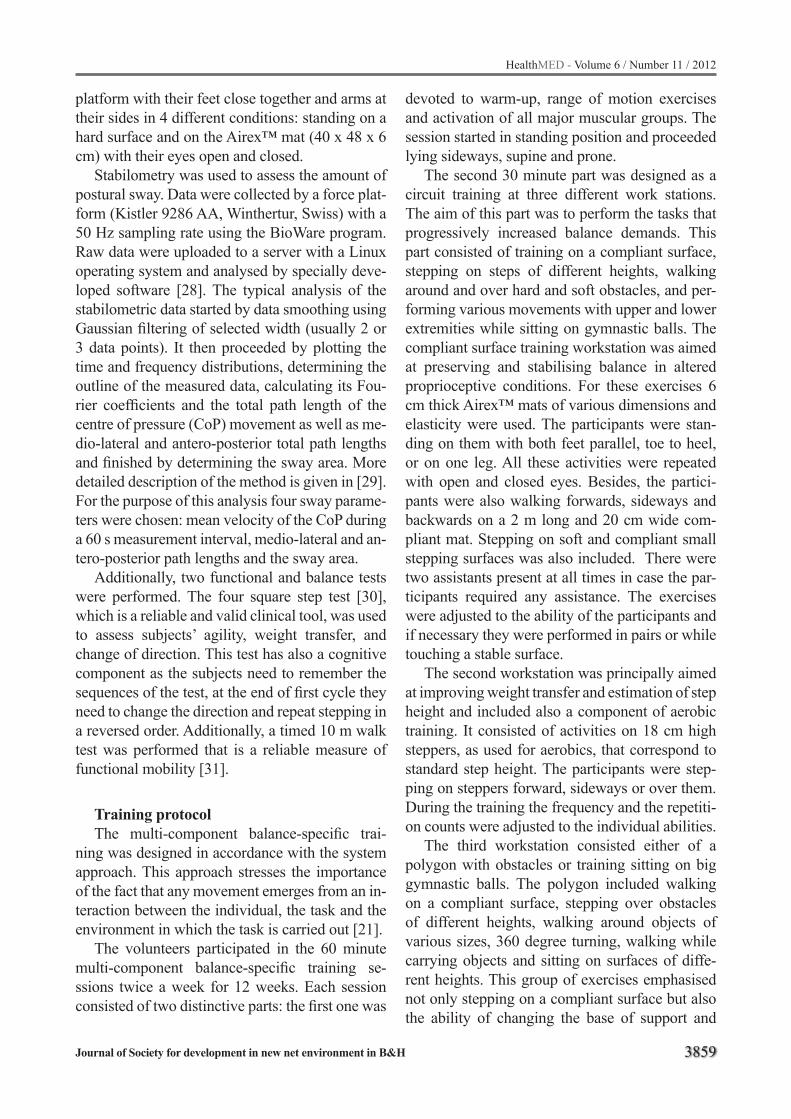
HealthMED - Volume 6 / Number 11 / 2012
Journal of Society for development in new net environment in B&H 3859
platform with their feet close together and arms at their sides in 4 different conditions: standing on a hard surface and on the Airex™ mat (40 x 48 x 6 cm) with their eyes open and closed.
Stabilometry was used to assess the amount of postural sway. Data were collected by a force plat-form (Kistler 9286 AA, Winthertur, Swiss) with a 50 Hz sampling rate using the BioWare program. Raw data were uploaded to a server with a Linux operating system and analysed by specially deve-loped software [28]. The typical analysis of the stabilometric data started by data smoothing using Gaussian filtering of selected width (usually 2 or 3 data points). It then proceeded by plotting the time and frequency distributions, determining the outline of the measured data, calculating its Fou-rier coefficients and the total path length of the centre of pressure (CoP) movement as well as me-dio-lateral and antero-posterior total path lengths and finished by determining the sway area. More detailed description of the method is given in [29]. For the purpose of this analysis four sway parame-ters were chosen: mean velocity of the CoP during a 60 s measurement interval, medio-lateral and an-tero-posterior path lengths and the sway area.
Additionally, two functional and balance tests were performed. The four square step test [30], which is a reliable and valid clinical tool, was used to assess subjects’ agility, weight transfer, and change of direction. This test has also a cognitive component as the subjects need to remember the sequences of the test, at the end of first cycle they need to change the direction and repeat stepping in a reversed order. Additionally, a timed 10 m walk test was performed that is a reliable measure of functional mobility [31].
Training protocolThe multi-component balance-specific trai-
ning was designed in accordance with the system approach. This approach stresses the importance of the fact that any movement emerges from an in-teraction between the individual, the task and the environment in which the task is carried out [21].
The volunteers participated in the 60 minute multi-component balance-specific training se-ssions twice a week for 12 weeks. Each session consisted of two distinctive parts: the first one was
devoted to warm-up, range of motion exercises and activation of all major muscular groups. The session started in standing position and proceeded lying sideways, supine and prone.
The second 30 minute part was designed as a circuit training at three different work stations. The aim of this part was to perform the tasks that progressively increased balance demands. This part consisted of training on a compliant surface, stepping on steps of different heights, walking around and over hard and soft obstacles, and per-forming various movements with upper and lower extremities while sitting on gymnastic balls. The compliant surface training workstation was aimed at preserving and stabilising balance in altered proprioceptive conditions. For these exercises 6 cm thick Airex™ mats of various dimensions and elasticity were used. The participants were stan-ding on them with both feet parallel, toe to heel, or on one leg. All these activities were repeated with open and closed eyes. Besides, the partici-pants were also walking forwards, sideways and backwards on a 2 m long and 20 cm wide com-pliant mat. Stepping on soft and compliant small stepping surfaces was also included. There were two assistants present at all times in case the par-ticipants required any assistance. The exercises were adjusted to the ability of the participants and if necessary they were performed in pairs or while touching a stable surface.
The second workstation was principally aimed at improving weight transfer and estimation of step height and included also a component of aerobic training. It consisted of activities on 18 cm high steppers, as used for aerobics, that correspond to standard step height. The participants were step-ping on steppers forward, sideways or over them. During the training the frequency and the repetiti-on counts were adjusted to the individual abilities.
The third workstation consisted either of a polygon with obstacles or training sitting on big gymnastic balls. The polygon included walking on a compliant surface, stepping over obstacles of different heights, walking around objects of various sizes, 360 degree turning, walking while carrying objects and sitting on surfaces of diffe-rent heights. This group of exercises emphasised not only stepping on a compliant surface but also the ability of changing the base of support and

3860
HealthMED - Volume 6 / Number 11 / 2012
Journal of Society for development in new net environment in B&H
approaching its limits, vestibulo-ocular stabili-sation, changing the direction of movement and double attention. The exercises performed while sitting on a big gymnastic ball presented a moving base of support and constant changes of its size and the number of available fixed points. This gro-up of exercises enabled the training of proactive balance where the activities demanded anticipa-tory postural adjustments and of the reactive ba-lance with the demands for reacting to the moving base of support.
Statistical analysis
The Statistical Package for Social Sciences (SPSS 17, SPSS Inc., Chicago, IL USA) and Microsoft Excel 2003 (Microsoft Inc, Redmond; WA, ZDA) was used for statistical analysis. A pai-red t-test was performed to identify the difference between pre- and post-training outcome measures. The significance level was set at p < 0.008 after a Bonferroni correction was applied.
Results
Pre-training motor performancesBefore training the functional level of balance
was determined, as well as the hand grip strength and the strength of triceps sure muscle of lower extremities. The results (Table 2) show high func-tional level for the age group [32]. Thus, most of the subjects were graded with over 49 points at Berg balance scale and the time of the stance on toes test was 54.6 ± 11.6 seconds. The results for the hand grip strength (Table 2) are given separa-tely for males and females and left and right hands. In all cases the dominant hand was the right one.
StabilometryPostural steadiness was defined as the move-
ment of the body centre of pressure (CoP) on the force platform (postural sway) in a given time interval. The results in Table 3 show that three months of balance-specific training did not affect postural sway while standing on a solid surfa-
Table 2. Pre-training Berg balance scale and muscle strength for hand and foot.Average ± SD Minimum Maximum
Berg balance scale (scale) 54 ± 2 49 56Stance on toes (s) 54.6 ± 11.6 20.1 60Hand grip strength – female left hand (kg) 24.9 ± 5.5 12 34 right hand (kg) 28.2 ± 6.4 18 42Hand grip strength – male left hand (kg) 48.3 ± 12.1 39 62 right hand (kg) 46.3 ± 7.8 40 55
Table 3. Centre of pressure movements during four conditions of sensory organisation test before and after trai-ning. (The significantly different results are bolded. The level of significance is indicated as: ** p < 0.01, *** p < 0.001)
Solid surface, eyes open
Solid surface, eyes closed
Compliant surface, eyes
open
Compliant surface, eyes
closedMedio-lateral path (cm)
pre-training post-training
58.4 ± 21.457.9 ±17.2
96.6 ± 36.4100 ± 53.4
***132.3 ± 35.2110.1 ± 30
293.7 ± 79.8272.8 ± 87.3
Antero-posterior path (cm) pre-training post-training
46.5 ± 19.643.2 ± 15.1
75.6 ± 4075.5 ± 44
104.5 ± 2498.2 ± 33.5
**274.7 ± 84.6236 ± 79.9
Mean velocity (cm/s) pre-training post-training
1.4 ± 0.51.3 ± 0.4
2.3 ± 0.92.3 ± 1.2
**3.1 ± 0.72.7 ± 0.8
7.5 ± 2.16.9 ± 2.1
Sway area (cm2) pre-training post-training
4.7 ± 2.84.2 ± 1.6
8.7 ± 7.27.9 ± 5.4
***14. 6 ± 4.311.1 ± 3.8
55. 6 ± 29.241 ± 17.9
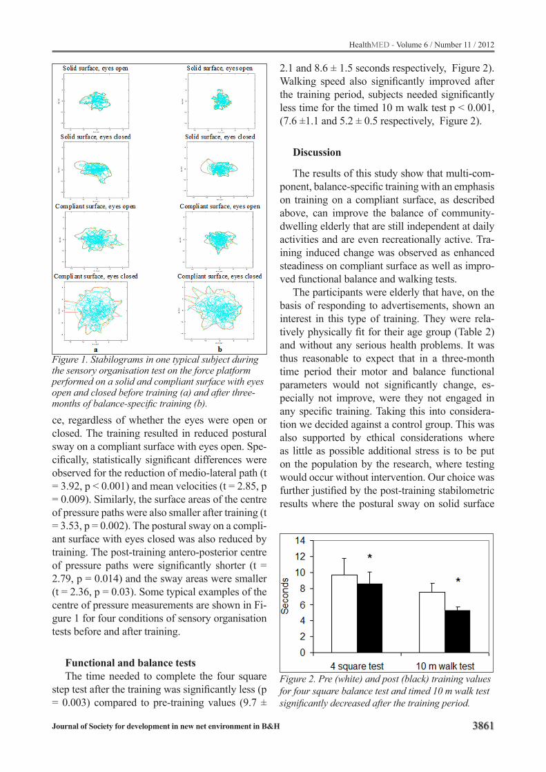
HealthMED - Volume 6 / Number 11 / 2012
Journal of Society for development in new net environment in B&H 3861
ce, regardless of whether the eyes were open or closed. The training resulted in reduced postural sway on a compliant surface with eyes open. Spe-cifically, statistically significant differences were observed for the reduction of medio-lateral path (t = 3.92, p < 0.001) and mean velocities (t = 2.85, p = 0.009). Similarly, the surface areas of the centre of pressure paths were also smaller after training (t = 3.53, p = 0.002). The postural sway on a compli-ant surface with eyes closed was also reduced by training. The post-training antero-posterior centre of pressure paths were significantly shorter (t = 2.79, p = 0.014) and the sway areas were smaller (t = 2.36, p = 0.03). Some typical examples of the centre of pressure measurements are shown in Fi-gure 1 for four conditions of sensory organisation tests before and after training.
Functional and balance testsThe time needed to complete the four square
step test after the training was significantly less (p = 0.003) compared to pre-training values (9.7 ±
2.1 and 8.6 ± 1.5 seconds respectively, Figure 2). Walking speed also significantly improved after the training period, subjects needed significantly less time for the timed 10 m walk test p < 0.001, (7.6 ±1.1 and 5.2 ± 0.5 respectively, Figure 2).
Discussion
The results of this study show that multi-com-ponent, balance-specific training with an emphasis on training on a compliant surface, as described above, can improve the balance of community-dwelling elderly that are still independent at daily activities and are even recreationally active. Tra-ining induced change was observed as enhanced steadiness on compliant surface as well as impro-ved functional balance and walking tests.
The participants were elderly that have, on the basis of responding to advertisements, shown an interest in this type of training. They were rela-tively physically fit for their age group (Table 2) and without any serious health problems. It was thus reasonable to expect that in a three-month time period their motor and balance functional parameters would not significantly change, es-pecially not improve, were they not engaged in any specific training. Taking this into considera-tion we decided against a control group. This was also supported by ethical considerations where as little as possible additional stress is to be put on the population by the research, where testing would occur without intervention. Our choice was further justified by the post-training stabilometric results where the postural sway on solid surface
Figure 1. Stabilograms in one typical subject during the sensory organisation test on the force platform performed on a solid and compliant surface with eyes open and closed before training (a) and after three-months of balance-specific training (b).
Figure 2. Pre (white) and post (black) training values for four square balance test and timed 10 m walk test significantly decreased after the training period.

3862
HealthMED - Volume 6 / Number 11 / 2012
Journal of Society for development in new net environment in B&H
with eyes open or closed remained the same after three-months training period. It is thus reasonable to attribute all eventual post-training differences to the direct or indirect impact of the balance-spe-cific training, though possible placebo effects are recognised.
The participants of our training programme had well preserved balance function which is in-dicated by high scores of Berg balance scale as reported in Table 2. Besides, the results of the ti-med stance on toes and of hand grip strength te-sts are on the upper part of those expected for the appropriate age group [33].This data exhibits that subjects were fit and despite this fact were able to improve balance on compliant surfaces as well as in the 4 square and walking speed tests. This implies that low intensity but functionally specific training is capable of inducing change even when subjects are fit and highly functioning.
The sensory organization test on the force plat-form showed improved stabilization of the cen-tre of pressure on a compliant surface after trai-ning whereas there were no significant changes for standing on a hard surface with eyes open or closed. These results suggest that the participants learned during the training to rely more on the vi-sual and vestibular inputs and thus compensate for the change imposed by the compliant surface on the information coming from the somato-sen-sory system. It is unlikely that the somato-sensory system (as represented by conduction velocity, number of receptors present, etc) was influenced by the exercises, the change was more likely on the level of central processing of these stimuli. This is proposed due to the limited plasticity of the somatory system. The increased ability of sta-bilisation of the COP as a result of the described training protocol could be explained by the theory of sensory re-weighting [34], which suggests that the central nervous system can dynamically adjust the relative weights of the incoming sensory data and thus optimize the balance control. It seems that after the balance-specific training the partici-pants were able to more effectively use the inputs of their muscular and ligament stretch receptors which are particularly active during constant body adjustments when standing on a compliant surface [15].
The ability of weighting and re-weighting of the incoming sensory inputs remains present in the elderly as does the ability for adjustments of the neuromuscular and musculoskeletal systems [35,36]. Regular physical activity, even if started later in life, can result in reorganization of postu-ral control components and improve balance un-der conflicting sensory information [35]. When properly stimulated, the adaptability of systems involved in the control and performance of motor function is known to be preserved also in very old subjects. This applies to the acquisition of muscle force and capacity [36], as well as to the complex function of balance [13]. Besides, moderate physi-cal activity is also beneficial for diminishing oxi-dative stress and reducing the inflammatory pro-cess in the elderly [37].
Besides better stabilisation on compliant surfa-ces, improvement in functional balance was ob-served. Changes were observed in the four square step test and 10 m walk test. In both tests subjects performed faster after training indicating the increased balance skills that consequently allowed subjects to walk faster. Subjects were also able to stand significantly longer on a narrow supporting surface with their eyes closed. Previous results showed improvement in gait speed and balance tests as a result of balance-specific training in su-bjects with reduced balance [38] and nursing home residents [13]. These results support the functio-nally-oriented, multi-component group training protocol that has the potential to be transferred to different environments as well as to everyday life, which should be goal of any motor learning pro-cess [39]. In contrast, traditional group training, consisting of exercises that activate the whole body and include stretching and relaxation, is not reported to be of great value for balance improve-ment [11,12].
Doubts have been proposed as to the feasibility and capability for eliciting change in balance and functional performance when training consists of strength, aerobic and balance training in the same session [22]. In the present study range of motion, strength maintenance and balance-specific trai-ning was administered in the same session, with positive results. Similar studies exist in the litera-ture with the major difference between the present work and these studies being the multi-component

HealthMED - Volume 6 / Number 11 / 2012
Journal of Society for development in new net environment in B&H 3863
nature of the present work that included training on compliant surfaces. Frye et al. [40] reported an increased speed in the up and go test in the group who performed low intensity exercises with main elements of strength, flexibility, endurance and balance as compared to a control group. Iwamo-to [41] programme consisted of callistenics, body balance training, muscle power training, and wal-king and reported an increase of tandem and one leg standing time and increased gait velocity in the exercise group as compared to controls. The pro-gramme also decreased the incidence of falls in the exercise group. It may be concluded that mul-ti-component, balance-specific training that inclu-des senso-motor training (standing on compliant surfaces) is effective for a predominantly female group (present study). Others have found training on a compliant surface to be effective in active, community-dwelling males [19]. However, it is still not possible to conclude that balance-specific training, with an emphasis on somato-sensory tra-ining, is more effective than other types of training as the research results are often conflicting. Some could find no difference between functional trai-ning and resistance exercise training [42] and no difference could be determined between functio-nal and traditional training [38]. Only a systematic study of different training protocols in groups of elderly of various, but matched, balance perfor-mance skills can resolve this question.
Conclusion
A multi-component, balance-specific training which emphasised standing on a compliant surfa-ce improves balance during postural stabilisation tests and is transferred to other functional activi-ties. These results show that even active elderly persons can improve their balance function provi-ded it is reasonably well-preserved at the initiation of training. We can also conclude that multi-com-ponent, balance-specific training can be organi-sed ingroup training and that combining different types of exercises is feasible and effective. The effectiveness of this training needs to be compared with other training protocols.
Acknowledgements
The research was part of the project “Active and healthy ageing” that was partly supported by the Slovenian Research Agency, contract L3-0191-0382-08 and Krka, Novo mesto.
References
1. Giannakouris K. Ageing characterises the demo-graphic perspectives of the European societies. EU-ROSTAT. Stat Focus 2008; 72. Available at: http://epp.eurostat.ec.europa.eu/cache/ITY_OFFPUB/KS-SF-08-072/EN/KS-SF-08-072-EN.PDF. <28.1.2012>
2. Bolnišnične obravnave zaradi poškodb in zastru-pitev. In: Zdravstveni statistični letopis. Ljubljana: Inštitut za varovanje zdravja Republike Sloveni-je, 2009: 483-512. Available at: http://www.ivz.si/Mp.aspx/?ni=0&pi=7&_7_Filename=attName.png&_7_MediaId=3419&_7_AutoResize=false&pl=0-7.3.<8.12.2011>
3. Tinetti ME. Preventing falls in elderly persons. N Engl J Med 2003; 348: 42-9.
4. Massion J, Postural control system. Current opinion in Neurobiology 1944; 4: 877-887.
5. Horak FB, Mirka A, Shupert L. The role of peripheral vestibular disorders in postural dyscontrol in the el-derly. In: Wollacott MH, Shumway-Cook A, eds. The developement of posture and gait across the lifespan. Columbia: University of South Carolina Press, 1989; 253-79.
6. Hu MH, Woollacott MH. Multisensory training of standing balance in older adults. I, Postural stability and one-leg stance balance. J Gerontol 1994; M52- M61.
7. Wollacott M, Shumway-Cook A. Attention and the control of posture and gait: a review of an emerging area of research. Gait Posture 2002; 16: 1-14.
8. Shubert TE, Evidence based exercise prescription for balance and falls prevention: A current review of the literature. Proceedings: Exercise and physical acti-vity in aging 2011; 34: 100-108.
9. Orr R, Raymond J, Fiatarone Singh M. Efficasy of progressive resistance training on balance perfor-mance in older adults. A systemic review of randomi-zed controlled trials. Sports Med 2008; 38: 317-343.
10. Grilly R, Willems D, Trenholm K, Hayes K, Delaqu-erruere-Richardson L. Effects of exercise on postural sway in the elderly. Gerontology 1989; 35: 137-3.
3864
HealthMED - Volume 6 / Number 11 / 2012
Journal of Society for development in new net environment in B&H
11. Skelton DA, Young A, Greig CA, Malbut KE. Effect of resistance training on strength, power and selec-ted functional abilities of woman aged 75 and over. J Am Geriatr Soc 1995; 43: 1081-7.
12. Keysor JJ, Jette AM. Have we oversold the benefit of late-life exercise? J Gerontol A Biol Sci Med Sci 2001; 56: M412-23.
13. Rugelj D. The effect of functional balance training in frail nursing home residents. Arch Gerontol Geriatr 2010; 50: 192-7.
14. Wu G, Chiang JH. The signifficance of somatosen-sory stimulations to the human foot in the control of postural reflexes. Exp Brain Res 1997; 114: 163-169.
15. Horak FB, Hlavacka F. Somatosensory loss increa-ses vestibulospinal sensitivity. J Neurophysiol 2001; 86: 575 -585.
16. Zech A, Hubscher M, Vogt L, Banzer W, Hansel F. Neuromuscular training for rehabilitation of sport injuries: A systemic rewiew. Med Scie Sport Exer 2009; 41: 1831-1841.
17. Hubscher M Zech A, Pfeifer K, Hansel F, Vogt L. Neuromuscular training for sports injury preven-tion: Asystemic review. Med Scie Sport Exer 2010; 42: 413-421.
18. Westlake KP, Gulham EG. Sensory specific balance training in older adults: effect on proprioceptive re-integration and cognitive demands. Phys Ther 2007; 87: 1274- 83.
19. Granacher U, Gruber M, Golhofer A. The impact of sensoriomotor training on postural control in el-derly men. Deutche Z Sportmed 2009; 60: 387-393.
20. Schilling BK, Falvo MJ, Karlage RE, Weiss LW, Lohnes CA, Chiu LZF. Effects of unstable surface training on measures of balance in older adults. J Strength Conditioning 2009; 23: 1211-1216.
21. Shumway-Cook A, Woollacott MH, eds. Motor con-trol: translating research into clinical practice. 3rd ed. Philadelphia: Lippincott Williams & Wilkins; 2007.
22. Baker KM, Atlantis E, Fiatarone Singh. Multi-modal exercise programs for older adults. Age and Ageing 2007; 36: 375-381.
23. Berg K, Wood-Dauphinee S, Williams JI, Gayton D. Measuring balance in the elderly: preliminary de-velopment of an instrument. Physiother Can 1989; 41: 304-11.
24. Berg K, Wood-Dauphinee S, Williams JI, Maki B. Measuring balance in the elderly: validation of an instrument. Can J Public Health 1992; 83 Supl 2: S71.
25. Wood-Dauphinee S, Berg K, Bravo G, Williams JI. The balance scale: responsiveness to clinically me-aningful changes. Can J Rehabil 1996; 10: 35-50.
26. Buchman AS, Boyle PA, Wilson RS, Bienias JL, Bennett DA. Physical activity and motor decline in older persons. Muscle Nerve 2007; 35: 354-62.
27. Shumway-Cook A, Horak FB. Assessing the influ-ence of sensory interaction on balance. Phys Ther 1986; 66: 1548-50.
28. Sevšek F. Stabilometrija V 1.0. Ljubljana: Visoka šola za zdravstvo, 2009. Available at: http://digitus.zf.uni-lj.si/~sevsekf/Programi/Stabilometrija.
29. Rugelj D, Sevšek F. The effect of load mass and its placement on postural sway. Appl. Ergon. 2011; 42(6): 860-866.
30. Dite W, TempleVA. A clinical test of stepping and changes of direction to identify multiple falling ol-der adults. Arch Phys Med Rehabil 2002; 83 (11): 1566–71.
31. Steffen TM, Hacker, TA, Mollinger L. Age- and gen-der-related test performance in community-dwelling elderly people: six-minute walk test, Berg balance scale, timed up & go test, and gait speeds. Phys Ther 2002; 82 (2): 128–37.
32. Lajoie Y, Gallagher SP. Predicting falls within the elderly community: comparison of postural sway, reaction time, the Berg balance scale and the acti-vities-specific balance confidence (ABS) scale for comparing fallers and non-fallers. Arch Gerontol Geriatr 2004; 11-26.
33. Puh U. Age and gender-related differences in hand and pinch grip strength in adults. Int J Rehabil Res 2010; 33: 4-11.
34. Oie KS, Kiemel T, Jeka JJ. Multisensory fusion: si-multaneous re-weighting of vision and touch for the control of human posture. Cognitive Brain Research 2002; 14: 164-76.
35. Buatois S, Gauchard GC, Aubry C, Benetos A, Perrin P. Current physical activity improves balance control during sensory conflicting conditions in ol-der adults. Int J Sports Med 2007; 28: 53-8.
HealthMED - Volume 6 / Number 11 / 2012
Journal of Society for development in new net environment in B&H 3865
36. Vandervort AA. Aging of the human neuromuscular system. Muscle Nerve 2002; 25: 17-25.
37. Rosado-Pérez J, Santiago-Osorio E, Ortiz R, Mendoza-Núñez VM. Moderate physical activitydi-minishes oxidative stress and the inflammatory pro-cess in elderly. HealthMed 2011; 5: 173-179
38. Steadman J, Donaldson N, Kalra L. A randomized controlled trial of an enhanced training program to improve mobility and reduce falls in elderly patients. J Am Geriatr Soc 2003; 51: 847-52.
39. Schmidt RA, Lee TD. Motor control and learning. 4th ed. Champaign: Human Kinetics, 2005; 432-59.
40. Frye, B Scheinthal, S Kemarskaya, T Pruchno, R. Tai chi and low impact exercise: Effects on the physi-cal functioning and psychological well-being of ol-der people. J Appl Gerontol 2007; 26 (5): 433-45
41. Iwamoto, J Suzuki, H Tanaka, K Kumakubo, T Hi-rabayashi, H Miyazaki, Y Sato, Y Takeda, T Matsu-moto, H. Preventative effect of exercise against falls in the elderly: a randomized controlled trial. Osteo-poros Int 2009; 20 (7): 1233-1240.
42. DeVreede PL, Samson MM, VanMeeteren NL, Van der Bom JG, Duursma SA, Verhaar HJ. Functional task exercise versus resistance exercise to improve daily function in older women: a feasibility study. Arch Phys Med Rehabil 2004; 85: 1952-61.
Corresponding Author Darja Rugelj,University of Ljubljana, Faculty of Health Sciences,Ljubljana,Slovenia,E-mail: [email protected]
3866
HealthMED - Volume 6 / Number 11 / 2012
Journal of Society for development in new net environment in B&H
Insufficient caloric intake worsens the outcome of Intensive Care Unit patientsVjollca Shpata1, Albana Gjyzari1, Irena Kito1, Ilir Ohri2
1 Nursing Faculty, University of Tirana, Albania2 Department of Anesthesia and Intensive Care, University Hospital Center “Mother Teresa”, Tirana, Faculty of medicine, University of Tirana, Albania
Abstract
Background: In the patients of the ICU (in-tensive care unit), there is a delayed or inadequate nutrition support. The purpose of this study was to examine the relationship between energy balance and clinical outcome in critically ill patients.
Methods: Prospective observational study conducted in consecutive patients staying ≥4 days in the mixed ICU of the QSUT “Mother Tere-sa”. Data were collected on the nutrition support, energy delivery, and cumulated energy balance (on discharge), length of ICU stay, total compli-cations, infectious complications and mortality. Energy balance was calculated as energy delivery minus nutritional requirements. Multiple regre-ssion analysis was used to analyze the effect of cumulated energy balance on length of ICU stay, complications and on mortality.
Results: Multiple regression analysis identi-fied cumulated energy deficits as being indepen-dently associated with infectious complications and the ventilator stay and ICU stay. Kaplan-Meier analysis showed that patients with a cumu-lated energy deficit ≥ -10000kcal, had a higher ICU mortality rate than patients with lower energy deficit.
Conclusions: Negative energy balance cumu-lated during inadequate nutrition support was associated with a higher rate of infectious compli-cations, mortality, longer ventilator stay and ICU stay. Our findings suggest the need for implemen-tation of quality improvement measures by the he-althcare team to enhance the provision of nutrition support to the patients of the intensive care unit.
Keywords: critically ill, nutritional support, energy balance, morbidity and mortality.
Introduction
Protein energy malnutrition is a major problem in severely ill hypercatabolic patients in the inten-sive care unit (1). In the intensive care unit 20-40% of the patients were reportedly malnourished (2,3,4). The rapidity of development of severe malnutrition depends on the degree of insufficient intake of nutrition as well as on the metabolic rate. In the critically ill patient, malnutrition results in impaired immunologic function, impaired ven-tilator drive, and weakened respiratory muscles leading to prolonged ventilator dependence and increased infectious morbidity and mortality rates (5,6). Nutrition care is considered to be a basic and mandatory (essential) element of modern in-tensive care treatment. Nutritional support influ-ences morbidity and mortality rates in critically ill patients (6). Administration of nutritional support is required in critically ill patients to limit the ne-gative energy and protein balance observed in these patients (3,7). Prevention of nutrient deple-tion during nutritional support can eliminate the excess morbidity and mortality associated with malnutrition. The general benefits of nutritional support include improved wound healing (6) a decreased catabolic response to injury (8), impro-ved gastrointestinal permeability (9), decreased bacterial translocation (10), and improved clinical outcomes, including a decrease in complication rates and length of stay with accompanying cost savings (11-14). Prolonged hypocaloric feeding is associated with clinical complications; energy ba-lance should be calculated in the most ill patients. Underfeeding ICU patients for periods longer than a few days can cause unnecessary protein losses, may lead to further nutritional deterioration, and consequent complications (increased nosocomial infections, poor wound healing and respiratory muscle dysfunction) (15). Particularly in the pa-
HealthMED - Volume 6 / Number 11 / 2012
Journal of Society for development in new net environment in B&H 3867
tients of the ICU, there is a delayed or inadequate nutrition support (4,16,17). The purpose of this study was to examine the relationship between energy balance and clinical outcome in critically ill patients.
Material and methods
The study was designed as a prospective obser-vational study, conducted in consecutive patients staying ≥4 days in the mixed ICU of the Univer-sity Hospital Centre “Mother Teresa” of Tirana, Albania, collecting data during 2010 and 2011. As nutrition support may only be relevant to critically ill patients who remain in the ICU for a prolonged period of time, we examined the use of nutrition support in patients above 18 years old, who re-mained in the ICU longer than 4 days. Data were made anonymous for analysis.
Patient dataAge, sex, weight, height, and BMI, diagnosis
and Acute Physiology and Chronic Health Eva-luation (APACHE II) prognosis score (18) were recorded upon admission to the ICU. Nutritional status on admission was assessed according to Nu-tritional Risk Screening 2002 (19). The patients were characterized by scoring the components “undernutrition” and “severity of disease” in 4 ca-tegories (absent, mild, moderate or severe). The patient could have a score of 0-3 for each compo-nent (undernutrition and severity of disease), and any patient with a total score ≥ 3 was considered at nutritional risk. Undernutrition was evaluated by any of the 3 variables (BMI, recent weight loss, recent food intake).
Nutritional data and calculationsDetermination of energy requirements: Indirect
calorimetry, despite being the gold standard for de-termination of energy requirements, remains unava-ilable in the vast majority of ICUs (20). Predictive equations however over- or underestimate require-ments (21). Indirect calorimetry studies have shown that, in most situations, a value of 25 kcal/kg/day can be used to estimate energy requirements. This is also recommended by ESPEN (22,23). As in our clinic is not available indirect calorimetry, energy target was set at 25 kcal/kg/day.
Energy delivery Total delivery includes energy from enteral
and parenteral feeds, from non-nutritional sources (glucose and gluco-saline infusions used for drug dilution and fluid support). Energy balance was calculated as energy delivery – energy target, on daily basis. Data were collected on the nutritional risk screening, the time of start of feeding, energy delivery, and cumulated energy balance after first week and on discharge from ICU.
Clinical follow-upDuration of ICU stay and length of ventilator
stay, total complications, infectious complicati-ons and ICU mortality were recorded. Infectious complications were defined as sepsis or systemic inflammatory response syndrome (24), pneumo-nia, urinary tract infection, central venous catheter sepsis, and wound infection. Other complications were: post-operative (open abdominal wound, post-operative bleeding, anastomotic leak), neu-rological, respiratory, gasto-intestinal, cardiovas-cular, hepatic failure (by SOFA), renal failure (by SOFA) (25), and coagulation disorder. The durati-on of time in the ICU was defined as the time from
Table 1. Patient characteristicsVariable Mean SD(median) RangeAge years years 60 +16 (61) 18/92
Sex ratio M/F M/F 156M/121F APACHE II 17.5±5.7(16) 5/32
Time from admission to initiation of nutrition days 2.9 ±3.6(1) 1/23Mechanical ventilation days days 1.8 ± 3.9 (0) 0/25
Complications n (%) 132(47.7%)Length of ICU stay days days 8.3±7.2 (6) 4/62
ICU Mortality n (%) 86 (31%)
3868
HealthMED - Volume 6 / Number 11 / 2012
Journal of Society for development in new net environment in B&H
admission of patients until they were ready for discharge. All patients were followed clinically until leaving the ICU or death and their outcome recorded.
Statistical analysisData are presented as means, medians and
ranges for numerical variables and as number or percentages for categorical variables. Multiple li-near regression analysis was used to analyze the effect of cumulated energy balance on length of ICU stay, length of ventilator stay, total compli-cations, infectious complications and mortality. Spearman’s non parametric test was used to assess the correlation between variables. Kaplan-Meier analysis was used to compare the survival rate in patients with energy deficit. Statistical significan-ce was considered at the level of p ≤ 0.05. All te-sts were two tailed. SPSS 18.0 statistical package used to analyze the data.
Results
277 patients were included in the study. The mean age was 60.1years (±16.2yrs). 141 (43.7%) patients were females. The mean APACHE II sco-re 17.5 (±5.7). According to NRS 2002: 61.3% of
the patient were at nutritional risk. ICU length of stay was 8.3 days (±7.2); Mechanical ventilation lasted 1.8 days (± 3.9). ICU mortality was 31% (Table 1). 19.1% of the patients had malign disea-se. 201 (72.6%) patients were post operative (Ta-ble 2).
Nutritional risk (NRS-2002 ≥ 3) was positively correlated with total and infectious complications (rho=0.5, p<0.01), length of stay at ICU (rho=0.3, p<0.01), mortality (rho=0.2, p<0.01), and days in mechanical ventilation (rho=0.2, p<0.01).
Nutritional support: Of the 277 study patients, 109 (39.4%) received some form of nutrition support: 16 (5.7%) received enteral nutrition (EN) only, 87 (31.4%) received parenteral nutrition (PN) only, and 6 (2.2%) received both, and 168 (60.6%) did not receive any nutritional support. The days without feeding were characterized by the unintentional delivery of 150-200 kcal from glucose infusions.
For those patients who received nutritional support, the mean time from admission to initia-tion of any form of nutrition support was 2.9 days (±3.6); all patients were in negative balance at the end of their ICU stay: -10384.0 (±9133.2kcal) (Table 3).
Impact of energy deficit on intensive care unit
Table 2. Major diagnoses of the total study population on ICU admissionMajor diagnosis N Frequency (%)
Nonoperative conditions:Respiratory neoplasm 2 0.7Pulmonary edema (non-cardiogenic) 2 0.7Pulmonary embolism 5 1.8Respiratory failure 11 4.0Multiple trauma (excluding head trauma) 4 1.4Metabolic coma 7 2.5Renal disease/Acute renal failure 6 2.2Multiorgan failure 8 3.0
Post-operative conditions:GI perforation/rupture; fistula 12 4.3GI inflammatory disease 8 2.9GI obstruction 5 1.8GI bleeding 55 19.9Pancreatitis 14 5.1GI neoplasm 46 16.6GI cholecystitis / cholangitis 28 10.1Other GI disease; tromboembolitic event 23 8.3Renal neoplasm 7 2.5
GI = gastrointestinal
HealthMED - Volume 6 / Number 11 / 2012
Journal of Society for development in new net environment in B&H 3869
Table 3. Patients characteristics by nutritional group
EN (n=16) PN (n=87) EN+PN (n=6) No nutrition support
Total (n=277)
Age (years)- Mean - SD
66,81 19,229
60,41 17,665 57,8321,720
59,5614,983
60,2116,272
APACHE II- Mean - SD
18,756,105
16,995,627
20,006,663
17,565,688
17,505,708
LOS ICU- Mean - SD
9,945,183
11,9010,361
16,1710,147
5,993,219
8,307,212
LOV- Mean- SD
1,132,527
3,54 (5,383) 6,337,421
0,83 2,321
1,823,958
Energy cumulative end balance (kcal)
- Mean- SD
-12090,63-5736,846
-10188,79-8743,575
-5126.6-4859.9
-9910,78-5206,461
-10397,74-9146,962
Admission diagnosis (%)- N o n - o p e r a t i v e
conditions- P o s t - o p e r a t i v e
conditions
37.5%
62.5%
19.54
80.45
100
0
28.57
71.42
72.6
27.43LOV = length of ventilationLOS ICU = length of stay at intensive care unit
outcome: Multiple regression analysis identified energy deficits after 7 days as being independently associated with the infectious complications (p=0.007). It did not find any association of ne-gative energy balance after 7 days with the length of mechanical ventilation (p=0.85), and the length of stay on ICU (p=0.16), with the total number of complications (p=0.56), or mortality (p=0.68).
The regression analysis identified cumula-ted energy deficits during the ICU stay as being independently associated with infectious com-plications (p<0.001) the length of stay on ICU (p<0.0001) and the length of mechanical ventila-tion (p<0.001). It did not find any association of negative energy balance with the total number of complications (p=0.19), or mortality (p=0.11) (Ta-ble 4) (Figure 1). Kaplan-Meier analysis showed that 104 patients with a cumulated energy deficit ≥ -10000kcal, had a higher ICU mortality rate than patients with lower energy deficit (< -10000kcal; n=173). Values were significantly different (P<0.001; log-rank test) (Figure 2).
Discussion
Of 277 patients studied, 61.3% were at nutriti-onal risk according to Nutritional Risk Screening 2002. In gastrointestinal surgery was reported that, 60% of the patients were with malnutrition (2). Previous studies have shown the impact of nutritional status on morbidity, mortality, LOS (6, 26). Similarly to these studies we demonstrated that length of ICU stay, invasive ventilator stay, incidence of complications and mortality were greater in the malnourished patients than in the well-nourished. Our study showed that negati-ve energy balance correlated with the infectious
Table 4. Relationship between outcome and cumula-ted energy deficit by regression analysis
Variables of outcome p FLength of stay <0.001 334.5
Total complications 0.196 1.6Infectious complications <0.001 65.1
Length of mechanical ventilation <0.001 53.8
Mortality 0.116 2.4
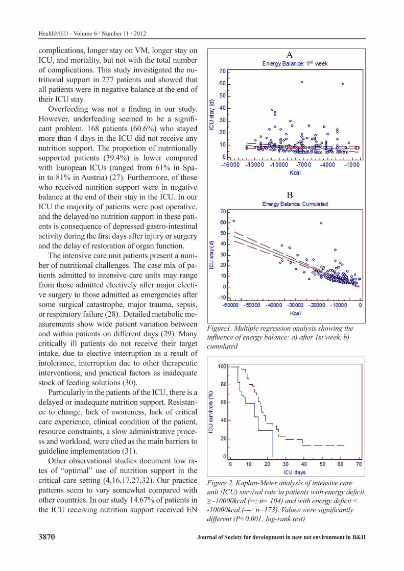
3870
HealthMED - Volume 6 / Number 11 / 2012
Journal of Society for development in new net environment in B&H
complications, longer stay on VM, longer stay on ICU, and mortality, but not with the total number of complications. This study investigated the nu-tritional support in 277 patients and showed that all patients were in negative balance at the end of their ICU stay.
Overfeeding was not a finding in our study. However, underfeeding seemed to be a signifi-cant problem. 168 patients (60.6%) who stayed more than 4 days in the ICU did not receive any nutrition support. The proportion of nutritionally supported patients (39.4%) is lower compared with European ICUs (ranged from 61% in Spa-in to 81% in Austria) (27). Furthermore, of those who received nutrition support were in negative balance at the end of their stay in the ICU. In our ICU the majority of patients were post operative, and the delayed/no nutrition support in these pati-ents is consequence of depressed gastro-intestinal activity during the first days after injury or surgery and the delay of restoration of organ function.
The intensive care unit patients present a num-ber of nutritional challenges. The case mix of pa-tients admitted to intensive care units may range from those admitted electively after major electi-ve surgery to those admitted as emergencies after some surgical catastrophe, major trauma, sepsis, or respiratory failure (28). Detailed metabolic me-asurements show wide patient variation between and within patients on different days (29). Many critically ill patients do not receive their target intake, due to elective interruption as a result of intolerance, interruption due to other therapeutic interventions, and practical factors as inadequate stock of feeding solutions (30).
Particularly in the patients of the ICU, there is a delayed or inadequate nutrition support. Resistan-ce to change, lack of awareness, lack of critical care experience, clinical condition of the patient, resource constraints, a slow administrative proce-ss and workload, were cited as the main barriers to guideline implementation (31).
Other observational studies document low ra-tes of “optimal” use of nutrition support in the critical care setting (4,16,17,27,32). Our practice patterns seem to vary somewhat compared with other countries. In our study 14.67% of patients in the ICU receiving nutrition support received EN
Figure1. Multiple regression analysis showing the influence of energy balance: a) after 1st week, b) cumulated
Figure 2. Kaplan-Meier analysis of intensive care unit (ICU) survival rate in patients with energy deficit ≥ -10000kcal (; n= 104) and with energy deficit < -10000kcal (---; n=173). Values were significantly different (P<0.001; log-rank test)
HealthMED - Volume 6 / Number 11 / 2012
Journal of Society for development in new net environment in B&H 3871
compared with 74% in Canada (16), 33% in the UK (33), and 92% in Switzerland (27). Our study showed that the use of PN (79.8% of ICU patients receiving nutrition support) is among the highest rates reported compared with 12% in Canada (16), 19% in Austria and 71% in Sweden (27).
Some recent studies had shown that infectious are a classical complication of malnutrition and underfeeding (17, 34). Another study including 200 medical ICU patients, observed a reduction in length of mechanical ventilation associated with improved nutritional support (35). The multiple regression analysis showed that energy balance at the end of the first week, and the cumulated energy balance of the ICU stay were the strongest predictors of prolonged ICU stay. These results are similar with the results of the study of Villet at al. conducted in 48 critically ill patients with prolonged ICU stay (17). A recent study including 38 patients intubated at least 7 days suggested that large negative energy balance seems to be an in-dependent determinant of ICU mortality in a very sick medical population requiring prolonged acute mechanical ventilation (36). In other studies was confirmed that increased intakes of energy and protein was associated with reduced mortality, whereas only reaching energy targets is not asso-ciated with a reduction in mortality (37, 38, 39). In our study we did not examine the relationship between the amount of protein administered and clinical outcomes, but we find that the survival rate was significantly higher among patients with energy deficit < -10000kcal, which highlights the impact of nutrition on mortality.
Conclusions
This study confirms that negative energy balan-ce cumulated during inadequate nutrition support was associated with a higher rate of infectious complications, mortality, longer ventilator stay and longer ICU stay. As a conclusion we suggest that attempting to meet caloric targets may be associated with improved clinical outcomes in cri-tically ill patients (40).
In the patients of our ICU there is a delayed or inadequate nutrition support. In our country many critically ill patients do not receive their target in-take, due to practical factors such as inadequate
stock of feeding solutions, difficulty of jejunal access, delayed pharmacy supply and to the fact that nutrition is still not considered first line the-rapy. Our findings suggest the need for implemen-tation of quality improvement measures by the he-althcare team to enhance the provision of nutrition support to the patients of the intensive care unit.
References
1. Jolliet P, Pichard C, Biolo G, Chiolero R, Grimble G, Leverve X, et al. Enteral nutrition in intensive care patients: a practical approach. Working Group on Nutrition and Metabolism, ESICM. European Soci-ety of Intensive Care Medicine. Intensive Care Med 1998;24(8):848-859
2. Stratton RJ, Elia M. Prevalence of malnutricion. In: Sobotka L, Allison S, Forbes A, Ljungqvist O, Meier R, Pertkiewicz, Soeters P, editors. Basics in clinical nutrition. 4th ed. Prague: Galen; 2011. p. 47-52.
3. Faisy C, Guerot E, Diehl JL, Labrousse J, Fagon JY. Assessment of resting energy expenditure in mecha-nically ventilated patients. Am J Clin Nutr 2003; 78:241-9
4. Heyland D, Cook DJ, Winder B, Brylowski L, Van de-Mark H, Guyatt G. Enteral nutrition in the critically ill patient: A prospective survey. Crit Care Med 1995; 23:1055-1060
5. Dark SK, Pingleton SK. Nutrition and nutritional support in critically ill patients. J Intensive Care Med. 1993;8:16
6. Giner M , Laviano A, Meguid MM, & Gleason JR. In 1995 a correlation between malnutrition and poor outcome in critically ill patients still exists. Nutrition. 1996;12:23-29.
7. Weissman C, Kemper M, Hyman AI. Variation in the resting metabolic rate of mechanically ventilated cri-tically ill patients. Anesth Analg 1992; 68: 457-61.
8. Mann S, Westenskow DR, Houtchens BA. Measured and predicted caloric expenditure in the acutely ill. Crit Care Med 1985; 13:173-7.
9. Mochizuki H, Trocki O, Dominioini L, Brackett KA, Joffe SN, Alexander JW. Mechanism of prevention of postburn hypermetabolism and catabolism by early enteral feeding. Ann Surg 1984;200:297
10. Hadfield RJ, Sinclair DG, Houldsworth PE, Evans TW. Effects of enteral and parenteral nutrition on gut mucosal permeability in the critically ill. Am J Respir Crit Care Med 1995;152(5):1545-1548

3872
HealthMED - Volume 6 / Number 11 / 2012
Journal of Society for development in new net environment in B&H
11. Gianotti L, Alexander JW, Nelson JL, Fukushima R, Pyles T, Chalk CL. Role of early enteral feeding and acute starvation on postburn bacterial translocati-on and host defence: prospective, randomized trials. Crit Care Med 1994;22(2):265-272.
12. Carr CS, Ling KD, Boulos P, Singer M. Randomized trial of safety and efficacy of immediate postoperati-ve enteral feeding in patients undergoing gastrointe-stinal resection. Br Med J 1996;312:869-871.
13. Taylor SJ, Fettes SB, Jewkes C, Nelson RJ. Pros-pective, randomized, controlled trial to determine the effect of early enhanced enteral nutrition on cli-nical outcome in mechanically ventilated patients suffering head injury. Crit Care Med 1999;27:2525-2531.
14. Kudsk KA,Kroce MA, Fabian TC, Minard G, Tolley EA, Poret A,et al. Enteral versus parenteral feeding: effects on septic morbidity after blunt and penetra-ting abdominal trauma. Ann Surg 1992;215:503-511.
15. Heidegger CP, Romand JA, Treggiari MM, Pic-hard C. Is it now time to promote mixed enteral and parenteral nutrition for the critically ill patient? In-tensive Care Med 2007;33(6):963–969
16. Heyland DK, Schroter-Noppe D, Drover JW, Jain M, Keefe L, Dhaliwal R, Day A. Nutrition support in the critical care setting: current practice in cana-dian ICUs-opportunities for improvement? JPEN J Parenter Enteral Nutr 2003;27:74-83
17. Villet, S, Chiolero R, Bollmann M, Revelly J, Cayeux M, Delarue J, et al. Negative impact of hypocaloric feeding and energy balance on clinical outcome in ICU patients. Clin Nutr 2005;24(4):502-9
18. Knaus WA, Draper EA, Wagner DP, Zimmerman JE. APACHE II: a severity of disease classification system. Crit Care Med 1985;13(10):818-29.
19. Kondrup J, Allison SP, Elia M, Vellas B, Plauth M. ESPEN Guidelines for Nutrition Screening 2002. Clin Nutr 2003; 22:415-421
20. Stapleton RD, Jones N, Heyland DK. Feeding cri-tically ill patients: what is the optimal amount of energy? Crit Care Med 2007;35:S535-S540.
21. Zauner A, Schneeweiss B, Kneidinger N, Lindner G, Zauner C. Weight-adjusted resting energy expendi-ture is not constant in critically ill patients. Intensive Care Med 2006;32:428-434.
22. Kreymann KG, Berger MM, Deutz NEP, Hiesmayr M, Jolliet P, Kazandjiev G, et al. ESPEN guidelines on enteral nutrition: intensive care. Clin Nutr 2006; 25:210-223
23. Singer P, Berger MM, Van den Berghe G , Biolo G, Calder Ph, Forbes A, et al. ESPEN Guidelines on Parenteral Nutrition: Intensive care. Clin Nutr 2009;28:387-400
24. Bone RC, Balk RA, Cerra FB. Definitions for sepsis and organ failure and guidelines for the use of inno-vative therapies in sepsis. The ACCP/SCCM Con-sensus Conference Committee. American College of Chest Physicians/Society of Critical Care Medicine. Chest 1992;101:1644-1655.
25. Vincent JL, de Mendonca A, Cantraine F, Moreno R, Takala J, Suter PM, et al. Use of the SOFA score to assess the incidence of organ dysfunction/failu-re in intensive care units: results of a multicenter, prospective study. Working group on sepsis-related problems of the European Society of Intensive Care Medicine. Crit Care Med 1998;26:1793-800.
26. Correia M, Waitzberg DL. The impact of malnutri-tion on morbidity, mortality, length of hospital stay and costs evaluated through a multivariate model analysis. Clinical nutrition 2003;22(3):235-239.
27. Preiser JC, Berre J, Carpentier Y, Jolliet P, Pichard C, Van Gossum A, Vincent JL. Management of nu-trition in European intensive care units: results of a questionnaire. Working Group on Metabolism and Nutrition of the European Society of Intensive Care Medicine. Intensive Care Med 1999;25:95-101
28. Griffiths RD, Bongers T. Nutrition support for pa-tients in the intensive care unit. Postgrad Med J 2005;81:629-636
29. Reid CL. Nutritional requirements of surgical and critically-ill patients: do we really know what they need? Proc Nutr Soc 2004;63:467–72.
30. Webster N. R, Galley HF. Educational review. Nutri-tion in the critically ill patient. J R Coll Surg Edinb 2000; 45:373-379.
31. Jones NE, Suurdt J, Ouelette-Kuntz H, Heyland DK. Implementation of the Canadian Clinical Practice Guidelines for Nutrition Support: A Multiple Case Study of Barriers and Enablers . Nutr Clin Pract 2007;22(4):449-457
32. De Jonghe B, Appere-De-Vechi C, Fournier M, Tran B, Merrer J, Melchior JC, Outin H. A prospective survey of nutritional support practices in intensive care unit patients: What is prescribed? What is deli-vered? Crit Care Med 2001;29:8-12
33. Payne-James JJ, De Gara CJ, Grimble GK, Silk DBA. Artificial nutrition support in hospitals in the United Kingdom, 1994: Third national survey. Clin Nutr 1995;14:329-335

HealthMED - Volume 6 / Number 11 / 2012
Journal of Society for development in new net environment in B&H 3873
34. Rubinson L, Diette GB, Song X, Brower RG, Kris-hnan JA. Low caloric intake is associated with no-socomial bloodstream infections in patients in the medical intensive care unit. Crit Care Med 2004; 32:350-7.
35. Barr J, Hecht M, Flavin KE, Khorana A, Gould MK. Outcomes in critically ill patients before and after the implementation of an evidence-based nutritional management protocol. Chest 2004; 125:1446-57.
36. Faisy C, Lerolle N, Dachraoui F, Savard JF, Abboud I, Tadie JM, Fagon JY. Impact of energy deficit cal-culated by a predictive method on outcome in medi-cal patients requiring prolonged acute mechanical ventilation. Br J Nutr 2009; 101(7):1079-87.
37. Alberda C, Gramlich L, Jones N, Jeejeebhoy K, Day AG, Dhaliwal R, Heyland DK. The relationship between nutritional intake and clinical outcomes in critically ill patients: results of an international multicenter observational study. Intensive Care Med 2009; 35(10):1821.
38. Weijs P, Stapel S, W de Groot S, Driessen RH, Jond de E, Girbes ARJ, et al.et al. Optimal protein and energy nutrition decreases mortality in mechanically ventilated, critically ill patients. A Prospective Ob-servational Cohort Study. J Parenter Enteral Nutr 2012;36(1): 60-68
39. Strack van Schijndel RJ, Weijs PJ, Koopmans RH, Sauerwein HP, Beishuizen A, Girbes AR. Optimal nutrition during the period of mechanical ventilation decreases mortality in critically ill, long-term acute female patients: a prospective observational cohort study. Crit Care 2009;13(4):R132
40. Heyland DK, Naomi C, Andrew G. Optimal amou-nt of calories for critically ill patients: depends on how you slice the cake! Critical Care Med 2011; 39(12):2619-2626.
Corresponding Author Vjollca Shpata, Lecturer at Nursing Faculty,University of Tirana,Albania, E-mail: [email protected]
3874
HealthMED - Volume 6 / Number 11 / 2012
Journal of Society for development in new net environment in B&H
Results of bone tumor treatment in children Edo Hasanbegovic
Pediatric Clinic, University Clinical Center of Sarajevo, Sarajevo, Bosnia and Herzegovina
Abstract
The aim of this study is to present the results of treatment of children suffering from bone cancer at the Hemato-oncology Department of Pediatric Clinic University Clinical Center of Sarajevo in the ten-year period.
Patients and methods: The study population included children who were treated for bone can-cer at the Pediatric Clinic University Clinical Cen-ter of Sarajevo, during the period from 01/01/2002 to 31/12/2011. Clinical trial was conducted as a retrospective analysis. The study analyzed 30 chil-dren, aged 0-15 years, 16 boys and 14 girls. All data are presented through tables and graphs by the number and percentage of cases. Chi-square test with Fisher correction was used to test the difference from a normal distribution.
Results: The study included 30 patients, 16 (53%) boys and 14 (47%) girls, which makes the ratio 1,14:1 in favor of males. Most of respon-dents were in the age group 11 -15 years with 17 (56.67%) patients. The average age at the onset was 9.72 years. The largest number of children was diagnosed with osteosarcoma 15 (50%) and 11 of them with Ewing sarcoma (36.67%), which represents 2/3 of the total number of patients. The most common site of occurrence of bone tumors is femur, in 14 (46.67%) children and osteosarcoma is a leading histological type of tu-mor with 10 children with affected femur. In all 10 patients is distal part of femur was affected. Of the total number of 30 patients, 20 (67%) are alive, and 10 (33%) died.
Conclusion: The results of treatment are similar to those in other hematological cen-ters of the world. Early diagnosis and timely diagnosis of a malignant bone tumors le-ads to preservation of limb in the final outco-me, rescued life and completely cured patient. Keywords: bone tumors, children, treatment outcome.
Introduction
Bone tumors account for 5% of tumors in child-hood and are sixth on the list of all malignant di-seases in children under the age of 15 years. They are more often occurring in adolescence, when they are coming right after leukemia and lymp-homa regarding frequency. One half of all bone tumors are malignant. Bone tumors are classified as primary (tumors arising primarily in the bone) and secondary (metastasis resulting from a distant primary tumor to the bone). In this study, the most important are primary malignant bone tumors. They are characterized by rapid growth, high rate of malignancy, the occurrence of metastasis and high mortality rate. The main clinical symptoms are pain, swelling and limited movement in almost 80% of cases. Osteosarcoma and Ewing’s sarco-ma commonly occur in children and young peo-ple up to 25 years, while chondrosarcoma occurs most commonly in the elderly. During the first de-cade of life, their occurrence is the reciprocal ratio of 60:40 in favor of osteosarcoma, while studies show that Ewing sarcoma is more common histo-logical type in children up to ten years of life. The most common locations of osteosarcoma of bone were knee (50%) and proximal humerus (25%). Ewing sarcoma most commonly affects the pel-vis, femur, tibia and fibula in about 60% of cases. Although the number of cases of bone tumors is small, high mortality rate is emphasized (1, 2).
The aim of the research
The main aim is to present the results of tre-atment of children suffering from bone cancer at the Hemato-oncology Department of Pediatric Clinic University Clinical Center of Sarajevo.
Patients and methods research
The study was conducted as a retrospective studie that included children affected by bone cancer, in a ten-year period from 01/01/2002 to
HealthMED - Volume 6 / Number 11 / 2012
Journal of Society for development in new net environment in B&H 3875
31/12/2011. We analyzed 30 patients aged 0-15 ye-ars. The study included 16 (53%) boys and 14 girls. Criteria for inclusion of investigated patients are clearly defined. Data were collected from the medical records and were analyzed: age, gender, tumors with histopathological findings, location, manner and outcome of bone tumors treatment. Statistical analysis was performed using chi-squa-re test to show statistical significance of observed differences between the groups. All values of p <0.05 were considered statistically significant.
Results
Table 1 shows gender distribution of patients treated for bone tumors. Out of total 30 patients there were 16 (53%) boys and 14 (47%) girls.
Table 1. Gender distribution of children affected by bone tumors
Gender Girls Boys TOTAL p-valueNumber 14 16 30 0,9148
% 46.67% 53,33% 100% Chi-squared test=0,001 p=0,9148
Table 2. shows age distribution of patients affected by the bone tumors. The majority of pa-tients were in age group 11 - 15 years, relatively 17 (56,67%) patients. Age group 6 – 10 years was represented with 9 (30%) children. The least num-ber of affected children was in age group 0 -5 ye-ars, relatively 4 (13,33%) patients.
Table 3. shows frequency of bone tumors li-sted by pathohistological finding. Children are commonly affected by osteosarcoma 15 (50%), Ewing sarcoma 11 (36,67%) and PNET 4 (13%).
Table 4 shows the localization of diagnosed bone tumors. In patients with osteosarcoma femur was the most commonly affected bone, relatively in 10 patients (in all 10 patients distal femur was affected), while the Ewing sarcoma femur was first site for 4 patients, then smaller bones as os coxae with 3 and thoracic spine with 2 patients. Statistical analysis using Chi-square test shows that there are significant differences (Chi-square = 32.415, p = 0.0196) in frequency of certain types of sarcomas regarding localization (p <0.05). Treatment opti-ons for bone tumor patients are displayed in table 5. All 30 (100%) patients received hemato-onco-
Table 2. Age distribution of patients affected by the bone tumorsAge 0 – 5 years 6 - 10 years 11 - 15 years TOTAL p-value
Number 4 9 17 30 0,9556% 13,33% 30% 56,67% 100%
Chi-square test=0,089 p=0,9556
Table 3. Histological classification of bone tumorsTumor Osteosarcoma Ewing sarcoma PNET TOTALNumber 15 11 4 30
% 50% 36,67% 13,33% 100%
Table 4. Localization of single bone tumors Bone Osteosarcoma Ewing sarcoma PNET TOTAL Femur 10 4 14Pubis 2 1 3Tibia 1 1
Lumbosacral spine 1 1 1 3Occipital bone 1 1Thoracic spine 2 2
Coxae 3 3Gluteal region 1 1
Cruris 2 2Forearm 2 2
Upper arm 1 1Chi-square test =32,415 p=0,0196
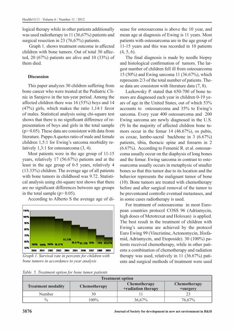
3876
HealthMED - Volume 6 / Number 11 / 2012
Journal of Society for development in new net environment in B&H
logical therapy while in other patients additionally was used radiotherapy in 11 (36,67%) patients and surgical resection in 23 (76,67%) patients.
Graph 1. shows treatment outcome in affected children with bone tumors. Out of total 30 affec-ted, 20 (67%) patients are alive and 10 (33%) of them died.
Discussion
This paper analyzes 30 children suffering from bone cancer who were treated at the Pediatric Cli-nic in Sarajevo in the ten-year period. Among the affected children there was 16 (53%) boys and 14 (47%) girls, which makes the ratio 1,14:1 favor of males. Statistical analysis using chi-square test shows that there is no significant difference of re-presentation of boys and girls in the total sample (p> 0.05). These data are consistent with data from literature. Pappo A quotes ratio of male and female children 1,5:1 for Ewing’s sarcoma morbidity re-latively 1,3:1 for osteosarcoma (3, 4).
Most patients were in the age group of 11-15 years, relatively 17 (56.67%) patients and at the least in the age group of 0-5 years, relatively 4 (13.33%) children. The average age of all patients with bone tumors in childhood was 9.72. Statisti-cal analysis using chi-square test shows that there are no significant differences between age groups in the total sample (p> 0.05).
According to Alberto S the average age of di-
Table 5. Treatment option for bone tumor patientsTreatment option
Treatment modality Chemotherapy Chemotherapy +radiation therapy
Chemotherapy +surgery
Number 30 11 23% 100% 36,67% 76,67%
Graph 1. Survival rate in percents for children with bone tumors in accordance to year analysis
sease for osteosarcoma is above the 10 year, and mean age at diagnosis of Ewing is 11 years. Most patients with osteosarcoma are in the age group of 11-15 years and this was recorded in 10 patients (4, 5, 6).
The final diagnosis is made by needle biopsy and histological confirmation of tumors. The lar-gest number of children fell ill from osteosarcoma 15 (50%) and Ewing sarcoma 11 (36.67%), which represents 2/3 of the total number of patients. The-se data are consistent with literature data (7, 8).
Lazkowsky P. stated that 650-700 of bone tu-mors are diagnosed each year in children 0-19 ye-ars of age in the United States, out of which 53% accounts to osteosarcoma and 35% to Ewing’s sarcoma. Every year 400 osteosarcoma and 200 Ewing sarcoma are newly diagnosed in the U.S. (9) In the majority of affected children bone tu-mors occur in the femur 14 (46.67%), os pubis, os coxae, lumbo-sacral backbone in 3 (6.67%) patients, tibia, thoracic spine and forearm in 2 (6.67%). According to Femenić R. et al. osteosar-coma usually occur on the diaphysis of long bones and the femur. Ewing sarcoma in contrast to oste-osarcoma usually occurs in metaphysic of smaller bones so that this tumor due to its location and the behavior represents the malignant tumor of bone (10). Bone tumors are treated with chemotherapy before and after surgical removal of the tumor to be preventeand controlle eventual metastases, and in some cases radiotherapy is used.
For treatment of osteosarcoma in most Euro-pean countries protocol COSS 96 (Adriamycin, high doses of Metotrexat and Holoxan) is applied. The best result in the treatment of children with Ewing’s sarcoma are achieved by the protocol Euro Ewing 99 (Vincristine, Actonomycin, Ifosfa-mid, Adriamycin, and Etoposide). 30 (100%) pa-tients received chemotherapy, while in other pati-ents a combination of chemotherapy and radiation therapy was used, relatively in 11 (36.67%) pati-ents and surgical methods of treatment were used
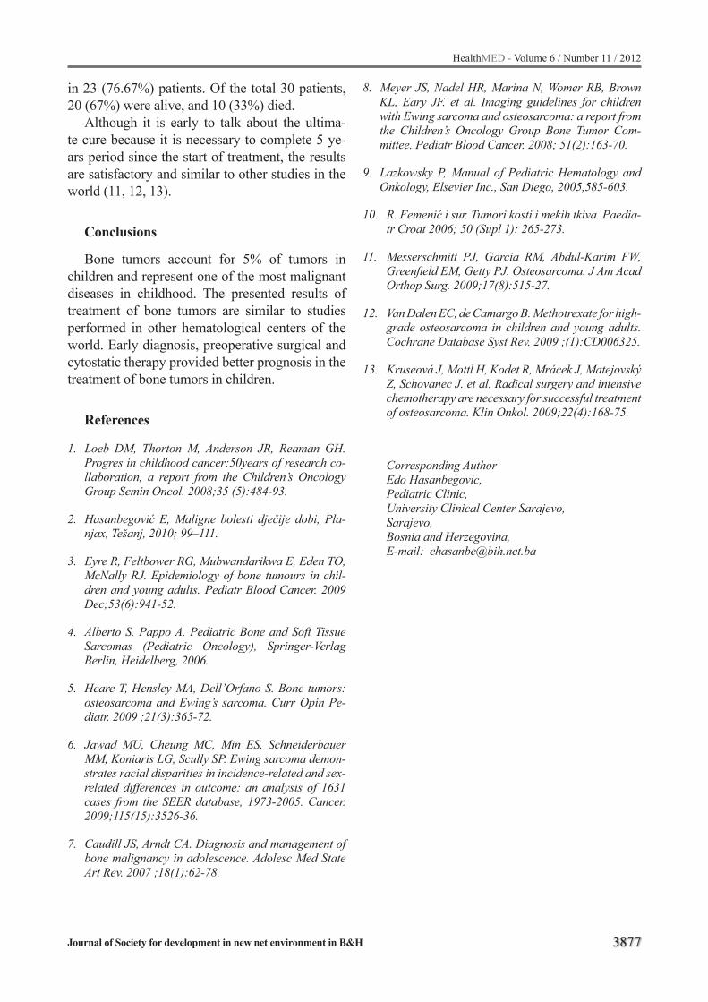
HealthMED - Volume 6 / Number 11 / 2012
Journal of Society for development in new net environment in B&H 3877
in 23 (76.67%) patients. Of the total 30 patients, 20 (67%) were alive, and 10 (33%) died.
Although it is early to talk about the ultima-te cure because it is necessary to complete 5 ye-ars period since the start of treatment, the results are satisfactory and similar to other studies in the world (11, 12, 13).
Conclusions
Bone tumors account for 5% of tumors in children and represent one of the most malignant diseases in childhood. The presented results of treatment of bone tumors are similar to studies performed in other hematological centers of the world. Early diagnosis, preoperative surgical and cytostatic therapy provided better prognosis in the treatment of bone tumors in children.
References
1. Loeb DM, Thorton M, Anderson JR, Reaman GH. Progres in childhood cancer:50years of research co-llaboration, a report from the Children’s Oncology Group Semin Oncol. 2008;35 (5):484-93.
2. Hasanbegović E, Maligne bolesti dječije dobi, Pla-njax, Tešanj, 2010; 99–111.
3. Eyre R, Feltbower RG, Mubwandarikwa E, Eden TO, McNally RJ. Epidemiology of bone tumours in chil-dren and young adults. Pediatr Blood Cancer. 2009 Dec;53(6):941-52.
4. Alberto S. Pappo A. Pediatric Bone and Soft Tissue Sarcomas (Pediatric Oncology), Springer-Verlag Berlin, Heidelberg, 2006.
5. Heare T, Hensley MA, Dell’Orfano S. Bone tumors: osteosarcoma and Ewing’s sarcoma. Curr Opin Pe-diatr. 2009 ;21(3):365-72.
6. Jawad MU, Cheung MC, Min ES, Schneiderbauer MM, Koniaris LG, Scully SP. Ewing sarcoma demon-strates racial disparities in incidence-related and sex-related differences in outcome: an analysis of 1631 cases from the SEER database, 1973-2005. Cancer. 2009;115(15):3526-36.
7. Caudill JS, Arndt CA. Diagnosis and management of bone malignancy in adolescence. Adolesc Med State Art Rev. 2007 ;18(1):62-78.
8. Meyer JS, Nadel HR, Marina N, Womer RB, Brown KL, Eary JF. et al. Imaging guidelines for children with Ewing sarcoma and osteosarcoma: a report from the Children’s Oncology Group Bone Tumor Com-mittee. Pediatr Blood Cancer. 2008; 51(2):163-70.
9. Lazkowsky P, Manual of Pediatric Hematology and Onkology, Elsevier Inc., San Diego, 2005,585-603.
10. R. Femenić i sur. Tumori kosti i mekih tkiva. Paedia-tr Croat 2006; 50 (Supl 1): 265-273.
11. Messerschmitt PJ, Garcia RM, Abdul-Karim FW, Greenfield EM, Getty PJ. Osteosarcoma. J Am Acad Orthop Surg. 2009;17(8):515-27.
12. Van Dalen EC, de Camargo B. Methotrexate for high-grade osteosarcoma in children and young adults. Cochrane Database Syst Rev. 2009 ;(1):CD006325.
13. Kruseová J, Mottl H, Kodet R, Mrácek J, Matejovský Z, Schovanec J. et al. Radical surgery and intensive chemotherapy are necessary for successful treatment of osteosarcoma. Klin Onkol. 2009;22(4):168-75.
Corresponding AuthorEdo Hasanbegovic,Pediatric Clinic,University Clinical Center Sarajevo,Sarajevo, Bosnia and Herzegovina,E-mail: [email protected]
3878
HealthMED - Volume 6 / Number 11 / 2012
Journal of Society for development in new net environment in B&H
Characterization of acetylsalicylic acid with thin-layer chromatography and hot–stage microscopy depending to solvent systemEkrem Pehlic1, Mirza Nuhanovic2, Aida Sapcanin3, Bozo Banjanin5, Husein Nanic4, Safeta Redzic1, Melita Poljakovic5
1 University of Bihac, Biotechnical faculty, Bosnia and Herzegovina,2 University of Sarajevo, Faculty of Natural Sciences and Mathematics, Bosnia and Herzegovina,3 University of Sarajevo, Faculty of Pharmacy, Bosnia and Herzegovina,4 Agency for Higher Education and quality assurance of Bosnia and Herzegovina,5 University of Tuzla, Faculty of Natural Sciences and Mathematics, Bosnia and Herzegovina.
Abstract
Aim: Characterization of acetylsalicylic acid with thin-layer chromatography and hot–stage microscopy depending to solvent system.
Material and methods: Synthesis of aspirin is classified as esterification reaction. In this reac-tion, to salicylic acid as reactant is added acetate anhydrate, a derivative of acetic acid, and in pres-ence of 85% phosphoric acid (sometime sulphu-ric acid is used) as catalyst gives acetylsalicylic acid and as by-product acetic acid [1]. Obtained quantity of raw aspirin is 15 g. Precrystalisation of raw aspirin is done with these solvents: ethanol, methanol and petroleum ether. For synthesised as-pirin identification following instrumental meth-ods is used:Thin Layer Chromatography (TLC) and Hot-stage microscopy (HSM). As stationary phase:HPTLC plate F 254 MERC, silica gel G 20x20, thickness 0.25 mm [13]. Detection of compounds is performed with UV/VIS detector–CAMAG. Mobile phase: benzene–ethanol (8:2). Thermal properties are observed on OLYMPUS BX 51 thermal microscope with polarised light. Sample is mounted and heated on 10 °C/min in temperature range 40 °C-150 °C. On given visible terminal crossovers pictures are made (1 picture/3 sec) [12].
Results: Identification of given compounds is studied by TLC method in different systems. By identification of acetylsalicylic acid (precrystal-ised acetylsalicylic acid in ethanol, methanol and petroleum ether) benzene and ethanol are used, but in different ratios:
benzene–ethanol (8 : 2)benzene–ethanol (9 : 1)benzene–ethanol (7 : 3)
Sample precrystalised in ethanol is solved in range of 0.5–1°C, what is proof that acetylsalicyl-ic acid precrystalised in ethanol shows superb pu-rity, thus ethanol could be used as suitable solvent for acetylsalicylic acid precrystalisation. Sample precrystalised in methanol is also solved in range 0.5–1°C. In petroleum ether precrysalised saml-ple is melting on 99.8 °C, where is visible that in sample which is precrystalised in petroleum ether some impurities remains, so it cannot be suitable solvent for purification of acetylsalicylic-acid.
Discussion: Drugs which contain derivates of salicylic acid, but with structure similar to aspirin, are used in medicine since ancient times. Extracts from willow bark are rich in salicylates which are known by specific effects on elevated tempera-ture, pain and inflammation, since mid 20th centu-ry. Aspirin also have a property of inhibiting blood coagulation, because of thromboxane prostaglan-dins inhibition, which normally bind molecules of platelets for making thromboses on open or dam-aged blood vessel or injured tissue [2]. The aim is evaluation of the influence of different precrystalii-sation solvents on acetylsalicylic acid purity as-sessed by thermal microscopy and the influence of different mobile phase composition on acetylsali-cylic acid identification in the presence of salicylic acid by thin-layer chromatography.
Conclusions: Most suitable solvents for puri-fication of raw aspirin are ethanol and methanol, thus petroleum ether is not, because after petro-leum ether precrystalisation remains impurities. Sample precrystalised in ethanol and methanol is solved in range of 0.5 – 1 °C, what gives conclu-sion acetylsalicylic acid precrystalised in ethanol shows high purity so ethanol is suitable solvent for
HealthMED - Volume 6 / Number 11 / 2012
Journal of Society for development in new net environment in B&H 3879
acetylsalicylic acid precrystalisation. Rf value by mobile phase benzene – ethanol in ratio (9:1,7:3) is such that spots are on the same position what means there were no separation, i.e. this system isn’t appropriate for aspirin identification by thin-layer chromatography method.
Keywords: Acetylsalicylic acid, thin layer chromatography (TLC), Hot stage micro-scopy (HSM).
Introduction
Drugs which contain derivates of salicylic acid, but with structure similar to aspirin, are used in medicine since ancient times. Extracts from wil-low bark are rich in salicylates which are known by specific effects on elevated temperature, pain and inflammation, since mid 20th century. Aspirin also have a property of inhibiting blood coagula-tion, because of thromboxane prostaglandins inhi-bition, which normally bind molecules of platelets for making thromboses on open or damaged blood vessel or injured tissue. For those reasons, aspirin in long-term uses in low dosages for preventing myocardial infarct, brain stroke or building plate-lets in person whose have such a risk [4]. Aspirin is one of the first drugs from non-steroid anti-in-flammatory drugs group, in which many are not salicylates, but many of them have similar effects inhibiting cyclooxygenase synthesis as their basic mechanism of usage [10]. Low dosage of aspirin is recommended in prevention of brain stroke and infarct by patients with diagnosed heart diseases [7]. In high dosage, aspirin and other salicylates are used by treatment rheumatic fever, rheumatic arthritis and other joint and bones inflammatory diseases. Evaluation of the influence of different precrystalisation solvents on acetylsalicylic acid purity assessed by thermal microscopy. The in-fluence of different mobile phase composition on acetylsalicylic acid identification in the presence of salicylic acid by thin-layer chromatography.
Aim
In using the different solvent systems by using TLC method one get a better opportunity to iden-tify acetylsalicylic acid. Also, through the HSM method we would like to see how the purity of this synthesized acetylsalicylic acid.
Material and methods
Synthesis of aspirin is classified as esterifica-tion reaction. In this reaction, to salicylic acid as reactant is added acetate anhydrate, a derivative of acetic acid, and in presence of 85% phosphoric acid (sometime sulphuric acid is used) as catalyst gives acetylsalicylic acid and as by-product ace-tic acid [5]. By reaction itself, acetate anhydrate binds to phenol group of salicylic acid (OH-) giving acetyl group (R– OH→R–OCOCH3) [3]. In three headed Erlenmeyer is added 10 g of salicylic acid and 25 ml acetic acid anhydrate. Two–three drops of concentrated sulphuric acid is added as catalyst too. The magnet is also putted into bottle. On bottle top stands thermometer, for showing temperature. Whole apparatus has mounted on return cooler. Ingredients in bottle are heated for 15 minutes on 50–60 °C with constant stirring. After the end, the bottle is unmounted, cooled under water shower, afterwards cooled on ice container until acetylsali-cylic acid crystals are separated [8]. After crystal separation, content is filtrated through Büchner funnel under vacuum. Obtained crystals are dried in vacuum on temperature 105°C until constant mass. Obtained quantity of raw aspirin is 15 g. Pre-crystalisation of raw aspirin is done with these sol-vents: ethanol, methanol and petroleum ether. For synthesised aspirin identification following instru-mental methods is used: Thin Layer Chromatogra-phy (TLC) and Hot-stage microscopy (HSM) [15]. As stationary phase: HPTLC plate F 254 MERC, silica gel G 20x20, thickness 0.25 mm. Detection of compounds is performed with UV/VIS detector – CAMAG. Mobile phase: benzene–ethanol (8:2). On plate is applied by 2 µL of probe and chromato-gram is developed in mobile phase until front height reach 2/3 of plate. Plate is removed out and dried in air for 15 minutes, examined on UV light on wave length of 254 nm [11]. Thermal properties are ob-served on OLYMPUS BX 51 thermal microscope with polarised light. Sample is mounted and heated on 10 °C/min in temperature range 40 °C - 150 °C. On given visible terminal crossovers pictures are made (1 picture/3 sec).
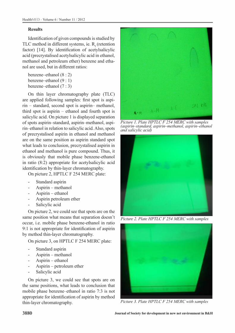
3880
HealthMED - Volume 6 / Number 11 / 2012
Journal of Society for development in new net environment in B&H
Results
Identification of given compounds is studied by TLC method in different systems, ie. Rf (retention factor) [14]. By identification of acetylsalicylic acid (precrystalised acetylsalicylic acid in ethanol, methanol and petroleum ether) benzene and etha-nol are used, but in different ratios:
benzene–ethanol (8 : 2)benzene–ethanol (9 : 1)benzene–ethanol (7 : 3)
On thin layer chromatography plate (TLC) are applied following samples: first spot is aspi-rin – standard, second spot is aspirin– methanol, third spot is aspirin – ethanol and fourth spot is salicylic acid. On picture 1 is displayed separation of spots aspirin–standard, aspirin–methanol, aspi-rin–ethanol in relation to salicylic acid. Also, spots of precrystalised aspirin in ethanol and methanol are on the same position as aspirin standard spot what leads to conclusion, precrystalised aspirin in ethanol and methanol is pure compound. Thus, it is obviously that mobile phase benzene-ethanol in ratio (8:2) appropriate for acetylsalicylic acid identification by thin-layer chromatography.
On picture 2, HPTLC F 254 MERC plate:
- Standard aspirin- Aspirin – methanol- Aspirin – ethanol- Aspirin petroleum ether- Salicylic acidOn picture 2, we could see that spots are on the
same position what means that separation doesn’t occur, i.e. mobile phase benzene-ethanol in ratio 9:1 is not appropriate for identification of aspirin by method thin-layer chromatography.
On picture 3, on HPTLC F 254 MERC plate:
- Standard aspirin- Aspirin – methanol- Aspirin – ethanol- Aspirin – petroleum ether- Salicylic acid
On picture 3, we could see that spots are on the same positions, what leads to conclusion that mobile phase benzene–ethanol in ratio 7:3 is not appropriate for identification of aspirin by method thin-layer chromatography.
Picture 1. Plate HPTLC F 254 MERC with samples (aspirin–standard, aspirin–methanol, aspirin–ethanol and salicylic acid)
Picture 2. Plate HPTLC F 254 MERC with samples
Picture 3. Plate HPTLC F 254 MERC with samples
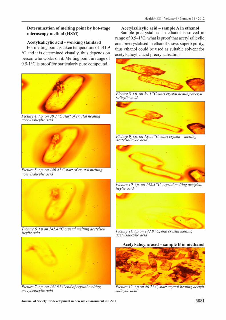
HealthMED - Volume 6 / Number 11 / 2012
Journal of Society for development in new net environment in B&H 3881
Determination of melting point by hot-stage microscopy method (HSM)
Acetylsalicylic acid - working standard
Picture 4. t.p. on 30.2 °C start of crystal heating acetylsalicylic acid
Picture 5. t.p. on 140.4 °C start of crystal melting acetylsalicylic acid
Picture 6. t.p on 141.4 °C crystal melting acetylsan-licylic acid
Picture 7. t.p. on 141.9 °C end of crystal melting acetylsalicylic acid
For melting point is taken temperature of 141.9 °C and it is determined visually, thus depends on person who works on it. Melting point in range of 0.5-1°C is proof for particularly pure compound.
Acetylsalicylic acid – sample A in ethanol
Picture 8. t.p. on 29.3 °C start crystal heating acetylt-salicylic acid
Picture 9. t.p. on 139.9 °C, start crystal meltingacetylsalicylic acid
Picture 10. t.p. on 142.3 °C, crystal melting acetylsa,-licylic acid
Picture 11. t.p on 142.9 °C, end crystal meltingacetylsalicylic acid
Sample precrystalised in ethanol is solved in range of 0.5–1°C, what is proof that acetylsalicylic acid precrystalised in ethanol shows superb purity, thus ethanol could be used as suitable solvent for acetylsalicylic acid precrystalisation.
Picture 12. t.p on 40.7 °C, start crystal heating acetylt-salicylic acid
Acetylsalicylic acid – sample B in methanol
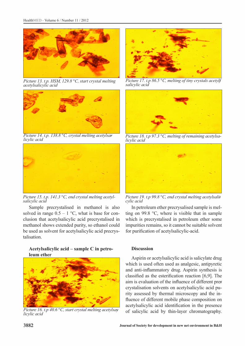
3882
HealthMED - Volume 6 / Number 11 / 2012
Journal of Society for development in new net environment in B&H
Picture 13. t.p. HSM, 129.8 °C, start crystal melting acetylsalicylic acid
Picture 14. t.p. 138.8 °C, crystal melting acetylsar-licylic acid
Picture 15. t.p. 141.3 °C, end crystal melting acetyl-salicylic acid
Sample precrystalised in methanol is also solved in range 0.5 – 1 °C, what is base for con-clusion that acetylsalicylic acid precrystalised in methanol shows extended purity, so ethanol could be used as solvent for acetylsalicylic acid precrys-talisation.
Acetylsalicylic acid – sample C in petro-leum ether
Picture 16. t.p 40.6 °C, start crystal melting acetylsay-licylic acid
Picture 17. t.p 86.5 °C, melting of tiny crystals acetylf-salicylic acid
Picture 18. t.p 97.3 °C, melting of remaining acetylsa -licylic acid
Picture 19. t.p 99.8 °C, end crystal melting acetylsalit-cylic acid
In petroleum ether precrysalised sample is mel-ting on 99.8 °C, where is visible that in sample which is precrystalised in petroleum ether some impurities remains, so it cannot be suitable solvent for purification of acetylsalicylic-acid.
Discussion
Aspirin or acetylsalicylic acid is salicylate drug which is used often used as analgesic, antipyretic and anti-inflammatory drug. Aspirin synthesis is classified as the esterification reaction [6,9]. The aim is evaluation of the influence of different prer-crystalisation solvents on acetylsalicylic acid pu-rity assessed by thermal microscopy and the in-fluence of different mobile phase composition on acetylsalicylic acid identification in the presence of salicylic acid by thin-layer chromatography.
HealthMED - Volume 6 / Number 11 / 2012
Journal of Society for development in new net environment in B&H 3883
Reaction environment during synthesis includes precrystalisation of synthesised aspirin in different solvents and selection of best suitable solvent for precrystalisation. Precrystalised aspirin in ethanol identified by HSM method showing good purity what imply that ethanol is suited solvent for aspi-rin precrystalisation. Melting point of aspirin by HSM method is 141.9 °C. Sample precrystalised in ethanol melts in range of 0.5–1°C, what show us that acetylsalicylic acid precrystalised in ethanol shows good purity and ethanol is suitable solvent for acetylsalicylic acid precrystalisation. Sample precrystalised in petroleum ether melts on 99.8°C, what means that in sample precrystalised in petro-leum ether remains impurities, so petroleum ether is not suitable for purifying of acetylsalicylic acid. Sample precrystalised in methanol melts in range of 0,5–1°C, what also shows that acetylsalicylic acid precrystalised in methanol shows good pu-rity, so methanol also could be used as solvent for acetylsalicylic acid precrystalisation.
Conclusions
Most suitable solvents for purification of raw aspirin are ethanol and methanol, thus petroleum ether is not, because after petroleum ether pren-crystalisation remains impurities. Melting point by hot-stage microscopy method (HSM) is 141.9 °C. Melting point of compound is in range 0.5 – 1 °C what indicates high purity. Sample precrys-talised in ethanol is solved in range of 0.5 – 1 °C, what gives conclusion acetylsalicylic acid pre-crystalised in ethanol shows high purity so ethanol is suitable solvent for acetylsalicylic acid precrys-talisation. Sample precrystalised in methanol is solved in range 0.5–1°C, what leads to conclusion that acetylsalicylic acid precrystalised in methanol shows extended purity, so methanol could be used as solvent for acetylsalicylic acid precrystalisa-tion. On picture 1. is visible spots separation aspin-rin–standard, aspirin–ethanol and aspirin –metha-nol in relation to salicylic acid. It can be concluded that the second two mobile phases can not be used for aspirin identification in presence of salicylic acid under these conditions, which indicates the presence the potential of the impurities. Rf value by mobile phase benzene– ethanol in ratio (9:1) is such that spots are on the same position what
means there were no separation, i.e. this system isn’t appropriate for aspirin identification by thin-layer chromatography method. Ratio of system benzene–ethanol 7:3 for mobile phase is not suit-able for aspirin identification by TLC method, be-cause the spots are on the same positions.
References
1. Fatemeh F. Bamoharram,Heravi M.M, Roshani M, Ali Ghariba,Jahangira M, Catalytic Method for Synthesis of Aspirin by a Green, Efficient and Recyc-lable Solid Acid Catalyst (Preyssler’s Anion) at Room Temperature, Journal of the Chinese Chemical Soci-ety, 2007, 54, 1017-1020.
2. González-Correa JA, Arrebola MM, Guerrero A, Muñoz-Marín J, Ruiz-Villafranca D, Sánchez de La Cuesta F, De La Cruz JP. Influence of vitamin E on the antiplatelet effect of acetylsalicylic acid in human blood. Platelets. 2005;16(3-4):171-179.
3. Christopher F. J. Hickman, Fincke N.M, Melissa A. Lavine D.H, Barry. Identification and Quantitative Analysis of Acetaminophen, Acetylsalicylic Acid, and Caffeine in Commercial Analgesic Tablets by LC-MS, Journal of Chemical Education, 2010; v87 n8, 838-841.
4. Papathanasiou A, Goudevenos J, Tselepis AD.Aspirin resistance in cardiovascular disease: pathogene-sis, diagnosis and clinical impact. Curr Pharm Des. 2009;15(10):1085-94.
5. A. Nikolin , B. Nikolin, Nukleofilne supstitucije na nezasićenom ugljikovom atomu, Praktikum organske hemije, Svijetlost oour zavod za udžbenike i nastavna sredstva Sarajevo, 1984; pp: 69-75.
6. H. Stanley, Pine, Nukleofilne adicije i supstitucije u sintezi, Organska hemija, Školska knjiga Zagreb, 1994; pp: 445-449.
7. Papathanasiou A, Goudevenos J, Tselepis AD.Aspirin resistance in cardiovascular disease: pathogene-sis, diagnosis and clinical impact. Curr Pharm Des. 2009;15(10):1085-94.
8. A. Nikolin , B. Nikolin, Nukleofilne supstitucije na nezasićenom ugljikovom atomu, Praktikum organske hemije, Svijetlost oour zavod za udžbenike i nastavna sredstva Sarajevo, 1984; pp: 69-75.
9. H. Stanley, Pine, Nukleofilne adicije i supstitucije u sintezi, Organska hemija, Školska knjiga Zagreb, 1994; pp: 445-449.
10. E. Mujicic, M. Kulic, F.Kucukalic, S.Straus, E. Donlic (2001) Aspirin therapy and postoperative bleeding after coronary artery by pass grafting, Se-cond Congress of Cardiology and Angiology of Bo-snia & Herzegovina.
3884
HealthMED - Volume 6 / Number 11 / 2012
Journal of Society for development in new net environment in B&H
11. Poyraz, M. Banti, C.N. Kourkoumelis, N.Dokorou, V.Manos, M.J.Simčič, M.Golič-Grdadolnik,S.Mavromoustakos,T.Giannoulis, A.D. Verginadis, I.I. Charalabopoulos, K. Hadjikakou, S.K. Synthesis, structural characterization and biological studies of novel mixed ligand Ag(I) complexes with triphenylp-hosphine and aspirin or salicylic acid. Inorganica Chimica Acta, 2011, Vol. 375,N.8. 114-121.
12. Gerber M, Breytenbach JC, Hadgraft J, du Plessis J. Synthesis and transdermal properties of acetylsa-licylic acid and selected esters. Int J Pharm. 2006 ;310(1-2):31-6.
13. Hussain M, Javeed A, Ashraf M, Zhao Y, Mukhtar MM, Rehman MU. Aspirin and immune system. Int Immunopharmacol. 2012;12(1):10-20.
14. Mitić S.S. Miletić G.Z. Pavlovic A. Tošić S.B. Suna-ric S.M.Quantitative Analysis of Acetylsalicylic Acid in Commercial Pharmaceutical Formulations and Human Control Serum Using Kinetic Spectrophoto-metry, Acta Chim. Slov. 2008, 55, 508–515.
15. Long G.T., Sergey Vyazovkin, Nicoleigh Gam-ble, Charles A Wight, Kinetics and mechanism of the anhydrous-thermal-decomposition of acetylsa-licylic acid.J.Pharm. Sci.; 2002; 91 (3):800-9.
16. Pehlic E, ar all. Identification of synthesized 2-(4-benzoylphenyl)-2-methylpropionic acid by thin layer chromatography in the system ethylacetate-cyclohexane and benzene cyclohexene. HealthMED 2010; 4:867-878.
17. Pehlic E, ar all. Synthesis control of 2-(4-benzoylphenyl)-2-methyl propanoic acid by TLC in diethyl ether-cyclohexane and petroleu-mether–ethylacetate system. HealthMED 2011; 5: 413-418.
18. Ekrem Pehlić, ar all. Qualitative methods of identifi-cation of acetylsalicylic acid by differential scanning calorimetry and melting point method. HealthMED 2011; 5:N.6. 1782-1787.
Corresponding Author Pehlic Ekrem,University of Bihac, Biotechnical Faculty, Bosnia and Herzegovina,E-mail: [email protected]
HealthMED - Volume 6 / Number 11 / 2012
Journal of Society for development in new net environment in B&H 3885
Staphylococcus epidermidis biofilmsMonia Avdic1, Suad Habes2, Elida Avdic3
1 Prirodno-matematicki fakultet, Sarajevo, Bosnia & Herzegovina2 Fakultet zdravstvenih studija, Sarajevo, Bosnia & Herzegovina3 Regionalni Medicinski Centar „Dr. Safet Mujic“ Mostar, Bosnia & Herzegovina
Abstract
Staphylococcus epidermidis, which is the nor-mal microbiota of healthy human skin and mucous membranes, rarely causes disease in immunocom-petent persons. However, in recent decades, this microorganism is becoming a common cause of many hospital infections which are usually linked to imunocompromised patients. The emergence of Staphylococcus epidermidis as a pathogen is asso-ciated with the use of intravascular catheters, pa-cemakers, urinary catheters and other polymeric and metallic implants. The ability of this microor-ganism to causes infections is primarily due to its ability to form biofilms on synthetic surfaces of implanted medical devices.
The aim of this paper was to examine and show the ability of hospital strains of Staphylococcus epidermidis, which were isolated from smears of surfaces in the hospital environment RMC “Dr. Safet Mujić to form slime. The formation of slime by the majority of coagulase-negative staphylo-cocci (CNS) is associated with their capacity to produce biofilms and is considered one of the fac-tors of virulence.
In this study we tested 36 isolates of Staphylo-coccus epidermidis on their ability to produce sli-me. For the determination of the ability of slime production we used the qualitative tube test met-hod (Christiansen, 1982). Applying this test, it was noted that 21 isolates of Staphylococcus epidermi-dis (58.33%) produced slime, while in 15 isolates (41.66%) the production of slime was absent. Ta-king into account the fact that biofilms formed by Staphylococcus epidermidis on implanted bioma-terials cause chronic and persistent infections with serious clinical consequences the application of the tube test method as a screening test during the testing of these isolates would be very significant.
Keywords: Staphylococcus epidermidis, CNS, biofilm, the tube test method
Introduction
Coagulase-negative staphylococci (CNS) re-present one of the main components of normal human microbiota and are generally considered nonpathogenic. But over the past two decades, there has been an increase in the number of in-fections caused by coagulase-negative staphylo-cocci, especially Staphylococcus epidermidis species (Murray, 2007). Today, the coagulase-ne-gative staphylococci have become a significant, often isolated pathogen in the clinical microbio-logy laboratories worldwide (Kloos and Banner-man, 1994; Cerca et al., 2005; Arciola et al. 2006; Bayram and Balci 2006; Widerström et al., 2006).
Among coagulase-negative staphylococci (CNS) Staphylococcus epidermidis is the mostly isolated and predominant species in the human environment, which generally has symbiotic re-lationships with its host. Disturbing the integrity of the system of cutane organs by trauma, need-le inoculation, or implantation of medical devi-ces Staphylococcus epidermidis enters the host and then gets the role of a pathogen. Today this microorganism is one of the leading causes of no-socomial infections (Stevens, 2003) as well as in-fections of implanted medical devices (Weigel et al. 2003; O ‘Gara and Humphreys, 2001). These infections are usually associated with the use of medical devices such as intravascular catheters, pacemakers, urinary tract catheters and a variety of other polymeric and metallic implants.
The ability of this microorganism to cause in-fection is primarily due to its ability to form bio-films on inert synthetic surfaces of implanted me-dical devices. Biofilms are communities of micro-organisms that stick to each other or to the base by the production of a extracellular matrix, which mainly consists of polysaccharides and proteins (Hall-Stoodley et al., 2004). S.epidermidis has no specific virulence factors and so its ability to form biofilms is considered one of the virulence fac-
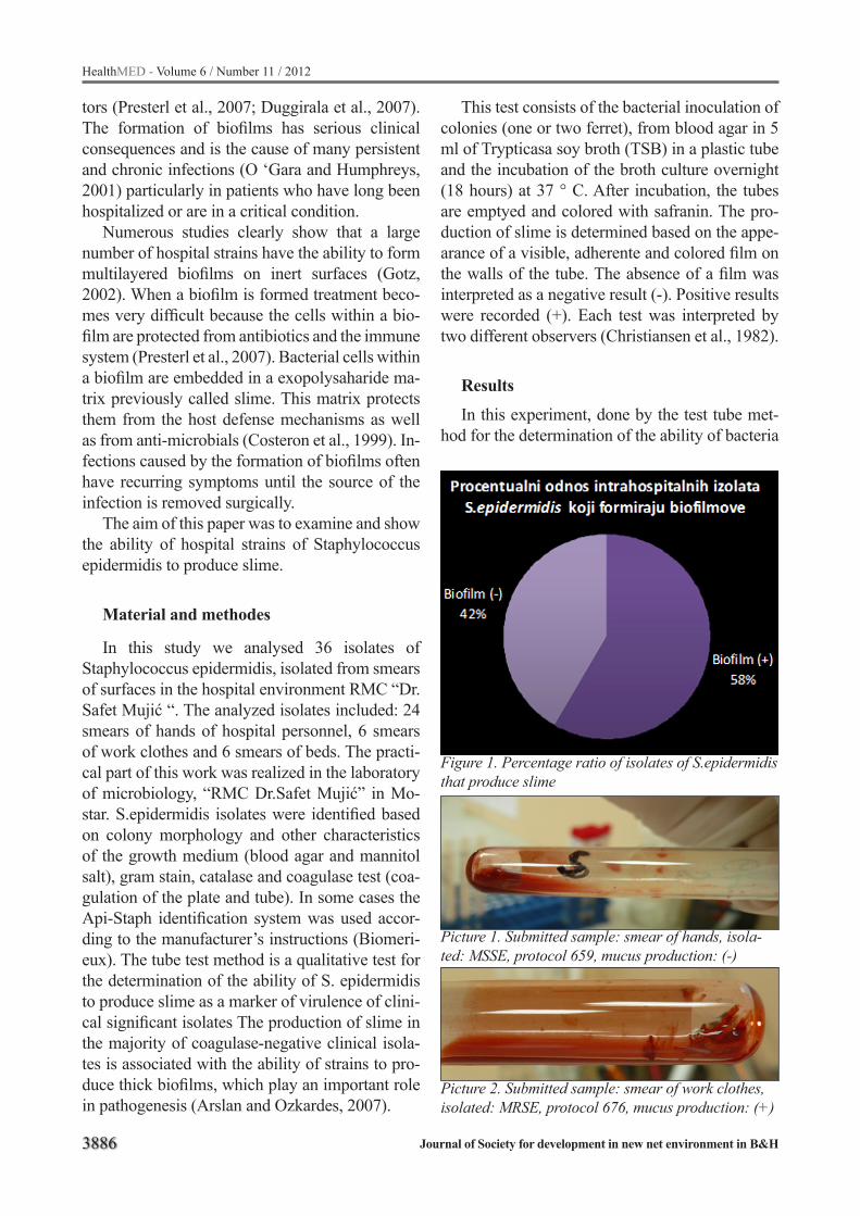
3886
HealthMED - Volume 6 / Number 11 / 2012
Journal of Society for development in new net environment in B&H
tors (Presterl et al., 2007; Duggirala et al., 2007). The formation of biofilms has serious clinical consequences and is the cause of many persistent and chronic infections (O ‘Gara and Humphreys, 2001) particularly in patients who have long been hospitalized or are in a critical condition.
Numerous studies clearly show that a large number of hospital strains have the ability to form multilayered biofilms on inert surfaces (Gotz, 2002). When a biofilm is formed treatment beco-mes very difficult because the cells within a bio-film are protected from antibiotics and the immune system (Presterl et al., 2007). Bacterial cells within a biofilm are embedded in a exopolysaharide ma-trix previously called slime. This matrix protects them from the host defense mechanisms as well as from anti-microbials (Costeron et al., 1999). In-fections caused by the formation of biofilms often have recurring symptoms until the source of the infection is removed surgically.
The aim of this paper was to examine and show the ability of hospital strains of Staphylococcus epidermidis to produce slime.
Material and methodes
In this study we analysed 36 isolates of Staphylococcus epidermidis, isolated from smears of surfaces in the hospital environment RMC “Dr. Safet Mujić “. The analyzed isolates included: 24 smears of hands of hospital personnel, 6 smears of work clothes and 6 smears of beds. The practi-cal part of this work was realized in the laboratory of microbiology, “RMC Dr.Safet Mujić” in Mo-star. S.epidermidis isolates were identified based on colony morphology and other characteristics of the growth medium (blood agar and mannitol salt), gram stain, catalase and coagulase test (coa-gulation of the plate and tube). In some cases the Api-Staph identification system was used accor-ding to the manufacturer’s instructions (Biomeri-eux). The tube test method is a qualitative test for the determination of the ability of S. epidermidis to produce slime as a marker of virulence of clini-cal significant isolates The production of slime in the majority of coagulase-negative clinical isola-tes is associated with the ability of strains to pro-duce thick biofilms, which play an important role in pathogenesis (Arslan and Ozkardes, 2007).
This test consists of the bacterial inoculation of colonies (one or two ferret), from blood agar in 5 ml of Trypticasa soy broth (TSB) in a plastic tube and the incubation of the broth culture overnight (18 hours) at 37 ° C. After incubation, the tubes are emptyed and colored with safranin. The pro-duction of slime is determined based on the appe-arance of a visible, adherente and colored film on the walls of the tube. The absence of a film was interpreted as a negative result (-). Positive results were recorded (+). Each test was interpreted by two different observers (Christiansen et al., 1982).
Results
In this experiment, done by the test tube met-hod for the determination of the ability of bacteria
Figure 1. Percentage ratio of isolates of S.epidermidis that produce slime
Picture 1. Submitted sample: smear of hands, isola-ted: MSSE, protocol 659, mucus production: (-)
Picture 2. Submitted sample: smear of work clothes, isolated: MRSE, protocol 676, mucus production: (+)
HealthMED - Volume 6 / Number 11 / 2012
Journal of Society for development in new net environment in B&H 3887
to produce slime (Christiansen et al., 1982), we fo-und that out of 36 isolates of Staphylococcus epi-dermidis isolated from smears of surfaces in the hospital enviroment 21 isolate (58.33%) produced slime, while in 15 isolates (41, 66%) slime pro-duction was absent (Fig. 1). Among the isolates that did not produce slime there were where three MRSE isolates and all MSSE isolates.
Discussion
To better understand the hospital infections cau-sed by S.epidermidis it is crucial to know whether the pathogenic strain originates from the hospital environment or outside it, since these strains are significantly different genetically and physiologi-cally. Numerous studies clearly show that a large
Table 1. Summary of slime production capacity of Staphylococcus epidermidis in samples showing resistance to meticillinNumber Protocol Sample MSSE MRSE Slime production (+, -)
1. 669 Work clothes + +2. 668 Hands + +3. 679 Bris Beda + -4. 661 Hands + +5. 673 Bed + +6. 659 Hands + -7. 29 Work clothes + -8. 104 Hands + +9. 212 Hands + -10. 214 Hands + -11. 682 Bed + -12. 644 Hands + -13. 653 Hands + +14. 676 Work clothes + +15. 647 Hands + -16. 41 Work clothes + -17. 642 Bed + +18. 253 Hands + -19. 241 Hands + -20. 131 Hands + +21. 664 Hands + +22. 651 Bed + +23. 1192 Work clothes + +
Number Protocol Sample MSSE MRSE Slime production (+, ++, +++)24. 159 Hands + +25. 689 Bed + -26. 272 Hands + -27. 301 Hands + +28. 697 Hands + +29. 54 Work clothes + -30. 223 Hands + -31. 691 Hands + +32. 326 Hands + +33. 303 Hands + +34. 327 Hands + +35. 323 Hands + +36. 320 Hands + +
Number MSSE/MRSE 12 24 Biofilm - Biofilm +Total 36 15 (41,66%) 21 (58,33%)
3888
HealthMED - Volume 6 / Number 11 / 2012
Journal of Society for development in new net environment in B&H
number of hospital strains have the ability to form multilayered biofilms on inert surfaces (Gotz, 2002). Although S.epidermidis has no specific vi-rulence factors, its ability to form biofilms is con-sidered one of the virulence factors (Presterl et al., 2007; Duggirala et al., 2007). When a biofilm is formed treatment becomes very difficult because the cells within a biofilm are protected from an-tibiotics and the immune system (Presterl et al., 2007). Within coagulase-negative staphylococci from human samples 18 species were isolated and only 5 of them were repeatedly associated with nosocomial infections. Among them Staphylo-coccus epidermidis is the most common cause of hospital infections. The skin of patients and health workers, medical equipment, clothes for hospital staff and a variety of surfaces in hospitals can be a source of resistant strains of Staphylococcus epi-dermidis (Aires et al., 2000; Hedin, 1993).
Biofilms are communities of microorganisms that stick to each other or to base the production of extracellular matrix, which mainly consists of polysaccharides and proteins (Hall-Stoodley et al., 2004). The emergence of Staphylococcus epidermidis as a pathogen is a synonym for the new widespread use of intravascular catheters in modern medicine. The capacity of these organi-sms to cause infection stimulates primarily from their ability to form biofilms on inert synthetic surfaces of implanted medical devices. This has serious clinical consequences and the cause of many persistent and chronic infections. Bacterial cells within a biofilm are embedded in a matrix exopolysaharide previously called slime. This ma-trix protects them from host defense mechanisms as well as from anti-microbials (Costeron et al., 1999). Infections caused by the formation of bi-ofilms often have recurring symptoms until the source of infection is removed surgically. Althou-gh the formation of biofilms on implanted medical devices is usually associated with coagulase ne-gative staphylococci in particular Staphylococcus epidermidis, Staphylococcus aureus strains also have the ability to form biofilms. The ability of Staphylococcus epidermidis to colonize biomate-rials of which implants are built depends on the composition of the biomaterial and the charac-teristics of the organism, such as the production of adhesins. Staphylococcus epidermidis biofilm
formation occurs in two stages and much rese-arch is devoted to the discovery of biochemical and molecular basis of this process. The process of accumulation of cells to form a mature biofilm rapidly follows after the initial binding to inert plastic surfaces. At the biochemical level, extra-cellular polysaccharide adhesins play an essential role in initial bacterial adherence and intracellular adhesion (biofilm formation).
Tube test method is a qualitative test for the determination of the ability of Staphylococcus epidermidis to produce slime as a marker of viru-lence of clinically significant isolates. The produc-tion of slime in the majority of coagulase-negati-ve clinical isolates is associated with the abilit to produces thick biofilms, which play an important role in pathogenesis (Arslan and Ozkardes, 2007). This test confirmed that 58% of the isolates of Staphylococcus epidermidis had the ability to pro-duce slime. All the strains that produces biofilms were methicillin resistant. Given that the clinical significance of the ability of biofilm formation, it is advisable to introduce the tube test method as a screening test in microbiological laboratories for the determination of the ability of biofilm forma-tion of isolated strains of Staphylococcus epider-midis.
Conclusions
The test tube method showed that 21 isolates of Staphylococcus epidermidis (58.33%) from sme-ars of surfaces in the hospital environment exhibi-ted the ability to produce slime, while in 15 isola-tes (41.66%) the production of slime was absent.
Among the isolates that had not formed slime there were three MRSE isolates and all MRSE iso-lates.
Taking into account the fact that biofilms for-med by Staphylococcus epidermidis on implanted biomaterials cause chronic and persistent infecti-ons with serious clinical consequences, the appli-cation of the test tube method as a screening test during the testing of these isolates would be very significant.
HealthMED - Volume 6 / Number 11 / 2012
Journal of Society for development in new net environment in B&H 3889
References
1. Aires De Sousa, M., Santos Sanches, I. i Ferro, M.L. (2000). Epidemiological study of staphylococcal co-lonization and cross-infection in two West African Hospitals. Microb Drug Resist., 6(2): 133-41.
2. Arciola, C.R., Campoccia, D. i An, Y.H. (2006). Preva-lence and antibiotic resistance of 15 minor staphylo-coccal species colonizing orthopedic implants. Int. J. Artif. Organs, (29): 395–401.
3. Arslan, S. i Özkardes, F. (2007). Slime production and antibiotic susceptibility in staphylococci isolated from clinical samples. Mem Inst Oswaldo Cruz, 102(1): 29-33.
4. Bayram, A. i Balci, I. (2006). Patterns of antimicrobi-al resistance in a surgical intensive care unit of a uni-versity hospital in Turkey. BMC Infect. Dis., (6): 155.
5. Cerca, N., Martins, S. i Cerca, F. (2005). Comparative assessment of antibiotic susceptibility of coagulase-negative staphylococci in biofilm versus planktonic culture as assessed by bacterial enumeration or ra-pid XTT colorimetry. J. Antimicrob. Chemother., (56): 331–6.
6. Christensen, G., Simpson, W., Bisno, A. i Beachey, E. (1982). Adherence of slime producing strains of Staphylococcus epidermidis to smooth surfaces. In-fect Immun 37: 318-326.
7. Costerton, J.W., Stewart, P.S. i Greenberg, E.P. (1999). Bacterial biofilms: a common cause of persi-stent infections. Science, (284): 1318–1322.
8. Duggirala, A., Kenchappa, P., Sharma, S., Peeters, J., Ahmed, N., Garg, P., Das, T. i Hasnain, S. (2007). High-Resolution Genome Profiling Differentiated Staphylococcus epidermidis Isolated from Patients with Ocular Infections and Normal Individuals. In-vestigative Ophthalmology and Visual Science, (48): 3239-3245.
9. Gotz, F. (2002). Staphylococcus and biofilms. Mol. Microbiol., (43): 1367–78.
10. Hall-Stoodley, L., Costerton, J.W. i Stoodley, P. (2004). Bacterial biofilms: from the natural envi-ronment to infectious diseases. Nat. Rev. Microbi-olb., (2): 95–108.
11. Hedin, G. (1996). A comparison of methods to de-termine whether clinical isolates of Staphylococcus epidermidis from the same patient are related. J. Hosp. Infect., 34(1): 31-42.
12. Kloos, W.E. i Bannerman, T.L. (1994). Update on clinical significance of coagulase-negative staphylo-cocci. Clin. Microbiol. Rev., (7): 117–40.
13. Murray, P.R., Baron, E.J., Jorgensen, J., Pfaller, M. i Yolken, R. (2007). Manual of Clinical Microbiology. Washington, DC: ASM Press.
14. O’ Gara, J. i Humphreys, H. (2001). Staphylococcus epidermidis biofilms: importance and implications. Journal of Medical Microbiology, (50): 582-587.
15. Presterl, E., Suchomel, M., Eder, M., Reichmann, S., Lassnigg, A., Graninger, W. I Rotter, M. (2007). Effects of alcohols, povidone-iodine and hydrogen peroxide on biofilms of Staphylococcus epidermdis”. Journal of Antimicrobial Chemothereapy, (60): 417-420.
16. Stevens, D. L. (2003). Community-acquired Staphylococcus aureus infections: increasing viru-lence and emerging methicillin resistance in the new millennium. Curr. Opin. Infect. Dis. (16): 189–191.
17. Weigel, L. M., D. B. Clewell, S. R. Gill, N. C. Clark, L. K. McDougal, S. E. Flannagan, J. F. Kolonay, J. Shetty, G. E. Killgore i F. C. Tenover. (2003). Ge-netic analysis of a high-level vancomycin-resistant isolate of Staphylococcus aureus. Science, (302): 1569–1571.
18. Widerström, M., Monsen, T. i Karlsson, C. (2006). Molecular epidemiology of methicillin-resistant co-agulase-negative staphylococci in a Swedish county hospital: evidence of intro- and interhospital clonal spread. J. Hosp. Infect, 64: 177–83.
Corresponding AuthorSuad Habes,Fakultet zdravstvenih studija, University of Sarajevo,Sarajevo,Bosnia & Herzegovina,E-mail: [email protected]

3890
HealthMED - Volume 6 / Number 11 / 2012
Journal of Society for development in new net environment in B&H
Every sent article gets its number, and author(s) will be notified if their paper is accepted and what is the num-ber of paper. Every corresponedence will use that num-ber. The paper has to be typed on a standard size paper (format A4), leaving left margins to be at least 3 cm. Ali materials, including tables and references, have to be typed double-spaced, so one page has no more than 2000 alphanumerical characters (30 lines). Sent paper needs to be in the form of triplicate, considering that original one enclosure of the material can be photocopied. Presenting paper depends on its content, but usually it consists of a page title, summary, text references, legends for pictures and pictures. Type your paper in MS Word and send if on a diskette or a CD-ROM.
Title page
Every article has to have a title page with a title of no more than 10 words: name (s), last and first of the aut-hor (s), name of the instituion the authors (s) belongs to, abstract with maximum of 45 letters (including space), footnote with acknowledgments, name of the first aut-hor or another person with whom correspondence will be maintained.
Abstract
Second page needs to contain paper abstract, 200 words at the most. abstract needs to hold all essential facts of the work-purpose of work, used methods (with specific data, if possible) and basic facts. Abstract must have review of underlined data, ideas and conclusi-ons from text. Abstract has no quoted references. For Keywords, at the most, need to be placed below the text.
Central part of the article
Authentic papers contain these parts: introduction, goal, methods, results, discussion and conclusion. Intro-duction is brief and clear review of a problem. Methods are shown so that interested reader is able to repeat des-cribed research. Known methods don’ t need to be iden-tified, it is cited (referenced). Results need to be shown clearly and legically, and their significance proven by statistical analysis. In discussion, results are interpreted and compared to existing, previously published findings in the same field. Conclusions have to give an answer to author’s goal.
Instructions for the authorsAll papers need to be sent to e-mail: [email protected]
References
Quoting references must be in a scale in which they are really used. Quoting most recent literature is reco-mmended. Only published articels (or articles accepted for publishing) can be used as references. Not-published observations and personal notifications need to be in text in brackets. Showing references is as how they appear in text. References cited in tables or pictures are also numbered according to quoting order. Citing paper with six or less authors must have cited names of all authors; if seven or more authors’ wrote the paper, the name of the first three authors are cited with a note “et all”. If the author is unknown, at the beginning of papers reference, the article is named as “unknown”. Titles of the publications are abbreviated in accordance to Index Medicus, but if not listed in the index, whole title of the journal has to be written.
Footnote-comments, explanations, etc., cannot be used in the paper.
Statisticial analysis
Tests used for statistical analysis need to be shown in text and in tables or pictures containing statistical analysis.
Tables and pictures
Tables have to be numbered and shown by their or-der, so they can be understood without having to read the paper. Every column needs to have title, every me-asuring unit (SI) has to be clearly marked, preferably in footnotes below the table, in Arabian numbers or sym-bols. Pictures also have to be numbered as they appear in text. Drawings need to be enclosed on a white paper or tracing paper, while black and white photo have to be printed on a radiant paper. Legends next to pictures and photos have to be written on a separate A4 format paper. All illustrations (pictures, drawings, diagrams) have to be original and on their backs contain illustration num-ber, first author last name, abbreviated title of the pa-per and picture top. It is appreciated if author marks the place for table or picture. Preferable the pictures format is TIF, quality 300 DPI.
Use of abbreaviations
Use of abbreviations has to be reduced to minimum. Conventional units can be used without their definitions.