internal/external rotation moment arms of muscles at the knee: moment arms for the normal knee and...
TRANSCRIPT

Ž .The Knee 8 2001 293�303
Internal�external rotation moment arms of muscles at theknee: moment arms for the normal knee and the
ACL-deficient knee
William L. Buford Jr�, F. Marty Ivey Jr, Takayuki Nakamura,Rita M. Patterson, Doan K. Nguyen
Orthopaedic Surgery and Rehabilitation, Biomechanics Laboratory, 301 Uni�ersity Boule�ard, Uni�ersity of Texas Medical Branch,Gal�eston, TX 77555-0892, USA
Accepted 10 May 2001
Abstract
Knowledge of the three-dimensional balance of loads at the knee joint is required to adequately assess the treatment andŽ .rehabilitation of the malfunctioning knee. This report focuses upon the moment arms for the knee in internal�external IE
rotation motion. It augments prior work that defined flexion�extension moment arms. Muscle excursions and angular motionof the lower leg during IE rotation were measured in 17 fresh-frozen hemi-pelvis specimens. Moment arms were calculated asthe derivatives of excursion with respect to the angle. Rotational motion was performed for the normal and anterior cruciate
Ž .ligament ACL -deficient knee. Of the 13 muscles measured at the knee, seven were significant contributors to IE rotation:the biceps femoris long and short head externally rotate opposite the gracilis, sartorious, semimembranosis, semitendonosusand popliteus, functioning as internal rotators. Moment arm magnitudes were greatest with the knee in a flexed positionŽ � � � � .internal external rotators peaked at 70� 90� flexion . At 30� flexion, the IE rotation moment arm minima and maxima were10.1�11.6, 6.8�9.0, 6.0�15.7, 8.2�14.1 and 0.0�10.4 mm for the semimembranosis, semitendonosus, gracilis, sartorius andpopliteus, and 14.7�27.9 and 18.5�31.5 mm for the biceps femoris short and long, respectively. Moment arms for theACL-deficient condition were significantly changed only at extremes of flexion�extension. � 2001 Elsevier Science B.V. Allrights reserved.
Keywords: Joint mechanics; Muscle mechanics; Moment arms; Muscle balance
1. Introduction
Muscles are joint actuators. Forces and momentsare transmitted through muscle tendons to the joints.Thus, structures and geometry of the joint should be
� Corresponding author. Tel.: �1-409-772-9072; fax: �1-409-747-4223.
Ž .E-mail address: [email protected] W.L. Buford .
analyzed to fully understand and simulate the func-tion of the joints. Anatomical data, such as bonegeometry, contact area between the bone, definedaxes of rotation and muscle�tendon geometry, arenecessary for establishing the joint model. In ourstudy, we emphasize determination of the momentarms of the joint muscles among all those anatomicalparameters. Knowledge of muscle moment arms isneeded to assess mechanical advantage and balance
Žof the muscles at the joint for each degree-of-free-
0968-0160�01�$ - see front matter � 2001 Elsevier Science B.V. All rights reserved.Ž .PII: S 0 9 6 8 - 0 1 6 0 0 1 0 0 1 0 6 - 5

( )W.L. Buford et al. � The Knee 8 2001 293�303294
.dom . Several studies of muscle moment arms for theknee in flexion�extension have been completed.
� �Recent work in this laboratory 4 summarized thelimitations of some prior work and presented resultsfor flexion�extension moment arms for 14 muscles atthe knee using 15 specimens. The purpose of thestudy reported here was to define the muscle momentarms for the normal and ACL-deficient knee duringinternal�external rotation. Two hypotheses are tested:Ž .1 that the flexor muscles have significant momentarms for either internal or external rotation motionŽconversely, the extensor muscles will not contribute
. Ž .significantly to either motion ; and 2 that the ACL-Ž .deficient knee will alter the internal�external IE
moment arms. Using 17 fresh, intact hemi-pelvis ca-daver specimens, the ultimate purpose of this re-search is to develop a more complete, realistic andaccurate model of the lower extremity that will beapplied to the understanding of three-dimensionalbalance at the knee.
2. Background
Movement at the knee has been described as thecombination of rotations about a polycentric
Ž .flexion�extension FE axis and about an internal�ex-Ž . � �ternal IE rotation axis. Hart et al. 8 demonstrated
a finite helical axis of the knee, which travels as theknee flexes or extends. Other similar studies havedescribed the methods for obtaining the variable dis-crete or instantaneous screw axis of the knee during
� �movement 2,12 . Using various types of motion analy-sis, these studies resulted in wide variations in axisplacement and orientation. However, Hollister et al.� �9 created a simple method for monitoring the angu-lar motion of the knee. They established the protocolto find two effective fixed axes of rotation for theknee joint. Recent work with 15 cadaveric knees by
� �Churchill et al. 5 helped to confirm the orientationand placement of these two axes, and that there is nosignificant difference between the optimal flexion axisand the transepicondylar axis. Our study utilized thisdefinition of FE and IE rotation axes for monitoringthe rotations of the knee.
Although the moment arms for IE rotation motionhave not been described, several studies have de-
Ž .scribed the range of motion ROM for the IE motion� �of the knee. Grood et al. 7 determined limits of
movement in the human cadaver knees, includingthose in IE rotation.
� �Lane et al. 11 evaluated IE rotation ROM fornormal and ACL-deficient knees measured from 0 to60� of flexion, with 14 fresh cadaver specimens stud-
ied. Their results showed there was no significantdifference between the normal and the ACLM condi-tion.
� �Ishii et al. 10 measured three-dimensional kneemotion in vivo. They used an instrumented spatiallinkage device fixed by intracortical pins to the fivehealthy male volunteers. They reported that IE rota-tion during 0�60� flexion�extension motion was 10.6�Ž .with 2.8� S.D. .
� �Zarins et al. 13 performed an in vivo study onROM for the IE motion of the knee. Normal andACL-deficient patients were examined. They reportedremarkably larger ROM data than the others refer-
� �enced here 7,11,10 .Knowledge of muscle moment arms is required for
improved solutions to joint reaction force and mo-ment problems. Various methods have been appliedto determine the knee muscle moment arms. An et al.� �1 reviewed the different methods, which includedetermination by geometric measurements, calcula-tion from tendon displacement, and a direct loadmeasurement method. Our study utilized the tendon
� �displacement method. Fick et al. 6 first reported thismethod, which calculates the muscle moment arm asthe derivative of the measured tendon displacementwith respect to the joint angle.
Several moment arm studies that use the tendonexcursion method have been reported. As applied tothe knee, these studies exclusively focused upon flex-ion extension motion only. Further background re-garding these studies is summarized in our prior work� �4 .
3. Procedure
This study determined the moment arms of theknee muscle-tendon units during the internal�exter-nal rotation about the longitudinal rotation axis of the
� �knee that is described by Hollister et al. 9 . Werepeated the same procedure reported in our past
� �study 4 , which described the flexion�extension mo-ment arms for the normal and ACL-deficient knee.The following 13 muscles were prepared using thesame protocol: vastus medialis; v. intermedius; v. lat-eralis; rectus femoris; gracilis; sartorius; biceps longand short; semimembranosis; semitendonosus; medialand lateral gastrocnemius; and the popliteus. Thepatellar tendon was also instrumented separately.
Ž .Each experiment conducted over a 3-day period wasperformed in a temperature-conditioned room. Speci-mens were covered with an ice bag during intermedi-ate periods and stored on ice in a cooler overnight toprevent decay. Fresh hemi-pelvis cadavers were re-

( )W.L. Buford et al. � The Knee 8 2001 293�303 295
Fig. 1. Sketch of a typical experimental connection for one muscle�tendon unit.
moved of skin above the ankle. Hip muscles that donot cross the knee and some intramuscular connectivetissue were also removed for unveiling the knee mus-
Žcles. Each specimen was inspected and X-rayed me-dial�lateral, anterior�posterior and full standing A�P
.views prior to the session to reject prior damagedknees and to measure the standard skeletal dimen-sions. Braided, flexible, coated stainless steel fishingleader wire sheathed by 1-mm-diameter polyethylenetubes were attached on each muscle belly to providethe measurable muscle motion pathway. The sheathcreates protection for the muscle and reduces frictionof the cable. Miniature swivels and crimps were pro-vided on each end. The distal end of the wire wassutured into the muscle insertion, on the center oftendon just distal to the insertion muscle fiber and is
Žcentered to the muscle belly. Exception: the popli-teus had its distal instrument fixed to the periosteumcentral to the origin on the tibia and its cable sheath
.was fixed central to the insertion on the femur.A size-0 Ethicon running suture was used to keep
the sheathed cable continuously in contact with themuscle belly, centrally between the distal and proxi-mal ends. The cable connection scheme is shown inFig. 1.
The specimen was then mounted on the loadingŽ .frame Fig. 2 . The femur was predrilled and was fixed
into the frame with four 5-mm Steinman pins. The hipand ankle joints were immobilized at the neutralposition using transarticular Steinman pins. Afterspecimen fixation, each proximal cable swivel wasconnected to the pulley potentiometer cable. Thepotentiometer pulley cables were maintained in ten-sion with 2-lb weights. Two Steinman pins were in-strumented 6�10 cm distal to the knee on the medialŽ .lateral side for the right knee side of the tibia.These pins were provided for rigid attachment of thepotentiometer shafts. The potentiometer for IE rota-
tion measurement was adjusted to align to the IErotational axis using an iterative procedure similar to
� �that defined by Hollister et al. 9 . The potentiometershaft for the FE axis was also aligned to the effectiveFE axis. The potentiometers were counterbalancedwith a pulley-weight combination in order to minimizepossible mechanical loading error.
The specimen was mounted on a supporting frameso that the lower leg was free to move about thefemur. The monitoring device measured the angularposition of the knee and muscle tendon excursionthroughout the ROM. Muscle�tendon displacementswere measured using 14 10-turn precision poten-tiometers. Data was acquired using LabViewTM
equipment and software. Calibration was performed� �in the same manner as described by Buford et al. 4 .
The resultant accuracy is �2� for angle and �0.012Ž .cm 0.005 inch for excursion. The sampling rate for
data acquisition was set at 20 Hz. After every connec-tion was completed, a preliminary run was made to
Fig. 2. Entire experimental set-up showing angle and tendon excur-sion potentiometers, and data acquisition system with a cadaverspecimen.

( )W.L. Buford et al. � The Knee 8 2001 293�303296
assure the system integrity. The lower leg was man-ually rotated through internal and external rotationcycles for 50 s while the leg was held by hand at
Žcertain flexed positions full flexion, 90, 70, 50, 30�.and at full extension . The 50-s acquisition period
provides at least 20 full cycles of IE motion for dataanalysis. The same procedure was performed afterACL resection. Muscle moment arms for IE rotationof this experiment were derived from the tendondisplacement calculation method, summarized as fol-lows:
m�dr�d�
where m is the moment arm, r is the excursion and �is the angle.
The moment arms for all muscles calculated fromexcursion and angle were measured for normal andfor ACL-deficient knees. A 4-mm medial arthrotomyallowed the ACL to be exposed. After the resection,the arthrotomy was anatomically sutured and dataacquisition was repeated.
4. Data analysis
ŽThe data acquired voltage for excursion and joint. �angle were stored as ASCII files. A Microsoft EX-
CELTM workbook with a VISUAL BASICTM program wasdeveloped to analyze the data. The workbook con-verts the raw voltage into muscle excursions and jointangles. The macro determines the local maximumŽ .maximum internal rotation and local minimumŽ .maximum external rotation values and separates theseries of data into individual cycles of rotation. TheROM for IE rotation is the difference between thelocal maximum and local minimum in each cycle. TheROM for one experiment is the average for 20 cyclesof motion. Least-squares polynomial approximatedifferentiation was used to calculate the time deriva-tive of the muscle excursions and the joint angle. Thetime derivative for the muscle excursion was thendivided by the time derivative for the joint angle toproduce the moment arm value. A five-point averagetechnique was used to produce the final effectivemoment arm values. A more detailed description of
� �data analysis is reported by Buford et al. 4 .Ž .The global moment arm GMA concept was intro-
duced to simplify the analysis of the muscle balanceat the knee joint. The GMA is defined as the totalexcursion of a muscle divided by the ROM during themuscle’s excursion. The internal and external rotatormuscles were grouped and approximated as two op-posing muscles. The GMA is representative of the
effective moment arm value for the total IE ROM atthe knee joint. For the internal rotators, the GMA istheir total average excursion divided by the average
ŽROM the calculation is identical to the method� �.previously described for flexion�extension motion 4 .
The GMA balance for the normal knee is definedas the external rotators GMA divided by the internalrotators GMA multiplied by 100. A GMA balancegreater than 1 would then indicate that the externalrotator muscle-moment arms are dominant in musclemoment arm balance at the joint. The GMA balancewas then used to compare the muscle balance of thenormal condition and ACLM condition. This normal-ized GMA balance is defined by the following equa-tion:
Normalized GMA balance
ext. rotator GMAACL minus ž /int. rotator GMA� 1� �100ext. rotator GMA
normal ž /int. rotator GMA
Normalized GMA balance indicates how the ACLresection affects the moment arm balance at the kneejoint. For example, a positive value would mean thatthe ACL-deficient condition changed the balance tofavor internal rotation compared to the normal knee.
Statistical analyses were performed on the dataŽ .using PC-SAS SAS Institute, Cary, NC . Initially, uni-
variant statistical analysis was carried out on eachŽ .condition normal or ACL-deficient and position to
determine the frequency distributions and the suit-Ž .ability of using analysis of variance ANOVA . A
two-way ANOVA using the Proc general linear modelŽ .GLM in PC-SAS was used to test for differences inROM, moment arm measurements and global mo-ment arm. A P value of less than 0.01 was consideredto be statistically significant.
5. Results
The effective muscle moment arms for IE rotationand the IE Range of motion were determined at sixdifferent flexion positions. Muscles which had mo-ment arms significantly different from 0 were thebiceps femoris short and long as external rotators,and the sartorius, gracilis, semimembranosis, semiten-donosus and popliteus as internal rotators. Thequadriceps and gastrocnemius muscles did not havesignificant moment arms for either motion. IE ROMfor the ACL-deficient condition was significantly dif-ferent from that of the normal knee only at the
Ž .extremes of flexion�extension FF, FE and at 30�
( )W.L. Buford et al. � The Knee 8 2001 293�303 297
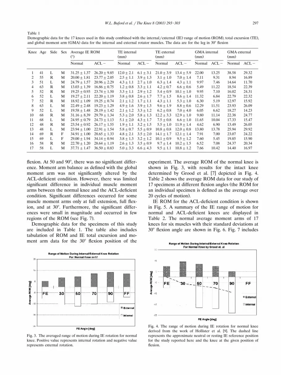
Table 1Ž . Ž . Ž .Demographic data for the 17 knees used in this study combined with the internal�external IE range of motion ROM , total excursion TE ,
Ž .and global moment arm GMA data for the internal and external rotator muscles. The data are for the leg in 30� flexion
Knee Age Side Sex Average IE ROM TE internal TE external GMA internal GMA externalŽ . Ž . Ž . Ž . Ž .� mm mm mm mm
Normal ACL� Normal ACL� Normal ACL� Normal ACL� Normal ACL�
1 41 L M 31.25�1.37 26.20�9.85 12.0�2.1 6.1�3.1 21.0�3.9 13.4�5.9 22.00 13.25 38.58 29.322 55 R M 20.00�1.81 23.77�2.05 2.5�1.1 3.9�1.3 3.1�1.0 7.0�1.4 7.11 9.31 8.94 16.893 51 L M 24.79�1.57 20.96�2.29 4.3�1.1 2.7�1.0 6.3�1.4 4.3�1.1 9.97 7.46 14.64 11.704 63 R M 13.03�1.39 16.86�0.75 1.2�0.8 3.3�1.1 4.2�0.7 6.6�0.6 5.49 11.22 18.54 22.395 52 R M 19.25�0.93 23.74�1.50 3.3�1.1 2.9�1.2 5.4�0.9 10.1�1.0 9.95 7.10 16.02 24.316 52 L M 19.27�2.11 22.20�1.19 3.8�0.8 2.6�1.7 7.7�1.5 8.6�1.4 11.32 6.84 22.79 22.327 52 R M 18.92�1.09 19.25�0.74 2.1�1.2 1.7�1.1 4.3�1.1 5.3�1.0 6.30 5.19 12.97 15.928 63 L M 22.49�2.48 19.23�1.29 4.9�1.6 3.9�1.3 9.6�1.9 8.8�0.6 12.29 11.51 23.93 26.099 52 L M 19.58�1.48 28.19�1.42 2.1�1.2 3.3�1.2 6.2�0.8 7.0�4.0 6.05 6.62 18.27 14.23
10 68 R M 31.16�8.39 29.79�1.34 5.3�2.0 5.8�1.3 12.2�3.3 12.9�1.0 9.80 11.14 22.38 24.7711 68 L M 24.95�0.79 24.73�1.13 5.1�2.0 4.3�1.7 7.5�0.8 6.6�1.0 11.65 10.04 17.33 15.4712 48 R M 23.54�0.92 26.17�1.55 1.9�1.1 3.2�1.5 5.5�1.0 11.9�1.4 4.62 6.90 13.49 26.0513 48 L M 23.94�1.00 22.91�1.54 5.8�0.7 5.5�0.9 10.8�0.8 12.0�0.8 13.80 13.78 25.94 29.9214 69 R F 34.91�1.00 28.65�1.33 4.8�2.1 3.5�2.0 14.1�1.7 12.1�1.4 7.91 7.00 23.07 24.2215 69 L F 29.00�1.94 34.14�0.94 3.8�1.3 3.2�1.2 10.1�0.9 9.5�1.2 7.60 5.45 19.85 15.9016 58 R M 22.70�1.20 28.64�1.19 2.6�1.3 3.5�0.9 9.7�1.4 10.2�1.5 6.52 7.08 24.37 20.3417 58 L M 37.71�1.47 36.50�0.83 5.0�3.3 6.6�4.3 9.5�1.1 10.8�1.2 7.66 10.42 14.40 16.97
flexion. At 50 and 90�, there was no significant differ-ence. Moment arm balance as defined with the globalmoment arm was not significantly altered by theACL-deficient condition. However, there was limitedsignificant difference in individual muscle momentarms between the normal knee and the ACL-deficientcondition. Significant differences occurred for somemuscle moment arms only at full extension, full flex-ion, and at 30�. Furthermore, the significant differ-ences were small in magnitude and occurred in few
Ž .regions of the ROM see Fig. 7 .Demographic data for the specimens of this study
are included in Table 1. The table also includestabulation of ROM and IE total excursion and mo-ment arm data for the 30� flexion position of the
Fig. 3. The averaged range of motion during IE rotation for normalknee. Positive value represents internal rotation and negative valuerepresents external rotation.
experiment. The average ROM of the normal knee isshown in Fig. 3, with results for the intact knee
� �determined by Grood et al. 7 depicted in Fig. 4.Table 2 shows the average ROM data for our study of
Ž17 specimens at different flexion angles the ROM foran individual specimen is defined as the average over
.20 cycles of motion .IE ROM for the ACL-deficient condition is shown
in Fig. 5. A summary of the IE range of motion fornormal and ACL-deficient knees are displayed inTable 2. The normal average moment arms of 17knees for six muscles with their standard deviations at30� flexion angle are shown in Fig. 6. Fig. 7 includes
Fig. 4. The range of motion during IE rotation for normal knee� �derived from the work of Hollister et al. 9 . The dashed line
represents the approximate neutral or resting IE reference positionfor the study reported here and the knee at the given position offlexion.

( )W.L. Buford et al. � The Knee 8 2001 293�303298
Table 2� �Internal�external range of motion for normal knee and the ACL-minus knee: results from this study and the study by Grood et al. 7
Ž . Ž .Normal IE range of motion � ACL� IE range of motion �
This study Normals adapted from This study� �Grood et al. 7
Internal External ROM Internal External ROM Internal External ROM
FE 9.96�4.26 5.83�3.35 15.79�5.46 10 10 20 10.1�4.33 7.97�4.2 18.08�5.330 15.26�5.49 8.44�4.5 23.71�6.57 26 14 40 13.52�4.74 11.77�5.06 25.29�5.7650 17.09�4.56 9.07�4.9 26.16�5.5 28 12 40 14.24�4.27 12.12�5.46 26.36�4.7870 14.83�4.19 11.55�5.45 26.39�5.31 29 10 39 14.23�4.53 12.11�4.85 26.34�4.7490 13.11�3.51 12.81�5.22 25.92�5.49 33 5 38 11.72�4.08 14.87�6.2 26.59�5.43FF 8.9�4.5 12.12�5.57 21.02�5.97 � � � 9.34�4.32 10.45�5.25 19.79�6.29
the average moment arms of normal and ACL-defi-cient knees plotted together for comparison. Normalmoment arms of each muscle at six flexion angles areshown three-dimensionally in Fig. 8.
6. Discussion
The major conclusions of this study are that theflexor muscles at the knee plus the popliteus havesignificant moment arms for internal�external rota-tion and that resection of the ACL has little influenceon the overall IE moment arm balance. As in our
� �prior study 4 , wherein the ACL-deficient conditionminimally effected the FE balance at the knee, wesurmise that the method, which modeled muscle load-ing with free motion, while maintaining congruentjoint motion, did not include external loading. Hence,we can conclude that the loss of the ACL structuremay have little effect upon muscle balance until ex-ternal loading becomes a factor.
Because the most significant investigations of IEmotion at the knee involved ROM and no otherstudies to date have investigated IE moment arms for
Fig. 5. Averaged range of motion of the ACL minus knee duringIE rotation.
the knee, we compared our ROM data with the� �representative work of Grood et al. 7 . The ROM
results for the intact knee determined by Grood et al.� � Ž7 are depicted in Fig. 4 adapted from Fig. 3A, p. 93
.in their publication for comparison with our dataŽ .Fig. 3 . Their study was based on eight specimens,and their ROM was defined at a fixed torque of 5 Nm. In our study, the ROM increases as the knee flexesuntil a certain degree of flexion. IE ROM is a mini-mum at full extension. This is in agreement with theGrood study. During 50�90� of flexion, the ROMdoes not change significantly, but is shifted internally.Then it decreases somewhat at full flexion. In Grood
� �et al. 7 , the ROM data exhibit greater contributionto internal rotation. However, the zero reference inthe Grood study is with the knee at full extension. Inour study, the zero reference is the resting position ateach of the six flexion�extension positions. This refer-ence would be the dashed line in Fig. 4, which issimilar to the neutral position described by Grood et
� �al. 7 . With this correction, the ROM envelopes inboth studies are similar. Also, the flexion limit in theGrood study is 90�. Thus, the decreased ROM at fullflexion in our study cannot be compared.
The ROM for the ACL-deficient condition doesnot show an obvious ‘shift to internal’ tendency from50�90� of flexion, as in the normal condition. TheROM is slightly higher at 30� and full extension andlower at full flexion, due to the ACL resection. How-ever, the absolute values of the ROM for the ACLMcondition were not significantly different from thenormal condition at 50�90�.
The moment arms are also significantly affected bythe position of the knee in flexion�extension. This isclear when viewing Fig. 8, from which the followinggeneralities may also be derived. The semimembra-nosis and semitendonosis moment arms peak near 10�of internal rotation. The gracilis and sartorius mo-ment arms are nearly constant through the IE ROM.The popliteus has a small moment arm that peaks
Žnear neutral. The external rotators biceps femoris.short and long peak near full external rotation. All

( )W.L. Buford et al. � The Knee 8 2001 293�303 299
Fig. 6. Averaged muscle moment arms during IE rotation for normal knee at 30� flexion with their standard deviations. Positive values areinternal rotation and negative values are external rotation.
muscles but the popliteus exhibit a maximum momentarm when the knee is at 70 or 90� flexion. Thepopliteus maximum is at 30 and 50� of flexion.
The shift in global IE moment arm balance as theleg moves from full extension to full flexion is de-picted in Fig. 9a. The external rotators clearly havethe mechanical advantage over the internal rotators
throughout the flexion-extension range of motion. Fig.9b shows the ACL-deficient GMA balance. Normal-ized GMA balance shown in Fig. 9c represents theeffect of the ACL-deficient condition as an averageover all 17 specimens. According to ANOVA, there isno significant difference between the ACL-deficientand the normal knee when comparing the IE GMA

( )W.L. Buford et al. � The Knee 8 2001 293�303300
Fig. 7. Averaged internal�external rotation muscle-moment arms for normal knee and ACL minus knee at 30� flexion.
values at each position of flexion�extension. How-ever, there appears to be a trend at full extension.This trend favoring an increased mechanical advan-tage for the external rotator muscles may be impor-tant to the design of muscle exercises for rehabilita-tion following ACL resection.
Fig. 10 combines the moment arm results of this
� �study with our earlier work 4 that defined the FEmoment arms. The figure is drawn with the FE com-ponent as the ordinate and the IE component as theabscissa. It is similar to the method reported by Brand
� �et al. 3 to display moment arm balance at the wrist.With a quick glance it is easy to see the relativecontribution of each muscle moment arm to motion

( )W.L. Buford et al. � The Knee 8 2001 293�303 301
Fig. 8. Averaged internal�external rotation muscle-moment arms for normal knee at six flexed positions plotted together three-dimensionally.
in the primary rotation directions. Such a plot willvary throughout the ROM, and the position of 30�FE�0� IE was selected here as representative of theknee central to its functional ROM. With thisknowledge, the impact of lost or atrophied musclefunction upon moment arm balance is readily appar-ent.
This project, which focused upon the internal�ex-ternal rotation balance of muscle moment arms at theknee, when combined with our knowledge of theflexion�extension moment arm balance, provides amore complete understanding of knee joint balance.With these results, it is clear that the IE componentof muscle moment arm balance at the knee is signifi-

( )W.L. Buford et al. � The Knee 8 2001 293�303302
Fig. 9. Global moment arm balance of internal rotators and exter-Ž . Ž . Ž .nal rotators for a normal knee; and b ACL minus knee. c
Normalized global moment arm balance. The shift in global mo-ment arm balance of the ACL minus knee relative to the normalknee.
cant. Hopefully, such knowledge will help to improvediagnosis, treatment and rehabilitation of the mal-functioning knee. Future work will focus upon theeffects of total knee arthroplasty upon the momentarms and the moment arm balance at the knee.
Fig. 10. The average IE moment arms for all muscles in the study� �combined with the FE moment arm results reported in 5 . Each
point is a vector that represents each muscle’s IE, FE moment arm� Ž .component the knee is at 30� flexion and 0� neutral internal�ex-
�ternal rotation .
Acknowledgements
Primary funding for this study was a research pro-ject supported by Sulzer Orthopaedics USA, Austin,TX. It is also supported by grant number 004952-0111-1999, from the Advanced Technology Programof the Texas Higher Education Coordinating Board.
References
� �1 An KN, Takahashi KT, Harrigan P, Chao EY. Determinationof muscle orientations and moment arms. J Biomech Eng1984;106:280�282.
� �2 Blankevoort L, Huiskes R, De Lange A. Helical axes ofŽ .passive knee joint motion. J Biomech 1990;23 12 :1219�1229.
� �3 Brand PW, Beach RB, Thompson DE. Relative tension andpotential excursion of muscles in the forearm and hand. J
Ž .Hand Surg 1981;6 3 :209�219.� �4 Buford WL, Ivey FM, Malone JD et al. Muscle balance at the
knee-moment arms for the normal knee and the ACL-defi-cient knee. IEEE Trans Rehab Eng 1997;5:367�379.
� �5 Churchill DL, Incavo SJ, Johnson CC, Beynnon BD. Thetransepicondylar axis approximates the optimal flexion axis ofthe knee. Clin Orthop Rel Res 1998;356:111�118.
� �6 Fick AE. Ueber zweigelenkige muskeln. Arch Anat PhysiolŽ . �Anat Abstr 1879 cited in Spoor and Van Leuwen, kneemuscle moment arms from MRI and from tendon travel, J.
Ž . �Biomech. 1992; 25 2 :201�239 .� �7 Grood ES, Stowers SF, Noyes FR. Limits of movement in the
human knee effect of sectioning the posterior cruciate liga-ment and posterolateral structures. J Bone Jt Surg
Ž .1988;70 1 :88�97.

( )W.L. Buford et al. � The Knee 8 2001 293�303 303
� �8 Hart RA, Mote Jr CD, Skinner HB. A finite helical axis as alandmark for kinematic reference of the knee. J BiomechEng 1991;113:215�222.
� �9 Hollister AM, Jatana S, Singh AK, Sullivan WW, LupichukAG. The axes of rotation of the knee. Clin Orthop Rel Res1993;290:259�268.
� �10 Ishii Y, Terajima K, Terashima S, Koga Y. Three-dimen-sional kinematics of the human knee with intracortical pinfixation. Clin Orthop Rel Res 1997;343:144�150.
� �11 Lane JG, Irby SE, Kaufman K, Rangger C, Daniel DM. Theanterior cruciate ligament in controlling axial rotation. Am J
Ž .Sports Med 1994;22 2 :289�293.� �12 Woltring HJ, Huiskes R, De Lange A, Veldpaus FE. Finite
centroid and helical axis estimation from noisy landmarkmeasurements in the study of human joints. J Biomech
Ž .1985;18 5 :379�389.� �13 Zarins B, Rowe CR, Harris BA, Watkins MP. Rotational
Ž .motion of the knee. Am J Sports Med 1983;11 3 :152�156.