inter-relationships between quinolinic acid, neuroactive kynurenines, neopterin and...
TRANSCRIPT

Journal of Neuroimmunology, 40 (1992) 71-80 71 © 1992 Elsevier Science Publishers B.V. All rights reserved 0165-5728/92/$05.00
JNI 02224
Inter-relationships between quinolinic acid, neuroactive kynurenines, neopterin and/32-microglobulin in cerebrospinal
fluid and serum of HIV-l-infected patients
Melvyn P. Heyes a, Bruce J. Brew b,1 Kuniaki Saito a, Bonnie J. Quearry a, Richard W. Price b,2 Kristin Lee a, Ravi B. Bhalla c, Margaret Der d
and Sanford P. Markey ~ " Section on Analytical Biochemistry, Laboratory of Clinical Science, NIMH, Bethesda, MD, USA, h Department of Neurology,
~' Department of Chemistry, Memorial Sloan-Kettering Cancer Center, New York, NY,, USA, and d Department of Nuclear Medicine, NIH, Bethesda, MD, USA
(Received 22 January 1992) (Revised, received 26 March 1992)
(Accepted 27 March 1992)
Key words: Quinolinic acid; Kynurenic acid; L-Tryptophan; L-Kynurenine; Indoleamine-2,3-dioxygenase; Pterine;/32-Microglobulin; NMDA receptor; AIDS dementia complex; Interferon-3,
Summary
Quinolinic acid (QUIN) is an neurotoxic N-methyl-D-aspartate receptor agonist and an L-tryptophan metabolite of the kynurenine pathway. Increased concentrations of QUIN occur in both cerebrospinal fluid (CSF) and blood of patients infected with human immunodeficiency virus (HIV)-I, particularly those with neurologic disturbances. In the present study of HIV-1 infected patients in Walter Reed stages 4, 5 and 6, reductions in L-tryptophan accompanied proportional increases in L-kynurenine and QUIN in both serum and CSF. Further, close inter-correlations exist between QUIN kynurenic acid and L-kynurenine with both /32-microglobulin and neopterin in CSF and serum. These correlations support the hypotheses that the kynurenine pathway is activated in association with inflammation and induction of indoleamine-2,3-dioxygenase. There were no relationships between CSF QUIN, L-kynurenine or kynurenic acid with the ratio of serum : CSF albumin concentrations, which indicates that the increases in CSF QUIN, L-kynurenine or kynurenic acid were not dependent on a breakdown of the blood-brain barrier. Kynurenic acid is also a kynurenine pathway metabolite that can attenuate the excitotoxic effects of QUIN when present in higher molar concentrations. While CSF kynurenic acid levels were increased in HIV-l-infected patients, the magnitude of the increases were smaller than those of QUIN and the molar concentrations of kynurenic acid were consistently lower than QUIN by at least one order of magnitude. We conclude that immune activation increases the levels of neuroactive kynurenines within the central nervous system of HIV-l-infected patients secondary to activation of indoleamine-2,3-di- oxygenase.
Correspondence to: M.P. Heyes, Section on Analytical Bio- chemistry, Laboratory of Clinical Science, Building 10, Room 3D40, National Institute of Mental Health, Bethesda, MD 20892, USA.
1 Present address: Department of Neurology and Centre for Immunology, St. Vincent's Hospital, Sydney, Australia.
2 Present address: Department of Neurology, University of Minnesota, Minneapolis, MN, USA.

72
Introduction
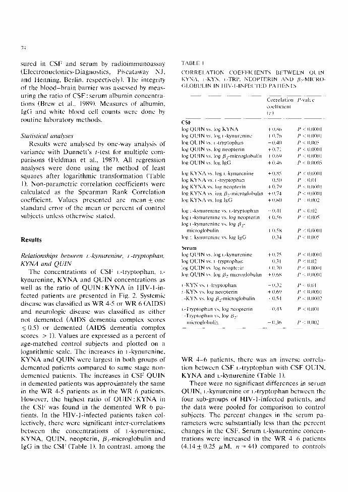
Substantially increased concentrations of the excitotoxin and kynurenine pathway metabolite, quinolinic acid (QU1N), are found in the cere- brospinal fluid (CSF) of patients infected with the human immunodeficiency virus (HIV-I; Heyes et al., 1989), particularly among patients with the AIDS dementia complex, aseptic meningitis or opportunistic central nervous system conditions (Heyes et a[., 1991a). QUIN is an agonist of N-methyl-D-aspartatc receptors and an excito- toxin. Notably, the concentrations achieved in the CSF of HIV-l-infected patients (Heyes et al., 1991a) exceeded levels reported to be neurotoxic in vitro (Giulian ct al., 1990; Whetsell and Schwarcz, 1989). Consequently, we have postu- lated that QUIN may be involved in the neuro- logic complications of HIV-I infection, including
the AIDS dementia complex (Hcycs el al., 1089, 1991a). The potential role of N-methyl-t>aspar- tate receptors in mediating neuronal damage in HIV-l-infection has been further highlighted by subsequent in vitro studies (Giulian et al., 1090; Lipton et al., 1991 ).
Other factors, however, may influence the neurologic effects of QUIN and other N-methyl- D-aspartate receptor agonists. In particular, the related kynurenine pathway metabolite kynurenic acid (KYNA) can attenuate the cxcitotoxic effects of QUIN by virtue of its antagonist effects on excitatory amino acid receptors, including N- methyl-D-aspartate receptors (Foster et al., 1984). Therefore, the balance between the conccntra- tions of QUIN and KYNA may influence whether the excitotoxic effects of QUIN or other neuro- toxins are manifest. The present study sought to determine whether the levels of KYNA are in-
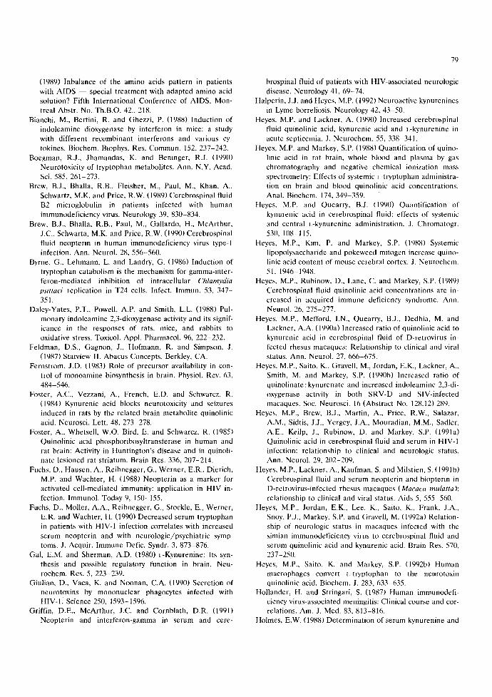
B L O O D A N D C S F
Neopterin
/
[32 -Microglobulin Kynurenines
d
b // /
/
Neopterin
e.g. IFN-y / /
~ , , LoTRP b GTP cvclohvdrolase-I ~ ~ KYNA . . . .
L-KYN ~
Triphosphate MI :~'4KYN
Tetrahydrobiopterin ; b QUIN . . . . . . . . . .
e.a. Macmohaae/Microalia
- ~ KYNA
\
N*uron~ AcVw~ty C~dum En~ Ion Germ E X l ~
ItlutommlmttDr R*l*e~ M*mbmn* Slructure
LNmI.¢ ~ r t S,F,m~Cqm.~. m m u o n
g
cam, .~ N M D A - R ~ p t o r Complex Mo(ludlloel
- --~.. QUIN
,o~* Toxine
Fig. 1. Model of the metabolic relationships between selected cytokines, kynurenine pathway metabolites, neopterin and /32-microglobulin and the potential role of N-methyl-D-aspartate (NMDA) in neuronal dysfunction and neurodegenerat ion in HIV-1 infection and other inflammatory diseases. Broken lines represent transfer of substrate from one metabolic compar tment to another, solid lines represents metabolic reactions or effects of agents on enzymes or receptors. Enzymes are underlined. Interferon-y (IFN-y) and other cytokines increase indoleamine-2,3-dioxygenase (IDO) activity in macrophages and other cells (a) and increase the conversion of L-tryptophan to L-kynurenine (L-KYN), KYNA, anthranilic acid (AA), 3-hydroxykynurenine (3-HKYN), 3-hydroxyanthranilic acid (3-HAA) and QUIN which may enter the blood, CSF and extracellular fluid space of the brain (b). IFN-T also activates guanosine tr iphosphate (GTP) cyclohydrolase-I and increases the synthesis of neopterin (c) which also appears in the blood and CSF (d). The release of/32-microglobulin is also increased by IFN-y (e). Increased concentrations of QUIN, kynurenic acid and other modulators of N-methyI-D-aspartate receptor activity may induce neuronal dysfunction and nerve cell death and thereby result in neuro[ogic symptoms (g). Strategies to alter the synthesis of N-methyl-D-aspartate receptor ligands
or a t tenuate their effects offer new approaches to therapy in inflammatory diseases.

creased in the CSF, and investigate the relation- ship of KYNA to QUIN and other neuroactive kynurenines in the CSF of HIV-l-infected pa- tients.
The increases in CSF and serum QUIN in HIV-l-infected patients have been attributed to induction of indoleamine-2,3-dioxygenase, the first enzyme of the kynurenine pathway which converts L-tryptophan to L-kynurenine (Fig. 1). The increases in both brain and lung in- doleamine-2,3-dioxygenase activity in non-human primate models of AIDS are consistent with this hypothesis (Saito et al., 1991a). To further inves- tigate the role of indoleamine-2,3-dioxygenase in changing kynurenine pathway metabolism, we used the concentrations of L-tryptophan and L- kynurenine in blood and CSF as an index of indoleamine-2,3-dioxygenase activity (Fuchs et al., 1990; Heyes and Lackner, 1990; Saito et al., 1991a) and determined their relationships to QUIN and KYNA. In addition, because host immune mediators such as interferon-y and tu- mor necrosis factor-c~ may increase indoleamine- 2,3-dioxygenase activity, QUIN production, neopterin synthesis, and/32-microglobulin expres- sion (Pfefferkorn and Guyre, 1984; Byrne et al., 1986; Bianchi et al., 1988; Saito et al., 1991b; Heyes et al., 1991, 1992a, b), we examined the relationship of kynurenine pathway metabolites with neopterin and /32-microglobulin, which are 'markers' of immune stimulation (see Fig. 1). The potential role of disruption of the blood-brain barrier was studied by measuring the CSF : serum albumin ratio.
Materials and methods
Subjects studied Samples of both CSF and serum were col-
lected from HIV-l-infected patients who were being studied at the Memorial Sloan-Kettering Cancer Center. Blood was collected from an arm vein and serum was isolated by centrifugation. CSF was collected from the lumbar sac. The clinical characteristics of these patients have been described previously (Brew et al., 1989, 1990; Heyes et al., 1991a). The systemic disease state of these subjects was classified according to the
73
Walter Reed (WR) Staging system (WR 4-5, n = 40; WR 6, n = 39 (Redfield et al., 1986). AIDS dementia complex scores (0-4) were determined according to published criteria (Brew et al., 1989). The number of patients in each group were: demented, WR 4-5, n = 20; WR 6, n = 28; or not demented, WR 4-5, n = 20; WR 6, n = 11. Pa- tients were studied in various stages of systemic and central nervous system disease. None of the patients had clinical aseptic meningitis (Hol- lander and Stringari, 1987), demonstrable oppor- tunistic central nervous system infections or neo- plasms. Because the samples were obtained be- fore the approval or widespread use of zudovu- dine (azidothymidine or AZT), none of the pa- tients were receiving anti-retroviral therapies at the time of sample collection. Control subjects were 22 age-matched healthy and neurologically unimpaired volunteers.
Biochemical measurements Samples were assayed by experienced labora-
tory personnel, using established and verified methods, without prior knowledge to the pa- tients' viral or clinical status. QUIN was quanti- fied by electron capture negative chemical ioniza- tion gas chromatography/mass spectrometry which uses [180]QUIN as internal standard, rather than structural isomers or chemical analogs (Heyes and Markey, 1988). The concentrations of KYNA, L-kynurenine and L-tryptophan in CSF and serum were quantified by high performance liquid chromatography with either fluorescence detection (Heyes and Quearry, 1990) or ultravio- let light absorbance spectrometry (adapted from Holmes, 1988) or electrochemical detection (Heyes and Markey, 1988) respectively. Gener- ally, measures were made within the same assay run. However, where more than one assay run was done, selected samples from previous assays were included in subsequent procedures to en- sure replicate values were within established and acceptable variability limits. In no case could group mean differences be attributed to system- atic assay errors . In other studies, no gradients for QUIN, KYNA or L-kynurenine have been noted along the CSF axis (Mouradian et al., 1989; Heyes and Sunderland, unpublished observa- tions)./32-Microglobulin and neopterin were mea-
74
sured in CSF and serum by radioimmunoassay (Electronuclonics-Diagnostics, Piscataway N J, and Henning, Berlin, respectively). The integrity of the blood-brain barrier was assessed by meas- uring the ratio of CSF : serum albumin concentra- tions (Brew et al., 1989). Measures of albumin, IgG and white blood cell counts were done by routine laboratory methods.
Statistical analyses Results were analysed by one-way analysis of
variance with Dunnett's t-test tot multiple com- parisons (Feldman et al., 1987). All regression analyses were done using the method of least squares after logarithmic transformation (Table 1). Non-parametric correlation coefficients were calculated as the Spearman Rank Correlation coefficient. Values presented arc mean + one standard error of the mean or percent of control subjects unless otherwise stated.
Results
Relationships between :.-kynurenine, L-tryptophan, KYNA and QUIN
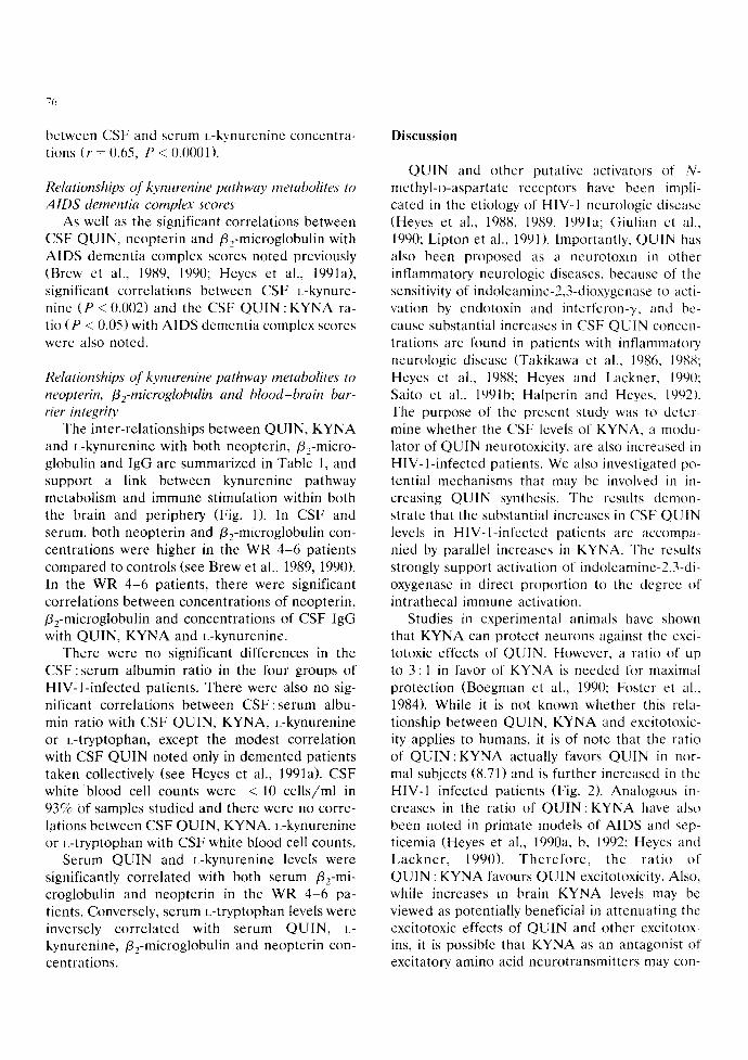
The concentrations of CSF k-tryptophan, L- kynurenine, KYNA and QUIN concentrations as well as the ratio of Q U I N : K Y N A in HIV-l-in- fected patients are presented in Fig. 2. Systemic disease was classified as WR 4-5 or WR 6 (AIDS) and neurologic disease was classified as either not demented (AIDS dementia complex scores _<0.5) or demented (AIDS dementia complex scores _> 1). Values are expressed as a percent of age-matched control subjects and plotted on a logarithmic scale. The increases in ~:kynurenine, KYNA and QUIN were largest in both groups of demented patients compared to same stage non- demented patients. The increases in CSF QUIN in demented patients was approximately the same in the WR 4-5 patients as in the WR 6 patients. However, the highest ratio of Q U I N : K Y N A in the CSF was found in the demented WR 6 pa- tients. In the H1V-l-infected patients taken col- lectively, there were significant inter-correlations between the concentrations of [-kynurenine, KYNA, QUIN, neopterin, /3:microglobulin and lgG in the CSF (Table 1). In contrast, among the
"FABLE I
C O R R E L A T I O N ( 7 O E F F I C I E N T S B E T W E E N Q U I N .
K Y N A , I - K Y N , I - T R P . N E O P T E R I N A N D / ? , 2 - M I ( R O -
G L O B U I J N IN H I V - I - I N F E C T E D P A T I E N T S
Correlation P-va lue
cocfficicnl ( r )
CSF log Q U I N vs. log K Y N A
log Q U I N vs. log I -kynureninc log Q U I N vs. i -tryptophan log Q U I N vs. log neopterin log Q U I N vs. log fie-microglobulin log Q U 1 N vs. log l g G
k)g K Y N A vs. log i.-kynurcninc log K Y N A vs. [ -tryptophan h)g K Y N A vs. log neopterin log K Y N A vs. log fl2-microglobulin h)g K Y N A vs. log l g G
log i - k y n u r e n i n e vs. i - t r y p t o p h a n
log I -kynureninc vs. log neoptcrin log [ -kynurenine vs. log /3 z-
microglobulin h)g t - k y n u r e n i n c vs. log l g G
Serum log Q U I N vs. log I -kynurenine log Q U I N vs. i -tryptophan log Q U I N vs. log neopterin log Q U I N vs. log ,(32-microgk~bulin
I - K Y N vs. I -tryptophan I - K Y N vs. log neopterin I - K Y N vs. log /32-microglobulin
i : T ~ ' p t o p h a n vs. k)g neopterin i - T r y p t o p h a n vs. log /32-
mic rog lobu l in
+ I).86 I" < O.O001
+ 0 . 7 6 P < O.(IO01
0.40 P < 0,005
+ 0.71 I ' , (}.0001
+ 0 . 6 9 P < 0.0001
+ 0.46 P < 0.0005
+ 0.85 t ' < 0.00(}I
0,50 P < 0.(}I
+ 0.79 I ' < 0.0001
+ 0.74 t ' - 0.0001
4- 0.80 I' < ().002
- 0.41 I ' ~ 0.02
+ 0.56 P < 0.005
+ 0.58 I ' <- 0.(}001
0.34 1' < ().005
+{L75 1" < 0.0001
0.31 f ' < 0.02
+ 0.70 I' < 0.(t001
+ 0.68 P < ().()()()1
- 0 . 3 2 l' < (k0t + 0.69 P < 0.0001
+ 0.54 t ' < 0 .0002
- 0 . 4 3 I' < ().0(11
0.36 P < 0.002
WR 4-6 patients, there was an inverse correla- tion between CSF k-tryptophan with CSF QUIN, KYNA and t.-kynurenine (Table 1).
There were no significant differences in serum QUIN, t.-kynurenine or l.-tryptophan between the four sub-groups of HIV-l-infected patients, and the data were pooled for comparison to control subjects. The percent changes in the serum pa- rameters were substantially less than the percent changes in the CSF. Serum L-kynurenine concen- trations were increased in the WR 4 - 6 patients (4.14_+ 0.25 /a,M, n = 44) compared to controls
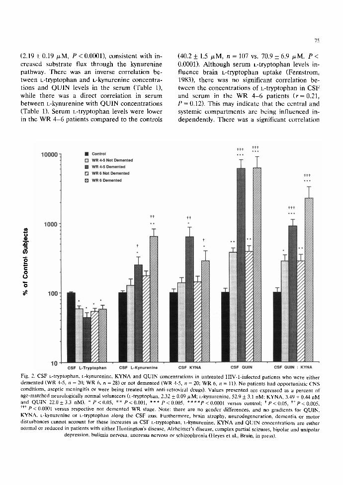
(2.19 + 0.19 /zM, P < 0.0001), consistent with in- creased substrate flux through the kynurenine pathway. There was an inverse correlation be- tween L-tryptophan and L-kynurenine concentra- tions and Q U I N levels in the serum (Table 1), while there was a direct correlation in serum between L-kynurenine with Q U I N concentrations (Table 1). Serum L-tryptophan levels were lower in the WR 4 -6 patients compared to the controls
75
(40.2 +_ 1.5 /zM, n = 107 vs. 70.9 +_ 6.9 ~M, P < 0.0001). Although serum L-tryptophan levels in- fluence brain L-tryptophan uptake (Fernstrom, 1983), there was no significant correlation be- tween the concentrations of L-tryptophan in CSF and serum in the WR 4 -6 patients ( r = 0 . 2 1 , P = 0.12). This may indicate that the central and systemic compartments are being influenced in- dependently. There was a significant correlation
m
1000
C 0 o
10000
t t
100
10
• Control
D WR 4-5 Not Demented
! WR 4-5 Demented
[ ] WR 6 Not Demented
[ ] WR 6 Demented
Q om ,,Q - !
00
t t t t t t * * * * * *
t t
CSF L-Tryptophan CSF L-Kynurenlne CSF KYNA CSF QUIN CSF QUIN : KYNA
Fig. 2. CSF L-tryptophan, L-kynurenine, KYNA and QUIN concentrations in untreated HIV-l-infected patients who were either demented (WR 4-5, n = 20; WR 6, n = 28) or not demented (WR 4-5, n = 20; WR 6, n = 11). No patients had opportunistic CNS conditions, aseptic meningitis or were being treated with anti-retroviral drugs). Values presented are expressed as a percent of age-matched neurologically normal volunteers (L-tryptophan, 2.32 _+ 0.09/xM; L-kynurenine, 52.9 _+ 3.1 riM; KYNA, 3.49 + 0.44 nM and QUIN 22.0 _+ 3.3 riM). * P < 0.05, ** P < 0.001, *** P < 0.005, * * * * P < 0.0001 versus control; t P < 0.05, t* p < 0.005, • **P < 0.0001 versus respective not demented WR stage. Note: there are no gender differences, and no gradients for QUIN, KYNA, L-kynurenine or L-tryptophan along the CSF axis. Furthermore, brain atrophy, neurodegeneration, dementia or motor disturbances cannot account for these increases as CSF L-tryptophan, L-kynurenine, KYNA and QUIN concentrations are either normal or reduced in patients with either Huntington's disease, Alzheimer's disease, complex partial seizures, bipolar and unipolar
depression, bulimia nervosa, anorexia nervosa or schizophrenia (Heyes et al., Brain, in press).
76
between CSF and serum L-kynurenine concentra- tions ( r = (/.65, P < 0.0001).
Relationships of kynurenine pathway metabolites to AIDS dementia complex scores
As well as the significant correlations between CSF QUIN, neopterin and /32-microglobulin with AIDS dementia complex scores noted previously (Brew ct al., 1989, 1990; Heyes et al., 1991a), significant correlations between CSF l_-kynure- nine (P < 0.002) and the CSF QUIN :KYNA ra- tio (P < 0.05)with AIDS dementia complex scores were also noted.
Relationships of kynurenine pathway metabolites to neopterin, lg2-microglobulin and blood-brain bar- rier integrity
The inter-relationships between QUIN, KYNA and l.-kynurenine with both neopterin, /32-micro- globulin and IgG are summarized in Table 1, and support a link between kynurenine pathway metabolism and immune stimulation within both the brain and periphery (Fig. 1). In CSF and serum, both neopterin and /32-microglobulin con- centrations were higher in the WR 4 -6 patients compared to controls (see Brew et aI., 1989, 1990). In the WR 4 -6 patients, there were significant correlations between concentrations of neopterin, /32-microglobulin and concentrations of CSF IgG with QUIN, KYNA and L-kynurenine.
There were no significant differences in the CSF: se rum albumin ratio in the four groups of HIV-l- infected patients. There were also no sig- nificant correlations between CSF: se rum albu- min ratio with CSF QUIN, KYNA, c-kynurenine or c-tryptophan, except the modest correlation with CSF QUIN noted only in demented patients taken collectively (see Heyes et al., 1991a). CSF white b lood cell counts were < 10 ce l l s /ml in 93% of samples studied and there were no corre- lations between CSF QUIN, KYNA, L-kynurenine or c-tryptophan with CSF white blood cell counts.
Serum QUIN and L-kynurenine levels were significantly correlated with both serum /3x-mi- croglobulin and neopterin in the WR 4-6 pa- tients. Conversely, serum L-tryptophan levels were inversely correlated with serum QUIN, i,- kynurenine, /32-microglobulin and neopterin con- centrations.
Discussion
QUIN and other putative activators of N- methyl-D-aspartate receptors have been impli- cated in the etiology of HIV-1 neurologic disease (Heyes et al., 1988, 1989, 1991a; Giulian ct al., 1990; Lipton et al., 1991). Importantly, QUIN has also been proposed as a neurotoxin in other inflammatory neurologic diseases, because of the sensitivity of indoleaminc-2,3-dioxygcnase to acti- vation by endotoxin and interferon-y, and be- cause substantial increases in CSF QUIN concen- trations are found in patients with inflammatory neurologic disease (Takikawa et al., 1986, 1988; Heyes et al., 1988; Heyes and Lackner, 1990: Saito ct al., 1991b; Halperin and Heyes, 1992). The purpose of the present study was to deter- mine whether the CSF levels of KYNA, a modu- lator of QUIN neurotoxicity, are also increased in HIV-l- infected patients. We also investigated po- tential mechanisms that may be involved in in- creasing QUIN synthesis. The results demon- strate that the substantial increases in CSF QUIN levels in HlV-l- infected patients are accompa- nied by parallel increases in KYNA. The results strongly support activation of indoleaminc-2,3-di- oxygenase in direct proportion to the degree of intrathecal immune activation.
Studies in experimental animals have shown that KYNA can protect neurons against the exci- totoxic effects of QUIN. However, a ratio of up to 3 : 1 in favor of KYNA is needed for maximal protection (Boegman et al., 1990; Foster et al., 1984). While it is not known whether this rela- tionship between QUIN, KYNA and excitotoxic- ity applies to humans, it is of note that the ratio of Q U I N : K Y N A actually favors QUIN in nor- real subjects (8.71) and is further increased in the HIV-1 infected patients (Fig. 2). Analogous in- creases in the ratio of Q U I N : K Y N A have also been noted in primate models of AIDS and sep- ticemia (Heyes et al., 1990a, b, 1992; Heyes and Lackner , 1990). T h e r e f o r e , the rat io of QUIN : KYNA favours QUIN excitotoxicity. Also, while increases in brain KYNA levels may be viewed as potentially beneficial in attenuating the excitotoxic effects of Q U I N and other excitotox- ins, it is possible that KYNA as an antagonist of excitatory amino acid neurotransmitters may con-
tribute to the neurologic deficits by blocking exci- tatory amino acid receptors during immune acti- vation. QUIN may also interfere with excitatory neurotransmission and thereby produce neuro- logic deficits by a non-cytolytic mechanism.
A model of possible mechanisms for increases in CSF and serum QUIN in HIV-1 infection, as well as other conditions of immune activation, also applies to the increases in CSF KYNA and L-kynurenine (Fig. 1; Takikawa et al., 1986; Heyes et al., 1990a, 1991a; Heyes and Lackner, 1990). KYNA may be derived from L-kynurenine that had been taken up either by the brain from the blood (Gal and Sherman, 1980; Heyes and Quearry, 1990), or synthesized within the brain (Heyes and Quearry, 1990) secondary to activa- tion of indoleamine-2,3-dioxygenase. The in- creases in the activity of indoleamine-2,3-di- oxygenase in both lung and brain of macaques infected with the Simian immunodeficiency virus or the type-D retrovirus are consistent with both systemic and central synthesis (Heyes et al., 1990b; Saito et al., 1991a). The inverse correlations in the present study between L-tryptophan with both QUIN and L-kynurenine in the CSF of HIV-l-in- fected patients, but not in the CSF of control subjects, is in accordance with increased brain indoleamine-2,3-dioxygenase activity (Table 1). Further, the positive correlations between CSF levels of L-KYN, KYNA and QUIN support in- creased substrate flux through the kynurenine pathway within the CNS (Table 1). It is of note that the magnitude of the increases in CSF L- kynurenine and QUIN are substantially greater than the increases in serum. This phenomenon has been noted in other inflammatory disease conditions (Halperin and Heyes, 1992; Heyes and Lackner, 1990; Heyes et al., 1990a, b, 1992a). Nevertheless, substrates derived from blood may be important sources of L-kynurenine, KYNA and QUIN, particularly if the levels of systemic immune activation is marked, for example during septicemia (Heyes and Lackner, 1990).
The model (Fig. 1) proposes that macrophages are a principle source for QUIN (Heyes et al., 1991a). Infiltrates of macrophages and reactive microglia are a well-established neuropathologic feature of HIV-1 infection, and are also found in many other conditions of CNS inflammation.
77
Macrophages convert [J3C6]-L-tryptophan to [13C6]-QUIN, particularly when stimulated with interferon-y, and the concentrations achieved in the incubation medium (24 p,m) exceed those noted in the CSF of HIV-l-infected patients (up to 15 p.M; Heyes et aI., 1992b; Brew and Heyes, unpublished observations). This observation demonstrates that macrophages contain the en- zymes necessary to convert L-tryptophan to QUIN. Consequently, it is likely that the activity of other enzymes of the kynurenine pathway are also increased following intracerebral immune ac- tivation and macrophage infiltration. Other cells may also convert precursors to QUIN, including astrocytes, which contain 3-hydroxyanthranilate- 3,4-dioxygenase (Okuno et al., 1987). The accu- mulation of QUIN may also reflect the relatively low activity of quinolinic acid phosphoribosyl- transferase, the degradation enzyme for QUIN (Foster et al., 1985).
Both indoleamine-2,3-dioxygenase and GTP cyclohydrolase I activity are increased by inter- feron-y, tumor necrosis factor-a and other cy- tokines in macrophages and other cell types (Fig. 1; Niederwieser et al., 1986; Bianchi et al., 1988; Fuchs et al., 1988; Heyes et al., 1992b). There- fore, strong inter-correlations between QUIN, KYNA, L-tryptophan and L-kynurenine with neopterin, /32-microglobulin and IgG concentra- tions in the CSF support a link between in- doleamine-2,3-dioxygenase induction with in- trathecal inflammatory responses (Table 1; Fuchs et al., 1990; Heyes et al., 1991b, 1992a). These correlations also suggest increased interferon-y activity within the central nervous system (Griffin et al., 1991). There was minimal evidence that the group increases in CSF QUIN, KYNA or L- kynurenine could be attributed to disruption of the blood-brain barrier. Similar conclusions have been drawn regarding the source of elevated neopterin and /32-microglobulin in CSF (Brew et al., 1989,1990; Griffin et al., 1991).
Dietary L-tryptophan intake was not regulated or quantified in the present study and we cannot exclude the possibility that at least some of the reductions in serum and CSF L-tryptophan con- centrations were diet-dependent. However, re- duced t.-tryptophan intake would be expected to either decrease not only L-tryptophan levels but

7~
also L-kynurenine, KYNA and QUIN concentra- tions. The reductions in CSF L-tryptophan levels were independent of blood L-tryptophan concen- trations, and indicate that the central and sys- temic L-tryptophan compartments are influenced separately, such as by different local in- doleamine-2,3-dioxygenasc activities in central nervous system and systemic tissues. The uptake of l.-tryptophan into the brain may have also been influenced by changes in the concentrations of large neutral amino acids in the blood of HIV-1- infected patients (Fernstrom, 1983). The levels of large neutral amino acids are reduced in some HIV-l-infected patients (Althoff ct al., 1989), which would promote L-tryptophan entry into the CNS (Fernstrom, 1983). Therefore, these obser- vation argue in favor of a role for indoleamine- 2,3-dioxygenase induction in accelerating the con- version of L-tryptophan to l.-kynurenine, KYNA and QUIN. Depletion of L-tryptophan may re- duce the synthesis of serotonin and other in- doleamincs (Larsson et al., 1989; Heyes et al., 1990a), as well as interfere with the metabolism of protein in both systemic and central nervous system tissues.
While it is clear that induction of indoleamine- 2,3-dioxygenase, the depletion of L-tryptophan and increased substrate flux through the kynure- nine pathway are associated with immune activa- tion, the reason for this response remains to be established. The magnitude of the increases in kynurenine pathway metabolism, particularly within the central nervous system, and the widespread circumstances in which it occurs, indi- cate that the reasons and consequences are not trivial. There are arguments that such responses may be both beneficial as well as detrimental. On the positive side, studies in vitro have suggested that activation of indoleamine-2,3-dioxygenase and depletion of intracellular k-tryptophan may be one mechanism by which interferon-y exerts anti-microbial and anti-proliferative effects on some intracellular parasites and tumor cells (Pfef- ferkorn and Guyre, 1984; Byrne et al., 1986; Takikawa et al., 1988), but not on others (Turco and Winkler, 1986: Takikawa et al., 1988). Also, the reactions catalysed by indoleamine-2,3-di- oxygenase metabolize potentially toxic oxygen- free radicals (Daley-Yates et al., 1988; Siesjo et
al., 1989; Sono, 1989). Conversely, depletion of L-tryptophan may impair protein synthesis and indoleamine metabolism. Thc production of po- tentially neurotoxic kynureninc pathway metabo- litcs, including QUIN, l-kynurenine and KYNA, may be another detrimental consequence of in- doleamine-2,3-dioxygenase induction. At this point in time, it is not possible to state where the balance between beneficial versus detrimental consequences lies.
lndoleamine-2,3-dioxygenase induction and production of kynurcnine pathway metabolites occur in a wide spectrum of immune stimulation (Pfefferkorn and Guyre, 1984; Byrne et al., 1986; Takikawa et al., 1986; Werner ct al., 1987, 1989; Bianchi et al., 1988; Heyes and Lackner, 1990: Heyes et al., 1988, 1990b, 1992; Saito et al., 1991a, b). In view of the neuroactive nature of kynurenine pathway metabolites, we propose that such compounds may be final common mediators of neuronal dysfunction and death in inflamma- tory neurologic disease. This disruption would include functions mediated via N-methyl-D- aspartate receptors, such as learning, memory and synaptic plasticity (Morris et al., 1987). Therefore, strategies to attenuate the neurotoxic effects of QUIN (without disrupting N-methyl- o-aspartate receptor function), or reducing the synthesis of neuroactive kynurenine pathway metabolites, may offer new approaches to therapy of the neurologic deficits associated with HIV-I infection. Notably, such strategies may also be of benefit in other inflammatory neurologic dis- eases.
Acknowledgements
We appreciate the assistance of M. Paul and H. Gallardo. This study was supported in part by US Public Health Service Research Grant NS- 25701, a grant from the Rudin Foundation, US Army Medical R & D C o m m a n d project 87PP7856, the Henry M. Jackson Foundation and the Walter Reed Retrovirus Research Group.
References
Althoff, P.-H., Schifferdecker, E., Forster, H., Michels, B., Hunold, P., St. Klauke, A., Flakenbach, E. and Helm, K.
(1989) lnbalance of the amino acids pattern in patients with AIDS - - special treatment with adapted amino acid solution? Fifth International Conference of AIDS, Mon- treal Abstr. No. Th.B.O. 42., 218.
Bianchi, M., Bertini, R. and Ghezzi, P. (1988) Induction of indoleamine dioxygenase by interferon in mice: a study with different recombinant interferons and various cy- tokines. Biochem. Biophys. Res. Commun. 152, 237-242.
Boegman, R.J., Jhamandas, K. and Beninger, R.J. (1990) Neurotoxicity of tryptophan metabolites. Ann. N.Y. Acad. Sci. 585, 261-273.
Brew, B.J., Bhalla, R.B., Fleisher, M., Paul, M., Khan, A., Schwartz, M.K. and Price, R.W. (1989) Cerebrospinal fluid B2 microglobulin in patients infected with human immunodeficiency virus. Neurology 39, 830-834.
Brew, B.J., Bhalla, R.B., Paul, M., Gallardo, H., McArthur, J.C., Schwarta, M.K. and Price, R.W. (1990)Cerebrospinal fluid neopterin in human immunodeficiency virus type-l infection. Ann. Neurol. 28, 556-560.
Byrne, G., Lehmann, L. and Landry, G. (1986) Induction of tryptophan catabolism is the mechanism for gamma-inter- feron-mediated inhibition of intracellular Chlamydia psittaci replication in T24 cells. Infect. Immun. 53, 347- 351.
Daley-Yates, P.T., Powell, A.P. and Smith, L.L. (1988) Pul- monary indoleamine 2,3-dioxygenase activity and its signif- icance in the responses of rats, mice, and rabbits to oxidative stress. Toxicol. Appl. Pbarmacol. 96, 222-232.
Feldman, D.S., Gagnon, J., Hofmann, R. and Simpson, J. (1987) Statview If. Abacus Concepts, Berkley, CA.
Fernstrom, J.D. (1983) Role of precursor availability in con- trol of monoamine biosynthesis in brain. Physiol. Rev. 63, 484-546.
Foster, A.C., Vezzani, A., French, E.D. and Schwarcz, R. (1984) Kynurenic acid blocks neurotoxicity and seizures induced in rats by the related brain metabolite quinolinic acid. Neurosci. Lett. 48, 273-278.
Foster, A., Whetsell, W.O. Bird, E. and Schwarcz, R. (1985) Quinolinic acid pbosphoribosyltransferase in human and rat brain: Activity in Huntington's disease and in quinoli- hate-lesioned rat striatum. Brain Res. 336, 207-214.
Fuchs, D., Hausen, A., Reibnegger, G., Werner, E.R., Dierich, M.P. and Wachter, H. (1988) Neopterin as a marker for activated cell-mediated immunity: application in HIV in- fection. Immunol. Today 9, 150-155.
Fuchs, D., Moiler, A.A., Reibnegger, G., Stockle, E., Werner, E.R. and Wachter, H. (1990) Decreased serum tryptophan in patients with HIV-1 infection correlates with increased serum neopterin and with neurologic/psychiatric symp- toms. J. Acquir. Immune Defic. Syndr. 3, 873-876.
Gal, E.M. and Sherman, A.D. (1980) L-Kynurenine: Its syn- thesis and possible regulatory function in brain. Neu- rochem. Res. 5, 223-239.
Giulian, D., Vaca, K. and Noonan, C.A. (1990) Secretion of neurotoxins by mononuclear phagocytes infected with HIV-1. Science 250, 1593-1596.
Griffin, D.E., McArthur, J.C. and Cornblath, D.R. (1991) Neopterin and interferon-gamma in serum and cere-
79
brospinal fluid of patients with HIV-associated neurologic disease. Neurology 41, 69-74.
Halperin, J.J. and Heyes, M.P. (1992) Neuroactive kynurenines in Lyme borreliosis. Neurology 42, 43-50.
Heyes, M.P. and Lackner, A. (1990) Increased cerebrospinal fluid quinolinic acid, kynurenic acid and L-kynurenine in acute septicemia. J. Neurochem. 55, 338-341.
Heyes, M.P. and Markey, S.P. (1988) Quantification of quino- linic acid in rat brain, whole blood and plasma by gas chromatography and negative chemical ionization mass spectrometry: Effects of systemic ~-tryptophan administra- tion on brain and blood quino[inic acid concentrations. Anal. Biochem. 174, 349-359.
Heyes, M.P. and Quearry, B.J. (1990) Quantification of kynurenic acid in cerebrospinal fluid: effects of systemic and central L-kynurenine administration. J. Chromatogr. 530, 108-115.
Heyes, M.P., Kim, P. and Markey, S.P. (1988) Systemic lipopolysaccharide and pokeweed mitogen increase quino- linic acid content of mouse cerebral cortex. J. Neurochem. 51, 1946 1948.
Heyes, M.P., Rubinow, D., Lane, C. and Markey, S.P. (1989) Cerebrospinal fluid quinolinic acid concentrations are in- creased in acquired immune deficiency syndrome. Ann. Neurol. 26, 275-277.
Heyes, M.P., Mefford, I.N., Quearry, B.J., Dedhia, M. and Lackner, A.A. (1990a) Increased ratio of quinolinic acid to kynurenic acid in cerebrospinal fluid of D-retrovirus in- fected rhesus macaques: Relationship to clinical and viral status. Ann. Neurol. 27, 666-675.
Heyes, M.P., Saito, K., Gravell, M., Jordan, E.K., Lackner, A., Smith, M. and Markey, S.P. (1990b) Increased ratio of quinolinate:kynurenate and increased indoleamine-2,3-di- oxygenase activity in both SRV-D and SIV-infected macaques. Soc. Neurosci. 16 (Abstract No. 128.12) 289.
Heyes, M.P., Brew, B.J., Martin, A., Price, R.W., Salazar, A.M., Sidtis, J.J., Yergey, J.A., Mouradian, M.M., Sadler, A.E., Keilp, J., Rubinow, D. and Markey, S.P. (1991a) Quinolinic acid in cerebrospinal fluid and serum in HIV-1 infection: relationship to clinical and neurologic status. Ann. Neurol. 29, 202-209.
Heyes, M.P., Lackner, A., Kaufman, S. and Milstien, S. (1991b) Cerebrospinal fluid and serum neopterin and biopterin in D-retrovirus-infected rhesus macaques (Macaca mulatta): relationship to clinical and viral status. Aids 5, 555-560.
Heyes, M.P., Jordan, E.K., Lee, K., Saito, K., Frank, J.A, Snoy, P.J., Markey, S.P. and Gravell, M. (1992a) Relation- ship of neurologic status in macaques infected with the simian immunodeficiency virus to cerebrospinal fluid and serum quinolinic acid and kynurenic acid. Brain Res. 570, 237-250.
Heyes, M.P., Saito, K. and Markey, S.P. (1992b) Human macrophages convert L.-tryptophan to the neurotoxin quinolinic acid. Biochem. J. 283, 633-635.
Hollander, H. and Stringari, S. (1987) Human immunodefi- ciency virus-associated meningitis: Clinical course and cor- relations. Am. J. Med. 83, 813-816.
Holmes, E.W. (1988) Determination of serum kynurenine and
80
hcpatic tryptophan dioxygenase activity by high-perfl~r- mance liquid chromatography. Anal. Biochem. 172, 518 525.
Larsson, M., Hagberg, L., Norkrans, G. and Forsman, A. (1989) Indole amine deficiency in blood and cerebrospinal fluid from patients with human immunodeficiency virus infection. J. Neurosci. Res. 23,441-446.
Lipton, S.A., Sucher, N.J., Kaiser, P.K. and Dreyer, E.B. (1991) Synergistic effects of HIV coal protein and NMDA-receptor mediated neurotoxicity. Neuron 7, 111- 118.
Morris, R.G., Hagan, J.J., Nadel, L., Jensen, J., Baudry, M. and Lynch, G.S. (1987) Spatial learning in the rat: impair- ment induced by the thiol-proteinase inhibitor, leupeptin, and an analysis of [3It]glutamate receptor binding in rela- tion to learning. Behav. Neural. Biol. 47, 333-345.
Mouradian, M.M., Heyes, M.P., Pan, J.-B., t teuser, I.J.E., Markey, S.P. and Chase, T.N. (198c, }) No changes in central quinolinic acid levels in Alzheimer 's disease. Neurosci. Left. 1(15, 233 238.
Niederwicser. A., Joller, P., Seger, R,, Blau, N., Prader, A., Bettex, J.D., Luthy, R., Hirschel, B., Schaedelin, J. and Verier, U. (1986) Neopterin in AIDS, other immunodefi- ciencies, and bacterial and viral infections. Kiln. Wochen- schr. 64, 333-337.
Okuno, E., Kohlcr, C. and Schwarcz, R. (1987) Rat 3-hydroxy- anthranilic acid oxygenase: Purification from the liver and immunocytochemical localization in the brain. J. Neu- rochem. 49, 771 780.
Pfefferkorn, IZ,R. and Guyre, P.M. (1984) Inhibition of growth of TOXol~lasma gondii in cultured fibroblasts by human recombinant gamma interferon. Infect. lmmun. 44. 211 216.
Redfield, R.R,. Wright, D.C. and Tramont , E.C. (1986) The Walter Reed staging classification for HTLV-I I1 /LAV infection. N. Engl. J. Med. 314, 131 132.
Sailo. K., Lackner, A., Markey, S.P. and Heyes, M.P. (1991a) Cerebral cortex and lung indoleamine-2,3-dioxygenase ac- tivity is increased in type-D retrovirus infected macaques. Brain Res. 54(I, 353-356.
Saito, K., Markey, S.P. and tieycs, M.P. (1991bt Chronic effects of gamma-interferon on qumolinic acid and in dolcamine-2,3-dioxygenase in brain of ( '57BL6 mice. Brain Res. 546, 151-154.
Siesjo, B.K., Agardh, (7".I). and Bengtsson, F. (1989) Free radicals and brain damage. Cerebrovasc. Brain. Metab. Rev. 1, 165-211.
Sono, M. (1989) The roles of superoxide anion and methylene blue in the reductive activation of indoleamine-2,3-di- oxygenase by ascorbic acid or by xanthine oxidase-hypo- xanthine. J. Biol. Chem. 264, 1616-1622.
Takikawa, O., Yoshida, R., Kido, R. and ttayaishi, (). (1986) Tryptophan degradation in mice initiated by indoleamine- 2,3-dioxygenase. J. Biol. Chem. 261, 3648-3653.
Takikawa, O., Kuroiwa, T., Yamazaki, F. and Kido, R. (1988) Mechanism of interferon-gamma action. Characterization of indoleamine 2,3-dioxygenase in cultured human cells indnced by interferon-gamma and cvaluation of thc cn- zyme mediated tryptophan degradation in its anticellular activity. J. Biol. Chem. 263, 2041 2048.
Turco, J. and Winkler, HAl, (1986) Gamma-interferon-in- duced inhibition of the growth of Rickettsia prowazekii in fibroblasts cannot be explained by the degradation of tryptophan or other amino acids. Infect. lmmun. 53, 38-46.
Werner, E.R., Bitterlich, G., Fuchs, D., Hausen, A., Reibneg- get, G., Szabo, G., Dierich, M.P. and Wachter, H. (1987) t tuman macrophages degrade tryptophan upon induction by interferon-gamma. Life Sci. 41, 273 280.
Werner, E.R.. Werner-Felmayer, G., Fuchs, D., Hausen, A., Reibnegger, G. and Wachter, H. (1989) Parallel induction of tetrahydrobiopterin biosynthesis and indoleamine-2,3- dioxygenase activity in human cells and cell lines by y-in- terferon. Biochem. J, 262, 861 866.
Whetsell, W.O. and Schwarcz, R. (1989) Prolonged exposure to submicromolar concentrations of quinolinic acid causes excitotoxic damage in organotypic cultures of rat cortico- striatal system. Neurosci. kerr. 97. 271 275.