inhibition of endogenous ngf degradation induces mechanical allodynia and thermal hyperalgesia in...
TRANSCRIPT

MOLECULAR PAINOsikowicz et al. Molecular Pain 2013, 9:37http://www.molecularpain.com/content/9/1/37
RESEARCH Open Access
Inhibition of endogenous NGF degradationinduces mechanical allodynia and thermalhyperalgesia in ratsMaria Osikowicz1†, Geraldine Longo1†, Simon Allard1, A Claudio Cuello1,2,3† and Alfredo Ribeiro-da-Silva1,2*†
Abstract
Background: We have previously shown a sprouting of sympathetic fibers into the upper dermis of the skinfollowing subcutaneous injection of complete Freund’s adjuvant (CFA) into the hindpaw. This sprouting correlatedwith an increase in pain-related sensitivity. We hypothesized that this sprouting and pain-related behavior werecaused by an increase in nerve growth factor (NGF) levels. In this study, we investigated whether the inhibition ofmature NGF degradation, using a matrix metalloproteinase 2 and 9 (MMP-2/9) inhibitor, was sufficient to reproducea similar phenotype.
Results: Behavioral tests performed on male Sprague–Dawley rats at 1, 3, 7 and 14 days after intra-plantar MMP-2/9inhibitor administration demonstrated that acute and chronic injections of the MMP-2/9 inhibitor inducedsensitization, in a dose dependent manner, to mechanical, hot and cold stimuli as measured by von Frey filaments,Hargreaves and acetone tests, respectively. Moreover, the protein levels of mature NGF (mNGF) were increased,whereas the levels and enzymatic activity of matrix metalloproteinase 9 were reduced in the glabrous skin of thehind paw. MMP-2/9 inhibition also led to a robust sprouting of sympathetic fibers into the upper dermis but therewere no changes in the density of peptidergic nociceptive afferents.
Conclusions: These findings indicate that localized MMP-2/9 inhibition provokes a pattern of sensitization and fibersprouting comparable to that previously obtained following CFA injection. Accordingly, the modulation ofendogenous NGF levels should be considered as a potential therapeutic target for the management ofinflammatory pain associated with arthritis.
Keywords: Matrix metalloproteinase, Nerve growth factor, Allodynia, Hyperalgesia, Sympathetic sprouting
BackgroundNerve growth factor (NGF) is the prototype of theneurotrophin family of growth factors [1,2]. As a trophicfactor, NGF supports the development and survival ofpeptidergic primary sensory and sympathetic neurons[3]. During early neuronal development, it has a criticalrole in the survival and growth of sympathetic and sen-sory neurons [4-6]. Though NGF is no longer requiredfor the survival of sensory neurons in adult animals, abody of evidence has shown that NGF continues to be
* Correspondence: [email protected]†Equal contributors1Department of Pharmacology and Therapeutics, McGill University, 3655Prom Sir-William-Osler, Montreal, QC H3G 1Y6, Canada2Department of Anatomy and Cell Biology, McGill University, Montreal, QCH3A 2B2, CanadaFull list of author information is available at the end of the article
© 2013 Osikowicz et al.; licensee BioMed CentCommons Attribution License (http://creativecreproduction in any medium, provided the or
involved in specifying the function of many sensory neu-rons and their interactions with sympathetic neurons [7].Within the mature organism, NGF plays a significant rolein mediating inflammatory and immune responses afterperipheral tissue injury [8]. NGF levels are elevated in sev-eral conditions associated with pain in humans, includingarthritis [9,10], cystitis [11,12] and chronic headaches [13].In animal studies, the concentration of NGF in the skinincreases in response to inflammation produced by eitherinjection of irritants [12] or ultraviolet-B irradiation [14].NGF has been implicated in the sensitization of peripheralnerves to noxious stimuli [15,16].It has recently been shown by Bruno and Cuello
(2006) that NGF is released as a precursor, proNGF, andnot in the the growth-promoting form, mature NGF(mNGF) [17]. This work revealed a protease cascade
ral Ltd. This is an Open Access article distributed under the terms of the Creativeommons.org/licenses/by/2.0), which permits unrestricted use, distribution, andiginal work is properly cited.

Osikowicz et al. Molecular Pain 2013, 9:37 Page 2 of 13http://www.molecularpain.com/content/9/1/37
responsible for proNGF maturation into mNGF and itsdegradation in the extracellular space. One of the keyregulatory enzymes involved in proNGF processingwithin the CNS is plasmin, which is converted fromplasminogen by tissue plasminogen activator (tPA) orurokinase plasminogen activator (uPA). In the above re-port it was proposed that this newly reported metabolicpathway should have an impact on pain mechanisms.However, the validation of this pathway in the peripheryand its impact on pain regulation remains unresolved.This biochemical pathway might be particularly relevantfor pain because NGF over-expression studies haveshown that NGF is an important signal for mechanicaland thermal sensitization [16]. Based on the above, wehave advanced the hypothesis that interfering with thelevels of endogenous mNGF by modulating its formationfrom proNGF should provide an attractive opportunityto develop a novel class of agents for the treatment ofpain. In the present study we aim at exploring whetherNGF processing within the periphery is analogous tothat already shown in the CNS of naive rats. As a proofof concept, we sought to verify whether the inhibition ofendogenous NGF degradation in naïve rats — by the ad-ministration of a matrix metalloproteinase 2 and 9(MMP-2/9) inhibitor — influenced: 1) the protein levelsof molecules involved in NGF processing, 2) the innerv-ation patterns of the skin by sensory and autonomicfibers as well as relationship between them, and 3) thepain thresholds.
ResultsEffect of repeated administration of the MMP-2/9inhibitor on the protein levels of NGF and MMP-9Western blot analyses of the glabrous skin samples werecarried out on the 14th day after two weeks of chronicinjections of the MMP-2/9 inhibitor - 20 μg, intraplantar(i.pl.). This time point and dose for biochemical analyseswere selected on the basis of behavioral experiments(described below) where the maximum effect on pain-related behavior was observed on the 14th day of re-peated injections of the MMP-2/9 inhibitor at a dose of20 μg. Both proNGF, mNGF and MMP-9 were identifiedbased on their molecular weights; proNGF migratedclose to 40 kDa, mNGF migrated at 14 kDa, and MMP-9 migrated at 92 kDa. The localization of these bands isconsistent with what is described in other publicationsusing the same antibodies [17-19]. Furthermore, theyaligned with the corresponding bands from positive con-trols (mouse submandibular gland extracts for NGF andkidney homogenates for MMP-9) (data not shown). Ourdata revealed an increase in the protein levels of the ma-ture form of NGF (mNGF) in the ipsilateral paw as com-pared to the contralateral paw (shown as an interruptedline on the graphs, 0.62 ± 0.06 vs 0.33 ± 0.02; Figure 1A)
and also when compared to ipsilateral paw samples fromvehicle-treated rats (0.62 ± 0.06 vs 0.36 ± 0.01). No sig-nificant changes in the protein levels of the precursorform of NGF (proNGF) were detected by Western blotanalysis in the ipsilateral paw as compared to the resultsfrom the contralateral paw (0.71 ± 0.14 vs 0.59 ± 0.03;Figure 1B) and to the ipsilateral paw of vehicle-treatedrats (0.71 ± 0.14 vs 0.62 ± 0.05). In addition, the treat-ment with the MMP-2/9 inhibitor resulted in the de-crease of MMP-9 protein levels in the ipsilateral pawwhen compared to the contralateral side (0.38 ± 0.04 vs0.61 ± 0.03; Figure 1C) and when compared to the ipsi-lateral paw of vehicle-treated rats (0.38 ± 0.04 vs 0.62 ±0.01). To validate this observation, we pre-treated thesamples with urea, a strong denaturing agent that pro-motes protein unfolding [20]. This treatment ensuredthat the primary antibody against MMP-9 was in factrecognizing its antigenic site and that the binding wasnot being masked by the MMP-2/9 inhibitor binding. In-deed, we did not find any difference in the data afterurea pre-treatment, an observation that provides suffi-cient evidence that the protein level is in fact decreased(data not shown).
Effect of repeated administration of the MMP-2/9inhibitor on the enzymatic activity of MMP-9Zymography analyses performed on the glabrous skinsamples from the hind paws revealed changes in the en-zymatic activity of both MMP-9 (82-kDa) and MMP-2(65-kDa) following the daily subcutaneous administra-tion of MMP-2/9 inhibitor (20 μg, i.pl.; Figure 2A, B).The treatment with the MMP-2/9 inhibitor reduced theenzymatic activity of MMP-9 and MMP-2 in the ipsilat-eral paw when compared to the contralateral paw(shown as an interrupted line on the graphs, 0.57 ± 0.03vs 1.14 ± 0.05 and 0.72 ± 0.04 vs 0.94 ± 0.06, respectively)and when compared to ipsilateral skin samples fromvehicle-treated rats (0.57 ± 0.03 vs 1.0 ± 0.06 and 0.72 ±0.04 vs 1.0 ± 0.06, respectively).
Effect of the repeated administration of MMP-2/9inhibitor on the skin innervation patternIn the current study, we hypothesized that inhibition ofMMP-9 in naïve animals would prevent the degradationof endogenous mNGF and lead to sympathetic fibersprouting in the skin. Indeed, there was an invasion ofthe upper dermis by sympathetic fibers, as detected byVMAT-2 immunoreactivity, in skin samples from ani-mals treated for 2 weeks with daily injections of MMP-2/9 inhibitor (Figure 3). The increased number of sympa-thetic fibers in the scanned area of the upper dermis wasstatistically significant at doses of 20 and 40 μg of MMP-2/9 inhibitor. Interestingly, the density of peptidergicsensory fibers, as detected by CGRP immunoreactivity,

V MMP-2/9I.
proNGF-actin
V MMP-2/9I.mNGF
-actin
V MMP-2/9I.
-actin
MMP-9
V MMP-2/9I.0.0
0.3
0.6
0.9
Op
tica
l Den
sity
(n
orm
aliz
ed t
o b
eta-
acti
n)
V MMP-2/9I.0.0
0.3
0.6
0.9
Op
tica
l Den
sity
(n
orm
aliz
ed t
o b
eta-
acti
n)
V MMP-2/9I.0.0
0.3
0.6
0.9
Op
tica
l Den
sity
(n
orm
aliz
ed t
o b
eta-
acti
n)
CA B
42 kDa 42 kDa 42 kDa
14 kDa 40 kDa 92 kDa
Figure 1 MMP-2/9 inhibitor administration altered the protein levels of NGF and MMP-9 in the skin of naive rats. Western blot analysisperformed on the glabrous skin samples following 14 days of repeated MMP-2/9 inhibitor (MMP-2/9I.; 20 μg, i.pl.) administration showed asignificant increase in the protein levels of mature NGF (mNGF) (A), with no influence on the protein level of the precursor proNGF (B). Repeatedadministration of MMP-2/9I. (20 μg, i.pl.) significantly reduced the protein levels of MMP-9 (C) in the glabrous skin. Examples of representativewestern blots are presented in the upper panels A, B and C. The densitometry results are presented as the mean ± SEM from all samples. Inter-group differences were analyzed by ANOVA with a Bonferroni’s multiple comparison test. ***p < 0.001 indicates a significant difference ascompared to glabrous skin sample of chronic vehicle-treated (V) naive rats. The interrupted line on the graphs indicates protein analyses formNGF, proNGF or MMP-9 in the contralateral glabrous skin of chronic MMP-2/9I.-treated naïve rats.
Osikowicz et al. Molecular Pain 2013, 9:37 Page 3 of 13http://www.molecularpain.com/content/9/1/37
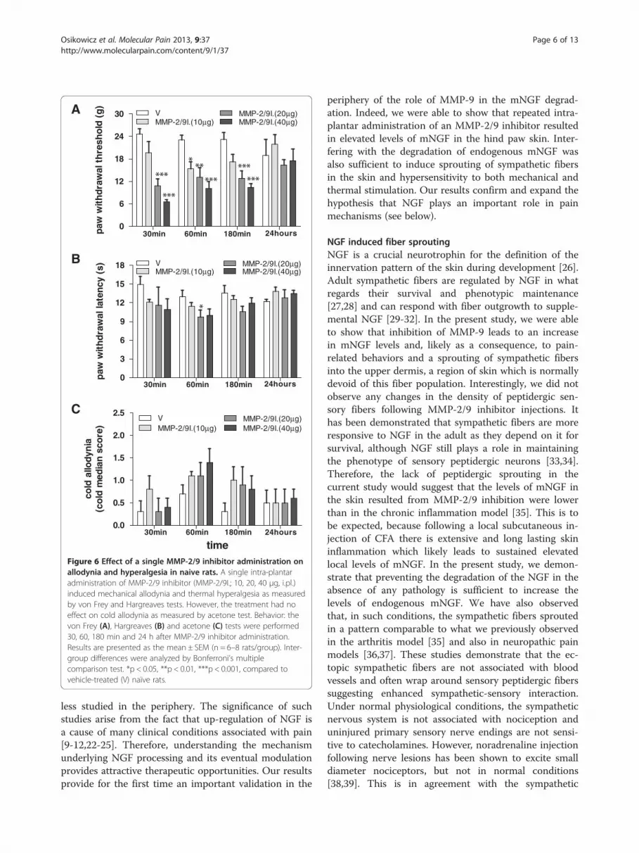
did not change compared to control levels (Figure 4).One interesting observation in MMP-2/9 inhibitortreated animals was that, in the upper dermis, theVMAT-2-immunoreactive fibers were often observedwrapping around CGRP-IR fibers (Figure 5).
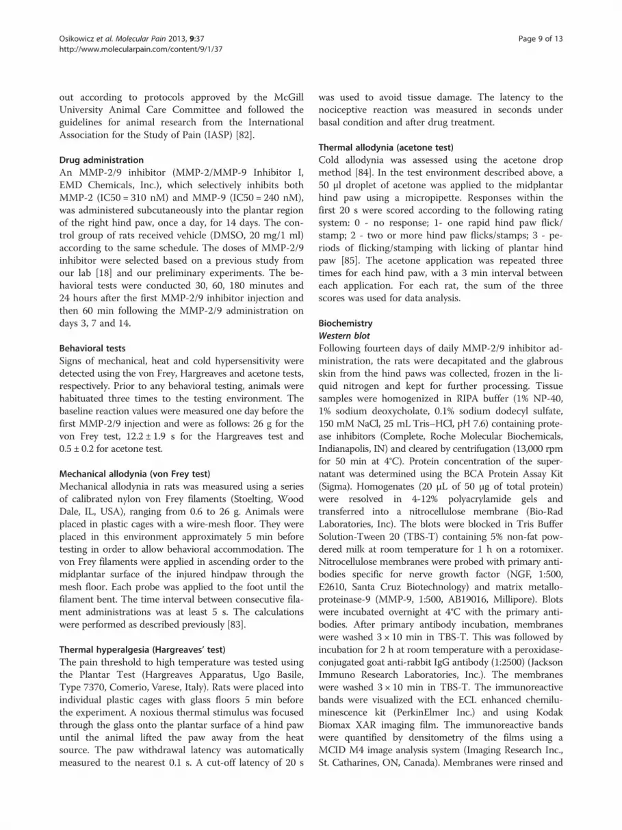
Effect of a single administration of the MMP-2/9 inhibitoron allodynia and hyperalgesiaA single subcutaneous i.pl. administration of matrixMMP-2/9 inhibitor (10, 20, 40 μg) induced mechanical
MMP-9
V MMP-2/9I.
V MMP-2/9I.0.0
0.3
0.6
0.9
1.2
fo
ld c
han
ge
of
con
tro
l (M
MP
-9)
A92 kDa
Figure 2 Effect of MMP-2/9 inhibition on the enzymatic activity of MMfollowing repeated MMP-2/9 inhibition (MMP-2/9I.; 20 μg, i.pl.), zymography wrevealed reduction in the enzymatic activity of both MMP-9 (A) and MMP-2 (Bpanels A and B. The densitometry results are presented as the mean ± SEM. Inmultiple comparison test. *p < 0.05, ***p < 0.001 indicate a significant differeninterrupted line on the graphs indicates zymogram analyses for MMP-9 and Mnaïve rats.
allodynia in naive rats, as measured by the von Frey test.Pain-related behavior was evaluated at 30, 60, 180 minutesand 24 hours after single injection of this inhibitor(Figure 6A). The MMP-2/9 inhibitor (20 μg; i.pl.) inducedthermal hyperalgesia as measured by the Hargreaves testat 60 minutes following a single injection (Figure 6B). Incontrast, a single injection of the MMP-2/9 inhibitor (10,20, 40 μg) did not influence cold allodynia as assessed bythe acetone test at 30, 60, 180 minutes and 24 hours afterthe injection (Figure 6C).
V MMP-2/9I.0.0
0.3
0.6
0.9
1.2
fo
ld c
han
ge
of
con
tro
l (M
MP
-2)
MMP-2
V MMP-2/9I.B66 kDa
P-9 and MMP-2 in the skin of naive rats. To evaluate MMP activityas performed on extracts from glabrous skin samples. The analysis). Examples of representative zymograms are presented in the upperter-group differences were analyzed by ANOVA with a Bonferroni’sce as compared to glabrous skin sample of vehicle-treated (V) rats. TheMP-2 in the contralateral glabrous skin of chronic MMP-2/9I.-treated

Figure 3 Changes in sympathetic fiber innervation in the skin of naïve rats following MMP-2/9 inhibition. A and B represent confocalimages of the rat’s glabrous hindpaw skin in vehicle-treated and MMP-2/9 inhibitor (MMP-2/9I.; 20 μg, i.pl.) injected rats, respectively, at the2 week end point. Note an invasion of sympathetic fibers into the upper dermis (B), mimicking what we had previously observed in a model ofinflammatory arthritis [35]. This fiber population was not normally found in control (vehicle-treated) animals as shown in A. Arrows in B indicateVMAT2-IR fibers in the upper dermis. The results in C are from all experimental groups and presented as the mean ± SEM. Inter-group differenceswere analyzed by ANOVA with a Dunnett’s post hoc test. *p < 0.05, **p < 0.01 indicates a significant difference as compared to glabrous skinsample of chronic vehicle-treated (V) naive rats. The dashed lines represent the dermal-epidermal junction.
Osikowicz et al. Molecular Pain 2013, 9:37 Page 4 of 13http://www.molecularpain.com/content/9/1/37
Effect of repeated administration of the MMP-2/9inhibitor on allodynia and hyperalgesiaThe repeated administration of MMP-2/9 inhibitor (10,20, 40 μg; i.pl.) to naïve rats once a day for two weeksresulted in the development of hypersensitivity to bothmechanical and thermal stimuli as measured by the vonFrey, Hargreaves and acetone tests, respectively (Figure 7).The sensitivity to both mechanical and thermal stimuli in-creased over time. The analysis of the area under thecurve (AUC), which allows for the assessment of the glo-bal effect of treatment, shows that the MMP-2/9 inhibitortreatment induced a significant allodynia and hyperalgesiain naïve rats (p < 0.05) (Figure 7A,B,C; right panels). Wedid not observe any visible signs of inflammation (redden-ing or swelling of the skin) throughout the 14 days periodat the MMP-2/9 inhibitor or vehicle injection site. More-over, we found a statistically significant positive correlationbetween the number of sympathetic fibers in the upperdermis of the skin and mechanical allodynia, as measuredby the von Frey test (p = 0.0029), and with cold allodynia,as measured with the acetone test (p = 0.0086). There was
no positive correlation between the thermal hyperalgesiameasured by the Hargreaves test and the sympatheticsprouting, although there was trend (p = 0.08).
DiscussionThe present study provides evidence that the intra-plantar administration of an MMP-2/9 inhibitor inducedsensitization to mechanical, heat and cold stimuli innaïve rats. Our data also shows that the protein levels ofmNGF and MMP-9 were affected in the hind paw skinfollowing repeated MMP-2/9 inhibitor injections. Fur-thermore, we found that the MMP-2/9 inhibitor inducedsprouting of sympathetic fibers into the upper dermis inthe glabrous hind paw skin, but no change in the densityof sensory peptidergic fibers. Our results support the hy-pothesis that sympathetic sprouting and pain related-behavior are due to an increase in mNGF levels.
NGF metabolic pathwayNGF is secreted from cells in an activity dependentmanner as a precursor (proNGF), together with a full
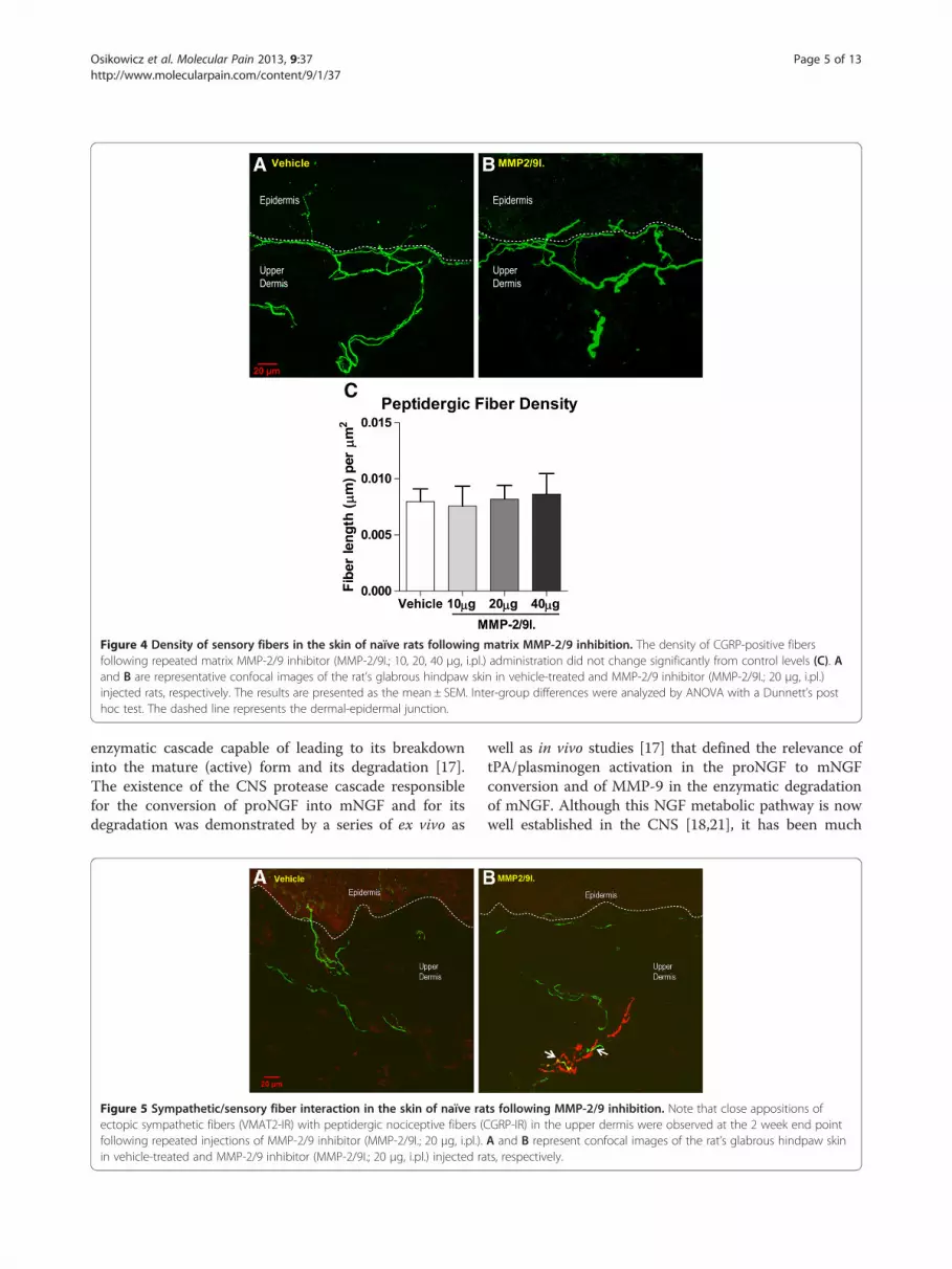
Figure 4 Density of sensory fibers in the skin of naïve rats following matrix MMP-2/9 inhibition. The density of CGRP-positive fibersfollowing repeated matrix MMP-2/9 inhibitor (MMP-2/9I.; 10, 20, 40 μg, i.pl.) administration did not change significantly from control levels (C). Aand B are representative confocal images of the rat’s glabrous hindpaw skin in vehicle-treated and MMP-2/9 inhibitor (MMP-2/9I.; 20 μg, i.pl.)injected rats, respectively. The results are presented as the mean ± SEM. Inter-group differences were analyzed by ANOVA with a Dunnett’s posthoc test. The dashed line represents the dermal-epidermal junction.
Osikowicz et al. Molecular Pain 2013, 9:37 Page 5 of 13http://www.molecularpain.com/content/9/1/37
enzymatic cascade capable of leading to its breakdowninto the mature (active) form and its degradation [17].The existence of the CNS protease cascade responsiblefor the conversion of proNGF into mNGF and for itsdegradation was demonstrated by a series of ex vivo as
Figure 5 Sympathetic/sensory fiber interaction in the skin of naïve raectopic sympathetic fibers (VMAT2-IR) with peptidergic nociceptive fibers (following repeated injections of MMP-2/9 inhibitor (MMP-2/9I.; 20 μg, i.pl.).in vehicle-treated and MMP-2/9 inhibitor (MMP-2/9I.; 20 μg, i.pl.) injected ra
well as in vivo studies [17] that defined the relevance oftPA/plasminogen activation in the proNGF to mNGFconversion and of MMP-9 in the enzymatic degradationof mNGF. Although this NGF metabolic pathway is nowwell established in the CNS [18,21], it has been much
ts following MMP-2/9 inhibition. Note that close appositions ofCGRP-IR) in the upper dermis were observed at the 2 week end pointA and B represent confocal images of the rat’s glabrous hindpaw skints, respectively.
0
6
12
18
24
30 VMMP-2/9I.(10µg)
MMP-2/9I.(20µg)MMP-2/9I.(40µg)
180min 24hours60min30min
Ap
aw w
ith
dra
wal
th
resh
old
(g
)
0
3
6
9
12
15
18
30min 60min 180min 24hours
VMMP-2/9I.(10µg)
MMP-2/9I.(20µg)MMP-2/9I.(40µg)
B
paw
wit
hd
raw
al la
ten
cy (
s)
0.0
0.5
1.0
1.5
2.0
2.5
30min 180min60min 24hours
VMMP-2/9I.(10µg)
MMP-2/9I.(20µg)MMP-2/9I.(40µg)
C
time
cold
allo
dyn
ia(c
old
med
ian
sco
re)
Figure 6 Effect of a single MMP-2/9 inhibitor administration onallodynia and hyperalgesia in naive rats. A single intra-plantaradministration of MMP-2/9 inhibitor (MMP-2/9I.; 10, 20, 40 μg, i.pl.)induced mechanical allodynia and thermal hyperalgesia as measuredby von Frey and Hargreaves tests. However, the treatment had noeffect on cold allodynia as measured by acetone test. Behavior: thevon Frey (A), Hargreaves (B) and acetone (C) tests were performed30, 60, 180 min and 24 h after MMP-2/9 inhibitor administration.Results are presented as the mean ± SEM (n = 6–8 rats/group). Inter-group differences were analyzed by Bonferroni’s multiplecomparison test. *p < 0.05, **p < 0.01, ***p < 0.001, compared tovehicle-treated (V) naïve rats.
Osikowicz et al. Molecular Pain 2013, 9:37 Page 6 of 13http://www.molecularpain.com/content/9/1/37
less studied in the periphery. The significance of suchstudies arise from the fact that up-regulation of NGF isa cause of many clinical conditions associated with pain[9-12,22-25]. Therefore, understanding the mechanismunderlying NGF processing and its eventual modulationprovides attractive therapeutic opportunities. Our resultsprovide for the first time an important validation in the
periphery of the role of MMP-9 in the mNGF degrad-ation. Indeed, we were able to show that repeated intra-plantar administration of an MMP-2/9 inhibitor resultedin elevated levels of mNGF in the hind paw skin. Inter-fering with the degradation of endogenous mNGF wasalso sufficient to induce sprouting of sympathetic fibersin the skin and hypersensitivity to both mechanical andthermal stimulation. Our results confirm and expand thehypothesis that NGF plays an important role in painmechanisms (see below).
NGF induced fiber sproutingNGF is a crucial neurotrophin for the definition of theinnervation pattern of the skin during development [26].Adult sympathetic fibers are regulated by NGF in whatregards their survival and phenotypic maintenance[27,28] and can respond with fiber outgrowth to supple-mental NGF [29-32]. In the present study, we were ableto show that inhibition of MMP-9 leads to an increasein mNGF levels and, likely as a consequence, to pain-related behaviors and a sprouting of sympathetic fibersinto the upper dermis, a region of skin which is normallydevoid of this fiber population. Interestingly, we did notobserve any changes in the density of peptidergic sen-sory fibers following MMP-2/9 inhibitor injections. Ithas been demonstrated that sympathetic fibers are moreresponsive to NGF in the adult as they depend on it forsurvival, although NGF still plays a role in maintainingthe phenotype of sensory peptidergic neurons [33,34].Therefore, the lack of peptidergic sprouting in thecurrent study would suggest that the levels of mNGF inthe skin resulted from MMP-2/9 inhibition were lowerthan in the chronic inflammation model [35]. This is tobe expected, because following a local subcutaneous in-jection of CFA there is extensive and long lasting skininflammation which likely leads to sustained elevatedlocal levels of mNGF. In the present study, we demon-strate that preventing the degradation of the NGF in theabsence of any pathology is sufficient to increase thelevels of endogenous mNGF. We have also observedthat, in such conditions, the sympathetic fibers sproutedin a pattern comparable to what we previously observedin the arthritis model [35] and also in neuropathic painmodels [36,37]. These studies demonstrate that the ec-topic sympathetic fibers are not associated with bloodvessels and often wrap around sensory peptidergic fiberssuggesting enhanced sympathetic-sensory interaction.Under normal physiological conditions, the sympatheticnervous system is not associated with nociception anduninjured primary sensory nerve endings are not sensi-tive to catecholamines. However, noradrenaline injectionfollowing nerve lesions has been shown to excite smalldiameter nociceptors, but not in normal conditions[38,39]. This is in agreement with the sympathetic

0
80
160
240
320
V 10µg 20µg 40µgMMP-2/9I.
AU
C
60
120
180
V 10µg 20µg 40µgMMP-2/9I.
AU
C
0 140
9
18
27V MMP-2/9I.(20µg)
MMP-2/9I.(40µg)MMP-2/9I.(10µg)
1 3 7
A
days
paw
wit
hd
raw
al t
hre
sho
ld (
g)
0 146
8
10
12
14
16
18
1 3 7
*
V
MMP-2/9I.(10µg) MMP-2/9I.(40µg)
MMP-2/9I.(20µg)B
days
paw
wit
hd
raw
al la
ten
cy (
s)
0 140.0
1.5
3.0
4.5
1 3 7
VMMP-2/9I.(10µg)
C MMP-2/9I.(20µg)MMP-2/9I.(40µg)
days
cold
allo
dyn
ia(m
edia
n c
old
sco
re)
0
10
20
30
40
V 10µg 20µg 40µgMMP-2/9I.
AU
C
Figure 7 Effect of repeated MMP-2/9 inhibition on allodynia and hyperalgesia in naive rats. Repeated intra-plantar administration of MMP-2/9 inhibitor (MMP-2/9I.; 10, 20, 40 μg, i.pl.) once daily for fourteen days induced allodynia and hyperalgesia. Behavioral tests: the von Frey test(A), Hargreaves test (B) and acetone test (C) were performed on the 1st, 3rd, 7th and 14th day of repeated MMP-2/9 inhibitor injections. Resultsare presented as the mean ± SEM (n = 6–8 rats/group). Inter-group differences were analyzed by Bonferroni’s multiple comparison test. *p < 0.05,**p < 0.01, ***p < 0.001, compared to vehicle-treated (V) naïve rats. The area under the curve (AUC) was calculated by the Trapezoidal & Simpson’sRules, Pharmacologic Calculation System, version 4.0-03/11/86. Inter-group differences were analyzed by Bonferroni’s Multiple Comparison Test.*p < 0.05, **p < 0.01, ***p < 0.001 indicate a significant difference as compared to chronic vehicle-treated (V) naive rats.
Osikowicz et al. Molecular Pain 2013, 9:37 Page 7 of 13http://www.molecularpain.com/content/9/1/37
participation in some forms of neuropathic pain [40].One possible mechanism for the role of the sympatheticsystem in pathological pain is the ectopic association ofsympathetic fibers with sensory peptidergic fibers inupper dermis of the skin [37,41]. Curiously, a similar ec-topic association of sensory and sympathetic fibers wasalso observed in the thick skin of the rat hind paw fol-lowing chronic inflammation [35]. We observed here asimilar fiber remodeling which paralleled the pain-likebehavior following MMP-9 inhibition, in the absence ofany lesion. Additionally, our results concur with previ-ous studies showing that sympathetic sprouting is
related to increased levels of NGF. Thus, it has beenshown that intraventricular infusion of nerve growth fac-tor per se is able to induce sympathetic fiber sproutingin the sensory ganglia [42], subpial region of the me-dulla, spinal cord [43] and in the intracranial vasculature[44,45] of naïve rats. Conversely, surgical or chemicalsympathectomy has been shown to reduce the thermaland mechanical hyperalgesia evoked by NGF [9,46,47].
NGF-evoked hyperalgesiaIt has been shown that exogenous treatment with NGFevoked nociceptive behavior in animals [46,48-51] and

Osikowicz et al. Molecular Pain 2013, 9:37 Page 8 of 13http://www.molecularpain.com/content/9/1/37
induced pain in humans [52,53]. A single NGF injectioninto the skin induced long-lasting mechanical and thermalhypersensitivity in humans [54,55]. Also, recently pub-lished data obtained in the rodent reported that the intra-plantar injection of NGF induced an immediate, long-lasting increase in mechanical and thermal sensitivity, asearly as one hour post-injection [56]. This study reportedthat the effect on thermal sensitivity was of shorter dur-ation compared with that on mechanical sensitivity, whatis in agreement with our results and indicates that mech-anical hypersensitivity requires a considerable longer timeto resolve than the heat hypersensitivity. This situation issimilar to that in the human, where heat hypersensitivityresolved much faster than the long-lasting increase inmechanical sensitivity [56]. In the current study, we ob-served changes in pain-related behavior when we inhibitedNGF degradation in naïve rats, with a different timecourse for mechanical and thermal sensitivities. The tem-poral differences in resolution between mechanical andthermal sensitivity suggest different mechanisms, howeverwhat drives these differences is still unclear. One of thepossible mechanisms involved in NGF-induced hyperalge-sia might be through sympathetic sprouting and abnormalsympathetic-sensory interactions [57,58]. Indeed, sympa-thetic fibers express trkA receptors and their activation re-sults in noradrenaline release which would increase inexcitability and spontaneous activity of petidergic sensoryneurons [57,58]. Also ATP, a co-transmitter released fromsympathetic fibers and damaged tissues [59,60], has beenshown to play a role in activating primary afferents viapurinergic receptors [61] and may play a role in thehyperexcitability as well. Recent data from our laboratoryprovided some support to these sympathetic-sensory in-teractions in an inflammatory arthritis animal model, inwhich there is sympathetic fiber sprouting in skin over thejoints as well as in the arthritic joint. Indeed, in that studywe have shown that pharmacological suppression of sym-pathetic fiber function with systemic guanethidine signifi-cantly decreased the pain-related behavior associated withinflammatory arthritis [62]. Our data following guanethi-dine administration was comparable with that obtained inother chronic pain models [63,64]. Moreover, there is alsoevidence from the literature that the sodium channel Nav1.8, expressed on sensory neurons, may be a major playerin NGF-induced thermal hyperalgesia [65]. Evidenceshows that NGF modulates this sodium channel currentdensity through the PKA pathway following trkA binding[66]. These changes in sodium channels might occur evenin the absence of peptidergic fibre sprouting as conse-quence of mNGF increases induced by MMP-2/9 inhib-ition. Since many NGF-responsive neurons contain thevanilloid receptor TRPV1, this receptor is also suspectedto play a role in NGF-mediated hypersensitivity [67-69].Cultured DRG neurons treated with NGF display
enhanced inward currents in response to the applicationof the TRPV1 agonist capsaicin [70,71]. NGF can increaseTRPV1 expression [72,73] and promote TRPV1 insertioninto the plasma membrane [74]. NGF also acts indirectlyby activating mast cells and neutrophils, which in turn re-lease additional inflammatory mediators causing hyper-sensitivity [46,47,75-77]. There is a growing body ofevidence indicating that NGF-evoked hyperalgesia iscaused by a sensitization of primary afferent nociceptorsthat supply an area of damage or inflammation [78,79].Matrix metalloproteinases have multiple functions.
Therefore, it is not surprising that there is evidence thatMMP-9 inhibition at the spinal cord level alleviates theearly phase of neuropathic pain, whereas intrathecal ad-ministration of MMP-9 itself is sufficient to produceneuropathic pain-like symptoms in normal animals [80].These results at the spinal cord level are very differentfrom those we obtained through a peripheral inhibitionof MMP-9, likely because, in the CNS, the MMP-9-in-duced pathophysiology involves interleukin-1β cleavageto its active form and microglial p38 activation [80],rather than effects through NGF. However, becauseMMP-2 is also responsible for the degradation of severalpain-related mediators such as TNF-α, interleukin-1β,and substance P [81], we cannot rule out that MMP-2inhibition might increase the levels of these mediatorsand subsequently contribute to the hyperalgesia througha peripheral mechanim.
ConclusionsOur data provides evidence that the metabolic processingof endogenous NGF can be modulated in the periphery ina manner similar to what was previously demonstrated inthe central nervous system. We show that inhibition ofNGF degradation in naïve rats induces sprouting of sym-pathetic fibers into the upper dermis of the skin, wherethey wrap around the peptidergic nociceptive afferents,suggesting an interaction between these two fiber popula-tions. We propose that the observed sensitization to bothmechanical and thermal stimuli is a consequence of ele-vated endogenous mNGF levels sensitizing peptidergicprimary afferents and promoting abnormal sensory-autonomic fiber interactions in the skin. As NGF has beenstrongly implicated in different pain conditions, modulat-ing its metabolic pathway provides an opportunity to in-fluence its endogenous levels to alleviate pain.
MethodsAnimalsAdult male Sprague–Dawley rats (275–300 g) were usedin this study. Animals were housed in groups of four percage with sawdust bedding under a standard 12 h/12 hlight/dark cycle (lights on at 08.00 AM) with food andwater available ad libitum. All experiments were carried
Osikowicz et al. Molecular Pain 2013, 9:37 Page 9 of 13http://www.molecularpain.com/content/9/1/37
out according to protocols approved by the McGillUniversity Animal Care Committee and followed theguidelines for animal research from the InternationalAssociation for the Study of Pain (IASP) [82].
Drug administrationAn MMP-2/9 inhibitor (MMP-2/MMP-9 Inhibitor I,EMD Chemicals, Inc.), which selectively inhibits bothMMP-2 (IC50 = 310 nM) and MMP-9 (IC50 = 240 nM),was administered subcutaneously into the plantar regionof the right hind paw, once a day, for 14 days. The con-trol group of rats received vehicle (DMSO, 20 mg/1 ml)according to the same schedule. The doses of MMP-2/9inhibitor were selected based on a previous study fromour lab [18] and our preliminary experiments. The be-havioral tests were conducted 30, 60, 180 minutes and24 hours after the first MMP-2/9 inhibitor injection andthen 60 min following the MMP-2/9 administration ondays 3, 7 and 14.
Behavioral testsSigns of mechanical, heat and cold hypersensitivity weredetected using the von Frey, Hargreaves and acetone tests,respectively. Prior to any behavioral testing, animals werehabituated three times to the testing environment. Thebaseline reaction values were measured one day before thefirst MMP-2/9 injection and were as follows: 26 g for thevon Frey test, 12.2 ± 1.9 s for the Hargreaves test and0.5 ± 0.2 for acetone test.
Mechanical allodynia (von Frey test)Mechanical allodynia in rats was measured using a seriesof calibrated nylon von Frey filaments (Stoelting, WoodDale, IL, USA), ranging from 0.6 to 26 g. Animals wereplaced in plastic cages with a wire-mesh floor. They wereplaced in this environment approximately 5 min beforetesting in order to allow behavioral accommodation. Thevon Frey filaments were applied in ascending order to themidplantar surface of the injured hindpaw through themesh floor. Each probe was applied to the foot until thefilament bent. The time interval between consecutive fila-ment administrations was at least 5 s. The calculationswere performed as described previously [83].
Thermal hyperalgesia (Hargreaves’ test)The pain threshold to high temperature was tested usingthe Plantar Test (Hargreaves Apparatus, Ugo Basile,Type 7370, Comerio, Varese, Italy). Rats were placed intoindividual plastic cages with glass floors 5 min beforethe experiment. A noxious thermal stimulus was focusedthrough the glass onto the plantar surface of a hind pawuntil the animal lifted the paw away from the heatsource. The paw withdrawal latency was automaticallymeasured to the nearest 0.1 s. A cut-off latency of 20 s
was used to avoid tissue damage. The latency to thenociceptive reaction was measured in seconds underbasal condition and after drug treatment.
Thermal allodynia (acetone test)Cold allodynia was assessed using the acetone dropmethod [84]. In the test environment described above, a50 μl droplet of acetone was applied to the midplantarhind paw using a micropipette. Responses within thefirst 20 s were scored according to the following ratingsystem: 0 - no response; 1- one rapid hind paw flick/stamp; 2 - two or more hind paw flicks/stamps; 3 - pe-riods of flicking/stamping with licking of plantar hindpaw [85]. The acetone application was repeated threetimes for each hind paw, with a 3 min interval betweeneach application. For each rat, the sum of the threescores was used for data analysis.
BiochemistryWestern blotFollowing fourteen days of daily MMP-2/9 inhibitor ad-ministration, the rats were decapitated and the glabrousskin from the hind paws was collected, frozen in the li-quid nitrogen and kept for further processing. Tissuesamples were homogenized in RIPA buffer (1% NP-40,1% sodium deoxycholate, 0.1% sodium dodecyl sulfate,150 mM NaCl, 25 mL Tris–HCl, pH 7.6) containing prote-ase inhibitors (Complete, Roche Molecular Biochemicals,Indianapolis, IN) and cleared by centrifugation (13,000 rpmfor 50 min at 4°C). Protein concentration of the super-natant was determined using the BCA Protein Assay Kit(Sigma). Homogenates (20 μL of 50 μg of total protein)were resolved in 4-12% polyacrylamide gels andtransferred into a nitrocellulose membrane (Bio-RadLaboratories, Inc). The blots were blocked in Tris BufferSolution-Tween 20 (TBS-T) containing 5% non-fat pow-dered milk at room temperature for 1 h on a rotomixer.Nitrocellulose membranes were probed with primary anti-bodies specific for nerve growth factor (NGF, 1:500,E2610, Santa Cruz Biotechnology) and matrix metallo-proteinase-9 (MMP-9, 1:500, AB19016, Millipore). Blotswere incubated overnight at 4°C with the primary anti-bodies. After primary antibody incubation, membraneswere washed 3 × 10 min in TBS-T. This was followed byincubation for 2 h at room temperature with a peroxidase-conjugated goat anti-rabbit IgG antibody (1:2500) (JacksonImmuno Research Laboratories, Inc.). The membraneswere washed 3 × 10 min in TBS-T. The immunoreactivebands were visualized with the ECL enhanced chemilu-minescence kit (PerkinElmer Inc.) and using KodakBiomax XAR imaging film. The immunoreactive bandswere quantified by densitometry of the films using aMCID M4 image analysis system (Imaging Research Inc.,St. Catharines, ON, Canada). Membranes were rinsed and
Osikowicz et al. Molecular Pain 2013, 9:37 Page 10 of 13http://www.molecularpain.com/content/9/1/37
reprobed with a mouse anti-β-actin antibody (1:40,000;Sigma) diluted in 5% milk in TBS-T for 1 h at roomtemperature, washed with TBS-T, and incubated with aperoxidase conjugated donkey anti-mouse IgG (1:5000,Santa Cruz) in 5% dry milk in TBS-T for 1 h. The mem-branes were washed and the signal was detected andqualified as described above. The levels of NGF andMMP-9 were normalized to the β-actin levels for eachsample.
ZymographyGelatinolytic activity was determined by zymographyusing gelatin-containing gels following the protocol pro-vided by supplier Millipore. The skin samples were col-lected at the same time point as those for western blotanalysis (see above). The skin sample extracts weremixed with an equal volume of non-reducing samplebuffer (0.5 M Tris–HCl, pH 6.8, SDS, glycerol andbromophenol blue). The samples (50 μg of protein) wereelectrophoresed on an 8% SDS–PAGE containing 0.1%gelatin as the substrate. After electrophoretic separation,the gels were incubated for 30 min in renaturing buffer(2.5% Triton X-100 in distilled water) to remove theSDS, washed for 2X10 min with water and then incu-bated for 24 h at 37°C in a developing buffer containing50 mM Tris–HCl, pH 7.78; 5 mM CaCl2 and 0.02% Brij35. Following incubation, the gels were stained for 1 hwith 0.5% Coomassie R-250 staining solution and thendifferentiated in a solution of methanol: acetic acid:water (50 : 10 : 40). Enzyme activity attributed to MMP-2 and MMP-9 was visualized (on the basis of molecularweight) in the gelatin-containing zymograms as clearbands against a blue background. The bands were quan-tified by densitometry using the MCID M4 image ana-lysis system.
ImmunohistochemistryTwo weeks after the daily injections of MMP-2/9 inhibi-tor, animals were deeply anesthetized with Equithesin(6.5 mg chloral hydrate and 3 mg sodium pentobarbitalin a volume of 0.3 mL, i.p., per 100 g body weight) andthen perfused through the left cardiac ventricle with100 mL of perfusion buffer, followed by 500 mL of 4%paraformaldehyde (PFA) in 0.1 M phosphate-buffer (PB),pH 7.4, at room temperature for 30 minutes. Subse-quently, the plantar glabrous skin was extracted andpost-fixed in the same fixative for 1 hour at 4°C. The tis-sue was cryoprotected in 30% sucrose in PB overnight at4°C for later immunohistochemical processing. Fifty-μmthick cross sections of skin were cut using a cryostat(Leica, Wetzlar, Germany). All sections were collected asfree-floating in phosphate-buffered saline (PBS) with0.2% Triton-X 100 (PBS-T). The tissue sections were in-cubated for 1 hour at room temperature in 10% normal
goat serum (Gibco, Carlsbad, CA) in PBS to block un-specific labeling. To label the peptidergic and sympa-thetic fiber populations, the sections were thenincubated at 4°C for 24 hours using either a rabbit anti-Calcitonin Gene Related Protein (CGRP) (Sigma-Aldrich, St. Louis, MO, C-8198, lot# 070 M4835, diluted1:2000) antibody or a rabbit anti-Vesicular MonoamineTransporter-2 (VMAT-2) (Phoenix Pharmaceuticals,Inc., CA, USA, H-V004, 01237–1, diluted 1:7500). Ascontrols, some sections were processed omitting the pri-mary antibody; no specific staining was observed. Afterthree rinses in PBS-T, the sections were incubated for2 hours at room temperature with goat anti-rabbit IgGconjugated to either Alexa Fluor 488 for CGRP or AlexaFluor 594 for VMAT-2 (Molecular Probes, diluted1:800). For double-labeling of CGRP and VMAT2, sec-tions were processed as described above except that weused a guinea pig anti-CGRP antibody at a 1:4000 dilu-tion (Peninsula, San Carlos, CA, T-5053) in a simultan-eous incubation with the rabbit anti-VMAT2 antibody;after washing, we used a mixture of an anti-guinea pigIgG antibody conjugated to Alexa Fluor 488 (1:800; Mo-lecular Probes) with the anti-rabbit IgG conjugated toAlexa Fluor 594. Finally, the sections were washed,mounted on gelatin-subbed slides, air-dried and coverslipped with an anti-fading mounting medium (AquaPolyMount, Polysciences Inc., Warrington, Pa.). Slideswere stored at 4°C until examined. Since we aimed tolocalize and evaluate the morphological changes in in-nervation and to quantify the relative changes in the im-munostaining intensity, the conditions of all procedures(dilutions of reagents and antibodies, washings, incuba-tion time and temperature, blocking of nonspecific stain-ing), were kept rigorously throughout the assays andwere identical for the sections from all tested groups.Within each experiment, immunohistochemical process-ing of tissue sections sampled from all groups was car-ried out simultaneously. Before quantitative analyses, allthe slides were coded so that the person who performedthe quantification was completely blinded regarding theexperimental groups. Codes were broken only after thequantification was completed.
CGRP-immunoreactive fiber quantificationQuantitative analyses were performed on sections ofglabrous skin from the hindpaw ipsilaterally to the injec-tion of MMP-2/9 inhibitor or vehicle. In this study, as inprevious work from our laboratory, we have divided thedermis into upper and lower dermis. We have definedthe upper dermis as the area of the dermis spanning150 μm below the dermal-epidermal junction [37]. Forthe measurement of the density of peptidergic fibers, asdetected by CGRP immunoreactivity, we used a ZeissAxioplan 2 imaging fluorescence microscope equipped
Osikowicz et al. Molecular Pain 2013, 9:37 Page 11 of 13http://www.molecularpain.com/content/9/1/37
with a PlanFluotar 40X oil-immersion objective. Thismicroscope has a high resolution digital cameraconnected to a computer equipped with the ZeissAxiovision 4.8 software (Carl Zeiss, Canada). Three sec-tions per slide were randomly selected, and 6 micro-scopic fields including upper dermis were photographedat random, for a total of 18 images per animal. Imageswere exported in the TIFF format for analysis with anMCID Elite image analysis system (Imaging ResearchInc., St. Catharines, ON, Canada). The upper dermis wasoutlined with the use of a tracing tool in the software.CGRP-IR fibers were automatically detected by the soft-ware using a brightness threshold and converted to 1pixel in thickness to compute the total fiber length (μm)per scan area (μm2).
Sympathetic fiber quantificationWe used a different approach to quantify the changes inautonomic innervation. Since these fibers are much lessabundant in the skin than the sensory, especially in theupper dermis, it was unpractical to measure fiber dens-ity. Therefore, we proceeded by photographing 6 entireskin sections per animal and counting all VMAT-2-IR fi-bers within the upper dermis, measured to be 150 μmfrom the dermal-epidermal junction. The mean numberof fibers in the upper dermis per total area (μm2) wasthen calculated.
Data analysesThe Western blot and zymography results represent thedensitometry analyses of all samples (4–6 samples pergroup). Inter-group differences were analyzed by one-way ANOVA and Bonferroni’s multiple comparison tests(Figures 1 and 2). The mean number of sympathetic fi-bers and density of sensory peptidergic fibers in theupper dermis was compared between groups by a one-way ANOVA and a Dunnett’s post hoc test, with a statis-tical significance accepted at p <0.05 (Figures 3, 4 and5). As no significant difference was detected among thecontrol groups of all time points, they were pooled. Thebehavioral data are presented as the mean ± SEM (6–8animals per group). The effect of single as well as re-peated intra-plantar injections of MMP-2/9 inhibitor onmechanical and thermal sensitivity in rats (Figures 6 and7) was analyzed by one-way analysis of variance(ANOVA) and Bonferroni’s multiple comparison test.The effect of repeated injections of MMP-2/9 inhibitoron allodynia and hyperalgesia was also shown as the areaunder the curve (AUC, Figure 7). To evaluate the AUCthe Trapezoidal and Simpson’s Rules, PharmacologicCalculation System, version 4.0-03/11/86 was used [86].Using the GraphPad Prism software, we performed acorrelation analysis between the behavioral responsesand sympathetic fiber sprouting, as this was the only
fiber population which changed following administrationof the MMP-2/9 inhibitor. The analysis was done oneach behaviorally tested rats from which the skin tissuewas collected for the immunohistochemical component.
Competing interestsThe authors declare they have no competing interests.
Authors’ contributionsMO, GL and SA designed all experimental protocols described in thismanuscript. MO performed the behavioral experiments, western blot,zymography as well as the writing of the initial draft of the manuscript. GLperformed behavioral experiments and immunohistochemistry. ARdS andACC provided supervision for data analysis, study direction, imageacquisition, manuscript design and revisions. All authors have read andapproved the final draft of this manuscript.
AcknowledgementsThe authors would like to thank Dr. Gary Bennett for allowing us to use hisanimal behavior testing facility. This work was supported by grants fromCIHR, Louise and Alan Edwards Foundation, and the Tri-Council MITACSAccelerate Program in partnership with Pfizer Canada. The authors discloseno conflict of interest in respect of this work.
Author details1Department of Pharmacology and Therapeutics, McGill University, 3655Prom Sir-William-Osler, Montreal, QC H3G 1Y6, Canada. 2Department ofAnatomy and Cell Biology, McGill University, Montreal, QC H3A 2B2, Canada.3Department of Neurology and Neurosurgery, McGill University, Montreal, QCH3A 2B4, Canada.
Received: 3 April 2013 Accepted: 24 July 2013Published: 29 July 2013
References1. Lindsay RM: Neurotrophic growth factors and neurodegenerative
diseases: therapeutic potential of the neurotrophins and ciliaryneurotrophic factor. Neurobiol Aging 1994, 15:249–251.
2. Cuello AC: Nerve Growth Factor. Berlin: Springer: Encyclopedia ofPsychopharmacology Volume 2; 2010:823–830.
3. Patel TD, Jackman A, Rice FL, Kucera J, Snider WD: Development of sensoryneurons in the absence of NGF/TrkA signaling in vivo. Neuron 2000,25:345–357.
4. Crowley C, Spencer SD, Nishimura MC, Chen KS, Pitts-Meek S, Armanini MP,Ling LH, McMahon SB, Shelton DL, Levinson AD, et al: Mice lacking nervegrowth factor display perinatal loss of sensory and sympathetic neuronsyet develop basal forebrain cholinergic neurons. Cell 1994, 76:1001–1011.
5. Frade JM, Barde YA: Nerve growth factor: two receptors, multiplefunctions. Bioessays 1998, 20:137–145.
6. Campenot RB: Local control of neurite sprouting in cultured sympatheticneurons by nerve growth factor. Brain Res 1987, 465:293–301.
7. Chen Y, Michaelis M, Janig W, Devor M: Adrenoreceptor subtypemediating sympathetic-sensory coupling in injured sensory neurons. JNeurophysiol 1996, 76:3721–3730.
8. Herzberg U, Eliav E, Dorsey JM, Gracely RH, Kopin IJ: NGF involvement inpain induced by chronic constriction injury of the rat sciatic nerve.Neuroreport 1997, 8:1613–1618.
9. Aloe L, Tuveri MA, Carcassi U, Levi-Montalcini R: Nerve growth factor in thesynovial fluid of patients with chronic arthritis. Arthritis Rheum 1992,35:351–355.
10. Halliday DA, Zettler C, Rush RA, Scicchitano R, McNeil JD: Elevated nervegrowth factor levels in the synovial fluid of patients with inflammatoryjoint disease. Neurochem Res 1998, 23:919–922.
11. Lowe EM, Anand P, Terenghi G, Williams-Chestnut RE, Sinicropi DV, OsborneJL: Increased nerve growth factor levels in the urinary bladder of womenwith idiopathic sensory urgency and interstitial cystitis. Br J Urol 1997,79:572–577.
12. Oddiah D, Anand P, McMahon SB, Rattray M: Rapid increase of NGF, BDNFand NT-3 mRNAs in inflamed bladder. Neuroreport 1998, 9:1455–1458.
Osikowicz et al. Molecular Pain 2013, 9:37 Page 12 of 13http://www.molecularpain.com/content/9/1/37
13. Sarchielli P, Alberti A, Floridi A, Gallai V: Levels of nerve growth factor incerebrospinal fluid of chronic daily headache patients. Neurology 2001,57:132–134.
14. Woolf CJ, Safieh-Garabedian B, Ma QP, Crilly P, Winter J: Nerve growthfactor contributes to the generation of inflammatory sensoryhypersensitivity. Neuroscience 1994, 62:327–331.
15. Dmitrieva N, McMahon SB: Sensitisation of visceral afferents by nervegrowth factor in the adult rat. Pain 1996, 66:87–97.
16. Rueff A, Mendell LM: Nerve growth factor NT-5 induce increasedthermal sensitivity of cutaneous nociceptors in vitro. J Neurophysiol1996, 76:3593–3596.
17. Bruno MA, Cuello AC: Activity-dependent release of precursor nervegrowth factor, conversion to mature nerve growth factor, and itsdegradation by a protease cascade. Proc Natl Acad Sci USA 2006,103:6735–6740.
18. Allard S, Leon WC, Pakavathkumar P, Bruno MA, Ribeiro-da-Silva A, CuelloAC: Impact of the NGF maturation and degradation pathway on thecortical cholinergic system phenotype. J Neurosci 2012, 32:2002–2012.
19. Khan KM, Falcone DJ, Kraemer R: Nerve growth factor activation of Erk-1and Erk-2 induces matrix metalloproteinase-9 expression in vascularsmooth muscle cells. J Biol Chem 2002, 277:2353–2359.
20. Bennion BJ, Daggett V: The molecular basis for the chemical denaturationof proteins by urea. Proc Natl Acad Sci USA 2003, 100:5142–5147.
21. Bruno MA, Mufson EJ, Wuu J, Cuello AC: Increased matrixmetalloproteinase 9 activity in mild cognitive impairment. J NeuropatholExp Neurol 2009, 68:1309–1318.
22. Dicou E, Perrot S, Menkes CJ, Masson C, Nerriere V: Nerve growth factor(NGF) autoantibodies and NGF in the synovial fluid: implications inspondylarthropathies. Autoimmunity 1996, 24:1–9.
23. Miller LJ, Fischer KA, Goralnick SJ, Litt M, Burleson JA, Albertsen P, KreutzerDL: Nerve growth factor and chronic prostatitis/chronic pelvic painsyndrome. Urology 2002, 59:603–608.
24. Watanabe T, Inoue M, Sasaki K, Araki M, Uehara S, Monden K, Saika T, NasuY, Kumon H, Chancellor MB: Nerve growth factor level in the prostaticfluid of patients with chronic prostatitis/chronic pelvic pain syndrome iscorrelated with symptom severity and response to treatment. BJU Int2011, 108:248–251.
25. Sarchielli P, Gallai V: Nerve growth factor and chronic daily headache: apotential implication for therapy. Expert Rev Neurother 2004, 4:115–127.
26. Albers KM, Wright DE, Davis BM: Overexpression of nerve growth factor inepidermis of transgenic mice causes hypertrophy of the peripheralnervous system. J Neurosci 1994, 14:1422–1432.
27. Purves D, Snider WD, Voyvodic JT: Trophic regulation of nerve cellmorphology and innervation in the autonomic nervous system. Nature1988, 336:123–128.
28. Ritter AM, Lewin GR, Kremer NE, Mendell LM: Requirement for nervegrowth factor in the development of myelinated nociceptors in vivo.Nature 1991, 350:500–502.
29. Ruit KG, Osborne PA, Schmidt RE, Johnson EM Jr, Snider WD: Nerve growthfactor regulates sympathetic ganglion cell morphology and survival inthe adult mouse. J Neurosci 1990, 10:2412–2419.
30. Davis BM, Albers KM, Seroogy KB, Katz DM: Overexpression of nervegrowth factor in transgenic mice induces novel sympathetic projectionsto primary sensory neurons. J Comp Neurol 1994, 349:464–474.
31. Davis BM, Wang HS, Albers KM, Carlson SL, Goodness TP, McKinnon D:Effects of NGF overexpression on anatomical and physiologicalproperties of sympathetic postganglionic neurons. Brain Res 1996,724:47–54.
32. Goodness TP, Albers KM, Davis FE, Davis BM: Overexpression of nervegrowth factor in skin increases sensory neuron size and modulates Trkreceptor expression. Eur J Neurosci 1997, 9:1574–1585.
33. Gorin PD, Johnson EM Jr: Effects of long-term nerve growth factordeprivation on the nervous system of the adult rat: an experimentalautoimmune approach. Brain Res 1980, 198:27–42.
34. Lewin GR, Barde YA: Physiology of the neurotrophins. Annu Rev Neurosci1996, 19:289–317.
35. Almarestani L, Longo G, Ribeiro-da-Silva A: Autonomic fiber sprouting inthe skin in chronic inflammation. Mol Pain 2008, 4:56.
36. Ruocco I, Cuello AC, Ribeiro-Da-Silva A: Peripheral nerve injury leads tothe establishment of a novel pattern of sympathetic fibre innervation inthe rat skin. J Comp Neurol 2000, 422:287–296.
37. Yen LD, Bennett GJ, Ribeiro-da-Silva A: Sympathetic sprouting andchanges in nociceptive sensory innervation in the glabrous skin of therat hind paw following partial peripheral nerve injury. J Comp Neurol2006, 495:679–690.
38. Sato J, Perl ER: Adrenergic excitation of cutaneous pain receptorsinduced by peripheral nerve injury. Science 1991, 251:1608–1610.
39. Bossut DF, Perl ER: Effects of nerve injury on sympathetic excitation of adelta mechanical nociceptors. J Neurophysiol 1995, 73:1721–1723.
40. Campbell NN, Reynolds GJ, Perkins G: Postoperative analgesia in neonates:an Australia-wide survey. Anaesth Intensive Care 1989, 17:487–491.
41. Grelik C, Bennett GJ, Ribeiro-da-Silva A: Autonomic fibre sprouting andchanges in nociceptive sensory innervation in the rat lower lip skinfollowing chronic constriction injury. Eur J Neurosci 2005, 21:2475–2487.
42. Nauta HJ, Wehman JC, Koliatsos VE, Terrell MA, Chung K: Intraventricularinfusion of nerve growth factor as the cause of sympathetic fibersprouting in sensory ganglia. J Neurosurg 1999, 91:447–453.
43. Winkler J, Ramirez GA, Kuhn HG, Peterson DA, Day-Lollini PA, Stewart GR,Tuszynski MH, Gage FH, Thal LJ: Reversible Schwann cell hyperplasia andsprouting of sensory and sympathetic neurites after intraventricularadministration of nerve growth factor. Ann Neurol 1997, 41:82–93.
44. Isaacson LG, Saffran BN, Crutcher KA: Nerve growth factor-inducedsprouting of mature, uninjured sympathetic axons. J Comp Neurol 1992,326:327–336.
45. Saffran BN, Woo JE, Mobley WC, Crutcher KA: Intraventricular NGF infusionin the mature rat brain enhances sympathetic innervation ofcerebrovascular targets but fails to elicit sympathetic ingrowth. Brain Res1989, 492:245–254.
46. Andreev N, Dimitrieva N, Koltzenburg M, McMahon SB: Peripheraladministration of nerve growth factor in the adult rat produces athermal hyperalgesia that requires the presence of sympathetic post-ganglionic neurones. Pain 1995, 63:109–115.
47. Woolf CJ, Ma QP, Allchorne A, Poole S: Peripheral cell types contributingto the hyperalgesic action of nerve growth factor in inflammation. JNeurosci 1996, 16:2716–2723.
48. Lewin GR, Ritter AM, Mendell LM: Nerve growth factor-inducedhyperalgesia in the neonatal and adult rat. J Neurosci 1993, 13:2136–2148.
49. Lewin GR, Mendell LM: Regulation of cutaneous C-fiber heat nociceptorsby nerve growth factor in the developing rat. J Neurophysiol 1994,71:941–949.
50. Pertens E, Urschel-Gysbers BA, Holmes M, Pal R, Foerster A, Kril Y, DiamondJ: Intraspinal and behavioral consequences of nerve growth factor-induced nociceptive sprouting and nerve growth factor-inducedhyperalgesia compared in adult rats. J Comp Neurol 1999, 410:73–89.
51. Malin SA, Molliver DC, Koerber HR, Cornuet P, Frye R, Albers KM, Davis BM:Glial cell line-derived neurotrophic factor family members sensitizenociceptors in vitro and produce thermal hyperalgesia in vivo. J Neurosci2006, 26:8588–8599.
52. Petty BG, Cornblath DR, Adornato BT, Chaudhry V, Flexner C, Wachsman M,Sinicropi D, Burton LE, Peroutka SJ: The effect of systemically administeredrecombinant human nerve growth factor in healthy human subjects.Ann Neurol 1994, 36:244–246.
53. Dyck PJ, Peroutka S, Rask C, Burton E, Baker MK, Lehman KA, Gillen DA,Hokanson JL, O’Brien PC: Intradermal recombinant human nerve growthfactor induces pressure allodynia and lowered heat-pain threshold inhumans. Neurology 1997, 48:501–505.
54. Rukwied R, Mayer A, Kluschina O, Obreja O, Schley M, Schmelz M: NGFinduces non-inflammatory localized and lasting mechanical and thermalhypersensitivity in human skin. Pain 2010, 148:407–413.
55. Weinkauf B, Obreja O, Schmelz M, Rukwied R: Differential effects oflidocaine on nerve growth factor (NGF)-evoked heat- and mechanicalhyperalgesia in humans. Eur J Pain 2011, 16:543–549.
56. Mills CD, Nguyen T, Tanga FY, Zhong C, Gauvin DM, Mikusa J, Gomez EJ,Salyers AK, Bannon AW: Characterization of nerve growth factor-inducedmechanical and thermal hypersensitivity in rats. Eur J Pain 2013, 17:469–479.
57. McLachlan EM, Janig W, Devor M, Michaelis M: Peripheral nerve injurytriggers noradrenergic sprouting within dorsal root ganglia. Nature 1993,363:543–546.
58. Xie W, Strong JA, Zhang JM: Increased excitability and spontaneousactivity of rat sensory neurons following in vitro stimulation ofsympathetic fiber sprouts in the isolated dorsal root ganglion. Pain 2010,151:447–459.
Osikowicz et al. Molecular Pain 2013, 9:37 Page 13 of 13http://www.molecularpain.com/content/9/1/37
59. Bertrand PP: ATP and sensory transduction in the enteric nervous system.Neuroscientist 2003, 9:243–260.
60. Burnstock G: Therapeutic potential of purinergic signalling for diseases ofthe urinary tract. BJU Int 2011, 107:192–204.
61. Mo G, Peleshok JC, Cao CQ, Ribeiro-da-Silva A, Seguela P: Control of P2X3channel function by metabotropic P2Y2 utp receptors in primarysensory neurons. Mol Pharmacol 2013, 83:640–647.
62. Longo G, Osikowicz M, Ribeiro-da-Silva A: Sympathetic fiber sprouting ininflamed joints and adjacent skin contributes to pain-related behavior inarthritis. J Neurosci 2013, 33:10066–10074.
63. Malmberg AB, Basbaum AI: Partial sciatic nerve injury in the mouse as amodel of neuropathic pain: behavioral and neuroanatomical correlates.Pain 1998, 76:215–222.
64. Xanthos DN, Coderre TJ: Sympathetic vasoconstrictor antagonism andvasodilatation relieve mechanical allodynia in rats with chronicpostischemia pain. J Pain 2008, 9:423–433.
65. Kerr BJ, Souslova V, McMahon SB, Wood JN: A role for the TTX-resistantsodium channel Nav 1.8 in NGF-induced hyperalgesia, but notneuropathic pain. Neuroreport 2001, 12:3077–3080.
66. Brackenbury WJ, Djamgoz MB: Nerve growth factor enhances voltage-gated Na + channel activity and Transwell migration in Mat-LyLu ratprostate cancer cell line. J Cell Physiol 2007, 210:602–608.
67. Caterina MJ, Schumacher MA, Tominaga M, Rosen TA, Levine JD, Julius D:The capsaicin receptor: a heat-activated ion channel in the painpathway. Nature 1997, 389:816–824.
68. Tominaga M, Caterina MJ, Malmberg AB, Rosen TA, Gilbert H, Skinner K,Raumann BE, Basbaum AI, Julius D: The cloned capsaicin receptorintegrates multiple pain-producing stimuli. Neuron 1998, 21:531–543.
69. Michael GJ, Priestley JV: Differential expression of the mRNA for thevanilloid receptor subtype 1 in cells of the adult rat dorsal root andnodose ganglia and its downregulation by axotomy. J Neurosci 1999,19:1844–1854.
70. Shu X, Mendell LM: Nerve growth factor acutely sensitizes the responseof adult rat sensory neurons to capsaicin. Neurosci Lett 1999, 274:159–162.
71. Caterina MJ, Leffler A, Malmberg AB, Martin WJ, Trafton J, Petersen-Zeitz KR, Koltzenburg M, Basbaum AI, Julius D: Impaired nociceptionand pain sensation in mice lacking the capsaicin receptor. Science2000, 288:306–313.
72. Donnerer J, Liebmann I, Schicho R: Differential regulation of 3-beta-hydroxysteroid dehydrogenase and vanilloid receptor TRPV1 mRNA insensory neurons by capsaicin and NGF. Pharmacology 2005, 73:97–101.
73. Xue Q, Jong B, Chen T, Schumacher MA: Transcription of rat TRPV1 utilizesa dual promoter system that is positively regulated by nerve growthfactor. J Neurochem 2007, 101:212–222.
74. Zhang X, Huang J, McNaughton PA: NGF rapidly increases membraneexpression of TRPV1 heat-gated ion channels. EMBO J 2005, 24:4211–4223.
75. Lewin GR, Rueff A, Mendell LM: Peripheral and central mechanisms ofNGF-induced hyperalgesia. Eur J Neurosci 1994, 6:1903–1912.
76. Bennett G, al-Rashed S, Hoult JR, Brain SD: Nerve growth factor inducedhyperalgesia in the rat hind paw is dependent on circulatingneutrophils. Pain 1998, 77:315–322.
77. Amann R, Schuligoi R, Herzeg G, Donnerer J: Intraplantar injection of nervegrowth factor into the rat hind paw: local edema and effects on thermalnociceptive threshold. Pain 1996, 64:323–329.
78. Heppenstall PA, Lewin GR: Neurotrophins, nociceptors and pain. Curr OpinAnaesthesiol 2000, 13:573–576.
79. Jankowski MP, Koerber HR: Neurotrophic Factors and NociceptorSensitization. In Translational Pain Research: From Mouse to Man. Edited byKruger L, Light AR. Boca Raton, FL; 2010. Chapter 2. Available from: http://www.ncbi.nlm.nih.gov/books/NBK57265/.
80. Kawasaki Y, Xu ZZ, Wang X, Park JY, Zhuang ZY, Tan PH, Gao YJ, Roy K,Corfas G, Lo EH, Ji RR: Distinct roles of matrix metalloproteases in theearly- and late-phase development of neuropathic pain. Nat Med 2008,14:331–336.
81. Sternlicht MD, Werb Z: How matrix metalloproteinases regulate cellbehavior. Annu Rev Cell Dev Biol 2001, 17:463–516.
82. Zimmermann M: Ethical guidelines for investigations of experimentalpain in conscious animals. Pain 1983, 16:109–110.
83. Osikowicz M, Mika J, Makuch W, Przewlocka B: Glutamate receptor ligandsattenuate allodynia and hyperalgesia and potentiate morphine effects ina mouse model of neuropathic pain. Pain 2008, 139:117–126.
84. Choi Y, Yoon YW, Na HS, Kim SH, Chung JM: Behavioral signs of ongoingpain and cold allodynia in a rat model of neuropathic pain. Pain 1994,59:369–376.
85. Flatters SJ, Bennett GJ: Ethosuximide reverses paclitaxel- and vincristine-induced painful peripheral neuropathy. Pain 2004, 109:150–161.
86. Tallarida RJ, Murray RB: Manual of Pharmacologic Calculations with ComputerPrograms. New York: Springer; 1987.
doi:10.1186/1744-8069-9-37Cite this article as: Osikowicz et al.: Inhibition of endogenous NGFdegradation induces mechanical allodynia and thermal hyperalgesia inrats. Molecular Pain 2013 9:37.
Submit your next manuscript to BioMed Centraland take full advantage of:
• Convenient online submission
• Thorough peer review
• No space constraints or color figure charges
• Immediate publication on acceptance
• Inclusion in PubMed, CAS, Scopus and Google Scholar
• Research which is freely available for redistribution
Submit your manuscript at www.biomedcentral.com/submit