influence of powdered dentin on the shear bond strength of dentin bonding systems
TRANSCRIPT
INTRODUCTION
In today’s dentistry market, contemporary adhesive systems used in dentistry interact with the enamel/dentin substrate using one of these two techniques: etch-and-rinse technique or self-etch technique. Etch-and-rinse technique is a two-step approach which removes the smear layer completely1) to demineralize the underlying dentin2). It involves a separate etching step using 35–37% phosphoric acid followed by a rinsing step to wash away the etchant from the tooth surface.
The self-etch technique does not require a separate etching step and a subsequent rinsing step. It is also called the “etch-and-dry” approach3) as the acidic monomers contained in the self-etch primer need only to be air-dried after application. With less procedural steps, the self-etch technique has the advantages of being less time-consuming, and more importantly, less technique-sensitive, for the clinicians4). Unlike the etch-and-rinse adhesive systems, the acidic resin monomers of self-etch primers/adhesives partially dissolve the smear layer, penetrate beyond the smear layer to demineralize the underlying intertubular dentin, and impregnate the dentinal tubules and collagen fibrils for bonding1).
Both etch-and-rinse and self-etch adhesive systems create a hybrid layer as a result of the impregnation and subsequent in situ polymerization of the resin monomers within the created microporosities in enamel and
dentin surfaces1). The chemical and physical features of the hybrid layer are different from the normal tooth structure: it is partially demineralized and infiltrated with resin8). With composite resin restorations, forming a hybrid layer at the resin-dentin interface is essential as it is key to obtaining proper bonding6,7). In the case of ‘mild’ or ‘intermediate’ two-step self-etching adhesives, the fundamental bonding mechanism of micromechanical interlocking through hybridization is augmented with chemical bonding. While all hydroxyapatite is removed from the hybrid layer in the case of etch-and-rinse adhesives, chemical bonding is provided through chemical interaction between the functional monomers of ‘mild’ or ‘intermediate’ self-etch adhesives and the residual hydroxyapatite crystals which remain within the submicron hybrid layer5).
Another striking difference between etch-and-rinse adhesives and self-etch adhesives is the absence of a separate acid etching procedure for the latter. Self-etch primers include a phosphonated resin molecule which simultaneously etches and primes the tooth structure1). Moreover, self-etch primers are not rinsed away after application —permitting further direct interaction between the acidic functional monomers and the dentin substrate, and hence continuous demineralization of dentin3). According to Maeda et al.9) and Iwasa et al.10), dentin has a strong buffering capacity and modulation effect against the acidity of self-etch primers. Thus, it was proposed to incorporate powdered dentin into self-etching primers9,10) to neutralize any continuing etching
Influence of powdered dentin on the shear bond strength of dentin bonding systemsEvrim ELIGUZELOGLU DALKILIC1, Özgür GENC2, Betül ÖZCOPUR2, Sema BELLI3, Gürcan ESKITASCIOGLU4 and Mutlu ÖZCAN5
1 Department of Restorative Dentistry, Faculty of Dentistry, Yüzüncü Yıl University, Kampüs, 65080, Van, Turkey2 Department of Endodontics, Faculty of Dentistry, Yüzüncü Yıl University, Kampüs, 65080, Van, Turkey3 Department of Endodontics, Faculty of Dentistry, Selcuk University, Konya, Turkey4 Department of Prosthetic Dentistry, Faculty of Dentistry, Yüzüncü Yıl University and Gazi University, Kampüs, 65080, Van, Turkey5 Dental Materials Unit, University of Zürich, Center for Dental and Oral Medicine, Clinic for Fixed and Removable Prosthodontics and Dental
Materials Science, Plattenstrasse 11, CH-8032 Zürich, SwitzerlandCorresponding author, Evrim ELIGUZELOGLU DALKILIC; E-mail: [email protected]
This study evaluated the effect of different amounts of dentin powder (DP) mixed in Clearfil SE Bond (CSB) or Single Bond (SB) on adhesion to dentin. Human third molars (n=96) were sectioned to expose the mid-coronal dentin and divided into eight experimental groups (n=12 per group), namely, Group 1: CSB, Group 2: CSB+1.5 mg DP, Group 3: CSB+3 mg DP, Group 4: CSB+4.5 mg DP, Group 5: SB, Group 6: SB+1.5 mg DP, Group 7: SB+3 mg DP, and Group 8: SB+4.5 mg DP. Filtek Z250 composite was bonded onto dentin, and all specimens were subjected to shear bond strength test at a crosshead speed of 1 mm/min. Highest bond strength was obtained in Groups 1, 2, and 3 (15.1, 13.5, and 16.4 MPa respectively; p>0.05) and the lowest in Groups 6, 7, and 8 (5.5, 5.6, 4 MPa; p>0.05). DP addition, regardless of amount, adversely affected the bond strength of SB. Bond strength of CSB was not affected when 1.5 or 3 mg of DP was added.
Keywords: Adhesive, Shear bond strength, Powdered dentin
Color figures can be viewed in the online issue, which is avail-able at J-STAGE.Received Dec 26, 2011: Accepted May 22, 2012doi:10.4012/dmj.2011-273 JOI JST.JSTAGE/dmj/2011-273
Dental Materials Journal 2012; 31(5): 758–764
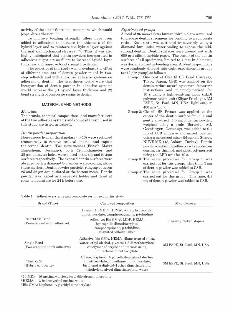
Table 1 Adhesive systems and composite resin used in this study
Brand (Type) Chemical composition Manufacturer
Clearfil SE Bond(Two-step self-etch adhesive)
Primer: 10-MDP1, HEMA2, water, hydrophilic dimethacrylate, camphoroquinone, p-toluidine
Adhesive: Bis-GMA3, MDP, HEMA, hydrophilic dimethacrylate,
camphorquinone, p-toluidine, silanated colloidal silica
Kuraray, Tokyo, Japan
Single Bond (Two-step total-etch adhesive)
Adhesive: bis-GMA, HEMA, silane-treated silica, water, ethyl alcohol, glycerol 1,3-dimethacrylate,
copolymer of acrylic and itaconic acids, diurethane dimethacrylate
3M ESPE, St. Paul, MN, USA
Filtek Z250(Hybrid composite)
Silane, bisphenol A polyethylene glycol diether dimethacrylate, diurethane dimethacrylate, bisphenol A diglycidyl ether dimethacrylate,
triethylene glycol dimethacrylate, water
3M ESPE, St. Paul, MN, USA
1 10-MDP: 10-methacryloyloxydecyl dihydrogen phosphate2 HEMA: 2-hydroxyethyl methacrylate3 Bis-GMA: bisphenol A glycidyl methacrylate
activity of the acidic functional monomers, which would jeopardize adhesion11,12).
To improve bonding strength, fillers have been added to adhesives to increase the thickness of the hybrid layer and to reinforce the hybrid layer against thermal and mechanical stresses13-20). Thus, it was also highly anticipated that dentin powders incorporated in adhesives might act as fillers to increase hybrid layer thickness and improve bond strength to dentin.
The objective of this study was to evaluate the effect of different amounts of dentin powder mixed in two-step self-etch and etch-and-rinse adhesive systems on adhesion to dentin. The hypotheses tested were that incorporation of dentin powder in adhesive systems would increase the (1) hybrid layer thickness and (2) bond strength of composite resins to dentin.
MATERIALS AND METHODS
MaterialsThe brands, chemical compositions, and manufacturers of the two adhesive systems and composite resin used in this study are listed in Table 1.
Dentin powder preparationNon-carious human third molars (n=10) were sectioned transversely to remove occlusal enamel and expose the coronal dentin. Two sieve meshes (Fritsch, Markt Einersheim, Germany), with 52-µm-diameter and 25-µm-diameter holes, were placed on the top and bottom surfaces respectively. The exposed dentin surfaces were abraded with a diamond bur under water-cooling above these meshes. Dentin powder particles ranging between 25 and 52 µm accumulated at the bottom mesh. Dentin powder was placed in a separate holder and dried at room temperature for 24 h before use.
Experimental groupsA total of 96 non-carious human third molars were used to prepare dentin specimens for bonding to a composite resin. Each tooth was sectioned transversely using a diamond bur under water-cooling to expose the mid-coronal dentin. Dentin surfaces were ground wet with 600-grit silicon carbide paper. The center of the dentin surfaces of all specimens, limited to 4 mm in diameter, was designated as the bonding area. All dentin specimens were randomly divided into eight experimental groups (n=12 per group) as follows:
Group 1: One coat of Clearfil SE Bond (Kuraray, Tokyo, Japan; CSB) was applied on the dentin surface according to manufacturer’s instructions and photopolymerized for 10 s using a light-emitting diode (LED) polymerization unit (Elipar FreeLight, 3M ESPE, St. Paul, MN, USA; light output: 400 mW/cm2).
Group 2: Clearfil SE Primer was applied to the center of the dentin surface for 20 s and gently air-dried. 1.5 mg of dentin powder, weighed using a scale (Sartorius AG, Goettingen, Germany), was added to 0.1 mL of CSB adhesive and mixed together using a motorized mixer (Magnetic Stirrer, NUVE MK 418, Ankara, Turkey). Dentin powder-containing adhesive was applied on dentin, air-thinned, and photopolymerized using the LED unit for 10 s.
Group 3: The same procedure for Group 2 was carried out for this group. This time, 3 mg of dentin powder was added to CSB.
Group 4: The same procedure for Group 2 was carried out for this group. This time, 4.5 mg of dentin powder was added to CSB.
759Dent Mater J 2012; 31(5): 758–764
Table 2 Mean and standard deviation (±SD) values of shear bond strength (MPa) based on two adhesive types with or without dentin powder incorporation at different amounts
Experimental group Shear bond strength (MPa)
1 15.1±1.2a,c
2 13.5±2.7a,c
3 16.4±4.7a
4 9.9±4.5b,c
5 12.6±4c
6 5.5±3.6d
7 5.6± 2.2d
8 4± 1.6d
Different small superscript letters indicate statistically significant differences (p<0.05).
Table 3 Distribution of failure types per group in percentage
Groups Score 0 Score 1 Score 2 Score 3
1 50 10 10 30
2 70 – – 30
3 80 – – 20
4 60 – – 40
5 50 10 20 20
6 60 – – 40
7 50 – – 50
8 50 – – 50
Score 0: Adhesive; Score 1: Cohesive in dentin; Score 2: Cohesive in composite; Score 3: Mixed (combination of Score 0 and Score 2).
Group 5: Single Bond (3M, St. Paul, MN, USA; SB) was used according to manufacturer’s instructions. Dentin surface was etched with 35% phosphoric acid gel for 15 s, rinsed with water, and gently blot-dried using a cotton pellet.
Group 6: Dentin was etched as described for Group 5. 1.5 mg of dentin powder was added to 0.1 mL of SB adhesive and mixed together using the motorized mixer. Dentin powder-containing adhesive was applied on dentin, air-thinned, and photopolymerized using the LED unit for 10 s.
Group 7: The same procedure for Group 6 was carried out for this group. This time, 3 mg of dentin powder was added to SB.
Group 8: The same procedure for Group 6 was carried out for this group. This time, 4.5 mg of dentin powder was added to SB.
After dentin conditioning, a polyethylene mold (4 mm diameter, 3 mm height) was positioned at the center of the dentin surfaces, i.e., the designated bonding area, of all the specimens. A hybrid composite (Filtek Z250, 3M ESPE) was packed into the polyethylene mold and photopolymerized using the LED unit for 20 s.
Shear bond strength testTen teeth from each group were embedded in acrylic blocks and subjected to shear bond strength test in a universal testing machine (Elista, İstanbul, Turkey) at a crosshead speed of 1 mm/min. A chisel-shaped shearing blade, which applied the shearing force, was positioned as close as possible to the resin-dentin interface. Maximum load (N) for debonding was recorded and divided by the contact surface area to determine the bond strength in mega pascals (MPa).
Failure analysisAfter debonding, the dentin surfaces were observed using a stereomicroscope (SZ-PT, Olympus, Tokyo, Japan) at ×30 magnification to determine the failure modes. Failure modes were categorized as: (a) Adhesive (Score 0); (b) Cohesive in dentin (Score 1); (c) Cohesive in composite (Score 2); and (d) Mixed (Score 3: combination of Score 0 and Score 2).
Hybrid layer analysisThe remaining two teeth from each group were sectioned perpendicularly to the bonding interface between the composite and dentin using a low-speed wheel saw under water. The sections were sequentially wet-polished using 600-, 1000-, and 1200-grit silicon carbide papers. Then, they were etched with 37% phosphoric acid for 5 s, rinsed with water, and treated with 5% sodium hypochlorite (NaOCL) for 10 min. After rinsing and air-drying, each section was sputter-coated with gold for scanning electron microscope (SEM) analysis. Hybrid layer thickness was measured at five locations on each section, and the mean thickness was calculated.
Statistical analysisStatistical analysis was performed using SPSS System 11.0 for Windows (SPSS Inc., Chicago, IL, USA). Bond strength data (MPa) were subjected to two-way analysis of variance (ANOVA). Multiple comparisons were performed using the Tukey’s test (α=0.05) with bond strength as the dependent variable and adhesive type (two levels) and dentin powder amount (three levels) as the independent variables. P values less than 0.05 were considered to be statistically significant.
760 Dent Mater J 2012; 31(5): 758–764
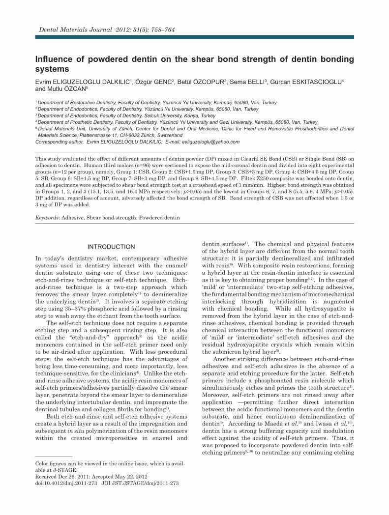
Fig. 1 SEM micrographs (×500) of: (a) Group 1. Hybrid layer was approximately 1.1±0.1 µm thick. Long resin tags could be observed with
no distortions or separation zones at resin-dentin interface. (b) Group 2. Hybrid layer was approximately 1.3±0.1 µm thick. No resin tags could be observed, but
resin-dentin interface showed no distortions or separation zones. (c) Group 3. Hybrid layer was approximately 2.4±0.4 µm thick. No resin tags could be observed, but
resin-dentin interface showed no distortions or separation zones. (d) Group 4. Hybrid layer was approximately 4.5±0.6 µm thick. Note the absence of resin tags. Resin-
dentin interface showed no distortions or separation zones. Hybrid layer (arrow); D: Dentin; C: Composite; H: Hybrid layer; R: Resin Tags.
RESULTS
Shear bond strengthStatistical analysis revealed that adhesive type (p<0.005) and dentin powder amount (p<0.005) had significant effects on shear bond strength. Interaction between these two factors was also significant (p=0.151).
Table 2 shows the shear bond strengths of all the experimental groups in this study. Highest bond strength was obtained in Groups 1, 2, and 3 (15.1, 13.5, and 16.4 MPa respectively; p>0.05) and the lowest in Groups 6, 7, and 8 (5.5, 5.6, 4 MPa; p>0.05). Dentin powder addition, regardless of amount, adversely affected the bond strength of SB. Bond strength of CSB was not affected when 1.5 or 3 mg of dentin powder was
added, but 4.5 mg addition significantly reduced its bond strength.
Failure modesFailure analysis revealed that adhesive failure (Score 0: 50 to 80%) was the predominant failure mode for all the experimental groups (Table 3). Only in Groups 1 and 5 were cohesive failure in dentin (Score 1: 10% and 10% respectively) and cohesive failure in composite (Score 2: 10% and 20% respectively) observed. The rest of the failures were of the mixed type (Score 3).
Hybrid layer thicknessFor Group 1 (Fig. 1(a)), mean hybrid layer thickness was 1.1±0.1 µm with the presence of long resin tags.
761Dent Mater J 2012; 31(5): 758–764
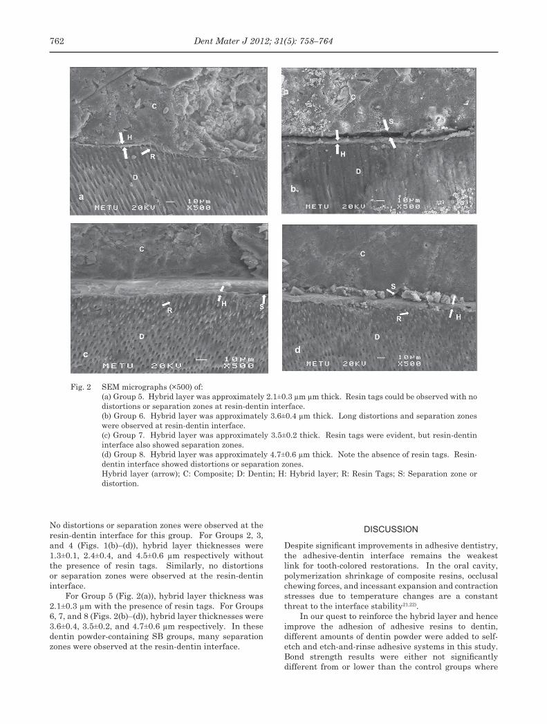
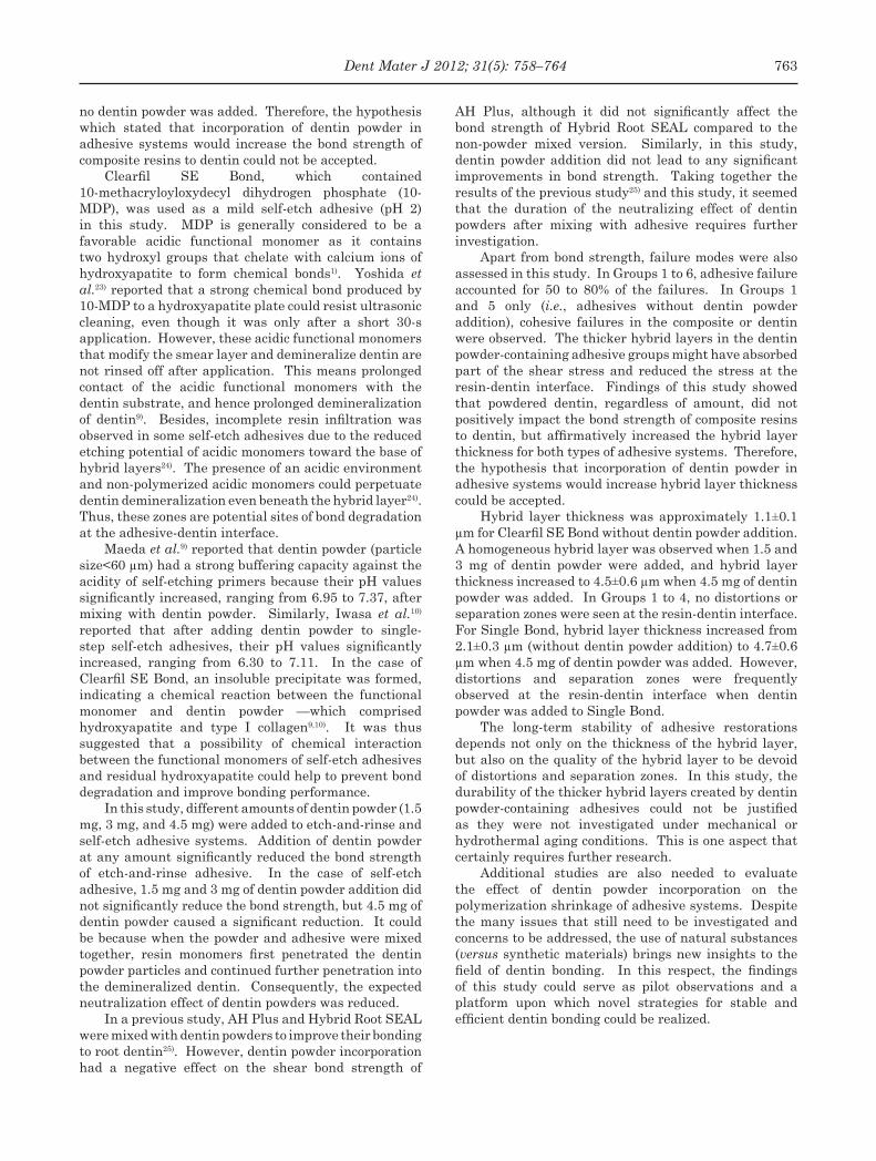
Fig. 2 SEM micrographs (×500) of: (a) Group 5. Hybrid layer was approximately 2.1±0.3 µm µm thick. Resin tags could be observed with no
distortions or separation zones at resin-dentin interface. (b) Group 6. Hybrid layer was approximately 3.6±0.4 µm thick. Long distortions and separation zones
were observed at resin-dentin interface. (c) Group 7. Hybrid layer was approximately 3.5±0.2 thick. Resin tags were evident, but resin-dentin
interface also showed separation zones. (d) Group 8. Hybrid layer was approximately 4.7±0.6 µm thick. Note the absence of resin tags. Resin-
dentin interface showed distortions or separation zones. Hybrid layer (arrow); C: Composite; D: Dentin; H: Hybrid layer; R: Resin Tags; S: Separation zone or
distortion.
No distortions or separation zones were observed at the resin-dentin interface for this group. For Groups 2, 3, and 4 (Figs. 1(b)–(d)), hybrid layer thicknesses were 1.3±0.1, 2.4±0.4, and 4.5±0.6 µm respectively without the presence of resin tags. Similarly, no distortions or separation zones were observed at the resin-dentin interface.
For Group 5 (Fig. 2(a)), hybrid layer thickness was 2.1±0.3 µm with the presence of resin tags. For Groups 6, 7, and 8 (Figs. 2(b)–(d)), hybrid layer thicknesses were 3.6±0.4, 3.5±0.2, and 4.7±0.6 µm respectively. In these dentin powder-containing SB groups, many separation zones were observed at the resin-dentin interface.
DISCUSSION
Despite significant improvements in adhesive dentistry, the adhesive-dentin interface remains the weakest link for tooth-colored restorations. In the oral cavity, polymerization shrinkage of composite resins, occlusal chewing forces, and incessant expansion and contraction stresses due to temperature changes are a constant threat to the interface stability21,22).
In our quest to reinforce the hybrid layer and hence improve the adhesion of adhesive resins to dentin, different amounts of dentin powder were added to self-etch and etch-and-rinse adhesive systems in this study. Bond strength results were either not significantly different from or lower than the control groups where
762 Dent Mater J 2012; 31(5): 758–764
no dentin powder was added. Therefore, the hypothesis which stated that incorporation of dentin powder in adhesive systems would increase the bond strength of composite resins to dentin could not be accepted.
Clearfil SE Bond, which contained 10-methacryloyloxydecyl dihydrogen phosphate (10-MDP), was used as a mild self-etch adhesive (pH 2) in this study. MDP is generally considered to be a favorable acidic functional monomer as it contains two hydroxyl groups that chelate with calcium ions of hydroxyapatite to form chemical bonds1). Yoshida et al.23) reported that a strong chemical bond produced by 10-MDP to a hydroxyapatite plate could resist ultrasonic cleaning, even though it was only after a short 30-s application. However, these acidic functional monomers that modify the smear layer and demineralize dentin are not rinsed off after application. This means prolonged contact of the acidic functional monomers with the dentin substrate, and hence prolonged demineralization of dentin9). Besides, incomplete resin infiltration was observed in some self-etch adhesives due to the reduced etching potential of acidic monomers toward the base of hybrid layers24). The presence of an acidic environment and non-polymerized acidic monomers could perpetuate dentin demineralization even beneath the hybrid layer24). Thus, these zones are potential sites of bond degradation at the adhesive-dentin interface.
Maeda et al.9) reported that dentin powder (particle size<60 µm) had a strong buffering capacity against the acidity of self-etching primers because their pH values significantly increased, ranging from 6.95 to 7.37, after mixing with dentin powder. Similarly, Iwasa et al.10) reported that after adding dentin powder to single-step self-etch adhesives, their pH values significantly increased, ranging from 6.30 to 7.11. In the case of Clearfil SE Bond, an insoluble precipitate was formed, indicating a chemical reaction between the functional monomer and dentin powder —which comprised hydroxyapatite and type I collagen9,10). It was thus suggested that a possibility of chemical interaction between the functional monomers of self-etch adhesives and residual hydroxyapatite could help to prevent bond degradation and improve bonding performance.
In this study, different amounts of dentin powder (1.5 mg, 3 mg, and 4.5 mg) were added to etch-and-rinse and self-etch adhesive systems. Addition of dentin powder at any amount significantly reduced the bond strength of etch-and-rinse adhesive. In the case of self-etch adhesive, 1.5 mg and 3 mg of dentin powder addition did not significantly reduce the bond strength, but 4.5 mg of dentin powder caused a significant reduction. It could be because when the powder and adhesive were mixed together, resin monomers first penetrated the dentin powder particles and continued further penetration into the demineralized dentin. Consequently, the expected neutralization effect of dentin powders was reduced.
In a previous study, AH Plus and Hybrid Root SEAL were mixed with dentin powders to improve their bonding to root dentin25). However, dentin powder incorporation had a negative effect on the shear bond strength of
AH Plus, although it did not significantly affect the bond strength of Hybrid Root SEAL compared to the non-powder mixed version. Similarly, in this study, dentin powder addition did not lead to any significant improvements in bond strength. Taking together the results of the previous study25) and this study, it seemed that the duration of the neutralizing effect of dentin powders after mixing with adhesive requires further investigation.
Apart from bond strength, failure modes were also assessed in this study. In Groups 1 to 6, adhesive failure accounted for 50 to 80% of the failures. In Groups 1 and 5 only (i.e., adhesives without dentin powder addition), cohesive failures in the composite or dentin were observed. The thicker hybrid layers in the dentin powder-containing adhesive groups might have absorbed part of the shear stress and reduced the stress at the resin-dentin interface. Findings of this study showed that powdered dentin, regardless of amount, did not positively impact the bond strength of composite resins to dentin, but affirmatively increased the hybrid layer thickness for both types of adhesive systems. Therefore, the hypothesis that incorporation of dentin powder in adhesive systems would increase hybrid layer thickness could be accepted.
Hybrid layer thickness was approximately 1.1±0.1 µm for Clearfil SE Bond without dentin powder addition. A homogeneous hybrid layer was observed when 1.5 and 3 mg of dentin powder were added, and hybrid layer thickness increased to 4.5±0.6 µm when 4.5 mg of dentin powder was added. In Groups 1 to 4, no distortions or separation zones were seen at the resin-dentin interface. For Single Bond, hybrid layer thickness increased from 2.1±0.3 µm (without dentin powder addition) to 4.7±0.6 µm when 4.5 mg of dentin powder was added. However, distortions and separation zones were frequently observed at the resin-dentin interface when dentin powder was added to Single Bond.
The long-term stability of adhesive restorations depends not only on the thickness of the hybrid layer, but also on the quality of the hybrid layer to be devoid of distortions and separation zones. In this study, the durability of the thicker hybrid layers created by dentin powder-containing adhesives could not be justified as they were not investigated under mechanical or hydrothermal aging conditions. This is one aspect that certainly requires further research.
Additional studies are also needed to evaluate the effect of dentin powder incorporation on the polymerization shrinkage of adhesive systems. Despite the many issues that still need to be investigated and concerns to be addressed, the use of natural substances (versus synthetic materials) brings new insights to the field of dentin bonding. In this respect, the findings of this study could serve as pilot observations and a platform upon which novel strategies for stable and efficient dentin bonding could be realized.
763Dent Mater J 2012; 31(5): 758–764
CONCLUSIONS
Within the limitations of this study, the following conclusions were drawn:
1. Two-step self-etch adhesive system showed significantly higher bond strength to demineralized dentin than etch-and-rinse adhesive system.
2. Addition of dentin powder to etch-and-rinse adhesive (Single Bond) adversely affected bond strength.
3. Addition of 1.5 and 3 mg of dentin powder did not significantly affect the bond strength of self-etch adhesive (Clearfil SE Bond), but 4.5 mg of dentin powder addition significantly reduced its bond strength.
4. Dentin powder addition increased the hybrid layer thickness for both types of adhesive systems tested.
REFERENCES
1) Van Meerbeek B, De Munck J, Yoshida Y, Inoue S, Vargas M, Vijay P, Van Landuyt K, Lambrechts P, Vanherle G. Adhesion to enamel and dentin: Current status and future challenges. Oper Dent 2003; 28: 215-235.
2) Perdigao J, Swift EJ. In: Roberson TM, Heymann HO, Swift EJ, editors. Sturdevant’s Art & Science of Operative Dentistry. 4th ed. St Louis, MO, USA: Mosby Co; 2002. p. 246.
3) Van Meerbeek B, Yoshihara K, Yoshida Y, Mine A, De Munck J, Van Landuyt KL. State of the art of self-etch adhesives. Dent Mater 2011; 27: 17-28.
4) Tay FR, Pashley DH. Aggressiveness of contemporary self-etching systems. 1: Depth of penetration beyond dentin smear layers. Dent Mater 2001; 17: 296-308.
5) Tay FR, Pashley DH, Yoshiyama M. Two modes of nanoleakage expression in single-step adhesives. J Dent Res 2002; 81: 472-476.
6) Eick JD, Gwinnett AJ, Pashley DH, Robinson SJ. Current concepts on adhesion to dentin. Crit Rev Oral Biol Med 1997; 8: 306-335.
7) Perdigão J. Dentin bonding as a function of dentin structure. Dent Clin North Am 2002; 46: 277-301.
8) Nakabayashi N, Pashley DH. In: Nakabayashi N, Pashley DH, editors. Hybridization of dental hard tissues. 1st ed. Tokyo: Quintessence Publishing; 1998. p. 8.
9) Maeda T, Yamaguchi K, Takamizawa T, Rikuta A, Tsubota K, Ando S, Miyazaki M. pH changes of self-etching primers mixed with powdered dentine. J Dent 2008; 36: 606-610.
10) Iwasa M, Tsubota K, Shimamura Y, Ando S, Miyazaki M, Platt JA. pH changes upon mixing of single-step self-etching
adhesives with powdered dentin. J Adhes Dent 2011; 13: 207-212.
11) Hiraishi N, Kitasako Y, Nikaido T, Foxton RM, Tagami J, Nomura S. Detection of acid diffusion through bovine dentine after adhesive application. Int Endod J 2004; 37: 455-462.
12) Oliveira SS, Marshall SJ, Habelitz S, Gansky SA, Wilson RS, Marshall GW Jr. The effect of a self-etching primer on the continuous demineralization of dentin. Eur J Oral Sci 2004; 112: 376-383.
13) Mitsui FH, Peris AR, Cavalcanti AN, Marchi GM, Pimenta LA. Influence of thermal and mechanical load cycling on microtensile bond strengths of total and self-etching adhesive systems. Oper Dent 2006; 31: 240-247.
14) Gallo JR, Comeaux R, Haines B, Xu X, Burgess JO. Shear bond strength of four filled dentin bonding systems. Oper Dent 2001; 26: 44-47.
15) Ausiello P, Apicella A, Davidson CL. Effect of adhesive layer properties on stress distribution in composite restorations — a 3D finite element analysis. Dent Mater 2002; 18: 295-303.
16) Lopes GC, Vieira LCC, Montreiro S, Caldeira de Andrada M Jr, Baratieri CM. Dentin bonding: effect of degree of mineralization and acid etching time. Oper Dent 2003; 28: 429-439.
17) Brackett WW, Ito S, Tay FR, Haisch LD, Pashley DH. Microtensile dentin bond strength of self-etching resins: effect of a hydrophobic layer. Oper Dent 2005; 30: 733-738.
18) Nakaoki Y, Sasakawa W, Horiuchi S, Nagano F, Ikeda T, Tanaka T, Inoue S, Uno S, Sano H, Sidhu SK. Effect of double-application of all-in-one adhesives on dentin bonding. J Dent 2005; 33: 765-772.
19) Silva ALF, Lima DANL, Souza GMD, Santos CTD, Paulillo LAMS. Influence of additional adhesive application on the microtensile bond strength of adhesive systems. Oper Dent 2006; 31: 562-568.
20) Ibrahim IM, Elkassas DW, Yousry MM. Effect of EDTA and phosphoric acid pretreatment on the bonding effectiveness of self-etch adhesives to ground enamel. Eur J Dent 2010; 4: 418-428.
21) Kemp-Scholte CM, Davidson CL. Marginal integrity related to bond strength and strain capacity of composite resin restorative systems. J Prosthet Dent 1990; 64: 658-664.
22) Pongprueksa P, Kuphasuk W, Senawongse P. Effect of elastic cavity wall and occlusal loading on microleakage and dentin bond strength. Oper Dent 2007; 32: 466-475.
23) Yoshida Y, Nagakane K, Fukuda R, Nakayama Y, Okazaki M, Shintani H, Inoue S, Tagawa Y, Suzuki K, De Munck J, Van Meerbeek B. Comparative study on adhesive performance of functional monomers. J Dent Res 2004; 83: 454-458.
24) Carvalho RM, Chersoni S, Frankenberger R, Pashley DH, Prati C, Tay FR. A challenge to the conventional wisdom that simultaneous etching and resin infiltration always occurs in self-etch adhesives. Biomaterials 2005; 26: 1035-1042.
25) Belli S, Cobankara FK, Ozcopur B, Eliguzeloglu E, Eskitascioglu G. An alternative adhesive strategy to optimize bonding to root dentin. J Endod 2011; 37: 1427-1432.
764 Dent Mater J 2012; 31(5): 758–764