influence des microparticules sur la fonction vasculaire lors de
TRANSCRIPT
Année 2011
: 1174
Influence des microparticules sur la fonction vasculaire lors de pathologies hypoxiques
Thèse de Doctorat
Spécialité : Physiologie & Physiopathologie Humaine
Ecole doctorale Biologie Santé
Présentée et soutenue publiquement
Le 12 octobre 2011, à Angers
Par
Simon TUAL-CHALOT
Devant le jury ci-dessous:
Pr Patrick LEVY Rapporteur PU-PH, Université de Grenoble
Pr Roger MARTHAN Rapporteur PU-PH, Université de Bordeaux
Pr Patrick SAULNIER Examinateur PU, Univ
Directeur de thèse:
Docteur Maria Del Carmen MARTINEZ
INSERM U694
tel-0
0978
657,
ver
sion
1 -

tel-0
0978
657,
ver
sion
1 -

!"
"
SO M M A IR E
tel-0
0978
657,
ver
sion
1 -
#"
"
SO M M A IR E ............................................................................................................................. 1
R E M E R C I E M E N TS ................................................................................................................ 6
L IST E D ES PUB L I C A T I O NS & C O M M UNI C A T I O NS ................................................. 11
A BBR E V I A T I O NS ................................................................................................................ 14
L IST E D ES F I G UR ES & T A B L E A U X ............................................................................... 19
A V A N T-PR OPOS .................................................................................................................. 22
C H API T R E 1 - D O NN E ES BIB L I O G R APH I Q U ES ......................................................... 25
I. Le système vasculaire et ses fonctions ................................................................ 26
1. La circulation sanguine .................................................................................. 26
1.1. Organisation et rôles ............................................................................... 26
1.1.1. La circulation systémique ou grande circulation ........................... 26
1.1.2. La circulation pulmonaire ou petite circulation ........................... 27
1.1.3. Morphologie et histologie de la paroi artérielle ............................ 28
1.2. Mécanismes de régulation de la vasomotricité....................................... 30
1.2.1. Contraction des cellules musculaires lisses ................................... 30
1.2.2. Relaxation des cellules musculaires lisses .................................... 32
2. ......................................................................... 35
2.1. ................ 35
2.2. ...................................... 35
2.2.1. ................................... 35
2.2.2. ............................ 41
2.2.3. Les autres médiateurs .................................................................... 43
2.3. La dysfonction endothéliale ................................................................... 45
II. Les microparticules circulantes ......................................................................... 46
1. Découverte et définition ................................................................................. 46
2. Formation des microparticules ....................................................................... 46
............................................................................ 47
.............................................................................................. 49
2.3. Autres mécanismes de formation ........................................................... 49
tel-0
0978
657,
ver
sion
1 -
$"
"
3. Composition et contenu des microparticules ................................................. 50
3.1. Contenu protéique .................................................................................. 50
3.2. Contenu lipidique ................................................................................... 51
4. Interaction entre les microparticules et les cellules cibles ............................. 51
4.1. Interaction ligand-récepteur ................................................................... 52
4.2. Transfert de composants ......................................................................... 53
4.3. Fusion et internalisation ......................................................................... 53
5. Les microparticules : biomarqueurs et effecteurs dans les pathologies cardio-vasculaires ........................................................................................... 54
5.1. Effets des microparticules sur la fonction vasculaire ............................. 54
5.2. Angiogenèse et microparticules ............................................................. 55
6. Modulation et action thérapeutique des microparticules ................................ 57
hypoxiques....................................................................................................... 59
1. Les pathologies pulmonaires .......................................................................... 59
............................................................. 59
2.1. Classification clinique des hypertensions pulmonaires .......................... 59
2.2. Manifestations cliniques et diagnostic ................................................... 61
2.3. Pathogénèse globale ............................................................................... 61
2.4. Stratégie thérapeutique ........................................................................... 63
2.5. Modèles animaux ................................................................................... 64
sommeil ............................................... 65
3.1. Manifestations cliniques et diagnostic ................................................... 65
3.2. Pathogénèse globale ............................................................................... 66
3.3. Stratégie thérapeutique ........................................................................... 69
O BJE C T I FS D ES T R A V A U X .............................................................................................. 70
C H API T R E 2 - R ESU L T A TS ............................................................................................... 72
Partie I : Les microparticules circulantes issues de rats hypoxiques chroniques entraînent une dysfonction endothéliale .......................................................................................................... 73
................................................................................................... 74
tel-0
0978
657,
ver
sion
1 -
%"
"
II. Matériel et méthodes........................................................................................ 75
1. Matériel biologique ........................................................................................ 75
: le rat hypoxique .............. 75
1.2. Cellules endothél ................ 78
2. Cytométrie en flux .......................................................................................... 79
3. Western blot ................................................................................................... 80
4. Dosage des radicaux libres par résonnance paramagnétique électronique ..... 82
.................................................. 83
6. Réactivité vasculaire ....................................................................................... 83
7. Analyse statistique .......................................................................................... 84
III. Résultats ........................................................................................................... 85
IV. Discussion ........................................................................................................ 95
Partie II hyperréactivité vasculaire ................................................................. 102
................................................................................................. 103
II. Matériel et méthodes....................................................................................... 104
1. Matériel biologique ...................................................................................... 104
2. Cytométrie en flux ........................................................................................ 105
3. Réactivité vasculaire ..................................................................................... 105
4. Western blot ................................................................................................. 106
..................................................................... 106
6. Dosage des divers prostanoïdes .................................................................... 107
7. Analyse statistique des résultats ................................................................... 107
III. Résultats ......................................................................................................... 108
IV. Discussion ...................................................................................................... 122
Partie III angiogenèse ............................................................................................................................ 127
................................................................................................. 128
II. Matériel et méthodes....................................................................................... 129
tel-0
0978
657,
ver
sion
1 -

&"
"
1. Matériel biologique ...................................................................................... 129
2. Mesure du taux de VEGF ............................................................................. 129
3. Western blot ................................................................................................. 130
4. Prolifération cellulaire .................................................................................. 130
5. Mesure de la formation de capillaire sur Matrigel ....................................... 131
6. Analyse statistique des résultats ................................................................... 131
III. Résultats ......................................................................................................... 132
IV. Discussion ...................................................................................................... 137
C H API T R E 3 - C O N C L USI O N & PE RSPE C T I V ES ...................................................... 141
A NN E X ES ............................................................................................................................. 145
BIB L I O G R APH I E ............................................................................................................... 148
PUB L I C A T I O NS & M A NUSC RI T ................................................................................... 167
Revues : Microvesicles: intercellular vector of biological messages Tual-Chalot S, Leonetti D, Andriantsitohaina R, MartinezMC. Molecular Interventions, 2011 ............................................................................................... 168
Microparticles: targets and tools in cardiovascular disease MartinezMC, Tual-Chalot S, Leonetti D, Andriantsitohaina R. Trends in Pharmacological Sciences, 2011 ............................................................................ 176
Publications : Circulating microparticles from pulmonary hypertensive rats induce endothelial dysfunction Tual-Chalot S American Journal of Respiratory and Critical Care Medicine, 2010 ..................................... 184
Nocturnal release of leukocyte-derived microparticles in males with obstructive sleep apnea Trzepizur W, Priou P, Paris A, Nardi J, Tual-Chalot S, Meslier N, Urban T, Andriantsitohaina R, Martinez MC, Gagnadoux F. European Respiratory Journal, 2011 ...................................................................................... 193 Manuscrit : Sleep apnea syndrome microparticles enhance vascular contraction: mandatory role of the endothelium Tual-Chalot S, Fatoumata K, Priou P, Trzepizur W, Contreras C, Prieto D, Martinez MC, Gagnadoux F, Andriantsitohaina R ........................................................................................ 197
tel-0
0978
657,
ver
sion
1 -

'"
"
R E M E R C I E M E N TS
tel-0
0978
657,
ver
sion
1 -

("
"
et guidé depuis mon master 2. Merci vivement de ton soutien constant, de ta grande
dispon
reconnaissance.
Ramaroson Andriantsitohaina. Je le remercie vivement pour nos échanges scientifiques que
nous avons pu avoir tous ensemble, notamment le lundi matin.
Je remercie monsieur le Professeur Patrick Lévy et monsieur le Professeur Roger
ma g
tel-0
0978
657,
ver
sion
1 -
)"
"
Un remerciement très particulier au Dr Christelle Guib
s, son accueil lors de ma visite et les échanges
particulièrement le Professeur Frédéric Gagnadoux, et les Docteur Pascaline Priou et
Wojciech Trzepizur pour les MPs, les discussions et la bonne humeur toujours présente !
Je tiens également à remercier le GRRC et la SFP de donner la possibilité aux jeunes
soirées de folies !!!!
Je souhaite remercier chaleureusement Carmen et Naina qui sont beaucoup plus que
des encadrants, et qui nous permettent de nous sentir vraiment bien au sein du laboratoire.
Vous avez placé la barre très haute pour mes futurs laboratoires !
laboratoire et qui rendent les conditions de travail si agréable, Matthieu un vrai collègue et
vrai ami, Lucie même si on ne joue plus dans la même catégorie et Raffa membre
indispensable
Je tiens à remercier également les anciens et actuels mem -
Martinez, Angela, Daniela (partenaire attitrée de myographie), Francesco et Mariele,
Carmina, Christina et Vannina, Ahmed, Abdel et Tarek, Alexis, Françoise, Nicolas et
ont participé à ma formation tout au long de ces derniers
années. Merci également à Céline qui a contribué à ma grande passion pour les
e a ce
travail. Merci à vous pour votre gentillesse et vos précieux conseils.
commencé -INSERM 771 du Docteur Daniel
Henrion. Emilie, Anne-Laure, Kévin, Bertrand, Jennifer, Lamine, Kahena et Emmanuelle
Maire, je les remercie. Ma dernière année a débuté par un grand déménagement (dans des
supers locaux r le Professeur Yves Malthiéry.
Audrey, Delphine, Jihane, Luisa (et les poissons obèses !); Aziz, Caroline, Emilie, Ghaceb
(bon courage avec Lucie !), Jacques; Amine (dernière année !) et Mireille (super intendante),
à tous merci e parfois précieuse dans la réalisation de mes
tel-0
0978
657,
ver
sion
1 -

*"
"
expériences, et dans la réponse à toutes les questions pratiques, techniques et administratives
auxquelles un étudiant en thèse se retrouve forcément confrontés.
Une petite pensée pour nos rivaux-a toujours prêts pour une pétanque à
Archidelaballe, toujours partant pour tout, dattebayo!!!
La famille Bastiat-
soulever un stade à lui tout seul et Maud P, Dr!, femme la plus forte du monde pour supporter
son homme.
Elisa, ma petite espagnole support de star, bon courage avec tes monkeys
Florian, mélodicien de talent, aussi étourdi que moi, ça me fait bien plaisir!!!
Marie Morille, organisatrice de t
Matt, que de bons moments à la piscine, au golf, au travail aussi!!!
Raff
Welsh le pub, Welsh la bière et Welsh le chat
ie
que je suis, tous les Lasséens, à
Je voudrais également remercier la très grande famille Roger-Maubert-Naulet de Vieil
lus folle représentante (désolé mais mes derniers
remerciements ne seront pas pour toi!)
Je voudrais enfin remercier ma famille, notamment mes parents, Yves et Patricia, qui
Matthieu,
Pierre et Camille.
tel-0
0978
657,
ver
sion
1 -

!+"
"
!"#$%&'()**+%
tel-0
0978
657,
ver
sion
1 -

!!"
"
L IST E D ES PUB L I C A T I O NS
& C O M M UNI C A T I O NS
tel-0
0978
657,
ver
sion
1 -
!#"
"
A R T I C L ES PUB L I ES
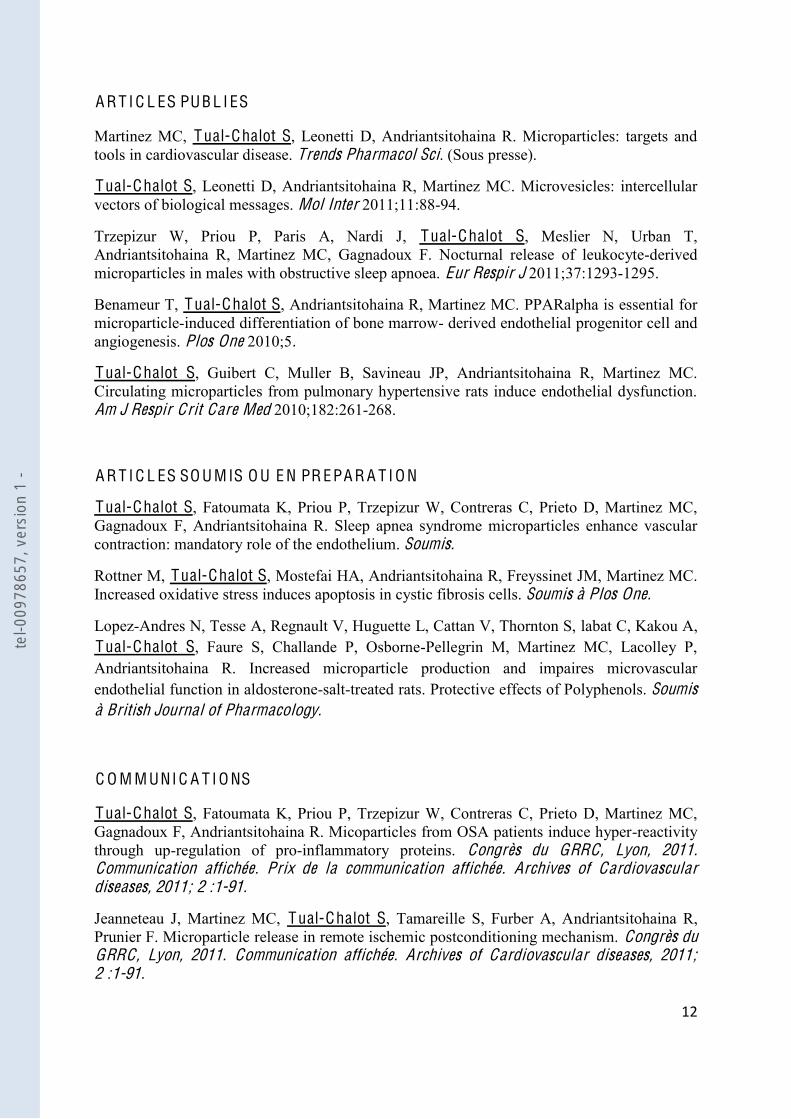
Martinez MC, Tual-Chalot S, Leonetti D, Andriantsitohaina R. Microparticles: targets and tools in cardiovascular disease. Trends Pharmacol Sci. (Sous presse).
Tual-Chalot S, Leonetti D, Andriantsitohaina R, Martinez MC. Microvesicles: intercellular vectors of biological messages. Mol Inter 2011;11:88-94.
Trzepizur W, Priou P, Paris A, Nardi J, Tual-Chalot S, Meslier N, Urban T, Andriantsitohaina R, Martinez MC, Gagnadoux F. Nocturnal release of leukocyte-derived microparticles in males with obstructive sleep apnoea. Eur Respir J 2011;37:1293-1295.
Benameur T, Tual-Chalot S, Andriantsitohaina R, Martinez MC. PPARalpha is essential for microparticle-induced differentiation of bone marrow- derived endothelial progenitor cell and angiogenesis. Plos One 2010;5.
Tual-Chalot S, Guibert C, Muller B, Savineau JP, Andriantsitohaina R, Martinez MC. Circulating microparticles from pulmonary hypertensive rats induce endothelial dysfunction. Am J Respir Crit Care Med 2010;182:261-268.
A R T I C L ES SO U M IS O U E N PR EPA R A T I O N
Tual-Chalot S, Fatoumata K, Priou P, Trzepizur W, Contreras C, Prieto D, Martinez MC, Gagnadoux F, Andriantsitohaina R. Sleep apnea syndrome microparticles enhance vascular contraction: mandatory role of the endothelium. Soumis.
Rottner M, Tual-Chalot S, Mostefai HA, Andriantsitohaina R, Freyssinet JM, Martinez MC. Increased oxidative stress induces apoptosis in cystic fibrosis cells. Soumis à Plos One.
Lopez-Andres N, Tesse A, Regnault V, Huguette L, Cattan V, Thornton S, labat C, Kakou A, Tual-Chalot S, Faure S, Challande P, Osborne-Pellegrin M, Martinez MC, Lacolley P, Andriantsitohaina R. Increased microparticle production and impaires microvascular endothelial function in aldosterone-salt-treated rats. Protective effects of Polyphenols. Soumis à British Journal of Pharmacology.
C O M M UNI C A T I O NS
Tual-Chalot S, Fatoumata K, Priou P, Trzepizur W, Contreras C, Prieto D, Martinez MC, Gagnadoux F, Andriantsitohaina R. Micoparticles from OSA patients induce hyper-reactivity through up-regulation of pro-inflammatory proteins. Congrès du GRRC , Lyon, 2011. Communication affichée. Prix de la communication affichée. Archives of Cardiovascular diseases, 2011; 2 :1-91.
Jeanneteau J, Martinez MC, Tual-Chalot S, Tamareille S, Furber A, Andriantsitohaina R, Prunier F. Microparticle release in remote ischemic postconditioning mechanism. Congrès du GRRC , Lyon, 2011. Communication affichée. Archives of Cardiovascular diseases, 2011; 2 :1-91.
tel-0
0978
657,
ver
sion
1 -

!$"
"
Tual-Chalot S, Fatoumata K, Priou P, Trzepizur W, Contreras C, Prieto D, Martinez MC, Gagnadoux F, Andriantsitohaina R. Congrès de la Société F rançaise de Pharmacologie et de Thérapeutique, Grenoble, 2011. Communication affichée. Fundam Clin. Pharmacol., 2011 ; 25 (suppl 1) : 112-125.
Tual-Chalot S, Guibert C, Savineau JP, Andriantsitohaina R, Martinez MC. Circulating microparticles from pulmonary hypertensive rats induce endothelial dysfunction. Journées de Recherche Respiratoire, Nantes, 2010. Communication affichée. Rev Mal Respir Actual, 2010 ; 2 : 3-58.
Tual-Chalot S, Guibert C, Savineau JP, Andriantsitohaina R, Martinez MC. Circulating microparticles from pulmonary hypertensive rats are vectors of oxidative stress. 5ème journée
Tual-Chalot S, Guibert C, Savineau JP, Andriantsitohaina R, Martinez MC. Circulating microparticles from pulmonary hypertensive rats are vectors of oxidative stress. Congrès du GRRC , Nantes, 2010. Communication affichée. Archives of Cardiovascular diseases, 2010; 17 (suppl 2).
Tual-Chalot S, Rottner M, Mostefai HA, Andriantsitohaina R, Freyssinet JM, Martinez MC. Increased oxidative stress induces apoptosis in cystic fibrosis cells. 132, Décembre 2009. Communication orale.
Benameur T, Tual-Chalot S, Andriantsitohaina R, Martinez MC PPARalpha is essential for microparticle-induced differentiation of bone marrow- derived endothelial progenitor cell and in vitro angiogenesis. EuropeanCouncil for Cardiovascular Research, Nice, 2009. Communication orale. Hypertension, 54 (5):1169-9.
Tual-Chalot S, Guibert C, Savineau JP, Andriantsitohaina R, Martinez MC. Circulating microparticles induce endothelial dysfunction in a rat model of pulmonary hypertension. 3ème
Tual-Chalot S, Guibert C, Savineau JP, Andriantsitohaina R, Martinez MC. Circulating microparticles induce endothelial dysfunction in a rat model of pulmonary hypertension. Congrès de la Société F rançaise de Pharmacologie et de Thérapeutique, Marseille, 2009. Prix de la communication orale. Fundam Clin. Pharmacol., 2009 ; 23 (suppl 1) : 1-112.
Benameur T, Tual-Chalot S, Andriantsitohaina R, Martinez MC. PPARalpha is essential for microparticle-induced differentiation of bone marrow- derived endothelial progenitor cell and in vitro angiogenesis. Congrès de la Société F rançaise de Pharmacologie et de Thérapeutique, Marseille, 2009. Communication orale. Fundam Clin. Pharmacol., 2009; 23 (suppl 1) : 1-112.
Tual-Chalot S, Guibert C, Savineau JP, Andriantsitohaina R, Martinez MC. Circulating microparticles induce endothelial dysfunction in a rat model of pulmonary hypertension. Congrès du GRRC , Nancy, 2009. Communication orale. Archives of Cardiovascular diseases, 2009; 102 (suppl 1).
Benameur T, Tual-Chalot S, Andriantsitohaina R, Martinez MC PPARalpha is essential for microparticle-induced differentiation of bone marrow- derived endothelial progenitor cell and in vitro angiogenesis. Congrès du GRRC , Nancy, 2009. Communication orale. Archives of Cardiovascular diseases, 2009; 102 (suppl 1).
tel-0
0978
657,
ver
sion
1 -

!%"
"
A BBR E V I A T I O NS
tel-0
0978
657,
ver
sion
1 -

!&"
"
5-H T Sérotonine
A A Acide arachidonique
Ac-L D L Lipoprotéines de basse densité acétylées
ActD Actinomycine D
A MPc Adénosine- -monophosphate cyclique
A N O V A Analyse globale de la variance
A RNm Acide ribonucléique messager
A TP Adénosine- - triphosphate
BSA Albumine sérique bovine
C Es Cellules endothéliales
C M H 1-hydroxy-3methoxycarbonyle 2,2,5,5-tetramethylpyrrolidine
C M L Cellules musculaires lisses
C O X Cyclooxygénase
C O X-1 Cyclooxygénase constitutive
C O X-2 Cyclooxygénase inductible
C X C R4 Co-récepteur à chémokine de type 4
D A G Diacylglycérol
D E T C Diéthyldithiocarbamate
D M E M Dulbecco's modified eagle's medium
E B M-2 Endothelial basal cell medium-2
E D H F Facteurs hyperpolarisants dépendants
Emax Effet maximal
E MPs Microparticules endothéliales
eN OS
E R O
E T Endothéline
E T B Récepteurs endoth
tel-0
0978
657,
ver
sion
1 -
!'"
"
Fe(D E T C)2 Diethyldithiocarbamate de fer
F I T C Isothiocyanate de fluorescéine
G Cs Guanylate cyclase soluble
G MPc Guanosine- -monophosphate cyclique
G TP Guanosine- -triphosphate
H T AP Hypertension artérielle pulmonaire
I A H -hypopnées
I C A M-1 Inter-cellular adhesion molecule 1
ID O
iN OS
IP3 Inositol triphosphate
LPS Lipopolysaccharide
M AP K Mitogen-activated protein kinase
miA RN Micro acide ribonucléique messager
M L C Chaîne légère de la myosine
M L C K Kinase des chaînes légères de la myosine
M L CP Phosphatase de la chaîne légère de la myosine
M MPs Métalloprotéinases
MPs Microparticules
M V2 Endothelial cell growth medium
N A DPH Nicotinamide adénine dinucléotide phosphate
N F- Nuclear factor kappa B
nN OS
N O
N OS
tel-0
0978
657,
ver
sion
1 -
!("
"
O2 Dioxygène
O2- Anions superoxydes
O NN O- Peroxynitrites
PAP Pression artérielle pulmonaire
PBS Phosphate buffer saline
PG D2 Prostaglandine D2
PG E2 Prostaglandine E2
PG F Prostaglandine F
PG I2 Prostacycline
PH A Phytohémagglutine
PI3K Phosphatidyl-inositol 3 kinase
PIP2 Phosphatidyl-inositol biphosphate
P K Protéine kinase
P K Gs Protéines kinases guanosine- -monophosphate cyclique -dépendantes
PL C Phospholipase C
PM A Phorbol-myristate-acétate
PMPs Microparticules plaquettaires
Peroxysome proliferator-activated receptor
PPC Pression positive continue
PPP Plasma pauvre en plaquettes
R IP3
R O C Receptor-operated channel
R O C K Rho-associated protein kinase
RPE Résonnance paramagnétique électronique
RyR Récepteur canal sensible à la ryanodine
SaO2 Saturation artérielle en oxygène
SA OS
SE M Erreur standard de la moyenne
SE R C A Sarco endoplasmic reticulum ATPases
SV F
tel-0
0978
657,
ver
sion
1 -

!)"
"
TP Récepteurs au thromboxane A2
T R A I L Tumor necrosis factor-related apoptosis inducing ligand
T X A2 Thromboxane A2
V E G F Vascular endothelial growth factor
V O C Voltage-operated channel
tel-0
0978
657,
ver
sion
1 -

!*"
"
L IST E D ES F I G UR ES
& T A B L E A U X
tel-0
0978
657,
ver
sion
1 -
#+"
"
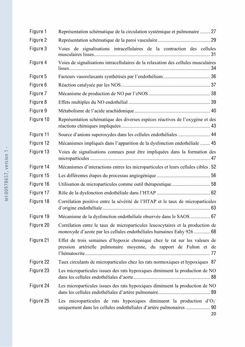
F igure 1 Représentation schématique de la circulation systémique et pulmonaire ........ 27
F igure 2 Représentation schématique de la paroi vasculaire .......................................... 29
F igure 3 Voies de signalisations intracellulaires de la contraction des cellules musculaires lisses .............................................................................................. 31
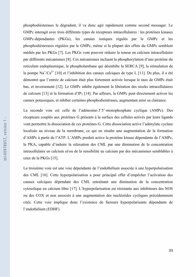
F igure 4 Voies de signalisations intracellulaires de la relaxation des cellules musculaires lisses .................................................................................................................. 34
F igure 5 Facteurs ...................................... 36
F igure 6 Réaction catalysée par les NOS ........................................................................ 37
F igure 7 .................................................. 38
F igure 8 Effets multiples du NO endothélial .................................................................. 39
F igure 9 ............................................................. 40
F igure 10 Représréactions chimiques impliquées ........................................................................ 43
F igure 11 .......................... 44
F igure 12 le ........ 45
F igure 13 Voies de signalisations connues pour être impliquées dans la formation des microparticules ................................................................................................. 47
F igure 14 s cellules cibles . 52
F igure 15 Les différentes étapes du processus angiogénique ........................................... 56
F igure 16 Utilisation de microparticules comme outil thérapeutique ............................... 58
F igure 17 ........................................... 62
F igure 18 microparticules ....................................................................................... 63
F igure 19 Mécanisme de la dysfonction endothéliale observée dans le SAOS ................ 67
F igure 20 Corrélation entre le taux de microparticules leucocytaires et la production de ............. 68
F igure 21 Effet de trois pression artérielle pulmonaire moyenne, du rapport de Fulton et de
..................................................................................................... 77
F igure 22 Taux circulants de microparticules chez les rats normoxiques et hypoxiques 87
F igure 23 Les microparticules issues des rats hypoxiques diminuent la production de NO .............................................................. 88
F igure 24 Les microparticules issues des rats hypoxiques diminuent la production de NO .......................................... 89
F igure 25 Les microparticules 2-
.................... 90
tel-0
0978
657,
ver
sion
1 -

#!"
"
F igure 26 Les microparticules hypoxiques diminuent la relaxation dépendante de .................................................................................................... 92
F igure 27 Réactivité vasculaire chez les rats normoxiques et injectés par les microparticules hypoxiques .............................................................................. 94
F igure 28 Mécanismes impliqués dans la dysfonction endothéliale induite par les microparticules ...................................................... 101
F igure 29 -hypopnées (IAH) et le nombre de microparticules issues des leucocytes activées (CD62L+) .............................. 110
F igure 30 Les microparticules SAOS augmentent la réponse contractile aux différents agents vasoconstricteurs ................................................................................. 112
F igure 31 Implication de la voie du NO rréactivité induite par les microparticules SAOS .................................................................................... 114
F igure 32 microparticules SAOS .............................................................................................................. 115
F igure 33 microparticules SAOS .................................................................................... 117
F igure 34 Microparticules SAOS et métabolites des COX ............................................. 118
F igure 35 2 microparticules SAOS .................................................................................... 119
F igure 36 Interaction entre les voies du NO et des COX ................................................ 121
F igure 37 Mécanismes impliquées dans la dysfonction vasculaire induite par les microparticules SAOS .................................................................................... 126
F igure 38 Taux plasmatique de VEGF............................................................................ 132
F igure 39 Expression du VEGF dans les microparticules SAOS ................................... 134
F igure 40 microparticules SAOS -1 ....................... 136
F igure 41 Implication des microparticules SAOS dans le processus angiogénique ....... 138
"
Tableau 1 Classification clinique des hypertensions pulmonaires .................................... 60
Tableau 2 Caractéristiques cliniques des patients ........................................................... 109
Tableau 3 Taux de microparticules circulantes ............................................................... 110
Tableau 4 Caractéristiques cliniques des patients ........................................................... 133
Tableau 5 Anticorps utilisés en Western Blot ................................................................. 146
Tableau 6 Composition des solutions salines de Krebs et hyperpotassique .................... 147
tel-0
0978
657,
ver
sion
1 -

##"
"
A V A N T PR OPOS
tel-0
0978
657,
ver
sion
1 -
#$"
"
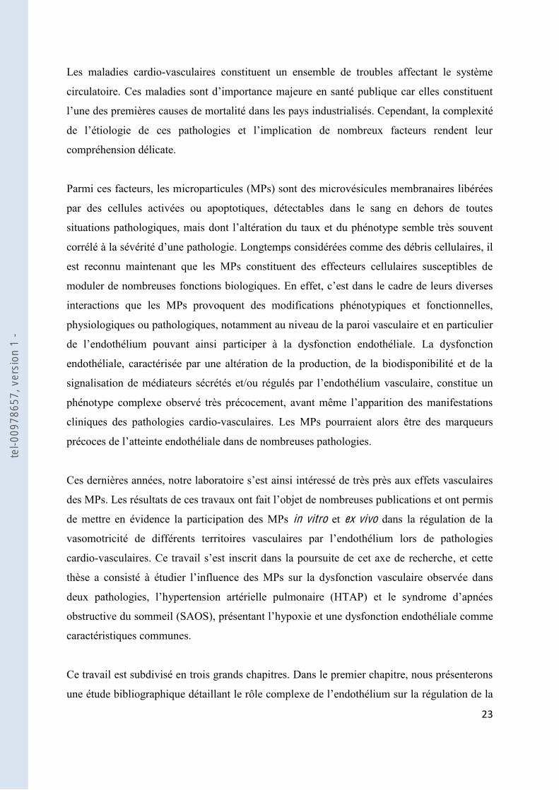
Les maladies cardio-vasculaires constituent un ensemble de troubles affectant le système
circulatoire. Ces maladies
ans les pays industrialisés. Cependant, la complexité
compréhension délicate.
Parmi ces facteurs, les microparticules (MPs) sont des microvésicules membranaires libérées
par des cellules activées ou apoptotiques, détectables dans le sang en dehors de toutes
situations pathologiques, mais taux et du phénotype semble très souvent
Longtemps considérées comme des débris cellulaires, il
est reconnu maintenant que les MPs constituent des effecteurs cellulaires susceptibles de
moduler de nombreuses fonctions biologiques. est dans le cadre de leurs diverses
interactions que les MPs provoquent des modifications phénotypiques et fonctionnelles,
physiologiques ou pathologiques, notamment au niveau de la paroi vasculaire et en particulier
pouvant ainsi participer à la dysfonction endothéliale. La dysfonction
endothéliale, caractérisée par une altération de la production, de la biodisponibilité et de la
, constitue un
cliniques des pathologies cardio-vasculaires. Les MPs pourraient alors être des marqueurs
précoces
ainsi intéressé de très près aux effets vasculaires
des MPs. Les résu nombreuses publications et ont permis
de mettre en évidence la participation des MPs in vitro et ex vivo dans la régulation de la
gies
cardio-vasculaires. , et cette
e dans
deux pathologies, et le
obstructive du sommeil (SAOS) et une dysfonction endothéliale comme
caractéristiques communes.
Ce travail est subdivisé en trois grands chapitres. Dans le premier chapitre, nous présenterons
une étude bibliographique détaillant le rôle régulation de la
tel-0
0978
657,
ver
sion
1 -

#%"
"
fonction vasculaire, les MPs en tant que vecteur biologique, et nous présenterons également
les deux pathologies hypoxiques étudiées. Le deuxième chapitre de ce manuscrit sera
cons exposition de la méthodologie suivie, aux résultats expérimentaux et aux
commentaires apportés. Nous déterminerons si les MPs participent à la dysfonction
tion de la réactivité vasculaire présente dans le SAOS. Dans un troisième temps, nous
observée dans le SAOS. Enfin, le troisième chapitre sera consacré à une synthèse de nos
résultats les plus significatifs et à la présentation des perspectives de notre travail.
tel-0
0978
657,
ver
sion
1 -

#&"
"
D O NN E ES BIB L I O G R APH I Q U ES
tel-0
0978
657,
ver
sion
1 -

#'"
"
I . L e système vasculaire et ses fonctions
1. La ci rculation sanguine
Décrit pour la première fois en 1628 par William Harvey dans son ouvrage Exercitatio
Anatomica de Motu Cordis et Sanguinis in animalibus, le système vasculaire comprend le
des veines et des veinules. Ce système constitue le premier organe qui se met en place durant
et les nutriments indispensab
transportant au niveau des reins ou des poumons. Ce système forme également un réseau de
1.1. O rganisation et rôles
assurant la
(Figure 1).
1.1.1. La ci rculation systémique ou grande ci rculation
Dans la circulation systémique, le sang sort du ventricule gauche du aorte pour
acheminer le sang vers les principales artères élastiques. Le sang progresse ensuite dans les
artères musculaires qui irriguent tous les organes et tissus de l'organisme. À proximité des
organes, les artères musculaires se ramifient en artérioles puis à l'intérieur même de l'organe,
les artérioles se ramifient en capillaires.
Ce circuit parcourt l'organisme en
entier; c'est pourquoi on parle de la grande circulation. Son rôle est essentiel étant donné qu'il
permet aux cellules d'être en contact avec les capillaires au niveau desquels les échanges sont
possibles.
tel-0
0978
657,
ver
sion
1 -

#("
"
F igure 1 : Représentation schématique de la ci rculation systémique et pulmonaire.
1.1.2. La ci rculation pulmonaire ou petite ci rculation
La circulation pulmonaire amène le sang veineux, pauvre en oxygène au contact des alvéoles
e, issue du ventricule droit, pour atteindre le lit capillaire pulmonaire où les
via les veines
pulmonaires (Figure 1). Les capillaires formés autour des alvéoles se raccordent aux veines
stance, basse
fois par des facteurs actifs (vasomotricité) et passifs (pression artérielle pulmonaire). Les
O2
O2
CO2
CO2
Veines
Veine cave
Ventricule droit
Ventricule gauche
Artérioles
Artères
Aorte
Artère pulmonaire
Veines pulmonaires
Muscle squelettique
Capillaires systémique
Capillaires pulmonaires
tel-0
0978
657,
ver
sion
1 -

#)"
"
vaisseaux pulmonaires ont une abondante innervation autonome, et la stimulation des
ganglions orthosympathiques peut diminuer de 30 % le débit sanguin pulmonaire. Ces
vaisseaux répondent également aux divers agents humoraux [2].
hypoxie par
par la quantité de dioxygène (O2) présente. Ce système permettant de détecter la diminution
2 est constitué par les muscles lisses des petites artères pulmonaires qui
2. Ces canaux sont responsables de la
émique. Il en va autrement pour les artères systémiques,
qui contiennent des canaux potassiques activés par dénosine- - triphosphate (ATP), en cas
vasodilatation.
1.1.3. Morphologie et histologie de la paroi artérielle
Le système vasculaire est complexe, non homogène et compartimenté en trois tuniques
concentriques distinctes. Cependant, les parois vasculaires possèdent toutes un modèle
intima, la
media
spécifiques (Figure 2).
intima est la tunique la plus interne et la plus fine. Elle est en contact direct avec la
est constituée par une monocouche de cellules endothéliales. Cet endothélium représente ainsi
la partie de la paroi vasculaire la plus exposée aux forces mécaniques mais également aux
nombreuses fonctions physiologiques fondamentales telles que le contrôle du tonus
vasomoteur, le trafic des cellules sanguines, la balance hémostatique, la perméabilité
intima est séparée de la media par la limitante élastique interne.
tel-0
0978
657,
ver
sion
1 -

#*"
"
F igure 2 : Représentation schématique de la paroi vasculaire ( Sanofis-Aventis®)
La media
cellules musculaires lisses (CML) arrangées de manière circulaire en couche dans une matrice
et de fibres de collagènes. Le nombre de ces couches varient suivant le
Les CML perçoivent différents signaux et leur état contractile définit le tonus vasomoteur
contrôlant le diamètre, modifie la distribution du débit sanguin et permet la répartition du flux
media
que dans les vaisseaux de gros
calibres.
environnant qui protège et ancre les artères aux structures avoisinantes. Son épaisseur varie
selon le type vasculaire et l
organisé, riche en collagène et en fibres élastiques, et contenant des fibroblastes et des
adipocytes. Grâce aux fibroblastes et aux fibres nerveuses qui rejoignent les fibres
musculaires lisses de la media
qui participent au contrôle du calibre vasculaire et de la vasomotricité. Dans les vaisseaux de
Limitante élastique interne
Tissus conjonctif
EndothéliumIntima
Tissus conjonctif
Limitante élastique externe
Cellules musculaires lissesMedia
Adventice
tel-0
0978
657,
ver
sion
1 -

$+"
"
vasa vasorum,
-même et la partie externe de la media.
équivalente à 30 % de
pulmonaires présentent une structure plus élastique que celle des vaisseaux systémiques [2].
1.2. Mécanisme de régulation de la vasomotricité
CML détermine le tonus vasomoteur et reflète
contrôlé par des stimuli physiques ou chimiques.
1.2.1. Contraction des cellules musculaires lisses
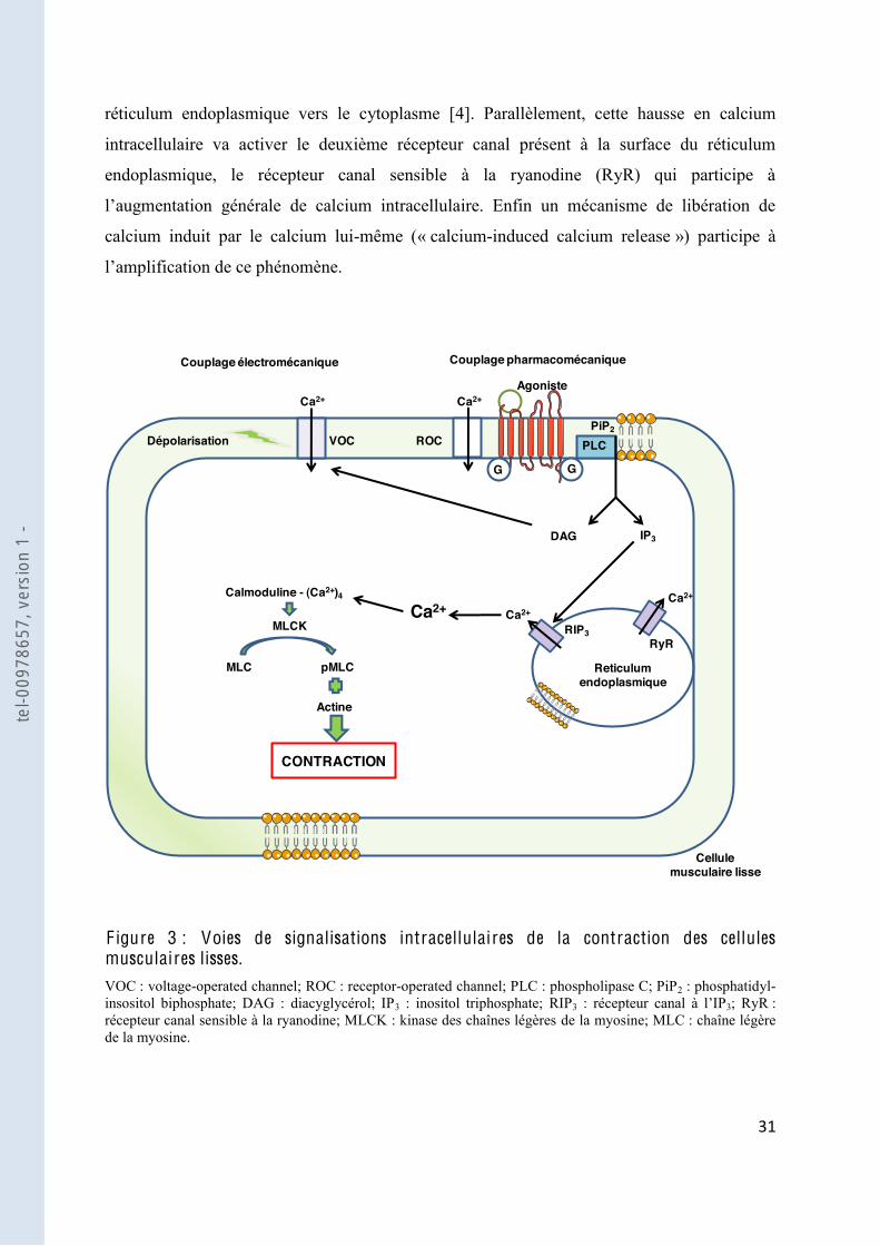
Le taux de contraction des CML dépend de la concentration intracellulaire en calcium. La
provient soit des réserves du réticulum endoplasmique, soit du milieu extracellulaire. Deux
voies conduisent ainsi à une augmentation rapide, importante et transitoire du calcium
intracellulaire nécessaire à la contraction des CML : le couplage électromécanique et le
couplage pharmacomécanique [3] (Figure 3).
Le couplage électromécanique consiste en une dépolarisation de la membrane cellulaire qui
voltage-operated channel »
(VOC) à travers lesquels le calcium entre dans la cellule pour initier la contraction.
membranaire conduisant à une augmentation du calcium intracellulaire, soit par activation de
« receptor-operated channel » (ROC), soit par libération du calcium à partir des réserves
intracellulaires.
Les récepteurs des agonistes vasoconstricteurs sont couplés à une protéine G couplée à une
phospholipase C (PLC). La PLC hydrolyse le phosphatidyl-inositol biphosphate (PIP2) de la
bicouche phospholipidique en inositol triphosphate (IP3 3 se
3 (RIP3) et permet la libération des réserves calciques du
tel-0
0978
657,
ver
sion
1 -
$!"
"
réticulum endoplasmique vers le cytoplasme [4]. Parallèlement, cette hausse en calcium
intracellulaire va activer le deuxième récepteur canal présent à la surface du réticulum
endoplasmique, le récepteur canal sensible à la ryanodine (RyR) qui participe à
calcium induit par le calcium lui-même (« calcium-induced calcium release ») participe à
F igure 3 : Voies de signalisations intracellulaires de la contraction des cellules musculaires lisses. VOC : voltage-operated channel; ROC : receptor-operated channel; PLC : phospholipase C; PiP2 : phosphatidyl-insositol biphosphate; DAG : diacyglycérol; IP3 : inositol triphosphate; RIP3 3; RyR : récepteur canal sensible à la ryanodine; MLCK : kinase des chaînes légères de la myosine; MLC : chaîne légère de la myosine.
G G
Ca2+ Ca2+
Ca2+Ca2+
Ca2+
Agoniste
Couplage électromécanique Couplage pharmacomécanique
Reticulum endoplasmique
PiP2
PLC
IP3DAG
ROCVOCDépolarisation
RIP3RyR
MLC pMLC
MLCK
Calmoduline - (Ca2+)4
Actine
CONTRACTION
Cellule musculaire lisse
tel-0
0978
657,
ver
sion
1 -
$#"
"
(MLCK). Cette enzyme va phosphoryler la chaîne légère de la myosine (MLC), stimuler
contraction de la CML.
ux de
augmentation de la sensibilité des protéines contractiles au calcium et implique
la phosphatase de la chaîne légère de la myosine (MLCP) [5].
1.2.2. Relaxation des cellules musculaire lisses
Les mécanismes conduisant à une diminution de la sensibilité au calcium et à une diminution
de sa concentration intracellulaire aboutis
une stimulation de la MLCP. La MLCP est très active dans les CML. Dès que la
phosphorylation par la MLCK diminue, la MLCP déphosphoryle la myosine et ainsi inhibe
les interactions myosine-actine. Si la ba
diminution de calcium, le fonctionnement de la MLCP est indépendant du calcium. La baisse
de la concentration en calcium se produit soit par expulsion du calcium intracellulaire vers
llule, soit par recaptage du calcium par les ATPases du reticulum
sarcoplasmique, les « sarco endoplasmic reticulum ATPases » (SERCA). Trois voies
interviennent dans la relaxation des CML : deux voies impliquant des nucléotides cycliques,
la voie de la gua
hyperpolarisation des CML (Figure 4).
La voie de la guanylate cyclase soluble (GCs)-guanosine- -monophosphate cyclique
(GMPc) est une des voies majeures impliquée dans la relaxation du tonus vasculaire. Le
diffuse dans les CML pour stimuler la GCs, enzyme clef de la régulation cardio-vasculaire. La
fixation du NO sur le fer du noyau héminique ac
guanosine- -triphosphate (GTP) en GMPc [6]. Une seconde voie de synthèse du GMPc
consiste en la fixation des peptides natriurétiques sur le récepteur membranaire de la GCs.
tel-0
0978
657,
ver
sion
1 -
$$"
"
phosphodiésterases le dégradant, il va donc agir rapidement comme second messager. Le
GMPc interagit avec trois différents types de récepteurs intracellulaires : les protéines kinases
GMPc-dépendantes (PKGs), les canaux ioniques régulés par le GMPc et les
phosphodiésterases régulées par le GMPc, même si la plupart des effets du GMPc semblent
médiés par les PKGs [7]. Les PKGs vont pouvoir réduire la teneur en calcium intracellulaire
par différents mécanismes [8]. Ces méca
reticulum endoplasmique, le phospholambane qui désinhibe la SERCA [9], la stimulation de
la pompe Na+/Ca2+ [10 1]. De plus, il a été
e de calcium était plus fortement activée lorsque le taux de GMPc était
bas, et inversement [12]. Le GMPc inhibe également la libération des stocks intracellulaires
de calcium [13 3 [14]. Par ailleurs, la GMPc peut directement activer les
canaux potassiques, et inhiber certaines phosphodiestérases, augmentant ainsi sa clairance.
adénosine- -monophosphate cyclique (AMPc). Des
récepteurs couplés aux protéines G présents à la surface des cellules activés par leurs ligands
ate cyclase
localisée au niveau de la membrane, ce qui en résulte une augmentation de la formation
intracellulaire en calcium et/ou de la sensibilité au calcium par des mécanismes semblables à
ceux de la PKGs [15].
La troisième voie
des CML [16
canaux calciques dépendant des CML entraînant une diminution de la concentration
cytosolique en calcium libre [17
ou des COX et non associés à une augmentation des nucléotides cycliques précédemment
l
tel-0
0978
657,
ver
sion
1 -
$%"
"
Ca2+
Reticulum
SERCA
Ca2+PLB
pMLC MLC
MLCP
RELAXATION
3Na+
GCs Ac
GCs
GTP GMPc ATP AMPcGTP
GMPc
NO
PKAPKGsCa2+L
Ca2+
K+
K+EDHF
NPAgonistes,
PGI2,PGE2
hyperpolarisation
K+
Cellule musculaire lisse "
F igure 4 : Voies de signalisations intracellulaires de la relaxation des cellules musculaires lisses. NP : peptide natriurétique; NO 2 : prostacycline; PGE2 : prostaglandine; GCs : guanylate cyclase soluble; GTP : guanosine- -triphosphate; GMPc : guanosine- -monophosphate cyclique; PKGs : protéines kinases GMPc-dépendantes; AC : adénylate cyclase; ATP : adénosine- - triphosphate; AMPc : adénosine- -monophosphate cyclique; PKA : protéine kinase A; PLB : phospholambane; SERCA : sarco endoplasmic reticulum ATPases; EDHF MLCK : kinase des chaînes légères de la myosine; MLC : chaîne légère de la myosine.
tel-0
0978
657,
ver
sion
1 -

$&"
"
2.
ium vasculaire est considéré comme un organe à part entière et forme la barrière
cellulaire intérieure qui limite les vaisseaux sanguins [18]. Dans des conditions pathologiques,
nction
endothéliale.
2.1. H étérogénéité structurelle
Les cellules endothéliales (CEs) qui composent cette barrière varient selon leur localisation
dans le système vasculaire impliquant une hétérogénéité structurelle et fonctionnelle de
des vaisseaux sanguins de rats a montré que les CEs aortiques sont plutôt longues et étroites
[19] fférents rôles en sécrétant différentes substances
suivant les stimuli reçus par les CEs.
2.2.
Le tonus vasculaire est régulé par de nombreux facteurs vasoconstricteurs et vasodilatateurs
um joue ainsi un rôle crucial en maintenant un équilibre entre
vasoconstriction et vasodilatation [20].
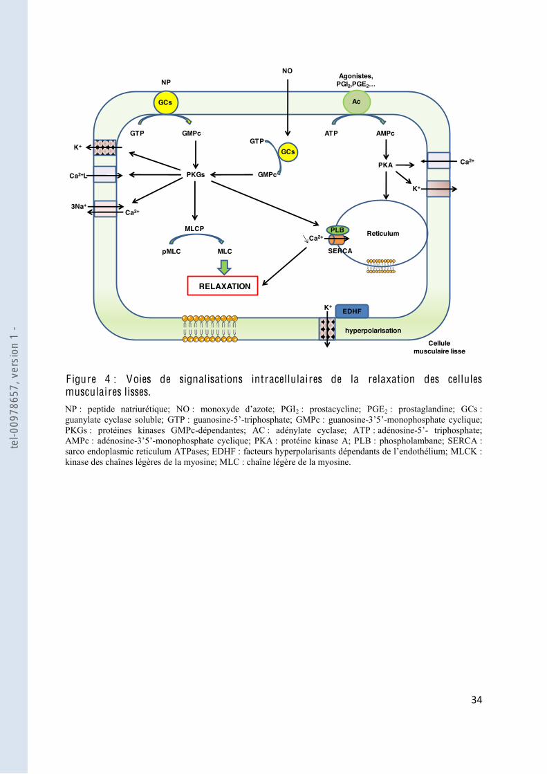
2.2.1. F
prostacycline (PGI2) (Figure 5).
tel-0
0978
657,
ver
sion
1 -
$'"
"
RELAXATION
RELAXATION
Hyperpolarisation
K+ PGI2 NO
eNOSCOX
Acide arachidonique L-arginine
GCsAc GMP
GMPc
AMP
AMPc
EDHF
Cellule endothéliale
Cellules musculaires lisses
"
F igure 5 EDHF ; COX : cyclooxygénase; PGI2 : prostacycline; AC : adénylate cyclase; ATP : adénosine- - triphosphate; AMPc : adénosine- -monophosphate cyclique; eNOS ; NO ; GCs : guanylate cyclase soluble; GTP : guanosine- -triphosphate; GMPc : guanosine- -monophosphate cyclique.
"
En 1980, Furchgott et Zawadzki ont réussi à démontrer que la relaxation des CML en réponse
[21]. Ce facteur
appelé facteur comme étant le
2], Ignarro [23] et Palmer
[24]. Cette découverte fondamentale dans la compréhension de la physiologie de
Le NO est une molécule gazeuse capable de diffuser rapidement à travers les membranes
ouant un
-vie, le NO est un
très faible quantité, le NO est libéré de façon continue par les CEs car les forces de
cisaillement du flux sanguin laminaire constituent un puissant activateur de la transcription du
gène de la eNOS.
tel-0
0978
657,
ver
sion
1 -
$("
"
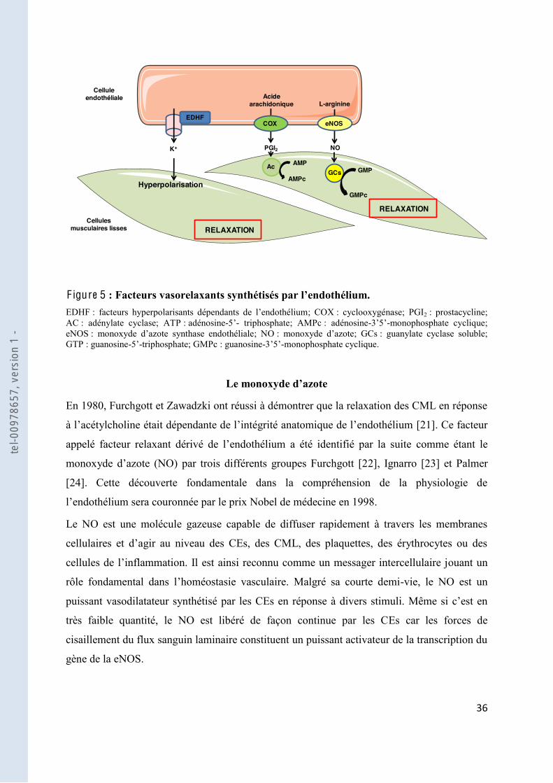
-arginine en L-citrulline par
la NO synthase (NOS) [25] (Figure 6). Les NOS sont des métalloenzymes dont trois
isoformes ont été décrites dans la littérature. Deux isoformes sont constitutivement actives, les
NOS endothéliale (eNOS) et neuronale (nNOS) et la troisième est une NOS inductible
(iNOS). Généralement, les NOS constitutives possèdent une activation dépendante du calcium
et ont donc une activité rapide, de courte durée avec une faible quantité de NO produite, alors
que la iNOS voit son expression augmenter en réponse à divers stimuli afin de produire de
forte quantité de NO sur une longue durée [27].
"
"
"
"
F igure 6 A lderton et al. [26]).
La eNOS est une protéine exprimée de manière importante dans les CEs de vaisseaux
peut être régulée au niveau de son expression
la cavéoline-1, une protéine présente dans les cavéoles qui joue un rôle dynamique dans le
trafic des protéines membranaires avec les compartiments intracellulaires. Cette protéine
séquestre à la membrane la eNOS et la maintient dans un état de faible activité. Lorsque la CE
est
eNOS-cavéoline-1. La eNOS libérée est alors accessible à différentes kinases qui vont
phosphorylation de la eNOS joue un rôle important dans la régulation de son activité : la
phosphorylation du résidu sérine 1177 conduit à son activation alors que celle sur le résidu
de résidus sérine par la voie phosphatidyl-inositol 3 kinase (PI3K)/Akt [29].
L-Arginine L-CitrullineN -Hydroxy-L-Arginine
tel-0
0978
657,
ver
sion
1 -

$)"
"
NO
Ca2+
Cellule endothéliale
eNOS
PI3K
Akt
eNOSeNOSCav-1 Cav-1
L-Arginine L-Citrulline
Ca2+
Calmoduline-Ca2+
Ca2+
Stimuli physique ou chimique
Toda et al. [28]). PI3K : phosphatidyl-inositol 3 kinase; Cav-1 : cavéoline-1; eNOS NO
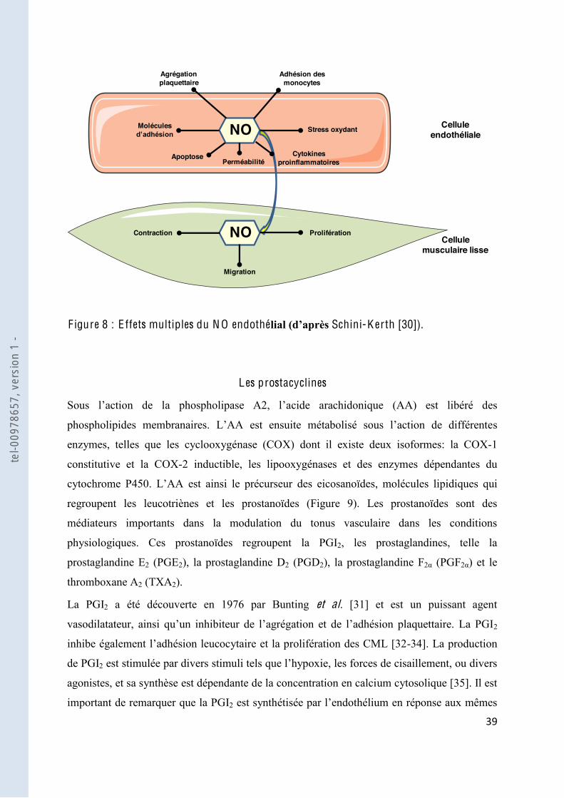
Dans le système cardio-vasculaire, les effets biologiques locaux du NO endothélial sont
multiples et lui confère un rôle vasculo-protecteur majeur (Figure 8). Une fois produit, il
diffuse très rapidement à la rencontre des CML, des macromolécules et des cellules
des
GMPc, il inhibe la contraction. Le NO peut également inhiber la migration, la prolifération et
la synthèse de protéines de la matrice extracellulaire, limitant ai
intima. Le NO exerce également des effets sur la CE où il est capable de moduler le
cellules.
tel-0
0978
657,
ver
sion
1 -
$*"
"
F igure 8 : E ffets multiples du N O endothé Schini-K erth [30]).
L es prostacyclines
(AA) est libéré des
s
enzymes, telles que les cyclooxygénase (COX) dont il existe deux isoformes: la COX-1
constitutive et la COX-2 inductible, les lipooxygénases et des enzymes dépendantes du
est ainsi le précurseur des eicosanoïdes, molécules lipidiques qui
regroupent les leucotriènes et les prostanoïdes (Figure 9). Les prostanoïdes sont des
médiateurs importants dans la modulation du tonus vasculaire dans les conditions
physiologiques. Ces prostanoïdes regroupent la PGI2, les prostaglandines, telle la
prostaglandine E2 (PGE2), la prostaglandine D2 (PGD2), la prostaglandine F (PGF ) et le
thromboxane A2 (TXA2).
La PGI2 a été découverte en 1976 par Bunting et al. [31] et est un puissant agent
2
inh -34]. La production
de PGI2
agonistes, et sa synthèse est dépendante de la concentration en calcium cytosolique [35]. Il est
important de remarquer que la PGI2
Cellule endothéliale
Cellule musculaire lisse
NO
NO Prolifération
Migration
Contraction
PerméabilitéApoptose
Molécules
Agrégation plaquettaire
Adhésion des monocytes
Stress oxydant
Cytokines proinflammatoires
tel-0
0978
657,
ver
sion
1 -

%+"
"
stimuli impliqués dans la libération du NO suggérant une action synergique sur les différentes
cellules cibles [36,37]. La PGI2 va diffuser librement et se fixer sur le récepteur à la PGI2
présent sur les cellules cibles [38]. Ces récepteurs vont ensuite activer via
- 2 participe
e de différents canaux potassiques renforçant la relaxation des CML
[39].
Phospholipides membranaires
Acide arachidonique
Voie enzymatique
Phospholipase
Cyclooxygénase 5-Lipooxygénase
Prostaglandines Leucotriènes
Voie non enzymatique 8-isoprostane
PGI2 TXA2
Métabolisme oxydatif
PGD2 PGE2
Vasodilatation
Vasodilatation Vasodilatation
Vasocontraction
Vasocontraction
F igure 9 : PGI2 : prostacycline; TXA2 : thromboxane A2; PGD2 : prostaglandine D2; PGE2 : prostaglandine E2.
tel-0
0978
657,
ver
sion
1 -
%!"
"
L es facteurs hyperpolarisants
structures chimiques de ces EDHF ne sont pas encore connues et semblent être variables selon
vaisseaux de résistances (comme les artères mésentériques), il apparait que la contribution des
EDHF est plus importante dans les petites artères, et semble inversement proportionnelle à la
taille du vaisseau [40]. Plusieurs candidats ont ainsi démontré leurs effets tels que le péroxyde
les ions potassiques [42].
vasculaire et les stimuli physiologiques ou pharmacologiques utilisés [43], mais il existe des
interactions entre les différents fact
biodisponibilité du NO est réduite, ce qui permet de maintenir partiellement les capacités
2.2.2.
maintien du tonus vasculaire, mais qui sont particulièrement exprimés dans les conditions
e la contraction. Par ailleurs, des interactions entre les voies intracellulaires des agents
libérer la production des autres. Il existe de nombreux facteurs contracturants produits par la
(ET) et
au TXA2.
tel-0
0978
657,
ver
sion
1 -

%#"
"
ET est un puissant vasoconstricteur qui permet le maintien du tonus vasculaire basal, et
dont la structure
aminés qui possèdent au moins trois isoformes, respectivement nommés ET-1, ET-2 et ET-3.
Les isoformes agissent en se liant à des récepteurs membranaires dont il existe au moins deux
types distincts ETA et ETB ET à son récepteur active
ET -1
peut également agir sur des récepteurs endothéliaux ETB et induire une vasorelaxation en
[46] que de la circulation pulmonaire [47].
L e Thromboxane A2
Le TXA2 est un prostanoïde vasoconstricteur produit en faible quantité par les plaquettes et
les vaisseaux systémiques. Le TXA2 libéré va alors se fixer aux récepteurs thromboxane (TP)
des CML qui vont permettre une augmentation de la quantité et de la sensibilisation au
de TXA2 est masquée par la production de facteurs vasorelaxants [48]. Dans les conditions
pathologiques, le TXA2 peut cependant con
récepteurs TP 2
sont plus complexes. En effet, le TXA2
tel-0
0978
657,
ver
sion
1 -
%$"
"
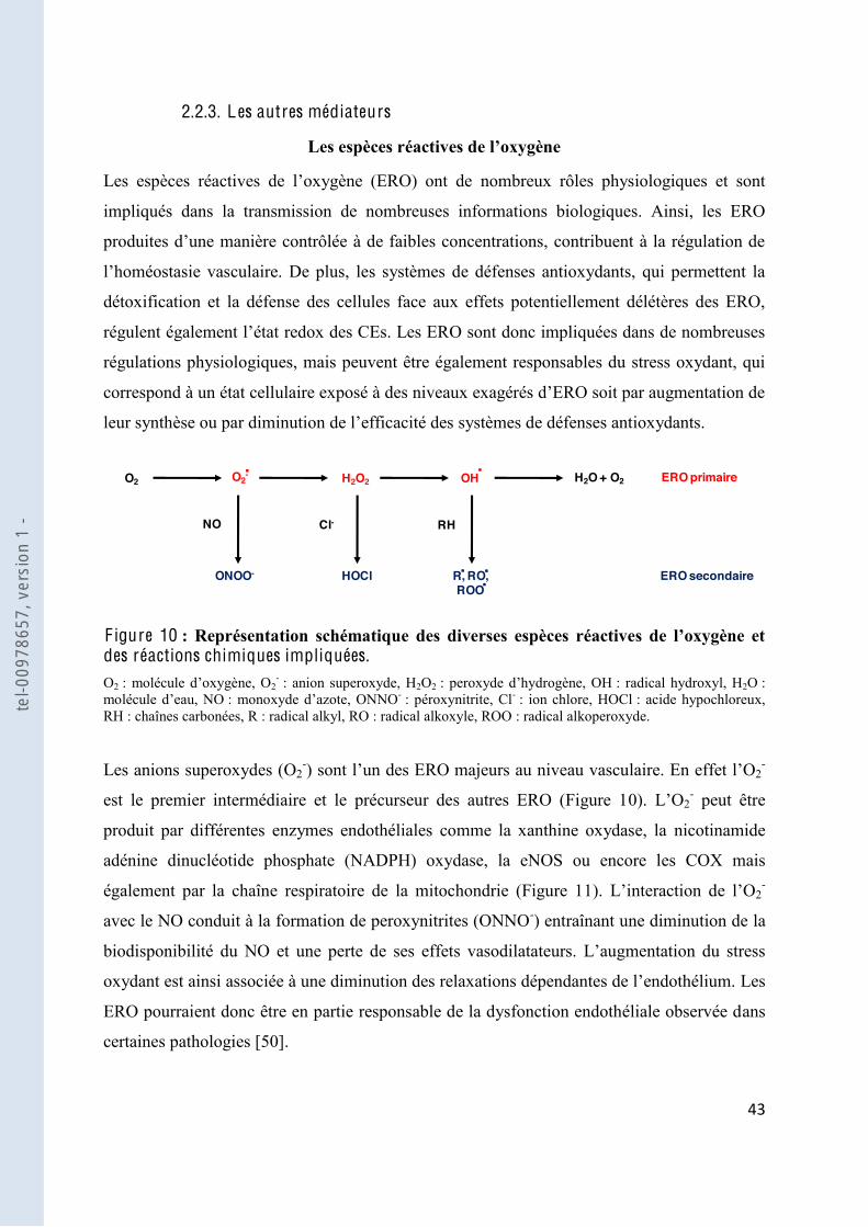
2.2.3. L es autres médiateurs
nombreux rôles physiologiques et sont
impliqués dans la transmission de nombreuses informations biologiques. Ainsi, les ERO
s systèmes de défenses antioxydants, qui permettent la
détoxification et la défense des cellules face aux effets potentiellement délétères des ERO,
régulations physiologiques, mais peuvent être également responsables du stress oxydant, qui
O2 O2- H2O2 OH H2O + O2
.
.
NO Cl-
ONOO- HOCl
RH
R, RO, ROO
.. ERO secondaire
ERO primaire.
"
F igure 10 des réactions chimiques impliquées. O2 2
- : anion superoxyde, H2O2 : radical hydroxyl, H2O : : monoxy - : péroxynitrite, Cl- : ion chlore, HOCl : acide hypochloreux,
RH : chaînes carbonées, R : radical alkyl, RO : radical alkoxyle, ROO : radical alkoperoxyde.
Les anions superoxydes (O2-) 2
-
est le premier intermédiaire et le précurseur des autres ERO (Figure 10). L 2- peut être
produit par différentes enzymes endothéliales comme la xanthine oxydase, la nicotinamide
adénine dinucléotide phosphate (NADPH) oxydase, la eNOS ou encore les COX mais
également par la chaîne respiratoire de la mitochondrie (Figure 11 2-
avec le NO conduit à la formation de peroxynitrites (ONNO-) entraînant une diminution de la
biodisponibilité du NO et une perte de ses effets vasodilata
Les
ERO pourraient donc être en partie responsable de la dysfonction endothéliale observée dans
certaines pathologies [50].
tel-0
0978
657,
ver
sion
1 -

%%"
"
Xanthine oxydase
Mitochondrie
NADPH oxydase
Cytochrome P450
eNOS découplée
O2-
H2O2
H2O
Superoxyde dismutase
Catalase
CytokineLipopolysaccharide
Facteur decroissance
InsulineLeptine
LDL oxydésAcides gras libres
Ischémie-reperfusionHypoxie-réoxygénation
Agonistes des récepteurs couplés aux
protéines G
Stress
F igure 11 O2
- : anion superoxyde, H2O2 2O .
L es microparticules
Les MPs, de petites vésicules membranaires libérées théoriquement par tous les types
cellulaires, sont présentes dans le sang de sujets sains, et des taux élevés de MPs de
différentes origines ont été retrouvés dans de nombreuses pathologies cardio-vasculaires. Les
MPs sont maintenant considérées comme de véritables effecteurs biologiques entraînant de
ésentées plus en détail dans la seconde partie
de ces données bibliographiques.
tel-0
0978
657,
ver
sion
1 -

%&"
"
2.3. La dysfonction endothéliale
grité vasculaire.
: on peut parler de dysfonction
endothéliale. Cette dysfonction est caractérisée par une diminution de facteurs vasorelaxants
également être due à une perte de sensibilité des CML aux facteurs vasorelaxants ou encore à
la participation du stress oxydant (Figure 12).
European Society of Cardiology » indique clairement que parmi les
biomarqueurs biologiques, les MPs sont un des nouveaux moyens disponibles pour détecter la
dysfonction endothéliale [51].
F igure 12 L-Arg : L-arginine; L-Cit : L-citrulline; eNOS : monoxyde
O2- : anion superoxyde; EDCF 2 : thromboxane A2;
PGH2 : prostaglandine H2; ET-1 : endothéline-1.
Cellule endothéliale
Cellule musculaire lisse
Relaxation Contraction
TXA2, ET-1
eNOS
L-Arg L-Cit
O2-
EDCF
NO
tel-0
0978
657,
ver
sion
1 -
%'"
"
I I . L es microparticules ci rculantes
1. Découverte et définition
Le concept des MPs a vu le jour dans les années 1970 et décrivait les MPs comme des débris
inertes ( débris plaquettaires par Wolf [52] qui avait noté la présence de petits fragments
connaissances a ensuite mené la communauté scientifique à étudier leur signification et un
possible rôle joué par les MPs a été proposé par Batisda et al. [53].
Actuellement, les MPs sont définies comme de petites vésicules hétérogènes en taille, en
composition, et possédant des propriétés pro-inflammatoires et pro-coagulantes. La taille des
MPs, comprise entre 0.05 nm et 1 µm, est une caractéristique souvent utilisée pour les
distinguer des exosomes (<0.1 µm) et des plaquettes (> 1µm).
ont susceptibles de libérer des MPs, la
détermination de leur origine a établi que les MPs étaient principalement libérées à partir de la
membrane des cellules circulantes du plasma (plaquettes, érythrocytes, cellules B et T et
monocytes), des cellules de la paroi vasculaire (cellules endothéliales et cellules musculaires
lisses) et des cellules tumorales [54-56]. Cette position centrale des MPs suggère un rôle
général des MPs dans les régulations cellulaires.
2. Formation des microparticules
le, même si les mécanismes concernant la formation des MPs sont complexes
les deux principaux processus aboutissant à la formation des MPs. Il a été élégamment reporté
par le groupe de Jimenez que le phénotype et la quantité des MPs libérées varient suivant la
les cellules endothéliales
[57]. La figure 13 schématise ces deux voies décrites par la suite.
tel-0
0978
657,
ver
sion
1 -

%("
"
Scramblase
Floppase
Translocase
Ca2+
Désorganisation du cytosquelette
Clivage Taline/Actine
Intégrines
CalpaïneActivation des kinases
Inhibition des phosphatase
Activation cellulaire
,-
Apoptose
Caspase-2
ROCK II
Thrombine
TRAIL
NF- B
Caspase-3
Contractilité de la cellule
ROCK I
p-MLCK
PS
PS
PS
Recepteur
Ligand
Phosphatidylsérine
ARNm, ARNmi "
"
F igure 13 : Voies de signalisations connues pour être impliquées dans la formation des microparticules. TRAIL : tumor necrosis factor-related apoptosis inducing ligand; NF- : nuclear factor-kappa B; ROCK : Rho-associated protein kinase; MLCK : kinase de la chaîne légère de la myosine.
2.1. L
plitude
enzyme requise pour la désorganisation du cytosquelette [58]. Bien que la relation entre
calpaïne dans le processus de libération des MPs est toujours controversée. Wiedmer et al. ont
ainsi montré que la formation des MPs dérivées des plaquettes (PMPs) est dépendante de
calpaïne, suggérant un mécanisme alternatif de microvésiculation indépendant cette enzyme
[60].
Le bourgeonnement de la membrane et la microvésiculation sont souvent précédés par une
rie de la membrane due à des perturbations locales de la structure en
bicouche lipidique des cellules. Il en résulte une redistribution des aminophospholipides, telle
tel-0
0978
657,
ver
sion
1 -
%)"
"
que la phosphatidylsérine, du feuillet interne vers le feuillet externe de la membrane [61]. La
régulation de la distribution asymétrique des aminophospholipides est régie par trois activités
distinctes qui permettent la redistribution bidirectionnelle à travers la bicouche lipidique [62].
Le premier complexe est une aminophospholipide translocase qui permet le transport de la
phosphatidylsérine et de la phosphatidyléthanolamine du feuillet externe vers le feuillet
interne de la membrane plasmique contre le gradient de co
différentielle des aminophospholipides à travers la bicouche lipidique est gouvernée par une
floppase ATP-dépendante qui agit en coopération avec la translocase. Le troisième complexe,
la scramblase lipidique, peut rapidement déplacer les aminophospholipides entre les feuillets
membranaires par un mécanisme dépendant du calcium et peut aboutir à une perte de
scramblase et bloque la coopération entre la translocase et la floppase. Cela aboutit à une
externalisation de la phosphatidylsérine vers le feuillet externe de la membrane et est suivi par
la libération des MPs. Ainsi, la phosphatidylsérine est exprimée sur la plupart des MPs, et a
des conséquences fonctionnelles sur la stimulation du processus de coagulation en se liant au
domaine chargé positivement des protéines de la coagulation.
Par ailleurs, il a été montré une implication des protéines kinases dans la régulation de la
formation de érine
perturbation du cytosquelette [63-65]. Récemment, il a été reporté que la protéine kinase A
plaquettaires et dans érine, grâce à un mécanisme dépendant
Pour illustrer un peu plus cette complexité de la formation des MPs, des mécanismes
indépendants du calcium peuvent être également impliqués dans la formation des MPs. Ainsi
Cauwenberghs et al. [67] ont trouvé que la voie des intégrines IIb 3 est responsable de la
te ou indépendante du calcium) agissaient
séparément ou en synergie.
tel-0
0978
657,
ver
sion
1 -
%*"
"
2.2.
est
t dynamique de la
membrane [68] ant les phases
processus de mort cellulaire [69]. Les corps apoptotiques sont plus larges que les MPs et
le clivage de la caspase 3 mène à une activation indépendante de Rho des « Rho-associated
protein kinase » (ROCK) I, qui va permettre la contractilité de la cellule par la génération
-actine, le bourgeonnement de la membrane et la formation des MPs sans
phosphatidylsérine [71,72].
2.3. Autres mécanismes de formation
Finalement, un autre mécanisme se situant
vation cellulaire. Une équipe française a démontré que la stimulation de
cellules endothéliales par la thrombine, une enzyme multifonctionnelle, permettait la
génération de MPs par une voie impliquant la caspase-2 et RhoA/ROCK II, en absence de
mort cellulaire [73]
du « tumor necrosis factor-related apoptosis inducing ligand » (TRAIL) et de son récepteur
TRAIL-R2 (également impliqué dans la voie de signalisation des cellules apoptotiques [74])
dans la libération de MPs endothéliales en initiant le recrutement de protéines adaptatrices et
nuclear factor kappa B » (NF- B). Ils ont également mis à jour une nouvelle
en réponse à la thrombine [75].
Ces différentes données de la littérature soulignent clairement la complexité et les possibles
interactions entre les différentes voies aboutissant à la formation des MPs. Une meilleure
compréhension des mécanismes de microvésiculation (activation ou apoptose) contribuerait à
déterminer des thérapies dans le but de cibler directement les MPs et diminuer leur taux dans
tel-0
0978
657,
ver
sion
1 -

&+"
"
formation des MPs est que très souvent elles ont été réalisées in vitro sur des lignées
cellulaires avec des inhibiteurs pharmacologiques. Il est donc nécessaire de déterminer si les
mêmes voies de signalisation sont impliquées dans la production in vivo de MPs.
3. Composition et contenu des microparticules
p
formation. En effet, les MPs contiennent des composants membranaires, cytoplasmiques et
nucléaires de leur cellule précurseur, permettant leur caractérisation en utilisant des anticorps
3.1. Contenu protéique
ue
des protéines associées au cytosquelette [76], alors que les protéines retrouvées dans les PMPs
sont des glycoprotéines de surface ou des chemokines [77,78].
Cependant la composition globale en protéine des MPs peut être en relation avec leur stimulus
d in vitro par activation cellulaire (par la
phytohémagglutine (PHA) et la phorbol-myristate-acétate (PMA)) et par apoptose (par
ctinomycine D (ActD)) à partir de lignée de lymphocyte T CEM ou des lymphocytes de
patients diabétiques expriment à leur surface le morphogène Sonic Hedgehog (impliqué dans
le développement embryonnaire et adulte). Cependant le traitement de ces mêmes cellules
Sonic Hedgehog sur
les MPs [79]. De plus, la comparaison de la composition protéique des MPs obtenues par
activation cellulaire ou par apoptose montre que les MPs apoptotiques sont plus riches en
ctivation des
monocytes THP-1 avec le lipopolysaccharide ou par la P-sélectine permet la production de
-coagulante similaire mais qui diffèrent sur la composition
protéique [81].
tel-0
0978
657,
ver
sion
1 -
&!"
"
3.2. Contenu lipidique
Concernant la composition lipidique, la bicouche membranaire des MPs est principalement
formée de phospholipides, et présente une charge négative due à la présence de
phosphatidylsérine et de phosphatidyléthanolamine. La déplétion en cholestérol inhibe
phosphatidylsérine sur le feuillet exoplasmique de la membrane et la
vésiculation indique que ion des radeaux
lipidiques [65]. Dean et al. [82] ont récemment reporté que les composants actifs des PMPs de
moyenne taille étaient lipidiques mais leur nature reste à déterminer. De plus, plusieurs
protéines portées par les MPs. Par exemple, un enrichissement en cholestérol de monocytes
humain induit la génération de MPs au potentiel fortement pro-coagulant [83]
ces données laissent penser que les MPs issues des patients avec des pathologies métaboliques
peuvent avoir une composition lipidique différente et engendrer des effets fonctionnels
4. Interaction entre les microparticules et les cellules cibles
La communication intercellulaire est basée sur des cascades de signalisation qui requièrent un
Les MPs peuvent interagir avec la cellule cible par action sur un ligand exprimé à la surface,
transférer des récepteurs de surface, ou délivrer des protéines, des acides ribonucléiques
messagers (ARNm), des microARN (miARN) et des lipides bioactifs. De plus, elles peuvent
transporter des particules infectieuses (mécanisme du cheval de Troie) et délivrer des
reportés 4).
tel-0
0978
657,
ver
sion
1 -

&#"
"
1
2 3
4
PSPS
PSPS
PS
PS
Recepteur
Ligand
Phosphatidylsérine
Cellules cibles
ARNm, ARNmi
"
F igure 14 s microparticules et leur cellules cibles. 1 : interaction ligand-récepteur, 2 : fusion, 4 : internalisation.
4.1. Interaction ligand-récepteur
Plusieurs études ont démontré que les MPs pouvaient directement stimuler les récepteurs
exprimés sur les cellules cibles soit par interaction avec le ligand exprimé sur la surface des
MPs ou par des médiateurs relâchés par les MPs. Ainsi, les PMPs exprimant la P-sélectine
à la P-sélectine
glycoprotéine ligand-1 [84]. Par ailleurs, des travaux menés au sein du laboratoire ont montré
que les MPs exprimant Sonic Hedgehog favorisaient la différenciation des mégacaryocytes, la
de ces mêmes cellules tous ces effet
laboratoire a également pu mettre en évidence que les MPs issues des cellules T agissaient sur
les CML par la voie de signalisation Fas/Fas Ligand [87].
tel-0
0978
657,
ver
sion
1 -

&$"
"
4.2. T ransfert de composants
Une autre voie utilisée par les MPs
de différents composants pouvant affecter différentes fonctions de la cellule cible. Les MPs
peuvent notamment transférer des récepteurs à la surface de la cellule cible. Rozmyslowicz et
al. ont ainsi montré que les MPs jouaient un r
le co-récepteur à chémokine de type 4 (CXCR4)
aux cellules CD4+/CXCR4- [88].
COX-2 et d «inter-cellular adhesion molecule 1 » (ICAM-1) [89].
4.3. Fusion et internalisation
Finalement, les MPs peuvent aussi être absorbées par fusion ou par internalisation. La fusion
des MPs avec leurs cellules cibles mène à un transfert non sélectif des composants des MPs et
affecte les propriétés des cellules. La fusion des MPs riches en facteur tissulaire avec des
plaquettes activées augmentent ainsi fortement leur activité pro-
entre les MPs libérées par les progéniteurs endothéliaux et les intégrines 4 et 1 des cellules
endothéliales
différentes voies et ces différentes po
interagit-elle seulement avec une seule cellule ou peut-elle influencer plusieurs cellules avant
? De plus, il est important de noter que les technologies actuelles ne
permettent pas d -
toujours pas complètement connue. Récemment, il a été rapporté que les PMPs après
transfusion sanguine possédaient une demi-
une sévère thrombocytopénie [92]. On ne peut cependant exclure que le mécanisme
que du message porté et transféré par les MPs est essentiel pour comprendre leurs propriétés
pathogéniques ou bénéfiques.
tel-0
0978
657,
ver
sion
1 -
&%"
"
5. L es microparticules : biomarqueurs et effecteurs dans les pathologies
cardiovasculaires
une élévation de ce taux a été
reportée dans de nombreuses conditions pathologiques. La principale cible des MPs est le
système cardio-vasculaire. Les MPs circulantes isolées à partir du sang sont maintenant
métabolique. Dans ces maladies, un taux élevé de MPs a été reporté et est souvent corrélé à la
sévérité de la pathologie. Depuis quelques années, les MPs sont également considérées
comme de véritables effecteurs capables de véhiculer un message biologique aux cellules
cibles. Il est intéressant de note
nombre de MPs mais concernerait plutôt la composition .
5.1. Effets des microparticules sur la fonction vasculaire
Concernant les effets directs des MPs sur la fonction vasculaire, il a été proposé par de
nombreux groupes que les MPs circulantes affectaient la quantité de NO en diminuant sa
Ainsi, les MPs issues de patients s
prééclampsie induisent une dysfonction endothéliale par une détérioration de la voie de
transduction du NO endothélial [95-97]. Chez les patients souffrant du syndrome
Ps participe à la dysfonction endothéliale en diminuant
CEs
[98] et en augmentant les marqueurs plasmatiques du stress oxydant [99].
Les MPs circulantes peuvent aussi interagir avec les CML, induire une inflammation
vasculaire et modifier la contractilité des vaisseaux. Durant la prééclampsie, le taux circulant
augmentation de la production de NO via la iNOS et des dérivés vasoconstricteurs issus des
métabolites de la COX-2 [100].
La séparation des MPs selon leurs origines cellulaires montre
oconstricteurs
-
tel-0
0978
657,
ver
sion
1 -

&&"
"
durant la prééclampsie.
Ces résultats suggèrent une implication des MPs dans les mécanismes responsables
de complications cardiovasculaires
ication de cette dysfonction prééxistante.
5.2. Angiogenèse et microparticules
partir de vaisseaux préexistants. Au cours du processus angiogénique, la vasodilatation initiale
CEs, et de la dégradation de la membrane basale et de la matrice extracellulaire environnante.
Les CEs activées sont alors capables de migrer, de proliférer pour former de nouveaux tubes
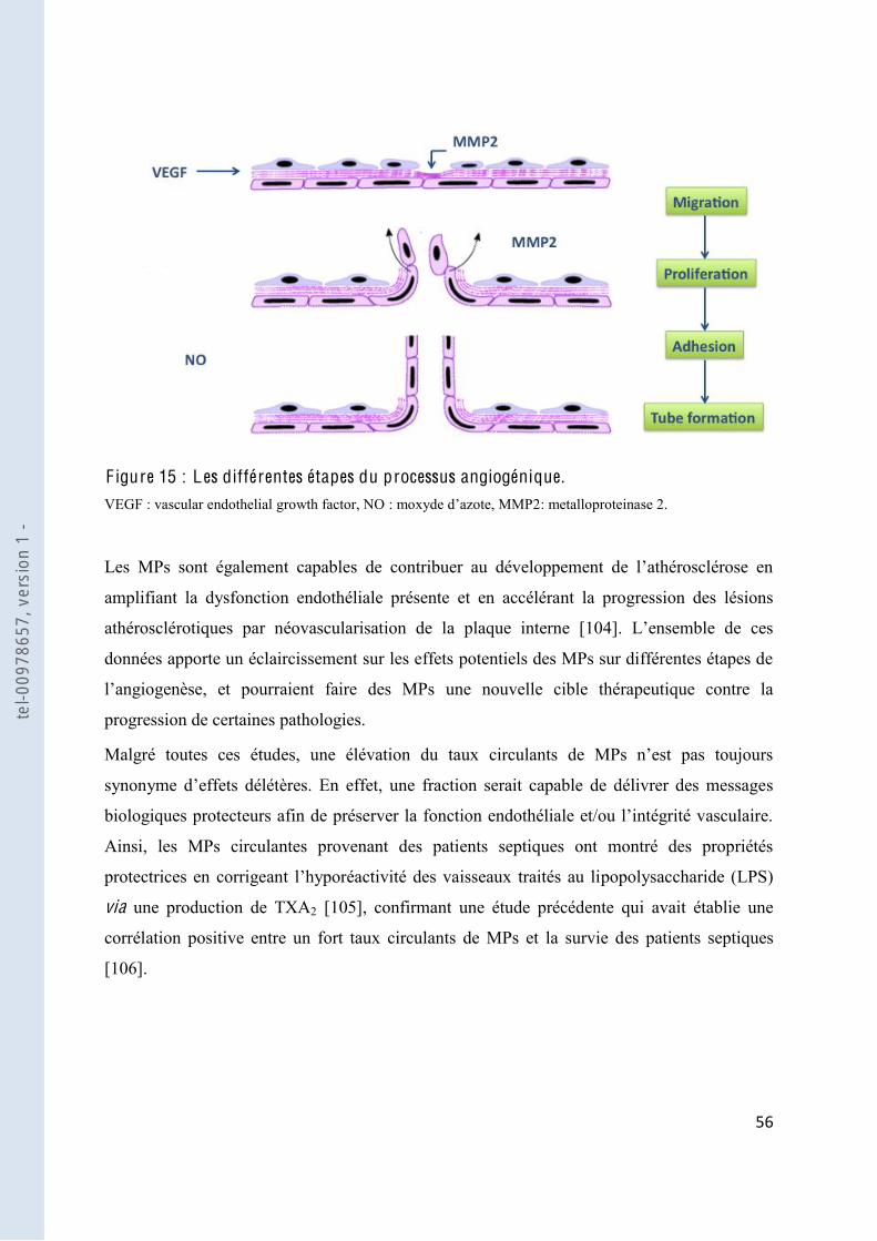
vasculaires (Figure 15 - ou anti-
« vascular endothelial growth factor » (VEGF), un des principaux facteurs pro-angiogénique,
est surexprimé. Un aspect clef des effets des MPs sur le système cardiovasculaire est leur
capacité à moduler le programme angiogénique à la fois des CEs matures et des cellules
progénitrices. Il est maintenant admis dans la littérature que les MPs sont capables
: le recrutement et la différenciation
des progéniteurs endothéliaux, la dégradation matricielle et la migration et la prolifération des
CEs. De plus, les effets des MPs seraient différentiels .
Taraboletti et al.
métalloprotéinases (MMPs) favorisaient la formation de structure tubulaire par les CEs
humaines, en r
[101]
« peroxysome proliferator-activated receptor » (PPAR ), portées par les MPs semblent
essentiels dans leur capacité à reprogrammer angiogéniquement les progéniteurs endothéliaux
via la voie de signalisation impliquant Akt et NF- B [102]. Récemment, les MPs isolées de
patients atteints de rétinopathie diabétique ont montré une capacité à stimuler la prolifération
des CEs et à augmenter la formation de nouveaux vaisseaux chez la souris, indiquant que ces
MPs pourraient contribuer à la progression de la pathologie grâce à leur capacité pro-
angiogénique [103].
tel-0
0978
657,
ver
sion
1 -
&'"
"
"
F igure 15 : L es différentes étapes du processus angiogénique. VEGF : vascular endothelial growth factor, NO : metalloproteinase 2.
amplifiant la dysfonction endothéliale présente et en accélérant la progression des lésions
athérosclérotiques par néovascularisation de la plaque interne [104]
données apporte un éclaircissement sur les effets potentiels des MPs sur différentes étapes de
une nouvelle cible thérapeutique contre la
progression de certaines pathologies.
En effet, une fraction serait capable de délivrer des messages
Ainsi, les MPs circulantes provenant des patients septiques ont montré des propriétés
popolysaccharide (LPS)
via une production de TXA2 [105], confirmant une étude précédente qui avait établie une
corrélation positive entre un fort taux circulants de MPs et la survie des patients septiques
[106].
tel-0
0978
657,
ver
sion
1 -
&("
"
6. Modulation et action thérapeutique des microparticules
Il est devenu clair avec les années que les MPs pouvaient être considérées comme de
véritables vecteurs biologiques capables de véhiculer différentes informations bénéfiques ou
délétères en interagissant avec les cellules cibles. En conséquence, la compréhension du rôle
thérapeutique.
Il a ainsi été proposé au laboratoire in
vitro de MPs [107]. Ces MPs sont générées spécifiquement dans le but de modifier leur
composition moléculaire, et leurs propriétés cardiovasculaires. Ces MPs produites à partir de
cellules T activées
production de NO, de restaurer la relaxation endothélium après une ischémie-
in vitro ainsi que la formation de nouveaux vaisseaux
8]. Ces données suggèrent
que les MPs porteuses de Sonic Hedgehog représentent un outil potentiel en stimulant la
néovascularisation dans les pathologies présentant un déficit angiogénique. Plus audacieux
encore, la transfection de MPs avec des nouvelles protéines ou ARNm afin de les délivrer aux
cellules cibles pourraient représenter une nouvelle opportunité de transférer le message
biologique désiré aux cellules, modifier leur phénotype ou ré
(Figure 16).
En résumé, les MPs un outil thérapeutique prometteur. En
effet,
données précliniques et cliniques déjà obtenues dans de nombreuses pathologies sont de
nombreux arguments en faveur des MPs.
"
"
tel-0
0978
657,
ver
sion
1 -
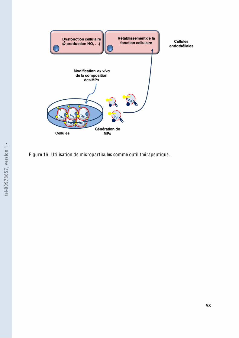
&)"
"
F igure 16: Utilisation de microparticules comme outil thérapeutique.
Modification ex vivode la composition
des MPs
Génération de MPs
Cellules endothéliales
Rétablissement de la fonction cellulaire
Dysfonction cellulaire
Cellules
tel-0
0978
657,
ver
sion
1 -
&*"
"
I I I . de deux maladies
hypoxiques
1. L es pathologies pulmonaires
Les pathologies pulmonaires incluent un grand nombre de pathologies et leurs relations avec
les MPs sont étudiées de plus en plus largement. Il apparait ainsi que les MPs peuvent
contribuer à la sévérité de ces pathologies non seulement en présentant une altération de leur
taux, mais également en potentialisant ces pathologies. Dans ce travail, nous nous sommes
.
En effet, HTAP est caractérisée par une hypoxie chronique alors que le SAOS est caractérisé
par une hypoxie intermittente.
2.
« sclérose des artères pulmonaires » et est considérée comme une maladie fatale dont la
est une maladie pulmonaire définit par
une pression artérielle pulmonaire (PAP) supérieure à 25 mmHg au repos, ou supérieure à 30
[109], conduisant à une défaillance ventriculaire droite, cause principale de
[110].
2.1. Classification clinique des hypertensions pulmonaires
hypox
partageant des caractéristiques cliniques, physiopathologiques et thérapeutiques, une nouvelle
classification a été réalisée lors du « World Symposium on Pulmonary Arterial Hypertension »
en 1998. La révision la plus récente date de 2008 (Tableau 1).
"
"
"
tel-0
0978
657,
ver
sion
1 -
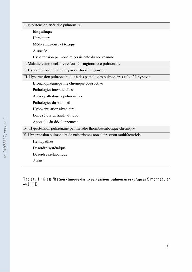
'+"
"
I. Hypertension artérielle pulmonaire
Idiopathique
Héréditaire
Médicamenteuse et toxique
Associée
Hypertension pulmonaire persistente du nouveau-né
-occlusive et/ou hémangiomatose pulmonaire
II. Hypertension pulmonaire par cardiopathie gauche
Bronchopneumopathie chronique obstructive
Pathologies intersticielles
Autres pathologies pulmonaires
Pathologies du sommeil
Hypoventilation alvéolaire
Long séjour en haute altitude
Anomalie du développement
IV. Hypertension pulmonaire par maladie thromboembolique chronique
V. Hypertension pulmonaire de mécanismes non clairs et/ou multifactoriels
Hémopathies
Désordre systémique
Désordre métabolique
Autres
Tableau 1 : C lassificat Simonneau et al. [111]).
tel-0
0978
657,
ver
sion
1 -

'!"
"
2.2. Manifestations cliniques et diagnostic
[112] un
toujours pas aisé, en raison de la relative rareté de
la pathologie, la nature non spécifique des symptômes et du fait que les tests de routine ne
sont pas adéquats [113]
[114]
orax
[115]
technique invasive, le cathétérisme cardiaque droit reste cependant la seule méthode qui
2.3. Pathogénèse globale
caractérise par une obstruction vasculaire conduisant à une augmentation progressive des
la postcharge du ventricule droit
conduit à une défaillance cardiaque droite [116]. La vasoconstriction et le remodelage de la
résistances vasculaires pulmonaires [117]
prolifération intimale et médiale est également considérée comme un facteur déterminant dans
[118]. Au plan physiopathologique, il est actuellement bien établi
que la principale anomalie
vasoconstricteurs puissants qui stimulent également la prolifération vasculaire pulmonaire
(Figure 17).
tel-0
0978
657,
ver
sion
1 -
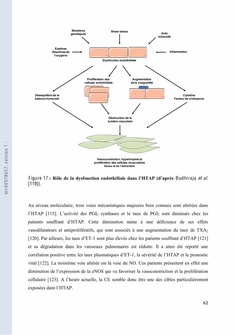
'#"
"
EspécesRéactives de
Mutations génétiques
Shear stress Auto immunité
Inflammation
Dysfonction endothéliale
Prolifération descellules endothéliale
Augmentation de la coagubilité
CytokineFacteur de croissance
Désequilibre de la balance humorale
Obstruction de la lumière vasculaire
Vasoconstriction, hypertrophie et prolifération des cellules musculaires
"
F igure 17 Budhiraja et al. [119]).
Au niveau moléculaire, trois voies mécanistiques majeures bien connues sont altérées dans
[115] 2 synthases et le taux de PGI2 sont diminués chez les
vasodilatateurs et antiprolifératifs, qui sont associés à une augmentation du taux de TXA2
[120] -1 sont plus élevés chez les patients so [121]
et sa dégradation dans les vaisseaux pulmonaires est réduite. Il a ainsi été reporté une
-
vital [122]. La troisième voie altérée est la voie du NO. Ces patients présentent en effet une
cellulaire [123] CE semble donc être une des cibles particulièrement
tel-0
0978
657,
ver
sion
1 -

'$"
"
Pa
[124, 125] (Figure 18),
sfonction
endothéliale observée.
"
F igure 18 Amabile et al. [124]).
2.4. Stratégie thérapeutique
Chez les patients, certaines précautions simples doivent être envisagées, et la plus importante
effet, chez ces patients, toute augmentation brutale du débit cardiaque peut entrainer une
[126]. Le traitement
cours pour éviter la formation de thrombus, et éventuellement une oxygénothérapie pour
maintenir une saturation en oxygène suffisamment élevé.
diamètre de la lumière des vaisseaux en utilisant des vasodilatateurs. Ces traitements incluent
NO PGI2
ET. Cependant, ces traitements ont durée
té limitée. Ces traitements sont également onéreux et sont
Pression artérielle pulmonaire moyenne (mmHg)
MPs
end
othé
liale
PEC
AM+
(MPs
/µl)
tel-0
0978
657,
ver
sion
1 -
'%"
"
associés à des effets secondaires non négligeables. Pour les patients qui ne répondent pas aux
options restantes [127,128].
2.5. Modèles animaux
L -
(notamment au niveau tissulaire), et dans la considération de nouvelles approches
thérapeutiques), il a donc été nécessaire de mettre en place des modèles animaux. Un bon
surtout être prédictif. Un modèle spontané ne peut être un modèle réaliste car il est non
contrôlable et souvent hétérog
même si toutes les composantes de la pathologie ne sont pas représentées.
monocrotaline (un alcaloïde de la famille de la pyrrolizidine présente dans la plante
Crotalaria spectabilis
alternatifs présentant une caractéristique particulière existent également, mais dans cette étude
nous avons utilisé le rat chronique hypoxique qui présente de forte homologie avec la
pathologie humaine [129].
vasoconstriction initiale. Le remodelage artériolaire se limite à une hypertrophie médiale, sans
épaississement intimal ni infiltrats inflammatoires. Au niveau hémodynamique, on retrouve
-1, de la NOS endothéliale et de la
synthèse de la PGI2 expression de la PGI2
.
tel-0
0978
657,
ver
sion
1 -
'&"
"
3. obstructive du sommeil
Le SAOS est le trouble nocturne le plus fréquent caractérisé par des épisodes partiels
(hypopnées) ou c
sommeil. Cette pathologie affecte dans la population adulte au moins 4% des hommes et 2%
des femmes [130].
3.1. Manifestations cliniques et diagnostic
Le diagnostic du SAOS implique la pré
bstruction répétée des voies aériennes supérieures. Cette obstruction
conduit à une diminution de la saturation artérielle en O2 (SaO2) et à une fragmentation du
sommeil. Le SAOS se caractérise ainsi par des symptomes nocturnes tels que le ronflement,
un sommeil non réparateur, des pauses respiratoires ainsi que par une somnolence dirune
excessive. Ce diagnostic est confirmé par un enregistrement nocturne par polysomnographie
dans un laboratoire du sommeil où le sommeil, le débit respiratoire nasal, la SaO2, les
[131].
Les apnées sont caractérisées par une absence de débit respiratoire (mouvements respiratoires)
pendant au moins 10 secondes, et les hypopnées par une diminution du débit respiratoire
( ) supérieure à 50% pendant au moins 10 secondes accompagnée
SaO2 -éveil. Les apnées sont dites
obstructives
occlusion du pharynx.
-hypopnées (IAH) supérieur ou égal à 5 par heure
accompagné notamment par une somnolence diurne excessive, deux ou plus épisodes de
suffocation pendant le sommeil, des épisodes récurrents de micro-éveil, un sommeil non
réparateur, une fatigue excessive, ou des défauts de concentration ou de mémoire [132].
-éveils qui
direct de la fréquence de ces évènements et de leurs
conséquences physiopathologiques : un IAH entre 5 et 15 signe un SAOS minime, entre 15 et
30 modéré, et un SAOS sévère si supérieur à 30. P
tel-0
0978
657,
ver
sion
1 -

''"
"
du SAOS est sévère (somnolence diurne, trouble neurocognitif), et il est maintenant reconnu
que la prévalence des accidents cardio-vasculaires augmente avec cet index [133]. Le SAOS
est ainsi considéré comme un facteur de risque cardio-vasculaire indépendant au même titre
q 4].
3.2. Pathogénèse globale
Le SAOS est une pathologie multi-composante et peut varier considérablement entre les
individus. La pathophysiologie du SAOS est influencé par de nombreux aspects inhérent à
dilatateurs des voies aériennes à répondre pendant le sommeil, la stabilité du système
respiratoire ou encore le volume pulmonaire.
Les épisodes répét
sur la fonction endothéliale. De nombreuses études ont mis en évidence une dysfonction
endothéliale aussi bien clinique que chez les modèles animaux par une diminution de la
product
encore davantage la biodisponibilité du NO, bien que les taux de ONOO- soient semblables
chez les patients SAOS et les sujets sains [135]. Cette réduction du NO accroit le risque de
pathologies vasculaires chez ces patients [136,137] (Figure 19).
tel-0
0978
657,
ver
sion
1 -
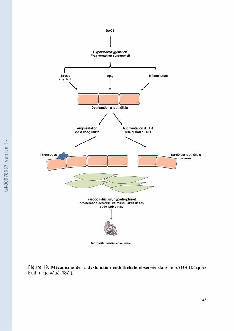
'("
"
SAOS
Stress oxydant
MPs Inflammation
Dysfonction endothéliale
-1Diminution du NO
Augmentation de la coagubilité
Vasoconstriction, hypertrophie et prolifération des cellules musculaires lisses
Hypoxie/réoxygénationFragmentation du sommeil
Thrombose Barrière endothéliale altérée
Morbidité cardio-vasculaire
F igure 19Budhiraja et al. [137]).
tel-0
0978
657,
ver
sion
1 -

')"
"
même si les données présentes sont contradictoires. En effet, Geiser et al [138] ont montré des
taux comparables de PMPs entre les patients SAOS et les témoins malgré une augmentation
plaquettaire in vivo. Plus récemment, Ayers et al [139] ont montré une augmentation
significative des MPs procoagulantes, leucocytaires et PMPs, sans modification des EMPs. A
es en évidence par une
surproduction de E [140,141]. Cependant, aucune de ces études
de leur phénotype, et notam
granulocytaires chez les patients SAOS [93] et que cette augmentation de MPs leucocytaires
avait lieu la nuit [142]. Plus intéressant, ils ont réussi à démontrer une association entre la
dysfonction endothéliale observée et le taux de MPs leucocytaires, suggérant ainsi la
participation des MPs parmi les multiples mécanismes physiopathologiques proposés pour
démontrer la morbidité cardio-vasculaire dans le SAOS (Figure 20).
F igure 20 : Cor rélation entre le taux de MPs leucocytaires et la production de monoxyde humaines Priou et al. [93]).
NO
(A/µ
g/µL
)
MPs L-sélectine+ (MPs/µL de plasma)
tel-0
0978
657,
ver
sion
1 -
'*"
"
3.3. Stratégie thérapeutique
Actuellement, la pression positive continue (PPC) est le traitement standard pour les patients
SAOS. La PPC maintient une pression positive transmurale des voies aériennes et cette
stratégie est bénéfique pour les patients qui la supportent. Des essais cliniques ont ainsi
montré une nette amélioration de la somnolence diurne [143], de la pression sanguine, de la
fonction vasculaire [144,145] et donc de la qualité de vie.
La PPC est très efficace, mais une proportion significative des patients ne tolère pas cette
thérapie [146] [147]. De nouvelles thérapies sont donc
indispensables pour ces patients. alternative comprend les appareils
avantages par rapport à la PPC [148]. Cependant ces appareils sont chers, peuvent être
inconfortables et leurs réponses sont imprédictibles [149].
tel-0
0978
657,
ver
sion
1 -

(+"
"
O BJE C T I FS D ES T R A V A U X
tel-0
0978
657,
ver
sion
1 -
(!"
"
En effet, les MPs ont la capacité de transférer une information biologique entre les cellules en
agissant comme de véritables vecteurs de signalisation, et leur taux est très souvent corrélé à
Parmi les paramètres biologiques altérés lor
nous avons déterminé si les MPs participaient à la dysfonction endothéliale observée dans
Par ailleurs, le rôle joué par les MPs dans la modulation de la réactivité vasculaire a été décrit
dans plusieurs pathologies cardiovasculaires. Dans la seconde partie, nous nous sommes
intéressés à la possible implication des MPs circulantes dans la régulation de la vasomotricité
au cours du SAOS.
Enfin dans le but de comprendre les mécanismes angiogénique observée dans le SAOS, nous
avons étudié le rôle éventuel des MPs associé à ce processus.
tel-0
0978
657,
ver
sion
1 -

(#"
"
R ESU L T A TS
tel-0
0978
657,
ver
sion
1 -
($"
"
Partie I :
L es microparticules circulantes issues de rats hypoxiques
Chroniques entraînent une dysfonction endothéliale
tel-0
0978
657,
ver
sion
1 -
(%"
"
I .
alement à en
comprendre les effets physiologiques et/ou pathologiques dans les pathologies vasculaires.
Ainsi, il a été rapporté une élévation du taux de MPs lors de la pré-éclampsie, le choc septique
et le syndrome métabolique [150,105,98]. Concernant les effets propres des MPs, lors de la
prééclampsie, il a été également montré que les MPs induisaient une hyporéactivité des
vaisseaux issus de souris gestantes grâce à une surproduction de NO [150]. En revanche dans
perréactivité à la sérotonine (5-HT) sans variation
présente chez ces patients [105]. Enfin, dans le syndrome métabolique, les MPs altèrent la
production de NO dans les CEs et participent à la dysfonction endothéliale en diminuant la
[98]. Des effets différentiels des
MPs sur la production de NO et la réactivité vasculaire ont donc été mis en évidence en
fonction de la pathologie étudiée.
1045
travail expérimental a donc eu pour but de vérifier dans un premier temps, si dans un modèle
déterminer si ces MPs pouvaient être un vecteur de la dysfonction vasculaire observée dans
cette pathologie.
tel-0
0978
657,
ver
sion
1 -
(&"
"
I I . Matériel et méthodes
1. Matériel biologique
1.1. : le rat hypoxique chronique
Hypoxie chronique hypobare
Des rats males Wistar (8-10 semaines, 250-300 g) ont été soumis à une hypoxie dans un
caisson hypobare (Sapratin, Arg
10,5 à 11,1 l/minute. La pression était diminuée progressivement pour atteindre 380 mmHg
ou 0,5 atmosphère, ce qui équivaut à une altitude de 5500 mètres. La pression inspirée en
oxygène dans le caisson est de 79.8 mmHg, valeur considérée au niveau alvéolaire comme
étant le seuil induisant une HTAP. Les rats hypoxiques y ont été maintenus durant 21 jours.
Le caisson a été remis à la pression atmosphérique 3 jours par semaine pendant un temps
limité (environ 30 minutes) pour changer les litières et permettre le réapprovisionnement en
eau et en nourriture. La dépressurisation était réalisée progressivement pour revenir à 760
mmHg.
Les rats normoxiques ont été maintenus dans le caisson pendant 21 jours dans les mêmes
conditions que les rats hypoxiques, exceptée que la pression atmosphérique y était maintenue
à 760 mmHg.
a été réalisée par
ppréciation de trois critères :
- la mesure de la PAP moyenne
-
-
Mesure de la pression artérielle pulmonaire
Après étaient retirés du caisson puis anesthésiés par un
mélange de Xylazine (10mg/kg) et de kétamine (50mg/kg) administrés par voie
intrapéritonéale. La PAP moyenne a été
insertion dans la veine jugulaire droite (thorax fermé), puis passage dans le ventricule droit
tel-0
0978
657,
ver
sion
1 -

('"
"
Les valeurs de PAP moyennes chez les rats normoxiques et hypoxiques ont été mesurées à
: les
rats normoxiques présentaient une PAP moyenne de 15.6 ± 0.8 mmHg et les rats hypoxiques
de 30.4 ± 1 mmHg (Figure 21 A). Après les mesures de pression, les rats ont été sacrifiés par
exposition au CO2.
C
rt du poids du ventricule droit sur le poids du
ventricule gauche plus septum. Le ventricule droit était délicatement disséqué du ventricule
gauche et du septum, les caillots de sang résiduels enlevés et les deux parties pesées. Une
élévation de ce rapport
Les rats normoxiques
présentaient un ratio de 0.26 ± 0.04 contre un ratio de 0.51 ± 0.01 pour les rats hypoxique
(Figure 21 B).
capillaire et centrifugé. Les rats hypoxiques présentaient un hématocrite plus élevés par
rapport aux rats normoxiques (Figure 21 C, Bonnet et al. [151]).
tel-0
0978
657,
ver
sion
1 -

(("
"
""
"
"
"
"
"
"
"
"
"
"
"
"
"
"
"
"
"
"
"
F igure 21 .
* P<0.05 et ***P<0.001.
0
***
Normoxique Hypoxique
Ratio
ven
tricu
le d
roit
/(v
entri
cule
gau
che
+ se
ptum
)
0.2
0.4
0.6
0
***
Normoxique Hypoxique
Pres
sion
arté
rielle
pulm
onai
re m
oyen
ne (m
mHg
)
10
20
30
40
0
*
Normoxique Hypoxique
Hém
atoc
rite
(%)
20
40
60
80
A
B
C
0
***
Normoxique Hypoxique
Ratio
ven
tricu
le d
roit
/(v
entri
cule
gau
che
+ se
ptum
)
0.2
0.4
0.6
0
***
Normoxique Hypoxique
Pres
sion
arté
rielle
pulm
onai
re m
oyen
ne (m
mHg
)
10
20
30
40
0
*
Normoxique Hypoxique
Hém
atoc
rite
(%)
20
40
60
80
A
B
C
tel-0
0978
657,
ver
sion
1 -

()"
"
Isolation des MPs
Une fois les animaux sacrifiés, le sang était
un tube citraté (3.8 % de citrate de sodium). Les tubes de sang ont été centrifugés à 1900 g
pendant 3 minutes. Le plasma riche en plaquettes a été récupéré et centrifugé à 5.000 g
pendant 4 minutes pour obtenir le plasma pauvre en plaquettes (PPP). Cent µl ont été stockés
à 80°C pour réaliser le phénotypage de MPs. Le volume restant a été soumis à trois étapes de
centrifugation à 15.000 g pendant 30 minutes. Le culot final était suspendu dans 150 µl de
solution saline (NaCl 0.9 %) et conservé à +4°C. Du sang des rats normoxiques étaient
extraites les MPs normoxiques et du sang des rats hypoxiques étaient extraites les MPs
hypoxiques.
Cette partie du projet a été réalisée U1045 (Bordeaux) et les échantillons de MPs
ont ensuite été transportés à Angers où les expériences ont été réalisées.
1.2.
Dissociation cellulaire
Les cellules utilisées étaient des CEs
8 à 12 semaines et pesant environs 350 à
du protocole de Kobayashi et al. [152]. Brièvement, après anesthési
incision thoracique était
pulmonaires étaient alors isolées et immergées dans une solution de « dulbecco's modified
eagle's medium » (DMEM, Lonza, Walkersville, Etats-Unis) contenant 20 % de sérum de
, Lonza). Une canule de 19 gauges était alors insérée et attachée au niveau
proximal de chaque artère afin de permettre le lavage de la lumière artérielle avec du DMEM.
Le niveau distal était alors ligaturé et une solution de collagénase de type II (Sigma, Saint
Quentin Fallavier, France) à 2 mg/ml était
les CEs étaient décollées avec une solution de DMEM/20 % SVF. Les cellules CEs étaient
alors récupérées par centrifugation (1.200 g, 5 minutes) et le culot resuspendu dans une
solution de DMEM/20 % SVF.
tel-0
0978
657,
ver
sion
1 -
(*"
"
Culture cellulaire
Les cellules étaient ensuite cultivées dans un puits de 9,6 cm! (Corning Incorporated, New-
York, Etats-Unis), et après 2 heure était remplacé par le
milieu de culture « endothelial basal cell medium-2 »/5 % SVF (EBM-2, Lonza). Le milieu de
culture a été changé tous les deux jours. Une fois à confluence, les cellules étaient repiquées
dans une flask de 75 cm2 ème passage. Les
cellules étaient ensemencées à 10.000 cellules par cm2.
Vérification du phénotype endothélial
Le phénotype endothélial a été vérifié en utilisant les lipoprotéines de basse densité acétylées
couplées au 1,1'-dioctadecyl-3,3,3',3'-tetramethyl-indocarbocyanine perchlorate (Dil-Ac-
LDL) (Harbor Bio-products, Norwood, Etats-Unis). Puisque seules les CEs sont perméables
aux Ac- CEs est basée sur leur capacit -LDL.
Ainsi, les cellules ont été incubées en absence ou en présence de Dil-Ac-LDL (10 µg/ml)
pendant 3 heures à 37°C. Les cellules étaient ensuite traitées avec de la trypsine (Lonza) puis
re en flux. En moyenne, 95 % des cellules
isolées correspondaient à des CEs.
T raitement des cellules
Les cellules (5.104 cellules/cm2) ont été traitées pendant 24 heures à la concentration
rminée par cytométrie en
flux.
2. Cytométrie en flux
Principe
La cytométrie en flux permet l
milieu liquide. Elle est généralement appliquée à des cellules, mais elle est une méthode de
référence au laboratoire pour la quantification et le phénotypage des MPs. Cette technique
consiste à faire défiler très rapidement (plusieurs centaines/seconde), les MPs en suspension
devant un faisceau laser. Pour chaque MP sont mesurées : la fluorescence émise à diverse
tel-0
0978
657,
ver
sion
1 -

)+"
"
On peut
ainsi calculer le nombre total de MPs et le pourcentage de MPs marquées.
Dispositif expérimental
Le phénotypage de MPs a été réalisé à partir du PPP
membranaires spécifiques. Pour cela, 8 µl de plasma étaient incubés
isothiocyanate de fluorescéine
(FITC) (BioLegend, San Diego, Etats-Unis), CD45 couplé au FITC (BioLegend), CD54
couplé à la biotine (BioLegend) ou erythroid couplé à la biotine (BD Biosciences)
érythocytaire. Pour les anticorps couplés à la biotine, un marquage secondaire avec la
streptavidine couplée au FITC (Rockland, Gilbertsville, Etats-Unis) était réalisé. Des
anticorps isotypiques contrôles ont été utilisés pour déterminer le marquage aspécifique pour
chaque anticorps. Après 45 minu -count (Beckman
Coulter, Roissy, France) étaient ajoutées pour quantifier la concentration de MPs dans chaque
échantillon. Cela permettait de déterminer la concentration circulante en MPs pour chaque
groupe de rat. Les échantillons (10.000 événements) étaient ensuite analysés au cytomètre en
flux (Beckman Coulter).
3. W estern Blot
Extraction des protéines
Après le traitement par les MPs, les cellules étaient lavées au « phosphate buffer saline »
(PBS), puis traitées par du tampon de lyse de composition suivante : 50 mM Tris-HCl, 150
mM NaCl, 0.8 % Nonidet P-
glace. Le lysat était récupéré et placé dans des eppendorfs. Le lysat était centrifugé (15.000 g,
10 minutes, 4°C) et le surnageant contenant les protéines conservé à -80°C.
La mesure de la concentration protéique de ces échantillons a été faite
de concentrations croissantes d BSA) (0, 0.2, 0.5, 0.8, 1, 1.5 et 2
mg/ml) selon la méthode de Lowry et al. [153] sur plaque 96 puits et mesurées au
spectrophotomètre (Biotek, Winooski, Etats-Unis Les
protéines étaient ensuite dénaturées par incubation dans le tampon contenant du NuPAGE®
tel-0
0978
657,
ver
sion
1 -

)!"
"
LDS Sample Buffer et NuPAGE® Reducing Agent (Invitrogen, Carlsbad, Etats-Unis), 10
minutes à 70°C.
Migration sur gel et transfert sur membrane
Trente microgrammes de protéines étaient déposés sur des gels précoulés NuPAGE®
Novex® Bis-Tris Mini gels (Invitro
fonction de leur poids moléculaire. Les protéines sont dénaturées dans un tampon fortement
chargé négativement, afin de neutraliser les charges propre de la protéine. La migration des
protéines dénaturées dépend alors uniquement de la longueur de la chaîne protéique et elles se
répartiront uniquement en fonction de leur poids moléculaire. La migration était réalisée dans
150 V et un ampérage
variant de 100-125 mA au début à 60-80 mA en fin de migration pendant 70 minutes selon les
instructions du fabricant. Les gels étaient ensuite transférés sur une membrane de
nitrocellulose ayant des pores de 0.2 µm dans une cuve de transfert verticale (BioRad,
Hercules, Etats-Unis ait sous une
tension constante de 100 V pendant 2 heures à 4°C.
Marquage immunologique et révélation
A la fin du transfert, les membranes étaient rincées, trois fois 10 minutes dans un tampon
TBS-Tween (10 mM Tris, 100 mM NaCl, 0.1 % Tween-20) à température ambiante, puis
incubées 90 minutes à température ambiante dans le tampon TBS-Tween contenant 5 % de
BSA afin de saturer les sites de liaison non spécifiques des anticorps. Les membranes étaient
anticorps primaires dilués dans le tampon TBS-Tween contenant 5 % de BSA (Tableau 5,
Annexes). Après trois lavages de 10 minutes dans le tampon TBS-Tween, les membranes
étaient incubées 90 minutes avec un anticorps secondaire conjugué à une peroxydase de
Raifort. -actine a été
évaluée en utilisant un premier anticorps s -
était révélée par un kit de détection (Santa
Cruz Technology, Santa Cruz, Etats-Unis), avec production de luminol, visualisée et
quantifiée, en chambre noire. La quantification par analyse densitométrique par
tel-0
0978
657,
ver
sion
1 -

)#"
"
Image Reader
Las-3000 (Fujifilm, Tokyo, Japon).
4. Dosage des radicaux libres par résonnance paramagnétique électronique
Principe
La résonnance paramagnétique électronique (RPE) est une technique spectroscopique
appariés. Le paramagnétisme résultant de la résonnance du spin électro
oscillant [154].
Protocole
Dosage de la pro
Pour mesurer le radical NO, nous avons utilisé le capteur de spin, le diethyldithiocarbamate
de fer Fe(DETC)2, qui forme le complexe NO-Fe(DETC)2 dont les propriétés
paramagnétiques sont détectables par RPE. Les cellules étaient mises en culture dans une
plaque 6 puits en absence ou en présence des MPs pendant 24 heures, puis elles étaient
mM HEPES, 1 mM L-Arginine, 3 mM CaCl2, 0,3 M BSA, pH 7,4). Les cellules étaient
ensuite incubées dans les mêmes conditions (37°C, 45 minutes) avec le capteur de spin,
diéthyldithiocarbamate (DETC, 1,5 mM) en présence de FeSO4. Les échantillons étaient
-80°C.
Les cellules étaient mises en culture dans une plaque 6 puits en absence ou en présence des
MPs pendant 24 heures, puis elles étaient récoltées et incubées pendant 45 minutes à 37°C en
-Hepes contenant du 1-hydroxy-3methoxycarbonyle
2,2,5,5-tetramethylpyrrolidine (CMH, Noxygen, Mainz, Allemagne) (10 mM), de la
deferoxamine (25 M) et du DETC (5 M). La réaction a été stoppée en déposant les
échantillons pendant 5 minutes à 4°C. Les échantillons étaient
liquide puis conservé à -80°C.
tel-0
0978
657,
ver
sion
1 -

)$"
"
était 200 (Magnettech, Berlin,
Allemagne). Les enregistrements ont été faits à 77°K, en utilisant un vase Dewar.
-ondes, à 1mT de modulation
spectre de 100 secondes répété 4 fois de suite. La quantité de NO et 2- mesurée était
quantité de protéines dans chaque échantillon était mesurée par dosage colorimétrique de
Lowry et déterminé au spectrophotomètre à la longu
5.
xanthine oxydase a été mesurée grâce à un kit adapté (BioVision Research Procucts, Moutain
Vi
6. Réactivité vasculaire
La contraction isométrique a été mesurée dans les aortes et les artères extra-pulmonaires de
rats injectés ou non avec des MPs à la concentration circulante déterminée dans le plasma des
rats. Nous avons alors pesé chaque rat pour déterminer le volume sanguin et la quantité de
MPs à injecter. En effet un rat Wistar male possède 6,4 ml de sang pour 100 g de poids [155].
Vingt-
Mulvany (Danish Myo Technology, Aarhus, Danemark) contenant la solution de Krebs
adaptée à 37°C, équilibrée avec un mélange carbogène (95 % O2, 5 % CO2, pH = 7,4)
(Tableau 6, Annexes).
Au début de chaque expérience, les anneaux étaient équilibrés contre une charge de 2 g pour
les aortes et 1 g pour les artères pulmonaires. Des études préalables ont montré que ces
laquelle la contraction isométrique développée était maximale après stimulation avec une
solution riche en potassium [156,157]
au cours de laquelle la solution de Krebs était changée toutes les 15 minutes. A la fin de
tel-0
0978
657,
ver
sion
1 -

)%"
"
étaient stimulés deux fois par une solution riche en potassium
(1 µM) pour les artères pulmonaires après une pré-contraction des anneaux à la phényléphrine
(1 µM). Des courbes concentrations cumulative-réponses aux agonistes ont ensuite été
construites.
7. Analyse statistique
Les résultats ont été exprimés en moyenne ± erreur standard de la moyenne (SEM) de n
observations indépendantes. Les données ont été ensuite analysées par le test non
paramétrique de Mann-Whitney.
Les comparaisons statistiques entre les courbes concentrations cumulatives-réponse ont été
été réalisé un test Mann- maximal (Emax) et de pD2.
pD2 = -log EC50, EC50
maximal.
Toutes les analyses statistiques ont été effectuées grâce au logiciel GrapPad Prism (GrapPad
Softaware, San Diego, Etats-Unis). Les différences sont considérées comme significative
lorsque P < 0,05 quelque soit le test statistique employé.
tel-0
0978
657,
ver
sion
1 -
)&"
"
I I I . Résultats
Taux ci rculants et origines cellulaires des MPs
Les résultats obtenus du dosage de MPs réalisé par cytométrie en flux montrent que le taux
circulant de MPs chez les rats hypoxiques est le double que celui de rats normoxiques (1568 ±
174 versus 852 ± 80 MPs/µl de plasma ; p<0.01) (Figure 22 A). Ces concentrations ont été
utilisées pour la suite de nos expériences.
De plus, le phénotypage de ces MPs a montré que les MPs circulantes sont principalement +) (87 ± 2 % et 85 ± 4 % chez les rats hypoxiques et
normoxiques, respectivement) et érythrocytaires (7 ± 2 % et 7 ± 1 % chez les rats hypoxiques
et nor +) et celles issues des
CEs (CD54+) ne dépassent pas le taux de 3 % dans les deux groupes. Enfin, nous avons
s
leucocytaires et endothéliales sont plus élevés chez les rats hypoxiques par rapport aux rats
normoxiques (Figure 22 B-E).
circulation systémique peut également être affectée [158]
étude nous avons isolé deux types cellulaires représentatifs de ces deux circulations : les
rats que nous avons traitées par les MPs à la concentration circulante déterminée par
cytométrie en flux.
Implication des MPs dans la production de N O
La production de NO étant un des éléments clefs de la fonction endothéliale, nous avons
plus de clarté, nous distinguerons les effets sur les
deux types cellulaires.
Le traitement des CEs
ne modifient pas la production de NO. Cependant, les MPs issues de rats hypoxiques
réduisent la production de NO de 45% (Figure 23 A). A la lumière de ces résultats, nous nous
diminution de production de pression et
CEs. Dans les
tel-0
0978
657,
ver
sion
1 -

)'"
"
CEs
de la eNOS. Cependant, le traitement par les MPs hypoxiques induit une augmentation de la
significative de la phosphorylation de la eNOS sur son site activateur (Ser 1177) (Figure 23
B-C). De plus, les MPs hypoxiques entraînent une
sur la Ser 473, kinase impliquée dans la phosphorylation de la eNOS et ainsi de son activation
(Figure 23 D
cavéoline-1, protéine régulatrice de la eNOS (Figure 23 E).
Dans les CEs
une diminution de 20% de la production de NO uniquement par les MPs hypoxiques (Figure
24 A). De plus
que seules les MPs hypoxiques sont capables de diminuer son activation. En effet le
traitement par les MPs hypoxiques induit une augmentation de la phosphorylation de la eNOS
sur son site inhibiteur et une diminution de la phosphorylation sur son site activateur (Figure
24 B-C). En revanche, contrairement aux CEs
Akt par les différents traitements (Figure 24 D). De
éoline-1 est diminuée par le traitement par les MPs hypoxiques
(Figure 24 E). Ces données suggèrent ainsi que le mécanisme de diminution de production de
NO diffèrent dans les deux types cellulaires.
2-
Un autre aspect prépondérant dans la fonction endothéliale concerne le stress oxydant. Nous
2- par les CEs traitées par les MPs.
La figure 24
2- par les CEs. En revanche, bien que les MPs hypoxiques ne modifient pas la production
2- par les CEs 25 A), nous pouvons observer que dans les CEs
2- (Figure 25
B). Ces données soulignent à nouveau une différence entre les deux types cellulaires.
tel-0
0978
657,
ver
sion
1 -
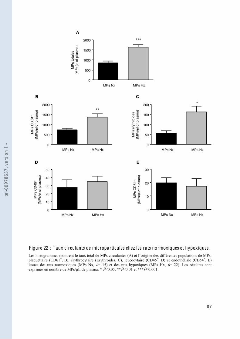
)("
"
0
500
1000
1500
2000 ***
MPs
tota
les
(MPs
/µl o
f pla
sma)
MPs Nx MPs Hx
A
*
0
50
100
150
200
MPs
éry
thro
ides
(MPs
/µl o
f pla
sma)
MPs Nx MPs Hx
C
0
500
1000
1500
2000**
MPs
CD
61+
(MPs
/µl o
f pla
sma)
MPs Nx MPs Hx
B
0
10
20
30
40
50
MPs
CD
45+
(MPs
/µl o
f pla
sma)
MPs Nx MPs Hx
D
0
10
20
30
MPs
CD
54+
(MPs
/µl o
f pla
sma)
MPs Nx MPs Hx
E
F igure 22 : Taux ci rculants de microparticules chez les rats normoxiques et hypoxiques. origine des différentes populations de MPs:
plaquettaire (CD61+, B), érythrocytaire (Erythroïdes, C), leucocytaire (CD45+, D) et endothéliale (CD54+, E) issues des rats normoxiques (MPs Nx, n= 15) et des rats hypoxiques (MPs Hx, n= 22). Les résultats sont exprimés en nombre de MPs/µL de plasma. * P<0.05, **P<0.01 et ***P<0.001.
tel-0
0978
657,
ver
sion
1 -

))"
"
F igure 23 : L es microparticules issues des rats hypoxiques diminuent la production de .
Les cellules ont été incubées pendant 24 heures en absence (Ctl) ou en présence des MPs issues des rats -Fe(DETC)2 dans
n=8-9). Les histogrammes vs
phospho-Akt Ser 473 (D) vs -1 (E). Les immunoblots sont quantifiés par analyse densitométriques et no(n=6-8). * P<0.05 et **P<0.01.
0.03
0.06
MPs Nx MPs HxCtl0Ex
pres
sion
de
cave
olin
-1
Caveolin-1
actin
E
0.5
1
1.5
MPs Nx MPs HxCtlp-
eNO
S Th
r/eNO
S (ra
tio)
0
*
P-eNOS Thr
eNOS
C
0
0.5
1
P-Ak
t Ser
/Akt
(rat
io)
MPs Nx MPs HxCtl
P-Akt Ser
Akt
D**
10
20
MPs Nx MPs HxCtl
*
0P-eN
OS
Ser/e
NOS
(ratio
)
P-eNOS Ser
eNOS
B
MPs Nx MPs HxCtl0
500
1000
1500
2000
2500
Prod
uctio
n de
NO
(A/µ
g/µl
)
***
Ate
l-009
7865
7, v
ersi
on 1
-

)*"
"
F igure 24 : L es microparticules issues des rats hypoxiques diminuent la production de ire.
Les cellules ont été incubées pendant 24 heures en absence (Ctl) ou en présence des MPs issues des rats -Fe(DETC)2 dans
s sont exprimés en amplitude/µg/µL de protéines (n=8-9). Les vs
totale (D), phospho-Akt Ser 473 (D) vs -1 (E). Les immunoblots sont quantifiés par analyse densitométriques et normalisés soit sur la forme totale de la protéine correspondante soit
n=6-8). * P<0.05 et **P<0.01.
500
1000
1500
Prod
uctio
n de
NO
(A/µ
g/µl
)
0
*
MPs Nx MPs HxCtl
A
0
0.5
1.0
1.5
P-Ak
t Ser
/Akt
(rat
io)
MPs Nx MPs HxCtl
P-Akt Ser
Akt
D
C
0.1
0.2
0.3
P-eN
OS
Thr
/eNO
S (ra
tio)
0
**
P-eNOS Thr
eNOS
MPs Nx MPs HxCtl
actin
0
0.05
0.1
Expr
essi
on d
e ca
veol
in-1
*
Caveolin-1
MPs Nx MPs HxCtl
E
0.2
0.4
MPs Nx MPs HxCtl
*
0P-eN
OS
Ser/e
NOS
(ratio
)
P-eNOS Ser
eNOS
B
tel-0
0978
657,
ver
sion
1 -

*+"
"
F igure 25 : L es microparticules de rats hypoxiques diminuent la produc 2-
. Les cellules ont été incubées pendant 24 heures en absence (Ctl) ou en présence des MPs issues des rats
2- dans les CEs
2- dans les CEs
(100 µM) puis pendant 24 heures par les MPs hypoxiques (C). Les données sont exprimées en amplitude/µg/µL de protéines par échantillon (A et B) ou en amplitude/µg/µL de protéines par échantillon normalisé par rapport au Ctl (C) (n = 8- ubation des cellules avec les MPs, les protéines sont extraites et
échantillon (n = 7). * P<0.05 et **P<0.01.
0
50
100
150
Ctl MPs Hx Rotenone
MPs Hx
2- (%
)
***
*
ApocyninAllopurinol
C
0
25
50
75
100
125
2- (A/
µg/µ
l)
MPs Nx MPs HxCtl
A
0
50
100
150
200
250
2- (A/
µg/µ
l)
MPs Nx MPs HxCtl
B*
*
Activ
ité d
e xa
nthi
ne o
xyda
se(m
U/m
g pr
otéi
ne)
MPs Nx MPs HxCtl
D
0.1
0.2
0.3
0
0.4
tel-0
0978
657,
ver
sion
1 -
*!"
"
Nous avons alors voulu déterminer la s . Les CEs
pulmonaire traitées par les MPs hypoxiques ont donc été pré-incubées en présence
et de la NADPH oxydase (apocyn
induite par les MPs hypoxiques (Figure 25 C
cette productio
unité catalytique de la NADPH oxydase (donnée non montrée
de la xanthine oxydase conforte ces résultats et montre que seul le traitement par les MPs
(Figure 25 D).
Implication des MPs dans la fonction vasculaire ex vivo
reçu une injection de MPs à la concentration circulante déterminée. Il est à noter que
PAP moyenne au bout de 24 heures
par rapport aux rats normoxiques.
Pour analyser
c
de la courbe concentration- : 7,35 ± 0,04 vs 6,72 ± 0,04 ;
normoxique vs MPs hypoxiques) (Figure 26 :
7,59 ± 0,04 vs 6,66 ± 0,05 ; normoxique vs MPs hypoxiques) (Figure 26 B), sans changement
- ci
semble être altérée par le traitement aux MPs hypoxiques (Figure 26 C). Pour vérifier si cet
effet était dû à une différence qualitative entre la composition des MPs ou juste dû à une
augmentation du taux de MPs, nous avons injecté deux fois plus de MPs normoxiques dans le
MPs hypoxiques et réalisé les expériences de relaxation
MPs hypoxiques conduit à un déplacement vers la droite de la courbe concentration réponse
au carbachol comparé à la double quantité de MPs normoxiques (pD2 : 7,16 ± 0,04) (Figure
26 MPs est principalement du à une différence
qualitative dans la composition des MPs.
tel-0
0978
657,
ver
sion
1 -

*#"
"
F igure 26 : L es microparticules hypoxiques diminue la relaxation dépendante de .
des MPs
par une double quantité de MPs Nx, ou par des MPs Hx (B). Les résultats sont exprimés en pourcentage de précontraction à la phényléphrine(n=4). ***P<0.001.
***-9 -8 -7 -6 -5
100
50
Log [Ach] (M)
Rela
xatio
n (%
)
0
NxMPs Hx
A
***-9 -8 -7 -6 -5
100
50
Log [CCH] (M)
Rela
xatio
n (%
)
0
B
2x MPs NxNx
MPs Hx ***
0Normoxique MPs Hx
Prod
uctio
n de
NO
(A/m
g)
200
400
600
C
tel-0
0978
657,
ver
sion
1 -
*$"
"
La réactivité des vaisseaux aux agonistes vasoconstricteurs a également été étudiée. La
phényléphrine induit une contraction dépendante de la concentration des aortes et des artères
pulmonaires des rats normoxiques ou traités par les MPs hypoxiques. Les vaisseaux traités par
les MPs hypoxiques présentent un déplacement de la courbe vers la droite (pD2 :
6,92 ± 0,02 vs 6,52 ±0,03; normoxique vs MPs hypoxiques) (Figure 27 A) et également dans
(pD2 : 7,99 ± 0,03 vs 7,28 ± 0,05; normoxique vs MPs hypoxiques)
(Figure 27 (Emax : 8,27 ± 0,07 vs
6,21 ± 0,08; normoxique vs MPs hypoxiques) : 2,77 ± 0,02
vs 1,80 ± 0,03; normoxique vs MPs hypoxiques). Cependant cette hypo-réactivité est
5-HT induit une contraction dépendante de la
concentration des artères pulmonaires similaire chez les rats normoxiques ou traités par les
dans les animaux traités par les MPs hypoxiques (Emax : 8,72 ± 0,02 vs 12,6 ± 0,03;
normoxique vs MPs hypoxiques) (Figure 27 C-D). Par ailleurs, la réponse au potassium est
diminution significative de cette réponse (Figure 27 E).
tel-0
0978
657,
ver
sion
1 -
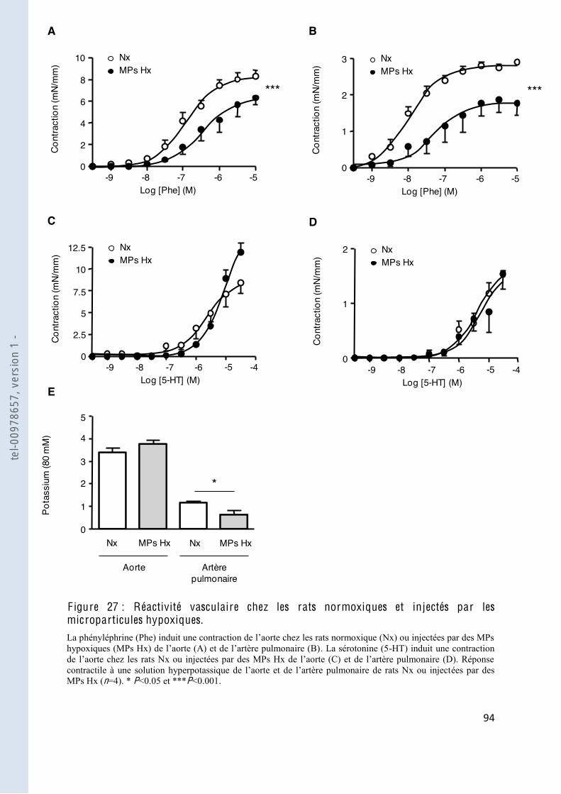
*%"
"
F igure 27 : Réactivité vasculaire chez les rats normoxiques et injectés par les microparticules hypoxiques.
. La sérotonine (5-HT) induit une contraction
ées par des MPs Hx (n=4). * P<0.05 et ***P<0.001.
0
*
Nx MPs Hx
Pota
ssiu
m (8
0 m
M)
1
2
3
4
5
Nx MPs Hx
Aorte Artèrepulmonaire
***
-9 -8 -7 -6 -50
Log [Phe] (M)
Cont
ract
ion
(mN/
mm
) 10 NxMPs Hx
2
4
6
8
-9 -8 -7 -6 -40
Log [5-HT] (M)
Cont
ract
ion
(mN/
mm
)
NxMPs Hx
2.5
5
-5
7.5
12.5
10
E
C
A
***
-9 -8 -7 -6 -50
Log [Phe] (M)
Cont
ract
ion
(mN/
mm
)
NxMPs Hx
1
2
3
-9 -8 -7 -6 -40
Log [5-HT] (M)
Cont
ract
ion
(mN/
mm
)
NxMPs Hx
1
-5
2
D
Bte
l-009
7865
7, v
ersi
on 1
-

*&"
"
I V . Discussion
in vitro, nous avons mis au point au laboratoire
CEs
injecté ces MPs dans des rats pour évaluer leur pertinence physiopathologique in vivo.
Dans cette étude, le taux circulant de MPs est plus élevé chez les rats hypoxiques que chez les
érythrocytaire. Les principaux résultats montrent que les MPs hypoxiques ont des effets
différents selon le territoire vasculaire étudié. Ainsi, dans la circulation systémique, les MPs
eNOS, sans modification du stress oxydant
hypoxiques, en plus de diminuer la production de NO, augmentent le stress oxydant par un
bien un effet délétère des MPs hypoxiques spécifique au territoire vasculaire pulmonaire en
contraction
, un taux élevés de MPs
endothéliales et leucocytaires a ét [124], alors que celui
de Bakouboula a montré une élévation des MPs procoagulantes [125]. Récemment, Diehl et
al.
augmentées chez ces patients [159]
hypoxique une augmentation du taux de MPs, mais que celles-ci étaient principalement
sation de cohortes de patients très hétérogènes et ne ressemblant pas exactement à une
HTAP hypoxique. Par ailleurs, différentes méthodes de centrifugation ainsi que différents
n et la
élevé que les rats normoxiques [151], cette augmentation de cellules sanguines pourrait
tel-0
0978
657,
ver
sion
1 -

*'"
"
ttaires et
érythrocytaires est spécifique de ces populations. Enfin, nous avons montré que seules les
pulmonaires indiquant que les effets des MPs étaient indépendants de leur quantité, mais
plutôt de leurs probables compositions et origines différentes.
Dans la majorité des maladies cardio-vasculaires, la paroi artérielle voit ses possibilités de
sécrétion des facteurs relaxants diminuer, tandis que ses capacités de libération de facteurs
contracturants restent inchangées ou augmentées. Le déséquilibre endothélial lié à la
réduction de la synthèse du NO dépendante de la eNOS peut retentir non seulement sur le
taire. La dysfonction
endothéliale devient dès lors un des éléments primordiaux des maladies vasculaires. Une
[160]. Le rôle de la
; une surproduction de la
[161], alors que les
souris déficiente pour la eNOS développe
modérées [162]. Dans cette étude, la fonction endothéliale in vitro a été évaluée par une
technique permettant de mesurer in situ la production de NO. Les résultats ont montré une
diminution de cette production aussi bien au niveau des CEs
pulmonaires traitées avec les MPs hypoxiques, et suggèrent que les MPs participeraient à la
de NO.
Dans les CEs
post-traductionnelles mettant en évidence son activité. Ainsi, la phosphorylation ou la
déphosphorylation de certains acides aminés est un mécanisme important de régulation post-
1177) et un résidu thréonine dans le domaine de liaison à la calmoduline (Thr 495) [163]. La
[164]. Quand ce
tel-0
0978
657,
ver
sion
1 -
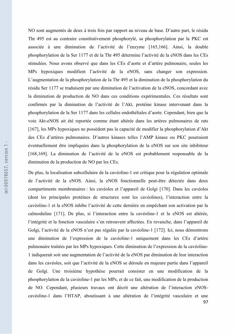
*("
"
NO sont augmentés de deux à trois fois p
Thr 495 est au contraire constitutivement phosphorylé, sa phosphorylation par la PKC est
[165,166]. Ainsi, la double
phosphorylation de la Ser 1177 et CEs
stimulées. Nous avons observé que dans les CEs
ylation de la Thr 495 et la diminution de la phosphorylation du
la diminution de production de NO dans ces conditions expérimentales. Ces résultats sont
confirmés
voie Akt-eNOS ait été reportée comme étant altérée dans les artères pulmonaires de rats
[167]
des CEs
éventuellement être impliquées dans la phosphorylation de la eNOS sur son site inhibiteur
[168,169]
diminution de la production de NO par les CEs.
De plus, la localisation subcellulaire de la cavéoline-1 est critique pour la régulation optimale
vité de la eNOS. Ainsi, la eNOS fonctionnelle peut-être détectée dans deux
compartiments membranaires [170]. Dans les cavéoles
cavéoline-
calmoduline [171] -1 et la eNOS est altérée,
-1 [172]. Ici, nous démontrons
-1 uniquement dans les CEs
pulmonaire traitées par les MPs hypoxiques. Cette dimin -
de Golgi. Une troisième hypothèse pourrait consister en une modification de la
phosphorylation de la cavéoline-1 par les MPs, et de ce fait, une modification de la production
-
cavéoline-
tel-0
0978
657,
ver
sion
1 -

*)"
"
-1 a été
[173]
hypoxique [174] ou celui de la monocrotaline [175]
eNOS-
iques
2-
2- interagit avec le NO avec des constantes de vitesses très élevées, ce qui
-, ERO de nature non radicalaire,
2- semble
jouer un rôle majeur dans la dysfonction endothéliale associée à diverses pathologies
vasculaires [50,176]. Si le rôle des ERO en physiopathologie vasculaire est assez bien
[177-179]
complètement défini [180]. Plusieurs études ont montré une implication de la NADPH
[178,181]. Par ailleurs,
NADPH oxydase et la mitochondrie [182,183]
de la xanthine oxydase est augmentée dans les conditions hypoxiques et spécialement dans la
[184]. Il a également été rapporté que la
xanthine oxydase participait à la dysfonction endothéliale et au remodelage vasculaire chez
[185] 2-
CEs
démontrant à nouveau une spécificité tissulaire des MPs hypoxiques envers le lit vasculaire
pulmonaire. Nous avons également pu montrer que cette modulation du stress oxydant dans
ces cellules était du à une participation à la fois de la xanthine oxydase et du complexe I de la
mitochondrie.
Enfin, pour évaluer la pertinence physiopathologique des MPs, les MPs hypoxiques ont été
injectées à des rats normoxiques. Bien que les MPs hypoxiques ne modifient pas la PAP, elles
sont capables de réduire la capacité des agonistes muscariniques à réduire la relaxation
tel-0
0978
657,
ver
sion
1 -

**"
"
Ces résultats sont en accord avec la littérature où est largement reportée la diminution de la
relaxation dépenda [186] ou systémique
[160]
associée à une hypertrophie des cellules musculaires lisses aboutissant à une augmentation
des résistances vasculaires. Il est raisonnable de penser que le traitement avec les MPs
réponse à la 5-HT -pulmonaires de rat [187], des études de
réactivité vasculaire ont montré une forte augmentation de la réponse contractile aux agents
activant des récepteurs (PGF ) ou non (solution hyperpotassique) dans les artères extra-
pulmonaires de souris hypoxiques. Les mécanismes contractiles situés en aval de
chronique [178]. Plusieurs autres groupes ont également rapporté ce phénomène
une différence régionale selon le territoire étudié [181,188-190]. Plusieurs mécanismes
-kinases [188]
superoxydes [181], la participation de COX-2 [191]
jonctions [192,193], ou encore une diminution du NO endothélial. Il a été démontré que les
MPs pouvaient agir indépendamment sur ces facteurs dans de nombreuses pathologies
vasculaires, nous avons donc étudié le rôle des MPs hypoxiques sur la contraction vasculaire.
Nous avons obtenues des données contradictoires avec celle de la littérature, puisque la
réponse à la phényléphrine est diminuée dans les deux types de vaisseaux, et que la réponse
au potassium est également diminuée dans les artères pulmonaires. En revanche, la réponse à
la 5- Les MPs
semblent donc participer à la réactivité vasculaire de façon différentielle, selon le territoire
En conclusion, les rats hypoxiques développant une HTAP possèdent une élévation du taux de
rats en réd
tel-0
0978
657,
ver
sion
1 -

!++"
"
suggère fortement que les MPs hypoxiques sont capables de participer à la dysfonction
mécanismes présentés sur la figure 28.
tel-0
0978
657,
ver
sion
1 -
!+!"
"
F igure 28 : Mécanismes impliquées dans la dysfonction endothéliale induite par les microparticules . Cav-1 : cavéoline-1; eNOS : sérine; Thr : thréonine; NO : monxyde 2
- : anion superoxyde.
NO
Cellule endothéliale
Microparticules Hypoxique
eNOS
Thr495Ser
1177
Akt
Cav-1
eNOS
Thr473
O2-=
Dysfonction endothéliale
=
NO
Cellule endothéliale
Microparticules Hypoxique
eNOS
Thr495Ser
1177
Xanthine oxydase
Akt
Cav-1
eNOS
Thr473
O2-
Dysfonction endothéliale
=Mitochondrie
tel-0
0978
657,
ver
sion
1 -
!+#"
"
Partie I I :
obstructives du sommeil et hyper réactivité vasculaire : rôle
tel-0
0978
657,
ver
sion
1 -
!+$"
"
I .
Des travaux précédemment réalisés au laboratoire en collaboration avec le service de
dentique chez
les patients SAOS et les sujets témoins. Cependant la caractérisation de ces MPs par
cytométrie en flux a montré une augmentation des taux de MPs granulocytaires (CD66b+) et
celles issues des leucocytes activés (CD62L+), et que ces dernières corrélaient positivement
SAOS. Le traitement des CEs par ces MPs a montré que les MPs issues de patients SAOS
vité de la eNOS et augmentation de
-1. Par ailleurs cette production de NO corrèle négativement avec
les MPs CD62L+
-2. Enfin, la pertinence physiopathologique de ces MPs a été
mise
, ainsi que la dilatation au flux des artères mésentériques
[93].
Des données contradictoires existent dans la littérature concernant la vasoconstriction des
vaisseaux chez les patients SAOS, mais généralement la réactivité vasculaire est altérée quel
que soit le territoire étudié [132] de la réactivité
vasculaire est avérée et reconnue dans de nombreuses pathologies cardio-vasculaires
[194,195].
ce travail
implication des MPs dans la régulation de la réactivité vasculaire et, pour cela, les MPs issues
du sang de patients SAOS ou de sujets sains ont été injectés par voie intraveineuse à des
souris et la réactivité vasculaire a été étudiée.
tel-0
0978
657,
ver
sion
1 -

!+%"
"
I I . Matériel et méthodes
1. Matériel biologique
Microparticules iss
du sommeil
Population
34 individus mâles (19 à 69 ans) ont été recrutés parmi les patients se présentant au
polysomnographie (enregistrement nocturne du sommeil et de la ventilation) devant une
étaient -vasculaire (dyslipidémie, diabète,
hypertension), une co-morbidité cardio-vasculaire associée, une obésité avec un indice de
masse corporelle supérieur à 35 kg/m2
traitement récent par anti- raitement
antérieur pour le SAOS. TM,
Cidelec, Angers, France). Les mesures incluaient les phases du sommeil
(électroencéphalogramme, électrooculogramme, électromyogramme), le f
thoracique et abdominale (plethymographie), ainsi que la SaO2 [196]. Les évènements
respiratoires étaient également enregistrés et évalués manuellement selon les critères
recommandés [131]. Les apnées sont définies par une cessation du flux aérien pendant au
moins 10 secondes, et les hypopnées doivent être associées à une désaturation en oxygène
supérieure à 4 %.
Les individus étaient ensuite séparés en de
cinq évènements par heure étaient inclus dans le groupe SAOS. Les sujets présentant moins
de cinq évènements par heure formaient le groupe témoin Non-SAOS (N-SAOS). Tous les
ation clinique et la somnolence diurne était mesurée par
[197]
tel-0
0978
657,
ver
sion
1 -
!+&"
"
Isolation des MPs
Une ponction veineuse à jeun était réalisée le lendemain de la polysomnographie, avec une
éthylène diamine tétracétique) (Vacutainers, Becton Dickinson, Le Pont de Claix, France)
successives réalisés dans les deux heures suivant le prélèvement. La première centrifugation
(15 minutes, 260 g) permettait de retirer les cellules nucléées, la seconde (20 minutes, 1500 g)
-
80°C pour phénotypage ultérieur des MPs. Les dernières centrifugations (45 minutes, 15.000
g) permettaient de laver et concentrer les MPs. Les MPs ont ensuite été resuspendues dans
150 µl de solution saline (NaCl 0.9 %) et conservées à +4°C.
2. Cytométrie en flux
A partir du PPP, un comptage et un phénotypage des MPs a été réalisé par cytométrie en flux.
Les MPs plaquettaires, leucocytaires, érythrocytaires, granulocytaires et endothéliales ont été
marquées par des anticorps anti-CD41, CD45, CD235a, CD66b et CD146, respectivement.
Les MPs pro- -sélectine
et P-sélect -CD62L et CD62P. Des anticorps
isotypiques contrôles étaient utilisés pour déterminer le marquage aspécifique pour chaque
-count étaient ajoutées pour la
numération des MPs dans chaque échantillon. Les échantillons (10.000 événements) étaient
analysés au cytomètre en flux. Les MPs concentrées ont également été quantifiées grâce aux
microbilles, afin de pouvoir utiliser les MPs à la même concentration sérique que celle du
patient.
3. Réactivité vasculaire
La contraction isométrique a été mesurée dans les aortes de souris injectées ou non avec des
MPs à la concentration circulante déterminée dans le plasma de chaque patient. Comme lors
de la première
Krebs adapté à 37°C, équilibrée avec un mélange carbogène. Au début de chaque expérience,
tel-0
0978
657,
ver
sion
1 -
!+'"
"
riche en potassium. Lorsque le vaisseau atteignait le plateau de contraction après la seconde
stimulation potassique, 1 µM de U-46619 (Sigma) était ajouté pour avoir la contraction
-contraction des anneaux au
U-46619 à 80 % de leur contraction maximale déterminée. Des courbes cumulative
concentrations-réponses aux agonistes ont été ensuite construites en présence ou non
-incubé pendant 30 minutes G-nitro-L-
arginine (L-NA, 100 µM; Sigma- de COX-2 N-(2-
cyclohexyloxy-4-nitrophenyl) methansulfonamide (NS-398, 10 µM; Sigma-Aldrich),
µM; Sigma-Aldrich), le SQ-29548,
antagoniste sélectif du récepteur au TXA2 (5 µM; Sigma-Aldrich) ou le L-NA plus
indométacine.
4. W estern Blot
-80°C. Le jour de
maintenir rigide, et réduite sous forme de poudre. Deux cent µl de tampon de lyse de
composition suivante : 500 mM Tris-HCl, 100 mM NaCl, 20 % SDS P-40, pH = 7.4 et 400 µl
sont laissés pendant 20 minutes sur la glace. Le lysat était récupéré et placé dans des
eppendorfs, puis centrifugé (15.000 g, 10 minutes, 4°C) et le surnageant contenant les
protéines a été conservé à -80°C. Le dosage protéique, la migration, le transfert et
éalisés suivant le protocole indiqué dans la première partie
expérimentale.
5.
minutes à 37°C en prés -Arginine, 3
mM CaCl2, 0,3 M BSA, pH 7,4).
(37°C, 45 minutes) avec le capteur de spin, DETC (1,5 mM) en présence de FeSO4. Les
échantillons étaient ens -
par RPE a été réalisée comme indiquée précédemment.
tel-0
0978
657,
ver
sion
1 -
!+("
"
6. Dosage des divers prostanoïdes
Le dosage du métabolite du TXA2 (TXB2) du métabolite du PGI2 (6-keto PGF ), des
métabolites du PGE2 et le 8-isoprostane
les aortes stimulées dans une solution de Krebs contenant 1 µM de 5-HT pendant 30 minutes.
Le milieu a ensuite été congelé et placé à - se par des kits commerciaux
(Cayman Chemical Company, Ann Harbor, Etats-Unis) basés sur la technique ELISA.
du TXB2, le métabolite stable du TXA2, le dosage est basé sur une compétition entre le TXB2
2 traceur (couplé à une enzyme,
quantité de thromboxane B2 traceur est inversement proportionnelle à la quantité de
thromboxane B2
présentées en pg/ml rapportées à la quantité de poids sec des aortes (mg).
7. Analyse statistique des résultats
Comme dans la première partie, les résultats sont exprimés en moyenne ± SEM de n
expériences indépendantes, n étant le nombre de souris. Les résultats ont été analysés en
utilisant le test Mann-
différence est considérée comme significative lorsque P < 0.05.
tel-0
0978
657,
ver
sion
1 -
!+)"
"
I I I . Résultats
Données cliniques et caractéristiques biologiques des patients
Pour réaliser cette étude, 34 patients ont été inclus. Ces patients ont été séparés en deux
groupes : N-SAOS (n=17) et SAOS (n=17) selon leur IAH déterminé par polysomnographie.
Comme montré dans le tableau 2
métabolisme lipidique et glucidique. Ces deux groupes ne présentent donc aucun facteur de
risque cardiovasculaire, les patients ont donc été considérés comme des SAOS « purs ».
MPs ci rculantes et leurs origines
Les résultats du taux circulants et de la caractérisation phénotypique des MPs pour cette
pathologie ont déjà été publiés par notre laboratoire [93]. Avec cette cohorte, nous ne
retrouvons pas de différences significatives entre le nombre de MPs totales, ainsi que dans les
différentes sous-populations (Tableau 2). Cependant, nous avons établi une corrélation + (Figure 29).
L es MPs SA OS augmentent la réponse des anneaux aortiques de souris aux agents
vasoconstricteurs
Pour vérifier si les MPs ont un effet sur la contraction vasculaire, nous avons injectés des
souris avec les MPs ou le sérum physiologique. Vingt-quatre heures après, les animaux ont
différents agonistes vasoconstricteurs. La sérotonine produit une augmentation de la tension
des anneaux aortiques selon une réponse dépendante de la concentration aussi bien dans les
souris contrôles que dans celles traitées avec les MPs N-SAOS ou SAOS. Cependant la
réponse vasculaire à cet agoniste est clairement augmentée chez les souris traitées avec les
MPs SAOS (Emax, 3.1 ± 0.2 mN/mm) comparé à celles traitées avec les MPs N-SAOS
(Emax, 2.2 ± 0.2 mN/mm) ou le groupe non traités (Emax, 2.4 ± 0.2 mN/mm). En revanche la
sensibilité à cet agoniste vasoconstricteur semble inchangée entre les différents traitements.
Le traitement par -
HT (Figure 30A).
tel-0
0978
657,
ver
sion
1 -

!+*"
"
Tableau 2: Caractéristiques cliniques des patients utilisés dans cette étude
N-SAOS
SAOS
P
n Age, années
17 41.7 ± 3
17 53.8 ± 2
0.0143
Indice de masse corporelle, kg/m2 25.8 ± 1 26.7 ± 0.5 NS
Poids, kg 82.8 ± 4 82 ± 2 NS
Pression systolique, mmHg 124.1 ± 4.1 137.7 ± 4.9 NS
Pression diastolique, mmHg 72.9 ± 2 80 ± 3.1 NS
Fumeurs, n 5 2 NS
Glucose plasmatique, mmol/L 5.3 ± 0.2 5.8 ± 0.3 NS
Hémoglobine A1c (%) 5.7 ± 0.1 5.8 ± 0.1 NS
Cholestérol total, mmol/dL 5.5 ± 0.3 5.7 ± 0.2 NS
Triglycérides, g/L 1.3 ± 0.1 1.5 ± 0.2 NS
Cholestérol HDL, g/dL 1.3 ± 0.1 1.4 ± 0.1 NS
Cholestérol LDL, g/dL 3.7 ± 0.3 3.7 ± 0.2 NS
9.3 ± 1.5 9.9 ± 1 NS
-hypopnées, n/h 1.3 ± 0.3 19 ± 3.8 <0.0001
SaO2 moyenne, % 93.7 ± 0.4 93 ± 0.4 NS 4% IDO, n/h 1.5 ± 0.5 13.3 ± 2.5 <0.0001 Toutes les valeurs sont exprimées en moyenne ± SEM. HDL: lipoprotéines de haute densité; LDL: lipoprotéines de basse densité; SaO2: saturation artérielle en oxygène; IDO: index de désaturation en oxygène.
tel-0
0978
657,
ver
sion
1 -
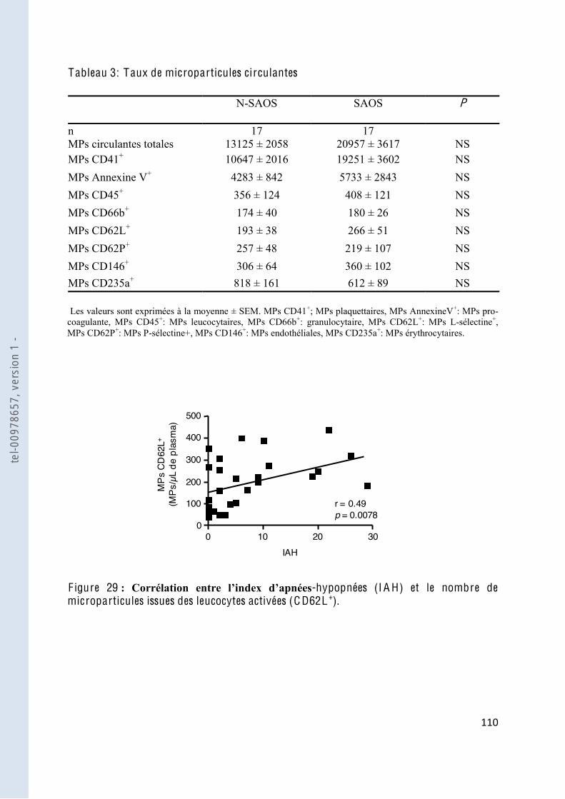
!!+"
"
Tableau 3: Taux de microparticules ci rculantes
N-SAOS
SAOS
P
n MPs circulantes totales
17 13125 ± 2058
17 20957 ± 3617
NS
MPs CD41+ 10647 ± 2016 19251 ± 3602 NS MPs Annexine V+ 4283 ± 842 5733 ± 2843 NS MPs CD45+ 356 ± 124 408 ± 121 NS MPs CD66b+ 174 ± 40 180 ± 26 NS MPs CD62L+ 193 ± 38 266 ± 51 NS MPs CD62P+ 257 ± 48 219 ± 107 NS MPs CD146+ 306 ± 64 360 ± 102 NS MPs CD235a+ 818 ± 161 612 ± 89 NS Les valeurs sont exprimées à la moyenne ± SEM. MPs CD41+; MPs plaquettaires, MPs AnnexineV+: MPs pro-coagulante, MPs CD45+: MPs leucocytaires, MPs CD66b+: granulocytaire, MPs CD62L+: MPs L-sélectine+, MPs CD62P+: MPs P-sélectine+, MPs CD146+: MPs endothéliales, MPs CD235a+: MPs érythrocytaires.
F igure 29 -hypopnées (I A H) et le nombre de microparticules issues des leucocytes activées (C D62L +).
0
300
MPs
CD
62L+
(MPs
/µL
de p
lasm
a)
100
200
400
r = 0.49p = 0.0078
500
0 10 20 30IAH
tel-0
0978
657,
ver
sion
1 -
!!!"
"
Nous
avons comparé les différentes réponses à une solution riche en potassium, et le traitement par
les MPs SAOS a montré une augmentation significative de la réponse par rapport aux souris
non traitées (Figure 30 B). -46619, un analogue du TXA2, qui
produit une augmentation de la tension des anneaux aortiques selon une réponse dépendante
-HT.
En effet, nous avons observé une augmentation -46619 chez les
souris traitées avec les MPs SAOS (Emax, 6.4 ± 0.4 mN/mm) comparés à celles traitées avec
les MPs N-SAOS (Emax, 4.9 ± 0.3 mN/mm) ou le groupe non traité (Emax, 5 ± 0.2 mN/mm).
En revanche la sensibilité à cet agoniste vasoconstricteur semble inchangée entre les
différents traitements (Figure 30 C). Le traitement par les MPs SAOS induit donc une
Pour plus de clarté, nous nous sommes par la suite seulement intéressés à la réponse à la 5-
musculaire lisse).
Pour répondre à cette question, la réponse à la 5-
dépourvus d -HT tout à
même significativement diminuée par rapport aux souris non traitées (Emax, 1.4 ± 0.2
mN/mm vs Emax, 0.7 ± 0.1 mN/mm) (Figure 30 D). Ces résultats suggèrent que
-
l
différents inhibiteurs des voies des NOS et des COX.
tel-0
0978
657,
ver
sion
1 -

!!#"
"
F igure 30 : L es microparticules SA OS augmentent la réponse contractile aux agents vasoconstricteurs. Courbe concentration-réponse à la 5-HT avec endothélium fonctionnel (A, nsouris non traitées ou traitées par les MPs N-SAOS ou SAOS. Réponse contractile à une solution hyperpotassique (B, n=6) et courbe concentration-réponse au U-46619 (C, nsouris non traitées ou traitées par les MPs N-SAOS ou SAOS. Courbe concentration-réponse à la 5-HT avec endothélium ou non fonctionnel (D, n raitées par les MPs N-SAOS ou SAOS. * P<0.05, **P<0.01 et ***P<0.001.
-10 -9 -8 -7 -5Log [U-46619] (M)
Cont
ract
ion
(mN/
mm
)
0
C
-6
2
4
6
8 CtlMPs N-SAOSMPs SAOS
** **
-9 -8 -7 -6 -4Log [5-HT] (M)
Cont
ract
ion
(mN/
mm
)
0
A
-5
1
2
3
4 CtlMPs N-SAOSMPs SAOS
*** ***
-9 -8 -7 -6 -4Log [5-HT] (M)
Cont
ract
ion
(mN/
mm
)
0
D
-5
1
2
3
4 CtlMPs N-SAOSMPs SAOS
**
0
5
10
15
Cont
ract
ion
(mN/
mm
)
N-SAOS SAOSCtl
B
tel-0
0978
657,
ver
sion
1 -

!!$"
"
vité vasculaire induite par les MPs
SA OS
-NA a été étudié en
réponse à la 5-
vasomoteur provoquait une hyperréactivité à la 5-HT dans le groupe contrôle ou traité par les
MPs N- NO
MPs N-SAOS ou au contrôle suggérant une altération de la voie du NO (Figure 31 A-C). Par
ailleurs, la mesure de la production de NO réalisée par RPE a montré une forte diminution de
anneaux non traités ou à ceux traités par les MPs N-SAOS (Figure 32 A).
tion de la
32 B). En revanche, uniquement le
traitement par les MPs SAOS provoque une diminution de la phosphorylation sur le site
activateur, ainsi que sur le site inhibiteur de la eNOS (Figure 32 C-D). Ainsi le ratio entre
eNOS (Figure 32 E).
er réactivité vasculaire induite par les
MPs SA OS
r
de la COX-2, le NS-398. -HT a été
la diminution était plus
importante dans le groupe SAOS (60 %) comparé aux groupes N-SAOS et contrôle (47 et 48
%, respectivement) (Figure 33 A-C). Ces résultats suggèrent que des métabolites sensibles à
par la 5-HT, et ceci spécialement dans les
aortes des animaux traités par les MPs SAOS.
tel-0
0978
657,
ver
sion
1 -
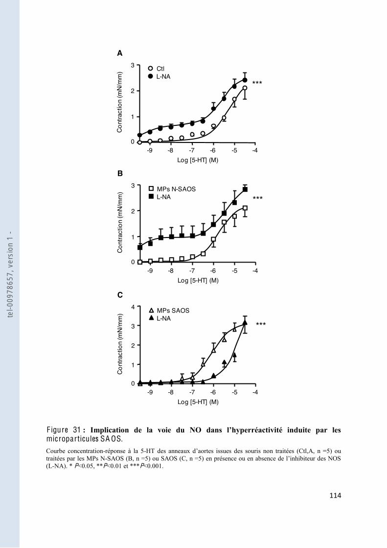
!!%"
"
F igure 31 microparticules SA OS. Courbe concentration-réponse à la 5-HT des atraitées par les MPs N-(L-NA). * P<0.05, **P<0.01 et ***P<0.001.
A
-9 -8 -7 -6 -4Log [5-HT] (M)
Cont
ract
ion
(mN/
mm
)
0-5
1
2
3 CtlL-NA
***
B
-9 -8 -7 -6 -4Log [5-HT] (M)
Cont
ract
ion
(mN/
mm
)
0-5
1
2
3 MPs N-SAOSL-NA ***
C
-9 -8 -7 -6 -4Log [5-HT] (M)
Cont
ract
ion
(mN/
mm
)
0-5
1
2
4 MPs SAOSL-NA
3 ***
tel-0
0978
657,
ver
sion
1 -
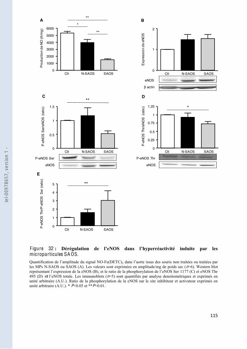
!!&"
"
F igure 32 microparticules SA OS.
-Fe(DETC)2 les MPs N-SAOS ou SAOS (A). Les valeurs sont exprimées en amplitude/mg de poids sec (n=6). Western blot
495 (D) vs n=5) sont quantifiés par analyse densitométriques et exprimés en unité arbitraire (A.U.). Ratio de la phosphorylation de la eNOS sur le site inhibiteur et activateur exprimés en unité arbitraire (A.U.). * P<0.05 et **P<0.01.
0
2000
4000Pr
oduc
tion
de N
O (A
/mg)
N-SAOS SAOSCtl
1000
3000
***
**
6000
5000
A
0
1
2
Expr
essi
on d
e eN
OS
N-SAOS SAOSCtl
eNOS
actin
B
**
0
1
P-eN
OS
Ser/e
NOS
(ratio
)
N-SAOS SAOSCtl
P-eNOS Ser
eNOS
0.5
1.5
C
*
0
0.5
1
P-eN
OS
Thr/e
NOS
(ratio
)
N-SAOS SAOSCtl
P-eNOS Thr
eNOS
0.25
0.75
1.25
D
0
2
4
P-eN
OS
Thr/P
-eNO
S Se
r (ra
tio)
N-SAOS SAOSCtl
1
3
5 **
E
tel-0
0978
657,
ver
sion
1 -
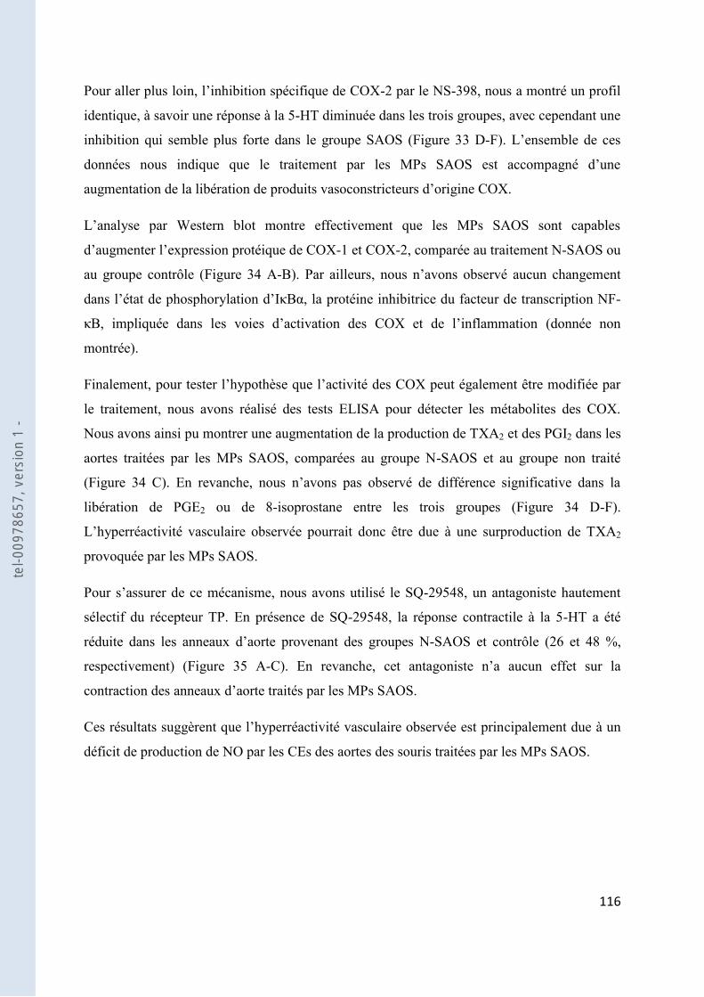
!!'"
"
-2 par le NS-398, nous a montré un profil
identique, à savoir une réponse à la 5-HT diminuée dans les trois groupes, avec cependant une
inhibition qui semble plus forte dans le groupe SAOS (Figure 33 D-
augmentation de la
-1 et COX-2, comparée au traitement N-SAOS ou
au groupe contrôle (Figure 34 A-B).
-
(donnée non
montrée).
le traitement, nous avons réalisé des tests ELISA pour détecter les métabolites des COX.
Nous avons ainsi pu montrer une augmentation de la production de TXA2 et des PGI2 dans les
aortes traitées par les MPs SAOS, comparées au groupe N-SAOS et au groupe non traité
(Figure 34 C
libération de PGE2 ou de 8-isoprostane entre les trois groupes (Figure 34 D-F).
XA2
provoquée par les MPs SAOS.
-29548, un antagoniste hautement
sélectif du récepteur TP. En présence de SQ-29548, la réponse contractile à la 5-HT a été
roupes N-SAOS et contrôle (26 et 48 %,
respectivement) (Figure 35 A-
cipalement due à un
déficit de production de NO par les CEs des aortes des souris traitées par les MPs SAOS.
tel-0
0978
657,
ver
sion
1 -

!!("
"
F igure 33 microparticules SA OS. Courbe concentration-réponse à la 5-traitées par les MPs N-(Indométacine). Courbe concentration-réponse à la 5- s issues des souris non traitées (Ctl, D, n =5) ou traitées par les MPs N-des COX-2 (NS-398). * P<0.05, **P<0.01 et ***P<0.001.
A
-9 -8 -7 -6 -4Log [5-HT] (M)
Cont
ract
ion
(mN/
mm
)
0-5
1
2
3 CtlIndométhacine
*
B
-9 -8 -7 -6 -4Log [5-HT] (M)
Cont
ract
ion
(mN/
mm
)
0-5
1
2
3 MPs N-SAOSIndométhacine
***
C
-9 -8 -7 -6 -4Log [5-HT] (M)
Cont
ract
ion
(mN/
mm
)
0-5
1
2
4 MPs SAOSindométhacine
3
***
D
-9 -8 -7 -6 -4Log [5-HT] (M)
Cont
ract
ion
(mN/
mm
)
0-5
1
2
3 CtlNS-398
*
E
-9 -8 -7 -6 -4Log [5-HT] (M)
Cont
ract
ion
(mN/
mm
)
0-5
1
2
3 MPs N-SAOSNS-398
**
F
-9 -8 -7 -6 -4Log [5-HT] (M)
Cont
ract
ion
(mN/
mm
)
0-5
1
2
4 MPs SAOSNS-398
3
**
tel-0
0978
657,
ver
sion
1 -
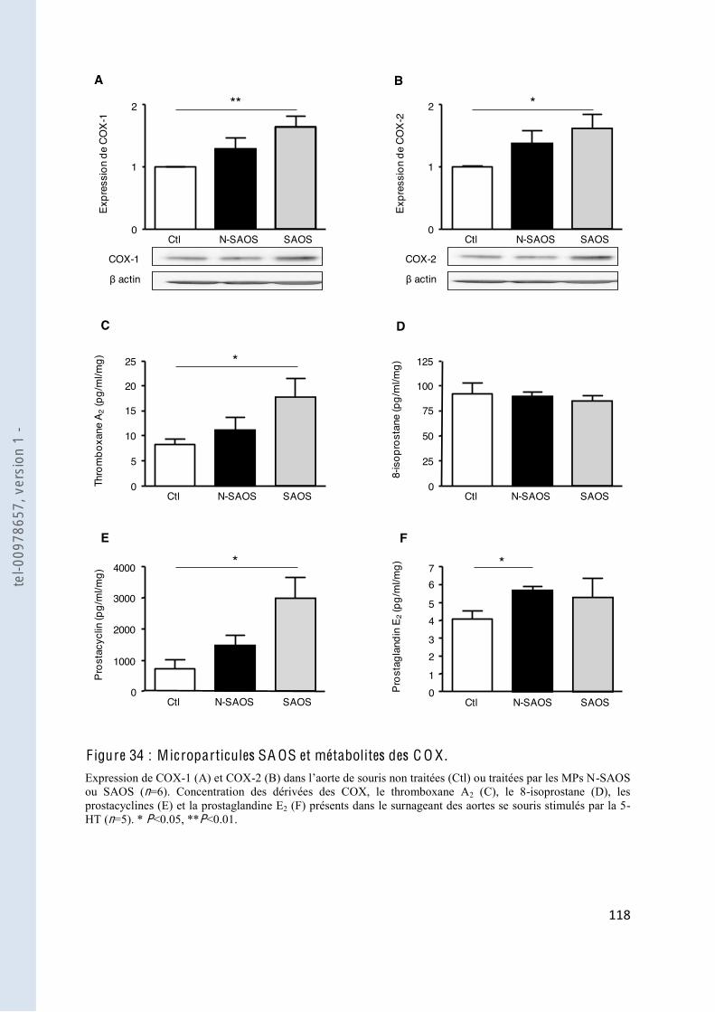
!!)"
"
F igure 34 : Microparticules SA OS et métabolites des C O X . Expression de COX-1 (A) et COX- -SAOS ou SAOS (n=6). Concentration des dérivées des COX, le thromboxane A2 (C), le 8-isoprostane (D), les prostacyclines (E) et la prostaglandine E2 (F) présents dans le surnageant des aortes se souris stimulés par la 5-HT (n=5). * P<0.05, **P<0.01.
0
1
2Ex
pres
sion
de
COX-
1
N-SAOS SAOSCtl
COX-1
actin
**
0
1
2
Expr
essi
on d
e CO
X-2
N-SAOS SAOSCtl
COX-2
actin
*
0
50
1008-
isop
rost
ane
(pg/
ml/m
g)
N-SAOS SAOSCtl
25
75
125
0
10
20
Thro
mbo
xane
A2
(pg/
ml/m
g)
N-SAOS SAOSCtl
5
15
25 *
0
2000
4000
Pros
tacy
clin
(pg/
ml/m
g)
N-SAOS SAOSCtl
1000
3000
*
0
2
4
Pros
tagl
andi
n E 2
(pg/
ml/m
g)
N-SAOS SAOSCtl
1
3
*
5
67
A B
C D
E F
tel-0
0978
657,
ver
sion
1 -
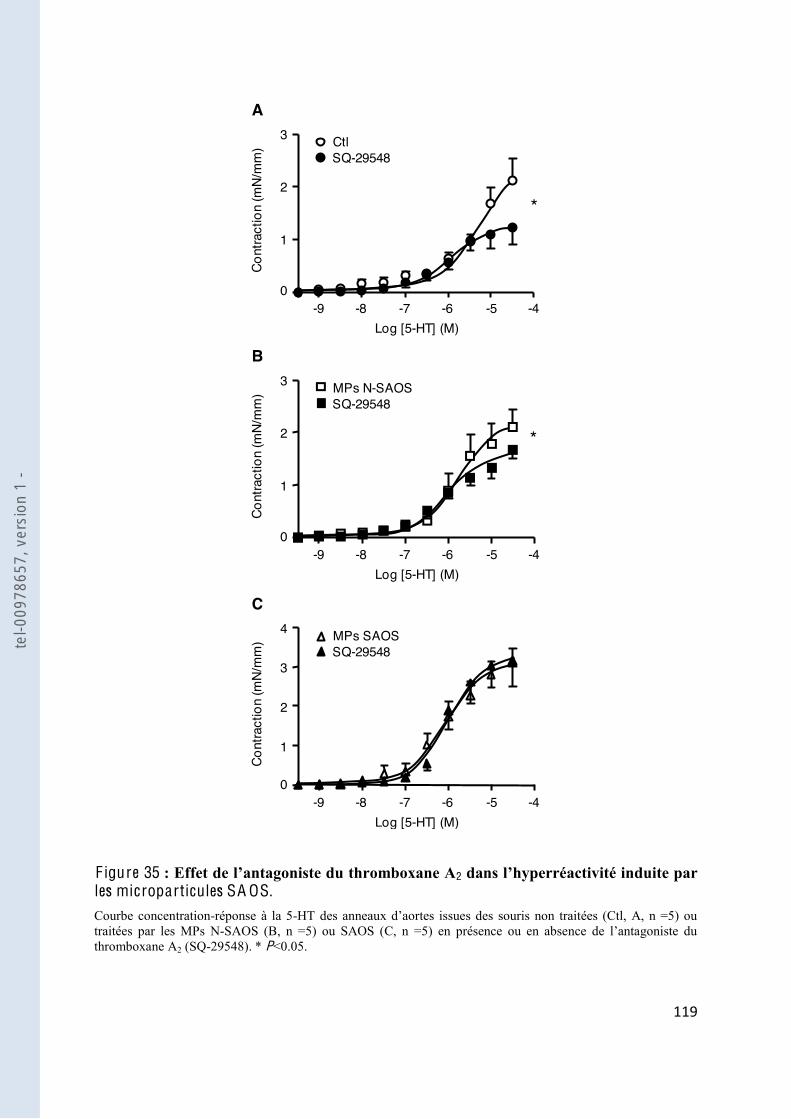
!!*"
"
F igure 35 2 les microparticules SA OS. Courbe concentration-réponse à la 5-traitées par les MPs N-thromboxane A2 (SQ-29548). * P<0.05.
C
-9 -8 -7 -6 -4Log [5-HT] (M)
Cont
ract
ion
(mN/
mm
)
0-5
1
2
4 MPs SAOSSQ-29548
3
A
-9 -8 -7 -6 -4Log [5-HT] (M)
Cont
ract
ion
(mN/
mm
)
0-5
1
2
3 CtlSQ-29548
*
B
-9 -8 -7 -6 -4Log [5-HT] (M)
Cont
ract
ion
(mN/
mm
)
0-5
1
2
3 MPs N-SAOSSQ-29548
*
tel-0
0978
657,
ver
sion
1 -
!#+"
"
Interaction de la voie du N O et des C O X
Finalement, pour étudier une éventuelle interaction entre les deux voies, nous avons utilisé en
-NA) et celui des COX (indométacine). Nous avons
-HT dans les aortes des animaux contrôles et
ceux traités par les MPs N-
vaisseaux des souris traitées par les MPs
SAOS (Figure 36
les MPs SAOS est liée à la fois à un mécanisme sensible à la voie du NO, mais également à la
voie des COX.
tel-0
0978
657,
ver
sion
1 -

!#!"
"
F igure 36 : Interaction entre les voies du N O et des C O X . Courbe concentration-réponse à la 5-traitées par les MPs N-SAOS (B, n =5) ou SAOS (C, n =5) en présence ou en absence de L-NA plus indométacine. ***P<0.001.
C
-9 -8 -7 -6 -4Log [5-HT] (M)
Cont
ract
ion
(mN/
mm
)
0-5
1
2
4 MPs SAOSL-NA + indométhacine
3
***
A
-9 -8 -7 -6 -4Log [5-HT] (M)
Cont
ract
ion
(mN/
mm
)
0-5
1
2
3 CtlL-NA + indométhacine
***
B
-9 -8 -7 -6 -4Log [5-HT] (M)
Cont
ract
ion
(mN/
mm
)
0-5
1
2
3 MPs N-SAOSL-NA + indométhacine ***
tel-0
0978
657,
ver
sion
1 -

!##"
"
I V . Discussion
Ce travail a fait suite à celui initié au sein du laboratoire par le Dr Priou sur le rôle essentiel
joué par les MPs issues de patients SAOS dans la dysfonction endothéliale observée dans
aortes de souris injectées par des MPs issues du sang de patients SAOS ou de sujets sains.
i.v. de MPs SAOS dans les souris
médiateur vasodilatateur
tel que le NO, et en augmentant la production des métabolites des COX. Cette hyperréactivité
e COX-1 et COX-
inter-cellulaire en favorisant la dysfonction vasculaire durant le SAOS.
De nombreuses pathologies cardiovasculaires sont associées à une dysfonction vasculaire
et/ou une altération
de la réponse vasoconstrictrice des CML. Il est maintenant largement accepté que les MPs
on de cette réponse vasculaire [194,195]. Concernant le
de MPs provenant des leucocytes activés et leur capacité à induire une dysfonction
endothéliale in vitro et ex vivo [93]. Peu de données existent dans la littérature sur la réponse
constrictrice chez les patients SAOS et ces données sont très contradictoires. Cependant la
[198] et un lien entre la
sév [199], lien confirmé grâce
aux modèles animaux [200,201]. La vasoconstriction induite par les apnées a été observée
[202] et les doigts [203] des patients SAOS. Ces données sont surprenantes
des lits vasculaires [204,205] suggérant que les épisodes répétés favorisent une
altération des mécanismes de base du contrôle du tonus vasculaire. Effectivement, dans
norépinephrine [206] [207]. Dans
tel-0
0978
657,
ver
sion
1 -
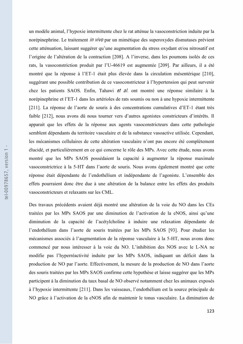
!#$"
"
ttente chez le rat atténue la vasoconstriction induite par la
norépinephrine. Le traitement in vivo par un mimétique des superoxydes dismutases prévient
ne de altération de la contraction [208]
-46619 est augmentée [209]. Par ailleurs, il a été
-1 était plus élevée dans la circulation mésentérique [210],
chez les patients SAOS. Enfin, Tahawi et al. ont montré une réponse similaire à la
-1 dans les artérioles de rats soumis ou non à une hypoxie intermittente
[211] -1 étant très
faible [212]
apparait que les effets de la réponse aux agents vasoconstricteurs dans cette pathologie
semblent dépendants du territoire vasculaire et de la substance vasoactive utilisée. Cependant,
élucidé, et particulièrement en ce qui concerne le rôle des MPs. Avec cette étude, nous avons
montré que les MPs SAOS possédaient la capacité à augmenter la réponse maximale
vasoconstrictrice à la 5-
réponse était
effets pourraient donc être due à une altération de la balance entre les effets des produits
vasoconstricteurs et relaxants sur les CML.
Des travaux précédents avaient déjà montré une altération de la voie du NO dans les CEs
es par les MPs SAOS [93]. Pour étudier les
-HT, nous avons donc
-NA ne
les MPs SAOS, indiquant un déficit dans la
des souris traitées par les MPs SAOS confirme cette hypothèse et laisse suggérer que les MPs
participent à la diminution du taux basal de NO observé notamment chez les animaux exposés
[211]
tel-0
0978
657,
ver
sion
1 -
!#%"
"
ation de la eNOS retrouvée dans les aortes des souris traitées par les MPs SAOS
Une seconde voie connue pour être altérée dans la réactivité vasculaire induite par des MPs
est la voie des COX que nous avons étudiée. De façon intéressante, le traitement par
-sélectif des COX, diminue de façon plus importante la
réponse à la 5- ré au traitement
par les MPs N-
-1 et COX-2 car le
NS-398 diminue également cette contraction mais de façon moins prononcé. Ainsi, les effets
-1 et de COX-2, et
également une augmentation du TXA2 et de PGI2 sans modifier le 8-isoprostane ou la PGE2.
2 et du TXA2 a déjà été montrée chez les patients
SAOS, et il a été suggéré que la hausse de PGI2 était un mécanisme compensatoire et
[213]. Il est raisonnable
la réponse maximale des agents vasoconstricteurs en
2. Cependant,
2 ne modifie pas la contraction en réponse à la 5-HT des
traitées par les MPs SAOS. Ces résultats suggèrent que
NO par les CEs des aortes des souris traitées par les MPs SAOS.
Même si elles sont complexes et pas encore très bien comprises, des interactions entre le NO
[214,215]. Par
[216]. Nous avons donc étudié les possibles interactions entre ces deux voies
des NOS prédomine dans les aortes de souris non traitées ou traitées par les MPs N-SAOS
favorisant une hyperréactivité à la 5-HT, le traitement des MPs SAOS provoque une
diminution de la réactivité des aortes. Il a été montré que le NO produit était capable de
diminuer la production de COX en inhibant la voie des « mitogen-activated protein kinase »
(MAPK) [217]
diminution du NO ne peut plus réguler cette voie provoquant ainsi une surexpression des
COX. Ces résultats suggèrent cependant que le NO et les COX agissent en combinaison selon
tel-0
0978
657,
ver
sion
1 -
!#&"
"
MPs SAOS.
En résumé, avec ce travail nous apportons des preuves selon lesquelles les MPs SAOS
production de NO et en activant la voie des COX. Par ailleurs, une éventuelle interaction entre
comme vecteur de la dysfonction vasculaire dans le SAOS est ainsi clairement souligné et
résumé sur la figure 37. Les effets combinés des MPs SAOS sur la relaxation et la contraction
des vaisseaux nous montrent clairement que les MPs ne doivent pas être considérées
uniquement comme
CEs comme
tel-0
0978
657,
ver
sion
1 -
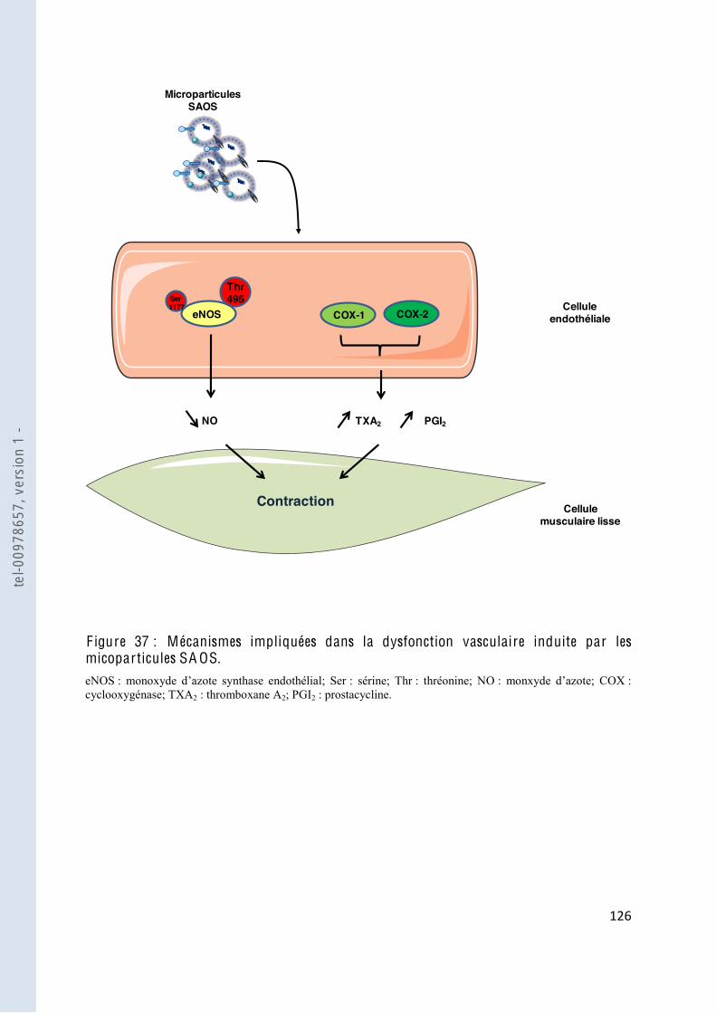
!#'"
"
Cellule endothéliale
Microparticules SAOS
Cellule musculaire lisse
eNOS
NO
COX-1
Thr495Ser
1177
Contraction
COX-2
TXA2 PGI2
F igure 37 : Mécanismes impliquées dans la dysfonction vasculaire induite par les micoparticules SA OS. eNOS : sérine; Thr : thréonine; NO : monxyde : cyclooxygénase; TXA2 : thromboxane A2; PGI2 : prostacycline.
tel-0
0978
657,
ver
sion
1 -

!#("
"
Partie I I I :
obstructives du sommeil et angiogenèse.
tel-0
0978
657,
ver
sion
1 -

!#)"
"
I .
Les travaux précédents effectués en collaboration avec le service de Pneumologie du CHU
SAOS.
Le SAOS est une pathologie caractérisée notamment par une hypoxie intermittente, et
c un
déséquilibre des facteurs angiogénique dans le sérum des patients [218,219]
-vasculaires chez les
patients SAOS. Par ailleurs, un des principaux effets des MPs sur le système cardio-vasculaire
est leur capacité à moduler le programme angiogénique en intervenant sur ses différentes
Nous avons donc voulu dans cette étude établir un lien entre les MPs issues de patients SAOS
tel-0
0978
657,
ver
sion
1 -
!#*"
"
I I . Matériel et méthodes
1. Matériel biologique
1.1. Microparticules issues de sujets présentant une suspicion de syndrome
Les patients utilisés pour cette étude sont issus de la même cohorte que pour le travail sur la
caractérisation des MPs ont donc été semblables au protocole décrit dans la partie
expérimentale.
1.2. Cellules endothé
Culture cellulaire
Les CEs humaines
(Promocell, Heidelberg, Allemagne). Les cellules ont été cultivées dans une flask de 75 cm2 et
le milieu « Endothelial cell growth medium » (MV2) (Promocell) était changé tous les 2 jours
10ème passage. Les cellules étaient ensuite réensemencées à une
densité de 10.000 cellules par cm!.
T raitement des cellules
Les cellules ont été traitées pendant 24 heures à la concentration circulante de MPs
déterminée pour chaque patient par cytométrie en flux. Lors des expériences utilisant
-1, les cellules étaient prétraitées pendant 45 minutes avec le
BQ-788 (5 µM, Sigma-Aldrich) puis les MPs étaient ajoutées.
2. Mesure du taux de V E G F
Le taux de VEGF a été mesuré sur le PPP
-Unis). Ce kit permettait la détection
du facteur de croissance polypeptidique de 165 acides aminés.
tel-0
0978
657,
ver
sion
1 -
!$+"
"
3. W estern blot
MPs
Les MPs concentrées étaient dosées directement par la méthode de Bradford afin de
déterminer la quantité de protéines à charger pour la migration.
Cellules
Après incubation avec les MPs pendant 24 heures, les cellules ont été lavées avec du PBS, et
traitées avec le tampon de lyse pendant 20 minutes sur la glace. Le lysat était récupéré et
placé dans des eppendorfs, puis centrifugé (15.000 g, 10 minutes, 4°C) et le surnageant
contenant les protéines était conservé à -80°C.
protocole indiqué dans la partie expérimentale I.
4. Prolifération cellulaire
Principe
Pour étudier la prolifération cellulaire, nous avons utilisé le kit CyQUANT® NF Cell
détermination du nombre de cellules en utilisant cet ADN grâce à une sonde fluorescente qui
.
Protocole
Les cellules ont été ensemencées dans une plaque 96 puits à raison de 5.104 cellules par cm2,
puis stimulées par les MPs. Après 24 heures, le milieu était aspiré doucement pour être
dye binding solution). Après
a été mesurée au spectrophotomètre (Biotek) à une
condition était réalisée en duplicat.
tel-0
0978
657,
ver
sion
1 -

!$!"
"
5. Mesure de la formation de capillaire sur Matrigel®
Principe
Le Matrigel® est une mixture protéique gélatineuse secrétée par les cellules de sarcomes
utilisé comme matrice extracellulaire pour la culture cellulaire. Ainsi les CEs sont capables de
Protocole
Après 24 heures
ng/ml) servant de contrôle positif, les cellules étaient détachées avec de la trypsine (Lonza).
Les cellules étaient ensuite réensemencées à une densité de 15.104 cellules par cm2 dans une
plaque 4 puits précoatés avec du Matrigel®.
Après avoir mis à +4°C le Matrigel pendant 16 heures, le co ait de la manière
suivante : sur la glace, 150 µl de Matrigel dilué dans du milieu sans sérum (dilution 1:1)
étaient déposés par puits dans une plaque 4 puits, en faisant bien attention à éviter les bulles.
Après solidification pendant 90 minutes à 37°C, 40 µl de Matrigel pur était rajouté pendant 60
minutes à 37°C pour bien homogénéiser la surface du coating.
La formation des tubes a été
phase (MOTIC AE21). La longueur des capillaires formés ont été
du logiciel ImageJ et représente la moyenne de 4 images différentes par expériences.
6. Analyse statistique des résultats
Les résultats étaient exprimés en moyenne ± SEM de n expériences, n représentant le nombre
de mesure pour les cellules. Un test Mann-Whitney (non paramétrique) a été utilisé. La
différence est considérée comme significative lorsque P < 0.05.
tel-0
0978
657,
ver
sion
1 -
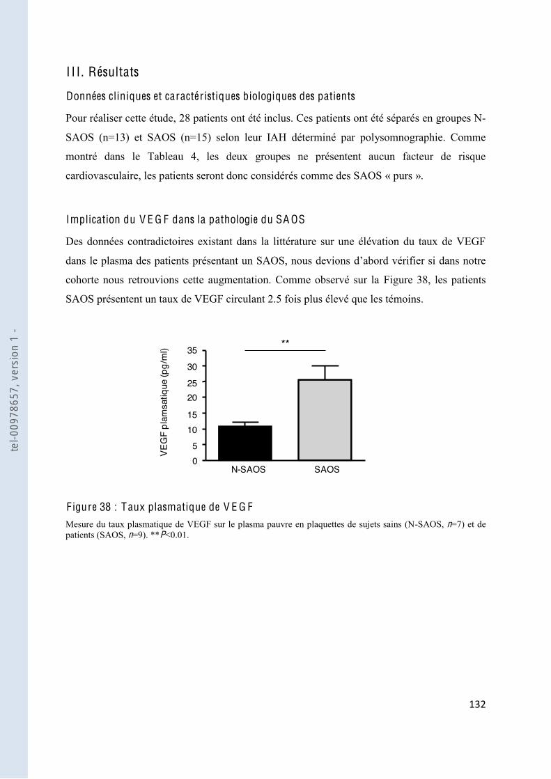
!$#"
"
I I I . Résultats
Données cliniques et caractéristiques biologiques des patients
Pour réaliser cette étude, 28 patients ont été inclus. Ces patients ont été séparés en groupes N-
SAOS (n=13) et SAOS (n=15) selon leur IAH déterminé par polysomnographie. Comme
montré dans le Tableau 4, les deux groupes ne présentent aucun facteur de risque
cardiovasculaire, les patients seront donc considérés comme des SAOS « purs ».
Implication du V E G F dans la pathologie du SA OS
Des données contradictoires existant dans la littérature sur une élévation du taux de VEGF
dans le plasma des patients présentant un SAOS, nous devions
cohorte nous retrouvions cette augmentation. Comme observé sur la Figure 38, les patients
SAOS présentent un taux de VEGF circulant 2.5 fois plus élevé que les témoins.
F igure 38 : Taux plasmatique de V E G F Mesure du taux plasmatique de VEGF sur le plasma pauvre en plaquettes de sujets sains (N-SAOS, n=7) et de patients (SAOS, n=9). **P<0.01.
0
10
20
VEG
F pl
amsa
tique
(pg/
ml)
N-SAOS SAOS
5
15
**
3025
35
tel-0
0978
657,
ver
sion
1 -
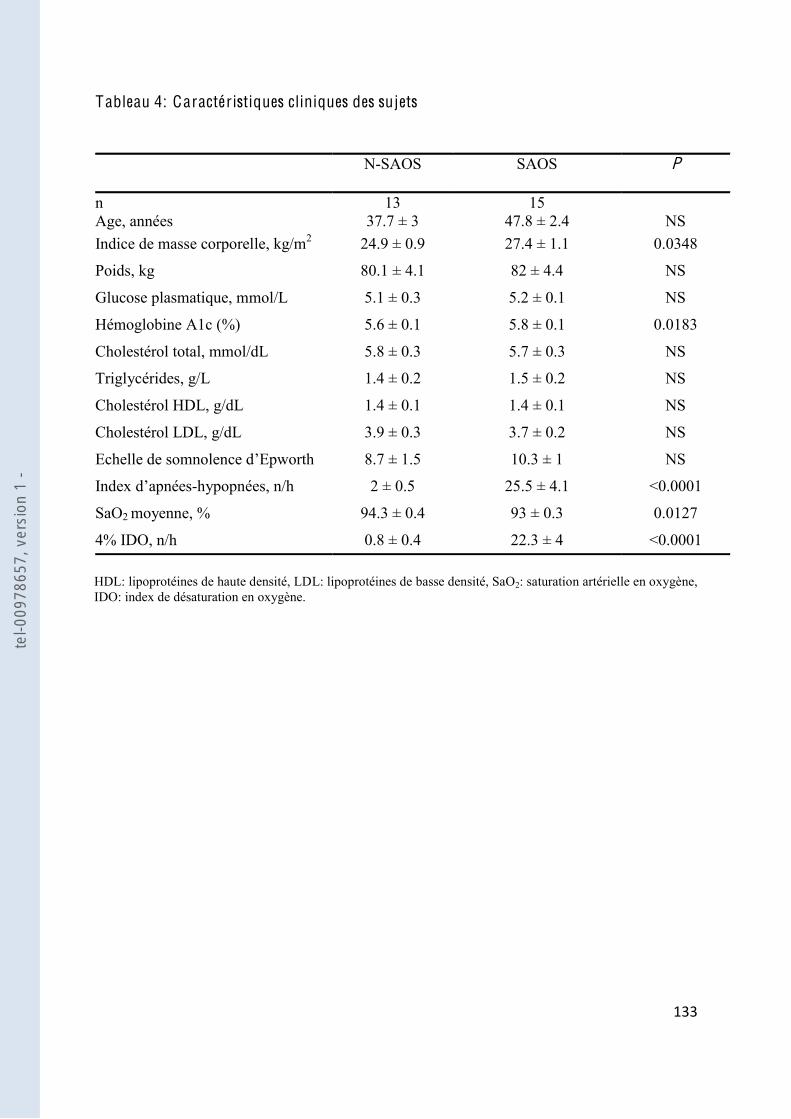
!$$"
"
Tableau 4: Caractéristiques cliniques des sujets
N-SAOS
SAOS
P
n Age, années
13 37.7 ± 3
15 47.8 ± 2.4
NS
Indice de masse corporelle, kg/m2 24.9 ± 0.9 27.4 ± 1.1 0.0348
Poids, kg 80.1 ± 4.1 82 ± 4.4 NS
Glucose plasmatique, mmol/L 5.1 ± 0.3 5.2 ± 0.1 NS
Hémoglobine A1c (%) 5.6 ± 0.1 5.8 ± 0.1 0.0183
Cholestérol total, mmol/dL 5.8 ± 0.3 5.7 ± 0.3 NS
Triglycérides, g/L 1.4 ± 0.2 1.5 ± 0.2 NS
Cholestérol HDL, g/dL 1.4 ± 0.1 1.4 ± 0.1 NS
Cholestérol LDL, g/dL 3.9 ± 0.3 3.7 ± 0.2 NS
8.7 ± 1.5 10.3 ± 1 NS
-hypopnées, n/h 2 ± 0.5 25.5 ± 4.1 <0.0001
SaO2 moyenne, % 94.3 ± 0.4 93 ± 0.3 0.0127
4% IDO, n/h 0.8 ± 0.4 22.3 ± 4 <0.0001 HDL: lipoprotéines de haute densité, LDL: lipoprotéines de basse densité, SaO2: saturation artérielle en oxygène, IDO: index de désaturation en oxygène.
tel-0
0978
657,
ver
sion
1 -
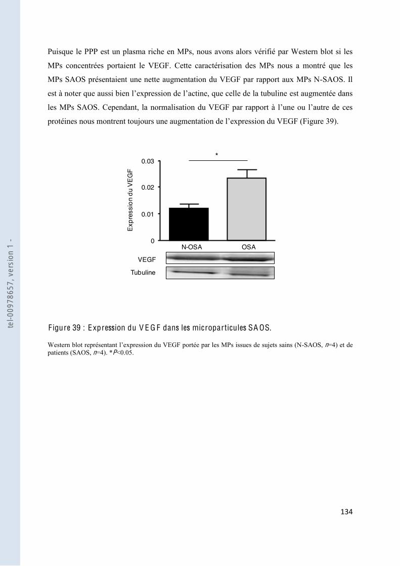
!$%"
"
Puisque le PPP est un plasma riche en MPs, nous avons alors vérifié par Western blot si les
MPs concentrées portaient le VEGF. Cette caractérisation des MPs nous a montré que les
MPs SAOS présentaient une nette augmentation du VEGF par rapport aux MPs N-SAOS. Il
est augmentée dans
les MPs SAOS. Cependant, la
39).
F igure 39 : Expression du V E G F dans les microparticules SA OS.
on du VEGF portée par les MPs issues de sujets sains (N-SAOS, n=4) et de patients (SAOS, n=4). *P<0.05.
VEGF
Tubuline
0
0.01
Expr
essi
on d
u VE
GF
N-OSA OSA
*
0.02
0.03
tel-0
0978
657,
ver
sion
1 -

!$&"
"
Implication des MPs SA OS dans le processus angiogénique
Avec ces données, nous avons voulu alors savoir si les MPs SAOS pouvaient être un vecteur
différentes étapes clefs du processus angiogénique.
Pour étudier ces effets, nous avons traités des CEs
les MPs à la concentration circulante déterminée pour chaque sujet et nous avons étudié la
formation in vitro de capillaires. En absence de traitement, les CEs sont capables de
de la longueur des capillaires formés. De la même façon, le traitement avec les MPs N-SAOS
façon similaire au VEGF la longueur des capillaires (Figure 38).
oxie, les CEs sont capables de libérer de nombreux médiateurs autocrines
-
-1, qui
Pour vérifier cette hypothèse nous avons prétraité les CEs avec le BQ-
récepteurs ETB présent sur ces cellules. Le traitement seul des CEs avec le BQ- it
aucun effet par rapport au contrôle. De la même façon, le traitement des CEs avec le BQ-788
ne modifie pas la formation de capillaires induite par les MPs N-SAOS. En revanche, le
traitement avec le BQ- es induit par les
MPs SAOS (Figure 40). Ces résultats suggèrent que les MPs SAOS seraient capables de
-1 par les CEs.
CEs. Nous avons ainsi observé
que 24 heures de traitement par les MPs SAOS permettaient une augmentation significative
de la prolifération par rapport aux cellules traitées par les MPs N-SAOS ou non traitées.
angiogénique décrit dans le SAOS.
tel-0
0978
657,
ver
sion
1 -

!$'"
"
Ctl N-SAOS SAOS
+BQ-788
VEGF
"
"
"
0
50
Long
ueur
des
cap
illaire
s (U
.A.)
N-SAOS SAOS
100
****
**
150
200
Ctl VEGF N-SAOS SAOSCtl
BQ-788 "
F igure 40 microparticules -1.
Images représentant la formation de réseau sur Matrigel par les cellules endothéliales en absence (Ctl) ou en présence de MPs de sujets sains (N- -
-788 (A). La , n=5). **P<0.01.
tel-0
0978
657,
ver
sion
1 -
!$("
"
I V . Discussion
Notre étude a permis de mettre en évidence un rôle des MPs dans différentes étapes de
résentaient bien une hausse
du VEGF, un facteur hautement angiogénique, dans leur plasma. Nous avons ensuite établi
importante chez les MPs issus de patients SAOS. Ces deux expériences nous ont permis de
supposer que les MPs pouvaient donc être impliquées dans le processus angiogénique. Nous
avons alors évalué la capacité des MPs à induire la formation de tubules par les CEs, et nous
avons remarqué que le traitement par les MPs SAOS augmentait la longueur des structures
-
-1 par les CEs en réponse à la stimulation par les MPs. Enfin, nous avons
qui est la prolifération des CEs.
peu étudié, et concerne principalement la mesure de paramètres biochimiques. Parmi ces
paramètres, le taux de VEGF [220], a été mesuré chez
les patients. Le VEGF est un vasodilatateur qui induit la prolifération des CEs, augmente la
perméabilité vasculaire, et favorise la production de NO, qui pourrait jouer un rôle dans la
pathogénèse de nombreuses maladies hypoxiques [221]. Un travail démontrant un rôle du
VEGF dans le SAOS serait très pertinent, puisque le SAOS est considéré comme un facteur
de risque cardiovasculaire aggravant [222]. Par ailleurs, puisque le VEGF est un facteur induit
Ainsi les
travaux montrant une altération du taux de VEGF suggèrent que le VEGF pourrait être un
marqueur sérique chez les patients SAOS [223].
En effet, il a été rapporté une augmentation du taux de VEGF dans le sérum des patients
corrélé à la sévérité du SAOS [224-226]. Ces études ont été assez critiquées et souffrent
sur le poids des sujets, il a en effet été rapporté une élévation du taux de VEGF chez les
patients obèses [227]. La seconde limite concerne le fait que le VEGF a été mesuré dans le
les plaquettes sont le principal
transporteur de VEGF [228], même si le décompte en plaquettes étaient le même dans les
tel-0
0978
657,
ver
sion
1 -
!$)"
"
deux populations. Deux autres études ont alors étudié le taux de VEGF dans le plasma des
patients, et ces études retrouvent un taux de VEGF beaucoup plus faible que les taux des
études précédentes. Lavie et al. ont ainsi montré une augmentation du taux de VEGF corrélé à
[229]
effectué sur le plasma des patients issus de
cohortes appariées ne démontre plus cette augmentation de VEGF, suggérant que cette
[230].
le principal transporteur de VEGF, nous avons mesuré le VEGF sur le plasma PPP de patients
Dans notre étude, nous montrons que les patients SAOS
présentent un taux plasmatique de VEGF significativement plus élevé par rapport aux sujets
t être porté par les MPs. Nous avons
alors vérifié que les MPs exprimaient le VEGF, et nous avons observé que les MPs SAOS
exprimaient plus fortement le VEGF que les MPs issus des sujets témoins. Les MPs
pourraient donc être une des explications possibles à la hausse du VEGF circulant.
Nous avons ensuite vérifié si les MPs possédaient une activité angiogénique. De nombreuses
données dans la littérature démontrent en effet une participation des MPs dans le processus
angiogénique, en favorisant ou en inhibant la formation de structures capillaires [86,231] et
[55]. Les MPs des patients SAOS ont la capacité
par les CEs suggérant une activité pro-angiogénique portée par les MPs SAOS. Puisque le
- -
1 peut engendrer une réponse angiogénique par les CEs in vitro [232], nous avons émis
- -1
présent sur les CEs, nous avons remarqué que la réponse des CEs aux MPs SAOS était abolie.
-1 par les CEs -1
va alors activer les CEs en se fixant sur le récepteur ETB. Par ailleurs, nous avons également
on des CEs, une autre étape
tel-0
0978
657,
ver
sion
1 -

!$*"
"
changement, indiquant probablement une régulation plus précoce de ces voies de signalisation
(résultats non montrés).
Le processus angiogénique dans le SAOS est très peu détaillé dans la littérature. Chez les
développement des vaisseaux coronaires collatéraux lorsque ces patients présentaient
également un SAOS [233]
dans les muscles squelettiques de patients SAOS, et ces capillaires expriment fortement les
marqueurs de prolifération des CEs. Ce réseau pourrait probablement favoriser
grandement diminuées, de ces patients [234]. Selon ces observations, il a été suggéré
cardio-vasculaires et le VEGF pourrait contribuer ainsi à ce mécanisme protecteur.
les MPs (Figure 41).
tel-0
0978
657,
ver
sion
1 -
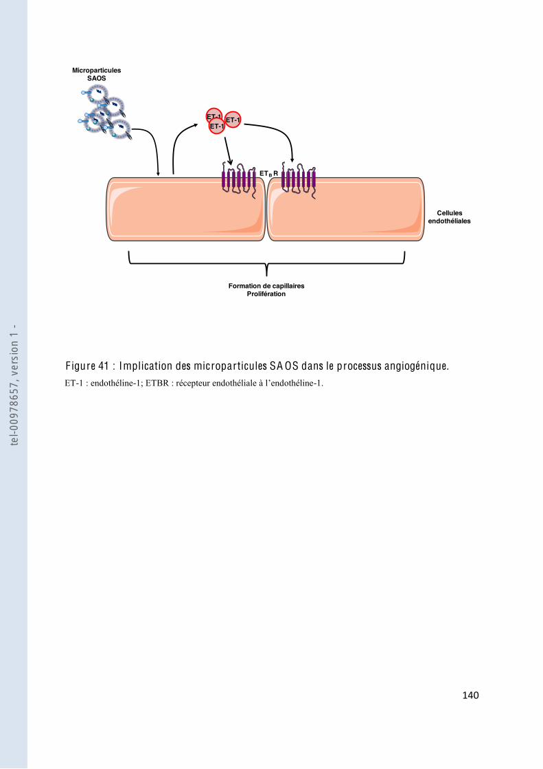
!%+"
"
Cellules endothéliales
Microparticules SAOS
ET-1 ET-1ET-1
ETB R
Formation de capillairesProlifération
F igure 41 : Implication des microparticules SA OS dans le processus angiogénique. ET-1 : endothéline-1; ETBR -1.
tel-0
0978
657,
ver
sion
1 -

!%!"
"
C O N C L USI O N & PE RSPE C T I V ES
tel-0
0978
657,
ver
sion
1 -

!%#"
"
Les travaux de ces 15 dernières années ont montré que les MPs jouent un rôle dans la
quées dans la progression des complications cardiovasculaires,
les MPs peuvent donc être une nouvelle cible thérapeutique. Paradoxalement, il existe des
preuves que les MPs peuvent avoir des effets bénéfiques, et les MPs pourraient être
potentiellement développés comme nouvel outil thérapeutique. Il y ainsi un équilibre fragile
entre les effets bénéfiques et délétères des MPs dans la physiopathologie vasculaire.
nouvel aspect important de la physiologie vasculaire et a permis de mettre en évidence de
nombreux points communs entre les deux pathologies vasculaires hypoxiques étudiées et les
MPs.
Nos premiers résultats ont mis en évidence une augmentation du taux circulant de MPs dans
hypoxiques est associée à un effet propre de ces MPs sur les CEs en diminuant la production
de NO. Les MPs ont également un effet différentiel selon le territoire vasculaire étudié en
augmentant le stress oxydant uniquement dans les CEs , ces MPs
nc établi que les MPs participaient à la dysfonction
endothéliale observée dans ce modèle. La question se pose maintenant de savoir si les MPs
normoxiques pendan
pathologie ou amplifient la dysfonction préexistante.
La seconde partie des résultats a confirmé des résultats préalablement établis dans la
pathologie du SAOS, à savoir une corrélation positive entre les MPs issues des leucocytes
activés et la sévérité de cette pathologie. Par ailleurs, avec cette étude, nous avons également
tion de MPs SAOS dans les souris favorisait une hyperréactivité vasculaire
on de
tel-0
0978
657,
ver
sion
1 -
!%$"
"
établi un effet délétère des MPs en participant à la dysfonction vasculaire observée dans le
SAOS.
Dans la dernière partie de nos résultats, nous avons montré que les MPs SAOS étaient
capables
CEs. Nous avons également montré que ce dernier mécanisme était dépendant du récepteur à
-1. Les MPs SAOS pourraient alors partic
raient alors avoir un rôle antagoniste (ange et
démon) dans le SAOS, elles seraient délétères au niveau de la fonction vasculaire, et
énotype
des MPs lors des pathologies étudiées. Puisque le taux de MPs circulantes est très
lors un enjeu primordial dans le traitement des pathologies. En effet, nous avons ainsi montré
particulière de MPs pourrait être un des marqueurs
raitement. Egalement, la modulation du taux de
MPs pourrait diminuer les effets délétères induits par les MPs. Dans cet optique, de nombreux
essai dans le temps du taux de
MPs ion de différentes thérapies (www.clinicaltrials.gov). Cependant, il est
important de souligner que les changements des taux plasmatiques de MPs peuvent être dus
-vie et
général de la pathologie.
Les études menées ont également mis en évidence la relevance physiopathologique des MPs
les pathologies étudiées. Il serait donc intéressant de pouvoir cibler directement les MPs.
tel-0
0978
657,
ver
sion
1 -
!%%"
"
Ps
devient dès lors compliquée car les acteurs impliqués dans la formation des MPs (kinases,
phosphatases, calcium) sont ubiquitaires et le ciblage spécifique de ces acteurs est impossible
Egalement, nous ne connaissons pas encore exactement les différentes interactions entre les
MPs et leurs cellules cibles, et les récepteurs impliqués, et les effets induits par les MPs. Des
analyses du transcriptome, du protéome et des ARNmi sont ainsi nécessaires pour identifier
tous les composants des MPs et accroitre nos connaissances dans les effets biologiques que
les MPs peuvent induire.
La compréhension du rôle précis des MPs dans les fonctions cardio-vasculaires peut être utile
non seulement dans la connaissance des mécanismes physiopathologiques, mais peut
également avoir des implications dans les traitements futurs de ces pathologies.
tel-0
0978
657,
ver
sion
1 -

!%&"
"
A NN E X ES
tel-0
0978
657,
ver
sion
1 -
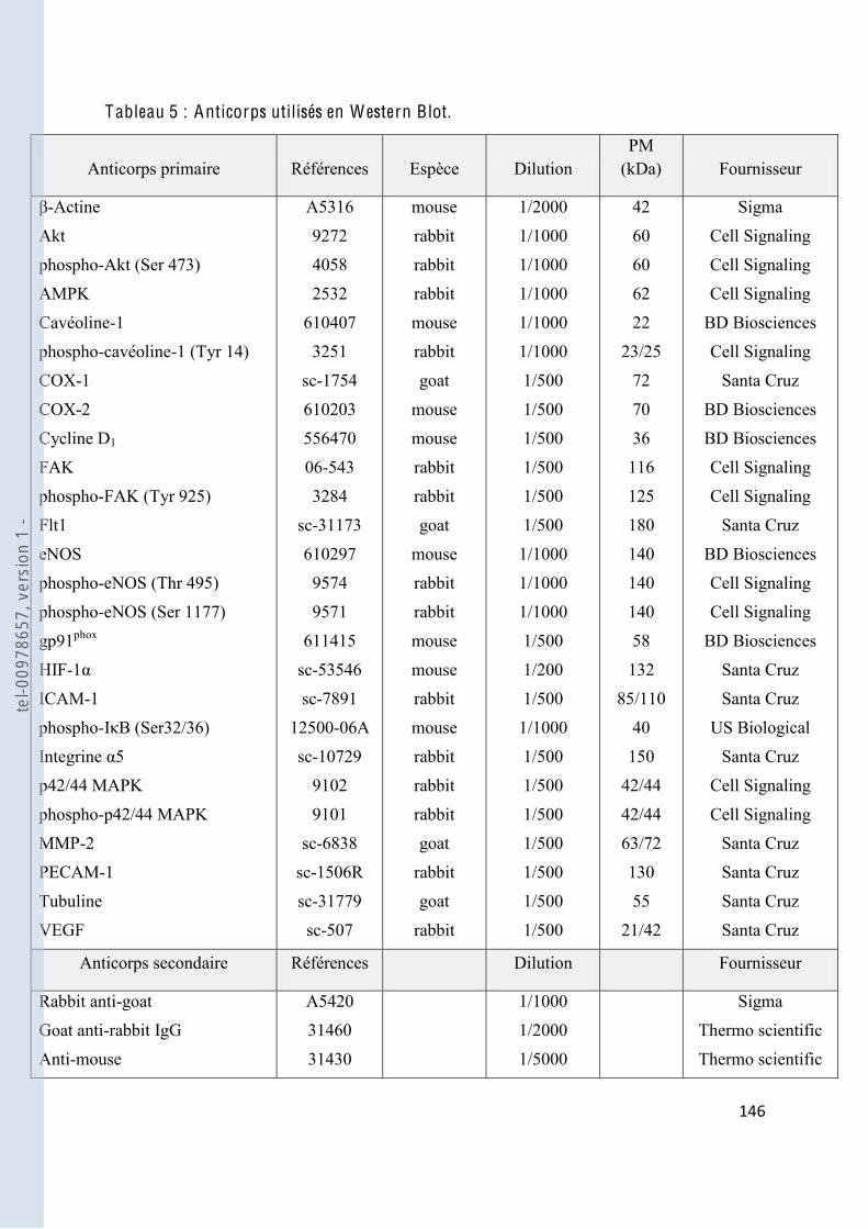
!%'"
"
Tableau 5 : Anticorps utilisés en W estern Blot.
Anticorps primaire Références Espèce Dilution PM
(kDa) Fournisseur
-Actine
Akt
phospho-Akt (Ser 473)
AMPK
Cavéoline-1
phospho-cavéoline-1 (Tyr 14)
COX-1
COX-2
Cycline D1
FAK
phospho-FAK (Tyr 925)
Flt1
eNOS
phospho-eNOS (Thr 495)
phospho-eNOS (Ser 1177)
gp91phox
HIF-1
ICAM-1
phospho-I B (Ser32/36)
Integrine 5
p42/44 MAPK
phospho-p42/44 MAPK
MMP-2
PECAM-1
Tubuline
VEGF
A5316
9272
4058
2532
610407
3251
sc-1754
610203
556470
06-543
3284
sc-31173
610297
9574
9571
611415
sc-53546
sc-7891
12500-06A
sc-10729
9102
9101
sc-6838
sc-1506R
sc-31779
sc-507
mouse
rabbit
rabbit
rabbit
mouse
rabbit
goat
mouse
mouse
rabbit
rabbit
goat
mouse
rabbit
rabbit
mouse
mouse
rabbit
mouse
rabbit
rabbit
rabbit
goat
rabbit
goat
rabbit
1/2000
1/1000
1/1000
1/1000
1/1000
1/1000
1/500
1/500
1/500
1/500
1/500
1/500
1/1000
1/1000
1/1000
1/500
1/200
1/500
1/1000
1/500
1/500
1/500
1/500
1/500
1/500
1/500
42
60
60
62
22
23/25
72
70
36
116
125
180
140
140
140
58
132
85/110
40
150
42/44
42/44
63/72
130
55
21/42
Sigma
Cell Signaling
Cell Signaling
Cell Signaling
BD Biosciences
Cell Signaling
Santa Cruz
BD Biosciences
BD Biosciences
Cell Signaling
Cell Signaling
Santa Cruz
BD Biosciences
Cell Signaling
Cell Signaling
BD Biosciences
Santa Cruz
Santa Cruz
US Biological
Santa Cruz
Cell Signaling
Cell Signaling
Santa Cruz
Santa Cruz
Santa Cruz
Santa Cruz
Anticorps secondaire Références Dilution Fournisseur
Rabbit anti-goat
Goat anti-rabbit IgG
Anti-mouse
A5420
31460
31430
1/1000
1/2000
1/5000
Sigma
Thermo scientific
Thermo scientific
tel-0
0978
657,
ver
sion
1 -
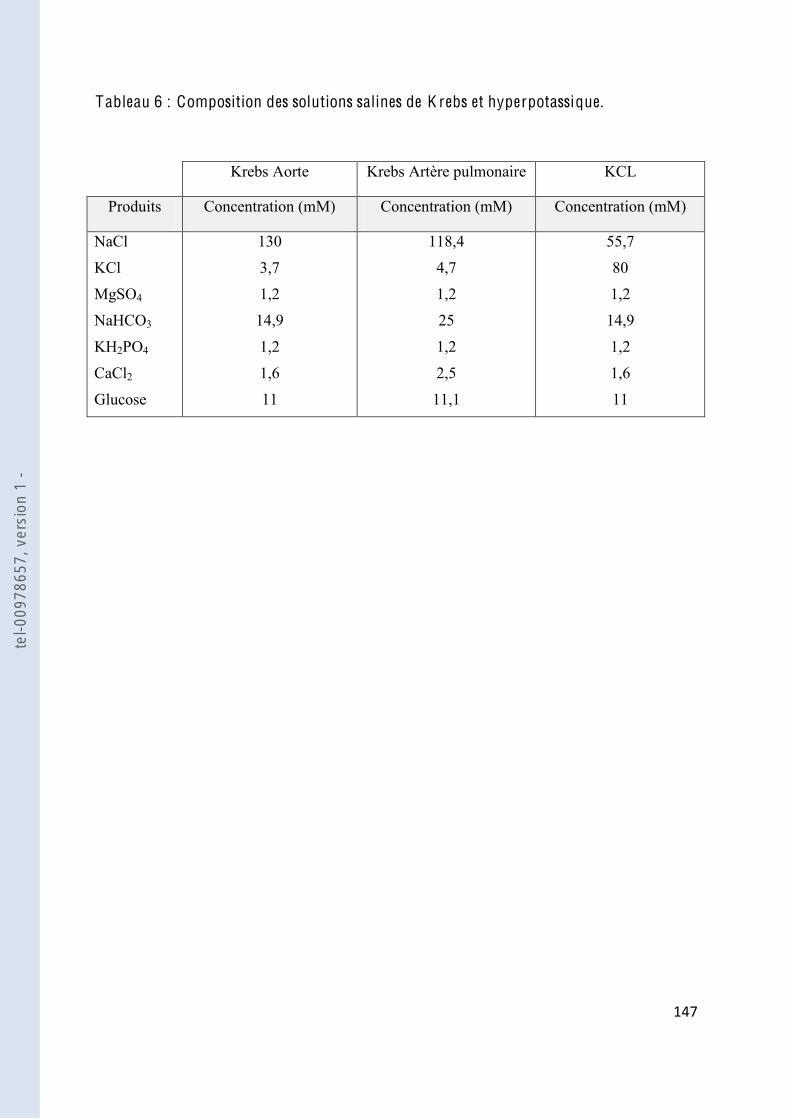
!%("
"
Tableau 6 : Composition des solutions salines de K rebs et hyperpotassique.
Krebs Aorte Krebs Artère pulmonaire KCL
Produits Concentration (mM) Concentration (mM) Concentration (mM)
NaCl
KCl
MgSO4
NaHCO3
KH2PO4
CaCl2
Glucose
130
3,7
1,2
14,9
1,2
1,6
11
118,4
4,7
1,2
25
1,2
2,5
11,1
55,7
80
1,2
14,9
1,2
1,6
11
tel-0
0978
657,
ver
sion
1 -

!%)"
"
B IB L I O G R APH I E
tel-0
0978
657,
ver
sion
1 -
!%*"
"
1. Carmeliet P (2003) Angiogenesis in health and disease. Nat Med 9(6):653-660.
2. Barnes PJ and Liu SF (1995) Regulation of pulmonary vascular tone. Pharmacol Rev 47(1):87-131.
3. Bolotina VM (2004) Store-operated channels: diversity and activation mechanisms. Sci STKE 2004(243):pe34.
4. Pozzan T, Rizzuto R, Volpe P and Meldolesi J (1994) Molecular and cellular physiology of intracellular calcium stores. Physiol Rev 74(3):595-636.
5. Somlyo AP and Somlyo AV (2003) Ca2+ sensitivity of smooth muscle and nonmuscle myosin II: modulated by G proteins, kinases, and myosin phosphatase. Physiol Rev 83(4):1325-1358.
6. Katsuki S and Murad F (1977) Regulation of adenosine cyclic 3',5'-monophosphate and guanosine cyclic 3',5'-monophosphate levels and contractility in bovine tracheal smooth muscle. Mol Pharmacol 13(2):330-341.
7. Friebe A and Koesling D (2003) Regulation of nitric oxide-sensitive guanylyl cyclase. Circ Res 93(2):96-105.
8. Nakashima S, Tohmatsu T, Hattori H, Okano Y and Nozawa Y (1986) Inhibitory action of cyclic GMP on secretion, polyphosphoinositide hydrolysis and calcium mobilization in thrombin-stimulated human platelets. Biochem Biophys Res Commun 135(3):1099-1104.
9. Cornwell TL, Pryzwansky KB, Wyatt TA and Lincoln TM (1991) Regulation of sarcoplasmic reticulum protein phosphorylation by localized cyclic GMP-dependent protein kinase in vascular smooth muscle cells. Mol Pharmacol 40(6):923-931.
10. Furukawa K, Ohshima N, Tawada-Iwata Y and Shigekawa M (1991) Cyclic GMP stimulates Na+/Ca2+ exchange in vascular smooth muscle cells in primary culture. J Biol Chem 266:12337-12341.
11. Mery PF, Lohmann SM, Walter U and Fischmeister R (1991) Ca2+ current is regulated by cyclic GMP-dependent protein kinase in mammalian cardiac myocytes. Proc Natl Acad Sci U S A 88(4):1197-1201.
12. Milbourne EA and Bygrave FL (1995) Do nitric oxide and cGMP play a role in calcium cycling? Cell Calcium 18(3):207-213.
13. Meisheri KD, Taylor CJ and Saneii H (1986) Synthetic atrial peptide inhibits intracellular calcium release in smooth muscle. Am J Physiol 250:C171-174.
14. Hirata M, Kohse KP, Chang CH, Ikebe T and Murad F (1990) Mechanism of cyclic GMP inhibition of inositol phosphate formation in rat aorta segments and cultured bovine aortic smooth muscle cells. J Biol Chem 265(3):1268-1273.
15. Adelstein RS and Hathaway DR (1979) Role of calcium and cyclic adenosine 3':5' monophosphate in regulating smooth muscle contraction. Mechanisms of excitation-contraction coupling in smooth muscle. Am J Cardiol 44(5):783-787.
tel-0
0978
657,
ver
sion
1 -
!&+"
"
16. Feletou M and Vanhoutte PM (2000) Endothelium-dependent hyperpolarization of vascular smooth muscle cells. Acta Pharmacol Sin 21(1):1-18.
17. Busse R, Edwards G, Feletou M, Fleming I, Vanhoutte PM and Weston AH (2002) EDHF: bringing the concepts together. Trends Pharmacol Sci 23(8):374-380.
18. Cines DB, Pollak ES, Buck CA, Loscalzo J, Zimmerman GA, McEver RP, Pober JS, Wick TM, Konkle BA, Schwartz BS, Barnathan ES, McCrae KR, Hug BA, Schmidt AM and Stern DM (1998) Endothelial cells in physiology and in the pathophysiology of vascular disorders. Blood 91(10):3527-3561.
19. Kibria G, Heath D, Smith P and Biggar R (1980) Pulmonary endothelial pavement patterns. Thorax 35(3):186-191.
20. Drexler H (1998) Factors involved in the maintenance of endothelial function. Am J Cardiol 82(10A):3S-4S.
21. Furchgott RF and Zawadzki JV (1980) The obligatory role of endothelial cells in the relaxation of arterial smooth muscle by acetylcholine. Nature 288(5789):373-376.
22. Furchgott RF and Vanhoutte PM (1989) Endothelium-derived relaxing and contracting factors. Faseb J 3:2007-2018.
23. Ignarro LJ, Buga GM, Wood KS, Byrns RE and Chaudhuri G (1987) Endothelium-derived relaxing factor produced and released from artery and vein is nitric oxide. Proc Natl Acad Sci U S A 84(24):9265-9269.
24. Palmer RM, Ferrige AG and Moncada S (1987) Nitric oxide release accounts for the biological activity of endothelium-derived relaxing factor. Nature 327(6122):524-526.
25. Palmer RM, Ashton DS and Moncada S (1988) Vascular endothelial cells synthesize nitric oxide from L-arginine. Nature 333(6174):664-666.
26. Alderton WK, Cooper CE and Knowles RG (2001) Nitric oxide synthases: structure, function and inhibition. Biochem J 357(Pt 3):593-615.
27. Arnal JF, Dinh-Xuan AT, Pueyo M, Darblade B and Rami J (1999) Endothelium-derived nitric oxide and vascular physiology and pathology. Cell Mol Life Sci 55(8-9):1078-1087.
28. Toda N, Ayajiki K and Okamura T (2007) Interaction of endothelial nitric oxide and angiotensin in the circulation. Pharmacol Rev 59(1):54-87.
29. Dimmeler S, Fleming I, Fisslthaler B, Hermann C, Busse R and Zeiher AM (1999) Activation of nitric oxide synthase in endothelial cells by Akt-dependent phosphorylation. Nature 399(6736):601-605.
30. Schini-Kerth VB (1999) Vascular biosynthesis of nitric oxide: effect on hemostasis and fibrinolysis. Transfus Clin Biol 6(6):355-363.
tel-0
0978
657,
ver
sion
1 -
!&!"
"
31. Bunting S, Gryglewski R, Moncada S and Vane JR (1976) Arterial walls generate from prostaglandin endoperoxides a substance (prostaglandin X) which relaxes strips of mesenteric and coeliac ateries and inhibits platelet aggregation. Prostaglandins 12(6):897-913.
32. Kawabe J, Ushikubi F and Hasebe N Prostacyclin in vascular diseases. - Recent insights and future perspectives. Circ J 74(5):836-843.
33. Fetalvero KM, Shyu M, Nomikos AP, Chiu YF, Wagner RJ, Powell RJ, Hwa J and Martin KA (2006) The prostacyclin receptor induces human vascular smooth muscle cell differentiation via the protein kinase A pathway. Am J Physiol Heart Circ Physiol 290(4):H1337-1346.
34. Fetalvero KM, Martin KA and Hwa J (2007) Cardioprotective prostacyclin signaling in vascular smooth muscle. Prostaglandins O ther Lipid Mediat 82(1-4):109-118.
35. White DG and Martin W (1989) Differential control and calcium-dependence of production of endothelium-derived relaxing factor and prostacyclin by pig aortic endothelial cells. Br J Pharmacol 97(3):683-690.
36. Niwano K, Arai M, Tomaru K, Uchiyama T, Ohyama Y and Kurabayashi M (2003) Transcriptional stimulation of the eNOS gene by the stable prostacyclin analogue beraprost is mediated through cAMP-responsive element in vascular endothelial cells: close link between PGI2 signal and NO pathways. Circ Res 93(6):523-530.
37. Radomski MW, Palmer RM and Moncada S (1987) Comparative pharmacology of endothelium-derived relaxing factor, nitric oxide and prostacyclin in platelets. Br J Pharmacol 92(1):181-187.
38. Smyth EM and FitzGerald GA (2002) Human prostacyclin receptor. Vitam Horm 65:149-165.
39. Feletou M and Vanhoutte PM (2006) Endothelium-derived hyperpolarizing factor: where are we now? Arterioscler Thromb Vasc Biol 26(6):1215-1225.
40. Nagao T and Vanhoutte PM (1992) Hyperpolarization as a mechanism for endothelium-dependent relaxations in the porcine coronary artery. J Physiol 445:355-367.
41. Matoba T, Shimokawa H, Nakashima M, Hirakawa Y, Mukai Y, Hirano K, Kanaide H and Takeshita A (2000) Hydrogen peroxide is an endothelium-derived hyperpolarizing factor in mice. J Clin Invest 106(12):1521-1530.
42. Edwards G, Dora KA, Gardener MJ, Garland CJ and Weston AH (1998) K+ is an endothelium-derived hyperpolarizing factor in rat arteries. Nature 396(6708):269-272.
43. Cohen RA and Vanhoutte PM (1995) Endothelium-dependent hyperpolarization. Beyond nitric oxide and cyclic GMP. Circulation 92(11):3337-3349.
44. Bauersachs J, Popp R, Hecker M, Sauer E, Fleming I and Busse R (1996) Nitric oxide attenuates the release of endothelium-derived hyperpolarizing factor. Circulation 94(12):3341-3347.
tel-0
0978
657,
ver
sion
1 -
!&#"
"
45. Yanagisawa M, Kurihara H, Kimura S, Tomobe Y, Kobayashi M, Mitsui Y, Yazaki Y, Goto K and Masaki T (1988) A novel potent vasoconstrictor peptide produced by vascular endothelial cells. Nature 332(6163):411-415.
46. Sabry S, Mondon F, Levy M, Ferre F and Dinh-Xuan AT (1995) Endothelial modulation of vasoconstrictor responses to endothelin-1 in human placental stem villi small arteries. Br J Pharmacol 115(6):1038-1042.
47. Zellers TM, McCormick J and Wu Y (1994) Interaction among ET-1, endothelium-derived nitric oxide, and prostacyclin in pulmonary arteries and veins. Am J Physiol 267(1 Pt 2):H139-147.
48. Mombouli JV and Vanhoutte PM (1999) Endothelial dysfunction: from physiology to therapy. J Mol Cell Cardiol 31(1):61-74.
49. Li JM and Shah AM (2004) Endothelial cell superoxide generation: regulation and relevance for cardiovascular pathophysiology. Am J Physiol Regul Integr Comp Physiol 287(5):R1014-1030.
50. Cai H and Harrison DG (2000) Endothelial dysfunction in cardiovascular diseases: the role of oxidant stress. Circ Res 87(10):840-844.
51. Lekakis J, Abraham P, Balbarini A, Blann A, Boulanger CM, Cockcroft J, Cosentino F, Deanfield J, Gallino A, Ikonomidis I, Kremastinos D, Landmesser U, Protogerou A, Stefanadis C, Tousoulis D, Vassalli G, Vink H, Werner N, Wilkinson I and Vlachopoulos C Methods for evaluating endothelial function: a position statement from the European Society of Cardiology Working Group on Peripheral Circulation. Eur J Cardiovasc Prev Rehabil.
52. Wolf P (1967) The nature and significance of platelet products in human plasma. Br J Haematol 13:269-288.
53. Bastida E, Ordinas A, Escolar G and Jamieson GA (1984) Tissue factor in microvesicles shed from U87MG human glioblastoma cells induces coagulation, platelet aggregation, and thrombogenesis. Blood 64:177-184.
54. Martinez MC, Tesse A, Zobairi F and Andriantsitohaina R (2005) Shed membrane microparticles from circulating and vascular cells in regulating vascular function. Am J Physiol Heart Circ Physiol 288:H1004-1009.
55. Mostefai HA, Andriantsitohaina R and Martinez MC (2008) Plasma membrane microparticles in angiogenesis: role in ischemic diseases and in cancer. Physiol Res 57:311-320.
56. Ratajczak J, Wysoczynski M, Hayek F, Janowska-Wieczorek A and Ratajczak MZ (2006) Membrane-derived microvesicles: important and underappreciated mediators of cell-to-cell communication. Leukemia 20:1487-1495.
57. Jimenez JJ, Jy W, Mauro LM, Soderland C, Horstman LL and Ahn YS (2003) Endothelial cells release phenotypically and quantitatively distinct microparticles in activation and apoptosis. Thromb Res 109:175-180.
tel-0
0978
657,
ver
sion
1 -

!&$"
"
58. Pasquet JM, Dachary-Prigent J and Nurden AT (1996) Calcium influx is a determining factor of calpain activation and microparticle formation in platelets. Eur J Biochem 239:647-654.
59. Miyoshi H, Umeshita K, Sakon M, Imajoh-Ohmi S, Fujitani K, Gotoh M, Oiki E, Kambayashi J and Monden M (1996) Calpain activation in plasma membrane bleb formation during tert-butyl hydroperoxide-induced rat hepatocyte injury. Gastroenterology 110:1897-1904.
60. Wiedmer T, Shattil SJ, Cunningham M and Sims PJ (1990) Role of calcium and calpain in complement-induced vesiculation of the platelet plasma membrane and in the exposure of the platelet factor Va receptor. Biochemistry 29:623-632.
61. Bevers EM, Tilly RH, Senden JM, Comfurius P, Zwaal RF (1989) Exposure of endogenous phosphatidylserine at the outer surface of stimulated platelets is reversed by restoration of aminophospholipid translocase activity. Biochemistry 28:2382-2387.
62. Bevers EM and Williamson PL (2010) Phospholipid scramblase: an update. F EBS Lett 584:2724-2730.
63. Heemskerk JW, Vuist WM, Feijge MA, Reutelingsperger CP and Lindhout T (1997) Collagen but not fibrinogen surfaces induce bleb formation, exposure of phosphatidylserine, and procoagulant activity of adherent platelets: evidence for regulation by protein tyrosine kinase-dependent Ca2+ responses. Blood 90:2615-2625.
64. Kunzelmann-Marche C, Freyssinet JM and Martinez MC (2001) Regulation of phosphatidylserine transbilayer redistribution by store-operated Ca2+ entry: role of actin cytoskeleton. J Biol Chem 276:5134-5139.
65. Kunzelmann-Marche C, Freyssinet JM and Martinez MC (2002) Loss of plasma membrane phospholipid asymmetry requires raft integrity. Role of transient receptor potential channels and ERK pathway. J Biol Chem 277:19876-19881.
66. Yan R, Wang Z, Yuan Y, Cheng H and Dai K (2009) Role of cAMP-dependent protein kinase in the regulation of platelet procoagulant activity. Arch Biochem Biophys 485:41-48.
67. Cauwenberghs S, Feijge MA, Harper AG, Sage SO, Curvers J and Heemskerk JW (2006) Shedding of procoagulant microparticles from unstimulated platelets by integrin-mediated destabilization of actin cytoskeleton. F EBS Lett 580:5313-5320.
68. Thompson CB (1995) Apoptosis in the pathogenesis and treatment of disease. Science 267:1456-1462.
69. Beyer C and Pisetsky DS (2010) The role of microparticles in the pathogenesis of rheumatic diseases. Nat Rev Rheumatol 6:21-29.
70. Hristov M, Erl W, Linder S and Weber PC (2004) Apoptotic bodies from endothelial cells enhance the number and initiate the differentiation of human endothelial progenitor cells in vitro. Blood 104:2761-2766.
tel-0
0978
657,
ver
sion
1 -

!&%"
"
71. Sebbagh M, Renvoize C, Hamelin J, Riche N, Bertoglio J and Breard J (2001) Caspase-3-mediated cleavage of ROCK I induces MLC phosphorylation and apoptotic membrane blebbing. Nat Cell Biol 3:346-352.
72. Coleman ML, Sahai EA, Yeo M, Bosch M, Dewar A and Olson MF (2001) Membrane blebbing during apoptosis results from caspase-mediated activation of ROCK I. Nat Cell Biol 3:339-345.
73. Sapet C, Simoncini S, Loriod B, Puthier D, Sampol J, Nguyen C, Dignat-George F and Anfosso F (2006) Thrombin-induced endothelial microparticle generation: identification of a novel pathway involving ROCK-II activation by caspase-2. Blood 108:1868-1876.
74. Zauli G and Secchiero P (2006) The role of the TRAIL/TRAIL receptors system in hematopoiesis and endothelial cell biology. Cytokine Growth Factor Rev 17:245-257.
75. Simoncini S, Njock MS, Robert S, Camoin-Jau L, Sampol J, Harle JR, Nguyen C, Dignat-George F and Anfosso F (2009) TRAIL/Apo2L mediates the release of procoagulant endothelial microparticles induced by thrombin in vitro: a potential mechanism linking inflammation and coagulation. Circ Res 104:943-951.
76. Banfi C, Brioschi M, Wait R, Begum S, Gianazza E, Pirillo A, Mussoni L and Tremoli E (2005) Proteome of endothelial cell-derived procoagulant microparticles. Proteomics 5:4443-4455.
77. Horstman LL, Jy W, Jimenez JJ, Bidot C and Ahn YS (2004) New horizons in the analysis of circulating cell-derived microparticles. Keio J Med 53:210-230.
78. Garcia BA, Smalley DM, Cho H, Shabanowitz J, Ley K and Hunt DF (2005) The platelet microparticle proteome. J Proteome Res 4:1516-1521.
79. Martinez MC, Larbret F, Zobairi F, Coulombe J, Debili N, Vainchenker W, Ruat M and Freyssinet JM (2006) Transfer of differentiation signal by membrane microvesicles harboring hedgehog morphogens. Blood 108:3012-3020.
80. Miguet L, Pacaud K, Felden C, Hugel B, Martinez MC, Freyssinet JM, Herbrecht R, Potier N, van Dorsselaer A and Mauvieux L (2006) Proteomic analysis of malignant lymphocyte membrane microparticles using double ionization coverage optimization. Proteomics 6:153-171.
81. Bernimoulin M, Waters EK, Foy M, Steele BM, Sullivan M, Falet H, Walsh MT, Barteneva N, Geng JG, Hartwig JH, Maguire PB and Wagner DD (2009) Differential stimulation of monocytic cells results in distinct populations of microparticles. J Thromb Haemost 7:1019-1028.
82. Dean WL, Lee MJ, Cummins TD, Schultz DJ and Powell DW (2009) Proteomic and functional characterisation of platelet microparticle size classes. Thromb Haemost 102:711-718.
tel-0
0978
657,
ver
sion
1 -
!&&"
"
83. Liu ML, Reilly MP, Casasanto P, McKenzie SE and Williams KJ (2007) Cholesterol enrichment of human monocyte/macrophages induces surface exposure of phosphatidylserine and the release of biologically-active tissue factor-positive microvesicles. Arterioscler Thromb Vasc Biol 27:430-435.
84. Forlow SB, McEver RP and Nollert MU (2000) Leukocyte-leukocyte interactions mediated by platelet microparticles under flow. Blood 95:1317-1323.
85. Agouni A, Mostefai HA, Porro C, Carusio N, Favre J, Richard V, Henrion D, Martinez MC and Andriantsitohaina R (2007) Sonic hedgehog carried by microparticles corrects endothelial injury through nitric oxide release. Faseb J 21:2735-2741.
86. Soleti R, Benameur T, Porro C, Panaro MA, Andriantsitohaina R and Martinez MC (2009) Microparticles harboring Sonic Hedgehog promote angiogenesis through the upregulation of adhesion proteins and proangiogenic factors. Carcinogenesis 30:580-588.
87. Tesse A, Martinez MC, Hugel B, Chalupsky K, Muller CD, Meziani F, Mitolo-Chieppa D, Freyssinet JM and Andriantsitohaina R (2005) Upregulation of proinflammatory proteins through NF-kappaB pathway by shed membrane microparticles results in vascular hyporeactivity. Arterioscler Thromb Vasc Biol 25:2522-2527.
88. Rozmyslowicz T, Majka M, Kijowski J, Murphy SL, Conover DO, Poncz M, Ratajczak J, Gaulton GN and Ratajczak MZ (2003) Platelet- and megakaryocyte-derived microparticles transfer CXCR4 receptor to CXCR4-null cells and make them susceptible to infection by X4-HIV. Aids 17:33-42.
89. Barry OP, Pratico D, Savani RC and FitzGerald GA (1998) Modulation of monocyte-endothelial cell interactions by platelet microparticles. J Clin Invest 102:136-144.
90. Breimo ES and Osterud B (2005) Generation of tissue factor-rich microparticles in an ex vivo whole blood model. Blood Coagul F ibrinolysis 16:399-405.
91. Deregibus MC, Cantaluppi V, Calogero R, Lo Iacono M, Tetta C, Biancone L, Bruno S, Bussolati B and Camussi G (2007) Endothelial progenitor cell derived microvesicles activate an angiogenic program in endothelial cells by a horizontal transfer of mRNA. Blood 110:2440-2448.
92. Rank A, Nieuwland R, Crispin A, Grutzner S, Iberer M, Toth B and Pihusch R (2011) Clearance of platelet microparticles in vivo. Platelets 22(2):111-116.
93. Priou P, Gagnadoux F, Tesse A, Mastronardi ML, Agouni A, Meslier N, Racineux JL, Martinez MC, Trzepizur W and Andriantsitohaina R (2010) Endothelial dysfunction and circulating microparticles from patients with obstructive sleep apnea. Am J Pathol 177:974-983.
94. Tual-Chalot S, Guibert C, Muller B, Savineau JP, Andriantsitohaina R and Martinez MC (2010) Circulating microparticles from pulmonary hypertensive rats induce endothelial dysfunction. Am J Respir Crit Care Med 182:261-268.
tel-0
0978
657,
ver
sion
1 -
!&'"
"
95. Boulanger CM, Scoazec A, Ebrahimian T, Henry P, Mathieu E, Tedgui A and Mallat Z (2001) Circulating microparticles from patients with myocardial infarction cause endothelial dysfunction. Circulation 104:2649-2652.
96. Martin S, Tesse A, Hugel B, Martinez MC, Morel O, Freyssinet JM and Andriantsitohaina R (2004) Shed membrane particles from T lymphocytes impair endothelial function and regulate endothelial protein expression. Circulation 109:1653-1659.
97. Vanwijk MJ, Svedas E, Boer K, Nieuwland R, Vanbavel E and Kublickiene KR (2002) Isolated microparticles, but not whole plasma, from women with preeclampsia impair endothelium-dependent relaxation in isolated myometrial arteries from healthy pregnant women. Am J Obstet Gynecol 187:1686-1693.
98. Agouni A, Lagrue-Lak-Hal AH, Ducluzeau PH, Mostefai HA, Draunet-Busson C, Leftheriotis G, Heymes C, Martinez MC and Andriantsitohaina R (2008) Endothelial dysfunction caused by circulating microparticles from patients with metabolic syndrome. Am J Pathol 173:1210-1219.
99. Helal O, Defoort C, Robert S, Marin C, Lesavre N, Lopez-Miranda J, Riserus U, Basu S, Lovegrove J, McMonagle J, Roche HM, Dignat-George F and Lairon D (2010) Increased levels of microparticles originating from endothelial cells, platelets and erythrocytes in subjects with metabolic syndrome: Relationship with oxidative stress. Nutr Metab Cardiovasc Dis.
100. Meziani F, Tesse A, David E, Martinez MC, Wangesteen R, Schneider F and Andriantsitohaina R (2006) Shed membrane particles from preeclamptic women generate vascular wall inflammation and blunt vascular contractility. Am J Pathol 169:1473-1483.
101. Taraboletti G, D'Ascenzo S, Borsotti P, Giavazzi R, Pavan A and Dolo V (2002) Shedding of the matrix metalloproteinases MMP-2, MMP-9, and MT1-MMP as membrane vesicle-associated components by endothelial cells. Am J Pathol 160:673-680.
102. Benameur T, Tual-Chalot S, Andriantsitohaina R and Martinez MC PPARalpha is essential for microparticle-induced differentiation of mouse bone marrow-derived endothelial progenitor cells and angiogenesis. PLoS One 5:e12392.
103. Chahed S, Leroyer AS, Benzerroug M, Gaucher D, Georgescu A, Picaud S, Silvestre JS, Gaudric A, Tedgui A, Massin P and Boulanger CM Increased vitreous shedding of microparticles in proliferative diabetic retinopathy stimulates endothelial proliferation. Diabetes 59:694-701.
104. Leroyer AS, Rautou PE, Silvestre JS, Castier Y, Leseche G, Devue C, Duriez M, Brandes RP, Lutgens E, Tedgui A and Boulanger CM (2008) CD40 ligand+ microparticles from human atherosclerotic plaques stimulate endothelial proliferation and angiogenesis a potential mechanism for intraplaque neovascularization. J Am Coll Cardiol 52:1302-1311.
tel-0
0978
657,
ver
sion
1 -
!&("
"
105. Mostefai HA, Meziani F, Mastronardi ML, Agouni A, Heymes C, Sargentini C, Asfar P, Martinez MC and Andriantsitohaina R (2008) Circulating microparticles from patients with septic shock exert protective role in vascular function. Am J Respir Crit Care Med 178:1148-1155.
106. Soriano AO, Jy W, Chirinos JA, Valdivia MA, Velasquez HS, Jimenez JJ, Horstman LL, Kett DH, Schein RM and Ahn YS (2005) Levels of endothelial and platelet microparticles and their interactions with leukocytes negatively correlate with organ dysfunction and predict mortality in severe sepsis. Crit Care Med 33:2540-2546.
107. Benameur T, Andriantsitohaina R and Martinez MC (2009) Therapeutic potential of plasma membrane-derived microparticles. Pharmacol Rep 61:49-57.
108. Benameur T, Soleti R, Porro C, Andriantsitohaina R and Martinez MC Microparticles carrying Sonic hedgehog favor neovascularization through the activation of nitric oxide pathway in mice. PLoS One 5:e12688.
109. Badesch DB, Champion HC, Sanchez MA, Hoeper MM, Loyd JE, Manes A, McGoon M, Naeije R, Olschewski H, Oudiz RJ and Torbicki A (2009) Diagnosis and assessment of pulmonary arterial hypertension. J Am Coll Cardiol 54(1 Suppl):S55-66.
110. Humbert M, Sitbon O, Chaouat A, Bertocchi M, Habib G, Gressin V, Yaici A, Weitzenblum E, Cordier JF, Chabot F, Dromer C, Pison C, Reynaud-Gaubert M, Haloun A, Laurent M, Hachulla E and Simonneau G (2006) Pulmonary arterial hypertension in France: results from a national registry. Am J Respir Crit Care Med 173(9):1023-1030.
111. Simonneau G, Robbins IM, Beghetti M, Channick RN, Delcroix M, Denton CP, Elliott CG, Gaine SP, Gladwin MT, Jing ZC, Krowka MJ, Langleben D, Nakanishi N and Souza R (2009) Updated clinical classification of pulmonary hypertension. J Am Coll Cardiol 54(1 Suppl):S43-54.
112. Fishman AP (2001) Clinical classification of pulmonary hypertension. Clin Chest Med 22(3):385-391, vii.
113. Park MH (2010) Pharmacotherapeutic options for pulmonary arterial hypertension. Pulmonary Hypertension 1(1):1-21.
114. Rich S, Dantzker DR, Ayres SM, Bergofsky EH, Brundage BH, Detre KM, Fishman AP, Goldring RM, Groves BM, Koerner SK and et al. (1987) Primary pulmonary hypertension. A national prospective study. Ann Intern Med 107(2):216-223.
115. McLaughlin VV and McGoon MD (2006) Pulmonary arterial hypertension. Circulation 114(13):1417-1431.
116. Perros F and Humbert M (2005) [Physiopathology of pulmonary arterial hypertension. Cellular and molecular aspects]. Presse Med 34(3):232-242.
tel-0
0978
657,
ver
sion
1 -

!&)"
"
117. Humbert M, Morrell NW, Archer SL, Stenmark KR, MacLean MR, Lang IM, Christman BW, Weir EK, Eickelberg O, Voelkel NF and Rabinovitch M (2004) Cellular and molecular pathobiology of pulmonary arterial hypertension. J Am Coll Cardiol 43(12 Suppl S):13S-24S.
118. Jeffery TK and Morrell NW (2002) Molecular and cellular basis of pulmonary vascular remodeling in pulmonary hypertension. Prog Cardiovasc Dis 45(3):173-202.
119. Budhiraja R, Tuder RM and Hassoun PM (2004) Endothelial dysfunction in pulmonary hypertension. Circulation 109(2):159-165.
120. Christman BW, McPherson CD, Newman JH, King GA, Bernard GR, Groves BM and Loyd JE (1992) An imbalance between the excretion of thromboxane and prostacyclin metabolites in pulmonary hypertension. N Engl J Med 327(2):70-75.
121. Giaid A, Yanagisawa M, Langleben D, Michel RP, Levy R, Shennib H, Kimura S, Masaki T, Duguid WP and Stewart DJ (1993) Expression of endothelin-1 in the lungs of patients with pulmonary hypertension. N Engl J Med 328(24):1732-1739.
122. Stewart DJ, Levy RD, Cernacek P and Langleben D (1991) Increased plasma endothelin-1 in pulmonary hypertension: marker or mediator of disease? Ann Intern Med 114(6):464-469.
123. Giaid A and Saleh D (1995) Reduced expression of endothelial nitric oxide synthase in the lungs of patients with pulmonary hypertension. N Engl J Med 333(4):214-221.
124. Amabile N, Heiss C, Real WM, Minasi P, McGlothlin D, Rame EJ, Grossman W, De Marco T and Yeghiazarians Y (2008) Circulating endothelial microparticle levels predict hemodynamic severity of pulmonary hypertension. Am J Respir Crit Care Med 177(11):1268-1275.
125. Bakouboula B, Morel O, Faure A, Zobairi F, Jesel L, Trinh A, Zupan M, Canuet M, Grunebaum L, Brunette A, Desprez D, Chabot F, Weitzenblum E, Freyssinet JM, Chaouat A and Toti F (2008) Procoagulant membrane microparticles correlate with the severity of pulmonary arterial hypertension. Am J Respir Crit Care Med 177(5):536-543.
126. Gaine SP and Rubin LJ (1998) Primary pulmonary hypertension. Lancet 352(9129):719-725.
127. Rubin LJ (2006) Pulmonary arterial hypertension. Proc Am Thorac Soc 3(1):111-115.
128. Doyle RL, McCrory D, Channick RN, Simonneau G and Conte J (2004) Surgical treatments/interventions for pulmonary arterial hypertension: ACCP evidence-based clinical practice guidelines. Chest 126(1 Suppl):63S-71S.
129. Stenmark KR, Meyrick B, Galie N, Mooi WJ and McMurtry IF (2009) Animal models of pulmonary arterial hypertension: the hope for etiological discovery and pharmacological cure. Am J Physiol Lung Cell Mol Physiol 297(6):L1013-1032.
130. Jennum P and Riha RL (2009) Epidemiology of sleep apnoea/hypopnoea syndrome and sleep-disordered breathing. Eur Respir J 33(4):907-914.
tel-0
0978
657,
ver
sion
1 -
!&*"
"
131. Kushida CA, Littner MR, Morgenthaler T, Alessi CA, Bailey D, Coleman J, Jr., Friedman L, Hirshkowitz M, Kapen S, Kramer M, Lee-Chiong T, Loube DL, Owens J, Pancer JP and Wise M (2005) Practice parameters for the indications for polysomnography and related procedures: an update for 2005. Sleep 28(4):499-521.
132. Dempsey JA, Veasey SC, Morgan BJ and O'Donnell CP Pathophysiology of sleep apnea. Physiol Rev 90(1):47-112.
133. Marin JM, Carrizo SJ, Vicente E and Agusti AG (2005) Long-term cardiovascular outcomes in men with obstructive sleep apnoea-hypopnoea with or without treatment with continuous positive airway pressure: an observational study. Lancet 365(9464):1046-1053.
134. Peker Y, Kraiczi H, Hedner J, Loth S, Johansson A and Bende M (1999) An independent association between obstructive sleep apnoea and coronary artery disease. Eur Respir J 14(1):179-184.
135. Svatikova A, Wolk R, Wang HH, Otto ME, Bybee KA, Singh RJ and Somers VK (2004) Circulating free nitrotyrosine in obstructive sleep apnea. Am J Physiol Regul Integr Comp Physiol 287(2):R284-287.
136. Atkeson A, Yeh SY, Malhotra A and Jelic S (2009) Endothelial function in obstructive sleep apnea. Prog Cardiovasc Dis 51(5):351-362.
137. Budhiraja R, Parthasarathy S and Quan SF (2007) Endothelial dysfunction in obstructive sleep apnea. J Clin Sleep Med 3(4):409-415.
138. Geiser T, Buck F, Meyer BJ, Bassetti C, Haeberli A and Gugger M (2002) In vivo platelet activation is increased during sleep in patients with obstructive sleep apnea syndrome. Respiration 69(3):229-234.
139. Ayers L, Ferry B, Craig S, Nicoll D, Stradling JR and Kohler M (2009) Circulating cell-derived microparticles in patients with minimally symptomatic obstructive sleep apnoea. Eur Respir J 33(3):574-580.
140. Jelic S, Lederer DJ, Adams T, Padeletti M, Colombo PC, Factor P and Le Jemtel TH (2009) Endothelial repair capacity and apoptosis are inversely related in obstructive sleep apnea. Vasc Health Risk Manag 5:909-920.
141. Yun CH, Jung KH, Chu K, Kim SH, Ji KH, Park HK, Kim HC, Lee ST, Lee SK and Roh JK Increased circulating endothelial microparticles and carotid atherosclerosis in obstructive sleep apnea. J Clin Neurol 6(2):89-98.
142. Trzepizur W, Priou P, Paris A, Nardi J, Tual-Chalot S, Meslier N, Urban T, Andriantsitohaina R, Martinez MC and Gagnadoux F Nocturnal release of leukocyte-derived microparticles in males with obstructive sleep apnoea. Eur Respir J 37(5):1293-1295.
143. Jenkinson C, Davies RJ, Mullins R and Stradling JR (1999) Comparison of therapeutic and subtherapeutic nasal continuous positive airway pressure for obstructive sleep apnoea: a randomised prospective parallel trial. Lancet 353(9170):2100-2105.
tel-0
0978
657,
ver
sion
1 -
!'+"
"
144. Massie CA, McArdle N, Hart RW, Schmidt-Nowara WW, Lankford A, Hudgel DW, Gordon N and Douglas NJ (2003) Comparison between automatic and fixed positive airway pressure therapy in the home. Am J Respir Crit Care Med 167(1):20-23.
145. Trzepizur W, Gagnadoux F, Abraham P, Rousseau P, Meslier N, Saumet JL and Racineux JL (2009) Microvascular endothelial function in obstructive sleep apnea: Impact of continuous positive airway pressure and mandibular advancement. Sleep Med 10(7):746-752.
146. Weaver TE and Grunstein RR (2008) Adherence to continuous positive airway pressure therapy: the challenge to effective treatment. Proc Am Thorac Soc 5(2):173-178.
147. Lin HS, Prasad AS, Pan CJ and Rowley JA (2007) Factors associated with noncompliance to treatment with positive airway pressure. Arch O tolaryngol Head Neck Surg 133(1):69-72.
148. Mehta A, Qian J, Petocz P, Darendeliler MA and Cistulli PA (2001) A randomized, controlled study of a mandibular advancement splint for obstructive sleep apnea. Am J Respir Crit Care Med 163(6):1457-1461.
149. Engleman HM, McDonald JP, Graham D, Lello GE, Kingshott RN, Coleman EL, Mackay TW and Douglas NJ (2002) Randomized crossover trial of two treatments for sleep apnea/hypopnea syndrome: continuous positive airway pressure and mandibular repositioning splint. Am J Respir Crit Care Med 166(6):855-859.
150. Tesse A, Meziani F, David E, Carusio N, Kremer H, Schneider F and Andriantsitohaina R (2007) Microparticles from preeclamptic women induce vascular hyporeactivity in vessels from pregnant mice through an overproduction of NO. Am J Physiol Heart Circ Physiol 293:H520-525.
151. Bonnet S, Dubuis E, Vandier C, Martin S, Marthan R and Savineau JP (2002) Reversal of chronic hypoxia-induced alterations in pulmonary artery smooth muscle electromechanical coupling upon air breathing. Cardiovasc Res 53:1019-1028.
152. Kobayashi M, Inoue K, Warabi E, Minami T and Kodama T (2005) A simple method of isolating mouse aortic endothelial cells. J Atheroscler Thromb 12:138-142.
153. Lowry OH, Rosebrough NJ, Farr AL and Randall RJ (1951) Protein measurement with the Folin phenol reagent. J Biol Chem 193:265-275.
154. Souchar JP, Vergely C, Rochette L (2007) Radicaux libres et stress oxydant
vasculaire. p.311-329.
155. Lee HB and Blaufox MD (1985) Blood volume in the rat. J Nucl Med 26:72-76.
156. Andriambeloson E, Kleschyov AL, Muller B, Beretz A, Stoclet JC and Andriantsitohaina R (1997) Nitric oxide production and endothelium-dependent vasorelaxation induced by wine polyphenols in rat aorta. Br J Pharmacol 120:1053-1058.
tel-0
0978
657,
ver
sion
1 -

!'!"
"
157. Pauvert O, Marthan R and Savineau J (2000) NO-induced modulation of calcium-oscillations in pulmonary vascular smooth muscle. Cell Calcium 27:329-338.
158. Dhein S, Giessler C, Heinroth-Hoffmann I, Leineweber K, Seyfarth T and Brodde OE (2002) Changes in alpha(1)-adrenergic vascular reactivity in monocrotaline-treated rats. Naunyn Schmiedebergs Arch Pharmacol 365:87-95.
159. Diehl P, Aleker M, Helbing T, Sossong V, Germann M, Sorichter S, Bode C and Moser M Increased platelet, leukocyte and endothelial microparticles predict enhanced coagulation and vascular inflammation in pulmonary hypertension. J Thromb Thrombolysis 31:173-179.
160. Toporsian M, Govindaraju K, Nagi M, Eidelman D, Thibault G and Ward ME (2000) Downregulation of endothelial nitric oxide synthase in rat aorta after prolonged hypoxia in vivo. Circ Res 86:671-675.
161. Ozaki M, Kawashima S, Yamashita T, Ohashi Y, Rikitake Y, Inoue N, Hirata KI, Hayashi Y, Itoh H and Yokoyama M (2001) Reduced hypoxic pulmonary vascular remodeling by nitric oxide from the endothelium. Hypertension 37:322-327.
162. Fagan KA, Fouty BW, Tyler RC, Morris KG, Jr., Hepler LK, Sato K, LeCras TD, Abman SH, Weinberger HD, Huang PL, McMurtry IF and Rodman DM (1999) The pulmonary circulation of homozygous or heterozygous eNOS-null mice is hyperresponsive to mild hypoxia. J Clin Invest 103:291-299.
163. Fleming I, Bauersachs J, Fisslthaler B and Busse R (1998) Ca2+-independent activation of the endothelial nitric oxide synthase in response to tyrosine phosphatase inhibitors and fluid shear stress. Circ Res 82:686-695.
164. Fulton D, Gratton JP, McCabe TJ, Fontana J, Fujio Y, Walsh K, Franke TF, Papapetropoulos A and Sessa WC (1999) Regulation of endothelium-derived nitric oxide production by the protein kinase Akt. Nature 399:597-601.
165. Michell BJ, Chen Z, Tiganis T, Stapleton D, Katsis F, Power DA, Sim AT and Kemp BE (2001) Coordinated control of endothelial nitric-oxide synthase phosphorylation by protein kinase C and the cAMP-dependent protein kinase. J Biol Chem 276:17625-17628.
166. Matsubara M, Hayashi N, Jing T and Titani K (2003) Regulation of endothelial nitric oxide synthase by protein kinase C. J Biochem 133:773-781.
167. Murata T, Sato K, Hori M, Ozaki H and Karaki H (2002) Decreased endothelial nitric-oxide synthase (eNOS) activity resulting from abnormal interaction between eNOS and its regulatory proteins in hypoxia-induced pulmonary hypertension. J Biol Chem 277(46):44085-44092.
168. Chen ZP, Mitchelhill KI, Michell BJ, Stapleton D, Rodriguez-Crespo I, Witters LA, Power DA, Ortiz de Montellano PR and Kemp BE (1999) AMP-activated protein kinase phosphorylation of endothelial NO synthase. F EBS Lett 443(3):285-289.
tel-0
0978
657,
ver
sion
1 -
!'#"
"
169. Fleming I, Fisslthaler B, Dimmeler S, Kemp BE and Busse R (2001) Phosphorylation of Thr(495) regulates Ca(2+)/calmodulin-dependent endothelial nitric oxide synthase activity. Circ Res 88:E68-75.
170. Fulton D, Fontana J, Sowa G, Gratton JP, Lin M, Li KX, Michell B, Kemp BE, Rodman D and Sessa WC (2002) Localization of endothelial nitric-oxide synthase phosphorylated on serine 1179 and nitric oxide in Golgi and plasma membrane defines the existence of two pools of active enzyme. J Biol Chem 277(6):4277-4284.
171. Michel JB, Feron O, Sase K, Prabhakar P and Michel T (1997) Caveolin versus calmodulin. Counterbalancing allosteric modulators of endothelial nitric oxide synthase. J Biol Chem 272(41):25907-25912.
172. Govers R, Bevers L, de Bree P and Rabelink TJ (2002) Endothelial nitric oxide synthase activity is linked to its presence at cell-cell contacts. Biochem J 361:193-201.
173. Achcar RO, Demura Y, Rai PR, Taraseviciene-Stewart L, Kasper M, Voelkel NF and Cool CD (2006) Loss of caveolin and heme oxygenase expression in severe pulmonary hypertension. Chest 129:696-705.
174. Mathew R, Huang J and Gewitz MH (2007) Pulmonary artery hypertension: caveolin-1 and eNOS interrelationship: a new perspective. Cardiol Rev 15:143-149.
175. Mathew R, Huang J, Shah M, Patel K, Gewitz M and Sehgal PB (2004) Disruption of endothelial-cell caveolin-1alpha/raft scaffolding during development of monocrotaline-induced pulmonary hypertension. Circulation 110:1499-1506.
176. Li H and Forstermann U (2000) Nitric oxide in the pathogenesis of vascular disease. J Pathol 190:244-254.
177. Herget J, Wilhelm J, Novotna J, Eckhardt A, Vytasek R, Mrazkova L and Ostadal M (2000) A possible role of the oxidant tissue injury in the development of hypoxic pulmonary hypertension. Physiol Res 49:493-501.
178. Fresquet F, Pourageaud F, Leblais V, Brandes RP, Savineau JP, Marthan R and Muller B (2006) Role of reactive oxygen species and gp91phox in endothelial dysfunction of pulmonary arteries induced by chronic hypoxia. Br J Pharmacol 148:714-723.
179. Bowers R, Cool C, Murphy RC, Tuder RM, Hopken MW, Flores SC and Voelkel NF (2004) Oxidative stress in severe pulmonary hypertension. Am J Respir Crit Care Med 169:764-769.
180. Nozik-Grayck E and Stenmark KR (2007) Role of reactive oxygen species in chronic hypoxia-induced pulmonary hypertension and vascular remodeling. Adv Exp Med Biol 618:101-112.
181. Liu JQ, Zelko IN, Erbynn EM, Sham JS and Folz RJ (2006) Hypoxic pulmonary hypertension: role of superoxide and NADPH oxidase (gp91phox). Am J Physiol Lung Cell Mol Physiol 290:L2-10.
tel-0
0978
657,
ver
sion
1 -

!'$"
"
182. Weissmann N, Zeller S, Schafer RU, Turowski C, Ay M, Quanz K, Ghofrani HA, Schermuly RT, Fink L, Seeger W and Grimminger F (2006) Impact of mitochondria and NADPH oxidases on acute and sustained hypoxic pulmonary vasoconstriction. Am J Respir Cell Mol Biol 34:505-513.
183. Redout EM, Wagner MJ, Zuidwijk MJ, Boer C, Musters RJ, van Hardeveld C, Paulus WJ and Simonides WS (2007) Right-ventricular failure is associated with increased mitochondrial complex II activity and production of reactive oxygen species. Cardiovasc Res 75:770-781.
184. Hoshikawa Y, Ono S, Suzuki S, Tanita T, Chida M, Song C, Noda M, Tabata T, Voelkel NF and Fujimura S (2001) Generation of oxidative stress contributes to the development of pulmonary hypertension induced by hypoxia. J Appl Physiol 90:1299-1306.
185. Jankov RP, Kantores C, Pan J and Belik J (2008) Contribution of xanthine oxidase-derived superoxide to chronic hypoxic pulmonary hypertension in neonatal rats. Am J Physiol Lung Cell Mol Physiol 294:L233-245.
186. Adnot S, Raffestin B, Eddahibi S, Braquet P and Chabrier PE (1991) Loss of endothelium-dependent relaxant activity in the pulmonary circulation of rats exposed to chronic hypoxia. J Clin Invest 87:155-162.
187. Rodat-Despoix L, Crevel H, Marthan R, Savineau JP and Guibert C (2008) Heterogeneity in 5-HT-induced contractile and proliferative responses in rat pulmonary arterial bed. J Vasc Res 45:181-192.
188. Abe K, Shimokawa H, Morikawa K, Uwatoku T, Oi K, Matsumoto Y, Hattori T, Nakashima Y, Kaibuchi K, Sueishi K and Takeshit A (2004) Long-term treatment with a Rho-kinase inhibitor improves monocrotaline-induced fatal pulmonary hypertension in rats. Circ Res 94(3):385-393.
189. Keegan A, Morecroft I, Smillie D, Hicks MN and MacLean MR (2001) Contribution of the 5-HT(1B) receptor to hypoxia-induced pulmonary hypertension: converging evidence using 5-HT(1B)-receptor knockout mice and the 5-HT(1B/1D)-receptor antagonist GR127935. Circ Res 89:1231-1239.
190. McCulloch KM, Docherty C and MacLean MR (1998) Endothelin receptors mediating contraction of rat and human pulmonary resistance arteries: effect of chronic hypoxia in the rat. Br J Pharmacol 123(8):1621-1630.
191. Delannoy E, Courtois A, Freund-Michel V, Leblais V, Marthan R and Muller B (2010) Hypoxia-induced hyperreactivity of pulmonary arteries: role of cyclooxygenase-2, isoprostanes, and thromboxane receptors. Cardiovasc Res 85:582-592.
192. Billaud M, Dahan D, Marthan R, Savineau JP and Guibert C (2011) Role of the gap junctions in the contractile response to agonists in pulmonary artery from two rat models of pulmonary hypertension. Respir Res 12:30.
tel-0
0978
657,
ver
sion
1 -
!'%"
"
193. Billaud M, Marthan R, Savineau JP and Guibert C (2009) Vascular smooth muscle modulates endothelial control of vasoreactivity via reactive oxygen species production through myoendothelial communications. PloS one 4(7):e6432.
194. Tual-Chalot S, Leonetti D, Andriantsitohaina R and Martinez MC (2011) Microvesicles: intercellular vectors of biological messages. Mol Interv 11:88-94.
195. Martinez MC, Tual-Chalot S, Leonetti D and Andriantsitohaina R (2011) Microparticles: targets and tools in cardiovascular disease. Trends Pharmacol Sci.
196. Pelletier-Fleury N, Meslier N, Gagnadoux F, Person C, Rakotonanahary D, Ouksel H, Fleury B and Racineux JL (2004) Economic arguments for the immediate management of moderate-to-severe obstructive sleep apnoea syndrome. Eur Respir J 23:53-60.
197. Johns MW (1991) A new method for measuring daytime sleepiness: the Epworth sleepiness scale. Sleep 14:540-545.
198. Fletcher EC (1995) The relationship between systemic hypertension and obstructive sleep apnea: facts and theory. Am J Med 98:118-128.
199. Peppard PE, Young T, Palta M and Skatrud J (2000) Prospective study of the association between sleep-disordered breathing and hypertension. N Engl J Med 342:1378-1384.
200. Brooks D, Horner RL, Kozar LF, Render-Teixeira CL and Phillipson EA (1997) Obstructive sleep apnea as a cause of systemic hypertension. Evidence from a canine model. J Clin Invest 99:106-109.
201. Fletcher EC, Lesske J, Qian W, Miller CC, 3rd and Unger T (1992) Repetitive, episodic hypoxia causes diurnal elevation of blood pressure in rats. Hypertension 19:555-561.
202. Imadojemu VA, Gleeson K, Gray KS, Sinoway LI and Leuenberger UA (2002) Obstructive apnea during sleep is associated with peripheral vasoconstriction. Am J Respir Crit Care Med 165:61-66.
203. O'Donnell CP, Allan L, Atkinson P and Schwartz AR (2002) The effect of upper airway obstruction and arousal on peripheral arterial tonometry in obstructive sleep apnea. Am J Respir Crit Care Med 166(7):965-971.
204. Ray CJ, Abbas MR, Coney AM and Marshall JM (2002) Interactions of adenosine, prostaglandins and nitric oxide in hypoxia-induced vasodilatation: in vivo and in vitro studies. J Physiol 544:195-209.
205. Aalkjaer C and Poston L (1996) Effects of pH on vascular tension: which are the important mechanisms? J Vasc Res 33:347-359.
206. Grote L, Kraiczi H and Hedner J (2000) Reduced alpha- and beta(2)-adrenergic vascular response in patients with obstructive sleep apnea. Am J Respir Crit Care Med 162:1480-1487.
tel-0
0978
657,
ver
sion
1 -

!'&"
"
207. Kraiczi H, Hedner J, Peker Y and Carlson J (2000) Increased vasoconstrictor sensitivity in obstructive sleep apnea. J Appl Physiol 89:493-498.
208. Phillips SA, Olson EB, Lombard JH and Morgan BJ (2006) Chronic intermittent hypoxia alters NE reactivity and mechanics of skeletal muscle resistance arteries. J Appl Physiol 100:1117-1123.
209. Snow JB, Kitzis V, Norton CE, Torres SN, Johnson KD, Kanagy NL, Walker BR and Resta TC (2008) Differential effects of chronic hypoxia and intermittent hypocapnic and eucapnic hypoxia on pulmonary vasoreactivity. J Appl Physiol 104:110-118.
210. Allahdadi KJ, Duling LC, Walker BR and Kanagy NL (2008) Eucapnic intermittent hypoxia augments endothelin-1 vasoconstriction in rats: role of PKCdelta. Am J Physiol Heart Circ Physiol 294:H920-927.
211. Tahawi Z, Orolinova N, Joshua IG, Bader M and Fletcher EC (2001) Altered vascular reactivity in arterioles of chronic intermittent hypoxic rats. J Appl Physiol 90:2007-2013; discussion 2000.
212. Russell A and Watts S (2000) Vascular reactivity of isolated thoracic aorta of the C57BL/6J mouse. J Pharmacol Exp Ther 294:598-604.
213. Kimura H, Niijima M, Abe Y, Edo H, Sakabe H, Kojima A, Hasako K, Masuyama S, Tatsumi K and Kuriyama T (1998) Compensatory excretion of prostacyclin and thromboxane metabolites in obstructive sleep apnea syndrome. Intern Med 37:127-133.
214. Prieto D Nitric oxide-mediated negative regulation of cyclooxygenase-2 induction in vascular inflammation. Am J Physiol Heart Circ Physiol 299:H600-601.
215. Meziani F, Tesse A and Andriantsitohaina R (2008) Microparticles are vectors of paradoxical information in vascular cells including the endothelium: role in health and diseases. Pharmacol Rep 60:75-84.
216. Jelic S, Padeletti M, Kawut SM, Higgins C, Canfield SM, Onat D, Colombo PC, Basner RC, Factor P and LeJemtel TH (2008) Inflammation, oxidative stress, and repair capacity of the vascular endothelium in obstructive sleep apnea. Circulation 117:2270-2278.
217. Lamon BD, Upmacis RK, Deeb RS, Koyuncu H and Hajjar DP Inducible nitric oxide synthase gene deletion exaggerates MAPK-mediated cyclooxygenase-2 induction by inflammatory stimuli. Am J Physiol Heart Circ Physiol 299:H613-623.
218. Chorostowska-Wynimko J, Radomska D, Plywaczewski R, Jonczak L, Stepniewska A, Gorecka D and Skopinska-Rozewska E (2005) Disturbed angiogenic activity in sera from obstructive sleep apnea patients. J Physiol Pharmacol 56 Suppl 4:71-77.
219. Takahashi S, Nakamura Y, Nishijima T, Sakurai S and Inoue H (2005) Essential roles of angiotensin II in vascular endothelial growth factor expression in sleep apnea syndrome. Respir Med 99(9):1125-1131.
220. Minchenko A, Bauer T, Salceda S and Caro J (1994) Hypoxic stimulation of vascular endothelial growth factor expression in vitro and in vivo. Lab Invest 71(3):374-379.
tel-0
0978
657,
ver
sion
1 -
!''"
"
221. Voelkel NF, Vandivier RW and Tuder RM (2006) Vascular endothelial growth factor in the lung. Am J Physiol Lung Cell Mol Physiol 290:L209-221.
222. Kasasbeh E, Chi DS and Krishnaswamy G (2006) Inflammatory aspects of sleep apnea and their cardiovascular consequences. South Med J 99:58-67; quiz 68-59, 81.
223. Gozal D, Lipton AJ and Jones KL (2002) Circulating vascular endothelial growth factor levels in patients with obstructive sleep apnea. Sleep 25:59-65.
224. Imagawa S, Yamaguchi Y, Higuchi M, Neichi T, Hasegawa Y, Mukai HY, Suzuki N, Yamamoto M and Nagasawa T (2001) Levels of vascular endothelial growth factor are elevated in patients with obstructive sleep apnea--hypopnea syndrome. Blood 98:1255-1257.
225. Schulz R, Hummel C, Heinemann S, Seeger W and Grimminger F (2002) Serum levels of vascular endothelial growth factor are elevated in patients with obstructive sleep apnea and severe nighttime hypoxia. Am J Respir Crit Care Med 165(1):67-70.
226. Teramoto S, Kume H, Yamamoto H, Ishii T, Miyashita A, Matsuse T, Akishita M, Toba K and Ouchi Y (2003) Effects of oxygen administration on the circulating vascular endothelial growth factor (VEGF) levels in patients with obstructive sleep apnea syndrome. Intern Med 42:681-685.
227. Miyazawa-Hoshimoto S, Takahashi K, Bujo H, Hashimoto N and Saito Y (2003) Elevated serum vascular endothelial growth factor is associated with visceral fat accumulation in human obese subjects. Diabetologia 46(11):1483-1488.
228. Gunsilius E, Petzer A, Stockhammer G, Nussbaumer W, Schumacher P, Clausen J and Gastl G (2000) Thrombocytes are the major source for soluble vascular endothelial growth factor in peripheral blood. Oncology 58:169-174.
229. Lavie L, Kraiczi H, Hefetz A, Ghandour H, Perelman A, Hedner J and Lavie P (2002) Plasma vascular endothelial growth factor in sleep apnea syndrome: effects of nasal continuous positive air pressure treatment. Am J Respir Crit Care Med 165:1624.1628.
230. Valipour A, Litschauer B, Mittermayer F, Rauscher H, Burghuber OC and Wolzt M (2004) Circulating plasma levels of vascular endothelial growth factor in patients with sleep disordered breathing. Respir Med 98:1180-1186.
231. Mause SF, Ritzel E, Liehn EA, Hristov M, Bidzhekov K, Muller-Newen G, Soehnlein O and Weber C Platelet microparticles enhance the vasoregenerative potential of angiogenic early outgrowth cells after vascular injury. Circulation 122:495-506.
232. Salani D, Taraboletti G, Rosano L, Di Castro V, Borsotti P, Giavazzi R and Bagnato A (2000) Endothelin-1 induces an angiogenic phenotype in cultured endothelial cells and stimulates neovascularization in vivo. Am J Pathol 157:1703-1711.
233. Steiner S, Schueller PO, Schulze V and Strauer BE Occurrence of coronary collateral vessels in patients with sleep apnea and total coronary occlusion. Chest 137:516-520.
234. Wahlin-Larsson B, Ulfberg J, Aulin KP and Kadi F (2009) The expression of vascular endothelial growth factor in skeletal muscle of patients with sleep disorders. Muscle Nerve 40:556-561.
tel-0
0978
657,
ver
sion
1 -

!'("
"
PUB L I C A T I O NS & M A NUSC RI T
tel-0
0978
657,
ver
sion
1 -
!')"
"
Revue :
Microvesicles: intercellular vector of biological messages
tel-0
0978
657,
ver
sion
1 -
Viewpoint
Microvesicles: Intercellular Vectors of Biological Messages
Simon Tual-Chalot, Daniela Leonetti, Ramaroson Andriantsitohaina, and M. Carmen Martínez
INSERM, U694, Faculté de Médecine, Université d’Angers, Angers, France
Cells communicate directly by cell-cell contact and indirectly via the release of mediators. But gaining a greater appreciation recently is the identification and characterization of intercellular com-munication through the secretion of microvesicles (MVs). MVs––small vesicles that comprise microparticles (MPs) and exosomes (1)––released from a wide variety of cells, can be considered micro-messengers. Whereas exosomes are released into the extracellular compartment by exocytosis, MPs are shed from the blebbing plasma membrane, and the composition and effects of both on target cells differ depending on the cell from which originate and the type of stimulus involved in their formation. MVs are obtained after several steps of centrifugation, whereby MPs are defined as the MVs obtained by centrifugation at < 100,000 ! g, whereas exo-somes are isolated by centrifugation at > 100,000 ! g. Because the processes by which exosomes and MPs work are different, some researchers have claimed that such broad observations impede the defining and understanding of MV actions (2).
MPs are small vesicles, heterogeneous in size [(0.05–1 µm, a characteristic which is often used to distinguish MPs from exosomes (< 0.1 µm) and platelets (> 1 µm), respectively] and composition, with pro-coagulant and pro-inflammatory properties. Although all cell types can theoretically release MPs, the determi-nation of the their origins has established that MPs can be released from the plasma membrane of circulating cells (such as platelets, erythrocytes, T and B cells, and monocytes); cells from the vascu-lar wall (endothelial and smooth muscle cells); and tumor cells (3, 4). Even if the mechanisms governing MP formation are complex and not well understood, it is well appreciated that cell activation and apoptosis are the two main cellular processes that lead to their formation (Figure 1).
During cell activation by agonists or different stresses, MP for-mation is dependent on the influx of extracellular calcium and is associated with calpain activation, which is required for cytoskeleton disruption (5). In addition, blebbing and microvesiculation are often, but not always, preceded by the loss of membrane asymmetry result-ing from local perturbation of the bilayer structure and the appear-ance of aminophospholipids such as phosphatidylserine, which is normally located in the inner leaflet, to the outer leaflet (6). The other main process of MP formation is apoptosis. Whereas MP bleb-bing occurs mainly during the early phases of apoptosis, apoptotic bodies are produced in the late stages of death process. Apoptotic bodies are larger than MPs and represent the compacted or con-densed remnants of the shrinking apoptotic cells (7).
A better understanding of the mechanisms governing cell activation– and apoptosis-induced MP release would contribute to emergent therapeutic approaches that could decrease MP produc-tion in pathological conditions. The main limitation concerning studies related to MP formation, however, is that they are per-formed in vitro and very often with pharmacological inhibitors. It remains to be elucidated whether the same pathways are involved for the in vivo production of MPs.
An important parameter that determines the biological effects of MPs is their protein and lipid compositions, which may vary depending on the cell they originate from and the type of stimulus involved in their formation. MPs contain membrane, cytoplasmic, and nuclear constituents of their precursor cells, allowing for their characterization using antibodies directed against specific epitopes.
Exosomes are small, natural membrane vesicles released by a wide variety of cell types into the extracellular compartment by exocytosis. Exosomes were described initially during the secretion of MVs of endocytic origin by reticulocytes (8). Indeed, exosomes are formed within endosomes by invagination of the limiting membranes, resulting in the formation of multivesicular bodies. Subsequently, multivesicular bodies fuse with the plasma mem-brane and release exosomes into the extracellular environment (9). Secretion of exosomes can be spontaneous or induced depending on the cell type. Reticulocytes, T cells, mastocytes, and resting B cells secrete detectable levels of exosomes following the activation of a cell surface receptor. In contrast, dendritic cells, macrophages, and epithelial cells constitutively secrete exosomes in vitro, as do most tumor cells (10). The identification of MVs as exosomes is based on morphological and biochemical criteria. Thus, exosomes are obtained after high speed centrifugation (10); to confirm their presence and purity, immunoblotting analysis with antibody directed against exosomal markers like tetraspanins, heat shock protein (HSP)70, HSP90, or elongation factor-1 are needed. Also, electron microscopy or flow cytometry may be used for detection of exosomes, although there are claims that exosomes would be too small to be detected by flow cytometry.
Although exosomes do not contain any proteins from nuclear, mitochondrial, endoplasmic reticulum or Golgi apparatus, as a consequence of their endosomal origin, they are also selec-tively enriched in mRNA and miRNA, allowing genetic exchange between cells (11). Because MVs “hijack” the cytoplasm, capturing cytoplasm components and miRNA prior to release from the cell, the packaging of miRNAs in the cell may be random (12).
The mechanisms by which MPs and exosomes transfer biologi-cal information to recipient cells are not completely understood, but they may involve direct interaction with surface-expressed ligands or the transfer of surface receptors, proteins, mRNA, miRNA, and bioactive lipids. Moreover, they may serve as vehicles to transfer infectious particles (e.g., “Trojan horse” mechanisms) and to deliver intact organelles. Up to now, four mechanisms (ligand-receptor interaction, transfer, fusion, and internalization) by which informa-tion is transmitted by MVs have been reported (Figure 1, Table 1).
tel-0
0978
657,
ver
sion
1 -
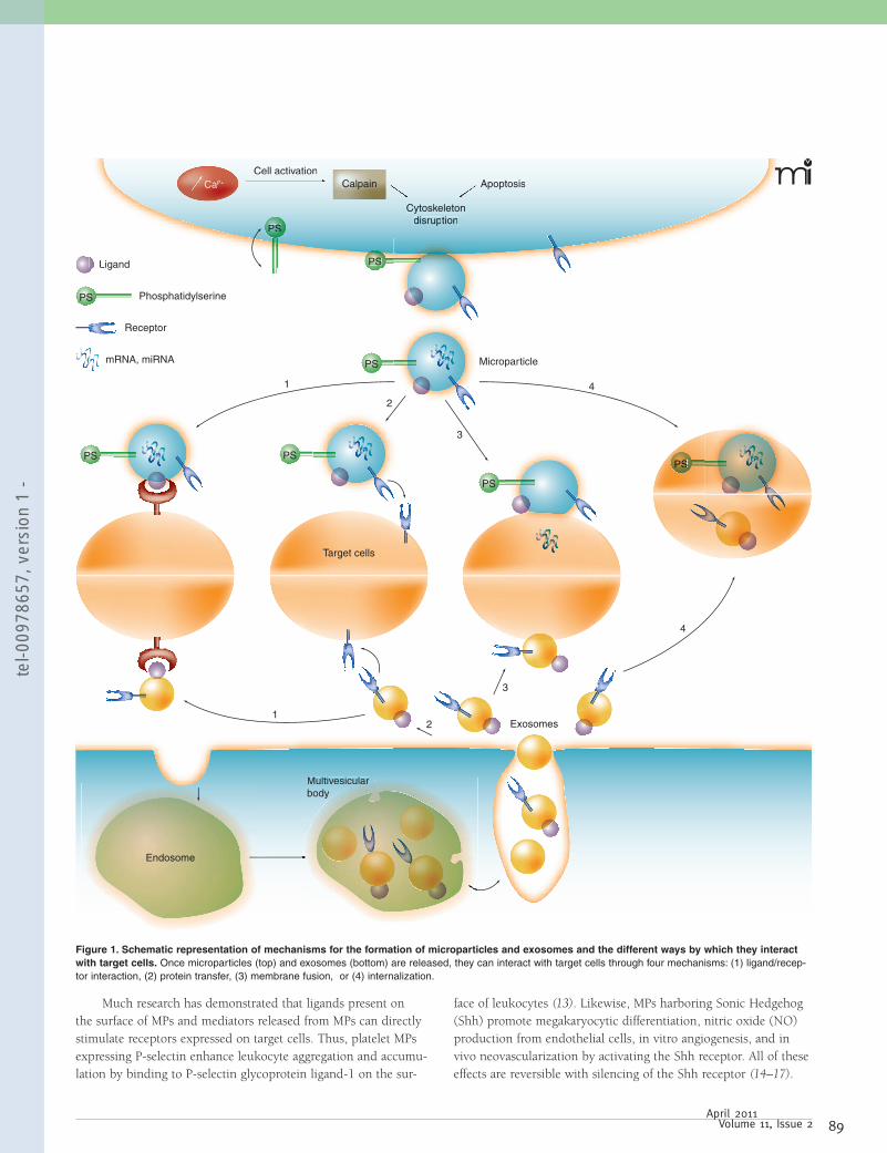
Much research has demonstrated that ligands present on the surface of MPs and mediators released from MPs can directly stimulate receptors expressed on target cells. Thus, platelet MPs expressing P-selectin enhance leukocyte aggregation and accumu-lation by binding to P-selectin glycoprotein ligand-1 on the sur-
face of leukocytes (13). Likewise, MPs harboring Sonic Hedgehog (Shh) promote megakaryocytic differentiation, nitric oxide (NO) production from endothelial cells, in vitro angiogenesis, and in vivo neovascularization by activating the Shh receptor. All of these effects are reversible with silencing of the Shh receptor (14–17).
PS
PS
PS
PS Phosphatidylserine
Receptor
mRNA, miRNA
PS PS
PS
Ligand
PSSPSPSP
Target cells
Endosome
Multivesicularbody
Exosomes
Microparticle
Cytoskeletondisruption
Cell activationApoptosisCa2+ Calpain
PSSPSPSLigand
SPSPSPS
CytCytoskoskeleeletontondidisdisruprupptitiotionn
CelCell al actictivatvationionApoptosisCa2+ Calpain
1
1
2
2
3
3
4
4
Multivesicularbody
larar
Figure 1. Schematic representation of mechanisms for the formation of microparticles and exosomes and the different ways by which they interact with target cells. Once microparticles (top) and exosomes (bottom) are released, they can interact with target cells through four mechanisms: (1) ligand/recep-tor interaction, (2) protein transfer, (3) membrane fusion, or (4) internalization.
tel-0
0978
657,
ver
sion
1 -

Viewpoint
Additionally, MPs released from T cells can interact with smooth muscle cells through the Fas–Fas Ligand (FasL) pathway to evoke NF- B activation (18).
Several studies point out that MPs may transfer receptors on the surface of target cells. Indeed, the release and intercellular trafficking of CD81+ MPs regulate the expression of CD81 sur-face receptors in lymphocytes (19). MPs might also participate in spreading HIV infection by transferring the CXCR4 co-receptor to CD4+/CXCR4– cells (20). Other instances of receptor transfer include arachidonic acid transport by platelet MPs to endothe-lial cells, leading to an increase in cyclooxygenase-2 (COX-2) and intercellular adhesion molecule 1 (ICAM-1) expression (21). Similarly, lipoxygenase 12 can be transferred from platelets to mast cells via MPs, leading to the production of the lipid mediator lipoxin A4, suggesting that platelet MPs pay a critical role as regu-lators of inflammation (22).
MPs can be also absorbed by fusion or by internalization (or engulfing). Fusion of MPs with their target cells leads to nonselec-tive transfer of MP components (23). Tissue factor-rich MPs fuse with activated, P-selectin–expressing platelets, increasing the lat-ter’s procoagulant activity. Concerning internalization, it has been shown that a mixture of MPs and exosomes released from endothe-lial progenitor cells are internalized in endothelial cells by interac-tion with 4 and 1 integrins. It is noteworthy that these MPs shuttle mRNAs that can activate angiogenesis in endothelial cells (24). Also, lymphocytic MPs interact with LDL receptors and can be internalized in tumor cells, decreasing tumor development (25).
Exosomes can bind to cells through receptor-ligand interac-tions. Dendritic cell–derived exosomes contain milk fat globule–epidermal growth factor 8 (MFG-E8) which can bind integrins
expressed on dendritic cells or macrophages (26). Alternatively, exosomes can transfer information to recipient cells. Delta-like 4, a Notch protein family member whose expression is increased during angiogenesis, is present in exosomes and promotes capil-lary-like structure formation in vitro and in vivo by a mechanism implicating the transfer of Delta-like 4 into the endothelium, indicating that the Delta-like/Notch pathway does not require direct cell-cell contact to expand its signaling potential on angio-genesis (27). Furthermore, exosomes might attach or fuse with the target cell membrane, delivering exosomal surface proteins and cytoplasm to the recipient cell (28). Finally, exosomes may also be internalized by target cells by endocytosis or phagocytosis. Exosomes are internalized efficiently by phagocytes, by actin- and phosphatidylinositol 3-kinase-dependent mechanisms, without the need of caveolae, macropynocytosis, or clathrin-coated vesicles (29). Exosomes can also be internalized by immature dendritic cells for presentation to CD4 T cells (30).
A better understanding of intercellular transfer by exosomes may lead to the development of strategies that interfere with the release of pathogenic components by exosomes. Recently, a novel mechanism of genetic exchange between cells, referred to as “exo-somal shuttle RNA,” by a transfer of mRNAs and miRNAs medi-ated by exosomes, has been proposed (11), opening the possibility of using exosomes for gene therapy. Also, exosomes secreted by dendritic cells could modulate immune responses directly by exposing MHC and T-cell costimulatory molecules and indirectly by carrying internal components to surrounding cells (10, 31).
Among the different messages transferred, MVs are able to modify function of cells from the cardiovascular system, immune system, and tumor cells. The transfer of information between MVs
Table 1. Different Mechanisms by which Microparticles and Exosomes Interact with Target Cells
Microparticles Interaction Molecular Pathway Cellular Effects References
Exosomes Interaction Molecular Pathway Cellular Effects References
tel-0
0978
657,
ver
sion
1 -

and cells is independent of the number of MVs but is dependent on the different composition and origin of the MVs (32, 33). Owing to the procoagulant ability of tissue factor and of phos-phatidylserine that MVs carry, elevated levels of MPs participate in the development and maintenance of prothrombotic status in ath-erosclerosis (34), pre-eclampsia (35), and hematological diseases (36), as well as in cancer-associated thrombosis (37).
Circulating MPs decrease NO production by either reduc-ing the activity of endothelial NO-synthase or by decreasing its bioavailability. Thus, MPs from patients with myocardial infarc-tion, diabetes, or pre-eclampsia induce endothelial dysfunction by impairing the endothelial NO transduction pathway (38–40). In patients with metabolic syndrome, circulating MPs evoke endothe-lial dysfunction by decreasing endothelial NO-synthase activity and NO production by increasing protein nitration in endothelial cells (41) and by enhancing plasmatic oxidative stress markers (42). Interestingly, patients with obstructive sleep apnea (OSA) display the same levels of circulating MPs as observed in healthy subjects, but levels of MPs from granulocytes and activated leukocytes (CD62L+) are higher in OSA patients. In addition, levels of CD62L+ MPs correlate with endothelial-NO dysfunction that may initiate atherogenic processes in patients with OSA (32). In a model of rat pulmonary arterial hypertension, circulating MPs from hypoxic rats exhibit reduced NO bioavailability owing to decreased endothelial NO-synthase activity and enhanced oxidative stress in pulmonary endothelial cells (33). Altogether these data underscore the deleteri-ous effects of circulating MPs on the cardiovascular system, and in particular, on the endothelial cells.
Finally, one key feature of the effects of MVs in the cardiovas-cular system is their ability to modulate the angiogenic program of both endothelial mature and progenitor cells. Indeed, proteins such as peroxisome proliferator–activated receptor–alpha (PPAR ), carried by MPs, seem essential to promote the pro-angiogenic reprogramming of endothelial progenitor cells through Akt- and NF- B–mediated pathways (43). Isolated MPs from vitreous fluid from patients with diabetic retinopathy stimulated endothelial cell proliferation and increased new vessel formation in mice, indicat-ing that MPs could contribute to disease progression through their ability to promote angiogenesis (44). MPs are able to amplify the initial endothelial dysfunction and accelerate the progression of atherosclerotic lesions by promoting intraplaque neovasculariza-tion and thrombogenicity (45).
Endothelial exosomes bearing metalloproteinases promote the formation of a tubule-like structure in human endothelial cells, suggesting that exosomes may regulate proteolytic activity vital to angiogenic steps involving migration of endothelial cells (46). Altogether, these data highlight the potential effects of circulating MVs on new vessel generation and may constitute a new therapeu-tic target against the progression of these diseases.
Immune cells are activated during many pathological situ-ations and subsequently generate MVs. MPs appear to serve as important mediators to promote inflammation whereas exosomes
most often drive immune responses and autoreactivity. Indeed, through the generation of MPs, activated T cells may facilitate acti-vation of mast cells that are not located in close physical proximity at the inflammatory sites (47). In addition, platelet-derived MPs may play an important role by acting as vectors for the CD154 (CD40L) signal. CD154 is a member of the TNF family expressed by activated CD4+, CD8+, and -T cells, B cells, and platelets. Among the effects of CD154, the CD154 signal plays a crucial role in the activation of T cell–dependent humoral immune responses (48). In particular, by concentrating the CD154 activity and car-rying it away from the site of platelet activation and aggregation, platelet-derived MPs stimulate antigen-specific production by cooperating with responses elicited by CD4+ T cells (49).
Depending on their origin, exosomes can serve as both immunostimulatory and immunoregulatory entities (50). As described above, T and B cells as well as dendritic cells release exosomes. Thus, in vitro studies have shown that exosomes released from B cells can directly activate CD4+ T cells (51). Also, dendritic cell-derived exosomes initiate specific cytotoxic T lym-phocyte response in vivo and suppress growth of established murine tumors in a T cell-dependent manner (52). In the same way, tumor-derived exosomes transfer tumor antigens to dendritic cells, which induce potent CD8+ T cell–dependent antitumor responses (53). Subsequently, numerous studies have reported the ability of exosomes to act as potential relevant elements for immu-no-interventions by amplifying the generation of donor-reactive T cells following transplantation (54) or by suppressing antigen-specific responses through a Fas/FasL-dependent mechanism in the treatment of autoimmune diseases (55).
The successful quantification and identification of several proteins carried by exosomes allows for the suggestion that exosomes may be useful as prognostic, diagnostic, or detection markers in cancers. For instance, claudin-4 can be shed from ovarian cancer cells and detected in exosomes from plasma from ovarian cancer patients (56). The number of exosomes expressing CD63 [a tetraspanin (a cell surface–localized membrane protein) involved in several cellular process including cell activation, adhe-sion, differentiation, and tumour invasion (57)] and caveolin-1 in the plasma of melanoma patients is significantly increased when compared to those in healthy donors (58). Also, the cytochrome P450 CYP17A1, a target for total androgen blockade in advanced prostate cancer patients, is expressed in plasma exosomes for these patients (59), whereas the presence of two known prostate cancer biomarkers, PCA-3 and TMPRSS2:ERG, have been found in exosomes isolated from urine of patients (60), suggesting that exosomes may be used as a novel approach in detection and pros-tate tumor surveillance. Also, exosomes from glioblastoma tumor cells containing mRNA, miRNA, and angiogenic proteins, such as VEGF, can be detected in serum from glioblastoma patients. Thus, exosomes promote angiogenesis favoring cancer growth, yet they can provide valuable information regarding the diagnosis and choice of appropriate therapy (61). Cancer specific miRNA isolated
tel-0
0978
657,
ver
sion
1 -

Viewpoint
from circulating tumor-derived exosomes from patients with ovar-ian cancer constitutes a new biomarker of this pathology (62).
Most interestingly, exosomes can represent a nexus between the immune system and tumor environment. Isolated exosomes from the blood of patients with colorectal cancer (63) contain not only proteins typical of exosomes (CD63) but also FasL and TNF-alpha-related apoptosis-inducing ligand (TRAIL), which mediate their pro-apoptotic effects on CD8+ T cells (63). This mechanism of immunosuppression has potential implications as a prognostic factor and could be targeted for the development of new antitumor therapies in colorectal cancer patients. Also, exosomes present in blood and ascites from patients with ovarian carcinoma promote differentiation and proliferation of regulatory T cells (Treg) as well as up-regulation of Treg-mediated suppression that may poten-tially contribute to tumor escape (64). Unlike helper T cells, which orchestrate antibody production by B cells, and cytotoxic T cells, which kill virally infected or abnormal cells, Treg act to suppress the immune response of a large number of distinct target cell types (65). Furthermore, these exosomes injected into tumor-bearing mice resulted in augmented tumor growth (66). Altogether, exosomes act as vectors for the transfer of immunological information at distance and can be considered as novel biomarkers and effectors.
In summary, MVs display two faces, as did the Roman god Janus: 1) they constitute potential biomarkers of activity disease, and 2) they can be considered as promising therapeutic targets, considering their ability to convey biological information. doi/10.1124/mi.11.2.5
This work was partially supported by Agence Nationale pour la Recherche-07-PHYSIO-010-01 and Fondation de France (n° 2007001918).
Aharon A, Tamari T, and Brenner B (2008) Monocyte-derived micropar-1. ticles and exosomes induce procoagulant and apoptotic effects on endothelial cells. Thromb Haemost 100:878-885.
Lai RC, Arslan F, Tan SS, Tan B, Choo A, Lee MM, Chen TS, Teh BJ, 2. Eng JK, Sidik H, et al. (2010) Derivation and characterization of human fetal MSCs: an alternative cell source for large-scale production of car-dioprotective microparticles. J Mol Cell Cardiol 48:1215-1224.
Martinez MC, Tesse A, Zobairi F, and Andriantsitohaina R (2005) Shed 3. membrane microparticles from circulating and vascular cells in regulat-ing vascular function. Am J Physiol Heart Circ Physiol 288:H1004-1009.
Ratajczak J, Wysoczynski M, Hayek F, Janowska-Wieczorek A, and 4. Ratajczak MZ (2006) Membrane-derived microvesicles: important and underappreciated mediators of cell-to-cell communication. Leukemia 20:1487-1495. One of the first reviews pointing to microvesicles as mediators of cell-to-cell communication.
Pasquet JM, Dachary-Prigent J, and Nurden AT (1996) Calcium influx is 5. a determining factor of calpain activation and microparticle formation in platelets. Eur J Biochem 239:647-654. This paper demonstrated the essential role of calcium influx in microparticle formation.
Bevers EM, Comfurius P, Dekkers DW, and Zwaal RF (1999) Lipid 6. translocation across the plasma membrane of mammalian cells. Biochem Biophys Acta 1439:317-330.
Distler JH, Pisetsky DS, Huber LC, Kalden JE, Gay S, and Distler O 7. (2005) Microparticles as regulators of inflammation: novel players of cel-lular crosstalk in the rheumatic diseases. Arthritis Rheum 52:3337-3348.
Pan BT and Johnstone RM (1983) Fate of the transferrin receptor dur-8. ing maturation of sheep reticulocytes in vitro: selective externalization of the receptor. Cell 33:967-978. This study is the first to describe the existence of exosomes.
Keller S, Sanderson MP, Stoeck A, and Altevogt P (2006) Exosomes: 9. from biogenesis and secretion to biological function. Immunol Lett 107:102-108.
Thery C, Ostrowsky M, and Segura E (2009) Membrane vesicles as 10. convoyers of immune responses. Nat Rev Immunol 9:581-593. This review is still quite up to date and provides an excellent introduc-tion to exosomes as modulators of immune responses.
Valadi H, Ekstrom K, Bossios A, Sjostrand M, Lee JJ, and Lotvall JO 11. (2007) Exosome-mediated transfer of mRNAs and microRNAs is a novel mechanism of genetic exchange between cells. Nat Cell Biol 9:654-659. These authors proved that mRNAs carried by exosomes are functional and transferable to recipient cells.
Hunter MP, Ismail N, Zhang X, Aguda BD, Lee EJ, Yu L, Xiao T, Schafer 12. J, Lee ML, Schmittgen TD, et al. (2008) Detection of microRNA expres-sion in human peripheral blood microvesicles. PLoS One 3:e3694. This study suggests a role for microvesicles in disseminating miRNA-associated information.
Forlow SB, McEver RP, and Nollert MU (2000) Leukocyte-leukocyte 13. interactions mediated by platelet microparticles under flow. Blood 95:1317-1323.
Martinez MC, Larbret F, Zobairi F, Coulombe J, Debili N, Vainchenker 14. W, Ruat M, and Freyssinet JM (2006) Transfer of differentiation signal by membrane microvesicles harboring hedgehog morphogens. Blood 108:3012-3020. This paper is the first to describe the transfer of a differentiation signal associated with morphogens harbored by microparticles.
Agouni A, Mostefai HA, Porro C, Carusio N, Favre J, Richard V, Henrion 15. D, Martinez MC, and Andriantsitohaina R (2007) Sonic hedgehog car-ried by microparticles corrects endothelial injury through nitric oxide release. Faseb J 21:2735-2741. These authors found that micropar-ticles carrying Sonic hedgehog displayed beneficial effects on endothelial cells.
Soleti R, Benameur T, Porro C, Panaro MA, Andriantsitohaina R, and 16. Martinez MC (2009) Microparticles harboring Sonic Hedgehog promote angiogenesis through the upregulation of adhesion proteins and proan-giogenic factors. Carcinogenesis 30:580-588.
Benameur T, Soleti R, Porro C, Andriantsitohaina R, and Martinez MC 17. (2010) Microparticles carrying sonic hedgehog favor neovasculariza-tion through the activation of nitric oxide pathway in mice. PLoS One 5:e12688.
Tesse A, Martinez MC, Hugel B, Chalupsky K, Muller CD, Meziani 18. F, Mitolo-Chieppa D, Freyssinet JM, and Andriantsitohaina R (2005) Upregulation of proinflammatory proteins through NF-kappaB pathway by shed membrane microparticles results in vascular hyporeactivity. Arterioscler Thromb Vasc Biol 25:2522-2527.
Fritzsching B, Schwer B, Kartenbeck J, Pedal A, Horejsi V, and Ott 19. M (2002) Release and intercellular transfer of cell surface CD81 via microparticles. J Immunol 169:5531-5537.
Rozmyslowicz T, Majka M, Kijowski J, Murphy SL, Conover DO, Poncz 20. M, Ratajczak J, Gaulton GN, and Ratajczak MZ (2003) Platelet- and megakaryocyte-derived microparticles transfer CXCR4 receptor to CXCR4-null cells and make them susceptible to infection by X4-HIV. Aids 17:33-42. Using an in vitro model, this study shows the trans-fer of CXCR4 that could increase susceptibility of infection by HIV.
Barry OP, Pratico D, Savani RC, and FitzGerald GA (1998) Modulation 21. of monocyte-endothelial cell interactions by platelet microparticles. J Clin Invest 102:136-144. This work illustrates the interaction between platelet-derived microparticles and endothelial cells.
tel-0
0978
657,
ver
sion
1 -
Tang K, Liu J, Yang Z, Zhang B, Zhang H, Huang C, Ma J, Shen GX, 22. Ye D, and Huang B (2010) Microparticles mediate enzyme transfer from platelets to mast cells: a new pathway for lipoxin A4 biosynthesis. Biochem Biophys Res Commun 400:432-436.
Breimo ES and Østerud B (2005) Generation of tissue factor-rich 23. microparticles in an ex vivo whole blood model. Blood Coagul Fibrinolysis 16:399-405.
Deregibus MC, Cantaluppi V, Calogero R, Lo Iacono M, Tetta C, 24. Biancone L, Bruno S, Bussolati B. and Camussi G (2007) Endothelial progenitor cell derived microvesicles activate an angiogenic program in endothelial cells by a horizontal transfer of mRNA. Blood 110:2440-2448.
Yang C, Gagnon C, Hou X, and Hardy P (2010) Low density lipoprotein 25. receptor mediates anti-VEGF effect of lymphocyte T-derived micropar-ticles in Lewis lung carcinoma cells. Cancer Biol Ther 10:448-456.
Thery C, Regnault A, Garin J, Wolfers J, Zitvogel L, Ricciardi-Castagnoli 26. P, Raposo G, and Amigorena S (1999) Molecular characterization of dendritic cell-derived exosomes. Selective accumulation of the heat shock protein hsc73. J Cell Biol 147:599-610.
Sheldon H, Heikamp E, Turley H, Dragovic R, Thomas P, Oon CE, Leek 27. R, Edelmann M, Kessler B, Sainson RCA, et al. (2010) New mechanism for notch signaling to endothelium at a distance by delta-like 4 incorpo-ration into exosomes. Blood 116:2385-2394.
Del Conde I, Shrimpton CN, Thiagarajan P, and Lopez JA (2005) 28. Tissue-factor-bearing microvesicles arise from lipid rafts and fuse with activated platelets to initiate coagulation. Blood 106:1604-1611.
Feng D, Zhao WL, Ye YY, Bai XC, Liu RQ, Chang LF, Zhou Q, and Sui 29. SF (2010) Cellular internalization of exosomes occurs through phagocy-tosis. Traffic 11:675-687.
Morelli AE, Larregina AT, Shufesky WJ, Sullivan ML, Stolz DB, Papworth 30. GD, Zahorchak AF, Logar AJ, Wang Z, Watkins SC, et al. (2004) Endocytosis, intracellular sorting, and processing of exosomes by den-dritic cells. Blood 104:3257-3266.
Thery C, Duban L, Segura E, Veron P, Lantz O, and Amigorena S 31. (2002) Indirect activation of naive CD4+ T cells by dendritic cell-derived exosomes. Nat Immunol 3:1156-1162.
Priou P, Gagnadoux F, Tesse A, Mastronardi ML, Agouni A, Meslier N, 32. Racineux JL, Martinez MC, Trzepizur W, and Andriantsitohaina R (2010) Endothelial dysfunction and circulating microparticles from patients with obstructive sleep apnea. Am J Pathol 177:974-983.
Tual-Chalot S, Guibert C, Muller B, Savineau JP, Andriantsitohaina R, 33. and Martinez MC (2010) Circulating microparticles from pulmonary hypertensive rats induce endothelial dysfunction. Am J Respir Crit Care Med 182:261-268.
Mallat Z, Hugel B, Ohan J, Leseche G, Freyssinet JM, and Tedgui A 34. (1999) Shed membrane microparticles with procoagulant potential in human atherosclerotic plaques: a role for apoptosis in plaque thrombo-genicity. Circulation 99:348-353. This study is one the first to demon-strate the pathophysiological involvement in atherosclerosis.
Bretelle F, Sabatier F, Desprez D, Camoin L, Grunebaum L, Combes 35. V, D’Ercole C, and Dignat-George F (2003) Circulating microparticles: a marker of procoagulant state in normal pregnancy and pregnancy complicated by preeclampsia or intrauterine growth restriction. Thromb Haemost 89:486-492.
Trappenburg MC, van Schilfgaarde M, Marchetti M, Spronk HM, Ten 36. Cate H, Leyte A, Terpstra WE, and Falanga A (2009) Elevated proco-agulant microparticles expressing endothelial and platelet markers in essential thrombocythemia. Haematologica 94:911-918.
Kasthuri RS, Taubman MB, and Mackman N (2009) Role of tissue factor 37. in cancer. J Clin Oncol 27:4834-4838.
Boulanger CM, Scoazec A, Ebrahimian T, Henry P, Mathieu E, Tedgui A, 38. and Mallat Z (2001) Circulating microparticles from patients with myo-cardial infarction cause endothelial dysfunction. Circulation 104:2649-2652.
Martin S, Tesse A, Hugel B, Martinez MC, Morel O, Freyssinet JM, 39. and Andriantsitohaina R (2004) Shed membrane particles from T lym-phocytes impair endothelial function and regulate endothelial protein expression. Circulation 109:1653-1659.
Vanwijk MJ, Svedas E, Boer K, Nieuwland R, Vanbavel E, and 40. Kublickiene KR (2002) Isolated microparticles, but not whole plasma, from women with preeclampsia impair endothelium-dependent relax-ation in isolated myometrial arteries from healthy pregnant women. Am J Obstet Gynecol 187:1686-1693.
Agouni A, Lagrue-Lak-Hal AH, Ducluzeau PH, Mostefai HA, 41. Draunet-Busson C, Leftheriotis G, Heymes C, Martinez MC, and Andriantsitohaina R (2008) Endothelial dysfunction caused by circulat-ing microparticles from patients with metabolic syndrome. Am J Pathol 173:1210-1219.
Helal O, Defoort C, Robert S, Marin C, Lesavre N, Lopez-Miranda J, 42. Riserus U, Basu S, Lovegrove J, McMonagle J, et al. (2010) Increased levels of microparticles originating from endothelial cells, platelets and erythrocytes in subjects with metabolic syndrome: Relationship with oxi-dative stress. Nutr Metab Cardiovasc Dis.
Benameur T, Tual-Chalot S, Andriantsitohaina R, and Martinez MC 43. (2010) PPAR is essential for microparticle-induced differentiation of mouse bone marrow-derived endothelial progenitor cells and angiogen-esis. PLos One 5:e12392.
Chahed S, Leroyer AS, Benzerroug M, Gaucher D, Georgescu A, 44. Picaud S, Silvestre JS, Gaudric A, Tedgui A, Massin P, et al. (2010) Increased vitreous shedding of microparticles in proliferative diabetic retinopathy stimulates endothelial proliferation. Diabetes 59:694-701.
Leroyer AS, Rautou PE, Silvestre JS, Castier Y, Leseche G, Devue C, 45. Duriez M, Brandes RP, Lutgens E, Tedgui A et al. (2008) CD40 ligand+ microparticles from human atherosclerotic plaques stimulate endothelial proliferation and angiogenesis a potential mechanism for intraplaque neovascularization. J Am Coll Cardiol 52:1302-1311.
Taraboletti G, D’Ascenzo S, Borsotti P, Giavazzi R, Pavan A, and Dolo V 46. (2002) Shedding of the matrix metalloproteinases MMP-2, MMP-9, and MT1-MMP as membrane vesicle-associated components by endothelial cells. Am J Pathol 160:673-680. This is one of the first studies show-ing the effects of microvesicles on angiogenesis.
Shefler I, Salamon P, Reshef T, Mor A, and Mekori YA (2010) T cell-47. induced mast cell activation: a role for microparticles released from activated T cells. J Immunol 185:4206-4212.
van Kooten C48. and Bancherau J (2000) CD40-CD40 ligand. J Leukoc Biol 67:2-17.
Sprague DL, Elzey BD, Christ SA, Waldschmidt TJ, Jensen RJ, and 49. Ratliff TL (2008) Platelet-mediated modulation of adaptative immunity: unique delivery of CD 154 signal by platelet-derived membrane vesicles. Blood 111:5028-5036.
Couzin J (2005) Cell biology: The ins and outs of exosomes.50. Science 308:1862-1863.
Raposo G, Nijman HW, Stoorvogel W, Liejendekker R, Harding CV, 51. Melief CJ, and Geuze HJ (1996) B lymphocytes secrete antigen-pre-senting vesicles. J Exp Med 183:1161-1172.
Zitvogel L, Regnault A, Lozier A, Wolfers J, Flament C, Tenza D, 52. Ricciardi-Castagnoli P, Raposo G, and Amigorena S (1998) Eradication of established murine tumors using a novel cell-free vaccine: dendritic cell-derived exosomes. Nat Med 4:594-600. A convincing study of the use of exosomes as vaccine against tumors.
Wolfers J, Lozier A, Raposo G, Regnault A, Thery C, Masurier C, 53. Flament C, Pouzieux S, Faure F, Tursz T, et al. (2001) Tumor-derived exosomes are a source of shared tumor rejection antigens for CTL cross-priming. Nat Med 7:297-303.
Montecalvo A, Shufesky WJ, Stolz DB, Sullivan MG, Wang Z, Divito 54. SJ, Papworth GD, Watkins SC, Robbins PD, Larregina AT, et al. (2008) Exosomes as a short-range mechanism to spread alloantigen between dendritic cells during T cell allorecognition. J Immunol 180:3081-3090.
tel-0
0978
657,
ver
sion
1 -
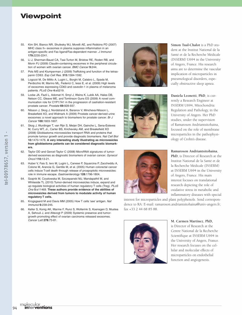
Viewpoint
Kim SH, Bianco NR, Shufesky WJ, Morelli AE, and Robbins PD (2007) 55. MHC class II+ exosomes in plasma suppress inflammation in an antigen-specific and Fas ligand/Fas-dependent manner. J Immunol 179:2235-2241.
Li J, Sherman-Baust CA, Tsai-Turton M, Bristow RE, Roden RB, and 56. Morin PJ (2009) Claudin-containing exosomes in the peripheral circula-tion of women with ovarian cancer. BMC Cancer 9:244.
Pols MS and Klumperman J (2009) Trafficking and function of the tetras-57. panin CD63. Exp Cell Res. 315:1584-1592.
Logozzi M, De Milito A, Lugini L, Borghi M, Calabro L, Spada M, 58. Perdicchio M, Marino ML, Federici C, Iessi E, et al. (2009) High levels of exosomes expressing CD63 and caveolin-1 in plasma of melanoma patients. PLoS One 4:e5219.
Locke JA, Fazli L, Adomat H, Smyl J, Weins K, Lubik AA, Hales DB, 59. Nelson CC, Gleave ME, and Tomlinson Guns ES (2009) A novel com-munication role for CYP17A1 in the progression of castration-resistant prostate cancer. Prostate 69:928-937.
Nilsson J, Skog J, Nordstrand A, Baranov V, Mincheva-Nilsson L, 60. Breakefield XO, and Widmark A (2009) Prostate cancer-derived urine exosomes: a novel approach to biomarkers for prostate cancer. Br J Cancer 100:1603-1607.
Skog J, Wurdinger T, van Rijn S, Meijer DH, Gainche L, Sena-Esteves 61. M, Curry WT, Jr., Carter BS, Krichevsky AM, and Breakefield XO (2008) Glioblastoma microvesicles transport RNA and proteins that promote tumour growth and provide diagnostic biomarkers. Nat Cell Biol 10:1470-1476. A very interesting study illustrating as microvesicles from glioblastoma patients can be considered diagnostic biomark-ers.
Taylor DD and Gercel-Taylor C (2008) MicroRNA signatures of tumor-62. derived exosomes as diagnostic biomarkers of ovarian cancer. Gynecol Oncol 110:13-21.
Huber V, Fais S, Iero M, Lugini L, Canese P, Squarcina P, Zaccheddu A, 63. Colone M, Arancia G, Gentile M, et al. (2005) Human colorectal cancer cells induce T-cell death through release of proapoptotic microvesicles: role in immune escape. Gastroenterology 128:1796-1804.
Szajnik M, Czystowska M, Szczepanski MJ, Mandapathil M, and 64. Whiteside TL (2010) Tumor-derived microvesicles induce, expand and up-regulate biological activities of human regulatory T cells (Treg). PLoS One 5:e11469. These authors provide evidence of the abilities of microvesicles derived from tumors to modulate activity of human regulatory T cells.
Krogsgaard M and Davis MM (2005) How T cells ‘see’ antigen. 65. Nat Immunol 6:239-245.
Keller S, Konig AK, Marme F, Runz S, Wolterink S, Koensgen D, Mustea 66. A, Sehouli J, and Altevogt P (2009) Systemic presence and tumor-growth promoting effect of ovarian carcinoma released exosomes. Cancer Lett 278:73-81.
Simon Tual-Chalot is a PhD stu-dent at the Institut National de la Santé et de la Recherche Médicale (INSERM) U694 in the University of Angers, France. His research aims are to determine the vascular implication of microparticles in pneumological disorders, espe-cially obstructive sleep apnea.
Daniela Leonetti, PhD, is cur-rently a Research Engineer at INSERM U694, Mitochondria: Regulation and Pathology, in the University of Angers. Her PhD studies, under the supervision of Ramaroson Andriantsitohaina, focused on the role of membrane microparticles in the pathophysi-ology of Crohn’s disease.
Ramaroson Andriantsitohaina, PhD, is Director of Research at the Institut National de la Santé et de la Recherche Médicale (INSERM) at INSERM U694 in the University of Angers, France. His main interest focuses on translational research depicting the role of oxidative stress in metabolic and inflammatory diseases with special
interest for microparticles and plant polyphenols. Send correspon-dence to RA: E-mail: [email protected]; fax +33 2 44 68 85 88.
M. Carmen Martinez, PhD, is Director of Research at the Centre National de la Recherche Scientifique at INSERM U694 in the University of Angers, France. Her research focuses on the cel-lular and molecular effects of microparticles on endothelial function and angiogenesis.
tel-0
0978
657,
ver
sion
1 -
!"#$$
Revue :
Microparticles: targets and tools in cardiovascular
diseases
$
tel-0
0978
657,
ver
sion
1 -
Microparticles: targets and tools incardiovascular diseaseMaria Carmen Martinez, Simon Tual-Chalot, Daniela Leonetti andRamaroson Andriantsitohaina
Institut National de la Sante et de la Recherche Me dicale (INSERM) Unite 694, Faculte de Me decine, Universite d’Angers, Angers,France
Cells communicate with other cells not only via directcell–cell contact and the production of signaling mole-cules but also through release of microparticles (MPs).MPs are small vesicles released from stimulated and/orapoptotic cells. They harbor membrane proteins that arecharacteristic of the original parent cell and intracellularcomponents involved in cell signaling. MPs are consid-ered to be both biomarkers and effectors of cell signalingthat maintain and/or initiate cell dysfunction. Thus, MPscan evoke endothelial dysfunction by decreasing nitricoxide (NO) production and promoting vascular inflam-mation which favor the prothrombotic state in athero-sclerosis. Novel pharmacological approaches targetingMP production or properties could be used to treatcardiovascular pathologies. Paradoxically, another use-ful approach might be to employ engineered MPs withmodified compositions as therapeutic agents to correctcardiovascular pathologies. This review is focused onthe mechanisms of MP formation and their effects ontarget cells under physiological or pathophysiologicalconditions.
Microparticles (MPs)Despite the progress achieved in the treatment of cardio-vascular disease, these conditions remain a major cause ofdeath worldwide [1]. The identification of new targets andtherapeutic agents to fight the most common cardiovascu-lar diseases (atherothrombotic vascular disease, hyperten-sion and heart failure) is therefore essential. In thiscontext, MPs could represent useful therapeutic tools ortargets for cardiovascular disease. MPs are small vesiclesthat can be potentially released from all cell types. Theyare involved in intercellular communication and appear tobe vectors of biological messages that could actively par-ticipate in the pathophysiology and development of dis-ease, notably cardiovascular disease.
MPs can be isolated from fluids of healthy donors, butelevated levels have been detected in many pathologicalconditions. Furthermore, a positive correlation has beenobserved between the increase of specific MP phenotypesand significant parameters of diseases with vascular com-plications [2,3].
This review focuses on the possible role of MPs in theinitiation and/or development of cardiovascular disease.
The purpose is to describe the mechanisms of MP forma-tion, their interaction on target cells, and particularly theeffects mediated by MPs in the cardiovascular system.Furthermore, we consider the possibility of defining MPsas potential biomarkers of disease activity and suggestthat MPs can be used as potential therapeutic tools.
Formation and composition of MPsInitially considered as inert cellular debris released fromactivated platelets in human plasma [4], MPs are nowunderstood to be small vesicles shed from the plasmamembrane. They have a diameter between 0.05 and1 mm and are heterogeneous in composition.
In the cardiovascular system, MPs can be produced bymany cell types including platelets, leukocytes, erythro-cytes, endothelial cells, and smooth-muscle cells, but plate-let-derived MPs represent approximately 70–90% ofcirculating MPs [5]. In addition, other cells including tu-mor cells, epithelial cells and fibroblasts can also be asource of MPs [6,7].
The mechanisms that lead to the formation of MPs arenot completely elucidated, but several studies provide evi-dence that the production of MPs takes place followingchemical and physical cell activation or apoptosis(Figure 1). The phenotype and the quantity of MPs releasedcan vary depending on their mode of production [8,9].
It is well accepted that MP formation can result fromcell activation by different agonists such as thrombin and/or collagen [10]. The activation stimulus triggers an influxof extracellular calcium, which is associated with calcium-dependent proteolysis of cytoskeletal proteins throughcalpain activation [11]. The sustained elevation of cytosoliccalcium concentration also causes kinase activation andphosphatase inhibition that contribute to cytoskeletondisruption, and this appears to be indispensable for thesubsequent blebbing (formation of irregular protuberancesin the plasma membrane) [12]. In addition, several authorshave described loss of membrane asymmetry as a result ofcell-surface exposure of phosphatidylserine, a negativelycharged aminophospholipid that is localized in resting cellsat the inner leaflet of the plasma membrane. Exposure ofphosphatidylserine at the outer leaflet of the plasma mem-brane leads to excess negative charge at the surface thatcould contribute to its destabilization and blebbing. Phos-phatidylserine is present in the outer surface of most (butnot all) MPs, as recently demonstrated by Connor et al [13].
Review
Corresponding author: Andriantsitohaina, R.([email protected]).
TIPS-902; No. of Pages 7
0165-6147/$ – see front matter ! 2011 Elsevier Ltd. All rights reserved. doi:10.1016/j.tips.2011.06.005 Trends in Pharmacological Sciences xx (2011) 1–7 1
tel-0
0978
657,
ver
sion
1 -

Although phosphatidylserine exposure has functional con-sequences (including the stimulation of coagulation bybinding the positively charged domains of coagulationproteins [14]), the significance of phosphatidylserine-neg-ative MPs is unclear and needs further investigation.Finally, several molecular targets linking the increase ofintracellular calcium concentration to MP release havebeen proposed. These include extracellular signal-regulat-ed kinase (ERK) [15], small GTPase Rho A [16], myosinlight chain and Rho-associated kinase (ROCK I) [17],NADPH oxidase, and reactive oxygen species (ROS) [18].
During apoptosis, MP formation is associated with theactivation of ROCK I and caspase 3, leading to disruptionof membrane skeleton structure and consequently mem-brane blebbing and MP formation [19]. Other studies havefound that tumor necrosis factor (TNF)-related apoptosis-inducing ligand (TRAIL) and its receptor TRAIL-R2, thatare involved in the signaling pathway of apoptosis, canpromote endothelial MP release by initiating the recruit-ment of adaptor proteins and the activation of nuclearfactor kappa B (NF-kB) [20]. These different data clearlyunderline the complexity of pathways that lead to theformation of MPs.
The stimulus that triggers MP formation regulates theselective sorting of specific constituents that determinethe composition of MPs and, consequently, the biologicalinformation that they transfer. MP composition reflectsthe organization of the plasma membrane of the cell oforigin at the time of MP generation. MPs carry differentmembrane, cytoplasmic and nuclear constituents from the
original cell and express antigens that allow their char-acterization with respect to their cellular source by the useof specific antibodies. For example, with regard to proteincontents, those of endothelial MPs are predominantlymetabolic enzymes, proteins involved in adhesion andfusion processes, and cytoskeleton-associated proteins[21], whereas proteins from platelet MPs are surfaceglycoproteins or chemokines [22]. The protein componentof MPs can thus vary depending on the phenotype of theMPs and the stimulus leading to their production [9,23].
Little is known about the lipid composition of MPs.Plasma MPs in healthy individuals are rich in cholesterol[24] and phospholipids including lysophosphatidylcholine,phosphatidylcholine, sphingomyelin, lysophosphatidylser-ine, phosphatidylserine, lysophosphatidylethanolamine,phosphatidylethanolamine and phosphatidylinositol [25].However, no data are available for MPs from patients withpathologies associated with an altered lipid profile. Inter-estingly, the lipid environment can modify the activity ofproteins carried by MPs. For example, cholesterol-enrich-ment of human monocytes/macrophages induces the gen-eration of highly procoagulant phosphatidylserine-positiveand tissue factor-positive MPs [26].
In addition, MPs can contain nucleic acids – notablymRNA and micro RNA (miRNA) – suggesting that theycould transfer genetic material to target cells. MP-associ-ated intercellular communication can take place throughdifferent pathways. They can (i) directly interact with theligands present on the surface of target cells and activatecascade signaling, and (ii) transfer proteins, mRNA,
Cellactivation Apoptosis
CalpainROCK ll
ROCK l
Cytoskeletondisruption
Ca2+
Kinase activationphosphatase
inhibition
Thrombin
Caspase-2 TRAILCaspase-3NF-!B
Ligand
Phosphatidylserine
Receptor
mRNA, miRNA
TRENDS in Pharmacological Sciences
Key:
Figure 1. Basic schematic representation of the different steps leading to MP formation. During cell activation an increase in intracellular calcium induces externalization ofphosphatidylserine. Calcium increase is also associated with calpain activation, and this is required for cytoskeleton disruption and MP formation. Under apoptosisinduction, cleavage of caspases induces ROCK activation, leading to cytoskeleton alteration and blebbing. ROCK, Rho kinase; TRAIL, TNF-related apoptosis-inducing ligand.
Review Trends in Pharmacological Sciences xxx xxxx, Vol. xxx, No. x
TIPS-902; No. of Pages 7
2
tel-0
0978
657,
ver
sion
1 -
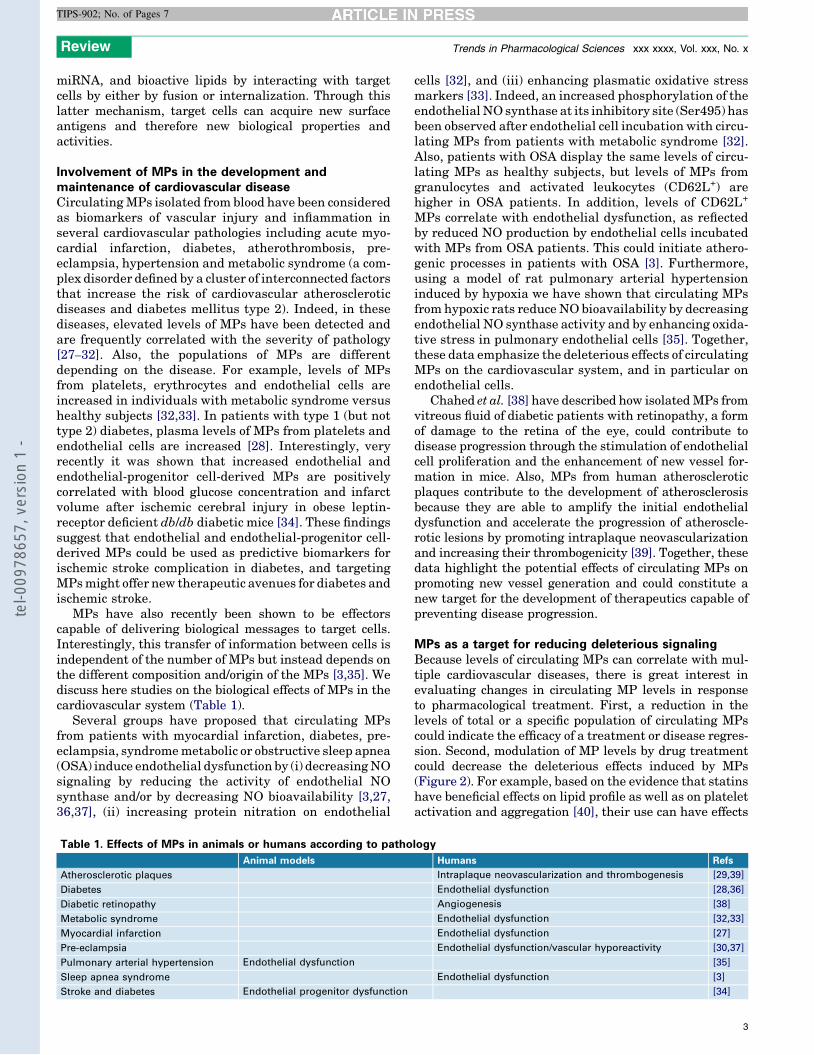
miRNA, and bioactive lipids by interacting with targetcells by either by fusion or internalization. Through thislatter mechanism, target cells can acquire new surfaceantigens and therefore new biological properties andactivities.
Involvement of MPs in the development andmaintenance of cardiovascular diseaseCirculating MPs isolated from blood have been consideredas biomarkers of vascular injury and inflammation inseveral cardiovascular pathologies including acute myo-cardial infarction, diabetes, atherothrombosis, pre-eclampsia, hypertension and metabolic syndrome (a com-plex disorder defined by a cluster of interconnected factorsthat increase the risk of cardiovascular atheroscleroticdiseases and diabetes mellitus type 2). Indeed, in thesediseases, elevated levels of MPs have been detected andare frequently correlated with the severity of pathology[27–32]. Also, the populations of MPs are differentdepending on the disease. For example, levels of MPsfrom platelets, erythrocytes and endothelial cells areincreased in individuals with metabolic syndrome versushealthy subjects [32,33]. In patients with type 1 (but nottype 2) diabetes, plasma levels of MPs from platelets andendothelial cells are increased [28]. Interestingly, veryrecently it was shown that increased endothelial andendothelial-progenitor cell-derived MPs are positivelycorrelated with blood glucose concentration and infarctvolume after ischemic cerebral injury in obese leptin-receptor deficient db/db diabetic mice [34]. These findingssuggest that endothelial and endothelial-progenitor cell-derived MPs could be used as predictive biomarkers forischemic stroke complication in diabetes, and targetingMPs might offer new therapeutic avenues for diabetes andischemic stroke.
MPs have also recently been shown to be effectorscapable of delivering biological messages to target cells.Interestingly, this transfer of information between cells isindependent of the number of MPs but instead depends onthe different composition and/origin of the MPs [3,35]. Wediscuss here studies on the biological effects of MPs in thecardiovascular system (Table 1).
Several groups have proposed that circulating MPsfrom patients with myocardial infarction, diabetes, pre-eclampsia, syndrome metabolic or obstructive sleep apnea(OSA) induce endothelial dysfunction by (i) decreasing NOsignaling by reducing the activity of endothelial NOsynthase and/or by decreasing NO bioavailability [3,27,36,37], (ii) increasing protein nitration on endothelial
cells [32], and (iii) enhancing plasmatic oxidative stressmarkers [33]. Indeed, an increased phosphorylation of theendothelial NO synthase at its inhibitory site (Ser495) hasbeen observed after endothelial cell incubation with circu-lating MPs from patients with metabolic syndrome [32].Also, patients with OSA display the same levels of circu-lating MPs as healthy subjects, but levels of MPs fromgranulocytes and activated leukocytes (CD62L+) arehigher in OSA patients. In addition, levels of CD62L+
MPs correlate with endothelial dysfunction, as reflectedby reduced NO production by endothelial cells incubatedwith MPs from OSA patients. This could initiate athero-genic processes in patients with OSA [3]. Furthermore,using a model of rat pulmonary arterial hypertensioninduced by hypoxia we have shown that circulating MPsfrom hypoxic rats reduce NO bioavailability by decreasingendothelial NO synthase activity and by enhancing oxida-tive stress in pulmonary endothelial cells [35]. Together,these data emphasize the deleterious effects of circulatingMPs on the cardiovascular system, and in particular onendothelial cells.
Chahed et al. [38] have described how isolated MPs fromvitreous fluid of diabetic patients with retinopathy, a formof damage to the retina of the eye, could contribute todisease progression through the stimulation of endothelialcell proliferation and the enhancement of new vessel for-mation in mice. Also, MPs from human atheroscleroticplaques contribute to the development of atherosclerosisbecause they are able to amplify the initial endothelialdysfunction and accelerate the progression of atheroscle-rotic lesions by promoting intraplaque neovascularizationand increasing their thrombogenicity [39]. Together, thesedata highlight the potential effects of circulating MPs onpromoting new vessel generation and could constitute anew target for the development of therapeutics capable ofpreventing disease progression.
MPs as a target for reducing deleterious signalingBecause levels of circulating MPs can correlate with mul-tiple cardiovascular diseases, there is great interest inevaluating changes in circulating MP levels in responseto pharmacological treatment. First, a reduction in thelevels of total or a specific population of circulating MPscould indicate the efficacy of a treatment or disease regres-sion. Second, modulation of MP levels by drug treatmentcould decrease the deleterious effects induced by MPs(Figure 2). For example, based on the evidence that statinshave beneficial effects on lipid profile as well as on plateletactivation and aggregation [40], their use can have effects
Table 1. Effects of MPs in animals or humans according to pathology
Animal models Humans Refs
Atherosclerotic plaques Intraplaque neovascularization and thrombogenesis [29,39]
Diabetes Endothelial dysfunction [28,36]
Diabetic retinopathy Angiogenesis [38]
Metabolic syndrome Endothelial dysfunction [32,33]
Myocardial infarction Endothelial dysfunction [27]
Pre-eclampsia Endothelial dysfunction/vascular hyporeactivity [30,37]
Pulmonary arterial hypertension Endothelial dysfunction [35]
Sleep apnea syndrome Endothelial dysfunction [3]
Stroke and diabetes Endothelial progenitor dysfunction [34]
Review Trends in Pharmacological Sciences xxx xxxx, Vol. xxx, No. x
TIPS-902; No. of Pages 7
3
tel-0
0978
657,
ver
sion
1 -
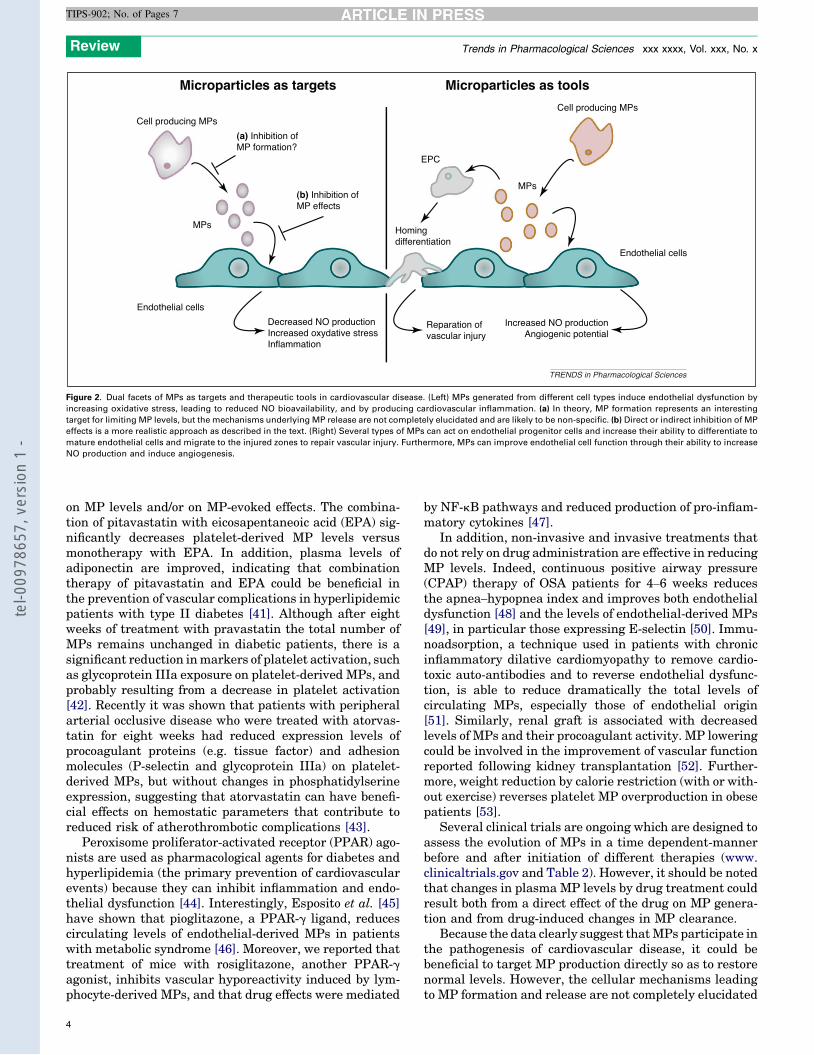
on MP levels and/or on MP-evoked effects. The combina-tion of pitavastatin with eicosapentaneoic acid (EPA) sig-nificantly decreases platelet-derived MP levels versusmonotherapy with EPA. In addition, plasma levels ofadiponectin are improved, indicating that combinationtherapy of pitavastatin and EPA could be beneficial inthe prevention of vascular complications in hyperlipidemicpatients with type II diabetes [41]. Although after eightweeks of treatment with pravastatin the total number ofMPs remains unchanged in diabetic patients, there is asignificant reduction in markers of platelet activation, suchas glycoprotein IIIa exposure on platelet-derived MPs, andprobably resulting from a decrease in platelet activation[42]. Recently it was shown that patients with peripheralarterial occlusive disease who were treated with atorvas-tatin for eight weeks had reduced expression levels ofprocoagulant proteins (e.g. tissue factor) and adhesionmolecules (P-selectin and glycoprotein IIIa) on platelet-derived MPs, but without changes in phosphatidylserineexpression, suggesting that atorvastatin can have benefi-cial effects on hemostatic parameters that contribute toreduced risk of atherothrombotic complications [43].
Peroxisome proliferator-activated receptor (PPAR) ago-nists are used as pharmacological agents for diabetes andhyperlipidemia (the primary prevention of cardiovascularevents) because they can inhibit inflammation and endo-thelial dysfunction [44]. Interestingly, Esposito et al. [45]have shown that pioglitazone, a PPAR-g ligand, reducescirculating levels of endothelial-derived MPs in patientswith metabolic syndrome [46]. Moreover, we reported thattreatment of mice with rosiglitazone, another PPAR-gagonist, inhibits vascular hyporeactivity induced by lym-phocyte-derived MPs, and that drug effects were mediated
by NF-kB pathways and reduced production of pro-inflam-matory cytokines [47].
In addition, non-invasive and invasive treatments thatdo not rely on drug administration are effective in reducingMP levels. Indeed, continuous positive airway pressure(CPAP) therapy of OSA patients for 4–6 weeks reducesthe apnea–hypopnea index and improves both endothelialdysfunction [48] and the levels of endothelial-derived MPs[49], in particular those expressing E-selectin [50]. Immu-noadsorption, a technique used in patients with chronicinflammatory dilative cardiomyopathy to remove cardio-toxic auto-antibodies and to reverse endothelial dysfunc-tion, is able to reduce dramatically the total levels ofcirculating MPs, especially those of endothelial origin[51]. Similarly, renal graft is associated with decreasedlevels of MPs and their procoagulant activity. MP loweringcould be involved in the improvement of vascular functionreported following kidney transplantation [52]. Further-more, weight reduction by calorie restriction (with or with-out exercise) reverses platelet MP overproduction in obesepatients [53].
Several clinical trials are ongoing which are designed toassess the evolution of MPs in a time dependent-mannerbefore and after initiation of different therapies (www.clinicaltrials.gov and Table 2). However, it should be notedthat changes in plasma MP levels by drug treatment couldresult both from a direct effect of the drug on MP genera-tion and from drug-induced changes in MP clearance.
Because the data clearly suggest that MPs participate inthe pathogenesis of cardiovascular disease, it could bebeneficial to target MP production directly so as to restorenormal levels. However, the cellular mechanisms leadingto MP formation and release are not completely elucidated
Microparticles as targets Microparticles as tools
Cell producing MPsCell producing MPs
(a) Inhibition ofMP formation?
(b) Inhibition ofMP effects
MPs
MPs
Endothelial cellsDecreased NO productionIncreased oxydative stressInflammation
Reparation ofvascular injury
Increased NO productionAngiogenic potential
Endothelial cells
EPC
Homingdifferentiation
TRENDS in Pharmacological Sciences
Figure 2. Dual facets of MPs as targets and therapeutic tools in cardiovascular disease. (Left) MPs generated from different cell types induce endothelial dysfunction byincreasing oxidative stress, leading to reduced NO bioavailability, and by producing cardiovascular inflammation. (a) In theory, MP formation represents an interestingtarget for limiting MP levels, but the mechanisms underlying MP release are not completely elucidated and are likely to be non-specific. (b) Direct or indirect inhibition of MPeffects is a more realistic approach as described in the text. (Right) Several types of MPs can act on endothelial progenitor cells and increase their ability to differentiate tomature endothelial cells and migrate to the injured zones to repair vascular injury. Furthermore, MPs can improve endothelial cell function through their ability to increaseNO production and induce angiogenesis.
Review Trends in Pharmacological Sciences xxx xxxx, Vol. xxx, No. x
TIPS-902; No. of Pages 7
4
tel-0
0978
657,
ver
sion
1 -
and it is not yet possible to target these processes specifi-cally (Box 1). Furthermore, the precise characterization ofcell origins and of the biological messages carried andtransferred by MPs will be essential for understandingwhether they possess pathogenic or beneficial propertiesand to permit their precise targeting.
MPs as pharmacological toolsRecent studies have reported that some types of MPspossess therapeutic potential through the modulation ofcellular processes and of secretomes (the molecules secret-ed by cells), and subsequently by inducing tissue repairafter the reprogramming of target cells (Figure 2). Notably,an enhanced level of circulating MPs is not always accom-panied by a deleterious effect; indeed, some populations ofMPs could deliver protective biological messages, preserv-ing endothelial function and/or vascular integrity. Forexample, circulating MPs from sepsis patients have beenshown be protective, rather than deleterious, because theyrestore vascular hyporeactivity in vessels treated withlipopolysaccharide through an enhancement of thrombox-ane A2 production [56]. This confirms findings from aprevious study that showed a positive correlation betweenhigh circulating levels of MPs and survival in sepsispatients [57]. In addition, MPs from sepsis patients treatedwith recombinant activated protein C carry this protein,and this could be related to the anti-apoptotic and barrierprotective effects mediated by activated protein C on en-dothelial cells [58].
Table 2. Clinical trials using MPs as biomarkers of drug treatment efficacy
Title Aim NCTa Estimatedenrollment (n)
Status
MPs in stored packed red blood cells To improve transfusion practice and safety 01185600 500 Currentlyrecruiting
Thromboprophylasis and bariatric surgery To assess the efficacy of enoxaparin on anti-factorXa and MP levels
00444652 150 Currentlyrecruiting
Rosuvastatin to lower circulating tissue factorbearing MPs in metastatic breast cancer
To reduce the risk for deep vein thrombosis 01299038 40 Currentlyrecruiting
Enoxparin thromboprophylaxis in cancer patientswith elevated levels of tissue factor bearing MPs
To improve and/or prevent thrombosis in peoplewith certain types of cancer
00908960 227 Currentlyrecruiting
Paclitaxel and bavituximab in treating patientswith HER2-negative metastatic breast cancer
To study the side effects of giving togetherpaclitaxel and bavituximab by measuring MPs asmarkers of tumor cell spread
01288261 14 Currentlyrecruiting
Clinical trial using metronomic oral low-dosecyclophosphamide alternating with low-dose oralmethotrexate with continuous celecoxib andweekly vinblastine in children and adolescentswith relapsed or progressing solid tumours
To define the safety profile of the combination ofdrugs by measuring angiogenic markersincluding MPs
01285817 90 Currentlyrecruiting
Patients overexposed for a prostateadenocarcinoma
To determine incidence and severity of adverseevents related to radiotherapy
00773656 433 Currentlyrecruiting
The interrelation of the vasculature, endothelium,bone metabolism and uremic toxins in peritonealdialysis
To assess the interrelation between endothelialdysfunction (MP levels) and uremic toxins
00433264 62 Completed
Effect of endothelin-1 receptor blockade oncirculating endothelial MP levels in patients withpulmonary hypertension
To improve of mean of pulmonary arterialpressure
00675051 20 Completed
Dose effects of hormone therapy (two doses ofestradiol associated to progesterone) oninflammatory markers
To reduce inflammation (soluble C reactiveprotein and secondly MPs)
00236301 99 Terminated
Importance in type 1 diabetes patients of anoptimized control of post-prandial glycaemia onoxidant stress prevention
To prevent oxidant stress 00934336 50 Ongoing butnot recruiting
Comparison of pitavastatin with atorvastatin inincreasing HDL-cholesterol and adiponectin inpatients with dislipidemia and coronary arterydisease
To reduce HDL-cholesterol, adiponectin and otherblood parameters including platelet MPs
00861861 200 Unknown
Pioglitazone versus metformin in type 2 diabetes Comparison of the effects of two drugs oncirculating endothelial MPs because theirputative role in inflammatory processes
00815399 150 Unknown
aClinicalTrials.gov registry number
Box 1. Unanswered questions on the potential inhibition of
MP formation
The mechanisms implicated in the MP blebbing depend on the celltype and on the type of stimulation. This complicates the possibilityof specifically inhibiting their formation. Among the cellularmechanisms directly involved in MP release, an increase ofintracellular calcium concentration via calcium channel activationhas been described. However, the nature of these channels remainsunclear. In circulating cells it has been proposed that calcium influxtakes place via transient receptor potential channels (TRPC) [54], andthus TRPC blockade could represent a potential approach toreducing MP formation. However, other tissues express TRPC (e.g.heart [55]), limiting the use of specific inhibitors of TRPC tomodulate MP release. Other intracellular actors in MP formationare also ubiquitous (e.g. kinases, phosphatases) and this also makesspecific targeting impossible.
Review Trends in Pharmacological Sciences xxx xxxx, Vol. xxx, No. x
TIPS-902; No. of Pages 7
5
tel-0
0978
657,
ver
sion
1 -
Benameur et al. [59] have reported that circulating MPsbearing PPARa promote differentiation of endothelial pro-genitor cells and angiogenesis through the activation ofPPARa/Akt/NF-kB pathway. Similar observations weremade by Mause et al. [10] using MPs derived from in vitroactivated platelets. Indeed, platelet-derived MPs are ableto reprogram endothelial progenitor cells towards a phe-notype competent to repair vascular injury. These effectsconsist of changes in the expression of endothelial markersand the modulation of secretome composition towards apro-angiogenic profile (vascular endothelial growth factor,epidermal growth factor, hepatocyte growth factor, IL-1band TNF-a) which can induce proliferation of matureendothelial cells.
Finally, as noted earlier, the composition of MPs andthe messages they transport (proteins, mRNA or miRNA)can differ depending on their origin. It has been reportedthat MPs can be engineered to over-express differentproteins by driving the synthesis of the relevant proteinin MP-producing cells. As demonstrated by Agouni et al.[60], MPs released by apoptotic/stimulated human T lym-phocytes harbor the morphogen Sonic hedgehog (Shh) andimprove endothelial function in the mouse aorta by in-creasing NO release. Also, endothelial dysfunction inmouse coronary artery after ischemia/reperfusion can bereversed by treatment with Shh-positive MPs. Moreover,MPs expressing Shh favor in vitro angiogenesis [61] andthe recovery of hindlimb flow after peripheral ischemiathrough the activation of endothelial NO synthase and theincrease of NO release and pro-angiogenic factor produc-tion [62]. These findings suggest that Shh-positive MPscould represent a potent tool for stimulating neovascular-ization in disease states associated with impaired angio-genesis.
Interestingly, MP-associated RNA and miRNA havebeen detected in the supernatant of mesenchymal stemcells. This suggests, on the one hand, that MP-associatedmembrane can protect both RNA and miRNA againstdegradation by forming an appropriate environmentand, on the other, that MPs can act as vectors ofmiRNA-dependent messages between cells [63].
Concluding remarksIt is now clear that MPs play roles in intercellular com-munication and are able to modulate important cellularregulatory functions. In particular, MPs can induce and/ormaintain endothelial dysfunction and could therefore beimplicated in cardiovascular disease progression, makingthem new drug-development targets. Paradoxically, thereis convincing evidence that some MPs can have beneficialproperties, and MPs could potentially be developed as newtherapeutic tools. There is thus a fragile balance betweenthe harmful and helpful effects of MPs. Further analyses ofthe transcriptomes, miRNAomes and proteomes of MPswill be required for the identification of all the componentsof MPs, and this will improve our understanding of thebiological effects they can induce.
AcknowledgmentsThis work was supported by grants from the Agence Nationale de laRecherche (07-PHYSIO-010-01) and the Fondation de France. S.T.C. was
supported by a doctoral fellowship from the Ministere de la Recherche(France). R.A. is supported by a ‘Contrat d’Interface’ from the INSERMand the Hospital of Angers, France.
References1 Dahlof, B. (2010) Cardiovascular disease risk factors: epidemiology and
risk assessment. Am. J. Cardiol. 105, 3A–9A2 Andoh, A. et al. (2005) Elevated circulating platelet-derived
microparticles in patients with active inflammatory. Am. J.Gastroenterol. 100, 2042–2048
3 Priou, P. et al. (2010) Endothelial dysfunction and circulatingmicroparticles from patients with obstructive sleep apnea. Am. J.Pathol. 177, 974–983
4 Wolf, P. (1967) The nature and significance of platelet products inhuman plasma. Br. J. Haematol. 13, 269–288
5 Flaumenhaft, R. et al. (2009) Megakaryocyte-derived microparticles:direct visualization and distinction from platelet-derivedmicroparticles. Blood 113, 1112–1121
6 Castellana, D. et al. (2009) Membrane microvesicles as actors in theestablishment of a favorable prostatic tumoral niche: a role foractivated fibroblasts and CX3CL1-CX3CR1 axis. Cancer Res. 69,785–793
7 Moulin, V.J. et al. (2010) Shedding of microparticles by myofibroblastsas mediator of cellular cross-talk during normal wound healing. J. Cell.Physiol. 225, 734–740
8 Jimenez, J.J. et al. (2003) Endothelial cells release phenotypically andquantitatively distinct microparticles in activation and apoptosis.Thromb. Res. 109, 175–180
9 Martinez, M.C. et al. (2006) Transfer of differentiation signal bymembrane microvesicles harboring hedgehog morphogens. Blood108, 3012–3020
10 Mause, S.F. et al. (2010) Platelet microparticles enhance thevasoregenerative potential of angiogenic early outgrowth cells aftervascular injury. Circulation 122, 494–506
11 Miyoshi, H. et al. (1996) Calpain activation in plasma membrane blebformation during tert-butyl hydroperoxide-induced rat hepatocyteinjury. Gastroenterology 110, 1897–1904
12 Yan, R. et al. (2009) Role of cAMP-dependent protein kinase in theregulation of platelet procoagulant activity. Arch. Biochem. Biophys.485, 41–48
13 Connor, D.E. et al. (2010) The majority of circulating platelet-derivedmicroparticles fail to bind annexin V, lack phospholipid-dependentprocoagulant activity and demonstrate greater expression ofglycoprotein Ib. Thromb. Haemost. 103, 1044–1052
14 Owens, A.P., 3rd and Mackman, N. (2011) Microparticles in hemostasisand thrombosis. Circ. Res. 108, 1284–1297
15 Kunzelmann-Marche, C. et al. (2001) Regulation of phosphatidylserinetransbilayer redistribution by store-operated Ca2+ entry: role of actincytoskeleton. J. Biol. Chem. 276, 5134–5139
16 Kunzelmann, C. et al. (2004) Rho A participates in the regulation of thephosphatidylserine-dependent procoagulant activity at the surface ofmegakaryocytic cells. J. Thromb. Haemost. 2, 644–650
17 Sebbagh, M. et al. (2001) Caspase-3-mediated cleavage of ROCKIinduces MLC phosphorylation and apoptotic membrane blebbing.Nat. Cell Biol. 3, 346–352
18 Burger, D. et al. (2011) Endothelial microparticle formation byangiotensin II is mediated via AT1R/NADPH oxidase/Rho kinasepathways targeted to lipid rafts. Arterioscler. Thromb. Vasc. Biol.DOI: 10.1161/ATVBAHA.110.222703 in press
19 Coleman, M.L. et al. (2001) Membrane blebbing during apoptosisresults from caspase-mediated activation of ROCK I. Nat. Cell Biol.3, 339–345
20 Simoncini, S. et al. (2009) TRAIL/Apo2L mediates the release ofprocoagulant endothelial microparticles induced by thrombin invitro: a potential mechanism linking inflammation and coagulation.Circ. Res. 104, 943–951
21 Banfi, C. et al. (2005) Proteome of endothelial cell-derived procoagulantmicroparticles. Proteomics 5, 4443–4455
22 Garcia, B.A. et al. (2005) The platelet microparticle proteome. J.Proteome Res. 4, 1516–1521
23 Bernimoulin, M. et al. (2009) Differential stimulation of monocytic cellsresults in distinct populations of microparticles. J. Thromb. Haemost.7, 1019–1028
Review Trends in Pharmacological Sciences xxx xxxx, Vol. xxx, No. x
TIPS-902; No. of Pages 7
6
tel-0
0978
657,
ver
sion
1 -
24 Biro, E. et al. (2005) The phospholipid composition and cholesterolcontent of platelet-derived microparticles: a comparison with plateletmembrane fractions. J. Thromb. Haemost. 3, 2754–2763
25 Weerheim, A.M. et al. (2002) Phospholipid composition of cell-derivedmicroparticles determined by one-dimensional high-performance thin-layer chromatography. Anal. Biochem. 302, 197–198
26 Liu, M.L. et al. (2007) Cholesterol enrichment of human monocyte/macrophages induces surface exposure of phosphatidylserine and therelease of biologically-active tissue factor-positive microvesicles.Arterioscler. Thromb. Vasc. Biol. 27, 430–435
27 Boulanger, C.M. et al. (2001) Circulating microparticles from patientswith myocardial infarction cause endothelial dysfunction. Circulation104, 2649–2652
28 Sabatier, F. et al. (2002) Type 1 and type 2 diabetic patients displaydifferent patterns of cellular microparticles. Diabetes 51, 2840–2845
29 Mallat, Z. et al. (2000) Elevated levels of shed membrane microparticleswith procoagulant potential in the peripheral circulating blood ofpatients with acute coronary sindrome. Circulation 101, 841–843
30 Meziani, F. et al. (2006) Shed membrane particles from preeclampticwomen generate vascular wall inflammation and blunt vascularcontractility. Am. J. Pathol. 169, 1473–1483
31 Nomura, S. et al. (2002) Effects of efonidipine on platelet and monocyteactivation markers in hypertensive patients with and without type 2diabetes mellitus. J. Hum. Hypertens. 16, 539–547
32 Agouni, A. et al. (2008) Endothelial dysfunction caused by circulatingmicroparticles from patients with metabolic syndrome. Am. J. Pathol.173, 1210–1219
33 Helal, O. et al. (2010) Increased levels of microparticles originatingfrom endothelial cells, platelets and erythrocytes in subjects withmetabolic syndrome: relationship with oxidative stress. Nutr. Metab.Cardiovasc. Dis. DOI: 10.1016/j.numecd.2010.01.004 in press
34 Chen, J. et al. (2011) Circulating endothelial progenitor cells andcellular membrane microparticles in db/db diabetic mouse: possibleimplications in cerebral ischemic damage. Am. J. Physiol. Endocrinol.Metab. 301, E62–E71 DOI: 10.1152/ajpendo.00026.2011
35 Tual-Chalot, S. et al. (2010) Circulating microparticles from pulmonaryhypertensive rats induce endothelial dysfunction. Am. J. Respir. Crit.Care Med. 182, 261–268
36 Martin, S. et al. (2004) Shed membrane particles from T lymphocytesimpair endothelial function and regulate endothelial proteinexpression. Circulation 109, 1653–1659
37 Vanwijk, M.J. et al. (2002) Isolated microparticles, but not wholeplasma, from women with preeclampsia impair endothelium-dependent relaxation in isolated myometrial arteries from healthypregnant women. Am. J. Obstet. Gynecol. 187, 1686–1693
38 Chahed, S. et al. (2010) Increased vitreous shedding of microparticlesin proliferative diabetic retinopathy stimulates endothelialproliferation. Diabetes 59, 694–701
39 Leroyer, A.S. et al. (2008) CD40 ligand+ microparticles from humanatherosclerotic plaques stimulate endothelial proliferation andangiogenesis a potential mechanism for intraplaqueneovascularization. J. Am. Coll. Cardiol. 52, 1302–1311
40 Dajani, E.Z. et al. (2002) Statins, platelet aggregation and coronaryheart disease. J. Assoc. Acad. Minor Phys. 13, 27–31
41 Nomura, S. et al. (2009) The effects of pitavastatin, eicosapentaenoicacid and combined therapy on platelet-derived microparticles andadiponectin in hyperlipidemic, diabetic patients. Platelets 20, 16–22
42 Sommeijer, D.W. et al. (2005) Pravastatin reduces fibrinogen receptorgpIIIa on platelet-derived microparticles in patients with type 2diabetes. J. Thromb. Haemost. 3, 1168–1171
43 Mobarrez, F. et al. (2011) Atorvastatin reduces thrombin generation andexpression of tissue factor, P-selectin and GPIIIa on platelet-derived
microparticles in patients with peripheral arterial occlusive disease.Thromb. Haemost. 106, DOI: 10.1160/TH10-12-0810 in press
44 Fernandez, A.Z. (2008) Peroxisome proliferator-activated receptors inthe modulation of the immune/inflammatory response inatherosclerosis. PPAR Res. 2008, 285842
45 Esposito, K. et al. (2006) Pioglitazone reduces endothelialmicroparticles in the metabolic syndrome. Arterioscler. Thromb.Vasc. Biol. 26, 1926
46 Kassi, E. et al. (2011) Metabolic syndrome: definitions andcontroversies. BMC Med. 9, 48
47 Tesse, A. et al. (2008) Rosiglitazone, a PPARg agonist, preventsmicroparticle-induced vascular hyporeactivity through the regulationof pro-inflammatory proteins. J. Pharmacol. Exp. Ther. 324, 539–547
48 Trzepizur, W. et al. (2009) Microvascular endothelial function inobstructive sleep apnea: Impact of continuous positive airwaypressure and mandibular advancement. Sleep Med. 10, 746–752
49 Jelic, S. et al. (2009) Endothelial repair capacity and apoptosis areinversely related in obstructive sleep apnea. Vasc. Health Risk Manag.5, 909–920
50 Yun, C.H. et al. (2010) Increased circulating endothelial microparticlesand carotid atherosclerosis in obstructive sleep apnea. J. Clin. Neurol.6, 89–98
51 Bulut, D. et al. (2011) Effects of immunoadsorption on endothelialfunction, circulating endothelial progenitor cells and circulatingmicroparticles in patients with inflammatory dilated cardiomyopathy.Clin. Res. Cardiol. 100, 603–610 DOI: 10.1007/s00392-011-0287-2
52 Al-Massarani, G. et al. (2009) Kidney transplantation decreases thelevel and procoagulant activity of circulating microparticles. Am. J.Transplant. 9, 550–557
53 Murakami, T. et al. (2007) Impact of weight reduction on production ofplatelet-derived microparticles and fibrinolytic parameters in obesity.Thromb. Res. 119, 45–53
54 Kunzelmann-Marche, C. et al. (2002) Loss of plasma membranephospholipid asymmetry requires raft integrity: role of TRPchannels and ERK pathway. J. Biol. Chem. 277, 19876–19881
55 Eder, P. and Molkentin, J.D. (2011) TRPC channels as effectors ofcardiac hypertrophy. Circ. Res. 108, 265–272
56 Mostefai, H.A. et al. (2008) Phosphatidylinositol 3-kinase and xanthineoxidase regulate and nitric oxide and reactive oxygen speciesproductions by apoptotic lymphocyte micro particles in endothelialcells. J. Immunol. 180, 5028–5035
57 Soriano, A.O. et al. (2005) Levels of endothelial and plateletmicroparticles and their interactions with leukocytes negativelycorrelate with organ dysfunction and predict mortality in severesepsis. Crit. Care Med. 33, 2540–2546
58 Perez-Casal, M. et al. (2009) Microparticle-associated endothelialprotein C receptor and the induction of cytoprotective and anti-inflammatory effects. Haematologica 94, 387–394
59 Benameur, T. et al. (2010) PPARa is essential for microparticle-induceddifferentiation of mouse bone marrow-derived endothelial progenitorcells and angiogenesis. PLoS ONE 5, e12392
60 Agouni, A. et al. (2007) Sonic hedgehog carried by microparticlescorrects endothelial injury through nitric oxide release. FASEB J.21, 2735–2741
61 Soleti, R. et al. (2009) Microparticles harboring Sonic Hedgehogpromote angiogenesis through the upregulation of adhesion proteinsand proangiogenic factors. Carcinogenesis 30, 580–588
62 Benameur, T. et al. (2010) Microparticles carrying Sonic Hedgehogfavor neovascularization through the activation of nitric oxide pathwayin mice. PLoS ONE 5, e12688
63 Chen, T.S. et al. (2010) Mesenchymal stem cell secretes microparticlesenriched in pre-microRNAs. Nucl. Acids Res. 38, 215–224
Review Trends in Pharmacological Sciences xxx xxxx, Vol. xxx, No. x
TIPS-902; No. of Pages 7
7
tel-0
0978
657,
ver
sion
1 -
!"#$$
Publication :
C irculating microparticles from pulmonary hypertensive
rats induce endothelial dysfunction
tel-0
0978
657,
ver
sion
1 -
Circulating Microparticles from PulmonaryHypertensive Rats Induce Endothelial DysfunctionSimon Tual-Chalot1,2,3, Christelle Guibert4,5, Bernard Muller4,5, Jean-Pierre Savineau4,5,Ramaroson Andriantsitohaina1,2,3, and M. Carmen Martinez1,2,3
1CNRS UMR 6214, Angers, France; 2INSERM 771, Angers, France; 3Universite d’Angers, Angers, France; 4INSERM 885, Bordeaux, France;and 5Universite Bordeaux 2, France
Rationale: Pulmonary arterial hypertension (PAH) is a severe diseasecharacterizedbyan increaseofpulmonaryvascular resistance,whichis accompanied by functional and structural changes in pulmonaryarteries. Microparticles (MPs) have been described as biologicalvector of endothelial dysfunction in other pathologies.Objectives: The purpose of this work was to characterize circulatingMPs during hypoxic PAH and to study their effects on endothelialfunction.Methods: Male Wistar rats were exposed or not to chronic hypoxia,andnormoxicorhypoxicMPs frombloodwerecharacterizedbyflowcytometry. Endothelial cells (ECs) from rat aorta or pulmonaryarteries were incubated with MPs, and then expression and phos-phorylation of enzymes involved in nitric oxide (NO) and reactiveoxygen species productions were analyzed. Hypoxic MPs wereinjected into rats, and endothelium-dependent relaxation wasassessed.Measurements and Main Results: Circulating levels of MPs fromhypoxic rats were twofold higher than those present in normoxicrats. In vitro treatment of ECs with hypoxic MPs reduced NO pro-duction in aortas and pulmonary arteries by enhancing phosphory-lation of endothelial NO synthase at the inhibitory site. HypoxicMPsincreased oxidative stress only in pulmonary ECs via xanthineoxidase and mitochondrial implication. In vivo injection of hypoxicMPs into rat impaired endothelium-dependent relaxation both inaorta and pulmonary arteries.Conclusions: These data provide evidence that hypoxic circulatingMPs induce endothelial dysfunction in rat aorta and pulmonaryarteries by decreasingNOproduction.Moreover,MPs display tissuespecificity with respect to increased oxidative stress, which occursonly in pulmonary ECs.
Keywords: pulmonary hypertension; microparticles; oxidative stress;endothelial NO synthase
Pulmonary arterial hypertension (PAH) is a rare and severedisease characterized by an increase in pulmonary vascularresistance leading to right ventricle overload and, ultimately,death (1). From a pathophysiological point of view, it is wellestablished that in PAH the main abnormality is an endothelialdysfunction (2), which correlates with a decrease in vasodilatorfactor release, such as nitric oxide (NO) (3) or prostacyclin, aswell as an increase in vasoconstrictor production, endothelin-1,
which is able to stimulate cell proliferation. NO generated fromendothelial NO synthase (eNOS) plays a key role in thevascular homeostasis. Besides its vasodilator properties onsmooth muscle cells, NO inhibits proliferation and migrationof these cells and can regulate vascular remodeling. In addition,several studies have shown that oxidative stress initiated by thereactive oxygen species (ROS) plays a determinant role in thereduced effect of endothelial NO and may take place invasculature from patients with PAH or in animal modelsdeveloping PAH (4–6). Among the ROS, superoxide anions(O2
2) are known to reduce biological activity of NO andgenerate deleterious metabolites such as peroxynitrite(ONOO2) (7).
Recently, two groups reported a positive correlation betweenplasma concentration of circulatingmicroparticles (MPs) and theseverity of PAH (8, 9). MPs are vesicles with procoagulant andproinflammatory properties shed from the blebbing plasmamembrane of various cells types, like circulating cells (such asplatelets, erythrocytes, T and B cells, and monocytes) or cellsfrom the vascular wall (endothelial and smooth muscle cells),during activation by agonists, shear stress, or apoptosis. MPs arepresent in blood from healthy individuals and many studies havereported elevated levels of circulating MPs under many patho-logical states, such as cardiovascular diseases (for review see 10),strengthening the notion thatMPsmay play a role in the initiationor/and maintenance of several components of these diseases.Indeed,MPs can be considered as vectors of biological messages,such as induction of vascular and endothelial dysfunction, themain factor of PAH pathophysiology.
Contradictory data concerning the origin of MPs frompatients with PAH have also been described. Although Bakou-boula and colleagues (8) have described that these patientsdisplay elevated levels of procoagulant MPs, Amabile andcolleagues (9) have shown that they are mainly from endothelialand leukocyte origins. However, no evidence of the potentialeffects of MPs on molecular mechanisms implicated in the
AT A GLANCE COMMENTARY
Scientific Knowledge on the Subject
Although several studies have shown that circulatingmicroparticles (MPs) are elevated during pulmonary arte-rial hypertension (PAH), the origin and the role played bythese MPs in the regulation of endothelial function are notknown.
What This Study Adds to the Field
Increased circulating MPs from hypoxic rats induce endo-thelial dysfunction through both the decrease in nitricoxide production and an increase in oxidative stress inpulmonary endothelial cells.
(Received in original form September 7, 2009; accepted in final form March 24, 2010)
This work was supported by institutional grants from ANR-Physio-2006–015,Fonds Europeen pour le Developpement Regional (n8 8,891), Fondation deFrance (2008002719), CNRS, INSERM, and Universite d’Angers. S.T.-C. isrecipient of a doctoral fellowship from the French Education Ministry.
Correspondence and requests for reprints should be addressed to M. CarmenMartinez, Ph.D., CNRS UMR 6214, Faculte de Medecine, Rue Haute de Reculee,Angers, F-49045 France. E-mail: [email protected]
This article has an online supplement, which is accessible from this issue’s table ofcontents at www.atsjournals.org
Am J Respir Crit Care Med Vol 182. pp 261–268, 2010Originally Published in Press as DOI: 10.1164/rccm.200909-1347OC on March 25, 2010Internet address: www.atsjournals.org
tel-0
0978
657,
ver
sion
1 -

pathophysiology of PAH have been provided. Therefore, thepresent study investigates the effects of MPs from hypoxic ratson endothelial function. Because endothelial function of bothsystemic (11, 12) and pulmonary arteries (6) can also be affectedby hypoxia, the present study assessed the effects and themechanisms by which hypoxic MPs affect NO and ROS inendothelial cells (ECs) from aorta and from pulmonary arteriesfrom the rat.
METHODS
Animals
All animal studies were performed using approved institutional pro-tocols. Male Wistar rats (aged 8–10 wk, 250–300 g) were separate intotwo groups. Control or normoxic rats were housed in ambient room air,whereas hypoxic rats were exposed to chronic hypoxia for 3 weeks ina controlled hypobaric chamber. PAH was assessed by measuring themean pulmonary artery pressure (15.6 6 0.81 vs. 30.4 6 1.02 mm Hg innormoxic vs. hypoxic rats, P , 0.05) and the ratio of right ventricle toleft ventricle plus septum weight (0.26 6 0.04 vs. 0.51 6 0.01 innormoxic vs. hypoxic rats, P , 0.05). Normoxic or hypoxic MPs wereextracted from rat blood.
MP Isolation
Blood from normoxic or hypoxic rats was collected in sodiumcitrate tubes after a cardiac puncture. Samples were centrifuged for3 minutes at 1,900 3 g, and then plasma was centrifuged for 4 minutesat 5,000 3 g to obtain platelet-free plasma (PFP). PFP (100 ml) wasfrozen and stored at 2808C until subsequent use. As previouslydescribed (13), remaining PFP was subjected to three series of centrifu-gations at 15,000 3 g for 30 minutes to eliminate plasma and to pelletMPs, and supernatant was replaced by 0.9% saline salt solution. Finally,MP pellets were stored at 48C until subsequent use. Washing mediumfrom the last supernatant was used as control (vehicle).
Characterization of MP Phenotype
Membrane MP subpopulations were discriminated in PFP according tothe expression of membrane-specific antigens. Numeration of platelet,endothelial, leukocyte, and erythrocyte MPs was performed using anti-CD61, anti-CD54, anti-CD45 (BioLegend, San Diego, CA), and anti-Erythroid cell (BD Biosciences, San Jose, CA) labeling, respectively.
Cell Culture
Primary ECs were isolated from rat aortic and pulmonary arteries. Theextraction method was adapted from the protocol of Kobayashi andcolleagues (14). Cells were treated for 24 hours in the absence orpresence of normoxic or hypoxic MPs at the circulating levels of MPsdetected in the plasma of normoxic or hypoxic rats, as previouslydescribed for other pathologies (15–17).
NO and O22 Determinations by Electron
Paramagnetic Resonance
Detection of NO and O22 productions were performed as previously
described (15–17).
Xanthine Oxidase Activity
Determination of xanthine oxidase activity was performed usingXanthine Oxidase Assay Kit (BioVision Research Products, MountainView, CA) according to the manufacturer’s instructions.
Western Blotting
Blots were performed as previously described (18).
Vascular Reactivity
Rats were treated in vivo by tail vein injection of hypoxic MPs at thecirculating level detected in the plasma of hypoxic rats or the samevolume of control. PAH was measured 24 hours after hypoxic MPinjection. In another set of experiments, aortic rings and extrapulmo-nary arteries were isolated 24 hours after MP injection and mounted on
Figure 1. Circulating microparticle (MP) levelsin hypoxic rats compared with normoxic rats.Histograms show (A) total circulating MP levels,and (B) platelet- (CD611), (C) erythrocyte-(erythroid), (D) leukocyte- (CD451), and (E)endothelium-derived (CD541) MPs from nor-moxic rats (NxMPs, n 5 15) and hypoxic rats(HxMPs, n 5 22). Results are expressed asevents/ml of plasma and given as mean 6SEM. *P , 0.1; **P , 0.01, and ***P , 0.001.
262 AMERICAN JOURNAL OF RESPIRATORY AND CRITICAL CARE MEDICINE VOL 182 2010te
l-009
7865
7, v
ersi
on 1
-

a wire myograph filled with physiological salt solution for measurementof vascular reactivity, as previously described (17).
Data Analysis
Data are expressed as mean 6 SEM, and n represents the number ofexperiments performed with ECs or the number of rats for vascularreactivity. pD2 5 2log EC50. EC50 being the molar concentration ofthe agonist that produces 50% of the maximal effect, Statisticalanalyses were performed by two-way analysis of variance, and non-parametric Mann-Whitney U tests or analysis of variance for repeatedmeasures and subsequent Bonferroni post hoc test. P values less than0.05 were considered to be statistically significant.
See the online supplement for additional details on the methods formaking these measurements.
RESULTS
Circulating Levels of MPs and Their Cellular Origins
The total number of circulating MPs was significantly increased inhypoxic rats compared with normoxic rats (Figure 1A). Pheno-typical characterization of the cellular origin of MPs showeda significant increase in the circulating levels of platelet(CD611)- and erythrocyte-derived MPs in hypoxic rats versusnormoxic rats (Figures 1B and 1C). MPs from other cellularorigins were not significantly different between the two groups ofrats, including those from leukocytes (CD451) and endothelial(CD541) cells (Figures 1D and 1E).
Hypoxic MPs Reduce NO Production in ECs
Control cells and those treated with MPs from either normoxicor hypoxic rats exhibited an electron paramagnetic resonancefeature of signals derived from NO-Fe(DETC)2. As shown inFigure 2A, treatment of aortic ECs with normoxic MPs did notaffect NO production. However, hypoxic MPs significantlyreduced NO release in ECs from aorta by approximately45%. To identify the molecular mechanisms implicated in thisdecrease in NO production, we analyzed by Western blottingthe expression levels and phosphorylation of eNOS as well asseveral enzymes linked to the NO pathway. In ECs from aorta,eNOS expression was not modified by MP treatment. Bycontrast, treatment by hypoxic MPs, but not by normoxicMPs, evoked a significant decrease in phosphorylation on theactivator (Ser 1177) site (Figure 2B) and a significant increase ineNOS phosphorylation on the inhibitor (Thr 495) site (Figure2C). Also, hypoxic, but not normoxic, MPs reduced phosphor-ylation of Akt on Ser 473 kinase involved in eNOS phosphor-ylation, without affecting its expression (Figure 2D). No effectof MPs was observed regarding caveolin-1 expression (Figure 2E).
In ECs from pulmonary arteries, hypoxic, but not normoxic,MPs reduced NO release (approximately 20% reduction)(Figure 3A). Although eNOS expression was not affected byMP treatment, hypoxic MPs induced a significant decrease ineNOS phosphorylation at Ser 1177 (Figure 3B) and a significantincrease in eNOS phosphorylation at Thr 495 (Figure 3C).
Figure 2. Microparticles (MPs) from hypoxicrats decrease nitric oxide (NO) production inendothelial cells (ECs) from aorta. Cells wereincubated for 24 hours in the presence ofvehicle (Ctl), MPs from normoxic rats(NxMPs), or MPs from hypoxic rats (HxMPs).(A) Quantification of the amplitude of the NO-Fe(DETC)2 signal in ECs from rat aorta. Valuesare expressed in units of amplitude/mg/ml ofproteins of samples (n 5 8–9). Histogramsshow the ratio of phosphorylation of (B)endothelial NO synthase (eNOS) Ser 1177and (C) eNOS Thr 495 versus total eNOS,and (D) phospho-Akt Ser 473 versus totalAkt and (E) caveolin-1. Immunoblots werequantified by densitometric analysis and nor-malized with either the full form of corre-sponding protein or with b-actin (forcaveolin-1). Data are representative six toeight separate blots, and the densitometryvalues are given as mean 6 SEM. *P , 0.05and **P , 0.01.
Tual-Chalot, Guibert, Muller, et al.: Hypoxic Microparticles and Nitric Oxide 263te
l-009
7865
7, v
ersi
on 1
-

However, in contrast to ECs from aorta, neither Akt phosphor-ylation on Ser 473 site nor its expression levels were changed(Figure 3D), whereas caveolin-1 expression was significantlyreduced after hypoxic MP treatment (Figure 3E).
Hypoxic MPs and O22 Release in ECs
Electron paramagnetic resonance measurement of O22 pro-
duction demonstrated that normoxic MPs did not modify O22
release from ECs from either the aorta or the extrapulmonaryarteries (Figures 4A and 4B). By contrast, although hypoxicMPs did not affect O2
2 production in aortic ECs (Figure 4A),these MPs significantly increased O2
2 production by approxi-mately 36% in ECs from pulmonary arteries compared withnormoxic MPs (Figure 4B). To determine the source of ROSproduction, ECs from pulmonary arteries were incubated in thepresence of inhibitors of xanthine oxidase (allopurinol), mito-chondrial complex I (rotenone), or nicotinamide adenine di-nucleotide phosphate reduced (NADPH) oxidase (apocynin),and then, O2
2 production was evaluated. Allopurinol androtenone significantly reduced the ability of hypoxic MPs toincrease O2
2 production in ECs from pulmonary arteries.Interestingly, O2
2 production was not significantly different inthe absence or in presence of apocynin after treatment of ECsfrom pulmonary arteries with hypoxic MPs (Figure 4C).
To strengthen the implication of xanthine oxidase, wequantified xanthine oxidase activity in ECs from pulmonaryarteries treated with either normoxic or hypoxic MPs. As shownin Figure 4D, treatment of ECs from pulmonary arteries withhypoxic, but not normoxic, MPs induced an increase in xanthineoxidase activity when compared with control.
Hypoxic MPs Impair Ex Vivo Endothelium-Dependent Relaxation
To analyze whether the MPs can induce acute changes inpulmonary artery blood pressure, hypoxic MPs were injectedinto rats, and 24 hours later mean pulmonary artery bloodpressure was recorded. Injection of hypoxic MPs did notsignificantly modify pulmonary artery blood pressure (valuesof pulmonary artery pressure being 15.6 6 0.8 vs. 18.7 62.7 mm Hg for normoxic noninjected rats (n 5 15) andnormoxic rats injected with hypoxic MPs (n 5 4), respectively).
To study the ex vivo endothelial function, we assessedrelaxation response to either acetylcholine in aorta or carbacholin extrapulmonary arteries from rats treated with hypoxic MPs.Injection of hypoxic MPs induced a rightward shift in theconcentration–response curves to vasodilators in both aorticrings (pD2: 7.35 6 0.04 vs. 6.72 6 0.04 in normoxic vs. hypoxicMPs, P , 0.05) (Figure 5A) and extrapulmonary arteries
Figure 3. Microparticles (MPs) fromhypoxic rats decrease nitric oxide (NO)production in endothelial cells (ECs)from pulmonary arteries. Cells were in-cubated for 24 hours in the presence ofvehicle (Ctl), MPs from normoxic rats(NxMPs), or MPs from hypoxic rats(HxMPs). (A) Quantification of the am-plitude of the NO-Fe(DETC)2 signalin ECs from rat pulmonary arteries.Values are expressed in units of ampli-tude/mg/ml of proteins of samples (n 58–9). Histograms show the ratio of phos-phorylation of (B) eNOS Ser 1177 or (C)eNOS Thr 495 versus total eNOS, and(D) phospho-Akt Ser 473 versus total Aktand (E) caveolin-1 expression. Immuno-blots were quantified by densitometricanalysis and normalized with either thefull form of corresponding protein orwith b-actin (for caveolin-1). Data arerepresentative of six to eight separateblots, and the densitometry values aregiven as mean 6 SEM. *P , 0.05 and**P , 0.01.
264 AMERICAN JOURNAL OF RESPIRATORY AND CRITICAL CARE MEDICINE VOL 182 2010te
l-009
7865
7, v
ersi
on 1
-
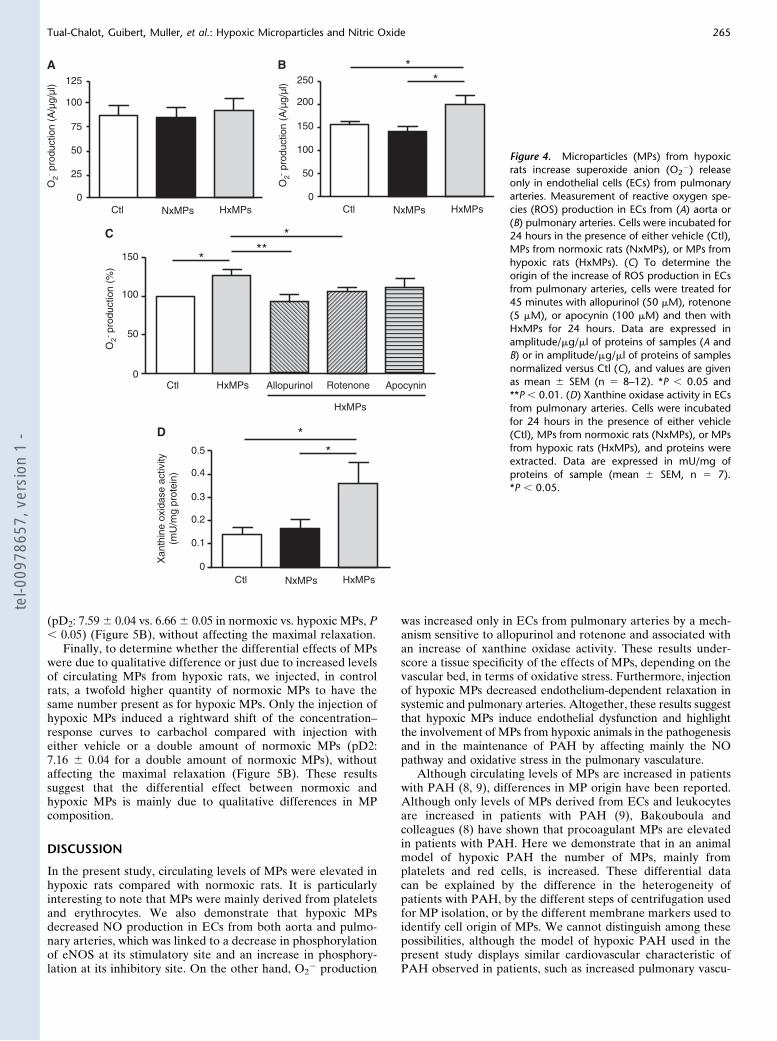
(pD2: 7.596 0.04 vs. 6.666 0.05 in normoxic vs. hypoxic MPs, P, 0.05) (Figure 5B), without affecting the maximal relaxation.
Finally, to determine whether the differential effects of MPswere due to qualitative difference or just due to increased levelsof circulating MPs from hypoxic rats, we injected, in controlrats, a twofold higher quantity of normoxic MPs to have thesame number present as for hypoxic MPs. Only the injection ofhypoxic MPs induced a rightward shift of the concentration–response curves to carbachol compared with injection witheither vehicle or a double amount of normoxic MPs (pD2:7.16 6 0.04 for a double amount of normoxic MPs), withoutaffecting the maximal relaxation (Figure 5B). These resultssuggest that the differential effect between normoxic andhypoxic MPs is mainly due to qualitative differences in MPcomposition.
DISCUSSION
In the present study, circulating levels of MPs were elevated inhypoxic rats compared with normoxic rats. It is particularlyinteresting to note that MPs were mainly derived from plateletsand erythrocytes. We also demonstrate that hypoxic MPsdecreased NO production in ECs from both aorta and pulmo-nary arteries, which was linked to a decrease in phosphorylationof eNOS at its stimulatory site and an increase in phosphory-lation at its inhibitory site. On the other hand, O2
2 production
was increased only in ECs from pulmonary arteries by a mech-anism sensitive to allopurinol and rotenone and associated withan increase of xanthine oxidase activity. These results under-score a tissue specificity of the effects of MPs, depending on thevascular bed, in terms of oxidative stress. Furthermore, injectionof hypoxic MPs decreased endothelium-dependent relaxation insystemic and pulmonary arteries. Altogether, these results suggestthat hypoxic MPs induce endothelial dysfunction and highlightthe involvement of MPs from hypoxic animals in the pathogenesisand in the maintenance of PAH by affecting mainly the NOpathway and oxidative stress in the pulmonary vasculature.
Although circulating levels of MPs are increased in patientswith PAH (8, 9), differences in MP origin have been reported.Although only levels of MPs derived from ECs and leukocytesare increased in patients with PAH (9), Bakouboula andcolleagues (8) have shown that procoagulant MPs are elevatedin patients with PAH. Here we demonstrate that in an animalmodel of hypoxic PAH the number of MPs, mainly fromplatelets and red cells, is increased. These differential datacan be explained by the difference in the heterogeneity ofpatients with PAH, by the different steps of centrifugation usedfor MP isolation, or by the different membrane markers used toidentify cell origin of MPs. We cannot distinguish among thesepossibilities, although the model of hypoxic PAH used in thepresent study displays similar cardiovascular characteristic ofPAH observed in patients, such as increased pulmonary vascu-
Figure 4. Microparticles (MPs) from hypoxicrats increase superoxide anion (O2
2) releaseonly in endothelial cells (ECs) from pulmonaryarteries. Measurement of reactive oxygen spe-cies (ROS) production in ECs from (A) aorta or(B) pulmonary arteries. Cells were incubated for24 hours in the presence of either vehicle (Ctl),MPs from normoxic rats (NxMPs), or MPs fromhypoxic rats (HxMPs). (C) To determine theorigin of the increase of ROS production in ECsfrom pulmonary arteries, cells were treated for45 minutes with allopurinol (50 mM), rotenone(5 mM), or apocynin (100 mM) and then withHxMPs for 24 hours. Data are expressed inamplitude/mg/ml of proteins of samples (A andB) or in amplitude/mg/ml of proteins of samplesnormalized versus Ctl (C), and values are givenas mean 6 SEM (n 5 8–12). *P , 0.05 and**P , 0.01. (D) Xanthine oxidase activity in ECsfrom pulmonary arteries. Cells were incubatedfor 24 hours in the presence of either vehicle(Ctl), MPs from normoxic rats (NxMPs), or MPsfrom hypoxic rats (HxMPs), and proteins wereextracted. Data are expressed in mU/mg ofproteins of sample (mean 6 SEM, n 5 7).*P , 0.05.
Tual-Chalot, Guibert, Muller, et al.: Hypoxic Microparticles and Nitric Oxide 265te
l-009
7865
7, v
ersi
on 1
-

lar tone, medial thickening, and neomuscularization of pulmo-nary arterioles (19). Moreover, as previously described (20),hypoxic rats displayed an increased hematocrit value whencompared with normoxic animals (data not shown). This in-crease in red blood cell count leading to volume contractionmay account for the increased level of total MPs and also for theincrease in erythroid MPs. However, neither levels of leuko-cyte- nor endothelial-derived MPs were increased to the sameextent, suggesting that the increase in MPs from platelets anderythrocytes is probably specific for these cell populations (bothplatelets and erythrocytes). In addition, we showed that hypoxicMPs, but not a double amount of normoxic MPs, are able toinduce endothelial dysfunction in extrapulmonary artery ringsindicating that the effects induced by hypoxic MPs are inde-pendent of the number of MPs but dependent on the differentcompositions and/or origin between normoxic and hypoxic MPs.
A prolonged exposure to hypoxia results in a decrease inaortic eNOS protein expression and an impaired endothelium-dependent relaxation to acetylcholine on aortic rings in rats(11). The role of eNOS in the PAH pathogenesis is welldemonstrated; thus overproduction of eNOS in transgenic miceprevents hypoxia-induced PAH (21), whereas exposure to mildhypoxia results in severe PAH in eNOS-deficient mice (22). Inthe present study, in vitro endothelial function was assessed bydirect measurement of NO production by ECs treated withMPs. We have shown a decrease in NO production in ECs fromboth aorta and pulmonary arteries treated with hypoxic MPs,indicating that hypoxic MPs might contribute to endothelialdysfunction observed in chronic hypoxia-induced PAH. Inaddition, with the changes in eNOS expression, NO productioncan be regulated by eNOS post-translational modifications.
When Ser 1177 is phosphorylated by several kinases, such asAkt (23), NO production is increased twice or three timescompared with basal levels, whereas Thr 495 phosphorylationby protein kinase C is associated with a decrease in NOproduction (24). Thus the double phosphorylation of Ser 1177and Thr 495 is crucial for eNOS activity in ECs. We found thathypoxic MPs were able to modify eNOS phosphorylationwithout affecting its expression both in ECs from aorta andpulmonary arteries. Indeed, hypoxic MPs increased Thr 495 anddecreased Ser 1177 phosphorylations in ECs from both typesof arteries. Furthermore, hypoxic MPs induced a decrease inAkt phosphorylation, Akt being the kinase involved in eNOSactivation-associated phosphorylation in aortic ECs. AlthougheNOS phosphorylation by Akt has been reported to be im-paired in hypoxic pulmonary arteries (25), hypoxic MPs werenot able to modify Akt phosphorylation in ECs from pulmonaryarteries under the experimental conditions used. Regarding thelatter, other kinases such as AMP-activated protein kinase orprotein kinase C (26, 27) may be implicated in the eNOS phos-phorylation at the inhibitory site evoked by hypoxic MPs. Nev-ertheless, decreased eNOS activity probably concurs to reduceNO production in ECs from both aorta and pulmonary arteries.
The subcellular localization of caveolin-1 is critical for eNOSregulation. In ECs, caveolin-1 negatively regulates NO signalingby binding eNOS and sequestering it at the plasma membrane.The activation of caveolar eNOS depends on its dissociationfrom caveolin-1 (28); in addition, if the interaction betweencaveolin-1 and eNOS is altered, vascular integrity and functionare affected. Here, we demonstrate a decrease in caveolin-1expression only in ECs from pulmonary arteries treated withhypoxic MPs, which may argue against a reduced eNOS activity.However, in this context, several works have described analteration in eNOS and caveolin-1 link in PAH leading to thealteration of vascular integrity and decrease in NO production.Thus, reduction in the expression of caveolin-1 has beenreported in primary PAH and in experimental models ofPAH induced by monocrotaline (29, 30) and hypoxic PAH(31). Altogether, pulmonary hypertension affects caveolin/eNOS balance, the consequence of which concurs with endo-thelial dysfunction. In line with these studies, we found that 24-hour treatment with hypoxic MPs induced both reduction incaveolin-1 expression and an increase in phosphorylation ofeNOS on its inhibitor site leading to a decrease in NO pro-duction. Thus, the present data strengthen the notion thathypoxic MPs participate in the changes in endothelium integrityduring PAH.
Superoxide anion can react with other reactive species,including NO. The O2
2/NO interaction occurs at very highspeed constant, which leads to NO inactivation and playsa major role in endothelial dysfunction associated with vascularpathologies (7). However, although the participation of ROSproduction in PAH pathogenesis is generally accepted (32), thesources responsible for ROS production in the hypoxic pulmo-nary arteries are not well defined (33). Several studies havesuggested an involvement of the NADPH oxidase in thedevelopment of hyperreactivity and endothelial dysfunctionduring PAH (6, 34). In other models of PAH, an increasedcapacity for ROS production by NADPH oxidase as well as bymitochondria has been described (35, 36). Hoshikawa andcolleagues (37) have found that in lung from adult rats, xanthineoxidase activity is elevated under hypoxic conditions, especiallyduring the induction phase (z7 d) of hypoxic exposure, becausetreatment with allopurinol in the first 3 days of hypoxia issufficient to limit right ventricular hypertrophy and pulmonaryvascular thickening. Most recently, in neonatal rats exposed tohypoxia, it has been reported that O2
2 generation from
Figure 5. Hypoxic microparticles (MPs) impair endothelium-dependentrelaxation. (A) Acetylcholine (Ach)-induced relaxation in aorta fromcontrol (Nx) and hypoxic (HxMPs) MP-injected rats. (B) Carbachol(CCH)-induced relaxation in pulmonary arteries from Nx, double amountof normoxic MP (2*NxMPs)- and HxMPs-injected rats. Results wereexpressed as a percentage of relaxation of phenylephrine-inducedprecontraction. ***P , 0.001.
266 AMERICAN JOURNAL OF RESPIRATORY AND CRITICAL CARE MEDICINE VOL 182 2010te
l-009
7865
7, v
ersi
on 1
-
xanthine oxidase is implicated in endothelial dysfunction andvascular remodeling (38). Our results demonstrate that O2
2
production by treatment with hypoxic MPs takes place only inECs from pulmonary arteries, indicating a tissue specificity ofhypoxic MPs toward ECs from the pulmonary bed. In addition,these effects are associated with a participation of both xanthineoxidase and mitochondrial complex I, without implicatingNADPH oxidase or changes in gp91 expression (data notshown). These data are in accordance with the implication ofxanthine oxidase activity on the deleterious effects in endothe-lial function induced by MPs derived from apoptotic lympho-cytes (15) and reveal the ability of hypoxic MPs to modulateoxidative status in pulmonary ECs.
Finally, although hypoxic MPs were not able to modifypulmonary blood pressure after 24 hours of in vivo treatment,they reduced the ability of muscarinic agonists to promoteendothelium-dependent relaxation in both aorta and pulmonaryarteries. These results highlight the distinction between endothe-lial dysfunction and changes in pulmonary blood pressure aftertreatment with hypoxic MPs for 24 hours. It should be noted thatthe increase in pulmonary blood pressure described in hypoxicanimal models is associated with hypertrophy of smooth musclecells resulting in an increased vascular resistance. Thus, one canadvance the hypothesis that acute treatment with hypoxic MPsmight not be able to affect smooth muscle proliferation.
In the present study, hypoxic MPs negatively affected theeNOS pathway, decreased NO release, and increased ROSproduction in ECs. These results are in concordance with otherdata present in the literature, wherein a decrease in endothe-lium-dependent relaxation is widely reported in pulmonary (6,39) and systemic (11, 12) circulation during hypoxia.
In summary,we report that hypoxic rats developingPAHhaveincreased levels of circulating MPs, especially platelet- anderythrocyte-derived MPs. Hypoxic MPs can impair endothelialfunction in both rat aorta and pulmonary arteries, by directlyreducing eNOS activity and by limiting NO bioavailability, and,in the pulmonary bed only, by increasing ROS production.Together, these data strongly suggest that circulating hypoxicMPs are able to induce endothelial dysfunction and demonstratefor the first time their pathophysiological relevance, in thismodel.From these results, one can advance the hypothesis that hypoxicMPs contribute to the pathophysiological process of PAH.
Conflict of Interest Statement: None of the authors has a financial relationshipwith a commercial entity that has an interest in the subject of this manuscript.
Acknowledgment: The authors thank Dr Mirela Delibegovic for careful reading ofthe manuscript and Guillaume Simon for assistance in measuring of pulmonaryblood pressure.
References
1. Humbert M, Morrell NW, Archer SL, Stenmark KR, MacLean MR,Lang IM, Christman BW, Weir EK, Eickelberg O, Voelkel NF, et al.Cellular and molecular pathobiology of pulmonary arterial hyperten-sion. J Am Coll Cardiol 2004;43:13S–24S.
2. Budhiraja R, Tuder RM, Hassoun PM. Endothelial dysfunction inpulmonary hypertension. Circulation 2004;109:159–165.
3. Giaid A, Saleh D. Reduced expression of endothelial nitric oxidesynthase in the lungs of patients with pulmonary hypertension.N Engl J Med 1995;333:214–221.
4. Bowers R, Cool C, Murphy RC, Tuder RM, Hopken MW, Flores SC,Voelkel NF. Oxidative stress in severe pulmonary hypertension. Am JRespir Crit Care Med 2004;169:764–769.
5. Farahmand F, Hill MF, Singal PK. Antioxidant and oxidative stresschanges in experimental cor pulmonale. Mol Cell Biochem 2004;260:21–29.
6. Fresquet F, Pourageaud F, Leblais V, Brandes RP, Savineau JP,Marthan R, Muller B. Role of reactive oxygen species and gp91phoxin endothelial dysfunction of pulmonary arteries induced by chronichypoxia. Br J Pharmacol 2006;148:714–723.
7. Cai H, Harrison DG. Endothelial dysfunction in cardiovascular diseases:the role of oxidant stress. Circ Res 2000;87:840–844.
8. Bakouboula B, Morel O, Faure A, Zobairi F, Jesel L, Trinh A, ZupanM, Canuet M, Grunebaum L, Brunette A, et al. Procoagulantmembrane microparticles correlate with the severity of pulmonaryarterial hypertension. Am J Respir Crit Care Med 2008;177:536–543.
9. Amabile N, Heiss C, Real WM, Minasi P, McGlothlin D, Rame EJ,Grossman W, De Marco T, Yeghiazarians Y. Circulating endothelialmicroparticle levels predict hemodynamic severity of pulmonaryhypertension. Am J Respir Crit Care Med 2008;177:1268–1275.
10. Martinez MC, Tesse A, Zobairi F, Andriantsitohaina R. Shed membranemicroparticles from circulating and vascular cells in regulating vascu-lar function. Am J Physiol Heart Circ Physiol 2005;288:H1004–H1009.
11. Toporsian M, Govindaraju K, Nagi M, Eidelman D, Thibault G, WardME. Downregulation of endothelial nitric oxide synthase in rat aortaafter prolonged hypoxia in vivo. Circ Res 2000;86:671–675.
12. Reboul C, Gibault A, Tanguy S, Dauzat M, Obert P. Alteration ofendothelium-mediated vasodilator response in the rat hindlimbvasculature consecutive to chronic hypoxic stress: NO and EDHFinvolvement. Vascul Pharmacol 2009;51:154–161.
13. Martinez MC, Larbret F, Zobairi F, Coulombe J, Debili N, VainchenkerW, Ruat M, Freyssinet JM. Transfer of differentiation signal bymembrane microvesicles harboring hedgehog morphogens. Blood2006;108:3012–3020.
14. Kobayashi M, Inoue K, Warabi E, Minami T, Kodama T. A simplemethod of isolating mouse aortic endothelial cells. J AtherosclerThromb 2005;12:138–142.
15. Mostefai HA, Agouni A, Carusio N, Mastronardi ML, Heymes C,Henrion D, Andriantsitohaina R, Martinez MC. Phosphatidylinositol3-kinase and xanthine oxidase regulate nitric oxide and reactiveoxygen species productions by apoptotic lymphocyte microparticlesin endothelial cells. J Immunol 2008;180:5028–5035.
16. Agouni A, Lagrue-Lak-Hal AH, Ducluzeau PH, Mostefai HA, Draunet-Busson C, Leftheriotis G, Heymes C, Martinez MC, Andriantsito-haina R. Endothelial dysfunction caused by circulating microparticlesfrom patients with metabolic syndrome. Am J Pathol 2008;173:1210–1219.
17. Agouni A, Mostefai HA, Porro C, Carusio N, Favre J, Richard V,Henrion D, Martinez MC, Andriantsitohaina R. Sonic hedgehogcarried by microparticles corrects endothelial injury through nitricoxide release. FASEB J 2007;21:2735–2741.
18. Tesse A, Martinez MC, Hugel B, Chalupsky K, Muller CD, Meziani F,Mitolo-Chieppa D, Freyssinet JM, Andriantsitohaina R. Upregula-tion of proinflammatory proteins through NF-kB pathway by shedmembrane microparticles results in vascular hyporeactivity. Arterios-cler Thromb Vasc Biol 2005;25:2522–2527.
19. Bonnet S, Dumas-de-La-Roque E, Begueret H, Marthan R, Fayon M,Dos Santos P, Savineau JP, Baulieu EE. Dehydroepiandrosterone(DHEA) prevents and reverses chronic hypoxic pulmonary hyper-tension. Proc Natl Acad Sci USA 2003;100:9488–9493.
20. Bonnet S, Dubuis E, Vandier C, Martin S, Marthan R, Savineau JP.Reversal of chronic hypoxia-induced alterations in pulmonary arterysmooth muscle electromechanical coupling upon air breathing. Car-diovasc Res 2002;53:1019–1028.
21. Ozaki M, Kawashima S, Yamashita T, Ohashi Y, Rikitake Y, Inoue N,Hirata KI, Hayashi Y, Itoh H, Yokoyama M. Reduced hypoxicpulmonary vascular remodeling by nitric oxide from the endothelium.Hypertension 2001;37:322–327.
22. Fagan KA, Fouty BW, Tyler RC, Morris KG Jr, Hepler LK, Sato K,LeCras TD, Abman SH, Weinberger HD, Huang PL, et al. The pul-monary circulation of homozygous or heterozygous eNOS-null miceis hyperresponsive to mild hypoxia. J Clin Invest 1999;103:291–299.
23. Fulton D, Gratton JP, McCabe TJ, Fontana J, Fujio Y, Walsh K, FrankeTF, Papapetropoulos A, Sessa WC. Regulation of endothelium-derived nitric oxide production by the protein kinase Akt. Nature1999;399:597–601.
24. Matsubara M, Hayashi N, Jing T, Titani K. Regulation of endothelialnitric oxide synthase by protein kinase C. J Biochem 2003;133:773–781.
25. Murata T, Sato K, Hori M, Ozaki H, Karaki H. Decreased endothelialnitric-oxide synthase (eNOS) activity resulting from abnormal in-teraction between eNOS and its regulatory proteins in hypoxia-induced pulmonary hypertension. J Biol Chem 2002;277:44085–44092.
26. Chen ZP, Mitchelhill KI, Michell BJ, Stapleton D, Rodriguez-Crespo I,Witters LA, Power DA, Ortiz de Montellano PR, Kemp BE. AMP-activated protein kinase phosphorylation of endothelial NO synthase.FEBS Lett 1999;443:285–289.
Tual-Chalot, Guibert, Muller, et al.: Hypoxic Microparticles and Nitric Oxide 267te
l-009
7865
7, v
ersi
on 1
-
27. Fleming I, Fisslthaler B, Dimmeler S, Kemp BE, Busse R. Phosphory-lation of Thr(495) regulates Ca(21)/calmodulin-dependent endothe-lial nitric oxide synthase activity. Circ Res 2001;88:E68–E75.
28. Michel JB, Feron O, Sase K, Prabhakar P, Michel T. Caveolin versuscalmodulin. Counterbalancing allosteric modulators of endothelialnitric oxide synthase. J Biol Chem 1997;272:25907–25912.
29. Achcar RO, Demura Y, Rai PR, Taraseviciene-Stewart L, Kasper M,Voelkel NF, Cool CD. Loss of caveolin and heme oxygenase expressionin severe pulmonary hypertension. Chest 2006;129:696–705.
30. Mathew R, Huang J, Shah M, Patel K, Gewitz M, Sehgal PB. Disruptionof endothelial-cell caveolin-1alpha/raft scaffolding during develop-ment of monocrotaline-induced pulmonary hypertension. Circulation2004;110:1499–1506.
31. Mathew R, Huang J, Gewitz MH. Pulmonary artery hypertension:caveolin-1 and eNOS interrelationship: a new perspective. CardiolRev 2007;15:143–149.
32. Herget J, Wilhelm J, Novotna J, Eckhardt A, Vytasek R, Mrazkova L,Ostadal M. A possible role of the oxidant tissue injury in thedevelopment of hypoxic pulmonary hypertension. Physiol Res 2000;49:493–501.
33. Nozik-Grayck E, Stenmark KR. Role of reactive oxygen species inchronic hypoxia-induced pulmonary hypertension and vascularremodeling. Adv Exp Med Biol 2007;618:101–112.
34. Liu JQ, Zelko IN, Erbynn EM, Sham JS, Folz RJ. Hypoxic pulmonaryhypertension: role of superoxide and NADPH oxidase (gp91phox).Am J Physiol Lung Cell Mol Physiol 2006;290:L2–L10.
35. Weissmann N, Zeller S, Schafer RU, Turowski C, Ay M, Quanz K,Ghofrani HA, Schermuly RT, Fink L, Seeger W, et al. Impact ofmitochondria and NADPH oxidases on acute and sustained hypoxicpulmonary vasoconstriction. Am J Respir Cell Mol Biol 2006;34:505–513.
36. Redout EM, Wagner MJ, Zuidwijk MJ, Boer C, Musters RJ, vanHardeveld C, Paulus WJ, Simonides WS. Right-ventricular failure isassociated with increased mitochondrial complex II activity and pro-duction of reactive oxygen species. Cardiovasc Res 2007;75:770–781.
37. Hoshikawa Y, Ono S, Suzuki S, Tanita T, Chida M, Song C, Noda M,Tabata T, Voelkel NF, Fujimura S. Generation of oxidative stresscontributes to the development of pulmonary hypertension inducedby hypoxia. J Appl Physiol 2001;90:1299–1306.
38. Jankov RP, Kantores C, Pan J, Belik J. Contribution of xanthineoxidase-derived superoxide to chronic hypoxic pulmonary hyperten-sion in neonatal rats. Am J Physiol Lung Cell Mol Physiol 2008;294:L233–L245.
39. Adnot S, Raffestin B, Eddahibi S, Braquet P, Chabrier PE. Loss ofendothelium-dependent relaxant activity in the pulmonary circulationof rats exposed to chronic hypoxia. J Clin Invest 1991;87:155–162.
268 AMERICAN JOURNAL OF RESPIRATORY AND CRITICAL CARE MEDICINE VOL 182 2010te
l-009
7865
7, v
ersi
on 1
-

!"#$$
Publication :
Nocturnal release of leukocyte-der ived microparticles in
males with obstructive sleep apnea
tel-0
0978
657,
ver
sion
1 -
Nocturnal release of leukocyte-derived microparticlesin males with obstructive sleep apnoeaTo the Editors:
Multiple pathophysiological mechanisms have been proposedto contribute to the increased cardiovascular morbidity inobstructive sleep apnoea (OSA), including autonomic dysfunc-tion, inflammation, oxidative stress and endothelial dysfunc-tion [1]. Microparticles (MPs) are small membrane vesicles thatare shed from circulating cells or from the components of thevessel wall in response to activation and apoptosis. There isgrowing evidence in support of a potential role of MPs in thefield of cardiovascular diseases. Increased levels of MPsderived from various cell types are found in patients at riskof cardiovascular diseases [2]. By modulating inflammation,coagulation, vasomotor reactivity and angiogenesis, MPsmight directly contribute to cardiovascular diseases [2].Recent case–control studies suggest a potential involvementof MPs in OSA-associated cardiovascular morbidity [3–6]. Anincrease in morning levels of MPs derived from activatedleukocytes has been demonstrated in otherwise healthy maleOSA patients with marked nocturnal desaturations [5]. In vitro,nitric oxide (NO) production by endothelial cells incubatedwith MPs from OSA patients correlates negatively withcirculating levels of activated leukocyte-derived MPs [5]. Exvivo, mice previously injected with MPs from OSA patientsdisplay endothelial dysfunction, reduced endothelial NOrelease and increased adhesion molecule expression [5].
A decrease in endothelial function from evening to morninghas previously been demonstrated in patients with OSA andfound to correlate with the severity of sleep-disorderedbreathing [7]. Since recent data suggest a potential role MPsin endothelial dysfunction [5], we aimed to study evening-to-morning change in activated leukocyte-derived MPs levels inotherwise healthy patients with OSA.
The study included 38 consecutive male patients (18–70 yrs ofage) investigated by polysomnography (n525) or respiratoryrecording (n513) in the Sleep Unit of the Dept of RespiratoryMedicine, Angers University (Angers, France) for suspectedOSA. Exclusion criteria were previous treatment for OSA,body mass index (BMI) o35 kg?m-2, history of coronary arterydisease, heart failure, stroke, hypertension, diabetes mellitus,dyslipidaemia and patients receiving any drug known to affectendothelial function. Patients with an apnoea–hypopnoeaindex (AHI) of o5 events?h-1 were included in the OSAgroup. Patients with an AHI ,5 events?h-1 were included inthe control group. The study was approved by the Universityof Angers ethics committee and patients gave their informedconsent.
MP characterisation was performed as described previously [5]in the evening before the night recording, at approximately18:00–19:00 h, before dinner, and after waking up in the
morning following sleep recording, at approximately 07:00–08:00 h, before breakfast. Routine laboratory tests, includingglucose, glycated haemoglobin, triglycerides, total cholesterol,high- and low-density lipoprotein cholesterol, and blood cellcounts, were also performed on a morning blood sample. ForMP isolation, evening and morning blood samples were drawnand collected in EDTA tubes (Vacutainers; Becton Dickinson,Le Pont de Claix, France) from a peripheral vein using a21-gauge needle to minimise platelet activation, and wereprocessed for assay within 2 h, as previously described [5].Samples were centrifuged for 20 min at 2706g, and plasmawas then harvested and centrifuged for 20 min at 1,5006gto obtain platelet-free plasma (PFP). 200 mL PFP were frozenand stored at -80uC until use. MPs derived from activatedL-selectin-positive leukocytes were identified by flow cytome-try using anti-CD62L antibody. Irrelevant human immunoglo-bulin G was used as an isotype-matched negative control foreach sample. To determine the MP concentration, 10 mL PFPwere incubated with 5 mL specific antibody (Beckman Coulter,Villepinte, France). After 45 min incubation, samples werediluted in 300 mL 0.9% NaCl saline solution or annexin Vlabelling buffer. Equal volumes of sample and Flowcount beadswere then added to calculate theMP concentration, and sampleswere analysed in a 500 MPL System flow cytometer (BeckmanCoulter). Regions corresponding to MPs were identified inforward and side-angle light scatter intensity dot plot repre-sentations set at logarithmic gain, depending on their diameter(0.1–1 mm). Sample analysis was stopped after counting 10,000events. MP analysis was performed without knowledge of thetime the blood sample was taken (i.e. evening or morning) andwhether it belonged to the OSA or the control group.
Mean¡SEM AHI was 1.9¡0.4 events?h-1 in the control group(n515) and 16.2¡2.4 events?h-1 in the OSA group (n523).There was no statistically significant difference between OSAand control patients for age, BMI, Epworth sleepiness scale,arterial blood pressure, lipid metabolism, glucose metabolismor percentage of smokers (see online supplementary materialfor detailed data).
Figure 1 shows the comparison between evening and morninglevels of CD62L+ MPs in the control group, the overall OSAgroup and patients with moderate-to-severe OSA (AHI o15events?h-1; n510). A significant decrease in CD62L+ MPs frommean¡SEM 261.9¡32.2 MPs per mL plasma in the evening to196.8¡36.9 MPs per mL plasma in the morning (p50.036) wasobserved in the control group. A trend toward an increase from230.9¡25.3 MPs per mL plasma in the evening to 259.8¡28.5 MPs per mL plasma in the morning (nonsignificant) wasobserved in the OSA group. In patients with moderate-to-severe OSA, CD62L+ MPs increased significantly (p50.047)from evening (253.1¡29.5 MPs per mL plasma) to morning
This article has supplementary material available from www.erj.ersjournals.com cEUROPEAN RESPIRATORY JOURNAL VOLUME 37 NUMBER 5 1293
tel-0
0978
657,
ver
sion
1 -

(311.7¡35.4 MPs per mL plasma). As shown in figure 2, asignificant correlation was observed in the OSA group betweenAHI and the morning–evening difference in CD62L+ MPs(r50.45; p50.03). In the OSA group, the morning level ofCD62L+ MPs was positively correlated with the AHI (r50.55;p50.006) and the 4% oxygen (r50.55; p50.006). No correlationwas observed between morning CD62L+ MPs and any otherclinical or laboratory parameters.
Accumulation of leukocytes and their adhesion to theendothelium play a central role in the formation of athero-sclerotic plaques. In asymptomatic subjects, circulating levelsof leukocyte-derived MPs predict subclinical atherosclerosisburden [8]. Leukocyte-derived MPs were demonstrated to becompetent inflammatory mediators able to induce endothelialcell activation and IL-6 release [9]. Systemic inflammation isconsidered as a key factor in the pathogenesis of cardio-vascular complication in OSA [1]. Previous studies havehighlighted the roles of leukocytes in endothelial cell injuryin OSA with decreased apoptosis and increased expression ofL-selectin adhesion molecule [10]. An increase in leukocyte-derived MPs has recently been described in OSA [3, 5]. Inaccordance with these previous results, the present studyunderscores a positive correlation between morning CD62L+MPs and the severity of sleep-disordered breathing. Thefinding that the evening-to-morning change in CD62L+ MPscorrelates with the AHI in otherwise healthy patients providesevidence in support of a direct link between sleep-disorderedbreathing and leukocyte-derived MPs release. Evidence of thepathophysiological relevance of MPs in OSA was recentlyprovided ex vivo by their ability to impair endothelium-dependent relaxation in response to acetylcholine and theendothelial response to flow when injected i.v. into mice [5]. Itcan be hypothesised that nocturnal release of leukocyte-derived MPs could be involved in the early morningattenuation of endothelial function [7] and contribute to theincreased rate of cardiovascular events during the first hoursafter waking from sleep.
However, randomised controlled trials including vascularreactivity assessment are still warranted to establish a causallink between circulating MPs and OSA, and the role of MPs,particularly leukocyte-derived MPs, in OSA-associated endo-thelial dysfunction.
W. Trzepizur*,#, P. Priou*,#, A. Paris*, J. Nardi*,
S. Tual-Chalot#, N. Meslier#, T. Urban*,
R. Andriantsitohaina#, M.C. Martinez# and F. Gagnadoux*,#
*Departement de Pneumologie, Centre Hospitalier Univer-
sitaire, and #INSERM, U694, Universite d’Angers, Faculte de
Medecine, Angers, France.
Correspondence: F. Gagnadoux, Departement de Pneumologie,
CHU, 4 rue Larrey, 49933 Angers Cedex 9, France. E-mail:
Statement of Interest: None declared.
REFERENCES1 Ryan S, Taylor CT, McNicholas WT. Systemic inflammation: a key
factor in the pathogenesis of cardiovascular complications inobstructive sleep apnoea syndrome?Postgrad Med J 2009; 85: 693–698.
2 Meziani F, Tesse A, Andriantsitohaina R. Microparticles are vectorsof paradoxical information in vascular cells including the endothe-lium: role in health and diseases. Pharmacol Rep 2008; 60: 75–84.
3 Ayers L, Ferry B, Craig S, et al. Circulating cell-derivedmicroparticles in patients with minimally symptomatic obstruc-tive sleep apnoea. Eur Respir J 2009; 33: 574–580.
4 Jelic S, Lederer DJ, Adams T, et al. Endothelial repair capacity andapoptosis are inversely related in obstructive sleep apnea. VascHealth Risk Manag 2009; 5: 909–920.
5 Priou P, Gagnadoux F, Tesse A, et al. Endothelial dysfunction andcirculating microparticles from patients with obstructive sleepapnea. Am J Pathol 2010; 177: 974–83.
6 Yun CH, Jung KH, Chu K, et al. Increased circulating endothelialmicroparticles and carotid atherosclerosis in obstructive sleepapnea. J Clin Neurol, 6: 89–98.
400
300
200
100
0
-100
-200
-300
!CD
62L+
MPs
µL-
1
AHI events·h-10 10 20 30 40 50 60
FIGURE 2. Scatter plot showing the relationship in the obstructive sleep
apnoea group (n523) between apnoea–hypopnoea index (AHI) and the morning–
evening difference in plasma levels of activated leukocyte-derived microparticles
(DCD62L+ MPs). r50.45; p50.03.
CD
62L+
MPs
µL-
1
350
300
250
200
150
100
50
0Controls All OSA Moderate-to-
severe OSA
*
*EveningMorning
FIGURE 1. Comparison of mean¡SEM evening and morning plasma levels of
activated leukocyte-derived microparticles (CD62L+ MPs) in controls (n515), all
obstructive sleep apnoea (OSA) cases (n523) and those with moderate-to-severe
OSA (n510). *: p,0.05 by Wilcoxon signed-rank test.
1294 VOLUME 37 NUMBER 5 EUROPEAN RESPIRATORY JOURNAL
tel-0
0978
657,
ver
sion
1 -

7 Itzhaki S, Lavie L, Pillar G, et al. Endothelial dysfunction inobstructive sleep apnea measured by peripheral arterial toneresponse in the finger to reactive hyperemia. Sleep 2005; 28:594–600.
8 Chironi G, Simon A, Hugel B, et al. Circulating leukocyte-derivedmicroparticles predict subclinical atherosclerosis burden inasymptomatic subjects. Arterioscler Thromb Vasc Biol 2006; 26:2775–2780.
9 Mesri M, Altieri DC. Endothelial cell activation by leukocytemicroparticles. J Immunol 1998; 161: 4382–4387.
10 Dyugovskaya L, Lavie P, Lavie L. Increased adhesion moleculesexpression and production of reactive oxygen species in leuko-cytes of sleep apnea patients. Am J Respir Crit Care Med 2002; 165:934–939.
DOI: 10.1183/09031936.00150010
EUROPEAN RESPIRATORY JOURNAL VOLUME 37 NUMBER 5 1295
tel-0
0978
657,
ver
sion
1 -

!"#$$
Manuscr it :
Sleep apnea syndrome microparticles enhance vascular
contraction: mandatory role of the endothelium
tel-0
0978
657,
ver
sion
1 -

!
Sleep apnea syndrome microparticles enhance vascular contraction: mandatory role of
the endothelium
Simon Tual-Chalot,1 Keita Fatoumata,1 Pascaline Priou,1,2 Wojciech Trzepizur,1,2 Cristina
Contreras,3 Dolores Prieto,3 M. Carmen Martinez,1 Frédéric Gagnadoux,1,2 Ramaroson
Andriantsitohaina1
From the 1 2Département de Pneumologie, CHU
France; 3Département de Physiologie, Faculté de Pharmacie, Universidad
Complutense de Madrid, España
Correspondence and requests for reprints should be addressed to Dr. Ramaroson
Andriantsitohaina, INSERM U694, PBH-IRIS, Rue des Capucins, Angers, F-49045 France.
Phone: +33 2 44 68 85 80; Fax: +33 2 44 68 85 88; e-mail:
Conceived and designed the experiments: STC, MCM, FG, RA.
Performed the experiments: STC, KF, PP, WT, CC. Analyzed of the datas: STC, KF, PP, WT,
MCM, FG, RA. Drafting the manuscript for important intellectual content: all authors.
Sources of support:
Hospital of Angers, -C. is recipient of a doctoral fellowship
from French Education Ministry.
tel-0
0978
657,
ver
sion
1 -
!
A BST R A C T
Obstructive sleep apnea (OSA) is characterized by repetitive apnea-hypopnea cycles during
sleep associated with oxygen desaturation and sleep disruption. Microparticles are biological
vectors of endothelial dysfunction in OSA, but their effects on vascular reactivity have not yet
been assessed. The purpose of this work was to evaluate the role of microparticles from OSA
patients in the regulation of vascular function. Two groups of patients undergoing
polysomnography for OSA were compared. Microparticles obtained from whole blood either
from OSA patients, control subjects, or a vehicle were injected i.v. to mice. Injection of
microparticles from OSA patients induced ex vivo vascular hyper-reactivity in response to
vasoconstrictor agents in aortas with functional endothelium but hypo-reactivity in vessels
without functional endothelium. Vascular hyper-reactivity was blunted in the presence of
either the nitric oxide (NO)-synthase inhibitor L-NA alone or in combination with the non-
selective inhibitor of cyclo-oxygenase, indomethacin. Microparticles from OSA patients
reduced activity of endothelial NO-synthase and NO production. They increased aortic cyclo-
oxygenase-1 and cyclo-oxygenase-2 expressions and the production of thromboxane A2 and
prostacyclin. The selective thromboxane A2 receptor antagonist, SQ-29548, reduced response
to serotonin in arteries from vehicle- and non-OSA microparticle- but not those from OSA
microparticle-treated mice. These data provide evidence that circulating microparticles from
OSA patients induce ex vivo vascular hyper-reactivity with the obligatory role of the
endothelium and subtle interactions between NO and cyclo-oxygenase pathways and
metabolites. These results highlight the participation of microparticles to vascular dysfunction
associated with OSA patients.
tel-0
0978
657,
ver
sion
1 -

!
I N T R O D U C T I O N
Obstructive sleep apnea (OSA) syndrome is a highly prevalent disease characterized by
recurrent episodes of partial or complete obstruction of the upper airways during sleep,
leading to repeated falls in oxygen saturation. OSA is associated with hypertension, coronary
artery disease, stroke, and premature death and considerable evidence is now available in
support of an independent association between OSA and cardiovascular diseases (1). Various
pathophysiological mechanisms have been proposed to contribute to the pathogenesis of
vascular dysfunction in OSA patients (2). In this view, circulating microparticles (MPs)
isolated from blood have been considered as biomarkers of vascular injury and inflammation
in several cardiovascular pathologies such as acute myocardial infarction, sepsis, or metabolic
syndrome (3-5). Indeed, in these diseases, an alteration of levels of MPs have been detected
and frequently correlated with the severity of the pathology. MPs are vesicles with
procoagulant and proinflammatory properties shed from the blebbing plasma membrane of
various cell types, like circulating cells (such as platelets, erythrocytes, T and B cells, and
monocytes) or cells from the vascular wall (endothelial and smooth muscle cells), during
activation by agonists, shear stress or apoptosis (6, 7). Only since recently, MPs are also
considered as effectors being able to vehicle biological messages on the target cells. However,
very few reports set out to demonstrate the importance of MPs in OSA, and they mainly
evoke an alteration of the origin of MP levels (8-11). Very recently, we provide evidence that
OSA patients display the same levels of circulating MPs than healthy subjects, but levels of
MPs from activated leukocytes (CD62L+ MPs) were higher in OSA patients (12). Most
interestingly, there is a positive correlation between circulating levels of CD62L+ MPs and
nocturnal desaturations severity (12, 13). The evening-to-morning change in activated
leucocytes-derived MPs in otherwise healthy OSA patients provided evidence in support of a
direct link between sleep disordered breathing and nocturnal MPs release (13). In vitro and ex
tel-0
0978
657,
ver
sion
1 -
!
vivo studies have also demonstrated that MPs from otherwise healthy patients with OSA can
induce endothelial dysfunction, reduce endothelial-NO release and increase endothelial
adhesion molecule expression (12). These recent data suggest that MPs could play a role in
the pathogenesis of endothelial dysfunction that may initiate atherogenic process in OSA.
However, to the best of our knowledge, the role played by MPs in the regulation of vascular
contractility with respect to exaggerated vasoconstriction that occurs in sleep apnea patients
(14) has never been assessed. Therefore the present study was designed to investigate the
effects of MPs from OSA patients on vascular reactivity in response to vasoconstrictor
agonists. The mechanisms by which OSA MPs affect vascular contractility were also
investigated. MPs were injected intravenously into mice to test their pathophysiological
relevance in vivo.
tel-0
0978
657,
ver
sion
1 -

!
M E T H O DS
Patients
Consecutive male patients (18 to 70 years old) investigated by polysomnography (PSG) or
overnight respiratory recording in the Sleep Unit of the Department of Respiratory Medicine
of Angers University for suspected OSA were screened for the study. Exclusion criteria were
2, history of coronary artery
disease, heart failure, stroke, hypertension, diabetes mellitus, dyslipidemia and patients
receiving any drug known to affect endothelial function. Patients with an apnea-hypopnea
index (AHI) of at least 5 events per hour were included in the OSA group. Patients with an
AHI < 5 were included in the control group. All patients underwent evaluation of clinical
profile and daytime sleepiness using the Epworth Sleepiness Scale. Standard in-laboratory
overnight PSG was performed as previously described using a computerized recording system
electrooculogram, chin electromyogram, arterial oxygen saturation (finger oximetry), nasal-
oral airflow (pressure cannula), tracheal sound (suprasternal microphone), electrocardiogram,
chest and abdominal wall motion (piezo electrodes), bilateral tibialis electromyogram, and
performed as previously described with the same channels except for electroencephalogram,
electrooculogram, chin electromyogram, and tibialis electromyogram. Respiratory events
4% oxygen desaturations. The University of Angers ethics committee approved the study and
patients gave their informed consent.
MP isolation and characterization
tel-0
0978
657,
ver
sion
1 -

!
MP characterization was performed in the morning following sleep recording, at
approximately 7:00 or 8:00 AM, before breakfast. Routine laboratory tests including glucose,
HbA1c, triglycerides, total cholesterol, HDL cholesterol, LDL cholesterol and blood cell
count were also performed on a morning blood sample. For MP isolation, blood sample were
drawn and collected in EDTA tubes (Vacutainers, Becton Dickinson, Le Pont de Claix,
France) from a peripheral vein using a 21-gauge needle to minimize platelet activation, and
were processed for assay within 2 hours. Samples were centrifuged for 20 minutes at 270 g,
and plasma was then harvested and centrifuged for 20 minutes at 1,500 g to obtain platelet-
free plasma (PFP). Two hundred µL of PFP were frozen and stored at 80°C until use. As
previously described (4, 5), remaining PFP was subjected to two series of centrifugation at
21,000 g for 45 minutes in order to eliminate plasma and to pellet MPs for studies, and
supernatant was replaced by 0.9 % saline salt solution. Finally MP pellets were suspended in
150 µl of 0.9 % saline salt solution and stored at 4°C until subsequent use.
MP subpopulations were discriminated into PFP according to the expression of membrane-
specific antigens by flow cytometry. MPs derived from platelets, lymphocytes and endothelial
cells were identified using anti-CD41, anti-CD45 and anti-CD146 antibodies, respectively.
Anti-CD62L antibody was used to identify MPs derived from activated L-selectin+
leukocytes. Irrelevant human IgG was used as an isotype-matched negative control for each
sample. Five µL of PFP were incubated with 5 µL of specific antibody (Beckman Coulter,
Villepinte, France), and after 45 minutes of incubation, samples were diluted in 300 µL of
0.9% NaCl. Annexin V-FITC (BioVision Research Products, Mountain View, CA) binding
was used to count phosphatidylserine-expressing MPs. To determine the MP concentration,
equal volumes of sample and Flowcount beads were then added to calculate the MP
concentration and samples were analyzed in a 500 MPL System flow cytometer (Beckman
Coulter). Regions corresponding to MPs were identified in forward light scatter and side-
tel-0
0978
657,
ver
sion
1 -
!
angle light scatter intensity dot plot representation set at logarithmic gain, depending on their
diameter (0.1-1 µm). Sample analysis was stopped after counting 10,000 events.
Vascular reactivity
All animal studies were carried out using approved institutional protocols and conformed by
the Guide for the Care and Use of Laboratory Animals published by US National Institutes of
Health (NIH Publication No. 85-23, revised 1996). Male Swiss mice (8-10 weeks old) were
treated by i.v. injection into the tail vein of MPs at the circulating levels detected in the blood
of each subject ( (OSA patients or non-OSA (N-OSA) patients) or 0.9% NaCl saline solution
(CTL), as previously performed in other studies (4, 12).
Aortic rings were isolated from mice treated with either MPs from patients or healthy subjects
during 24 hours. Mouse aorta was removed and carefully cleaned of adhering fat and
connective tissue and then cut into rings with or without endothelium (1.5-2 mm length) that
were mounted on a wire myograph filled with PSS as previously described (E7, E8).
Concentration-response curves were constructed by cumulative application of serotonin (5-
HT, 1 nM-10 µM; Sigma-Aldrich, St. Quentin, France) to vessels with functional endothelium
in the absence or presence of the given inhibitor pre-incubated for 30 minutes: the NO-
synthase inhibitor NG-nitro-L-arginine (L-NA, 100µM; Sigma-Aldrich), the selective
cyclooxygenase (COX)-2 inhibitor N-(2-cyclohexyloxy-4-nitrophenyl) methansulfonamide
(NS-398, 10 µM; Sigma-Aldrich), indomethacin, the non selective COX inhibitor (100 µM;
Sigma-Aldrich), SQ-29548 the selective thromboxane A2 receptor antagonist (5 µM; Sigma-
Aldrich) or L-NA plus indomethacin. All inhibitors were used at maximal active
concentrations at which they inhibit the release of either NO from all isoforms of NO
synthases, metabolites from COX-2 isoform or metabolites from COX in blood vessels, as
reported in many of our previous studies (E6, E9). Higher concentrations of L-NA, NS-398
tel-0
0978
657,
ver
sion
1 -

!
SQ-29548 or indomethacin did not induce further inhibition. Also, the contractile response to
a cumulative concentration-response to the thromboxane A2 agonist (9,11-dideoxy-11a,9a-
epoxymethanoprostaglandin F2) (U-46619, Cayman Chemicals, Ann Arbor, MI) or to the
combination of U-46619 (100 nM) and KCl (80 mM) was assessed in mouse aorta. Finally, a
concentration-response curve to 5-HT was performed in aorta without functional endothelium.
The endothelium was removed by rubbing the intimal surface of rings with a rough needle.
Aortas were considered denuded of their endothelium when acetylcholine (1 µM) failed to
produce any relaxation.
N O determination by electron paramagnetic resonance (EPR)
Detection of NO production was performed using a technique with Fe2+
diethyldithiocarbamate (DETC, Sigma-Aldrich) as spin trap as previously described (4).
Briefly, 24 hours after administration of MPs, animals were sacrificed. Aorta was dissected
and incubated for NO production in 250 µl of Krebs Hepes buffer solution, then treated with
250 µl of colloid Fe(DETC)2 and incubated for 45 minutes at 37°C. NO detection was
measured in situ by EPR, and values are expressed as amplitude signal per mg tissue (dry
weight).
W estern Blotting
Aorta was dissected, homogenized and lysed. Proteins (40 µg) were separated on NuPAGE®
gels (Invitrogen, Carlsbad, CA). Blots were probed with anti-endothelial NOS (eNOS) (BD
Biosciences, San Jose, CA), anti-phospho-eNOS Thr 495, anti-phospho-eNOS Ser 1117 (Cell
Signaling, Beverly, MA), and anti-COX-1 and 2 (Santa Cruz Biotechnology, Santa Cruz, CA)
-Aldrich) was used at 1/1,000
dilution to visualize protein gel loading. The membranes were then washed at least three times
in Tris buffer solution containing 0.05% Tween and incubated for 1 hour at room temperature
tel-0
0978
657,
ver
sion
1 -

!
with the appropriate horseradish peroxydase (HRP)-conjugated secondary antibody. The
protein antibody complexes were detected by ECL plus (Amersham Biosciences, Piscataway,
NJ) according to manufac
Determination of prostanoid production
After sacrificed of mice pre-treated with MPs, aorta was dissected and treated with 5-HT
(1µM, 37°C, 30 minutes). After collection of the medium, thromboxane A2, prostaglandin E
metabolites, prostacyclin and total 8-isoprostanes were measured by enzyme immunoassays
kits (Cayman Chemicals). The concentration of prostanoids was expressed as pg/ml/mg tissue
(dry weight).
Data analysis
Data were analyzed using GrapPad Prism Software (GrapPad Software, San Diego, CA). Data
are expressed as mean ± SEM, and n represents the number of patients. pD2 = -log EC50, EC50
being the molar concentration of the agonist that produces 50% of the maximal effect; EC50
values were calculated by logit-log regression. For experiment, statistical analyses were
performed with nonparametric Mann-Whitney U tests or two-way analysis of variance for
repeated measures and subsequent Bonferroni post hoc test. P < 0.05 was considered to be
statistically significant.
tel-0
0978
657,
ver
sion
1 -
!
R ESU L TS
Baseline clinical and biological characteristics
Thirty-four patients without any cardiovascular history of risk factor were included in this
study. Patients were split into groups of N-OSA (n=17) or OSA (n=17) according to AHI.
Excepted for age, the two groups did not differ for weight, body mass index, blood pressure
and daytime sleepiness as assessed by the Epworth sleepiness scale (18), lipid metabolism,
glucose metabolism and in the smoking rate (Table 1).
Circulating MPs and thei r cellular origin
As previously described (12), the total number of circulating MPs was not significantly
different between N-OSA and OSA patients being 13,125±2,058 and 20,957±3,617 MPs/µL
of plasma, respectively. Phenotypical characterization of MPs did not show significant
difference between N-OSA and OSA patients (data not shown). However, a positive
correlation was found between CD62L+ MPs and the AHI (Figure 1).
OSA MPs increase responsiveness of mouse aortic r ings to vasoconstrictor agents
Serotonin (5-HT) produced a concentration-dependent increase in tension in mouse aortic
rings with functional endothelium in the control group (CTL), as well in those treated with
circulating levels of either N-OSA or OSA MPs. Maximal response but not sensitivity to 5-
HT was markedly increased in mice treated with OSA MPs (Emax: 3.1 ± 0.2 mN/mm)
compared to those treated either with vehicle or N-OSA MPs (Emax: 2.3 ± 0.2 and 2.2 ± 0.2
mN/mm, respectively) (Figure 2A).
As for 5-HT, aortic rings taken from mice treated with OSA MPs displayed increased
maximal contraction in response to the stable analog of thromboxane A2, U-46619, compared
to those from vehicle- and N-OSA MP-treated animals (Figure 2B). Finally, aortas from OSA
tel-0
0978
657,
ver
sion
1 -

!
MP-treated mice exhibited increased contractile response to the concomitant application of
KCl (100 mM) and a single concentration of U-46619 (100 nM), compared to those from
vehicle- and N-OSA MP-treated animals (Figure 2C).
Altogether, these data suggest that the OSA MP-induced vascular hyper-reactivity is
independent of the agonist or vasoconstrictor used. The OSA MP-induced vascular hyper-
reactivity may result from the release of vasoconstrictor products from different cellular
origins (for example endothelial cells, smooth muscle cells, or fibroblasts).
Involvement of N O pathway in OSA MPs-induced vascular hyper-reactivity
To investigate the role of NO, the effect of the NO-synthase inhibitor, L-NA, was studied on
the response to 5-HT. L-NA alone increased basal tone in aorta from control and N-OSA MP-
treated mice but not in those taken from mice treated with OSA MPs. Moreover, L-NA
enhanced maximal response to 5-HT in control and N-OSA MP-treated vessels. Surprisingly,
in aorta from mice treated with OSA MPs, L-NA did not affect maximal response to 5-HT.
Moreover, it induced a rightward shift of the concentration-response curve to the agonist (pD2
being 5.7 ± 0.2, 5.5 ± 0.3 and 4.7 ± 0.2 for CTL, N-OSA or OSA, respectively) (Figure 3A-
D). Thus, L-NA rather inhibited than potentiated the response to 5-HT in aorta from mice
treated with OSA MPs (Figure 3D). NO production in the aorta taken from OSA MP-treated
mice was greatly lower compared to that measured in vessels from vehicle- or N-OSA MP-
treated mice (Figure 4A). To identify the molecular mechanism implicated in the decrease in
NO production, we analyzed by Western Blotting the level of expression and
phosphorylations of aortic eNOS. As shown in Figure 4B, eNOS expression was not
significantly modified by MP treatment although a trend toward an increase in eNOS
expression was observed after treatment with MPs. However, treatment by OSA MPs, but not
N-OSA MPs, induced a significant decrease of eNOS phosphorylation on the activator site
tel-0
0978
657,
ver
sion
1 -

!
(Ser 1177) and on the inhibitor site (Thr 495) (Figure 4C-D). After normalization of the
amount of phosphorylated eNOS to total amount of enzyme, the ratio of phosphorylated
eNOS at the inhibitor and activation sites was calculated. Interestingly, the ratio of
phosphorylation was greater in aortas from OSA MPs treated mice compared to those treated
with N-OSA MPs or untreated (Figure 4E). These results indicate a down-regulation of eNOS
activity caused by OSA MPs.
Involvement of C O X metabolites in OSA MP-induced vascular hyper-reactivity
To investigate the role of COX metabolites in 5-HT-induced vascular reactivity, the effects of
both a non-selective inhibitor of COX (indomethacin) and a selective inhibitor of COX-2
(NS398) were examined. In the presence of indomethacin, contractile response to 5-HT was
reduced in aortas from all groups of mice, the decrease was even greater in OSA group (60%)
compared to N-OSA or vehicle groups (47 and 48%, respectively) (Table 2). When COX-2
was specifically silenced using NS398, the response to 5HT was impaired in vessels from
control, N-OSA MPs and OSA MPs, with a stronger inhibition for aorta treated by OSA MPs
(Table 2). These results suggest that OSA MPs treatment leads to an increase of the release of
vasoconstrictor metabolites derived from COX including COX-2.
OSA MPs were able to enhance the expression level of COX-1 and COX-2 compared to N-
OSA MPs or vehicle (Figure 5A-B). To test the hypothesis that COX activity could be
affected by this treatment, we measured the production of different COX metabolites.
Interestingly, aortas treated with OSA-MPs displayed increased thromboxane A2 and
prostacyclin productions compared to N-OSA MPs or untreated aortas. By contrast, release
either of prostaglandin E2 or 8-isoprostane was not significantly different between the three
groups (Figure 5C-F). The hyper-reactivity observed might be due to the increase of
thromboxane A2 production. To test this hypothesis, we performed a concentration-response
tel-0
0978
657,
ver
sion
1 -

!
curve to 5-HT in presence of SQ-29548 the selective thromboxane A2 receptor antagonist.
Interestingly, SQ-29548 significantly reduced response to 5-HT in vessels from vehicle and
N-OSA MP-treated mice but not in those taken from animals injected with OSA MPs (Figure
6A-C). These results suggest that SQ-29548-sensitive pathway is involved in the response to
5-HT in vessels from vehicle and N-OSA MPs. In contrast in those from OSA MPs-treated
mice, SQ-29548 sensitive pathway might not be implicated in the response or alternatively,
vasodilator metabolites might mask the effect of thromboxane A2.
Interaction of N O and C O X pathway
In order to study the eventual interaction between NO and COX pathway, we used in
combination inhibitors, L-NA and indomethacin. On one hand blockade of the two pathways
enhanced response to in aorta from control or N-OSA MP-treated mice. On the other hand,
the presence of the two blockers induced a rightward shift of the response to 5-HT in vessels
from mice treated with OSA MPs (Figure 7).
Role of the endothelium
In vessels without functional endothelium, contraction to 5-HT was significantly reduced in
vessels taken from mice treated with OSA MPs compared to those taken either from control
of N-OSA MP-treated mice (Figure 8). These results strongly suggest the obligatory role of
the endothelium in the hyper-reactivity of the aorta to 5-HT after treatment of mice with OSA
MPs.
tel-0
0978
657,
ver
sion
1 -
!
DISC USSI O N
In the present study, we showed that i.v. injection of OSA MPs into mice promotes vascular
hyper-reactivity in response to vasoconstrictor agents with the obligatory role of the
endothelium. This effect was associated with the ability of OSA MPs to reduce the activity of
endothelial NO-synthase concomitant with a decrease in NO production. In addition, OSA
MPs increased aortic COX-1 and COX-2 expressions associated with enhanced production of
thromboxane A2 and prostacyclin and indomethacin or the selective COX-2 inhibitor, NS398,
reduced hyper-reactivity in response to 5-HT. SQ-29548-sensitive pathway might not be
implicated in vascular hyper-reactivity of OSA MPs or alternatively, vasodilator metabolites,
including prostacyclin, might mask the effect of thromboxane A2. These data provide
evidence of the pathophysiological relevance of MPs as vectors of trans-cellular exchange of
messages in promoting both endothelial (12) and vascular hyper-reactivity (the present study)
in patients with OSA.
Several cardiovascular pathologies are associated with vascular dysfunction including
attenuation of endothelium-dependent vasodilatation and/or alteration of vascular smooth
muscle cells responsiveness to vasoconstrictor stimuli. It is well accepted that MPs contribute,
at least in part, to the alteration of vascular function in several cardiovascular diseases (6, 7).
Evidence of the pathophysiological relevance of MPs in OSA was recently provided in vitro
and ex vivo, by their ability to impair endothelium-dependent relaxation in response to
acetylcholine and the endothelial response to flow when injected i.v. into mice (12). In line
with our previous study (12), circulating level of MPs was not different between OSA and
non OSA patients but levels of CD62L+ MPs were positively correlated with the severity of
sleep disordered breathing.
tel-0
0978
657,
ver
sion
1 -
!
In the present study, we provide further evidence that OSA MPs promote vascular hyper-
reactivity in response to vasoconstrictor agents in mouse aorta. These findings are in line with
our previous studies where MPs from septic shock patients enhance but not reduce,
contraction in response to 5-HT (4). Septic MPs may rather be protective in counteracting the
drop in peripheral resistance and progressive hypotension during severe sepsis and explain the
fact that increased levels of MPs may predict a more favorable outcome in severe sepsis in
terms of mortality rate and organ dysfunction (19). In contrast to septic shock patients,
increased vascular reactivity in OSA patients are deleterious and they are probably involved
in vascular remodeling leading to exacerbate vasoconstriction reported in this disease. Indeed,
exaggerated vasoconstriction occurs in forearms of OSA patients in response to angiotensin II
(14). In a rat model of intermittent hypoxia, enhanced responses to endothelin-1 has also been
reported in several vascular beds (20, 21). Furthermore, an increased contraction to
norepinephrine in mouse hindquarters has also been observed (22). In contrast, contractile
responses to potassium, endothelin-1, 5-HT and thromboxane A2 mimetic were not affected
in rat aorta after 35 days of intermittent hypoxia (21). Nevertheless, we provide evidence in
the present study that MPs from OSA induced vascular hyper-reactivity independently of the
vasoconstrictor used under the experimental conditions used and therefore participate in the
regulation of vascular tone in this syndrome.
It has to be noted that OSA MPs induced vascular hyper-reactivity in the presence of
functional endothelium only suggesting the obligatory role of this tunica. In contrast, OSA
MPs rather elicited vascular hypo-reactivity in the absence of functional endothelium
suggesting that they can also act directly on smooth muscle cells. The pathways involved in
this vascular hypo-reactivity were not assessed since, in vivo, MPs interact with intact blood
vessels in which the endothelium is present. Thus, the mechanisms associated with the
increase in vascular reactivity to vasoconstrictor agents in response to OSA MPs were further
tel-0
0978
657,
ver
sion
1 -

!
studied in terms of an alteration of the balance between the nature and the effects of
endothelial factors on smooth muscle cells including NO and COX metabolites.
We previously reported decreased endothelial-NO production in response with OSA MPs
(12). These data corroborate studies showing a decreased plasma concentration of NO
metabolites in OSA, which is correlated with OSA severity (23). In our previous work (12),
the mechanism by which MPs from OSA patients reduce NO production from endothelial
cells has been explained by a negative regulation of eNOS via both an increase of its
phosphorylation on the inhibitory site and up-regulation of caveolin-1. In conjunction with
these results, we recently reported that MPs from metabolic syndrome patients reduce
endothelial-NO release by a negative regulation of eNOS activity in the EA hy926 cell line. In
the present study, we reinforce this hypothesis inasmuch i.v. injection of mice with OSA MPs
decreased NO production in the aorta by a mechanism associated with reduced eNOS activity
without alteration of its expression. Moreover, blockade of NO pathway with L-NA alone
increased basal tone in aorta from control and N-OSA MP-treated mice but not in those taken
from mice treated with OSA MPs suggesting that OSA MPs reduced basal NO production. In
line with these data, decrease of basal level of NO has been reported in a rat model of
intermittent hypoxia (24). Furthermore, L-NA enhanced maximal response to 5-HT in control
and N-OSA MP-treated vessels. Surprisingly, in aorta from mice treated with OSA MPs, L-
NA did not affect maximal response to 5-HT. It rather induced a rightward shift of the
concentration-response curve to the agonist. Altogether, these data demonstrate that decreased
endothelial-NO participate in vascular hyper-reactivity elicited by OSA MPs. It is unlikely
that increased oxidative stress described as one of the possible mechanisms explaining
reduced endothelial-NO in OSA inasmuch an absence of a link between reactive oxygen
species and endothelial dysfunction by MPs from OSA patients has been observed in these
tel-0
0978
657,
ver
sion
1 -
!
patients (12). Finally, the present study pointed out that blockade of NO pathway unmasks the
existence of vasoconstrictor factor(s) released upon OSA MPs treatment.
Turning now to the participation of COX pathway in the increased vascular tone induced by
OSA MPs, the contraction in response to 5-HT involved the participation of COX
vasoconstrictor metabolites including those from COX-2 inhibitor-sensitive pathway in aorta
independently of the treatment (vehicle, N-OSA or OSA MPs). Interestingly, blockade either
of the non selective COX inhibitor, indomethacin, or the COX-2 inhibitor, resulted a greater
reduction of 5-HT response in vessels from mice treated with OSA MPs suggesting that COX
metabolites participates, in part, in the increased response. Indeed, both COX-1 and COX-2
expressions were increased in aorta taken from OSA MP-treated mice. With regard to COX-2
and in line with these results, up-regulation of this enzyme has been reported in endothelial
cells treated with MPs from OSA (12) and in venous endothelial cells harvested from patients
with untreated OSA (25). The nature of COX metabolites involved was further investigated
and it was found that OSA MPs increased thromboxane A2 and prostacyclin, without any
variation of 8-isoprostane or prostaglandin E2. Concerning thromboxane A2, blockade of Tp
receptor by SQ-29548 significantly reduced response to 5-HT in vessels from vehicle and N-
OSA MP-treated mice but not in those taken from animals injected with OSA MPs. These
results suggest that SQ-29548-sensitive pathway is involved in the response to 5-HT in
vessels from vehicle and N-OSA MPs. In contrast in those from OSA MP-treated mice, SQ-
29548-sensitive pathway might not be implicated in the response or alternatively, vasodilator
metabolites, probably prostacyclin, might mask the effect of thromboxane A2. These results
also indicate the implication of another vasoconstrictor factor, different to COX metabolites,
that participate to the vascular hyper-reactivity induced by OSA MPs. Indeed, concomitant
blockade of NO and COX pathways induced a rightward shift of the response to 5-HT in OSA
MP- treated aorta. The nature of this vasoconstrictor factor remains to be identified.
tel-0
0978
657,
ver
sion
1 -

!
In conclusion, we provide evidence that OSA MPs induce in vivo vascular hyper-reactivity to
vasoconstrictor agents in aorta with the obligatory role of the endothelium. This effect is
associated with a combination of a reduced endothelial NO synthase activity and NO
production and a subtle interaction with COX pathway and metabolites. The critical role of
MPs as a vector of biological messages leading to vascular dysfunction in OSA is therefore
underlined.
These effects of OSA MPs, in addition to their capacity in reducing endothelial vasodilatation
and increasing endothelial inflammation, strengthen the notion that MPs can be, not only
considered as surrogate markers of endothelial dysfunction or injury, but also as effectors able
to amplify pre-existing vascular dysfunctions, including vascular hyper-reactivity in this
syndrome.
tel-0
0978
657,
ver
sion
1 -

!
R E F E R E N C ES
1. Bradley TD, Floras JS. Obstructive sleep apnoea and its cardiovascular consequences.
Lancet 2009;373:82-93.
2. Shamsuzzaman AS, Gersh BJ, Somers VK. Obstructive sleep apnea: implications for
cardiac and vascular disease. Jama 2003;290:1906-1914.
3. Boulanger CM, Scoazec A, Ebrahimian T, Henry P, Mathieu E, Tedgui A, Mallat Z.
Circulating microparticles from patients with myocardial infarction cause endothelial
dyfunction. Circulation 2001;104:2649-2652.
4. Mostefai HA, Meziani F, Mastronardi ML, Agouni A, Heymes C, Sargentini C, Asfar
P, Martinez MC, Andriantsitohaina R. Circulating microparticles from patients with septic
shock exert protective role in vascular function. Am J Respir Crit Care Med 2008;178:1148-
1155.
5. Agouni A, Lagrue-Lak-Hal AH, Ducluzeau PH, Mostefai HA, Draunet-Busson C,
Leftheriotis G, Heymes C, Martinez MC, Andriantsitohaina R. Endothelial dysfunction
caused by circulating microparticles from patients with metabolic syndrome. Am J Pathol
2008;173:1210-1219.
6. Tual-Chalot S, Leonetti D, Andriantsitohaina R, Martinez MC. Microvesicles:
intercellular vectors of biological messages. Mol Inter 2011;11:88-94.
7. Martinez MC, Tual-Chalot S, Leonetti D, Andriantsitohaina R. Microparticles: targets
and tools in cardiovascular disease. Trends Pharmacol Sci. (In press).
8. Yun CH, Jung KH, Chu K, Kim SH, Ji KH, Park HK, Kim HC, Lee ST, Lee SK, Roh
JK. Increased circulating endothelial microparticles and carotid atherosclerosis in obstructive
sleep apnea. J Clin Neurol 2010;6:89-98
tel-0
0978
657,
ver
sion
1 -

!
9. Geiser T, Buck F, Meyer BJ, Bassetti C, Haeberli A, Gugger M. In vivo platelet
activation is increased during sleep in patients with obstructive sleep apnea syndrome.
Respiration 2002;69:229-234.!
10. Ayers L, Ferry B, Craig S, Nicoll D, Stradling JR, Kohler M. Circulating cell-derived
microparticles in patients with minimally symptomatic obstructive sleep apnoea. Eur Respir J
2009;33:574-580.
11. Jelic S, Lederer DJ, Adams T, Padeletti M, Colombo PC, Factor P, LeJemtel TH.
Endothelial repair capacity and apoptosis are inversely related in obstructive sleep apnea.
Vasc Health Risk Manag 2009;5:909-920.
12. Priou P, Gagnadoux F, Tesse A, Mastronardi ML, Agouni A, Meslier N, Racineux JL,
Martinez MC, Trzepizur W, Andriantsitohaina R. Endothelial Dysfunction and Circulating
Microparticles from Patients with Obstructive Sleep Apnea. Am J Pathol 2010;177:974-983.
13. Trzepizur W, Priou P, Paris A, Nardi J, Tual-Chalot S, Meslier N, Urban T,
Andriantsitohaina R, Martinez MC, Gagnadoux F. Nocturnal release of leukocyte-derived
microparticles in males with obstructive sleep apnoea. Eur Respir J 2011;37:1293-1295.
14. Kraiczi H, Hedner J, Peker Y, Carlson J. Increased vasoconstrictor sensitivity in
obstructive sleep apnea. J Appl Physiol 2000;89:493-498.
15. Tual-Chalot S, Guibert C, Muller B, Savineau JP, Andriantsitohaina R, Martinez MC.
Circulating microparticles from pulmonary hypertensive rats induce endothelial dysfunction.
Am J Respir Crit Care Med 2010;182:261-268.
16. Agouni A, Mostefai HA, Porro C, Carusio N, Favre J, Richard V, Henrion D,
Martinez MC, Andriantsitohaina R. Sonic hedgehog carried by microparticles corrects
endothelial injury through nitric oxide release. Faseb J 2007;21:2735-2741.
17. Mostefai HA, Agouni A, Carusio N, Mastronardi ML, Heymes C, Henrion D,
Andriantsitohaina R, Martinez MC. Phosphatidylinositol 3-kinase and xanthine oxidase
tel-0
0978
657,
ver
sion
1 -
!
regulate nitric oxide and reactive oxygen species productions by apoptotic lymphocyte
microparticles in endothelial cells. J Immunol 2008;180:5028-5035.
18. Johns MW. A new method for measuring daytime sleepiness: the Epworth sleepiness
scale. Sleep 1991;14:540-545.
19. Soriano AO, Wenche J, Chirinos JA, Valdivia MA, Velasquez HS, Jimenez JJ,
Horstman LL, Kett DH, Schein RM, Ahn YS. Levels of endothelial and platelet
microparticles and their interactions with leukocytes negatively correlate with organ
dysfunction and predict mortality in severe sepsis. Crit Care Med 2005;33:2540 2546.
20. Allahdadi KJ, Walker BR, Kanagy NL. Augmented endothelin vasoconstriction in
intermittent hypoxia-induced hypertension. Hypertension 2005;45:705-709.
21. Lefevre B, Godin-Ribuot D, Joyeux-Faure M, Caron F, Bessard G, Lévy P, Stanke-
Labesque F. Functional assessment of vascular reactivity after chronic intermittent hypoxia in
the rat. Respir Physiol Neurobiol 2006;150:278-286.
22. Dematteis M, Julien C, Guillermet C, Sturm N, Lantuejoul S, Mallaret M, Lévy P,
Gozal E. Intermittent hypoxia induces early functional cardiovascular remodeling in mice. Am
J Respir Crit Care Med 2008;177:227-235.
23. Noda A, Nakata S, Koike Y, Miyata S, Kitaichi K, Nishizawa T, Nagata K, Yasuma F,
Murohara T, Yokota M. Continuous positive aiway pressure improves daytime baroreflex
sensitivity and nitric oxide production in patients with moderate to severe obstructive sleep
apnea syndrome. Hypertens Res 2007;30:669-676.
24. Tahawi Z, Orolinova N, Joshua IG, Bader M, Fletcher EC. Altered vascular reactivity
in arterioles of chronic intermittent hypoxic rats. J Appl Physiol 2001;90:2007-2013.
25. Jelic S, Padeletti M, Kawut SM, Higgins C, Canfield SM, Onat D, Colombo PC,
Basner RC, Factor P, LeJemtel TH. Inflammation, oxidative stress, and repair capacity of the
vascular endothelium in obstructive sleep apnea. Circulation 2008;117:2270-2278.
tel-0
0978
657,
ver
sion
1 -

!
F I G UR E L E G E NDS
F igure 1: Positive correlation between C D62L+ MPs and the severity of OSA . Correlation
between CD62L+ MP levels and apnea-hypopnea index (AHI).
F igure 2: OSA MPs increase responsiveness of mouse aortic r ings to vasoconstrictors
agents. Concentration-responses curves to 5-HT (A, n=8) and to U-46619 (B, n=5) in mouse
aortic rings from vehicle- (CTL), N-OSA MP- or OSA MP-treated mice. Contractile response
to the concomitant application of KCl (80 mM) and a single concentration of U-46619 (1 µM)
(C, n=6) in aortic rings from mice treated with either vehicle, N-OSA MPs or OSA MPs.
*P<0.05, **P<0.01 and ***P<0.001.
F igure 3: Involvement of N O pathway in OSA MP-induced vascular hyper-reactivity.
Concentration-responses curves to 5-HT in mouse aortic rings from vehicle- (CTL, A, n=5),
N-OSA MP- (B, n=5) or OSA MP-treated mice (C, n=5) with or without the presence of NO-
synthase inhibitor (L-NA, 100 µM). Concentration-responses curves to 5-HT of aortic rings
from mice treated with either vehicle (CTL), N-OSA- or OSA- MPs in the presence of L-NA
(D, n=5). ***P<0.001.
F igure 4: Down-regulation of eN OS activity by OSA MPs. Quantification of the amplitude
of NO-Fe(DETC)2 signal in aorta from mice treated with either vehicle (CTL), N-OSA MPs
or OSA MPs (A). Values are expressed as amplitude/mg weight of dried aorta (n=6). Western
blots using antibodies raised against eNOS (B), phospho-eNOS Ser 1177 (C), or phosphor-
eNOS Thr 495 (D). Immunoblots were quantified by densitometric analysis. Data are
representative of five separate blots, and the densitometry values are expressed in arbitrary
units (A.U.) as mean ± SEM. The ratio of phosphorylation of eNOS at the inhibitory site (Thr
tel-0
0978
657,
ver
sion
1 -

!
495) and activation site (Ser 1177) (E) is expressed in arbitrary units (A.U.) as mean ± SEM.
*P<0.05 and **P<0.01.
F igure 5: OSA MPs and C O X pathways. Western blots revealing expression of COX-1 (A)
and COX-2 (B) in aorta from mice treated with either vehicle (CTL), N-OSA MPs or OSA
MPs. Data are representative of six separate blots. Concentration of COX derivatives
thromboxane A2 (C), 8-isoprostane (D), prostacyclin (E), and prostaglandin E2 (F) in the
supernatants of mouse aortas pretreated with either vehicle (CTL), N-OSA MPs or OSA MPs,
and stimulates with 5-HT (n=5). *P<0.05 and **P<0.01.
F igure 6: E ffect of thromboxane A2 antagonist on the increase of OSA MP-induced
contraction. Concentration-responses curves to 5-HT in mouse aortic rings from vehicle-
(CTL, A, n=5), N-OSA MP- (B, n=5) or OSA MP-treated mice (C, n=5) in the absence or the
presence of thromboxane A2 antagonist (SQ-29548, 5 µM). *P<0.05.
F igure 7: Interaction between N O and C O X pathways. Concentration-responses curves to
5-HT in mouse aortic rings from vehicle- (CTL, A, n=5), N-OSA MP- (B, n=5) or OSA MP-
treated mice (C, n=5) in the absence or the presence of L-NA plus indomethacin.
Concentration-responses curves to 5-HT of aortic rings from mice treated with either vehicle
(CTL), N-OSA MP- or OSA- MPs in the presence of L-NA plus indomethacin (D,
n=5).***P<0.001.
F igure 8: Role of the endothelium in the increased responsiveness to 5-H T induced by
OSA MPs. Concentration-responses curves to 5-HT without functional endothelium (n=5) in
mouse aortic rings from vehicle (CTL)-, N-OSA MP- or OSA MP-treated mice. *P<0.05 and
**P<0.01.
tel-0
0978
657,
ver
sion
1 -
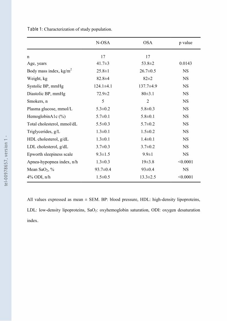
!
Table 1: Characterization of study population.
N-OSA OSA p value
n 17 17 Age, years 41.7±3 53.8±2 0.0143
Body mass index, kg/m2 25.8±1 26.7±0.5 NS
Weight, kg 82.8±4 82±2 NS
Systolic BP, mmHg 124.1±4.1 137.7±4.9 NS
Diastolic BP, mmHg 72.9±2 80±3.1 NS
Smokers, n 5 2 NS
Plasma glucose, mmol/L 5.3±0.2 5.8±0.3 NS
HemoglobinA1c (%) 5.7±0.1 5.8±0.1 NS
Total cholesterol, mmol/dL 5.5±0.3 5.7±0.2 NS
Triglycerides, g/L 1.3±0.1 1.5±0.2 NS
HDL cholesterol, g/dL 1.3±0.1 1.4±0.1 NS
LDL cholesterol, g/dL 3.7±0.3 3.7±0.2 NS
Epworth sleepiness scale 9.3±1.5 9.9±1 NS
Apnea-hypopnea index, n/h 1.3±0.3 19±3.8 <0.0001
Mean SaO2, % 93.7±0.4 93±0.4 NS
4% ODI, n/h 1.5±0.5 13.3±2.5 <0.0001
All values expressed as mean ± SEM. BP: blood pressure, HDL: high-density lipoproteins,
LDL: low-density lipoproteins, SaO2: oxyhemoglobin saturation, ODI: oxygen desaturation
index.
tel-0
0978
657,
ver
sion
1 -

!
Table 2: pD2 values and maximal contractions (Emax) of serotonin obtained in aorta from
mice treated with either vehicle (CTL), N-OSA MPs or OSA MPs in the absence or in the
presence of COX inhibitor (indomethacin, 10 µM) or in the presence of COX-2 inhibitor (NS-
398, 1µM).
pD2 Emax (mN/mm)
C T L Without inhibitors 5.4 ± 0.2 2.3 ± 0.2
With indomethacin 6.3 ± 0.2* 1.1 ± 0.1*
With NS398 5.8 ± 0.2 1.3 ± 0.1*
N-OSA Without inhibitors 5.8 ± 0.2 2.1 ± 0.2
With indomethacin 5.9 ± 0.2 1.1 ± 0.1***
With NS398 5.5 ± 0.2 1.1 ± 0.1**
OSA Without inhibitors 6.1 ± 0.1 3.1 ± 0.2
With indomethacin 5.6 ± 0.3 1.3 ± 0.2***
With NS398 5.1 ± 0.2 2.2 ± 0.4**
*P < 0.05, ***P < 0.001 vs without inhibitors.
tel-0
0978
657,
ver
sion
1 -
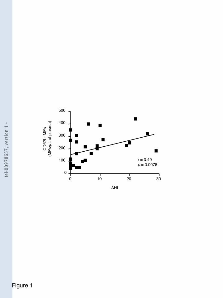
0
300
CD
62L+
MPs
(MPs
/µL
of p
lasm
a)
100
200
400
r = 0.49p = 0.0078
500
0 10 20 30
AHI
Figure 1
tel-0
0978
657,
ver
sion
1 -
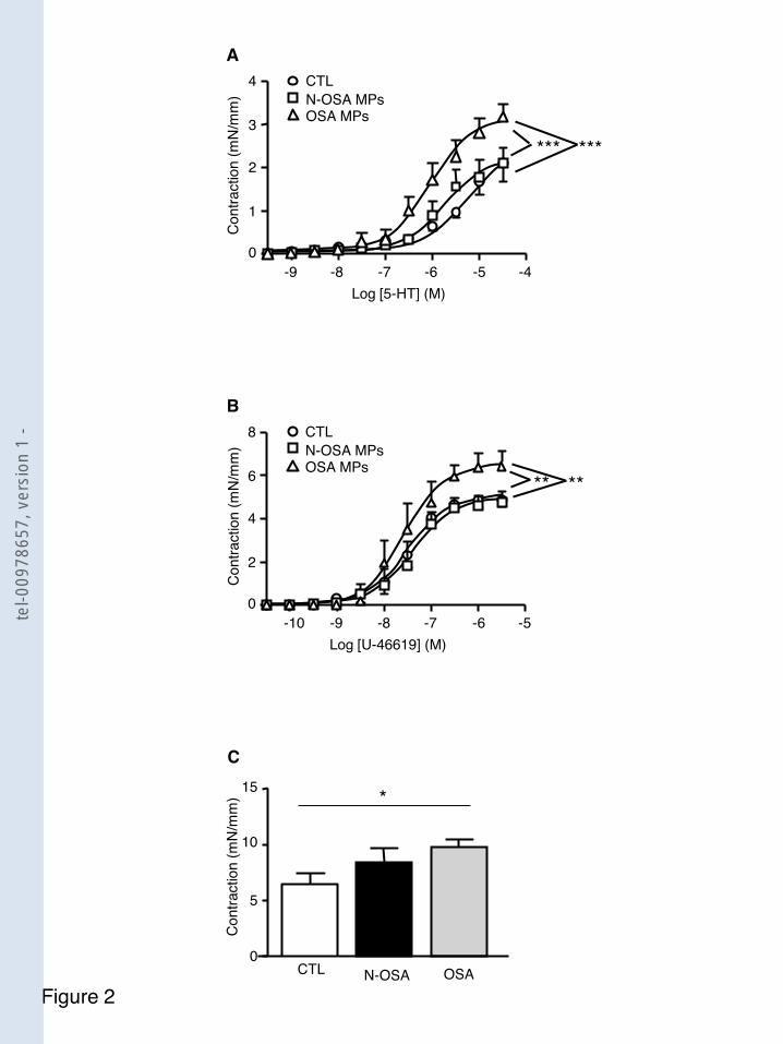
Figure 2
0
5
10
15
Cont
ract
ion
(mN/
mm
)
N-OSA OSACTL
C
*
-10 -9 -8 -7 -5Log [U-46619] (M)
Cont
ract
ion
(mN/
mm
)
0
B
-6
2
4
6
8 CTLN-OSA MPsOSA MPs
** **
-9 -8 -7 -6 -4Log [5-HT] (M)
Cont
ract
ion
(mN/
mm
)0
A
-5
1
2
3
4 CTLN-OSA MPsOSA MPs
*** ***
tel-0
0978
657,
ver
sion
1 -

Figure 3
A
-9 -8 -7 -6 -4Log [5-HT] (M)
Cont
ract
ion
(mN/
mm
)
0-5
1
2
3 CTLL-NA
***
B
-9 -8 -7 -6 -4Log [5-HT] (M)
Cont
ract
ion
(mN/
mm
)
0-5
1
2
3 N-OSA MPsL-NA ***
C
-9 -8 -7 -6 -4Log [5-HT] (M)
Cont
ract
ion
(mN/
mm
)
0-5
1
2
4 OSA MPsL-NA
3 ***
D
-9 -8 -7 -6 -4Log [5-HT] (M)
Cont
ract
ion
(mN/
mm
)
0-5
1
2
4
3
L-NAL-NA + N-OSA MPsL-NA + OSA MPs
tel-0
0978
657,
ver
sion
1 -

Figure 4
0
2000
4000
NO p
rodu
ctio
n (A
/mg)
N-OSA OSACTL
1000
3000
***
**
6000
5000
A
0
1
2
eNO
S ex
pres
sion
N-OSA OSACTL
eNOS
actin
B
**
0
1
P-eN
OS
Ser/e
NOS
(ratio
)
N-OSA OSACTL
P-eNOS Ser
eNOS
0.5
1.5
C
*
0
0.5
1
P-eN
OS
Thr/e
NOS
(ratio
)
N-OSA OSACTL
P-eNOS Thr
eNOS
0.25
0.75
1.25
D
0
2
4
P-eN
OS
Thr/P
-eNO
S Se
r (ra
tio) A
.U.
N-OSA OSACTL
1
3
5 **
E
tel-0
0978
657,
ver
sion
1 -

0
1
2
COX-
1 ex
pres
sion
N-OSA OSACTL
COX-1
actin
**A
0
1
2
COX-
2 ex
pres
sion
N-OSA OSACTL
COX-2
actin
*B
Figure 5
0
10
20
Thro
mbo
xane
A2
(pg/
ml/m
g)
N-OSA OSACTL
5
15
25 *C
0
50
100
8-iso
pros
tane
(pg/
ml/m
g)
N-OSA OSACTL
25
75
125
D
0
2
4
Pros
tagl
andi
n E 2
(pg/
ml/m
g)
N-OSA OSACTL
1
3
*
5
67
F
0
2000
4000
Pros
tacy
clin
(pg/
ml/m
g)
N-OSA OSACTL
1000
3000
*E
tel-0
0978
657,
ver
sion
1 -
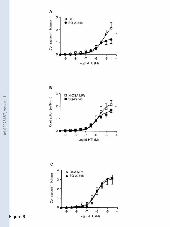
Figure 6
C
-9 -8 -7 -6 -4Log [5-HT] (M)
Cont
ract
ion
(mN/
mm
)
0-5
1
2
4 OSA MPsSQ-29548
3
A
-9 -8 -7 -6 -4Log [5-HT] (M)
Cont
ract
ion
(mN/
mm
)0
-5
1
2
3 CTLSQ-29548
*
B
-9 -8 -7 -6 -4Log [5-HT] (M)
Cont
ract
ion
(mN/
mm
)
0-5
1
2
3 N-OSA MPsSQ-29548
*
tel-0
0978
657,
ver
sion
1 -

Figure 7
C
-9 -8 -7 -6 -4Log [5-HT] (M)
Cont
ract
ion
(mN/
mm
)
0-5
1
2
4 OSA MPsL-NA + indomethacin
3***
A
-9 -8 -7 -6 -4Log [5-HT] (M)
Cont
ract
ion
(mN/
mm
)
0-5
1
2
3 CTLL-NA + indomethacin
***
B
-9 -8 -7 -6 -4Log [5-HT] (M)
Cont
ract
ion
(mN/
mm
)
0-5
1
2
3 N-OSA MPsL-NA + indomethacin ***
D
-9 -8 -7 -6 -4Log [5-HT] (M)
Cont
ract
ion
(mN/
mm
)
0-5
1
2
4L-NA + indomethacin + OSA MPs
3
L-NA + indomethacinL-NA + indomethacin + N-OSA MPs
tel-0
0978
657,
ver
sion
1 -
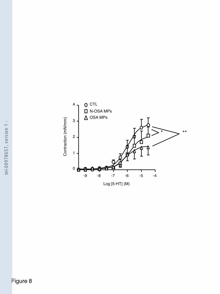
-9 -8 -7 -6 -4
Log [5-HT] (M)
Cont
ract
ion
(mN/
mm
)
0-5
1
2
3
4 CTLN-OSA MPsOSA MPs
* **
Figure 8
tel-0
0978
657,
ver
sion
1 -

Résumé
Les maladies cardio-vasculaires constituent un ensemble de troubles affectant le système
nombreux facteurs rendent leur compréhension délicate. Parmi ces facteurs, les microparticules (MPs) sont des microvésicules membranaires libérées par des cellules activées ou apoptotiques, détectables dans le sang en dehors de toutes situations
ouvent corrélés à De nombreuses publications ont également permis de mettre en
évidence la participation des MPs en tant que vecteur dans la régulation de nombreuses fonctions cellulaires. Cette fluence des MPs dans deux pathologies cardio-vasculaires,
commune. Nous avons ainsi établi une altération du taux circulants et de certaines populations
Ce travail participe ainsi à la compréhension du rôle des MPs dans les mécanismes physiopathologiques impliqués dans ces pathologies. Mots clés : Microparticules; Fonction vasculaire; Hypertension artérielle pulmonaire;
Abstract
Cardiovascular diseases are a group of disorders of the circulatory system. However, the complexity of the etiology of these diseases and the involvement of many factors make understanding difficult. Among these factors, the microparticles (MPs) are membrane vesicles released from activated or apoptotic cells, detectable in the blood, even in healthy subjects, but the alteration of the level and phenotype is very often correlated with the severity of the pathology. Many works have also contributed to highlight the participation of MPs as vectors in the regulation of many cellular functions. This thesis studied the influence of MPs in two cardiovascular diseases, pulmonary arterial hypertension (PAH) and obstructive sleep apnea syndrome (OSA), with hypoxia as a common feature. We have established an alteration of circulating levels and some populations of MPs as well as a participation of MPs in the vascular dysfunction which occurred in PAH and OSA. This work contributes to understanding the role of MPs in the pathophysiological mechanisms involved in these pathologies.
Keywords : Microparticles; Vascular function; Pulmonary artery hypertension; Obstructive sleep apnea syndrome.
tel-0
0978
657,
ver
sion
1 -