infectious diseases: tips for bedside diagnosis - american
TRANSCRIPT
Infectious Diseases:
Tips for Bedside Diagnosis
Joel D. Brown MD DTMH FIDSA
Colonel US Army (Ret.)
Clinical Professor of Medicine
The University of Hawaii
John A Burns School of Medicine
and
The Queens Medical Center
Honolulu, Hawaii
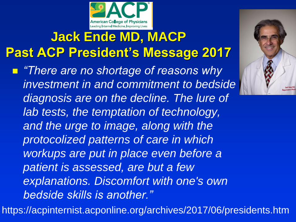
Jack Ende MD, MACP
Past ACP President’s Message 2017
https://acpinternist.acponline.org/archives/2017/06/presidents.htm
“There are no shortage of reasons why
investment in and commitment to bedside
diagnosis are on the decline. The lure of
lab tests, the temptation of technology,
and the urge to image, along with the
protocolized patterns of care in which
workups are put in place even before a
patient is assessed, are but a few
explanations. Discomfort with one's own
bedside skills is another.”
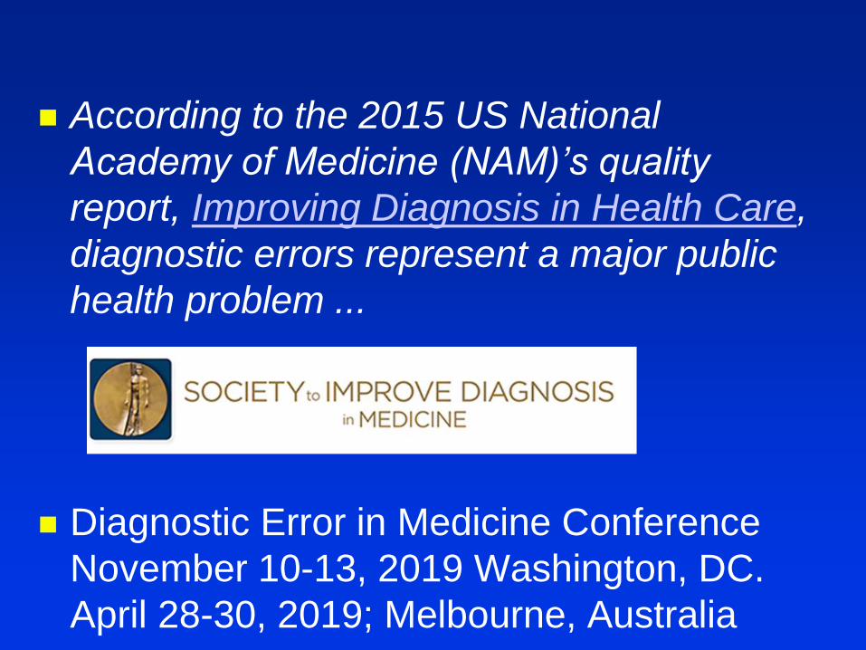
According to the 2015 US National
Academy of Medicine (NAM)’s quality
report, Improving Diagnosis in Health Care,
diagnostic errors represent a major public
health problem ...
Diagnostic Error in Medicine Conference
November 10-13, 2019 Washington, DC.
April 28-30, 2019; Melbourne, Australia
A Definition of Medical Error
...a diagnosis that was unintentionally
delayed (sufficient information was available
earlier), wrong (another diagnosis was
made before the correct one), or missed (no
diagnosis was ever made) ...
Cognitive errors:
• Faulty knowledge
• Faulty data gathering (Most common)
• Faulty synthesis
Graber ML, Franklin N, Gordon R. Diagnostic Error in Internal Medicine. Arch
Intern Med.2005;165(13):1493–1499. doi:10.1001/archinte.165.13.1493

“Tips for Bedside Diagnosis”
Objectives:
• Provide tips for diagnosing infectious diseases
• Improve history and examination skills
• Discuss diagnostic reasoning
Methods:
Present instructive clinical vignettes of my
patients, patients I encountered during bedside
teaching rounds and conferences, and the
medical literature.
We can learn by our errors.
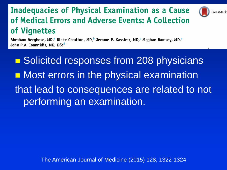
Solicited responses from 208 physicians
Most errors in the physical examination
that lead to consequences are related to not
performing an examination.
The American Journal of Medicine (2015) 128, 1322-1324
“We made too many of the wrong
mistakes.”
Yogi Berra. A Baseball
Legend known for his
malapropisms as well as
pithy and paradoxical
statements.

Case Presentation
A Micronesian man in his mid twenties
CC: Severe headache
HPI: He had been diagnosed with IgA
nephropathy and hypertension.
2 weeks PTA, he noted a progressive onset of a
generalized headache, nausea and vomiting.
On the morning of admission, he was awakened
by severe headache. He came to the ED and was
admitted.
Case Presentation
ROS: Otherwise unremarkable
Past History:
• MVA requiring laparotomy and splenectomy 5
years PTA.
• IgA nephropathy with CRF
Examination:
• T 36.9 C, HR 65, R 20, BP186/121,O2 100%
• He was alert and oriented but in moderate
distress due to his headache.
• The remainder of the examination was
unremarkable.
Initial Differential Diagnosis
Hypertensive Emergency
R/O intracranial bleed
R/O meningitis
Acute exacerbation of chronic renal failure due
to hypertension
Clinical Course:
• Severe headache and hypertension
persisted for several days despite intensive
intravenous anti-hypertensive therapy; the
creatinne increased to 6.6 mg per dL.
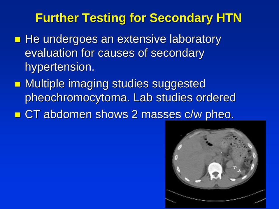
Further Testing for Secondary HTN
He undergoes an extensive laboratory
evaluation for causes of secondary
hypertension.
Multiple imaging studies suggested
pheochromocytoma. Lab studies ordered
CT abdomen shows 2 masses c/w pheo.
Patient Update Day 3
Hypertension and severe headache persist
BP max doses of beta-blocker, ACEI
Develops rigor, T max 104 F,
• CXR - no infiltrate, UA - normal
• WBC - 18,300
Has extensive evaluation for fever.
• Concern for tumor fever, e.g. lymphoma
Broad spectrum antibiotic Rx
Biopsy considered
Further Testing for Secondary HTN
He undergoes an extensive laboratory
evaluation for causes of secondary
hypertension
Multiple imaging studies suggested
pheochromocytoma. Lab studies ordered
PH splenectomy reconsidered.
Scans >
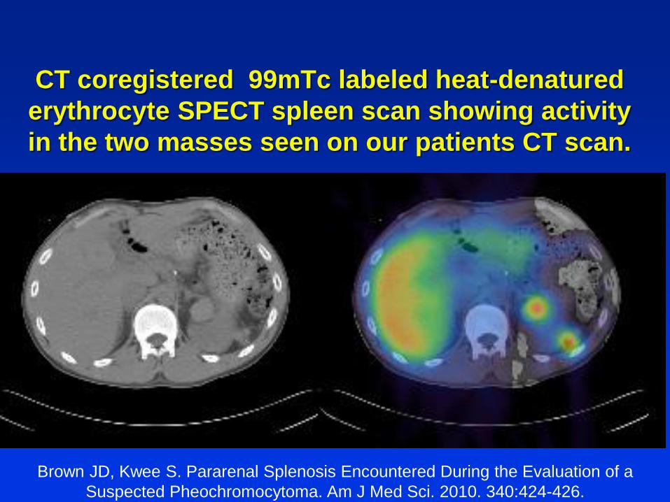
CT coregistered 99mTc labeled heat-denatured
erythrocyte SPECT spleen scan showing activity
in the two masses seen on our patients CT scan.
Brown JD, Kwee S. Pararenal Splenosis Encountered During the Evaluation of a
Suspected Pheochromocytoma. Am J Med Sci. 2010. 340:424-426.
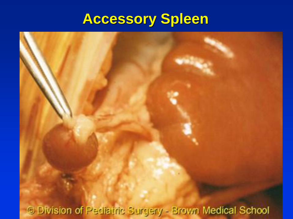
Accessory Spleen
Back to the case - day 4
Sub-Intern student visit:
“ Patient asks for pain medication for his left upper jaw
toothache. Chewing makes his headache worse.”
Additional history
• Patient says he had a chronic toothache.
• He had a severe toothache 1 mo. PTA.
• ED visit at another hospital for toothache. Told
to see a dentist, but he failed to do so. (unable
to pay).
Student is instructed to examine the teeth.
Needs a tongue depressor.
How many medical personnel does it take
to obtain a tongue depressor?
MD begins oralpharyngeal exam ...
Searches patient’s room cabinet ...
Searches for nurse ...
Nurse goes to center of ward ...
Opens “Omnicell” central dispenser ...
Selects tongue depressor ...
Returns to room, gives to Physician

Omnicell Supply and Pharmacy
Dispenser for Hospital Ward

Omnicell Supply Dispenser
“ ... inventory levels controlled ...
costs are reduced”

6-inch Tongue Depressor, Senior, N/S
$6.60 for box of 500 = 1.32 cents each
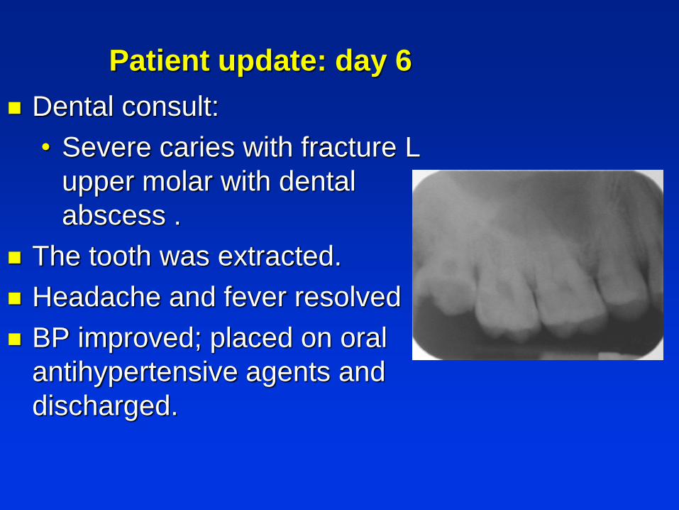
Patient update: day 6
Dental consult:
• Severe caries with fracture L
upper molar with dental
abscess .
The tooth was extracted.
Headache and fever resolved
BP improved; placed on oral
antihypertensive agents and
discharged.
Hawaii hospital charges (2010) for this
admission = $ 59,558.08
• Does not include physician fees
Amount collected = $ 0
• “self pay”
Hospital looses money
Bottom Line:
What went wrong with this patient’s care?
Initial impression framed as “headache.”
Use the patient’s terms; perhaps “face or
jaw pain” instead of headache.
Ask for exacerbating and relieving factors:
“pain worse with chewing.”
Ask patient what is causing his pain:
“toothache makes my head hurt.”
Careful with anchoring bias when Rx fails.

The “Complete History and Physical Exam”
Many texts preface diagnostic steps with the need
to do a “complete H&P.
A learning tool for Medical Students.
• But too time-consuming and inefficient for
Residents and Internists
• “10 point ROS negative” is not convincing
• Medical Records become templated and
inaccurate.
Better to perform a Hypothesis Generated H&P
based on patient's symptoms, focusing on
relevant review of systems and physical exam.
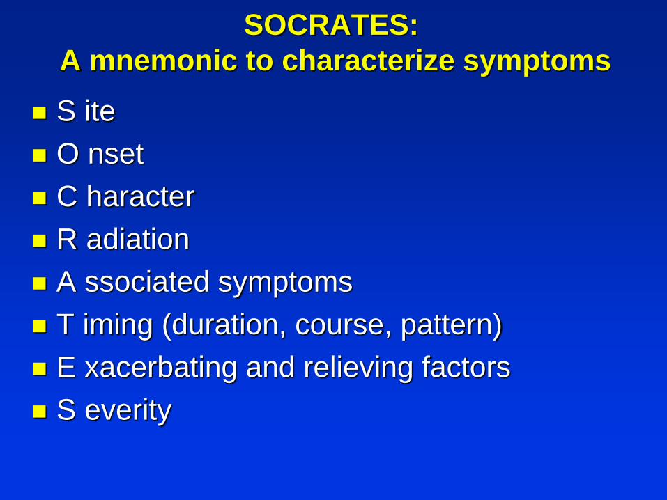
SOCRATES:
A mnemonic to characterize symptoms
S ite
O nset
C haracter
R adiation
A ssociated symptoms
T iming (duration, course, pattern)
E xacerbating and relieving factors
S everity

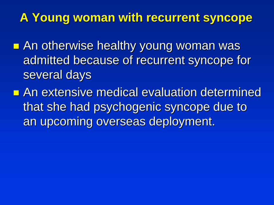
A Young woman with recurrent syncope
An otherwise healthy young woman was
admitted because of recurrent syncope for
several days
An extensive medical evaluation determined
that she had psychogenic syncope due to
an upcoming overseas deployment.
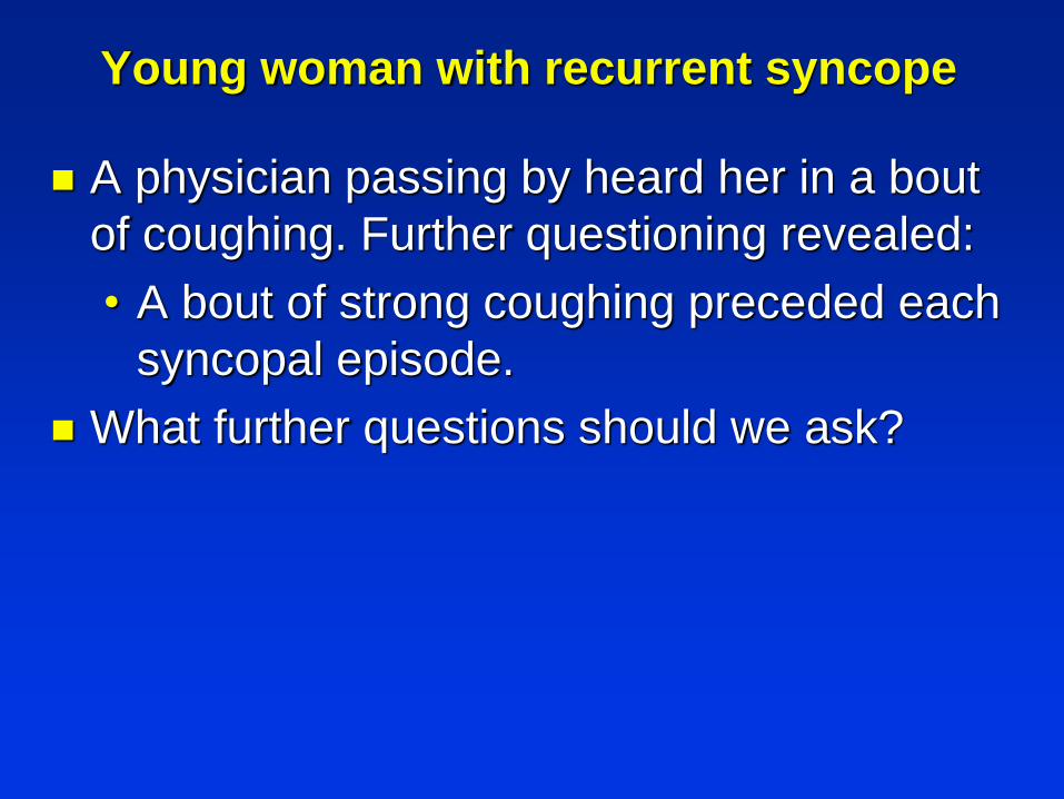
Young woman with recurrent syncope
A physician passing by heard her in a bout
of coughing. Further questioning revealed:
• A bout of strong coughing preceded each
syncopal episode.
What further questions should we ask?
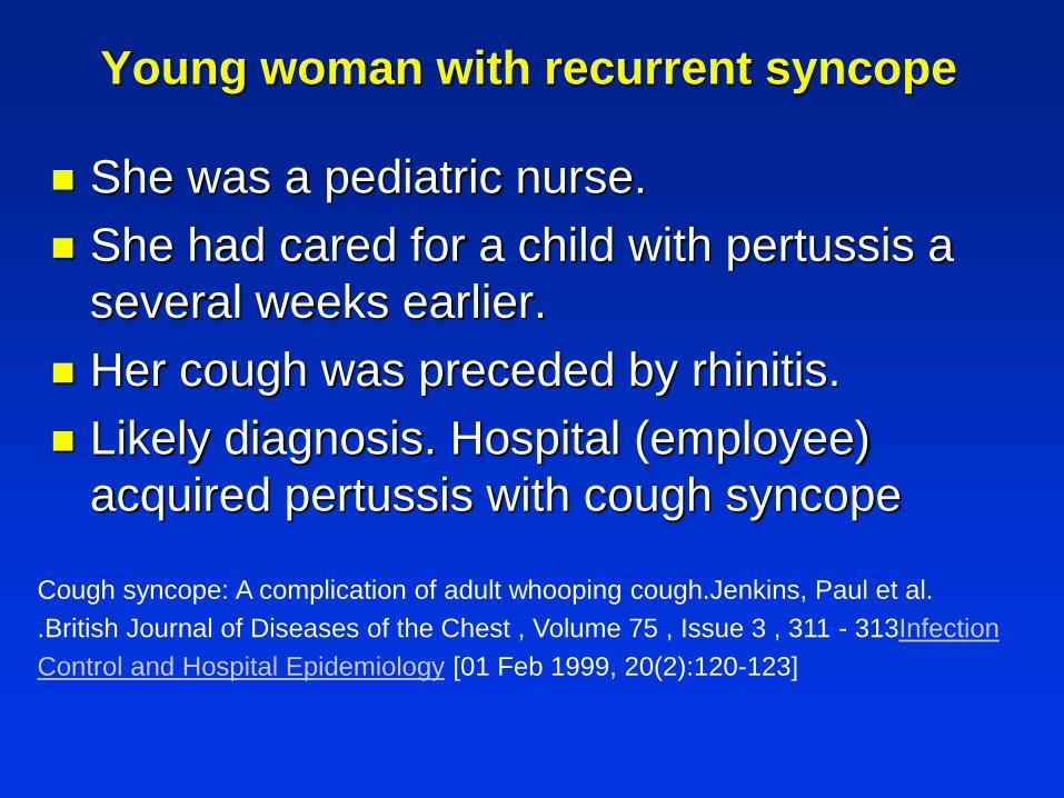
Young woman with recurrent syncope
She was a pediatric nurse.
She had cared for a child with pertussis a
several weeks earlier.
Her cough was preceded by rhinitis.
Likely diagnosis. Hospital (employee)
acquired pertussis with cough syncope
Cough syncope: A complication of adult whooping cough.Jenkins, Paul et al.
.British Journal of Diseases of the Chest , Volume 75 , Issue 3 , 311 - 313Infection
Control and Hospital Epidemiology [01 Feb 1999, 20(2):120-123]
Mechanism of syncope after coughing.
Syncope from coughing was found to be
common and to result from an acute
diminution of supply pressure of blood to
the brain. The violence of the coughing was
the determining factor, Subjects who
coughed continuously without drawing
breath were shown to have circulatory
changes which were exaggerated examples
of the effects of the Valsalva maneuver.
SHARPEY-SCHAFER,BMJ Oct 17, 1953
Syncope Patient: What went wrong?
Inadequate history of patient’s syncope
• Ask “what were you doing when you fainted”
Lack of knowledge of situational syncope:
• Micturition
• Gastrointestinal stimulation
• Cough, sneeze
• Post-exercise
Unaware of patient’s occupation (pediatrics)
• Recent exposure to pertussis patient

Case: A Clinical Evaluation Exercise
The subject patient had an endoscopy proven
gastroesophageal tear c/w a Mallory Weiss tear.
He vomited blood but denied a preceding bout of
vomiting or retching.
However, during the CEX patient described a
preceding bout of forceful hiccupping.
The resident’s oral and written presentation
omitted the hiccupping symptom.
Patient said he had described hiccups to the
admitting team and GI consultants. But they also
omitted this symptom in their records.
Brown JD, Hiccups: An unappreciated cause of the Mallory Weiss syndrome. Am
J Med. 2015.128(12):e19-20.
, Hiccups: An unappreciated cause of the
Mallory Weiss syndrome
A Report of 3 patients who had severe hiccups
before hematemesis; none had preceding
retching typically associated with MW tears.
• 1 patient had a total of 5 episodes, all preceded
by forceful hiccupping.
Although all these patients described intense
bouts of hiccups before hematemesis, their
physicians ignored this feature.
Forceful hiccups are contractures of the
diaphragm similar to retching and can cause MW
tears.Brown JD, Hiccups: An unappreciated cause of the Mallory Weiss syndrome. Am
J Med. 2015.128(12):e19-20.

What went wrong with this case
“You can’t see (feel, hear) what you don’t
know.”
In the H&P, describe the patient’s
symptoms or exam findings, even if you
don’t recognize or understand them.
A more experienced physician may know
the significance.
Or Google the symptoms.

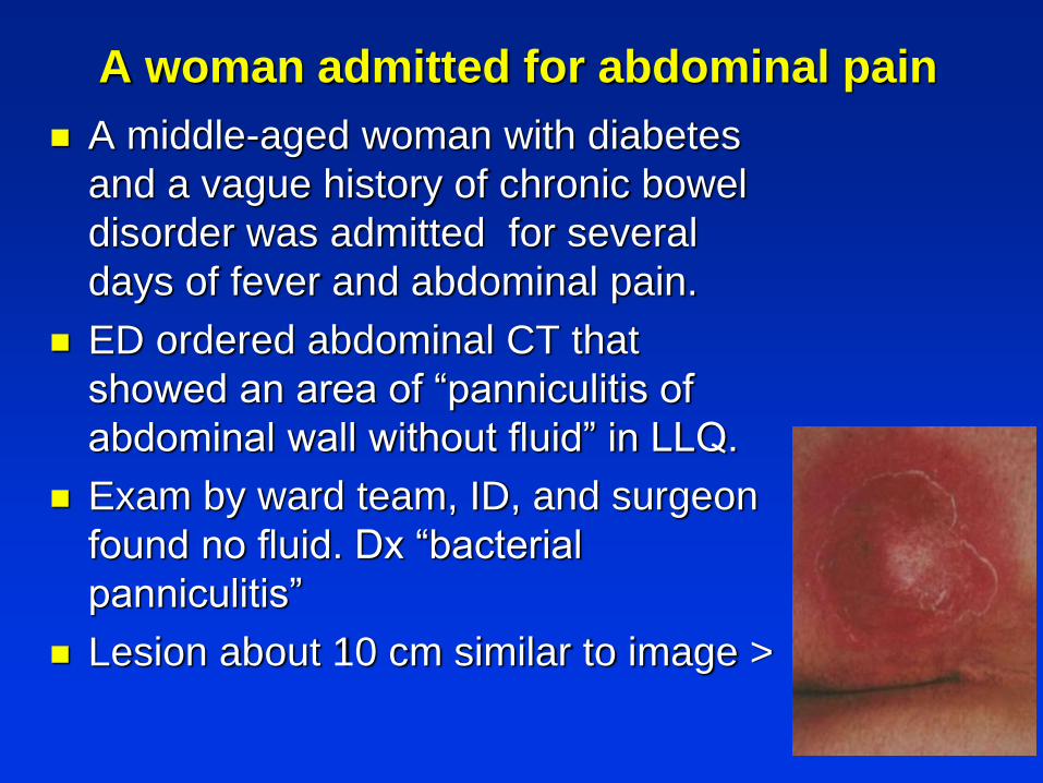
A woman admitted for abdominal pain
A middle-aged woman with diabetes
and a vague history of chronic bowel
disorder was admitted for several
days of fever and abdominal pain.
ED ordered abdominal CT that
showed an area of “panniculitis of
abdominal wall without fluid” in LLQ.
Exam by ward team, ID, and surgeon
found no fluid. Dx “bacterial
panniculitis”
Lesion about 10 cm similar to image >
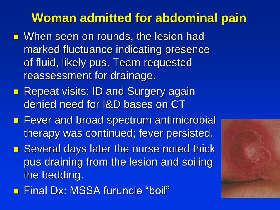
Woman admitted for abdominal pain
When seen on rounds, the lesion had
marked fluctuance indicating presence
of fluid, likely pus. Team requested
reassessment for drainage.
Repeat visits: ID and Surgery again
denied need for I&D bases on CT
Fever and broad spectrum antimicrobial
therapy was continued; fever persisted.
Several days later the nurse noted thick
pus draining from the lesion and soiling
the bedding.
Final Dx: MSSA furuncle “boil”
Abdominal pain: What went wrong?
Patient’s symptoms were identified as fever
and “abdominal pain.”
This led to search for intraabdominal source of
pain and fever. CT exam missed fluid
collection.
Consultants favored the CT exam over the
physical exam.
Most likely she was trying to indicate pain on
the abdomen.
Lesson. Think anatomically in 3 dimension:
Pain skin & subcutaneous tissue (cellulitis),
fascia, muscle, peritoneum or internal organs.
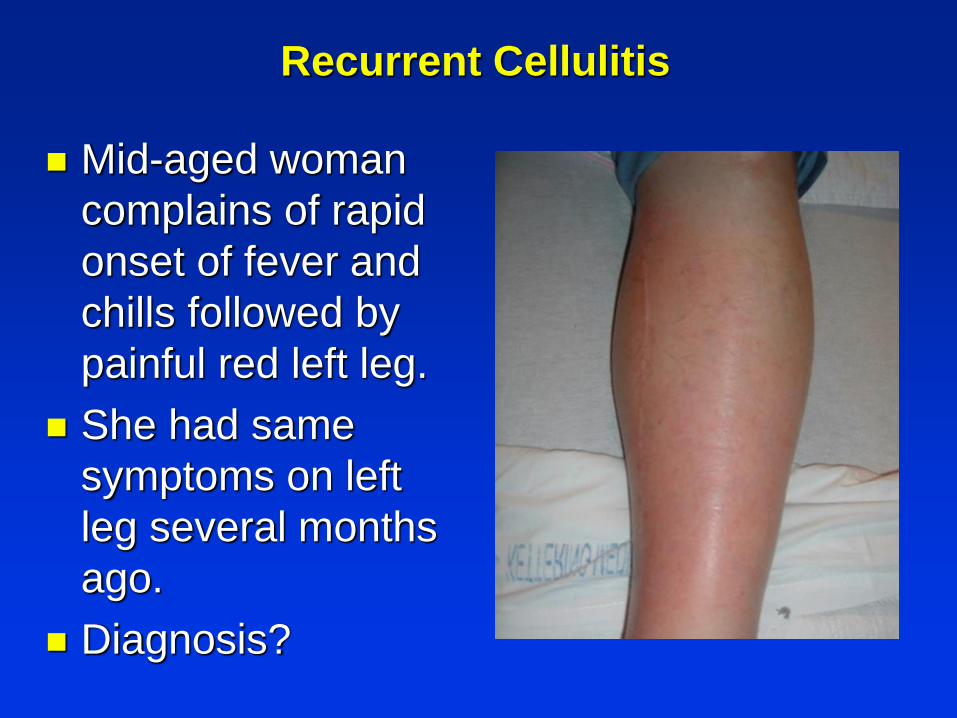
Recurrent Cellulitis
Mid-aged woman
complains of rapid
onset of fever and
chills followed by
painful red left leg.
She had same
symptoms on left
leg several months
ago.
Diagnosis?
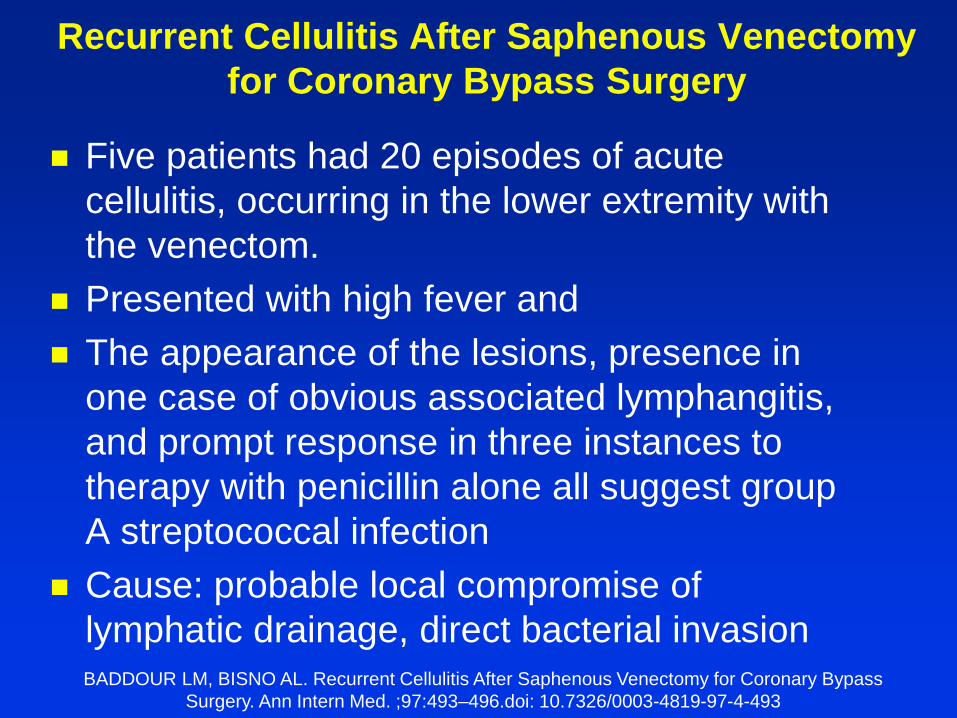
Recurrent Cellulitis After Saphenous Venectomy
for Coronary Bypass Surgery
Five patients had 20 episodes of acute
cellulitis, occurring in the lower extremity with
the venectom.
Presented with high fever and
The appearance of the lesions, presence in
one case of obvious associated lymphangitis,
and prompt response in three instances to
therapy with penicillin alone all suggest group
A streptococcal infection
Cause: probable local compromise of
lymphatic drainage, direct bacterial invasionBADDOUR LM, BISNO AL. Recurrent Cellulitis After Saphenous Venectomy for Coronary Bypass
Surgery. Ann Intern Med. ;97:493–496.doi: 10.7326/0003-4819-97-4-493
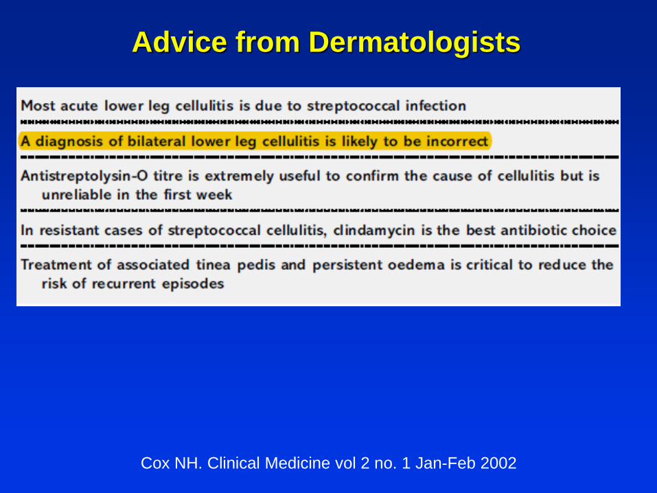
Advice from Dermatologists
Cox NH. Clinical Medicine vol 2 no. 1 Jan-Feb 2002
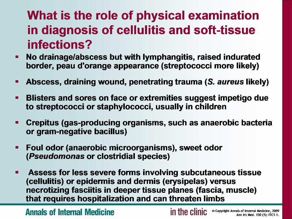
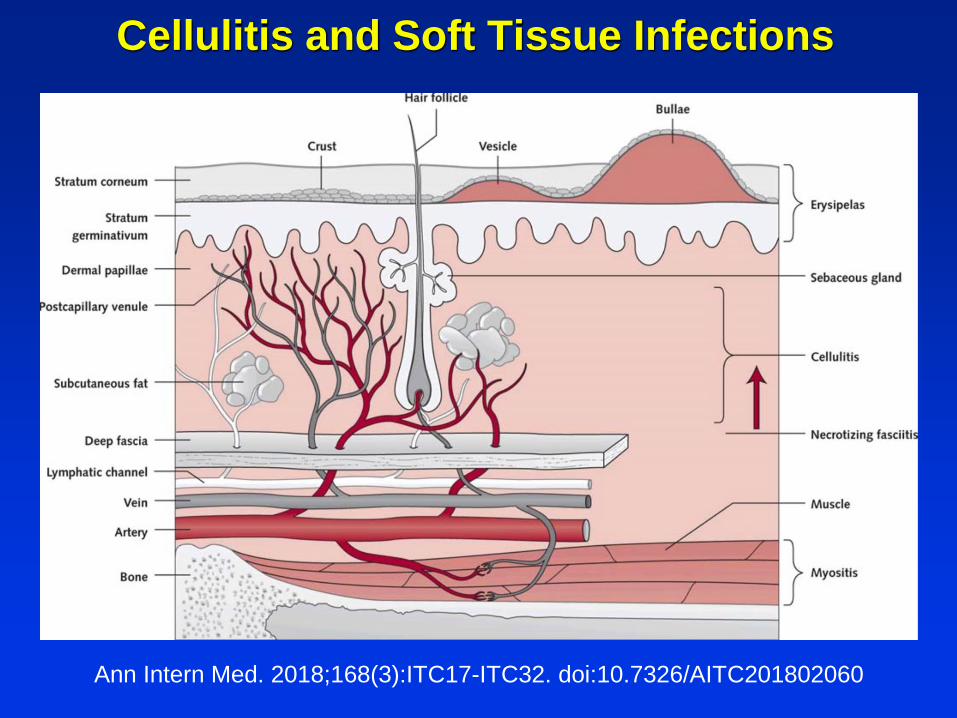
Cellulitis and Soft Tissue Infections
Ann Intern Med. 2018;168(3):ITC17-ITC32. doi:10.7326/AITC201802060


My experience with the Saigon Medical School:
“An Experiment in International Medical in South
Viet Nam (1966-1975)”
“The Vietnamese faculty, a dignified,
proud group with their own education in
France, an academic hierarchy based on
the French system ... found themselves
committed by the realpolitik of the war to
an American aid program that defined
that the medical school be reorganized
into an American-style academic medical
school.”
...Chief of Medicine invites me to visit
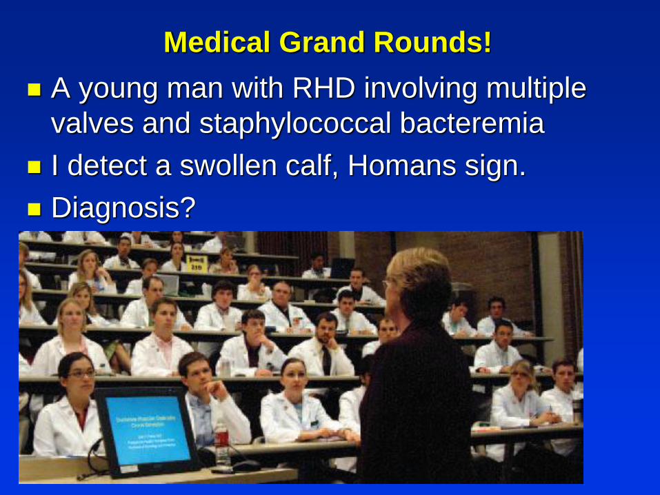
Medical Grand Rounds!
A young man with RHD involving multiple
valves and staphylococcal bacteremia
I detect a swollen calf, Homans sign.
Diagnosis?
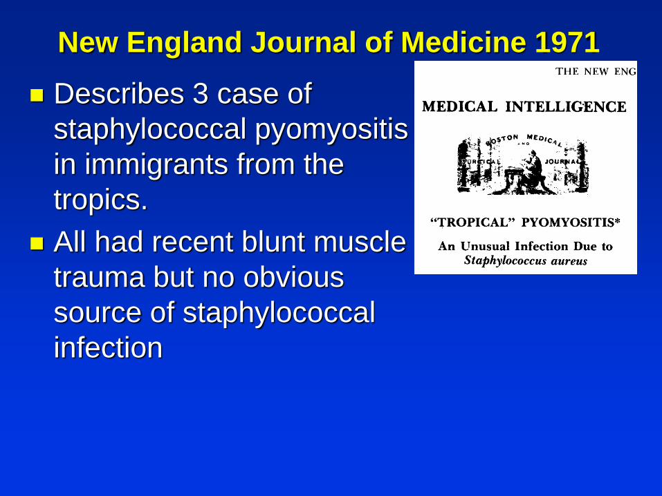
New England Journal of Medicine 1971
Describes 3 case of
staphylococcal pyomyositis
in immigrants from the
tropics.
All had recent blunt muscle
trauma but no obvious
source of staphylococcal
infection
How did I miss this probable case of tropical
pyomyositis?
I was unaware of the disease.
Availability bias: DVT common in hospital
patients.
But:
• Patients’ new problems are often related to a
pre-existing disease. (known feature of the
disease, treatments, Dx. procedures).
• Should be aware of metastatic infection in
bacteremia to almost any site.
Pyomyositis: Report of 18 Cases in Hawaii
Most were healthy young men or boys, and eight
had never traveled abroad.
Fever, muscle pain & swelling, leukocytosis were
common, but only 7 had erythema or fluctuance.
Muscle may be woody-hard, tumor-like.
Mimicked cellulitis, muscle hematoma,
thrombophlebitis, appendicitis, neoplasm.
12 had non-penetrating muscle injury and 13 had
pyoderma, suggesting that bacteria invade injured
muscle via the bloodstream or lymphatic system.
Brown JD; Wheeler B. Pyomyositis. Report of 18 cases in Hawaii. Arch Intern
Med 1984;144:1749-51.
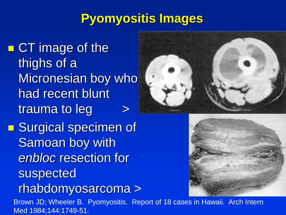
Pyomyositis Images
CT image of the
thighs of a
Micronesian boy who
had recent blunt
trauma to leg >
Surgical specimen of
Samoan boy with
enbloc resection for
suspected
rhabdomyosarcoma > Brown JD; Wheeler B. Pyomyositis. Report of 18 cases in Hawaii. Arch Intern
Med 1984;144:1749-51.

A Polynesian man with acute weakness
A 27-year-old Polynesian man was admitted
because of chronic diarrhea and 1 day of bilateral
leg weakness.
3 mos. PTA he began having 2-3 loose bowel
movements per day, which increased to 3-5 times
per day one week PTA.
One day PTA he had generalized weakness. On
the morning of admission he was too weak to get
out of bed and was admitted.
Exam: Muscle strength: 3/5 legs, 4/5 arms.
Serum potassium 2.2 mEq/L
Your Diagnosis?
Polynesian man with acute weakness
His hypokalemia was initially attributed to
intestinal losses. His weakness resolved within an
hour after replacement of potassium chloride
On the day after admission, further questioning
revealed that, in addition to increased frequency
of bowel movements, he had noticed several
months of increased appetite, heat intolerance,
sweating, irritability, palpitations, dyspnea.
For the past month he had noticed leg weakness
Polynesian man with acute weakness
Examination:
HR: 108 beats/min, temp 37.1 C, BP 152/83
mm of Hg,RR 24/min.
A fine tremor of the acanthosis nigricans on
the neck and axillae.
Your Diagnosis?
Polynesian man with acute weakness
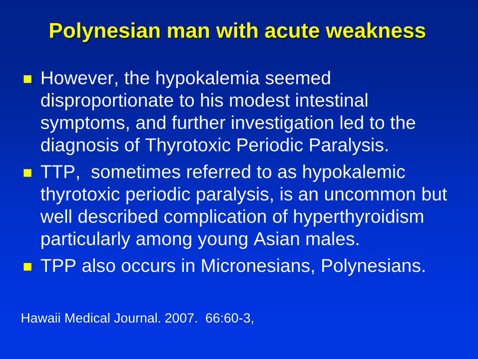
However, the hypokalemia seemed
disproportionate to his modest intestinal
symptoms, and further investigation led to the
diagnosis of Thyrotoxic Periodic Paralysis.
TTP, sometimes referred to as hypokalemic
thyrotoxic periodic paralysis, is an uncommon but
well described complication of hyperthyroidism
particularly among young Asian males.
TPP also occurs in Micronesians, Polynesians.
Hawaii Medical Journal. 2007. 66:60-3,
Polynesian man with acute weakness
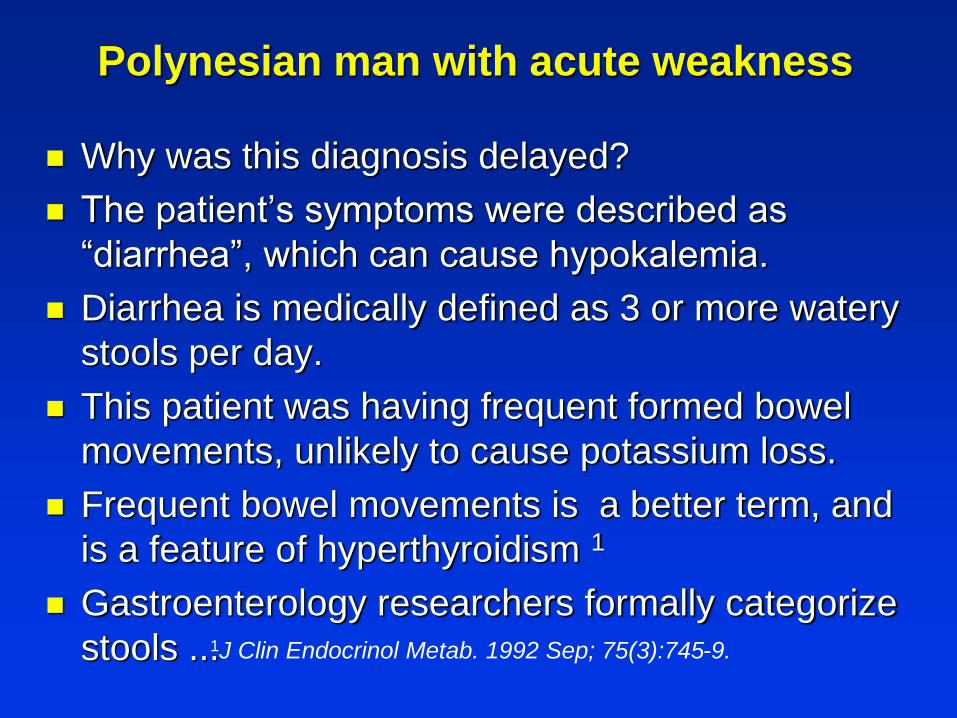
Why was this diagnosis delayed?
The patient’s symptoms were described as
“diarrhea”, which can cause hypokalemia.
Diarrhea is medically defined as 3 or more watery
stools per day.
This patient was having frequent formed bowel
movements, unlikely to cause potassium loss.
Frequent bowel movements is a better term, and
is a feature of hyperthyroidism 1
Gastroenterology researchers formally categorize
stools ...1J Clin Endocrinol Metab. 1992 Sep; 75(3):745-9.
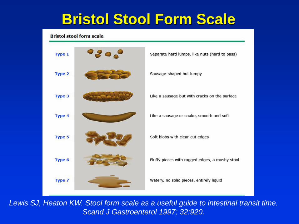
Bristol Stool Form Scale
Lewis SJ, Heaton KW. Stool form scale as a useful guide to intestinal transit time.
Scand J Gastroenterol 1997; 32:920.

NEJM: Clinical problem-solving case
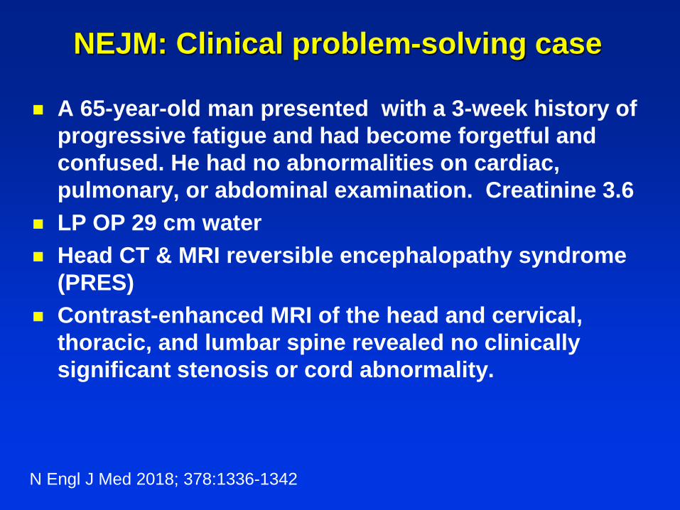
A 65-year-old man presented with a 3-week history of
progressive fatigue and had become forgetful and
confused. He had no abnormalities on cardiac,
pulmonary, or abdominal examination. Creatinine 3.6
LP OP 29 cm water
Head CT & MRI reversible encephalopathy syndrome
(PRES)
Contrast-enhanced MRI of the head and cervical,
thoracic, and lumbar spine revealed no clinically
significant stenosis or cord abnormality.
N Engl J Med 2018; 378:1336-1342
NEJM: Clinical problem-solving case
Ultrasound: a markedly distended bladder
with an estimated residual urine volume of
1785 ml after voiding. A urinary catheter
was placed, yielded 2900 ml of clear urine.
Symptoms and imaging findings resolved.
Final diagnosis obstructive nephropathy
leading to a hypertensive emergency and
PRES.
Subsequent Letters to Editor...
...critical of the normal abdominal examinations
that missed the very large distended bladder.
Palpation and Percussion would have detected
the bladder obstruction.
Editors reply:
We agree that the dependence of modern
physicians on laboratory and imaging studies
almost certainly makes the time to diagnosis
longer than it would be with a physical evaluation
by a skilled clinician.
GG Loscocco, M Piccini. N Engl J Med 2019;380:379-379.
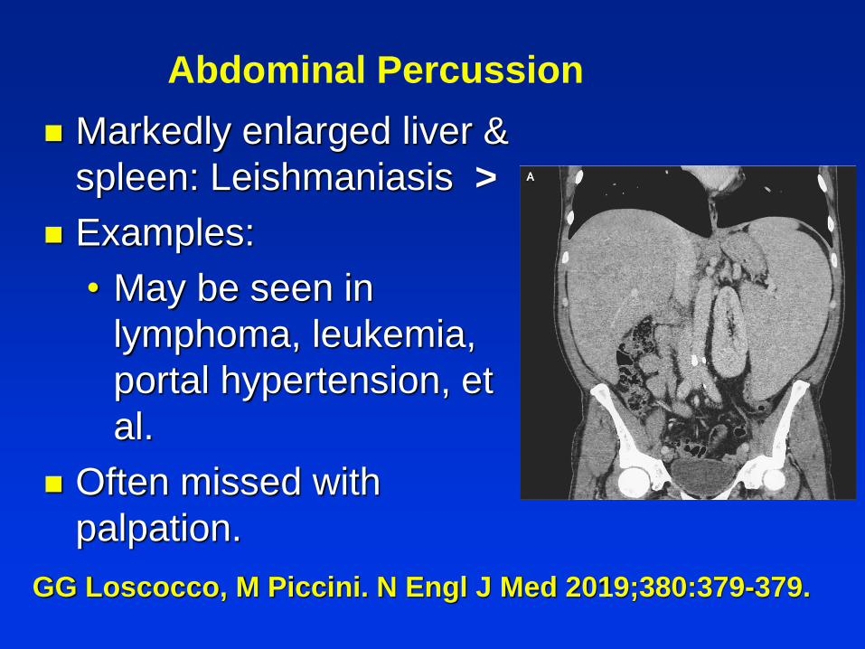
Markedly enlarged liver &
spleen: Leishmaniasis >
Examples:
• May be seen in
lymphoma, leukemia,
portal hypertension, et
al.
Often missed with
palpation.
Abdominal Percussion
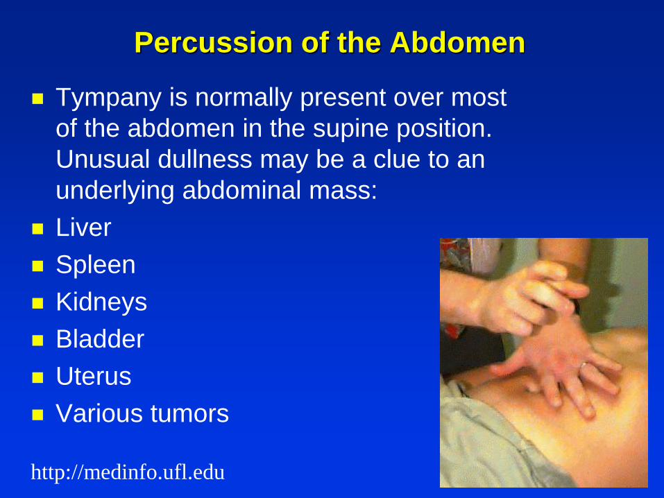
Percussion of the Abdomen
Tympany is normally present over most
of the abdomen in the supine position.
Unusual dullness may be a clue to an
underlying abdominal mass:
Liver
Spleen
Kidneys
Bladder
Uterus
Various tumors
http://medinfo.ufl.edu
Percussion for liver tenderness
Liver tenderness can be an early sign of
infections involving the liver.
Careful punch tenderness, similar to
examining the kidneys for pyelonephritis,
can be an early clue to liver inflammation.
Examples:
• Viral hepatitis, liver abscess,
leptospirosis, malaria, et al.
Ascites can be detected when there is ...

Shifting Dullness

A 19-yr-old man with fever
A 19-year-old man was well until 4 days
PTA: rapid onset of fever, headache, neck
pain, nausea, vomiting, diffuse myalgia and
dry cough.
ED visit: CXR showed right lung infiltration;
pneumonia was diagnosed and he was
treated with ceftriaxone 2g IV
• DC for follow-up by his physician
History 2
Later that day he noted increased dyspnea,
and began coughing blood.
He was admitted to the hospital
Social Hx: A greens worker at Kauai golf
course. No alcohol, drugs, or
tobacco, recent travel, sick contacts or local
disease outbreaks.
Directed History and Exam
No animal exposure
No recent dengue outbreak
Swam in Waimea River 2 weeks PTA
No sinus or upper airway signs or
symptoms
No rash, skin lesions, petechiae
No urine RBC casts
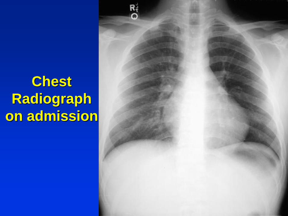
Chest
Radiograph
on admission
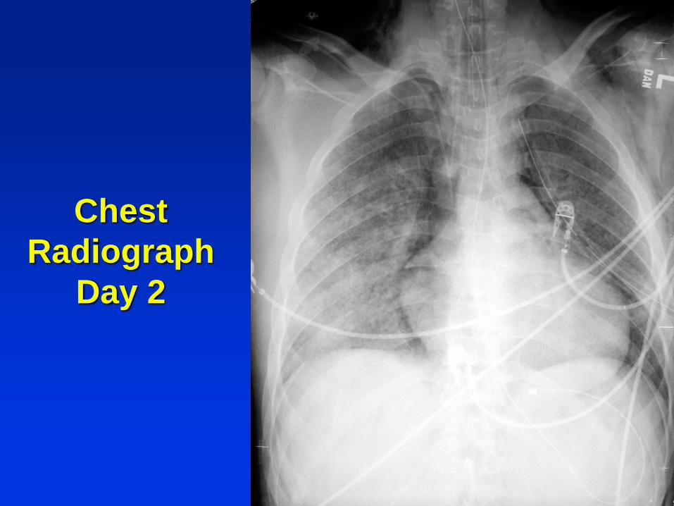
Chest
Radiograph
Day 2
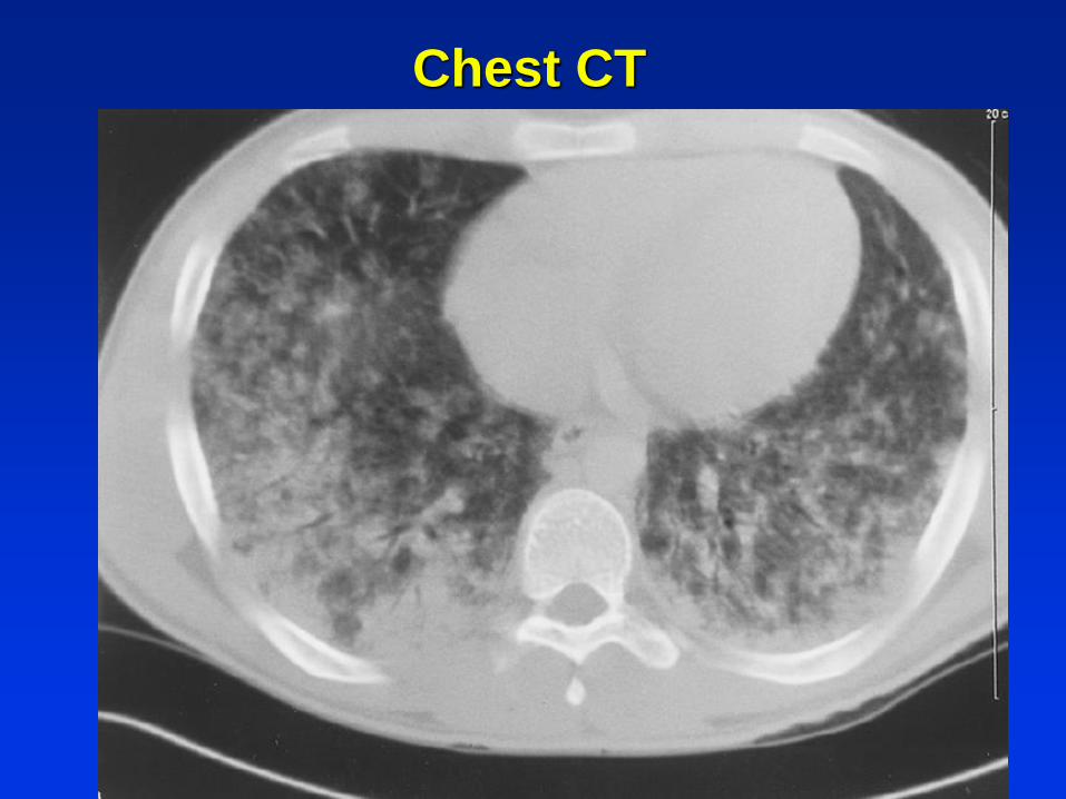
Chest CT
Course
DDx included multiple causes of diffuse
alveolar hemorrhage (DAH) and kidney
disease. (Pulmonary-renal syndrome)
Goodpasture Syndrome rare (1/million/yr)
Many special lab studies were all negative
Kidney biopsy:
• Pathology unremarkable was
unremarkable
• Caused retroperitoneal hemorrhage
Total CK was elevated 600
Course
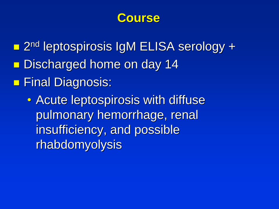
2nd leptospirosis IgM ELISA serology +
Discharged home on day 14
Final Diagnosis:
• Acute leptospirosis with diffuse
pulmonary hemorrhage, renal
insufficiency, and possible
rhabdomyolysis
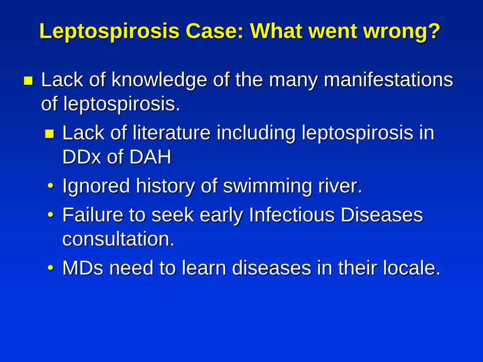
Leptospirosis Case: What went wrong?
Lack of knowledge of the many manifestations
of leptospirosis.
Lack of literature including leptospirosis in
DDx of DAH
• Ignored history of swimming river.
• Failure to seek early Infectious Diseases
consultation.
• MDs need to learn diseases in their locale.
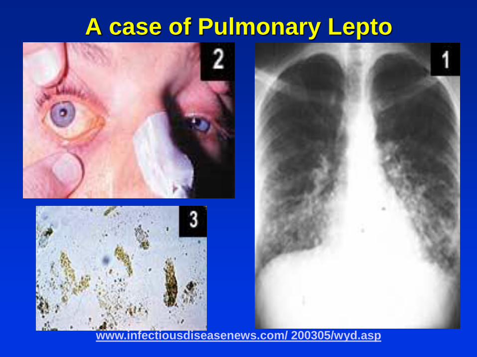
A case of Pulmonary Lepto
www.infectiousdiseasenews.com/ 200305/wyd.asp
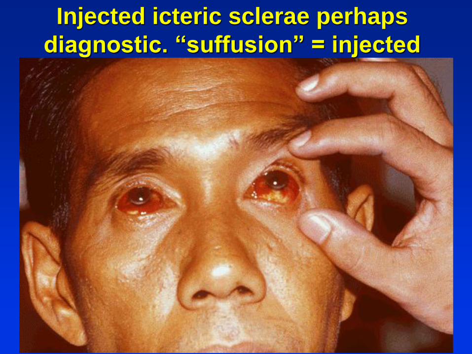
Injected icteric sclerae perhaps
diagnostic. “suffusion” = injected
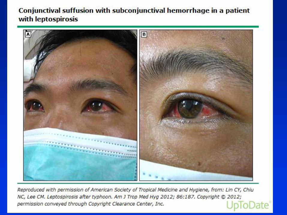
Copyrights apply

Diagnostic Clues to Weil Disease
Abrupt onset fever with chills common
Myalgia often remarkable, but may clear in
few days (elicit this Hx)
Hx.of exposure common, but not invariable.
Conjunctival injection common.
CPK often elevated.
Leukocytois, platelets decreased.
DAH seems common in Hawaii.
A good sign for lepto ...
Signs - Continued

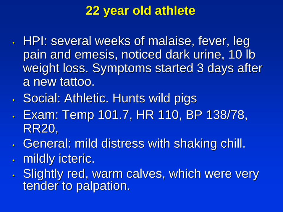
22 year old athlete
• HPI: several weeks of malaise, fever, leg pain and emesis, noticed dark urine, 10 lbweight loss. Symptoms started 3 days after a new tattoo.
• Social: Athletic. Hunts wild pigs
• Exam: Temp 101.7, HR 110, BP 138/78, RR20,
• General: mild distress with shaking chill.
• mildly icteric.
• Slightly red, warm calves, which were very tender to palpation.
Leptospiriosis
“Conjunctival suffusion and muscle
tenderness, most notable in the calf and
lumbar areas, are the most characteristic
physical findings but may occur in a minority
of cases”
Mandell, Douglas, and Bennett's Principles and Practice of Infectious Diseases, 8th Ed,
Gompf SG. Leptospirosis. Medscape; June 2013
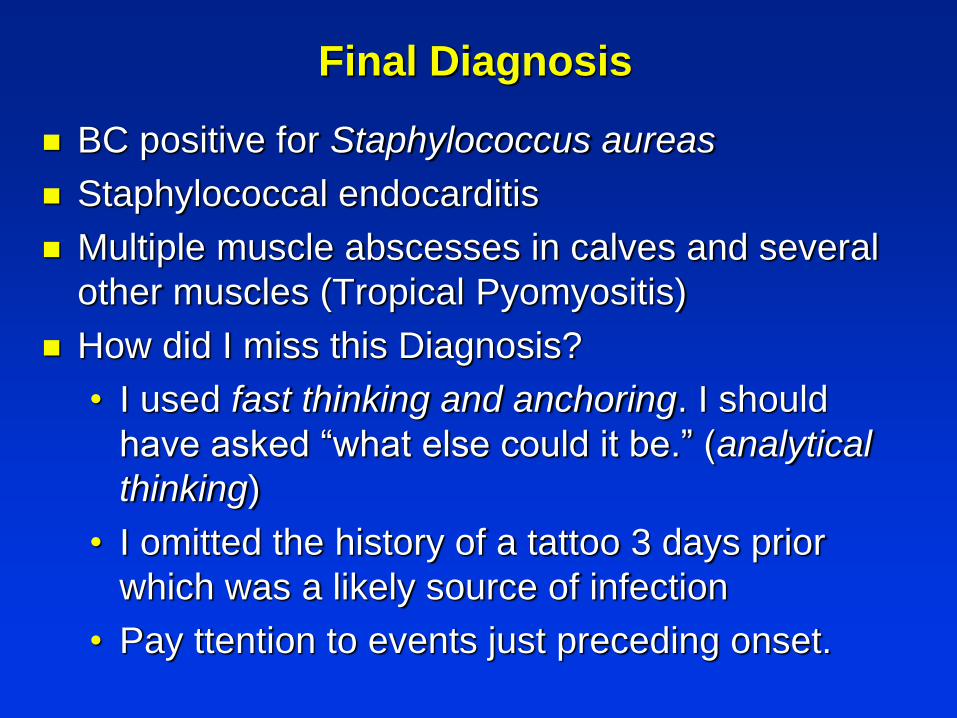
Final Diagnosis
BC positive for Staphylococcus aureas
Staphylococcal endocarditis
Multiple muscle abscesses in calves and several
other muscles (Tropical Pyomyositis)
How did I miss this Diagnosis?
• I used fast thinking and anchoring. I should
have asked “what else could it be.” (analytical
thinking)
• I omitted the history of a tattoo 3 days prior
which was a likely source of infection
• Pay ttention to events just preceding onset.

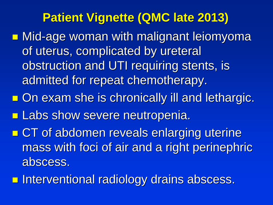
Patient Vignette (QMC late 2013)
Mid-age woman with malignant leiomyoma
of uterus, complicated by ureteral
obstruction and UTI requiring stents, is
admitted for repeat chemotherapy.
On exam she is chronically ill and lethargic.
Labs show severe neutropenia.
CT of abdomen reveals enlarging uterine
mass with foci of air and a right perinephric
abscess.
Interventional radiology drains abscess.
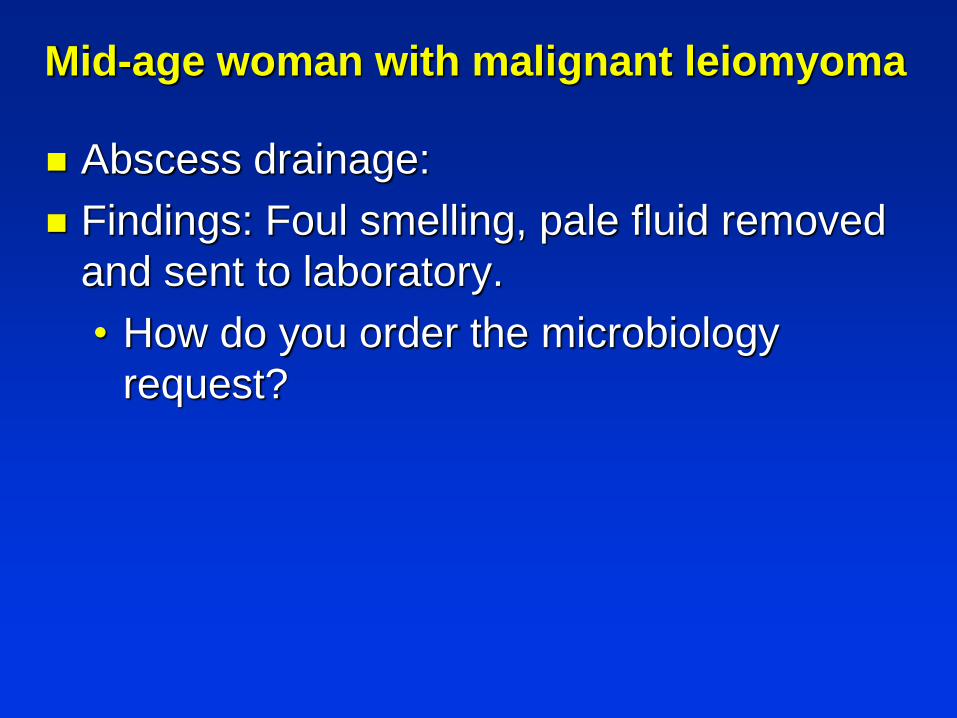
Mid-age woman with malignant leiomyoma
Abscess drainage:
Findings: Foul smelling, pale fluid removed
and sent to laboratory.
• How do you order the microbiology
request?
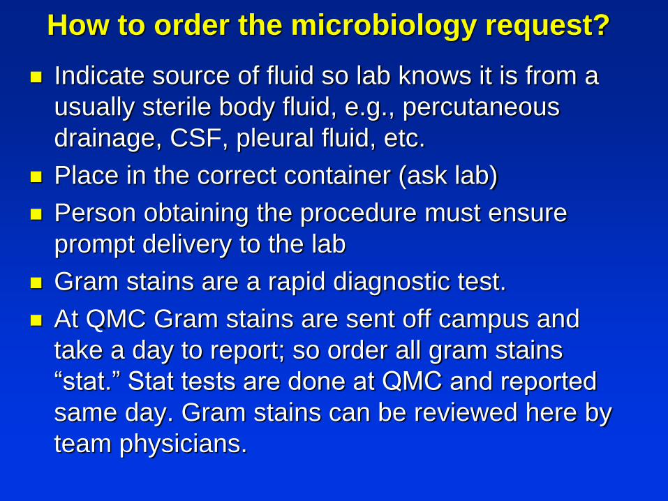
How to order the microbiology request?
Indicate source of fluid so lab knows it is from a
usually sterile body fluid, e.g., percutaneous
drainage, CSF, pleural fluid, etc.
Place in the correct container (ask lab)
Person obtaining the procedure must ensure
prompt delivery to the lab
Gram stains are a rapid diagnostic test.
At QMC Gram stains are sent off campus and
take a day to report; so order all gram stains
“stat.” Stat tests are done at QMC and reported
same day. Gram stains can be reviewed here by
team physicians.
Clinical Course
She becomes septic and is empirically
treated with imipenem, ciprofloxacin and
metronidazole.
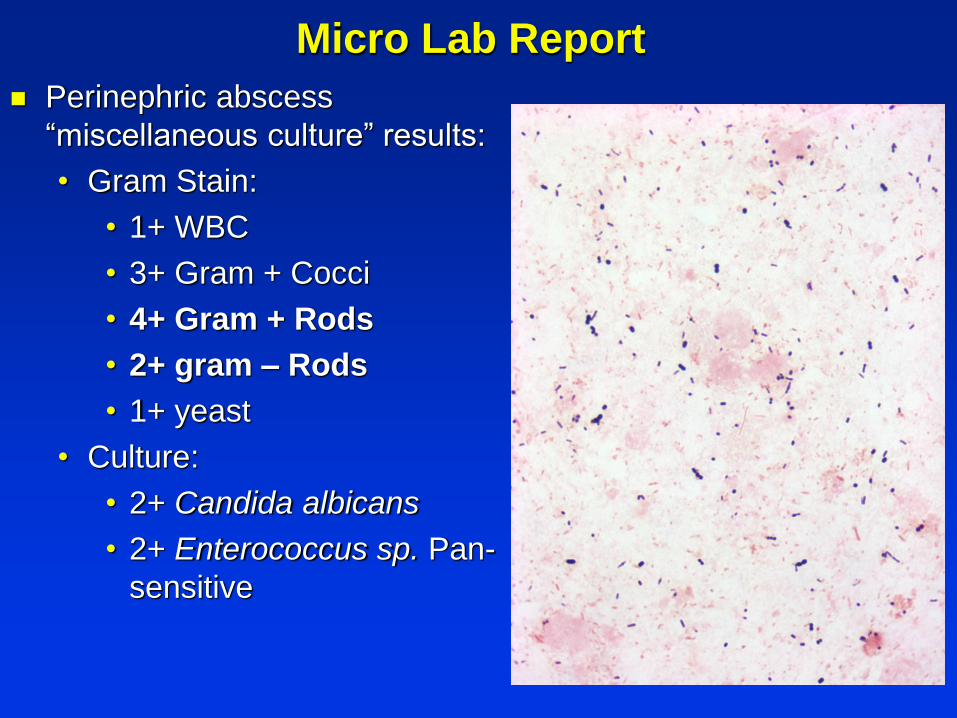
Micro Lab Report
Perinephric abscess
“miscellaneous culture” results:
• Gram Stain:
• 1+ WBC
• 3+ Gram + Cocci
• 4+ Gram + Rods
• 2+ gram – Rods
• 1+ yeast
• Culture:
• 2+ Candida albicans
• 2+ Enterococcus sp. Pan-
sensitive
Clinical Course
After culture results returned, treated with
vancomycin, cefepime and caspifungin
• No coverage for anaerobic bacteria
Patient, develops septic shock and expires.
No autopsy requested.
Comments
Perinephric abscess, “miscellaneous culture”
results:
• 2+ Candida albicans
• 2+ Enterococcus sp. Pan-sensitive
Blood culture positive for Candida
The “miscellaneous culture” does not culture for
anaerobes; additional methods required.
Anaerobic cultures take days; identification and
susceptibility testing is complex.
Check pus for putrid or foul odor.
Order aerobic & anaerobic cultures of pus from
most normally sterile sites.
Final Diagnosis
Perinephric abscesses usually originate in the
kidney.
• Anaerobic infections of the kidney are rare
Perineprhic abscess can also stem form
bacteremia (e.g., Staphylococcus)
Or from adjacent infection.
The subject patient had an extensive necrotic
pelvic tumor containing gas, suggestive of a pelvic
anaerobic infection that spread to adjacent
perinephric space.
The important clues are the foul odor and the
Gram stain morphology

History Anaerobic Bacteria
Altemeier , in the late 1930s, isolated anaerobic
bacteria from 96 of 100 appendicitis patients.
He also noted that putrid discharge was found
exclusively in the presence of infections
involving anaerobic bacteria and that these
were also the only organisms to produce the
characteristic odor both in vivo and in vitro.
Foul odor is due to volatile fatty acid production
typical of many anaerobes.
Altemeier WA. The cause of the putrid odor of perforated appendicitis
with peritonitis Ann Surg. 1938;107:634.
Anaerobic Bacterial Infections
Anaerobic bacteria are infrequent pulmonary pathogens,
but may cause serious disease.
Clues for anaerobic pneumonia include aspiration risks,
putrid discharge, indolent course, and necrotizing
pneumonia.
Putrid discharge as sometimes found with lung abscess
or empyema cases is considered diagnostic of anaerobic
infection because these agents, ... are the only microbes
that produce the short-chain volatile fatty acids responsible
for this distinctive odor.
Treatment includes drainage of pleural collections and
antimicrobials, including clindamycin or a β-lactamase/β-
lactamase inhibitor.John G. Bartlett MD
Infectious Disease Clinics of North America, 2013-03-01, Volume 27, Issue 1,
Pages 149-155,
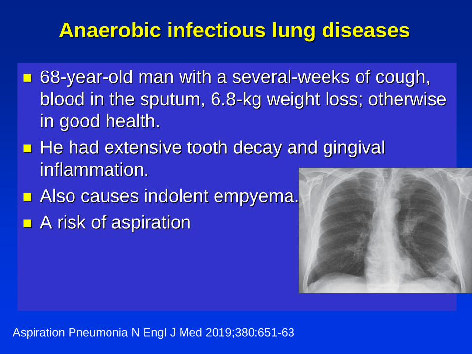
Anaerobic infectious lung diseases
68-year-old man with a several-weeks of cough,
blood in the sputum, 6.8-kg weight loss; otherwise
in good health.
He had extensive tooth decay and gingival
inflammation.
Also causes indolent empyema.
A risk of aspiration
Aspiration Pneumonia N Engl J Med 2019;380:651-63

Anaerobic bacteria inhabit gingival crevices.
Gingivitis allows increased anaerobes.
Increased numbers increase risk of
anaerobic infections of lung, brain, neck etc.
Rarely included in H&P notes, or perhaps
just “poor dentition.” meth mouth
An important clue to anaerobic infections
Bedside Diagnosis of Anaerobic Infections
Infections at particular sites, especially those
proximal to mucosal surfaces with indigenous
anaerobic flora, particularly in the GI tract, the
female genital tract, or the oral cavity. Anaerobes
are often associated with tissue necrosis and
abscess formation.
The presence of a foul odor or gas is highly
suggestive as well, although the absence of these
factors does not rule out anaerobic infection
Often polymicrobial, Gram stain of exudates
showing a polymicrobial flora and organisms
with morphologic features of anaerobes are
indicative of anaerobic infection

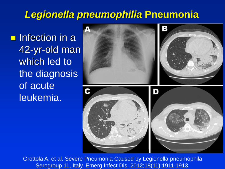
Legionella pneumophilia Pneumonia
Infection in a
42-yr-old man
which led to
the diagnosis
of acute
leukemia.
Grottola A, et al. Severe Pneumonia Caused by Legionella pneumophila
Serogroup 11, Italy. Emerg Infect Dis. 2012;18(11):1911-1913.
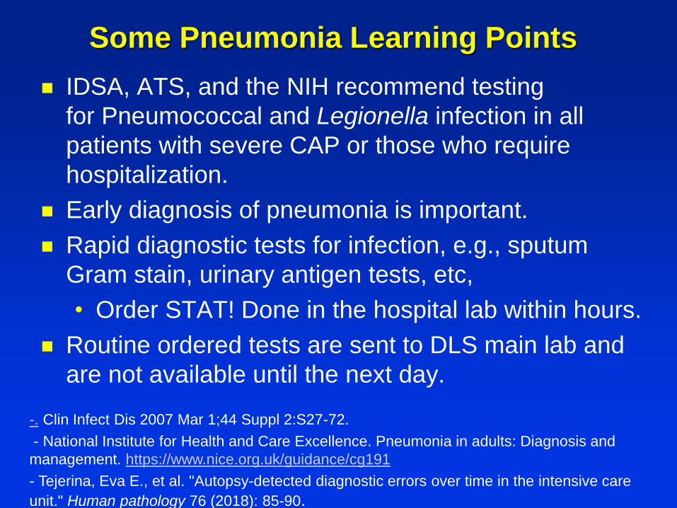
Some Pneumonia Learning Points
IDSA, ATS, and the NIH recommend testing
for Pneumococcal and Legionella infection in all
patients with severe CAP or those who require
hospitalization.
Early diagnosis of pneumonia is important.
Rapid diagnostic tests for infection, e.g., sputum
Gram stain, urinary antigen tests, etc,
• Order STAT! Done in the hospital lab within hours.
Routine ordered tests are sent to DLS main lab and
are not available until the next day.
-. Clin Infect Dis 2007 Mar 1;44 Suppl 2:S27-72.
- National Institute for Health and Care Excellence. Pneumonia in adults: Diagnosis and
management. https://www.nice.org.uk/guidance/cg191
- Tejerina, Eva E., et al. "Autopsy-detected diagnostic errors over time in the intensive care
unit." Human pathology 76 (2018): 85-90.
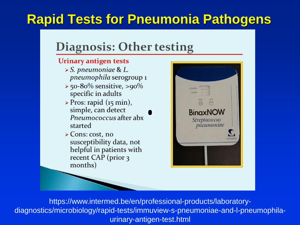
Rapid Tests for Pneumonia Pathogens
https://www.intermed.be/en/professional-products/laboratory-
diagnostics/microbiology/rapid-tests/immuview-s-pneumoniae-and-l-pneumophila-
urinary-antigen-test.html

Sepsis in an 88-year-Old
Okinawan-American Man with
New- Onset Asthma
An 82 yr Okinawan man with dyspnea
Born & raised Hawaii, visited Okinawa as
child, Italy WW II
1 yr. PTA diagnosed as asthma; treated
with bronchodilators and prednisone;
symptoms progressed, mental status
worsening.
Hospitalized, given high dose steroids for
asthma.
Patient Examination
Afebrile
Alert, moderately dyspneic
Bibasilar rhonchi
Remainder of exam unremarkable
Dx: Asthma/COPD exacerbation
Treated with higher doses of corticosteroids
and antimicrobial agents
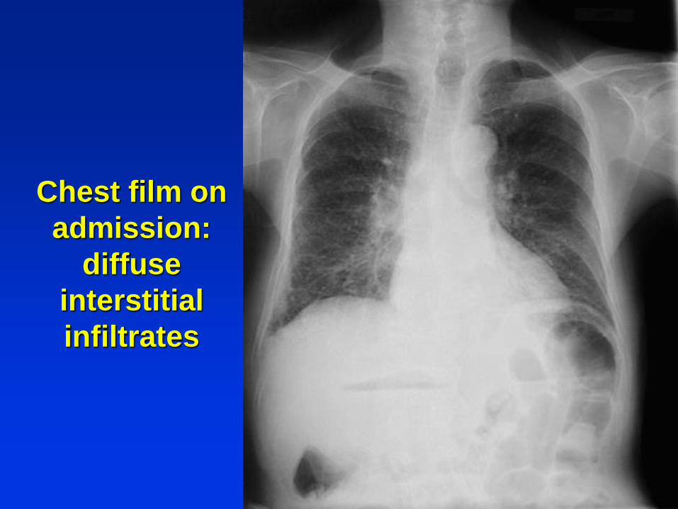
Chest film on
admission:
diffuse
interstitial
infiltrates
Patient Examination
Remains afebrile
2nd day: abdominal distension, sepsis,
coma, respiratory failure; intubated
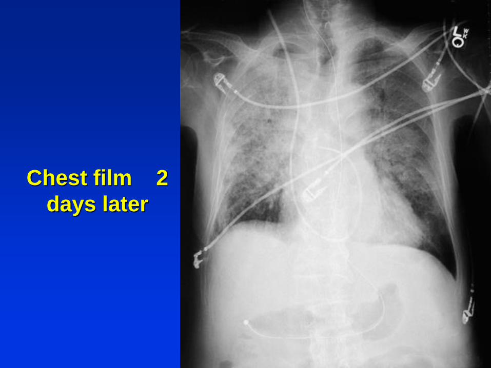
Chest film 2
days later
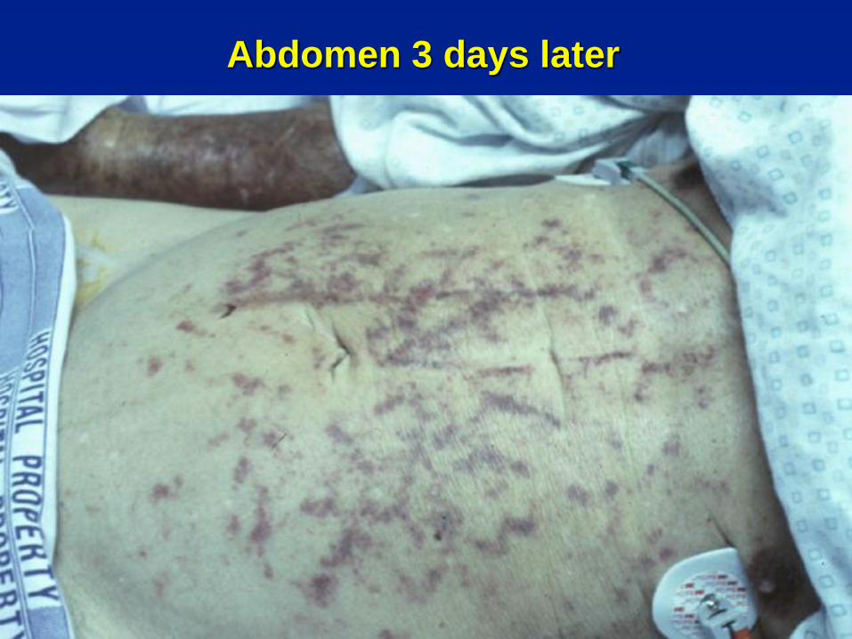
Abdomen 3 days later
Your assessment and plan
Differential Diagnosis ?
Management ?
BAL sputum sent for bacterial cultures and
cytology.
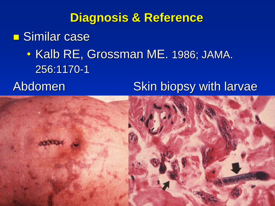
Diagnosis & Reference
Similar case
• Kalb RE, Grossman ME. 1986; JAMA.
256:1170-1
Abdomen Skin biopsy with larvae
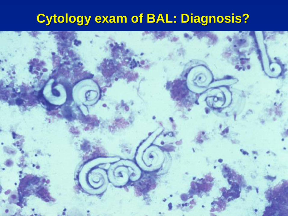
Cytology exam of BAL: Diagnosis?
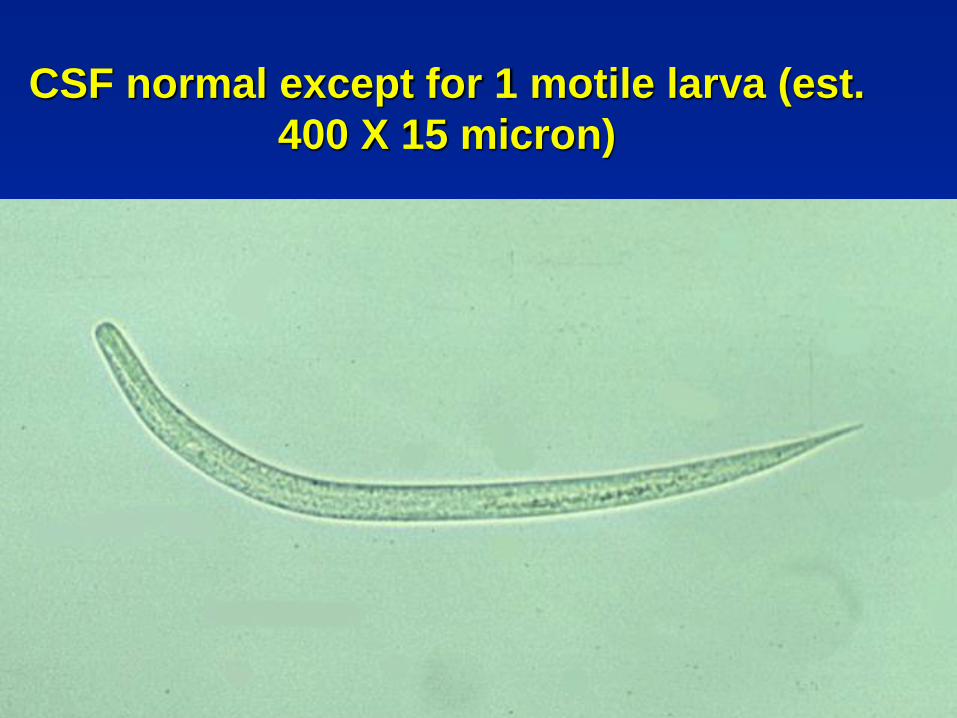
CSF normal except for 1 motile larva (est.
400 X 15 micron)
Diagnosis
Probable pulmonary strongyloidiaisis
manifesting as asthma for 1 yr.
Acut disseminated Strongyloides stercoralis
infection induced by high-dose
corticosteroid therapy for asthma
• Invasion of gut, skin, lung, CSF
• No CSF inflammation
Sepsis due to probable parasite-induced
enteric bacteremia
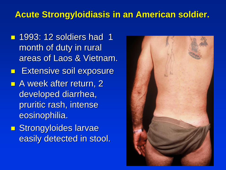
Acute Strongyloidiasis in an American soldier.
1993: 12 soldiers had 1
month of duty in rural
areas of Laos & Vietnam.
Extensive soil exposure
A week after return, 2
developed diarrhea,
pruritic rash, intense
eosinophilia.
Strongyloides larvae
easily detected in stool.
ACP Hawaii Chapter 2018 Poster 20
A SERIES OF UNFORTUNATE STRONGYLOIDES-
RELATED EVENTS Abigail Santos, MD1, Erin Crossey,
MD2, Therese Posas-Mendoza, MD3, James Joyner, MD4,
Heath Chung, MD
64 year-old man with vasculitis on chronic steroids ...was
re-admitted for acute respiratory failure ...and diffuse
alveolar hemorrhage {DAH) ... Repeat bronchoscopy was
performed and microscopy of BAL this time revealed
Strongyloides larvae. Ivermectin was initiated and steroids
were discontinued, however patient continued to clinically
deteriorate and ultimately expired.
Strongyloides infection is presumed to have been chronic
and subclinical given patient was from an endemic region
with no recent travel and no GI symptoms
Strongyloidiasis Cases in Hawaii
Strongyloides stercoralis (S.s.): an intestinal
helminth endemic in tropics, but not Hawaii.
Infection is lifelong and diagnosis is difficult.
Larvae may disseminate in steroid Rx hosts.
Four cases of dissemination at QMC 1996-98:
• All were immigrants from Asia-Pacific region
• 3 fatalities; hospital costs $ 157-350,000
A QMC review of clinical serodiagnostic studies
found latent infections in 10-40 % of Micronesians,
non-Hawaiian Polynesians, Filipinos, SE Asians.
Akiyama M, Brown JD. Human Strongyloidiasis in Hawaii: A Retrospective
Review of ELISA Serodiagnostic Testing. The American Journal of Tropical
Medicine and Hygiene, 2018:99(2) 370 – 374.

Disseminated Strongyloidiasis (DS)
True incidence of DS in developed world is
uncertain.
• Problem of under-diagnosis:
• Physician’s lack of knowledge
• Syndrome often non-specific
• Lack of patient geographic Hx
• Low index of suspicion
• Insensitivity of routine diagnostic tests
• 50% Dx at autopsy; low autopsy rates
Often fatal in spite of treatment.
Strongyloidiasis: Prevention
Before hi-dose steroid or other immunosuppressive
therapy, e.g. transplantation:
• Take immigration & travel Hx
• Serodiagnostic screening of patients who
resided in endemic areas, e.g. immigrants,
veterans.
• If sero +, ivermectin Rx 7d; rep. monthly
• ID consultation
If steroid therapy needed acutely, begin ivermectin
therapy while awaiting serodiagnostic test results.

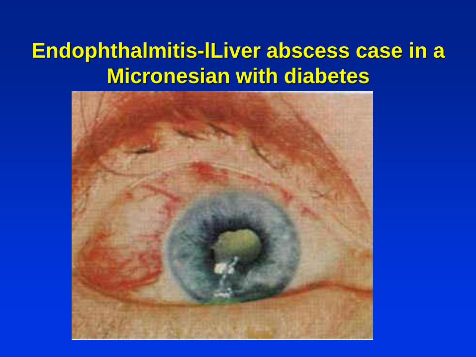
Endophthalmitis-lLiver abscess case in a
Micronesian with diabetes
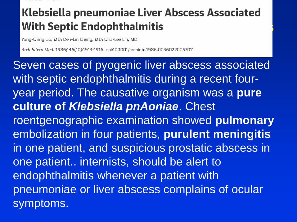
Klebsiella pneumoniae Liver Abscess
Associated With Septic Endophthalmitis
Seven cases of pyogenic liver abscess associated
with septic endophthalmitis during a recent four-
year period. The causative organism was a pure
culture of Klebsiella pnAoniae. Chest
roentgenographic examination showed pulmonary
embolization in four patients, purulent meningitis
in one patient, and suspicious prostatic abscess in
one patient.. internists, should be alert to
endophthalmitis whenever a patient with
pneumoniae or liver abscess complains of ocular
symptoms.
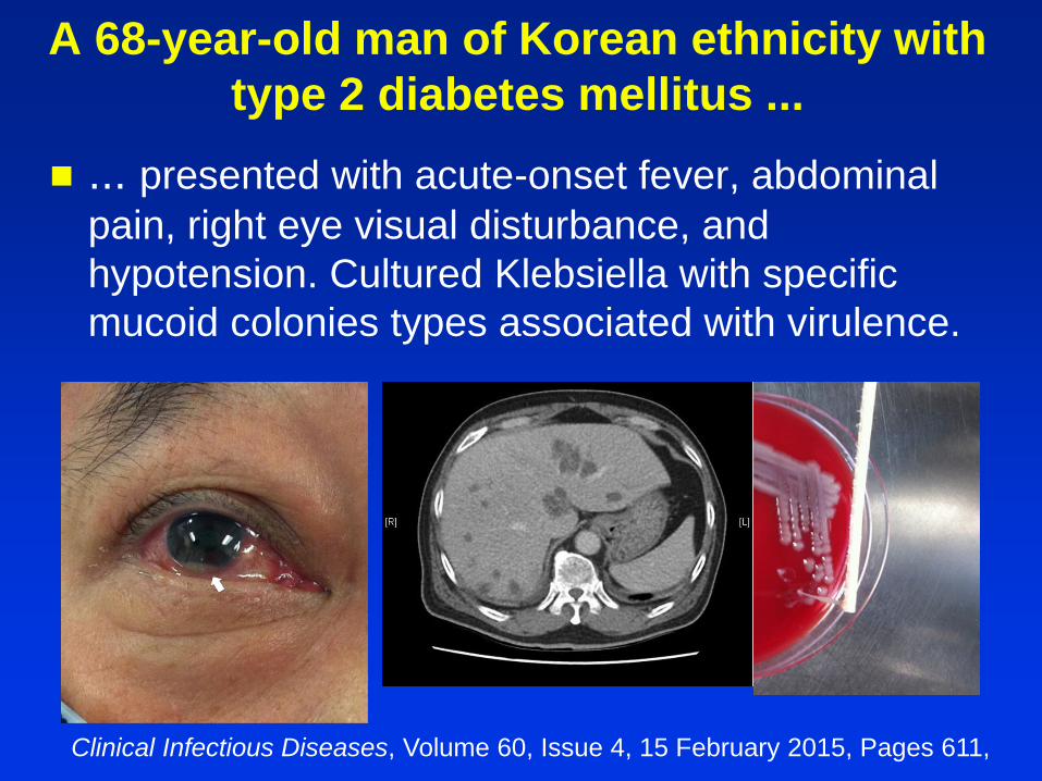
A 68-year-old man of Korean ethnicity with
type 2 diabetes mellitus ...
... presented with acute-onset fever, abdominal
pain, right eye visual disturbance, and
hypotension. Cultured Klebsiella with specific
mucoid colonies types associated with virulence.
Clinical Infectious Diseases, Volume 60, Issue 4, 15 February 2015, Pages 611,
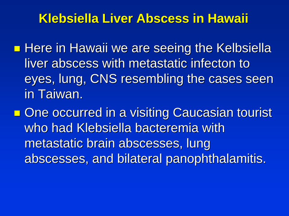
Klebsiella Liver Abscess in Hawaii
Here in Hawaii we are seeing the Kelbsiella
liver abscess with metastatic infecton to
eyes, lung, CNS resembling the cases seen
in Taiwan.
One occurred in a visiting Caucasian tourist
who had Klebsiella bacteremia with
metastatic brain abscesses, lung
abscesses, and bilateral panophthalamitis.
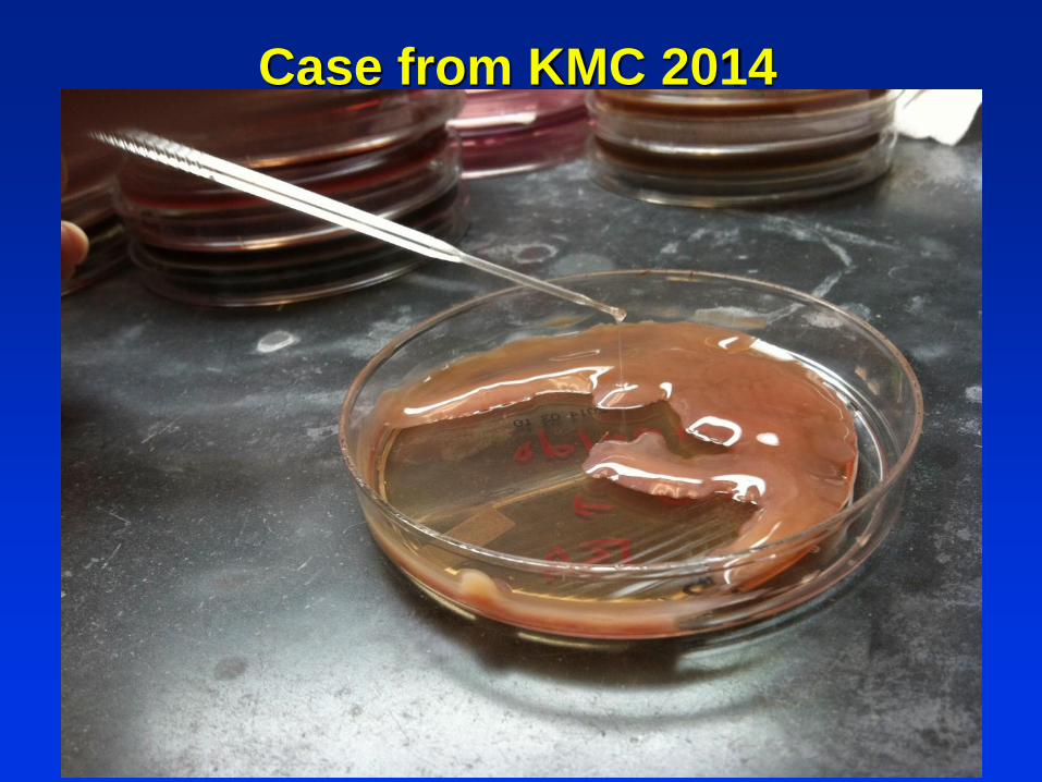
Case from KMC 2014

CONCLUSION
Tips for Diagnosing Infections
Who is the patient? Chronic diseases,
occupation, activities, travel, country of
origin, etc.
Clarify the symptoms the patient describes:
• Use the patient’s words.
• What improves/worsens, severity
(SOCRATES)
• Think anatomically.
What is the chronology of the acute illness?
“How did your problem begin”
Tell the patient’s story.
Tips for Diagnosing Infections
Create an acute problem list of all the
remarkable symptoms and signs, abnormal
labs, imaging, etc.
Symptom s A +B + C = Diagnosis
Create a differential Dx. for all the above.
• Don’t leave anything out.
• Don’t have parts left over.
• Consider the pros and cons for each
DDx.

Theme:
“The Art of Being an Internist”
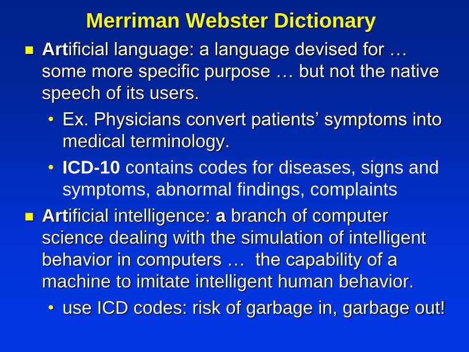
Merriman Webster Dictionary
Artificial language: a language devised for …
some more specific purpose … but not the native
speech of its users.
• Ex. Physicians convert patients’ symptoms into
medical terminology.
• ICD-10 contains codes for diseases, signs and
symptoms, abnormal findings, complaints
Artificial intelligence: a branch of computer
science dealing with the simulation of intelligent
behavior in computers … the capability of a
machine to imitate intelligent human behavior.
• use ICD codes: risk of garbage in, garbage out!
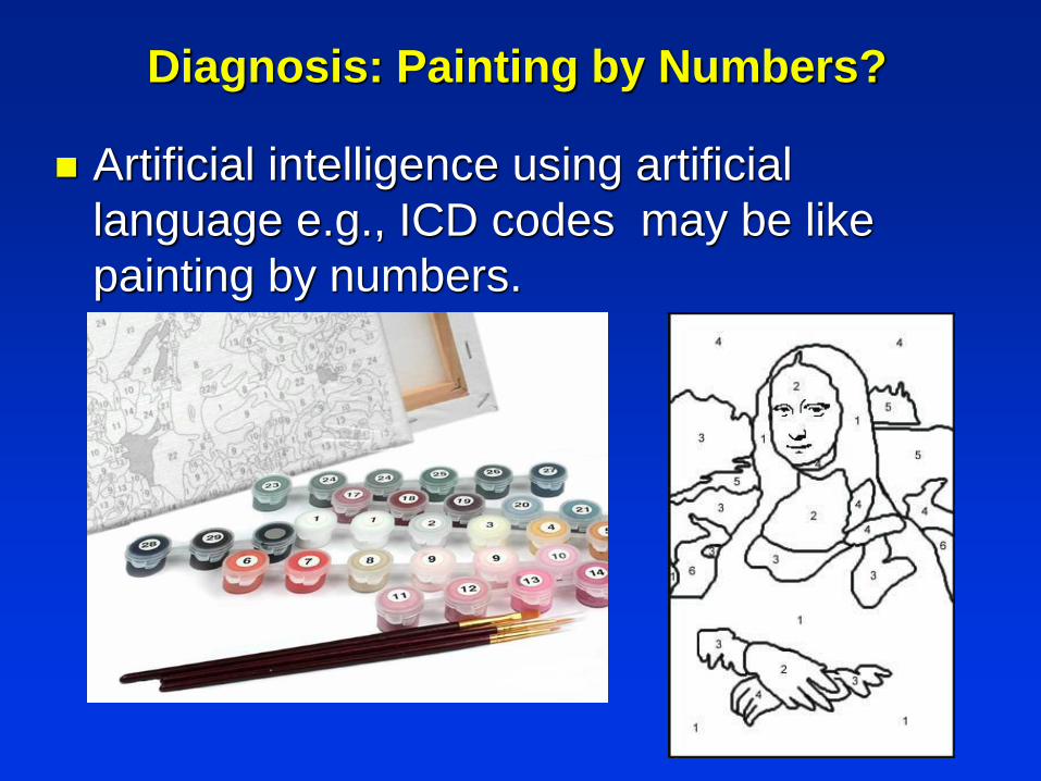
Diagnosis: Painting by Numbers?
Artificial intelligence using artificial
language e.g., ICD codes may be like
painting by numbers.

Art: another definition:
• a skill acquired by experience, study, or
observation
• an occupation requiring knowledge or
skill. Internal Medicine
• Thank you!