infection of west african dwarf rams with - research square
TRANSCRIPT
Page 1/21
Infection of West African Dwarf Rams WithTrypanosoma Brucei Brucei and TrypanosomaCongolense Signi�cantly Alter Serum Electrolytes,Redox Balance, Sperm Parameters, And GonadalMorphologyDavinson Chuka Anyogu ( [email protected] )
University of Nigeria, Nsukka https://orcid.org/0000-0003-2205-5400Shodeinde Vincent O. Shoyinka
University of Nigeria Faculty of Veterinary MedicineJohn Ikechukwu Ihedioha
University of Nigeria Faculty of Veterinary Medicine
Research Article
Keywords: Serum electrolytes, sperm parameters, Trypanosoma brucei brucei, Trypanosoma congolense,trypanotolerance, West African dwarf ram
Posted Date: November 12th, 2021
DOI: https://doi.org/10.21203/rs.3.rs-1017484/v1
License: This work is licensed under a Creative Commons Attribution 4.0 International License. ReadFull License
Version of Record: A version of this preprint was published at Veterinary Research Communications on April7th, 2022. See the published version at https://doi.org/10.1007/s11259-022-09921-8.

Page 2/21
AbstractTrypanotolerance of the West African dwarf (WAD) breeds may not rule out signi�cant pathophysiologicalchanges that may affect productivity. In this study, the effects of infection of WAD rams with Trypanosomabrucei brucei (Tbb) and Trypanosoma congolense (Tc) on their serum levels of electrolytes [calcium,phosphorus, sodium, potassium]; oxidative stress markers [superoxide dismutase (SOD), malondialdehyde(MDA)]; and sperm parameters [sperm count, motility, vitality, and morphology] were investigated. FifteenWAD rams, assigned to 3 groups (A, B & C) of 5 rams each, were used for the study. Group A rams wereinfected with Tbb, while Group B rams were infected with Tc, both intraperitoneally, at the dose of 106
trypanosomes/animal. Group C rams served as the uninfected control. The infections were monitored for 70days. Serum calcium levels were signi�cantly (p < 0.05) lower in Tbb and Tc infected rams compared to thecontrol throughout the study. Serum sodium was signi�cantly (p < 0.05) higher in the Tb infected ramscompared to the Tc infected and control rams on days 14 and 28 PI. Serum SOD activity decreased whileMDA levels increased in both infected groups of rams. Tbb infected rams were azoospermic, while Tcinfected rams had lower sperm motility, vitality and concentration and higher number of abnormal spermcells compared to the control. Necrotic and in�ammatory lesions occurred in the testis and epididymis ofboth infected rams. These results suggest that in spite of trypanotolerance, trypanosome infections in theWAD rams signi�cantly impact on health and reproduction.
IntroductionAfrican animal trypanosomosis (AAT) is a vector-borne parasitic disease of serious economic importance tolivestock production in sub-Saharan Africa, where the disease is endemic (Mulenga et al. 2020). It is causedby extracellular protozoa (trypanosomes), which are cyclically transmitted by several species of tsetse �y(Glossina spp.) (Serranito et al. 2021). The tsetse distribution area, or tsetse �y “belt,” covers 39 Africancountries, whose economic development is thus signi�cantly impacted (Alsan 2015). The effects of thedisease in livestock are mitigated by chemotherapeutic intervention, which is the most effective and widelyapplied method for the control of AAT in sub-Saharan Africa (Giordani et al. 2016). Other trypanosomosiscontrol methods involving the use of trypanotolerant livestock and direct tsetse control are sparingly utilizedin Africa owing to their expensive and laborious nature (Obi et al. 2019).
Routinely, little importance is attached to trypanosomosis in small ruminants compared to cattle (Gri�n1978). Most available data on the disease in small ruminants are sourced from abattoir reports (Kalejaiye etal. 1995; Nawathe et al. 1995; Dadah et al. 1997). On the other hand, it has been suggested that greateconomic losses occur in small ruminants due to trypanosome infections (Gri�n 1978; Gri�n and Allonby1979; Kalu and Edeghere 1985). Also, these animals have been incriminated as sources/reservoirs ofinfection to other animals and man (Mahmaud and Elmalik 1977; Scott et al. 1983). This neglect may be dueto the misplaced emphasis on trypanotolerance. It was estimated that approximately 32 % of sheep and 47 %of goats in West and Central Africa are trypanotolerant and this is so because the West African dwarf (WAD)breeds are predominant in these regions (Agyemang 2005; Geerts et al. 2009). Trypanotolerance is de�ned asa multigenic trait that confers on some breeds of cattle, sheep and goats the ability to survive, reproduce andremain productive under trypanosomosis risk without the need for the use of chemicals to control the vector
Page 3/21
or drugs to control the parasite, while susceptible breeds die without treatments (Murray et al. 1984; d’Ieterenet al. 1998; Geerts et al. 2009). It had been reported that phenotypically, trypanotolerant livestock are able tocontrol parasitaemia and anaemia and are less affected by weight loss during infection (Hanotte et al. 2003;Berthier et al. 2016). In spite of trypanotolerance, however, pathological changes that may signi�cantlyimpact on productivity may occur in trypanosome infected WAD breeds.
Calcium, phosphorus, sodium and potassium are electrolytes with diverse biological functions and which areessential for optimal growth, health and reproduction in mammals (Yasothai 2014; Escalera-Valente et al.2021). These electrolytes participate in the formation of bones and teeth, muscle contraction, permeability ofcell membranes, blood clotting, enzymatic reactions, secretion of hormones, metabolism of lipids,carbohydrates and proteins, genetic transmission of nucleic acids, maintenance of normal osmotic pressure,and neural transmission (Devlin 1992; McDowell 1992; Shaker and Deftos 2000). As regards reproductivefunction, while extracellular sodium, Na (+), suppresses hyper-activation of sperm motility via Na (+) – Ca(2+) exchanger (Takei and Fujinoki 2016), calcium is necessary for spermatogenesis, sperm motility,capacitation, acrosome reaction and fertilization (Li et al. 2016; Beigi Harchegani et al. 2019).
The fact that imbalances in these mineral elements coupled with oxidative stress, which also affects spermmorphology and physiology, may impact negatively on optimal health and productivity in mammals makes itpertinent to evaluate them in trypanosome-infected WAD rams. Such study may unveil “silent” but signi�cantimpacts of the disease in the WAD rams in regions where surveillance, vector control and chemotherapeuticinterventions are overlooked while trypanotolerance is emphasized. Moreover, in susceptible animals,trypanosomes have been reported to cause testicular degeneration and in�ammation, subnormal spermcount and structural damage to spermatozoa (Wada et al. 2016; Amin et al. 2020). If similar reproductivelesions occur in the trypanotolerant WAD rams, it would imply that trypanotolerance of the WAD sheep maybe overrated. The objective of this study was, therefore, to evaluate the effects of T. brucei brucei and T.congolense infections on some serum electrolytes, oxidative stress markers, sperm parameters, serumtestosterone levels, and gonadal morphology in WAD rams.
Materials And MethodsTrypanosomes species/strain used
The trypanosome parasites used were the Federer strains of T. brucei brucei and T. congolense (Savannahsubgroup), which were sourced from the Nigerian Institute for Trypanosomiasis Research (NITR), KadunaState, Nigeria and maintained in rats via passages.
Animals
Fifteen West African dwarf rams were used for the study. They were procured from local breeders in NsukkaNigeria, and allowed to acclimatize for 3 weeks, during which period they were dewormed. All the rams werescreened for trypanosomes and/or other haemoparasites, by buffy coat examination. They were placed onfresh forage and drinking water ad libitum. They were further randomly assigned to three groups (A, B & C) of�ve rams each. Group A rams were infected with T. brucei brucei, while Group B rams were infected with T.
Page 4/21
congolense, both intraperitoneally, at the dose of 106 trypanosomes/animal. Group C rams served as theuninfected control.
Estimation of parasitaemia
Parasitaemia was checked daily, beginning from day 2 post-infection of groups A and B. Once the infectionwas established, the levels of parasitaemia in the infected groups were determined weekly by wet mount andscored by rapid matching method (Paris et al. 1982; Herbert and Lumsden 1976).
Serum sample collection and analyses
Blood samples were collected before infection (day 0) and on days 14, 28, 42, 56 & 70 post infection (PI). Theblood samples collected (4 ml) were dispensed into clean labelled plain test tubes and allowed 45 minutes toclot. Sera used for analyses were obtained by centrifuging the clotted blood in the test tubes at 3000revolutions per minute (rpm) for 10 minutes. The serum samples were split into two aliquots and refrigerated.One aliquot was used for the assay of calcium, phosphorus, sodium, potassium, superoxide dismutase(SOD) and malondialdehyde (MDA), using commercially available test kits and a Diatek Blood ChemistryAnalyzer (Diatek Instruments Co. Ltd., Wuxi, China) following manufacturers’ instructions. The other aliquotwas used to assay for serum testosterone concentration using sheep-speci�c testosterone test kit(MBS701270) (Mybiosource Inc., California, USA).
Speci�cally, serum sodium was determined by the magnesium-uranyl acetate method (Scott et al. 2008),using a sodium test kit (Teco Diagnostics, Anaheim California, USA). The serum potassium was determinedby the direct turbidimetric spectrophotometric method (Hillman and Beyer 1967; Scott et al. 2008), using apotassium test kit (Teco Diagnostics, Anaheim, California, USA). Serum calcium levels were determined bythe ortho- cresolphthalein direct method (Connerty and Briggs 1966; Endres and Rude 2008), while the levelof phosphorus in serum was evaluated based on the Fiske-SubbaRow method (Fiske and SubbaRow 1925;Goodwin 1970; Endres and Rude 2008), using Quimica Clinica Applicada (QCA) Calcium and inorganicPhosphorus test kits (QCA, S. A. Spain), respectively. Serum superoxide dismutase (SOD) activity wasevaluated by hydroxylamine method (Weydert and Cullen 2010), while serum malondialdehyde levels wasmeasured by the modi�ed thiobarbituric acid method (Plaser and Cushman 1966; Draper and Hadley 1990),using the Elabscience SOD and Malondialdehyde Assay Kits (Elabscience Biotechnology Co. Ltd., SouthAfrica), respectively. All the analyses were completed within 24 hours of sample collection.
Epididymal sperm evaluation: motility, morphology, vitality and concentration
Three rams from each group were castrated on day 70 PI under lignocaine anaesthesia. Both testes andepididymis were removed from each ram. Sperm motility was determined using the sperm diffusion method(Seed et al. 1996). Brie�y, the epididymis was dissected from the testis. The cauda epididymis was sectionedfrom the vas deferens and the end of the tubule segment was immersed in a drop of pre-warmed phosphatebuffered saline (PBS; pH 7.4, 37 °C) on a clean glass slide to facilitate sperm dispersion into the buffer. Thesperm cells were allowed to diffuse into the medium for 2 min. The tissue was removed, and the sperm cellswere incubated for 5 minutes until adequately dispersed for analysis. Sperm motility (%) was evaluated byexamining the sample at x100 magni�cation using a phase-contrast microscope (Motic B3; Motic, Carlsbad,

Page 5/21
CA, USA) equipped with a stage slide warmer set at 37 °C (TCS-100; Amscope, Ivrine, CA, USA). A total of 200sperm cells were counted and the number of motile sperm cells recorded as percentage epididymal spermmotility.
Sperm vitality was determined after staining with eosin-nigrosin vital stain (Seed et al. 1996). Brie�y, equalvolume of caudal epididymal sperm suspension and eosin-nigrosin stain were mixed for 30 seconds and athin smear made on a microscope slide and air-dried. Live sperm (unstained head) and dead sperm (red-stained head) were identi�ed using light microscopy (Motic B3; Motic, Carlsbad, CA, USA) at x1000magni�cation under oil immersion. A total of 200 sperm cells were counted and the number of live spermcells recorded as percentage vitality of the sample. All micrographs were captured using Moticam 2.0 imagesystem (Motic, Carlsbad, CA, USA).
For sperm morphology, a wet mount of caudal epididymal sperm suspension was evaluated for spermmorphology and structural abnormalities at x1000 magni�cation using phase-contrast microscopy (MoticB3; Motic, Carlsbad, CA, USA). Sperm morphology was also determined using eosin-nigrosin staining method.A total of 200 sperm cells were counted and the number of abnormal sperm cells was recorded. This wasexpressed in percentage as epididymal total sperm abnormalities (TSA) (Seed et al. 1996).
To determine the sperm concentration, the cauda epididymal tissue was thoroughly minced andhomogenized in 2 ml of PBS (pH 7.4). A 1:200 dilution of the homogenate was made in sperm dilution �uidcontaining formalin and gentian violet stain followed by counting of sperm cells using a haemocytometer(Weber, England). Total sperm count was expressed as number of spermatozoa/g epididymal tissue (Seed etal. 1996).
Histopathology
The testes were �xed in Bouin’s �xative for 8 hours before transferring them to 70 % alcohol. Theepididymides were �xed in 10 % neutral-buffered formalin for 48 hours. Both tissues were routinely processedand sectioned at 5µm thickness and stained with haematoxylin and eosin (H&E).
Statistical analysis
The data were subjected to one way analysis of variance (ANOVA) using SPSS version 21. Parasitaemiascores were analysed by Student’s t-test. The variant means were separated post hoc using the leastsigni�cant difference method. Probability, p < 0.05 was considered statistically signi�cant.
ResultsParasitaemia
All the infected rams became parasitaemic between days 5 and 6 PI and remained parasitaemic till the endof the study. Group A rams were signi�cantly (p < 0.05) more parasitaemic than Group B rams on days 14, 35and 56 PI (Figure 1).
Serum minerals and electrolytes
Page 6/21
The mean serum calcium concentration of the infected rams were signi�cantly (p < 0.05) lower than that ofthe uninfected control throughout the experiment, starting from day 14 PI for the T. brucei brucei infectedrams and day 28 PI for T. congolense infected rams (Figure 2).
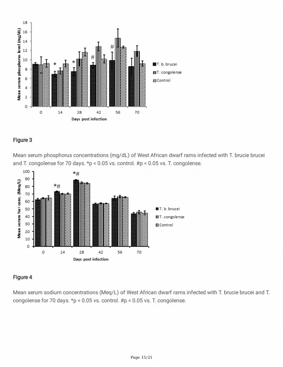
Serum phosphorus levels of the T. brucei brucei infected group was signi�cantly (p < 0.05) lower than thecontrol on days 14 and 28 PI. On days 42 and 56 PI, the mean serum phosphorus level of the T. congolenseinfected group became signi�cantly (p < 0.05) higher than that of the T. brucei brucei infected group (Figure3). Calcium/phosphorus ratio (Ca:P) was not affected in the T. brucei brucei infected rams (normal Ca:P = 1-2:1) but tilted towards hyperphosphataemia in T. congolense infected rams, from day 28 PI (Ca:P = 1:1.2) today 70 PI (Ca:P = 1:1.5).
Serum sodium levels of Group A (T. brucei brucei) rams was signi�cantly (p < 0.05) higher than that of GroupB (T. congolense) and C (Control) on days 14 and 28 PI. Beyond day 28 PI, there were no signi�cantvariations in serum sodium concentrations across the groups (Figure 4). Potassium concentration in serumdid not also vary signi�cantly (p > 0.05) across the groups throughout the period of the experiment.
Oxidative stress markers
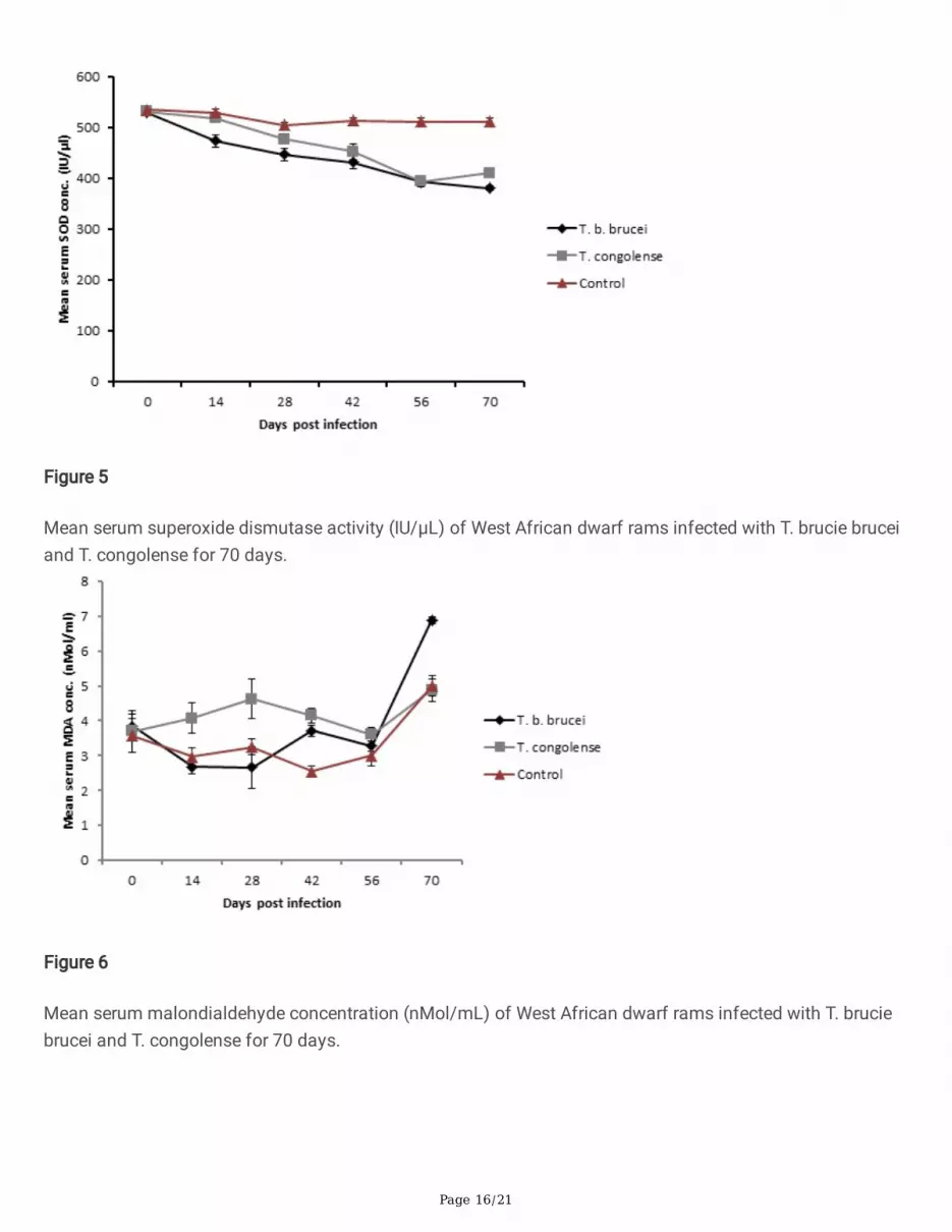
The mean serum superoxide dismutase (SOD) activity of the T. brucei brucei infected group was signi�cantly(p < 0.05) lower than those of both the T. congolense infected group and the uninfected control on days 14,28 and 42 PI. However, by days 56 and 70 PI, it was signi�cantly (p < 0.05) lower than only that of the controlgroup. On the other hand, the mean serum SOD activity of the T. congolense infected group was signi�cantly(p < 0.05) lower than that of the control from day 28 PI to the termination of the experiment (Figure 5).
The mean serum malondialdehyde (MDA) levels of the T. brucei brucei infected group was signi�cantly (p <0.05) higher than that of the control group by days 42 and 70 PI. Similarly, the mean serum MDA levels of theT. congolense infected group was signi�cantly (p < 0.05) higher than the control by days 14 and 42 PI andalso signi�cantly (p < 0.05) higher than that of Group A by days 14 and 28 PI (Figure 6).
Sperm parameters: concentration, motility, vitality, and morphology
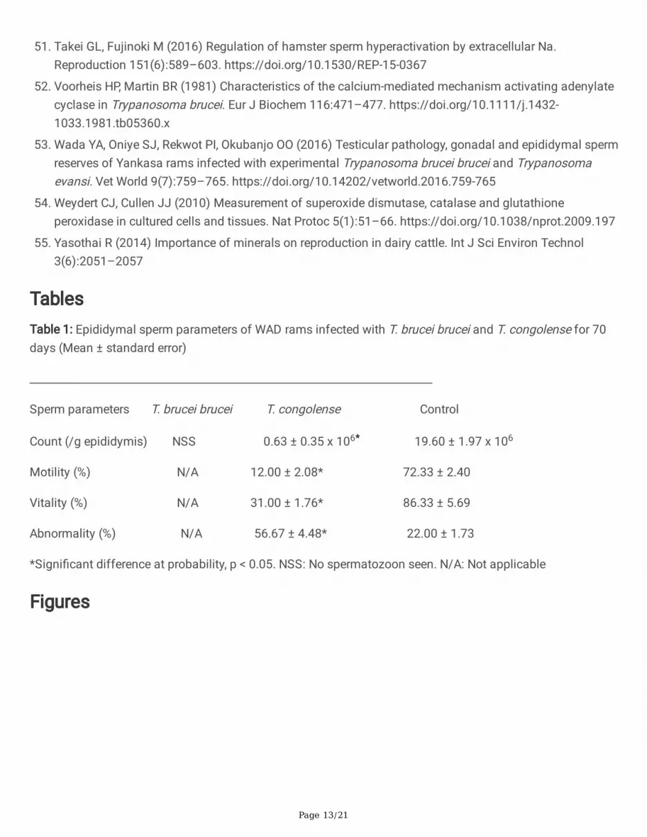
No spermatozoon was found in the epididymal �uid of the T. brucei brucei infected rams (Group A). Spermcells were present in the epididymides of Groups B and C rams. The mean serum epididymal sperm count ofT. congolense infected rams (Group B) was signi�cantly (p < 0.05) lower than that of the control (Group C).The mean percentage sperm motility and vitality of the Group B rams were signi�cantly (p < 0.05) lower thanthose of the control, while the mean percentage abnormality of spermatozoa of Group B rams weresigni�cantly (p < 0.05) higher than that of the control group (Table 1).
Stained smear of the epididymal �uid of Group A rams did not reveal any sperm cells, only necrotic debriswas observed. Motile trypanosomes (T. congolense) were found in the epididymal semen of Group B rams(Figure 7A). The sperm morphological abnormalities observed in Group B rams were headless sperm cells,dead sperm cells, coil-tailed spermatozoa, short-tailed spermatozoa, and cytoplasmic droplets (Figure 7B).Group B had fewer and mainly abnormal sperms cells (Figure 7B), while Group C was a mixture of normaland abnormal sperm cells (Figure 7C).
Page 7/21
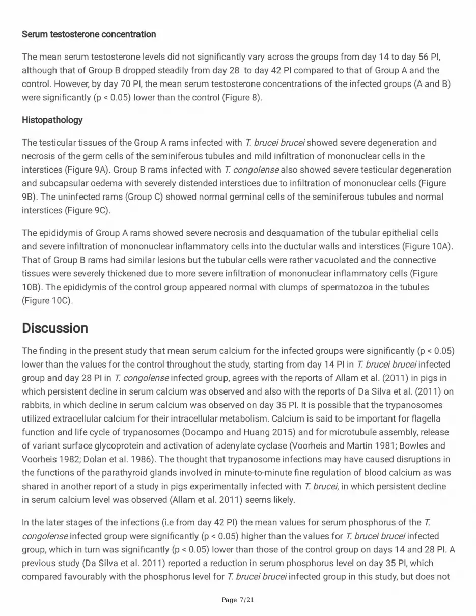
Serum testosterone concentration
The mean serum testosterone levels did not signi�cantly vary across the groups from day 14 to day 56 PI,although that of Group B dropped steadily from day 28 to day 42 PI compared to that of Group A and thecontrol. However, by day 70 PI, the mean serum testosterone concentrations of the infected groups (A and B)were signi�cantly (p < 0.05) lower than the control (Figure 8).
Histopathology
The testicular tissues of the Group A rams infected with T. brucei brucei showed severe degeneration andnecrosis of the germ cells of the seminiferous tubules and mild in�ltration of mononuclear cells in theinterstices (Figure 9A). Group B rams infected with T. congolense also showed severe testicular degenerationand subcapsular oedema with severely distended interstices due to in�ltration of mononuclear cells (Figure9B). The uninfected rams (Group C) showed normal germinal cells of the seminiferous tubules and normalinterstices (Figure 9C).
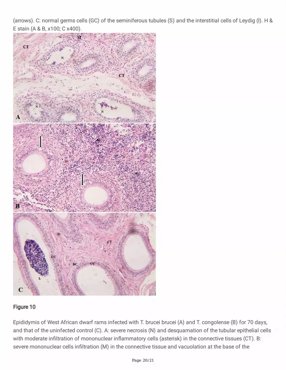
The epididymis of Group A rams showed severe necrosis and desquamation of the tubular epithelial cellsand severe in�ltration of mononuclear in�ammatory cells into the ductular walls and interstices (Figure 10A).That of Group B rams had similar lesions but the tubular cells were rather vacuolated and the connectivetissues were severely thickened due to more severe in�ltration of mononuclear in�ammatory cells (Figure10B). The epididymis of the control group appeared normal with clumps of spermatozoa in the tubules(Figure 10C).
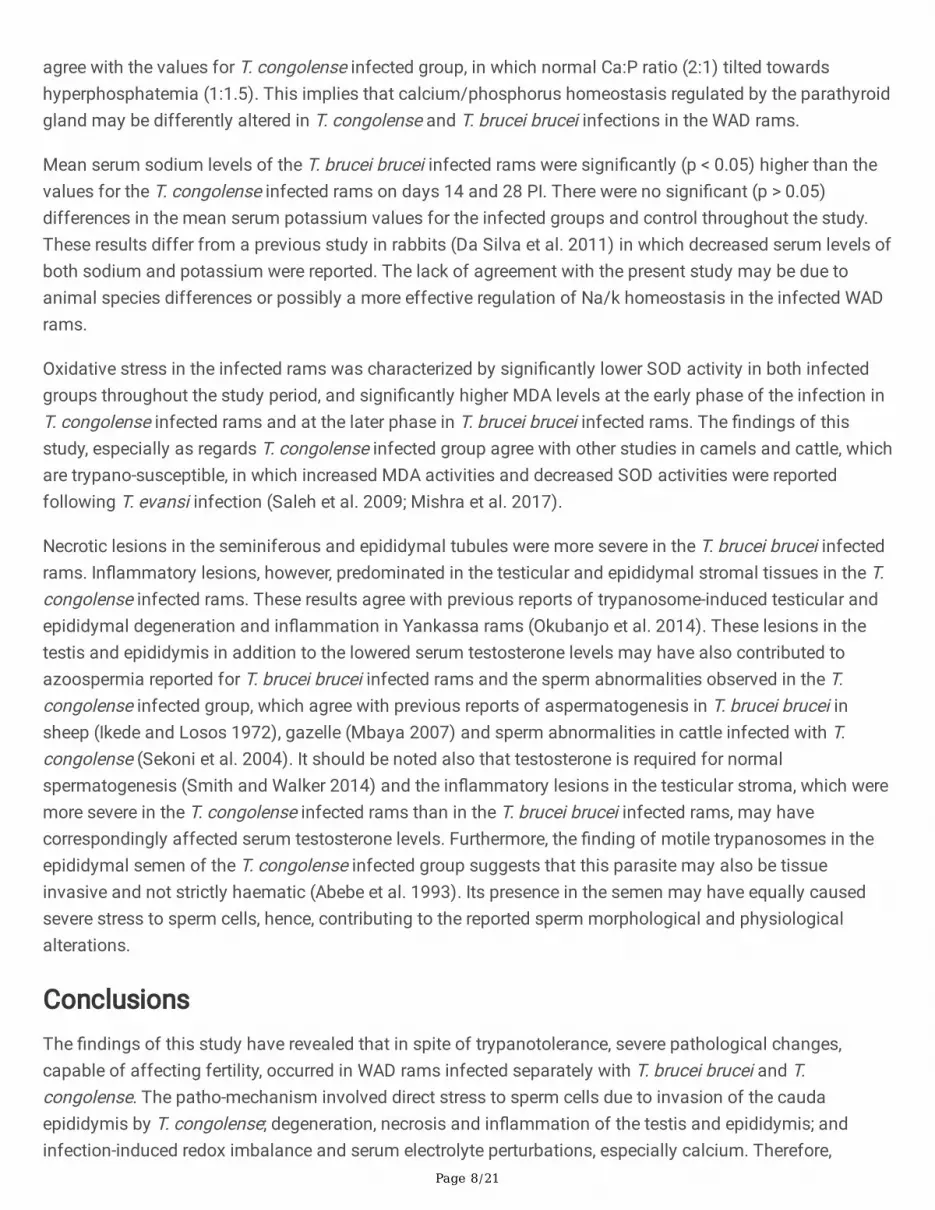
DiscussionThe �nding in the present study that mean serum calcium for the infected groups were signi�cantly (p < 0.05)lower than the values for the control throughout the study, starting from day 14 PI in T. brucei brucei infectedgroup and day 28 PI in T. congolense infected group, agrees with the reports of Allam et al. (2011) in pigs inwhich persistent decline in serum calcium was observed and also with the reports of Da Silva et al. (2011) onrabbits, in which decline in serum calcium was observed on day 35 PI. It is possible that the trypanosomesutilized extracellular calcium for their intracellular metabolism. Calcium is said to be important for �agellafunction and life cycle of trypanosomes (Docampo and Huang 2015) and for microtubule assembly, releaseof variant surface glycoprotein and activation of adenylate cyclase (Voorheis and Martin 1981; Bowles andVoorheis 1982; Dolan et al. 1986). The thought that trypanosome infections may have caused disruptions inthe functions of the parathyroid glands involved in minute-to-minute �ne regulation of blood calcium as wasshared in another report of a study in pigs experimentally infected with T. brucei, in which persistent declinein serum calcium level was observed (Allam et al. 2011) seems likely.
In the later stages of the infections (i.e from day 42 PI) the mean values for serum phosphorus of the T.congolense infected group were signi�cantly (p < 0.05) higher than the values for T. brucei brucei infectedgroup, which in turn was signi�cantly (p < 0.05) lower than those of the control group on days 14 and 28 PI. Aprevious study (Da Silva et al. 2011) reported a reduction in serum phosphorus level on day 35 PI, whichcompared favourably with the phosphorus level for T. brucei brucei infected group in this study, but does not
Page 8/21
agree with the values for T. congolense infected group, in which normal Ca:P ratio (2:1) tilted towardshyperphosphatemia (1:1.5). This implies that calcium/phosphorus homeostasis regulated by the parathyroidgland may be differently altered in T. congolense and T. brucei brucei infections in the WAD rams.
Mean serum sodium levels of the T. brucei brucei infected rams were signi�cantly (p < 0.05) higher than thevalues for the T. congolense infected rams on days 14 and 28 PI. There were no signi�cant (p > 0.05)differences in the mean serum potassium values for the infected groups and control throughout the study.These results differ from a previous study in rabbits (Da Silva et al. 2011) in which decreased serum levels ofboth sodium and potassium were reported. The lack of agreement with the present study may be due toanimal species differences or possibly a more effective regulation of Na/k homeostasis in the infected WADrams.
Oxidative stress in the infected rams was characterized by signi�cantly lower SOD activity in both infectedgroups throughout the study period, and signi�cantly higher MDA levels at the early phase of the infection inT. congolense infected rams and at the later phase in T. brucei brucei infected rams. The �ndings of thisstudy, especially as regards T. congolense infected group agree with other studies in camels and cattle, whichare trypano-susceptible, in which increased MDA activities and decreased SOD activities were reportedfollowing T. evansi infection (Saleh et al. 2009; Mishra et al. 2017).
Necrotic lesions in the seminiferous and epididymal tubules were more severe in the T. brucei brucei infectedrams. In�ammatory lesions, however, predominated in the testicular and epididymal stromal tissues in the T.congolense infected rams. These results agree with previous reports of trypanosome-induced testicular andepididymal degeneration and in�ammation in Yankassa rams (Okubanjo et al. 2014). These lesions in thetestis and epididymis in addition to the lowered serum testosterone levels may have also contributed toazoospermia reported for T. brucei brucei infected rams and the sperm abnormalities observed in the T.congolense infected group, which agree with previous reports of aspermatogenesis in T. brucei brucei insheep (Ikede and Losos 1972), gazelle (Mbaya 2007) and sperm abnormalities in cattle infected with T.congolense (Sekoni et al. 2004). It should be noted also that testosterone is required for normalspermatogenesis (Smith and Walker 2014) and the in�ammatory lesions in the testicular stroma, which weremore severe in the T. congolense infected rams than in the T. brucei brucei infected rams, may havecorrespondingly affected serum testosterone levels. Furthermore, the �nding of motile trypanosomes in theepididymal semen of the T. congolense infected group suggests that this parasite may also be tissueinvasive and not strictly haematic (Abebe et al. 1993). Its presence in the semen may have equally causedsevere stress to sperm cells, hence, contributing to the reported sperm morphological and physiologicalalterations.
ConclusionsThe �ndings of this study have revealed that in spite of trypanotolerance, severe pathological changes,capable of affecting fertility, occurred in WAD rams infected separately with T. brucei brucei and T.congolense. The patho-mechanism involved direct stress to sperm cells due to invasion of the caudaepididymis by T. congolense; degeneration, necrosis and in�ammation of the testis and epididymis; andinfection-induced redox imbalance and serum electrolyte perturbations, especially calcium. Therefore,

Page 9/21
trypanosome infections in the trypanotolerant WAD rams signi�cantly affect health and reproduction even inthe absence of mortality or clear signs of morbidity.
DeclarationsAcknowledgements: The authors are grateful to the Nigerian Institute for Trypanosomiasis Research (NITR)for donating the trypanosomes used for the study; we thank Mr. I.O. Agbakwuru for assisting in tissueprocessing, and Foundation for Education and Research in Health (FERH), Nsukka, Nigeria for bench spaceand other technical assistance provided.
Authors’ contributions: Conceptualization: [Davinson C Anyogu, Shodeinde VO Shoyinka, John I Ihedioha];Methodology: [Davinson C Anyogu, John I Ihedioha]; Formal analysis and investigation: [Davinson C Anyogu, John I Ihedioha]; Writing—original draft preparation: [Davinson C Anyogu]; Writing—review and editing: [Davinson C Anyogu, John I Ihedioha,Shodeinde VO Shoyinka]; Resources: [Davinson C Anyogu, John I Ihedioha, Shodeinde VO Shoyinka];Supervision: [Shodeinde VO Shoyinka, John I Ihedioha].
Ethics approval: Approval was obtained from the Institutional Animal Care and Use Committee (IACUC) of theFaculty of Veterinary Medicine, University of Nigeria, Nsukka (Approval Reference Number: FVM-UNN-IACUC-2020-0345). All efforts were made to minimise the discomfort of the animals during sampling.
Consent to participate: All authors participated voluntarily in the research.
Consent for publication: All authors read and approved the �nal manuscript.
Con�icts of interest/Competing interest: The authors declare no con�ict of interest.
Funding
This study was supported by Institutional Based Research (IBR) grant from Tertiary Education Trust Fund(TETFund/DESS/UNI/NSUKKA/2018/RP/VOL.1).
References1. Abebe G, Eley RM, ole-MoiYoi OK (1993) Reduced responsiveness of the hypothalamic-pituitary-adrenal
axis in Boran (Bos inducus) cattle infected with Trypanosoma congolense. Acta Endocrinol (Copenh)129(1):75–80. https://doi.org/10.1530/acta.0.1290075
2. Agyemang K (2005) Trypanotolerant livestock in the context of trypanosomiasis intervention strategies.PAAT Techn Sci Ser 7:1–66
3. Allam L, Ogwu D, Agbede RIS, Sackey AKB (2011) Haematological and serum biochemical changes ingilts experimentally infected with Trypanosoma brucei. Vet Arh 81(5):597–609
4. Alsan M (2015) The effect of the tsetse �y on African development. Am Economic Rev 105:382–410.https://doi.org/10.1257/aer.20130604

Page 10/21
5. Amin YA, Noseer EA, Fouad SS, Ali RA, Mahmoud H (2020) Changes of reproductive indices of the testisdue to Trypanosoma evansi infection in dromedary bulls (Camelus dromedarius): Semen picture,hormonal pro�le, histopathology, oxidative parameters, and hematobiochemical pro�le. J Adv Vet AnimRes 7(3):537–545. https://doi.org/10.5455/javar.2020.g451
�. Beigi Harchegani A, Irandoost A, Mirnamniha M, Rahmani H, Tahmasbpour E, Shahriary A (2019)Possible Mechanisms for the Effects of Calcium De�ciency on Male Infertility. Int J Fertil Steril12(4):267–272. https://doi.org/10.22074/ijfs.2019.5420
7. Berthier D, Brenière SF, Bras-Gonçalves R, Lemesre JL, Jamonneau V, Solano P et al (2016) Tolerance totrypanosomatids: A threat, or a key for disease elimination? Trends Parasitol 32:157–168.https://doi.org/10.1016/j.pt.2015.11.001
�. Bowles DJ, Voorheis HP (1982) Release of the surface coat from the plasma membrane of intactbloodstream forms of Trypanosoma brucei requires Ca2+. FEBS Lett 139: 17-21.https://doi.org/1016/0014-5793(82)80477-8
9. Connerty HV, Briggs AR (1966) Determination of serum calcium by means of ortho-cresolphthaleincomplexone. Am J Clin Pathol 45(3):290–296. https://doi.org/10.1093/ajcp/45.3.290
10. Da Silva AS, Costa MM, Moreira CM, Zenette RA, Thome GR, Otto MA, Flores EMM, Lopes STA, MonterioSG (2011) Experimental infection by Trypanosome evansi in rabbits: Levels of sodium, potassium,calcium and phosphorus in serum. Acta Sci Vet 39(2):959
11. Dadah AJ, Duhlinska-Popova DD, Daniel AD, Dede PM (1997) Trypanosomosis among sheep and goatsat slaughter in Jos abattoir. Nigeria Rev élev méd vét pays trop (En ligne) 50:214–216
12. Devlin TM (1992) Text book of Biochemistry with Clinical Correlations. John Wiley & Sons Inc., New York,pp 289–1185
13. d'Ieteren GD, Authié E, Wissocq N, Murray M (1998) Trypanotolerance, an option for sustainable livestockproduction in areas at risk from trypanosomosis. Rev Sci Tech 17(1):154–175.https://doi.org/10.20506/rst.17.1.1088
14. Docampo R, Huang G (2015) Calcium signalling in trypanosomatid parasites. Cell Calcium 57(3):194–202. https://doi.org/10.1016/j.ceca.2014.10.015
15. Dolan MT, Reid CG, Voorheis HP (1986) Calcium ions initiate the selective development of the pellicularmicrotubules in bloodstream forms of Trypanosome brucei. J Cell Sci 80:123–140
1�. Draper HH, Hadley M (1990) Malondialdehyde determination as an index of lipid peroxidation. MethodsEnzymol 186:421–431. https://doi.org/10.1016/0076-6879(90)86135-i
17. Endres DB, Rude RK (2008) Measurement of calcium. In: Burtis CA, Ashwood ER, Bruns DE (eds) TietzFundamentals of Clinical Chemistry, 6th edn. Missouri, Saunders Elsevier, pp 715–717
1�. Escalera-Valente F, Alonso ME, Lomillos JM, Gaudioso VR, Alonso ÁJ, González-Montaña JR (2021)Effect of Intense Exercise on Plasma Macrominerals and Trace Elements in Lidia Bulls. Vet Sci 8(6):97.https://doi.org/10.3390/vetsci8060097
19. Fiske CH, SubbaRow Y (1925) The colorimetric determination of phosphorus. J Biol Chem 66:375–400
20. Geerts S, Osaer S, Goossens B, Faye D (2009) Trypanotolerance in small ruminants of sub-SaharanAfrica. Trends Parasitol 25(3):132–138. https://doi.org/10.1016/j.pt.2008.12.004

Page 11/21
21. Giordani F, Morrison LJ, Rowan TG, De Koning HP, Barrett MP (2016) The animal trypanosomiases andtheir chemotherapy: a review. Parasitology 143:1862–1889.https://doi.org/10.1017/S0031182016001268
22. Goodwin JF (1970) Quanti�cation of serum inorganic phosphorus, phosphatase and urinary phosphatewithout preliminary treatment. Clin Chem 16(9):776–780
23. Gri�n L (1978) African trypanosomiasis in sheep and goats: a review. Vet Bull 48:819–823
24. Gri�n L, Allonby EW (1979) The economic effects of trypanosomiasis in sheep and goats in a rangeresearch station in Kenya. Trop Anim Health Prod 11:127–132. https://doi.org/10.1007/BF02237788
25. Hanotte O, Ronin Y, Agaba M, Nilsson P, Gelhaus A, Horstmann R et al (2003) Mapping of quantitativetrait loci controlling trypanotolerance in a cross of tolerant West African N’Dama and susceptible EastAfrican Boran cattle. Proc Natl Acad Sci USA 100:7443–7448.https://doi.org/10.1073/pnas.1232392100
2�. Herbert WJ, Lumsden WHR (1976) Trypanosoma brucei: A rapid “matching” method for estimating thehost’s parasitaemia. Exp Parasitol 40:427–431. https://doi.org/10.1016/0014-4894(76)90110-7
27. Hillman G, Beyer G (1967) Turbidimetric determination of potassium in serum. J Klin Chem Klin Biochem5:92–93
2�. Ikede BO, Losos GJ (1972) Pathology of the disease in sheep produced experimentally by Trypanosomabrucei. Vet Pathol 9:278–289. https://doi.org/10.1177/030098587200900408
29. Kalejaiye JO, Ayanwale FO, Ocholi RA, Daniel AD (1995) The prevalence of trypanosome in sheep andgoats at slaughter. Isr J Vet Med 56:57–59
30. Kalu AU, Edeghere HU (1985) Trypanosoma vivax in Nigerian goats. Economic effects of the infection.Niger J Anim Prod 12:45–50
31. Li X, Wang L, Li Y, Zhao N, Zhen L, Fu J, Yang Q (2016) Calcium regulates motility and proteinphosphorylation by changing cAMP and ATP concentrations in boar sperm in vitro. Anim Reprod Sci172:39–51. https://doi.org/10.1016/j.anireprosci.2016.07.001
32. Mahmaud MM, Elmalik KH (1977) Trypanosomiasis: goats as a possible reservoir of Trypanosomacongolense in the Republic of Sudan. Trop Anim Health Prod 9:167–170.https://doi.org/10.1007/BF02236591
33. Mbaya AW (2007) Studies on trypanosomosis in captive red fronted gazelles (Gazella ru�frons) inNigeria. PhD thesis, University of Maiduguri, Nigeria. pp.100-240
34. McDowell LR (1992) Minerals in Animal and Human Nutrition. Academic Press, San Diego, p 524
35. Mishra RR, Senapati SK, Sahoo SC, Das MR, Sahoo G, Patra RC (2017) Trypanosomiasis inducedoxidative stress and hemato-biochemical alteration in cattle. J Entomol Zool Stud 5(6):721–727
3�. Mulenga GM, Henning L, Chilongo K, Mubamba C, Namangala B, Gummow B (2020) Insights into theControl and Management of Human and Bovine African Trypanosomiasis in Zambia between 2009 and2019-A Review. Trop Med Infect Dis 5(3):115. https://doi.org/10.3390/tropicalmed5030115
37. Murray M, Trail JCM, Davis CE, Black SJ (1984) Genetic Resistance to African Trypanosomiasis. J InfectDis 149:311–319. https://doi.org/10.1093/infdis/149.3.311
Page 12/21
3�. Nawathe DR, Strivastava GC, Basu AK, Kollere MA (1995) Trypaosomiasis in small ruminants in the aridzone, Nigeria. Bull Anim Health Prod Afr 43:293–294
39. Obi CF, Nzeakor TA, Okpala MI, Ezeh IO, Nwobi LG, Omeje MO, Ezeokonkwo RC (2019) Evaluation ofantitrypanosomal activity of Pterocarpus santalinoides hydroethanol leaf extract in rats experimentallyinfected with Trypanosoma brucei. J Ethnopharmacol 243:112085.https://doi.org/10.1016/j.jep.2019.112085
40. Okubanjo OO, Sekoni VO, Ajanusi OJ, Nok AJ, Adeyeye AA (2014) Testicular and epididymal pathology inYankasa rams experimentally infected with Trypanosoma congolense. Asian Pac J Trop Dis 4(3):185–189
41. Paris J, Murray M, McOdimba F (1982) A comparative evaluation of parasitological techniques currentlyavailable for the diagnosis of African trypanosomiasis in cattle. Acta Trop 39:307–316
42. Plaser ZA, Cushman LL (1966) Estimation of product of lipid peroxidation (malonyldialdehyde) inbiochemical systems. Anal Biochem 16:359–364. https://doi.org/10.1016/0003-2697(66)90167-9
43. Saleh MA, Al-Salahy MB, Sanousi SA (2009) Oxidative stress in blood of camels (Camelus dromedaries)naturally infected with Trypanosoma evansi. Vet Parasitol 162:192–199.https://doi.org/10.1016/j.vetpar.2009.03.035
44. Scott MC, Frezil JL, Toudic A, Godfrey DG (1983) A sheep as potential reservoir of humantrypanosomiasis in the Republic of Congo. Trans R Soc Trop Med Hyg 77:397–401.https://doi.org/10.1016/0035-9203(83)90172-4
45. Scott MG, Legrys VA, Klutts JS (2008) Analytical methodology for the determination of sodium andpotassium. In: Burtis CA, Ashwood ER, Bruns DE (eds) Tietz Fundamentals of Clinical Chemistry, 6th edn.Missouri, Saunders Elsevier, pp 433–435
4�. Seed J, Chapin RE, Clegg ED, Dostal LA, Foote RH, Hurtt ME, Klinefelter GR, Makris SL, Perreault SD,Schrader S, Seyler D, Sprando R, Treinen KA, Veeramachaneni DN, Wise LD, ILSI Risk Science InstituteExpert Working Group on Sperm Evaluation (1996) Methods for assessing sperm motility, morphology,and counts in the rat, rabbit, and dog: a consensus report. Reprod Toxicol 10(3):237–244.https://doi.org/10.1016/0890-6238(96)00028-7
47. Sekoni VO, Rekwot PI, Bawa EK (2004) The effects of trypanosomosis on sperm morphology in Zebu xFriesian crossbred bulls. Trop Anim Health Prod 36(1):55–64.https://doi.org/10.1023/b:trop.0000009528.91525.01
4�. Serranito B, Taurisson-Mouret D, Harkat S, Laoun A, Ouchene-Kheli� NA, Pompanon F, Benjelloun B,Cecchi G, Thevenon S, Lenstra JA, Da Silva A (2021) Search for Selection Signatures Related toTrypanosomosis Tolerance in African Goats. Front Genet 12:715732.https://doi.org/10.3389/fgene.2021.715732
49. Shaker JL, Deftos L Calcium and Phosphate Homeostasis. [Updated 2018 Jan 19]. In: Feingold KR,Anawalt B, Boyce A, editors. Endotext [Internet]. SouthDartmouth(MA):MDText.com,Inc.;2000.Availablefrom:https://www.ncbi.nlm.nih.gov/books/NBK279023/
50. Smith LB, Walker WH (2014) The regulation of spermatogenesis by androgens. Semin Cell Dev Biol30:2–13. https://doi.org/10.1016/j.semcdb.2014.02.012
Page 13/21
51. Takei GL, Fujinoki M (2016) Regulation of hamster sperm hyperactivation by extracellular Na.Reproduction 151(6):589–603. https://doi.org/10.1530/REP-15-0367
52. Voorheis HP, Martin BR (1981) Characteristics of the calcium-mediated mechanism activating adenylatecyclase in Trypanosoma brucei. Eur J Biochem 116:471–477. https://doi.org/10.1111/j.1432-1033.1981.tb05360.x
53. Wada YA, Oniye SJ, Rekwot PI, Okubanjo OO (2016) Testicular pathology, gonadal and epididymal spermreserves of Yankasa rams infected with experimental Trypanosoma brucei brucei and Trypanosomaevansi. Vet World 9(7):759–765. https://doi.org/10.14202/vetworld.2016.759-765
54. Weydert CJ, Cullen JJ (2010) Measurement of superoxide dismutase, catalase and glutathioneperoxidase in cultured cells and tissues. Nat Protoc 5(1):51–66. https://doi.org/10.1038/nprot.2009.197
55. Yasothai R (2014) Importance of minerals on reproduction in dairy cattle. Int J Sci Environ Technol3(6):2051–2057
TablesTable 1: Epididymal sperm parameters of WAD rams infected with T. brucei brucei and T. congolense for 70days (Mean ± standard error)
___________________________________________________________________________
Sperm parameters T. brucei brucei T. congolense Control
Count (/g epididymis) NSS 0.63 ± 0.35 x 106* 19.60 ± 1.97 x 10⁶
Motility (%) N/A 12.00 ± 2.08* 72.33 ± 2.40
Vitality (%) N/A 31.00 ± 1.76* 86.33 ± 5.69
Abnormality (%) N/A 56.67 ± 4.48* 22.00 ± 1.73
*Signi�cant difference at probability, p < 0.05. NSS: No spermatozoon seen. N/A: Not applicable
Figures

Page 14/21
Figure 1
Mean weakly parasitaemia scores of West African dwarf rams infected with T. brucie brucei and T.congolense for 70 days.
Figure 2
Mean serum calcium concentrations (mg/dL) of West African dwarf rams infected with T. brucie brucei andT. congolense for 70 days. *p < 0.05 vs. control. #p < 0.05 vs. T. congolense.
Page 15/21
Figure 3
Mean serum phosphorus concentrations (mg/dL) of West African dwarf rams infected with T. brucie bruceiand T. congolense for 70 days. *p < 0.05 vs. control. #p < 0.05 vs. T. congolense.
Figure 4
Mean serum sodium concentrations (Meq/L) of West African dwarf rams infected with T. brucie brucei and T.congolense for 70 days. *p < 0.05 vs. control. #p < 0.05 vs. T. congolense.
Page 16/21
Figure 5
Mean serum superoxide dismutase activity (IU/µL) of West African dwarf rams infected with T. brucie bruceiand T. congolense for 70 days.
Figure 6
Mean serum malondialdehyde concentration (nMol/mL) of West African dwarf rams infected with T. bruciebrucei and T. congolense for 70 days.

Page 17/21
Figure 7
A: Epididymal semen droplet (diluted with normal saline) of T. congolense infected West African dwarf (WAD)ram showing trypanosome (black arrow) captured in motion, and short-tailed spermatozoon (white arrow) onday 70 PI. Phase contrast microscope (x400). B: Semen smear of T. congolense infected WAD ram showingdead sperm cells (D) and cytoplasmic droplet (arrow). C: Semen smear of the uninfected group showing bothnormal (N) and coil-tailed sperm cells (arrows). Eosin-negrosin vital stain, day 70 PI, (B, x1000; C, x400).

Page 18/21
Figure 8
Mean serum testosterone concentration (ng/mL) of West African dwarf rams infected with T. brucie bruceiand T. congolense for 70 days.

Page 19/21
Figure 9
Testicular tissues of West African dwarf (WAD) rams infected with T. brucei brucei (A) and T. congolense (B)for 70 days, and that of the uninfected control (C). A: degeneration and necrosis of germ cells of theseminiferous tubules (S) with mild in�ltration of mononuclear in�ammatory cells in the interstices. B: severedegeneration and necrosis of germ cells of the seminiferous tubules (S) with severe in�ltration ofmononuclear in�ammatory cells in the interstices. Note the blood vessels (BV) and sub-capsular oedema
Page 20/21
(arrows). C: normal germs cells (GC) of the seminiferous tubules (S) and the interstitial cells of Leydig (I). H &E stain (A & B, x100; C x400).
Figure 10
Epididymis of West African dwarf rams infected with T. brucei brucei (A) and T. congolense (B) for 70 days,and that of the uninfected control (C). A: severe necrosis (N) and desquamation of the tubular epithelial cellswith moderate in�ltration of mononuclear in�ammatory cells (asterisk) in the connective tissues (CT). B:severe mononuclear cells in�ltration (M) in the connective tissue and vacuolation at the base of the

Page 21/21
pseudostrati�ed ciliated columnar epithelial cells of the tubules (arrows). C: normal epididymal tubules withclump of spermatozoa in the lumen (L). Note normal columnar cells (CC) and basal cells (BC). H & E Stainx100.