infection by intracellular bacterium in red claw crayfish, cherax quadricarinatus (von martens), in...
TRANSCRIPT
Aquaculture Research, 1997. 28, 923-929
Infection by intracellular bacterium in red clawcrayfish, Cherax quadricarinatus (Von Martens), inEcuador
R JimenezAcuatecnos S.A., Guayaquil. Ecuador
X RomeroGuayaquil, Ecuador
Correspondence: Dr Roberto limenez. Acuatecnos S.A.. PO Box (0901) 5738. Guayaquil. Ecuador (E-maU: aqualabl@aqua]ab.com.ec)
Abstract
An intracellular bacterium was detected on severaloccasions in commercial farms in Ecuador culturingred claw crayfish, Cherax quadricarinatus (VonMartens). Cases A, B and C presented mortality ratesof 80%. 45% and 80%, respectively. Each incidentoccurred in grow-out ponds. Moribund crayfish wereobserved at the edge of the ponds in some cases.Affected crayfish presented no external differencesfrom healthy ones, except their smaller size.Histological examination of affected red claw crayfishrevealed that the cytoplasm of cells in thehepatopancreas, cuticular epithelium, andconnective tissue of all organs had been replaced bymassive granular and basophilic material. In thehepatopancreas, the tubules were intact but theintertubular connective tissue was replaced byGram-negative microcolonies. Columnar cells in thecuticular epithelium also contained Gram-negativemicrocolonies and were necrotic. The heart andnerve cords also contained basophilic material.
Transmission electron microscopy showed thatthe microcolonies consisted of a rod-shaped,obligately intracellular bacterium. The bacteriumwas delimited by a cell membrane that was composedof two electron-dense zones. Cells contained a single,compact, cytoplasmic condensation that wascircumscribed by an electron-lucent halo. Thebacterium replicated within the cytoplasm of hostcells, which is pathognomic of rickettsialesinfections. No host cell nuclei were infected.
Introduction
The Australian red claw crayfish, Cheraxquadricarinatus (von Martens), is a species withan interesting potential for growth in the tropics.Consequently a new industry based on the cultureof this species is developing in different parts of theworld (Medley. Jones & Avault 1994), especially inEcuador where reported growth rates have beenvery encouraging (Rouse 1994; Salame 1995). Farmproduction of C. quadricarinatus has been reportedto be around 2000 kg ha"' after a 6-8 monthsgrow-out cycle (Rouse 1995). It is predicted that by1996 the largest red claw crayfish farms will bebuilt in Ecuador and production from this countrywill be important in the establishment of worldmarkets (O'SuUivan 1996).
Although C. quadricarinatus has many of therequirements for commercial aquaculture (Jones1990; Masser & Rouse 1992) and is able to surviveadverse environmental conditions, there is littleexperience and few reports of diseases undercommercial conditions. This is mainly a consequenceof the small size of the industry and the relativelyshort time this crustacean has been cultured. Redclaw crayfish have even been promoted as '100%disease free'.
Virus, bacteria and microsporidlans have recentlybeen shown to cause disease in red claw in Australia(Herbert 1987, 1988; Anderson 1990; Anderson &Prior 1992; Ketterer, Taylor & Prior 1992; Owens,Muir, Sutton & Wingfield 1992; Eaves & Ketterer1994; Edgerton, Owens, Glasson & de Beer 1994;
© 1997 Blackwell Science Ltd.923
Intracellular bacterial infection in red claw crayfish R Jiminez & X Romero Aquaculture Research, 1997, 28, 923-929
Edgerton 1995; Edgerton, Owens, Harris, Thomas& Wingfield 1995). Also, there is a record of thepresence of Cherax baculovirus (CqBV) in red clawcrayfish outside Australia (Groff, McDowell,Friedman & Hedrick 1993).
A large and diverse group of Gram-negative,intracellular bacteria cause diseases in aquaticpoikilotherms. The majority of these infections arein marine or anadromous hosts, but they also occurin animals that spend their entire life cycle inthe freshwater environment. Only one of theseprokaryotic organisms has been isolated andcharacterized in vitro; most have been described onlyby light and electron microscopy. Although they arecommonly termed rickettsia-like or chlamydia-like,their precise taxonomic placement has not beendetermined (Fryer & Lannan 1994). Reports ofintracellular bacteria or rickettsia affecting C.quadricarinatus in Australia have been recorded byKetterer etal. (1992), Owens etal. (1992) andEdgerton etal. (1995). No reports of this diseasehave been published elsewhere where C.quadricarinatus have been introduced.
Mortalities (> 40%) have affected the productioncycle of several red claw crayfish farms in Ecuador.Three cases of heavy mortalities in C. quadricarinatusgrow-out ponds in Ecuador were associated with anintracellular bacterium. This paper reports the firstoccurrence of this disease in association with poorsurvival in commercial red claw crayfish farmsin Ecuador.
Materials and methods
As part of a routine animal health programme,red claw crayfish were collected from shelters orfrom the edge of affected ponds experiencing highmortalities during 1995 and 1996 in Ecuador.Rickettsia-like organisms were detected on threeoccasion in red claw crayfish in different farms(named case A, case B and case C). Crayfish fromeach farm originated from different stocks. Theworst-affected ponds were harvested and mortalitywas estimated. Case A was a 0.07-ha pondharvested 70 days after stocking; four animals,10, 12, 12 and 14 g, were selected for histologicalexamination. Case B was a G.4-ha pond harvested130 days after stocking; four crayfish, 2, 2, 3and 4 g, were collected and fixed for histologicalexamination. Case C was a 0.5-ha pond harvested65 days alter stocking. Sampled animals weighed
8, 8 and 9 g. Ponds were supplied with waterfrom a nearby dam (case A) or from deep wells(cases B and C). Crayfish were fed with 22%crude protein'shrimp feed pellets.
Davidson's AFA fixative was used to preserve allsamples taken for routine histopathological study.Crayfish tissues were processed according to theprocedures outlined by Bell & Lightner (1988),Histological stains used included Mayer Bennet H&E,Brown and Brenn tissue Gram stain (Luna 1968),Steiner and Steiner stain (Steiner & Steiner 1944),Ziehl-Neelsen (Luna 1968) and Macchiavello (Luna1968). Specimens selected for electron microscopywere transferred from Davidson fixative toKarnovsky, Tissues were post fixed in 1% OSO4 in0,1 sodium cacodylate buffer for 1 hour, furtherprocessed and embedded in Spurr's resin, sectioned,stained with lead citrate and uranyl acetate andexamined with a JEOL/JEM 1200 EX-II transmissionelectron microscope.
Results
Gross signs
All affected ponds had unusually high numbers ofweak, moribund or dead crayfish at the edge of thepond. Sick crayfish were usually stunted. Dependingon the circumstances, crayfish selected for sampleshad a normal aspect or were moribund.
The population in case A experienced a mortalityrate of 80%. Crayfish selected for histologicalexamination were among the smallest that wererecovered from this pond. One of the crayfish hadvery deep blue colour and dark-blue spots on theexoskeleton. The three other crayfish appearednormal, with the exception of their smaller size, andnone of the crayfish were weak. Average weightfor the population of the crayfish in this pondwas 24.4 g.
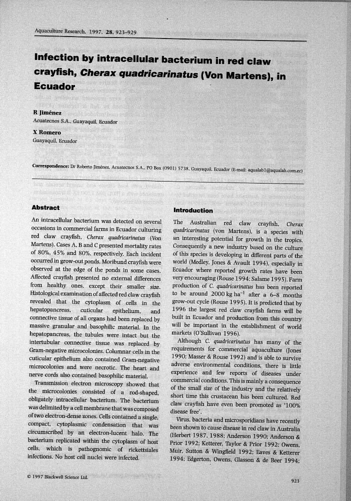
The population in case B had a mortality rate of45% after 130 days of stocking, when harvestingtook place. The presence of 15 small, stunted redclaw crayfish with light blue-green colour wasobserved 45 days after stocking (Fig, 1), Thesecrayfish had very small or no antennae, and wereweak and dying at the edge of the pond. The averagesize of sampled animals was 2.75 g. which wasconsiderately less than the average of 12.85 g orthat expected for C. quadricarinatus of their age(Romero & Murillo 1996).
A mortality rate of 80% was observed in case C
924 €> 1997 Blackweii Science Ltd, Aquaculture Research. 28 . 923-929
Aquaculture^search, 1997. 28 . 923-929 IntraceUular bacterial infection in red claw crayfish R Jimenez & X Romero
Figure 1 Macroscopic view of thesize differences observed betweenhealthy C. quadricarinatus (back,8 cm) and another infected (front.3 cm) by intracellular bacteria taken45 days after stocking. Crayfish werestocked at the same size.
when harvested at 63 days after stocking. Weakcrayfish were sampled at this time. The averageweight of the sampled crayfish was 8.33 g, whichwas considerately lower than expected (18 g)(Romero & Murillo 1996). The external colour ofthe sampled crayfish was normal.
Histology and ultrastructure
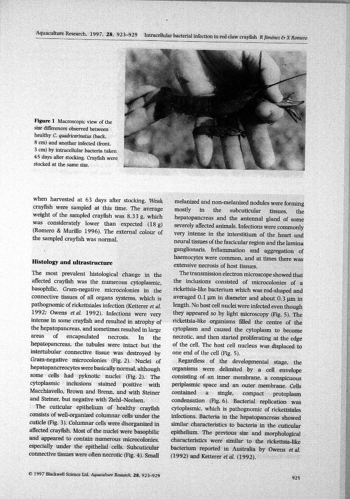
The most prevalent histological change in theaffected crayfish was the numerous cytoplasmic,basophilic. Gram-negative microcolonies in theconnective tissues of all organs systems, which ispathognomic of rickettsiales infection (Ketterer et al.1992; Owens etaJ. 1992). Infections were veryintense in some crayfish and resulted in atrophy ofthe hepatopancreas. and sometimes resulted in largeareas of encapsulated necrosis. In thehepatopancreas, the tubules were intact but theintertubular connective tissue was destroyed byGram-negative microcolonies (Fig. 2). Nuclei ofhepatopancreocytes were basically normal, althoughsome cells had pyknotic nuclei (Fig. 2). Thecytoplasmic inclusions stained positive withMacchiavello, Brown and Brenn, and with Steinerand Steiner, but negative with Ziehl-Neelsen.
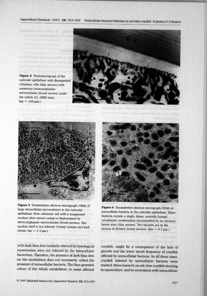
The cuticular epithelium of healthy crayfishconsists of well-organized columnar cells under thecuticle (Fig. 3). Columnar cells were disorganized inaffected crayfish. Most of the nuclei were basophilicand appeared to contain numerous microcolonies,especially under the epithelial cells. Subcuticularconnective tissues were often necrotic (Fig. 4). Small
melanized and non-melanized nodules were formingmostly in the subcuticular tissues, thehepatopancreas and the antennal gland of someseverely affected animals. Infections were commonlyvery intense in the interstitium of the heart andneural tissues of the fascicular region and the laminaganglionaris. Infiammation and aggregation ofhaemocytes were common, and at times there wasextensive necrosis of host tissues.
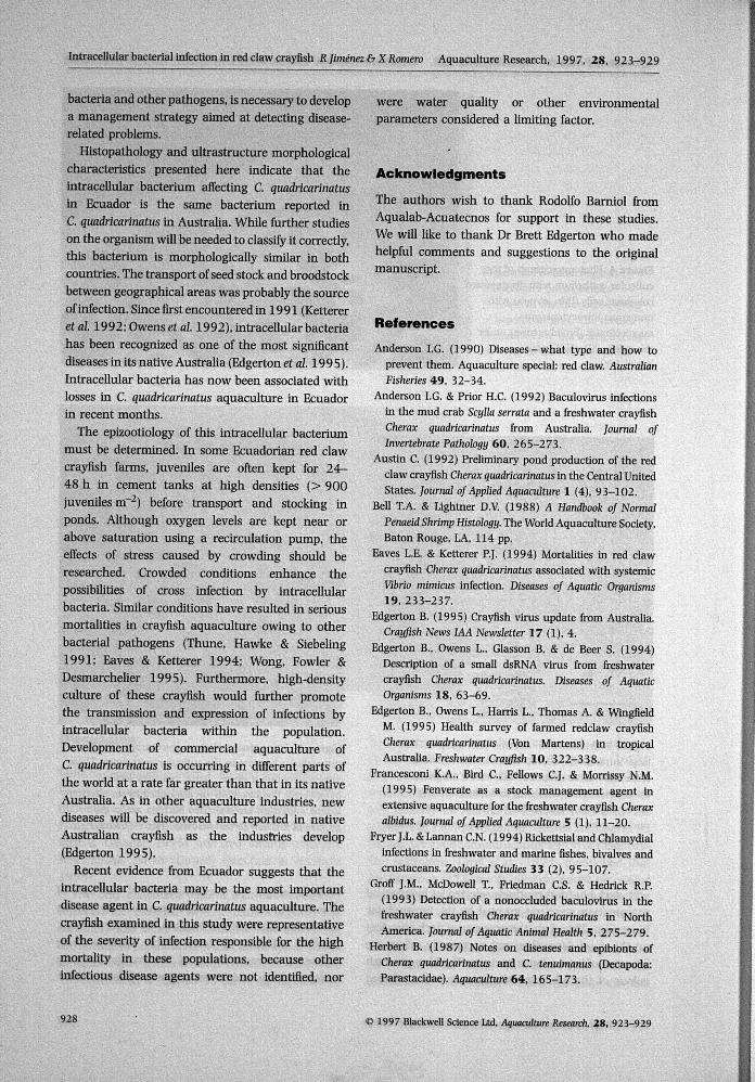
The transmission electron microscope showed thatthe inclusions consisted of microcolonies of arickettsia-like bacterium which was rod-shaped andaveraged 0.1 iim in diameter and about 0.3 jim inlength. No host cell nuclei were infected even thoughthey appeared so by light microscopy (Fig. 5). Therickettsia-like organisms filled the centre of thecytoplasm and caused the cytoplasm to becomenecrotic, and then started proliferating at the edgeof the cell. The host cell nucleus was displaced toone end of the cell (Fig. 5).
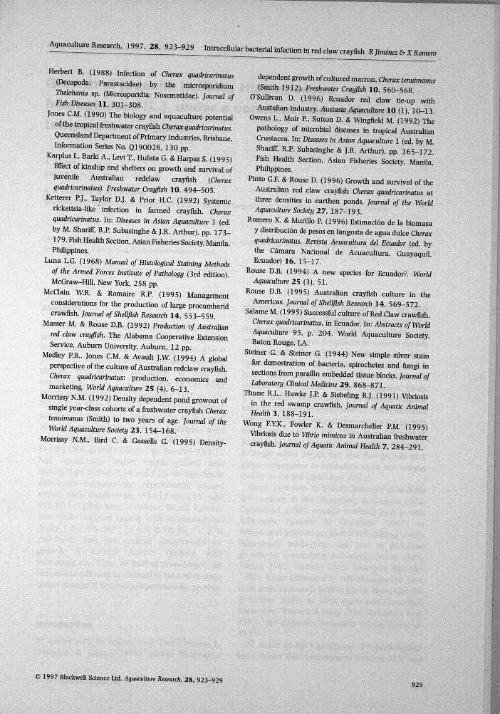
Regardless of the developmental stage, theorganisms were delimited by a cell envelopeconsisting of an inner membrane, a conspicuousperiplasmic space and an outer membrane. Cellscontained a single. compact protoplasmcondensation (Fig. 6). Bacterial replication wascytoplasmic, which is pathognomic of rickettsialesinfections. Bacteria in the hepatopancreas showedsimilar characteristics to bacteria in the cuticularepithelium. The previous size and morphologicalcharacteristics were similar to the rickettsia-likebacterium reported in Australia by Owens etal.(1992) and Ketterer et al. (1992).
© 1997 Biackwell Science Ltd. Aquaculture Research, 28 , 923-929925
Intracellular bacterial infection in red claw crayfish R Jimenez & X Romero Aquaculture Research, 1997. 28, 923-929
Figure 2 Photomicrograph ofintracellular microcolonies
". (arrows) in the connective tissueof hepatopancreas. The tubules
" y themselves are not infected, onlythe intertubular tissues. (H&Estain, bar = 100 |im.)
Figure 3 Photomicrograph of thecuticular epithelium in healthycrayfish. Organized columnar cells(arrows) of the cuticularepithelium are under the cuticle(C). (H&E stain, bar = 100 nm.)
Discussion
Density-dependent growth rates and size differenceshave been reported from different species of crayfishincluding the genus Cherax (Morrissy 1992;Francesconi. Bird, Fellows & Morrissy 1995; Morrissy,Bird & Gassells 1995) and red swamp crayfish,Procambarus clarkii (Girard) (McClain & Romaire1995).
The only consistent clinical sign of infection byintracellular bacteria in red claw crayfish wasmorbidity and small size. However, size differencesare normal in populations of red claw crayfish grownin aquaculture ponds (Austin 1992; Pinto & Rouse1996; Romero & Murillo 1996) or in experiments
with red claw crayfish juveniles in aquariums(Karplus, Barki, Levi, Hulata & Harpaz 1995).Stunted animals in red claw cra5^sh can then he aconsequence of density-dependent growth rates, orcan possihly he related to diseases caused by thepresence o{ Psorospermium sp., Cherax baculovirus (L.
Owens, personal communication 1995) or rickettsia-like organisms. Groff etal. (1993) mentioned thatthe presence of Cherax baculovirus might be relatedto the poor growth experienced in red claw crayfish.The natural differences in size of red claw crayfishmay mask the presence of rickettsia-like organisms.
Only one crayfish infected hy the intracellularbacterium had deep dark-blue spots on theexoskeleton. Furthermore, other red claw crayfish
926 © 1997 BlackweU Science Ltd. A uacidfure Research. 28, 923-929
Aquaculture Researcii, 1997, 28, 923-929 Intracellular bacterial infection in red ciaw crayfish R Jimenez & X Romero
Figure 4 Photomicrograph of thecuticular epithelium with disorganizedcolumnar cells (thin arrows) withnumerous intracytoplasmicmicrocolonies (broad arrows) underthe cuticle (C). (H&E stain,bar = 100 nm.)
Figure 5 Transmission electron micrograph (TEM) oflarge intracellular microcolonies in the cuticularepithelium. Note columnar cell with a marginatednucleus (thin arrow) owing to displacement byintracytoplasmic microcolonies (broad arrows). Thenucleus itself is not infected. (Uranyl acetate and leadcitrate, bar = 1.
Figure 6 Transmission electron micrograph (TEM) ofintracellular bacteria in the cuticular epithelium. Thesebacteria contain a single, dense, centrally locatedcytoplasmic condensation circumscribed by an electron-lucent zone (thin arrows). IVo bacteria are in theprocess of division (broad arrows). (Bar = 0,3 |im,)
with dark-blue dots routinely selected for histological
examination were not infected by the intracellular
bacterium. Therefore, the presence of dark-blue dots
on the exoskeleton does not necessarily reflect the
presence of intracellular bacteria. The blue-greenish
colour of the whole exoskeleton, in some affected
crayfish, might be a consequence of the lack of
growth and the lower moult frequency of crayfish
affected by intracellular bacteria. In all three cases,
crayfish infected by intracellular bacteria were
stunted. More research on red claw crayfish stunting
in aquaculture, and its association with intracellular
1997 Blackweii Science Ltd. Aquacuhuix fieseare/i, 28 , 923-929927
Intracellular bacterial infection in red claw crayfish R Jimenez & X Romero Aquaculture Research, 1997, 28, 923-929
bacteria and other pathogens, is necessary to developa management strategy aimed at detecting disease-related problems.
Histopathology and ultrastructure morphologicalcharacteristics presented here indicate that theintracellular bacterium affecting C. quadricarinatusin Ecuador is the same bacterium reported inC. quadricarinatus in Australia. While further studieson the organism will be needed to classify it correctly,this bacterium is morphologically similar in bothcountries. The transport of seed stock and broodstockbetween geographical areas was probably the sourceof infection. Since first encountered in 1991 (Kettereretal 1992. Owens etal. 1992), intracellular bacteriahas been recognized as one of the most significantdiseases in its native Australia (Edgerton et al. 1995).Intracellular bacteria has now been associated withlosses in C. quadricarinatus aquaculture in Ecuadorin recent months.
The epizootiology of this intracellular bacteriummust be determined. In some Ecuadorian red clawcrayfish farms, juveniles are often kept for 24 -48 h in cement tanks at high densities (> 900juveniles m~ ) before transport and stocking inponds. Although oxygen levels are kept near orabove saturation using a recirculation pump, theeffects of stress caused by crowding should beresearched. Crowded conditions enhance thepossibilities of cross infection by intracellularbacteria. Similar conditions have resulted in seriousmortalities in crayfish aquaculture owing to otherbacterial pathogens (Thtine, Hawke & Siebeling1991: Eaves & Ketterer 1994; Wong, Fowler &Desmarchelier 1995). Furthermore, high-densityculture of these crayfish would further promotethe transmission and expression of infections byintracellular bacteria within the population.Development of commercial aquaculture ofC. quadricarinatus is occurring in different parts ofthe world at a rate far greater than that in its nativeAustralia. As in other aquaculture industries, newdiseases will be discovered and reported in nativeAustralian crayfish as the industries develop(Edgerton 1995).
Recent evidence from Ecuador suggests that theintracellular bacteria may be the most importantdisease agent in C. quadricarinatus aquaculture. Thecrayfish examined in this study were representativeof the severity of infection responsible for the highmortality in these populations, because otherinfectious disease agents were not identified, nor
were water quality or other environmentalparameters considered a limiting factor.
Acknowledgments
The authors wish to thank Rodolfo Bamiol fromAqualab-Acuatecnos for support in these studies.We will like to thank Dr Brett Edgerton who madehelpful comments and suggestions to the originalmanuscript.
References
Anderson I.G. (1990) Diseases - what type and how toprevent them. Aquaculture special: red claw. AustralianFisheries 49, 32-34.
Anderson I.G. & Prior H.C. (1992) Baculovirus infectionsin the mud crab Scylla serrata and a freshwater crayfishCherax quadricarinatus from Australia. Journal ofInvertebrate Pathology 60, 265-273.
Austin C. (1992) Preliminary pond production of the redclaw crayfish Cherax quadricarinatus in the Central UnitedStates. Journal of Applied Aquaculture 1 (4), 93-102.
Bell T.A. & Lightner D.V. (1988) A Handbook of NormalPenaeid Shrimp Histology. The World Aquaculture Society,Baton Rouge, LA, 114 pp.
Eaves L.E. & Ketterer P.J. (1994) Mortalities in red clawcrayfish Cherax quadricarinatus associated with systeniicVibrio mimicus infection. Diseases of Aquatic Organisms19. 233-237.
Edgerton B. (1995) Crayfish virus update from Australia.Crayfish News lAA Newsietter 17 (1), 4.
Edgerton B.. Owens L, Glasson B. & de Beer S. (1994)Description of a small dsRNA virus from freshwatercrayfish Cherax quadricarinatus. Diseases of AquaticOrganisms 18, 63-69.
Edgerton B., Owens L., Harris L, Thomas A. & WingfieldM. (1995) Health survey of farmed redclaw crayfishCiierax quadricarinatus (Von Martens) in tropicalAustralia. Freshwater Crayfish 10, 322-338.
Erancesconi K.A., Bird C, Fellows C.J. & Morrissy N.M.(1995) Fenverate as a stock management agent inextensive aquaculture for the freshwater crayfish Cheraxalbidus. Journal of Applied Aquaculture 5 (1), 1 1 - 2 0 .
Fryer J.L. &Lannan C.N. (1994) Rlckettsial and Chlamydialinfections in freshwater and marine fishes, bivalves andcrustaceans. Zoological Studies 33 (2), 95-107.
Groff J.M., McDowell T., Friedman C.S. & Hedrick R.P.(1993) Detection of a nonoccluded baculovirus in thefreshwater crayfish Cherax quadricarinatus in NorthAmerica. Journal of Aquatk Animal Health 5, 275-279.
Herbert B. (1987) Notes on diseases and epibionts ofCherax quadricarinatus and C. tenuimanus (Decapoda:Parastacidae). Aquaculture 64, 165-173.
I
928 © 1997 Biackweil Science Ltd. Aquacuiture Research. 28 , 923-929
Aquaculture Research, 1997. 28 . 923-929Jntracellular bacterial infection in red claw crayfish R Jimenez & X Romero
Herbert B. (1988) Infection of Cherax quadricarinatus(Decapoda: Parastacidae) by the microsporidiumThehhania sp. (Microsporidia: Nosematidae). Journal ofFish Diseases 11. 301-308.
Jones CM. (1990) The biology and aquaculture potentialof the tropical freshwater crayfish Cherax quadricarinatus.Queensland Department of Primary Industries. Brisbane.Information Series No. Q190028. 130 pp.
Karplus I.. Barki A.. Levi T.. Hulata G. & Harpaz S. (1995)Effect of kinship and shelters on growth and survival ofjuvenile Australian redclaw crayfish {Cheraxquadricarinatus). Freshwater Crayfish 10. 494-505.
Ketterer P.J.. Taylor D.J. & Prior H.C. (1992) Systemicrickettsia-like infection in farmed crayfish. Cheraxquadricarinatus. In: Diseases in Asian Aquaculture 1 (ed.by M. Shariir. R.P. Subasinghe & J.R. Arthur), pp. 173-179. Fish Health Section. Asian Fisheries Society. Manila,Philippines.
Luna L.G. (1968) Manual of Histological Staining Methodsof the Anned Forces Institute of Pathology (3rd edition).McGraw-Hill. New York. 258 pp.
McClain W.R. & Romaire R.P. (1995) Managementconsiderations for the production of large procambaridcrawfish. Journal of Shellfish Research 14. 553-559.
Masser M. & Rouse D.B. (1992) Production of Australian
red claw crayfish. The Alabama Cooperative ExtensionService. Auburn University. Auburn, 12 pp.
Medley P.B.. Jones CM. & Avault J.W. (1994) A globalperspective of the culture of Australian redclaw crayfish.Cherax quadricarinatus: production, economics andmarketing. World Aquaculture 25 (4), 6-13.
Morrissy N.M. (1992) Density dependent pond growout ofsingle year-class cohorts of a freshwater crayfish Cherax
tenuimanus (Smith) to two years of age. Journal of the
World Aquaculture Society 23, 154-168.Morrissy N.M.. Bird C & Gassells G. (1995) Density-
dependent growth of cultured marron. Cherax tenuimanus(Smith 1912). Freshwater Crayfish 10. 560-568.
O'Sullivan D. (1996) Ecuador red claw tie-up withAustalian industry. Austasia Aquaculture 10 (1). 10-13.
Owens L.. Muir P.. Sutton D. & Wingfield M. (1992) Thepathology of microbial diseases in tropical AustralianCrustacea. In: Diseases in Asian Aquaculture 1 (ed. by M.Shariff, R.P. Subasinghe & J.R. Arthur), pp. 165-172.Fish Health Section. Asian Fisheries Society, Manila,Philippines.
Pinto G.F. & Rouse D. (1996) Growth and survival of theAustralian red claw crayfish Cherax quadricarinatus atthree densities in earthen ponds. Journal of the WorldAquaculture Society 27. 187-193.
Romero X. & Murillo P (1996) Estimacion de la biomasay distribucion de pesos en langosta de agua dulce Cheraxquadricarinatus. Revista Acuacultura del Ecuador (ed. bythe Camara Nacional de Acuacultura. Guayaquil.Ecuador) 16. 15-17.
Rouse D.B. (1994) A new species for Ecuador?. World
Aquaculture 25 (3). 51.
Rouse D.B. (1995) Australian crayfish culture in theAmericas. Journal of Shellfish Research 14, 569-572.
Salame M. (1995) Successful culture of Red Claw crawfish,Cherax quadricarinatus. in Ecuador. In: Abstracts of WorldAquaculture 95. p. 204. World Aquaculture Society.Baton Rouge. LA.
Steiner G. & Steiner G. (1944) New simple silver stainfor demostration of bacteria, spirochetes and fungi insections from paraffin embedded tissue blocks. Journal ofLaboratory Clinical Medicine 29, 868-871.
Thune R.L., Hawke J.P. & Siebeling R.J. (1991) Vibriosisin the red swamp crawfish. Journal of Aquatic AnimalHealth 3. 188-191.
Wong F.Y.K.. Fowler K. & DesmarcheUer P.M. (1995)Vibriosis due to Vibrio mimicus in Australian freshwatercrayfish. Journal of Aquatic Animal Health 7, 284-291.
© 1997 Blackweli Science Ltd. Aquaculture Research, 28 , 923-929929