immunopathogenesis based adamantiades- behcet disease vasculitis treatment
TRANSCRIPT

9
Immunopathogenesis Based Adamantiades-Behcet Disease Vasculitis Treatment
Panagiota Boura, Konstantinos Tselios, Ioannis Gkougkourelas and Alexandros Sarantopoulos
Clinical Immunology Unit, 2nd Department of Internal Medicine, Hippokration General Hospital, Aristotle University of Thessaloniki
Greece
1. Introduction
All immune cell lineages arise from the multipotent stem cells in the bone marrow. The development and regulation of immune system cells (immune homeostasis) depends on the programmed appearance of specific cell surface molecules (e.g. receptors) and responsiveness to certain cytokines and chemokines. After maturation, the immune system cells include antigen presenting cells (APCs), other phagocytic cells like neutrophils, eosinophils, basophils and lymphocytes which include T lymphocytes, Β lymphocytes and natural killer (NK) cells. Depending on an initial stimulus and the microenvironmental conditions at a given time, each lymphocyte lineage can be subdivided to different populations with discrete specialized functions. Subpopulations of crucial importance are CD4+ T cells (mainly Th1, Th2, Th17 and T regulatory cells) and CD8+ T cells, CD16- and CD16+ NK cells and CD5+ and CD5- B cells. As a structure, the immune system consists of central locations of immune cell production and differentiation (bone marrow and thymus) and peripheral organs, where encounter with antigen and response to it occurs, that is the spleen, lymph nodes, tonsils and mucosa-associated lymphoid tissue (MALT). Mucosal surfaces and the skin provide the main primary access for foreign antigens to meet cells of the immune system, where they first react primarily with APCs. Innate immunity includes skin and mucosal barriers, as well as the innate immune cells that are triggered and react in minutes after they encounter ‘foreign’ material. The innate immune system, as it represents the first line of host defense against foreign entities, is regarded as a nonspecific system and its main role is to engulf and eliminate pathogens, trigger pro-inflammatory responses and present antigens to adaptive immune cells, which are subsequently activated. More recently it has been shown that the innate immune system has a necessary degree of specificity that enables it to discriminate between self and foreign particles (e.g. microorganisms). Germline encoded and constitutively expressed pattern recognition receptors (PRRs)- like Toll receptors on innate immune cells, help them to discriminate highly conserved pathogen associated molecular patterns (PAMPs), which activate specific signaling pathways that lead to robust but well-defined innate immune responses. Research on innate immunity is progressively uncovering molecular and cellular mechanisms by which it recognizes environment and reacts to it, leading to protective or
Advances in the Diagnosis and Treatment of Vasculitis
168
pathological and harmful immune responses. Understanding innate and adaptive immunity pathways in detail will have a great impact on revealing the immunopathology of immune-mediated diseases like Adamantiades-Behcet Disease (ABD) and developing more targeted immunotherapeutic agents to manipulate, even life-threatening, ABD vasculitis.
2. General considerations in ABD immunopathogenesis
ABD owes its name to Benediktos Adamantiades (Greek ophthalmologist), who had described it first in 1930 and Hulusi Behcet (Turkish dermatologist), who described hypopyon, iritis and orogenital aphthosis in 1937 (Adamantiades, 1931; Behcet, 1937). However, the first description of the disease dates back, 2500 years, to Hippokrates, to the 5th century BC, in the Epidemion book, case 7, where it is described as aphthous ulceration, genital ulceration and iridocyclitis. There were other forms of fever… Many developed aphthae, ulcerations… Many ulcerations about the genital parts… Watery ophthalmies of a chronic character, with pains… Since the early descriptions, additional clinical features have been described and added to disease spectrum. ABD can be manifested with different organ involvement, articular, neurological, pulmonary and gastrointestinal, besides the classical clinical triad proposed by the International Study Group for Behcet’s Disease. Nowadays, ABD diagnosis rests upon a set of established criteria, where no single clinical or laboratory feature is pathognomonic (International Study Group for Behcet’s Disease, 1990, Table 1).
Recurrent oral ulcerations plus two Recurrent genital ulcers Skin lesions (erythema nodosum, acne,
folliculitis) Ocular lesions (uveitis) Positive pathergy test
Table 1. Diagnostic Criteria for ABD
From early descriptions, it was shown that patients with ABD cluster according to a geographic pattern that coincides with the ancient “Silk Route”. It has a higher incidence (100 times more common) in the countries of the Mediterranean Basin, Middle East and Far East, which includes Greece, Turkey, Saudi Arabia, Iran, Korea ,China and Japan while it is rare in North America. Although sporadic cases are described, there is an increased frequency among relatives (familial aggregation), mainly in HLA-B51 class I gene positive relatives. Genetic susceptibility factors seem to be connected with more severe disease expression. Particularly, in Asia, up to 80% of patients are HLA-B51 positive. Recent studies focus on new genetic markers that could amplify immune responses responsible for disease expression (HLA and non-HLA genes), probably under an environmental challenge e.g. an infectious agent (the infectious model), (Fietta, 2005; Kaklamani et al, 1998; Verity et al, 2003; Yazici H et al, 1999). ABD etiopathogenesis still remains a mystery to be elucidated. There is substantial evidence for an infectious background participating in disease initiation, especially, in genetically predisposed individuals. A number of viruses like HSV, Parvovirus B19 or bacteria like
Immunopathogenesis Based Adamantiades-Behcet Disease Vasculitis Treatment
169
uncommon serotypes of Streptococcus have been implicated. At the moment there is no concrete proof of the existence of certain microorganisms in the etiology of ABD and the initiation of the immunological process. Molecular mimicry between bacterial (HSV, streptococci) and human heat shock proteins (HSP), due to significant peptide homology between bacterial and human HSPs has been widely accepted, might be the basic link between infection and (auto)immunity in predisposed subjects, and could initiate and augment T and B cell immune responses. Besides, the option of exposure to chemicals was not supported for long. However, although immunological abnormalities concerning innate and adaptive immunity in ABD have been extensively studied, there is no adequate proof that ABD is either a typical autoimmune disease- it does not fulfill substantial criteria- or an auto-inflammatory disease- it is a very weak hypothesis at the moment (Direskeneli & Saruhan-Direskeneli, 2003; Kapsimali et al, 2010; Mumcu et al, 2007; Zouboulis & May, 2003). ABD is a systemic vasculitis which may affect arteries and veins of all sizes in any organ and underlies disease manifestations. To a great extent, disease pathology points to a vascular inflammation. Biopsies of affected organs usually reveal vasculitis, where intense infiltrations of neutrophils predominate and CD4+ T lymphocytes are present in the early stages. Venous vascular involvement and thrombosis is the characteristic feature of the disease, probably responsible for most of the tissue damage seen. Inflammatory changes in large arteries are typical of vasculitis, lack wall thickening and granuloma formation and have a tendency towards aneurysm formation. It is more severe in males. ABD lacks an increased risk of atherosclerosis. The term ‘vasculo-Behcet’ has been adopted to describe cases where vascular features dominate in disease expression. After initial vascular lesion (usually venous thrombosis), other vascular lesions may follow and lead to progressive, multifocal vascular disease with an unfavorable prognosis (Melikoglu et al, 2008). Vascular inflammation is mainly due to complex innate-immune, pro-inflammatory mechanisms which activate and perpetuate adaptive/specific immune responses against antigens not identified clearly. Endothelial dysfunction, partly due to anti-endothelial autoimmune reactions and non-specific coagulation/fibrinolytic abnormalities, has also been demonstrated and participates in disease pathophysiology to some extent. In such cases aggressive treatment consisting of immunosuppressive drugs is recommended (Calamia et al, 2011; Calamia & Kaklamanis, 2008). Management decisions are often difficult to take and there is a lack of controlled trials. A sequential therapeutic approach, based on ABD immunopathology and the immunomodulatory action achieved by immunosuppressive drugs is discussed.
2.1 Innate immunity cells abnormalities in ABD Neutrophil involvement in ABD pathogenesis represents a challenging issue, as neutrophil hyperactivation seems to play a pivotal role in the inflammatory vasculitic characteristics of disease lesions. Neutrophils are the dominant subpopulation of the inflammatory infiltrates in the vessel wall (Hayasaki et al, 2004). Neutrophil hypereactivity is a major contributor to the well-known pathergy skin reaction, which might be positive during periods of active disease. Pathergy reaction is a non-specific skin hyper-reactivity to minor trauma, such as a needle prick (Boura et al, 2007; Ozdemir et al, 2007).

Advances in the Diagnosis and Treatment of Vasculitis
170
At the tissue level, neutrophils were described to be present in the anterior chamber of the eye and the corneal epithelium, as well as in the ciliary body and chorioid tissue (Mendoza-Pinto et al, 2010). These cells are reported to be functionally impaired in the disease. Eksioglu-Demiralp et al described specific defects concerning neutrophil activation, oxidative burst and phagocytic activity (Eksioglu-Demiralp et al, 2001). On the other hand, increased synthesis of reactive oxygen species (ROS) has been reported in ABD. ROS-mediated oxidative stress seems to play an important role in pathogenesis, as advanced oxidation protein products were described to correlate with disease activity (Yazici C et al, 2004). This contradiction might mean that functional activity of neutrophils might correlate to disease activity or other, poorly defined, parameters. Additionally, it has been postulated that endothelial cells cooperate with neutrophils and monocytes in the vasculitic process. Endothelial cell activation and subsequently increased expression of cell adhesion molecules is a characteristic feature of the disease, which can trigger neutrophils and monocytes to initiate natural immune responses on the vessel wall. Recently, a novel mechanism by which neutrophils may interact with cytokine-activated endothelial cells in the presence of anti-endothelial cell antibodies has been described. Main receptors were Fc-gamma-RIIa and CXCR1/2. Moreover, endothelium activation leads to excessive thrombin formation and impaired fibrinolytic function, thus contributing to the generation of a prothrombotic state in ABD (Florey et al, 2007; Lee MT et al, 2007; Probst et al, 2004). Furthermore, neutrophils are primed by overproduced pro-inflammatory cytokines, secreted by antigen presenting cells (APCs) and the endothelium (e.g. TNF-α, IL-1, IL-6, IL-8 etc). Under such conditions, they secrete other cytokines that prime themselves, cause Th1 polarization and maintain a non-specific, pro-inflammatory microenvironment to retain enhanced activity (Carletto et al, 1997; Katsantonis et al, 2000; Zouboulis et al, 2000). Recently, the role of matrix metalloproteinases, particularly MMP-2 and MMP-9 was underlined in ABD. Pay et al found increased levels of these molecules in the serum of ABD patients and this was strongly correlated to vasculo-Behcet disease, especially aneurysm formation. Other investigators confirmed the elevated MMP-9 levels in the cerebrospinal fluid of ABD patients, suggesting that they might be involved in the pathogenesis of neuro-ABD (Hamzaoui K et al, 2009; Pay et al, 2007). It is reported that neutrophils are more resistant to spontaneous apoptosis (via Fas/Fas ligand interactions) during the remission of ABD uveitis, while resistance was restored in the active phase of the disease (Fujimori et al, 2008). Monocytes are also involved in ABD pathogenesis. Recent studies support the possible role of bacterially-induced innate immune responses through monocyte Toll-like receptor (TLR) activation. It was reported that TLR-2 was decreased after stimulation with LPS and HSP-60, or increased after bacterial lipoteichoic acid stimulation. TLR-6 expression was found to be enhanced after stimulation with HSP-60 and Streptococcus sanguis (Neves et al, 2009; Yavuz et al, 2008). As mentioned above, HSP-60 and Streptococcus sanguis represent candidate antigens in ABD pathogenesis. Antigen-presenting cells are reported to be activated in ABD patients, since ex vivo cultured monocytes were able to produce increased amounts of TNF-α, IL-6 and IL-8 (Mege et al, 1993). Moreover, there is strong evidence suggesting that APCs may be primarily involved in ABD immunopathogenesis. Early data comes from pathergy reaction histology, where

Immunopathogenesis Based Adamantiades-Behcet Disease Vasculitis Treatment
171
Saito et al showed that Langerhans cells were increased in the dermis layer shortly after needle prick (Saito et al, 1980). However, it has not been clarified whether APC hypereactivity is a primary defect of these cells or the result of disease related conditions. Nevertheless, IL-12 and IL-18, cytokines mostly produced by APCs, are known to strongly promote Th1 polarization of immune responses. Both these cytokines have been found in increased levels during active disease, implying a central role for APCs in ABD pathogenesis (Hamzaoui K et al, 2002). Finally, Pay et al described decreased numbers of peripheral plasmacytoid dendritic cells, along with decreased levels of IFN-β and suggested that this cell subset may also participate in disease pathogenesis (Pay et al, 2007).
2.1.1 Gamma-delta (γδ) T cells These specialized T cells (TCR comprising of γ and δ heterodimer) have been increasingly investigated during recent years. They are thought to play a critical role in mucosal immunity, being the first line of defense against invading micro-organisms, as well as the link between innate and adaptive immune response (Casetti et al, 2008). A subset of γδ T cells (Vgamma9+Vdelta2+) was found in increased numbers in the blood of active ABD patients. These cells were reported to express IL-2 receptor and HLA-DR surface molecules, indicating a high activation status. The Vgamma9+Vdelta2+ T cells were described to infiltrate target organs, such as the eye, while other studies found that they strongly proliferate after stimulation with bacterial extracts of the oral flora (Bank et al, 2003; Verjans et al, 2002; Yamashita et al, 1997). The critical role of gammadelta T cells in ABD is underlined in the study of Treusch et al, who found a decrease in their numbers after treatment with interferon-alpha-2a (Treusch et al, 2004). Later, it was shown that expanded CD56+ and CD8+ gammadelta T cells in ABD were capable of secreting IFN-γ and TNF-α. Data is further supported by the fact that these cells seem to be polyclonally rather than oligoclonally activated in ABD patients with oral ulcers. Probably because of repeated inflammation of the oral mucosa, due to a wide variety of antigenic stimuli, activation and proliferation of these cells might play a role in disease clinical heterogeneity (Freysdottir, 1999, 2006).
2.1.2 NK and NKT cells Early studies reported an increase in NK cell numbers in ABD patients. However, others described a decrease in their cytotoxic activity in the active phase of the disease, which was restored in remission periods. However, recent studies reported that, although increased in active disease, NK cells do not seem to exert impaired cytotoxic activity, as they were shown to adequately suppress IFN-γ production by CD4+ T cells, thus regulating a Th1 response in ABD (Onder et al, 1994; Suzuki et al, 1992; Yamaguchi et al, 2010). Natural killer T cells (NKT) comprise a sublineage of T cells that share characteristics of conventional T cells and NK cells and bridge innate and adaptive immunity. Their activation is restricted by the MHC molecule CD1d. Early studies reported an increase of CD4+CD16+ and CD4+CD56+ T cells in ABD. Other investigators confirmed the presence of increased CD56+ T cells in the periphery of patients with active ABD uveitis, in contrast with other forms of immune-mediated uveitis (Eksioglu-Demiralp et al, 1999; Yato & Matsumoto, 1999).

Advances in the Diagnosis and Treatment of Vasculitis
172
In the tissue level, NKT cells were found to be increased in the aqueous humor of uveitis patients. Additionally, in neuro-ABD patients, NKT cells were reported to be decreased in the periphery and increased in the cerebrospinal fluid, thus suggesting a recruitment of these cells in the site of inflammation. It should be mentioned that these cells were strongly Th1 polarized. Further studies demonstrated that CD8+CD56+ T cells represent potent cytotoxic effectors in ABD uveitis, as they were found to express both surface FasL and preformed intracellular perforin (Ahn et al, 2005; Hamzaoui K et al, 2006; Yu et al, 2004).
2.2 Adaptive immunity cells abnormalities in ABD Although the exact pathogenetic mechanisms behind ABD remain to be fully elucidated, much evidence has been accumulated regarding the involvement of adaptive immune response in the disease. Recent studies have revealed the central role of T-cell mediated immunity; in parallel, histopathological studies have demonstrated, mainly, T-cell-dominated perivascular infiltrates in skin, oral lesions and retina, as well as in choroids and periretinal scar tissue (Charteris et al, 1992a, 1992b; Gul et al, 1995, Poulter & Lehner, 1989).
2.2.1 T cell abnormalities CD4+ T helper cells differentiate into, at least, four distinctive functional subsets, namely Th1, Th2, Th17 and T regulatory cells (Mucida & Cheroutre, 2010). For all of these lymphocyte subpopulations quantitive and/or qualitive abnormalities have been shown in ABD (Chi et al, 2008; Hamzaoui K et al, 2006, Ilhan et al, 2008; Mantas et al, 1999). Genetic and environmental factors may have an influence independently or in association with other parameters, such as the ligation of T-cell receptor (TCR), the activation of co-stimulatory molecules, the predominance of a given cytokine in the microenvironment and others (Direskeneli et al, 1999). Many investigators suggest that a Th1 polarization of the immune response is present, at least, in active ABD. Th1 type proinflammatory cytokines, such as IL-2 and IFN-γ, are reported not only to be increased in peripheral blood of patients with ABD, but also to correlate with disease activity. Additionally, IL-12, which drives Th1 immune response from naïve T cells, is also reported to be elevated in ABD. Interleukin-12 is suggested to play a substantial role in ABD; when recombinant IL-12 was added to peripheral blood lymphocytes from patients with active disease, it prevented spontaneous and Fas-induced apoptosis, resulting to proliferation of autoreactive Th1 lymphocytes that could contribute to a prolonged autoimmune inflammation (Esin et al, 1997; Frassanito et al, 1999; Nara et al, 2008; Sugi-Ikai et al, 1998). Recently, IL-15 was reported to be increased in ABD, either in serum or in the aqueous humor of uveitis patients; IL-15 is able to induce pro-inflammatory cytokines, such as TNF-α and IL-1β, and facilitate maintenance of auto-reactive CD8+ T cells (Curnow et al, 2008; Waldman, 2004). The Th1 cells were proven to expand with high individual variability of the Vβ TCR, suggesting that there is no single antigen responsible for T cell activation (Direskeneli et al, 1999). Histopathological studies also point to a Th1 type tissue infiltration in ABD, possibly driven by the highly expressed IL-12 and IL-23 in intestinal and cutaneous lesions. Interestingly, both streptococcal antigens and auto-antigens, such as aB-crystallin also drive an IL-12

Immunopathogenesis Based Adamantiades-Behcet Disease Vasculitis Treatment
173
mediated response in ABD (Imamura et al, 2005; Kulaber et al, 2007; Lew et al, 2008; Yanagihori et al, 2006). Concerning Th2 type of immune response, both, cells and signature cytokines, such as IL-4, IL-10 and IL-13 are reported to be unaffected or decreased in ABD by most researchers in the serum or in the aqueous humor. However, other studies reported a significant expression of IL-4 in oral ulcers from ABD patients, suggesting the presence of a more complex antigenic stimulus during the disease (Ahn et al, 2006; Dalghous et al, 2006, Mantas et al, 1999). Th17 cells represent a relatively new functional subset of T helper cells, which mainly produce IL-17A-F, IL-22, IL-23 and TNFa. Interleukin-6 and TGF-β are considered to be the major cytokines responsible for the differentiation of these cells from naïve T cells. Recent studies have shown a possible role for these cells in the majority of systemic and organ-specific autoimmune diseases (Mesquita et al, 2009). Concerning ABD, active disease is characterized by high serum levels of IL-6 and IL-17, compared to patients in remission, while serum levels of IL-23 and IL-17 are reported to be elevated in active uveitis. However, an increased Th17 response is not confirmed by all studies (Chi et al, 2008; Ferrante et al, 2010; Saruhan-Direskeneli et al, 2003). Recent papers underline Th17 involvement in disease pathogenesis in the context of their ratio to Th1 cells. Kim et al reported an increased peripheral Th17/Th1 ratio, especially in uveitis and folliculitis patients, while there were no significant differences concerning Th1/Th2 and Th17/Th2 ratios (Kim et al, 2010). Histopathological studies, however, did not confirm these results in the tissue level (Canete et al, 2009). Cytotoxic T cells are also reported to be activated in ABD, probably as a response to a profound inflammation. CD8+ T cells were also found in increased numbers in the aqueous humor in uveitis patients. Furthermore, recent studies support that these cells play a fundamental role in ABD flares, as they were found in increased numbers in active disease, along with γδT cells. Additionally, CD8+ T cells were shown to express the CD69 molecule (early activation marker), while their numbers and function returned to normal after successful management of the disease flare (Houman et al, 2004; Yasuoka et al, 2008).
2.2.2 Immune regulation in ABD Firstly described by Sakaguchi et al in the ‘70s, T regulatory cells represent potent regulators of the immune response. These cells are divided into two broad categories, namely the “naturally occurring” and the “inducible” T regulatory cells (Sakaguchi et al, 1995). Interest for immune regulation in ABD intensified as early as 1982, when Victorino et al and Sakane et al independently reported abnormal T cell suppressor activity (Sakane et al, 1982; Victorino et al, 1982). Recent studies were not able to definitely shed light in this issue. Paradoxically, CD4+CD25+FoxP3+ T regulatory cells were reported to be increased in active ABD compared to inactive disease and healthy controls, while they could suppress their T effector cell counterparts. The authors suggested that this may be due to the need for inflammation suppression in ABD (Hamzaoui K et al, 2006). On the other hand, other investigators showed that T regulatory cells were significantly decreased before ocular attack in ABD patients, suggesting that they may serve as a predictive marker in ocular disease, thus, allowing treatment intensification before relapse (Nanke et al, 2008).

Advances in the Diagnosis and Treatment of Vasculitis
174
The exact interactions between T regulatory cells and other immune cells in ABD are not well understood. Hamzaoui suggested a possible cross talk between these cells and NKT cells, as these subpopulations can modulate each other through cell-to-cell contact and IL-2 dependent mechanisms, respectively (Hamzaoui K, 2007). Furthermore, other cells with possible regulatory activity, such as gammadelta-T-cells were reported to be incapable of maintaining a regulatory profile in ABD (Clemente et al, 2010). T regulatory cells represent a promising therapeutic target for suppressing inflammation in ABD. Recently, Sugita et al reported T-reg expansion after in vitro stimulation with infliximab and significant suppression of bystander T effector cells in active ABD uveitis (Sugita et al, 2011). Additionally, other investigators reported that vitamin D is an important promoter of T cell regulation in vivo in ABD patients, as its levels were strongly correlated to T regulatory cells (Hamzaoui K et al, 2010).
2.2.3 B cell abnormalities and autoantibodies in ABD Although not a typical autoimmune disease, ABD is characterized by B cell activation, despite the absence of disease-specific autoantibodies. Early studies demonstrated that the total numbers of B cells were normal during the disease; however, an increased expression of activation markers, such as CD13, CD133, CD80 and memory marker CD45RO was reported. Despite the presumed B cell activation, plasma cells were found in low levels, in contrast to other well-defined autoantibody-mediated disorders. These results suggest a modified B cell function in ABD, although it is not known if it is related to a weak stimulus by an unknown external antigen or it represents an epiphenomenon in the context of the general dysfunction of the immune system (Eksioglu-Demiralp et al, 1999). After the identification of the multiple roles of B cells in the immune response, such as antigen presentation and cytokine release, newer studies reported some interesting results. B cells (along with plasma cells) were found in histopathological studies in the synovium, the central nervous system and the vessel wall of patients with ABD (Hirohata, 2008; Hirohata & Kikuchi, 2009; Suh et al, 2001). Furthermore, it was recently reported that B-cell activating factor (BAFF) receptor was upregulated on B cells in patients with ABD vasculitis, as well as serum BAFF levels and mRNA in skin biopsies. Later, Hamzaoui A et al showed that BAFF, along with IL-6 and IL-13, were, also, increased in the broncho-alveolar lavage (BAL) fluid from ABD patients with active pulmonary involvement; All these factors are known to actively participate in the maturation and activation of B cells (Hamzaoui A, 2010; Hamzaoui K, 2008). Consequently to B cell activation, various autoantibodies have been detected in the sera of ABD patients; however, their role in disease pathogenesis remains to be defined. Antinuclear antibodies (ANA) and rheumatoid factors are typically absent. On the other hand, autoantibodies against cell surface antigens, such as anti-endothelial cells antibodies (AECA), or against mucosal antigens have repeatedly been reported. Recent studies, based on proteomics, have revealed the presence of antibodies against more specific antigens, such as α-enolase, α-tropomyosin, kinectin, selenium-binding protein, esterase-D and carboxy-terminal subunit of Sip-1 (Delunardo et al, 2006; Direskeneli et al, 1995; Lee KH et al, 2003; Lu et al, 2005; Mahesh et al, 2005; Michelson et al, 1985; Mor et al, 2002; Okunuki et al, 2007, 2008).

Immunopathogenesis Based Adamantiades-Behcet Disease Vasculitis Treatment
175
Concerning AECA, their prevalence in ABD is reported to vary from 18-50%; however, difficulties in method standardization led to significant discrepancy between the different studies and raised concerns about their clinical utility. Furthermore, other studies confirmed that AECA were not quite specific for the disease; instead, they are frequently detected in other systemic vasculitides and/or autoimmune diseases. Despite their inspecificity, AECA were shown to mediate enhanced polymorphonuclear adhesion to activated endothelial cells, thus promoting tissue injury during systemic inflammation. An association between IgG-AECA and central nervous system involvement in ABD was reported (Dinc et al, 2003; Souza et al, 2007; Zeng et al, 2004). Alpha-enolase represents a target antigen of IgM AECA, reported to be detectable in approximately half of ABD patients, especially in those with overt vascular involvement; however, its specificity is not satisfying (Lee JH et al, 2009).
3. Coagulation and fibrinolytic abnormalities in ABD
Although no specific defect in the coagulation cascade has so far been detected, ABD is characterized by thrombotic manifestations in 7-38% of the patients. In most studies, both coagulation and fibrinolytic pathways seem to be activated (Leiba et al, 2004). Various pro-coagulant conditions, such as deficiencies of protein C, protein S, antithrombin III and factor V Leiden and prothrombin G20210A mutations have been described in ABD patients (Ates et al, 2003; Caramaschi et al, 2010). Additionally, higher levels of homocysteine, factor VIII, thrombin-antithrombin III complexes (TAT) and prothrombin fragment 1+2 maintain the intravascular generation of thrombin in these patients (Espinosa et al, 2002; La Regina et al, 2010). Despite all these data, it seems that thrombophilic factors do not play a major role in the thrombotic tendency of ABD. Leiba et al, in a tightly designed study, showed that the only factor that was strongly correlated to thrombosis in these patients was hypertriglyceridemia (Leiba et al, 2004). Other studies reported that active ABD patients were characterized by hypertriglyceridemia, low HDL and high Lp(a) levels (Musabak et al, 2005). Regarding the fibrinolytic cascade, most studies have revealed defective fibrinolysis with lower levels of tissue plasminogen activator (t-PA) and increased thrombin activatable fibrinolysis inhibitor (TAFI) and thrombomodulin. Other studies involve the plasminogen activator inhibitor (PAI), as this molecule appears to be increased in ABD, resulting in defective tPA/PAI complex formation (Donmez et al, 2010; Ozturk et al, 2004). Furthermore, the endothelial dysfunction due to immune-mediated vasculitis may also play an important role in the thrombotic tendency of the disease. Another important thrombogenic factor in ABD is the presence of antiphospholipid antibodies. Indeed, anticardiolipin (ACA) and anti-β2-glycoprotein 1 (anti-β2-GPI) antibodies, as well as lupus anticoagulant (LA) have been described in selected ABD patients (Bonnet et al, 2004; Kandolf-Sekulovic et al, 2005). Additionally, newer epitopes, such as antibodies against the phosphatidylserine-prothrombin complex (anti-PS/PT) or annexin V, have also been reported to be associated with thrombosis in these patients (Aslan et al, 2004; Kawakami et al, 2009). The significance of these antibodies in thrombotic manifestations of ABD remains to be fully elucidated. In Table 2, a synopsis of the main abnormalities of the immune cells in ABD is presented.

Advances in the Diagnosis and Treatment of Vasculitis
176
Neutrophils Hyperactivation Cytokine and MMPs secretion Resistance to apoptosis Tissue infiltration
Monocytes/Macrophages Altered TLR expression Secretion of IFN-γ, IL-1, IL-8, IL-12, TNF-α
Gammadelta T cells Vgamma9+Vdelta2+ expansion Secretion of IFN-γ, TNF-α Polyclonal proliferation
NK cells Increased numbers Impaired cytotoxic activity? Abnormal KIR repertoire
NKT cells Recruitment to the site of inflammation High expression of FasL and perforin Interaction with T regulatory cells?
CD8+ T cells Increased numbers Highly activated (CD69)
CD4+ T cells (Th1 polarized) Secretion of IL-2, IL-12, IFN-γ Resistance to apoptosis Tissue infiltration
CD4+ T cells (Th17 polarized) Disease initiation? Secretion of IL-17, IL-23 Increased Th17/Th1 ratio
T regulatory cells Probably low numbers during active disease Impaired suppressive activity?
B cells Normal numbers, but activated Increased expression of BAFF receptor Autoantibody production (AECA and others)
Table 2. Main abnormalities in immune cells in ABD
A proposed model for ABD vasculitis pathogenesis
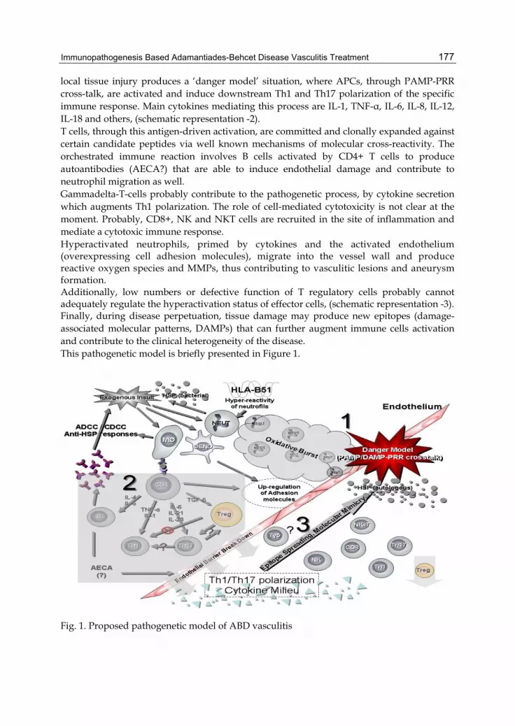
(schematic representation -1, -2, -3) In genetically predisposed individuals, due to an exogenous stress, e.g. an infectious insult, a systemic or local non-specific immune response is mediated via innate immune cells and the endothelium. Respiratory burst products and proinflammatrory cytokines and chemokines secreted at the site of the initial insult further attract monocytes, neutrophils and dendritic cells around the area and the blood-endothelial barrier breaks down (schematic representation -1). Initial insult is neutralized, but HSPs derived from bacterial load, as well as HSPs from affected tissue/s give rise to a second, more specific T-cell-mediated immune response, according to molecular mimicry and/or epitope spreading model. These events might be augmented and spread out in case of repeated antigenic stimuli (second hit). Endothelial-innate cell contact and activation perpetuates the proinflammatory milieu and along with
Immunopathogenesis Based Adamantiades-Behcet Disease Vasculitis Treatment
177
local tissue injury produces a ‘danger model’ situation, where APCs, through PAMP-PRR cross-talk, are activated and induce downstream Th1 and Th17 polarization of the specific immune response. Main cytokines mediating this process are IL-1, TNF-α, IL-6, IL-8, IL-12, IL-18 and others, (schematic representation -2). T cells, through this antigen-driven activation, are committed and clonally expanded against certain candidate peptides via well known mechanisms of molecular cross-reactivity. The orchestrated immune reaction involves B cells activated by CD4+ T cells to produce autoantibodies (AECA?) that are able to induce endothelial damage and contribute to neutrophil migration as well. Gammadelta-Τ-cells probably contribute to the pathogenetic process, by cytokine secretion which augments Th1 polarization. The role of cell-mediated cytotoxicity is not clear at the moment. Probably, CD8+, NK and NKT cells are recruited in the site of inflammation and mediate a cytotoxic immune response. Hyperactivated neutrophils, primed by cytokines and the activated endothelium (overexpressing cell adhesion molecules), migrate into the vessel wall and produce reactive oxygen species and MMPs, thus contributing to vasculitic lesions and aneurysm formation. Additionally, low numbers or defective function of T regulatory cells probably cannot adequately regulate the hyperactivation status of effector cells, (schematic representation -3). Finally, during disease perpetuation, tissue damage may produce new epitopes (damage-associated molecular patterns, DAMPs) that can further augment immune cells activation and contribute to the clinical heterogeneity of the disease. This pathogenetic model is briefly presented in Figure 1.
Fig. 1. Proposed pathogenetic model of ABD vasculitis
Advances in the Diagnosis and Treatment of Vasculitis
178
4. Therapeutic approach to ABD vasculitis
4.1 General principles Although treatment of ABD was reviewed recently, management of complicated cases, especially those with vascular involvement, still poses a great challenge to experienced physicians. Nowadays, the recently published EULAR (European League Against Rheumatism) recommendations are considered to be the accepted guidelines for the management of the disease. Nonetheless, treatment concerning vascular involvement, either major vessel or visceral disease (medium-small vessel vasculitis) is largely based on expert opinion and uncontrolled evidence from open trials and observational studies. So the need for further properly designed controlled clinical trials is obvious (Hatemi et al, 2008). In general, topical therapy should be applied in mucocutaneous lesions and should be the only treatment for mild symptoms. Although antiseptics seem to prevent secondary infections and reduce pain intensity, antibiotics face antigen burden and they are important in identifying disease relapses as well. Drugs with anti-inflammatory action are used as adjuvants but, beyond this, it is imperative to emphasize that the physician must be vigilant for alarming clues of systemic vasculitis and ready to implement immunomodulating regimen (Alpsoy, 2005; Alpsoy et al, 2007). Treatment decisions depend on the age, sex, comorbidities, patient’s willingness to cooperate and other parameters, such as the site and severity of involvement, as well as the frequency of recurrences. Fortunately, a part of disease burden can be managed with mild systemic immunosuppression. Early and aggressive immunosuppression is mandatory in severe or life- threatening cases. Below, the most commonly used immunomodulating drugs for the management of major vessel disease or visceral involvement will be discussed. (Table 3)
4.2 Glucocorticosteroids (C/S) It is well known that glucocorticosteroids modify immune response to diverse stimuli by suppressing inflammatory reactions in general. They have profound metabolic effects and they affect the replication, movement and activity of virtually all cells involved in inflammatory process. Although C/S exert cytotoxic effects to activated T cells at high doses, their immunomodulating effects are probably mostly attributed to their ability to modify cellular immune functions when low doses are used orally. At the cellular level C/S bind to their intracellular receptor forming an active complex. The complex moves to the nucleus, where it binds to DNA sequences modifying chromatin structure and altering the transcription machinery. Another mechanism of action is that glucocorticosteroids-glucocorticosteroids receptor complex directly increases IκB (inhibitory factor κB) expression. IκB inhibits the actions of transcription nuclear factor NFκB. It is well known that NFκB promotes gene induction of cytokines, chemokines and adhesion molecules, thus amplifying lymphocyte proliferation and leading to the perpetuation of the inflammatory process (Bruton et al, 2005; Rhen & Chidlowski, 2005). Through these mechanisms C/S suppress cellular immunity, both Th17 and Th1 responses. This action explains their therapeutic use in ABD. Although more detailed trials are needed, worldwide clinical experience has shown that C/S are the cornerstone of treatment for many ABD complications. High oral doses, up to 100mg/day for a long period of time, or pulse treatment with 1gr/day for 3 consecutive days or more are given, in serious or life-threatening cases, like aneurysm formation or cavernous sinus thrombosis (Hatemi et al,

Immunopathogenesis Based Adamantiades-Behcet Disease Vasculitis Treatment
179
2008). It was demonstrated that in large-vessel arterial disease (arterial occlusion or aneurysm) high dose glucocorticosteroids alone did not improve occlusive disease and mortality (Li Thi Huong et al, 1995). On the contrary, other studies have shown that C/S in combination with other immunosuppressants had a significant beneficial effect in vascular complications of ABD (Alpsoy & Alkman, 2009, Sharaf & Yazici, 2009). In another recently conducted randomized trial, low dose intramuscular depot C/S was used and proved to be beneficial in milder manifestations, like erythema nodosum, but no for genital ulcers. The trial used the drug in depot form for intramuscular injection, so no conclusions can be drawn for the efficacy of the usual oral or intravenous regimen (Mat et al, 2006). Adverse effects such as osteoporosis, metabolic disturbances, psychosis must been taken into account. EULAR committee recommends the use of C/S as a first line therapy for managing all ABD manifestations. Different level of evidence for every case is probably attributed to data availability. Based on immunopathology and clinical data, we suggest the administration of high dose intravenous corticosteroids for the achievement of remission in large vessel or CNS vasculitis, followed by a slow tapering schedule to the lowest effective dose which sustains remission, under close clinical and laboratory follow up (Bruton et al, 2005; Hatemi et al, 2008; Li Thi Huong, 1995; Sharaf & Yazici, 2009).
4.3 Azathioprine Azathioprine is a prodrug of 6-mercaptopurine, containing an imidazole ring and acts like an antimetabolite. Azathioprine and mercaptopurine appear to induce immunosuppression by interfering with purine nucleic acid metabolism. The drug intervenes with the process of cell division at steps that are required for lymphoid cell proliferation following antigenic stimulation. Stimulated lymphoid cells incapable of entering cell mitotic phase undergo apoptosis, establishing a relevant state of immunosuppression. Although RNA synthesis is blocked at the same time, these agents appear to have less effect on cell function if implemented after antigen exposure than on nucleic acid synthesis in proliferating, antigen encountering, T cells (Bruton et al, 2005). The efficacy of azathioprine in ABD is well established. The usual dose is 1-2.5 mg/kg/day (not exceeding 200 mg/day) and an average of 3 months is required for its effect to be established. This slow immunomodulating action limits the use of azathioprine as a sole agent of therapy in acute vasculitis presentation. However, it can be used additionally in the acute phase as a corticosteroid sparing agent, as well as prophylactically. Yazici H et al propose azathioprine as a first line therapy for non-life-threatening cases of neurological complications (Yazici H et al, 1990). According to the EULAR recommendations, azathioprine should be used in any ABD patient having inflammatory eye disease with posterior segment involvement (Hatemi et al, 2008). Its efficacy and safety was evaluated in a randomized, double-blind, placebo-controlled trial. In this trial, there were fewer new cases of ocular involvement in azathioprine plus corticosteroids group comparative to corticosteroids alone (one versus eight). Additionally, the patients taking azathioprine had less frequent oral ulcers, genital ulcers and arthritis (Yazici H et al, 1990). Contemplating the pathophysiology of ABD (see above), immunosuppressants like azathioprine may play a role in the management of vessel thrombosis through anti-inflammatory action on vessel wall. Results from Sharaf et al confirm the clinical experience that by dampening immune system activation, pro-thrombotic diathesis can be, at least partially, reversed (Sharaf & Yazici, 2009).

Advances in the Diagnosis and Treatment of Vasculitis
180
In general, azathioprine could be used as a first line treatment in combination with corticosteroids in almost all serious manifestations of the disease and is preferable for maintenance therapy after remission induction. Common side effects are gastrointestinal disturbances, liver toxicity and bone marrow suppression but rarely do they lead to drug discontinuation. In parallel, due to B cell impairment and mild hypoglobulinemia, humoral immunity can be weakened by these cytotoxic agents as well.
4.4 Cyclophosphamide (CyP) Cyclophosphamide is a highly active alkylating agent, which impairs DNA duplication and cell division. Although it is not a cell specific drug, the greater effect is observed upon rapidly proliferating cells, such as T cell clones after their encounter with the antigen. Initially, cyclophosphamide has proven a reliable agent in management of lupus nephritis and ANCA associated vasculitis and, eventually, has found a distinguished place in ABD vasculitis (Bruton et al, 2005). It has been known that pulse CyP, combined with intravenous C/S regimen, help to achieve remission in ABD vascular and/or visceral involvement (Du et al, 1990; Hamza et al, 1992). According to EULAR recommendations, large vessel vasculitis, such as pulmonary and/or peripheral aneurysms, need to be treated with CyP for at least 2 years (Hatemi et al, 2008). The drug may be administrated either intravenously, as pulse treatment in doses 400-700mg/m2, or orally in doses of 1-3 mg/day. The exact dose depends upon the severity of the disease and considerations of renal and hepatic impairment. Deep immunosuppression following CyP administration has a life-saving effect. Also in a double-blind cross over trial, CyP has proven beneficial in patients with intractable uveitis (Davatchi et al, 1999). Due to its severe toxicity (bone marrow suppression, mucositis, neurotoxicity, syndrome of inappropriate ADH secretion, cancer), this drug must be used in life-threatening or non-responding cases. We prefer to administer CyP intravenously, as monthly or 3-weekly pulses of 500-1000mgr bolus, in order to partially avoid bladder toxicity. For safety reasons, mesna (chelic agent) and adequate fluids must be administered concurrently.
4.5 Cyclosporine Cyclosporine is a cyclic polypeptide, which forms a complex with cyclophylin, an intracellular factor mediating signaling pathways. This complex binds to calcineurin and inhibits Ca2+-stimulated dephosphorylation of the cytosolic component of the transcription factor NFAT. Activated cytoplasmic NFAT translocates to the nucleus and binds to nuclear components required for complete T-cell activation, blocking of IL-2 and other Th1 cytokine producing genes (Th1 to Th2 switch). Besides this, calcineurin also possess peptidyl-prolyl cis-trans-isomerase enzymatic activity. The peptidyl-prolyl cis-trans-isomerase domain of calcineurin facilitates binding to CD147, also known as an inducer of extracellular-matrix metalloproteinases (MMPs). This binding causes CD147 to translocate to the cell surface, where it plays a critical role in stimulating matrix-metalloproteinase activity, leading to matrix degradation. Thus, this agent inhibits the enzymatic activation of MMPs, which leads to vessel wall thinning (Weintraub, 2009). Cyclosporine is effective against T-cell–dependent immune reactions and a suitable drug for immune intervention in the pathophysiological process of ABD vasculitis (see above). Additionally, cyclosporine, in synergy with C/S, downregulates the activity of NKT cells controlling inflammatory process (Bruton et al, 2005; Chi et al, 2010).
Immunopathogenesis Based Adamantiades-Behcet Disease Vasculitis Treatment
181
The efficacy of cyclosporine in ABD has been evaluated primarily in patients with ocular, mucocutaneous and articular manifestations. Approximately 50% of patients showed clinical remission when treated with cyclosporine. Two well designed trials proved that cyclosporine protects ocular degeneration and visual acuity better than conventional regimen containing prednisolone (Atmaca, 1994; Ernakova, 2003). A direct comparison of the therapeutic potential of cyclosporine to oral cyclophosphamide was conducted by Ozyazgan Y et al in patients with ocular inflammation. Therapeutic superiority concerning visual acuity was seen in the cyclosporine group at six months, but similar results were noted for both agents at two years (Ozyazgan et al, 1992). Similarly, comparison of cyclosporine with glucocorticoids or chloramboucil indicates its superiority to control ocular symptoms. EULAR recommendations suggest cyclosporine or infliximab in combination with azathioprine and corticosteroids for refractory eye involvement and major vessel disease as well (strength of recommendation C) (Hatemi et al, 2008). Side effects are hypertension, nephrotoxicity and predisposition to infections. Furthermore, the role of cyclosporine in vascular neurological involvement in ABD has not been clarified yet and its use is discouraged (Bruton et al, 2005).
4.6 Colchicine Colchicine exerts a variety of pharmacological effects, especially in cellular immunity. The drug arrests cell division in G1 phase by interfering with microtubule and spindle formation. This anti-mitotic effect is greatest on cells with rapid turnover (e.g. neutrophils and expanding lymphocyte clones). Colchicine also renders cell membranes more rigid and decreases the secretion of chemotactic factors from activated neutrophils, thus inhibiting inflammation in many ways (Actulga et al, 1980; Bruton et al, 2005). Clinical experience and randomized trials support that the use of colchicine, alone or in combination with penicillin, is beneficial in reducing the number of episodes of aphthous mucositis and arthritis (Al Waiz et al, 2005). According to the authors’ opinion and EULAR recommendations, colchicine has no role in severe large vessel vasculitis management and in acute relapses. Slow onset of action and lack of T cell specificity render this drug improper for use in severe cases. Beyond that, there is strong recommendation (category Ib) for the management of cases where the dominant lesion is erythema nodosum (medium size vasculitis) (Hatemi et al, 2008). Side effects of colchicine such as nausea, vomiting, diarrhea and hematologic disturbances are well controlled with stepwise use of the drug.
4.7 Methotrexate (MTX) Methotrexate competitively inhibits dihydrofolate reductase (DHFR), an enzyme that participates in the tetrahydrofolate synthesis. Methotrexate acts specifically during DNA and RNA synthesis and, thus, it is cytotoxic during the S-phase of the cell cycle. MTX, by inhibiting the enzymes involved in purine metabolism, and leads to intracellular accumulation of adenosine and suppression of intercellular adhesion molecule expression by T cells. These two mechanisms provoke T cell inhibition (Bruton et al, 2005; Johnston et al, 2005). Concerning ABD vasculitis, literature for MTX is relatively poor. However, Borhani Haghighi A proposes MTX as a first line therapy in neurological complications (Borhani Haghighi, 2009). The relatively low cost and the acceptable safety profile make MTX a preferable choice (Davatchi et al, 2003; Kikuchi et al, 2003). EULAR committee recommends

Advances in the Diagnosis and Treatment of Vasculitis
182
MTX for CNS involvement (strength of recommendation C), but no distinction is made between vascular and parenchymal involvement (Hatemi et al, 2008). Common side effects are liver toxicity and macrocytosis.
4.8 Biologic agents 4.8.1 Interferon Although interferons (alpha, beta, and gamma) were identified by their antiviral activity, these agents also have important immunomodulatory functions. Interferons bind to membrane receptors type I and II and produce their immunomodulating effects through a cascade of gene induction and transcriptional changes. The final result is enhancement of macrophage phagocytic activity and augmentation of specific cytotoxicity by CD8+T lymphocytes. How these changes modify immune system reaction in ABD has not been clarified. Possible mechanisms include reduction of viral antigen load and antiproliferative properties on lymphocytes and damaged tissue cells. Another probable mechanism of action is the up-regulation of sTNF-α-RII (soluble TNF-α receptor) and IL-1ra (interleukin 1 receptor antagonist). It should be emphasized that other studies entangle IFN-α action in the induction and propagation of autoimmune process (Kotter et al, 2004; Theofilopoulos et al, 2005). In a systematic review by Kotter I et al, the authors concluded that nearly all patients with mucocutaneous symptoms and/or arthritis and/ or uveitis responded to IFN-α administration in a dose related manner (Kotter et al, 2004). Additionally, in other studies including cases of refractory uveitis, IFN-α had similar positive results. These reports were confirmed by Tugal–Tutkun et al, although they reported lower rates of complete response (Tugal-Tutkun et al, 2006). EULAR committee justifies the use of IFN-α with or without corticosteroids for eye disease, instead of cyclosporine or azathioprine. On the contrary, EULAR recommendations do not consider the use of IFN-α for major artery vasculitis due to insufficient data from large studies (Hatemi et al, 2008). We consider that potent, traditional immunosuppressants are preferable in these cases and that administration of an agent with parallel immunostimulant properties in full-blown inflammation is ambiguous (Boura et al, 2006). Side effects of IFN-α include psychosis, nausea, vomiting, diarrhea, hematologic changes, liver toxicity and leucoencephalopathy (Bruton et al, 2005).
4.8.2 Anti – TNF- α (tumor necrosis factor) agents TNF-α, as a pro-inflammatory cytokine, promotes immune reactions and inflammation. Infliximab is a chimeric anti–TNF-α monoclonal antibody, which binds with high affinity to TNF-α and prevents the cytokine from binding to its receptors. A number of studies (uncontrolled, open labeled, case series and reports) indicate that infliximab control mucocutaneous, joint, neural, gastrointestinal and eye manifestations of disease, inducing remission (Boura et al, 2006; Sfikakis et al, 2001, 2004). Additionally, in these studies, infliximab proved adequate to sustain remission in cases refractory to azathioprine and/or cyclosporine. Sfikakis et al propose infliximab as a second line therapy for ABD relapses when usual therapy fails or as a first line regimen in severe sight-threatening uveitis (Sfikakis et al, 2004). Our experience is in agreement. Etanercept contains the ligand binding portion of a human TNF-α receptor fused to the Fc portion of human IgG1, and binds to TNF-α, thus preventing it from interacting with its receptors. Melikoglu M et al conducted a controlled study using etanarcept in ABD.

Immunopathogenesis Based Adamantiades-Behcet Disease Vasculitis Treatment
183
Etanercept was superior to placebo in controlling oral ABD manifestations, whereas the rate of genital ulcers healing, joint symptoms and papulopustular skin lesions were not statistically different between the two groups (Melikoglu et al, 2005). Different results were reported from the Study Group on Autoimmune Diseases (GEAS), who published an impressive rate of response in ABD using etanercept (96%), (Ramos-Casals et al, 2008). These contradictory results indicate that more studies need to be undertaken in order to assess etanercept in ABD. Like infliximab and etanercept, adalimumab binds to TNF-α, preventing it from activating TNF-α receptors. There is an increasing number of reports that adalimumab is promising on patients with symptoms refractory to other agents (Bawazeer et al, 2010). EULAR recommendations include anti TNF-α agents in possible therapeutic regimens for gastrointestinal and CNS involvement, but no mention is made for major vessel inflammation (Hatemi et al, 2008). Side effects (infections, cancer) and cost must be taken into account. TNF-α antagonists could be reserved as a second line therapy in intractable posterior uveitis and CNS vascular disease as well.
4.8.3 Anakinra (anti-interleukin-1) IL-1 mediates its pro-inflammatory actions through IL-1 receptor binding on the surface of immunocompetent cells. For homeostatic reasons there is a naturally occurring IL-1 receptor antagonist (IL-1ra) in serum, which competes with IL-1 for receptor binding, blocks IL-1 activity and ameliorates its pro-inflammatory actions. The balance between IL-1 and IL-1ra may contribute to the extent of an inflammatory response. Anakinra, a recombinant non-glycosylated version of human IL-1 receptor antagonist, may have a therapeutic role in ABD. Although literature is limited, a case report of a patient with severe vascular and gastrointestinal ABD, refractory to conventional treatment, who responded only to anakinra has been published (Botsios et al, 2008). However, later reports question the long term efficacy of anakinra in preventing secondary complications, such as amyloidosis (Bilginer et al, 2010). These ambiguous results confirm the view that further studies are needed.
4.9 Other treatments There are limited data available regarding the use of alternative therapeutic options in ABD. Intravenous immunoglobulins have been tested either in a small number of patients or in a heterogeneous group of patients with eye inflammation (Seider et al, 2001). IL-6 plays a central role in disease pathophysiology. IL-6 levels have been reported to be elevated in the cerebrospinal fluid of patients with active CNS disease. Probably anti–IL-6 treatment could be useful in vascular manifestations of ABD. In fact, Borhani Haghighi A proposed tocilizumab (anti-interleukin-6 agent), as an additional weapon to the therapeutic armentarium (Borhani Haghighi & Safari, 2008). Lim SH and de Cata A reported resolution of ABD manifestations after non myeloablative hematopoietic stem cell transplantation (autologous or derived from allogeneic bone marrow or umbilical cord blood). There is an increasing number of studies proposing this practice as an upcoming treatment for refractory ABD. The rational is the depletion of activated immune cells from the body (De Cata et al, 2007; Lim et al, 2009) All these therapeutic modalities have not been tested adequately and are not mentioned in current guidelines.

Advances in the Diagnosis and Treatment of Vasculitis
184
5. Specific considerations for vascular inflammation in ABD
5.1 Major vessel involvement Although large artery disease is uncommon in ABD, it can lead to significant morbidity and mortality. The percentage of ABD patients with arterial damage varies from 5 to 18%. The two main presentations of arterial involvement is lumen stenosis, or more likely, aneurysm formation. Although pathogenesis is yet unclear, the hypothetical model already proposed for other large vessel vasculitides might be of value. According to this model, IL-1 and TNF-α secretion from dendritic cells, macrophages and neutrophils results in the production of radical oxygen species and mettalloproteinases with subsequent elastic fiber destruction, thinning of media layer and expansive remodeling of the artery (Weyand & Goronzy, 2003). Chronic uncontrolled inflammation also leads also to arterial occlusions due to intimal thickening. These two forms of vessel remodeling may be detected in the same patient (Buggage et al, 2005). Aneurysm formation and/or occlusive lesions may require combined medical and surgical intervention under the supervision of an experienced clinical immunologist. Urgent surgery is mandatory in case of enlarging or ruptured aneurysms or due to organ-threatening ischemia. Equally significant is the implementation of postoperative immunosuppressive therapy, to prevent relapse and common complications after arterial bypass surgery, such as graft occlusion and new aneurysm formation at the site of anastomosis (Calamia et al, 2011; Tuzun et al, 1997). The first goal is to achieve remission with immunosuppressive drugs. We suggest aggressive therapy with methylprednisolone 1000mg x 3 or more consecutive days, followed by oral C/S and CyP in intravenous pulse treatment (4-8mg/kg) until clinical and laboratory remission. A number of patients with major-vessel arterial disease were studied in a retrospective manner. This study illustrated that C/S, even in high doses, did not improve occlusive disease when fibrosis of intima was irreversible, emphasizing the importance of early immune intervention (Le Thi Huong, 1999). Anticoagulation is employed successfully in several cases, but EULAR committee do not recommend anticoagulants or antiplatelet drugs due to lack of evidence from randomized controlled trials and possible danger of bleeding in sites of aneurysmal rupture (Hatemi et al, 2008). (see algorithm 1)
5.2 Medium - small vessel involvement Although large vessel involvement is responsible for deaths in ABD, small and medium vessel vasculitis maintains the main characteristic in disease pathophysiology. Pathological findings compatible with vasculitis from different anatomical sites have been reported. More specifically, the histopathological findings range from minor neutrophilic vascular infiltration to true leukocytoclastic (oral lesions) or lymphocytic (genital lesions) vasculitis. Although such interesting clues indicate a variance in the pathophysiologic process, there are no data to suggest different therapeutic approaches in patients with different type of lesions (Chun et al, 1990; Nazarro et al, 1966). Retinal vasculitis is a serious finding in posterior uveitis of ABD patients. EULAR committee recommends a treatment regimen that includes azathioprine and systemic corticosteroids for the management of the first attack. Doses of prednisone up to 1 mg/kg/day for one

Immunopathogenesis Based Adamantiades-Behcet Disease Vasculitis Treatment
185
month with tapering thereafter as tolerated proved to be adequate in most of the cases. Furthermore, long term follow-up of patients proves that local and/or systemic corticosteroids and azathioprine are beneficial for maintenance treatment as well (Hatemi et al, 2008; Iscan et al, 2005; Yazici et al, 2010). In severe relapsing or refractory eye inflammation, initial pulse therapy with intravenous methylprednisolone (1 g/day for three days or more) is needed. A number of observational studies have suggested efficacy of infliximab for the treatment of inflammatory eye disease. Combination of the above agents with cyclosporine or IFN-α can be used for intractable eye inflammation (EULAR recommendations 1 and 2), although the level of evidence is low (grade of evidence III) (Hatemi et al, 2008; Yazici et al, 2010). Alternatively, the addition of cyclophosphamide pulse treatment prior to infliximab in refractory ocular ABD could be used. There is a possible synergistic effect of cyclophosphamide and infliximab in controlling ocular inflammation when used consecutively (submitted for publication). In general, neurological complications could be treated in the same manner as posterior uveitis. The combination of high-dose C/S (3–7 pulses of intravenous methylprednisolone 1 g/day) with another immunosuppressive agent (preferably azathioprine) is required. Cyclophosphamide, methotrexate and TNF-α antagonists represent quite useful alternatives in severe parenchymal disease. For dural sinus thrombosis, short pulses of intravenous C/S are usually adequate. In neurological involvement, cyclosporine should not be introduced due to its potential neurotoxicity (strength of recommendation C) (Borhani Haghighi, 2009; Borhani-Haghighi & Safari, 2010; Hatemi et al, 2008; Kotter et al, 2006). Serious renal lesions are rare and range from minor IgA deposits in glomerulus to catastrophic crescentic glomerulonephritis. In case of clinically important glomerulonephritis, treatment varies according to other organ involvement. A reasonable approach is cyclophosphamide 750-1000 mg /month plus glucocorticosteroids 500-1000 mg /day for three days and 60-100 mg oral prednisolone in tapering doses until clinical remission (Altiparmak et al, 2001). (see algorithm 2)
5.3 Venous thrombosis Deep vein thrombosis (DVT) is the most frequent vascular manifestation of ABD. DVT is believed to result from endothelial inflammation leading to thrombosis through pro–thrombotic alterations in the vessel wall, upregulation of adhesion molecules and endothelial dysfunction. DVT can occur in any site of the vascular tree, most commonly in lower extremities, but also in hepatic veins, cerebral venous sinuses, superior and inferior vena cava (Ames et al, 2001). Thrombotic events are prevented by ameliorating systemic inflammation, rather than by the institution of primary anticoagulation (Kuzu et al, 1994; Mader et al, 1999; Sarica-Kucukoglu et al, 2006). EULAR does not recommend the use of antiplatelet drugs, anticoagulants or fibrinolytic agents for thrombotic events in ABD, due to insufficient data from large studies (Hatemi et al, 2008). The authors suggest that venous thrombosis must be faced on case-by-case basis, through cooperation with appropriate subspecialists. When anti-thrombotic intervention is considered, aspirin is a rational choice. Acetylosalicylic acid interferes with the transcriptional activation of the interferon-γ gene, a mechanism of action that may be

Advances in the Diagnosis and Treatment of Vasculitis
186
particularly useful in inhibiting T-cell function in ABD. Furthermore, from the pathophysiological models of giant cell arteritis, we know that IFN-γ favor intimal hyperplasia and endothelial dysfunction. (see algorithm 1)
Target Therapeutic Goals Current or emerging approach
Interleukin-1 Suppression of acute phase response Inhibition of macrophages
Anakinra, C/S
Interleukin-6 Suppression of acute phase response Inhibition of macrophages
Tocilizumab, C/S
Interferon-γ or interferon– γ inducing genes
Disruption of adaptive T-cell response
acetosalicylic acid, C/S
Metalloproteinases Inhibition of matrix degradation- -inhibition of expanding remodeling
Cyclosporine
Innate immune responses
Inhibition of nuclear factor k-B
C/S, Interferons
Adaptive immune responses
Inhibition of T cell clones activation
Cyclophosphamide, Cyclosporine
Vascular thrombosis Inhibition of prothrombotic changes of endothelium
acetosalicylic acid, anticoagulants, immunosuppressants
TNF-α Inhibition of TNF-α mediated immune response
etanercept, infliximab, adalimumab
Table 3. Therapeutic targets for pathogenic pathways in Adamantiades- Behcet disease and indicative approaches

Immunopathogenesis Based Adamantiades-Behcet Disease Vasculitis Treatment
187
Algorithm 1. Therapeutic intervention for major vessel involvement in ABD.
Major vessel involvement
Aneurysms
and/or occlusion
Deep venous thrombosis
(DVT)
Urgent invasive repair
CyP + CS pulses
Post-operative immunosuppress
ion Oral CS + AZA
Response Maintenance
treatment Oral CS + AZA
No response
Invasive repair
Extremities CS + AZA +
Aspirin
Central veins
Pulses CyP + CS + Aspirin

Advances in the Diagnosis and Treatment of Vasculitis
188
Algorithm 2. Therapeutic intervention for visceral involvement (eye, CNS) in ABD.
6. Conclusions
The multi-level pathogenetic process of visceral ABD implies that inflammation control by one single agent is seemingly impossible; indeed, various combinations of immunomodulating agents are proposed. Early administration of immunosuppressive therapy is critical before permanent damage is established. Invasive approaches must be implemented when needed. Urgent intervention is mandatory in life threatening circumstances. Although there is an increasing amount of data from controlled trials regarding specific issues in ABD therapy (targeting management of early ABD and particular disease mechanisms), current guidelines are largely based on the opinion of experts.

Immunopathogenesis Based Adamantiades-Behcet Disease Vasculitis Treatment
189
7. References
Adamantiades B. ((1931) Sur un cas d’iritis a hypopion recidivant. Ann Ocul (Paris) 168: 271-8
Ahn JK, Chung H, Lee DS, Yu YS & Yu HG. (2005) CD8brightCD56+ T cells are cytotoxic effectors in patients with active Behcet’s uveitis. J Immunol 175: 6133-42
Ahn JK, Yu HG, Chung H & Park YG. (2006) Intraocular cytokine environment in active Behcet uveitis. Am J Ophthalmol 142: 429-34
Aktulga E, Altac M, Muftuoglu A et al. (1980) A double blind study of colchicine in Behcet’s disease. Haematologica 65: 399–402
Alpsoy E. (2005) Behçet’s disease: treatment of mucocutaneous lesions. Clin Exp Rheumatol 23: 532–9
Alpsoy E, Zouboulis CC & Ehrlich CE. (2007) Mucocutaneous lesions of Behçet’s disease. Yonsei Med J 48: 573–85
Alpsoy E & Alkman A. (2009) Behçet's disease: an algorithmic approach to its treatment. Arch Dermatol Res 301: 693-702
Altiparmak MR, Tanverdi M, Pamuk ON, Tunu R & Hamuryudan V. (2002) Glomerulonephritis in Behcet's disease: report of seven cases and review of the literature. Clin Rheumatol 21: 14-8
Al-Waiz MM, Sharquie KE, A-Qaissi MH & Hayani RK. (2005) Colchicine and benzathine penicillin in the treatment of Behcet disease: a case comparative study. Dermatol Online J 11: 3
Ames PR, Steuer A, Pap A & Denman AM. (2001) Thrombosis in Behçet’s disease: a retrospective survey from a single UK centre. Rheumatology (Oxford) 40: 652–5
Aslan H, Pay S, Gok F et al. (2004) Antiannexin V autoantibody in thrombophilic Behcet’s disease. Rheumatol Int 24: 77-9
Ates A, Duzgun AN, Ulu A, Tiryaki AO & Akar N. (2003) Factor V gene (1691A and 4070G) and prothrombin gene 20210A mutations in patients with Behcet’s disease. Pathophysiol Haemost Thromb 33: 157-63
Atmaca LS & Batioglu F. (1994) The efficacy of cyclosporin-a in the treatment of Behcet's disease. Ophthalmic Surg 25: 321-7
Bank I, Duvdevani M & Livneh A. (2003) Expansion of gammadelta T-cells in Behcet’s disease: role of disease activity and microbial flora in oral ulcers. J Lab Clin Med 141: 33-40
Bawazeer A, Raffa LH & Nizamuddin. (2010) Clinical experience with adalimumab in the treatment of ocular Behçet disease. Ocul Immunol Inflamm 18: 226-32
Behcet H. (1937) Uber rezidivierende, aphthose, durch ein Virus verursachte Geschwure im Mund, am Auge und an den Genitalien. Dermatol Wochenschr 105: 1152-7
Bilginer Y, Ayaz NA & Ozen S. (2010) Anti-IL-1 treatment for secondary amyloidosis in an adolescent with FMF and Behçet's disease. Clin Rheumatol 29: 209-10
Bonnet F, Debruxelles S, Dubourguet L et al. (2004) Lupus anticoagulant and Behcet’s disease: increase of vascular risk? Rev Med Interne 25: 835-6
Borhani Haghighi A & Safari A. (2008) Tocilizumab may be a potential addition to our weapons against neuro-Behçet's disease. Med Hypotheses 71: 156-7
Borhani Haghighi A. (2009) Treatment of neuro-Behçet's disease: an update. Expert Rev Neurother 9: 565-74

Advances in the Diagnosis and Treatment of Vasculitis
190
Borhani Haghighi A & Safari A. (2010) Proposing an algorithm for treatment of different manifestations of neuro-Behcet's disease. Clin Rheumatol 29: 683-6
Botsios C, Sfriso P, Furlan A, Punzi L & Dinarello CA. (2008) Resistant Behçet disease responsive to anakinra. Ann Intern Med 149: 284-6
Boura P, Tselios K, Kamali S, Skendros P, Sarantopoulos A & Topouzis F. (2006) Concurrent relapsing central nervous system and ocular involvement in a case of life-threatening Adamantiades-Behçet Disease (ABD). Neurol Sci 27: 432-5
Boura P, Tselios K, Skendros P, Kamali S, Sarantopoulos A & Raptopoulou-Gigi M. (2007) Adamantiades-Behcet disease (ABD) in northern Greece: experience from a single center. Hippokratia 11: 210-5
Bruton L. Lazo J & Parker K. (2005) In: Goodman & Gilman’s “The pharmacological basis of therapeutics”. 11th edition McGrawHill, pp 1408-17
Buggage RR, Levy-Clarke G, Sen HN et al. (2005) Major vessel involvement in Behcet disease. Curr Opin Rheumatol 17:1-8
Calamia KT & Kaklamanis PG. (2008) Behcet’s disease: recent advances in early diagnosis and effective treatment. Curr Rheumatol Rep 10: 349-55
Calamia KT, Schirmer M & Melikoglu M. (2011) Major vessel involvement in Behçet's disease: an update. Curr Opin Rheumatol 23: 24-31
Canete JD, Celis R, Noordenbos T et al. (2009) Distinct synovial immunopathology in Behcet’s disease and psoriatic arthritis. Arthritis Res Ther 11: R17
Caramaschi P, Poli G, Bonora A et al. (2010) A study on thrombophilic factors in Italian Behcet’s patients. Joint Bone Spine 77: 330-4
Carletto A, Pacor ML, Biasi D et al. (1997) Changes of neutrophil migration without modification of in vitro metabolism and adhesion in Behcet’s disease. J Rheumatol 24: 1332-6
Casetti R & Martino A. (2008) The plasticity of γδ T cells: innate immunity, antigen presentation and new immunotherapy. Cell Mol Immunol 5: 161-70
Charteris DG, Champ C, Rosenthal AR & Lightman SL. (1992) Behcet’s disease: activated T lymphocytes in retinal perivasculitis. Br J Ophathlmol 76: 499-501
Charteris DG, Barton K, McCartney AC & Lightman SL. (1992) CD4+ lymphocyte involvement in ocular Behcet’s disease. Autoimmunity 12: 201-6
Chi W, Zhu X, Yang P et al. (2008) Upregulated IL-23 and IL-17 in Behcet’s patients with active uveitis. Invest Ophthalmol Vis Sci 49: 3058-64
Chi W, Yang P, Zhu X et al. (2010) Production of interleukin-17 in Behcet's disease is inhibited by cyclosporin A. Mol Vis 16: 880-6
Chun SI, Su WP & Lee S. (1990) Histopathologic study of cutaneous lesions in Behçet’s syndrome. J Dermatol 17: 333–41
Clemente A, Cambra A, Munoz-Saa I et al. (2010) Phenotype markers and cytokine intracellular production by CD8+
gammadelta T lymphocytes do not support a regulatory T profile in Behcet’s disease patients and healthy controls. Immunol Lett 129: 57-63
Curnow SJ, Pryce K, Modj N et al. (2008) Serum cytokine profiles in Behcet’s dsease : is there a role for IL-15 in pathogenesis? Immunol Lett 121: 7-12
Dalghous AM, Freysdottir J & Fortune F. (2006) Expression of cytokines, chemokines and chemokine receptors in oral ulcers of patients with Behcet’s disease (BD) and

Immunopathogenesis Based Adamantiades-Behcet Disease Vasculitis Treatment
191
recurrent aphthous stomatitis is Th1 associated, although Th2 association is also observed in patients with BD. Scand J Rheumatol 35: 472-5
Davatchi F, Shahram F, Chams H & Akbarian M. (1999) Pulse cyclophosphamide for ocular lesions of Behçet’s disease: double blind crossover study. Arthritis Rheum 42: 320
Davatchi F, Shahram F, Chams H et al. (2003) High dose methotrexate for ocular lesions of Behcet’s disease. Preliminary short-term results. Adv Exp Med Biol 528: 579-84
De Cata A, Intiso D, Bernal M et al. (2007) Prolonged remission of neuro-Behçet’s disease following autologous transplantation. Int J Immunopathol Pharmacol 20: 91–6
Delunardo F, Conti F, Margutti P et al. (2006) Identification and characterization of the carboxy-terminal region of Sip-1, a novel autoantigen in Behcet’s disease. Arthritis Res Ther 8: R71
Dinc A, Takafuta T, Jiang D, Melikoglu M, Saruhan-Direskeneli G & Shapiro SS. (2003)Anti-endothelial cell antibodies in Behcet’s disease. Clin Exp Rheumatol 21(suppl 30): S27-30
Direskeneli H, Keser G, D’Cruz D et al. (1995) Antiendothelial cell antibodies, endothelial proliferation and von Willebrand factor antigen in Behcet’s disease. Clin Rheumatol 14: 55-61
Direskeneli H, Eksioglu-Demiralp E, Kibaroglu A, Yavuz S, Ergun T & Akoglu T. (1999) Oligoclonal T cell expansions in patients with Behcet’s disease. Clin Exp Immunol 117: 166-70
Direskeneli H & Saruhan-Direskeneli G. (2003) The role of heat shock proteins in Behcet’s disease. Clin Exp Rheumatol 21(suppl 30): S44-8
Donmez A, Aksu K, Advin H et al. (2010) The plasma levels of activated thrombin activatable fibrinolysis inhibitor and thrombomodulin in Behcet’s disease and their association with thrombosis. Thromb Res 126: 207-10
Du LT, Fain O, Wechsler B et al. (1990) Value of "bolus" cyclophosphamide injections in Behcet's disease. Experience of 17 cases. Presse Med 19: 1355-8
Eksioglu-Demiralp E, Direskeneli H, Ergun T, Fresko I & Akoglu T. (1999) Increased CD4+CD16+ and CD4+CD56+ T cell subsets in Behcet’s disease. Rheumatol Int 19: 23-6
Eksioglu-Demiralp E, Kibaroglu A, Direskeneli H et al. (1999) Phenotypic characteristics of B cells in Behcet’s disease: increased activity in B cell subsets. J Rheumatol 26: 826-32
Eksioglu-Demiralp E, Direskeneli H, Kibaroglu A, Yavuz S, Ergun T & Akoglu T. (2001) Neutrophil activation in Behcet’s disease. Clin Exp Rheumatol 19(suppl 24): S19-24
Ermakova NA. (2003) Efficacy of corticosteroids and cyclosporin in the treatment of retinal vasculitis in patients with Behcet’s disease. Adv Exp Med Biol 528: 563–5
Esin S, Gul A, Hodara V et al. (1997) Peripheral blood T cell expansions in patients with Behcet’s disease. Clin Exp Immunol 107: 520-7
Espinosa G, Font J, Tassies D et al. (2002) Vascular involvement in Behcet’s disease: relation with thrombophilic factors, coagulation activation and thrombomodulin. Am J Med 112: 37-43
Ferrante A, Ciccia F, Principato A et al. (2010) A Th1 but not a Th17 response is present in the gastrointestinal involvement of Behcet’s disease. Clin Exp Rheumatol 28 (suppl 60): S27-S30
Fietta P. (2005) Behcet’s disease: familial clustering and immunogenetics. Clin Exp Rheumatol 23(suppl 38): S96-105

Advances in the Diagnosis and Treatment of Vasculitis
192
Florey OJ, Johns M, Esho OO, Mason JC & Haskard DO. (2007) Antiendothelial cell antibodies mediate enhanced leukocyte adhesion to cytokine-activated endothelial cells through a novel mechanism requiring cooperation between Fc-gamma-RIIa and CXCR1/2. Blood 109: 3881-9
Frassanito M, Dammacco R, Cafforio P & Dammacco F. (1999) Th1 polarization of the immune response in Behcet’s disease: a putative pathogenetic role of interleukin-12. Arthritis Rheum 42: 1967-74
Freysdottir J, Lau S & Fortune F. (1999) Gammadelta T cells in Behcet’s disease (BD) and recurrent aphthous stomatitis (RAS). Clin Exp Immunol 118: 451-7
Freysdottir J, Hussain L, Farmer I, Lau SH & Fortune F. (2006) Diversity of gammadelta T cells in patients with Behcet’s disease is indicative of polyclonal activation. Oral Dis 12: 271-7
Fujimori K, Ohi K, Takeuchi M et al. (2008) Circulating neutrophils in Behcet’s disease are resistant for apoptotic cell death in the remission phase of uveitis. Graefes Arch Clin Exp Ophthalmol 246: 285-90
Gul A, Esin S, Dilsen S, Konice M, Wigzell S & Biberfeld P. (1995) Immunohistology of skin pathergy reaction in Behcet’s disease. Br J Dermatol 132: 901-7
Hamza M, Meddeb S, Mili I & Ouertani A. (1992) Bolus of cyclophosphamide and methylprednisolone in uveitis in Behcet's disease. Preliminary results with the use of new criteria of evaluation. Ann Med Interne (Paris) 143: 438-41
Hamzaoui K, Hamzaoui M, Guemira F, Bessioud M, Hamza M & Ayed K. (2002) Cytokine profile in Behcet’s disease patients. Relationship with disease activity. Scand J Rheumatol 31: 205-10
Hamzaoui K, Kamoun M, Houman H et al. (2006) Discrepancies of NKT cells expression in peripheral blood and in cerebrospinal fluid from Behcet’s disease. J Neuroimmunol 175: 160-8
Hamzaoui K, Hamzaoui A & Houman H. (2006) CD4+CD25+ regulatory T cells in patients with Behcet’s disease. Clin Exp Rheumatol 24(suppl 42): S71-8
Hamzaoui K. (2007) Paradoxical high regulatory T cell activity in Behcet’s disease. Clin Exp Rheumatol 25(suppl 45): s107-13
Hamzaoui K, Houman H, Ben Dhifallah I, Kamoun M & Hamzaoui A. (2008) Serum BAFF levels and skin mRNA expression in patients with Behcet’s disease. Clin Exp Rheumatol 26(suppl 50): S64-71
Hamzaoui K, Maitre B & Hamzaoui A. (2009) Elevated levels of MMP-9 and TIMP-1 in the cerebrospinal fluid of neuro-Behcet’s disease. Clin Exp Rheumatol 27(Suppl 53): S52-7
Hamzaoui A, Chelbi H, Sassi FH & Hamzaoui K. (2010) Release of B-cell activating factor of the TNF family in bronchoalveolar lavage from Behcet’s disease with pulmonary involvement. Oxid Med Cell Longev 3: 122-8
Hamzaoui K, Ben Dhifallah I, Karray E, Sassi FH & Hamzaoui A. (2010) Vitamin D modulates peripheral immunity in patients with Behcet’s disease. Clin Exp Rheumatol 28(suppl 60): S50-7
Hatemi G, Silman A, Bang D et al. (2008) EULAR Expert Committee. EULAR recommendations for the management of Behçet disease. Ann Rheum Dis 67: 1656-62

Immunopathogenesis Based Adamantiades-Behcet Disease Vasculitis Treatment
193
Hayasaki N, Ito M, Suzuki T et al. (2004) Neutrophilic phlebitis is characteristic of intestinal Behcet’s disease and simple ulcer syndrome. Histopathology 45: 377-83
Hippocrates. (1993) Endemic Diseases. Third Book. Case 7. Kaktos (ed). 13: 209 Hirohata S. (2008) Histopathology of central nervous system lesions in Behcet’s disease. J
Neurol Sci 267: 41-7 Hirohata S & Kikuchi H. (2009) Histopathology of the ruptured pulmonary artery aneurysm
in a patient with Behcet’s disease. Clin Exp Rheumatol 27(Suppl 53): S91-5 Houman H, Hamzaoui A, Ben Ghorbal I, Khanfir MS, Feki M & Hamzaoui K. (2004)
Tc1/Tc2 ratio in the inflammatory process in patients with Behcet’s disease. Mediators Inflamm 13: 247-53
Ilhan F, Demir T, Turkcuoglu P, Turgut B, Demir N & Godekmerdan A. (2008) Th1 polarization of the immune response in uveitis in Behcet’s disease. Can J Ophthalmol 43: 105-8
Imamura Y, Kurokawa MS, Yoshikawa H et al. (2005) Involvement of Th1 cells and heat shock protein 60 in the pathogenesis of intestinal Behcet’s disease. Clin Exp Immunol 139: 371-8
International Study Group for Behcet’s disease. (1990) Criteria for diagnosis of Behcet’s disease. Lancet 335: 1078-80
Iscan ZH, Vural KM & Bayazit M. (2005) Compelling nature of arterial manifestations in Behcet disease. J Vasc Surg 41: 53-8
Johnston A, Gudjonsson JE, Sigmundsdottir H, Runar LB & Valdimarsson H. (2005) The anti-inflammatory action of methotrexate is not mediated by lymphocyte apoptosis, but by the suppression of activation and adhesion molecules. Clin Immunol 114: 154–63
Kaklamani VG, Vaiopoulos G & Kaklamanis PG. (1998) Behcet’s disease. Semin Arthritis Rheum 27: 197-217
Kandolf-Sekulovic L, Pavlovic MD, Glisic B et al. (2005) Adamantiades-Behcet’s disease, deep venous thrombosis and anticardiolipin antibodies: report of two cases. J Eur Acad Dermatol Venereol 19: 484-6
Kapsimali VD, Kanakis MA, Vaiopoulos GA & Kaklamanis PG. (2010) Etiopathogenesis of Behcet’s disease with emphasis on the role of immunological aberrations. Clin Rheumatol 29: 1211-6
Katsantonis J, Adler Y, Orfanos CE & Zouboulis CC. (2000) Adamantiades-Behcet’s disease: serum IL-8 is a more reliable marker for disease activity than C-reactive protein and erythrocyte sedimentation rate. Dermatology 201: 37-9
Kawakami T, Yamazaki M, Mizoguchi M & Soma Y. (2009) Antiphosphatidylserine-prothrombin complex antibodies in 3 patients with Behcet’s disease involving superficial vein thrombophlebitis. Arch Dermatol 145: 171-5
Kikuchi H, Aramaki K & Hirohata S. (2003) Low dose MTX for progressive neuro-Behcet’s disease. A follow-up study for 4 years. Adv Exp Med Biol 528: 575-8
Kim J, Park JA, Lee EY, Lee YJ, Song YW & Lee EB. (2010) Imbalance of Th17 to Th1 cells in Behcet’s disease. Clin Exp Rheumatol 28(suppl 60): S16-9
Kotter I, Gunaydin I, Zierhut M & Stobiger N. (2004) The use of interferon alpha in Behcet’s disease: review of the literature. Semin Arthritis Rheum 33: 320-35
Kotter I, Gunaydin I, Batra M et al. (2006) CNS involvement occurs more frequently in patients with Behcet’s disease under cyclosporin A (CSA) than under other

Advances in the Diagnosis and Treatment of Vasculitis
194
medications – results of a retrospective analysis of 117 cases. Clin Rheumatol 25: 482–6
Kulaber A, Tugal-Tutkun I, Yentur SP et al. (2007) Proinflammatory cellular immune response in Behcet’s disease. Rheumatol Int 27: 1113-8
Kuzu MA, Ozaslan C, Koksoy C, Gürler A & Tüzüner A. (1994) Vascular involvement in Behçet’s disease: 8 year audit. World J Surg 18: 948–53
La Regina M, Orlandini F, Prisco D & Dentali F. (2010) Homocysteine in vascular Behcet’s disease: a meta-analysis. Arterioscler Thromb Vasc Biol 30: 2067-74
Lee JH, Cho SB, Bang D et al. (2009) Human anti-alpha-enolase antibody in sera from patients with Behcet’s disease and rheumatologic disorders. Clin Exp Rheumatol 27(suppl 53): S63-6
Lee KH, Chung HS, Kim HS et al. (2003) Human alpha-enolase from endothelial cells as a target antigen of anti-endothelial cell antibody in Behcet’s disease. Arthritis Rheum 48: 2025-35
Lee MT, Hooper LC, Kump L et al. (2007) Interferon-beta and adhesion molecules (E-selectin and s-ICAM-1) are detected in sera from patients with retinal vasculitis and are induced in retinal vascular endothelial cells by Toll-like receptor-3 signalling. Clin Exp Immunol 147: 71-80
Leiba M, Seligsohn U, Sidi Y et al. (2004) Thrombophilic factors are not the leading cause of thrombosis in Behcet’s disease. Ann Rheum Dis 63: 1445-9
Lew W, Chang JY, Jung JY & Bang D. (2008) Increased expression of interleukin-23 p19 mRNA in erythema nodosum-like lesions of Behçet’s disease. Br J Dermatol 158: 505–11
Li Thi Huong D, Wechsler B, Du LT et al. (1989) Arterial manifestations of Behçet’s disease. 12 cases. Rev Med Interne 10: 303–11
Li Thi Huong D, Wechsler B, Papo T et al. (1995) Arterial lesions in Behcet's disease. A study in 25 patients. J Rheumatol 22: 2103-13
Lim SH, Hulsey M & Esler WV. (2009) Resolution of Behcet's disease after non-myeloablative allogeneic stem cell transplant for acute myeloid leukaemia. Rheumatology (Oxford) 48: 88-9
Lu Y, Ye P, Chen SL, Tan EM & Chan EK. (2005) Identification of kinectin as a novel Behcet’s disease autoantigen. Arthritis Res Ther 7: R1133-9
Mader R, Ziv M, Adawi M, Mader R & Lavi I. (1999) Thrombophilic factors and their relation to thromboembolic and other clinical manifestations in Behcet's disease. J Rheumatol 26: 2404-8
Mahesh SP, Li Z, Buggage R et al. (2005) Alpha-tropomyosin as a self-antigen in patients with Behcet’s disease. Clin Exp Rheumatol 140: 368-75
Mantas C, Direskeneli H, Eksioglu-Demiralp E & Akoglu T. (1999) Serum levels of Th2 cytokines IL-4 and IL-10 in Behcet’s disease. J Rheumatol 26: 510-2
Mat C, Yurdakul S, Ozyazgan Y, Uysal S, Uysal O & YazΒcΒ H. (2006) A double-blind trial of depot corticosteroids in Behçet’s syndrome. Rheumatology (Oxford) 45: 348–52
Mege JL, Dilsen N, Sanguedolce V et al. (1993) Overproduction of monocyte derived tumor necrosis factor alpha, interleukin (IL)-6, IL-8 and increased neutrophil superoxide generation in Behcet’s disease. A comparative study with familial Mediterranean fever and healthy subjects. J Rheumatol 20: 1544-9

Immunopathogenesis Based Adamantiades-Behcet Disease Vasculitis Treatment
195
Melikoglu M, Fresko I, Mat C et al. (2005) Short-term trial of etanercept in Behcet's disease: a double blind, placebo controlled study. J Rheumatol 32: 98-105
Melikoglu M, Kural-Seyahi E, Tascilar K & Yazici H. (2008) The unique features of vasculitis in Behcet’s syndrome. Clin Rev All Immunol 35: 40-6
Mendoza-Pinto C, Garcia-Carrasco M, Jimenez-Hernandez M et al. (2010) Etiopathogenesis of Behcet’s disease. Autoimmun Rev 9: 241-5
Mesquita D, Cruvinel WM, Camara NOS, Kallas EG & Andrade LEC. (2009) Autoimmune diseases in the Th17 era. Braz J Med Biol Res 42: 47-86
Michelson JB, Chisari FV & Kansu T. (1985) Antibodies to oral mucosa in patients with ocular Behcet’s disease. Ophthalmology 92: 1277-81
Mor F, Weinberger A & Cohen IR. (2002) Identification of alpha-tropomyosin as a target self-antigen in Behcet’s syndrome. Eur J Immunol 32: 356-65
Mucida D & Cheroutre H. (2010) The many face-lifts of CD4 T helper cells. Adv Immunol 107: 139-52
Mumcu G, Inanc N, Yavuz S & Direskeneli H. (2007) The role of infectious agents in the pathogenesis, clinical manifestations and treatment strategies in Behcet’s disease. Clin Exp Rheumatol 25(suppl 45): S27-33
Musabak U, Baylan O, Cetin T et al. (2005) Lipid profile and anticardiolipin antibodies in Behcet’s disease. Arch Med Res 36: 387-92
Nanke Y, Kotake S, Goto M, Ujihara H, Matsubara M & Kamatani N. (2008) Decreased percentages of regulatory T cells in peripheral blood of patients with Behcet’s disease before ocular attack: a possible predictive marker of ocular attack. Mod Rheumatol 18: 354-8
Nara K, Kurokawa MS, Chiba S et al. (2008) Involvement of innate immunity in the pathogenesis of intestinal Behcet’s disease. Clin Exp Immunol 152: 245-51
Nazarro P. (1966) Cutaneous manifestations of Behçet’s disease. Clinical and histopathological findings. In: Monacelli M, Nazzaro P. International symposium on Behçet’s disease. Rome. Karger, Basel. pp 15–41
Neves FS, Carrasco S, Goldenstein-Schainberg C, Concalves CR & De Mello SB. (2009) Neutrophil hyperchemotaxis in Behcet’s disease: a possible role for monocytes orchestrating bacterial-induced innate immune responses. Clin Rheumatol 28: 1403-10
Okunuki Y, Usui Y, Takeuchi M et al. (2007) Proteomic surveillance of autoimmunity in Behcet’s disease with uveitis: selenium-binding protein is a novel autoantigen in Behcet’s disease. Exp Eye Res 84: 823-31
Okunuki Y, Usui Y, Kezuka T et al. (2008) Proteomic surveillance of retinal auto-antigens in endogenous uveitis: implication of esterase-D and brain type creatine kinase as novel autoantigens. Mol Vis 14: 1094-1104
Onder M, Bozkurt M, Gurer MA, Gulekon A, Sezgin P && Imir T. (1994) Natural cellular cytotoxicity in Behcet’s disease. J Dermatol 21: 239-43
Ozdemir M, Balevi S, Deniz F & Mevlitoglu I. (2007) Pathergy reaction in different body areas in Behcet’s disease. Clin Exp Dermatol 32: 85-7
Ozturk MA, Ertenli I, Kiraz S et al. (2004) Plasminogen activator inhibitor-1 as a link between pathological fibrinolysis and arthritis of Behcet’s disease. Rheumatol Int 24: 98-102

Advances in the Diagnosis and Treatment of Vasculitis
196
Ozyazgan Y, Yurdakul S & Yazici H. (1992) Low dose cyclosporin A versus pulsed cyclophosphamide in Behcet's syndrome: a single masked trial. Br J Ophthalmol 76: 241-3
Pay S, Simsek I, Erdern H et al. (2007) Dendritic cell subsets and type I interferon system in Behcet’s disease: does functional abnormality in plasmacytoid dendritic cells contribute to Th1 polarization? Clin Exp Rheumatol 25(suppl 45): S34-40
Pay S, Abbasov T, Erdem H et al. (2007) Serum MMP-2 and MMP-9 in patients with Behcet’s disease: do their higher levels correlate to vasculo-Behcet’s disease associated with aneurysm formation? Clin Exp Rheumatol 25(Suppl 45): S70-5
Poulter LW & Lehner T. (1989) Immunohistology of oral lesions from patients with recurrent oral ulcers and Behcet’s syndrome. Clin Exp Immunol 78: 189-95
Probst K, Fijnheer R & Rothova A. (2004) Endothelial cell activation and hypercoagulability in ocular Behcet’s disease. Am J Ophthalmol 137: 850-7
Ramos-Casals M, Brito-Zeron P, Munoz S & Soto MJ. (2008) A systematic review of the off-label use of biological therapies in systemic autoimmune diseases. Medicine (Baltimore) 87: 345-64
Rhen T & Cidlowski JA. (2005) Antiinflammatory action of glucocorticoids--new mechanisms for old drugs. N Engl J Med 353: 1711-23
Saito T, Honma T & Saigo K. (1980) Epidermal Langerhans cells after the prick test for Behcet’s disease. Dermatologica 161: 152-6
Sakaguchi S, Sakaguchi N, Asano M, Itoh M & Toda M. (1995) Immunologic self-tolerance maintained by activated T cells expressing IL-2 receptor alpha-chains (CD25). Breakdown of a single mechanism of self-tolerance causes various autoimmune diseases. J Immunol 155: 1151-64
Sakane T, Kotani H, Takada S & Tsunematsu T. (1982) Functional aberration of T cell subsets in patients with Behcet’s disease. Arthritis Rheum 25: 1343-51
Sarica-Kucukoglu R, Akdag-Kose A, Kayabali M et al. (2006) Vascular involvement in Behçet’s disease: a retrospective analysis of 2319 cases. Int J Dermatol 45: 919–21
Saruhan-Direskeneli G, Yentur SP, Akman-Demir G, Isik N & Serdaroglu P. (2003) Cytokines and chemokines in neuro-Behcet’s disease compared to multiple sclerosis and other neurological diseases. J Neuroimmunol 145: 127–34
Seider N, Beiran I, Scharf J & Miller B. (2001) Intravenous immunoglobulin therapy for resistant ocular Behcet's disease. Br J Ophthalmol 85: 1287-8
Sfikakis PP, Theodossiadis PG, Katsiari CG, Kaklamanis P & Markomichelakis NN. (2001) Effect of infliximab on sight-threatening panuveitis in Behçet’s disease. Lancet 358: 295–6
Sfikakis PP, Kaklamanis PH, Elezoglou A et al. (2004) Infliximab for recurrent, sight-threatening ocular inflammation in Adamantiades–Behçet disease. Ann Intern Med 140: 404–6
Sharaf PH & Yazici Y. (2009) Necrotizing vasculitis--a 2009 update. Bull NYU Hosp Jt Dis 67: 303-5
Souza RC, Lage L, Goldenstein-Schainberg C, Macedo AR, Carrasco S & Concalves CR. (2007) Anti-endothelial cell antibodies and central nervous system involvement in Behcet’s disease. Clinics (Sao Paulo) 62: 685-90

Immunopathogenesis Based Adamantiades-Behcet Disease Vasculitis Treatment
197
Sugi-Ikai N, Nakazawa M, Nakamura S, Ohno S & Minami M. (1998) Increased frequencies of interleukin-2 and interferon-gamma-producing T cells in patients with active Behcet’s disease. Invest Ophthalmol Vis Sci 39: 996-1004
Sugita S, Yamada Y, Kaneko S, Horie S & Mochizuki M. (2011) Induction of regulatory T cells by infliximab in Behcet’s disease. Invest Ophthalmol Vis Sci 52: 476-84
Suh CH, Park YB, Song J, Lee CH & Lee SK. (2001) Oligoclonal B lymphocyte expansion in the synovium of a patient with Behcet’s disease. Arthritis Rheum 44: 1707-12
Suzuki Y, Hoshi K, Matsuda T & Mizushima Y. (1992) Increased peripheral blood gammadelta+ T cells and natural killer cells in Behcet’s disease. J Rheumatol 19: 588-92
Theofilopoulos AN, Baccala R, Beutler B & Kono DH. (2005) Type I interferons (alpha/beta) in immunity and autoimmunity. Annu Rev Immunol 23: 307-36
Treusch M, Vonthein R, Baur M et al. (2004) Influence of human recombinant interferon-alpha-2a on altered lymphocyte subpopulations and monocytes in Behcet’s disease. Rheumatology (Oxford) 43: 1275-82
Tugal-Tutkun I, Güney-Tefekli E & Urgancioglu M. (2006) Results of interferon-alpha therapy in patients with Behçet uveitis. Graefes Arch Clin Exp Ophthalmol 244: 1692-5
Tuzun H, Besirli K, Sayin A et al. (1997) Management of aneurysms in Behçet’s syndrome: an analysis of 24 patients. Surgery 121: 150–6
Verjans GM, van Hagen PM, van der Kooi A, Osterhaus AD & Baarsma GS. (2002) Vgamma9Vdelta2 T cells recovered from eyes of patients with Behcet’s disease recognize non-peptide prenyl pyrophosphate antigens. J Neuroimmunol 130: 46-54
Victorino RM, Ryan P, Hughes GR & Hodgson HJ. (1982) Cell-mediated immune functions and immunoregulatory cells in Behcet’s syndrome. Clin Exp Immunol 48: 121-8
Waldman TA. (2004) Targeting the interleukin-15/interleukin-15 receptor system in inflammatory autoimmune diseases. Arthritis Res Ther 6: 174-7
Weintraub NL. (2009) Understanding abdominal aortic aneurysm. N Engl J Med 361: 1114-6 Weyand CM & Goronzy JJ. (2003) Medium- and large-vessel vasculitis. N Engl J Med 349:
160-9 Yamaguchi Y, Takahashi H, Satoh T et al. (2010) Natural killer cells control a T-helper 1
response in patients with Behcet’s disease. Arthritis Res Ther 12: R80 Yamashita N, Kaneoka H, Kaneko S et al. (1997) Role of gammadelta T lymphocytes in the
development of Behcet’s disease. Clin Exp Immunol 107: 241-7 Yanagihori H ,Oyama N, Nakamura K, Mizuki N, Oguma K & Kaneko F. (2006) Role of IL-
12B promoter polymorphism in Adamantiades-Behcet’s disease susceptibility: an involvement of Th1 immunoreactivity against Streptococcus sanguinis antigen. J Invest Dermatol 126: 1534-40
Yasuoka H, Yamaguchi Y, Mizuki N, Nishida T, Kawakami Y & Kuwana M. (2008) Preferential activation of circulating CD8+ and gammadelta T cells in patients with active Behcet’s disease and HLA-B51. Clin Exp Rheumatol 26(suppl 50): S59-63
Yato H & Matsumoto Y. (1999) CD56+ T cells in the peripheral blood of uveitis patients. Br J Ophthalmol 83: 1386-8
Yavuz S, Elbir Y, Tulunay A, Eksioglu-Demiralp E & Direskeneli H. (2008) Differential expression of Toll-like receptor-6 on granulocytes and monocytes implicates the role of micro-organisms in Behcet’s disease etiopathogenesis. Rheumatol Int 28: 401-6

Advances in the Diagnosis and Treatment of Vasculitis
198
Yazici C, Kose K, Calis M, Demir M, Kirnap M & Ates F. (2004) Increased advanced oxidation protein products in Behcet’s disease: a new activity marker? Br J Dermatol 151: 105-11
Yazici H, Pazarli H, Barnes CG et al. (1990) A controlled trial of azathioprine in Behcet's syndrome. N Engl J Med 322: 281-5
Yazici H, Yurdakul S & Hamuryudan V. (1999) Behcet’s syndrome. Curr Opin Rheumatol 11: 53-7
Yazici Y, Yurdakul S & Yazici H. (2010) Behçet's syndrome. Curr Rheumatol Rep 12: 429-35 Yu HG, Lee DS, Seo JM et al. (2004) The number of CD8+ T cells and NKT cells increases in
the aqueous humor of patients with Behcet’s uveitis. Clin Exp Immunol 137: 437-43 Zeng XJ, Zhu WG, Deng XX, Tang FL & Dong Y. (2004) Anti-endothelial cell antibodies in
systemic vasculitis: detection and correlation with disease activity. Zhongua Yi Xue Za Zhi 84: 1629-32
Zouboulis CC, Katsantonis J, Ketteler R et al. (2000) Adamantiades-Behcet’s disease: interleukin-8 is increased in serum of patients with active oral and neurological manifestations and is secreted by small vessel endothelial cells. Arch Dermatol Res 292: 279-84
Zouboulis CC & May T. (2003) Pathogenesis of Adamantiades-Behcet’s disease. Med Microbiol Immunol 192: 149-55