immune dysregulation in patients persistently infected with human papillomaviruses 6 and 11
TRANSCRIPT
J. Clin. Med. 2015, 4, 375-388; doi:10.3390/jcm4030375
Journal of Clinical Medicine
ISSN 2077-0383 www.mdpi.com/journal/jcm
Review
Immune Dysregulation in Patients Persistently Infected with Human Papillomaviruses 6 and 11
Alexandra V. Lucs 1, James A. DeVoti 1,2, Lynda Hatam 1,2, Ali Afzal 1,3, Allan L. Abramson 1,4,
Bettie M. Steinberg 1,3,4,5 and Vincent R. Bonagura 1,2,3,*
1 Feinstein Institute for Medical Research, Manhasset, NY 11030, USA;
E-Mails: [email protected] (A.V.L.); [email protected] (J.A.D.);
[email protected] (L.H.); [email protected] (A.A.); [email protected] (A.L.A.);
[email protected] (B.M.S.) 2 Division of Allergy and Immunology, Department of Pediatrics, Hofstra North Shore-LIJ School of
Medicine, Great Neck, NY 11549, USA 3 Elmezzi Graduate School of Molecular Medicine, Manhasset, NY 11030, USA 4 Department of Otolaryngology, Hofstra North Shore-LIJ School of Medicine, New Hyde Park,
NY 11549, USA 5 Department of Molecular Medicine, Hofstra North Shore-LIJ School of Medicine, Manhasset,
NY 11549, USA
* Author to whom correspondence should be addressed; E-Mail: [email protected];
Tel.: +1-516-562-1192; Fax: +1-516-562-1077.
Academic Editor: Christopher Downing
Received: 4 December 2014 / Accepted: 28 January 2015 / Published: 3 March 2015
Abstract: Human Papillomaviruses (HPVs) 6 and 11 are part of a large family of small
DNA viruses, some of which are commensal. Although much of the population can contain
or clear infection with these viruses, there is a subset of individuals who develop persistent
infection that can cause significant morbidity and on occasion mortality. Depending on the
site of infection, patients chronically infected with these viruses develop either recurrent,
and on occasion, severe genital warts or recurrent respiratory papillomas that can obstruct
the upper airway. The HPV-induced diseases described are likely the result of a complex
and localized immune suppressive milieu that is characteristic of patients with persistent
HPV infection. We review data that documents impaired Langerhans cell responses and
maturation, describes the polarized adaptive T-cell immune responses made to these
viruses, and the expression of class select II MHC and KIR genes that associate with
OPEN ACCESS

J. Clin. Med. 2015, 4 376
severe HPV6 and 11 induced disease. Finally, we review evidence that documents the
polarization of functional TH2 and T-regulatory T-cells in tissues persistently infected with
HPV6 and 11, and we review evidence that there is suppression of natural killer cell
function. Together, these altered innate and adaptive immune responses contribute to the
cellular and humoral microenvironment that supports HPV 6 and 11-induced disease.
Keywords: HPV; recurrent respiratory papillomatosis; anogenital warts; innate immunity;
Langerhans cells; T cells; natural killer cells
1. Introduction
Human papillomaviruses (HPVs) comprise a large family of viruses which are considered to be part
of the normal flora of the human epithelium. Periodically these commensal viruses cause benign
growths, resulting in warts (cutaneous) or papillomas (mucosal). However, most immune competent
people are capable of clearing or containing, periodic flares of active HPV infection, and the virus
remains in a latent state for most or all of the life of the host. The most infamous of these viruses are
HPV types 16 and 18, the prototypical “high-risk virus” that cause over 70% of cervical cancers and
are known for their roles in other anogenital and, more recently, oropharyngeal cancers. HPVs 6 and
11, known as “low-risk viruses” because of the rarity with which they induce cancers, also impose a
heavy burden to society. They are responsible for most cases of anogenital warts (genital warts), which
have an annual worldwide incidence of 195 per 100,000 [1]. Despite the lack of serious medical
complications, the negative psychosocial impact of genital warts causes most people to seek treatment,
creating an economic burden for the economy. HPVs 6 and 11 can also cause a rare, but on occasion, a
much more severe disease, known as recurrent respiratory papillomatosis (RRP). As in genital warts,
the HPVs cause papillomas to form, but in this case in the upper airway, primarily the larynx. The
narrowed airway becomes obstructed by papilloma tissue and these patients, if left untreated, can
asphyxiate. Moreover, patients with severe RRP may require greater than 150 surgical procedures to
maintain a patent airway, with an associated annual cost of $100 million US dollars [2].
A subset of patients with genital warts and respiratory papillomas exhibit multiple recurrences of
large papillomas, indicating that there is an inability of the immune system of these patients to clear or
contain this infection. However, analysis of the lymphocyte phenotypes found in peripheral blood
mononuclear cells (PBMC) of either set of patients reveals no significant difference from the
lymphocyte populations found in normal controls [3,4], indicating that any immune dysfunction is
likely to be contained to the site of HPV infection. Indeed, a long list of papers examining immune
cells at these sites have found defects in Langerhans cells, enrichment of T regulatory (Treg) cells,
skewing of the T helper cells to the TH2 phenotype, and natural killer (NK) cell dysfunction. The
obvious alterations in the localized immune environment, combined with the normal immune profile in
the PBMC, indicate that there is a localized immune suppression induced by HPVs 6 and 11 in these
patients. Although mechanisms of immune evasion of HPV16 have been described and thoroughly
reviewed [5–7], there exists, to date, no comprehensive review of the immune suppression in HPV 6
J. Clin. Med. 2015, 4 377
and 11-induced papillomas. Here we will combine data from genital warts and RRP to assemble the
known phenotypes and mechanisms of immune dysfunction found in HPV 6 and 11 persistent infections.
We have extensively studied the adaptive cellular responses and major histocompatibility (MHC)
gene expression of patients with RRP who have persistent HPV 6 and 11 infection [8–10]. These
responses are associated with select class II MHC expression that clearly polarizes T-cell responses
towards TH2-like regulatory T-cell (Treg) function [11–13]. These responses generate TH2-like
cytokine and chemokine expression that is characteristic of patients with severe RRP [10]. Moreover,
we have reported that patients with RRP have a restricted natural killer cell immunoglobulin-like
receptor (KIR) gene haplotypes that associates with increased NK cell numbers with reduced killing
capacity [14]. These skewed adaptive and innate responses are demonstrated in patients with RRP, and
may be similar to HPV 6/11 induced disease at other anatomical sites. Figure 1 is a schematic of the
alteration in adaptive and innate responses that would lead to HPV 6/11-induced disease at mucosal
and non-mucosal sites of infection with these viruses.
More recently, we have begun the study of the role played by innate immune signaling in the
polarization of adaptive immune responses that we have characterized in patients with RRP. Figure 2
summarizes our present understanding of the defect in immune responses made in the epithelium of
HPV 6/11 infected individuals with RRP that polarizes the adaptive immune responses, which we
hypothesize may be similar in individuals infected with these HPVs at other anatomical sites.
2. Langerhans Cells
Langerhans cells (LCs) are the principle subset of dendritic cells found in the epithelial tissue. They
are migratory cells that specialize in uptake, transport, processing, and presentation of antigens to
T-cells [15–18]. Immature LCs are essential in generating regulatory T-cells that induce tolerance
in vitro and in vivo [19,20]. LCs mature during antigen encounter, and migrate to lymph nodes [21]
where they present peptides to T-cells. LCs are known targets of multiple viral infections. HIV directly
infects LCs, while HSV infection of keratinocytes results in a downregulation of LC costimulatory
molecules and resistance to maturation stimuli such as LPS [22]. HPVs have been thought to escape
recognition by the Langerhans cells, as they fail to induce costimulatory molecules and maturation
markers [23]. And yet, excepting the β genus, most HPVs induce a clearance of Langerhans cells
(LCs) from the site of infection, preventing T-cell activation. The noticeable exceptions to this pattern
are HPVs 6 and 11 [24,25]. Although most authors agree that LC numbers are reduced in lesions
caused by 6 and 11, the reduction is nowhere near as dramatic as the almost complete disappearance of
LCs observed in HPV-induced cervical disease [25,26]. However, the role of the LCs in control of
HPV 6 and 11 infections is still unclear. Multiple attempts to correlate LC presence with regression of
genital warts demonstrate no difference in Langerhans cell number in regressing and non-regressing
warts [27,28], with one exception that showed lower numbers of LCs in non-regressing [29]. Closer
analysis, however, reveals clear indications that the LCs present in HPV 6 and 11-induced lesions have
altered distribution and morphology, indicating impaired function. In contrast to LCs in normal tissue,
which are evenly distributed in the tissue and have characteristic dendritic processes, Langerhans cells
in both genital warts and laryngeal papillomas are found in clumps toward the basal layer of the
epidermis and dendritic processes are shortened or absent [30,31]. Moreover, we have shown that LCs
J. Clin. Med. 2015, 4 378
in papillomas lack the maturation marker CD83 [32]. Therefore, it is clear that the LCs present in HPV
6 and 11-induced lesions are not classically activated.
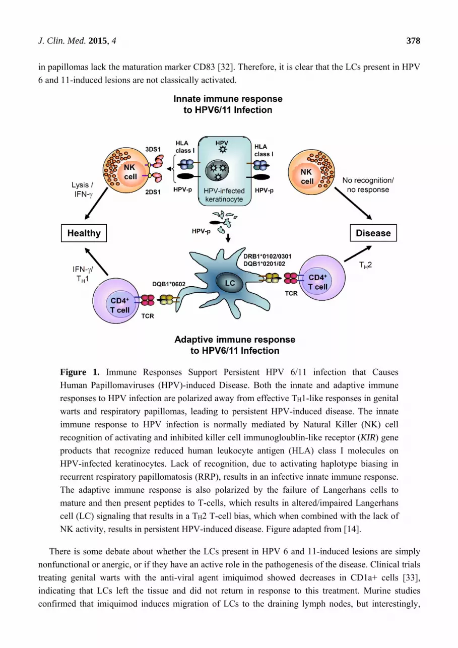
Figure 1. Immune Responses Support Persistent HPV 6/11 infection that Causes
Human Papillomaviruses (HPV)-induced Disease. Both the innate and adaptive immune
responses to HPV infection are polarized away from effective TH1-like responses in genital
warts and respiratory papillomas, leading to persistent HPV-induced disease. The innate
immune response to HPV infection is normally mediated by Natural Killer (NK) cell
recognition of activating and inhibited killer cell immunogloublin-like receptor (KIR) gene
products that recognize reduced human leukocyte antigen (HLA) class I molecules on
HPV-infected keratinocytes. Lack of recognition, due to activating haplotype biasing in
recurrent respiratory papillomatosis (RRP), results in an infective innate immune response.
The adaptive immune response is also polarized by the failure of Langerhans cells to
mature and then present peptides to T-cells, which results in altered/impaired Langerhans
cell (LC) signaling that results in a TH2 T-cell bias, which when combined with the lack of
NK activity, results in persistent HPV-induced disease. Figure adapted from [14].
There is some debate about whether the LCs present in HPV 6 and 11-induced lesions are simply
nonfunctional or anergic, or if they have an active role in the pathogenesis of the disease. Clinical trials
treating genital warts with the anti-viral agent imiquimod showed decreases in CD1a+ cells [33],
indicating that LCs left the tissue and did not return in response to this treatment. Murine studies
confirmed that imiquimod induces migration of LCs to the draining lymph nodes, but interestingly,
J. Clin. Med. 2015, 4 379
they showed that neither the costimulatory factor CD80 or CD86 was upregulated [34], bringing into
question the functionality of these cells.
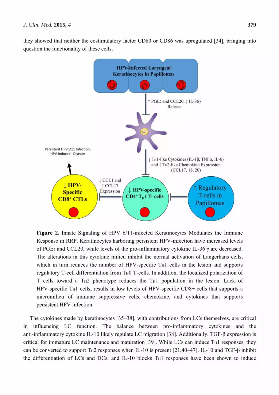
Figure 2. Innate Signaling of HPV 6/11-infected Keratinocytes Modulates the Immune
Response in RRP. Keratinocytes harboring persistent HPV-infection have increased levels
of PGE2 and CCL20, while levels of the pro-inflammatory cytokine IL-36 γ are decreased.
The alterations in this cytokine milieu inhibit the normal activation of Langerhans cells,
which in turn reduces the number of HPV-specific TH1 cells in the lesion and supports
regulatory T-cell differentiation from TH0 T-cells. In addition, the localized polarization of
T cells toward a TH2 phenotype reduces the TH1 population in the lesion. Lack of
HPV-specific TH1 cells, results in low levels of HPV-specific CD8+ cells that supports a
micromilieu of immune suppressive cells, chemokine, and cytokines that supports
persistent HPV infection.
The cytokines made by keratinocytes [35–38], with contributions from LCs themselves, are critical
in influencing LC function. The balance between pro-inflammatory cytokines and the
anti-inflammatory cytokine IL-10 likely regulate LC migration [38]. Additionally, TGF-β expression is
critical for immature LC maintenance and maturation [39]. While LCs can induce TH1 responses, they
can be converted to support TH2 responses when IL-10 is present [21,40–47]. IL-10 and TGF-β inhibit
the differentiation of LCs and DCs, and IL-10 blocks TH1 responses have been shown to induce

J. Clin. Med. 2015, 4 380
tolerance to tumors [48], while preserving LC and DC induction of TH2 responses to antigens [20].
Thus, LCs can function as either pro- or anti-inflammatory mediators of adaptive immunity [49–54].
Although the small biopsy sample sizes in RRP have to date made functional studies of LCs from
these tissues difficult, monocyte-derived immature Langerhans cells from RRP patients show an
attenuated response to the proinflammatory cytokine IL-36γ expressed by keratinocytes [32],
suggesting that these cells may be less functional. The reduced LC response may be a general response
to HPV infection, as reported in a recent study of LCs from cervical cancer, which showed a lack of
cytokine expression when LCs were stimulated with Toll 7 or 8 ligands, suggesting that LCs can
become anergic during active HPV infection [55]. However, in 2008, Cao et al. proposed that the LCs
in genital warts are not simply inactive, but instead are the sources of CCL17 and CCL22, cytokines
shown to recruit Tregs to local tissues [3,56]. Both by co-staining experiments and by qPCR they
showed that LCs produce these cytokines, and furthermore, they demonstrated that antibody-induced
blockade of either CCL17 or CCL22 inhibits migration of Tregs towards wart tissue in culture. The
proposed mechanism of Treg recruitment is consistent with the data found in RRP which shows that
CCL17 and CCL22 are produced by papilloma tissues and disseminated widely enough to be found in
plasma [10,12]. Moreover, remission of RRP patients in a clinical trial of celecoxib occurs in concert
with decreased plasma levels of these chemokines [10]. If it can be confirmed that LCs are the source
of CCL17 and CCL22 in HPV 6 and 11-induced papillomas, it would suggest that LCs play an active
role in creating and maintaining local immune suppression by recruiting T regulatory cells into HPV 6
and 11 infected epithelial tissues.
3. T Cells
Tregs, increased in many different cancer tissues, are known to be suppressors of the immune
response and they are both present and functional in genital warts and respiratory papillomas. FOXP3+
Tregs isolated from either site suppressed PBMC proliferation in micro co-culture assays [3,12]. In
genital warts, size of the wart correlated with the relative abundance of Tregs (<1% of all T cells in
small warts and >6% in large warts [3]. Direct assessment of the effects of Treg depletion was
attempted in a small clinical trial in patients with condylomata acuminata using cyclophosphamide, a
common chemotherapeutic drug known to block proliferation of dividing cells by interfering with
DNA replication, to treat patients for one week after warts were removed by laser therapy. The
obvious caveat to this study is the lack of specificity of this drug. The authors suggest that low doses of
this drug resulted in selective depletion of the Treg population, but only showed that the NK
population is not depleted in response at these low doses. Given these caveats, the clinical trial did
show that low-dose cyclophosphamide treatment depletes Tregs and increases T cell proliferation and
cytolysis. The net effect of the treatment was a decrease in detectable HPV DNA and in wart
recurrence. Thirty weeks after treatment only 17% of patients had recurrence of disease compared with
81% in the placebo group [57]. Further experiments need to be performed to show that the
non-recurrence observed in this trial is truly due to the depletion of Tregs, but it does lend support to
the predominate theory held in the field that the high proportion of Tregs found in HPV 6 and
11-induced lesions actively suppresses the HPV-specific immune response and allows for the
development and recurrence of disease.

J. Clin. Med. 2015, 4 381
It should be noted that Tregs comprise only a subset of the CD4+ T cells in HPV 6 and 11 induced
lesions, being approximately 6% in genital warts and 25% in respiratory papillomas [12,57]. However,
we have shown that in papillomas half of the CD4+ T cell population express very low levels of
CD127, indicating that these cells, although present, are exhausted [12]. Although direct analysis of
TH1 vs. TH2 cells within the CD4+ T cell subpopulation in the tissues has not been completely
characterized in sites infected with HPV 6/11, IL-10 and TGF-β, which are markers of TH2-like cells,
have been shown to be highly expressed in genital warts and laryngeal papillomas [3,8,13,58]. There is
also evidence of TH2-like polarization of the adaptive immune response to HPV 6/11 in laryngeal
papillomas and in the peripheral blood. Genital wart patients have been shown to actually have an
increased ratio of TH2/TH1 cells in PBMC, while RRP patients have an increase in serum levels of TH2
chemokines [10,59].
As a final note on T cells, CD8+ T cells are present in both genital warts and respiratory
papillomas [8,27]. These cells are likely to be immature cytotoxic T-cells, suggesting that the dominant
TH2/Treg microenvironment in respiratory papillomas has an impact on CD8+ T-cell maturation that
further prevents HPV clearance or suppression of HPV6/11-infected keratinocytes [8,32].
4. Natural Killer Cells
Class I MHC expression is markedly reduced or absent in laryngeal papillomas [4], and natural
killer (NK) cells present in papilloma tissues should remove keratinocytes that lack class I MHC
expression. However, the NK cells could be inhibited by the high proportion of Tregs in the lesions
since Tregs are known to regulate NK activity [60]. Moreover, studies suggest that suppression of NK
cell function is not restricted to the HPV-induced lesions since NKs derived from PBMC of patients
with genital warts or RRP also exhibit suppressed cytolytic activity [14,61]. The mechanism for this
suppressed activity is thought to be decreased production of IL-2 and IFNγ.
Interestingly, the frequency of killer cell immunogloublin-like receptor (KIR) gene haplotypes,
which control natural killer cell cytotoxicity to viral infection, show differences between controls and
RRP patients. In addition, it has been shown that HLA class II alleles DRB1*0102, DRB1*0301,
DQB1*0201 and DQB1*0202 confer susceptibility to RRP [4] and that patients expressing these
alleles are less likely to have the activating the KIR genes 2DS1 and 3DS1 in their NK haplotype, a
correlation that grows increasingly significant with the severity of the disease [14]. These studies
suggest that there is likely to be a genetic predisposition to develop persistent HPV6 and 11 infection
and more so, severe disease.
5. Discussion
For decades laboratories that study genital warts and RRP have been characterizing a series of
localized immune deficiencies in HPV 6- and 11-induced lesions. We have discovered that LCs are
present, but have an altered distribution and morphology, are immature in respiratory papillomas, and
that T cells in these lesions appear to have a TH2/Treg bias. Furthermore, the relatively large
proportion of Tregs in these lesions suppresses NK cells so that they fail to compensate for defective
effector T-cell function. As we learn more about each of the functions of these immunocytes in
patients with persistent HPV 6 and 11 infections, a new paradigm is developing to explain the

J. Clin. Med. 2015, 4 382
deviation in immune regulation that supports this process. Langerhans cells express chemokines that
recruit TH0 T-cells that can differentiate into inducible Tregs as well as recruiting natural Tregs to
papillomas. Both types of Tregs can polarize naïve TH0 T-cells to become TH2 cells that can inhibit
NK cell function, thus creating a localized cellular and cytokine/chemokine micromilieu that can block
the TH1-like effector T-cell responses that should clear or at least control HPV 6/11 activation (Figure 1).
What remains unclear is the role for HPV 6 and 11 in this process, other than in exploiting this
immunosuppressed microenvironment. Studies of HPV 16 indicate that early protein E6 and E7 are
responsible for downregulating IFN-γ signaling and regulate Cox-2 transcription [62], however it is
well known that HPV16 E6 and E7 proteins have required functions that are unique to HPV 16
successful replication. The dearth of HPV 6 and 11-specific information regarding the role of these
early proteins in skewing immune function is due to the lack of an animal model in which to study
HPVs 6 and 11. The vast majority of what we have learned about the localized immune suppression
was discovered by analyzing cells from tissue biopsies, either immediately or after brief passage in cell
culture, and the use of PBMC stimulated by HPV 6/11-infected tissues or purified HPV early
proteins [8,58]. Although this has afforded us great insights into the function of the immune cells in the
blood or tissue responding to HPV 6 and 11, these studies are not conducive to manipulation of viral
proteins. However we do know that HPV E7 blocks MHC class I expression [63,64] and E6 has been
shown to promote TH2-like T-cell polarization of peripheral blood mononuclear cells [8].
It is clear that HPVs 6 and 11 alone are not solely responsible for this local immune suppression,
and that a defect(s) in host response is essential in development of persistent HPV infection. Selective
enrichment of HLA class II MHC genotypes (DRB1*0102, DRB1*0301, DQB1*0201 and
DQB1*0202) and restricted KIR allele haplotypes (2DS1 and 3DS1) in the RRP population indicate
that there is indeed a genetic predisposition of these patients to develop RRP and severe disease [4,14].
It is also possible that the decreased cytolytic function of NK cells and absence of mature
HPV-specific CD8+ T-cells in PBMC and in HPV-induced lesions may be important host
susceptibility factors independent of the cytokines secreted by Tregs. More experiments are needed to
define the role of each of these observations.
Although we have focused in this review on the many similarities of immunosuppression in patients
with genital warts and RRP, it is notable that some differences do exist. Tregs are found in both
tissues, but in significantly different proportions, as stated above: 6% of CD4+ T cells in genital warts,
and as many as 25% of CD4+ T cells in laryngeal papillomas. In genital warts the proportion of Tregs
increases with the size of the wart. It may be that only the most severe of the laryngeal papillomas
were large enough to provide tissues for analysis, and thus we selected papillomas with the highest
possible proportion of Tregs. However, this is unlikely since not all of the large papillomas we assayed
contained high Treg numbers. In addition, it has been reported that expression of TLRs 2, 3, 4, 7, 8 and
9 is upregulated in genital warts [65] while our data showed no indication of changes in TLR
expression in laryngeal papillomas [66]. This may be due to differences in TLR expression in
keratinocytes at different anatomical locations.
J. Clin. Med. 2015, 4 383
6. Conclusions
Despite the minor variations in immune suppression observed in RRP and genital warts, it is clear in
both these diseases that persistent HPV 6 and 11 infections are most likely the result of impaired innate
immune responses made by resident Langerhans cells and natural killer cells. This permissive innate
response would lead to a polarization of the adaptive T-cell immune response towards an unapposed
TH2/Treg bias in the T cell population present in both patients with genital warts and RRP (Figure 2).
The major questions that remain are what the direct effects of individual HPV early proteins on this
immune dysfunction, and what novel medical strategy could be used to effectively modulate the
defective host responsiveness. Ultimately, further understanding is needed of why only a select few of
the individuals fail to contain or eliminate acute HPV6/11 infection in the larynx or perineum and are
subject to persistence or recurrent activation. The long term goal of these studies is to better understand
the innate and adaptive immune dysregulation induced by HPV6/11 in individuals who show persistent
infection and thereby improve the current treatment strategy for these diseases.
Acknowledgments
This work was supported by the National Institute of Dental and Craniofacial Research of the
National Institutes of Health under Award Number R01DE017227, and the National Institute of
Allergy and Infectious Diseases of the National Institutes of Health under Award Number
R21AI105987, and by the Feinstein Institute for Medical Research, North Shore-LIJ Health System.
The content is solely the responsibility of the authors and does not necessarily represent the official
views of the National Institutes of Health.
Author Contributions
Alexandra Lucs did the literature search, synthesized the relevant arguments and wrote the majority
of the manuscript. Vincent Bonagura contributes to the synthesis of arguments, provided guidance on
the direction of the paper and wrote sections on the manuscript. Bettie Steinberg contributed to
discussion on the details of the paper and edited the manuscript. Allan Abramson contributed clinical
samples and thoughtful discussion, without which none of this lab’s research on RRP would have been
possible. James DeVoti, Lynda Hatam and Ali Afzal contributed thoughtful discussion on the topics of
immune dysregulation and cytokine and chemokine production and release.
Conflicts of Interest
The authors declare no conflict of interest.
References
1. Patel, H.; Wagner, M.; Singhal, P.; Kothari, S. Systematic review of the incidence and prevalence
of genital warts. BMC Infect. Dis. 2013, 13, doi:10.1186/1471-2334-13-39.
2. Derkay, C.S.; Wiatrak, B. Recurrent respiratory papillomatosis: A review. Laryngoscope 2008,
118, 1236–1247.
J. Clin. Med. 2015, 4 384
3. Cao, Y.; Zhao, J.; Lei, Z.; Shen, S.; Liu, C.; Li, D.; Liu, J.; Shen, G.X.; Zhang, G.M.;
Feng, Z.H.; et al. Local accumulation of FOXP3+ regulatory T cells: Evidence for an immune
evasion mechanism in patients with large condylomata acuminata. J. Immunol. 2008, 180,
7681–7686.
4. Bonagura, V.R.; Siegal, F.P.; Abramson, A.L.; Santiagoschwarz, F.; Oreilly, M.E.; Shah, K.;
Drake, D.; Steinberg, B.M. Enriched Hla-Dq3 phenotype and decreased class-I major
histocompatibility complex antigen expression in recurrent respiratory papillomatosis.
Clin. Diagn. Lab. Immunol. 1994, 1, 357–360.
5. Stanley, M. Immunobiology of HPV and HPV vaccines. Gynecol. Oncol. 2008, 109 (Suppl. S2),
15–21.
6. Amador-Molina, A.; Hernández-Valencia, J.F.; Lamoyi, E.; Contreras-Paredes, A.; Lizano, M.
Role of innate immunity against human papillomavirus (HPV) infections and effect of adjuvants
in promoting specific immune response. Viruses 2013, 5, 2624–2642.
7. Sasagawa, T.; Takagi, H.; Makinoda, S. Immune responses against human papillomavirus (HPV)
infection and evasion of host defense in cervical cancer. J. Infect. Chemother. 2012, 18, 807–815.
8. Bonagura, V.R.; Hatam, L.; DeVoti, J.; Zeng, F.F.; Steinberg, B.M. Recurrent respiratory
papillomatosis: Altered CD8(+) T-cell subsets and T(h)1/T(h)2 cytokine imbalance.
Clin. Immunol. 1999, 93, 302–311.
9. Bonagura, V.R.; Vambutas, A.; DeVoti, J.A.; Rosenthal, D.W.; Steinberg, B.M.; Abramson, A.L.;
Shikowitz, M.J.; Gjertson, D.W.; Reed, E.F. Hla alleles, IFN-gamma responses to HPV-11 E6,
and disease severity in patients with recurrent respiratory papillomatosis. Hum. Immunol. 2004, 65,
773–782.
10. Rosenthal, D.W.; DeVoti, J.A.; Steinberg, B.M.; Abramson, A.L.; Bonagura, V.R. T(h)2-like
chemokine patterns correlate with disease severity in patients with recurrent respiratory
papillomatosis. Mol. Med. 2012, 18, 1338–1345.
11. Hatam, L.J.; Rosenthal, D.W.; DeVoti, J.A.; Lam, F.; Abramson, A.; Steinberg, B.M.;
Bonagura, V.R. CD4(+)FOXP3(+)CD127(+low) T-regulatory cells are increased in HPV infected
papillomas in patients with recurrent respiratory papillomatosis (RRP). J. Allergy Clin. Immunol.
2008, 121, doi:10.1016/j.jaci.2007.12.789.
12. Hatam, L.J.; DeVoti, J.A.; Rosenthal, D.W.; Lam, F.; Abramson, A.L.; Steinberg, B.M.;
Bonagura, V.R. Immune suppression in premalignant respiratory papillomas: Enriched functional
CD4(+)FOXP3(+) regulatory T cells and PD-1/PD-L1/L2 expression. Clin. Cancer Res. 2012, 18,
1925–1935.
13. Bonagura, V.R.; Hatam, L.J.; Rosenthal, D.W.; De Voti, J.A.; Lam, F.; Steinberg, B.M.;
Abramson, A.L. Recurrent respiratory papillomatosis: A complex defect in immune
responsiveness to human papillomavirus-6 and -11. Apmis 2010, 118, 455–470.
14. Bonagura, V.R.; Du, Z.; Ashouri, E.; Luo, L.; Hatam, L.J.; DeVoti, J.A.; Rosenthal, D.W.;
Steinberg, B.M.; Abramson, A.L.; Gjertson, D.W.; et al. Activating killer cell
immunoglobulin-like receptors 3DS1 and 2DS1 protect against developing the severe form of
recurrent respiratory papillomatosis. Hum. Immunol. 2010, 71, 212–219.
15. Steinman, R.M. The dendritic cell system and its role in immunogenicity. Annu. Rev. Immunol.
1991, 9, 271–296.
J. Clin. Med. 2015, 4 385
16. Hart, D.N. Dendritic cells: Unique leukocyte populations which control the primary immune
response. Blood 1997, 90, 3245–3287.
17. Matzinger, P. Tolerance, danger, and the extended family. Annu. Rev. Immunol. 1994, 12, 991–1045.
18. Noel, W.; Raes, G.; Hassanzadeh Ghassabeh, G.; De Baetselier, P.; Beschin, A. Alternatively
activated macrophages during parasite infections. Trends Parasitol. 2004, 20, 126–133.
19. Lutz, M.B.; Suri, R.M.; Niimi, M.; Ogilvie, A.L.; Kukutsch, N.A.; Rossner, S.; Schuler, G.;
Austyn, J.M. Immature dendritic cells generated with low doses of GM-CSF in the absence of
IL-4 are maturation resistant and prolong allograft survival in vivo. Eur. J. Immunol. 2000, 30,
1813–1822.
20. McGuirk, P.; McCann, C.; Mills, K.H. Pathogen-specific T regulatory 1 cells induced in the
respiratory tract by a bacterial molecule that stimulates interleukin 10 production by dendritic cells:
A novel strategy for evasion of protective T helper type 1 responses by bordetella pertussis.
J. Exp. Med. 2002, 195, 221–231.
21. Shortman, K.; Liu, Y.J. Mouse and human dendritic cell subtypes. Nat. Rev. Immunol. 2002, 2,
151–161.
22. Cunningham, A.L.; Carbone, F.; Geijtenbeek, T.B. Langerhans cells and viral immunity.
Eur J Immunol 2008, 38, 2377–2385.
23. Da Silva, D.M.; Movius, C.A.; Raff, A.B.; Brand, H.E.; Skeate, J.G.; Wong, M.K.; Kast, W.M.
Suppression of langerhans cell activation is conserved amongst human papillomavirus α and β
genotypes, but not a µ genotype. Virology 2014, 452–453, 279–286.
24. Leong, C.M.; Doorbar, J.; Nindl, I.; Yoon, H.S.; Hibma, M.H. Deregulation of e-cadherin by
human papillomavirus is not confined to high-risk, cancer-causing types. Br. J. Dermatol. 2010,
163, 1253–1263.
25. Bhawan, J.; Dayal, Y.; Bhan, A.K. Langerhans cells in molluscum contagiosum, verruca vulgaris,
plantar wart, and condyloma acuminatum. J. Am. Acad. Dermatol. 1986, 15, 645–649.
26. Viac, J.; Chardonnet, Y.; Euvrard, S.; Chignol, M.C.; Thivolet, J. Langerhans cells, inflammation
markers and human papillomavirus infections in benign and malignant epithelial tumors from
transplant recipients. J. Dermatol. 1992, 19, 67–77.
27. Coleman, N.; Birley, H.D.; Renton, A.M.; Hanna, N.F.; Ryait, B.K.; Byrne, M.;
Taylor-Robinson, D.; Stanley, M.A. Immunological events in regressing genital warts.
Am. J. Clin. Pathol. 1994, 102, 768–774.
28. McMillan, A.; Bishop, P.E.; Fletcher, S. An immunohistological study of condylomata acuminata.
Histopathology 1990, 17, 45–52.
29. Arany, I.; Tyring, S.K. Status of local cellular immunity in interferon-responsive and -nonresponsive
human papillomavirus-associated lesions. Sex. Transm. Dis. 1996, 23, 475–480.
30. Morelli, A.E.; Belardi, G.; DiPaola, G.; Paredes, A.; Fainboim, L. Cellular subsets and epithelial
ICAM-1 and HLA-DR expression in human papillomavirus infection of the vulva.
Acta Derm. Venereol. 1994, 74, 45–50.
31. Feng, J.Y.; Peng, Z.H.; Tang, X.P.; Geng, S.M.; Liu, Y.P. Immunohistochemical and
ultrastructural features of langerhans cells in condyloma acuminatum. J. Cutan. Pathol. 2008, 35,
15–20.
J. Clin. Med. 2015, 4 386
32. Devoti, J.; Hatam, L.; Lucs, A.; Afzal, A.; Abramson, A.; Steinberg, B.; Bonagura, V. Decreased
langerhans cell responses to IL-36γ: Altered innate immunity in patients with recurrent respiratory
papillomatosis. Mol. Med. 2014, 20, 372–380.
33. Arany, I.; Tyring, S.K.; Stanley, M.A.; Tomai, M.A.; Miller, R.L.; Smith, M.H.; McDermott, D.J.;
Slade, H.B. Enhancement of the innate and cellular immune response in patients with genital
warts treated with topical imiquimod cream 5%. Antivir. Res. 1999, 43, 55–63.
34. Suzuki, H.; Wang, B.; Shivji, G.M.; Toto, P.; Amerio, P.; Tomai, M.A.; Miller, R.L.; Sauder, D.N.
Imiquimod, a topical immune response modifier, induces migration of langerhans cells.
J. Investig. Dermatol. 2000, 114, 135–141.
35. Shirakata, Y. Regulation of epidermal keratinocytes by growth factors. J. Dermatol. Sci. 2010, 59,
73–80.
36. Pastore, S.; Mascia, F.; Girolomoni, G. The contribution of keratinocytes to the pathogenesis of
atopic dermatitis. Eur. J. Dermatol. 2006, 16, 125–131.
37. Werner, S.; Krieg, T.; Smola, H. Keratinocyte-fibroblast interactions in wound healing.
J. Investig. Dermatol. 2007, 127, 998–1008.
38. Wang, B.; Amerio, P.; Sauder, D.N. Role of cytokines in epidermal langerhans cell migration.
J. Leukoc. Biol. 1999, 66, 33–39.
39. Kel, J.M.; Girard-Madoux, M.J.; Reizis, B.; Clausen, B.E. TGF-beta is required to maintain the
pool of immature langerhans cells in the epidermis. J. Immunol. 2010, 185, 3248–3255.
40. Banchereau, J.; Briere, F.; Caux, C.; Davoust, J.; Lebecque, S.; Liu, Y.J.; Pulendran, B.; Palucka, K.
Immunobiology of dendritic cells. Annu. Rev. Immunol. 2000, 18, 767–811.
41. Del Hoyo, G.M.; Martin, P.; Vargas, H.H.; Ruiz, S.; Arias, C.F.; Ardavin, C. Characterization of a
common precursor population for dendritic cells. Nature 2002, 415, 1043–1047.
42. Banchereau, J.; Paczesny, S.; Blanco, P.; Bennett, L.; Pascual, V.; Fay, J.; Palucka, A.K.
Dendritic cells: Controllers of the immune system and a new promise for immunotherapy.
Ann. N. Y. Acad. Sci. 2003, 987, 180–187.
43. Caux, C.; Massacrier, C.; Vanbervliet, B.; Dubois, B.; Durand, I.; Cella, M.; Lanzavecchia, A.;
Banchereau, J. CD34+ hematopoietic progenitors from human cord blood differentiate along two
independent dendritic cell pathways in response to granulocyte-macrophage colony-stimulating
factor plus tumor necrosis factor alpha: II. Functional analysis. Blood 1997, 90, 1458–1470.
44. Steinman, R.M.; Turley, S.; Mellman, I.; Inaba, K. The induction of tolerance by dendritic cells
that have captured apoptotic cells. J. Exp. Med. 2000, 191, 411–416.
45. Iwasaki, A.; Kelsall, B.L. Freshly isolated peyer’s patch, but not spleen, dendritic cells produce
interleukin 10 and induce the differentiation of T helper type 2 cells. J. Exp. Med. 1999, 190,
229–239.
46. Enk, A.H.; Angeloni, V.L.; Udey, M.C.; Katz, S.I. Inhibition of langerhans cell antigen-presenting
function by IL-10. A role for il-10 in induction of tolerance. J. Immunol. 1993, 151, 2390–2398.
47. Beissert, S.; Hosoi, J.; Grabbe, S.; Asahina, A.; Granstein, R.D. IL-10 inhibits tumor antigen
presentation by epidermal antigen-presenting cells. J. Immunol. 1995, 154, 1280–1286.
48. Cottrez, F.; Groux, H. Specialization in tolerance: Innate CD(4+)CD(25+) versus acquired TR1
and TH3 regulatory T cells. Transplantation 2004, 77 (Suppl. S1), 12–15.

J. Clin. Med. 2015, 4 387
49. Johnston, A.; Xing, X.; Guzman, A.M.; Riblett, M.; Loyd, C.M.; Ward, N.L.; Wohn, C.; Prens, E.P.;
Wang, F.; Maier, L.E.; et al. IL-1F5, -F6, -F8, and -F9: A novel IL-1 family signaling system that
is active in psoriasis and promotes keratinocyte antimicrobial peptide expression. J. Immunol.
2011, 186, 2613–2622.
50. Romani, N.; Ebner, S.; Tripp, C.H.; Flacher, V.; Koch, F.; Stoitzner, P. Epidermal langerhans
cells—Changing views on their function in vivo. Immunol. Lett. 2006, 106, 119–125.
51. Elias, A.; Nanda, V.; Barr, R. CD1A expression in psoriatic skin following treatment with
propylthiouracil, an antithyroid thioureylene. BMC Dermatol. 2003, 3, doi:10.1186/1471-5945-3-3.
52. Kaplan, D.H.; Jenison, M.C.; Saeland, S.; Shlomchik, W.D.; Shlomchik, M.J. Epidermal
langerhans cell-deficient mice develop enhanced contact hypersensitivity. Immunity 2005, 23,
611–620.
53. Klechevsky, E.; Morita, R.; Liu, M.; Cao, Y.; Coquery, S.; Thompson-Snipes, L.; Briere, F.;
Chaussabel, D.; Zurawski, G.; Palucka, A.K.; et al. Functional specializations of human epidermal
langerhans cells and CD14+ dermal dendritic cells. Immunity 2008, 29, 497–510.
54. Bennett, C.L.; Fallah-Arani, F.; Conlan, T.; Trouillet, C.; Goold, H.; Chorro, L.; Flutter, B.;
Means, T.K.; Geissmann, F.; Chakraverty, R. Langerhans cells regulate cutaneous injury by
licensing CD8 effector cells recruited to the skin. Blood 2011, 117, 7063–7069.
55. Kumar, M.M.; Adurthi, S.; Ramachandran, S.; Mukherjee, G.; Joy, O.; Krishnamurthy, H.;
Krishna, S.; Bafna, U.D.; Uma, D.K.; Jayshree, R.S. Toll-like receptors 7, 8, and 9 expression
and function in primary human cervical cancer langerhans cells: Evidence of anergy.
Int. J. Gynecol. Cancer 2013, 23, 184–192.
56. Sather, B.D.; Treuting, P.; Perdue, N.; Miazgowicz, M.; Fontenot, J.D.; Rudensky, A.Y.;
Campbell, D.J. Altering the distribution of FOXP3(+) regulatory T cells results in tissue-specific
inflammatory disease. J. Exp. Med. 2007, 204, 1335–1347.
57. Cao, Y.; Zhao, J.; Yang, Z.; Cai, Z.; Zhang, B.; Zhou, Y.; Shen, G.X.; Chen, X.; Li, S.; Huang, B.
CD4+FOXP3+ regulatory T cell depletion by low-dose cyclophosphamide prevents recurrence in
patients with large condylomata acuminata after laser therapy. Clin. Immunol. 2010, 136, 21–29.
58. DeVoti, J.A.; Steinberg, B.M.; Rosenthal, D.W.; Hatam, L.; Vambutas, A.; Abramson, A.L.;
Shikowitz, M.J.; Bonagura, V.R. Failure of gamma interferon but not interleukin-10 expression in
response to human papillomavirus type 11 e6 protein in respiratory papillomatosis.
Clin. Diagn. Lab. Immunol. 2004, 11, 538–547.
59. Xu, Y.; Zhu, K.J.; Zhu, N.; Jiang, D.H.; Chen, X.Z.; Cheng, H. Expression of
FOXP3+CD4+CD25+ regulatory T cells and TH1/TH2, TC1/Tc2 profiles in the peripheral blood
of patients with condyloma acuminatum. Clin. Exp. Dermatol. 2009, 34, 229–235.
60. Pedroza-Pacheco, I.; Madrigal, A.; Saudemont, A. Interaction between natural killer cells and
regulatory T cells: Perspectives for immunotherapy. Cell. Mol. Immunol. 2013, 10, 222–229.
61. Cauda, R.; Tyring, S.K.; Grossi, C.E.; Tilden, A.B.; Hatch, K.D.; Sams, W.M.; Baron, S.;
Whitley, R.J. Patients with condyloma acuminatum exhibit decreased interleukin-2 and interferon
gamma production and depressed natural killer activity. J. Clin. Immunol. 1987, 7, 304–311.
62. Subbaramaiah, K.; Dannenberg, A.J. Cyclooxygenase-2 transcription is regulated by human
papillomavirus 16 e6 and e7 oncoproteins: Evidence of a corepressor/coactivator exchange.
Cancer Res 2007, 67, 3976–3985.

J. Clin. Med. 2015, 4 388
63. Vambutas, A.; DeVoti, J.; Pinn, W.; Steinberg, B.M.; Bonagura, V.R. Tap-1 down-regulation in
laryngeal papillomas: Contribution of HPV 6/11 E7 protein. FASEB J. 2001, 15, A1009.
64. Li, H.; Zhan, T.; Li, C.; Liu, M.; Wang, Q.K. Repression of MHC class I transcription by
HPV16E7 through interaction with a putative rxrbeta motif and NF-kappaB cytoplasmic
sequestration. Biochem. Biophys. Res. Commun. 2009, 388, 383–388.
65. Shi, Y.J.; Yang, J.; Yang, W. Mechanistic investigation of immunosuppression in patients with
condyloma acuminata. Mol. Med. Rep. 2013, 8, 480–486.
66. DeVoti, J.A.; Rosenthal, D.W.; Wu, R.; Abramson, A.L.; Steinberg, B.M.; Bonagura, V.R.
Immune dysregulation and tumor-associated gene changes in recurrent respiratory papillomatosis:
A paired microarray analysis. Mol. Med. 2008, 14, 608–617.
© 2015 by the authors; licensee MDPI, Basel, Switzerland. This article is an open access article
distributed under the terms and conditions of the Creative Commons Attribution license
(http://creativecommons.org/licenses/by/4.0/).