imidazolium 4-aminobenzoate
TRANSCRIPT

Imidazolium 4-aminobenzoate
Rodolfo Moreno-Fuquen,a* Alan R. Kennedy,b Denise
Gilmour,b R. H. De Almeida Santosc and Rommel B.
Vianac
aDepartamento de Quımica - Facultad de Ciencias, Universidad del Valle, Apartado
25360, Santiago de Cali, Colombia, bWestCHEM, Department of Pure and Applied
Chemistry, University of Strathclyde, 295 Cathedral Street, Glasgow G1 1XL,
Scotland, and cInstituto de Quımica de Sao Carlos, Universidade de Sao Paulo, USP,
Sao Carlos, SP, Brazil
Correspondence e-mail: [email protected]
Received 22 October 2009; accepted 3 November 2009
Key indicators: single-crystal X-ray study; T = 123 K; mean �(C–C) = 0.002 A;
R factor = 0.048; wR factor = 0.115; data-to-parameter ratio = 15.4.
In the title salt, C3H5N2+�C7H6NO2
�, the carboxylate group of
the 4-aminobenzoate anion forms a dihedral angle of
13.23 (17)� with respect to the benzene ring. There are N—
H� � �O hydrogen-bonding interactions between the anion and
cation, and weak intermolecular C—H� � �O contacts with
carboxylate O-atom acceptors of the 4-aminobenzoate anion
result in extended three-dimensional R44(22) and R5
6(30) edge-
fused rings along the [100], [010] and [001] directions.
Related literature
For the antimicrobial and antiprotozoal biological activity of
imidazole, see: Kopanska et al. (2004); Sondhi et al. (2002). For
the biological activity of 4-aminobenzoic acid, see: Lai &
Marsh (1967); Robinson (1966). For related structures, see:
Moreno-Fuquen et al. (1996, 2009); McMullan et al. (1979). For
hydrogen-bond motifs, see: Etter (1990). For hydrogen bonds,
see: Nardelli (1995).
Experimental
Crystal data
C3H5N2+�C7H6NO2
�
Mr = 205.22Monoclinic, P21=na = 7.2038 (5) Ab = 11.6812 (6) Ac = 12.0152 (6) A� = 105.223 (6)�
V = 975.59 (10) A3
Z = 4Mo K� radiation� = 0.10 mm�1
T = 123 K0.20 � 0.15 � 0.12 mm
Data collection
Oxford Diffraction Gemini Sdiffractometer
Absorption correction: multi-scan(CrysAlis CCD; OxfordDiffraction, 2009)Tmin = 0.945, Tmax = 1.000
8533 measured reflections2360 independent reflections1700 reflections with I > 2�(I)Rint = 0.049
Refinement
R[F 2 > 2�(F 2)] = 0.048wR(F 2) = 0.115S = 1.022360 reflections153 parameters
H atoms treated by a mixture ofindependent and constrainedrefinement
��max = 0.28 e A�3
��min = �0.36 e A�3
Table 1Hydrogen-bond geometry (A, �).
D—H� � �A D—H H� � �A D� � �A D—H� � �A
N3—H4N� � �O1i 0.93 (2) 1.76 (2) 2.6869 (15) 171 (2)N2—H3N� � �O1ii 0.956 (19) 1.742 (19) 2.6938 (15) 173.5 (19)N1—H1N� � �O2iii 0.94 (2) 2.17 (2) 2.9495 (16) 139.3 (17)N1—H2N� � �O1ii 0.89 (2) 2.20 (2) 3.0149 (17) 152.0 (16)C6—H6� � �O2iv 0.95 2.55 3.3533 (16) 142
Symmetry codes: (i) �xþ 1;�yþ 2;�zþ 1; (ii) xþ 12;�yþ 3
2; z� 12; (iii)
�x þ 12; y� 1
2;�zþ 12; (iv) x� 1
2;�yþ 32; z� 1
2.
Data collection: CrysAlis CCD (Oxford Diffraction, 2009); cell
refinement: CrysAlis CCD; data reduction: CrysAlis RED (Oxford
Diffraction, 2009); program(s) used to solve structure: SHELXS97
(Sheldrick, 2008); program(s) used to refine structure: SHELXL97
(Sheldrick, 2008); molecular graphics: ORTEP-3 for Windows
(Farrugia, 1997) and Mercury (Macrae et al., 2006); software used to
prepare material for publication: WinGX (Farrugia, 1999).
RMF is grateful to the Spanish Research Council (CSIC)
for the use of a free-of-charge licence to the Cambridge
Structural Database (Allen, 2002). RMF also wishes to thank
the Universidad del Valle, Colombia, and Instituto de Quımica
de Sao Carlos, Brazil for partial financial support.
Supplementary data and figures for this paper are available from theIUCr electronic archives (Reference: FJ2253).
References
Allen, F. H. (2002). Acta Cryst. B58, 380–388.Etter, M. (1990). Acc. Chem. Res. 23, 120–126.Farrugia, L. J. (1997). J. Appl. Cryst. 30, 565.Farrugia, L. J. (1999). J. Appl. Cryst. 32, 837–838.Kopanska, K., Najda, A., Justyna, Z., Chomicz, L., Piekarczyk, J., Myja, P. &
Bretner, M. (2004). Bioorg. Med. Chem. 12, 2617–2624.Lai, T. F. & Marsh, R. E. (1967). Acta Cryst. 22, 885–893.Macrae, C. F., Edgington, P. R., McCabe, P., Pidcock, E., Shields, G. P., Taylor,
R., Towler, M. & van de Streek, J. (2006). J. Appl. Cryst. 39, 453–457.McMullan, R. K., Epstein, J., Ruble, J. R. & Craven, B. M. (1979). Acta Cryst.
B35, 688–691.Moreno-Fuquen, R., De Almeida Santos, R. H. & Lechat, J. R. (1996). Acta
Cryst. C52, 220–222.Moreno-Fuquen, R., Ellena, J. & Theodoro, J. E. (2009). Acta Cryst. E65,
o2717.Nardelli, M. (1995). J. Appl. Cryst. 28, 659.
organic compounds
o3044 Moreno-Fuquen et al. doi:10.1107/S160053680904625X Acta Cryst. (2009). E65, o3044–o3045
Acta Crystallographica Section E
Structure ReportsOnline
ISSN 1600-5368
Oxford Diffraction (2009). CrysAlis CCD and CrysAlis RED. OxfordDiffraction Ltd, Yarnton, England.
Robinson, F. A. (1966). The Vitamin Co-factors of Enzyme Systems, pp. 541–662. London: Pergamon.
Sheldrick, G. M. (2008). Acta Cryst. A64, 112–122.Sondhi, S. M., Rajvanshi, S., Johar, M., Bharti, N., Azam, A. & Singh, A. K.
(2002). Eur. J. Med. Chem. 37, 835–843.
organic compounds
Acta Cryst. (2009). E65, o3044–o3045 Moreno-Fuquen et al. � C3H5N2+�C7H6NO2
� o3045
supplementary materials
supplementary materials
sup-1
Acta Cryst. (2009). E65, o3044-o3045 [ doi:10.1107/S160053680904625X ]
Imidazolium 4-aminobenzoate
R. Moreno-Fuquen, A. R. Kennedy, D. Gilmour, R. H. De Almeida Santos and R. B. Viana
Comment
The title adduct, C7H6NO2-, C3H5N2
+ (imidazolium 4-aminobenzoate), (I), is part of a series of studies on imidazole, which
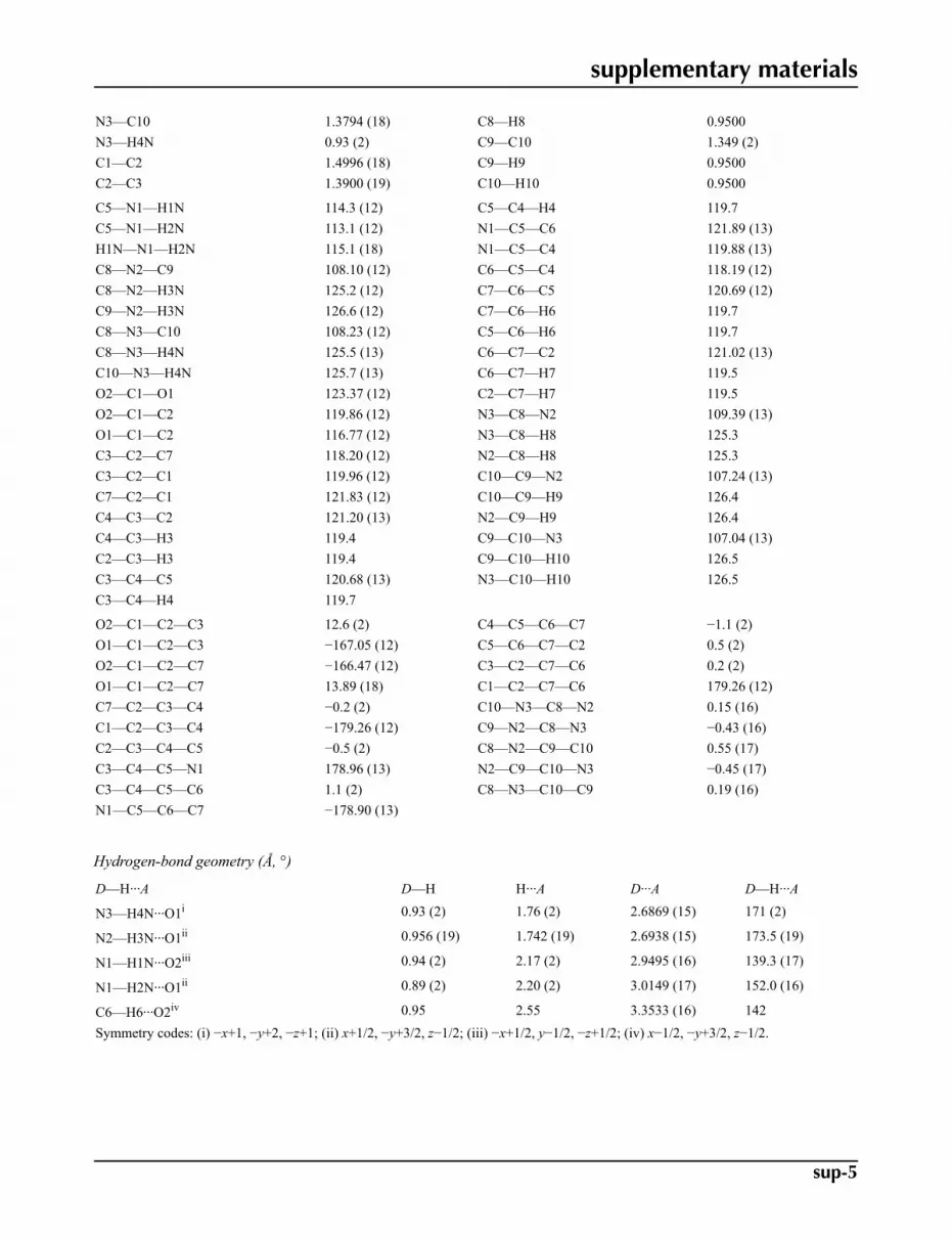
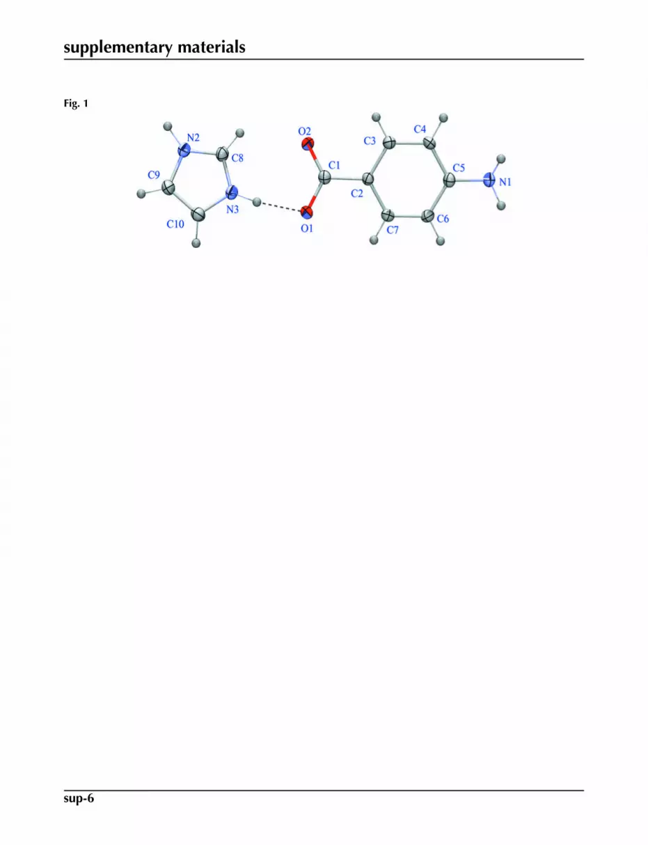
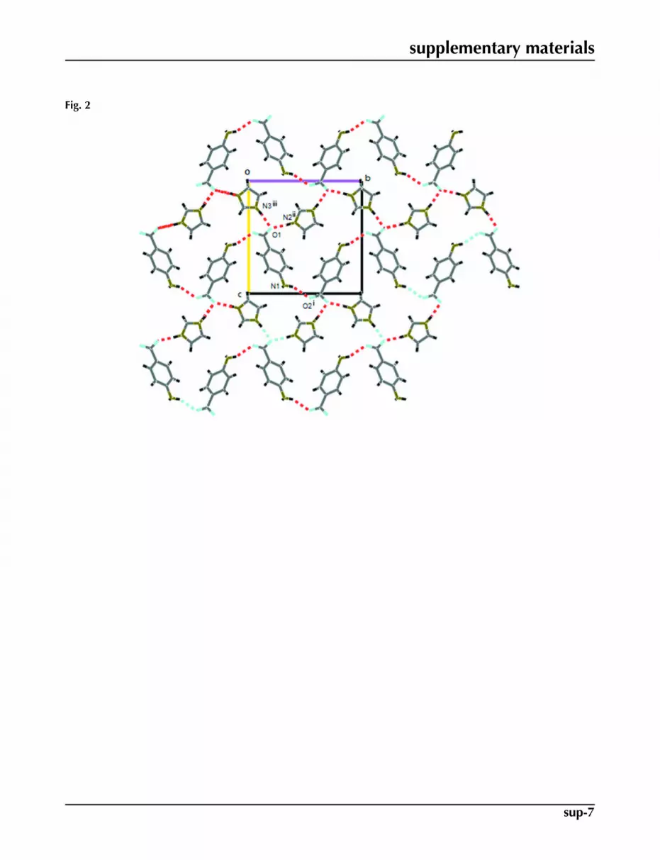
have been made in our research group (Moreno-Fuquen et al., 2009). Imidazole derivatives have a wide variety of agentsholding biological activities and is used in the field of pharmaceuticals and medicine like antimicrobial and antiprotozoal(Kopanska et al., 2004) or anti-inflammatory agents (Sondhi et al., 2002). In turn, 4-Aminobenzoic acid (PABA) (Lai &Marsh, 1967) is an important biological molecule, involving the synthesis of folic acid (Robinson, 1966) and promoting theextension of hydrogen-bonded network structures. To continue research on the structural behavior of the imidazole moleculewith different hydrogen bond donors, the system imidazolium 4-aminobenzoate adduct (I), is reported. The 4-aminoben-zoic acid and 4-nitropyridine N-oxide (PABA+NPNO) molecular complex (Moreno-Fuquen et al., 1996) and the PABAand imidazole (IM) free molecules (McMullan et al., 1979) may be used as reference systems in order to compare to thetitle imidazolium salt. The molecular structure of (I) is shown in Fig. 1. The title compound shows a dihedral angle of23.71 (8)°, between benzene and imidazole planes. In turn, the carboxylate group of 4-aminobenzoate shows a dihedralangle of 13.23 (17)° with respect to the benzene ring, following the same structural behavior of the group in the free PABAmolecule and in the PABA+NPNO adduct. The (PABA), as well as other organic acids, shows the formation of centrosym-metric hydrogen-bonded dimers in its structure. As a product of the reaction with imidazole (IM), the dimer in the PABAmolecule is broken, and begins the transference of the proton to the basic N-atom of the IM molecule forming the title adduct.Some structural changes in the formation of the imidazolium salt, are observed: N2—C8 bond length changes from 1.358in the free IM molecule, to 1.3281 (17) Å in (I); C1—O1 and C6—C7 bond lengths change from 1.210 (4) and 1.366 (5)Å in the (PABA +NPNO) adduct to 1.2368 (16) and 1.3850 (18) Å in the title adduct. The other bond lengths and bondangles of (I) are in good agreement with the standard values and correspond to those observed in the IM free moleculeand (PABA+NPNO) reference systems. The formation of the salt, resulting in N—H···O hydrogen-bonding interactionsand C—H···O intermolecular weak contacts with carboxylate O-atoms acceptors: The two components of the adduct areconnected via intermolecular N—H···O hydrogen bonds and C—H···O weak contacts, (Table 1) (Nardelli, 1995) and theseinteractions define an infinite three dimensional framework. In a first substructure, the strongest hydrogen bonds N—H···Ointeractions are responsible for crystal growth. Indeed, there are two intermolecular N—H···O hydrogen bond interactionswhich link one molecule of PABA and 2 molecules of IM. A third N—H···O hydrogen bond links two PABA molecules.
All these interactions link the moieties into molecular sheets that extend in the b and c directions forming R56(30) (Etter,
1990) edge-fused rings (Fig. 2). In a second substructure, the PABA molecules are linked by N—H···O hydrogen-bonding
and intermolecular C—H···O weak interactions which form a R44(22) e dge-fused rings along a and c directions (Fig 3). All
of these interactions define the bulk structure of the crystal.
Experimental
The synthesis of the title compound (I) was carried out by slow evaporation of equimolar quantities of 4-aminobenzoicacid (0.625 g, 0.0046 mol) and imidazole (0.310 g) in 100 ml of a mixture of dry acetonitrile. Colourless blocks of a good

supplementary materials
sup-2
quality, suitable for X-ray analysis with a melting point of 371 (1) K were obtained. The initial reagents were purchasedfrom Aldrich Chemical Co., and were used as received.
Refinement
All H-atoms were located from difference maps and were positioned geometrically and refined using a riding model withC–H= 0.93–0.97 Å and Uiso(H)= 1.2Ueq(C).
Figures
Fig. 1. An ORTEP-3 (Farrugia, 1997) plot of the title (I) compound, with the atomic labellingscheme. The shapes of the ellipsoids correspond to 50% probability contours of atomic dis-placement and, for the sake of clarity, H atoms are shown as spheres of arbitrary radius.
Fig. 2. The packing in the unit cell of (I) viewed down the a axis, showing the formation ofR5
6(30) e dge-fused rings and also the hydrogen-bonding interactions as broken lines. Sym-metry code: (i) -x + 1/2, y - 1/2, -z + 1/2; (ii) x - 1/2, -y + 3/2, z - 1/2; (iii) -x, -y + 2, -z + 1.
Fig. 3. The packing in the unit cell of (I) viewed down the b axis, showing the formation ofR4
4(22) e dge-fused rings and also the hydrogen-bonding interactions as broken lines. Sym-metry code: (i) x + 1/2, -y + 3/2, z - 1/2; (ii) x - 1/2, -y + 3/2, z - 1/2.
Imidazolium 4-aminobenzoate
Crystal data
C3H5N2+·C7H6NO2
– F000 = 432
Mr = 205.22 Dx = 1.397 Mg m−3
Monoclinic, P21/n Melting point: 443.0(10) KHall symbol: -P 2yn Mo Kα radiation, λ = 0.71073 Åa = 7.2038 (5) Å Cell parameters from 3056 reflectionsb = 11.6812 (6) Å θ = 2.5–30.9ºc = 12.0152 (6) Å µ = 0.10 mm−1
β = 105.223 (6)º T = 123 K
V = 975.59 (10) Å3 Fragment, colourlessZ = 4 0.20 × 0.15 × 0.12 mm
Data collection
Oxford Diffraction Gemini Sdiffractometer 2360 independent reflections
Radiation source: fine-focus sealed tube 1700 reflections with I > 2σ(I)

supplementary materials
sup-3
Monochromator: graphite Rint = 0.049
T = 123 K θmax = 28.0º
ω scans θmin = 2.5ºAbsorption correction: multi-scan(CrysAlis CCD; Oxford Diffraction, 2009) h = −9→9
Tmin = 0.945, Tmax = 1.000 k = −13→158533 measured reflections l = −15→15
Refinement
Refinement on F2 Hydrogen site location: inferred from neighbouringsites
Least-squares matrix: full H atoms treated by a mixture ofindependent and constrained refinement
R[F2 > 2σ(F2)] = 0.048 w = 1/[σ2(Fo
2) + (0.067P)2]where P = (Fo
2 + 2Fc2)/3
wR(F2) = 0.115 (Δ/σ)max < 0.001
S = 1.02 Δρmax = 0.28 e Å−3
2360 reflections Δρmin = −0.36 e Å−3
153 parametersExtinction correction: SHELXL97 (Sheldrick, 2008),Fc*=kFc[1+0.001xFc2λ3/sin(2θ)]-1/4
Primary atom site location: structure-invariant directmethods Extinction coefficient: 0.213 (14)
Secondary atom site location: difference Fourier map
Special details
Geometry. All e.s.d.'s (except the e.s.d. in the dihedral angle between two l.s. planes) are estimated using the full covariance mat-rix. The cell e.s.d.'s are taken into account individually in the estimation of e.s.d.'s in distances, angles and torsion angles; correlationsbetween e.s.d.'s in cell parameters are only used when they are defined by crystal symmetry. An approximate (isotropic) treatment ofcell e.s.d.'s is used for estimating e.s.d.'s involving l.s. planes.
Refinement. Refinement of F2 against ALL reflections. The weighted R-factor wR and goodness of fit S are based on F2, convention-
al R-factors R are based on F, with F set to zero for negative F2. The threshold expression of F2 > σ(F2) is used only for calculating R-
factors(gt) etc. and is not relevant to the choice of reflections for refinement. R-factors based on F2 are statistically about twice as largeas those based on F, and R- factors based on ALL data will be even larger.
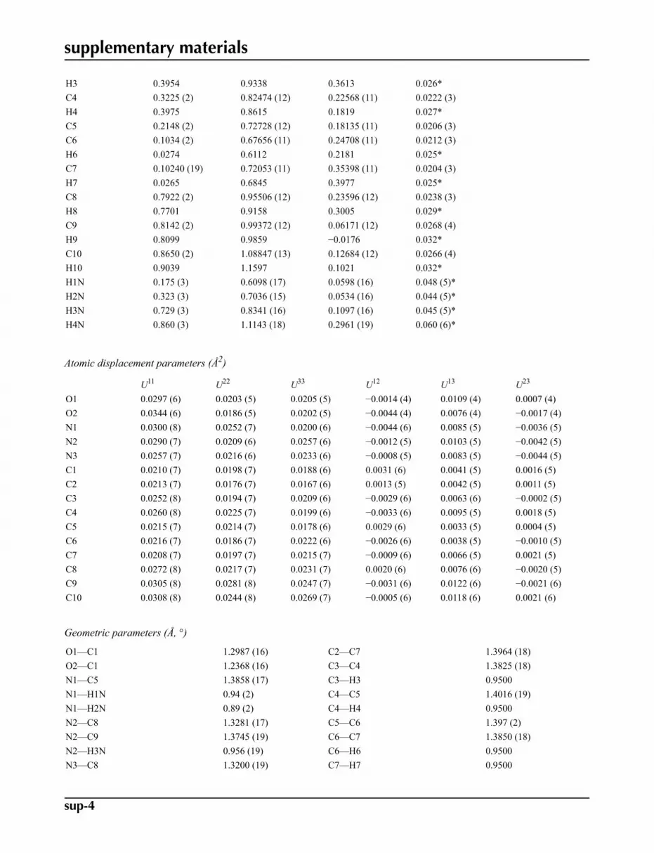
Fractional atomic coordinates and isotropic or equivalent isotropic displacement parameters (Å2)
x y z Uiso*/Ueq
O1 0.13700 (14) 0.80374 (8) 0.58020 (7) 0.0227 (3)O2 0.27846 (15) 0.96304 (8) 0.54056 (8) 0.0243 (3)N1 0.2159 (2) 0.68622 (11) 0.07332 (10) 0.0247 (3)N2 0.77001 (18) 0.91059 (11) 0.13156 (10) 0.0246 (3)N3 0.84977 (17) 1.06234 (10) 0.23605 (10) 0.0231 (3)C1 0.2104 (2) 0.86668 (12) 0.51343 (11) 0.0201 (3)C2 0.21134 (19) 0.81695 (11) 0.39852 (10) 0.0187 (3)C3 0.3206 (2) 0.86798 (12) 0.33249 (11) 0.0218 (3)
supplementary materials
sup-4
H3 0.3954 0.9338 0.3613 0.026*C4 0.3225 (2) 0.82474 (12) 0.22568 (11) 0.0222 (3)H4 0.3975 0.8615 0.1819 0.027*C5 0.2148 (2) 0.72728 (12) 0.18135 (11) 0.0206 (3)C6 0.1034 (2) 0.67656 (11) 0.24708 (11) 0.0212 (3)H6 0.0274 0.6112 0.2181 0.025*C7 0.10240 (19) 0.72053 (11) 0.35398 (11) 0.0204 (3)H7 0.0265 0.6845 0.3977 0.025*C8 0.7922 (2) 0.95506 (12) 0.23596 (12) 0.0238 (3)H8 0.7701 0.9158 0.3005 0.029*C9 0.8142 (2) 0.99372 (12) 0.06171 (12) 0.0268 (4)H9 0.8099 0.9859 −0.0176 0.032*C10 0.8650 (2) 1.08847 (13) 0.12684 (12) 0.0266 (4)H10 0.9039 1.1597 0.1021 0.032*H1N 0.175 (3) 0.6098 (17) 0.0598 (16) 0.048 (5)*H2N 0.323 (3) 0.7036 (15) 0.0534 (16) 0.044 (5)*H3N 0.729 (3) 0.8341 (16) 0.1097 (16) 0.045 (5)*H4N 0.860 (3) 1.1143 (18) 0.2961 (19) 0.060 (6)*
Atomic displacement parameters (Å2)
U11 U22 U33 U12 U13 U23
O1 0.0297 (6) 0.0203 (5) 0.0205 (5) −0.0014 (4) 0.0109 (4) 0.0007 (4)O2 0.0344 (6) 0.0186 (5) 0.0202 (5) −0.0044 (4) 0.0076 (4) −0.0017 (4)N1 0.0300 (8) 0.0252 (7) 0.0200 (6) −0.0044 (6) 0.0085 (5) −0.0036 (5)N2 0.0290 (7) 0.0209 (6) 0.0257 (6) −0.0012 (5) 0.0103 (5) −0.0042 (5)N3 0.0257 (7) 0.0216 (6) 0.0233 (6) −0.0008 (5) 0.0083 (5) −0.0044 (5)C1 0.0210 (7) 0.0198 (7) 0.0188 (6) 0.0031 (6) 0.0041 (5) 0.0016 (5)C2 0.0213 (7) 0.0176 (7) 0.0167 (6) 0.0013 (5) 0.0042 (5) 0.0011 (5)C3 0.0252 (8) 0.0194 (7) 0.0209 (6) −0.0029 (6) 0.0063 (6) −0.0002 (5)C4 0.0260 (8) 0.0225 (7) 0.0199 (6) −0.0033 (6) 0.0095 (5) 0.0018 (5)C5 0.0215 (7) 0.0214 (7) 0.0178 (6) 0.0029 (6) 0.0033 (5) 0.0004 (5)C6 0.0216 (7) 0.0186 (7) 0.0222 (6) −0.0026 (6) 0.0038 (5) −0.0010 (5)C7 0.0208 (7) 0.0197 (7) 0.0215 (7) −0.0009 (6) 0.0066 (5) 0.0021 (5)C8 0.0272 (8) 0.0217 (7) 0.0231 (7) 0.0020 (6) 0.0076 (6) −0.0020 (5)C9 0.0305 (8) 0.0281 (8) 0.0247 (7) −0.0031 (6) 0.0122 (6) −0.0021 (6)C10 0.0308 (8) 0.0244 (8) 0.0269 (7) −0.0005 (6) 0.0118 (6) 0.0021 (6)
Geometric parameters (Å, °)
O1—C1 1.2987 (16) C2—C7 1.3964 (18)O2—C1 1.2368 (16) C3—C4 1.3825 (18)N1—C5 1.3858 (17) C3—H3 0.9500N1—H1N 0.94 (2) C4—C5 1.4016 (19)N1—H2N 0.89 (2) C4—H4 0.9500N2—C8 1.3281 (17) C5—C6 1.397 (2)N2—C9 1.3745 (19) C6—C7 1.3850 (18)N2—H3N 0.956 (19) C6—H6 0.9500N3—C8 1.3200 (19) C7—H7 0.9500
supplementary materials
sup-5
N3—C10 1.3794 (18) C8—H8 0.9500N3—H4N 0.93 (2) C9—C10 1.349 (2)C1—C2 1.4996 (18) C9—H9 0.9500C2—C3 1.3900 (19) C10—H10 0.9500
C5—N1—H1N 114.3 (12) C5—C4—H4 119.7C5—N1—H2N 113.1 (12) N1—C5—C6 121.89 (13)H1N—N1—H2N 115.1 (18) N1—C5—C4 119.88 (13)C8—N2—C9 108.10 (12) C6—C5—C4 118.19 (12)C8—N2—H3N 125.2 (12) C7—C6—C5 120.69 (12)C9—N2—H3N 126.6 (12) C7—C6—H6 119.7C8—N3—C10 108.23 (12) C5—C6—H6 119.7C8—N3—H4N 125.5 (13) C6—C7—C2 121.02 (13)C10—N3—H4N 125.7 (13) C6—C7—H7 119.5O2—C1—O1 123.37 (12) C2—C7—H7 119.5O2—C1—C2 119.86 (12) N3—C8—N2 109.39 (13)O1—C1—C2 116.77 (12) N3—C8—H8 125.3C3—C2—C7 118.20 (12) N2—C8—H8 125.3C3—C2—C1 119.96 (12) C10—C9—N2 107.24 (13)C7—C2—C1 121.83 (12) C10—C9—H9 126.4C4—C3—C2 121.20 (13) N2—C9—H9 126.4C4—C3—H3 119.4 C9—C10—N3 107.04 (13)C2—C3—H3 119.4 C9—C10—H10 126.5C3—C4—C5 120.68 (13) N3—C10—H10 126.5C3—C4—H4 119.7
O2—C1—C2—C3 12.6 (2) C4—C5—C6—C7 −1.1 (2)O1—C1—C2—C3 −167.05 (12) C5—C6—C7—C2 0.5 (2)O2—C1—C2—C7 −166.47 (12) C3—C2—C7—C6 0.2 (2)O1—C1—C2—C7 13.89 (18) C1—C2—C7—C6 179.26 (12)C7—C2—C3—C4 −0.2 (2) C10—N3—C8—N2 0.15 (16)C1—C2—C3—C4 −179.26 (12) C9—N2—C8—N3 −0.43 (16)C2—C3—C4—C5 −0.5 (2) C8—N2—C9—C10 0.55 (17)C3—C4—C5—N1 178.96 (13) N2—C9—C10—N3 −0.45 (17)C3—C4—C5—C6 1.1 (2) C8—N3—C10—C9 0.19 (16)N1—C5—C6—C7 −178.90 (13)
Hydrogen-bond geometry (Å, °)
D—H···A D—H H···A D···A D—H···A
N3—H4N···O1i 0.93 (2) 1.76 (2) 2.6869 (15) 171 (2)
N2—H3N···O1ii 0.956 (19) 1.742 (19) 2.6938 (15) 173.5 (19)
N1—H1N···O2iii 0.94 (2) 2.17 (2) 2.9495 (16) 139.3 (17)
N1—H2N···O1ii 0.89 (2) 2.20 (2) 3.0149 (17) 152.0 (16)
C6—H6···O2iv 0.95 2.55 3.3533 (16) 142Symmetry codes: (i) −x+1, −y+2, −z+1; (ii) x+1/2, −y+3/2, z−1/2; (iii) −x+1/2, y−1/2, −z+1/2; (iv) x−1/2, −y+3/2, z−1/2.
supplementary materials
sup-6
Fig. 1
supplementary materials
sup-7
Fig. 2
supplementary materials
sup-8
Fig. 3