identification and characterization of mirnas expressed in the bovine ovary
TRANSCRIPT

BioMed CentralBMC Genomics
ss
Open AcceResearch articleIdentification and characterization of miRNAs expressed in the bovine ovaryMd Munir Hossain, Nasser Ghanem, Michael Hoelker, Franca Rings, Chirawath Phatsara, Ernst Tholen, Karl Schellander and Dawit Tesfaye*Address: Institute of Animal Science, Animal Breeding and Husbandry Group, University of Bonn, Endenicher Allee 15, 53115 Bonn, Germany
Email: Md Munir Hossain - [email protected]; Nasser Ghanem - [email protected]; Michael Hoelker - [email protected]; Franca Rings - [email protected]; Chirawath Phatsara - [email protected]; Ernst Tholen - [email protected]; Karl Schellander - [email protected]; Dawit Tesfaye* - [email protected]
* Corresponding author
AbstractBackground: MicroRNAs are the major class of gene-regulating molecules playing diverse rolesthrough sequence complementarity to target mRNAs at post-transcriptional level. Tightlyregulated expression and interaction of a multitude of genes for ovarian folliculogenesis could beregulated by these miRNAs. Identification of them is the first step towards understanding miRNA-guided gene regulation in different biological functions. Despite increasing efforts in miRNAsidentification across various species and diverse tissue types, little is known about bovine ovarianmiRNAs. Here, we report the identification and characterization of miRNAs expressed in thebovine ovary through cloning, expression analysis and target prediction.
Results: The miRNA library (5'-independent ligation cloning method), which was constructedfrom bovine ovary in this study, revealed cloning of 50 known and 24 novel miRNAs. Among allidentified miRNAs, 38 were found to be new for bovine and were derived from 43 distinct locishowing characteristic secondary structure. While 22 miRNAs precursor loci were found to bewell conserved in more than one species, 16 were found to be bovine specific. Most of the miRNAswere cloned multiple times, in which let-7a, let-7b, let-7c, miR-21, miR-23b, miR-24, miR-27a, miR-126 and miR-143 were cloned 10, 28, 13, 4, 11, 7, 6, 4 and 11 times, respectively. Expressionanalysis of all new and some annotated miRNAs in different intra-ovarian structures and in othermultiple tissues showed that some were present ubiquitously while others were differentiallyexpressed among different tissue types. Bta-miR-29a was localized in the follicular cells at differentdevelopmental stages in the cyclic ovary. Bio-informatics prediction, screening and Gene Ontologyanalysis of miRNAs targets identified several biological processes and pathways underlying theovarian function.
Conclusion: Results of this study suggest the presence of miRNAs in the bovine ovary, therebyelucidate their potential role in regulating diverse molecular and physiological pathways underlyingthe ovarian functionality. This information will give insights into bovine ovarian miRNAs, which canbe further characterized for their role in follicular development and female fertility as well.
Published: 18 September 2009
BMC Genomics 2009, 10:443 doi:10.1186/1471-2164-10-443
Received: 9 February 2009Accepted: 18 September 2009
This article is available from: http://www.biomedcentral.com/1471-2164/10/443
© 2009 Hossain et al; licensee BioMed Central Ltd. This is an Open Access article distributed under the terms of the Creative Commons Attribution License (http://creativecommons.org/licenses/by/2.0), which permits unrestricted use, distribution, and reproduction in any medium, provided the original work is properly cited.
Page 1 of 17(page number not for citation purposes)
BMC Genomics 2009, 10:443 http://www.biomedcentral.com/1471-2164/10/443
BackgroundFolliculogenesis is the result of series of complex andcoordinated processes, which include morphological andfunctional changes in different types of follicular cells andtheir interactions. Sequential recruitment, selection andgrowth of the follicles, atresia, ovulation and luteolysis aredynamically regulated events that occur on a cyclical basiswithin the ovary. These processes are under control ofclosely coordinated endocrine and paracrine factors todevelope a number of ovulatory follicles that are speciesand breed dependent [1]. All those events entail substan-tial changes and balance between many processes such asthe cell cycle, cellular growth, proliferation, differentia-tion, angiogenesis, steroidogenesis and atresia to deter-mine the ultimate fate of follicles. All of these steady statecyclic changes are controlled by tightly regulated expres-sion and interaction of a multitude of genes in differentcompartments of the ovary (oocyte, cumulus granulosa,mural granulosa cells and theca cells) to facilitate oocytedevelopment [2].
In oogenesis and embryo development, there are differentmechanisms regulating gene expression at the post-tran-scriptional level. These include events of mRNA adenyla-tion and deadenylation, the CAP structure at the 5' end ofthe mRNA and the effective action of mRNA binding fac-tors [3,4]. Recently, a new post-transcriptional gene regu-lation is opened up after promising discovery of hundredsof miRNAs in different mammalian species. Diverseexpression pattern of miRNAs and high number of theirpotential target mRNAs suggests their involvement in theregulation of various developmentally related genes atpost-transcriptional level [5-11]. The tiny (18-24 nt inlength) and single-stranded miRNAs are derived from pri-mary transcripts termed as "pri-miRNAs", which have anRNA hairpin structure of 60-120 nt with a mature miRNAin one of the two strands. This hairpin in turn is cleavedfrom the pri-miRNA in the nucleus by the double-strand-specific ribonuclease, Drosha [12]. The resulting precursormiRNA (or pre-miRNA) is transported to the cytoplasmvia a process that involves Exportin-5 [13] and subse-quently cleaved by Dicer [14] to generate a short, double-stranded (ds) RNA duplex. One of the strands of themiRNA duplex is incorporated into a protein complextermed RNA induced silencing complex (RISC). RISC isguided by the incorporated miRNA strand to mRNAs con-taining complementary sequences in 3' untranslatedregion, which primarily results in inhibition of mRNAtranslation [15]. Those mRNAs which are repressed bymiRNAs are further stored in the cytoplasmic foci called P-bodies [16-18].
Several studies have shown the involvement of miRNAs inanimal development. Inhibition of miRNA biogenesis hasresulted in developmental arrest in mouse and fish [19-
21]. Similarly, loss of important miRNA processingmachinery, Dicer1 resulted in female infertility in mouse[22,23]. Targeted knockdown of mir-17-5p and let-7p inwild type mice revealed impaired corpus luteum (CL)angiogenesis and decreased serum progesterone levels. Inthe same study, injection of these miRNAs revealed therestoration of vasculature within the CL and increasedprogesterone levels [23]. In addition to loss-of-functionapproach, efforts have been done to identity miRNAs bycloning. For example, small RNA-cDNA libraries from theovaries of 2-wk-old and adult mice have generated anumber of miRNAs with potential role in ovarian func-tion [24]. Subsequent study on ovarian miRNAs in mouseshowed the post-transcriptional regulation of CtBP1 geneby miR-132 and miR-212 in cultured granulosa cells [25].In addition to miRNA, several other non-coding smallRNAs including rapiRNAs, napiRNAs, rasiRNAs andtncRNAs are identified and reported in different species[26-28].
Bio-informatic approaches and construction of smallRNA-cDNA libraries from bovine adipose tissue, mam-mary gland, embryo, thymus, small intestine, mesentericlymph node and abomasum lymph node have identifiedmost of presently annotated bovine miRNAs [29,30]. Thenumber of bovine miRNAs (117) in comparison toHuman (695), Chicken (475), Mouse (488), Chimpanzee(595), Rhesus Monkey (463) in miRBase 12.0 are inade-quate to disclose global miRNAs regulation of geneexpression for various biological functions and diseaseconditions. Recently, we have shown the dynamics ofmiRNAs expression during bovine oocyte maturation invitro using heterologous approach [31]. This togetherwith previous report in mouse supports the possible roleof miRNAs during follicular development and oocytegrowth. Identifying entire set as well as ovary-specificmiRNAs may lead to understanding miRNA-guided generegulation in the ovary. So, the present study has beenconducted to get insight into the miRNA populationpresent in bovine ovary by investigating their characteris-tics, expression pattern and features of their target genes.
ResultsDescription of the bovine ovarian small RNA libraryTo identify miRNAs in the ovary, RNAs of 18 to 26 nt inlength from bovine ovarian small RNAs (~200 nt) werepurified, cloned, sequenced and analyzed. About 233 con-catemer clones were sequenced to generate 479 sequences(after discarding non-quality and self ligated linkersequences). Of these 80 small RNA-cDNA sequences werebeyond the expected range of nucleotides (18-26nt) inlength. Only sequences of 18 nt or more in length weresubjected to detail analysis. Distribution of differentlengths of nucleotide sequences found in this library ispresented in figure 1. We categorized all identified
Page 2 of 17(page number not for citation purposes)

BMC Genomics 2009, 10:443 http://www.biomedcentral.com/1471-2164/10/443
sequences according to their properties as determined byin-silico analysis based on the criteria reported elsewherefor different types of small RNAs [26,27,32,33]. The 479sequences identified in the library represented 41% miR-NAs, 12% mRNA, 12% rRNA, 6.3% tRNA, 6.0% repeatassociated siRNA, 2.7% small antisense RNA, 3.5% tinynoncoding RNA, 1% small nuclear RNA and 15.2%sequences that did not match to bovine genome(Figure 2).
Distinct miRNAs identified in the bovine ovaryIn cDNA library a total of 196 sequences were found to bemiRNA like molecules, of which 74 revealed distinct miR-NAs (Table 1, Additional file 1). Out of these 74 miRNAs,36 were found to be reported in miRBase 12.0 for differ-ent species including bovine, 14 are registered only forother species and 24 were completely new. Of these 38new bovine miRNAs, 15 miRNAs were identical or dif-
fered by only one or two nucleotides from known mam-malian miRNAs. We denoted all the new miRNAs startingwith prefix 'bomir' followed by their homologue miRNAnumber or by clone name in case of no sequence homol-ogy. Already annotated miRNAs were named as they werestated in miRBase.
Two miRNAs, namely: mir-22/22* and 140/140* whichare cloned from 5' fold back arm of the hairpin precursor,have shown exact match to human miRNAs but not tobovine as annotated in miRBase. So, previously annotatedbta-miR-22 and 140 seem to be miR-22* and miR-140*,respectively. The number of times that each miRNAcloned in the library ranged from 28 clones for let-7b to asingle clone (singleton) for 39 of the 73 miRNAs. All inall, 22 of the 73 miRNAs were cloned for three or moretimes (Figure 3, Additional file 1).
The corresponding bovine genomic sequences and theirlocations were identified for each miRNA. The 5' or 3'flanking genomic sequences were then tested for the abil-ity to fold into canonical ~70-nt miRNA precursor hairpinstructures by using the MFOLD web server [34]. SmallRNA clones with proper positioning within an arm of thehairpin suggest that they have been excised during dicerprocessing in the cells. Nearly in all of those cases,sequences were found to be conserved in different speciesincluding the predicted precursors (Additional file 2). TheBomir-652, which could not be located in bovinegenome, was found to be cloned for five times in thelibrary and share sequence homology with already identi-fied miRNA in other species.
Genomic distribution, properties and clustering of new miRNAsGenomic locations and properties of the new miRNAs areshown in table 1. All newly identified bovine miRNAs(except bomir-652) are corresponded to 43 distinct loci.Putative precursor hairpin structures have been predictedfor all these 43 loci using genomic sequences flanked fromcandidate miRNAs (Additional file 2). Thirty three ofthese are found to be encoded by single copy miRNAgenes, whereas the other five (bomir-378, bomir-C0533-5p, bomir-F0522-3p, bomir-A3341-3p and bomir-A4052-5p) have multiple loci in the bovine genome (Additionalfile 2). The analysis of the genomic positions of 61sequences corresponding to 38 distinct new miRNA genesshowed that the majority (23 out of 44 loci) are localizedto intergenic regions and the rest corresponded to theintragenic regions in either sense or antisense orientation(Additional file 2). However, 11 sequences are found tobe exclusively from known intronic region.
Characterization of our miRNAs was done based on theannotation in the bovine genome data base Ensembl 52:
Size distribution of 479 small RNAs sequences cloned from the bovine ovaryFigure 1Size distribution of 479 small RNAs sequences cloned from the bovine ovary.
Frequency (%) of different types of RNA represented in the libraryFigure 2Frequency (%) of different types of RNA represented in the library.
Page 3 of 17(page number not for citation purposes)

BMC Genomics 2009, 10:443 http://www.biomedcentral.com/1471-2164/10/443
Table 1: List of new miRNAs cloned from bovine ovary
miR ID Length Homolog Copy Strand Sequence Genomic Locatione Transcript
bomir-22*/22-5pa 22 hsa-miR-22 3 +/- ACAGUUCUUCAACUGGCAGCUU
19:22901905:22901926:1f
miR trans.
bomir140*/140-5pb 22 hsa-miR-140 1 + CAGUGGUUUUACCCUAUGGUAG
18:35987052:35987073:1f
miR trans.
bomir-143-3p 22 ggo-miR-143 11 +/- UGAGAUGAAGCACUGUAGCUCG
7:60268857:60268878:1f
Intergenic
bomir-152-5p 21 hsa-miR-152 1 - CCAAGUUCUGUCAUGCACUGA
19:39650399:39650419:-1f
Intragenic
bomir-193a-2-3pc 19 bta-miR193a 1 - GGGACUUUGUAGGCCAGUU
14:889828:889846:-1f
Intronic
bomir-378-1-3p 21 hsa-miR-378 1 + CUGGACUUGGAGUCAGAAGGC
7:60536513:60536533:1f
Intronic
bomir-378-2-5p 21 hsa-miR-378 - - + CUGGACUUGGAGUCAGAAGGC
4:11116898:11116918:1h
Intronic
bomir-382-3p 22 hsa-miR-382 1 - GAAUCCACCACGAACAACUUC
21:66031757:66031777:-1f
Intronic
bomir-409-5p 22 hsa-miR-409 2 - GGGGUUCACCGAGCAACAUUC
21:66042162:66042182:-1f
Intronic
bomir-424-3p 22 hsa-miR-424* 1 - CAAAACGUGAGGCGCUGCUAU
Un.04.53:446874:446894:-1f
Intronic
bomir-503-3p 23 mmu-miR-503 1 + UGCAGUACUGUUCCCGCUGCUA
Un.004.53:446563:446584:1f
Intergenic
bomir-542-3p 23 hsa-miR-542 1 + UCUCGUGACAUGAUGAUCCCCGA
Un.004.53:441604:441626:1f
Intergenic
bomir-574-5p 22 hsa-miR-574 1 - UGUGGGUGUGUGCAUGUGCGUG
16:59370677:59370698:-1f
Intergenic
bomir-652-3pd 21 hsa-miR-652 5 + CACAACCCTAGTGGCGCCATT
(from H. sap.) -----
bomir-940-5p 18 hsa-miR-940 1 - GCAGGGCCCCCGCUCCCC
20:75274475:75274492:-1h
Intergenic
bomir-F0131-5p 18 mmu-miR-667 1 + GGGGCGGGGGGGCGGGUG
7:10905965:10905982:1h
Intergenic
bomir-F0132-5p 19 hsa-miR-1469 1 + AGCCCGGGCCCCUCCCCUG
7:13891718:13891736:1h
Intragenic
bomir-H0121-3p 19 hsa-miR-1471 1 + CUUCCCGUGUGUUGAGCC
18:7202610:7202627:1h
Intergenic
bomir-F0244-5p 19 osa-miR1423 1 - GCUACUACCGAUUGGAUGG
12:45758300:45758318:-1g
Intergenic
bomir-H0222-3p 22 cre-miR1172.1 1 - GGACGGCGGCAGCGCCGGGGCG
29:41706141:41706159:-1f
Intergenic
bomir-A0321-3p 18 mml-miR-638 1 + AGCGCCGCCGGCCGCACC
19:39110507:39110524:1g
Intronic
bomir-C0533-5p 20 oan-miR-1418* 1 + CGGGACCGGGGUCCGGUGCG
18:59928733:59928752:1f
Intergenic
21:52041918:52041937:-1f
Intergenic
bomir-F0522-1-3p 19 hsa-miR-1234 1 + GGUGGGGUGGGGGGGUUGG
21:35870379:35870397:1h
Intergenic
22:59347395:59347413:1h
Intronic
bomir-B0821-5p 21 oan-miR-1394 1 - GUCCCCGGGGCUCCCGCCGGC
20:19373746:19373766:-1h
Intergenic
bomir-F1351-3p 20 gga-miR-1607 3 + GCCCCGGCCGCUCCCGGCCU
25:41129497:41129516:1h
Intergenic
bomir-F1353-5p 20 dre-miR-430c 1 + AUCUUUGGGCUAGGUUAGUU
28:27885036:27885055:1h
Intronic
bomir-D1431-5p 22 pta-miR1310 2 - GGCGACGGAGGCGCGACCCCCC
12:75102030:75102051:-1g
Intergenic
bomir-C1511-5p 20 hsa-miR-877 1 + GUGGAGGAGAAUGCCCGGGG
Un.04.1059:20639:20658:1h
Intronic
bomir-F1821-3p 21 hsa-miR-631 1 + AGCCCUGGCCCUGCCAUCGUG
Un.04.152:123191:123211:1h
Intronic
Page 4 of 17(page number not for citation purposes)

BMC Genomics 2009, 10:443 http://www.biomedcentral.com/1471-2164/10/443
Btau_4.0 [35]. Bomir-F0522-3p and bomir-A4052-5pwere mapped to both intergenic and intronic locations.Bomir-F0132-5p (sense), bomir-E2664-3p (antisense)and bomir-A4052-5p (antisense) are originated from theexons of protein-coding genes. While searching thegenomic location for all miRNAs, we found six newgenomic locations for annotated miRNAs like bta-mir-106, 24, 26, 199a and let-7b (Additional file 1).
All the 50 new genomic loci were found to be distributedin 19 chromosomes (Chr.) namely: Chr. 3, 4, 5, 7, 8, 9,11, 12, 14, 16, 18, 19, 20, 21, 22, 25, 26, 28 and 29. How-ever, eight loci were found to be mapped to unknownchromosome in the Ensembl 52: Btau_4.0 (end note).Among all newly identified loci, eight miRNA genes werefound to be located on Chr. 18 and five miRNAs found onChr. 7 and 21. Further analysis of the already annotatedmiRNAs and the newly predicted loci has revealed sixmiRNAs gene clusters which were mapped within < 10 kb.This clusters are i) bta-miR-10a and bomiR-A0321 onChr. 19; ii) bta-miR-23b, bta-miR-27b and bta-miR-24-3on Chr. 8; iii) bta-let-7a-3 and let 7b-2-3P on Chr. 5; iv)bomiR-A4052-1 and bomiR-C0533 on Chr. 18; v) bta-miR-487a, bta-miR-487-b, bomiR-382 and bomiR-409on Chr. 21; vi) bomiR-C0533-2 and bomiR-A4052-2 onChr. 21.
To determine whether our new miRNAs are conservedamong closely related species, we have searched forhomology for precursor sequence in the ENSEMBLgenome databases. Results revealed that 17 precursor loci(out of 43 loci for 38 new bovine miRNAs) were found tobe conserved in at least six species. While five miRNAs(bomiR-F0244, bomiR-A0321, bomiR-F2531, bomiR-D3011 and bomiR-A3711) were found to be conserved in
bomir-C1931-5p 23 gma-miR1523 1 + CCUGCUGAUCUCACAUUAAUUCA
26:12405838:12405860:1h
Intergenic
bomir-A2143-3p 18 oan-miR-181c* 1 + CGGCAGAUGAAGUCCAUC
16:47801336:47801353:1h
Intronic
bomir-F2422-5p 20 hsa-miR-659 1 + GGUGGGAGGGUCCCACCGAG
18:53584142:53584161:1h
Intragenic
bomir-F2531-3p 18 ppt-miR1030i 3 + UGGUGGAGAUGCCGGGGA
8:77307661:77307678:1g
Intergenic
bomir-G2511-3p 18 bmo-miR-92 1 + AGGCGGGCCGGGGUUGGA
18:41190536:41190553:1h
Intergenic
bomir-E2664-3p 20 mml-miR-638 1 - AGGGCGGGCGGCGACUGGAA
18:64361001:64361020:-1h
Intragenic
bomir-D3011-3p 21 mml-miR-650b 1 + CCGAGUGCUCCCGCGAGCGCU
18:39424938:39424958:1g
Intragenic
bomir-A3341-1-3p 22 bta-miR-487a 1 + GUGGCUGUCCCUGGAGGUGGG
3:124988008:124988028:1h
Intergenic
Un.04.4799:1335:1355:1h
Intergenic
bomir-A3711-5p 19 hsa-miR-937 2 + UUCCGCGCUCUACGCCAGC
9:63475804:63475822:1g
Intergenic
bomir-A4052-1-5p 19 hsa-miR-615 1 + GGGAGCCUCGGUUGGCCUC
18:59928630:59928648:1f
Intergenic
21:52042022:52042040:-1f
Intragenic
Un.04.2732:16069:16087:-1f
Intergenic
a: Cloned sequence is homolog to has-miR 22 but not to bta-miR-22, may bta-miR-22 presented in miRBase v. 12 is bta-miR-22*, b: Cloned sequence is homologue to has-miR-140 but not to bta-miR-140, may bta-miR-140 presented in miRBase v. 12 is bta-miR-140*c: Sequence is smaller than bta-miR-193a and has different genomic locus. d: Sequence does not match to bovine genome, e: Genomic location presenting chromosome number with start and end position along with sense/antisense orientation by 1/-1 of cloned mature sequence. Conservation pattern of the predicted precursor sequences from flanking bovine genome sequence is indicated by- f: found in more than 6 mammalian species, g: present at least in 2 species, h: only in bovine.
Table 1: List of new miRNAs cloned from bovine ovary (Continued)
Frequency (%) of cloned miRNAs along with their copy numberFigure 3Frequency (%) of cloned miRNAs along with their copy number.
Page 5 of 17(page number not for citation purposes)

BMC Genomics 2009, 10:443 http://www.biomedcentral.com/1471-2164/10/443
at least two species, 21 miRNA loci were specific tobovine. All of the newly cloned miRNAs were found to beconserved as mature sequences in the genome of differentspecies. Thermo-dynamically stable hairpin structureswere found for those conserved and new miRNAs asshown in additional file 2.
Other small RNAs and their genomic properties found in the libraryAnalysis of small RNA library in the present study has ena-bled us to identify 57 different endogenous siRNAs. Wecategorized them broadly into two groups, namely: 29sequences composed of 27 distinct RNAs derived fromgenomic repetitive region as repeat associated small inter-fering RNAs (rasiRNAs) and other 30 RNAs associated tonon repetitive regions as non-repeat associated smallinterfering RNAs (nasiRNAs). According to their sequenceproperties 13 out of 30 nasiRNAs were found to be naturalantisense transcripts with ~20 nt in length. Therefore,since they seem to be endogenous siRNAs, we denotedthem as small antisense RNAs (santRNAs) and the rest 17as tiny non-coding RNAs (tncRNAs). Size ranges for rasiR-NAs were 18-28 nt (with mean ± SD 21.5 ± 3.1 nt), whichdid not revealed a sharp size distribution characteristic.However, for the santRNAs and tncRNAs the size distribu-tion was 19.6 ± 1.9 and 19.5 ± 1.1 nt, respectively. ClonedrasiRNAs were found to be distributed on various chro-mosomes and mapped to repeat sequences mostly corre-sponding to retrotransposons in both sense and antisenseorientation. Total numbers of hits for 27 rasiRNAs were581 (ranging from 4 to100). Seventy five percent of therasiRNAs were found to have preference for uridine andadenine residues in either 3' or 5' end position. Whileseven of the santRNAs were precisely mapped to inter-genic region, six fitted to intronic region. All the 13 sant-RNAs were cloned as antisense orientation to the genomeor intron of the protein coding genes on 12 different chro-mosomes.
Secondary structure analysis of all santRNAs revealed nocharacteristic hairpin as found for the miRNAs. Whileeleven tncRNAs were mapped to intergenic region, fivewere mapped to intronic and two to exonic regions. Twoof the seventeen tncRNAs were predicted to form poten-tial fold back structures like the miRNAs. However, theseputative tncRNA precursor structures deviated signifi-cantly from the miRNA hairpins in key features and theywere found to be poorly conserved in closely related spe-cies.
Detection and expression of miRNAs in the ovary and other bovine tissuesThe expression of all new miRNAs including nine anno-tated miRNAs (let-7b, mir-15b, mir-18a, mir-29a, mir-125b, mir-126, mir-145, mir-199a and mir-222) in 11 dif-ferent bovine tissues were analyzed using semi-quantita-
tive RT-PCR (details in Figure 4, Table 2 and Additionalfile 2). As small RNAs were cloned in the library derivedfrom all compartments of the ovary, samples from ovar-ian cortex, cumulus cells and matured corpus luteum wereused to determine the sub-cellular expression profile ofthe new miRNA using RT-PCR (Table 2). This is becauseof two facts: firstly, the bovine ovary is continuouslychanging throughout the process of folliculogenesis andsecondly, the distinct nature of function of intra-ovariancells and tissues compartments in the ovary.
Of all 47 miRNAs (38 new and 9 already annotated miR-NAs) 44 were detected in both ovarian cells and multipletissues. Five miRNAs (bta-mir-126, bomir-F0132, bomir-A0321 and bomir-F1821) were found to be expressed atsimilar level in all experimental tissues. Seven miRNAs(bta-mir-18a, bta-mir-29a, bomir-140, bta-mir-199,bomir-378, bomir-F0132 and bomir-F2422) were foundto be expressed at relatively higher levels in ovarian corti-cal portion (Table 2). On the other hand, all undetectedor less expressed miRNAs in ovarian cortex were found tobe highly expressed in cumulus cells or corpus luteum.Most of the miRNAs were found to be differentiallyexpressed between adult ovarian tissues and fetal ovary.Among them bta-mir-15b, bomir-409, bomir-652, bomir-C0533 and bomir-D1431 were highly expressed in thefetal ovary compared to that of adult ovarian cortex. How-ever, bta-mir-29a, bta-mir-199 and bomir-F2422 werefound to be expressed at higher level in the adult ovariancortex than that of the fetal ovary (Table 2). Bta-mir-125b,bta-mir-222, bomir-542, bomir-652, bomir-H0222,bomir-F0522, bomir-C1931 and bomir-A2143 werefound to be expressed at very low level or not detected atall in the ovarian cortex. However, their abundance washigher in the cumulus cells and matured corpus luteum.The expression of bta-mir-222 was detected exclusively inthe cumulus cells. In addition, higher expression of bta-mir-125b, bomir-409, bomir-503 and bomir-F0244 wasalso observed in the cumulus cells. The expression ofbomir-652, bomir-H0222, bomir-C1931 and bomir-A2143 was higher in the corpus luteum.
Moreover, higher expression level of different miRNAs invarious reproductive tissues was also observed. Thisincludes bomir-940 in the oviduct; bta-mir-222, bomir-F2422 and bomir-G2511 in the uterus; and bta-mir-29a,bomir-143, bta-mir-145, bta-mir-199, bomir-542 in theplacenta. All these investigated miRNAs were detected atleast in one of the non-ovarian somatic tissues includingheart, liver, lung and spleen (Table 2). The RT-PCR analy-sis did not confirm the expression of three novel miRNAs(bomir-F0131, bomir-H0121 and bomir-B0821) in anyof the tissues under investigation (image not shown).
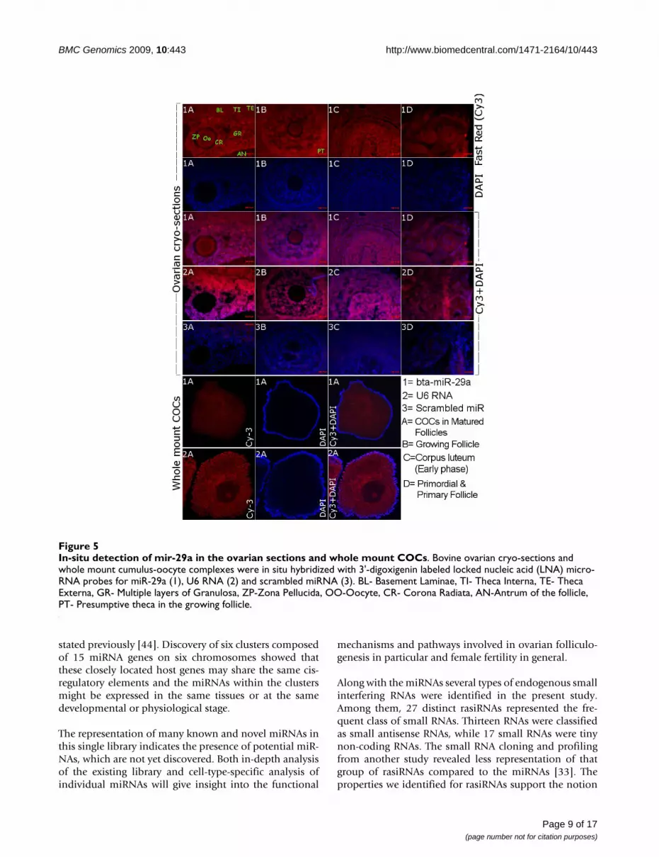
In order to elucidate the cellular localization of onemiRNA, bta-miR-29a was selected due to its differential
Page 6 of 17(page number not for citation purposes)

BMC Genomics 2009, 10:443 http://www.biomedcentral.com/1471-2164/10/443
Page 7 of 17(page number not for citation purposes)
Table 2: Detection and expression of selected miRNAs in multiple tissues
miRNAs Ovarya Fetal Ovaryb Cumulus cells
Corpus luteumc
Oviduct Uterus Placenta Heart Liver Lung Spleen
5s rRNA +++ +++ +++ +++ +++ +++ +++ +++ +++ +++ +++U6 RNA +++ +++ +++ +++ +++ +++ +++ +++ +++ +++ +++bta-let7b ++ +++ +++ +++ ++ + +++ ++ + ++ +++bta-mir-15b + +++ +++ +++ +++ +++ +++ +++ +++ + +++bta-mir-18a +++ +++ +++ +++ +++ +++ +++ +++ +++ +++ +++bomir-22*/22-5p + + + - - - - + - + +bta-mir-29a +++ - +++ - - - +++ +++ + ++ ++bta-mir-125b - ++ +++ ++ ++ + ++ ++ - +++ +bta-mir-126 ++ +++ ++ +++ +++ +++ +++ +++ +++ +++ +++bomir140*/140-5p +++ +++ +++ ++ ++ ++ ++ ++ ++ - ++bomir-143-3p ++ +++ + ++ + + +++ ++ + + ++bta-mir-145 ++ +++ - ++ - - +++ ++ ++ - +++bomir-152-5p ++ ++ - ++ +++ +++ ++ ++ +++ +++ +++bomir-193a-2-3p + ++ ++ - ++ ++ ++ + + ++ ++Bta-mir-199 +++ ++ +++ ++ ++ ++ +++ +++ ++ +++ +++bta-mir-222 - - +++ - - ++ - - + + +bomir-378-3p +++ +++ - - ++ +++ + ++ + + -bomir-382-3p + - - - +++ +++ +++ +++ - - +++bomir-409-5p + +++ +++ ++ +++ +++ +++ +++ +++ +++ ++bomir-424-3p ++ - - - + ++ - - ++ - +bomir-503-3p ++ +++ +++ ++ + + ++ ++ + ++ ++bomir-542-3p - ++ ++ ++ +++ ++ +++ ++ ++ + +bomir-574-5p + + - + + ++ ++ ++ ++ ++ ++bomir-652-3p + +++ - +++ ++ +++ - - ++ - -bomir-940-5p + - - - +++ + - + +++ ++ -bomir-F0132-5p +++ +++ +++ +++ +++ +++ +++ +++ +++ +++ +++bomir-F0244-5p + ++ +++ ++ + ++ ++ + ++ + +bomir-H0222-3p - ++ - +++ + + ++ ++ - + +bomir-A0321-3p ++ ++ ++ ++ ++ ++ ++ ++ ++ + ++bomir-C0533-5p + +++ ++ + - + + + - - -bomir-F0522-3p - - ++ ++ - - - + - - -bomir-F1351-3p ++ - - +++ ++ +++ + +++ +++ ++ +++bomir-F1353-5p ++ ++ - ++ + +++ +++ + +++ - +bomir-D1431-5p ++ +++ ++ +++ ++ + ++ +++ - + -bomir-C1511-5p + + - + ++ ++ + ++ ++ + +bomir-F1821-3p ++ ++ ++ ++ ++ ++ ++ ++ ++ ++ ++bomir-C1931-5p + ++ - +++ +++ +++ +++ +++ +++ - +++bomir-A2143-3p - ++ - +++ +++ +++ ++ ++ ++ ++ +++bomir-F2422-5p +++ - - + + +++ - - ++ - +bomir-F2531-3p + - - + ++ ++ + +++ ++ + -bomir-G2511-3p + - - + + +++ + +++ ++ ++ -
bomir-E2664-3p ++ ++ ++ ++ + ++ ++ + ++ + ++bomir-D3011-3p + + ++ + + + + + + ++ +bomir-A3341-3p + + + + + + + + + + +bomir-A3711-5p + + - + + + +++ + + + +bomir-A4052-5p + + - ++ ++ ++ ++ ++ +++ ++ ++
Expression profiles of 44 miRNAs including all new miRNAs in multiple tissues by PCR approach. Amplicons were analyzed on 2% agarose gel. 5S rRNAs and U6 RNA were used as a loading control. Relative band intensity was categorized into four groups like '+++' for Strong signal, '++' for Medium signal, '+' for Weak signal or smear like product and '-' for not detected by comparing the expression signal of each miRNA to the expression level of 5S rRNA and U6 RNA. a: Ovarian cortex with no visible corpus luteum, b: Ovary from fetus at about 5th month of pregnancy, c: Mature corpus luteum from the same Ovary.

BMC Genomics 2009, 10:443 http://www.biomedcentral.com/1471-2164/10/443
expression between adult and fetal ovary, which are dis-tinct in their functional activity. Accordingly, in-situ local-ization of this miRNA in the sections of bovine ovarianfollicle revealed its expression in the different intra-ovar-ian cells (theca, mural granulosa, cumulus granulosa andoocyte) of different stages of development including pri-mordial, primary, growing and matured/tertiary follicles(Figure 5). Stable expression was detected in the wholemount cumulus-oocyte-complexes derived from the folli-cles of more than 8 mm in diameter. In the semi-quanti-tative RT-PCR data, expression of this miRNA was foundin the cortex region of the adult ovary where follicles withcumulus cells are residing. Moreover, the expression ofthis miRNA was detected further until early stage of cor-pus luteum (Figure 5), but very low or no expression inthe matured corpus luteum (Figure 4).
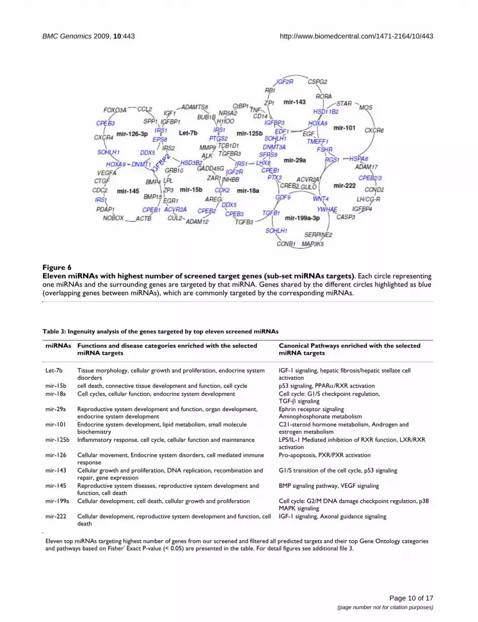
Prediction and functional categorization of cloned miRNA targetsThe goal of this prediction and analysis was to find themajor biological processes and signaling pathways in theovary that are most likely affected by a group of miRNAs.Even though there were many potential target genes pre-dicted for the cloned miRNAs, several filtering and screen-ing procedures (see materials and methods) have enabledus to generate a comprehensive target list consisting of115 potential genes from all the predicted targets (Addi-tional file 3). From this screened target set, we found thatlet-7b, mir-15b, mir-18a, mir-29a, mir-101, mir-125b,
mir-126, mir-143, mir-145, mir-199a and mir-222 tohave the highest number and overlapping targets (Figure6). Interestingly, we found that all of these targeting miR-NAs were represented at higher frequency in our con-structed library.
Detailed Gene Ontology (GO) analysis of the screenedand sub-sets of miRNAs target genes were found to beassociated with reproductive system development, func-tion and disorders. These include cell cycle, morphology,cell death, cell to cell signaling, cellular growth, develop-ment and proliferation, DNA replication, recombination& repair, endocrine system disorder and different path-ways underlying the ovarian functions. To further eluci-date the specific functions of these genes, a detailedpathway analysis was performed using Ingenuity PathwayAnalysis (Redwood City, California) for all target sets (Fig-ure 7) as well as for the sub-set of genes (Table 3, Addi-tional file 3).
DiscussionIdentification of small RNAsMicroRNAs play an integral part of animal gene regulatorynetworks as one of the most abundant classes of gene reg-ulators. They are estimated to comprise 1-5% of animalgenes [8,36,37] or a given genome could encode nearlythousands of miRNAs [36]. Moreover, a typical miRNAregulates hundreds of target genes [38-41] and altogetherthey could target a large proportion of genes up to 30% ofthe genome [42]. Changes in the expression of even a sin-gle miRNA could have a significant impact on the out-come of diverse cellular activities regulated by the productof those genes. Beyond the strict conservation of miRNAsacross different species, some miRNAs appear to be spe-cies specific [32,36,43]. Compared with computational orheterologus approaches, direct cloning has the advantageof identifying non-conserved and new miRNAs.
Our cloning and expression analysis led to the identifica-tion of 74 miRNAs out of which 38 are new in bovine.Mature sequences were found to be conserved in closelyrelated species, but when considering precursor sequenceonly 51% was found to be conserved in human, mouse,rat, dog, horse and also in other non-mammalian verte-brates. However, in the present study, 17 miRNA precur-sors corresponding to 21 genomic loci were found to benot conserved (Table 1). This could be either due to thelack of sequences in draft genome assembly or these miR-NAs are bovine specific. The genomic properties of ournew miRNAs showed that they are derived from exon,intron and intergenic region. This may suggest that thesemiRNAs can be transcribed in parallel with their host tran-scripts. In addition, two different transcription classes ofmiRNAs ('exonic' and 'intronic') recognized here mayrequire somewhat different mechanisms of bio-genesis as
Detection and expression analysis of selected miRNAs in multiple tissuesFigure 4Detection and expression analysis of selected miR-NAs in multiple tissues. Expression profiles of some rep-resentative miRNAs (out of detected 44 miRNAs) in multiple tissues by PCR approach. While the figures are presented in the additional file 2 and the expression for all are summa-rized in the table 2. Amplicons were analyzed on 2% agarose gel. 5S rRNAs and U6 RNA were used as a loading control. A DNA ladder (M) indicating the size of the fragments (50-100-150 nt) on each side. Ovary denotes only the ovarian cortex without corpus luteum.
Page 8 of 17(page number not for citation purposes)
BMC Genomics 2009, 10:443 http://www.biomedcentral.com/1471-2164/10/443
stated previously [44]. Discovery of six clusters composedof 15 miRNA genes on six chromosomes showed thatthese closely located host genes may share the same cis-regulatory elements and the miRNAs within the clustersmight be expressed in the same tissues or at the samedevelopmental or physiological stage.
The representation of many known and novel miRNAs inthis single library indicates the presence of potential miR-NAs, which are not yet discovered. Both in-depth analysisof the existing library and cell-type-specific analysis ofindividual miRNAs will give insight into the functional
mechanisms and pathways involved in ovarian folliculo-genesis in particular and female fertility in general.
Along with the miRNAs several types of endogenous smallinterfering RNAs were identified in the present study.Among them, 27 distinct rasiRNAs represented the fre-quent class of small RNAs. Thirteen RNAs were classifiedas small antisense RNAs, while 17 small RNAs were tinynon-coding RNAs. The small RNA cloning and profilingfrom another study revealed less representation of thatgroup of rasiRNAs compared to the miRNAs [33]. Theproperties we identified for rasiRNAs support the notion
In-situ detection of mir-29a in the ovarian sections and whole mount COCsFigure 5In-situ detection of mir-29a in the ovarian sections and whole mount COCs. Bovine ovarian cryo-sections and whole mount cumulus-oocyte complexes were in situ hybridized with 3'-digoxigenin labeled locked nucleic acid (LNA) micro-RNA probes for miR-29a (1), U6 RNA (2) and scrambled miRNA (3). BL- Basement Laminae, TI- Theca Interna, TE- Theca Externa, GR- Multiple layers of Granulosa, ZP-Zona Pellucida, OO-Oocyte, CR- Corona Radiata, AN-Antrum of the follicle, PT- Presumptive theca in the growing follicle.
Page 9 of 17(page number not for citation purposes)
BMC Genomics 2009, 10:443 http://www.biomedcentral.com/1471-2164/10/443
Page 10 of 17(page number not for citation purposes)
Eleven miRNAs with highest number of screened target genes (sub-set miRNAs targets)Figure 6Eleven miRNAs with highest number of screened target genes (sub-set miRNAs targets). Each circle representing one miRNAs and the surrounding genes are targeted by that miRNA. Genes shared by the different circles highlighted as blue (overlapping genes between miRNAs), which are commonly targeted by the corresponding miRNAs.
Table 3: Ingenuity analysis of the genes targeted by top eleven screened miRNAs
miRNAs Functions and disease categories enriched with the selected miRNA targets
Canonical Pathways enriched with the selected miRNA targets
Let-7b Tissue morphology, cellular growth and proliferation, endocrine system disorders
IGF-1 signaling, hepatic fibrosis/hepatic stellate cell activation
mir-15b cell death, connective tissue development and function, cell cycle p53 signaling, PPARα/RXR activationmir-18a Cell cycles, cellular function, endocrine system development Cell cycle: G1/S checkpoint regulation,
TGF-β signalingmir-29a Reproductive system development and function, organ development,
endocrine system developmentEphrin receptor signalingAminophosphonate metabolism
mir-101 Endocrine system development, lipid metabolism, small molecule biochemistry
C21-steroid hormone metabolism, Androgen and estrogen metabolism
mir-125b Inflammatory response, cell cycle, cellular function and maintenance LPS/IL-1 Mediated inhibition of RXR function, LXR/RXR activation
mir-126 Cellular movement, Endocrine system disorders, cell mediated immune response
Pro-apoptosis, PXR/PXR activation
mir-143 Cellular growth and proliferation, DNA replication, recombination and repair, gene expression
G1/S transition of the cell cycle, p53 signaling
mir-145 Reproductive system diseases, reproductive system development and function, cell death
BMP signaling pathway, VEGF signaling
mir-199a Cellular development, cell death, cellular growth and proliferation Cell cycle: G2/M DNA damage checkpoint regulation, p38 MAPK signaling
mir-222 Cellular development, reproductive system development and function, cell death
IGF-1 signaling, Axonal guidance signaling
Eleven top miRNAs targeting highest number of genes from our screened and filtered all predicted targets and their top Gene Ontology categories and pathways based on Fisher' Exact P-value (< 0.05) are presented in the table. For detail figures see additional file 3.

BMC Genomics 2009, 10:443 http://www.biomedcentral.com/1471-2164/10/443
that they are presumably emerged from dsRNA producedby annealing of sense and antisense transcripts that con-tain repeat sequences related to transposable elements[27]. These rasiRNAs are known to repress the repeatsequences at the transcriptional or post-transcriptionallevel and maintain a centromeric heterochromatic struc-ture [45]. Identity and properties of new types of smallRNAs in the present study showed the presence of diversemodes of small RNA-mediated gene regulation in bovineovary, as reported in other species [26]. Therefore, identi-fication and characterization of other small RNAs andtheir expression patterns are important for elucidatingdetailed gene regulatory networks involved in the ovary.So, all these endogenous small interfering RNAs need tobe further characterized to elucidate their cellular func-tions.
Expression of miRNAs in diverse tissue typesExpression analysis of 44 miRNAs in different ovariancells and tissues types has enabled us to determine theirsite of action in terms of tissue specific abundance as wellas functional regulation (Table 2). We have detectednearly all of these miRNAs in at least one part of the entireovary and other somatic tissues. In the present study,some miRNAs appear to be extremely tissue specific. Forexample; bomir-C0533 and bomir-F0522 were found tobe exclusively expressed in ovarian tissues suggesting theirpotential role in ovary-specific miRNA-dependent regula-tory processes. Five miRNAs (miR-29a, miR-125b, bomir-409, bomir-503 and bomir-F0244) were found to behighly abundant in the cumulus cells and four (bomir-652, bomir-H0222, bomir-C1931 and bomir-A2143) incorpus luteum. These cumulus enriched miRNAs in thepresent study may represent those miRNAs with potential
Top biological function, disease categories and pathways enriched with predicted and screened miRNA target genesFigure 7Top biological function, disease categories and pathways enriched with predicted and screened miRNA target genes. A. Top biological functions and disease categories and B. pathways enriched with predicted and screened miRNA target genes. Ratio is the number of affected genes to total number of genes in the pathway. Threshold p < 0.05 is shown as yellow line. Bars that are above the line indicate significant enrichment of a functional category or pathway.
Page 11 of 17(page number not for citation purposes)

BMC Genomics 2009, 10:443 http://www.biomedcentral.com/1471-2164/10/443
association with the regulation of cumulus secreted fac-tors, which are important for cumulus-oocyte communi-cation and subsequent oocyte development. Similar studyin mouse showed hormonal regulation of miRNAs expres-sion in preovulatory mural granulosa cell [25].
Altered expression of various ovary related genes wasreported in ovaries from fetal, new born and adult ani-mals [46-49]. Furthermore, alteration in expression ofsmall RNAs has been addressed at different stages ofmouse ovary [24]. Similarly, in the present study wefound differential expression of mir-29a, bomir-140, mir-199, mir-378, bomir-F0132 and bomir-F2422 in the ovar-ian cortical portion between fetal and adult cows. Thismay indicate their possible involvement in regulating fol-licular development in the adult cyclic ovarian function.This notion was further supported by higher detection ofmiR-29a in different follicular cells (theca, cumulus-gran-ulosa, and oocyte) of adult ovary by in situ hybridization(Figure 5) and higher expression in cumulus cells by RT-PCR but no detection in the fetal ovary. The expression ofmiRNAs in ovarian cells is reported to be regulated by FSHand LH/hCG [25,50] which functions in the cyclic ovarybut not in fetal ovary [51]. Moreover, most of the targetspredicted for this miRNA (Figure 6) are known to beinvolved in various cyclic adult ovarian functions.
Noticeable expression level of miR-29a was found in dif-ferent phases of corpus luteum (CL) development.According to RT-PCR and in situ hybridization results,expression of miR-29a was detected in the early phase CLbut not in mid phase (matured) CL. These two phases ofCL development are known to vary in multiple aspects ofluteal physiology, angiogenesis and sensitivity to luteo-lytic actions, which are accompanied by differentialexpression of multiple genes [52-55]. Bovine corpusluteum is reported to be resistant to luteolysis by exoge-nous PGF2α in early stage of CL (before Day 5) due to dif-ferential expression of genes associated with the PGF2αreceptor [55]. Considering these facts and restrictedexpression of miR-29a in early phase of CL in the presentstudy, it is possible to suggest that miR-29a is involved ingene regulatory action during early phase of CL. All in all,our results on miR-29a may elucidate the potentialinvolvement of this regulatory miRNA in growth and dif-ferentiation of cumulus cells, endocrine regulation oftheca cells and early luteinisation in cyclic ovary.
Cloning, determining potential secondary structures andexpression analysis of all new miRNAs in multiple tissuesindicate their tissue specific existence and regulation ofgene expression. Only 7.8% of the new miRNAs could notbe detected by the RT-PCR procedure in various reproduc-tive tissues. This may be due to the fact that these tran-scripts were cloned at lower frequency (only once)
showing their lower abundance and subsequent difficultyto detect them [30]. In general, the expression profilinganalysis in the present study revealed that our cloned miR-NAs were either ubiquitously expressed in multiple tissuesor preferentially expressed in a few tissues including theintra-ovarian cells and tissues.
Features of predicted target genesMultiple genes contributing to mammalian folliculogene-sis have been identified in mouse knockout study [56].Primarily, oocyte-specific transcriptional regulators suchas Figla, Nobox, Sohlh1 and Lhx8, oocyte-secreted factorssuch as Gdf9 and Bmp15, as well as genes expressed in thegranulosa and cumulus cells (FSHR and PTX3) werefound to initiate and control follicular growth [56,57].Among the key intra-ovarian factors, the transforminggrowth factor β (TGFβ) family members, of which bonemorphogenetic protein-4 have been identified as regulatorsof primordial germ cell generation [58].
In response to FSH, the granulosa cell-derived factors suchas kit ligand, transforming growth factor alfa (TGF-α) andepidermal growth factor (EGF) activate the resting follicu-lar growth. The interactions between ovarian germ andsomatic cells (granulosa cells and the oocytes) and expres-sion of several intra-ovarian autocrine/paracrine regula-tors (FSH, estrogen and androgen receptors) are the majorcontributing factors in the ovary leading to preantral andantral follicles development [59].
During follicle growth, IGF system works in synergy withgonadotrophins (follicle-stimulating hormone andluteinising hormone) to regulate proliferation and differ-entiation of granulosa and theca cells [60,61]. In addition,it has been shown that the processes of follicular dynam-ics (Recruitment, selection, dominance and ovulation) areassociated with temporal changes of peripheral gonado-tropins concentration and IGF system [62-68]. All theabove-mentioned genes are represented in our predictedand analyzed targets. Altogether 115 genes were amongpotential target genes of our identified miRNA. These tar-get genes are already experimentally validated for poten-tial ovary related functions in different mammalianspecies (references in additional file 3). Interestingly, sev-eral well-known target genes including IRS1, IGFBP3,DNMT3A, HOXA9, TNF, etc. which are identified by ournew screening approach, were already validated in wet labexperiments and reported as targets of multiple miRNAs(miR-145, miR-125b, miR-126 and miR-29) [69-73].Accordingly these studies have elucidated the potentialinvolvement of these miRNAs in broad class of functionsrelated to apoptosis, differentiation signal, cell differenti-ation, tumorogenesis, DNA methylation and innateimmune responses.
Page 12 of 17(page number not for citation purposes)
BMC Genomics 2009, 10:443 http://www.biomedcentral.com/1471-2164/10/443
Gonadotropins, intra-ovarian mediators and their recep-tors which are identified as target genes for our miRNAsmight mediate important intracellular actions necessaryfor normal follicular development and other ovarianfunctions. Alterations in the expression of these mediatorsby miRNAs will result in various ovarian dysfunctionscausing infertility, polycystic ovary syndrome and tumor-igenesis. Recent evidences also support our hypothesis,where at least six of our 11 top ranked ovarian miRNAswere found to be related to cancer or tumors in the ovary.For example, miR-199a, miR-145, miR-125b and let-7clusters were found to be the most differentially regulatedmiRNAs in human ovarian cancer [74,75]. While miR-145 [76] and mir-199a [77] have recently been shown tobe down-modulated in the tumor cells, the miR-222 isreported to be down-regulated in ovarian epithelial carci-nomas [74]. Furthermore, higher expression of miR-18aand lower expression of let-7b and miR-199a were shownto be correlated with serous ovarian carcinoma [78]. Inanother study, miR17-5p and let-7b were found to beinvolved in the regulation of development and functionof the ovarian corpus luteum specially angiogenesis ofcorpus luteum [23]. Interestingly, nearly all of these 11selected miRNAs (Figure 6) in the present study arereported to be differentially regulated in endometrium ofwomen with and without endometriosis [79]. Takentogether, our findings and other evidences support the rel-evance of these 11 miRNAs to ovarian physiology andmay be the most important regulatory miRNA group inovary, as their predicted and analyzed target genes areinvolved in a broad range of signaling cascades and path-ways of the ovarian function.
ConclusionThe presence of distinct miRNAs and other small RNAs,with different expression patterns and various target genesin bovine ovary revealed the potential role of such miR-NAs in follicular development in particular and femalefertility in general. Further functional characterization ofsome selected miRNAs including expression profiling andin situ localization in the ovarian follicles at differentcyclic stages will supplement the results of this study andhelp to elucidate their specific roles in the ovarian func-tion. The information we generated from this study willhelp to identify candidate miRNAs targeting specificmolecular and cellular pathways important for ovarianfollicular development, atresia, ovulation as well as ovar-ian dysfunction.
MethodsIsolation of Small RNAs and subsequent miRNAs fractionationBovine ovaries were obtained from a cyclic heifer with theage of 30 months at a stage of mid cycle with a visiblemature corpus luteum including normally distributed dif-ferent types of follicles. Small RNA samples from various
bovine tissues and cells were isolated using mirVanamiRNA isolation kit (Applied Biosystems Inc, Foster City,CA) according to the manufacturer's instructions. Forcloning, 10 μg of the ovarian small RNA was loaded into12% denaturing poly acrylamide gel electrophoresis withsize markers miSPIKE (Integrated DNA Technologies,Inc., Iowa, USA) and fractions of 18-26 nt were recoveredusing DTR gel filtration cartridge (Edge BioSystems, Mar-yland, USA).
Cloning of small RNAsFor cloning the small RNAs, we followed "5' Ligationindependent Cloning" to ensure complete recovery ofconventional small RNAs as well as small RNAs with 5'modifications or non-standard 5' ends. All the linkers andprimers were obtained from Integrated DNA Technolo-gies, Inc., Iowa, USA. List and sequence of linkers andprimers are given in additional file 4. Briefly, once theenriched small RNA fraction was recovered from the acry-lamide gel slice, the small RNAs were ligated with a 3'linker - adenylated oligos, modified with a 3'-terminaldideoxy-C (ddC) containing Ban-I restriction site [5]. Theligated products were loaded on dPAGE for purificationand reverse transcription was performed. An exonucleasedigestion was carried out after first strand cDNA synthesisand then a second 3' ligation was carried out using a dif-ferent linker sequences. The second 3' linkered product(60 nt) was purified from dPAGE to remove free linkers.Subsequently, the amplification of the RT-PCR productwas performed using linker specific primer set with thethermocycler program of 95.0°C for 10 minutes, 35 cyclesof (95.0°C for 30 seconds, 52.0°C for 30 seconds 72.0°Cfor 30 seconds) and followed by incubation at 72.0°C for5 minutes. Then, the amplicon was subjected to Ban Idigestion, concatemerization and end filling with non-template adenosine followed by cloning into TOPO TACloning® vector (Invitrogen, Carlsbad, CA). Concatemerclones were picked up, cultured and then colony PCR wasperformed for screening the insert size. Plasmid DNApreparation and DNA sequencing were performed forscreened clones and small RNAs which were separated bywell defined linker units with the reconstituted Ban I site.
Bioinformatic analysis of small RNA sequencesThe small RNA sequences were first compared with thesequences in miRBase [32,80-82]. Small RNAs completelyor partially matched by less than two mismatches to anyregistered miRNA in miRBase were considered putativebovine miRNA. The remaining sequences were comparedto the bovine nucleotide collection (nr/nt) and theexpressed sequence tags (EST) database in NCBI [83] anddifferent noncoding RNA databases [84-88]. Sequences,which were matched 100% to any mRNA, rRNA or tRNAwere excluded from further evaluation to generate novelmiRNA candidates. All the remaining sequences and theputative bovine miRNA sequences were submitted to
Page 13 of 17(page number not for citation purposes)
BMC Genomics 2009, 10:443 http://www.biomedcentral.com/1471-2164/10/443
BLAST-search in the Ensembl 52: bovine genome assem-bly (Btau_4.0) [35] and the 75 bp genomic flankingsequence upstream from the 3' end or downstream fromthe 5' end of the miRNA was considered putative precur-sor of the matching miRNA.
All the putative precursor sequences were analyzed forhairpin structure using the mfold Web server (version 3.2)[89] to evaluate the ability to form thermodynamicallystable hairpin structures [90] based on other criteriadescribed elsewhere [32]. Chromosome locations, orien-tation and genomic features of the predicted miRNA pre-cursors as well as other small RNAs sequences (notmeeting miRNAs criteria) and whether they were locatedin intragenic or intergenic genomic regions were deter-mined using ensembl. Other small RNAs were categorizedaccording to published research articles [26,27,33].
Detection of miRNAs expression by semi-quantitative RT-PCRSmall RNA samples isolated from the 11 different tissuesand cells, such as ovarian cortex, fetal ovary at about sixmonth of pregnancy, cumulus cells, matured corpusluteum, oviduct (entire), uterus (horn), placenta, heart,liver, lung and spleen were used for the detection of miR-NAs by PCR method according to Ro et al [91] with somemodifications. Briefly, the poly (A)-tailed small RNA waspurified by acid phenol: chloroform: iso-amyl alcoholand ethanol precipitation method. All small RNA-cDNAsamples were diluted to the same concentration of 6 ng/μl(which was the lowest amount obtained from cumuluscells). Three microliters of cDNA was used as template forconventional PCR and the products were analyzed on a2% agarose gel. List of primers and oligos used are shownin the additional file 4. Some representative RT-PCR prod-ucts were cloned into PGEM-T easy vector (Promega Cor-poration, Wisconsin, USA) and transformed to E. coli andsequenced to verify the specificity of PCR amplification.
In situ hybridization of miRNAs in ovarian cryo-sections and whole mount COCsFor in-situ hybridization of miRNAs, bovine ovary (21/0)days of estrus cycle was fixed in 4% PFA overnight at 4°Cfollowed by overnight incubation in PBS with 30%sucrose at 4°C and frozen in Tissue-Tek OCT reagent(Sakura Finetek, Zoeterwoude, NL). Cryo-sections (10μm) preparation, post-fixation, acetylation and protein-ase K treatment were carried out as described previously[92]. Two hours of pre-hybridization was performed at52°C in hybridization solution (50% formamide, 5×sodium chloride/sodium citrate [SSC; pH 6.0], 0.1%Tween-20, 50 μg/ml heparin, and 500 mg/ml yeast tRNA).Ovarian sections were incubated overnight at 52°C with3'-Digoxigenin (DIG) labeled LNA-modified oligonucle-otide probes (1 pM) for miR-29a, U6 RNA and scrambled
miR (Exiqon, Vedbaek, Denmark) in hybridization bufferin a humidified chamber. Blocking, incubation with anti-DIG-AP antibody, washing and color development usingFast Red reaction was performed as described previously[92]. The slides were mounted with VectaShield contain-ing DAPI (Vector laboratories, Burlingame, CA) and ana-lyzed by confocal laser scanning microscope (CLSM LSM-510, Carl Zeiss, Germany). For whole mount in-situhybridization, cumulus oocyte complexes were aspiratedfrom more than 8 mm of ovarian follicles. Pre-fixation,processing, digestion with Proteinase K, pre-hybridiza-tion, hybridization, post-hybridization washing was per-formed in 4-well embryo culture dishes according to thehigh-resolution whole mount in situ hybridization proto-col from Exiqon. The rest of the procedures were similarto cryo-section hybridization protocol.
Prediction and analysis of ovarian miRNA targetsFor this purpose, initially a raw list of all genes found tobe targeted by our cloned miRNA was generated usingMIRANDA algorithm, miRBase target version 5 [93]. Sub-sequently, about 800 distinct important genes related tomammalian reproductive system development, functionand disorders were extracted from Ingenuity knowledgebase (IPA 7.0) by key word search. Then, we applied twofiltration steps to generate a comprehensive list of targetgenes. Firstly, raw target set and genes set extracted fromdatabase were cross-matched and common genes wereextracted. Secondly, we applied the condition that multi-ple genes targeted by multiple miRNAs from the commontarget list. From these screened target sets, 11 miRNAshaving the highest number as well as overlapping targetgenes were enlisted. Then, the Gene Ontology (GO) anal-ysis of the screened and sub sets of miRNAs target geneswere performed in order to predict the possible biologicalprocesses and functions that were most likely to beaffected by miRNAs using web delivered tools of Ingenu-ity Pathway Analysis (Redwood City, California). Top sig-nificant GO categories, biological functions and differentcanonical pathways were analyzed for miRNA specific tar-gets as well as for all screened targets based on significantover-representation of genes using a selected threshold forp-values ≤ 0.05 of hypergeometric distribution [94].
NoteWe have submitted our newly identified miRNAs into themiR-base and they are annotated accordingly.
Authors' contributionsMMH was responsible for miRNAs cloning, detection anddrafting the manuscript. NG analyzed predicted targetgenes and reviewed the manuscript. MH supplied repro-ductive samples. CP, ET in coordination of bioinformaticanalysis. KS contributed by supervising the work with nec-essary suggestion. DT was responsible for project develop-
Page 14 of 17(page number not for citation purposes)

BMC Genomics 2009, 10:443 http://www.biomedcentral.com/1471-2164/10/443
ment, reviews the manuscript and is the correspondingauthor. All contributing authors reviewed and approvedbefore submitting the final copy of this manuscript.
Additional material
References1. Hunter MG, Robinson RS, Mann GE, Webb R: Endocrine and para-
crine control of follicular development and ovulation rate infarm species. Animal Reproduction Science 2004, 82-83:461-477.
2. Bonnet A, Dalbies-Tran R, Sirard MA: Opportunities and chal-lenges in applying genomics to the study of oogenesis and fol-liculogenesis in farm animals. Reproduction 2008,135(2):119-128.
3. Eichenlaub-Ritter U, Peschke M: Expression in in-vivo and in-vitro growing and maturing oocytes: focus on regulation ofexpression at the translational level. Hum Reprod Update 2002,8(1):21-41.
4. Piccioni F, Zappavigna V, Verrotti AC: Translational regulationduring oogenesis and early development: the cap-poly(A)tail relationship. C R Biol 2005, 328(10-11):863-881.
5. Lau NC, Lim LP, Weinstein EG, Bartel DP: An abundant class oftiny RNAs with probable regulatory roles in Caenorhabditiselegans. Science 2001, 294(5543):858-862.
6. Alvarez-Garcia I, Miska EA: MicroRNA functions in animal devel-opment and human disease. Development 2005,132(21):4653-4662.
7. Ambros V: The functions of animal microRNAs. Nature 2004,431(7006):350-355.
8. Bartel DP: MicroRNAs: genomics, biogenesis, mechanism,and function. Cell 2004, 116(2):281-297.
9. Chen K, Rajewsky N: The evolution of gene regulation by tran-scription factors and microRNAs. Nat Rev Genet 2007,8(2):93-103.
10. Lai EC: microRNAs: Runts of the Genome Assert Them-selves. Current Biology 2003, 13(23):R925-R936.
11. Plasterk RH: Micro RNAs in animal development. Cell 2006,124(5):877-881.
12. Lee Y, Jeon K, Lee JT, Kim S, Kim VN: MicroRNA maturation:stepwise processing and subcellular localization. EMBO J 2002,21(17):4663-4670.
13. Yi R, Qin Y, Macara IG, Cullen BR: Exportin-5 mediates thenuclear export of pre-microRNAs and short hairpin RNAs.Genes Dev 2003, 17(24):3011-3016.
14. Lee Y, Ahn C, Han J, Choi H, Kim J, Yim J, Lee J, Provost P, RadmarkO, Kim S, et al.: The nuclear RNase III Drosha initiates micro-RNA processing. Nature 2003, 425(6956):415-419.
15. Pillai RS, Bhattacharyya SN, Artus CG, Zoller T, Cougot N, Basyuk E,Bertrand E, Filipowicz W: Inhibition of translational initiation byLet-7 MicroRNA in human cells. Science 2005,309(5740):1573-1576.
16. Liu J, Rivas FV, Wohlschlegel J, Yates JR 3rd, Parker R, Hannon GJ: Arole for the P-body component GW182 in microRNA func-tion. Nat Cell Biol 2005, 7(12):1261-1266.
17. Liu J, Valencia-Sanchez MA, Hannon GJ, Parker R: MicroRNA-dependent localization of targeted mRNAs to mammalianP-bodies. Nat Cell Biol 2005, 7(7):719-723.
18. Rehwinkel J, Behm-Ansmant I, Gatfield D, Izaurralde E: A crucialrole for GW182 and the DCP1:DCP2 decapping complex inmiRNA-mediated gene silencing. RNA 2005, 11(11):1640-1647.
19. Bernstein E, Kim SY, Carmell MA, Murchison EP, Alcorn H, Li MZ,Mills AA, Elledge SJ, Anderson KV, Hannon GJ: Dicer is essentialfor mouse development. Nat Genet 2003, 35(3):215-217.
20. Giraldez AJ, Cinalli RM, Glasner ME, Enright AJ, Thomson JM, Basker-ville S, Hammond SM, Bartel DP, Schier AF: MicroRNAs regulatebrain morphogenesis in zebrafish. Science 2005,308(5723):833-838.
21. Wienholds E, Koudijs MJ, van Eeden FJ, Cuppen E, Plasterk RH: ThemicroRNA-producing enzyme Dicer1 is essential forzebrafish development. Nat Genet 2003, 35(3):217-218.
22. Otsuka M, Jing Q, Georgel P, New L, Chen J, Mols J, Kang Young J,Jiang Z, Du X, Cook R, et al.: Hypersusceptibility to VesicularStomatitis Virus Infection in Dicer1-Deficient Mice Is Due toImpaired miR24 and miR93 Expression. Immunity 2007,27(1):123-134.
23. Otsuka M, Zheng M, Hayashi M, Lee JD, Yoshino O, Lin S, Han J:Impaired microRNA processing causes corpus luteum insuf-ficiency and infertility in mice. J Clin Invest 2008,118(5):1944-1954.
24. Ro S, Song R, Park C, Zheng H, Sanders KM, Yan W: Cloning andexpression profiling of small RNAs expressed in the mouseovary. RNA 2007, 13(12):2366-2380.
25. Fiedler SD, Carletti MZ, Hong X, Christenson LK: Hormonal reg-ulation of MicroRNA expression in periovulatory mousemural granulosa cells. Biol Reprod 2008, 79(6):1030-1037.
26. Ambros V, Lee RC, Lavanway A, Williams PT, Jewell D: MicroRNAsand other tiny endogenous RNAs in C. elegans. Curr Biol 2003,13(10):807-818.
27. Aravin AA, Lagos-Quintana M, Yalcin A, Zavolan M, Marks D, SnyderB, Gaasterland T, Meyer J, Tuschl T: The small RNA profile dur-ing Drosophila melanogaster development. Dev Cell 2003,5(2):337-350.
28. Reinhart BJ, Weinstein EG, Rhoades MW, Bartel B, Bartel DP: Micro-RNAs in plants. Genes Dev 2002, 16(13):1616-1626.
29. Coutinho LL, Matukumalli LK, Sonstegard TS, Van Tassell CP, Gas-barre LC, Capuco AV, Smith TP: Discovery and profiling ofbovine microRNAs from immune-related and embryonic tis-sues. Physiol Genomics 2007, 29(1):35-43.
30. Gu Z, Eleswarapu S, Jiang H: Identification and characterizationof microRNAs from the bovine adipose tissue and mammarygland. FEBS Lett 2007, 581(5):981-988.
31. Tesfaye D, Worku D, Rings F, Phatsara C, Tholen E, Schellander K,Hoelker M: Identification and expression profiling of microR-NAs during bovine oocyte maturation using heterologousapproach. Mol Reprod Dev 2009, 76(7):665-677.
32. Ambros V, Bartel B, Bartel DP, Burge CB, Carrington JC, Chen X,Dreyfuss G, Eddy SR, Griffiths-Jones S, Marshall M, et al.: A uniformsystem for microRNA annotation. RNA 2003, 9(3):277-279.
33. Aravin A, Tuschl T: Identification and characterization of smallRNAs involved in RNA silencing. FEBS Letters 2005,579(26):5830-5840.
34. Webb R, Gong JG, Law AS, Rusbridge SM: Control of ovarian func-tion in cattle. J Reprod Fertil Suppl 1992, 45:141-156.
Additional file 1List and Bio-informatic analysis of the sequences.Click here for file[http://www.biomedcentral.com/content/supplementary/1471-2164-10-443-S1.XLS]
Additional file 2Predicted secondary structure of new miRNAs and detection of expres-sion in multiple tissues.Click here for file[http://www.biomedcentral.com/content/supplementary/1471-2164-10-443-S2.PDF]
Additional file 3Screened target genes list for cloned miRNAs and figures of GO anal-ysis.Click here for file[http://www.biomedcentral.com/content/supplementary/1471-2164-10-443-S3.PDF]
Additional file 4List of oligos and primers used for this study.Click here for file[http://www.biomedcentral.com/content/supplementary/1471-2164-10-443-S4.PDF]
Page 15 of 17(page number not for citation purposes)

BMC Genomics 2009, 10:443 http://www.biomedcentral.com/1471-2164/10/443
35. Ensembl 52: B. taurus [http://www.ensembl.org/Bos_taurus/Info/Index]
36. Bentwich I, Avniel A, Karov Y, Aharonov R, Gilad S, Barad O, BarzilaiA, Einat P, Einav U, Meiri E, et al.: Identification of hundreds ofconserved and nonconserved human microRNAs. Nat Genet2005, 37(7):766-770.
37. Berezikov E, Guryev V, Belt J van de, Wienholds E, Plasterk RH, Cup-pen E: Phylogenetic shadowing and computational identifica-tion of human microRNA genes. Cell 2005, 120(1):21-24.
38. Brennecke J, Stark A, Russell RB, Cohen SM: Principles of micro-RNA-target recognition. PLoS Biol 2005, 3(3):e85.
39. Krek A, Grun D, Poy MN, Wolf R, Rosenberg L, Epstein EJ, MacMe-namin P, da Piedade I, Gunsalus KC, Stoffel M, et al.: CombinatorialmicroRNA target predictions. Nat Genet 2005, 37(5):495-500.
40. Lewis BP, Burge CB, Bartel DP: Conserved seed pairing, oftenflanked by adenosines, indicates that thousands of humangenes are microRNA targets. Cell 2005, 120(1):15-20.
41. Xie X, Lu J, Kulbokas EJ, Golub TR, Mootha V, Lindblad-Toh K,Lander ES, Kellis M: Systematic discovery of regulatory motifsin human promoters and 3' UTRs by comparison of severalmammals. Nature 2005, 434(7031):338-345.
42. Lim LP, Lau NC, Garrett-Engele P, Grimson A, Schelter JM, Castle J,Bartel DP, Linsley PS, Johnson JM: Microarray analysis shows thatsome microRNAs downregulate large numbers of targetmRNAs. Nature 2005, 433(7027):769-773.
43. Stark A, Lin MF, Kheradpour P, Pedersen JS, Parts L, Carlson JW,Crosby MA, Rasmussen MD, Roy S, Deoras AN, et al.: Discovery offunctional elements in 12 Drosophila genomes using evolu-tionary signatures. Nature 2007, 450(7167):219-232.
44. Rodriguez A, Griffiths-Jones S, Ashurst JL, Bradley A: Identificationof mammalian microRNA host genes and transcriptionunits. Genome Res 2004, 14(10A):1902-1910.
45. Lippman Z, Martienssen R: The role of RNA interference in het-erochromatic silencing. Nature 2004, 431(7006):364-370.
46. Herrera L, Ottolenghi C, Garcia-Ortiz JE, Pellegrini M, Manini F, KoMS, Nagaraja R, Forabosco A, Schlessinger D: Mouse ovary devel-opmental RNA and protein markers from gene expressionprofiling. Dev Biol 2005, 279(2):271-290.
47. Baillet A, Mandon-Pepin B, Cabau C, Poumerol E, Pailhoux E, CotinotC: Identification of transcripts involved in meiosis and follicleformation during ovine ovary development. BMC Genomics2008, 9:436.
48. Vaskivuo TE, Anttonen M, Herva R, Billig H, Dorland M, te Velde ER,Stenback F, Heikinheimo M, Tapanainen JS: Survival of humanovarian follicles from fetal to adult life: apoptosis, apoptosis-related proteins, and transcription factor GATA-4. J ClinEndocrinol Metab 2001, 86(7):3421-3429.
49. Olesen C, Nyeng P, Kalisz M, Jensen TH, Moller M, Tommerup N,Byskov AG: Global gene expression analysis in fetal mouseovaries with and without meiosis and comparison of selectedgenes with meiosis in the testis. Cell Tissue Res 2007,328(1):207-221.
50. Yao N, Lu CL, Zhao JJ, Xia HF, Sun DG, Shi XQ, Wang C, Li D, CuiY, Ma X: A network of miRNAs expressed in the ovary areregulated by FSH. Front Biosci 2009, 14:3239-3245.
51. Abel MH, Wootton AN, Wilkins V, Huhtaniemi I, Knight PG, Charl-ton HM: The effect of a null mutation in the follicle-stimulat-ing hormone receptor gene on mouse reproduction.Endocrinology 2000, 141(5):1795-1803.
52. Wiltbank MC, Shiao TF, Bergfelt DR, Ginther OJ: Prostaglandin F2alpha receptors in the early bovine corpus luteum. Biol Reprod1995, 52(1):74-78.
53. Copelin JP, Smith MF, Garverick HA, Youngquist RS, McVey WR Jr,Inskeep EK: Responsiveness of bovine corpora lutea to pros-taglandin F2 alpha: comparison of corpora lutea anticipatedto have short or normal lifespans. J Anim Sci 1988,66(5):1236-1245.
54. Watts TL, Fuquay JW: Response and fertility of dairy heifers fol-lowing injection with prostaglandin F(2alpha) during early,middle or late diestrus. Theriogenology 1985, 23(4):655-661.
55. Goravanahally MP, Salem M, Yao J, Inskeep EK, Flores JA: Differen-tial gene expression in the bovine corpus luteum during tran-sition from early phase to midphase and its potential role inacquisition of luteolytic sensitivity to prostaglandin F2 alpha.Biol Reprod 2009, 80(5):980-988.
56. Matzuk MM, Lamb DJ: Genetic dissection of mammalian fertil-ity pathways. Nat Cell Biol 2002, 4(Suppl 9):s41-49.
57. Dumesic DA, Abbott DH: Implications of polycystic ovary syn-drome on oocyte development. Semin Reprod Med 2008,26(1):53-61.
58. Hurk R van den, Zhao J: Formation of mammalian oocytes andtheir growth, differentiation and maturation within ovarianfollicles. Theriogenology 2005, 63(6):1717-1751.
59. Filicori M, Cognigni GE, Pocognoli P, Ciampaglia W, Bernardi S: Cur-rent concepts and novel applications of LH activity in ovarianstimulation. Trends Endocrinol Metab 2003, 14(6):267-273.
60. Campbell BK, Baird DT, Webb R: Effects of dose of LH on andro-gen production and luteinization of ovine theca cells cul-tured in a serum-free system. J Reprod Fertil 1998, 112(1):69-77.
61. Gutierrez CG, Campbell BK, Webb R: Development of a long-term bovine granulosa cell culture system: induction andmaintenance of estradiol production, response to follicle-stimulating hormone, and morphological characteristics.Biol Reprod 1997, 56(3):608-616.
62. Austin EJ, Mihm M, Evans AC, Knight PG, Ireland JL, Ireland JJ, RocheJF: Alterations in intrafollicular regulatory factors and apop-tosis during selection of follicles in the first follicular wave ofthe bovine estrous cycle. Biol Reprod 2001, 64(3):839-848.
63. Fortune JE, Rivera GM, Evans AC, Turzillo AM: Differentiation ofdominant versus subordinate follicles in cattle. Biol Reprod2001, 65(3):648-654.
64. Ginther OJ, Bergfelt DR, Beg MA, Kot K: Role of low circulatingFSH concentrations in controlling the interval to emergenceof the subsequent follicular wave in cattle. Reproduction 2002,124(4):475-482.
65. Ireland JJ, Mihm M, Austin E, Diskin MG, Roche JF: Historical per-spective of turnover of dominant follicles during the bovineestrous cycle: key concepts, studies, advancements, andterms. J Dairy Sci 2000, 83(7):1648-1658.
66. Mihm M, Evans AC: Mechanisms for dominant follicle selectionin monovulatory species: a comparison of morphological,endocrine and intraovarian events in cows, mares andwomen. Reprod Domest Anim 2008, 43(Suppl 2):48-56.
67. Quintal-Franco JA, Kojima FN, Melvin EJ, Lindsey BR, Zanella E, FikeKE, Wehrman ME, Clopton DT, Kinder JE: Corpus luteum devel-opment and function in cattle with episodic release of lutei-nizing hormone pulses inhibited in the follicular and earlyluteal phases of the estrous cycle. Biol Reprod 1999,61(4):921-926.
68. Schams D, Berisha B, Kosmann M, Einspanier R, Amselgruber WM:Possible role of growth hormone, IGFs, and IGF-binding pro-teins in the regulation of ovarian function in large farm ani-mals. Domest Anim Endocrinol 1999, 17(2-3):279-285.
69. Fabbri M, Garzon R, Cimmino A, Liu Z, Zanesi N, Callegari E, Liu S,Alder H, Costinean S, Fernandez-Cymering C, et al.: MicroRNA-29family reverts aberrant methylation in lung cancer by target-ing DNA methyltransferases 3A and 3B. Proc Natl Acad Sci USA2007, 104(40):15805-15810.
70. Shen WF, Hu YL, Uttarwar L, Passegue E, Largman C: MicroRNA-126 regulates HOXA9 by binding to the homeobox. Mol CellBiol 2008, 28(14):4609-4619.
71. Shi B, Sepp-Lorenzino L, Prisco M, Linsley P, deAngelis T, Baserga R:Micro RNA 145 targets the insulin receptor substrate-1 andinhibits the growth of colon cancer cells. J Biol Chem 2007,282(45):32582-32590.
72. Shi XB, Xue L, Yang J, Ma AH, Zhao J, Xu M, Tepper CG, Evans CP,Kung HJ, deVere White RW: An androgen-regulated miRNAsuppresses Bak1 expression and induces androgen-inde-pendent growth of prostate cancer cells. Proc Natl Acad Sci USA2007, 104(50):19983-19988.
73. Tili E, Michaille JJ, Cimino A, Costinean S, Dumitru CD, Adair B, Fab-bri M, Alder H, Liu CG, Calin GA, et al.: Modulation of miR-155and miR-125b levels following lipopolysaccharide/TNF-alphastimulation and their possible roles in regulating theresponse to endotoxin shock. J Immunol 2007,179(8):5082-5089.
74. Iorio MV, Visone R, Di Leva G, Donati V, Petrocca F, Casalini P, Tac-cioli C, Volinia S, Liu CG, Alder H, et al.: MicroRNA signatures inhuman ovarian cancer. Cancer Res 2007, 67(18):8699-8707.
75. Yang H, Kong W, He L, Zhao JJ, O'Donnell JD, Wang J, Wenham RM,Coppola D, Kruk PA, Nicosia SV, et al.: MicroRNA expression
Page 16 of 17(page number not for citation purposes)

BMC Genomics 2009, 10:443 http://www.biomedcentral.com/1471-2164/10/443
Publish with BioMed Central and every scientist can read your work free of charge
"BioMed Central will be the most significant development for disseminating the results of biomedical research in our lifetime."
Sir Paul Nurse, Cancer Research UK
Your research papers will be:
available free of charge to the entire biomedical community
peer reviewed and published immediately upon acceptance
cited in PubMed and archived on PubMed Central
yours — you keep the copyright
Submit your manuscript here:http://www.biomedcentral.com/info/publishing_adv.asp
BioMedcentral
profiling in human ovarian cancer: miR-214 induces cell sur-vival and cisplatin resistance by targeting PTEN. Cancer Res2008, 68(2):425-433.
76. Iorio MV, Ferracin M, Liu CG, Veronese A, Spizzo R, Sabbioni S, MagriE, Pedriali M, Fabbri M, Campiglio M, et al.: MicroRNA geneexpression deregulation in human breast cancer. Cancer Res2005, 65(16):7065-7070.
77. Murakami Y, Yasuda T, Saigo K, Urashima T, Toyoda H, Okanoue T,Shimotohno K: Comprehensive analysis of microRNA expres-sion patterns in hepatocellular carcinoma and non-tumor-ous tissues. Oncogene 2006, 25(17):2537-2545.
78. Nam EJ, Yoon H, Kim SW, Kim H, Kim YT, Kim JH, Kim JW, Kim S:MicroRNA expression profiles in serous ovarian carcinoma.Clin Cancer Res 2008, 14(9):2690-2695.
79. Pan Q, Luo X, Toloubeydokhti T, Chegini N: The expression pro-file of micro-RNA in endometrium and endometriosis andthe influence of ovarian steroids on their expression. MolHum Reprod 2007, 13(11):797-806.
80. Griffiths-Jones S: The microRNA Registry. Nucleic Acids Res2004:D109-111.
81. Griffiths-Jones S, Grocock RJ, van Dongen S, Bateman A, Enright AJ:miRBase: microRNA sequences, targets and gene nomencla-ture. Nucleic Acids Res 2006:D140-144.
82. miRBase_12.0 [http://microrna.sanger.ac.uk/sequences/]83. BLAST cow sequences [http://www.ncbi.nlm.nih.gov/genome/
seq/BlastGen/BlastGen.cgi?taxid=9913]84. Genomic tRNA database [http://lowelab.ucsc.edu/GtRNAdb/]85. Ribosomal RNA BLAST [http://bioinformatics.psb.ugent.be/
webtools/rRNA/blastrrna.html]86. Blast ncRNA database [http://ncrnadb.trna.ibch.poznan.pl/
blast.html]87. RNAdb [http://research.imb.uq.edu.au/rnadb/default.aspx]88. tRNAscan-SE [http://lowelab.ucsc.edu/tRNAscan-SE/]89. Mfold web server_3.2 [http://frontend.bioinfo.rpi.edu/applica
tions/mfold/cgi-bin/rna-form1.cgi]90. Zuker M: Mfold web server for nucleic acid folding and hybrid-
ization prediction. Nucleic Acids Res 2003, 31(13):3406-3415.91. Ro S, Park C, Jin J, Sanders KM, Yan W: A PCR-based method for
detection and quantification of small RNAs. Biochem BiophysRes Commun 2006, 351(3):756-763.
92. Obernosterer G, Martinez J, Alenius M: Locked nucleic acid-based in situ detection of microRNAs in mouse tissue sec-tions. Nat Protoc 2007, 2(6):1508-1514.
93. miRBase Targets Version 5 [http://microrna.sanger.ac.uk/targets/v5/]
94. Delfour O, Vilanova D, Atzorn V, Michot B: The passionate racefor miRNA detection and function deciphering. In miRNA: Biol-ogy, Function and Expression Edited by: Clarke NJ. Sanseau P: DNAPress; 2007:335-362.
Page 17 of 17(page number not for citation purposes)