identification and characterization of antimicrobial peptides
TRANSCRIPT

Identification and Characterization of Antimicrobial Peptides with Therapeutic Potential
www.mdpi.com/journal/pharmaceuticals
Edited by
Guangshun WangPrinted Edition of the Special Issue Published in Pharmaceuticals
pharmaceuticals

Identification and Characterization of Antimicrobial Peptides with Therapeutic Potential Special Issue Editor Guangshun Wang
MDPI • Basel • Beijing • Wuhan • Barcelona • Belgrade
Special Issue Editor Guangshun Wang University of Nebraska Medical Center USA Editorial Office MDPI AG St. Alban-Anlage 66 Basel, Switzerland This edition is a reprint of Special Issues published online in the open access journal Pharmaceuticals (ISSN 1424-8247) from 2014–2015 (available at: http://www.mdpi.com/ journal/pharmaceuticals/special_issues/therapeutic-potential; http://www.mdpi.com/ journal/pharmaceuticals/special_issues/antimicrobial-peptides2015). For citation purposes, cite each article independently as indicated on the article page online and as indicated below: Author 1; Author 2. Article title. Journal Name Year, Article number, page range. First Edition 2017 ISBN 978-3-03842-462-8 (Pbk) ISBN 978-3-03842-463-5 (PDF)
Articles in this volume are Open Access and distributed under the Creative Commons Attribution license (CC BY), which allows users to download, copy and build upon published articles even for commercial purposes, as long as the author and publisher are properly credited, which ensures maximum dissemination and a wider impact of our publications. The book taken as a whole is © 2017 MDPI, Basel, Switzerland, distributed under the terms and conditions of the Creative Commons license CC BY-NC-ND (http://creativecommons.org/licenses/by-nc-nd/4.0/).
iii
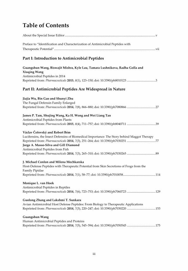
Table of Contents About the Special Issue Editor ..................................................................................................................... v
Preface to “Identification and Characterization of Antimicrobial Peptides with Therapeutic Potential” ................................................................................................................................... vii
Part I: Introduction to Antimicrobial Peptides
Guangshun Wang, Biswajit Mishra, Kyle Lau, Tamara Lushnikova, Radha Golla and Xiuqing Wang
Antimicrobial Peptides in 2014 Reprinted from: Pharmaceuticals 2015, 8(1), 123–150; doi: 10.3390/ph8010123 ..................................... 3
Part II: Antimicrobial Peptides Are Widespread in Nature
Jiajia Wu, Bin Gao and Shunyi Zhu
The Fungal Defensin Family Enlarged Reprinted from: Pharmaceuticals 2014, 7(8), 866–880; doi: 10.3390/ph7080866 ..................................... 27 James P. Tam, Shujing Wang, Ka H. Wong and Wei Liang Tan
Antimicrobial Peptides from Plants Reprinted from: Pharmaceuticals 2015, 8(4), 711–757; doi: 10.3390/ph8040711 ..................................... 39 Václav Čeřovský and Robert Bém
Lucifensins, the Insect Defensins of Biomedical Importance: The Story behind Maggot Therapy Reprinted from: Pharmaceuticals 2014, 7(3), 251–264; doi: 10.3390/ph7030251 ..................................... 77 Jorge A. Masso-Silva and Gill Diamond
Antimicrobial Peptides from Fish Reprinted from: Pharmaceuticals 2014, 7(3), 265–310; doi: 10.3390/ph7030265 ..................................... 89 J. Michael Conlon and Milena Mechkarska
Host-Defense Peptides with Therapeutic Potential from Skin Secretions of Frogs from the Family Pipidae Reprinted from: Pharmaceuticals 2014, 7(1), 58–77; doi: 10.3390/ph7010058.......................................... 114 Monique L. van Hoek
Antimicrobial Peptides in Reptiles Reprinted from: Pharmaceuticals 2014, 7(6), 723–753; doi: 10.3390/ph7060723 ..................................... 129 Guolong Zhang and Lakshmi T. Sunkara
Avian Antimicrobial Host Defense Peptides: From Biology to Therapeutic Applications Reprinted from: Pharmaceuticals 2014, 7(3), 220–247; doi: 10.3390/ph7030220 ..................................... 153 Guangshun Wang
Human Antimicrobial Peptides and Proteins Reprinted from: Pharmaceuticals 2014, 7(5), 545–594; doi: 10.3390/ph7050545 ..................................... 175

iv
Part III: Therapeutic Potential of Antimicrobial Peptides
Marina Berditsch, Hannah Lux, Oleg Babii, Sergii Afonin and Anne S. Ulrich
Therapeutic Potential of Gramicidin S in the Treatment of Root Canal Infections Reprinted from: Pharmaceuticals 2016, 9(3), 56; doi: 10.3390/ph9030056 ................................................ 215 I-Ni Hsieh and Kevan L. Hartshorn
The Role of Antimicrobial Peptides in Influenza Virus Infection and Their Potential as Antiviral and Immunomodulatory Therapy Reprinted from: Pharmaceuticals 2016, 9(3), 53; doi: 10.3390/ph9030053 ................................................ 229 Nongnuj Tanphaichitr, Nopparat Srakaew, Rhea Alonzi, Wongsakorn Kiattiburut, Kessiri Kongmanas, Ruina Zhi, Weihua Li, Mark Baker, Guanshun Wang and Duane Hickling
Potential Use of Antimicrobial Peptides as Vaginal Spermicides/Microbicides Reprinted from: Pharmaceuticals 2016, 9(1), 13; doi: 10.3390/ph9010013 ................................................ 244

v
About the Special Issue Editor Guangshun Wang is an Associate Professor at the University of Nebraska Medical Center (Omaha, USA). He is interested in developing new compounds to treat human diseases, such as drug-resistant superbugs, viruses and cancer. His laboratory utilizes an integrated approach by combining chemistry, biophysics, bioinformatics, genetics, and structural biology. Two general methods are exploited to identify novel drug candidates. First, his laboratory has constructed the Antimicrobial Peptide Database (http://aps.unmc.edu/AP) to search and screen starting templates. At present, this database contains over 2800 antibacterial, antiviral, antifungal, antiparasitic and anticancer peptides, primarily from natural sources, including bacteria, fungi, protists, plants, and animals. Second, three-dimensional structural information is harnessed to conduct structure-based or rational design. Recently, his laboratory has also combined the library approach with structure-based design to obtain compounds with desire properties. Dr. Wang has edited a book entitled Antimicrobial Peptides: Discovery, Design and Novel Therapeutic Strategies (CABI, 1st version 2010; 2nd version 2017). Dr. Wang has published 90 original articles, invited reviews, and book chapters.


vii
Preface to “Identification and Characterization of Antimicrobial Peptides with Therapeutic Potential”
Antimicrobial peptides are key defense molecules adopted by all life forms. The Antimicrobial peptide field is advancing rapidly as a result of the following: First, the antibiotic resistance issue has caught the attention of politicians, scientists, and the general public. There is the consensus that measures should be taken to curb the growth of antibiotic resistance. Efforts in this direction may range from discovering alternative antimicrobials to exploring different applications of existing antibiotics [1]. Second, there is a great desire to understand the functional roles of antimicrobial peptides in humans, animals, and plants. Studies in this direction will elucidate when and where such peptides are expressed in response to invading pathogens and how they work together with other components in the immune system to maintain a healthy host. Third, it is now appreciated that commensal bacteria play an essential role in human health. An elegant example is human microbiota. A current view is that a loss of beneficial commensal bacteria opens the door to infection by microbial intruders [2]. This view indicates the importance to maintain the homeostasis of our microbiota to prevent related diseases. It also implies that one may try to restore the commensal microbes as a therapeutic option [3]. Whichever the direction may be, the identification and characterization of naturally occurring antimicrobial peptides clearly constitute an important step toward these goals. Consequently, there is an increasing effort in discovering novel antimicrobial peptides from natural resources by genomic mining or proteomic approaches [4]. Identification and Characterization of Antimicrobial Peptides with Therapeutic Potential, an eBook consisting of three parts, is constructed to tell the story of antimicrobial peptides discovered from a variety of kingdoms or classes. These articles were selected from the two Special Issues on antimicrobial peptides originally published in the open access journal Pharmaceuticals during the period 2014–2016.
The first part (Chapter 1) highlights leaps made in peptide discovery, new function, mechanisms of action, and potential applications of antimicrobial peptides. Based on the antimicrobial Peptide Database (APD; http://aps.unmc.edu/AP), roughly 100–300 new peptides with demonstrated antimicrobial activity are discovered each year [5], including unique sequences, which cannot be predicted by modern machine learning algorithms. Then, my research team and I continue to describe new mechanisms for existing antimicrobial peptides that have been known for decades. We propose that multiple functions of antimicrobial peptides are realized by their ability to recognize and interact with different molecular targets or cells. Finally, we discuss various potential applications of antimicrobial peptides, ranging from peptide therapeutics, surface coating, nanoparticle-based drug delivery systems and biosensors to detection devices.
The second part is the main body of this eBook that contains a series of articles on natural antimicrobial peptides from diverse sources. Fungi, as eukaryotes, provide a useful source for antimicrobial peptides. There are well-known examples such as plectasin, eurocin, micasin, and copsin. Among them, plectasin is extensively characterized in terms of structure, activity, and potential applications [6]. In Chapter 2, Shunyi Zhu and colleagues describe the great opportunity to identify many more defensin-like peptides from the sequenced fungal genomes. A sequence similarity search leads to 69 new peptides from 26 fungal species. A recent article from the same lab demonstrates that, however, not all such peptides possess antimicrobial activity, at least based on the in vitro tests of select bacterial strains [7]. It is likely that numerous peptides expressed simultaneously are endowed with distinct functions to maximize the benefits to the host.
Plants constitute another important source for novel antimicrobial peptides (341 entries in the current APD). Many plant antimicrobial peptides comprise disulfide bonds that usually form β-sheet structures. Additionally, disulfide-linked sequences can also form α-helical structures. In Chapter 3, James Tam and colleagues systematically deal with the sequence, structure and activity of these plant peptides. Cyclotides, circular peptides stabilized by three disulfide bonds, are a special class of plant peptides whose antimicrobial activity remains to be tested. Their stable scaffold and recognition of bacterial phosphatidylethanolamine (PE) are outstanding traits for developing antimicrobials with the needed properties.

viii
Animals are diverse and can be broadly separated into invertebrates and vertebrates. Most animals are invertebrates, such as insects, spiders, and scorpions. In Chapter 4, Václav Čeřovský and his associate vividly tell the story of lucifensin, which is believed to be the long-sought-after magic molecule for Maggot therapy. For spiders and scorpions, Xiuqing Wang and I have recently conducted a structural and bioinformatics analysis [8]. While spiders use both glycine-rich and helical peptides for defense, scorpions use only helical peptides. Vertebrates are also versatile, covering fish in water, reptiles on land, and birds in the sky. In Chapter 5, Gill Diamond and his associate discuss a variety of fish antimicrobial peptides that kill both human and fish pathogens. In addition, these peptides also modulate immune responses. Of particular interest is that fish antimicrobial peptides may work at high concentrations of salts or low pH, useful properties for developing the next generation of antimicrobials. In Chapter 6, Michael Conlon and his colleague describe select antimicrobial peptides from skin secretions of frogs (~1000 in the current APD). The authors comment that “the therapeutic potential of frog skin peptides as anti-infective agents has not been realized so that alternative clinical applications as anti-cancer, anti-viral, anti-diabetic, or immunomodulatory drugs are being explored.” In Chapter 7, Monique van Hoek discusses antimicrobial peptides from reptiles. Recently, she and her colleagues have proposed an innovative approach that enriches peptides from reptile blood [9]. This method accelerates the discovery of additional reptile peptides. Chapter 8, by Guolong Zhang and his associates, summarizes the biology and therapeutic potential of avian antimicrobial peptides. Structure–activity relationship studies help identify candidates for antimicrobial use or vaccine adjuvants. Dietary additives have been utilized to modulate the expression of antimicrobial peptides to prevent diseases in chicken. In Chapter 9, I describe human antimicrobial peptides, covering discovery, antimicrobial/anticancer activities, mechanisms of action, three-dimensional structure and therapeutic strategies. Provided in Table 1 is a timeline for the discovery of select human antimicrobial peptides (over 100 in the APD). These peptides and proteins can adopt various structural scaffolds to attack pathogens by different mechanisms. These include disruption of membranes, inhibition of metal uptake or cell wall synthesis, and association with ribosomes. Finally, various factors such as isoleucine, sunlight and vitamin D can be utilized to induce the expression of human antimicrobial peptides, opening a new avenue to curb pathogen infection.
The third part of this book highlights the therapeutic potential of antimicrobial peptides. The moonlighting of antimicrobial peptides (i.e., multiple functions) lays the foundation for us to explore their potential uses. Interestingly enough, bacteria also produce antimicrobial peptides (generally called bacteriocins). Several bacterial peptides (e.g., daptomycin, gramicidin, and nisin) are already in use. In Chapter 10, Anne Ulrich and her colleagues report the treatment of patients with root canal infections using gramicidin S. Antiviral effects of antimicrobial peptides are also of high interest considering viral infections such as SARS, Zika, and Ebola in the news. In Chapter 11, Kevan Hartshorn and his colleague describe the role and therapeutic potential of antiviral antimicrobial peptides, including human α-defensins, β-defensins, retrocyclins, LL-37, histones, amyloid peptides, lactoferrin and bacterial/permeability increasing protein (BPI) derived peptides. While intact LL-37 itself lost activity, its central fragment (GI-20) remained effective against a pandemic influenza virus strain H1N1 of 2009. Antimicrobial peptides may also be useful in preventing undesired pregnancies and as anti-HIV microbiocides [10]. In Chapter 12, Nongnuj Tanphaichitr and her colleagues discuss spermicidal AMPs. They propose that human cathelicidin LL-37 is the most promising peptide for this purpose. Many peptides are currently under active development for therapeutic applications [1]. It appears that the potential of these natural peptides is unlimited and remains to be unlocked by the young generation of researchers for the benefit of our biosphere. I hope that this eBook provides a useful introduction to newcomers and refreshes the minds of the veterans. Finally, I would like to take this opportunity to thank all the authors who contributed their insightful work to this eBook and the Pharmaceuticals editorial team for publishing it.
Guangshun Wang Special Issue Editor

ix
References
1. Mishra, B.; Reiling, S.; Zarena, D.; Wang, G. Host defense antimicrobial peptides as antibiotics: design and application strategies. Curr. Opin. Chem. Biol. 2017, 38, 87–96.
2. Nakatsuji, T.; Chen, T.H.; Narala, S.; Chun, K.A.; Two, A.M.; Yun, T.; Shafiq, F.; Kotol, P.F.; Bouslimani, A.; Melnik, A.V.; et al. Antimicrobials from human skin commensal bacteria protect against Staphylococcus aureus and are deficient in atopic dermatitis. Sci. Transl Med. 2017, 9, doi:10.1126/scitranslmed.aah4680.
3. Kommineni, S.; Kristich, C.J.; Salzman, N.H. Harnessing bacteriocin biology as targeted therapy in the GI tract. Gut Microbes. 2016, 7, 512–517.
4. Wang, G. (Ed.) Antimicrobial Peptides: Discovery, Design and Novel Therapeutic Strategies; 2nd Version; CABI: Wallingford, UK, 2017.
5. Wang, G.; Li, X.; Wang, Z. APD3: The antimicrobial peptide database as a tool for research and education. Nucleic Acids Res. 2016, 44, D1087–D1093.
6. Mygind, P.H.; Fischer, R.L.; Schnorr, K.M.; Hansen, M.T.; Sönksen, C.P.; Ludvigsen, S.; Raventós, D.; Buskov, S.; Christensen, B.; De Maria, L.; et al. Plectasin is a peptide antibiotic with therapeutic potential from a saprophytic fungus. Nature 2005, 437, 975–980.
7. Wu, Y.; Gao, B.; Zhu, S. New fungal defensin-like peptides provide evidence for fold change of proteins in evolution. Biosci. Rep. 2017, 37, doi:10.1042/BSR20160438.
8. Wang, X.; Wang, G. Insights into Antimicrobial Peptides from Spiders and Scorpions. Protein Pept. Lett. 2016, 23, 707–721.
9. Bishop, B.M.; Juba, M.L.; Russo, P.S.; Devine, M.; Barksdale, S.M.; Scott, S.; Settlage, R.; Michalak, P.; Gupta, K.; Vliet, K.; et al. Discovery of Novel Antimicrobial Peptides from Varanus komodoensis (Komodo Dragon) by Large-Scale Analyses and De-Novo-Assisted Sequencing Using Electron-Transfer Dissociation Mass Spectrometry. J. Proteome Res. 2017, 16, 1470–1482.
10. Wang, G. Database-guided discovery of potent peptides to combat HIV-1 or Superbugs. Pharmaceuticals 2013, 6, 728–758.


Part I: Introduction to Antimicrobial Peptides


pharmaceuticals
Review
Chapter 1:
Antimicrobial Peptides in 2014
Guangshun Wang 1,*, Biswajit Mishra 1, Kyle Lau 1, Tamara Lushnikova 1, Radha Golla 1 and
Xiuqing Wang 1,2
1 Department of Pathology and Microbiology, University of Nebraska Medical Center, 986495 NebraskaMedical Center, Omaha, NE 68198-6495, USA
2 Institute of Clinical Laboratory, Ningxia Medical University, Yinchuan 750004, China* Author to whom correspondence should be addressed; [email protected]; Tel.: +402-559-4176;
Fax: +402-559-4077.
Academic Editor: Jean Jacques Vanden EyndeReceived: 11 February 2015; Accepted: 17 March 2015; Published: 23 March 2015
Abstract: This article highlights new members, novel mechanisms of action, new functions, andinteresting applications of antimicrobial peptides reported in 2014. As of December 2014, over100 new peptides were registered into the Antimicrobial Peptide Database, increasing the totalnumber of entries to 2493. Unique antimicrobial peptides have been identified from marine bacteria,fungi, and plants. Environmental conditions clearly influence peptide activity or function. Humanα-defensin HD-6 is only antimicrobial under reduced conditions. The pH-dependent oligomerizationof human cathelicidin LL-37 is linked to double-stranded RNA delivery to endosomes, where theacidic pH triggers the dissociation of the peptide aggregate to release its cargo. Proline-rich peptides,previously known to bind to heat shock proteins, are shown to inhibit protein synthesis. A modelantimicrobial peptide is demonstrated to have multiple hits on bacteria, including surface proteindelocalization. While cell surface modification to decrease cationic peptide binding is a recognizedresistance mechanism for pathogenic bacteria, it is also used as a survival strategy for commensalbacteria. The year 2014 also witnessed continued efforts in exploiting potential applications ofantimicrobial peptides. We highlight 3D structure-based design of peptide antimicrobials andvaccines, surface coating, delivery systems, and microbial detection devices involving antimicrobialpeptides. The 2014 results also support that combination therapy is preferred over monotherapy intreating biofilms.
Keywords: antimicrobial peptide; bacterial detection; biofilms; mechanism of action; nanoparticle;peptide discovery; sensors; structure-based design; surface coating
1. Introduction
Antimicrobial peptides, or host defense peptides, are important components of innate immunesystems. This field is currently moving rapidly. On one hand, there is an urgent demand for novelantimicrobials due to the current trend of reduction in the potency of commonly used antibiotics. On theother hand, our research now pays more attention to innate immune systems where antimicrobialpeptides play an essential role. An imbalanced expression of antimicrobial peptides has been implicatedin human disease [1–5]. All these research activities around the world led to a substantial increase inthe number of scientific papers on antimicrobial peptides. In 2014 alone, a search of the PubMed using“antimicrobial peptides and 2014” returned 7562 publications (~20 articles per day) [6]. About 10% ofthese publications are review articles. However, a summary in the style of annual report is lacking.
Pharmaceuticals 2015, 8, 123–150 3 www.mdpi.com/journal/pharmaceuticals

Pharmaceuticals 2015, 8, 123–150
During our regular update of the Antimicrobial Peptide Database (APD) (http://aps.unmc.edu/AP) [7–9]in the past years, we noticed new peptides of outstanding interest and created a website for them(http://aps.unmc.edu/AP/timeline.php) [10]. We also felt the need to write a story on these interestingmolecules. Since the antimicrobial peptide field is rather broad, a detailed report on every aspect ofthe research is out of the scope of this review. Instead, we chose to highlight the antimicrobial peptideresearch by focusing on the following topics: (1) Of the new peptides discovered in 2014, are there anyunique members that expand our current knowledge of natural antimicrobial peptides? (2) For knownpeptides, have we uncovered new functions that fill in our knowledge gap? (3) What progress havewe made in mechanistic studies? Have any of our existing views been challenged? What about thegenetic basis of bacterial resistance? (4) Are there advances in peptide applications? In the following,we discuss these four aspects of the antimicrobial peptide research based on the new discoveries madeduring 2014. We apologize if your important work did not fit into the scope of this article or escapedour attention.
2. New Host Defense Peptides Reported in 2014
This section features new antimicrobial peptides discovered in 2014. Two major methods wereutilized for peptide discovery: a combination of chromatographic approaches [11–21] and genomic andproteomic approaches [22–25]. The proteomic approach has the potential of identifying a large numberof peptides. However, we only registered peptides into the APD database if they have a known aminoacid sequence (usually less than 100 amino acids) and demonstrated antimicrobial activity. In 2014,104 new antimicrobial peptides were registered in the APD [7,8]. This 2014 total is comparable tothose annual totals of peptides (over 100) collected into the APD since 2000 [26]. In the following, wehighlight unique peptides from various life kingdoms.
Of the 104 new antimicrobial peptides, 29 bacteriocins (i.e., bacterial antimicrobial peptides) wereisolated from the bacterial kingdom. The peptide BacFL31 is unusual in that its N-terminal amino acidsequence contains six hydroxyprolines (X in the sequence GLEESXGHXGQXGPXGPXGAXGP) [11].Baceridin, a non-ribosomally synthesized circular peptide with only six amino acids (50% D-aminoacids), was isolated from a plant-associated Bacillus strain [12]. This peptide can inhibit cell cycleprogression and causes apoptosis in cancer cells independent of p53. It is the most hydrophobicpeptide (100%) and the shortest circular peptide in the APD (Table 1). Lassomycin was found to besimilar to lassos since its aspartic acid 8 forms a bond with the N-terminal glycine. This peptide killsMycobacterium tuberculosis by binding to ATP-dependent protease ClpC1P1P2 [13]. It is exciting thathumans have reached organisms deep in the sea. Using transformation-associated recombination (TAR)technology, Yamanaka et al. succeeded in cloning and expression of a silent lipopeptide biosyntheticgene cluster from the marine actinomycete Saccharomonospora sp. CNQ-490 to produce taromycin A,a daptomycin analog [14]. In addition, several lipopeptides were found from a marine bacteriumBacillus subtilis. One of them, gageotetrin A, consists of only leucine and glutamic acid followedby a new fatty acid 3-hydroxy-11-methyltridecanoic acid at the C-terminus [27]. Interestingly, thesepeptides displayed rather good antibacterial activity (0.01–0.06 μM) against Staphylococcus aureus, B.subtilis, Salmonella typhimurium, Pseudomonas aeruginosa, Rhizoctonia solani, Colletotrichum acutatum,and Botrytis cinera. Although gageotetrin A is a conjugate, it possesses the shortest peptide sequence(Table 1) in the APD. The structure of anionic gageotetrin A (peptide + fatty acid) is clearly differentfrom synthetic ultra-short lipopeptides (fatty acid + peptide), which are usually cationic to mimiccationic antimicrobial peptides [28]. Gageotetrin A has a simpler molecular design compared toanionic daptomycin, the first lipopeptide antimicrobial approved by FDA in 2003 [29]. Anotherpeptide, sonorensin with broad activity spectrum against both Gram-positive and Gram-negativebacteria, was also identified from a marine bacterium Bacillus sonorensis MT93. It possesses a uniqueamino acid sequence with multiple copies of the CWSCXGHS motif, where X is methionine oralanine (Table 1). Sonorensin is the first characterized bacteriocin from the heterocycloanthracin
4
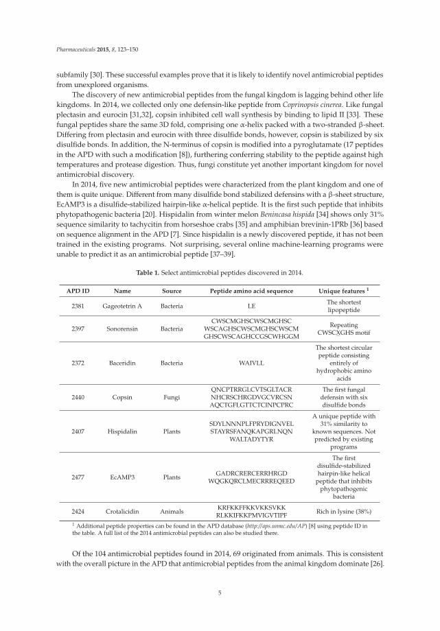
Pharmaceuticals 2015, 8, 123–150
subfamily [30]. These successful examples prove that it is likely to identify novel antimicrobial peptidesfrom unexplored organisms.
The discovery of new antimicrobial peptides from the fungal kingdom is lagging behind other lifekingdoms. In 2014, we collected only one defensin-like peptide from Coprinopsis cinerea. Like fungalplectasin and eurocin [31,32], copsin inhibited cell wall synthesis by binding to lipid II [33]. Thesefungal peptides share the same 3D fold, comprising one α-helix packed with a two-stranded β-sheet.Differing from plectasin and eurocin with three disulfide bonds, however, copsin is stabilized by sixdisulfide bonds. In addition, the N-terminus of copsin is modified into a pyroglutamate (17 peptidesin the APD with such a modification [8]), furthering conferring stability to the peptide against hightemperatures and protease digestion. Thus, fungi constitute yet another important kingdom for novelantimicrobial discovery.
In 2014, five new antimicrobial peptides were characterized from the plant kingdom and one ofthem is quite unique. Different from many disulfide bond stabilized defensins with a β-sheet structure,EcAMP3 is a disulfide-stabilized hairpin-like α-helical peptide. It is the first such peptide that inhibitsphytopathogenic bacteria [20]. Hispidalin from winter melon Benincasa hispida [34] shows only 31%sequence similarity to tachycitin from horseshoe crabs [35] and amphibian brevinin-1PRb [36] basedon sequence alignment in the APD [7]. Since hispidalin is a newly discovered peptide, it has not beentrained in the existing programs. Not surprising, several online machine-learning programs wereunable to predict it as an antimicrobial peptide [37–39].
Table 1. Select antimicrobial peptides discovered in 2014.
APD ID Name Source Peptide amino acid sequence Unique features 1
2381 Gageotetrin A Bacteria LE The shortestlipopeptide
2397 Sonorensin BacteriaCWSCMGHSCWSCMGHSC
WSCAGHSCWSCMGHSCWSCMGHSCWSCAGHCCGSCWHGGM
RepeatingCWSCXGHS motif
2372 Baceridin Bacteria WAIVLL
The shortest circularpeptide consisting
entirely ofhydrophobic amino
acids
2440 Copsin FungiQNCPTRRGLCVTSGLTACRNHCRSCHRGDVGCVRCSNAQCTGFLGTTCTCINPCPRC
The first fungaldefensin with sixdisulfide bonds
2407 Hispidalin PlantsSDYLNNNPLFPRYDIGNVELSTAYRSFANQKAPGRLNQN
WALTADYTYR
A unique peptide with31% similarity to
known sequences. Notpredicted by existing
programs
2477 EcAMP3 Plants GADRCRERCERRHRGDWQGKQRCLMECRRREQEED
The firstdisulfide-stabilizedhairpin-like helical
peptide that inhibitsphytopathogenic
bacteria
2424 Crotalicidin Animals KRFKKFFKKVKKSVKKRLKKIFKKPMVIGVTIPF Rich in lysine (38%)
1 Additional peptide properties can be found in the APD database (http://aps.unmc.edu/AP) [8] using peptide ID inthe table. A full list of the 2014 antimicrobial peptides can also be studied there.
Of the 104 antimicrobial peptides found in 2014, 69 originated from animals. This is consistentwith the overall picture in the APD that antimicrobial peptides from the animal kingdom dominate [26].
5

Pharmaceuticals 2015, 8, 123–150
Moreover, amphibians remain a major source for discovering natural antimicrobial peptides,accounting for 35% of the 2014 total (38.8% of the entire database entries). Most of these new sequencesresemble the known frog antimicrobial peptides, which are linear and have the potential to form ahelical structure [18,19]. Although cathelicidins have been identified from a variety of animals, rangingfrom birds, fish, and reptiles, to mammals [40], candidates from amphibians were not reported until2012 [41]. In 2014, two new members appeared [42], leading to a total of six amphibian cathelicidinsin the APD (five helical and one glycine-rich). These cathelicidins are quite distinct from the mainbody of amphibian peptides. For example, cathlicidin RC-1 has a high content of lysines (32%).Crotalicidin [17], a homologous snake cathelicidin, contains an even higher content of lysines (38%)(Table 1).
Also in 2014, some known human peptides or proteins were demonstrated to be antimicrobial.These include human α-defensin 6 (HD-6), β-defensin 120 (DEFB120), chemokine CCL24 (eotaxin-2),CCL26 (eotaxin-3), and human ribonuclease 6 (RNase 6). While HD-6 is active against Bifidobacteriumadolescentis [43], recombinant DEFB120 is active against Escherichia coli, S. aureus, and Candidaalbicans [44]. Eotaxin-1 (CCL11), eotaxin-2, and eotaxin-3 are known chemokines, which are alsoactive against the airway pathogens Streptococcus pneumoniae, S. aureus, Haemophilus influenzae, andP. aeruginosa [45]. In addition, human RNase 6 is inducible and shows activity against uropathogens,underscoring its defense role in the urinary tract [46]. These characterized members further expandthe known reservoir of human host defense peptides and proteins reviewed in 2014 [47].
3. New Light on Known Human Antimicrobial Peptides
Antimicrobial peptides may be constitutively expressed to keep defined loci in a healthy state [1,48].Compared to neonatal and adult keratinocytes, the corresponding fetal cells express much more humanantimicrobial peptides for host defense [49]. Alternatively, these molecules can also be induced uponbacterial invasion. For example, a human cathelicidin peptide is induced in skin fat cells upon S. aureusinfection, underscoring the significance of adipocytes in host defense [50]. Previously, Gallo andcolleagues also found that overexpression of cathelicidin PR-39 protected animals from group AStreptococcus (GAS) infection [51]. Interestingly, the gut possesses both constitutively expressedand induced antimicrobial peptides. While human cathelicidin LL-37 and β-defensins 2-4 (hBD-2 toHBD-4) are induced, human α-defensin 5 (HD-5), HD-6, and β-defensin 1 (hBD-1) are constitutivelyexpressed [52]. These constitutively expressed human peptides also play a special role in host defense.In 2014, HD-5 was shown to be especially potent against the most virulent form of Clostridium difficile,thereby preventing its infection of small intestine. This is a significant observation considering thatC. difficile can evade the action of other host microbicidal peptides and disturb the balance of gutmicrobiota [53]. Human papillomavirus (HPV) infections can lead to cervical cancer and HD-5 canprevent viral entry [54]. In addition, Wiens and Smith showed that HD-5 directly interferes with acritical host-mediated viral processing step, furin cleavage of L2, at the cell surface [55]. Structurally,HD-5 can form a disulfide bond swapped dimer in vitro [56]. It should be interesting to test whetherthis dimer is linked to host defense in vivo.
How HD-6 plays the defense role in human gut has been puzzling for years. Similar to humanhBD-1 [57], Schroeder et al. found that human Paneth cell HD-6 only exerted antibacterial activityunder reduced conditions, establishing it as a bona fide antimicrobial peptide [43]. This reduction maybe achieved in vivo by the NADPH thioredoxin-reductase system. In vitro, removal of the N-terminaltwo amino acid residues of HD-6 enabled a full reduction by dithiothreitol without influencing itsactivity. Such a truncated form was isolated previously from ileal neobladder urine [58]. In addition,HD-6 can form neutrophil extracellular traps (NETs) to trap invading microbes [59]. Thus, recentbreakthroughs have uncovered two possible mechanisms for HD-6 in host defense.
The importance of environmental conditions for antimicrobial activity is not limited to α-defensins.In 2014, Abou Alaiwa et al. showed that the composition of the airway surface liquid is critical forhuman LL-37 and hBD-3 to kill inhaled and aspirated bacteria. A decrease in pH from 8 to 6.8 in
6

Pharmaceuticals 2015, 8, 123–150
pulmonary airway reduced the activity of both peptides against S. aureus as well as synergistic effectsbetween innate immune peptides [60].
It is known that pH modulates the oligomerization state of human LL-37. At acidic pH,LL-37 is monomeric; it aggregates at physiological pH [61]. The mode of oligomerization was alsostudied in 2014 by using disulfide-linked dimers [62]. NMR studies confirmed this pH-dependentphenomenon [63]. However, the link of this phenomenon to biology was not clear. In 2014, Gao andcolleagues reported that LL-37 enhancement of signal transduction by Toll-like receptor 3 (TLR3) isregulated by pH [64]. Upon acidification in endosomes, oligomerized LL-37 dissociates, allowing therelease of delivered dsRNA to act as an agonist for TLR3 signaling. In contrast, LL-29, a naturalfragment of human LL-37 that lacks the C-terminal portion [65], is unable to do so. Since ourprevious NMR studies found that nearly all the residues of LL-37 (residues 1-36) are involved inoligomerization [63,66], the C-terminal portion of LL-37 might be involved in the tetramer formationof LL-37. The salt bridges, likely involving R34 and/or E36, can be disrupted at acidic pH, providing amolecular basis for pH-dependent oligomer dissociation and dsRNA release.
All these examples above underscore that environmental conditions are an important mediator ofthe function of antimicrobial peptides. There are also other mediators that regulate peptide activity,including proteases, metals, salts, and chemical modifications. While 3D triple-resonance NMRstudies show that the helical region (residues 2-31) of LL-37 is responsible for both membrane andlipopolysaccharides (LPS) binding [63], citrullination of arginines can reduce its ability in preventingendotoxin-induced sepsis [67]. Likewise, ADP-ribosylation of four out of the five arginines of humanLL-37 may regulate this property [68]. A more recent discovery reveals that during S. aureus invasion,skin adipocytes can replicate rapidly and produce a cathelicidin peptide longer than LL-37 to preventinfection [50]. Previously, a different form of human cathelicidin, ALL-38, was also isolated from thehuman reproductive system [69]. Therefore, human proteases at a defined location play an importantrole in determining the exact molecular form of mature antimicrobial peptides required for hostdefense [47].
4. Mechanisms of Action of Antimicrobial Peptides and Genetic Basis of Bacterial Resistance
4.1. Peptide at Work
Although there are anionic peptides, natural antimicrobial peptides are usually cationic with anaverage net charge of +3.2 [7,8]. A leading view is that these cationic peptides target negativelycharged bacterial membranes. However, other mechanisms are possible [4,5,47,63]. In the caseof membrane targeting, how it damages the membranes remains debatable. A variety of possiblemembrane-weakening mechanisms have been summarized by Vogel [70] and three of them aredepicted in Figure 1. These models are helpful and may inspire the design of new experiments to checktheir validity. In the carpet model [71], antimicrobial peptides are assumed to locate on the membranesurface. Is the peptide flat? In 2014, solid-state NMR studies of piscidins revealed peptide tiltingto achieve an optimal interaction. The extent of tilting depends on both peptide sequence and lipidcomposition. The glycine at position 13 may be important for peptide plasticity [72]. Numerous peptideexamples, including human cathelicidin LL-37 [66], possess a similar glycine that may modulatepeptide activity against different bacteria. However, there are only a few examples with a defined pore.Structural determination yielded evidence for channel or pore formation. Gramicidin and alamethicinare two known examples [73,74]. Recently, the crystal structure of human dermcidin implies anotherpossible channel [75,76]. In 2014, a C-type lectin is proposed to form a pore in bacterial membranesbased on a combined structural determination by X-ray diffraction with electron microscopy data.In this model, six copies of human RegIIIα assemble into a ring structure with a hole in the center [77].
7

Pharmaceuticals 2015, 8, 123–150
Figure 1. Mechanisms of action of antimicrobial peptides in 2014. Membrane channel formation(A) is proposed for dermicidin [76] and transmembrane pore formation for C-type lectin RegIIIα [77].While human LL-37 [78] may form a toroidal pore (B), it started with a carpet model [79] (C) whereantimicrobial peptides such as piscidins [72] are located on the membrane surface. Receptor mediatedbinding was observed for Lactococcin G and Enterocin 1071, which bind to UppP, an enzyme involvedin cell wall synthesis (D) [80]. In addition, Gravicin ML binds to maltose ABC receptors (E) [81].Further, RTD2, as well as lantibiotic Pep5, interacts with membranes causing the release of autolysin(F) [82]. Beyond membranes, bacterial MccJ25 could inhibit RNA polymerase (G) [83], while apidaecins,oncocins [84] and Bac7 [85] inhibit protein synthesis by binding to ribosomal proteins (H). Abbreviationsused in the figures are OM, outer membrane; IM, inner membrane; PGN, peptidoglycan; LTA,lipoteichoic acid; MLT, maltose transporter. In addition, refer to the text.
Human α-defensin HNP1 and β-defensin hBD-3 are known to inhibit cell wall synthesis [86,87].What about θ-defensins [16]? In 2014, Selsted and colleagues asked this question. In collaboration withSahl, they found that RTD-2 did not bind to lipid II. Rather, it interacted with bacterial membranesin the presence of glucose. In addition, they detected the release of peptidoglycan lytic enzymes (orautolysins) by S. aureus. Interestingly, bacterial lantibiotic Pep5 can work in the same manner [82].There is precedence for such a similarity. Like defensins, it is common for lantibiotics to inhibit cellwall synthesis by binding to lipid II [88]. In addition, some lantibitics and cyclotides share the samephosphatidylethanolamine (PE) lipid target [89,90]. Such a cyclotide binding to PE-rich membranerafts is proposed to be responsible for activity against cancer and HIV-1 [91]. The similarity betweencyclotides and lantibiotics was initially suggested by their similar amino acid composition plots [63].The mechanistic similarities between thioether bonded lantibiotics and disulfide bonded defensing-likepeptides are remarkable (Table 2), supporting a recent universal peptide classification that groupsthem into one big class: sidechain-sidechain linked peptides [9].
Table 2. Mechanistic similarities between thioether-bonded lantibiotics and disulfide-boned peptides.
Mechanism Lantibiotic Examples Disulfide-Linked Examples
Inhibition of cell wall synthesis 1 Nisin A, lacticin 3147, mersacidin,bovicin HJ50
HNP1, hBD-3, plectasin,lucifensin, eurocin, copsin
Membrane and autolysin release Pep5 θ-defensins such as RTD-2
Binding to lipid PE Duramycins, cinnamycin Kalata B1, cycloviolacin O21 Selected from the APD [7,8]. A more complete list can be searched in the APD.
8

Pharmaceuticals 2015, 8, 123–150
For years, proline-rich peptides were proposed to act by binding to heat shock proteins [92]. Twopapers published in 2014 challenged this view. Krizsan et al. found that insect-derived proline-richapidaecins and oncocins inhibited bacterial protein translation at the 70S ribosome [84]. Both cationicand hydrophobic amino acids of the peptides were involved in such binding. Interestingly, Mardirossianet al. also observed that Bac7(1-35), another proline-rich peptide corresponding to N-terminal 35residues of bovine cathelicidin Bac7, could accumulate within E. coli to a high concentration of 340 μMand inhibits protein synthesis by targeting ribosomal proteins [85]. These studies indicate that thewell-documented chaperone DnaK is unlikely the major target for proline-rich peptides. Of note,bacterial lasso peptides such as microcin J25 (MccJ25) can inhibit RNA polymerase [83].
Some antimicrobial peptides can associate with DNA. In 2014, Ghosh et al. found that the WWmotif of indolicidin is essential for DNA binding. They provided high-resolution structural informationfor the interaction of indolicidin with duplex DNA [93]. Such a structure can be useful for designingnovel antibiotics.
Interestingly, some bacteriocins use receptors as the target. While garvicin ML recognizes amaltose ABC transporter, LsbB uses metallopeptidase as the targets [81]. In 2014, Kjos et al. foundlactococcin G and enterocin 1071 (two-chain bacteriocins) used UppP as the receptor. UppP is anenzyme involved in cell wall synthesis [80]. Such findings not only enrich our view on the mechanismsof action of antimicrobial peptides, but also open new opportunities for antimicrobial development.
Although it is likely that some antimicrobial peptides mainly utilize one mechanism to inhibitbacteria, a single peptide may also deploy multiple mechanisms, rendering it difficult for pathogens todevelop resistance. In 2014, Wenzel et al. illustrated this using a model arginine and tryptophan-richpeptide RWRWRW-NH2 (C-terminal amidation) [94]. The peptide is primarily membrane targeting(e.g., D and L-forms have same activity) and only a very small population can enter the bacterial cell.As a new mechanism, the authors found that multiple surface proteins could be delocalized by thepeptide. While the replacement of cytochrome C hinders bacterial energy metabolism, delocalizationof MinD interferes with bacterial replication. Another surface protein, MurG, can also be delocalized,leading to impaired bacterial cell wall synthesis. The authors proposed that such bacterial surfaceprotein delocalization by cationic antimicrobial peptides may be a general mechanism. Our own recentfindings may provide additional insight into this protein delocalization. Surface attachment is usuallymediated by a short amphipathic sequence, which weakly interacts with bacterial membranes [95].In contrast, cationic antimicrobial peptides are able to better interact with bacterial membranes sincethey have a broader hydrophobic surface or higher membrane perturbation potential [96]. Such amembrane-binding difference could be one of the fundamental reasons for surface proteins to bereplaced by cationic antimicrobial peptides. In addition, membrane binding of short amphipathicsequences requires anionic lipids [95]. When short cationic peptides cause lipid domain formation [94,97,98], the migration of anionic lipids toward cationic antimicrobial peptides could weaken theattachment of surface proteins to the re-organized membranes, causing protein delocalization and lossof function as demonstrated by Wenzel et al. [94].
A single peptide can also possess multiple functions and human cathelicidin LL-37 is a typicalexample for this [99]. The observation that LL-37 can associate with DNA led to the idea that DNAbinding may be part of the bacterial killing mechanism as well. However, Mardirossian et al. showedthat only 5% of LL-37 inhibited protein synthesis [85]. In agreement, we did not observe a correlationbetween peptide activity and DNA retardation (Lau, K.; Lushnikova, T.; Wang, G., unpublishedresults). However, we did observe a correlation between membrane permeation and antimicrobialactivity of LL-37 fragments [100]. These results support the existing view that membrane permeationand disruption by the helical region (residues 2-31) is the major mechanism via which LL-37 killsbacteria [78,79,97]. It appears that nucleic acid binding plays a more important role in RNA deliveryinto endosomes [101] and in stabilizing neutrophil extracellular traps to prevent DNA cleavage [102].Moreover, human LL-37 can associate with cell receptors to trigger signal transduction [103,104].Interestingly, LL-37 also modulates innate immunity by promoting macrophages to phagocyte
9
Pharmaceuticals 2015, 8, 123–150
bacteria [105] or influencing neutrophil responses to influenza A virus [106]. In addition, overexpressedLL-37, as an antigen, can be specifically recognized by CD4+ and/or CD8+ T cells in psoriasis [107].It is clear that the multifunctional roles of human LL-37 are realized by its ability to recognize andinteract with different molecular targets and immune cells.
4.2. Resistance Genes for Pathogens and Survival Skills for Commensal Bacteria
It has been appreciated that bacteria have been co-evolving with host defense peptides [108].Some have learned how to avoid the attack by cationic peptides. The major mechanism appears toalter cell envelope charge and composition. In addition, an ATP-binding cassette (ABC) transportercoupled with an adjacent two-component system (TCS) also constitutes a resistance module againstantimicrobial peptides [109,110]. Elucidation of the genetic basis of bacterial resistance may be helpfulfor the design of more potent antibiotics. In the following sections, we highlight progress made in thisdirection during 2014.
4.2.1. Gram-Positive Bacteria
One can identify the bacterial genes involved in peptide response by two methods: proteomeanalysis or genome analysis. Using proteome analysis of bacitracin-treated and untreated cells,Gebhard et al. identified an ABC transporter EF2050-2049 of Enterococcus faecalis that mediates resistanceagainst bacitracin [109]. To validate this, they transferred the resistance and regulatory pathway to B.subtilis, leading to bacitracin resistance. Thus, the ABC transporter and the TCS are indeed requiredfor resistance to antimicrobial peptides. Nevertheless, a previous genomic analysis identified twosuch ABC transporters [110], which were induced by bacitracin [109]. Based on these results, theauthors proposed a model for the bacitracin resistance network of E. faecalis. The presence of bacitracinis initially detected by the EF2752-51 ABC transporter, which relays this signal to an adjacent TCS(EF0926-27). Activation of the regulatory domain of the TCS leads to an increase in expression of theEF2050-49 ABC transporter that confers resistance to antimicrobial peptides. This two transporters andone TCS network mechanism [109] differs from those single ABC transporter and TCS cases where thetransporter senses the peptide and relays this signal to the adjacent TCS that upregulates the sameABC transporter for resistance [110].
The five-component system GraXSR-VraFG of S. aureus is well-established as the major sensing andresistance system [111] that reduces the toxic effect of cationic antimicrobial peptides by upregulatinggenes such as mprF and dltABCD. While MprF can put lysines on anionic phophatidylglycerols (PGs),DltABCD can modify the cell wall by transferring of D-alanine into teichoic acids [108,112]. GraSR wasfound to regulate the dltABCD and mprF genes [113,114]. In 2014, a loop region of sensor protein GraSwas identified to recognize cationic peptides. Cheung et al. generated mutants of graS from the MRSAstrain MW2. Deletion of graS (ΔgraS strain) or its 9-amino acid extracellular loop region (ΔEL strain)made the strain more susceptible to daptomycin, polymyxin B, human neutrophil defensin 1 (HNP-1),and RP-1 (a platelet factor 4 derived peptide that retains activity in blood). Meanwhile, these mutantsbecame less infectious in vivo in an endocarditis model. Interestingly, a synthetic trimeric loop regionEL mimic, i.e., (DYDFPIDSL)3, could protect the parental MW2 strain from killing by those cationicpeptides. These results suggest that the acidic residues in the extracellular loop region of GraS candirectly interact with cationic peptides for sensing and activation [115].
It has been elucidated that Group A Streptococcus responds to the human antimicrobial peptideLL-37 by upregulating virulence factors controlled by the CsrRS system. In 2014, Velarde et al. identifiedRI-10, the smallest LL-37 fragment required to bind to CsrRS using a series of synthetic LL-37 fragments.RI-10 can directly bind to sensor kinase CsrS to activate the expression of virulence factors [116]. Thesame peptide was previously found by us to have no antibacterial activity against bacteria [97,98].Since antimicrobial activity is not required for this recognition by kinase receptor, such a responsecould occur in vivo at a very low level of LL-37, which is not high enough to kill the bacteria. Wepropose that interfacial cationic residues R23 and K25, which are important for membrane permeation
10

Pharmaceuticals 2015, 8, 123–150
and bacterial killing [100], are also important residues for interaction with the acidic amino acids onthe CsrS receptor.
4.2.2. Gram-Negative Bacteria
Different from Gram-positive bacteria, LPS is the major component in the outer membranes ofGram-negative bacteria [66]. Modifications of bacterial LPS provide a general mechanism that confersresistance to cationic antimicrobial peptides [117]. The 2014 research on the genetic basis of bacterialresistance yielded additional support for this. First, pathogenic Vibrio cholerae strains can be >100-foldmore resistant to polymyxins by modifying LPS (i.e., glycylation). Henderson et al. confirmed AlmFas an aminoacyl carrier protein, which is activated by the enzyme AlmE. Interestingly, these proteinsin the AlmEFG trio system function in a manner similar to nonribosomal peptide synthetases [118].Second, the resistance of human pathogen Neisseria gonorrhoeae results from phosphoethanolamine(PEA) decoration of lipid A by transferase encoded by the lptA gene [119–121]. Kandler et al. foundthat high-frequency mutation in a polynucleotide repeat of the lptA gene influences bacterial resistance.An lptA mutant is highly susceptible to cationic peptides [122]. In addition, the PEA-modification oflipid A has an immunostimulatory role during infection [123]. Third, in the case of the gastrointestinalpathogen S. typhimurium, resistance genes involving both LPS defects and mutation in phoP werefound from a transposon library in 1992 [124]. The PhoPQ two-component system regulates peptideresistance, bacterial lipid A remodeling, and intracellular survival within acidified phagosomes. In2014, the PhoPQ system was found to also regulate acidic glycerophospholipid content in the outermembrane [125]. These authors have recently summarized the resistance strategies of S. typhi [126,127].
In summary, both Gram-positive and Gram-negative bacteria are able to decorate their cellularsurfaces to make them less attractive to cationic antimicrobial peptides. Interestingly, a recent studyreveals that gut bacteria can use a similar mechanism by removing a phosphate group from LPS [128].Here a resistant mechanism for “bad bugs” has become a survival strategy for “good bugs”. Therefore,such surface decorations achieved by a different chemistry provide a general mechanism that enablesbacteria, for good or bad, to “work under an umbrella” to dodge the “bullets” of the host.
5. Potential Applications of Antimicrobial Peptides
5.1. Toward Therapeutic Uses
There is continued interest in developing therapeutic uses for antimicrobial peptides. Because ofmolecular simplicity and easy synthesis, linear peptides have been extensively explored for favorableproperties. Here we highlight a structure-based design based on the human cathelicidin LL-37. Wangidentified a chymotrypsin-resistant template by screening an LL-37 peptide library [129]. This templatecontains partial D-amino acids and has a novel amphipathic structure [130]. However, it is notactive against community-associated methicillin-resistant S. aureus (MRSA) USA300. Based on the 3Dstructure, we enhanced anti-MRSA activity of the peptide by inserting a bulkier hydrophobic groupinto the structural cavity. One of the peptide analogs, 17BIPHE2, not only eliminated MRSA, but alsorecruited monocytes to the infection site [129]. In addition, other approaches were explored to make useof LL-37 or hBD-2. Since cathelicidin can reverse intestinal fibrosis in models of colitis [131], this peptidemay be used to treat inflammatory bowel disease (IBD) by introducing cathelicidin-overexpressingbacteria. Using an adenoviral vector to deliver the gene of hBD-2, Woo et al. found both viral inhibitionand clearance for experimental otitis media [132]. In 2014, LL-37-containing vector was electroporatedto promote skin wound healing [133]. In addition, 1,25-dihydroxyvitamin D3 (active form) was alsoused to induce both LL-37 and hBD-2 production in keratinocytes from diabetic foot ulcers, promotingwound healing [134]. UV light or sunlight may be an alternative since hydroxylation of vitamin Dcan occur [135]. It is notable that traditional practice can also boost our immune systems. While yogastretching significantly increases human hBD-2 [136], green tea helps the production of lactoferrin in
11

Pharmaceuticals 2015, 8, 123–150
saliva after exercise [137]. Although at the early stage, these positive results on antimicrobial peptidesimply that our traditional life styles can be helpful to keep us healthy.
Unlike linear peptides that can be rapidly degraded by proteases in hours, circular peptides haveinherent stability. This is because these peptides such as cyclotides comprise three conserved disulfidebonds in addition to a peptide bond that connects the N- and C-termini. Consequently, there is greatinterest in utilizing these natural templates to engineer useful therapeutics [138,139]. In 2014, Craikand colleagues demonstrated the molecular grafting technology where a desired antigenic peptidewas inserted into an exposed loop region of kalata B1 [140]. This technology confers protease stabilityto the sequence motif, which can otherwise be rapidly degraded when tested alone. MOG3, one out ofthe seven peptides grafted to loop 5, was used to vaccinate mice and found to display in vivo efficacyin an animal model of multiple sclerosis, an inflammatory disease of the central nervous system. Takentogether with previous examples [138,139], these authors proved the concept of segment graftingat loops 5 and 6 of cyclotides. It appears that sequence composition rather than length determineswhether the grafted segment is tolerated without disrupting the protein fold. To obtain a sufficientamount of material for research, cyclotides were initially isolated from plants [141]. Later, chemicalsynthesis was established [142]. In 2014, two laboratories reported an alternative approach by usingengineered sortase A to make the circular molecule [143,144]. A more efficient synthesis will bring usone step closer to practical use of these interesting templates.
5.2. Peptide Surface Coating
Immobilization of antimicrobial peptides (either covalently or adsorbed) onto solid supportsextends their capabilities as surface-active molecules. This direction of research aims at improvingbiomedical devices, drug delivery systems, bio-sensor and detection, and so on. Recent advancesin the field include simplification of the chemistry for surface attachment, development of novelsubstrate-attaching platforms, including nanomaterial for wider applications, product developmentwith promising proof of concepts and in vivo testing in animal models.
Of all the problems related to loss in efficiency of medically implantable medical devices isthe development of microbial biofilms. Peptide immobilization has been shown to reduce bacterialcolonization and biofilm formation. However, the major challenges that often hinder the immobilizationof antimicrobial peptides are the inefficiency of the conjugation chemistries and their inability to achievea sufficient surface concentration of peptides, along with the limited number of usable biomaterials.A recent study by Lim et al. [145] demonstrated a simple dopamine based chemical reaction for thefunctionalization of antimicrobial peptides onto a commonly used Silicon Foley catheter. Not onlydid the catheter prototype reduce biofilm formation by common pathogens that caused a urinarytract infection, it was also stable for 21 days. In addition, hLF1-11 immobilized onto titanium [146]has been shown to possess excellent anti-biofilm properties. There have also been improvementsin other chemical platforms, including the development of thiolated self-assembled monolayer ona gold surface to which small peptides, temporin-SHf, can be tethered, resulting in broad-spectrumactivity [147].
To understand the possible influence of structure and dynamics on immobilized antimicrobialpeptides and to apply rational design, molecular dynamics simulation studies were carried out oncecropin P1 immobilized on silane-EG4-maleimide self-assembled monolayers [148]. Other factorsgoverning immobilization reactions, such as spacer chain length, reactant concentration and energydependence, are demonstrated by Mishra et al. [149]. Additional coating strategies can be found in arecent review [150]. Moving toward practical applications, a significant in vivo study is presented byDutta et al. [151]. Melimine immobilized eye lenses are not only cytotoxically safe but also possessantibacterial activity after worn as tested in both rabbit and human. In addition, another peptideSESB2V immobilized on titanium surfaces prevents perioperative corneal infection in a rabbit keratitismodel [152].
12

Pharmaceuticals 2015, 8, 123–150
5.3. Nanoparticle-Based Drug Delivery Systems
Apart from the anti-biofilm and antibacterial functions, antimicrobial peptides tagged tonanoparticles impart a site-specific targeting and delivery of drug molecules. It can be used in treatinga variety of diseases, including cancer. Currently, dermaseptin entrapped chitosan nanoparticleshave been shown to possess excellent antitumor activities [153]. Moving one step forward, dualtargeting nanoparticles with both blood−brain barrier (BBB) and blood−brain tumor barrier (BBTB)including glioma cell were achieved by functionalizing lactoferrin to the surface of poly(ethyleneglycol)−poly(lactic acid) nanoparticles. Administration with tLyP-1, a tumor-homing peptide thatmediates tissue penetration through the neuropilin-1-dependent internalization pathway, resulted indeep penetration of the nanoparticle into the glioma parenchyma [154]. It opens a new direction foradministration of antitumerogenic drugs with high penetration capability.
Additionally, glutamic acid substitution of basic residues in LL-37, melittin, and bombolitin Vlinked to lipid nanoparticles could be used for endosomal escape and efficient gene delivery usingintravenous injections. These yield expression levels comparable to those obtained using Lipofectamine2000 and the probable mode of action resembles viruses [155]. Antimicrobial peptides have also beenshown to have superb drug releasing properties in PEG-PLGA microparticles [156]. The direct evidenceis presented by encapsulating LL-37 in PLGA nanoparticles by Chereddy et al. [157]. PLGA-LL-37nanoparticles as a biodegradable drug delivery system were found to promote wound closure.It functions due to the sustained release of both LL-37 and lactate, and induction of enhanced cellmigration without effects on the metabolism and proliferation of keratinocytes. Moreover, peptides asshort as dimer conjugated to naphthalene could be used as antimicrobial nanomaterials in eliminatingbiofilm infections and for drug delivery [158].
5.4. Biosensors and Detection
Antimicrobial peptides can also serve as indicators and diagnostic agents. The approach is morecost effective than standard PCR or antibody-based techniques. Detection of bacterial pathogens in amicrofluidic chip where antimicrobial peptides are immobilized via cysteine-gold interaction couldproduce a rapid electrical detection with sufficient sensor signal that allows the detection of pathogens(both Gram-negative and Gram-positive) at a density as low as 105 cfu/mL within 25 min [159]. Whileanother platform for the detection of only Gram-positive bacterial strains could reach 103 cfu/mL viaimmobilizing class IIa bacteriocins [160]. In addition, specific detection of fungal C. albicans can also bemade by using peptide nucleic acid probes [161].
6. Perspectives
It is great news that acquired resistance did not become an issue after decade-long use ofantimicrobial peptides such as tyrothricin [162]. Such an observation is encouraging to the currenteffort in development of antimicrobial peptides into novel therapeutic molecules [63]. From 2000to 2014, about 100 new antimicrobial peptides were entered into the APD every year [26]. As ofDecember 2014, there were 2493 peptides in this database [7–9]. We anticipate that the importantwork on the isolation and characterization of novel antimicrobial peptides from new organisms willcontinue. Scientifically, new peptide sequences will improve our knowledge of natural antimicrobialpeptides. As shown in Table 1, the new members can refine the boundary parameters for naturalantimicrobial peptides [9]. With the identification of a sufficient number of representative peptidesequences, the APD database will more accurately identify natural peptides most similar to the inputsequence. It will facilitate the development of new programs to better predict the likelihood of a newpeptide to be antimicrobial. In addition, a unique peptide may directly become a candidate lead forthe development of novel antimicrobials to meet the challenge of the antibiotic resistance problem.However, it is important to validate whether a bacteriocin has any undesired virulent property thatpromotes infection [21].
13
Pharmaceuticals 2015, 8, 123–150
The 2014 research also enabled us to connect the dots for known human antimicrobial peptides,leading to an improved understanding of their functional roles in innate immunity. Surprisingly, adifferent form of human cathelicidin peptide can be rapidly expressed by skin fat cells in responseto the S. aureus invasion, underscoring a direct defense role of human cathelicidin peptides [50]. It isremarkable that a single LL-37 molecule can perform multiple functions by recognizing a variety ofmolecular partners or receptors on immune cells. In 2014, pH-dependent oligomerization of LL-37is connected to the delivery of dsRNA into endosomes for subsequent interactions with TLR3 [64].Although the details are to be elucidated, we propose that the terminal regions of LL-37 contain animportant molecular switch based on NMR data [63]. It is demonstrated that local conditions areessential for a proper function of human antimicrobial peptides. While human HD-6, like hBD-1, isactive only after disruption of disulfide bonds under reduced conditions [43], many other defensinsin the folded form can directly recognize specific lipids in pathogen’s membranes [163,164]. Duringsuch a molecular recognition process, the flexible residues in the loop regions of these small defenseproteins are found to be essential based on several structural studies [163–166].
The 2014 results further expanded our view on the mechanism of action of antimicrobial peptides(summarized in Figure 1). While select antimicrobial peptides are known to inhibit cell wall synthesis,many target bacterial membranes. Surface-binding peptides are shown to be able to replace weaklyattached membrane proteins, thereby interfering with bacterial physiology globally [94]. Beyondmembranes, bacteriocins can use cell receptors as a target [81]. It has been known for a while thatproline-rich peptides interact with heat shock proteins [92]. However, the molecular target has nowbeen traced to ribosomal proteins. The binding of proline-rich peptides leads to inhibition of proteinsynthesis [84,85].
Although there are few examples, structure-based design has been demonstrated [129,140],leading to antimicrobials or vaccines with desired properties. The overall goal of peptide engineeringis to establish or identify a proper peptide template with desired potency, stability, and cell selectivity.In addition, one may also mimic nature’s wisdom. Based on the precursor protein construction, onecan design pro-peptide forms to minimize potential cytotoxicity and protease degradation. In 2014,Forde et al. illustrated this strategy as a potential therapy for cystic fibrosis [167,168]. Nature hascreated other strategies to circumvent rapid peptide degradation by forming a complex structure. Forinstance, human LL-37 can oligomerize at physiological pH into nanoparticles to resist the actionof proteases [63]. Likewise, LL-37 can bind to DNA to stabilize the entire NETs structure [169]. In2014, Bachrach and colleagues showed that human LL-37 could also be protected by actin, therebymaintaining its antimicrobial activity in vivo [170,171]. There are also natural ways to reduce potentialcytotoxicity of antimicrobial peptides. In 2014, Svensson et al. discovered that peptide p33 expressed onthe surface of various cell types can reduce the potential cytotoxicity of human LL-37 [172]. Moreover,Hiemstra et al. discovered the nanoparticle-like vesicles in the human urinary tract [173]. It ispredictable that novel functional modes of human innate immune peptides will continue to emerge.All these natural mechanisms may hold the key to future therapeutics.
We anticipate continued efforts in the development of potential applications of antimicrobialpeptides, including peptide production methods. Peptide engineering, formulation, and deliverytechnologies may further expand the horizon of antimicrobial peptides in benefiting humanbeings [174]. Such applications can vary from medical surface cleaning, water quality monitoring anddisinfection, sterile surface materials, to new drugs for infectious diseases [175,176]. Antimicrobialpeptides may be included in existing detergent formulation and disinfectants to reduce bacterialbiofilms on hospital surfaces [177]. Bovicin HC5 and nisin can be used to treat food-contact surface toreduce bacterial attachment and subsequent biofilm formation [178,179]. It has been demonstratedthat injection of an engineered LL-37 peptide or coating peptides to the device surface can preventbiofilm formation [129,145,146]. Importantly, a combined use of antimicrobial peptides with traditionalantibiotics can be a more effective strategy to treat bacterial biofilms [180–182]. Finally, molecules that
14

Pharmaceuticals 2015, 8, 123–150
interfere with or even weaken the process of biofilm formation [183–189] can also be combined withantimicrobial peptides to achieve an optimal treatment of bacterial biofilms.
Acknowledgments: This study is supported by the grants from the NIAID/NIH R01AI105147 and the state ofNebraska to GW. We thank Cheryl Putnam for final editing.
Author Contributions: GW conceived the project. GW and BM drafted the manuscript. All authors were involvedin literature search, discussion and writing.
Conflicts of Interest: No conflicts of interest was declared.
References
1. Zasloff, M. Antimicrobial peptides of multicellullar organisms. Nature 2002, 415, 359–365. [CrossRef]2. Lai, Y.; Gallo, R.L. AMPed up immunity: How antimicrobial peptides have multiple roles in immune defense.
Trends Immunol. 2009, 30, 131–141. [CrossRef] [PubMed]3. Hancock, R.E.; Lehrer, R. Cationic peptides: A new source of antibiotics. Trends Biotechnol. 1998, 16, 82–88.
[CrossRef] [PubMed]4. Yeaman, M.R.; Yount, N.Y. Mechanisms of Antimicrobial Peptide Action and Resistance. Pharmacol. Rev.
2003, 55, 27–55. [CrossRef] [PubMed]5. Brogden, K.A. Antimicrobial peptides: Pore formers or metabolic inhibitors in bacteria? Natl. Rev. Microbiol.
2005, 3, 238–250. [CrossRef]6. NCBI Resource Coordinators. Database resources of the National Center for Biotechnology Information.
Nucleic Acids Res. 2015, 43, D6–D17.7. Wang, Z.; Wang, G. APD: The antimicrobial peptide database. Nucleic Acids Res. 2004, 32, D590–D592.
[CrossRef] [PubMed]8. Wang, G.; Li, X.; Wang, Z. The updated antimicrobial peptide database and its application in peptide design.
Nucleic Acids Res. 2009, 37, D933–D937. [CrossRef] [PubMed]9. Wang, G. Improved methods for classification, prediction, and design of antimicrobial peptides. Methods
Mol. Biol. 2015, 1268, 43–66. [PubMed]10. Timeline of Antimicrobial Peptide Discovery. http://aps.unmc.edu/AP/timeline.php. Accessed on 16
March 2015.11. Chakchouk-Mtibaa, A.; Elleuch, L.; Smaoui, S.; Najah, S.; Sellem, I.; Abdelkafi, S.; Mellouli, L. An antilisterial
bacteriocin BacFL31 produced by Enterococcus faecium FL31 with a novel structure containing hydroxyprolineresidues. Anaerobe 2014, 27C, 1–6. [CrossRef] [PubMed]
12. Niggemann, J.; Bozko, P.; Bruns, N.; Wodtke, A.; Gieseler, M.T.; Thomas, K.; Jahns, C.; Nimtz, M.; Reupke, I.;Brüser, T.; et al. Baceridin, a cyclic hexapeptide from an epiphytic Bacillus strain, inhibits the proteasome.Chembiochem 2014, 15, 1021–1029. [CrossRef] [PubMed]
13. Gavrish, E.; Sit, C.S.; Cao, S.; Kandror, O.; Spoering, A.; Peoples, A.; Ling, L.; Fetterman, A.; Hughes, D.;Bissell, A.; et al. Lassomycin, a ribosomally synthesized cyclic peptide, kills mycobacterium tuberculosis bytargeting the ATP-dependent protease ClpC1P1P2. Chem. Biol. 2014, 21, 509–518. [CrossRef] [PubMed]
14. Yamanaka, K.; Reynolds, K.A.; Kersten, R.D.; Ryan, K.S.; Gonzalez, D.J.; Nizet, V.; Dorrestein, P.C.; Moore, B.S.Direct cloning and refactoring of a silent lipopeptide biosynthetic gene cluster yields the antibiotic taromycinA. Proc. Natl. Acad. Sci. U.S.A. 2014, 111, 1957–1962. [CrossRef] [PubMed]
15. Scholz, R.; Vater, J.; Budiharjo, A.; Wang, Z.; He, Y.; Dietel, K.; Schwecke, T.; Herfort, S.; Lasch, P.; Borriss, R.Amylocyclicin, a Novel Circular Bacteriocin Produced by Bacillus amyloliquefaciens FZB42. J. Bacteriol. 2014,196, 1842–1852. [CrossRef] [PubMed]
16. Tang, Y.Q.; Yuan, J.; Osapay, G.; Osapay, K.; Tran, D.; Miller, C.J.; Ouellette, A.J.; Selsted, M.E. A cyclicantimicrobial peptide produced in primate leukocytes by the ligation of two truncated alpha-defensins.Science 1999, 286, 498–502. [CrossRef] [PubMed]
17. Falcao, C.B.; de La Torre, B.G.; Pérez-Peinado, C.; Barron, A.E.; Andreu, D.; Rádis-Baptista, G. Vipericidins: anovel family of cathelicidin-related peptides from the venom gland of South American pit vipers. AminoAcids 2014, 46, 2561–2571. [CrossRef] [PubMed]
18. Conlon, J.M.; Mechkarska, M. Host-defense peptides with therapeutic potential from skin secretions of frogsfrom the family pipidae. Pharmaceuticals 2014, 7, 58–77. [CrossRef] [PubMed]
15

Pharmaceuticals 2015, 8, 123–150
19. Guo, C.; Hu, Y.; Li, J.; Liu, Y.; Li, S.; Yan, K.; Wang, X.; Liu, J.; Wang, H. Identification of multiple peptideswith antioxidant and antimicrobial activities from skin and its secretions of Hylarana taipehensis, Amolopslifanensis, and Amolops granulosus. Biochimie. 2014, 105, 192–201. [CrossRef] [PubMed]
20. Ryazantsev, D.Y.; Rogozhin, E.A.; Dimitrieva, T.V.; Drobyazina, P.E.; Khadeeva, N.V.; Egorov, T.A.;Grishin, E.V.; Zavriev, S.K. A novel hairpin-like antimicrobial peptide from barnyard grass (Echinochloacrusgalli L.) seeds: Structure-functional and molecular-genetics characterization. Biochimie 2014, 99, 63–70.[CrossRef] [PubMed]
21. Li, M.F.; Zhang, B.C.; Li, J.; Sun, L. Sil: A Streptococcus iniae bacteriocin with dual role as an antimicrobial andan immunomodulator that inhibits innate immune response and promotes S. iniae infection. PLoS ONE 2014,9, e96222. [CrossRef] [PubMed]
22. Weisshoff, H.; Hentschel, S.; Zaspel, I.; Jarling, R.; Krause, E.; Pham, T.L. PPZPMs—A novel group of cycliclipodepsipeptides produced by the Phytophthora alni associated strain Pseudomonas sp. JX090307—Themissing link between the viscosin and amphisin group. Nat. Prod. Commun. 2014, 9, 989–996. [PubMed]
23. Trindade, F.; Amado, F.; Pinto da Costa, J.; Ferreira, R.; Maia, C.; Henriques, I.; Colaco, B.; Vitorino, R.Salivary peptidomic as a tool to disclose new potential antimicrobial peptides. J. Proteomics 2014, 115C, 49–57.[CrossRef] [PubMed]
24. Bouzid, W.; Verdenaud, M.; Klopp, C.; Ducancel, F.; Noirot, C.; Vetillard, A. de novo sequencing andtranscriptome analysis for tetramorium bicarinatum: A comprehensive venom gland transcriptome analysisfrom an ant species. BMC Genomics 2014, 15, 987. [CrossRef] [PubMed]
25. Capriotti, A.L.; Cavaliere, C.; Foglia, P.; Piovesana, S.; Samperi, R.; Zenezini Chiozzi, R.; Lagana, A.Development of an analytical strategy for the identification of potential bioactive peptides generated byin vitro tryptic digestion of fish muscle proteins. Anal. Bioanal. Chem. 2015, 407, 845–854. [CrossRef] [PubMed]
26. Wang, G. Database-guided discovery of potent peptides to combat HIV-1 or superbugs. Pharmaceuticals 2013,6, 728–758. [CrossRef] [PubMed]
27. Tareq, F.S.; Lee, M.A.; Lee, H.S.; Lee, Y.J.; Lee, J.S.; Hasan, C.M.; Islam, M.T.; Shin, H.J. Gageotetrins A-C,Noncytotoxic antimicrobial linear lipopeptides from a marine bacterium Bacillus subtilis. Org. Lett. 2014, 16,928–931. [CrossRef] [PubMed]
28. Makovitzki, A.; Avrahami, D.; Shai, Y. Ultrashort antibacterial and antifungal lipopeptides. Proc. Natl. Acad.Sci. U.S.A. 2006, 103, 15997–16002. [CrossRef] [PubMed]
29. First in a New Class of Antibiotics. FDA Consum 2003, 37, 4.30. Chopra, L.; Singh, G.; Choudhary, V.; Sahoo, D.K. Sonorensin: an antimicrobial peptide, belonging to the
heterocycloanthracin subfamily of bacteriocins, from a new marine isolate, Bacillus sonorensis MT93. Appl.Environ. Microbiol. 2014, 80, 2981–2990. [CrossRef] [PubMed]
31. Mygind, P.H.; Fischer, R.L.; Schnorr, K.M.; Hansen, M.T.; Sonksen, C.P.; Ludvigsen, S.; Raventos, D.;Buskov, S.; Christensen, B.; De Maria, L.; et al. Plectasin is a peptide antibiotic with therapeutic potentialfrom a saprophytic fungus. Nature 2005, 437, 975–980. [CrossRef] [PubMed]
32. Oeemig, J.S.; Lynggaard, C.; Knudsen, D.H.; Hansen, F.T.; Norgaard, K.D.; Schneider, T.; Vad, B.S.;Sandvang, D.H.; Nielsen, L.A.; Neve, S.; et al. Eurocin, a new fungal defensin: structure, lipid binding, andits mode of action. J. Biol. Chem. 2012, 287, 42361–42372. [CrossRef] [PubMed]
33. Essig, A.; Hofmann, D.; Munch, D.; Gayathri, S.; Kunzler, M.; Kallio, P.T.; Sahl, H.G.; Wider, G.; Schneider, T.;Aebi, M. Copsin, a novel peptide-based fungal antibiotic interfering with the peptidoglycan synthesis. J. Biol.Chem. 2014, 289, 34953–34964. [CrossRef] [PubMed]
34. Sharma, S.; Verma, H.N.; Sharma, N.K. Cationic bioactive peptide from the seeds of Benincasa hispida. Int. J.Pept. 2014, 2014, 156060. [CrossRef] [PubMed]
35. Kawabata, S.; Nagayama, R.; Hirata, M.; Shigenaga, T.; Agarwala, K.L.; Saito, T.; Cho, J.; Nakajima, H.;Takagi, T.; Iwanaga, S. Tachycitin, a small granular component in horseshoe crab hemocytes, is anantimicrobial protein with chitin-binding activity. J. Biochem. 1996, 120, 1253–1260. [CrossRef] [PubMed]
36. Conlon, J.M.; Mechkarska, M.; Emanahmed Coquet, L.; Jouenne, T.; Jérômeleprince Vaudry, H.; Hayes, M.P.;Padgett-Flohr, G. Host defense peptides in skin secretions of the Oregon spotted frog Ranapretiosa:Implications for species resistance to chytridiomycosis. Dev. Comp. Immunol. 2011, 35, 644–649. [CrossRef][PubMed]
37. Thomas, S.; Karnik, S.; Barai, R.S.; Jayaraman, V.K.; Idicula-Thomas, S. CAMP: A useful resource for researchon antimicrobial peptides. Nucleic Acids Res. 2010, 38, D774–D780. [CrossRef] [PubMed]
16

Pharmaceuticals 2015, 8, 123–150
38. Lata, S.; Mishra, N.K.; Raghava, G.P. AntiBP2: Improved version of antibacterial peptide prediction. BMCBioinformatics 2010, 11, S19. [CrossRef] [PubMed]
39. Xiao, X.; Wang, P.; Lin, W.Z.; Jia, J.H.; Chou, K.C. iAMP-2L: A two-level multi-label classifier for identifyingantimicrobial peptides and their functional types. Anal. Biochem. 2013, 436, 168–177. [CrossRef] [PubMed]
40. Zanetti, M. The role of cathelicidins in the innate host defenses of mammals. Curr. Issues Mol. Biol. 2005, 7,179–196. [PubMed]
41. Hao, X.; Yang, H.; Wei, L.; Yang, S.; Zhu, W.; Ma, D.; Yu, H.; Lai, R. Amphibian cathelicidin fills theevolutionary gap of cathelicidin in vertebrate. Amino acids 2012, 43, 677–685. [CrossRef] [PubMed]
42. Ling, G.; Gao, J.; Zhang, S.; Xie, Z.; Wei, L.; Yu, H.; Wang, Y. Cathelicidins from the bullfrog Rana catesbeianaprovides novel template for peptide antibiotic design. PLoS ONE 2014, 9, e93216. [CrossRef] [PubMed]
43. Schroeder, B.O.; Ehmann, D.; Precht, J.C.; Castillo, P.A.; Kuchler, R.; Berger, J.; Schaller, M.; Stange, E.F.;Wehkamp, J. Paneth Cell Alpha-defensin 6 (HD-6) is an antimicrobial peptide. Mucosal Immunol. 2014.[CrossRef]
44. Liu, H.; Yu, H.; Xin, A.; Shi, H.; Gu, Y.; Zhang, Y.; Diao, H.; Lin, D. Production and characterization ofrecombinant human beta-defensin DEFB120. J. Pept. Sci. 2014, 20, 251–257. [CrossRef] [PubMed]
45. Gela, A.; Kasetty, G.; Jovic, S.; Ekoff, M.; Nilsson, G.; Morgelin, M.; Kjellstrom, S.; Pease, J.E.; Schmidtchen, A.;Egesten, A. Eotaxin-3 (CCL26) Exerts innate host defense activities that are modulated by mast cell proteases.Allergy 2015, 70, 161–170. [CrossRef] [PubMed]
46. Becknell, B.; Eichler, T.E.; Beceiro, S.; Li, B.; Easterling, R.S.; Carpenter, A.R.; James, C.L.; McHugh, K.M.;Hains, D.S.; Partida-Sanchez, S.; et al. Ribonucleases 6 and 7 have antimicrobial function in the human andmurine urinary tract. Kidney Int. 2015, 87, 151–161. [CrossRef] [PubMed]
47. Wang, G. Human antimicrobial peptides and proteins. Pharmaceuticals 2014, 7, 545–594. [CrossRef] [PubMed]48. Boman, H.G. Antibacterial peptides: Basic facts and emerging concepts. J. Inter. Med. 2003, 254, 197–215.
[CrossRef]49. Gschwandtner, M.; Zhong, S.; Tschachler, A.; Mlitz, V.; Karner, S.; Elbe-Bürger, A.; Mildner, M. Fetal
human keratinocytes produce large amounts of antimicrobial peptides: Involvement of histone-methylationprocesses. J. Invest Dermatol. 2014, 134, 2192–2201. [CrossRef] [PubMed]
50. Zhang, L.J.; Guerrero-Juarez, C.F.; Hata, T.; Bapat, S.P.; Ramos, R.; Plikus, M.V.; Gallo, R.L. Innate immunity.Dermal adipocytes protect against invasive Staphylococcus aureus skin infection. Science 2015, 347, 67–71.
51. Lee, P.H.; Ohtake, T.; Zaiou, M.; Murakami, M.; Rudisill, J.A.; Lin, K.H.; Gallo, R.L. Expression of anadditional cathelicidin antimicrobial peptide protects against bacterial skin infection. Proc. Natl. Acad. Sci.U.S.A. 2005, 102, 3750–3755. [CrossRef] [PubMed]
52. Zhao, C.; Wang, I.; Lehrer, R.I. Widespread expression of beta-defensin hBD-1 in human secretory glandsand epithelial cells. FEBS Lett. 1996, 396, 319–322. [CrossRef] [PubMed]
53. Furci, L.; Baldan, R.; Bianchini, V.; Trovato, A.; Ossi, C.; Cichero, P.; Cirillo, D.M. A new role for humanalpha-defensin 5 in the fight against Clostridium difficile hypervirulent strains. Infect. Immun. 2015, 83,986–995. [CrossRef] [PubMed]
54. Hubert, P.; Herman, L.; Roncarati, P.; Maillard, C.; Renoux, V.; Demoulin, S.; Erpicum, C.; Foidart, J.M.;Boniver, J.; Noel, A.; et al. Altered alpha-defensin 5 expression in cervical squamocolumnar junction:implication in the formation of a viral/tumour-permissive microenvironment. J. Pathol. 2014, 234, 464–477.[CrossRef] [PubMed]
55. Wiens, M.E.; Smith, J.G. Alpha-Defensin HD5 Inhibits Furin Cleavage of HPV16 L2 to Block Infection. J.Virol. 2014, 89, 2866–2874.
56. Wommack, A.J.; Ziarek, J.J.; Tomaras, J.; Chileveru, H.R.; Zhang, Y.; Wagner, G.; Nolan, E.M. Discoveryand characterization of a disulfide-locked C(2)-symmetric defensin peptide. J. Am. Chem. Soc. 2014, 136,13494–13497. [CrossRef] [PubMed]
57. Schroeder, B.O.; Wu, Z.; Nuding, S.; Groscurth, S.; Marcinowski, M.; Beisner, J.; Buchner, J.; Schaller, M.;Stange, E.F.; Wehkamp, J. Reduction of disulphide bonds unmasks potent antimicrobial activity of humanbeta-defensin 1. Nature 2011, 469, 419–423. [CrossRef] [PubMed]
58. Porter, E.M.; Poles, M.A.; Lee, J.S.; Naitoh, J.; Bevins, C.L.; Ganz, T. Isolation of human intestinal defensinsfrom ileal neobladder urine. FEBS Lett. 1998, 434, 272–276. [CrossRef] [PubMed]
17

Pharmaceuticals 2015, 8, 123–150
59. Chu, H.; Pazgier, M.; Jung, G.; Nuccio, S.P.; Castillo, P.A.; de Jong, M.F.; Winter, M.G.; Winter, S.E.;Wehkamp, J.; Shen, B.; et al. Human Alpha-Defensin 6 Promotes mucosal innate immunity throughself-assembled peptide nanonets. Science 2012, 337, 477–481. [CrossRef] [PubMed]
60. Abou Alaiwa, M.H.; Reznikov, L.R.; Gansemer, N.D.; Sheets, K.A.; Horswill, A.R.; Stoltz, D.A.; Zabner, J.;Welsh, M.J. PH Modulates the activity and synergism of the airway surface liquid antimicrobialsbeta-defensin-3 and LL-37. Proc. Natl. Acad. Sci. U.S.A. 2014, 111, 18703–18708. [CrossRef] [PubMed]
61. Johansson, J.; Gudmundsson, G.H.; Rottenberg, M.E.; Berndt, K.D.; Agerberth, B. Conformation-dependentantibacterial activity of the naturally occurring human peptide LL-37. J. Biol. Chem. 1998, 273, 3718–3724.[CrossRef] [PubMed]
62. Xhindoli, D.; Pacor, S.; Guida, F.; Antcheva, N.; Tossi, A. Native oligomerization determines the mode of actionand biological activities of human cathelicidin LL-37. Biochem. J. 2014, 457, 263–275. [CrossRef] [PubMed]
63. Wang, G. Antimicrobial Peptides: Discovery, Design and Novel Therapeutic Strategies; CABI: Wallingford, UK, 2010.64. Singh, D.; Vaughan, R.; Kao, C.C. LL-37 peptide enhancement of signal transduction by toll-like receptor
3 is regulated by pH: identification of a peptide antagonist of LL-37. J. Biol. Chem. 2014, 289, 27614–27624.[CrossRef] [PubMed]
65. Yamasaki, K.; Schauber, J.; Coda, A.; Lin, H.; Dorschner, R.A.; Schechter, N.M.; Bonnart, C.; Descargues, P.;Hovnanian, A.; Gallo, R.L. Kallikrein-mediated proteolysis regulates the antimicrobial effects of cathelicidinsin skin. FASEB J. 2006, 20, 2068–2080. [CrossRef] [PubMed]
66. Wang, G.; Mishra, B.; Epand, R.F.; Epand, R.M. High-quality 3D structures shine light on antibacterial,anti-biofilm and antiviral activities of human cathelicidin LL-37 and its fragments. Biochim. Biophys. Acta2014, 1838, 2160–2172. [CrossRef] [PubMed]
67. Koziel, J.; Bryzek, D.; Sroka, A.; Maresz, K.; Glowczyk, I.; Bielecka, E.; Kantyka, T.; Pyrc, K.; Svoboda, P.;Pohl, J.; et al. Citrullination alters immunomodulatory function of ll-37 essential for prevention ofendotoxin-induced sepsis. J. Immunol. 2014, 192, 5363–5372. [CrossRef] [PubMed]
68. Picchianti, M.; Russo, C.; Castagnini, M.; Biagini, M.; Soldaini, E.; Balducci, E. NAD-Dependent ADP-ribosylationof the human antimicrobial and immune-modulatory peptide LL-37 by ADP-ribosyltransferase-1. Innate Immun.2015, 21, 314–321. [CrossRef] [PubMed]
69. Sørensen, O.E.; Gram, L.; Johnsen, A.H.; Andersson, E.; Bangsbøll, S.; Tjabringa, G.S.; Hiemstra, P.S.;Malm, J.; Egesten, A.; Borregaard, N. Processing of seminal plasma hCAP-18 to ALL-38 by gastricsin: Anovel mechanism of generating antimicrobial peptides in vagina. J. Biol. Chem. 2003, 278, 28540–28546.[CrossRef] [PubMed]
70. Nguyen, L.T.; Haney, E.F.; Vogel, H.J. The expanding scope of antimicrobial peptide structures and theirmodes of action. Trends Biotechnol. 2011, 29, 464–472. [CrossRef] [PubMed]
71. Gazit, E.; Miller, I.R.; Biggin, P.C.; Sansom, M.S.; Shai, Y. Structure and orientation of the mammalianantibacterial peptide cecropin p1 within phospholipid membranes. J. Mol. Biol. 1996, 258, 860–870. [CrossRef][PubMed]
72. Perrin, B.S., Jr.; Tian, Y.; Fu, R.; Grant, C.V.; Chekmenev, E.Y.; Wieczorek, W.E.; Dao, A.E.; Hayden, R.M.;Burzynski, C.M.; Venable, R.M.; et al. High-resolution structures and orientations of antimicrobial peptidespiscidin 1 and piscidin 3 in fluid bilayers reveal tilting, kinking, and bilayer immersion. J. Am. Chem. Soc.2014, 136, 3491–3504. [CrossRef] [PubMed]
73. Fox, R.O., Jr.; Richards, F.M. A voltage-gated ion channel model inferred from the crystal structure ofalamethicin at 1.5-Å resolution. Nature 1982, 300, 325–330. [CrossRef] [PubMed]
74. Kovacs, F.; Quine, J.; Cross, T.A. Validation of the single-stranded channel conformation of gramicidin A bysolid-state NMR. Proc. Natl. Acad. Sci. U.S.A. 1999, 96, 7910–7915. [CrossRef] [PubMed]
75. Burian, M.; Schittek, B. The secrets of dermcidin action. Int. J. Med. Microbiol. 2015, 305, 283–286. [CrossRef][PubMed]
76. Song, C.; Weichbrodt, C.; Salnikov, E.S.; Dynowski, M.; Forsberg, B.O.; Bechinger, B.; Steinem, C.; deGroot, B.L.; Zachariae, U.; Zeth, K. Crystal structure and functional mechanism of a human antimicrobialmembrane channel. Proc. Natl. Acad. Sci. U.S.A. 2013, 110, 4586–4591. [CrossRef] [PubMed]
77. Mukherjee, S.; Zheng, H.; Derebe, M.G.; Callenberg, K.M.; Partch, C.L.; Rollins, D.; Propheter, D.C.; Rizo, J.;Grabe, M.; Jiang, Q.X.; et al. Antibacterial membrane attack by a pore-forming intestinal c-type lectin. Nature2014, 505, 103–107. [CrossRef] [PubMed]
18

Pharmaceuticals 2015, 8, 123–150
78. Henzler Wildman, K.A.; Lee, D.K.; Ramamoorthy, A. Mechanism of lipid bilayer disruption by the humanantimicrobial peptide, LL-37. Biochemistry 2003, 42, 6545–6558. [CrossRef] [PubMed]
79. Oren, Z.; Lerman, J.C.; Gudmundsson, G.H.; Agerberth, B.; Shai, Y. Structure and organization of thehuman antimicrobial peptide LL-37 in phospholipid membranes: Relevance to the molecular basis for itsnon-cell-selective activity. Biochem. J. 1999, 341 (Pt 3), 501–513. [CrossRef] [PubMed]
80. Kjos, M.; Oppegard, C.; Diep, D.B.; Nes, I.F.; Veening, J.W.; Nissen-Meyer, J.; Kristensen, T. Sensitivity tothe two-peptide bacteriocin lactococcin G is dependent on UppP, an enzyme involved in cell-wall synthesis.Mol. Microbiol. 2014, 92, 1177–1187. [CrossRef] [PubMed]
81. Cotter, P.D. An 'Upp'-turn in bacteriocin receptor identification. Mol. Microbiol. 2014, 92, 1159–1163.[CrossRef] [PubMed]
82. Wilmes, M.; Stockem, M.; Bierbaum, G.; Schlag, M.; Gotz, F.; Tran, D.Q.; Schaal, J.B.; Ouellette, A.J.;Selsted, M.E.; Sahl, H.G. Killing of staphylococci by theta-defensins involves membrane impairment andactivation of autolytic enzymes. Antibiotics (Basel) 2014, 3, 617–631. [CrossRef]
83. Mukhopadhyay, J.; Sineva, E.; Knight, J.; Levy, R.M.; Ebright, R.H. Antibacterial peptide microcin J25 inhibitstranscription by binding within and obstructing the RNA polymerase secondary channel. Mol. Cell 2004, 14,739–751. [CrossRef] [PubMed]
84. Krizsan, A.; Volke, D.; Weinert, S.; Strater, N.; Knappe, D.; Hoffmann, R. Insect-derived proline-richantimicrobial peptides kill bacteria by inhibiting bacterial protein translation at the 70S ribosome. Angew.Chem. Int. Ed. Engl. 2014, 53, 12236–12239. [CrossRef] [PubMed]
85. Mardirossian, M.; Grzela, R.; Giglione, C.; Meinnel, T.; Gennaro, R.; Mergaert, P.; Scocchi, M. The Hostantimicrobial peptide Bac7(1–35) binds to bacterial ribosomal proteins and inhibits protein synthesis. Chem.Biol. 2014, 21, 1639–1647. [CrossRef] [PubMed]
86. De Leeuw, E.; Li, C.; Zeng, P.; Li, C.; Diepeveen-de Buin, M.; Lu, W.Y.; Breukink, E.; Lu, W. Functionalinteraction of human neutrophil peptide-1 with the cell wall precursor lipid II. FEBS Lett. 2010, 584,1543–1548. [CrossRef] [PubMed]
87. Sass, V.; Schneider, T.; Wilmes, M.; Körner, C.; Tossi, A.; Novikova, N.; Shamova, O.; Sahl, H.G. Humanbeta-defensin 3 inhibits cell wall biosynthesis in Staphylococci. Infect. Immun. 2010, 78, 2793–2800. [CrossRef][PubMed]
88. Bierbaum, G.; Sahl, H.G. Lantibiotics: Mode of action, biosynthesis and bioengineering. Curr. Pharm.Biotechnol. 2009, 10, 2–18. [CrossRef] [PubMed]
89. Iwamoto, K.; Hayakawa, T.; Murate, M.; Makino, A.; Ito, K.; Fujisawa, T.; Kobayashi, T. Curvature-dependentrecognition of ethanolamine phospholipids by duramycin and cinnamycin. Biophys. J. 2007, 93, 1608–1619.[CrossRef] [PubMed]
90. Henriques, S.T.; Huang, Y.H.; Castanho, M.A.; Bagatolli, L.A.; Sonza, S.; Tachedjian, G.; Daly, N.L.; Craik, D.J.phosphatidylethanolamine binding is a conserved feature of cyclotide-membrane interactions. J. Biol. Chem.2012, 287, 33629–33643. [CrossRef] [PubMed]
91. Troeira Henriques, S.; Huang, Y.H.; Chaousis, S.; Wang, C.K.; Craik, D.J. Anticancer and toxic propertiesof cyclotides are dependent on phosphatidylethanolamine phospholipid targeting. Chembiochem 2014, 15,1956–1965. [CrossRef] [PubMed]
92. Otvos, L., Jr.; Insug, O.; Rogers, M.E.; Consolvo, P.J.; Condie, B.A.; Lovas, S.; Bulet, P.; Blaszczyk-Thurin, M.Interaction between heat shock proteins and antimicrobial peptides. Biochemistry 2000, 39, 14150–14159.[CrossRef] [PubMed]
93. Ghosh, A.; Kar, R.K.; Jana, J.; Saha, A.; Jana, B.; Krishnamoorthy, J.; Kumar, D.; Ghosh, S.; Chatterjee, S.;Bhunia, A. Indolicidin targets duplex DNA: Structural and mechanistic insight through a combination ofspectroscopy and microscopy. ChemMedChem 2014, 9, 2052–2058. [CrossRef] [PubMed]
94. Wenzel, M.; Chiriac, A.I.; Otto, A.; Zweytick, D.; May, C.; Schumacher, C.; Gust, R.; Albada, H.B.; Penkova, M.;Kramer, U.; et al. Small cationic antimicrobial peptides delocalize peripheral membrane proteins. Proc. Natl.Acad. Sci. U.S.A. 2014, 111, E1409–E1418. [CrossRef] [PubMed]
95. Wang, G.; Peterkofsky, A.; Clore, G.M. A novel membrane anchor function for the N-terminal amphipathicsequence of the signal-transducing protein IIAglucose of the Escherichia coli phosphotransferase system. J. Biol.Chem. 2000, 275, 39811–39814. [CrossRef] [PubMed]
19

Pharmaceuticals 2015, 8, 123–150
96. Wang, G.; Li, Y.; Li, X. Correlation of three-dimensional structures with the antibacterial activity of a groupof peptides designed based on a nontoxic bacterial membrane anchor. J. Biol. Chem. 2005, 280, 5803–5811.[CrossRef] [PubMed]
97. Wang, G. Structures of human host defense cathelicidin LL-37 and its smallest antimicrobial peptide KR-12in lipid micelles. J. Biol. Chem. 2008, 283, 32637–32643. [CrossRef] [PubMed]
98. Epand, RF; Wang, G.; Berno, B.; Epand, R.M. Lipid segregation explains selective toxicity of a series offragments derived from the human cathelicidin LL-37. Antimicrob. Agents Chemother. 2009, 53, 3705–3714.[CrossRef] [PubMed]
99. Bals, R.; Wilson, J.M. Cathelicidins—A family of multifunctional antimicrobial peptides. Cell. Mol. Life Sci.2003, 60, 711–720. [CrossRef] [PubMed]
100. Wang, G.; Epand, R.F.; Mishra, B.; Lushnikova, T.; Thomas, V.C.; Bayles, K.W.; Epand, R.M. Decodingthe functional roles of cationic side chains of the major antimicrobial region of human cathelicidin LL-37.Antimicrob. Agents Chemother. 2012, 56, 845–856. [CrossRef] [PubMed]
101. Nakagawa, Y.; Gallo, R.L. Endogenous intracellular cathelicidin enhances TLR9 activation in dendritic cellsand macrophages. J. Immunol. 2015, 194, 1274–1284. [CrossRef] [PubMed]
102. Neumann, A.; Berends, E.T.; Nerlich, A.; Molhoek, E.M.; Gallo, R.L.; Meerloo, T.; Nizet, V.; Naim, H.Y.; vonKockritz-Blickwede, M. The antimicrobial peptide LL-37 facilitates the formation of neutrophil extracellulartraps. Biochem. J. 2014, 464, 3–11. [CrossRef] [PubMed]
103. Yang, D.; Chen, Q.; Schmidt, A.P.; Anderson, G.M.; Wang, J.M.; Wooters, J.; Oppenheim, J.J.; Chertov, O.LL-37, the neutrophil granule- and epithelial cell-derived cathelicidin, utilizes formyl peptide receptor-like 1(FPRL1) as a receptor to chemoattract human peripheral blood neutrophils, monocytes, and T cells. J. Exp.Med. 2000, 192, 1069–1074. [CrossRef] [PubMed]
104. Elssner, A.; Duncan, M.; Gavrilin, M.; Wewers, M.D. A novel P2X7 receptor activator, the humancathelicidin-derived peptide LL37, induces IL-1 beta processing and release. J. Immunol. 2004, 172, 4987–4994.[CrossRef] [PubMed]
105. Wan, M.; van der Does, A.M.; Tang, X.; Lindbom, L.; Agerberth, B.; Haeggstrom, J.Z. Antimicrobial peptideLL-37 promotes bacterial phagocytosis by human macrophages. J. Leukoc. Biol. 2014, 95, 971–981. [CrossRef][PubMed]
106. Tripathi, S.; Verma, A.; Kim, E.J.; White, M.R.; Hartshorn, K.L. LL-37 modulates human neutrophil responsesto influenza a virus. J. Leukoc. Biol. 2014, 96, 931–938. [CrossRef] [PubMed]
107. Lande, R.; Botti, E.; Jandus, C.; Dojcinovic, D.; Fanelli, G.; Conrad, C.; Chamilos, G.; Feldmeyer, L.;Marinari, B.; Chon, S.; et al. The antimicrobial peptide LL37 is a T-Cell autoantigen in psoriasis. Nat.Commun. 2014, 5, 5621. [CrossRef]
108. Peschel, A. How do bacteria resist human antimicrobial peptides? Trends Microbiol. 2002, 10, 179–186.[CrossRef] [PubMed]
109. Gebhard, S.; Fang, C.; Shaaly, A.; Leslie, D.J.; Weimar, M.R.; Kalamorz, F.; Carne, A.; Cook, G.M. Identificationand characterization of a bacitracin resistance network in Enterococcus faecalis. Antimicrob. Agents Chemother.2014, 58, 1425–1433. [CrossRef] [PubMed]
110. Dintner, S.; Staron, A.; Berchtold, E.; Petri, T.; Mascher, T.; Gebhard, S. Coevolution of ABC transportersand two-component regulatory systems as resistance modules against antimicrobial peptides in FirmicutesBacteria. J. Bacteriol. 2011, 193, 3851–3862. [CrossRef] [PubMed]
111. Falord, M.; Karimova, G.; Hiron, A.; Msadek, T. GraXSR proteins interact with the VraFG ABC transporterto form a five-component system required for cationic antimicrobial peptide sensing and resistance inStaphylococcus aureus. Antimicrob. Agents Chemother. 2012, 56, 1047–1058. [CrossRef] [PubMed]
112. Weidenmaier, C.; Peschel, A.; Kempf, V.A.; Lucindo, N.; Yeaman, M.R.; Bayer, A.S. DltABCD- andmprF-mediated cell envelope modifications of Staphylococcus aureus confer resistance to platelet microbicidalproteins and contribute to virulence in a rabbit endocarditis model. Infect. Immun. 2005, 73, 8033–8038.[CrossRef] [PubMed]
113. Li, M; Cha, D.J.; Lai, Y.; Villaruz, A.E.; Sturdevant, D.E.; Otto, M. The antimicrobial peptide-sensing systemaps of Staphylococcus aureus. Mol. Microbiol. 2007, 66, 1136–1147. [CrossRef] [PubMed]
114. Yang, S.-J.; Bayer, A.S.; Mishra, N.N.; Meehl, M.; Ledala, N.; Yeaman, M.R.; Xiong, Y.Q.; Cheung, A.L. TheStaphylococcus aureus two-component regulatory system, GraRS, senses and confers resistance to selectedcationic antimicrobial peptides. Infect. Immun. 2012, 80, 74–81. [CrossRef] [PubMed]
20

Pharmaceuticals 2015, 8, 123–150
115. Cheung, A.L.; Bayer, A.S.; Yeaman, M.R.; Xiong, Y.Q.; Waring, A.J.; Memmi, G.; Donegan, N.; Chaili, S.;Yang, S.J. Site-specific mutation of the sensor kinase GraS in Staphylococcus aureus alters the adaptive responseto distinct cationic antimicrobial peptides. Infect. Immun. 2014, 82, 5336–5345. [CrossRef] [PubMed]
116. Velarde, J.J.; Ashbaugh, M.; Wessels, M.R. The human antimicrobial peptide LL-37 binds directly to CsrS, asensor histidine kinase of group A Streptococcus, to activate expression of virulence factors. J. Biol. Chem.2014, 289, 36315–36324. [CrossRef] [PubMed]
117. Chen, H.D.; Groisman, E.A. The biology of the PmrA/PmrB two-component system: The major regulator oflipopolysaccharide modifications. Annu. Rev. Microbiol. 2013, 67, 83–112. [CrossRef] [PubMed]
118. Henderson, J.C.; Fage, C.D.; Cannon, J.R.; Brodbelt, J.S.; Keatinge-Clay, A.T.; Trent, M.S. Antimicrobialpeptide resistance of Vibrio cholerae results from an lps modification pathway related to nonribosomalpeptide synthetases. ACS Chem. Biol. 2014, 9, 2382–2392. [CrossRef] [PubMed]
119. Tzeng, Y.L.; Ambrose, K.D.; Zughaier, S.; Zhou, X.; Miller, Y.K.; Shafer, W.M.; Stephens, D.S. Cationicantimicrobial peptide resistance in Neisseria meningitidis. J. Bacteriol. 2005, 187, 5387–5396. [CrossRef][PubMed]
120. Balthazar, J.T.; Gusa, A.; Martin, L.E.; Choudhury, B.; Carlson, R.; Shafer, W.M. Lipooligosaccharide structureis an important determinant in the resistance of Neisseria gonorrhoeae to antimicrobial agents of innate hostdefense. Front. Microbiol. 2011, 2, 30. [CrossRef] [PubMed]
121. Handing, J.W.; Criss, A.K. The lipooligosaccharide-modifying enzyme LptA enhances gonococcal defenceagainst human neutrophils. Cell. Microbiol. 2014. [CrossRef]
122. Kandler, J.L.; Joseph, S.J.; Balthazar, J.T.; Dhulipala, V.; Read, T.D.; Jerse, A.E.; Shafer, W.M. Phase-variableexpression of lpta modulates the resistance of Neisseria gonorrhoeae to cationic antimicrobial peptides.Antimicrob. Agents Chemother. 2014, 58, 4230–4233. [CrossRef] [PubMed]
123. Packiam, M.; Yedery, R.D.; Begum, A.A.; Carlson, R.W.; Ganguly, J.; Sempowski, G.D.; Ventevogel, M.S.;Shafer, W.M.; Jerse, A.E. Phosphoethanolamine decoration of Neisseria gonorrhoeae lipid a plays a dualimmunostimulatory and protective role during experimental genital tract infection. Infect. Immun. 2014, 82,2170–2179. [CrossRef] [PubMed]
124. Groisman, E.A.; Parra-Lopez, C.; Salcedo, M.; Lipps, C.J.; Heffron, F. Resistance to host antimicrobialpeptides is necessary for Salmonella virulence. Proc. Natl. Acad. Sci. U.S.A. 1992, 89, 11939–11943. [CrossRef][PubMed]
125. Dalebroux, Z.D.; Matamouros, S.; Whittington, D.; Bishop, R.E.; Miller, S.I. PhoPQ regulates acidicglycerophospholipid content of the Salmonella typhimurium outer membrane. Proc. Natl. Acad. Sci. U.S.A.2014, 111, 1963–1968. [CrossRef] [PubMed]
126. Matamouros, S.; Miller, S.I. S. Typhimurium strategies to resist killing by cationic antimicrobial peptides.Biochim. Biophys. Acta 2015. [CrossRef]
127. Dalebroux, Z.D.; Miller, S.I. Salmonellae PhoPQ regulation of the outer membrane to resist innate immunity.Curr Opin Microbiol. 2014, 17, 106–113. [CrossRef] [PubMed]
128. Cullen, T.W.; Schofield, W.B.; Barry, N.A.; Putnam, E.E.; Rundell, E.A.; Trent, M.S.; Degnan, P.H.; Booth, C.J.;Yu, H.; Goodman, A.L. Gut Microbiota. Antimicrobial peptide resistance mediates resilience of prominentgut commensals during inflammation. Science 2015, 347, 170–175. [CrossRef] [PubMed]
129. Wang, G.; Hanke, M.L.; Mishra, B.; Lushnikova, T.; Heim, C.E.; Chittezham Thomas, V.; Bayles, K.W.;Kielian, T. Transformation of human cathelicidin LL-37 into selective, stable, and potent antimicrobialcompounds. ACS Chem. Biol. 2014, 9, 1997–2002. [CrossRef] [PubMed]
130. Li, X.; Li, Y.; Han, H.; Miller, D.W.; Wang, G. Solution structures of human LL-37 fragments and NMR-basedidentification of a minimal membrane-targeting antimicrobial and anticancer region. J. Am. Chem. Soc. 2006,128, 5776–5785. [CrossRef] [PubMed]
131. Leake, I. IBD: cathelicidin can reverse intestinal fibrosis in models of colitis. Nat. Rev. Gastroenterol. Hepatol.2015, 12, 3. [PubMed]
132. Steinstraesser, L.; Lam, M.C.; Jacobsen, F.; Porporato, P.E.; Chereddy, K.K.; Becerikli, M.; Stricker, I.;Hancock, R.E.; Lehnhardt, M.; Sonveaux, P.; et al. Skin electroporation of a plasmid encoding hCAP-18/LL-37host defense peptide promotes wound healing. Mol. Ther. 2014, 22, 734–742. [CrossRef] [PubMed]
133. Woo, J.I.; Kil, S.H.; Brough, D.E.; Lee, Y.J.; Lim, D.J.; Moon, S.K. Therapeutic potential of adenovirus-mediateddelivery of beta-defensin 2 for experimental otitis media. Innate Immun. 2015, 21, 215–224. [CrossRef][PubMed]
21

Pharmaceuticals 2015, 8, 123–150
134. Gonzalez-Curiel, I.; Trujillo, V.; Montoya-Rosales, A.; Rincon, K.; Rivas-Calderon, B.; deHaro-Acosta, J.;Marin-Luevano, P.; Lozano-Lopez, D.; Enciso-Moreno, J.A.; Rivas-Santiago, B. 1,25-Dihydroxyvitamin D3induces LL-37 and HBD-2 production in keratinocytes from diabetic foot ulcers promoting wound healing:An in vitro model. PLoS ONE 2014, 9, e111355. [CrossRef] [PubMed]
135. Mallbris, L.; Edstrom, D.W.; Sundblad, L.; Granath, F.; Stahle, M. UVB upregulates the antimicrobial proteinhCAP18 mRNA in human skin. J. Invest. Dermatol. 2005, 125, 1072–1074. [CrossRef] [PubMed]
136. Eda, N.; Shimizu, K.; Suzuki, S.; Tanabe, Y.; Lee, E.; Akama, T. Effects of yoga exercise on salivarybeta-defensin 2. Eur. J. Appl. Physiol. 2013, 113, 2621–2627. [CrossRef] [PubMed]
137. Lin, S.P.; Li, C.Y.; Suzuki, K.; Chang, C.K.; Chou, K.M.; Fang, S.H. Green tea consumption after intensetaekwondo training enhances salivary defense factors and antibacterial capacity. PLoS ONE 2014, 9, e87580.[CrossRef] [PubMed]
138. Aboye, T.L.; Ha, H.; Majumder, S.; Christ, F.; Debyser, Z.; Shekhtman, A.; Neamati, N.; Camarero, J.A. Designof a novel cyclotide-based CXCR4 antagonist with anti-human immunodeficiency virus (HIV)-1 activity. J.Med. Chem. 2012, 55, 10729–10734. [CrossRef] [PubMed]
139. Gunasekera, S.; Foley, F.M.; Clark, R.J.; Sando, L.; Fabri, L.J.; Craik, D.J.; Daly, N.L. Engineering stabilizedvascular endothelial growth factor-a antagonists: synthesis, structural characterization, and bioactivity ofgrafted analogues of cyclotides. J. Med. Chem. 2008, 51, 7697–7704. [CrossRef] [PubMed]
140. Wang, C.K.; Gruber, C.W.; Cemazar, M.; Siatskas, C.; Tagore, P.; Payne, N.; Sun, G.; Wang, S.; Bernard, C.C.;Craik, D.J. Molecular grafting onto a stable framework yields novel cyclic peptides for the treatment ofmultiple sclerosis. ACS Chem. Biol. 2014, 9, 156–163. [CrossRef] [PubMed]
141. Craik, D.J.; Henriques, S.T.; Mylne, J.S.; Wang, C.K. Cyclotide isolation and characterization. MethodsEnzymol. 2012, 516, 37–62. [PubMed]
142. Gunasekera, S.; Daly, N.L.; Anderson, M.A.; Craik, D.J. Chemical synthesis and biosynthesis of the cyclotidefamily of circular proteins. IUBMB Life 2006, 58, 515–524. [CrossRef] [PubMed]
143. Jia, X.; Kwon, S.; Wang, C.I.; Huang, Y.H.; Chan, L.Y.; Tan, C.C.; Rosengren, K.J.; Mulvenna, J.P.;Schroeder, C.I.; Craik, D.J. Semienzymatic cyclization of disulfide-rich peptides using sortase A. J. Biol.Chem. 2014, 289, 6627–6638. [CrossRef] [PubMed]
144. Stanger, K.; Maurer, T.; Kaluarachchi, H.; Coons, M.; Franke, Y.; Hannoush, R.N. Backbone cyclization ofa recombinant cystine-knot peptide by engineered sortase A. FEBS Lett. 2014, 588, 4487–4496. [CrossRef][PubMed]
145. Lim, K.; Chua, R.R.; Ho, B.; Tambyah, P.A.; Hadinoto, K.; Leong, S.S. Development of a catheter functionalizedby a polydopamine peptide coating with antimicrobial and antibiofilm properties. Acta Biomater. 2014, 15,127–138. [CrossRef] [PubMed]
146. Godoy-Gallardo, M.; Mas-Moruno, C.; Fernandez-Calderon, M.C.; Perez-Giraldo, C.; Manero, J.M.;Albericio, F.; Gil, F.J.; Rodriguez, D. Covalent immobilization of hLf1-11 peptide on a titanium surfacereduces bacterial adhesion and biofilm formation. Acta Biomater. 2014, 10, 3522–3534. [CrossRef] [PubMed]
147. Lombana, A.; Raja, Z.; Casale, S.; Pradier, C.M.; Foulon, T.; Ladram, A.; Humblot, V. Temporin-SHa peptidesgrafted on gold surfaces display antibacterial activity. J. Pept. Sci. 2014, 20, 563–569. [CrossRef] [PubMed]
148. Wang, Z.; Han, X.; He, N.; Chen, Z.; Brooks, C.L., 3rd. Molecular structures of C- and N-terminus cysteinemodified cecropin P1 chemically immobilized onto maleimide-terminated self-assembled monolayersinvestigated by molecular dynamics simulation. J. Phys. Chem. B 2014, 118, 5670–5680. [CrossRef] [PubMed]
149. Mishra, B.; Basu, A.; Chua, R.R.Y.; Saravanan, R.; Tambyah, P.P.; Ho, B.; Chang, M.W.; Leong, S.S.J. Sitespecific immobilization of a potent antimicrobial peptide onto silicone catheters: evaluation against urinarytract infection pathogens. J. Mater. Chem. B 2014, 2, 1706–1716. [CrossRef]
150. Salwiczek, M.; Qu, Y.; Gardiner, J.; Strugnell, R.A.; Lithgow, T.; McLean, K.M.; Thissen, H. Emerging rulesfor effective antimicrobial coatings. Trends Biotechnol. 2014, 32, 82–90. [CrossRef] [PubMed]
151. Dutta, D.; Ozkan, J.; Willcox, M.D. Biocompatibility of antimicrobial melimine lenses: Rabbit and humanstudies. Optom. Vis. Sci. 2014, 91, 570–581. [CrossRef] [PubMed]
152. Tan, X.W.; Goh, T.W.; Saraswathi, P.; Nyein, C.L.; Setiawan, M.; Riau, A.; Lakshminarayanan, R.; Liu, S.;Tan, D.; Beuerman, R.W.; et al. Effectiveness of antimicrobial peptide immobilization for preventingperioperative cornea implant-associated bacterial infection. Antimicrob. Agents Chemother. 2014, 58, 5229–5238.[CrossRef] [PubMed]
22

Pharmaceuticals 2015, 8, 123–150
153. Medeiros, K.A.; Joanitti, G.A.; Silva, L.P. Chitosan nanoparticles for dermaseptin peptide delivery towardtumor cells in vitro. Anticancer Drugs 2014, 25, 323–331. [CrossRef] [PubMed]
154. Miao, D.; Jiang, M.; Liu, Z.; Gu, G.; Hu, Q.; Kang, T.; Song, Q.; Yao, L.; Li, W.; Gao, X.; et al. Co-administrationof dual-targeting nanoparticles with penetration enhancement peptide for antiglioblastoma therapy. Mol.Pharm. 2014, 11, 90–101. [CrossRef] [PubMed]
155. Ahmad, A.; Ranjan, S.; Zhang, W.; Zou, J.; Pyykko, I.; Kinnunen, P.K. Novel endosomolytic peptides forenhancing gene delivery in nanoparticles. Biochim. Biophys. Acta 2015, 1848, 544–553. [CrossRef] [PubMed]
156. Keohane, K.; Brennan, D.; Galvin, P.; Griffin, B.T. Silicon microfluidic flow focusing devices for the productionof size-controlled PLGA based drug loaded microparticles. Int. J. Pharm. 2014, 467, 60–69. [CrossRef][PubMed]
157. Chereddy, K.K.; Her, C.H.; Comune, M.; Moia, C.; Lopes, A.; Porporato, P.E.; Vanacker, J.; Lam, M.C.;Steinstraesser, L.; Sonveaux, P.; et al. PLGA Nanoparticles loaded with host defense peptide ll37 promotewound healing. J. Control. Release 2014, 194, 138–147. [CrossRef] [PubMed]
158. Laverty, G.; McCloskey, A.P.; Gilmore, B.F.; Jones, D.S.; Zhou, J.; Xu, B. Ultrashort cationic naphthalene-derived self-assembled peptides as antimicrobial nanomaterials. Biomacromolecules 2014, 15, 3429–3439.[CrossRef] [PubMed]
159. Lillehoj, P.B.; Kaplan, C.W.; He, J.; Shi, W.; Ho, C.M. Rapid, electrical impedance detection of bacterialpathogens using immobilized antimicrobial peptides. J. Lab. Autom. 2014, 19, 42–49. [CrossRef] [PubMed]
160. Etayash, H.; Jiang, K.; Thundat, T.; Kaur, K. Impedimetric detection of pathogenic gram-positive bacteriausing an antimicrobial peptide from class IIa bacteriocins. Anal. Chem. 2014, 86, 1693–1700. [CrossRef][PubMed]
161. Kim, H.J.; Brehm-Stecher, B.F. Design and evaluation of peptide nucleic acid probes for specific identificationof Candida albicans. J. Clin. Microbiol. 2015, 53, 511–521. [CrossRef] [PubMed]
162. Strauss-Grabo, M.; Atiyem, S.; Le, T.; Kretschmar, M. Decade-long use of the antimicrobial peptidecombination tyrothricin does not pose a major risk of acquired resistance with gram-positive bacteriaand Candida spp. Pharmazie 2014, 69, 838–841.
163. Shenkarev, Z.O.; Gizatullina, A.K.; Finkina, E.I.; Alekseeva, E.A.; Balandin, S.V.; Mineev, K.S.; Arseniev, A.S.;Ovchinnikova, T.V. Heterologous expression and solution structure of defensin from lentil Lens culinaris.Biochem. Biophys. Res. Commun. 2014, 451, 252–257. [CrossRef] [PubMed]
164. De Medeiros, L.N.; Angeli, R.; Sarzedas, C.G.; Barreto-Bergter, E.; Valente, A.P.; Kurtenbach, E.; Almeida, F.C.Backbone dynamics of the antifungal Psd1 pea defensin and its correlation with membrane interaction byNMR spectroscopy. Biochim. Biophys. Acta 2010, 1798, 105–113. [CrossRef] [PubMed]
165. Neves de Medeiros, L.; Domitrovic, T.; Cavalcante de Andrade, P.; Faria, J.; Barreto Bergter, E.;Weissmüller, G.; Kurtenbach, E. Psd1 binding affinity toward fungal membrane components as assessed bySPR: The role of glucosylceramide in fungal recognition and entry. Biopolymers 2014, 102, 456–464. [CrossRef][PubMed]
166. De Paula, V.S.; Pomin, V.H.; Valente, A.P. Unique properties of human β-defensin 6 (hBD6) andglycosaminoglycan complex: sandwich-like dimerization and competition with the chemokine receptor 2(CCR2) binding site. J. Biol. Chem. 2014, 289, 22969–22979. [CrossRef] [PubMed]
167. Forde, E.; Humphreys, H.; Greene, C.M.; Fitzgerald-Hughes, D.; Devocelle, M. Potential of host defensepeptide prodrugs as neutrophil elastase-dependent anti-infective agents for cystic fibrosis. Antimicrob. AgentsChemother. 2014, 58, 978–985. [CrossRef] [PubMed]
168. Forde, E.; Devocelle, M. Pro-moieties of antimicrobial peptide prodrugs. Molecules 2015, 20, 1210–1227.[CrossRef] [PubMed]
169. Neumann, A.; Völlger, L.; Berends, E.T.; Molhoek, E.M.; Stapels, D.A.; Midon, M.; Friães, A.; Pingoud, A.;Rooijakkers, S.H.; Gallo, R.L.; et al. Novel role of the antimicrobial peptide LL-37 in the protection ofneutrophil extracellular traps against degradation by bacterial nucleases. J. Innate Immun. 2014, 6, 860–868.[CrossRef] [PubMed]
170. Sol, A.; Skvirsky, Y.; Nashef, R.; Zelentsova, K.; Burstyn-Cohen, T.; Blotnick, E.; Muhlrad, A.; Bachrach, G.Actin enables the antimicrobial action of LL-37 peptide in the presence of microbial proteases. J. Biol. Chem.2014, 289, 22926–22941. [CrossRef] [PubMed]
171. Sol, A.; Wang, G.; Blotnick, E.; Golla, R.; Bachrach, G. Interaction of the core fragments of the LL-37 hostdefense peptide with actin. RSC Adv. 2015, 5, 9361–9367. [CrossRef]
23

Pharmaceuticals 2015, 8, 123–150
172. Svensson, D.; Westman, J.; Wickstrom, C.; Jonsson, D.; Herwald, H.; Nilsson, B.O. Human EndogenousPeptide p33 Inhibits Detrimental Effects of LL-37 on Osteoblast Viability. J. Periodontal. Res. 2015, 50, 80–88.[CrossRef] [PubMed]
173. Hiemstra, T.F.; Charles, P.D.; Gracia, T.; Hester, S.S.; Gatto, L.; Al-Lamki, R.; Floto, R.A.; Su, Y.; Skepper, J.N.;Lilley, K.S.; et al. Human Urinary Exosomes as Innate Immune Effectors. J. Am. Soc. Nephrol. 2014, 50, 80–88.
174. Carmona-Ribeiro, A.M.; de Melo Carrasco, L.D. Novel formulations for antimicrobial peptides. Int. J. Mol.Sci. 2014, 15, 18040–18083. [CrossRef] [PubMed]
175. Pina, A.S.; Batalha, I.L.; Fernandes, C.S.; Aoki, M.A.; Roque, A.C. Exploring the potential of magneticantimicrobial agents for water disinfection. Water Res. 2014, 66, 160–168. [CrossRef] [PubMed]
176. Silva, R.R.; Avelino, K.Y.; Ribeiro, K.L.; Franco, O.L.; Oliveira, M.D.; Andrade, C.A. Optical and dielectricsensors based on antimicrobial peptides for microorganism diagnosis. Front. Microbiol. 2014, 5, 443.[PubMed]
177. Otter, J.A.; Vickery, K.; Walker, J.T.; deLancey Pulcini, E.; Stoodley, P.; Goldenberg, S.D.; Salkeld, J.A.;Chewins, J.; Yezli, S.; Edgeworth, J.D. Surface-attached cells, biofilms and biocide susceptibility: implicationsfor hospital cleaning and disinfection. J. Hosp. Infect. 2015, 89, 16–27. [CrossRef] [PubMed]
178. Pimentel-Filho Nde, J.; Martins, M.C.; Nogueira, G.B.; Mantovani, H.C.; Vanetti, M.C. Bovicin HC5 and nisinreduce Staphylococcus aureus adhesion to polystyrene and change the hydrophobicity profile and gibbs freeenergy of adhesion. Int. J. Food Microbiol. 2014, 190, 1–8. [CrossRef] [PubMed]
179. Dong, X.; McCoy, E.; Zhang, M.; Yang, L. Inhibitory effects of nisin-coated multi-walled carbon nanotubesheet on biofilm formation from Bacillus anthracis spores. J. Environ. Sci. (China) 2014, 26, 2526–2534.[CrossRef]
180. Singh, A.P.; Preet, S.; Rishi, P. Nisin/β-Lactam adjunct therapy against Salmonella enterica serovarTyphimurium: a mechanistic approach. J. Antimicrob. Chemother. 2014, 69, 1877–1887. [CrossRef] [PubMed]
181. Dosler, S.; Karaaslan, E. Inhibition and destruction of Pseudomonas aeruginosa biofilms by antibiotics andantimicrobial peptides. Peptides 2014, 62, 32–37. [CrossRef] [PubMed]
182. Maiti, S.; Patro, S.; Purohit, S.; Jain, S.; Senapati, S.; Dey, N. Effective control of Salmonella infections byemploying combinations of recombinant antimicrobial human β-defensins hBD-1 and hBD-2. AntimicrobAgents Chemother. 2014, 58, 6896–6903. [CrossRef] [PubMed]
183. Donelli, G.; Francolini, I.; Romoli, D.; Guaglianone, E.; Piozzi, A.; Ragunath, C.; Kaplan, J.B. Synergisticactivity of dispersin B and cefamandole nafate in inhibition of staphylococcal biofilm growth onpolyurethanes. Antimicrob Agents Chemother. 2007, 51, 2733–2740. [CrossRef] [PubMed]
184. Tong, Z.; Zhang, L.; Ling, J.; Jian, Y.; Huang, L.; Deng, D. An in vitro study on the effect of free amino acidsalone or in combination with nisin on biofilms as well as on planktonic bacteria of Streptococcus mutans.PLoS ONE 2014, 9, e99513. [CrossRef] [PubMed]
185. De la Fuente-Nunez, C.; Reffuveille, F.; Haney, E.F.; Straus, S.K.; Hancock, R.E. Broad-Spectrum anti-biofilmpeptide that targets a cellular stress response. PLoS Pathog. 2014, 10, e1004152. [CrossRef] [PubMed]
186. Bommarius, B.; Anyanful, A.; Izrayelit, Y.; Bhatt, S.; Cartwright, E.; Wang, W.; Swimm, A.I.; Benian, G.M.;Schroeder, F.C.; Kalman, D. A family of indoles regulate virulence and Shiga toxin production in pathogenicE. coli. PLoS ONE 2013, 8, e54456. [CrossRef] [PubMed]
187. Scopel, M.; Abraham, W.R.; Antunes, A.L.; Henriques, A.T.; Macedo, A.J. Mevalonolactone: an inhibitorof Staphylococcus epidermidis adherence and biofilm formation. Med. Chem. 2014, 10, 246–251. [CrossRef][PubMed]
188. Liaqat, I.; Bachmann, R.T.; Edyvean, R.G. Type 2 quorum sensing monitoring, inhibition and biofilmformation in marine microrganisms. Curr. Microbiol. 2014, 68, 342–351. [CrossRef] [PubMed]
189. Pereira, U.A.; Barbosa, L.C.; Maltha, C.R.; Demuner, A.J.; Masood, M.A.; Pimenta, A.L. γ-Alkylidene-γ-lactones and isobutylpyrrol-2(5H)-ones analogues to rubrolides as inhibitors of biofilm formation bygram-positive and gram-negative bacteria. Bioorg. Med. Chem. Lett. 2014, 24, 1052–1056. [CrossRef][PubMed]
© 2015 by the authors. Licensee MDPI, Basel, Switzerland. This article is an open accessarticle distributed under the terms and conditions of the Creative Commons Attribution(CC BY) license (http://creativecommons.org/licenses/by/4.0/).
24

Part II: Antimicrobial Peptides Are Widespread in Nature


pharmaceuticals
Review
Chapter 2:
The Fungal Defensin Family Enlarged
Jiajia Wu, Bin Gao and Shunyi Zhu *
Group of Animal Innate Immunity, State Key Laboratory of Integrated Management of Pest Insects andRodents, Institute of Zoology, Chinese Academy of Sciences, 1 Beichen West Road, Chaoyang District,Beijing 100101, China* Author to whom correspondence should be addressed; [email protected].
Received: 5 June 2014; in revised form: 5 August 2014; Accepted: 8 August 2014; Published: 18 August 2014
Abstract: Fungi are an emerging source of peptide antibiotics. With the availability of a large numberof model fungal genome sequences, we can expect that more and more fungal defensin-like peptides(fDLPs) will be discovered by sequence similarity search. Here, we report a total of 69 new fDLPsencoded by 63 genes, in which a group of fDLPs derived from dermatophytes are defined as a newfamily (fDEF8) according to sequence and phylogenetic analyses. In the oleaginous fungus Mortierellaalpine, fDLPs have undergone extensive gene expansion. Our work further enlarges the fungaldefensin family and will help characterize new peptide antibiotics with therapeutic potential.
Keywords: peptide antibiotic; gene duplication; exon-intron structure; cysteine-stabilized α-helicaland β-sheet motif
1. Introduction
Fungal defensin-like peptides (fDLPs) are emerging as attractive anti-infective agents due totheir therapeutic efficacy, low toxicity and high serum stability [1,2]. On the basis of a combinedanalyses of sequence, structural, and phylogenetic data, we has identified seven fDLP families [2,3], inwhich three members (plectasin, micasin and eurocin), classified as ancient invertebrate-type defensins(AITDs) [1,2,4,5], have been structurally and functionally characterized. These fDLPs exhibit activityagainst several antibiotic-resistant clinical isolates with significant therapeutic potential [1,2,5,6]. Someefforts have been taken to improve antimicrobial efficacy and to reduce undesirable side effects offDLPs. For example, an improved mutant of plectasin (NZ2114) is superior to two conventionalantibiotics (vancomycin and daptomycin) in inhibiting methicillin-resistant Staphylococcus aureus(MRSA) with even more enhanced serum stability and extended in vivo half-life [7–9]. In this work,we describe 69 new fDLPs in terms of their sequences, structural characteristics, and phylogeneticrelationship. This provides an array of candidates for development of new anti-infective agents againstantibiotic-resistant human pathogens.
2. Discovery of New fDLPs
The database search strategy used here has been described previously [3]. Through an exhaustivesearch of 26 fungal species, we retrieved a total of 69 new fDLPs. As previously stated, overall thisclass of molecules exhibits a taxa-specific distribution pattern in the fungus kingdom, of which 21fDLPs are derived from Ascomycota, 39 from Zygomycota, eight from Basidiomycota and one fromGlomeromycota. In the basal fungi (Microsporidia and Chytridiomycota), no typical fDLP has beenidentified (Figure 1). The general features of these peptides are listed in Tables 1 and 2. They can begrouped into six families based on sequence similarity, five of which are classified into the previously
Pharmaceuticals 2014, 7, 866–880 27 www.mdpi.com/journal/pharmaceuticals

Pharmaceuticals 2014, 7, 866–880
known families (fDEF1, fDEF2, fDEF3, fDEF4, and fDEF6) [3] (Figures 2 and 3). This grouping isconsistent with the phylogenetic analysis supported by high bootstrap values (Figure 4).
Figure 1. Phylogenetic distribution of fDLPs. The left: A parsimony tree of fungal species, animalia isused as an outgroup. This tree is a modification of the SSU and LSU r-RNA analyses of Lutzoni et al.for the fungal kingdom [10]. The right: “+” means presence and “−” means absence.
All the fDLPs characterized here have a signal peptide located in the N-terminus. In comparisonwith fDEF1 and fDEF2 that possess a propeptide located between signal and mature peptides, fDEF6and fDEF8 lack a propeptide. Five precursors (maglosin, beauvesin2, manisin, pochlasin2 and asosin)could release two defensins from a single precursor after the removal of a spacer propeptide (Figure 5).The malpisin family from Mortierella alpine exhibits two types of precursor organization: (1) the first typecontains 10 members, all having a propeptides identified by its acidic feature and single or two basicamino acids at their ends as putative cleavage site of proprotein convertase [11]; (2) the second typecontains 14 members that lack a propeptide and thus no further processing step is needed (Figure S1).
Table 1. Sources and characteristics of newly discovered non-Mortierella fDLPs.
Name|Accession No. Class Species (phylum: subphylum: class) Size MW NC
Pyronesin1|CATG01000243 (G)
fDEF1
Pyronema omphalodes (Ascomycota:Pezizomycotina: Pezizomycetes) 40 4317 +1.2
Pyronesin2|CATG01000243 (G) P. omphalodes 40 4402 +0.2
Pyronesin3|CATG01000243 (G) P. omphalodes 40 4389 +1.2
Pyronesin4|CATG01000243 (G) P. omphalodes 40 4416 +2.2
Pyronesin5|CATG01000243 (G) P. omphalodes 40 4375 +1.2
Pyronesin6|CATG01000243 (G) P. omphalodes 40 4291 +2.4
Abisin1|AEOK01000166 (G) Agaricus bisporus (Basidiomycota:Agaricomycotina: Agaricomycetes) 40 4097 -3.8
Abisin2|AEOK01000166 (G) A. bisporus 40 4097 -3.8
Abisin3|AEOK01000166 A. bisporus 39 3926 -2.8
Beauvesin1|ADAH01000714 (G) Beauveria bassiana (Ascomycota:Pezizomycotina: Sordariomycetes) 52 5475 +2.9
Pyrelysin|GAJI01023341 (T) Pyrenochaeta lycopersici (Ascomycota:Pezizomycotina: Dothideomycetes) 55 5858 +5.4
Risin|JAQX01005622 Rhizophagus irregularis(Glomeromycota: Glomeromycetes) 55 5972 +6.1
Trimensin|FG132536 (E)Trichophyton mentagrophytes(Ascomycota: Pezizomycotina:Eurotiomycetes)
38 4156 +2.2
Lecasin|AWYC01000479 Lecanosticta acicola (Ascomycota:Pezizomycotina: Dothideomycetes) 42 4314 −4.8
28

Pharmaceuticals 2014, 7, 866–880
Table 1. Cont.
Name|Accession No. Class Species (phylum: subphylum: class) Size MW NC
Pochlasin1|AOSW01002431
fDEF2
Pochonia chlamydosporia (Ascomycota:Pezizomycotina: Sordariomycetes) 43 4339 −3.5
Perisin|AFRD01000258 Periglandula ipomoeae (Ascomycota:Pezizomycotina: Sordariomycetes) 43 4080 −1.5
Masysin|CANK01000016Malassezia sympodialis (Basidiomycota:Ustilaginomycotina:Exobasidiomycetes)
35 3432 +2.2
Maglosin1N|AAYY01000039 (G)Malassezia globosa (Basidiomycota:Ustilaginomycotina:Exobasidiomycetes)
40 3980 +1.2
Maglosin2N|AAYY01000024 (G) M. globosa 40 4022 +0.2
Maglosin1C|AAYY01000039 (G) M. globosa 41 3910 +2.7
Maglosin2C|AAYY01000024 (G) M. globosa 40 3835 +2.7
Beauvesin2C|ADAH01000123 (G)
fDEF3
B. bassiana 41 4243 +0.9
ManisinC|ADNJ01000735 Metarhizium anisopliae (Ascomycota:Pezizomycotina: Sordariomycetes) 41 4211 −0.1
Pochlasin2C|AOSW01005877 P. chlamydosporia 41 4381 +0.2
AsosinC|BACA01000303 Aspergillus sojae (Ascomycota:Pezizomycotina: Eurotiomycetes) 38 4002 −1.0
Beauvesin2N|ADAH01000123 (G)
fDEF4
B. bassiana 48 5067 +2.9
ManisinN|ADNJ01000735 M. anisopliae 46 4921 +0.2
Pochlasin2N|AOSW01005877 P. chlamydosporia 49 5185 +2.9
AsosinN|BACA01000303 A. sojae 49 5140 −1.1
Rhimisin1|ANKS01000620
fDEF6
Rhizopus microsporus (Zygomycota:Mucoromycotina: Mucorales) 45 4867 +10.0
Rhimisin2|ANKS01000620 R. microsporus 44 4638 +3.4
Rhimisin3|ANKS01001486 R. microsporus 44 4768 +1.5
Rhimisin4|ANKS01001486 R. microsporus 45 4811 +8.0
Rhidesin1|AACW02000043 Rhizopus delemar (Zygomycota:Mucoromycotina: Mucorales) 55 5885 +10.4
Rhidesin2|AACW02000259 R. delemar 48 5270 +0.5
Mirresin|AZYI01000143 Mucor irregularis (Zygomycota:Mucoromycotina: Mucorales) 60 6424 +13.4
Mucisin|AOCY01001156 (G) Mucor circinelloides (Zygomycota:Mucoromycotina: Mucorales) 53 5548 +2.2
Phycomysin|EX863311 (E) Phycomyces blakesleeanus (Zygomycota:Mucoromycotina: Mucorales) 50 5342 +9.4
TritoDLP|ACPI01000196 (G)
fDEF8
Trichophyton tonsurans (Ascomycota:Pezizomycotina: Eurotiomycetes) 41 4323 +3.7
TrequiDLP|ABWI01000729 (G) Trichophyton equinum (Ascomycota:Pezizomycotina: Eurotiomycetes) 41 4323 +3.7
TriveDLP|ACYE01000402 Trichophyton verrucosum (Ascomycota:Pezizomycotina: Eurotiomycetes) 42 4403 +4.7
ArgyDLP|ABQE01000293 Arthroderma gypseum (Ascomycota:Pezizomycotina: Eurotiomycetes) 41 4247 +3.7
ArbeDLP|ABSU01000004 Arthroderma benhamiae (Ascomycota:Pezizomycotina: Eurotiomycetes) 42 4493 +4.7
TriruDLP|ACPH01000567 (G) Trichophyton rubrum (Ascomycota:Pezizomycotina: Eurotiomycetes) 42 4479 +4.7
MicaDLP|ABVF01000093 Arthroderma otae (Ascomycota:Pezizomycotina: Eurotiomycetes) 43 4745 +3.2
Note: MW: molecular weight; NC (net charge) is estimated at pH 7.0 with protein calculation V3.4. “E” meanspeptides from the Expressed Sequence Tags (EST) database and “T” means peptides from the Transcriptome ShotgunAssembly (TSA) database. “G” means proteins currently annotated in the GenBank database as hypothetical proteins(http://www.ncbi.nlm.nih.gov/) [12].
29
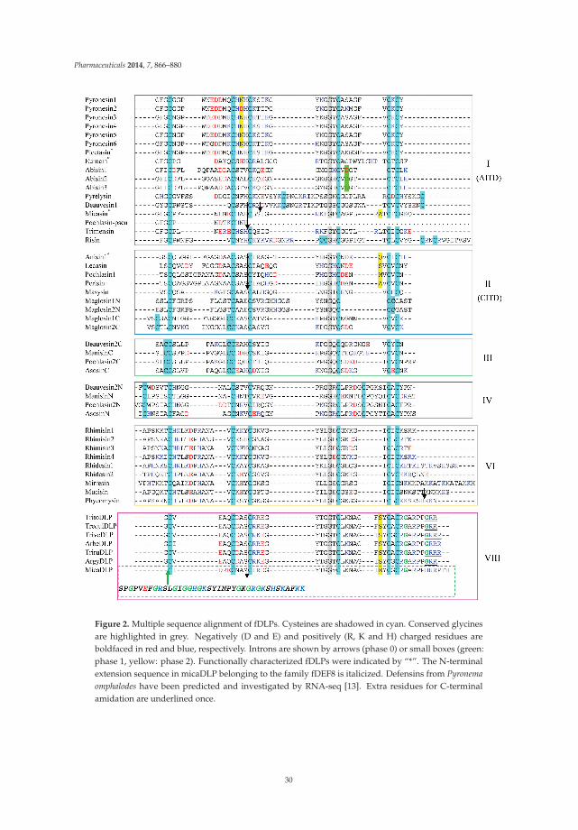
Pharmaceuticals 2014, 7, 866–880
Figure 2. Multiple sequence alignment of fDLPs. Cysteines are shadowed in cyan. Conserved glycinesare highlighted in grey. Negatively (D and E) and positively (R, K and H) charged residues areboldfaced in red and blue, respectively. Introns are shown by arrows (phase 0) or small boxes (green:phase 1, yellow: phase 2). Functionally characterized fDLPs were indicated by “*”. The N-terminalextension sequence in micaDLP belonging to the family fDEF8 is italicized. Defensins from Pyronemaomphalodes have been predicted and investigated by RNA-seq [13]. Extra residues for C-terminalamidation are underlined once.
30

Pharmaceuticals 2014, 7, 866–880
Table 2. Sources and characteristics of the malpisin family.
Name|Accession No. Organism Scaffold (Contig) Range Size MW NC
Malpisin1-1|AZCI01001104
Mortierellaalpina B6842
jtg7180000084593 f_7180000084594f
55070–55405 41 4048 −0.0
Malpisin1-2|AZCI01001104 55870–56127 48 5166 −3.3
Malpisin1-3|AZCI01001104 56393–56635 45 5047 +0.7
Malpisin1-4|AZCI01001104 63869–64117 39 4117 −1.0
Malpisin1-5|AZCI01000882
Contig 7180000084767
22045–22248 37 4259 -2.8
Malpisin1-6|AZCI01000882 25851–26051 39 4543 −3.5
Malpisin1-7|AZCI01000882 42456–42677 33 3624 −1.0
Malpisin1-8|AZCI01000882 43573–43800 47 5078 +1.7
Malpisin1-9|AZCI01000882 45037–45261 48 5203 +3.0
Malpisin1-10|AZCI01000882 45559–45738 35 3914 −0.0
Malpisin1-11|AZCI01000882 46707–46913 43 4941 +5.4
Malpisin1-12|AZCI01001135 jtg7180000084204f_7180000084205f_7180000084206f 135437–135676 44 4722 −0.0
Malpisin1-13|AZCI01001084 jtg7180000084699f_7180000084700f 362415–362627 47 4919 −1.8
Malpisin1-14|AZCI01001006 jtg7180000084769f_7180000084770f_ 7180000084771f_7 180000084772f 179488–179673 38 4188 +0.2
Malpisin2-1|ADAG01001070
Mortierellaalpina ATCC
32222
Contig 1070
9785–10114 39 4105 +1.0
Malpisin2-2|ADAG01001070 10532–10792 48 5187 −2.3
Malpisin2-3|ADAG01001070 11052–11297 44 4783 −0.0
Malpisin2-4|ADAG01001070 11773–12021 39 4052 −1.8
Malpisin2-5|ADAG01000791 Contig 791 4894–5097 37 4259 −2.8
Malpisin2-7|ADAG01000903
Contig 903
13145–13357 33 3899 +1.2
Malpisin2-8|ADAG01000903 14223–14450 47 5065 +1.7
Malpisin2-9|ADAG01000903 15634–15852 45 4917 +4.0
Malpisin2-10|ADAG01000903 16158–16337 35 3937 +0.2
Malpisin2-11|ADAG01000903 17264–17446 39 4531 +5.0
Figure 3. Multiple sequence alignment of malpisins. Color codes and symbol notes used here are thesame as those in Figure 2. Pink box indicates the N-terminus of DLPs with variable length. Sequenceidentity (%) to micasin is shown on the right.
31

Pharmaceuticals 2014, 7, 866–880
Fig
ure
4.
Phy
loge
neti
ctr
eeof
fDL
Ps.
The
tree
was
cons
tru
cted
from
the
alig
ned
amin
oac
idse
quen
ces
pre
sent
edin
Figu
res
2an
d3
wit
hth
ene
ighb
or-jo
inin
gm
etho
d.Th
enu
mbe
rson
node
sre
pres
entb
oots
trap
valu
es,a
ndon
lyva
lues
≥50%
are
show
n.
32

Pharmaceuticals 2014, 7, 866–880
Fig
ure
5.
Com
pari
son
ofpr
ecur
sor
orga
niza
tion
and
exon
-intr
onst
ruct
ures
betw
een
fDLP
san
dan
imal
defe
nsin
s.(A
)fD
LPs;
(B)A
nim
alde
fens
ins.
Sign
al,p
ro-a
ndm
atur
epe
ptid
esar
esh
own
inpi
nk,g
rey
and
blue
,res
pect
ivel
y.In
tron
phas
esar
esh
own
inth
esa
me
colo
rsas
Figu
re2.
Rep
rese
ntat
ive
anim
alde
fens
ins
are
deri
ved
from
Bran
chio
stom
aflo
rida
e,D
roso
phila
mel
anog
aste
r,A
noph
eles
arab
iens
is,A
pis
mel
lifer
a,Ix
odes
scap
ular
is,M
.mar
tens
ii,C
rass
ostr
eagi
gas,
Cae
norh
abdi
tisre
man
ei,a
ndC
.bre
nner
i.
33

Pharmaceuticals 2014, 7, 866–880
Peptides in fDEF8, all derived from dermatophytes, are characterized as a new family with ashort N-terminus and an extra C-terminal extension rich in arginines, prolines and glycines (Figure 2).The C-terminal extension has been considered as a common mechanism for the complexity increaseof some invertebrate antimicrobial peptides (AMPs). For example, the hymenopteran defensin-1subfamily has an extended C-terminus relative to its ancestral defensin-2 subfamily by a so-calledintron exonization-mediated mechanism [14,15]. It thus appears that fungal and invertebrate defensinsboth convergently evolved their C-termini. The extension of a C-terminal sequence via convergentevolution was also recently observed in interleukin 6 (IL-6), a class-I helical cytokine, of two leporids(Oryctolagus and Pentalagus) [16]. The presence of C-terminal Gly-Arg or Gly-Arg-Arg in somedermatophyte-derived fDLPs suggest that they may be amidated, as previously observed in someanimal toxins, e.g., the Mesobuthus α-toxins [17]. Interestingly, the mature peptide of micaDLP islarger in size than that of other members in this family, as identified by an N-terminal extension of 38amino acids (Figure 2). High content of glycines together with a cationic characteristic hints a putativeantimicrobial role of this extended unit.
M. alpine is a saprophytic species of Mucoromycotina, known as an oleaginous fungus [18]. Thedraft genome sequences of two M. alpina isolates (B6842 and ATCC 32222) [18,19] provide a possibilityto undertake comparative study of their fDLPs. We found that the M. alpine B6842 genome encodes14 fDLPs (Figure 3) but only 10 were found in M. alpine ATCC 32222. The failure to detect thefour homologs (i.e., malpisin1-6, 1-12, 1-13, 1-14) in M. alpine ATCC 32222 could be due to theincompletely-assembled genome sequences. Our phylogenetic analysis divides all malpisins intofDEF1 and fDEF6 (Figure 4). Some malpisin members of fDEF1 extended their N-termini with diversesequences and variable lengths (Figure 3).
3. Gene Duplication of fDLPs
Gene duplication extensively occurs in antimicrobial peptides from insects to humans [15,20,21].In fungi, initial annotation of defense molecules of Pyronema omphalodes also identified gene duplicationas a minor multigene family of fDLPs (herein termed pyronesin1 to pyronesin6) [13]. These fDLPsare highly similar to plectasin (Figure 6A). Our studies revealed new gene duplication event in otherfungal species. Malpisin is a representative example of gene duplication. As mentioned previously,there are 14 and 10 members in M. alpine B6842 and M. alpine ATCC 32222, respectively. Malpisin1-1,1-2, 1-3 and 1-4 are tandem located on one contig (jtg7180000084593f _7180000084594f), and malpisin1-5to malpisin1-11 on another contig (Contig 7180000084767). In addition, malpisin1-12, malpisin 1-13and malpisin 1-14 reside on other three contigs, as shown in Figure 6B. In M. alpine ATCC 32222,malpisin2-1 to malpisin2-4 are located on contig 1070 and malpisin2-7 to malpisin2-11 on contig 903.Only malpisin2-5 is located on contig 791.
In the widely cultivated mushroom Agaricus bisporus, there are three paralogous fDLPs (abisin1to abisin3) (Figure 6C), two of which (abisin1 and abisin3) share completely identical amino acidsequences in the mature peptide region but exhibit four synonymous substitutions at the nucleotidelevel. In the Pochonia chlamydosporia paralogues, pochlasin1 is highly similar to CITDs and pochlasin2possesses two defensin-domains. In addition, a putative pseudogene (herein named pochlasin-pseu)was also identified in scaffold 1191 and assigned to AITDs in view of its high sequence similarityto micasin in the first exon. Pochlasin1 and pochlasin-pseu share a conserved phase 0 intron withinthe α-helical region. The loss of the last two exons (2 and 3) results in the lack the last four cysteinesinvolved in the Csαβ folding of a mature peptide (Figure 2).
Gene duplication also occurs in the Mucorales-derived fDLPs, which leads to four and two genecopies in Rhizopus microsporus (Figure 6D) and R. delemar, respectively. In a Neighbor-Joining (NJ)tree, rhimisin1 and rhimisin4 (R. microsporus) constitutes a single clade clustering with the other threefDLPs (rhidesin1 from R. delemar, phycomycin from Phycomyces blakesleeanus and mirresin from Mucorirregularis) whereas rhimisin2 and rhimisin3 (R. microsporus) cluster with rhidesin2 (R. delemar) and
34

Pharmaceuticals 2014, 7, 866–880
mucisin (M. circinelloides) (Figure 4), suggesting that the gene duplication event could have occurred inthe ancestor of the Mucorales prior to their speciation.
Figure 6. The arrangement of defensin genes in chromosomes. Color arrows refer to differentorientation of the genes. A to D represent the genome location of defensins in four species: Pyronemaomphalodes, Mortierella alpine, Agaricus bisporus and Rhizopus microsporus. Malpisins in M. alpine B6842 isindicated in red and blue while in pink and green in M. alpine ATCC 32222. Pseudogenes of pyronesinsare shown in gradient blue.
4. Variable Gene Structures of fDLPs
Analysis of the exon-intron structures of the newly-discovered fDLPs revealed their variabilitythat can be described as follows: (1) all the fDLPs retain the integrity of the signal peptide exceptrisin (Rhizophagus irregularis) and malpisin1-1 (or malpisin2-1) that have a phase 1 or phase 0 introndisrupting their signal peptides; (2) all of the genes in fDEF8 and three genes in fDEF2 (i.e., lecasin,pochlasin1 and perisin) have the same gene organization as previously identified dermatophyticdefensins (micasin, arbesin, trivesin, tritosin and trirusin) and they contain two introns: the first intron(phase 0) disrupting the α-helical region; the second intron (phase 2) disrupting the c-loop; (3) thepyronesin and abisin multi-gene family in fDEF1 have only one intron disrupting either the α-helicalor the c-loop region; (4) In addition to these intron-containing fDLP genes, there are some memberswithout introns (Figures 2 and 5).
The highly variable gene structures in fDLPs are reminiscent of invertebrate defensins that alsoexhibit diverse gene structures [22,23] (Figure 5 and Figure S2). Compared with invertebrate defensinsof 5'-biased intron positions, introns of fDLPs occur preferentially in the 3'-end of the precursor-codedsequences. Because all eukaryotic Csαβ-type defensins are hypothesized to be originated from acommon bacterial ancestor [24], it is reasonable to infer that considerable intron gains might haveoccurred in defensins from some eukaryotic lineages, and later they differentially lost in some specificspecies. Such a dynamic intron evolution thus shapes the biased intron location pattern between fDLPs
35

Pharmaceuticals 2014, 7, 866–880
and animal DLPs after the animal-fungi split. It is also worth mentioning that some recognizableorthologues of defensins in Branchiostoma floridae [25,26], the basal chordate amphioxus, also contain aphase 0 intron located in their c-loop (Figure 5 and Figure S2). Given a remote evolutionary distancebetween fungi and amphioxus, their intron position conservation could be a consequence of convergentinsertion in a similar position due to the existence of “protosplice sites” [27,28]. However, the evolutionvia ancestral origin can be not completely ruled out in the case of the lack of gene structure informationin many animal defensins from different lineages.
5. Conclusions
It is estimated that there are as many as 1.5 million species of fungi in this world. However, onlya small fraction has been described and even fewer have been sequenced. To date, only about sixhundred genomes were being sequenced or completely sequenced. Fungal genome project (FGP)allows us to systematically exploit peptide antibiotics instead of accidental discovery or complicatedbiochemical screening. This work sheds light on the persistent discovery of fDLPs from model fungalgenome data. Despite this, in the lack of experimental data, it cannot be stated that all these fDLPspossess antibacterial function because in fact a classical insect-type fungal defensing - pechrysin wasfound to lack antibacterial activity [29] likely due to the absence of cationic residues on its molecularsurface. In addition, anisin1, a DLP from Aspergillus giganteus, was found to be involved in thefitness of the species by linking stress signaling with developmental regulation [30]. Recent studieshave also shown that although some peptides of fungal origin contain a similar defensin structure,they exhibit diverse or alternative biological functions beyond antimicrobial activity. An interestingoverview is given by Hegedüs and Marx [31]. Therefore, further biochemical characterization of thesenewly-discovered fDLPs will help evaluate their potential as human medicines.
Acknowledgments: This work was supported by the National Basic Research Program of China (2010CB945300),the National Natural Science Foundation of China (31221091), and the State Key Laboratory of IntegratedManagement of Pest Insects and Rodents (Grant No. ChineseIPM1307).
Author Contributions: J.W. and S.Z. discovered all fDLP genes described here. J.W. and S.Z. wrote the paper. B.G.revised the paper.
Conflicts of Interest: The authors declare no conflict of interest.
References
1. Mygind, P.H.; Fischer, R.L.; Schnorr, K.M.; Hansen, M.T.; Sonksen, C.P.; Ludvigsen, S.; Raventos, D.;Buskov, S.; Christensen, B.; De Maria, L.; et al. Plectasin is a peptide antibiotic with therapeutic potentialfrom a saprophytic fungus. Nature 2005, 437, 975–980. [CrossRef]
2. Zhu, S.; Gao, B.; Harvey, P.J.; Craik, D.J. Dermatophytic defensin with antiinfective potential. Proc. Natl.Acad. Sci. USA 2012, 109, 8495–8500. [CrossRef]
3. Zhu, S. Discovery of six families of fungal defensin-like peptides provides insights into origin and evolutionof the CSαβ defensins. Mol. Immunol. 2008, 45, 828–838. [CrossRef]
4. Schneider, T.; Kruse, T.; Wimmer, R.; Wiedemann, I.; Sass, V.; Pag, U.; Jansen, A.; Nielsen, A.K.; Mygind, P.H.;Raventos, D.S.; et al. Plectasin, a fungal defensin, targets the bacterial cell wall precursor Lipid II. Science2010, 328, 1168–1172. [CrossRef]
5. Oeemig, J.S.; Lynggaard, C.; Knudsen, D.H.; Hansen, F.T.; Norgaard, K.D.; Schneider, T.; Vad, B.S.;Sandvang, D.H.; Nielsen, L.A.; Neve, S.; et al. Eurocin, a new fungal defensin: Structure, lipid binding, andits mode of action. J. Biol. Chem. 2012, 287, 42361–42372. [CrossRef]
6. Hara, S.; Mukae, H.; Sakamoto, N.; Ishimoto, H.; Amenomori, M.; Fujita, H.; Ishimatsu, Y.; Yanagihara, K.;Kohno, S. Plectasin has antibacterial activity and no affect on cell viability or IL-8 production. Biochem.Biophys. Res. Commun. 2008, 374, 709–713. [CrossRef]
36
Pharmaceuticals 2014, 7, 866–880
7. Xiong, Y.Q.; Hady, W.A.; Deslandes, A.; Rey, A.; Fraisse, L.; Kristensen, H.H.; Yeaman, M.R.; Bayer, A.S.Efficacy of NZ2114, a novel plectasin-derived cationic antimicrobial peptide antibiotic, in experimentalendocarditis due to methicillin-resistant Staphylococcus aureus. Antimicrob. Agents Chemother. 2011, 55,5325–5330. [CrossRef]
8. Ostergaard, C.; Sandvang, D.; Frimodt-Moller, N.; Kristensen, H.H. High cerebrospinal fluid (CSF)penetration and potent bactericidal activity in CSF of NZ2114, a novel plectasin variant, during experimentalpneumococcal meningitis. Antimicrob. Agents Chemother. 2009, 53, 1581–1585. [CrossRef]
9. Andes, D.; Craig, W.; Nielsen, L.A.; Kristensen, H.H. In vivo pharmacodynamic characterization of a novelplectasin antibiotic, NZ2114, in a murine infection model. Antimicrob. Agents Chemother. 2009, 53, 3003–3009.[CrossRef]
10. Lutzoni, F.; Kauff, F.; Cox, C.J.; McLaughlin, D.; Celio, G.; Dentinger, B.; Padamsee, M.; Hibbett, D.; James, T.Y.;Baloch, E.; et al. Assembling the fungal tree of life: Progress, classification, and evolution of subcellular traits.Am. J. Bot. 2004, 91, 1446–1480. [CrossRef]
11. Seidah, N.G.; Chrétien, M. Proprotein and prohormone convertases: A family of subtilases generatingdiverse bioactive polypeptides. Brain Res. 1999, 848, 45–62.
12. Benson, D.A.; Karsch-Mizrachi, I.; Lipman, D.J.; Ostell, J.; Wheeler, D.L. GenBank. Nucleic Acids Res. 2008,36, D25–30. [CrossRef]
13. Traeger, S.; Altegoer, F.; Freitag, M.; Gabaldon, T.; Kempken, F.; Kumar, A.; Marcet-Houben, M.; Poggeler, S.;Stajich, J.E.; Nowrousian, M. The genome and development-dependent transcriptomes of Pyronema confluens:A window into fungal evolution. PLoS Genet. 2013, 9, e1003820. [CrossRef]
14. Tian, C.; Gao, B.; Fang, Q.; Ye, G.; Zhu, S. Antimicrobial peptide-like genes in Nasonia vitripennis: A genomicperspective. BMC Genomics 2010, 11, 187. [CrossRef]
15. Zhang, Z.; Zhu, S. Comparative genomics analysis of five families of antimicrobial peptide-like genes inseven ant species. Dev. Comp. Immunol. 2012, 38, 262–274. [CrossRef]
16. Neves, F.; Abrantes, J.; Pinheiro, A.; Almeida, T.; Costa, P.P.; Esteves, P.J. Convergent evolution of IL-6 in twoleporids (Oryctolagus and Pentalagus) originated an extended protein. Immunogenetics 2014. [CrossRef]
17. Zhu, S.; Peigneur, S.; Gao, B.; Lu, X.; Cao, C.; Tytgat, J. Evolutionary diversification of Mesobuthus α-scorpiontoxins affecting sodium channels. Mol. Cell. Proteomics 2012. [CrossRef]
18. Wang, L.; Chen, W.; Feng, Y.; Ren, Y.; Gu, Z.; Chen, H.; Wang, H.; Thomas, M.J.; Zhang, B.; Berquin, I.M.; et al.Genome characterization of the oleaginous fungus Mortierella alpina. PLoS ONE 2011, 6, e28319. [CrossRef]
19. Etienne, K.A.; Chibucos, M.C.; Su, Q.; Orvis, J.; Daugherty, S.; Ott, S.; Sengamalay, N.A.; Fraser, C.M.;Lockhart, S.R.; Bruno, V.M. Draft genome sequence of Mortierella alpina isolate CDC-B6842. Genome Announc.2014, 2, e01180-13.
20. Schutte, B.C.; Mitros, J.P.; Bartlett, J.A.; Walters, J.D.; Jia, H.P.; Welsh, M.J.; Casavant, T.L.; McCray, P.B., Jr.Discovery of five conserved β-defensin gene clusters using a computational search strategy. Proc. Natl. Acad.Sci. USA 2002, 99, 2129–2133. [CrossRef]
21. Semple, C.A.; Rolfe, M.; Dorin, J.R. Duplication and selection in the evolution of primate β-defensin genes.Genome Biol. 2003, 4, R31. [CrossRef]
22. Froy, O.; Gurevitz, M. Arthropod and mollusk defensins - evolution by exon-shuffling. Trends Genet. 2003,19, 684–687. [CrossRef]
23. Rodriguez de la Vega, R.C.; Possani, L.D. On the evolution of invertebrate defensins. Trends Genet. 2005, 21,330–332. [CrossRef]
24. Gao, B.; Rodriguez Mdel, C.; Lanz-Mendoza, H.; Zhu, S. AdDLP, a bacterial defensin-like peptide, exhibitsanti-Plasmodium activity. Biochem. Biophys. Res. Commun. 2009, 387, 393–398. [CrossRef]
25. Yu, J.K.; Wang, M.C.; Shin-I, T.; Kohara, Y.; Holland, L.; Satoh, N.; Satou, Y. A cDNA resource for thecephalochordate amphioxus Branchiostoma floridae. Dev. Genes Evol. 2008, 218, 723–727. [CrossRef]
26. Zhu, S.; Peigneur, S.; Gao, B.; Umetsu, Y.; Ohki, S.; Tytgat, J. Experimental conversion of a defensin into aneurotoxin: implications for origin of toxic function. Mol. Biol. Evol. 2014, 31, 546–559. [CrossRef]
27. Sverdlov, A.V.; Rogozin, I.B.; Babenko, V.N.; Koonin, E.V. Reconstruction of ancestral protosplice sites. Curr.Biol. 2004, 14, 1505–1508. [CrossRef]
28. Rogozin, I.B.; Sverdlov, A.V.; Babenko, V.N.; Koonin, E.V. Analysis of evolution of exon-intron structure ofeukaryotic genes. Brief. Bioinformatics 2005, 6, 118–134. [CrossRef]
37
Pharmaceuticals 2014, 7, 866–880
29. Wu, Y.; Gao, B.; Zhu, S. Fungal defensins, an emerging source of anti-infective drugs. Chin. Sci. Bull. 2014,59, 931–935. [CrossRef]
30. Eigentler, A.; Pocsi, I.; Marx, F. The anisin1 gene encodes a defensin-like protein and supports the fitness ofAspergillus nidulans. Arch. Microbiol. 2012, 194, 427–437. [CrossRef]
31. Hegedus, N.; Marx, F. Antifungal proteins: more than antimicrobials? Fungal Biol. Rev. 2013, 26, 132–145.[CrossRef]
© 2014 by the authors. Licensee MDPI, Basel, Switzerland. This article is an open accessarticle distributed under the terms and conditions of the Creative Commons Attribution(CC BY) license (http://creativecommons.org/licenses/by/4.0/).
38

pharmaceuticals
Review
Chapter 3:
Antimicrobial Peptides from Plants
James P. Tam 1,*, Shujing Wang 1,2, Ka H. Wong 1 and Wei Liang Tan 1
1 School of Biological Sciences, Nanyang Technological University, Singapore, Singapore;[email protected] (S.W.); [email protected] (K.H.W.); [email protected] (W.L.T.)
2 Department of Pharmacology and Pharmaceutical Sciences, School of Medicine, Tsinghua University,Beijing 100084, China
* Author to whom correspondence should be addressed; [email protected]; Tel.: +65-63162863.
Academic Editor: Guangshun WangReceived: 16 July 2015; Accepted: 1 September 2015; Published: 16 November 2015
Abstract: Plant antimicrobial peptides (AMPs) have evolved differently from AMPs from other lifeforms. They are generally rich in cysteine residues which form multiple disulfides. In turn, thedisulfides cross-braced plant AMPs as cystine-rich peptides to confer them with extraordinary highchemical, thermal and proteolytic stability. The cystine-rich or commonly known as cysteine-richpeptides (CRPs) of plant AMPs are classified into families based on their sequence similarity, cysteinemotifs that determine their distinctive disulfide bond patterns and tertiary structure fold. Cystine-richplant AMP families include thionins, defensins, hevein-like peptides, knottin-type peptides (linearand cyclic), lipid transfer proteins, α-hairpinin and snakins family. In addition, there are AMPswhich are rich in other amino acids. The ability of plant AMPs to organize into specific familieswith conserved structural folds that enable sequence variation of non-Cys residues encased in thesame scaffold within a particular family to play multiple functions. Furthermore, the ability of plantAMPs to tolerate hypervariable sequences using a conserved scaffold provides diversity to recognizedifferent targets by varying the sequence of the non-cysteine residues. These properties bode wellfor developing plant AMPs as potential therapeutics and for protection of crops through transgenicmethods. This review provides an overview of the major families of plant AMPs, including theirstructures, functions, and putative mechanisms.
Keywords: plant antimicrobial peptides; cysteine-rich peptides; cystine knot; thionin; defensin;hevein; knottin
1. Introduction
Higher plants have a broad range of defense mechanisms to counter physical, chemical andbiological stress such as drought, cold, heavy metal, pollutants and pathogen attacks from fungi,bacteria and viruses. In response to infection by a variety of pathogens, plants display up-regulation ofa set of genes associated with systemic acquired resistance [1]. General resistance is accomplished bythe release of secondary metabolites like phytoalexins, tannins and polyphenolic compounds, and thegeneration of pathogenesis-related (PR) proteins. PR proteins were first discovered in the early 1970sin tobacco leaves in response totobacco mosaic virus infections and were later defined as the inducedproteins that are released during pathogenic attacks [2,3]. According to a recent review, there are atleast 17 families that have been detected and isolated that possess a wide range of defense-relatedproperties, including antibacterial, antifungal, antiviral, anti-oxidative activity, chitinase and proteinaseinhibitory activities [1–4]. This review will focus on peptides which possess antimicrobial activity,
Pharmaceuticals 2015, 8, 711–757 39 www.mdpi.com/journal/pharmaceuticals

Pharmaceuticals 2015, 8, 711–757
namely thionin (PR-13 family), defensin (PR-12 family), hevein-like peptide, knottin, α-hairpinin, lipidtransfer protein (PR-14 family) and snakin.
Antimicrobial peptides (AMPs) are ubiquitous and found as host defenses against pathogens andpests in diverse organisms ranging from microbes to animals [5]. AMPs exist in different molecularforms, although the majority of them are linear peptides from insects, animals, and plants. Nevertheless,bacteria produce polycyclic peptides such as lantibiotics, and all major forms of life produce circularpeptides which include bacteriocins from bacteria, cyclotides from plants and theta-defensins fromanimals [6–9]. In plants, the majority of AMPs are Cys-rich [10], a feature that enables the formation ofmultiple disulfide bonds (usually two to six) that contribute to a compact structure and resistance tochemical and proteolytic degradation.
In general, plant AMPs share several common characteristics with those from microbes, insectsand animals. They include features such as their molecular forms, positive charge and amphipathicnature, all of which are primarily related to their defensive role(s) as membrane-active antifungals,antibacterials, and antivirals. These features, in addition to being Cys-rich, are well representedby two plant AMP families, thionins and plant defensins. Other families of plant AMPs act onpathogens differently from animal AMPs. For example, hevein-like peptides bind chitins, knottin-typepeptides inhibit enzymes such as proteases, and lipid transfer proteins bind lipids to disrupt microbialpenetration into cell membranes.
Classification of plant AMP families is largely based on their Cys motifs which exhibit acharacteristic Cys pattern with a defined number of non-Cys residues between the two neighboring Cys.Currently, the number of AMPs isolated from a limited number of plants already exceeds a thousandand is likely to increase in the future. Sequence analysis and genomic data mining using these Cysmotifs have revealed that Cys-rich peptides (CRPs) with AMP characteristics are under-predicted [11].In model plants, such as rice and Arabidopsis, CRPs may account for about 3% of the expressed proteins.
Like animal AMPs, AMP expression in plants is constitutive or induced and often tissue-specific.Moreover, plant AMPs are evolvable with hypervariable sequences encased in a particular scaffoldcharacteristic of a given family of AMPs, which is analogous, but to a much lesser extent, to themolecular diversity of vertebrate immunoglobulin-based immunity.
This review aims to provide a general overview of the major families of plant AMPs, includingtheir structures, functions, and putative mechanisms of defense. Additional details on plant AMPscan be found in a number of excellent previous reviews [12–25]. Furthermore, information on AMPsfrom diverse organisms can be accessed through several databases, such as APD, APD2 [26,27],YADAMP [28], DAMPD [29], and PhytAMP (specific for plant AMPs) [10].
2. Classification and Characteristics
Plant AMPs are divided into families based on their sequence similarity, Cys motifs, anddistinctive disulfide bond patterns which, in turn, determine their tertiary structure folding. Table 1lists the major families of plant AMPs based on these criteria; families include thionins, defensins,hevein-like peptides, knottin-type peptides (linear and cyclic), lipid transfer proteins, α-hairpininfamilies, snakins, and unclassified CRP-AMPs. In addition, non-CRP AMPs, which may be rich inother amino acids, are also described in this review, including the Gly-rich peptide (GRP) Pg-AMP1,the Gly- and His-rich peptide shepherins, and peptides of less than 10 amino acids (aa), such asCn-AMP1 and Cr-ACP1. The following defining features are found in plant AMPs:
1. Mostly characterized as moderate-size (MW of 2–6 kDa), basic, CRPs with two to sixintra-molecular disulfide bonds.
2. Members within a family are classified based on Cys motif, sequence similarity and are conservedin secondary and tertiary structure.
40
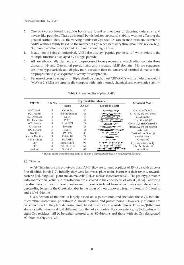
Pharmaceuticals 2015, 8, 711–757
3. One or two additional disulfide bonds are found in members of thionins, defensins, andhevein-like peptides. These additional bonds bolster structural stability without affecting thegeneral scaffold. Because the varying number of Cys residues can create confusion, we refer toAMPs within a family based on the number of Cys when necessary throughout this review (e.g.,6C-thionins contain six Cys and 8C-thionins have eight Cys).
4. In addition to being antimicrobial, AMPs also display “peptide promiscuity”, which refers to themultiple functions displayed by a single peptide.
5. All are ribosomally derived and bioprocessed from precursors, which often contain threedomains: N- and C-terminal pro-domains and a mature AMP domain. Mature sequencesare often hypervariable and display more variation than the conserved terminal domains in thepreproprotein to give sequence diversity for adaptation.
6. Because of cross-bracing by multiple disulfide bonds, most CRP-AMPs with a molecular weight(MW) of 2–6 kDa are structurally compact with high thermal, chemical, and enzymatic stability.
Table 1. Major families of plant AMPs.
Peptide S-S No.Representative Member
Structural MotifName AA No. Disulfide Motif
6C-Thionin 3 Crambin 46 Gamma (Г) foldβ1-α1-α2-β2-coil motif8C-Thionin 4 β-Purothionin 45
8C-Defensin 4 NaD1 47 CSαβ motifβ1-coil-α-β2-β310C-Defensin 5 PhD1 47
6C-Hevein 3 Ac-AMP1 29 Gly & Cys rich Central βstrands & (short helical)
side coils8C-Hevein 4 Hevein 4310C-Hevein 5 EAFP1 41
Knottin 3 PAFP-S 38 Cystine knot Short βstrand & coilCyclic Knottin 3 Kalata B1 29
α-Hairpinin 2 Ec-AMP1 37 α1-turn-α2LTP 4 Maize LTP1 93 Hydrophobic cavity
α1-α2-α3-α4-coilLTP 4 Wheat LTP2 67Snakin * 6 Snakin-1 63 α -helices
* The disulfide and structural motif of Snakin-1 is predicted based on homology modelling.
2.1. Thionins
α-/β-Thionins are the prototypic plant AMP; they are cationic peptides of 45–48 aa with three orfour disulfide bonds [12]. Initially, they were known as plant toxins because of their toxicity towardsbacteria [30], fungi [31], plant and animal cells [32], as well as insect larvae [33]. The prototypic thioninwith antimicrobial activity, α-purothionin, was isolated in the endosperm of wheat [30,34]. Followingthe discovery of α-purothionin, subsequent thionins isolated from other plants are labeled withdescending letters of the Greek alphabet in the order of their discovery (e.g., α-thionins, β-thionins,and γ1/γ1-thionins).
Classification of thionins is largely based on α-purothionin and includes the α-/β-thinoinsof crambin, viscotoxins, phoratoxin A, hordothionins, and purothinoins. However, γ-thionins areconsidered part of the plant defensin family based on structural considerations. Thus, α-/β-thioninsshare a similar structural fold different from that of γ-thionins. For convenience, α/β-thionins witheight Cys residues will be hereafter referred to as 8C-thionins and those with six Cys designated6C-thionins (Figure 1A,B).
41

Pharmaceuticals 2015, 8, 711–757
Figure 1. Sequences (A) and structures (B) of representative thionins. The secondary structure is representedby different colors: cyan-α helix; magenta-β strand; pink-random coil and yellow-disulfide bonds.
2.1.1. Occurrences, Distribution, and Biosynthesis
Thionins have been identified from monocots and dicots and are expressed in different tissues,such as seeds, leaves, and roots [35–37]. The expression of thionins can be induced by infection withvarious microbes [38,39] and has been shown to be related to the release of the hormone methyljasmonate upon plant wounding or microorganism invasion [38,40,41]. Thionins are ribosomallyderived and expressed as preproproteins, wherein the prothionin domain is flanked by two conservedsequences, the N-terminal signaling peptide and C-terminal acidic domain [35,38,42]. Mature thioninsequences display more variation than the conserved terminal domains in the preproprotein due toevolutionary pressure [43]. The three-domain precursor of plant AMPs in CRP families is typical andfound in other CRP families, including defensins, hevein-like peptides, and knottin-type peptides.
2.1.2. Structure
From the limited number of members identified thus far, thionins have relatively conserved aminoacid sequences compared to other plant defense peptides (Figure 1A). They also share a conservedβ1-α1-α2-β2-coil secondary structural motif, which forms a gamma (Г) fold, a special turn consists ofthree amino acid residues with the first and third residue connected by a hydrogen bond in the tertiarystructure (Figure 1A,B).
Thionins can be loosely classified as pseudocyclics because of an end-to-end disulfide bondlinking the N- and C-termini, conferring a circular structural topology. They are, however, not truepseudocyclics because there are additional non-cysteine amino acids located at both the N- andC-termini. 8C-Thionins contain four conserved stabilizing disulfide bonds between CysI-CysVIII (Cysnumbering in Roman numerals from N-to-C-termini) linking β1 to the C-terminal coil, CysII-CysVIIlinking the end of β1 with the beginning of β2, CysIII-CysVI linking α1 and the loop after α2, andCysIV-CysV linking α1 and α2. The 6C-thionins have the same Cys pairing as 8C-thionins except for theabsence of the CysII-CysVII disulfide bond. The structure of 8C-thionins, including α-/β-purothionins,α-/β-hordothionin, and hellethionin D, are similar to that of 6C-thionins with the Г-shape but with
42

Pharmaceuticals 2015, 8, 711–757
minor differences in the C-terminal coil region [44–46]. The long arm of the Г fold comprises theα1-α2 region, and the short arm consists of β1 and the β2-coil. The large groove between the twoarms is proposed to be important for the interaction between thionins and membrane lipids [43]. Mosthydrophobic side chains cluster around the outer surface of the long arm, while the hydrophilic chainsare located on the surface within the groove or outer face of the short arm (Figure 2A).
2.1.3. Structure-Function Relationship
The 6C-thionins with three disulfide bonds include crambin, viscotoxins, and phoratoxin A.Most 6C-thionins are highly basic, amphipathic, and toxic, with the exception of crambin. Crambinis a neutral, hydrophobic, non-toxic peptide identified from Crambe abyssinica with two isomers,P22/L25 and S22/I25 [47,48]. High resolution structures of crambin have been determined by NMRand X-ray/neutron crystallography in both water and detergent [47,49–53]. Viscotoxins (includingA1, A2, A3, B, B2, C1, and 1-PS) and phoratoxins from the mistletoes share a similar Г-shape withβ1-α1-α2-β2-coil motif [54–60].
8C-Thionins with four disulfide bonds include α-/β-purothionins, α-/β-hordothionins,hellethionin-D, Pyrularia Pubera thionin (PpTH), and Tulipa gensneriana bulb-purified AMPs (Tu-AMPs).The monomeric conformation of the 45-aa α-hordothionin isolated from barley [61,62] was previouslydetermined by NMR [62], while X-ray crystallography revealed a dimeric structure [61]. A study byVila-Perello showed that removal of one disulfide bond from PpTH is sufficient to significantly alter itsfolding [63]. A 45% size-reduced form of PpTH was synthesized, which only contains residues 7–32with the two antiparallel α-helices stabilized by two disulfide bonds. Size-reduced PpTH appearedto display the same antimicrobial activity and mechanism of action as intact PpTH in selected testmicroorganisms [64].
Figure 2. Cartoon illustration and surface plot of representative members of different antimicrobialpeptides. The orientation of surface plot (left) of each representative member is the same as the cartoonillustration, while that of the right one is evolved from the left one by 180◦ turn around the y axis.Color representation rules for different amino acids are as follows: blue-positively charged (R, K & H),red-negatively charged (D & E), orange-hydrophobic (I, L, V, A, P & M), cyan-hydrophilic (S, T, E & Q),magenta-aromatic (F, Y & W), yellow-cysteine (C) and gray-glycine (G).
43

Pharmaceuticals 2015, 8, 711–757
Tu-AMP1 and Tu-AMP2 are antibacterial, antifungal, and can reversibly bind chitin, a keyconstituent of the cell wall of fungi and exoskeletons of invertebrates, such as insects, anthropods,and nematodes. Initially, they were suggested to be thionin-like peptides, although Tu-AMP2 is aheterodimer of two chains joined by disulfide bonds [65]. However, it is worthwhile to point out thatin our review of plant CRPs, the occurrence of heterodimer is exceedingly rare. In our view, it remainsto be determined whether the heterodimeric formation occurs during the isolation process.
2.1.4. Mechanism of Action
Thionins are hydrophobic and likely elicit their toxicity to bacteria, fungi, and animal and plantcells via membrane interactions with their hydrophobic residues or/and positive surface charge [12,13,18,66–68]. The proposed mechanism of toxicity is attributed to lysis of cell membranes, but it isstill under investigation [30,39,68–70]. Stec proposed a structural model of the thionin-phospholipidinteraction to explain the solubilization and lysis of cell membranes [43].
Thionins are known to directly interact with membrane lipids apart from protein receptors [67,71,72]. Pyrularia thionin from the nuts of Pyrularia pubera mediates the influx of Ca2+ during certaincellular responses, while Tyr iodination reduces its hemolysis, phospholipase A2 activation, andcytotoxicity [32]. Structure-function studies have demonstrated that Lys1 and Tyr13 in thionins arehighly conserved and proposed to be crucial to their toxicity, with the exception of non-toxic, non-lyticcrambin. Instead, crambin contains Thr1 and Phe13 residues [32,43,73,74]. Furthermore, Arg10 issuggested to be important to the folding stability of all thionins, as it is an abundant source of hydrogenbonds between β1, α1, and the C-terminal coil [75].
2.2. Plant Defensins
Plant defensins are the best known, and likely most abundant, of all plant AMPs withmembranolytic functions, according to data mining of selected plant genomes. They are cationicpeptides of 45–54 aa with four to five disulfide bonds [76]. Plant defensins have diverse biologicalfunctions which include antifungal [77–81], antibacterial [82,83], and α-amylase and trypsin inhibitoryactivity [84,85]. In addition to being antimicrobial, plant defensins are also involved in the biotic stressresponse, as well as plant growth and development.
Plant defensins were first identified as γ-thionins, γ1-hordothionin, and γ1-/γ2-purothioninsfrom wheat and barley grains [86,87]. Thus, they were initially classified as γ-thionins due to theirlimited sequence identity (25%) with α-/β-thionins. Later, they were found to be unrelated to thioninsbased on structural features [88]. In 1995, they were grouped as plant defensins based on their sequence,structure, and function similarities with mammalian and insect defensins [76,78,88,89].
2.2.1. Occurrences, Distribution, and Biosynthesis
Plant defensins include over 100 members from a wide range of plants, including wheat, barley,tobacco, radish, mustard, turnip, arabidopsis, potato, sorghum, soybean, cowpea, and spinach, amongothers [15,90]. They have been identified in multiple tissues, tubers [79,91], leaves [79], pods [92], andflowers [93–95], with the majority identified from seeds and roots [96]. Two types of precursors havebeen identified in plant defensins, wherein the dominant group is composed of the N-terminal signalpeptide and a mature plant defensin domain [97], while the minor group is composed of an extraC-terminal acidic pro-domain of 33 aa reportedly associated with the vacuolar sorting mechanismsince defensins with this domain were found in vacuoles and those without were found in the outercell layers [16,79,95].
2.2.2. Structures
Plant defensins are generally characterized by four conserved disulfide bonds (except for PhDs, a10C-plant defensin isolated from Petunia hydrida) with the outer disulfide pair as an end-to-end innerdisulfide bridge and the inner three pairs of disulfide bonds forming a cystine knot (see knottins).
44

Pharmaceuticals 2015, 8, 711–757
A secondary structure characteristic of plant defensins is a Cys-stabilized αβ (CSαβ) motif of thecysteine knot. The CSαβ motif was first characterized in charybdotoxin, a K+ channel blocker isolatedfrom scorpions [98] and named by Cornet et al. [99]. This motif forms a β1-coil-α-β2-β3 pattern inthe secondary structure, where the α-helix is parallel to three antiparallel β-strands (Figure 3A,B).The CSαβ scaffold is stabilized and characterized by: (1) two disulfide bonds between the CXXXCmotif from the α-helix and the CXC motif in the central β3 strand; (2) one disulfide bond betweenthe β2 strand and the first loop before the α-helix; and (3) an end-to-end disulfide bond between theN- and C-termini, a disulfide connective similar to thionins. However, plant defensins (γ-thionins)differ greatly from α-/β-thinoins in their secondary and tertiary structures. In the secondary structure,α-/β-thionins show a typical β1-α1-α2-β2-coil motif, whereas γ-thionins display a β1-coil-α-β2-β3motif. At the tertiary structural level, β1 and β2 orient antiparallel in α-/β-thinoins, whereas thecorresponding β1and β3 in γ-thionins are oriented in parallel.
Figure 3. Sequences (A) and structures (B) of representative plant defensins. The secondarystructure is represented by different colors: cyan-α helix; magenta-β strand; pink-random coil andyellow-disulfide bonds.
Similar to thionins, plant defensins are pseudocyclics (see thionins), with conservation of thedisulfide bond between the first and last Cys, conferring a circular structural topology by connectingthe side chains of the N- and C- termini.
Plant defensins have been reported to be stable in harsh conditions that mimic the digestivesystem and decoction process, including high temperature (~85 ◦C), low pH (~2.0), and oxidative andproteolytic environments [100–102]. Although defensins show conservation of the CSαβ motif, thereare variations in the number of disulfide bonds, primary sequences, and tertiary structures [89,103,104].It has been shown that Ala substitutions of non-Cys residues on VrD1 are tolerated in the CSαβ motifalthough the structural stability and inhibitory effects vary among the mutants [105].
45

Pharmaceuticals 2015, 8, 711–757
2.2.3. Structure-Function Relationship
Based on the number of Cys, plant defensins are further divided into 8C-plant defensins withfour disulfides and 10C-plant defensins with five disulfide bonds.
The 8C-plant defensins include NaD1, VrD1, AlfAFP, ω-hordothionin, Cp-thionin II, Rs-AFPs,Psd1, Fabatins, and Ms-Def1 (Figure 3A,B). NaD1 (47 aa) was identified from the outer cell layers ofdifferent flower parts of the ornamental tobacco Nicotiana alata. Its distribution is consistent with itsprotective roles in reproductive organs. In vitro results showed that NaD1 inhibits the growth of plantpathogens Botrytis cinerea and Fusarium oxysporus [95,106]. VrD1 (46 aa) from the mung bean Vignaradiata not only inhibits protein synthesis, but is also antimicrobial and insect (bruchids)-resistant [107,108]. ω-Hordothionin (48 aa) purified from barley endosperm inhibits translational activity in botheukaryotic and prokaryotic cell-free systems [109]. The crystal structure of SPE10, identified fromPachyrrhizus erosus seeds [110], shows a dimer with each unit adopting the typical CSαβ motif [111].Based on structural analysis and mutation studies, the dimeric conformation of SPE10 was suggestedto be associated with its function, while the hydrophobic patch on the molecular head is necessary forits antifungal activity.
AlfAFP from Medicago sativa seeds is an antifungal peptide that provides robust resistanceto the fungal pathogen Verticillium dahliae in transgenic potato plants [77,106]. Rs-AFPs (51 aa)purified from Raphanus sativus radish seeds are highly basic oligomeric proteins with an N-terminalpyroglutamic acid. They have a broad antifungal spectrum with an IC50 = 0.3–100 μg/mL [78,79,81].Rs-AFPs are abundant in near-mature and mature seeds and released to create a fungal-suppressingmicroenvironment after disruption of the seed coat [79]. Ms-Def1 from Medicago sativa seeds stronglyinhibits the fungal growth of Fusarium graminearum in vitro [112]; the inhibitory effect is reduced in thepresence of Ca2+ ions. Homologous antifungal peptides were also reported from seeds of Aesculushippocastanum, Clitoria ternatea (Ct-AMP1), Dahlia merckii (Dm-AMP1), Lens culinaris (Lc-def) andHeuchera sanguine (Hs-AFP1) [80,97,113].
Cp-thionin II (47 aa) was identified from Vigna unguiculata cowpea seeds and is antibacterialagainst both Gram-positive and Gram-negative bacteria such as Staphylococcus aureus, Escherichiacoli, and Pseudomonas syringae with minimal inhibitory concentrations of 128, 64, and 42 μg/mL,respectively [90]. Psd1, a 46 aa peptide identified from seeds of the pea Pisum sativum, is antibacterialand acts as a K+ channel inhibitor based on a surface charge distribution analysis [114]. Fabatinsisolated from the broad bean Vicia faba are active against both Gram-positive and Gram-negativebacteria, but inactive against the yeasts Saccharomyces cerevisiae and Candida albicans [115].
PhDs are 10C-plant defensins. From flowers of Petunia hybrids, Lay et al. reported two AMPs withantifungal activity, PhD1 (47 aa) and PhD2 (49 aa) [95]. A fifth disulfide bond exists between the α-helixand the loop after β1 within these defensins that do not alter the typical CSαβ topology [108]. Thefifth disulfide bond appears only to change the corresponding hydrophobic interaction and hydrogenbond, as in 8C-plant defensins, to a covalent disulfide bond in PhD1 without altering the side-chainorientation of substituted residues.
The γ-thionins which have been reclassified as defensins, γ-hordothionin and ω-hordothioninfrom Hordeum vulgare, were shown to inhibit protein translation in rabbit reticulocytes and mouseliver extracts [87]. However, this inhibitory effect was not observed on plants, such as Triticumaestivum, Cucumis sativus, Vicea sativa and H. vulgare, and which may indicate a certain specificity in itsmechanism of action. Similarly, no direct interaction was observed between the plant defensins andnucleic acids, which is the proposed mechanism of the inhibitory activity of protein synthesis by theplant thionin family [116]. Instead of protein translation, Mendez et al. suggested that ω-hordothioninmay act on protein synthesis at the initiation and elongation step [109].
Defensins isolated from the seeds of Sorghum bicolor, S1α1, S1α2 and S1α3, were demonstrated todisplay inhibitory activity against α-amylase activity [84]. They are able to inhibit α-amylase obtainedfrom the gut of the insects Periplaneta americana and Locusta migratoria migratorioides, and weakly inhibitthe α-amylase from human saliva. Defensins from Vigna unguiculata and ω-hordothionin, from H.
46

Pharmaceuticals 2015, 8, 711–757
vulgare, also exhibit similar inhibitory activity against insect α-amylase [109,117,118], however, nosignificant inhibitory effect was observed on porcine α-amylase by plant defensins [119]. Several plantdefensins exhibit the ability to inhibit ion channels. Kushmerick et al. showed that γ1-zeathionin andγ2-zeathionin from maize kernels block voltage-gated Na+ channels reversibly in intact mammalianGH3 cells using the patch-clamp technique [120]. These authors postulated that the ion channelinhibitory effect of defensins may be related to the similar 3D structure with scorpion neurotoxin,a well-known Na+ channel blocker. Plant defensins were also shown to inhibit Ca2+ channels [112].However, no significant inhibitory effect was observed on K+ channels by plant defensins [119].
Two homologous peptides that display high sequence similarity to plant defensins were isolatedfrom the plant Cassia fistula, denoted 5459 and 5144 according to their molecular weight [85]. The 5459defensin was demonstrated to exhibit trypsin inhibitory activity, whereas no inhibitory effect wasobserved by the 5144 defensin. Another defensin peptide, Cp-thionin from V. uguiculata seeds, wasreported to display inhibitory activity against pancreatic bovine trypsin [121].
An enzymatic activity was attributed to a plant defensin. Huang et al. cloned the defensin SPD1from the roots of Ipomoea batatas and showed that it has the ability to regenerate dehydroascorbate(DHA) to ascorbic acid (AsA) in the presence of glutathione [122]. The peptide is also able to convertmonodehydroascrobate (MDA) to AsA in the presence of NADH, functioning as a glutathione-dependentdehydroascorbate reductase. SPD1 is likely to function as a regulator of the redox state of AsA, which hasbeen implicated in the response of the plant cell to reactive oxidative stress [123].
2.2.4. Mechanism of Action
The structure-function relationship of plant defensins has been suggested to correlate withtheir positive charge and amphipathic nature, as illustrated by the structure-surface plot of NaD1 inFigure 2B. Thus, plant defensins could initially bind to microbial membranes through interactions withspecific binding sites (“receptors”), as reported for Rs-AFP2, Hs-AFP1, and Dm-AMP1 [16,124,125].Binding of plant defensins, such as Rs-AFP2 and Dm-AMP1, to the cell membrane results in the influxand efflux of positive ions like Ca2+ and K+ [126,127]. Lastly, Ms-Def1 is able to block the Ca2+ channelin a manner similar to the Ca2+ channel blocker KP4 [112]. Van der Weerden et al. demonstratedthat NaD1 does not cause membranes permeabilization via a canonical mechanism which involvesnonspecific insertion into membranes [128] but rather a cell wall dependent process, likely requiring aspecific receptor. The mechanism of the fungicidal action of NaD1 is likely through permeabilization ofthe hyphae of Fusarium oxysporum, entering into the cytoplasm of the cell and inducing ROS oxidativestress [129]. Hayes et al. reported that the high-osmolarity glycerol (HOG) pathway is involved in theprotection of the cell against NaD1 [130], indicating that the inhibition of the HOG pathway increasesthe activity of antimicrobial peptides against Candida albicans. Several reviews have discussed in detailthe plant defensin mechanism of action [16,101,131,132].
2.3. Hevein-Like Peptides
Hevein-like peptides are basic peptides of 29–45 aa with three to five disulfide bonds. They arerich in Gly and contain conserved aromatic residues found in the hevein domain of lectins. Heveindomains bind to chitin [133–135], which is their primary target.
Hevein was first identified as the most abundant protein component from the latex of the rubbertree Hevea brasiliensis and displays strong antifungal activity in vitro [136,137]. It was also reported tobe a major allergen from latex involved in human latex-fruit syndrome [138,139]. Similar to hevein,hevein-like peptides inhibit the growth of chitin-containing fungi and defend plants against attackfrom a wide range of fungal pathogens [133,140].
2.3.1. Occurrences, Distribution and Biosynthesis
As a chitin-binding domain, the hevein domain is found in several plant lectins, natural variantsof heveins (pseudo-hevein, wheat germ agglutinin, Urtica dioica agglutinin), and AMPs [134,141,142].
47

Pharmaceuticals 2015, 8, 711–757
Similar to other families of CRP-AMPs, the hevein-like peptide is processed from a three-domain precursor.For example, the cDNA of the Ar-AMP precursor comprises a 25 aa N-terminal signal sequence, 30 aamature peptide, and 34 aa C-terminal region which is cleaved during post-translational processing [143].In 10C-hevein (hevein-like peptide containing 10 cysteine residues), there are two different precursorpeptide structures in the C-terminal prodomain. The WAMP 10C-hevein from Triticum kiharae haveprecursor similar to other families CRP-AMPs, with a 45aa C-terminal region [144], while Ee-CBP10C-hevein from Euonymus europaeus is produced as a chimeric precursor consisting of the mature peptidedomain linked to a long C-terminal chitinase-like domain [145]. Andreev et al. [144] also showed that theWAMP-1 and WAMP-2 gene may have originated from ancestral chitinase genes and that a frame-shiftdeletion of the coding region for the catalytic domain led to the WAMP gene formation.
2.3.2. Structure
Hevein-like peptides share conserved Cys, Gly, and several aromatic amino acid residues. Theyvary substantially in their primary sequences and number of disulfide bonds (from three to five;Figure 4A,B). Thus, hevein-like peptides can be divided into 6C-, 8C-, and 10C-hevein-like peptidesubgroups based on the number of Cys they contain. All hevein-like peptides has a cysteine knot motif(see knottins). The solid-state and solution structures of the hevein domain, as determined by X-raycrystallography and NMR, respectively [142,146,147], provide the basis for analyzing the carbohydratebinding ability of this domain. Generally, hevein-like peptides contain a coil-β1-β2-coil-β3 secondarystructural motif with variations based on the presence of short turns in the two long coils and β3strand. Antiparallel β-strands form the central β-sheet of the hevein motif with the two long coilslocated on each side stabilized by disulfide bonds.
Figure 4. Sequences (A) and structures (B) of representative hevein-like peptides. The secondarystructure is represented by different colors: cyan-α helix; magenta-β strand; pink-random coil andyellow-disulfide bonds.
48

Pharmaceuticals 2015, 8, 711–757
2.3.3. Structure-Function Study
6C-Hevein-like peptides Ac-AMP1 and Ac-AMP2 isolated from Amaranthus caudatus seeds exhibitantimicrobial activity against both Gram-positive bacteria and plant pathogenic fungi with an IC50 2–10μg/mL [148]. Interestingly, their antimicrobial activity is antagonized by cations. Both Ac-AMP1 andAc-AMP2 are similar to chitin-binding proteins and known to reversibly bind chitin in the C-terminaltruncated fold of hevein [149]. This structure consists of a β-sheet with two antiparallel β-strands asthe main central element, an N-terminal coil region, and C-terminal helical turn. The N-terminal coilregion is linked to the two central β-strands by two disulfide bonds, while the C-terminal helical coil isconnected to the first β-strand through a third disulfide bond. Ar-AMP (30 aa) purified from the seedsof amaranth (Amaranthus retroflexus) is another 6C-hevein-like peptide with antifungal activity [143].
Compared to Ac-AMPs, most hevein-like AMPs, including hevein, are 8C-hevein-like peptideswith an additional C-terminal sequence containing the fourth disulfide bond. Thus, 8C-hevein containsa central β-sheet of three antiparallel β-stands, wherein the last β-strand is formed from the additionalC-terminal sequences and oriented parallel to the fourth disulfide bond. The 6C-hevein32, a truncatedform of hevein with the N-terminal 32 aa of hevein similar to Ac-AMP2, is defined as the minimumhevein domain since it presents comparable binding affinity for chito-oligosaccharides as nativehevein [150]. Pn-AMP1 and Pn-AMP2 from seeds of the morning glory Pharbitis nil are highly basic (pI12.02) and thermally stable. They exhibit potent antifungal activity against both chitin-containing andnon-chitin-containing fungi with an IC50 0.6–75 μg/mL, but lose their antifungal activity in acidic (pH2.0) or reducing conditions [151]. As the first hevein-like peptides reported antifungal activity similarto thionins, Pn-AMPs have been successfully cloned into tomato and tobacco plants, endowing thesetransgenic plants with potent antifungal activities [152,153]. Fa-AMP1 and Fa-AMP2 from seeds of thebuckwheat Fagopyrum esculentum, have antifungal and antibacterial activities with an IC50 of 11–36μg/mL [82]. Avesin A from seeds of the oat Avena sativa represents another chitin-binding peptidewith weak to moderate antifungal properties [154].
Several hevein-like AMPs contain five disulfide bonds (10C-hevein-like peptides), although thelocation of the fifth disulfide bond varies by peptide. For example, EAFP1 and EAFP2, each 41 aalong, were purified from the bark of the olive Eucommia ulmoides Oliv with an N-terminal pyroglutamicacid [155]. These peptides show inhibitory effects on both chitin-containing and chitin-free fungiwith an IC50 18–155 μg/mL and can be antagonized by Ca2+. Both solution and crystal structuresindicate that EAFPs contain a chitin-binding domain similar to hevein-like peptides with a distinctCys7-Cys37 fifth disulfide bond bridging the N-terminal coiled region with the third β-strand [156,157].In contrast, the fifth disulfide bond within WAMP-1a isolated from the wheat Triticum kiharae connectsthe C-terminus to the central region of the structure [158,159]. Similarly, Ee-CBP from the bark of thespindle tree Euonymus europaeus is a potent antifungal peptide with an IC50 1 μg/mL for the fungusBotrytis cinerea [160]. Ee-CBP has a primary sequence similar to other hevein-like peptides but with thefifth disulfide bond at the C-terminus.
2.3.4. Mechanism of Action
As a chitin-binding domain, hevein is an excellent model for studying the carbohydrate-peptideinteraction, which is reportedly mediated by hydrogen bonding and van der Waals forces. Thecarbohydrate-induced conformational change to the hevein domain is small based on NMRinvestigations of pseudo-hevein, a wheat germ agglutinin and truncated hevein mutant [142,161,162].The interaction between the hydrophobic C-H groups of carbohydrates and the π-electron systems ofaromatic amino acids (Trp21, Trp23, and Tyr30 in hevein; Figures 3A and 8C) of hevein-like peptidesappear to play an important role in chitin binding, as observed in Ac-AMP synthetic mutants, hevein,and a truncated form of hevein (hevein32) at key interacting positions [150,161,163,164]. Studies onPn-AMPs showed that they rapidly penetrate fungal hyphae, leading to hyphal tip bursting, whichdisrupts the fungal membrane causing leakage of cytoplasmic materials [151].
49

Pharmaceuticals 2015, 8, 711–757
In addition to the chitin binding function of hevein, Slavokhotova et al. showed an alternativefunction in which hevein plays a role in the plant defense against fungal infection [165]. WAMPswhich contains an additional Ser at position 36 is able to inhibit the proteolytic activity of the secretedfungal protease fungalysin (Fv-cmp), a Zn-metalloproteinase, isolated from Fusarium verticillioides. Thisprotease is able to truncate corn and Arabidopsis class IV chitinases by cleaving within the Gly-Cyssite located in the chitin-binding domain of the plant chitinase. The presence of Ser36 prevents WAMPfrom being digested by Fv-cmp, allowing it to bind to fungalysin, and displace the plant chitinase,thus enabling the chitinase to remain intact and active [165].
2.4. Knottin-Type Peptides
Plant knottins belong to a superfamily, with members containing approximately 30 aa. Theyinclude inhibitors of α-amylase, trypsin and carboxypeptidase families as well as cyclotides. In general,they are among the smallest in size, but most diverse in functions of plant CRP-AMPs. Knottinstypically comprise six Cys residues with conserved disulfide bonds between CysI-CysIV, CysII-CysV,and CysIII-CysVI, forming a cystine knot, but their Cys motifs differ among different subfamilies. Bothplant defensins and hevein-like peptides also contain a cysteine-knot motif but they differ in theircysteine spacing.
One characteristic of this family is that they display a very broad range of bioactive functionswhich include hormone-like functions as well as enzyme-inhibitory, cytotoxic, antimicrobial,insecticidal, and anti-HIV activities [166]. Certain cystine-knot (CK) peptides with identical scaffoldstructures involved in multiple biological functions has been viewed as “peptide promiscuity” [167].
Historically, the knottin-type peptides were discovered as protease inhibitors sharing in commononly in a cystine knot motif, and they are named collectively as cystine-knot inhibitor peptides, knottins.The prototypic knottin scaffold was first discovered in the subfamily of potato carboxypeptidaseinhibitor (PCI) in 1982 [168]. The use of knottins also distinguishes the CK-CRPs from those initiallydescribed in the structures of the protein growth factors found in animals [169]. As a superfamily, theyare believed to be the largest group of plant peptides associated with AMPs, surpassing defensins inthe number of molecular forms and sequence diversity.
Knottins in the cyclotide and trypsin inhibitor families are found in two molecular forms, cyclicand linear, based on the presence or absence of backbone (head-to-tail) cyclization. In literature, cyclicknottins of the squash trypsin subfamily are often included in the cyclotide subfamily. Apart fromtheir Cys residues, cyclic knottins and cyclotides share little sequence identity. Currently, both linear(acyclotides) and cyclic (cyclotides) forms of the cyclotide subfamily are found in plants.
2.4.1. Occurrences, Distribution, and Biosynthesis
Linear knottins are found not only in plants, but also in other biological sources, including fungi,insects, and spiders. Thus, CK peptides with identical or related scaffold structures found in diverselife forms provide an example of parallel evolution of protein structures. Cyclotides and their acyclicvariants are found only in plants, from the dicot plants of the Rubiaceae, Violaceae, Cucurbitaceae, Fabaceae,and Solanaceae families to a monocot plant of the Poaceae family, with predicted wide and abundantdistribution [170–177].
Cyclotides and certain members of cyclic knottins of the squash family are produced fromprecursor proteins encoding one or more cyclotide domains. The precursor is composed of anendoplasmic reticulum signal region, pro-domain, one (or more) mature cyclotide domain(s), anda short C-terminal tail [172]. However, there are variations in their biosynthesis. A recent reporton cyclotides such as cliotides (cT1-cT12) identified from Clitoria ternatea showed that they originatefrom chimeric precursors consisting of Albumin-1 chain A and cyclotide domains [173]. Studieshave shown that an asparaginyl endoproteinase could be involved in the backbone cyclization ofcyclotides [178–181]. Our laboratory has isolated one of the bioprocessing enzymes responsible forthe backbone cyclization process from C. ternatea, butelase 1 [181]. Butelase 1 acts as a transamidase,
50
Pharmaceuticals 2015, 8, 711–757
cyclase and ligase and is C-terminal specific to produce Asx-Xaa bonds, with Xaa being a diversegroup of residues. Butelase 1 cyclizes various peptides of plant and animal origin efficiently and is thefastest peptide ligase known. Linear variants of cyclotides share high sequence identity and containa similar knottin scaffold but are biosynthetically unable to cyclize from their precursors [173,182].Violacin A, a naturally occurring linear cyclotide from Viola odorata, lacks the essential bioprocessingsignal, the C-terminal Asn residue required for cyclization due to the presence of a stop codon earlierin the C-terminal sequence [177].
Cystine knot α-amylase inhibitors (CKAIs) are plant-derived α-amylase inhibitors originallyisolated from Amaranthus hypocondriacus [183]. They are the smallest family of proteinacous α-amylaseinhibitors among the seven known families [184]. Unlike other knottins, these peptides are rich inproline residues, with at least one of them existing in a cis- configuration [185]. In recent studies,Nguyen et al. have isolated an additional three members of CKAIs from the leaves and flowers ofWrightia religiosa [186] and another five members from the leaves of Allamanda cathartica [187]. TheseCKAIs contain 30 residues, two residues shorter than AAI, and share high sequence homology toeach other.
2.4.2. Structure
A common knottin structural motif was initially defined in 1994 as a CK and triple-strandedβ-sheet with a long loop connecting the first and second β-strand [166]. The first two disulfide bonds(between CysI-CysIV and CysII-CysV), together with their connecting backbone, form an embeddedring that is penetrated by the third disulfide bond (between CysIII-CysVI). Studies on the subfamily ofsquash trypsin inhibitors and PCIs showed that only two disulfide bonds (between CysII-CysV andCysIII-CysVI) in the knottin scaffold are highly conserved and sufficient to maintain the Cys-stabilizedβ-sheet motif [188,189]. It is worthwhile to point out that cystine-knot motifs appear to be commonoccurrence in plant CRP-AMPs. They are found, at the primary structure level, in plant defensins andheveins. However, they differ in the secondary and tertiary structure levels.
Despite the common knottin motif, knottin-type peptides have hypervariable sequences, differingby their amino acid sequences, the length between CysIII-CysIV and CysIV-CysV, and the linear andcyclic nature of the peptide backbone (Figures 4 and 5). Owing to the high sequence tolerance of theknottin scaffold and its diverse biological functions, the knottin scaffold has been used as a templatefor drug design. Knottins engineered by substitution of individual or several consecutive amino acidsand/or insertion of additional amino acids without changing the structural integrity have been shownto provide novel bioactivity or increase stability [190].
Plant knottin-type peptides, particularly the subfamily of cyclotides, have been reported topossess high thermal, chemical, and enzymatic stability [186,191,192]. Cyclotides are also resistantto gastrointestinal proteases like trypsin, chymotrypsin, pepsin, or elastase [191,193], and certainmembers of cyclotide family can even penetrate the intestinal mucosa excised from rats [194,195].Disulfide bonds in the knottin scaffold are crucial to its chemical and enzymatic stability based onstudies of various members of cyclotides such as violacin A, kalata B1, and kalata B2 [177,191], whereasthe cyclized backbone is important for exopeptidase resistance. For example, stability tests show thatacyclic vilacin A is resistant to endopeptidase, trypsin, and thermolysin, comparable to cyclotides.The exoprotease aminopeptidase M cleaves the first two N-terminal residues of violacin A but leavesthe third and fourth residues intact due to their proximity to the disulfide bond. Although no largedifferences are observed in the structure and flexibility between cyclotides and their correspondinglinear analogs under standard conditions, simulation studies of linear and circular squash inhibitorsrevealed that cyclization increases resistance to high temperatures by limiting structure unfolding [192].
51
Pharmaceuticals 2015, 8, 711–757
CKAIs exhibit another method in which these peptides are able to maintain stability againstexopeptidases without a cyclic backbone structure, a pseudocyclic structure [186]. In wrightides,CKAIs isolated from the plant W. religiosa, the N-terminus and C-terminus are protected by theformation of disulfide bonds at the ultimate or penultimate residues. Structural analysis showed thatthis arrangement allows the termini to loop back to the peptide chain via the disulfide bonds especiallyat the N-terminal, forming a pseudocyclic structure. Allotides, CKAIs from A. cathartica, and AAI alsoexhibit similar structural features [183,187].
2.4.3. Structure-Function Relationship
With the exception of the cyclotide subfamily, the majority of knottins, are linear. They includethe subfamilies of PAFP-S, Mj-AMPs, insect α-amylase inhibitor, squash trypsin inhibitor CMTI-1 andcarboxypeptidase A inhibitor, as well as several linear homology analogs of cyclotides (violacin A andpanitide L2; Figure 5A,B). Several knottin-type peptides were identified as plant AMPs, such as PAFP-S,Mj-AMPs, and Psacotheasin (Ps). PAFP-S was identified from seeds of Phytolacca americana and modelsthe typical knottin structure of antifungal peptides [196]. Mj-AMP1 and Mj-AMP2 extracted fromMirabilis jalapa seeds have a broad spectrum of antimicrobial activity, being active against all 13 testedfungal pathogens and two tested Gram-positive bacteria but inactive against Gram-negative bacteriaand cultured human cells. Reduced and non-reduced SDS-PAGE results suggest that Mj-AMP1 andMj-AMP2 exist as dimers in their native form [197]. Ps from Psacothea hilaris is a 34 aa antibacterialpeptide with a minimal inhibitory concentration of 12.5–25 μM [198].
Knottin-type peptides act as α-amylase or protease (carboxypeptidase A or trypsin) inhibitors(Figure 5A,B) and propagate plant defense mechanisms by conferring resistance to insects, pests, andpathogens [199]. The α-amylase inhibitor identified from A. cathartica and W. religiosa are the smallestpeptide inhibitor of α-amylase activity (30 aa) [186,187]. Wr-AI1 and Wr-AI2, CKAIs from the plantW. religiosa, have been demonstrated to inhibit the α-amylase activity isolated from Tenebrio molitor(yellow mealworm), but no inhibitory was observed in fungal or mammalian α-amylase. Similarα-amylase inhibitory activity was observed from allotide Ac4. However, it was found that allotidesinteract with TMA differently from AAI and wrightides due to variation in the N-terminal sequencesand high content of cis-proline. AAI confers pest resistance to plants by targeting insect α-amylase,but does not interfere with α-amylase from mammalian digestive systems, suggesting that amaranthseed AAI could be an attractive candidate to endow pest resistance to transgenic plants.
Squash trypsin inhibitors are 27–32 aa knottin-type peptides (e.g., CMTI-II of both pumpkinCucurbita maxima and fig leaf gourd Cucurbita ficifolia seeds) [200]. The first squash inhibitor containingthe knottin scaffold was reported from the seeds from squash of the Cucurbitaceae family [201]. Mostsquash trypsin inhibitors have linear backbones, except for MCoTI-I and MCoTI-II. However, not all theprotease inhibitor peptides are knottin-type, such as sun flower trypsin inhibitor, a 14 aa cyclic peptidebraced by a central disulfide bond [202]. Another type of knottin with protease inhibitory functionincludes carboxypeptidase inhibitors from potatoes [145] and tomatoes [203]. Potato carboxypeptidaseinhibitor (PCI) (39 aa) has long loops instead of the typical β-strands observed in other knottin-typepeptides. It binds the active site of carboxypeptidase A with the C-terminal tail as an active fragmentbinding to protease in a stopper-like manner and uses some aromatic residues as secondary bindingsites [204,205].
52

Pharmaceuticals 2015, 8, 711–757
Figure 5. Sequences (A) and structures (B) of representative linear knottin-type peptides. The secondarystructure is represented by different colors: cyan-α helix; magenta-β strand; pink-random coil andyellow-disulfide bonds.
A few linear variants of cyclotides have also been identified from the monocot rice plant Panicumlaxum of the Poaceae family as Panitide L1-12 [175]. Several Panitides are active against Escherichiacoli and cytotoxic to HeLa cells. The other subgroup of knottin-type peptides is cyclotides, includingtypical cyclotides and cyclic knotttins such as MCoTIs (Figure 6A,B). Cyclotides are 29–37 aa in lengthwith a CK arrangement of three disulfide bonds which are widely involved in plant defense, asdeduced from their activity against insects, nematodes, and mollusks [206–208]. The first cyclotide wasidentified in 1973 as an uterotonic agent from the African plant Oldenlandia affinis, a main component ofmedicinal tea used to accelerate childbirth [209]. Cyclotides have potential pharmacological functions,considering their antimicrobial, anti-HIV, anti-tumor, and neurotensin activities [22]. Cyclotides arefound mainly in Rubiaceae (coffee), Violaceae (violet) and families. They are highly variable in sequence,but conserved in structure, and are divided into two types: Möbius and bracelet. Möbius types containone cis-Pro in loop 5 and a twist in the cyclic backbone, while the bracelet type does not [210]. Yet, bothtypes do not significantly differ from one another in the general scaffold structure.
53

Pharmaceuticals 2015, 8, 711–757
Figure 6. Sequences (A) and structures (B) of representative cyclic knottin-type peptides. Thesecondary structure is represented by different colors: magenta-β strand; pink-random coil andyellow-disulfide bonds.
Antimicrobial activity of cyclotides was first reported for four synthetically produced cyclotidesisolated from coffee plants, kalata B1, circulin A, circulin B, and cyclopsychotride [211]. Subsequently,more cyclotides were reported to have antimicrobial activity. Cyclotides kalata B1 and B7 (29 aaeach) from the tropical plant Oldenlandia affinis display antibiotic effects both in salt and salt-freemedium [212]. While the 30 aa cycloviolacin O2 from Viola odorata is resistant to Gram-negativebacteria [213], cliotides cT1 and cT4 from Clitoria ternatea show antimicrobial activity againstGram-negative bacteria and cytotoxicity to HeLa cells [173]. Hedyotide B1, on the other hand, isa positively charged bracelet cyclotide from Hedyotis biflora, rich in aromatic residues and active againstboth Gram-positive and Gram-negative bacteria [214].
MCoTI-I and MCoTI-II are two squash trypsin inhibitors from Momordica cochinchinensis thatcontain a cyclized backbone [215] and have large sequences differences from typical cyclotides,as discussed above (Figure 6A). The C- to N-cyclization in MCoTI-II has no significant impacton the protein structure, though cyclized squash inhibitors are postulated to be less sensitive toexopeptidases [216]. Based on structural studies of MCoTI-II in free and complex forms, Heitz revealedthat the cyclization and active site loops of MCoTI-II are flexible in solution, but converge into a single,well-defined conformation upon binding trypsin [217]. Compared to previously defined cyclotides,such as kalata B1 or circulin A, MCoTI-I and MCoTI-II share similar motifs wherein two disulfidebonds stabilize the β-sheet but differ greatly in their amino acid sequences and in loops 3 and 6 [217].Furthermore, MCoTIs have an entirely charged surface versus the amphipathic nature of circulinA. These differences may explain functional disparities such as squash inhibitor MCoTIs having noantibacterial activity, unlike circulins and kalata B1.
54

Pharmaceuticals 2015, 8, 711–757
2.4.4. Mechanism of Action
Generally, knottin-type peptides with membranolytic functions are amphipathic in nature likeother AMPs, a characteristic necessary for membrane interactions which implement their antimicrobialeffects. For example, the surface plots of PAFP-S and kalata B1 show hydrophobic patches surroundedby several hydrophilic residues (Figure 2E,F). However, in contrast the strongly cationic-chargedthionins and plant defensins, most cyclotides are unlikely to have a strong electrostatic interaction withmembranes since they are normally weakly positive or neutral at physiological pH (Figure 2F) [218].The interaction of cyclotides with membranes has been previously investigated in vitro using thedetergent dodecylphosphocholine [219]. In this study, the structure of kalata B1 was not significantlyaltered upon binding the detergent; binding was largely mediated by the strong hydrophobicinteractions between cyclotide loops and lipid tails of the detergent, as well as favored by the weakinteractions between positively charged kalata B1 and the polar head of the detergent However, similarstudies on kalata B2 and cycloviolacin O2 suggest that different cyclotides have different membranebinding modalities because of the varied location of hydrophobic patches in cyclotides [220].
2.4.5. Knottin Scaffold in Pharmaceutical Engineering
The knottin scaffold is an excellent candidate for peptide-based pharmaceutical engineering [221]as a result of several features: (1) remarkable proteolytic, thermal, and chemical stability due to the CKand backbone cyclization of cyclotides; (2) feasibility of chemical synthesis due to small size; and (3)excellent sequence tolerance due to sequence variation in loop regions.
Synthesis of cyclotides and cyclic knottins. To exploit cyclotides and cyclic knottins for agriculturaland medical use, it is necessary to develop efficient methods for their production. Methods employed todate include solid phase peptide synthesis [9,211,222–227], as well as chemo-enzymatic and biologicalmethods using modified inteins [210–213] and Asn-endoprotease such as butelase-1 [228–231].
Irrespective of the means of its preparation, a linear precursor containing a macrocycle sequenceis first generated, followed by a macrocyclizaion step to give a head-to-tail backbone-cyclizedcompound. The head-to-tail cyclization poses a substantial hurdle and challenge because it is highlyentropy-disfavored due to the great distance between the N- and C-termini, but has been solvedelegantly by the discovery of the thia zip cyclization reaction in 1997 [232]. Thia zip cyclization is anentropic reaction, characterized by a series of entropic ring expansion by making use of the multiple Cysresidues in a CRP-linear precursor functionalized with an N-terminal Cys and a C-terminal thioester.This construct enables a thiol-thioester to form a thiolactone and then thiol-thiolactone exchangereactions in tandem, ending with a head-to-tail thiolactone which spontaneously forms a peptide bondthrough an S, N-acyl shift. In 1997, the first report on both Möbius and bracelet cyclotides have beensynthesized by the thia zip reaction and regio-selective disulfide bond formation to guarantee thecorrect knottin scaffold [225]. Since then, many successful syntheses of cyclic CRPs based on thia zipcyclization have been reported. We have recently reviewed the progress of macrocyclization relevant tocyclotides and cyclic knottins [9]. Since 2012, new advances based on amide-to-amide transpeptidationreaction have made preparation of cyclotides, cyclic knottins and other macrocycles possible throughchemical or biological means [181].
2.5. α-Hairpinin Family
The α-hairpinin family is composed of Lys/Arg-rich plant defense peptides. α-Hairpinin AMPsshare a characteristic C1XXXC2-(X)n-C3XXXC4 motif in their primary sequence and, more importantly,a helix-loop-helix secondary structure (Figure 7A,B). The helix-loop-helix or α1-turn-α2 motif has bothα-helices oriented antiparallel and is stabilized by two disulfide bonds in the tertiary structure. Thisstructure is a surprise finding in plant CRPs, and the α-hairpinin family is structurally distinguishedfrom the β-strand decorated CRP-AMPs, such as thionins, defensins, and knottin-type peptides.
55

Pharmaceuticals 2015, 8, 711–757
Thus far, only a limited number of α-hairpinin AMPs have been reported, including MBP-1,MiAMP2s, Ec-AMP1, Luffin P1, VhT1, BWI-2c, Tk-AMP-Xs, and Sm-AMP-X. MBP-1, a 33 aa peptideisolated from the maize kernel, inhibits spore germination and hyphal enlongation of several plantpathogenic fungi and bacteria in vitro [233]. MiAMP2 peptides (50 aa) isolated from the nut kernel ofMacadamia integrifolia inhibit various plant pathogenic fungi in vitro [234]. They are produced from a666 aa precursor protein homologous to vicilin 7S globulin. Ec-AMP1 from the seeds of the baryardgrass Echinochloa crus-gali was reported to have antifungal activity against several phytopathogenicfungi with an IC50 = 1–10 μM. A confocal microscopy study showed that Ec-AMP1 binds the fungalconidia surface and then internalizes and accumulates in the cytoplasm without disturbing membraneintegrity [235].
Tk-AMP-X1 and Tk-AMP-X2 extracted from the wheat Triticum kiharae and Sm-AMP-X from seedsof the chickweed Stellaria media are another two α-hairpinin members with antifungal activity [236,237].Both are produced from multimodular precursor proteins. The Tk-AMP-X-related sequences havebeen shown to be widespread in crops such as barley, rice, and maize, which suggest the importance ofthis type of plant defense peptide. VhT1 from the seeds of Veronica hederifolia and BWI-2c from seeds ofthe buckwheat Fagopyrum esculentum represent a new family of trypsin inhibitors with an α-hairpininstructure and act as defensive peptides in plants [238,239]. Luffin P1, extracted from the seeds of thesponge gourd Luffa cylindrical, has been shown to have anti-HIV-1 activity in HIV-1-infected C8166T-cell lines in vitro [240]. This study proposed that Luffin P1 displays a novel inhibitory mechanismowing to its charge complementation with viral and cellular proteins.
Figure 7. Sequences (A) and structures (B) of representative α-hairpinins. The secondary structure isrepresented by different colors: cyan-α helix; pink-random coil and yellow-disulfide bonds.
2.6. Lipid Transfer Proteins
Plant lipid transfer proteins (LTPs) and snakins (described in the following section) are twofamilies of CRP-AMPs with MW >7 kDa, and are considered proteins. LTPs are cationic proteins ofapproximately 70 and 90 aa with eight Cys residues. They are distinguished from other CRP-AMPs bytheir lipid transfer activity, in which they bind a wide range of lipids including fatty acids (C10–C14),phospholipids, prostaglandin B2, lyso-derivatives, and acyl-coenzyme A. Consequently, they are also
56

Pharmaceuticals 2015, 8, 711–757
called non-specific LTPs [4,241,242]. LTPs can inhibit growth of fungus and some bacterial pathogensand are involved in the plant defense system. LTPs are subdivided into LTP1s (MW = 9 kDa) andLTP2s (MW = 7 kDa) based on their molecular mass.
2.6.1. Occurrences, Distribution, and Biosynthesis
Plant LTPs have been identified in various species, such as seeds of the radish, barley, maize,Arabidopsis, spinach, grapevine, wheat, and onion [4,241–246]. They are synthesized as precursorscontaining a signal peptide of 20–25 aa and a mature protein with eight Cys [247].
2.6.2. Structure
Although LTPs vary in their primary sequence, they share a defining structural feature, aconserved inner hydrophobic cavity surrounded by α-helices (Figure 8A,B). Surface plots of LTP1and LTP2 representatives are illustrated in Figure 2G, H. LTP1s and LTP2s share the same Cyssignature and similar tertiary fold, but vary in amino acid sequence and disulfide bonding at the CXCmotif [248,249]. LTP1 contains four α-helices stabilized by four disulfide bonds (between CysI-CysVI,CysII-CysIII, CysIV-CysVII, and CysV-CysVIII) and a flexible C-terminal coil. In contrast to LTP1, LTP2contains three extended helices, two single-turn helices, and four disulfide bonds between CysI-CysV,CysII-CysIII, CysIV-CysVII, and CysVI-CysVIII (Figure 8A) [249,250]. The helices in LTP1 and LTP2form a hydrophobic cavity which accommodates a variety of lipids [251–254].
Figure 8. Sequences (A) and structures (B) of representative lipid transfer proteins. The secondarystructure is represented by different colors: cyan-α helix; pink-random coil and yellow-disulfide bonds.
LTP1s and LTP2s have a conserved structure reported in rice [253,255,256], wheat [257,258],barley [259,260], maize [261,262], lentils [263], and mung beans [264]. Their lipid-binding propertiescan be modulated by subtle changes in their conserved global structure, as evidenced by structural andfunctional differences of LTP1s in tobacco, wheat, and maize [265]. Mutagenesis studies have revealedthat different residues forming the hydrophobic cavity of LTP2 play various roles in maintaining
57

Pharmaceuticals 2015, 8, 711–757
structure and conferring function [266]. Although LTP2 is found to have a similar but smallerhydrophobic cavity compared to LTP1 [249,250,253,254,259], it is quite flexible and can accommodatevoluminous sterol molecules [267].
2.6.3. Structure-Function Relationship
Plant LTPs inhibit the growth of bacterial and fungal pathogens to different degrees [243,246,268].Transgenic expression of barley LTP2 in tobacco and Arabidopsis thaliana leaves endows higher toleranceto bacterial pathogens [269]. The antifungal activity of LTPs from Brassica and mung beans is found tobe thermally, pH-, and pepsin/trypsin treatment-stable [268].
Ace-AMP1 (onion LTP1; Figure 8A) isolated from onion seeds, exhibits a higher antimicrobialactivity than the LTP extracted from radish seeds [89,243]. Ace-AMP1 shares a similar global fold asLTP1s, but does not possess a continuous cavity for lipid binding nor does it bind fluorescently labeledphospholipids in solution [252]. In addition, Ace-AMP1 does not exhibit characteristic LTP behavior,such as transferring lipids between membranes in vitro. While DIR1 from Arabidopsis thaliana sharessimilar structural and lipid binding properties with plant LTP2, it is distinguished by its acidic pI andability to bind two long-chain fatty acid derivatives in its cavity, suggesting DIR1 could be a new typeof plant LTP [270]. The functions of plant LTPs, such as defensive signaling, cuticle deposition andplant defense, have been previously reviewed by Yeats and Rose [271].
2.6.4. Mechanism of Action
Initially, LTPs were reported to facilitate lipid transfer between membranes of vesicles ororganelles in vitro [272,273]. However, later discoveries have shown that LTP1s are extracellularcell wall proteins, making in vivo intracellular lipid transfer activity unlikely [274,275]. Thus, LTPspromote membrane permeabilization in pathogens rather than host cells [246,276]. Although structuralstudies have shown that LTPs can “cage” lipid molecules in their hydrophobic cavity, a detailedmechanism of antimicrobial activity mediated by lipid transport remains unclear [271].
2.7. Snakins
Snakin-1 (63 aa) and snakin-2 (66 aa) are AMPs with 12 Cys isolated from potato tubers (Solanumtuberosum) [277,278] found to be active against fungal and bacterial pathogens at 1–20 μM. Snakinsinduce the aggregation of both Gram-positive and Gram-negative bacteria and therefore, are recognizedas components of constitutive and inducible plant defense barriers. The structure of snakin is predictedto have two long α-helices with disulfide bonds between CysI-CysIX, CysII-CysVII, CysIII-CysIV,CysV-CysXI, CysVI-CysXII, and CysVIII-CysX, which demonstrates a small degree of structuralsimilarity to thionins and α-hairpinins [279].
A 64 aa homolog of snakin-2, also containing 12 Cys, was identified from the French bean to bea domain of a 42 kDa Pro-rich protein with chitin-binding ability that is involved in plant-pathogeninteractions [280]. Snakin-Z (31 aa), identified from the fruit of Zizyphus jujuba, contains a sequencesimilar to the C-terminal region of Snakin-2. It has antimicrobial activity against different bacterial andfungal strains at minimal concentrations (7.65–28.8 μg/mL) [281].
2.8. Other Plant CRP-AMPs
In addition to the different classes of CRP-AMPs mentioned above, there are several unclassifiedCys-containing AMPs, such as Ps-AFP1, Ib-AMPs, Pp-AMPs, ToAMPs, and MiAMP1 that contain twoto eight Cys. Ps-AFP1 (38 aa) from the pea Pisum sativum root contains two disulfide bonds betweenCysI-CysII and CysIII-CysIV, has been proposed to adopt a novel αβ-trumpet fold, and is capable ofbinding the cell wall of fungi [282]. Ib-AMPs1-4, purified from the seed of Impatiens balsamina, arefour basic 20 aa peptides encoded by a single transcript. Ib-AMPs represent a novel class of AMPsthat lack homology to other peptides [283,284]. They contain four Cys which share a common CCand CXXXC sequence motif and form two disulfide bonds between CysI-CysIII and CysII-CysIV. The
58

Pharmaceuticals 2015, 8, 711–757
structure of Ib-AMP1 is well-defined, with loops and turns stabilized by two disulfide bonds [285].Ib-AMPs can inhibit the growth of a range of fungi and Gram-positive bacteria but is not cytotoxic tomost Gram-negative bacteria or cultured human cells. Studies of Ib-AMP1 analogs without disulfidebonds have shown these bonds are not essential for its antimicrobial activity, and that Ib-AMP1 targetsintracellular components instead of bacterial cell membranes [286].
Pp-AMP1 and Pp-AMP2 are two chitin-binding AMPs from the shoots of the Japanese bambooPhyllostachys pubescens that have antimicrobial activity against pathogenic bacteria and fungi [287].Pp-AMP1 contains four Cys in its 44 aa sequence, while Pp-AMP2 (45 aa) is composed of six Cys.Although they share relatively high sequence homology with thionins, especially the N-terminalcontinuous CC motif, their differences in the C-terminal sequence and Cys distribution pattern do notsuggest Pp-AMPs are thionins. ToAMPs1-4 are four basic 38–44 aa AMPs identified from Taraxacumofficinale flowers that have antifungal and antibacterial activity [288,289]. Sequence analysis has shownthat ToAMP1, ToAMP2, and ToAMP4 possess a novel peptide motif of six Cys with a –CC– motifbetween CysII and CysIII, while ToAMP3 has eight Cys with a –CC– motif between CysVI and CysVII.
MiAMP1 is a 76 aa basic AMP identified from the nut kernel of Macadamia integrifolia. MiAMP1inhibits several plant microbial pathogens in vitro [290], yet presents no sequence homology with otherplant AMPs and has a β-barrelin structure. The β-barrelins contain eight β-strands forming two Greekkey motifs that are stabilized by three disulfide bonds. MiAMP1 has a structure similar to that of ayeast killer toxin from Williopsis mrakii, which inhibits β-glucan synthesis and thereby disturbs the cellwall of yeast. The structural similarity between these two peptides suggests a similar antimicrobialmode of action.
2.9. Non-CRP Plant AMPs
Although the majority of known plant AMPs are stabilized by multiple disulfide bonds, exceptionsinclude Cn-AMPs, Cr-ACP1, as well as GRPs Pg-AMP1 and shepherins. All of these plant AMPs eitherdo not contain Cys or contain only one and thus, possess high structural flexibility. Cn-AMP1 is a 9 aaCys-free AMP identified from green coconut (Cocos nucifera) water with “promiscuous” activity, as it isantibacterial, antifungal, and immune stimulatory [291]. Cr-ACP1 is a 9 aa AMP from the seeds ofCycas revoluta with pro-apoptotic and antimicrobial activities [292]. A previous bioinformatics studysuggests that these Cr-AMP1 activities are likely mediated by hydrogen bond-mediated DNA bindingwithin cells.
Pg-AMP1 is a 55 aa GRP extracted from seeds of the guava Psidium guajava. It contains 14 Glyand only one Cys [293] and inhibits the growth of both Gram-positive and Gram-negative bacteria,pathogens involved in urinary and gastro-intestinal infections [294]. Shepherins I and II are Gly- andHis-rich AMPs identified from the roots of Shepherd’s Purse, Capsella bursa-pastoris [295]. Shepherin I(28 aa) and shepherin II (38 aa) are produced from a single 120 aa propeptide precursor composed ofan N-terminal signal peptide, shepherin I, a linker dipeptide, shepherin II, and a C-terminal peptide.Both exhibit antimicrobial activity against Gram-negative bacteria and fungi with an IC50 of 2.5–8μg/mL.
2.10. Mechanism of AMP Action
AMPs are generally moderate-to-large size, positively charged, amphipathic CRPs. Structurally,AMPs fall into diverse and distinct groups (Table 1; Figure 2), including α-helical peptides (α-hairpininfamily and lipid transfer proteins), β-sheet peptides (hevein and knottin-type peptides) as well asmixed α-helical and β-sheet peptides (thionins and plant defensins). Generally, the mechanism ofAMP interaction with microbes is believed to be associated with cell lysis due to membrane disruptionand/or peptide penetration of lipid membranes followed by attack of intracellular targets [14]. Variousmechanistic models have been proposed, such as the barrel-stave model, toroidal pore model, andcarpet model, in previous reviews by Barbosa, Rahnamaeian, and Nawrot [14,296,297]. Besidesperturbing the lipid membrane, AMPs can also form ion channels, which can induce ion leakage
59

Pharmaceuticals 2015, 8, 711–757
(e.g., K+) in addition to other intracellular contents [298,299]. All of these actions lead to inhibition ofmicrobial cell growth and cell death.
3. Conclusions and Perspective
A striking feature of plant AMPs is that the majority are families of CRPs, with each familysharing a characteristic motif. In turn, these cysteinyl motifs enable plant AMPs to organize intospecific families with conserved structural folds that enable sequence variation of non-Cys residuesencased in the same scaffold within a particular family to play multiple functions. This evolvablephenomenon is particularly evident in the family of knottins, and to a lesser extent, in the defensins.Knottins are known to play diverse roles, being antimicrobial, insecticidal, enzyme inhibitory, andagonistic/antagonistic to hormones. The ability of plant AMPs to tolerate hypervariable sequencesusing a conserved scaffold mimic is, in certain respects, similar to that of immunoglobulins, whichrecognize diverse targets by varying the sequence of complementary binding regions.
The presence of multiple disulfide bonds in a particular CRP family gives plant AMPs acompact structure and a specific scaffold. In turn, they confer stability against thermal and chemicaldenaturation and enzymatic degradation. These properties bode well for developing plant AMPsas potential therapeutics and for protection of crops through transgenic methods [300,301]. Acystine-stabilized structure opens the possibility to use it as a stable scaffold for grafting biologicalactive peptides sequences to intracellular targets. Specifically, the backbone portions between cysteineresidues can be modified to incorporate bioactive peptides which are normally unstable againstdigestive enzymes and other physiological conditions [302,303]. This technique has successfullybeen applied to graft bradykinin B1 antagonists onto kalata B1 as an orally active analgesic [221],human kallikrein-related peptidase 4 inhibitor onto sunflower trypsin inhibitor-1 [228], angiogenicpeptides, laminin and osteopontin, onto Momordica cochinca cochinchinensis trypsin inhibitor-II [304]and melanocortin onto kalata B1 [305]. More importantly, these grafted peptides have been shown topossess the same biological property with similar potency as compared with the original bioactivepeptides, as well as resistance to digestive enzymes due to the robust scaffold. In addition, plantsAMPs, in particular defensin, have been shown to protect transgenic plants against microorganisminfection. Expression of alfalfa antifungal peptide defensin, from the seeds of Medicago sativa, intransgenic potato plants has been found to provide resistance against phyto-pathogen Verticilliumdahilae [77,306].
AMPs offer several advantages as compared to current antibiotic drugs as they represent anaturally occurring defense mechanism that has been used by plants for thousands of years incombating external pathogenic challenges. Most of the recent studies included in this review werefocused on the anti-fungal and anti-bacterial effect of AMPs, while the anti-viral property of AMPswas still under-explored. Further investigation in tackling this issue is urgently warranted. Since plantAMPs have been identified in only a small fraction of plants, it is anticipated that many more plantAMPs, both in molecular form and sequence, will be forthcoming.
Acknowledgments: This work was supported in part by the Singapore National Research Foundationgrant NRF-CRP8-2011-05.
Author Contributions: All the authors contributed to manuscript preparation.
Conflicts of Interest: The authors declare no conflict of interest.
References
1. Stintzi, A.; Heitz, T.; Prasad, V.; Wiedemann-Merdinoglu, S.; Kauffmann, S.; Geoffroy, P.; Legrand, M.;Fritig, B. Plant “pathogenesis-related” proteins and their role in defense against pathogens. Biochimie 1993,75, 687–706. [CrossRef]
2. Sinha, M.; Singh, R.P.; Kushwaha, G.S.; Iqbal, N.; Singh, A.; Kaushik, S.; Kaur, P.; Sharma, S.; Singh, T.P.Current overview of allergens of plant pathogenesis related protein families. Sci. World J. 2014, 2014, 543195.
60

Pharmaceuticals 2015, 8, 711–757
3. Ebrahim, S.; Usha, K.; Singh, B. Pathogenesis related (pr) proteins in plant defense mechanism. Sci. AgainstMicrob. Pathog. 2011, 2, 1043–1054.
4. Sels, J.; Mathys, J.; De Coninck, B.M.; Cammue, B.P.; De Bolle, M.F. Plant pathogenesis-related (pr) proteins:A focus on pr peptides. Plant Physiol. Biochem. 2008, 46, 941–950. [CrossRef] [PubMed]
5. Egorov, T.A.; Odintsova, T.I.; Pukhalsky, V.A.; Grishin, E.V. Diversity of wheat anti-microbial peptides.Peptides 2005, 26, 2064–2073. [CrossRef] [PubMed]
6. Rao, A.G. Antimicrobial peptides. Mol. Plant Microbe Interact. 1995, 8, 6–13. [CrossRef] [PubMed]7. Reddy, K.V.; Yedery, R.D.; Aranha, C. Antimicrobial peptides: Premises and promises. Int. J. Antimicrob.
Agents 2004, 24, 536–547. [CrossRef] [PubMed]8. Montalban-Lopez, M.; Sanchez-Hidalgo, M.; Cebrian, R.; Maqueda, M. Discovering the bacterial circular
proteins: Bacteriocins, cyanobactins, and pilins. J. Biol. Chem. 2012, 287, 27007–27013. [CrossRef] [PubMed]9. Tam, J.P.; Wong, C.T. Chemical synthesis of circular proteins. J. Biol. Chem. 2012, 287, 27020–27025. [CrossRef]
[PubMed]10. Hammami, R.; Ben Hamida, J.; Vergoten, G.; Fliss, I. Phytamp: A database dedicated to antimicrobial plant
peptides. Nucleic Acids Res. 2009, 37, D963–D968. [CrossRef] [PubMed]11. Silverstein, K.A.; Moskal, W.A., Jr.; Wu, H.C.; Underwood, B.A.; Graham, M.A.; Town, C.D.;
VandenBosch, K.A. Small cysteine-rich peptides resembling antimicrobial peptides have beenunder-predicted in plants. Plant. J. 2007, 51, 262–280. [CrossRef] [PubMed]
12. Stec, B. Plant thionins–the structural perspective. Cell. Mol. Life Sci. 2006, 63, 1370–1385. [CrossRef][PubMed]
13. Garcia-Olmedo, F.; Molina, A.; Alamillo, J.M.; Rodriguez-Palenzuela, P. Plant defense peptides. Biopolymers1998, 47, 479–491. [CrossRef]
14. Nawrot, R.; Barylski, J.; Nowicki, G.; Broniarczyk, J.; Buchwald, W.; Gozdzicka-Jozefiak, A. Plantantimicrobial peptides. Folia Microbiol. (Praha) 2013, 59, 181–196. [CrossRef] [PubMed]
15. Padovan, L.; Scocchi, M.; Tossi, A. Structural aspects of plant antimicrobial peptides. Curr. Protein Pept. Sci.2010, 11, 210–219. [CrossRef] [PubMed]
16. Lay, F.T.; Anderson, M.A. Defensins–components of the innate immune system in plants. Curr. Protein Pept.Sci. 2005, 6, 85–101. [CrossRef] [PubMed]
17. Das, S.N.; Madhuprakash, J.; Sarma, P.V.; Purushotham, P.; Suma, K.; Manjeet, K.; Rambabu, S.;Gueddari, N.E.; Moerschbacher, B.M.; Podile, A.R. Biotechnological approaches for field applicationsof chitooligosaccharides (cos) to induce innate immunity in plants. Crit. Rev. Biotechnol. 2013, 35, 29–43.[CrossRef] [PubMed]
18. Mander, L.N.; Liu, H.-W. Comprehensive Natural Products ii Chemistry and Biology; Elsevier Science: Oxford,England, 2010; p. 1.
19. Tavares, L.S.; Santos Mde, O.; Viccini, L.F.; Moreira, J.S.; Miller, R.N.; Franco, O.L. Biotechnological potentialof antimicrobial peptides from flowers. Peptides 2008, 29, 1842–1851. [CrossRef] [PubMed]
20. De Lucca, A.J.; Cleveland, T.E.; Wedge, D.E. Plant-derived antifungal proteins and peptides. Can. J. Microbiol.2005, 51, 1001–1014. [CrossRef] [PubMed]
21. Gruber, C.W.; Cemazar, M.; Anderson, M.A.; Craik, D.J. Insecticidal plant cyclotides and related cystine knottoxins. Toxicon 2007, 49, 561–575. [CrossRef] [PubMed]
22. Craik, D.J. Host-defense activities of cyclotides. Toxins (Basel) 2012, 4, 139–156. [CrossRef] [PubMed]23. van der Weerden, N.L.; Bleackley, M.R.; Anderson, M.A. Properties and mechanisms of action of naturally
occurring antifungal peptides. Cell. Mol. Life Sci. 2013, 70, 3545–3570. [CrossRef] [PubMed]24. Ng, T.B. Antifungal proteins and peptides of leguminous and non-leguminous origins. Peptides 2004, 25,
1215–1222. [CrossRef] [PubMed]25. Harris, F.; Dennison, S.R.; Phoenix, D.A. Anionic antimicrobial peptides from eukaryotic organisms. Curr.
Protein Pept. Sci. 2009, 10, 585–606. [CrossRef] [PubMed]26. Wang, G.S.; Li, X.; Wang, Z. Apd2: The updated antimicrobial peptide database and its application in peptide
design. Nucleic Acids Res. 2009, 37, D933–D937. [CrossRef] [PubMed]27. Wang, Z.; Wang, G.S. Apd: The antimicrobial peptide database. Nucleic Acids Res. 2004, 32, D590–D592.
[CrossRef] [PubMed]28. Piotto, S.P.; Sessa, L.; Concilio, S.; Iannelli, P. Yadamp: Yet another database of antimicrobial peptides. Int. J.
Antimicrob. Agents 2012, 39, 346–351. [CrossRef] [PubMed]
61

Pharmaceuticals 2015, 8, 711–757
29. Sundararajan, V.S.; Gabere, M.N.; Pretorius, A.; Adam, S.; Christoffels, A.; Lehvaslaiho, M.; Archer, J.A.C.;Bajic, V.B. Dampd: A manually curated antimicrobial peptide database. Nucleic Acids Res. 2012, 40,D1108–D1112. [CrossRef] [PubMed]
30. Fernandez de Caleya, R.; Gonzalez-Pascual, B.; Garcia-Olmedo, F.; Carbonero, P. Susceptibility ofphytopathogenic bacteria to wheat purothionins in vitro. Appl. Microbiol. 1972, 23, 998–1000. [PubMed]
31. Ebrahimnesbat, F.; Behnke, S.; Kleinhofs, A.; Apel, K. Cultivar-related differences in the distribution ofcell-wall-bound thionins in compatible and incompatible interactions between barley and powdery mildew.Planta 1989, 179, 203–210. [CrossRef] [PubMed]
32. Evans, J.; Wang, Y.D.; Shaw, K.P.; Vernon, L.P. Cellular responses to pyrularia thionin are mediated by Ca2+
influx and phospholipase a2 activation and are inhibited by thionin tyrosine iodination. Proc. Natl. Acad. Sci.USA 1989, 86, 5849–5853. [CrossRef] [PubMed]
33. Kramer, K.J.; Klassen, L.W.; Jones, B.L.; Speirs, R.D.; Kammer, A.E. Toxicity of purothionin and its homologuesto the tobacco hornworm, manduca sexta (l.) (lepidoptera:Sphingidae). Toxicol. Appl. Pharmacol. 1979, 48,179–183. [CrossRef]
34. Balls, A.K.; Hale, W.S.; Harris, T.H. A crystalline protein obtained from a lipoprotein of wheat flour. CerealChem. 1942, 19, 951–961.
35. Ponz, F.; Paz-Ares, J.; Hernandez-Lucas, C.; Carbonero, P.; Garcia-Olmedo, F. Synthesis and processing ofthionin precursors in developing endosperm from barley (hordeum vulgare l.). EMBO J. 1983, 2, 1035–1040.[PubMed]
36. Steinmuller, K.; Batschauer, A.; Apel, K. Tissue-specific and light-dependent changes of chromatinorganization in barley (hordeum vulgare). Eur. J. Biochem. 1986, 158, 519–525. [CrossRef] [PubMed]
37. Gausing, K. Thionin genes specifically expressed in barley leaves. Planta 1987, 171, 241–246. [CrossRef][PubMed]
38. Epple, P.; Apel, K.; Bohlmann, H. An arabidopsis-thaliana thionin gene is inducible via a signal-transductionpathway different from that for pathogenesis-related proteins. Plant. Physiol. 1995, 109, 813–820. [CrossRef][PubMed]
39. Bohlmann, H.; Clausen, S.; Behnke, S.; Giese, H.; Hiller, C.; Reimann-Philipp, U.; Schrader, G.; Barkholt, V.;Apel, K. Leaf-specific thionins of barley-a novel class of cell wall proteins toxic to plant-pathogenic fungiand possibly involved in the defence mechanism of plants. EMBO J. 1988, 7, 1559–1565. [PubMed]
40. Andresen, I.; Becker, W.; Schluter, K.; Burges, J.; Parthier, B.; Apel, K. The identification of leaf thionin as oneof the main jasmonate-induced proteins of barley (hordeum vulgare). Plant. Mol. Biol. 1992, 19, 193–204.[CrossRef] [PubMed]
41. Penninckx, I.A.; Eggermont, K.; Terras, F.R.; Thomma, B.P.; De Samblanx, G.W.; Buchala, A.; Metraux, J.P.;Manners, J.M.; Broekaert, W.F. Pathogen-induced systemic activation of a plant defensin gene in arabidopsisfollows a salicylic acid-independent pathway. Plant Cell. 1996, 8, 2309–2323. [CrossRef] [PubMed]
42. Castagnaro, A.; Marana, C.; Carbonero, P.; Garciaolmedo, F. Extreme divergence of a novel wheat thioningenerated by a mutational burst specifically affecting the mature protein domain of the precursor. J. Mol.Biol. 1992, 224, 1003–1009. [CrossRef]
43. Stec, B.; Markman, O.; Rao, U.; Heffron, G.; Henderson, S.; Vernon, L.P.; Brumfeld, V.; Teeter, M.M. Proposalfor molecular mechanism of thionins deduced from physico-chemical studies of plant toxins. J. Pept. Res.2004, 64, 210–224. [CrossRef] [PubMed]
44. Rao, U.; Stec, B.; Teeter, M.M. Refinement of purothionins reveals solute particles important for latticeformation and toxicity. Part 1: Alpha1-purothionin revisited. Acta Crystallogr. D Biol. Crystallogr. 1995, 51,904–913. [CrossRef] [PubMed]
45. Stec, B.; Rao, U.; Teeter, M.M. Refinement of purothionins reveals solute particles important for latticeformation and toxicity. Part 2: Structure of beta-purothionin at 1.7 a resolution. Acta Crystallogr. D Biol.Crystallogr. 1995, 51, 914–924. [CrossRef] [PubMed]
46. Milbradt, A.G.; Kerek, F.; Moroder, L.; Renner, C. Structural characterization of hellethionins from helleboruspurpurascens. Biochemistry 2003, 42, 2404–2411. [CrossRef] [PubMed]
47. Yamano, A.; Heo, N.H.; Teeter, M.M. Crystal structure of ser-22/ile-25 form crambin confirms solvent, sidechain substate correlations. J. Biol. Chem. 1997, 272, 9597–9600. [PubMed]
48. Yamano, A.; Teeter, M.M. Correlated disorder of the pure pro22/leu25 form of crambin at 150 k refined to1.05-a resolution. J. Biol. Chem. 1994, 269, 13956–13965. [PubMed]
62

Pharmaceuticals 2015, 8, 711–757
49. Hendrickson, W.A.; Teeter, M.M. Structure of the hydrophobic protein crambin determined directly from theanomalous scattering of sulfur. Nature 1981, 290, 107–113. [CrossRef]
50. Ahn, H.C.; Juranic, N.; Macura, S.; Markley, J.L. Three-dimensional structure of the water-insoluble proteincrambin in dodecylphosphocholine micelles and its minimal solvent-exposed surface. J. Am. Chem. Soc.2006, 128, 4398–4404. [CrossRef] [PubMed]
51. Schmidt, A.; Teeter, M.; Weckert, E.; Lamzin, V.S. Crystal structure of small protein crambin at 0.48 aresolution. Acta Crystallogr. Sect. F Struct. Biol. Cryst. Commun. 2010, 67, 424–428. [CrossRef] [PubMed]
52. Chen, J.C.; Hanson, B.L.; Fisher, S.Z.; Langan, P.; Kovalevsky, A.Y. Direct observation of hydrogen atomdynamics and interactions by ultrahigh resolution neutron protein crystallography. Proc. Natl. Acad. Sci.USA 2012, 109, 15301–15306. [CrossRef] [PubMed]
53. Jelsch, C.; Teeter, M.M.; Lamzin, V.; Pichon-Pesme, V.; Blessing, R.H.; Lecomte, C. Accurate proteincrystallography at ultra-high resolution: Valence electron distribution in crambin. Proc. Natl. Acad. Sci. USA2000, 97, 3171–3176. [CrossRef] [PubMed]
54. Pal, A.; Debreczeni, J.E.; Sevvana, M.; Gruene, T.; Kahle, B.; Zeeck, A.; Sheldrick, G.M. Structures ofviscotoxins a1 and b2 from european mistletoe solved using native data alone. Acta Crystallogr. D Biol.Crystallogr. 2008, 64, 985–992. [CrossRef] [PubMed]
55. Romagnoli, S.; Fogolari, F.; Catalano, M.; Zetta, L.; Schaller, G.; Urech, K.; Giannattasio, M.; Ragona, L.;Molinari, H. Nmr solution structure of viscotoxin c1 from viscum album species coloratum ohwi: Toward astructure-function analysis of viscotoxins. Biochemistry 2003, 42, 12503–12510. [CrossRef] [PubMed]
56. Coulon, A.; Mosbah, A.; Lopez, A.; Sautereau, A.M.; Schaller, G.; Urech, K.; Rouge, P.; Darbon, H.Comparative membrane interaction study of viscotoxins a3, a2 and b from mistletoe (viscum album)and connections with their structures. Biochem. J. 2003, 374, 71–78. [CrossRef] [PubMed]
57. Romagnoli, S.; Ugolini, R.; Fogolari, F.; Schaller, G.; Urech, K.; Giannattasio, M.; Ragona, L.; Molinari, H.Nmr structural determination of viscotoxin a3 from viscum album l. Biochem. J. 2000, 350 (Pt. 2), 569–577.[CrossRef] [PubMed]
58. Debreczeni, J.E.; Girmann, B.; Zeeck, A.; Kratzner, R.; Sheldrick, G.M. Structure of viscotoxin a3: Disulfidelocation from weak sad data. Acta Crystallogr. D Biol. Crystallogr. 2003, 59, 2125–2132. [CrossRef] [PubMed]
59. Clore, G.M.; Sukumaran, D.K.; Nilges, M.; Gronenborn, A.M. 3-dimensional structure of phoratoxinin solution - combined use of nuclear-magnetic-resonance, distance geometry, and restrainedmolecular-dynamics. Biochemistry 1987, 26, 1732–1745. [CrossRef]
60. Mellstra, S.T.; Samuelss, G. Phoratoxin, a toxic protein from mistletoe phoradendron-tomentosum subsp.Macrophyllum (loranthaceae) - improvements in isolation procedure and further studies on properties. Eur.J. Biochem. 1973, 32, 143–147. [CrossRef]
61. Johnson, K.A.; Kim, E.; Teeter, M.M.; Suh, S.W.; Stec, B. Crystal structure of alpha-hordothionin at 1.9angstrom resolution. FEBS Lett. 2005, 579, 2301–2306. [CrossRef] [PubMed]
62. Han, K.H.; Park, K.H.; Yoo, H.J.; Cha, H.; Suh, S.W.; Thomas, F.; Moon, T.S.; Kim, S.M. Determination of thethree-dimensional structure of hordothionin-alpha by nuclear magnetic resonance. Biochem. J. 1996, 313 (Pt.3), 885–892. [CrossRef] [PubMed]
63. Vila-Perello, M.; Andreu, D. Characterization and structural role of disulfide bonds in a highly knottedthionin from pyrularia pubera. Biopolymers 2005, 80, 697–707. [CrossRef] [PubMed]
64. Vila-Perello, M.; Sanchez-Vallet, A.; Garcia-Olmedo, F.; Molina, A.; Andreu, D. Structural dissection of ahighly knotted peptide reveals minimal motif with antimicrobial activity. J. Biol. Chem. 2005, 280, 1661–1668.[CrossRef] [PubMed]
65. Fujimura, M.; Ideguchi, M.; Minami, Y.; Watanabe, K.; Tadera, K. Purification, characterization, andsequencing of novel antimicrobial peptides, tu-amp 1 and tu-amp 2, from bulbs of tulip (tulipa gesneriana l.).Biosci. Biotechnol. Biochem. 2004, 68, 571–577. [CrossRef] [PubMed]
66. Florack, D.E.; Stiekema, W.J. Thionins: Properties, possible biological roles and mechanisms of action. PlantMol. Biol. 1994, 26, 25–37. [CrossRef] [PubMed]
67. Osorio e Castro, V.R.; Vernon, L.P. Hemolytic activity of thionin from pyrularia pubera nuts and snakevenom toxins of naja naja species: Pyrularia thionin and snake venom cardiotoxin compete for the samemembrane site. Toxicon 1989, 27, 511–517. [CrossRef]
68. Hughes, P.; Dennis, E.; Whitecross, M.; Llewellyn, D.; Gage, P. The cytotoxic plant protein, beta-purothionin,forms ion channels in lipid membranes. J. Biol. Chem. 2000, 275, 823–827. [CrossRef] [PubMed]
63

Pharmaceuticals 2015, 8, 711–757
69. Carrasco, L.; Vazquez, D.; Hernandez-Lucas, C.; Carbonero, P.; Garcia-Olmedo, F. Thionins: Plant peptidesthat modify membrane permeability in cultured mammalian cells. Eur. J. Biochem. 1981, 116, 185–189.[CrossRef] [PubMed]
70. Oka, T.; Murata, Y.; Nakanishi, T.; Yoshizumi, H.; Hayashida, H.; Ohtsuki, Y.; Toyoshima, K.; Hakura, A.Similarity, in molecular-structure and function, between the plant toxin purothionin and the mammalianpore-forming proteins. Mol. Biol. Evol. 1992, 9, 707–715. [PubMed]
71. Richard, J.A.; Kelly, I.; Marion, D.; Pezolet, M.; Auger, M. Interaction between beta-purothionin anddimyristoylphosphatidylglycerol: A p-31-nmr and infrared spectroscopic study. Biophysic. J. 2002, 83,2074–2083. [CrossRef]
72. Richard, J.A.; Kelly, I.; Marion, D.; Auger, M.; Pezolet, M. Structure of beta-purothionin in membranes: Atwo-dimensional infrared correlation spectroscopy study. Biochemistry 2005, 44, 52–61. [CrossRef] [PubMed]
73. Wada, K.; Ozaki, Y.; Matsubara, H.; Yoshizumi, H. Studies on purothionin by chemical modifications. J.Biochem. 1982, 91, 257–263. [PubMed]
74. Fracki, W.S.; Li, D.; Owen, N.; Perry, C.; Naisbitt, G.H.; Vernon, L.P. Role of tyr and trp in membraneresponses of pyrularia thionin determined by optical and nmr spectra following tyr iodination and trpmodification. Toxicon 1992, 30, 1427–1440. [CrossRef]
75. Rao, A.G.; Hassan, M.; Hempel, J. Validation of the structure-function properties of alpha-hordothionin andderivatives through protein modeling. Protein Eng. 1993, 6, 117–117.
76. Pelegrini, P.B.; Franco, O.L. Plant gamma-thionins: Novel insights on the mechanism of action of amulti-functional class of defense proteins. Int. J. Biochem. Cell. Biol. 2005, 37, 2239–2253. [CrossRef][PubMed]
77. Gao, A.G.; Hakimi, S.M.; Mittanck, C.A.; Wu, Y.; Woerner, B.M.; Stark, D.M.; Shah, D.M.; Liang, J.;Rommens, C.M. Fungal pathogen protection in potato by expression of a plant defensin peptide. Nat.Biotechnol. 2000, 18, 1307–1310. [PubMed]
78. Terras, F.R.; Schoofs, H.M.; De Bolle, M.F.; Van Leuven, F.; Rees, S.B.; Vanderleyden, J.; Cammue, B.P.;Broekaert, W.F. Analysis of two novel classes of plant antifungal proteins from radish (raphanus sativus l.)seeds. J. Biol. Chem. 1992, 267, 15301–15309. [PubMed]
79. Terras, F.R.; Eggermont, K.; Kovaleva, V.; Raikhel, N.V.; Osborn, R.W.; Kester, A.; Rees, S.B.; Torrekens, S.;Van Leuven, F.; Vanderleyden, J.; et al. Small cysteine-rich antifungal proteins from radish: Their role in hostdefense. Plant Cell. 1995, 7, 573–588. [CrossRef] [PubMed]
80. Fant, F.; Vranken, W.F.; Borremans, F.A. The three-dimensional solution structure of aesculus hippocastanumantimicrobial protein 1 determined by 1h nuclear magnetic resonance. Proteins 1999, 37, 388–403. [CrossRef]
81. Fant, F.; Vranken, W.; Broekaert, W.; Borremans, F. Determination of the three-dimensional solution structureof raphanus sativus antifungal protein 1 by 1h nmr. J. Mol. Biol. 1998, 279, 257–270. [CrossRef] [PubMed]
82. Fujimura, M.; Minami, Y.; Watanabe, K.; Tadera, K. Purification, characterization, and sequencing of a noveltype of antimicrobial peptides, fa-amp1 and fa-amp2, from seeds of buckwheat (Fagopyrum esculentumMoench.). Biosci. Biotechnol. Biochem. 2003, 67, 1636–1642. [CrossRef] [PubMed]
83. Sitaram, N. Antimicrobial peptides with unusual amino acid compositions and unusual structures. Curr.Med. Chem. 2006, 13, 679–696. [CrossRef] [PubMed]
84. Bloch, C., Jr.; Richardson, M. A new family of small (5 kda) protein inhibitors of insect alpha-amylases fromseeds or sorghum (Sorghum bicolar (L.) Moench.) have sequence homologies with wheat gamma-purothionins.FEBS Lett. 1991, 279, 101–104. [CrossRef]
85. Wijaya, R.; Neumann, G.M.; Condron, R.; Hughes, A.B.; Polya, G.M. Defense proteins from seed of cassiafistula include a lipid transfer protein homologue and a protease inhibitory plant defensin. Plant Sci. 2000,159, 243–255. [CrossRef]
86. Colilla, F.J.; Rocher, A.; Mendez, E. Gamma-purothionins: Amino acid sequence of two polypeptides of anew family of thionins from wheat endosperm. FEBS Lett. 1990, 270, 191–194. [CrossRef]
87. Mendez, E.; Moreno, A.; Colilla, F.; Pelaez, F.; Limas, G.G.; Mendez, R.; Soriano, F.; Salinas, M.; de Haro, C.Primary structure and inhibition of protein synthesis in eukaryotic cell-free system of a novel thionin,gamma-hordothionin, from barley endosperm. Eur. J. Biochem. 1990, 194, 533–539. [CrossRef] [PubMed]
88. Bruix, M.; Jimenez, M.A.; Santoro, J.; Gonzalez, C.; Colilla, F.J.; Mendez, E.; Rico, M. Solution structure ofgamma 1-h and gamma 1-p thionins from barley and wheat endosperm determined by 1h-nmr: A structuralmotif common to toxic arthropod proteins. Biochemistry 1993, 32, 715–724. [CrossRef] [PubMed]
64

Pharmaceuticals 2015, 8, 711–757
89. Broekaert, W.F.; Terras, F.R.G.; Cammue, B.P.A.; Osborn, R.W. Plant defensins - novel antimicrobial peptidesas components of the host-defense system. Plant Physiol. 1995, 108, 1353–1358. [CrossRef] [PubMed]
90. Franco, O.L.; Murad, A.M.; Leite, J.R.; Mendes, P.A.; Prates, M.V.; Bloch, C., Jr. Identification of a cowpeagamma-thionin with bactericidal activity. FEBS J. 2006, 273, 3489–3497. [CrossRef] [PubMed]
91. Moreno, M.; Segura, A.; Garcia-Olmedo, F. Pseudothionin-st1, a potato peptide active against potatopathogens. Eur. J. Biochem. 1994, 223, 135–139. [CrossRef] [PubMed]
92. Chiang, C.C.; Hadwiger, L.A. The fusarium solani-induced expression of a pea gene family encoding highcysteine content proteins. Mol. Plant Microbe Interact. 1991, 4, 324–331. [CrossRef] [PubMed]
93. Park, H.C.; Kang, Y.H.; Chun, H.J.; Koo, J.C.; Cheong, Y.H.; Kim, C.Y.; Kim, M.C.; Chung, W.S.; Kim, J.C.;Yoo, J.H.; et al. Characterization of a stamen-specific cdna encoding a novel plant defensin in chinese cabbage.Plant Mol. Biol. 2002, 50, 59–69. [CrossRef] [PubMed]
94. Milligan, S.B.; Gasser, C.S. Nature and regulation of pistil-expressed genes in tomato. Plant Mol. Biol. 1995,28, 691–711. [CrossRef] [PubMed]
95. Lay, F.T.; Brugliera, F.; Anderson, M.A. Isolation and properties of floral defensins from ornamental tobaccoand petunia. Plant Physiol. 2003, 131, 1283–1293. [CrossRef] [PubMed]
96. Sharma, P.; Lonneborg, A. Isolation and characterization of a cdna encoding a plant defensin-like proteinfrom roots of norway spruce. Plant Mol. Biol. 1996, 31, 707–712. [CrossRef] [PubMed]
97. Finkina, E.I.; Shramova, E.I.; Tagaev, A.A.; Ovchinnikova, T.V. A novel defensin from the lentil lens culinarisseeds. Biochem. Biophys. Res. Commun. 2008, 371, 860–865. [CrossRef] [PubMed]
98. Bontems, F.; Roumestand, C.; Boyot, P.; Gilquin, B.; Doljansky, Y.; Menez, A.; Toma, F. Three-dimensionalstructure of natural charybdotoxin in aqueous solution by 1h-nmr. Charybdotoxin possesses a structuralmotif found in other scorpion toxins. Eur. J. Biochem. 1991, 196, 19–28. [CrossRef] [PubMed]
99. Cornet, B.; Bonmatin, J.M.; Hetru, C.; Hoffmann, J.A.; Ptak, M.; Vovelle, F. Refined three-dimensional solutionstructure of insect defensin A. Structure 1995, 3, 435–448. [CrossRef]
100. Broekaert, W.F.; Cammue, B.P.A.; DeBolle, M.F.C.; Thevissen, K.; DeSamblanx, G.W.; Osborn, R.W.Antimicrobial peptides from plants. Crit. Rev. Plant. Sci. 1997, 16, 297–323. [CrossRef]
101. Thomma, B.P.; Cammue, B.P.; Thevissen, K. Plant defensins. Planta 2002, 216, 193–202. [CrossRef] [PubMed]102. Raj, P.A.; Dentino, A.R. Current status of defensins and their role in innate and adaptive immunity. FEMS
Microbiol. Lett. 2002, 206, 9–18. [CrossRef] [PubMed]103. Bulet, P.; Hetru, C.; Dimarcq, J.L.; Hoffmann, D. Antimicrobial peptides in insects; structure and function.
Dev. Comp. Immunol. 1999, 23, 329–344. [CrossRef]104. Carvalho Ade, O.; Gomes, V.M. Plant defensins and defensin-like peptides - biological activities and
biotechnological applications. Curr. Pharm. Des. 2011, 17, 4270–4293. [CrossRef] [PubMed]105. Yang, Y.F.; Cheng, K.C.; Tsai, P.H.; Liu, C.C.; Lee, T.R.; Lyu, P.C. Alanine substitutions of noncysteine residues
in the cysteine-stabilized alphabeta motif. Protein Sci. 2009, 18, 1498–1506. [CrossRef] [PubMed]106. Lay, F.T.; Schirra, H.J.; Scanlon, M.J.; Anderson, M.A.; Craik, D.J. The three-dimensional solution structure of
nad1, a new floral defensin from nicotiana alata and its application to a homology model of the crop defenseprotein alfafp. J. Mol. Biol. 2003, 325, 175–188. [CrossRef]
107. Chen, K.C.; Lin, C.Y.; Kuan, C.C.; Sung, H.Y.; Chen, C.S. A novel defensin encoded by a mungbean cdnaexhibits insecticidal activity against bruchid. J. Agric. Food Chem. 2002, 50, 7258–7263. [CrossRef] [PubMed]
108. Janssen, B.J.; Schirra, H.J.; Lay, F.T.; Anderson, M.A.; Craik, D.J. Structure of petunia hybrida defensin 1, anovel plant defensin with five disulfide bonds. Biochemistry 2003, 42, 8214–8222. [CrossRef] [PubMed]
109. Mendez, E.; Rocher, A.; Calero, M.; Girbes, T.; Citores, L.; Soriano, F. Primary structure ofomega-hordothionin, a member of a novel family of thionins from barley endosperm, and its inhibition ofprotein synthesis in eukaryotic and prokaryotic cell-free systems. Eur. J. Biochem. 1996, 239, 67–73. [CrossRef][PubMed]
110. Song, X.; Zhou, Z.; Wang, J.; Wu, F.; Gong, W. Purification, characterization and preliminary crystallographicstudies of a novel plant defensin from pachyrrhizus erosus seeds. Acta Crystallogr. D Biol. Crystallogr. 2004,60, 1121–1124. [CrossRef] [PubMed]
111. Song, X.; Zhang, M.; Zhou, Z.; Gong, W. Ultra-high resolution crystal structure of a dimeric defensin spe10.FEBS Lett. 2011, 585, 300–306. [CrossRef] [PubMed]
65

Pharmaceuticals 2015, 8, 711–757
112. Spelbrink, R.G.; Dilmac, N.; Allen, A.; Smith, T.J.; Shah, D.M.; Hockerman, G.H. Differential antifungaland calcium channel-blocking activity among structurally related plant defensins. Plant Physiol. 2004, 135,2055–2067. [CrossRef] [PubMed]
113. Osborn, R.W.; De Samblanx, G.W.; Thevissen, K.; Goderis, I.; Torrekens, S.; Van Leuven, F.; Attenborough, S.;Rees, S.B.; Broekaert, W.F. Isolation and characterisation of plant defensins from seeds of asteraceae, fabaceae,hippocastanaceae and saxifragaceae. FEBS Lett. 1995, 368, 257–262. [CrossRef]
114. Almeida, M.S.; Cabral, K.M.; Kurtenbach, E.; Almeida, F.C.; Valente, A.P. Solution structure of pisum sativumdefensin 1 by high resolution nmr: Plant defensins, identical backbone with different mechanisms of action.J. Mol. Biol. 2002, 315, 749–757. [CrossRef] [PubMed]
115. Zhang, Y.; Lewis, K. Fabatins: New antimicrobial plant peptides. FEMS Microbiol. Lett. 1997, 149, 59–64.[CrossRef] [PubMed]
116. Garciaolmedo, F.; Carbonero, P.; Hernandezlucas, C.; Pazares, J.; Ponz, F.; Vicente, O.; Sierra, J.M. Inhibitionof eukaryotic cell-free protein-synthesis by thionins from wheat endosperm. Biochim. Biophys. Acta 1983, 740,52–56. [CrossRef]
117. Pelegrini, P.B.; Lay, F.T.; Murad, A.M.; Anderson, M.A.; Franco, O.L. Novel insights on the mechanism ofaction of alpha-amylase inhibitors from the plant defensin family. Proteins 2008, 73, 719–729. [CrossRef][PubMed]
118. Dos Santos, I.S.; Carvalho Ade, O.; de Souza-Filho, G.A.; do Nascimento, V.V.; Machado, O.L.; Gomes, V.M.Purification of a defensin isolated from vigna unguiculata seeds, its functional expression in escherichia coli,and assessment of its insect alpha-amylase inhibitory activity. Protein Expr. Purif. 2010, 71, 8–15. [CrossRef][PubMed]
119. Carvalho Ade, O.; Gomes, V.M. Plant defensins–prospects for the biological functions and biotechnologicalproperties. Peptides 2009, 30, 1007–1020. [CrossRef] [PubMed]
120. Kushmerick, C.; de Souza Castro, M.; Santos Cruz, J.; Bloch, C., Jr.; Beirao, P.S. Functional and structuralfeatures of gamma-zeathionins, a new class of sodium channel blockers. FEBS Lett. 1998, 440, 302–306.[CrossRef]
121. Melo, F.R.; Rigden, D.J.; Franco, O.L.; Mello, L.V.; Ary, M.B.; Grossi de Sa, M.F.; Bloch, C., Jr. Inhibition oftrypsin by cowpea thionin: Characterization, molecular modeling, and docking. Proteins 2002, 48, 311–319.[CrossRef] [PubMed]
122. Huang, G.J.; Lai, H.C.; Chang, Y.S.; Sheu, M.J.; Lu, T.L.; Huang, S.S.; Lin, Y.H. Antimicrobial,dehydroascorbate reductase, and monodehydroascorbate reductase activities of defensin from sweet potato[ipomoea batatas (l.) lam. 'Tainong 57'] storage roots. J. Agric. Food Chem. 2008, 56, 2989–2995. [CrossRef][PubMed]
123. Chen, Z.; Gallie, D.R. Dehydroascorbate reductase affects leaf growth, development, and function. PlantPhysiol. 2006, 142, 775–787. [CrossRef] [PubMed]
124. Thevissen, K.; Osborn, R.W.; Acland, D.P.; Broekaert, W.F. Specific binding sites for an antifungal plantdefensin from dahlia (dahlia merckii) on fungal cells are required for antifungal activity. Mol. Plant MicrobeInteract. 2000, 13, 54–61. [CrossRef] [PubMed]
125. Thevissen, K.; Warnecke, D.C.; Francois, I.E.; Leipelt, M.; Heinz, E.; Ott, C.; Zahringer, U.; Thomma, B.P.;Ferket, K.K.; Cammue, B.P. Defensins from insects and plants interact with fungal glucosylceramides. J. Biol.Chem. 2004, 279, 3900–3905. [CrossRef] [PubMed]
126. Thevissen, K.; Ghazi, A.; De Samblanx, G.W.; Brownlee, C.; Osborn, R.W.; Broekaert, W.F. Fungal membraneresponses induced by plant defensins and thionins. J. Biol. Chem. 1996, 271, 15018–15025. [PubMed]
127. Thevissen, K.; Terras, F.R.; Broekaert, W.F. Permeabilization of fungal membranes by plant defensins inhibitsfungal growth. Appl. Environ. Microbiol. 1999, 65, 5451–5458. [PubMed]
128. Van der Weerden, N.L.; Hancock, R.E.; Anderson, M.A. Permeabilization of fungal hyphae by the plantdefensin nad1 occurs through a cell wall-dependent process. J. Biol. Chem. 2010, 285, 37513–37520. [CrossRef][PubMed]
129. Van der Weerden, N.L.; Lay, F.T.; Anderson, M.A. The plant defensin, nad1, enters the cytoplasm of fusariumoxysporum hyphae. J. Biol. Chem. 2008, 283, 14445–14452. [CrossRef] [PubMed]
130. Hayes, B.M.; Bleackley, M.R.; Wiltshire, J.L.; Anderson, M.A.; Traven, A.; van der Weerden, N.L. Identificationand mechanism of action of the plant defensin nad1 as a new member of the antifungal drug arsenal againstcandida albicans. Antimicrob. Agents Chemother. 2013, 57, 3667–3675. [CrossRef] [PubMed]
66

Pharmaceuticals 2015, 8, 711–757
131. De Coninck, B.; Cammue, B.P.A.; Thevissen, K. Modes of antifungal action and in planta functions of plantdefensins and defensin-like peptides. Fungal Biol. Rev. 2013, 26, 109–120. [CrossRef]
132. Gachomo, E.W.; Jimenez-Lopez, J.C.; Kayode, A.P.; Baba-Moussa, L.; Kotchoni, S.O. Structuralcharacterization of plant defensin protein superfamily. Mol. Biol. Rep. 2012, 39, 4461–4469. [CrossRef][PubMed]
133. Beintema, J.J. Structural features of plant chitinases and chitin-binding proteins. FEBS Lett. 1994, 350, 159–163.[CrossRef]
134. Jimenez-Barbero, J.; Javier Canada, F.; Asensio, J.L.; Aboitiz, N.; Vidal, P.; Canales, A.; Groves, P.; Gabius, H.J.;Siebert, H.C. Hevein domains: An attractive model to study carbohydrate-protein interactions at atomicresolution. Adv. Carbohydr. Chem. Biochem. 2006, 60, 303–354. [PubMed]
135. Kini, S.G.; Nguyen, P.Q.; Weissbach, S.; Mallagaray, A.; Shin, J.; Yoon, H.S.; Tam, J.P. Studies on the chitinbinding property of novel cysteine-rich peptides from alternanthera sessilis. Biochemistry 2015, 54, 6639–6649.[CrossRef] [PubMed]
136. Archer, B.L. The proteins of hevea brasiliensis latex. 4. Isolation and characterization of crystalline hevein.Biochem. J. 1960, 75, 236–240. [CrossRef] [PubMed]
137. Van Parijs, J.; Broekaert, W.F.; Goldstein, I.J.; Peumans, W.J. Hevein: An antifungal protein from rubber-tree(Hevea brasiliensis) latex. Planta 1991, 183, 258–264. [CrossRef] [PubMed]
138. Diaz-Perales, A.; Collada, C.; Blanco, C.; Sanchez-Monge, R.; Carrillo, T.; Aragoncillo, C.; Salcedo, G.Cross-reactions in the latex-fruit syndrome: A relevant role of chitinases but not of complex asparagine-linkedglycans. J. Allergy Clin. Immunol. 1999, 104, 681–687. [CrossRef]
139. Blanco, C.; Diaz-Perales, A.; Collada, C.; Sanchez-Monge, R.; Aragoncillo, C.; Castillo, R.; Ortega, N.;Alvarez, M.; Carrillo, T.; Salcedo, G. Class i chitinases as potential panallergens involved in the latex-fruitsyndrome. J. Allergy Clin. Immunol. 1999, 103, 507–513. [CrossRef]
140. Gidrol, X.; Chrestin, H.; Tan, H.L.; Kush, A. Hevein, a lectin-like protein from hevea brasiliensis (rubber tree)is involved in the coagulation of latex. J. Biol. Chem. 1994, 269, 9278–9283. [PubMed]
141. Peumans, W.J.; Van Damme, E.J. Plant lectins: Specific tools for the identification, isolation, andcharacterization of o-linked glycans. Crit. Rev. Biochem. Mol. Biol. 1998, 33, 209–258. [PubMed]
142. Asensio, J.L.; Siebert, H.C.; von Der Lieth, C.W.; Laynez, J.; Bruix, M.; Soedjanaamadja, U.M.;Beintema, J.J.; Canada, F.J.; Gabius, H.J.; Jimenez-Barbero, J. Nmr investigations of protein-carbohydrateinteractions: Studies on the relevance of trp/tyr variations in lectin binding sites as deduced from titrationmicrocalorimetry and nmr studies on hevein domains. Determination of the nmr structure of the complexbetween pseudohevein and n,n',n"-triacetylchitotriose. Proteins 2000, 40, 218–236. [PubMed]
143. Lipkin, A.; Anisimova, V.; Nikonorova, A.; Babakov, A.; Krause, E.; Bienert, M.; Grishin, E.; Egorov, T.An antimicrobial peptide ar-amp from amaranth (Amaranthus retroflexus L.) seeds. Phytochemistry 2005, 66,2426–2431. [CrossRef] [PubMed]
144. Andreev, Y.A.; Korostyleva, T.V.; Slavokhotova, A.A.; Rogozhin, E.A.; Utkina, L.L.; Vassilevski, A.A.;Grishin, E.V.; Egorov, T.A.; Odintsova, T.I. Genes encoding hevein-like defense peptides in wheat:Distribution, evolution, and role in stress response. Biochimie 2012, 94, 1009–1016. [CrossRef] [PubMed]
145. Van den Bergh, K.P.; Rouge, P.; Proost, P.; Coosemans, J.; Krouglova, T.; Engelborghs, Y.; Peumans, W.J.;Van Damme, E.J. Synergistic antifungal activity of two chitin-binding proteins from spindle tree (Euonymuseuropaeus L.). Planta 2004, 219, 221–232. [CrossRef] [PubMed]
146. Asensio, J.L.; Canada, F.J.; Bruix, M.; Gonzalez, C.; Khiar, N.; Rodriguez-Romero, A.; Jimenez-Barbero, J.Nmr investigations of protein-carbohydrate interactions: Refined three-dimensional structure of the complexbetween hevein and methyl beta-chitobioside. Glycobiology 1998, 8, 569–577. [CrossRef] [PubMed]
147. Harata, K.; Muraki, M. Crystal structures of urtica dioica agglutinin and its complex withtri-n-acetylchitotriose. J. Mol. Biol. 2000, 297, 673–681. [CrossRef] [PubMed]
148. Broekaert, W.F.; Marien, W.; Terras, F.R.; De Bolle, M.F.; Proost, P.; Van Damme, J.; Dillen, L.; Claeys, M.;Rees, S.B.; Vanderleyden, J.; et al. Antimicrobial peptides from amaranthus caudatus seeds with sequencehomology to the cysteine/glycine-rich domain of chitin-binding proteins. Biochemistry 1992, 31, 4308–4314.[CrossRef] [PubMed]
149. Martins, J.C.; Maes, D.; Loris, R.; Pepermans, H.A.; Wyns, L.; Willem, R.; Verheyden, P. H nmr study of thesolution structure of ac-amp2, a sugar binding antimicrobial protein isolated from amaranthus caudatus. J.Mol. Biol. 1996, 258, 322–333. [CrossRef] [PubMed]
67

Pharmaceuticals 2015, 8, 711–757
150. Aboitiz, N.; Vila-Perello, M.; Groves, P.; Asensio, J.L.; Andreu, D.; Canada, F.J.; Jimenez-Barbero, J. Nmrand modeling studies of protein-carbohydrate interactions: Synthesis, three-dimensional structure, andrecognition properties of a minimum hevein domain with binding affinity for chitooligosaccharides.ChemBioChem 2004, 5, 1245–1255. [CrossRef] [PubMed]
151. Koo, J.C.; Lee, S.Y.; Chun, H.J.; Cheong, Y.H.; Choi, J.S.; Kawabata, S.; Miyagi, M.; Tsunasawa, S.; Ha, K.S.;Bae, D.W.; et al. Two hevein homologs isolated from the seed of pharbitis nil l. Exhibit potent antifungalactivity. Biochim. Biophys. Acta 1998, 1382, 80–90. [CrossRef]
152. Lee, O.S.; Lee, B.; Park, N.; Koo, J.C.; Kim, Y.H.; Prasad, D.T.; Karigar, C.; Chun, H.J.; Jeong, B.R.; Kim, D.H.;et al. Pn-amps, the hevein-like proteins from pharbitis nil confers disease resistance against phytopathogenicfungi in tomato, lycopersicum esculentum. Phytochemistry 2003, 62, 1073–1079. [CrossRef]
153. Koo, J.C.; Chun, H.J.; Park, H.C.; Kim, M.C.; Koo, Y.D.; Koo, S.C.; Ok, H.M.; Park, S.J.; Lee, S.H.; Yun, D.J.; et al.Over-expression of a seed specific hevein-like antimicrobial peptide from pharbitis nil enhances resistance toa fungal pathogen in transgenic tobacco plants. Plant Mol. Biol. 2002, 50, 441–452. [PubMed]
154. Li, S.S.; Claeson, P. Cys/gly-rich proteins with a putative single chitin-binding domain from oat (Avenasativa) seeds. Phytochemistry 2003, 63, 249–255. [CrossRef]
155. Huang, R.H.; Xiang, Y.; Liu, X.Z.; Zhang, Y.; Hu, Z.; Wang, D.C. Two novel antifungal peptides distinct witha five-disulfide motif from the bark of eucommia ulmoides oliv. FEBS Lett. 2002, 521, 87–90. [CrossRef]
156. Huang, R.H.; Xiang, Y.; Tu, G.Z.; Zhang, Y.; Wang, D.C. Solution structure of eucommia antifungal peptide:A novel structural model distinct with a five-disulfide motif. Biochemistry 2004, 43, 6005–6012. [CrossRef][PubMed]
157. Xiang, Y.; Huang, R.H.; Liu, X.Z.; Zhang, Y.; Wang, D.C. Crystal structure of a novel antifungal proteindistinct with five disulfide bridges from eucommia ulmoides oliver at an atomic resolution. J. Struct. Biol2004, 148, 86–97. [CrossRef] [PubMed]
158. Odintsova, T.I.; Vassilevski, A.A.; Slavokhotova, A.A.; Musolyamov, A.K.; Finkina, E.I.; Khadeeva, N.V.;Rogozhin, E.A.; Korostyleva, T.V.; Pukhalsky, V.A.; Grishin, E.V.; et al. A novel antifungal hevein-typepeptide from triticum kiharae seeds with a unique 10-cysteine motif. FEBS J. 2009, 276, 4266–4275. [CrossRef][PubMed]
159. Dubovskii, P.V.; Vassilevski, A.A.; Slavokhotova, A.A.; Odintsova, T.I.; Grishin, E.V.; Egorov, T.A.;Arseniev, A.S. Solution structure of a defense peptide from wheat with a 10-cysteine motif. Biochem. Biophys.Res. Commun. 2011, 411, 14–18. [CrossRef] [PubMed]
160. Van den Bergh, K.P.; Proost, P.; Van Damme, J.; Coosemans, J.; Van Damme, E.J.; Peumans, W.J. Five disulfidebridges stabilize a hevein-type antimicrobial peptide from the bark of spindle tree (Euonymus europaeus L.).FEBS Lett. 2002, 530, 181–185. [CrossRef]
161. Chavez, M.I.; Vila-Perello, M.; Canada, F.J.; Andreu, D.; Jimenez-Barbero, J. Effect of a serine-to-aspartatereplacement on the recognition of chitin oligosaccharides by truncated hevein. A 3d view by using nmr.Carbohydr. Res. 2010, 345, 1461–1468. [CrossRef] [PubMed]
162. Espinosa, J.F.; Asensio, J.L.; Garcia, J.L.; Laynez, J.; Bruix, M.; Wright, C.; Siebert, H.C.; Gabius, H.J.;Canada, F.J.; Jimenez-Barbero, J. Nmr investigations of protein-carbohydrate interactions binding studiesand refined three-dimensional solution structure of the complex between the b domain of wheat germagglutinin and n,n',n"-triacetylchitotriose. Eur. J. Biochem. 2000, 267, 3965–3978. [CrossRef] [PubMed]
163. Muraki, M. The importance of ch/pi interactions to the function of carbohydrate binding proteins. ProteinPept. Lett. 2002, 9, 195–209. [CrossRef] [PubMed]
164. Chavez, M.I.; Andreu, C.; Vidal, P.; Aboitiz, N.; Freire, F.; Groves, P.; Asensio, J.L.; Asensio, G.; Muraki, M.;Canada, F.J.; et al. On the importance of carbohydrate-aromatic interactions for the molecular recognition ofoligosaccharides by proteins: Nmr studies of the structure and binding affinity of acamp2-like peptides withnon-natural naphthyl and fluoroaromatic residues. Chemistry 2005, 11, 7060–7074. [CrossRef] [PubMed]
165. Slavokhotova, A.A.; Naumann, T.A.; Price, N.P.; Rogozhin, E.A.; Andreev, Y.A.; Vassilevski, A.A.;Odintsova, T.I. Novel mode of action of plant defense peptides - hevein-like antimicrobial peptides fromwheat inhibit fungal metalloproteases. FEBS J. 2014, 281, 4754–4764. [CrossRef] [PubMed]
166. Pallaghy, P.K.; Nielsen, K.J.; Craik, D.J.; Norton, R.S. A common structural motif incorporating a cystineknot and a triple-stranded beta-sheet in toxic and inhibitory polypeptides. Protein Sci. 1994, 3, 1833–1839.[CrossRef] [PubMed]
68

Pharmaceuticals 2015, 8, 711–757
167. Franco, O.L. Peptide promiscuity: An evolutionary concept for plant defense. FEBS Lett. 2011, 585, 995–1000.[CrossRef] [PubMed]
168. Rees, D.C.; Lipscomb, W.N. Refined crystal structure of the potato inhibitor complex of carboxypeptidase aat 2.5 a resolution. J. Mol. Biol. 1982, 160, 475–498. [CrossRef]
169. McDonald, N.Q.; Hendrickson, W.A. A structural superfamily of growth factors containing a cystine knotmotif. Cell 1993, 73, 421–424. [CrossRef]
170. Craik, D.J.; Daly, N.L.; Bond, T.; Waine, C. Plant cyclotides: A unique family of cyclic and knotted proteinsthat defines the cyclic cystine knot structural motif. J. Mol. Biol. 1999, 294, 1327–1336. [CrossRef] [PubMed]
171. Gruber, C.W.; Elliott, A.G.; Ireland, D.C.; Delprete, P.G.; Dessein, S.; Goransson, U.; Trabi, M.; Wang, C.K.;Kinghorn, A.B.; Robbrecht, E.; et al. Distribution and evolution of circular miniproteins in flowering plants.Plant Cell. 2008, 20, 2471–2483. [CrossRef] [PubMed]
172. Poth, A.G.; Mylne, J.S.; Grassl, J.; Lyons, R.E.; Millar, A.H.; Colgrave, M.L.; Craik, D.J. Cyclotides associatewith leaf vasculature and are the products of a novel precursor in petunia (solanaceae). J. Biol. Chem. 2012,287, 27033–27046. [CrossRef] [PubMed]
173. Nguyen, G.K.; Zhang, S.; Nguyen, N.T.; Nguyen, P.Q.; Chiu, M.S.; Hardjojo, A.; Tam, J.P. Discovery andcharacterization of novel cyclotides originated from chimeric precursors consisting of albumin-1 chain a andcyclotide domains in the fabaceae family. J. Biol. Chem. 2011, 286, 24275–24287. [CrossRef] [PubMed]
174. Poth, A.G.; Colgrave, M.L.; Lyons, R.E.; Daly, N.L.; Craik, D.J. Discovery of an unusual biosynthetic originfor circular proteins in legumes. Proc. Natl. Acad. Sci. USA 2011, 108, 10127–10132. [CrossRef] [PubMed]
175. Mylne, J.S.; Chan, L.Y.; Chanson, A.H.; Daly, N.L.; Schaefer, H.; Bailey, T.L.; Nguyencong, P.; Cascales, L.;Craik, D.J. Cyclic peptides arising by evolutionary parallelism via asparaginyl-endopeptidase-mediatedbiosynthesis. Plant Cell. 2012, 24, 2765–2778. [CrossRef] [PubMed]
176. Nguyen, G.K.; Lian, Y.; Pang, E.W.; Nguyen, P.Q.; Tran, T.D.; Tam, J.P. Discovery of linear cyclotides inmonocot plant panicum laxum of poaceae family provides new insights into evolution and distribution ofcyclotides in plants. J. Biol. Chem. 2012, 288, 3370–3380. [CrossRef] [PubMed]
177. Ireland, D.C.; Colgrave, M.L.; Nguyencong, P.; Daly, N.L.; Craik, D.J. Discovery and characterization of alinear cyclotide from viola odorata: Implications for the processing of circular proteins. J. Mol. Biol. 2006,357, 1522–1535. [CrossRef] [PubMed]
178. Gruber, C.W.; Cemazar, M.; Clark, R.J.; Horibe, T.; Renda, R.F.; Anderson, M.A.; Craik, D.J. A novel plantprotein-disulfide isomerase involved in the oxidative folding of cystine knot defense proteins. J. Biol. Chem.2007, 282, 20435–20446. [CrossRef] [PubMed]
179. Saska, I.; Gillon, A.D.; Hatsugai, N.; Dietzgen, R.G.; Hara-Nishimura, I.; Anderson, M.A.; Craik, D.J.An asparaginyl endopeptidase mediates in vivo protein backbone cyclization. J. Biol. Chem. 2007, 282,29721–29728. [CrossRef] [PubMed]
180. Conlan, B.F.; Gillon, A.D.; Craik, D.J.; Anderson, M.A. Circular proteins and mechanisms of cyclization.Biopolymers 2010, 94, 573–583. [CrossRef] [PubMed]
181. Nguyen, G.K.T.; Wang, S.J.; Qiu, Y.B.; Hemu, X.; Lian, Y.L.; Tam, J.P. Butelase 1 is an asx-specific ligaseenabling peptide macrocyclization and synthesis. Nat. Chem. Biol. 2014, 10, 732–738. [CrossRef] [PubMed]
182. Cao, Y.; Nguyen, G.K.; Tam, J.P.; Liu, C.F. Butelase-mediated synthesis of protein thioesters and its applicationfor tandem chemoenzymatic ligation. Chem. Commun. (Camb) 2015. [CrossRef] [PubMed]
183. Chagolla-Lopez, A.; Blanco-Labra, A.; Patthy, A.; Sanchez, R.; Pongor, S. A novel alpha-amylase inhibitorfrom amaranth (Amaranthus hypocondriacus) seeds. J. Biol. Chem. 1994, 269, 23675–23680. [PubMed]
184. Svensson, B.; Fukuda, K.; Nielsen, P.K.; Bonsager, B.C. Proteinaceous alpha-amylase inhibitors. Biochim.Biophys. Acta 2004, 1696, 145–156. [CrossRef] [PubMed]
185. Martins, J.C.; Enassar, M.; Willem, R.; Wieruzeski, J.M.; Lippens, G.; Wodak, S.J. Solution structure of themain alpha-amylase inhibitor from amaranth seeds. Eur. J. Biochem. 2001, 268, 2379–2389. [CrossRef][PubMed]
186. Nguyen, P.Q.T.; Wang, S.J.; Kumar, A.; Yap, L.J.; Luu, T.T.; Lescar, J.; Tam, J.P. Discovery and characterizationof pseudocyclic cystine-knot alpha-amylase inhibitors with high resistance to heat and proteolyticdegradation. FEBS J. 2014, 281, 4351–4366. [CrossRef] [PubMed]
187. Nguyen, P.Q.; Luu, T.T.; Bai, Y.; Nguyen, G.K.; Pervushin, K.; Tam, J.P. Allotides: Proline-rich cystine knotalpha-amylase inhibitors from allamanda cathartica. J. Nat. Prod. 2015, 78, 695–704. [CrossRef] [PubMed]
69
Pharmaceuticals 2015, 8, 711–757
188. Le-Nguyen, D.; Heitz, A.; Chiche, L.; el Hajji, M.; Castro, B. Characterization and 2d nmr study of the stable[9–21, 15–27] 2 disulfide intermediate in the folding of the 3 disulfide trypsin inhibitor eeti ii. Protein Sci.1993, 2, 165–174. [CrossRef] [PubMed]
189. Heitz, A.; Le-Nguyen, D.; Chiche, L. Min-21 and min-23, the smallest peptides that fold like acystine-stabilized beta-sheet motif: Design, solution structure, and thermal stability. Biochemistry 1999,38, 10615–10625. [CrossRef] [PubMed]
190. Kolmar, H. Biological diversity and therapeutic potential of natural and engineered cystine knot miniproteins.Curr. Opin. Pharmacol. 2009, 9, 608–614. [CrossRef] [PubMed]
191. Colgrave, M.L.; Craik, D.J. Thermal, chemical, and enzymatic stability of the cyclotide kalata b1: Theimportance of the cyclic cystine knot. Biochemistry 2004, 43, 5965–5975. [CrossRef] [PubMed]
192. Heitz, A.; Avrutina, O.; Le-Nguyen, D.; Diederichsen, U.; Hernandez, J.F.; Gracy, J.; Kolmar, H.; Chiche, L.Knottin cyclization: Impact on structure and dynamics. BMC Struct. Biol. 2008, 8, 54. [CrossRef] [PubMed]
193. Ireland, D.C.; Colgrave, M.L.; Craik, D.J. A novel suite of cyclotides from viola odorata: Sequence variationand the implications for structure, function and stability. Biochem. J. 2006, 400, 1–12. [CrossRef] [PubMed]
194. Werle, M.; Kafedjiiski, K.; Kolmar, H.; Bernkop-Schnurch, A. Evaluation and improvement of the propertiesof the novel cystine-knot microprotein mcoeeti for oral administration. Int. J. Pharm. 2007, 332, 72–79.[CrossRef] [PubMed]
195. Werle, M.; Schmitz, T.; Huang, H.L.; Wentzel, A.; Kolmar, H.; Bernkop-Schnurch, A. The potential ofcystine-knot microproteins as novel pharmacophoric scaffolds in oral peptide drug delivery. J. Drug Target.2006, 14, 137–146. [CrossRef] [PubMed]
196. Gao, G.H.; Liu, W.; Dai, J.X.; Wang, J.F.; Hu, Z.; Zhang, Y.; Wang, D.C. Solution structure of pafp-s: A newknottin-type antifungal peptide from the seeds of phytolacca americana. Biochemistry 2001, 40, 10973–10978.[CrossRef] [PubMed]
197. Cammue, B.P.A.; Debolle, M.F.C.; Terras, F.R.G.; Proost, P.; Vandamme, J.; Rees, S.B.; Vanderleyden, J.;Broekaert, W.F. Isolation and characterization of a novel class of plant antimicrobial peptides frommirabilis-jalapa l seeds. J. Biol. Chem. 1992, 267, 2228–2233. [PubMed]
198. Hwang, J.S.; Lee, J.; Hwang, B.; Nam, S.H.; Yun, E.Y.; Kim, S.R.; Lee, D.G. Isolation and characterizationof psacotheasin, a novel knottin-type antimicrobial peptide, from psacothea hilaris. J. Microbiol. Biotechnol.2010, 20, 708–711. [CrossRef] [PubMed]
199. Konarev, A.V.; Anisimova, I.N.; Gavrilova, V.A.; Vachrusheva, T.E.; Konechnaya, G.Y.; Lewis, M.; Shewry, P.R.Serine proteinase inhibitors in the compositae: Distribution, polymorphism and properties. Phytochemistry2002, 59, 279–291. [CrossRef]
200. Bode, W.; Greyling, H.J.; Huber, R.; Otlewski, J.; Wilusz, T. The refined 2.0 a x-ray crystal structure of thecomplex formed between bovine beta-trypsin and cmti-i, a trypsin inhibitor from squash seeds (Cucurbitamaxima). Topological similarity of the squash seed inhibitors with the carboxypeptidase a inhibitor frompotatoes. FEBS Lett. 1989, 242, 285–292. [PubMed]
201. Polanowski, A.; Wilusz, T.; Nienartowicz, B.; Cieslar, E.; Slominska, A.; Nowak, K. Isolation and partialamino acid sequence of the trypsin inhibitor from the seeds of cucurbita maxima. Acta Biochim. Pol. 1980, 27,371–382. [PubMed]
202. Korsinczky, M.L.; Schirra, H.J.; Craik, D.J. Sunflower trypsin inhibitor-1. Curr. Protein Pept. Sci. 2004, 5,351–364. [CrossRef] [PubMed]
203. Hass, G.M.; Ryan, C.A. Carboxypeptidase inhibitor from ripened tomatoes - purification and properties.Phytochemistry 1980, 19, 1329–1333. [CrossRef]
204. Arolas, J.L.; Lorenzo, J.; Rovira, A.; Vendrell, J.; Aviles, F.X.; Ventura, S. Secondary binding site of the potatocarboxypeptidase inhibitor. Contribution to its structure, folding, and biological properties. Biochemistry2004, 43, 7973–7982. [CrossRef] [PubMed]
205. Marino-Buslje, C.; Venhudova, G.; Molina, M.A.; Oliva, B.; Jorba, X.; Canals, F.; Aviles, F.X.; Querol, E.Contribution of c-tail residues of potato carboxypeptidase inhibitor to the binding to carboxypeptidase a amutagenesis analysis. Eur. J. Biochem. 2000, 267, 1502–1509. [CrossRef] [PubMed]
206. Craik, D.J. Plant cyclotides: Circular, knotted peptide toxins. Toxicon 2001, 39, 1809–1813. [CrossRef]207. Gould, A.; Ji, Y.; Aboye, T.L.; Camarero, J.A. Cyclotides, a novel ultrastable polypeptide scaffold for drug
discovery. Curr. Pharm. Des. 2011, 17, 4294–4307. [CrossRef] [PubMed]
70

Pharmaceuticals 2015, 8, 711–757
208. Daly, N.L.; Rosengren, K.J.; Craik, D.J. Discovery, structure and biological activities of cyclotides. Adv. DrugDeliv. Rev. 2009, 61, 918–930. [CrossRef] [PubMed]
209. Gran, L. On the effect of a polypeptide isolated from "kalata-kalata" (oldenlandia affinis dc) on the oestrogendominated uterus. Acta Pharmacol. Toxicol. (Copenh) 1973, 33, 400–408. [CrossRef] [PubMed]
210. Rosengren, K.J.; Daly, N.L.; Plan, M.R.; Waine, C.; Craik, D.J. Twists, knots, and rings in proteins. Structuraldefinition of the cyclotide framework. J. Biol. Chem. 2003, 278, 8606–8616. [CrossRef] [PubMed]
211. Tam, J.P.; Lu, Y.A.; Yang, J.L.; Chiu, K.W. An unusual structural motif of antimicrobial peptides containingend-to-end macrocycle and cystine-knot disulfides. Proc. Natl. Acad. Sci. USA 1999, 96, 8913–8918. [CrossRef][PubMed]
212. Gran, L.; Sletten, K.; Skjeldal, L. Cyclic peptides from oldenlandia affinis dc. Molecular and biologicalproperties. Chem. Biodivers. 2008, 5, 2014–2022. [CrossRef] [PubMed]
213. Pranting, M.; Loov, C.; Burman, R.; Goransson, U.; Andersson, D.I. The cyclotide cycloviolacin o2 from violaodorata has potent bactericidal activity against gram-negative bacteria. J. Antimicrob. Chemother. 2010, 65,1964–1971. [CrossRef] [PubMed]
214. Wong, C.T.; Taichi, M.; Nishio, H.; Nishiuchi, Y.; Tam, J.P. Optimal oxidative folding of the novel antimicrobialcyclotide from hedyotis biflora requires high alcohol concentrations. Biochemistry 2011, 50, 7275–7283.[CrossRef] [PubMed]
215. Hernandez, J.F.; Gagnon, J.; Chiche, L.; Nguyen, T.M.; Andrieu, J.P.; Heitz, A.; Trinh Hong, T.; Pham, T.T.;Le Nguyen, D. Squash trypsin inhibitors from momordica cochinchinensis exhibit an atypical macrocyclicstructure. Biochemistry 2000, 39, 5722–5730. [CrossRef] [PubMed]
216. Chiche, L.; Heitz, A.; Gelly, J.C.; Gracy, J.; Chau, P.T.; Ha, P.T.; Hernandez, J.F.; Le-Nguyen, D. Squashinhibitors: From structural motifs to macrocyclic knottins. Curr. Protein Pept. Sci. 2004, 5, 341–349. [CrossRef][PubMed]
217. Heitz, A.; Hernandez, J.F.; Gagnon, J.; Hong, T.T.; Pham, T.T.; Nguyen, T.M.; Le-Nguyen, D.; Chiche, L.Solution structure of the squash trypsin inhibitor mcoti-ii. A new family for cyclic knottins. Biochemistry2001, 40, 7973–7983. [CrossRef] [PubMed]
218. Henriques, S.T.; Craik, D.J. Cyclotides as templates in drug design. Drug Discov. Today 2010, 15, 57–64.[CrossRef] [PubMed]
219. Shenkarev, Z.O.; Nadezhdin, K.D.; Sobol, V.A.; Sobol, A.G.; Skjeldal, L.; Arseniev, A.S. Conformation andmode of membrane interaction in cyclotides. Spatial structure of kalata b1 bound to a dodecylphosphocholinemicelle. FEBS J. 2006, 273, 2658–2672. [CrossRef] [PubMed]
220. Wang, C.K.; Colgrave, M.L.; Ireland, D.C.; Kaas, Q.; Craik, D.J. Despite a conserved cystine knot motif,different cyclotides have different membrane binding modes. Biophys. J. 2009, 97, 1471–1481. [CrossRef][PubMed]
221. Wong, C.T.T.; Rowlands, D.K.; Wong, C.-H.; Lo, T.W.C.; Nguyen, G.K.T.; Li, H.-Y.; Tam, J.P. Orally activepeptidic bradykinin b1 receptor antagonists engineered from a cyclotide scaffold for inflammatory paintreatment. Angew. Chem. Int. Ed. 2012, 51, 5620–5624. [CrossRef] [PubMed]
222. Hemu, X.; Taichi, M.; Qiu, Y.; Liu, D.-X.; Tam, J.P. Biomimetic synthesis of cyclic peptides using novelthioester surrogates. Pept. Sci. 2013, 100, 492–501. [CrossRef] [PubMed]
223. Qiu, Y.; Hemu, X.; Liu, D.X.; Tam, J.P. Selective bi-directional amide bond cleavage of N-methylcysteinylpeptide. Eur. J. Org. Chem. 2014, 2014, 4370–4380. [CrossRef]
224. Taichi, M.; Hemu, X.; Qiu, Y.; Tam, J.P. A thioethylalkylamido (tea) thioester surrogate in the synthesis of acyclic peptide via a tandem acyl shift. Org. Lett. 2013, 15, 2620–2623. [CrossRef] [PubMed]
225. Tam, J.P.; Lu, Y.A. Synthesis of large cyclic cystine-knot peptide by orthogonal coupling strategy usingunprotected peptide precursor. Tetrahedron Lett. 1997, 38, 5599–5602. [CrossRef]
226. Tam, J.P.; Lu, Y.A. A biomimetic strategy in the synthesis and fragmentation of cyclic protein. Protein Sci.1998, 7, 1583–1592. [CrossRef] [PubMed]
227. Daly, N.L.; Love, S.; Alewood, P.F.; Craik, D.J. Chemical synthesis and folding pathways of large cyclicpolypeptide: Studies of the cystine knot polypeptide kalata b1. Biochemistry 1999, 38, 10606–10614. [CrossRef][PubMed]
228. Thongyoo, P.; Roque-Rosell, N.; Leatherbarrow, R.J.; Tate, E.W. Chemical and biomimetic total syntheses ofnatural and engineered mcoti cyclotides. Org. Biomol. Chem. 2008, 6, 1462–1470. [CrossRef] [PubMed]
71

Pharmaceuticals 2015, 8, 711–757
229. Thongyoo, P.; Jaulent, A.M.; Tate, E.W.; Leatherbarrow, R.J. Immobilized protease-assisted synthesis ofengineered cysteine-knot microproteins. ChemBioChem 2007, 8, 1107–1109. [CrossRef] [PubMed]
230. Kimura, R.H.; Tran, A.T.; Camarero, J.A. Biosynthesis of the cyclotide kalata b1 by using protein splicing.Angew. Chem. Int. Ed. 2006, 45, 973–976. [CrossRef] [PubMed]
231. Austin, J.; Wang, W.; Puttamadappa, S.; Shekhtman, A.; Camarero, J.A. Biosynthesis and biological screeningof a genetically encoded library based on the cyclotide mcoti-i. ChemBioChem 2009, 10, 2663–2670. [CrossRef][PubMed]
232. Tam, J.P.; Lu, Y.A.; Yu, Q.T. Thia zip reaction for synthesis of large cyclic peptides: Mechanisms andapplications. J. Am. Chem. Soc. 1999, 121, 4316–4324. [CrossRef]
233. Duvick, J.P.; Rood, T.; Rao, A.G.; Marshak, D.R. Purification and characterization of a novel antimicrobialpeptide from maize (Zea mays L.) kernels. J. Biol. Chem. 1992, 267, 18814–18820. [PubMed]
234. Marcus, J.P.; Green, J.L.; Goulter, K.C.; Manners, J.M. A family of antimicrobial peptides is produced byprocessing of a 7s globulin protein in macadamia integrifolia kernels. Plant J. 1999, 19, 699–710. [CrossRef][PubMed]
235. Nolde, S.B.; Vassilevski, A.A.; Rogozhin, E.A.; Barinov, N.A.; Balashova, T.A.; Samsonova, O.V.; Baranov, Y.V.;Feofanov, A.V.; Egorov, T.A.; Arseniev, A.S.; et al. Disulfide-stabilized helical hairpin structure and activity ofa novel antifungal peptide ecamp1 from seeds of barnyard grass (Echinochloa crus-galli). J. Biol. Chem. 2011,286, 25145–25153. [CrossRef] [PubMed]
236. Utkina, L.L.; Andreev, Y.A.; Rogozhin, E.A.; Korostyleva, T.V.; Slavokhotova, A.A.; Oparin, P.B.;Vassilevski, A.A.; Grishin, E.V.; Egorov, T.A.; Odintsova, T.I. Genes encoding 4-cys antimicrobial peptides inwheat triticum kiharae dorof. Et migush.: Multimodular structural organization, instraspecific variability,distribution and role in defence. FEBS J. 2013, 280, 3594–3608. [CrossRef] [PubMed]
237. Slavokhotova, A.A.; Rogozhin, E.A.; Musolyamov, A.K.; Andreev, Y.A.; Oparin, P.B.; Berkut, A.A.;Vassilevski, A.A.; Egorov, T.A.; Grishin, E.V.; Odintsova, T.I. Novel antifungal alpha-hairpinin peptidefrom stellaria media seeds: Structure, biosynthesis, gene structure and evolution. Plant. Mol. Biol. 2014.[CrossRef] [PubMed]
238. Conners, R.; Konarev, A.V.; Forsyth, J.; Lovegrove, A.; Marsh, J.; Joseph-Horne, T.; Shewry, P.; Brady, R.L.An unusual helix-turn-helix protease inhibitory motif in a novel trypsin inhibitor from seeds of veronica(veronica hederifolia l.). J. Biol. Chem. 2007, 282, 27760–27768. [CrossRef] [PubMed]
239. Oparin, P.B.; Mineev, K.S.; Dunaevsky, Y.E.; Arseniev, A.S.; Belozersky, M.A.; Grishin, E.V.; Egorov, T.A.;Vassilevski, A.A. Buckwheat trypsin inhibitor with helical hairpin structure belongs to a new family of plantdefence peptides. Biochem. J. 2012, 446, 69–77. [CrossRef] [PubMed]
240. Ng, Y.M.; Yang, Y.; Sze, K.H.; Zhang, X.; Zheng, Y.T.; Shaw, P.C. Structural characterization and anti-hiv-1activities of arginine/glutamate-rich polypeptide luffin p1 from the seeds of sponge gourd (Luffa cylindrica).J. Struct. Biol. 2011, 174, 164–172. [CrossRef] [PubMed]
241. Kader, J.C. Lipid-transfer proteins in plants. Annu Rev. Plant. Physiol Plant. Mol. Biol 1996, 47, 627–654.[CrossRef] [PubMed]
242. Carvalho Ade, O.; Gomes, V.M. Role of plant lipid transfer proteins in plant cell physiology-a concise review.Peptides 2007, 28, 1144–1153. [CrossRef] [PubMed]
243. Terras, F.R.G.; Goderis, I.J.; Vanleuven, F.; Vanderleyden, J.; Cammue, B.P.A.; Broekaert, W.F. Invitroantifungal activity of a radish (Raphanus-sativus L.) seed protein homologous to nonspecific lipid transferproteins. Plant. Physiol. 1992, 100, 1055–1058. [CrossRef] [PubMed]
244. Molina, A.; Segura, A.; Garcia-Olmedo, F. Lipid transfer proteins (nsltps) from barley and maize leaves arepotent inhibitors of bacterial and fungal plant pathogens. FEBS Lett. 1993, 316, 119–122. [CrossRef]
245. Segura, A.; Moreno, M.; Garcia-Olmedo, F. Purification and antipathogenic activity of lipid transfer proteins(ltps) from the leaves of arabidopsis and spinach. FEBS Lett. 1993, 332, 243–246. [CrossRef]
246. Cammue, B.P.; Thevissen, K.; Hendriks, M.; Eggermont, K.; Goderis, I.J.; Proost, P.; Van Damme, J.;Osborn, R.W.; Guerbette, F.; Kader, J.C.; et al. A potent antimicrobial protein from onion seeds showingsequence homology to plant lipid transfer proteins. Plant Physiol. 1995, 109, 445–455. [CrossRef] [PubMed]
247. Arondel, V.V.; Vergnolle, C.; Cantrel, C.; Kader, J. Lipid transfer proteins are encoded by a small multigenefamily in arabidopsis thaliana. Plant Sci. 2000, 157, 1–12. [CrossRef]
72

Pharmaceuticals 2015, 8, 711–757
248. Douliez, J.P.; Pato, C.; Rabesona, H.; Molle, D.; Marion, D. Disulfide bond assignment, lipid transfer activityand secondary structure of a 7-kda plant lipid transfer protein, ltp2. Eur. J. Biochem. 2001, 268, 1400–1403.[CrossRef] [PubMed]
249. Pons, J.L.; de Lamotte, F.; Gautier, M.F.; Delsuc, M.A. Refined solution structure of a liganded type 2 wheatnonspecific lipid transfer protein. J. Biol. Chem. 2003, 278, 14249–14256. [CrossRef] [PubMed]
250. Samuel, D.; Liu, Y.J.; Cheng, C.S.; Lyu, P.C. Solution structure of plant nonspecific lipid transfer protein-2from rice (Oryza sativa). J. Biol. Chem. 2002, 277, 35267–35273. [CrossRef] [PubMed]
251. Gomar, J.; Sodano, P.; Sy, D.; Shin, D.H.; Lee, J.Y.; Suh, S.W.; Marion, D.; Vovelle, F.; Ptak, M. Comparison ofsolution and crystal structures of maize nonspecific lipid transfer protein: A model for a potential in vivolipid carrier protein. Proteins 1998, 31, 160–171. [CrossRef]
252. Tassin, S.; Broekaert, W.F.; Marion, D.; Acland, D.P.; Ptak, M.; Vovelle, F.; Sodano, P. Solution structureof ace-amp1, a potent antimicrobial protein extracted from onion seeds. Structural analogies with plantnonspecific lipid transfer proteins. Biochemistry 1998, 37, 3623–3637. [CrossRef] [PubMed]
253. Lee, J.Y.; Min, K.; Cha, H.; Shin, D.H.; Hwang, K.Y.; Suh, S.W. Rice non-specific lipid transfer protein: The 1.6a crystal structure in the unliganded state reveals a small hydrophobic cavity. J. Mol. Biol. 1998, 276, 437–448.[CrossRef] [PubMed]
254. Han, G.W.; Lee, J.Y.; Song, H.K.; Chang, C.; Min, K.; Moon, J.; Shin, D.H.; Kopka, M.L.; Sawaya, M.R.;Yuan, H.S.; et al. Structural basis of non-specific lipid binding in maize lipid-transfer protein complexesrevealed by high-resolution x-ray crystallography. J. Mol. Biol. 2001, 308, 263–278. [CrossRef] [PubMed]
255. Cheng, H.C.; Cheng, P.T.; Peng, P.; Lyu, P.C.; Sun, Y.J. Lipid binding in rice nonspecific lipid transfer protein-1complexes from Oryza sativa. Protein Sci. 2004, 13, 2304–2315. [CrossRef] [PubMed]
256. Poznanski, J.; Sodano, P.; Suh, S.W.; Lee, J.Y.; Ptak, M.; Vovelle, F. Solution structure of a lipid transfer proteinextracted from rice seeds. Comparison with homologous proteins. Eur. J. Biochem. 1999, 259, 692–708.[CrossRef] [PubMed]
257. Gincel, E.; Simorre, J.P.; Caille, A.; Marion, D.; Ptak, M.; Vovelle, F. Three-dimensional structure in solutionof a wheat lipid-transfer protein from multidimensional 1h-nmr data. A new folding for lipid carriers. Eur. J.Biochem. 1994, 226, 413–422. [CrossRef] [PubMed]
258. Charvolin, D.; Douliez, J.P.; Marion, D.; Cohen-Addad, C.; Pebay-Peyroula, E. The crystal structure of awheat nonspecific lipid transfer protein (ns-ltp1) complexed with two molecules of phospholipid at 2.1 aresolution. Eur. J. Biochem. 1999, 264, 562–568. [CrossRef] [PubMed]
259. Lerche, M.H.; Kragelund, B.B.; Bech, L.M.; Poulsen, F.M. Barley lipid-transfer protein complexed withpalmitoyl coa: The structure reveals a hydrophobic binding site that can expand to fit both large and smalllipid-like ligands. Structure 1997, 5, 291–306. [CrossRef]
260. Lerche, M.H.; Poulsen, F.M. Solution structure of barley lipid transfer protein complexed with palmitate.Two different binding modes of palmitate in the homologous maize and barley nonspecific lipid transferproteins. Protein Sci. 1998, 7, 2490–2498. [CrossRef] [PubMed]
261. Gomar, J.; Petit, M.C.; Sodano, P.; Sy, D.; Marion, D.; Kader, J.C.; Vovelle, F.; Ptak, M. Solution structure andlipid binding of a nonspecific lipid transfer protein extracted from maize seeds. Protein Sci. 1996, 5, 565–577.[CrossRef] [PubMed]
262. Castro, M.S.; Gerhardt, I.R.; Orru, S.; Pucci, P.; Bloch, C., Jr. Purification and characterization of a small (7.3kda) putative lipid transfer protein from maize seeds. J. Chromatogr. B Analyt. Technol. Biomed. Life Sci. 2003,794, 109–114. [CrossRef]
263. Gizatullina, A.K.; Finkina, E.I.; Mineev, K.S.; Melnikova, D.N.; Bogdanov, I.V.; Telezhinskaya, I.N.;Balandin, S.V.; Shenkarev, Z.O.; Arseniev, A.S.; Ovchinnikova, T.V. Recombinant production and solutionstructure of lipid transfer protein from lentil lens culinaris. Biochem. Biophys. Res. Commun. 2013, 439,427–432. [CrossRef] [PubMed]
264. Lin, K.F.; Liu, Y.N.; Hsu, S.T.; Samuel, D.; Cheng, C.S.; Bonvin, A.M.; Lyu, P.C. Characterization and structuralanalyses of nonspecific lipid transfer protein 1 from mung bean. Biochemistry 2005, 44, 5703–5712. [CrossRef][PubMed]
265. Da Silva, P.; Landon, C.; Industri, B.; Marais, A.; Marion, D.; Ponchet, M.; Vovelle, F. Solution structure of atobacco lipid transfer protein exhibiting new biophysical and biological features. Proteins 2005, 59, 356–367.[CrossRef] [PubMed]
73
Pharmaceuticals 2015, 8, 711–757
266. Cheng, C.S.; Chen, M.N.; Lai, Y.T.; Chen, T.; Lin, K.F.; Liu, Y.J.; Lyu, P.C. Mutagenesis study of rice nonspecificlipid transfer protein 2 reveals residues that contribute to structure and ligand binding. Proteins 2008, 70,695–706. [CrossRef] [PubMed]
267. Cheng, C.S.; Samuel, D.; Liu, Y.J.; Shyu, J.C.; Lai, S.M.; Lin, K.F.; Lyu, P.C. Binding mechanism of nonspecificlipid transfer proteins and their role in plant defense. Biochemistry 2004, 43, 13628–13636. [CrossRef][PubMed]
268. Lin, P.; Xia, L.; Wong, J.H.; Ng, T.B.; Ye, X.; Wang, S.; Shi, X. Lipid transfer proteins from brassica campestrisand mung bean surpass mung bean chitinase in exploitability. J. Pept. Sci. 2007, 13, 642–648. [CrossRef][PubMed]
269. Molina, A.; Garcia-Olmedo, F. Enhanced tolerance to bacterial pathogens caused by the transgenic expressionof barley lipid transfer protein ltp2. Plant J. 1997, 12, 669–675. [CrossRef] [PubMed]
270. Lascombe, M.B.; Bakan, B.; Buhot, N.; Marion, D.; Blein, J.P.; Larue, V.; Lamb, C.; Prange, T. The structure of“defective in induced resistance” protein of arabidopsis thaliana, dir1, reveals a new type of lipid transferprotein. Protein Sci. 2008, 17, 1522–1530. [CrossRef] [PubMed]
271. Yeats, T.H.; Rose, J.K. The biochemistry and biology of extracellular plant lipid-transfer proteins (ltps).Protein Sci. 2008, 17, 191–198. [CrossRef] [PubMed]
272. Kader, J.C. Proteins and intracellular exchange of lipids.1. Stimulation of phospholipid exchange betweenmitochondria and microsomal fractions by proteins isolated from potato-tuber. Biochem. Biophys. Acta 1975,380, 31–44. [CrossRef]
273. Kader, J.C.; Julienne, M.; Vergnolle, C. Purification and characterization of a spinach-leaf protein capableof transferring phospholipids from liposomes to mitochondria or chloroplasts. Eur. J. Biochem. 1984, 139,411–416. [CrossRef] [PubMed]
274. Thoma, S.; Kaneko, Y.; Somerville, C. A non-specific lipid transfer protein from arabidopsis is a cell wallprotein. Plant J. 1993, 3, 427–436. [CrossRef] [PubMed]
275. Pyee, J.; Yu, H.S.; Kolattukudy, P.E. Identification of a lipid transfer protein as the major protein in the surfacewax of broccoli (Brassica oleracea) leaves. Archiv. Biochem. Biophys. 1994, 311, 460–468. [CrossRef]
276. Regente, M.C.; Giudici, A.M.; Villalain, J.; de la Canal, L. The cytotoxic properties of a plant lipid transferprotein involve membrane permeabilization of target cells. Lett. Appl. Microbiol. 2005, 40, 183–189. [CrossRef][PubMed]
277. Segura, A.; Moreno, M.; Madueno, F.; Molina, A.; Garcia-Olmedo, F. Snakin-1, a peptide from potato that isactive against plant pathogens. Mol. Plant Microbe Interact. 1999, 12, 16–23. [CrossRef] [PubMed]
278. Berrocal-Lobo, M.; Segura, A.; Moreno, M.; Lopez, G.; Garcia-Olmedo, F.; Molina, A. Snakin-2, anantimicrobial peptide from potato whose gene is locally induced by wounding and responds to pathogeninfection. Plant Physiol. 2002, 128, 951–961. [CrossRef] [PubMed]
279. Porto, W.F.; Franco, O.L. Theoretical structural insights into the snakin/gasa family. Peptides 2013, 44, 163–167.[CrossRef] [PubMed]
280. Bindschedler, L.V.; Whitelegge, J.P.; Millar, D.J.; Bolwell, G.P. A two component chitin-binding protein fromfrench bean – association of a proline-rich protein with a cysteine-rich polypeptide. FEBS Lett. 2006, 580,1541–1546. [CrossRef] [PubMed]
281. Daneshmand, F.; Zare-Zardini, H.; Ebrahimi, L. Investigation of the antimicrobial activities of snakin-z, anew cationic peptide derived from zizyphus jujuba fruits. Nat. Prod. Res. 2013, 27, 2292–2296. [CrossRef][PubMed]
282. Mandal, S.M.; Porto, W.F.; Dey, P.; Maiti, M.K.; Ghosh, A.K.; Franco, O.L. The attack of the phytopathogensand the trumpet solo: Identification of a novel plant antifungal peptide with distinct fold and disulfide bondpattern. Biochimie 2013, 2013, 1939–1948. [CrossRef] [PubMed]
283. Tailor, R.H.; Acland, D.P.; Attenborough, S.; Cammue, B.P.; Evans, I.J.; Osborn, R.W.; Ray, J.A.; Rees, S.B.;Broekaert, W.F. A novel family of small cysteine-rich antimicrobial peptides from seed of impatiens balsaminais derived from a single precursor protein. J. Biol. Chem. 1997, 272, 24480–24487. [CrossRef] [PubMed]
284. Thevissen, K.; Francois, I.E.; Sijtsma, L.; van Amerongen, A.; Schaaper, W.M.; Meloen, R.;Posthuma-Trumpie, T.; Broekaert, W.F.; Cammue, B.P. Antifungal activity of synthetic peptides derived fromimpatiens balsamina antimicrobial peptides ib-amp1 and ib-amp4. Peptides 2005, 26, 1113–1119. [CrossRef][PubMed]
74
Pharmaceuticals 2015, 8, 711–757
285. Patel, S.U.; Osborn, R.; Rees, S.; Thornton, J.M. Structural studies of impatiens balsamina antimicrobialprotein (ib-amp1). Biochemistry 1998, 37, 983–990. [CrossRef] [PubMed]
286. Wang, P.; Bang, J.K.; Kim, H.J.; Kim, J.K.; Kim, Y.; Shin, S.Y. Antimicrobial specificity and mechanism ofaction of disulfide-removed linear analogs of the plant-derived cys-rich antimicrobial peptide ib-amp1.Peptides 2009, 30, 2144–2149. [CrossRef] [PubMed]
287. Fujimura, M.; Ideguchi, M.; Minami, Y.; Watanabe, K.; Tadera, K. Amino acid sequence and antimicrobialactivity of chitin-binding peptides, pp-amp 1 and pp-amp 2, from japanese bamboo shoots (phyllostachyspubescens). Biosci. Biotechnol. Biochem. 2005, 69, 642–645. [CrossRef] [PubMed]
288. Astafieva, A.A.; Rogozhin, E.A.; Odintsova, T.I.; Khadeeva, N.V.; Grishin, E.V.; Egorov Ts, A. Discovery ofnovel antimicrobial peptides with unusual cysteine motifs in dandelion taraxacum officinale wigg. Flowers.Peptides 2012, 36, 266–271. [CrossRef] [PubMed]
289. Astafieva, A.A.; Rogozhin, E.A.; Andreev, Y.A.; Odintsova, T.I.; Kozlov, S.A.; Grishin, E.V.; Egorov, T.A.A novel cysteine-rich antifungal peptide toamp4 from taraxacum officinale wigg. Flowers. Plant Physiol.Biochem. 2013, 70, 93–99. [CrossRef] [PubMed]
290. McManus, A.M.; Nielsen, K.J.; Marcus, J.P.; Harrison, S.J.; Green, J.L.; Manners, J.M.; Craik, D.J. Miamp1,a novel protein from macadamia integrifolia adopts a greek key beta-barrel fold unique amongst plantantimicrobial proteins. J. Mol. Biol. 1999, 293, 629–638. [CrossRef] [PubMed]
291. Silva, O.N.; Porto, W.F.; Migliolo, L.; Mandal, S.M.; Gomes, D.G.; Holanda, H.H.; Silva, R.S.; Dias, S.C.;Costa, M.P.; Costa, C.R.; et al. Cn-amp1: A new promiscuous peptide with potential for microbial infectionstreatment. Biopolymers 2012, 98, 322–331. [CrossRef] [PubMed]
292. Mandal, S.M.; Migliolo, L.; Das, S.; Mandal, M.; Franco, O.L.; Hazra, T.K. Identification and characterizationof a bactericidal and proapoptotic peptide from cycas revoluta seeds with DNA binding properties. J. Cell.Biochem. 2012, 113, 184–193. [CrossRef] [PubMed]
293. Pelegrini, P.B.; Murad, A.M.; Silva, L.P.; Dos Santos, R.C.; Costa, F.T.; Tagliari, P.D.; Bloch, C., Jr.; Noronha, E.F.;Miller, R.N.; Franco, O.L. Identification of a novel storage glycine-rich peptide from guava (psidium guajava)seeds with activity against gram-negative bacteria. Peptides 2008, 29, 1271–1279. [CrossRef] [PubMed]
294. Tavares, L.S.; Rettore, J.V.; Freitas, R.M.; Porto, W.F.; Duque, A.P.; Singulani Jde, L.; Silva, O.N.; DetoniMde, L.; Vasconcelos, E.G.; Dias, S.C.; et al. Antimicrobial activity of recombinant pg-amp1, a glycine-richpeptide from guava seeds. Peptides 2012, 37, 294–300. [CrossRef] [PubMed]
295. Park, C.J.; Park, C.B.; Hong, S.S.; Lee, H.S.; Lee, S.Y.; Kim, S.C. Characterization and cdna cloning of twoglycine- and histidine-rich antimicrobial peptides from the roots of shepherd's purse, capsella bursa-pastoris.Plant. Mol. Biol. 2000, 44, 187–197. [CrossRef] [PubMed]
296. Rahnamaeian, M. Antimicrobial peptides: Modes of mechanism, modulation of defense responses. PlantSignal. Behav. 2011, 6, 1325–1332. [CrossRef] [PubMed]
297. Barbosa Pelegrini, P.; Del Sarto, R.P.; Silva, O.N.; Franco, O.L.; Grossi-de-Sa, M.F. Antibacterial peptidesfrom plants: What they are and how they probably work. Biochem. Res. Int. 2011, 2011, 250349. [CrossRef][PubMed]
298. Kagan, B.L.; Selsted, M.E.; Ganz, T.; Lehrer, R.I. Antimicrobial defensin peptides form voltage-dependention-permeable channels in planar lipid bilayer membranes. Proc. Natl. Acad. Sci. USA 1990, 87, 210–214.[CrossRef] [PubMed]
299. Miyazaki, Y.; Aoki, M.; Yano, Y.; Matsuzaki, K. Interaction of antimicrobial peptide magainin 2 withgangliosides as a target for human cell binding. Biochemistry 2012, 51, 10229–10235. [CrossRef] [PubMed]
300. Sarika; Iquebal, M.A.; Rai, A. Biotic stress resistance in agriculture through antimicrobial peptides. Peptides2012, 36, 322–330. [CrossRef] [PubMed]
301. Montesinos, E. Antimicrobial peptides and plant disease control. FEMS Microbiol. Lett. 2007, 270, 1–11.[CrossRef] [PubMed]
302. Jagadish, K.; Camarero, J.A. Cyclotides, a promising molecular scaffold for peptide-based therapeutics.Biopolymers 2010, 94, 611–616. [CrossRef] [PubMed]
303. Yu, Q.; Lehrer, R.I.; Tam, J.P. Engineered salt-insensitive alpha-defensins with end-to-end circularizedstructures. J. Biol. Chem. 2000, 275, 3943–3949. [CrossRef] [PubMed]
304. Chan, L.Y.; Gunasekera, S.; Henriques, S.T.; Worth, N.F.; Le, S.J.; Clark, R.J.; Campbell, J.H.; Craik, D.J.;Daly, N.L. Engineering pro-angiogenic peptides using stable, disulfide-rich cyclic scaffolds. Blood 2011, 118,6709–6717. [CrossRef] [PubMed]
75

Pharmaceuticals 2015, 8, 711–757
305. Eliasen, R.; Daly, N.L.; Wulff, B.S.; Andresen, T.L.; Conde-Frieboes, K.W.; Craik, D.J. Design, synthesis,structural and functional characterization of novel melanocortin agonists based on the cyclotide kalata b1. J.Biol. Chem. 2012, 287, 40493–40501. [CrossRef] [PubMed]
306. Abdallah, N.A.; Shah, D.; Abbas, D.; Madkour, M. Stable integration and expression of a plant defensin intomato confers resistance to fusarium wilt. GM Crops 2010, 1, 344–350. [CrossRef] [PubMed]
© 2015 by the authors. Licensee MDPI, Basel, Switzerland. This article is an open accessarticle distributed under the terms and conditions of the Creative Commons Attribution(CC BY) license (http://creativecommons.org/licenses/by/4.0/).
76

pharmaceuticals
Review
Chapter 4:
Lucifensins, the Insect Defensins of BiomedicalImportance: The Story behind Maggot Therapy
Václav Cerovský 1,* and Robert Bém 2
1 Institute of Organic Chemistry and Biochemistry, Academy of Sciences of the Czech Republic,Flemingovo nám. 2, Prague 6, 16610 Czech Republic
2 Diabetes Centre, Institute for Clinical and Experimental Medicine, Vídenská 1958/9,Prague 4, 14021 Czech Republic; [email protected]
* Author to whom correspondence should be addressed; [email protected];Tel.: +420-220-183-378; Fax: +420-220-183-578.
Received: 10 December 2013; in revised form: 12 February 2014; Accepted: 20 February 2014; Published:27 February 2014
Abstract: Defensins are the most widespread antimicrobial peptides characterised in insects. Thesecyclic peptides, 4–6 kDa in size, are folded into α-helical/β-sheet mixed structures and have acommon conserved motif of three intramolecular disulfide bridges with a Cys1-Cys4, Cys2-Cys5and Cys3-Cys6 connectivity. They have the ability to kill especially Gram-positive bacteria andsome fungi, but Gram-negative bacteria are more resistant against them. Among them are themedicinally important compounds lucifensin and lucifensin II, which have recently been identified inthe medicinal larvae of the blowflies Lucilia sericata and Lucilia cuprina, respectively. These defensinscontribute to wound healing during a procedure known as maggot debridement therapy (MDT)which is routinely used at hospitals worldwide. Here we discuss the decades-long story of theeffort to isolate and characterise these two defensins from the bodies of medicinal larvae or fromtheir secretions/excretions. Furthermore, our previous studies showed that the free-range larvae ofL. sericata acutely eliminated most of the Gram-positive strains of bacteria and some Gram-negativestrains in patients with infected diabetic foot ulcers, but MDT was ineffective during the healingof wounds infected with Pseudomonas sp. and Acinetobacter sp. The bactericidal role of lucifensinssecreted into the infected wound by larvae during MDT and its ability to enhance host immunity byfunctioning as immunomodulator is also discussed.
Keywords: antimicrobial peptide; insect defensin; lucifensin; maggot therapy; Lucilia sericata;Lucilia cuprina; peptide isolation; peptide identification
1. Introduction
Over the course of their evolution, insects have developed an amazing resistance to bacterialinfection, resulting in exceptional adaptation to a variety of natural environments often consideredrather unsanitary by human standards. Insects respond to bacterial challenge or injury by rapidproduction of antimicrobial peptides (AMPs) that have a broad spectrum of activity againstGram-positive and Gram-negative bacteria and fungi. These peptides are evolutionary conservedcomponents of the host’s innate immune system that form the first line of defence against infectionsand have been identified in almost all classes of life. Among the more than 2,000 AMPs listed inthe Antimicrobial Peptide Database [1], peptides isolated from insects comprise the most abundantgroup. AMPs are synthesised in the fat body (the equivalent of the mammalian liver), epithelial cells,
Pharmaceuticals 2014, 7, 251–264 77 www.mdpi.com/journal/pharmaceuticals

Pharmaceuticals 2014, 7, 251–264
and in the certain cells of the haemolymph (the equivalent of mammalian blood) and then spreadby the haemolymph over the entire body to fight infection [2]. The majority of these peptides belongto the class of cationic AMPs of molecular masses below 5 kDa [3]. Upon interacting with biologicalmembrane or environments that mimic biological membranes, such as artificially made liposomes orsodium dodecyl sulfate, most are able to fold into highly amphipathic conformations with separatedareas rich in positively charged and hydrophobic amino acid residues on the molecular surface [3–5].The frequent occurrence of positively charged amino acid residues (Arg, Lys) in their molecules allowsthem to interact with the anionic phospholipids of bacterial membranes. This is followed by integrationof the peptides into the lipid bilayer and disruption of the membrane structure via different modes thatlead to leakage of cytoplasmic components and cell death [4–6]. Some studies have revealed that thekilling process may proceed with relatively little membrane disruption but occurs rather by interferingwith bacteria metabolism or interactions with putative key intracellular targets [7]. In contrast toconventional antibiotics, AMPs do not appear to induce microbial resistance and require only a shorttime to induce killing [6].
The AMPs isolated from insects may be classified on the basis of their sequence and structuralfeatures into three categories: (i) linear peptides which can form an α-helical structure and do notcontain cysteine residues, such as cecropins; (ii) cyclic peptides containing disulfide bridges of whichdefensins are the most typical example and (iii) linear peptides with noticeable high content of one ortwo amino acid residues, mostly proline and/or glycine residues (pyrrhocoricins and diptericins) [2].In this study, we will focus on the lucifensins [8,9]—two almost identical cyclic peptides of 40 aminoacids residues and three intramolecular disulfide bridges belonging to the widely distributed family ofinsect defensins [10,11]. Lucifensin are the key antimicrobial peptides involved in the defence systemof the blowfly larvae Lucilia sericata and Lucilia cuprina. These fly larvae are routinely used at hospitalsworldwide during a procedure known as maggot debridement therapy (MDT) [12,13].
2. Insect Defensins
The first insect defensins were isolated from an embryonic cell line of Sarcophaga peregrina (fleshfly) [14] and from the haemolymph of immunised larvae of the black blowfly Phormia terranovae [15].Since then, more than 70 defensins have been identified in various arthropods such as spiders, ticks,scorpions and in every insect species of the orders Diptera, Lepidoptera, Coleoptera, Hymenoptera,Hemiptera and Odonata investigated to date [10,11]. The defensins isolated from insects are 33 to 46amino acid residues long with a few exceptions, such as the N-terminally extended defensin from the flyStomoxys calcitrans [16] and C-terminally extended defensin found in the bee [17] and bumblebee [18].They show sequence similarities ranging from 58 to 95% [2]. They may be further classified in twosub-families according to their in vitro activity against bacteria or filamentous fungi [11]: antimicrobialdefensins that possesses activity against Gram-positive bacteria, including human pathogens, butare less effective against Gram-negative bacteria and fungi, and antifungal defensins that are mainlyeffective against filamentous fungi. Structurally, insect defensins possess an N-terminal flexible loop,a central α-helix and a C-terminal anti parallel β-sheet as has been determined by two-dimensional1H-NMR spectroscopy carried out on isolated Sarcophaga peregrina defensin [19] and on a recombinantPhormia terranovae defensin [20]. The antimicrobial defensins contain six cysteine residues engaged ina characteristic conserved motif of three intramolecular disulfide bridges connected in a Cys1-Cys4,Cys2-Cys5 and Cys3-Cys6 pattern. On the other hand, the antifungal defensin drosomycin fromDrosophila encompasses an additional short terminal β-strand and four disulfide bridges [21]. With theexception of royalisin, the defensin of the royal jelly of the honeybee [17] and bumblebee defensin [18],the C-terminal residue of insect defensins is not amidated. Although insect defensins were originallythought to be structurally similar to mammalian defensins, their three-dimensional structure anddisulfide bridges pattern are different.
78
Pharmaceuticals 2014, 7, 251–264
3. Maggot Therapy
Maggot debridement therapy is a controlled application of cultured sterile larvae of the fliesL. sericata or L. cuprina to an infected chronic non-healing wound, especially in patients with impairedhealing due to underlying disorders (e.g., diabetes and cardiovascular disease). The maggots gentlyand completely remove necrotic tissue by mechanical action (debridement) and by proteolytic digestionover 3–5 days of application. They rapidly eliminate infecting microorganisms which pass throughtheir digestive tract [22], stimulate wound granulation and repair and thus enhance the healingprocess [12,13]. In addition, the larvae both secrete (by salivary glands) and excrete into the woundnumerous substances including antimicrobial compounds, and alkalise the wound environment [23].
Since the introduction of maggot therapy into clinical practice by Baer [24], many researchers,influenced by successful therapeutic experience, have been focusing on the identification ofantimicrobial agents secreted/excreted by maggots in the infected wound. It is quite surprisingthat up to now only a few active compounds have been identified in maggot excretions/secretions (ES)with explicitly determined chemical structures. These compounds include low molecular mass organiccompounds and recently discovered insect defensins—lucifensins [8,9].
4. The Brief History of the Search for Antimicrobial Agents in Medicinal Larvae
Starting in the 1930s, researchers began to investigate the underlying mechanisms which may beresponsible for some of the beneficial effects of maggot therapy. The main focus of interest has beenexamining the antimicrobial activity of the components of larval secretions and faecal waste products.In one of the initial studies of Simmons [25], published in 1935, it was found that the excretionsobtained from the washings of the non-sterile L. sericata maggots exhibited considerable antimicrobialactivity against several species of pyogenic bacteria which were killed during five- to ten-minutes ofexposure. The activity of the excretion was not destroyed by autoclaving. In the research carried outtwo decades later by Pavillard and Wright [26], the washings of maggots combined with a suspensionof their excretions were fractionated using paper chromatography. The active fraction was activeagainst S. aureus. By means of a cellulose column and a modification of the chromatography technique,it was possible to obtain relatively pure samples of the antibiotic fraction. A series of injections of thispreparation protected mice from the lethal effects of intraperitoneal inoculation with pneumococci.The final purification of this active compound was never implemented. Subsequent research doneat several laboratories has demonstrated that larval excretions/secretions (ES) of L. sericata contain avariety of alkaline components inhibiting bacterial growth and that the pH increase provides optimalconditions for the activity of larvae-secreted proteolytic enzymes that liquidise necrotic tissues [23].It also has been proposed that larvae release antimicrobial ingredients into the wound in responseto infection. Some of these ingredients are bacteriostatic low molecular weight compounds such asp-hydroxybenzoic acid, p-hydroxyphenylacetic acid, proline dioxopiperazine [27] or an “enigmaticcompound” of the empirical formula C10H16N6O9 known as the antibiotic seraticin [13]. The othercompounds may possibly be antimicrobial peptides originating from the larval immune system whichare released into the wound and thus contribute to wound healing [28,29]. These peptides belong tothe groups of insect defensins, cecropins and diptericins [10,11].
Since 2000, several research groups have been aiming to isolate and characterise such antimicrobialpeptides from the ES by utilising current methods of protein purification. In the laboratory of Bexfield [29],the ES of maggots was fractionated using an ultrafiltration device with a 10 kDa and 500 Da molecularweight cut-off membrane generating three fractions of molecular weights: >10 kDa, 500 Da–10 kDa and<500 Da. The activity against S. aureus was detected in <500 Da fraction and 500 Da–10 kDa fraction,but not in the fraction above 10 kDa. Even though these fractions were investigated in further detailregarding their physicochemical properties and antimicrobial activities [30], their constituents werenot identified. The antimicrobial properties of L. sericata larval ES and the attempts to characteriseits components were independently studied in several other laboratories [31,32]. For example, thestudy of Kerridge et al. [32] revealed in the secretions the presence of small (<1 kDa) antimicrobial
79

Pharmaceuticals 2014, 7, 251–264
factors active against Gram-positive bacteria such as S. aureus, including both methicillin-resistantS. aureus (MRSA) and methicillin-sensitive S. aureus (MSSA), and Streptococcus pyogenes. However,Gram-negative Pseudomonas aeruginosa was less sensitive. This active factor passed through the filterof the 3 kDa cut-off when the secretion was fractionated by ultrafiltration procedure. In this case,anti-MRSA activity was also detected in the retenates of the 10 kDa and 5 kDa filters indicatingthe presence of at least one additional larger antimicrobial agent. The authors concluded that theactivities in the secretions possess characteristics consistent with insect antimicrobial peptides and areconsidered to be of low molecular weight, highly stable and a systemic part of the larva [32].
In 2013, Chinese researchers described the isolation of antimicrobial protein from an extract of thehomogenate of L. sericata larvae using an ultrafiltration procedure [33]. The crude material obtainedwas named “antibacterial protein from maggots” (MAMP). MAMP demonstrated inhibitory activityagainst both standard strains and clinically isolated antibiotic-resistant strains of S. aureus in vitro.The topical use of MAMP effectively decreased the viability of S. aureus and promoted wound healingin an S. aureus mouse skin infection model. Although the authors claim the molecular weight of MAMPto be lower than 10 kDa, neither the chemical structure nor other specific identification of this “protein”was published.
Russian researchers [34] detected several inducible antimicrobial compounds by the“chromato-mass-spectrometry” method in the L. sericata larvae haemolymph and in the exosecretionreleased by the larvae. According to the authors, some of these compounds correspond to insect defensinsand diptericins. Particularly, the molecular mass 4,117 Da of the peptide detected in the haemolymphmatches well the molecular mass of lucifensin [8]. All the other compounds were identified solely basedon their molecular masses, but their primary structures were not determined.
5. Lucifensin—The Defensin from L. sericata
5.1. Purification and Sequence Determination
Since 2007, we have been engaged in the identification of L. sericata AMPs by focusing on insectdefensins. We have aimed to detect defensins in larval ES as well as different parts of the larval bodies,purify them and determine their primary structure. In our experience, it is evident that only the use ofmodern separation techniques such as high performance liquid chromatography (HPLC) as a part ofthe purification procedure may result in the discovery of the sought peptides.
The physicochemical properties of insect defensins (medium size, cationic molecule, containsdisulfide bridges) influenced us in the selection of the purification procedure. Starting with theextractions of L. sericata larval guts, a strongly acidic acetonitrile/water/0.5% trifluoroacetic acidmixture, which provides good solubility for cationic peptides while protecting its stability againstenzymatic digestion and disulfide bridges reshuffling, was the extraction solvent of choice. Successiveultrafiltration of crude extract, the size exclusion HPLC and following reversed phase HPLC (RP-HPLC)applied as the final steps of the purification procedure resulted in the peptide of the purity satisfactoryfor sequencing by Edman degradation [8].
The Edman degradation using 40 cycles yielded the following N-terminal sequence:Ala-Thr-X-Asp-Leu-Leu-Ser-Gly-Thr-Gly-Val-Lys-His-Ser-Ala-X-Ala-Ala-His-X-Leu-Leu-Arg-Gly-Asn-Arg-Gly-Gly-Tyr-X-Asn-Gly-Arg-Ala-Ile-X-Val-X-Arg-Asn, assuming that all six undeterminedamino acid residues (X) were cysteines. The molecular mass of this defensin measured by ESI-QTOFMS was determined to be 4113.6. This was in good agreement with the calculated value of 4113.89,based on the sequence determined by Edman degradation and assuming that the six cysteine residuesform three disulfide bridges [8]. Our results showed that L. sericata defensin, which we term lucifensin,differs from Phormia terranovae defensins A and B and from Sarcophaga peregrina sapecin by five aminoacid residues (Val11, Lys12, Arg33, Ala34, and ILe35).
Knowing the properties of lucifensin, we were able to detect its presence in the extracts of otherlarval tissues such as the salivary glands, fat body, haemolymph as well as in the larval ES [8]. However,
80

Pharmaceuticals 2014, 7, 251–264
no antimicrobial peptide from other families such as cecropins, diptericins or Pro-rich peptides wasdetected in the frame of our study.
5.2. Synthetic Lucifensin
In 2011, we reported a total chemical synthesis of lucifensin using the methodology of solid phasepeptide synthesis [35]. In the first step of the synthesis, we prepared the linear peptide of 40 amino acidresidues containing six cysteines in the sequence. Oxidative folding of this linear peptide yielded acyclic peptide with the disulfide bridges formed between Cys3-Cys30, Cys16-Cys36 and Cys20-Cys38;this disulfide bridges pattern corresponds to that of natural lucifensin.
Synthetic lucifensin was highly active against M. luteus and Bacillus subtilis with MIC values of 0.6and 1.2 μM, respectively, while lower but significant activity was observed against S. aureus with MICvalue of 41 μM. No activity was detected against E. coli, thus confirming the generally recognised factthat insect defensins are more active against Gram-positive than Gram-negative bacteria. The peptideshowed slight antifungal activity against C. albicans (MIC = 86 μM) and was not haemolytic againsthuman red blood cells [35]. These findings corresponded to the clinical effect of maggot therapy andsupported our hypothesis that lucifensin is the long-sought antimicrobial factor of medicinal maggots.
To confirm the importance of disulfide bridges for its activity and structure, we synthesised threelucifensin analogs, each of which was cyclised through only one native disulfide bridge in differentpositions and having the remaining four cysteines substituted by alanine [35]. The analog cyclisedthrough a Cys16-Cys36 disulfide bridge showed weak antimicrobial activity, while the other twoanalogs containing one disulfide bridge were inactive. These results indicate that the presence ofdisulfide bridges in lucifensin is essential for its antimicrobial activity as it is necessary for preservingits three-dimensional structure. The synthesis of truncated lucifensin at the N-terminal by 10 aminoacid residues resulted in an almost inactive analog [35].
5.3. Three Dimensional Structure and Mode of Action
The tertiary structure of lucifensin determined using NMR [36] showed a high degree of similarityto the structure of other insect defensins: sapecin [19] and insect defensin A [20]. Lucifensin adoptsa characteristic insect defensin structure that includes an N-terminal loop (residues 1–12), followedby an α-helix (residues 13–23), which is linked by a turn to a pair of β-strands (residues 28–31 and34–38) folded into an antiparallel β-sheet (Figure 1). The Cys3-Cys30 disulfide bridge connects theN-terminal loop with the first β-strand and the other two bridges (Cys16-Cys36, Cys20-Cys38) linkthe α-helix and second β-strand [36]. The α-helix and β-structure connected by two disulfide bridgesform a common structural element typical for insect defensins, known as the cysteine-stabilised αβ
(CS αβ) motif, which is essential for their antimicrobial activity [19,20].The action mechanism of lucifensin relates to the study on homologous sapecin—the defensin
of Sarcophaga peregrina for which a putative mechanistic model for membrane permeabilisation hasbeen already proposed [37]. According to this model obtained on the basis of NMR experiments,sapecin oligomerises in the bacterial membrane and thus forms the channels therein which resultsin consequent leakage of cytoplasmic components and bacterial cell death. This putative model ofsapecin oligomerisation is based on an electrostatic interaction between Asp4 of one sapecin moleculeand Arg23 of another sapecin molecule, as these two residues are situated at opposite ends of theoligomerisation site. Since the sequences of the lucifensin differs from that of sapecin by only fouramino acid residues (positions 11, 12, 33 and 35) and residues Asp4 and Arg23 are conserved inlucifensin, we may speculate that the mechanism of the lucifensins antimicrobial action is the same asthat proposed for sapecin [37]. We may then suppose that the absence of Asp4 in the truncated analogof lucifensin might be the reason that its antimicrobial activity significantly decreased.
81

Pharmaceuticals 2014, 7, 251–264
Figure 1. An illustrated representation of the three-dimensional structure of lucifensin (L. sericatadefensin) which was generated in Pymol [38] by using the solution structure of lucifensin (PDB code2LLD).
As illustrated in Figure 2, the treatment of B. subtilis by lucifensin followed by transmissionelectron microscopy revealed significant changes in the bacterial envelope leading to final breakupof bacterial cells [35], just demonstrating the generally accepted mechanism of the action for cationicantimicrobial peptides, such as insect defensins.
Figure 2. Electron micrographs of negatively stained Bacillus subtilis either untreated (A) or treated bylucifensin for 60 min (B). Scale bars represent 1 μm.
6. Lucifensin II—The Defensin from L. cuprina
The homolog of insect defensin designated lucifensin II was recently isolated from an extract of thehaemolymph of the fly larva Lucilia cuprina in our laboratory [9]. We applied an improved purificationprocedure comprising of two ultrafiltration steps, RP-HPLC, modified size exclusion HPLC and afinal RP-HPLC purification leading to the successful determination of its full-length primary sequence.This sequence determined by ESI-orbitrap mass spectrometry and Edman degradation shows almostthe same identity to the sequence of lucifensin (Lucilia sericata defensin). The lucifensin II sequencediffers from that of lucifensin by only one amino acid residue; that is by isoleucine instead of valine at
82

Pharmaceuticals 2014, 7, 251–264
position 11. The presence of lucifensin II was also detected in the extracts of other larval tissues suchas gut, salivary glands, fat body and whole body extract [9].
We isolated lucifensin II from the haemolymph of non-sterile maggots which has led to thequestion of whether lucifensin II is produced in response to poly-microbial challenge or is itconstitutively expressed by the larval immune system. Accordingly, we analysed the anti-M. luteusactive RP-HPLC fractions of 50 kDa filtrate obtained either from the haemolymph of non-sterilemaggots or haemolymph of maggots treated individually with the S. aureus, P. aeruginosa and Proteusmirabilis, by mass spectrometry. The presence of lucifensin II was detected in all correspondingfractions independently of whether the maggots were challenged by infection or were kept sterile [9].This observation is not in agreement with a hypothesis predicting no antibacterial activity in larvaewithout bacterial challenge [39]. In addition, we were not able to detect any other cationic antimicrobialpeptides in the haemolymph of L. cuprina in the course of lucifensin II purification.
7. Molecular Biology Approaches for the Identification of Lucifensin in Medicinal Larvae
Using suppression subtractive hybridisation methodology, Altincicek et al. [40] identified numerousgenes that are up-regulated in larvae of L. sericata upon septic injury. These genes encode signallingproteins, proteinases and homeostasis proteins and also potential antimicrobial peptides. The deducedpeptides share sequence similarities with insect defensins, diptericins and proline-rich peptides whichare conserved within Diptera. However, none of these deduced sequences match to that of lucifensin.
Danish researchers used for the identification of lucifensin in L. sericata maggots atransposon-assisted signal trapping, a methodology specially developed for identification of secretedproteins and peptides. They applied this method to L. sericata maggots induced with external stimulimimicking those encountered by the maggots during MDT [41]. The lucifensin sequence determinedin that laboratory [41] was identical to that published by us [8]. They also produced a few milligramsof recombinant peptide and estimated its antimicrobial activity against both Gram-positive andGram-negative bacteria. Lucifensin was active against S. carnosus, Streptococcus pyogenes and Streptococcuspneumoniae with MIC values of 2 mg/L, and against Enterococcus faecalis and S. aureus with MIC values of32 and 16 mg/L, respectively, but did not show any antimicrobial activity towards the Gram-negativebacteria tested at concentrations <128 mg/L. The MIC of lucifensin for a selection of 15 MRSA andglycopeptide-intermediate S. aureus isolates tested ranged from 8 to 128 mg/L [41].
The expression of lucifensin in various larval tissues during L. sericata development and inmaggots exposed to infections was recently examined by Slovak researchers [42]. Using an in situhybridisation method, they revealed lucifensin expression in the salivary glands of all larval stages.No differences were detected in the salivary glands after stimulation by bacteria. However, lucifensinexpression was strongly stimulated in the fat body in response to the infectious environment and itwas found that it is secreted solely from this tissue into the haemolymph [42].
8. Lucifensin Released by Maggots to the Wound
We analysed the extract of the swabs taken from the infected diabetic foot ulcers (DFU) duringmaggot treatment or immediately after removal of the maggots from the wound (Figure 3). The extractsof these samples were pre-purified by ultrafiltration through 10 kDa molecular weight cut-offmembrane to remove high molecular mass components and then the filtrates were lyophilised. TheHPLC profile of obtained material (Figure 3) indicates the presence of a tiny amount of lucifensintogether with two human α-defensins (HNP1 and HNP2). In a drop diffusion test against M. luteus, thefraction of lucifensin exhibited almost equal antimicrobial activity against Gram-positive bacteria as thefraction corresponding to the mixtures of these two HNPs (Figure 3). These two host defence peptideswere apparently produced and released into the wound by the components of the human immunesystem, including some blood cells (neutrophils) as the innate immune response against infection.
83

Pharmaceuticals 2014, 7, 251–264
Figure 3. RP-HPLC profile (at 220 nm) of the lyophilised filtrate obtained by ultrafiltration through10 kDa cut-off membrane of the swab extract taken from the wound (photo) immediately after removalof the larvae. An elution gradient of solvents from 5% to 70% acetonitrile/water/0.1% TFA was appliedfor 60 min at a flow rate 1 mL/min. Arrows indicate the anti-M. luteus active peak (10) containinglucifensin. The larger peak at tR = 25 min (12) represents a mixture of two human α-defensins: HNP1and HNP2. Inset: Anti-M. luteus activity (clear zones in the drop diffusion test) of selected peaksdelineated in the profile.
The antimicrobial effect of maggots was investigated in several in vivo studies by comparingbacterial diversity in the wounds before and after their application. Our study in 91 patientswith DFU demonstrated that maggot therapy by free-range larvae applied to the wound for anaverage 3 days acutely eliminated most of the Gram-positive and Gram-negative strains includingmethicillin-resistant Staphylococcus aureus (MRSA), but maggots were ineffective against Pseudomonas sp.and Acinetobacter sp. [43]. The antimicrobial effect persisted 7–13 days after removal of the larvae.These results are in accordance with the observations of other researchers [31,44] and signify thatlucifensin, as an external antimicrobial peptide presented in DFU, may play a key role as a microbicideand as a healing factor in the majority of maggot-treated DFU. In cases of ulcers infected by P. aeruginosaor some other Gram-negative bacteria, we hypothesize that maggot therapy fails due to the specificactivity of lucifensin against Gram-positive bacteria.
In addition to killing bacteria directly, lucifensin and other antimicrobial compounds frommaggots have a number of immunomodulatory functions that may be involved in the clearanceof infection and support of wound healing, including the ability to influence host innate and adaptiveimmune response. Regarding the innate immune system, a wide variety of human antimicrobialpeptides is expressed by the epidermal cells and neutrophils, such as human β-defensins andHNPs [45]. Besides their antimicrobial effect, AMPs also support processes of wound healing, suchas proliferation and angiogenesis or keratinocyte migration [46]. In contrast to acute wound healing,chronic wounds are marked by a prolonged and dysregulated inflammatory phase. Inflammatory cellslike neutrophils, monocytes and macrophages are not only present in excess numbers, they also have
84

Pharmaceuticals 2014, 7, 251–264
enhanced production and release of pro-inflammatory cytokines, proteases and reactive oxygen species,leading to growth factor inactivation and tissue destruction [47]. Moreover, chronic ulcers from diabeticpatients showed β-defensins up-regulation; the production of these antimicrobial peptides mightbe insufficient to mount proper antimicrobial control and wound healing [48]. Lucifensin and otherantimicrobial compounds from maggots may turn this unfavourable situation and transfer the woundto satisfactory healing. Maggot secretions potently inhibit the pro-inflammatory responses of humanneutrophils without affecting their antimicrobial activities [49]. In addition to reducing the productionof proinflammatory cytokines and host antimicrobial peptides, maggot secretions also increased theproduction of pro-angiogenic growth factors bFGF and VEGF in anti-inflammatory macrophages [50].Simultaneously, the increased pro-angiogenic activity of anti-inflammatory macrophages may induceneovascularisation and the concurrent formation of granulation tissue. In addition, maggots increasethe expression of bFGF in ulcer tissue and induce the formation of granulation tissue.
9. Perspectives on the Future of Lucifensins
Bacterial resistance to conventional antibiotics is a major concern and the main reason forextensive, ongoing research to develop new therapeutics. Antimicrobial peptides could both affectthe pathogens and simultaneously activate and modulate innate and adaptive immune systems ofthe host. Lucifensins have interesting features for topical application to treat wound infection andpromote wound healing. These peptides that act simultaneously on the pathogens as well as on thehost offer a unique opportunity to minimise the direct selective pressures for pathogen resistance.For lucifensin, there are several different potential strategies for its therapeutic application: (i) as singleanti-infective agent, (ii) in combination with conventional antibiotics, (iii) in combination with otherantimicrobial peptides. The reason for using lucifensin with conventional antibiotics is a formation ofbacterial biofilm in the wound. Bacteria within chronic wounds often reside in biofilms that protectthem from antibiotics and the immune system. A combination of lucifensin and antibiotics may ensurecomplete breakdown of the biofilms, thereby preventing bacterial re-growth from the remaining matrix,and prompt antibiotic action against the bacteria released from the biofilms. Preclinical studies withlucifensin for testing of safety, pharmacokinetics and toxicity are needed. After that, clinical studiesmay be initiated.
10. Conclusions
We propose that lucifensins are key antimicrobial factors involved in the defence system ofmedicinal larvae L. sericata and L. cuprina which protect maggots when they are exposed to the highlyinfectious environment of a wound during maggot therapy. They act as a microbicide and healingfactor within the wound. Their discovery as a crucial disinfectant secreted/excreted by maggots to thewound broadened the understanding of the healing mechanism of maggot therapy. As the deliberatetreatment of non-healing wounds by maggots has been in practice since the 1930s, can we possiblyconsider lucifensin as a prime example of the practical application of antimicrobial peptide in medicine?
Acknowledgments: This study was supported by Czech Science Foundation, Grant no. 203/08/0536, by researchproject RVO 61388963 of the Institute of Organic Chemistry and Biochemistry, Academy of Sciences of the CzechRepublic and by project MH CZ – DRO, IN 00023001 of the Institute for Clinical and Experimental Medicine.
Conflicts of Interest: The authors declare that they have no conflicts of interest
References
1. Wang, G.; Li, X.; Wang, Z. APD2: The updated antimicrobial peptide database and its application in peptidedesign. Nucleic Acids Res. 2009, 37, D933–D937. [CrossRef]
2. Bulet, P.; Hetru, C.; Dimarcq, J.-L.; Hoffmann, D. Antimicrobial peptides in insects; structure and function.Dev. Comp. Immunol. 1999, 23, 329–344. [CrossRef]
85

Pharmaceuticals 2014, 7, 251–264
3. Brown, K.L.; Hancock, R.E.W. Cationic host defense (antimicrobial) peptides. Curr. Opin. Immunol. 2006, 18,24–30. [CrossRef]
4. Tossi, A.; Sandri, L.; Giangaspero, A. Amphipathic, α-helical antimicrobial peptides. Biopolymers 2000, 55,4–30. [CrossRef]
5. Toke, O. Antimicrobial peptides: New candidates in the fight against bacterial infections. Biopolymers 2005,80, 717–735. [CrossRef]
6. Yeaman, M.R.; Yount, N.Y. Mechanisms of antimicrobial peptide action and resistance. Pharmacol. Rev. 2003,55, 27–55. [CrossRef]
7. Giuliani, A.; Pirri, G.; Nicoletto, S.F. Antimicrobial peptides: An overview of a promising class of therapeutics.Centr. Eur. J. Biol. 2007, 2, 1–33. [CrossRef]
8. Cerovský, V.; Ždárek, J.; Fucík, V.; Monincová, L.; Voburka, Z.; Bém, R. Lucifensin, the long-soughtantimicrobial factor of medicinal maggots of the blowfly Lucilia sericata. Cell. Mol. Life Sci. 2010, 67,455–466. [CrossRef]
9. El Shazely, B.; Veverka, V.; Fucík, V.; Voburka, Z.; Žd’árek, J.; Cerovský, V. Lucifensin II, a defensin ofmedicinal maggots of the blowfly Lucilia cuprina (Diptera: Calliphoridae). J. Med. Entomol. 2013, 50, 571–578.[CrossRef]
10. Hoffmann, J.A.; Hetru, C. Insect defensins: inducible antimicrobial peptides. Immunol. Today 1992, 13,411–415. [CrossRef]
11. Bulet, P.; Stöcklin, R. Insect antimicrobial peptides: Structures, properties and gene regulation. Protein PeptideLett. 2005, 12, 3–11. [CrossRef]
12. Sherman, R.A.; Hall, M.J.R.; Thomas, S. Medicinal maggots: An ancient remedy for some contemporaryafflictions. Annu. Rev. Entomol. 2000, 45, 55–81. [CrossRef]
13. Nigam, Y.; Dudley, E.; Bexfield, A.; Bond, A.E.; Evans, J.; James, J. The physiology of wound healing by themedicinal maggot, Lucilia sericata. Adv. Insect Physiol. 2010, 39, 39–81. [CrossRef]
14. Matsuyama, K.; Natori, S. Purification of three antibacterial proteins from the culture medium of NIH-Sape-4,an embryonic cell line of Sarcophaga peregrina. J. Biol. Chem. 1988, 263, 17112–17116.
15. Lambert, J.; Keppi, E.; Dimarcq, J.-L.; Wicker, C.; Reichhart, J.-M.; Dunbar, B.; Lepage, P.; Van Dorsselaer, A.;Hoffmann, J.; Forthergill, J.; Hoffmann, D. Insect immunity: isolation from immune blood of the dipteranPhormia terranovae of two insect antibacterial peptides with sequence homology to rabbit lung macrophagebactericidal peptides. Proc. Natl. Acad. Sci. USA 1989, 86, 262–266. [CrossRef]
16. Lehane, M.J.; Wu, D.; Lehane, S.M. Midgut-specific immune molecules are produced by the blood-suckinginsect Stomoxys calcitrans. Proc. Natl. Acad. Sci.USA 1997, 94, 11502–11507. [CrossRef]
17. Fujiwara, S.; Imai, J.; Fujiwara, M.; Yaeshima, T.; Kawashima, T.; Kobayashi, K. A potent antimicrobialprotein in royal jelly. Purification and determination of the primary structure of royalisin. J. Biol. Chem. 1990,265, 11333–11337.
18. Rees, J.A.; Moniatte, M.; Bulet, P. Novel antimicrobial peptides isolated from a European bumblebee, Bombuspascuorum (Hymenoptera, Apoidea). Insect. Biochem. Molec. Biol. 1997, 27, 413–422. [CrossRef]
19. Hanzawa, H.; Shimada, I.; Kuzuhara, T.; Komano, H.; Kohda, D.; Inagaki, F.; Natori, S.; Arata, Y. 1H nuclearmagnetic resonance study of the solution conformation of an antibacterial protein, sapecin. FEBS Lett. 1990,269, 413–420. [CrossRef]
20. Cornet, B.; Bonmatin, J.-M.; Hetru, C.; Hoffmann, J.A.; Ptak, M.; Vovelle, F. Refined three-dimensionalsolution structure of insect defensin A. Structure 1995, 3, 435–448. [CrossRef]
21. Landon, C.; Sodano, P.; Hetru, C.; Hoffmann, J.; Ptak, M. Solution structure of drosomycin, the first inducibleantifungal protein from insects. Protein Sci. 1997, 6, 1878–1884. [CrossRef]
22. Mumcuoglu, K. Y.; Miller, J.; Mumcuoglu, M.; Friger, M.; Tarshis, M. Destruction of bacteria in the digestivetract of the maggot of Lucilia sericata (Diptera: Calliphoridae). J. Med. Entomol. 2001, 38, 161–166. [CrossRef]
23. Parnés, A.; Lagan, K. M. Larval therapy in wound management: A review. Int. J. Clin. Pract. 2007, 61,488–493. [CrossRef]
24. Baer, W.S. The treatment of chronic osteomyelitis with the maggots (larva of the blowfly). J. Bone Joint. Surg.1931, 13, 438.
25. Simons, S.W. A bactericidal principle in excretions of surgical maggots which destroys important etiologicalagents of pyogenic infections. J. Bacteriol. 1935, 30, 253–267.
26. Pavillard, E.R.; Wright, E.A. An antibiotic from maggots. Nature 1957, 180, 916–917. [CrossRef]
86

Pharmaceuticals 2014, 7, 251–264
27. Huberman, L.; Gollop, N.; Mumcuoglu, K.Y.; Breuer, E.; Bhusare, S.R.; Shai, Y.; Galun, R. Antibacterialsubstances of low molecular weight isolated from the blowfly, Lucilia sericata. Med. Vet. Entomol. 2007, 21,127–131. [CrossRef]
28. Thomas, S.; Andrews, A.M.; Hay, N.P.; Bourgoise, S. The anti-microbial activity of maggot secretions: resultsof a preliminary study. J. Tissue Viability 1999, 9, 127–132.
29. Bexfield, A.; Nigam, Y.; Thomas, S.; Ratcliffe, N.A. Detection and partial characterisation of two antibacterialfactors from the excretions/secretions of the medicinal maggot Lucilia sericata and their activity againstmethicillin-resistant Staphylococcus aureus (MRSA). Microbes Infect. 2004, 6, 1297–1304. [CrossRef]
30. Bexfield, A.; Bond, A.E.; Roberts, E.C.; Dudley, E.; Nigam, Y.; Thomas, S.; Newton, R.P.; Ratcliffe, N.A.The antibacterial activity against MRSA strains and other bacteria of a <500 Da fraction from maggotexcretions/secretions of Lucilia sericata (Diptera: Calliphoridae). Microbes Infect. 2008, 10, 325–333. [CrossRef]
31. Jaklic, D.; Lapanje, A.; Zupancic, K.; Smrke, D.; Gunde-Cimerman, N. Selective antimicrobial activity ofmaggots against pathogenic bacteria. J. Med. Microbiol. 2008, 57, 617–625. [CrossRef]
32. Kerridge, A.; Lappin-Scott, H.; Stevens, J.R. Antibacterial properties of larval secretions of the blowfly,Lucilia sericata. Med. Vet. Entomol. 2005, 19, 333–337. [CrossRef]
33. Zhang, Z.; Wang, J.; Zhang, B.; Liu, H.; Song, W.; He, J.; Lv, D.; Wang, S.; Xu, X. Activity of antimicrobialprotein from maggots against Staphylococcus aureus in vitro and in vivo. Int. J. Mol. Med. 2013, 31, 1159–1165.
34. Kruglikova, A.A.; Chernysh, S.I. Antimicrobial compounds from the excretions of surgical maggots,Lucilia sericata (Meigen) (Diptera, Calliphoridae). Entomol. Rev. 2011, 91, 813–819. [CrossRef]
35. Cerovský, V.; Slaninová, J.; Fucík, V.; Monincová, L.; Bednárová, L.; Malon, P.; Štokrová, J. Lucifensin, a novelinsect defensin of medicinal maggots: Synthesis and structural study. ChemBioChem 2011, 12, 1352–1361.[CrossRef]
36. Nygaard, M.K.E.; Andersen, A.S.; Kristensen, H-H.; Krogfelt, K.A.; Fojan, P.; Wimmer, R. The insect defensinlucifensin from Lucilia sericata. J. Biomol. NMR 2012, 52, 277–282. [CrossRef]
37. Takeuchi, K.; Takahashi, H.; Sugai, M.; Iwai, H.; Kohno, T.; Sekimizu, K.; Natori, S.; Shimada, I.Channel-forming membrane permeabilization by an antimicrobial protein, sapecin. J. Biol. Chem. 2004, 279,4981–4987.
38. Pymol. Available online: http://www.pymol.org/ (accessed on 12 February 2014).39. Cociancich, S.; Bulet, P.; Hetru, C.; Hoffmann, J.A. The inducible antimicrobial peptides of insects.
Parasitol. Today 1994, 10, 132–138. [CrossRef]40. Altincicek, B.; Vilcinskas, A. Septic injury-inducible genes in medicinal maggots of the green blow fly Lucilia
sericata. Insect Mol. Biol. 2009, 18, 119–125. [CrossRef]41. Andersen, A.S.; Sandvang, D.; Schnorr, K.M.; Kruse, T.; Neve, S.; Joergensen, B.; Karlsmark, T.; Krogfelt, K.A.
A novel approach to the antimicrobial activity of maggot debridement therapy. J. Antimicrob. Chemother.2010, 65, 1646–1654. [CrossRef]
42. Valachová, I.; Bohová, J.; Pálošová, Z.; Takác, P.; Kozánek, M.; Majtán, J. Expression of lucifensin in Luciliasericata medicinal maggots in infected environments. Cell Tissue Res. 2013, 353, 165–171. [CrossRef]
43. Bém, R.; Jirkovská, A.; Fejfarová, V.; Dubský, M.; Skibová, J.; Cerovský, V. Acute antimicrobial effect ofmaggot therapy on diabetic foot ulcer infection as a basis for identification of antimicrobial peptides frommaggots (Abstract). Diabetologia 2010, 53, 56.
44. Bowling, F.L.; Salgami, E.V.; Boulton, A.J. Larval therapy: a novel treatment in eliminatingmethicillin-resistant Staphylococcus aureus from diabetic foot ulcers. Diabetes Care 2007, 30, 370–371. [CrossRef]
45. Harder, J.; Meyer-Hoffert, U.; Wehkamp, K.; Schwichtenberg, L.; Schroder, J.M. Differential gene induction ofhuman beta-defensins (hBD-1, -2, -3, and -4) in keratinocytes is inhibited by retinoic acid. J. Invest. Dermatol.2004, 123, 522–529. [CrossRef]
46. Khanolkar, M.P.; Bain, S.C.; Stephens, J.W. The diabetic foot. QJM 2008, 101, 685–695. [CrossRef]47. Lobmann, R.; Schultz, G.; Lehnert, H. Proteases and the diabetic foot syndrome: mechanisms and therapeutic
implications. Diabetes Care 2005, 28, 461–471. [CrossRef]48. Rivas-Santiago, B.; Trujillo, V.; Montoya, A.; Gonzalez-Curiel, I.; Castaneda-Delgado, J.; Cardenas, A.;
Rincon, K.; Hernandez, M.L.; Hernandez-Pando, R. Expression of antimicrobial peptides in diabetic footulcer. J. Dermatol. Sci. 2012, 65, 19–26. [CrossRef]
87

Pharmaceuticals 2014, 7, 251–264
49. Van der Plas, M.J.A; van der Does, A.M; Baldry, M.; Dogterom-Ballering, H.C.M; van Gulpen, C;van Dissel, J.T.; Nibbering, P.H; Jukema, G.N. Maggot excretions/secretions inhibit multiple neutrophilpro-inflammatory responses. Microbes Infect. 2007, 9, 507–514. [CrossRef]
50. Horobin, A.J.; Shakesheff, K.M.; Pritchard, D.I. Promotion of human dermal fibroblast migration, matrixremodeling and modification of fibroblast morphology within a novel 3D model by Lucilia sericata larvalsecretions. J. Invest. Dermatol. 2006, 126, 1410–1418. [CrossRef]
© 2014 by the authors. Licensee MDPI, Basel, Switzerland. This article is an open accessarticle distributed under the terms and conditions of the Creative Commons Attribution(CC BY) license (http://creativecommons.org/licenses/by/4.0/).
88

pharmaceuticals
Review
Chapter 5:
Antimicrobial Peptides from Fish
Jorge A. Masso-Silva 1 and Gill Diamond 2,*1 Department of Pediatrics and Graduate School of Biomedical Sciences, Rutgers New Jersey Medical School,
Newark, NJ 07101, USA; [email protected] Department of Oral Biology, University of Florida, Box 100424, Gainesville, FL 32610, USA* Author to whom correspondence should be addressed; [email protected];
Tel.: +1-352-273-8861; Fax: +1-352-273-8829.
Received: 15 January 2014; in revised form: 6 February 2014; Accepted: 18 February 2014; Published: 3 March 2014
Abstract: Antimicrobial peptides (AMPs) are found widely distributed through Nature, andparticipate in the innate host defense of each species. Fish are a great source of these peptides, as theyexpress all of the major classes of AMPs, including defensins, cathelicidins, hepcidins, histone-derivedpeptides, and a fish-specific class of the cecropin family, called piscidins. As with other species, thefish peptides exhibit broad-spectrum antimicrobial activity, killing both fish and human pathogens.They are also immunomodulatory, and their genes are highly responsive to microbes and innateimmuno-stimulatory molecules. Recent research has demonstrated that some of the unique propertiesof fish peptides, including their ability to act even in very high salt concentrations, make them goodpotential targets for development as therapeutic antimicrobials. Further, the stimulation of their geneexpression by exogenous factors could be useful in preventing pathogenic microbes in aquaculture.
Keywords: defensin; pleurocidin; cathelicidin; hepcidin; piscidin
1. Introduction
Antimicrobial peptides (AMPs) represent a broad category of different families of highly conservedpeptides widely found throughout Nature, which exhibit broad-spectrum antimicrobial activity in vitroand in vivo. While vertebrate antimicrobial peptides were initially discovered in amphibians, humans andrabbits in the mid-1980s [1–3], The antimicrobial activity of fish peptides was not described for anotherdecade. Initially, a toxic peptide from the Moses sole fish Pardachirus marmoratus, called pardaxin, wascharacterized in 1980 [4], but its antimicrobial activity wasn’t observed until 1996 [5]. Shortly thereafter,Cole et al. described a peptide isolated from the skin secretions of the winter flounder (Pleuronectesamericanus) [6] using antimicrobial activity as a screening method. Since then, the field has progressedas with other vertebrate species, with the identification of homologous peptides in the piscidin family(unique to fish, but homologous to cecropins), as well as the defensin, cathelicidin, and hepcidin families,which are found in many other species. Many of the peptides were identified by purification of thepeptides with antibiotic activity, although as with other species, the increased use of bioinformaticstechniques has allowed the identification of even more peptides [7]. The results of the research describedhere demonstrate that AMPs from fish exhibit many if not all of the same characteristics as othervertebrate AMPs, like broad-spectrum (but often species-specific) antimicrobial activities, as well asimmunomodulatory functions. In addition, there appear to be interesting differences, specific to fish, thathave evolved to address the unique aquatic environments and microbes encountered by these species.There has also been a recent effort to study the potential for using these peptides as therapeutic agents,both in human medicine and in aquaculture. Below we will examine the various peptide families (whosemembers are shown in Table 1), and discuss their role in host defense and potential for future use.
Pharmaceuticals 2014, 7, 265–310 89 www.mdpi.com/journal/pharmaceuticals

Pharmaceuticals 2014, 7, 265–310
Ta
ble
1.
Cha
ract
eriz
edan
tim
icro
bial
pept
ides
from
fish,
bysp
ecie
s.Li
sted
isth
enu
mbe
rof
pept
ides
ofea
chfa
mily
[ref
eren
ce].
Sp
eci
es
Pis
cid
ins
Defe
nsi
ns
Hep
cid
ins
Cath
eli
cid
ins
His
ton
e-d
eri
ved
Co
mm
on
nam
eS
cien
tifi
cn
am
eH
ab
itat
Am
eric
anpl
aice
Hip
pogl
osso
ides
plat
esso
ides
Mar
ine
2[8
]A
ntar
ctic
toot
hfish
Dis
sost
ichu
sm
awso
niM
arin
e3
[9]
Atl
anti
cco
dG
adus
mor
hua
Mar
ine
and
brac
kish
2[1
0]1
[11]
1[1
2]1
[13]
1[1
4]A
ntar
ctic
eelp
out
Lyco
dich
thys
dear
born
iM
arin
e2
[9]
Atl
anti
cha
gfish
Myx
ine
glut
inos
aM
arin
e3
[15]
Atl
anti
csa
lmon
Salm
osa
lar
Mar
ine,
brac
kish
and
fres
hwat
er2
[16]
2[1
7]1
[18]
Ayu
Plec
oglo
ssus
altiv
elis
Mar
ine,
brac
kish
and
fres
hwat
er1
[19]
1[2
0]Ba
rram
undi
Late
sca
lcar
ifer
Mar
ine,
brac
kish
and
fres
hwat
er2
[21]
Blac
kpo
rgy
Aca
ntho
pagr
ussc
hleg
elii
Mar
ine
and
brac
kish
7[2
2,23
]Bl
ack
rock
fish
Seba
stes
schl
egel
iiM
arin
e2
[24]
Blot
ched
snak
ehea
dC
hann
am
acul
ata
Fres
hwat
er1
[25]
Blue
catfi
shIc
talu
rus
furc
atus
Fres
hwat
eran
dbr
acki
sh1
[26]
Blun
tsno
utbr
eam
Meg
alob
ram
aam
blyc
epha
laFr
eshw
ater
1[2
7]Br
ook
trou
tSa
lvel
inus
font
inal
isM
arin
e,br
acki
shan
dfr
eshw
ater
2[2
8]Br
own
trou
tSa
lmo
trut
tafa
rio
Mar
ine,
brac
kish
and
fres
hwat
er1
[28]
Cha
nnel
catfi
shIc
talu
rus
punc
tatu
sFr
eshw
ater
1[2
6]1
[29]
Chi
nese
loac
hPa
ram
isgu
rnus
dabr
yanu
sFr
eshw
ater
1[3
0]C
omm
onca
rpC
ypri
nus
carp
ioL.
Fres
hwat
eran
dbr
acki
sh2
[31]
1[3
2]Eu
rope
anse
abas
sD
icen
trar
chus
labr
axM
arin
e,br
acki
shan
dfr
eshw
ater
1[3
3]1
[34]
Gilt
head
seab
ream
Spar
usau
rata
Mar
ine
and
brac
kish
1[3
5]1
[36]
Gra
ylin
gTh
ymal
lus
thym
allu
sFr
eshw
ater
and
brac
kish
1[2
8]H
alf-
smoo
thto
ngue
sole
Cyn
oglo
ssus
sem
ilaev
isM
arin
e,br
acki
shan
dfr
eshw
ater
1[3
7]A
tlan
tic
halib
utH
ippo
glos
sus
hipp
oglo
ssus
Mar
ine
1[8
]1
[38]
Hyb
rid
stri
ped
bass
Mor
one
saxa
tilis
xM
.chr
ysop
sM
arin
e,br
acki
shan
dfr
eshw
ater
4[3
9–41
]1
[42]
Icefi
shC
hion
odra
coha
mat
usM
arin
e1
[43]
Oliv
eflo
unde
rPa
ralic
hthy
sol
ivac
eus
Mar
ine
2[1
6]Ja
pane
seri
cefis
hO
ryzi
asla
tipes
Fres
hwat
eran
dbr
acki
sh1
[16]
Japa
nese
puff
erfis
hTa
kifu
guru
brip
esM
arin
e,br
acki
shan
dfr
eshw
ater
1[4
4]Ja
pane
sese
abas
sLa
teol
abra
xja
poni
cus
Mar
ine,
brac
kish
and
fres
hwat
er1
[45]
Larg
emou
thba
ssM
icro
pter
ussa
lmoi
des
Fres
hwat
er2
[46]
Larg
eye
llow
croa
ker
Pseu
dosc
iaen
acr
ocea
Mar
ine
and
brac
kish
1[4
7]1
[48,
49]
Man
dari
nfis
hSi
nipe
rca
chua
tsi
Fres
hwat
er1
[50]
1[5
1]M
aori
chie
fN
otot
heni
aan
gust
ata
Mar
ine
5[9
]M
edak
aO
ryzi
asm
elas
tigm
aFr
eshw
ater
and
brac
kish
1[5
2]2
[53]
Miiu
ycr
oake
rM
iicht
hys
miiu
yM
arin
ean
dbr
acki
sh1
[54]
90
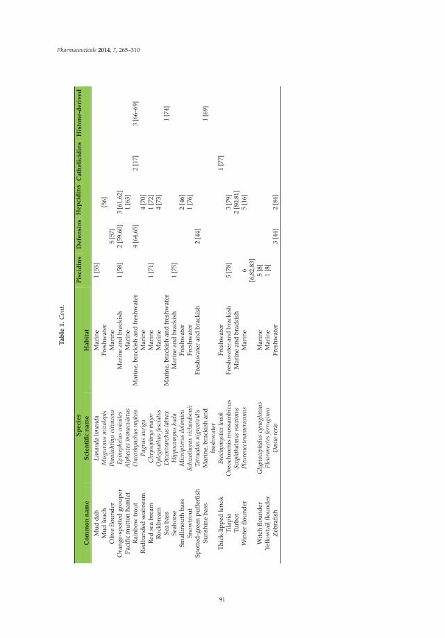
Pharmaceuticals 2014, 7, 265–310
Ta
ble
1.
Con
t.
Sp
eci
es
Pis
cid
ins
Defe
nsi
ns
Hep
cid
ins
Cath
eli
cid
ins
His
ton
e-d
eri
ved
Co
mm
on
nam
eS
cien
tifi
cn
am
eH
ab
itat
Mud
dab
Lim
anda
liman
daM
arin
e1
[55]
Mud
loac
hM
isgu
rnus
miz
olep
isFr
eshw
ater
[56]
Oliv
eflo
unde
rPa
ralic
hthy
sol
ivac
eus
Mar
ine
5[5
7]O
rang
e-sp
otte
dgr
oupe
rEp
inep
helu
sco
ioid
esM
arin
ean
dbr
acki
sh1
[58]
2[5
9,60
]3
[61,
62]
Paci
ficm
utto
nha
mle
tA
lphe
stes
imm
acul
atus
Mar
ine
1[6
3]R
ainb
owtr
out
Onc
orhy
nchu
sm
ykis
sM
arin
e,br
acki
shan
dfr
eshw
ater
4[6
4,65
]2
[17]
3[6
6–69
]R
edba
nded
seab
ream
Pagr
usau
riga
Mar
ine
4[7
0]R
edse
abr
eam
Chr
ysop
hrys
maj
orM
arin
e1
[71]
1[7
2]R
ockb
ream
Opl
egna
thus
fasc
iatu
sM
arin
e4
[73]
Sea
bass
Dic
entr
arch
usla
brax
Mar
ine,
brac
kish
and
fres
hwat
er1
[74]
Seah
orse
Hip
poca
mpu
sku
daM
arin
ean
dbr
acki
sh1
[75]
Smal
lmou
thba
ssM
icro
pter
usdo
lom
ieu
Fres
hwat
er2
[46]
Snow
trou
tSc
hizo
thor
axri
char
dson
iiFr
eshw
ater
1[7
6]Sp
otte
d-gr
een
puff
erfis
hTe
trao
don
nigr
ovir
idis
Fres
hwat
eran
dbr
acki
sh2
[44]
Suns
hine
bass
Mar
ine,
brac
kish
and
fres
hwat
er1
[69]
Thic
k-lip
ped
leno
kBr
achy
mys
tax
leno
kFr
eshw
ater
1[7
7]Ti
lapi
aO
reoc
hrom
ism
ossa
mbi
cus
Fres
hwat
eran
dbr
acki
sh5
[78]
3[7
9]Tu
rbot
Scop
htha
lmus
max
imus
Mar
ine
and
brac
kish
2[8
0,81
]W
inte
rflo
unde
rPl
euro
nect
esam
eric
anus
Mar
ine
6[6
,82,
83]
5[1
6]
Wit
chflo
unde
rG
lypt
ocep
halu
scy
nogl
ossu
sM
arin
e5
[8]
Yello
wta
ilflo
unde
rPl
euro
nect
esfe
rrug
inea
Mar
ine
1[8
]Z
ebra
fish
Dan
iore
rio
Fres
hwat
er3
[44]
2[8
4]
91
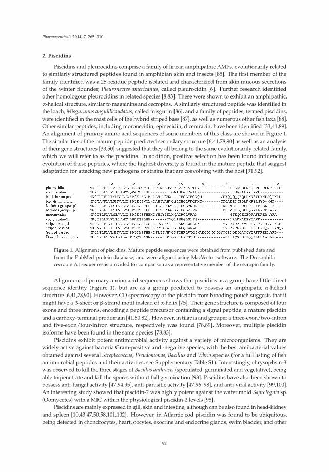
Pharmaceuticals 2014, 7, 265–310
2. Piscidins
Piscidins and pleurocidins comprise a family of linear, amphipathic AMPs, evolutionarily relatedto similarly structured peptides found in amphibian skin and insects [85]. The first member of thefamily identified was a 25-residue peptide isolated and characterized from skin mucous secretionsof the winter flounder, Pleuronectes americanus, called pleurocidin [6]. Further research identifiedother homologous pleurocidins in related species [8,83]. These were shown to exhibit an amphipathic,α-helical structure, similar to magainins and cecropins. A similarly structured peptide was identified inthe loach, Misgurunus anguillicaudatus, called misgurin [86], and a family of peptides, termed piscidins,were identified in the mast cells of the hybrid striped bass [87], as well as numerous other fish taxa [88].Other similar peptides, including moronecidin, epinecidin, dicentracin, have been identified [33,41,89].An alignment of primary amino acid sequences of some members of this class are shown in Figure 1.The similarities of the mature peptide predicted secondary structure [6,41,78,90] as well as an analysisof their gene structures [33,50] suggested that they all belong to the same evolutionarily related family,which we will refer to as the piscidins. In addition, positive selection has been found influencingevolution of these peptides, where the highest diversity is found in the mature peptide that suggestadaptation for attacking new pathogens or strains that are coevolving with the host [91,92].
Figure 1. Alignment of piscidins. Mature peptide sequences were obtained from published data andfrom the PubMed protein database, and were aligned using MacVector software. The Drosophilacecropin A1 sequences is provided for comparison as a representative member of the cecropin family.
Alignment of primary amino acid sequences shows that piscidins as a group have little directsequence identity (Figure 1), but are as a group predicted to possess an amphipatic α-helicalstructure [6,41,78,90]. However, CD spectroscopy of the piscidin from brooding pouch suggests that itmight have a β-sheet or β-strand motif instead of α-helix [75]. Their gene structure is composed of fourexons and three introns, encoding a peptide precursor containing a signal peptide, a mature piscidinand a carboxy-terminal prodomain [41,50,82]. However, in tilapia and grouper a three-exon/two-intronand five-exon/four-intron structure, respectively was found [78,89]. Moreover, multiple piscidinisoforms have been found in the same species [78,83].
Piscidins exhibit potent antimicrobial activity against a variety of microorganisms. They arewidely active against bacteria Gram-positive and -negative species, with the best antibacterial valuesobtained against several Streptococcus, Pseudomonas, Bacillus and Vibrio species (for a full listing of fishantimicrobial peptides and their activities, see Supplementary Table S1). Interestingly, chrysophsin-3was observed to kill the three stages of Bacillus anthracis (sporulated, germinated and vegetative), beingable to penetrate and kill the spores without full germination [93]. Piscidins have also been shown topossess anti-fungal activity [47,94,95], anti-parasitic activity [47,96–98], and anti-viral activity [99,100].An interesting study showed that piscidin-2 was highly potent against the water mold Saprolegnia sp.(Oomycetes) with a MIC within the physiological piscidin-2 levels [98].
Piscidins are mainly expressed in gill, skin and intestine, although can be also found in head-kidneyand spleen [10,43,47,50,58,101,102]. However, in Atlantic cod piscidin was found to be ubiquitous,being detected in chondrocytes, heart, oocytes, exocrine and endocrine glands, swim bladder, and other
92

Pharmaceuticals 2014, 7, 265–310
tissues [103]. Nevertheless, the expression profiles vary depending on the isoform [39,78,83]. Moreover,specifically among the cell types where piscidin has shown to be expressed are mast cells, rodletcells, phagocytic granulocytes and eosinophilic granular cells [43,88,102,104,105]. Interestingly, thereis evidence that granulocytes can destroy bacteria in phagosome by intracellular release of piscidin,meaning that piscidin can act against extra and intracellular bacteria [102]. In addition, pleurocidinexpression is expressed at 13 days post-hatch in the winter flounder, which is suggested to play animportant role in defense during development [101].
Like AMP genes from mammals, piscidin genes can be induced by a variety of stimuli, includingGram-positive and -negative bacteria [78], bacteria cell components like LPS [43,50,58] or the bacterialantigen ASAL [10]. The LPS-mediated induction of epinecidin-1 in zebrafish was shown to requirehepatocyte nuclear factor 1 [89]. Furthermore, piscidin genes are induced by parasites [47,104,106],viruses [107], and poly I:C [43,58]. Another study demonstrated that high biomass density (i.e., ahigher concentration of fish per volume water in an experimental tank) used as an acute stressorcomponent, led to an to up-regulation of dicentracin in gills and skin as well [74].
Besides microorganisms, piscidin-mediated anti-tumor activity has been shown by the growthinhibition and/or killing of a variety of different cancer-derived cell lines like A549 [108], HT1080 [108–110], U937 [111], HL60 [112], U937 [110], HeLa [110] and different breast cancer-derived cell linesincluding MDA-MB-468, T47-D, SKBR3, MCF7, MCF7-TX400 (paclitaxel-resistant MCF7), MDA-MB-231and 4T1 [113]. Furthermore, pleurocidin is able to kill breast cancer xenografts in NOD SCID mice,where cell death was caused by mitochondrial membrane damage and ROS production [113]. Inaddition, disruption of cancer cell membrane has been also shown to occur [110]. Moreover, in vitroinhibition of proliferation of U937 and HT1080 was suggested to occur by inducing apoptosis inresponse to cytokine production like TNF-α, IL-10, IL-15, IL-6, the tumor suppressor p53 [111], andcaspases [110]. Also, pleurocidin showed the ability to inhibit HT1080 migration in a dose-dependentmanner [109] as well as the rapid killing of a human leukemia cell line [112]. In contrast, pleurocidinshowed no lysis of human dermal fibroblasts, umbilical vein endothelial cells and erythrocytes [113].
Several studies have shown that piscidin can disrupt the plasma membranes and cause cellularmaterial efflux by pore formation [114,115]. However, use of membrane models has suggestedthat membrane composition is an important factor in the lytic capacity of piscidin to disrupt cellmembranes [116]. In addition, using site-specific high-resolution solid-state NMR orientationalrestraints and circular dichroism it was shown that piscidin-1 and -3 induce a membrane-AMPinteraction by parallel orientation of the α-helical in membrane model surfaces where fast and largeamplitude backbone motions occur [117,118]. Moreover, at very low inhibitory concentrations piscidindoes not cause significant cell membrane damage but is capable to inhibit macromolecular synthesisin bacteria [119]. Against fungi, pleurocidin was active against C. albicans by causing protoplastregeneration and membrane disruption [95,112] and it has been suggested to cause oxidative stress,triggering apoptosis in C. albicans by inducing intracellular reactive oxygen species (ROS) and activationof metacaspases, leading to externalization of phosphatidylserine [94].
Among other attractive features of piscidin includes their ability to retain antibacterial activityat high salt concentrations [41], thermostability (piscidin from seahorse brooding pouch retainedfull activity after exposing from 20–80 ◦C for 30 min, and only 20% loss of activity when boiling at100 ◦C for 30 min) [75], and relatively low cytotoxicity against mammalian cells [120]. However, intilapia some piscidin isoforms were hemolytic for tilapia red blood cells. The peptide with the greatesthemolysis activity was also the one with the best antibacterial activity, which is associated with theamphiphilic α-helical cationic structure [78].
The immunomodulatory capacity of piscidins is another feature that has been widely assessed.In fish, they are able to modulate the expression of pro-inflammatory and other immune-relatedgenes like IL-1β, IL-10, IL-22, IL-26, TNF-α, IFN-γ, NF-κB, lysozyme, NOS2, MyD88, TLR4a, TLR1,TLR3, [121–125]. Moreover, in mice this immunomodulatory effect also has been observed, withthe modulation of the genes encoding IL-6, IL-10, IL-12, MCP-1, TNF-α, IFN-γ and IgG1 [126–128].
93

Pharmaceuticals 2014, 7, 265–310
Recently, some pleurocidins have shown to be able stimulate human mast cell chemotaxis increasingCa2+ mobilization, and inducing the production of pro-inflammatory cytokines (like CCL2, 1β/CCL4)in mast cells, which was suggested to occur through G-proteins. In addition, it is able to cause mastcells to adhere, migrate, degranulate, and release cysteinyl leukotrienes and prostaglandin D2 [129].
Overall, it appears that piscidins represent an evolutionarily conserved family of peptides, which,while unique to fish, exhibit broad homology to the linear, amphipathic classes of antimicrobialpeptides found in many other species.
3. β-Defensins
A general term for cysteine-rich, cationic antimicrobial peptides found in plants, fungi,invertebrates and vertebrates [130–132], defensins exhibit a general conformation made bycysteine-stabilized α-helical and β–sheet folds (reviewed in [131,133]). In mammals three types ofdefensins have been identified based on their structure, α-, β-, and θ-defensins (last one found only incertain nonhuman primates, including the rhesus macacque) [130,134,135]. However in fish, sequenceand structural analysis have revealed that fish defensins are solely β-defensin-like proteins [35,44,51,64]including the conserved 6-cyteine motif (Figure 2). To date, up to four genes and five isoforms ofdefensins have been found in a single species [57,65], apparently as result of gene duplication eventsthat had occurred in vertebrate β-defensins [133]. Fish defensins were first identified in zebrafish,Fugu and tetraodon by a database mining approach [44], but currently defensins have been identifiedin many other marine and freshwater fish species (see Table 1). Interestingly, a phylogenetic analysisusing defensins from human and fish revealed that hBD-4 is the only human defensin that clusteredwith fish defensins, suggesting possible similar biological properties [35].
The human β-defensin gene has two exons and one intron, fairly typical of most β-defensin genes.Furthermore, mammalian β-defensins are translated as prepeptides, with the mature peptide sequenceimmediately downstream from the signal sequence [136]. However, in fish three exons and two intronsare found [65], encoding a prepropeptide (including signal peptide, propeptide and mature peptide)comprised of 60 to 77 amino acids, and a mature peptide from 38 to 45 amino acids with cationicnature with a pI around 8 (except for those in olive flounder, which are around 4, indicating anionicnature [57]). Due its cationic nature they present a net positive charge that can go from +1 to +5. Aswith all vertebrate β-defensins, there are six conserved cysteines, although in human and birds theseare located in a single exon, while in fish they span two exons [57].
Figure 2. Alignment of β-defensins. Precursor peptide sequences were obtained from published dataand from the PubMed protein database, and were aligned using MacVector software. The bovineβ-defensin, Tracheal Antimicrobial Peptide (TAP) is shown for comparison. The conserved β-defensincysteine spacing is shown in the consensus line. The first residue of the mature peptide region (basedon the isolated TAP sequence) is denoted with an asterisk.
Fish β-defensins have proven to be active against both Gram-negative and -positive bacteria(for specific inhibitory values see Supplementary Table S1), although with rather moderate activity.Exceptions to those reports of MICs in the high μM range are Planococcus citreus (Gram-positive) [11]and Aeromonas hydrophila (Gram-negative) [52], with low MIC values. Other studies using supernatantof lysates HEK293T cells transfected with β-defensins from the Chinese loach or the gilthead seabream
94

Pharmaceuticals 2014, 7, 265–310
showed significant growth inhibition of the Gram-negative A. hydrophila and the Gram-positive B.subtilis [30,35]. Moreover, β-defensins are also active against fish-specific viruses such as Singaporegrouper iridovirus (SGIV), viral nervous necrosis virus (VNNV), haemorrhagic septicaemia virus (VHSV),and the frog-specific Rana grylio virus (RGV) [59,60,64]. In addition it has been shown that theα-defensin human defensin-1 (HD-1) is highly active against VHSV, a salmonid rhabdovirus, causingits inactivation and inhibition [137]. However, no assessment has been carried out testing fish-deriveddefensins against human viruses so far, nor about their potential mechanism of action. Similarly, thereare no published studies examining the activity of fish defensins against parasites. A small numberof studies demonstrate the activity of human defensins against parasites, showing, for example, thatHD-1 is capable to destroy the parasite Trypanosoma cruzi by pore formation and induction of nuclearand mitochondrial DNA fragmentation [138], and that hd-5 is able to reduce Toxoplasma gondii viabilityby aggregation [139]. This is an area with great potential both for fish and human biology. As withparasites, there are no studies related to the antifungal activity of fish defensins, in spite of the manystudies showing such activity of β-defensins from other species (e.g., those described in [140–142].
In addition to their antimicrobial activities, β-defensins have been shown to exhibit multipleimmunomodulatory activities (reviewed in [143]). For example, recombinant mBD4 and hBD2(both β-defensins in mice and human, respectively) have shown to possess chemotactic activityfor CCR6-expressing cells (which include monocytes, dendritic cells and T-lymphocytes), which wasconfirmed using its chemokine ligand CCL20 that competed with these β-defensins [144]. Similaractivity has been observed in a fish homologue. β-defensins from the gilthead seabream exhibitedchemotactic activity, showing the capacity to attract head-kidney leukocytes [35]. There is evidenceof CCR6 mammalian orthologs in zebrafish [145] and rainbow trout [146] that may help addressthe mechanism. In addition, chemotactic capacity of HNP1 (human α-defensin) towards troutleucocytes has been shown [147]. Furthermore, a β-defensin from Atlantic cod is capable of stimulatingantimicrobial activity in phagocytes [11]. Together, the studies suggest that fish β-defensins functionsimilarly to their mammalian counterparts, contributing to the innate host defense in multiple ways.
In mammals, β-defensin expression was initially identified and studied predominantly inskin and mucosal membranes from respiratory, gastrointestinal and genitourinary tracts (reviewedin [148]. More recently, however, numerous β-defensin isoforms have been identified in sperm, withassociations to reproduction being demonstrated [149]. While some β-defensins (mostly hBD-1 andits homologue) are constitutively expressed, the expression of most β-defensins can be induced by avariety of factors, including many innate immune mediators and microbe-associated molecular patterns(reviewed in [150]). Furthermore, their expression is observed not only in adult tissues, but duringembryonic development as well [151,152]. In fish, constitutive expression seems to start early in thedevelopment probably as part of the need of defense in vulnerable stages that rely significantly in theinnate immune response [11,57]. However, it is hard to establish specific expression patterns, becausethis appears to vary between species and isoform [31,44,59,60,65], although in most of the characterizedfish β-defensins, skin is one of the tissues with the highest basal expression [31,35,44,65], a widelydistributed feature among vertebrate defensins [148,153]. After skin, head-kidney and spleen are thetissues with also high expression, which are the main immune organs in fish [51,52,65]. Nevertheless,in some studies some isoforms of fish β-defensins have shown to possess a widespread constitutiveexpression [30,52,65]. Furthermore, high expression in eye has been found, suggesting a relevant rolein ocular infections [30,52]. In addition, a study in the orange-spotted grouper suggest a relationship offish β-defensin and reproduction endocrine regulation, finding an isoform that is exclusively expressedin pituitary and testis, where such expression is up-regulated from intersexual gonad to testis in sexreversal, and a deeper analysis proved that the pituitary-specific POU1F1 transcription binding siteand the testis-specific SRY site are responsible for this phenomenon [60]. Fish β-defensin genes areinduced by a variety of stimuli including cell wall components like LPS [52,59], β-glucans [31] andpeptidoglycan [154]. They are stimulated by bacterial challenges from A. hydrophila [30], Y. ruckeri [65],V. anguillarum [11] and E. tarda [57]; and by viral challenges, including SGIV [59] or the TLR-3 agonist
95

Pharmaceuticals 2014, 7, 265–310
poly(I:C), to emulate a viral infection [59,65]. In addition, supplemented diets with the diatom Navilucasp. and the lactobacillus Lactobacillus sakei have shown to induced β-defensin in gilthead seabream [155].Thus, β-defensins in fish are true orthologues, exhibiting both structural and functional similarities tomammalian peptides, as well as their patterns of expression. This further supports the hypothesis thatβ-defensins are an ancient and highly conserved mechanism of host defense in animal species [133].
4. Hepcidins
Hepcidins are cysteine-rich peptides with antimicrobial activity that were first discovered inhumans [156,157]. Since then, hepcidins have been identified in many other vertebrates includingreptiles, amphibians and fish. Although, in birds the existence of a hepcidin needs to be betterconfirmed [158]. Fish hepcidin was first identified and isolated from the hybrid striped bass [42] andsince then hepcidins have been identified in at least 37 fish species. The general structure of humanhepcidin is a β-sheet-composed harpin-shaped with four disulfide bridges (formed by eight cysteines)with an unusual vicinal bridge at the hairpin turn [159], which is also the general structure in fishhepcidin [76,79]. However, sequence analysis of fish hepcidins has shown the presence of hepcidinscontaining 7, 6 or 4 cysteines [9,48].
Fish hepcidin genes have undergone duplication and diversification processes that have producedmultiple gene copies [9], and up to eight copies have been identified [22,34,73]. Hepcidin genes arecomposed of three exons and two introns encoding a signal peptide, a prodomain and a maturepeptide [22,34,72]. The pre-prohepcidin size can range from 81 to 96 amino acids, and the maturehepcidin from 19 to 31, with a molecular weight around 2–3 kDa. Representative sequences are shownin Figure 3. An average pI generally above 8 demonstrates their cationic nature. However, a predictedlow pI of 5.4 of the orange-spotted grouper, indicates the existence of anionic hepcidin [61].
Figure 3. Alignment of hepcidins. Representative precursor peptide sequences were obtained frompublished data and from the PubMed protein database, and were aligned using MacVector software.Human hepcidin is shown for comparison. The first residue of the mature peptide region is denotedwith an asterisk.
Fish have two types of hepcidins, HAMP1 and HAMP2. However, although HAMP1 is presentin actinopterygian and non-actinopterygian fish, HAMP2 has been only found in actinopterygianfish [54,63,158]. Moreover, a phylogenetic study has shown positive Darwinian selection in HAMP2(but not HAMP1 and its mammalian orthologue) that suggest adaptive evolution probably associatedwith the host-pathogen interaction in different environments [9,54,160].
Hepcidin expression can be detected as early as in the fertilized egg in blunt snout bream [27] orat 8 h after fertilization in channel catfish [26]. However, in winter flounder and tongue sole it wasnot detected until day 5 and 6, respectively (larvae stage) [16,37]. Nevertheless, it has been shownthat hepcidin isoforms have different expression pattern and kinetics in larval development [70]. Inaddition, different hepcidin types in a single species can have different rates of expression within thesame tissue [80] that can be affected by different stimuli [24,70,73,79]. Interestingly, some isoformshave high basal hepcidin expression in liver, but some have not, where the highest expression oftenoccurs in spleen, kidney and intestine [9,70,73,79].
Similar to other AMP genes, fish hepcidins can be induced by exposure to both Gram-positiveand Gram-negative bacteria [12,19,25–27,34,36,37,42,48,53,56,61,63,72,73,161–166]. Moreover, fungilike Saccharomyces cerevisiae [36,61], and tumor cell lines like L-1210 and SAF-1 have shown to induce
96

Pharmaceuticals 2014, 7, 265–310
hepcidin expression as well [36]. Hepcidin genes in fish are also induced by viruses [36,61,73,165],and poly I:C [12,36,164], as well as mitogens [36]. Moreover, environmental estrogenic endocrinedisrupting chemicals like 17β-estradiol down-regulates one of the hepcidin isoforms expression inliver in largemouth bass [46].
In humans, hepcidin acts as a type II actue-phase protein [167]. Related to this, time-courseexperiments under bacterial challenge of fish have shown that the highest expression of hepcidinoccurs at 3–6 h and decay after that [23,32,163]. Also, the expression of hepcidin occurs along withother acute phase response proteins like IL-1β, serum amyloid A and precerebellin after infection withYersinia ruckeri in rainbow trout [168]. Related to this, mud loach infected with Gram-negative bacteriashowed a high IL-1β-like gene expression-mediated response [56]. Together, these results suggest thathepcidins can also act as a type II acute phase protein, and function as part of a broad innate immuneresponse in fish.
Fish hepcidins are active against a wide variety of bacteria, both Gram-positive and -negative at thelow μM range, including potent activity against a large number of fish pathogens (see SupplementaryTable S1 for a complete listing). This includes rapid killing kinetics against S. aureus and Pseudomonasstutzeri [23,62]. Furthermore, synergy between bass hepcidin and moronecidin against S. iniae and Y.enterocolitica has been demonstrated [161]. In addition, they are active against a number of viruses [80,99,169–171], and a recent study indicates that human Hepc25 is able to affect HCV replication in cellculture by inducing STAT3 activation leading to an antiviral state of the cell [172]. In contrast, theirquantified activity against fungi appears to be rather low [23,48,161].
A few studies have tried to elucidate the mechanism of action of hepcidin against bacteria.With human Hepc25, the lack of SYTOX uptake showed that membrane permeabilization does notoccur [173] in contrast to most antimicrobial peptides [150]. Similar results have been observed withfish peptides, using light emission kinetics, which showed that Medaka recombinant pro-hepcidin andsynthetic hepcidin also do not cause membrane permeabilization in E. coli [171]. However, humanHepc25 has shown binding to DNA with high efficiency in a retardation assay [173].
Fish hepcidins have also shown the capacity of affect cancer cells viability. For example, tilapiahepcidin TH2-3, have shown inhibition of proliferation and migration of human fibrosarcoma cell lineHT1080a in a concentration-dependent manner. Furthermore, TH2-3 was able to cause cell membranedisruption in HT1080 and results also suggest that TH2-3 down-regulates c-Jun leading to apoptosis [174].TH1-5 inhibit the proliferation of tumor cells (HeLa, HT1080 and HepG2) altering membrane structureand inducing apoptosis at low dose. Also, TH1-5 showed modulation of immune-related genes [175].A study with medaka hepcidin showed 40% decrease in HepG2 cell viability by addition of 25 and5 μM of synthetic mature Om-hep1 and recombinant pro-Om-hep1 (prohepcidin), respectively [171].Interestingly, Pro-Omhep1 has better anti-tumor activity compared with Om-hep1, using HepG2cells [171].
Fish hepcidins have also shown the ability to modulate the expression of different immune-relatedgenes not only in fish but also in mice. Transgenic zebrafish expressing TH1-5 showed upregulationof IL-10, IL-21, IL-22, lysozyme, TLR-1, TLR-3, TNF-α and NF-κB [176]. However, TH2-3 showed todownregulate some of those upregulated by TH1-5 [177]. In the same context, TH2-3 reduced theamount of TNF-α, IL-1α, IL-1β, IL-6 and COX-2 in mouse macrophages challenged with LPS [178].Related to this, in turbot it has been shown that hepcidin is able to increase the activation ofNF-κB (which control a variety of inflammatory cytokines) through an undetermined yet signalingpathway [165]. TH2-3 have also shown to be able to modulate protein kinase C isoforms in the mousemacrophage RAW264.7 cell line [179], and was also capable to induce morphology changes in thesecells similar to PMA-induced changes [179]. Moreover, TH1-5 modulates the expression of certaininterferons and annexin (viral-responsive genes) in pancreatic necrosis virus-infected fish [170].
However, despite the potential antimicrobial and immunomodulatory effect, hepcidin is betterknown for being a key iron regulator controlling ferroportin, which is able to degrade by itsinternalization, which decrease iron transfer into blood [180]. In fish, although ferroportin internalization
97

Pharmaceuticals 2014, 7, 265–310
by hepcidin has not been proven yet, there is evidence suggesting that fish hepcidin also controlsiron [34,56,73,80,162,165,166,181]. It may also serve as a pleiotropic sensor for other divalent metals,because it is up-regulated by exposure to other metals like copper [56] and cadmium [182], which canbe considered waterborne or toxic.
5. Cathelicidins
Unusual among the AMPs, cathelicidins share little sequence homology between the maturepeptides. Rather, they are defined by a homologous N-terminal region of the precursor peptide, called acathelin domain, found just after a conserved signal domain (reviewed in [183]). The active, maturepeptide is released upon protolytic cleavage by elastase and possibly other enzymes [184]. In mammals,the mature AMP sequence varies greatly, not only between species but also among the often multiplecathelicidin peptides within a single species. In general, however, all mammalian cathelicidin maturepeptides are cationic and exhibit an amphipathic characteristic, as well as broad-spectrum antimicrobialactivity in vitro. As can be seen by the alignment of primary amino acid sequences in Figure 4, thereare significant sequence similarities in the C-terminal region, and in several short domains, which arehighly cationic and glycine-rich.
The first cathelicidins identified in fish were initially isolated as antimicrobial peptides fromthe Atlantic hagfish, Myxine glutinosa [15]. Upon sequence analysis of the cDNA that encoded thesepeptides, it was discovered that they exhibited homology to cathelicidins, previously only found inmammals to this point. As cathelicidins were discovered in other more conventional fish species,primarily by sequence homology from the cathelin region (e.g., [17,28,77,185]), or more recently bypeptide isolation [186], new patterns emerged. In some of the more recently studied fish cathelicidins,while a high degree of homology is maintained in the cathelin domain (see [185] for a comprehensivealignment), there appears to be a higher degree of sequence similarity of the mature peptide than seenin mammals. Thus, fish cathelicidins can now be subdivided into two classes—the linear peptides,and those that exhibit a characteristic disulphide bond. In contrast to mammalian cathelicidins, thereis significant sequence homology among members of the classes (up to 90%), and little homologybetween the classes. In addition, the recently identified cathelicidins found in cod appear to comprisea third class, based on sequence homology between themselves, and a lack of homology with either ofthe other two classes [185]. An alignment of representative fish cathelicidins is shown in Figure 4.
Figure 4. Alignment of fish cathelicidins. Mature peptide sequences were obtained from published dataand from the PubMed protein database, and were aligned using MacVector software. Characteristiccysteine residues found in certain classes of fish cathelicidins are underlined. As, Atlantic salmon; Rt,Rainbow trout; Cs, Chinook salmon; Btr, Brown trout; Ac, Arctic char; Bt, Brook trout; Je, Japanese eel.
As cathelicidin peptides are purified from more species, their in vitro antibacterial activities appearto exhibit significant variability with respect to selectivity, depending on the species. For example, codcathelicidin (codCATH) is highly active against those Gram-negative bacterial species examined, butalmost inactive against the Gram-positive species. It also exhibits potent antifungal activity against C.albicans [13]. In contrast, the hagfish cathelicidins are active against both Gram-positive and -negative
98

Pharmaceuticals 2014, 7, 265–310
bacteria, but inactive against Candida [15]. Even more specifically, rainbow trout cathelicidins areactive against Y. ruckeri, while Atlantic salmon cathelicidins are not [187]. Thus, the variability inthe mature peptide sequence of these molecules appears to direct the antimicrobial activities, and isprobably a result of an evolutionary divergence to address specific pathogens.
Based on their antimicrobial activity, most of the work to elucidate their role in vivo has examinedthe expression of the cathelicidin genes in the various fish species with respect to induction by innateimmune regulators, such as bacteria and to different pathogen-associated molecular patterns (PAMPs).Importantly, cathelicidin expression is observed early in embryonic development, suggesting that itsrole in immunity is present early on [186]. In vitro, both bacteria and bacterial DNA were sufficientto induce cathelicidin expression in a cultured embryonic salmon cell line [188], suggesting thatlike mammalian cathelicidins, the fish homologs play a similar role in antibacterial host defense.Surprisingly, purified LPS (that is, treated with DNase I) could not induce the gene. This regulation hasbeen further elucidated by the demonstration of a wide variability of the Chinook salmon embryoniccell line’s response to different bacteria and to poly I:C, LPS and flagellin [189]. Further in vitro evidenceof this role was demonstrated by the induction of rainbow trout cathelicidin in a macrophage cellline from that species by IL-6, an important mediator of the innate immune response [190], and by anovel TNF-α isoform [191]. Similarly, stimulation of a cell line from Atlantic cod with poly I:C inducedexpression of the gmCath1 (from cod) gene promoter [192]. In addition to bacteria and their products,trout cell lines were shown to induce cathelicidin gene expression upon incubation with the oomyceteSaprolegnia parasitica [193].
In vivo studies further support this hypothesis. When ayu were injected with live pathogenicbacteria, there was a time-dependent induction of cathelicidin expression in numerous tissues,including gill, liver, spleen and intestine [20]. Further, Atlantic salmon and rainbow trout infectedwith Y. ruckeri led to an induction in cathelicidin expression [187,194]. In the Atlantic cod, a differencein inducibility was observed. Cathelicidin experession in the gills was induced by a 3-h incubationwith Aeromonas salmonicida, but not V. anguillarum, suggesting a more complex role is played by thesepeptides in host defense.
In mammals, cathelicidins have been demonstrated to exhibit multiple activities, both immune andnon-immune, well in excess of their in vitro antimicrobial activities (reviewed in [195]). While researchin fish has not approached this level, a recent study demonstrated that two Atlantic salmon cathelicidinsinduced the rapid and transient expression of IL-8 in peripheral blood leukocytes [187]. This suggeststhat the immunomodulatory activities seen by mammalian cathelicidins may be shared by their fishcounterparts, and may thus be an evolutionarily conserved mechanism of innate immune regulation.
6. Histone-Derived Peptides
While examining an amphibian species for novel antimicrobial peptides, Park et al. [196] describeda new peptide from the Asian toad, Bufo bufo gargarizans, which they called Buforin I. This turned outto be identical to the N-terminal portion of histone 2A. This led to the demonstration of antimicrobialactivity of histone fragments from numerous species (reviewed in [197]), suggesting that theseproteolytic fragments are part of an ancient innate immune mechanism. Histone-derived AMPshave been identified in a number of fish species, with broad-spectrum activity against both humanand fish pathogens (reviewed in [198]), including water molds [199] and a parasitic dinoflagellate [69].They are expressed and secreted in fish skin, and found in other tissues, including gill, speen and thegut. They are not limited to the N-terminus of the histones, as was found for the Buforin peptide, butcan be found as fragments of both termini, from histones H1, H2A, H2B and H6 (see Figure 5). Furtherevidence that they play a role in host defense of the fish comes from studies showing that expressionof histone-derived AMP genes are induced under conditions of stress in specific tissues of differentfish species [74,200].
99

Pharmaceuticals 2014, 7, 265–310
Figure 5. Alignment of histone-derived peptides. Representative peptide sequences were obtainedfrom published data and from the PubMed protein database, and were aligned using MacVectorsoftware. Since the sequences are homolgous to different histone peptide fragments, there is no sharedsequence homology.
7. Therapeutics
All AMPs have common characteristics that support their development as therapeuticantimicrobials. These include broad-spectrum activity against a wide variety of pathogens; potentactivity under a wide range of conditions, including temperature and in secretions such as saliva; and areduced capacity to the development of resistance by bacteria. The identification and characterization ofpeptides from fish has provided a unique contribution in this arena. While the structural characteristicsof fish peptides do not appear particularly different from their mammalian, insect or amphibianhomologues, there may be specific differences with respect to activity. It appears that overall theirantimicrobial activities against human pathogens is in the same range as AMPs from other species.However, it is possible that they are more active against fish pathogens, as they most likely haveevolved together with those pathogens. It is difficult to know this, however, as few studies havecompared non-fish peptides with fish peptides against fish pathogens. One area where fish peptidesmay provide an advantage is in food preservation [201], as they are derived from a natural food source,and thus may be more amenable to being consumed.
Since many AMPs are sensitive to high salt concentrations [202–204], the ability of some fishAMPs to kill microbes even at extremely high salt concentrations, such as those found in the marineenvironment, make them important targets for investigation. Pleurocidin, for example, maintains itsantibacterial activity even up to 300 mM NaCl [6], similar to other piscidins [41,205]. Understandingthe structural foundation that supports this salt-independent activity could aid in the design ofnovel peptides [206] or mimetics that could address infections under a wide range of normal andabnormal salt concentrations, whether in serum, tear film hyperosmolarity, or in saliva to addressdental caries [207]. In addition to their potential uses as antimicrobials, some fish AMPs have beenobserved to exhibit in vitro cytotoxic activity against a variety of cancer cells [113,208].
Different applications of piscidin have been promising. For example, epinecidin-1, whenadministrated orally or injected (in pre-, co- and post-infection) can significantly enhance survivalin zebrafish and grouper that were challenged with Vibrio vulnificus [58,123,124]. Related to this,electrotransfer of epinecidin-1 in zebrafish and grouper muscle showed significant reduction in V. vulnificusand Streptococcus agalactiae bacterial counts [121,122,125]. Moreover, treatment of lethally-challengedmethicillin-resistant S. aureus (MRSA) mice with epinecidin-1 allowed mice to survive by decreasingconsiderably the bacterial counts, where also there was evidence of wound closure and angiogenesisenhancement [128].
In oral disease treatment piscidins are also promising due to the potent effect of chrysophsin-1 inkilling the cariogenic pathogen Streptococcus mutans [209]. Furthermore, pleurocidin also demonstratedanti-cariogenic activity by being able to kill both S. mutans and S. sobrinus, where killing of biofilmsoccurred in a dose-dependent manner. In addition, it showed to retained its activity in physiologicalor higher salt concentration, and was relatively stable in presence of human saliva and no hemolysiswas found [207,210].
Epinecidin-1 showed to be a potential candidate for topical application that can prevent vaginalor skin infections due to the synergistic effect that possess with commercial cleaning solutions, wheresuch effect was not affected by low pH or after being stored at room temperature and at 4 ◦C for up
100

Pharmaceuticals 2014, 7, 265–310
to 14 days [211]. The synergistic effect of pleurocidin and several antibiotics [212], bacteroricin [213]and histone-derived [214] has also been shown [212,213]. Furthermore, the creation of antimicrobialsurfaces has been made by the immobilization of chrysophsin-1 resulting in a surface with antibacterialactivity capable to killed around 82% of E. coli bacteria [215].
A recent interest finding is the ability of epinecidin-1 to create inactivated virus for vaccinationpurposes. Huang et al. found that mice injected with Epi-1-based inactivated Japanese encephalitisvirus (JEV) reached 100% survival (in a dose-dependent manner), and the performance was better thanthe formalin-based JEV-inactivated vaccine. This was caused by the modulation of immune-relatedgenes, including the increase of anti-JEV-neutralizing antibodies in serum, which suppressed themultiplication of JEV in brain sections [126].
Fish hepcidins are also under examination for development as therapeutics. One example ofthis is the study carried out by Pan et al. [216], where injections with pre-incubated tilapia hepcidinTH2-3 and 108 cfu of Vibrio vulnificus for 30 min enhanced the survival of infected and re-infectedmice, obtaining up to 60% of survival with a dose of 40 μg/mice. In addition, TH2-3 also showedsignificant prophylactic effect by administration prior infection, where survival rates of 100% wereobtained after 7 days of infection. Also, curative effects were shown when fish were first infected andlater injected with 40 μg/mice of TH2-3, obtaining survival rates up to 50%. But more interesting,was the fact that TH2-3 had better bacteriostatic effect than tetracycline in controlling the bacterialburden in blood, although in liver there was no significant difference, demonstrating the capacity ofTH2-3 to control multiplication of V. vulnificus in mice. Although the direct in vivo TH2-3-mediatedkilling was not confirmed, a microarray analysis showed that TH2-3 clearly altered the gene expressionprofiles improving the host response in mice [216]. In addition, a transgenic zebrafish expressingTH2-3 showed to be able to decrease V. vulnificus burden significantly, but not S. agalactiae [177].Zebrafish expressing TH1-5 exhibited enhanced bacterial resistance by decreasing the bacterial burdenof both same pathogens [176]. In addition, TH1-5 has showed to be effective at increasing survival anddecreasing the number of infectious bacteria in ducks challenged with Riemerella anatipestifer, whichalso showed to be able to modulate the expression of immune-related genes [114].
However, as with other AMPs, they share the similar problems that hinder their furtherdevelopment, especially for use in human medicine. These include a tendency to be inactivatedin the body, increased expense of peptide synthesis, and sensitivity to protease digestion. Attemptsto address these issues with fish peptides, include the identification of smaller peptide fragmentsthat might exhibit better activity with smaller molecules [217], and the observation of high levels ofsynergy with bacterial AMPs [213,214], as well as conventional antibiotics [218] allowing for reducedconcentrations. One strategy that may have some success is the design of small molecule peptidemimetics that incorporate the structural characteristics of the peptides necessary for their activity(reviewed in [219]). Initial in vitro and in vivo results with molecules designed from magainins anddefensins have been encouraging, demonstrating antibacterial [220] and antifungal [221] activities,as well as immunomodulatory activity [222]. Another strategy that has been examined extensivelyin other species (reviewed in [223]) is the use of exogenous agents to modulate the expression ofendogenous AMPs in the fish. Terova et al. have demonstrated the induction of an AMP initiallyisolated from sea bass [33] by feeding them a cell wall extract from S. cerevisiae [224], suggesting anovel method for enhancing the natural defense mechanism of the fish. Incorporation of an enhancerof AMP expression in the diet could be a cost-effective part of an overall strategy of modulating theinnate immune system of the fish to control infection in aquaculture (reviewed in [198]).
8. Conclusions
The comprehensive characterization of AMPs from fish, on the structural, genetic and functionallevels, has provided a wealth of information. Examination of AMPs in a single species, such as theAtlantic cod, where members of all five groups of AMPs have been identified can help understandthe role of these peptides in innate host defense of the fish. Studies on the similarities and differences
101

Pharmaceuticals 2014, 7, 265–310
with peptides from non-fish species contribute to our understanding of the evolutionary relationshipsof innate host defense mechanisms among vertebrates. Furthermore, they can provide importantinformation for the better design of novel therapeutic agents, both for microbial infections as wellas cancer and other conditions. Unique for the field of fish AMPs is the potential application toaquaculture. The constant risk of large-scale microbial infection that can lead to significant economiclosses demands new strategies to prevent or treat these pathogens. Many of the studies described abovehave demonstrated that specific fish AMPs have potent activity against fish pathogens. Furthermore,other studies have shown that some pathogens induce potent innate immune responses in the fish.Complicating this is the evolutionary battle with the pathogens. For example, challenge of Atlanticcod with the pathogen V. anguillarum induces the expression of a β-defensin, which is antibacterialagainst other species, but not the inducing V. anguillarum [11]. Thus, the large body of work describedabove provides a solid foundation for strong future work to better understand both the role of thesepeptides in host defense of the fish, as well as the development of these peptides and their derivativesas potential therapeutics.
Acknowledgments: GD is funded by US Public Health Service Grants R01 DE22723 and R21 AI100379.
Author Contributions: Both authors contributed to the writing and editing of this review
Conflicts of Interest: The authors declare no conflict of interest.
References
1. Ganz, T.; Selsted, M.E.; Szklarek, D.; Harwig, S.S.; Daher, K.; Bainton, D.F.; Lehrer, R.I. Defensins. Naturalpeptide antibiotics of human neutrophils. J. Clin. Investig. 1985, 76, 1427–1435. [CrossRef]
2. Lehrer, R.I.; Selsted, M.E.; Szklarek, D.; Fleischmann, J. Antibacterial activity of microbicidal cationic proteins1 and 2, natural peptide antibiotics of rabbit lung macrophages. Infect. Immun. 1983, 42, 10–14.
3. Zasloff, M. Magainins, a class of antimicrobial peptides from Xenopus skin: Isolation, characterization oftwo active forms, and partial cDNA sequence of a precursor. Proc. Natl. Acad. Sci. USA 1987, 84, 5449–5453.[CrossRef]
4. Primor, N.; Tu, A.T. Conformation of pardaxin, the toxin of the flatfish Pardachirus marmoratus. Biochim.Biophys. Acta 1980, 626, 299–306. [CrossRef]
5. Oren, Z.; Shai, Y. A class of highly potent antibacterial peptides derived from pardaxin, a pore-formingpeptide isolated from Moses sole fish Pardachirus marmoratus. Eur. J. Biochem. 1996, 237, 303–310.
6. Cole, A.M.; Weis, P.; Diamond, G. Isolation and characterization of pleurocidin, an antimicrobial peptide inthe skin secretions of winter flounder. J. Biol. Chem. 1997, 272, 12008–12013. [CrossRef]
7. Tessera, V.; Guida, F.; Juretic, D.; Tossi, A. Identification of antimicrobial peptides from teleosts and anuransin expressed sequence tag databases using conserved signal sequences. FEBS J. 2012, 279, 724–736. [CrossRef]
8. Patrzykat, A.; Gallant, J.W.; Seo, J.K.; Pytyck, J.; Douglas, S.E. Novel antimicrobial peptides derived fromflatfish genes. Antimicrob. Agents Chemother. 2003, 47, 2464–2470. [CrossRef]
9. Xu, Q.; Cheng, C.H.; Hu, P.; Ye, H.; Chen, Z.; Cao, L.; Chen, L.; Shen, Y.; Chen, L. Adaptive evolution ofhepcidin genes in antarctic notothenioid fishes. Mol. Biol. Evol. 2008, 25, 1099–1112. [CrossRef]
10. Browne, M.J.; Feng, C.Y.; Booth, V.; Rise, M.L. Characterization and expression studies of Gaduscidin-1 andGaduscidin-2; paralogous antimicrobial peptide-like transcripts from Atlantic cod (Gadus morhua). Dev.Comp. Immunol. 2011, 35, 399–408. [CrossRef]
11. Ruangsri, J.; Kitani, Y.; Kiron, V.; Lokesh, J.; Brinchmann, M.F.; Karlsen, B.O.; Fernandes, J.M. A novelbeta-defensin antimicrobial peptide in Atlantic cod with stimulatory effect on phagocytic activity. PLoS One2013, 8, e62302.
12. Solstad, T.; Larsen, A.N.; Seppola, M.; Jorgensen, T.O. Identification, cloning and expression analysis of ahepcidin cDNA of the Atlantic cod (Gadus morhua L.). Fish Shellfish Immunol. 2008, 25, 298–310. [CrossRef]
13. Broekman, D.C.; Zenz, A.; Gudmundsdottir, B.K.; Lohner, K.; Maier, V.H.; Gudmundsson, G.H. Functionalcharacterization of codCath, the mature cathelicidin antimicrobial peptide from Atlantic cod (Gadus morhua).Peptides 2011, 32, 2044–2051. [CrossRef]
102

Pharmaceuticals 2014, 7, 265–310
14. Bergsson, G.; Agerberth, B.; Jornvall, H.; Gudmundsson, G.H. Isolation and identification of antimicrobialcomponents from the epidermal mucus of Atlantic cod (Gadus morhua). FEBS J. 2005, 272, 4960–4969.[CrossRef]
15. Uzzell, T.; Stolzenberg, E.D.; Shinnar, A.E.; Zasloff, M. Hagfish intestinal antimicrobial peptides are ancientcathelicidins. Peptides 2003, 24, 1655–1667. [CrossRef]
16. Douglas, S.E.; Gallant, J.W.; Liebscher, R.S.; Dacanay, A.; Tsoi, S.C. Identification and expression analysis ofhepcidin-like antimicrobial peptides in bony fish. Dev. Comp. Immunol. 2003, 27, 589–601. [CrossRef]
17. Chang, C.I.; Zhang, Y.A.; Zou, J.; Nie, P.; Secombes, C.J. Two cathelicidin genes are present in both rainbowtrout (Oncorhynchus mykiss) and atlantic salmon (Salmo salar). Antimicrob. Agents Chemother. 2006, 50,185–195. [CrossRef]
18. Richards, R.C.; O’Neil, D.B.; Thibault, P.; Ewart, K.V. Histone H1: An antimicrobial protein of Atlanticsalmon (Salmo salar). Biochem. Biophys. Res. Commun. 2001, 284, 549–555. [CrossRef]
19. Chen, M.Z.; Chen, J.; Lu, X.J.; Shi, Y.H. Molecular cloning, sequence analysis and expression pattern ofhepcidin gene in ayu (Plecoglossus altivelis). Dongwuxue Yanjiu 2010, 31, 595–600.
20. Lu, X.J.; Chen, J.; Huang, Z.A.; Shi, Y.H.; Lv, J.N. Identification and characterization of a novel cathelicidinfrom ayu, Plecoglossus altivelis. Fish Shellfish Immunol. 2011, 31, 52–57. [CrossRef]
21. Barnes, A.C.; Trewin, B.; Snape, N.; Kvennefors, E.C.; Baiano, J.C. Two hepcidin-like antimicrobialpeptides in Barramundi Lates calcarifer exhibit differing tissue tropism and are induced in response tolipopolysaccharide. Fish Shellfish Immunol. 2011, 31, 350–357. [CrossRef]
22. Yang, M.; Wang, K.J.; Chen, J.H.; Qu, H.D.; Li, S.J. Genomic organization and tissue-specific expressionanalysis of hepcidin-like genes from black porgy (Acanthopagrus schlegelii B). Fish Shellfish Immunol. 2007,23, 1060–1071. [CrossRef]
23. Yang, M.; Chen, B.; Cai, J.J.; Peng, H.; Ling, C.; Yuan, J.J.; Wang, K.J. Molecular characterization of hepcidinAS-hepc2 and AS-hepc6 in black porgy (Acanthopagrus schlegelii): Expression pattern responded to bacterialchallenge and in vitro antimicrobial activity. Comp. Biochem. Physiol. B Biochem. Mol. Biol. 2011, 158, 155–163.[CrossRef]
24. Kim, Y.O.; Park, E.M.; Nam, B.H.; Kong, H.J.; Kim, W.J.; Lee, S.J. Identification and molecular characterizationof two hepcidin genes from black rockfish (Sebastes schlegelii). Mol. Cell. Biochem. 2008, 315, 131–136.[CrossRef]
25. Gong, L.C.; Wang, H.; Deng, L. Molecular characterization, phylogeny and expression of a hepcidin gene inthe blotched snakehead Channa maculata. Dev. Comp. Immunol. 2014, 44, 1–11. [CrossRef]
26. Bao, B.; Peatman, E.; Li, P.; He, C.; Liu, Z. Catfish hepcidin gene is expressed in a wide range of tissuesand exhibits tissue-specific upregulation after bacterial infection. Dev. Comp. Immunol. 2005, 29, 939–950.[CrossRef]
27. Liang, T.; Ji, W.; Zhang, G.R.; Wei, K.J.; Feng, K.; Wang, W.M.; Zou, G.W. Molecular cloning and expressionanalysis of liver-expressed antimicrobial peptide 1 (LEAP-1) and LEAP-2 genes in the blunt snout bream(Megalobrama amblycephala). Fish Shellfish Immunol. 2013, 35, 553–563. [CrossRef]
28. Scocchi, M.; Pallavicini, A.; Salgaro, R.; Bociek, K.; Gennaro, R. The salmonid cathelicidins: A gene familywith highly varied C-terminal antimicrobial domains. Comp. Biochem. Physiol. B Biochem. Mol. Biol. 2009, 152,376–381. [CrossRef]
29. Park, I.Y.; Park, C.B.; Kim, M.S.; Kim, S.C. Parasin I, an antimicrobial peptide derived from histone H2A inthe catfish, Parasilurus asotus. FEBS Lett. 1998, 437, 258–262. [CrossRef]
30. Chen, Y.; Zhao, H.; Zhang, X.; Luo, H.; Xue, X.; Li, Z.; Yao, B. Identification, expression and bioactivityof Paramisgurnus dabryanus beta-defensin that might be involved in immune defense against bacterialinfection. Fish Shellfish Immunol. 2013, 35, 399–406. [CrossRef]
31. Marel, M.; Adamek, M.; Gonzalez, S.F.; Frost, P.; Rombout, J.H.; Wiegertjes, G.F.; Savelkoul, H.F.;Steinhagen, D. Molecular cloning and expression of two beta-defensin and two mucin genes in commoncarp (Cyprinus carpio L.) and their up-regulation after beta-glucan feeding. Fish Shellfish Immunol. 2012, 32,494–501. [CrossRef]
32. Li, H.; Zhang, F.; Guo, H.; Zhu, Y.; Yuan, J.; Yang, G.; An, L. Molecular characterization of hepcidin gene incommon carp (Cyprinus carpio L.) and its expression pattern responding to bacterial challenge. Fish ShellfishImmunol. 2013, 35, 1030–1038. [CrossRef]
103
Pharmaceuticals 2014, 7, 265–310
33. Salerno, G.; Parrinello, N.; Roch, P.; Cammarata, M. cDNA sequence and tissue expression of an antimicrobialpeptide, dicentracin; a new component of the moronecidin family isolated from head kidney leukocytes ofsea bass, Dicentrarchus labrax. Comp. Biochem. Physiol. B Biochem. Mol. Biol. 2007, 146, 521–529. [CrossRef]
34. Rodrigues, P.N.; Vazquez-Dorado, S.; Neves, J.V.; Wilson, J.M. Dual function of fish hepcidin: Response toexperimental iron overload and bacterial infection in sea bass (Dicentrarchus labrax). Dev. Comp. Immunol.2006, 30, 1156–1167. [CrossRef]
35. Cuesta, A.; Meseguer, J.; Esteban, M.A. Molecular and functional characterization of the gilthead seabreambeta-defensin demonstrate its chemotactic and antimicrobial activity. Mol. Immunol. 2011, 48, 1432–1438.[CrossRef]
36. Cuesta, A.; Meseguer, J.; Esteban, M.A. The antimicrobial peptide hepcidin exerts an important role in theinnate immunity against bacteria in the bony fish gilthead seabream. Mol. Immunol. 2008, 45, 2333–2342.[CrossRef]
37. Wang, Y.; Liu, X.; Ma, L.; Yu, Y.; Yu, H.; Mohammed, S.; Chu, G.; Mu, L.; Zhang, Q. Identification andcharacterization of a hepcidin from half-smooth tongue sole Cynoglossus semilaevis. Fish Shellfish Immunol.2012, 33, 213–219. [CrossRef]
38. Birkemo, G.A.; Luders, T.; Andersen, O.; Nes, I.F.; Nissen-Meyer, J. Hipposin, a histone-derived antimicrobialpeptide in Atlantic halibut (Hippoglossus hippoglossus L.). Biochim. Biophys. Acta 2003, 1646, 207–215.[CrossRef]
39. Salger, S.A.; Reading, B.J.; Baltzegar, D.A.; Sullivan, C.V.; Noga, E.J. Molecular characterization of twoisoforms of piscidin 4 from the hybrid striped bass (Morone chrysops x Morone saxatilis). Fish ShellfishImmunol. 2011, 30, 420–424. [CrossRef]
40. Noga, E.J.; Silphaduang, U.; Park, N.G.; Seo, J.K.; Stephenson, J.; Kozlowicz, S. Piscidin 4, a novel memberof the piscidin family of antimicrobial peptides. Comp. Biochem. Physiol. B Biochem. Mol. Biol. 2009, 152,299–305. [CrossRef]
41. Lauth, X.; Shike, H.; Burns, J.C.; Westerman, M.E.; Ostland, V.E.; Carlberg, J.M.; van Olst, J.C.; Nizet, V.;Taylor, S.W.; Shimizu, C.; et al. Discovery and characterization of two isoforms of moronecidin, a novelantimicrobial peptide from hybrid striped bass. J. Biol. Chem. 2002, 277, 5030–5039. [CrossRef]
42. Shike, H.; Lauth, X.; Westerman, M.E.; Ostland, V.E.; Carlberg, J.M.; van Olst, J.C.; Shimizu, C.; Bulet, P.;Burns, J.C. Bass hepcidin is a novel antimicrobial peptide induced by bacterial challenge. Eur. J. Biochem.2002, 269, 2232–2237. [CrossRef]
43. Buonocore, F.; Randelli, E.; Casani, D.; Picchietti, S.; Belardinelli, M.C.; de Pascale, D.; de Santi, C.;Scapigliati, G. A piscidin-like antimicrobial peptide from the icefish Chionodraco hamatus (Perciformes:Channichthyidae): Molecular characterization, localization and bactericidal activity. Fish Shellfish Immunol.2012, 33, 1183–1191. [CrossRef]
44. Zou, J.; Mercier, C.; Koussounadis, A.; Secombes, C. Discovery of multiple beta-defensin like homologues inteleost fish. Mol. Immunol. 2007, 44, 638–647. [CrossRef]
45. Ren, H.L.; Wang, K.J.; Zhou, H.L.; Yang, M. Cloning and organisation analysis of a hepcidin-like gene andcDNA from Japan sea bass, Lateolabrax japonicus. Fish Shellfish Immunol. 2006, 21, 221–227. [CrossRef]
46. Robertson, L.S.; Iwanowicz, L.R.; Marranca, J.M. Identification of centrarchid hepcidins and evidence that17beta-estradiol disrupts constitutive expression of hepcidin-1 and inducible expression of hepcidin-2 inlargemouth bass (Micropterus salmoides). Fish Shellfish Immunol. 2009, 26, 898–907. [CrossRef]
47. Niu, S.F.; Jin, Y.; Xu, X.; Qiao, Y.; Wu, Y.; Mao, Y.; Su, Y.Q.; Wang, J. Characterization of a novel piscidin-likeantimicrobial peptide from Pseudosciaena crocea and its immune response to Cryptocaryon irritans. FishShellfish Immunol. 2013, 35, 513–524. [CrossRef]
48. Wang, K.J.; Cai, J.J.; Cai, L.; Qu, H.D.; Yang, M.; Zhang, M. Cloning and expression of a hepcidin gene from amarine fish (Pseudosciaena crocea) and the antimicrobial activity of its synthetic peptide. Peptides 2009, 30,638–646. [CrossRef]
49. Zhang, J.; Yan, Q.; Ji, R.; Zou, W.; Guo, G. Isolation and characterization of a hepcidin peptide from the headkidney of large yellow croaker, Pseudosciaena crocea. Fish Shellfish Immunol. 2009, 26, 864–870. [CrossRef]
50. Sun, B.J.; Xie, H.X.; Song, Y.; Nie, P. Gene structure of an antimicrobial peptide from mandarin fish, Sinipercachuatsi (Basilewsky), suggests that moronecidins and pleurocidins belong in one family: The piscidins.J. Fish Dis. 2007, 30, 335–343. [CrossRef]
104
Pharmaceuticals 2014, 7, 265–310
51. Wang, G.; Li, J.; Zou, P.; Xie, H.; Huang, B.; Nie, P.; Chang, M. Expression pattern, promoter activity andbactericidal property of beta-defensin from the mandarin fish Siniperca chuatsi. Fish Shellfish Immunol. 2012,33, 522–531. [CrossRef]
52. Zhao, J.G.; Zhou, L.; Jin, J.Y.; Zhao, Z.; Lan, J.; Zhang, Y.B.; Zhang, Q.Y.; Gui, J.F. Antimicrobial activity-specificto Gram-negative bacteria and immune modulation-mediated NF-kappaB and Sp1 of a medaka beta-defensin.Dev. Comp. Immunol. 2009, 33, 624–637.
53. Bo, J.; Cai, L.; Xu, J.H.; Wang, K.J.; Au, D.W. The marine medaka Oryzias melastigma—A potential marinefish model for innate immune study. Mar. Pollut. Bull. 2011, 63, 267–276. [CrossRef]
54. Xu, T.; Sun, Y.; Shi, G.; Wang, R. Miiuy croaker hepcidin gene and comparative analyses reveal evidence forpositive selection. PLoS One 2012, 7, e35449.
55. Brocal, I.; Falco, A.; Mas, V.; Rocha, A.; Perez, L.; Coll, J.M.; Estepa, A. Stable expression of bioactiverecombinant pleurocidin in a fish cell line. Appl. Microbiol. Biotechnol. 2006, 72, 1217–1228. [CrossRef]
56. Nam, Y.K.; Cho, Y.S.; Lee, S.Y.; Kim, B.S.; Kim, D.S. Molecular characterization of hepcidin gene from mudloach (Misgurnus mizolepis; Cypriniformes). Fish Shellfish Immunol. 2011, 31, 1251–1258. [CrossRef]
57. Nam, B.H.; Moon, J.Y.; Kim, Y.O.; Kong, H.J.; Kim, W.J.; Lee, S.J.; Kim, K.K. Multiple beta-defensin isoformsidentified in early developmental stages of the teleost Paralichthys olivaceus. Fish Shellfish Immunol. 2010, 28,267–274. [CrossRef]
58. Pan, C.Y.; Chen, J.Y.; Cheng, Y.S.; Chen, C.Y.; Ni, I.H.; Sheen, J.F.; Pan, Y.L.; Kuo, C.M. Gene expression andlocalization of the epinecidin-1 antimicrobial peptide in the grouper (Epinephelus coioides), and its role inprotecting fish against pathogenic infection. DNA Cell Biol. 2007, 26, 403–413. [CrossRef]
59. Guo, M.; Wei, J.; Huang, X.; Huang, Y.; Qin, Q. Antiviral effects of beta-defensin derived from orange-spottedgrouper (Epinephelus coioides). Fish Shellfish Immunol. 2012, 32, 828–838. [CrossRef]
60. Jin, J.Y.; Zhou, L.; Wang, Y.; Li, Z.; Zhao, J.G.; Zhang, Q.Y.; Gui, J.F. Antibacterial and antiviral roles of a fishbeta-defensin expressed both in pituitary and testis. PLoS One 2010, 5, e12883.
61. Zhou, J.G.; Wei, J.G.; Xu, D.; Cui, H.C.; Yan, Y.; Ou-Yang, Z.L.; Huang, X.H.; Huang, Y.H.; Qin, Q.W.Molecular cloning and characterization of two novel hepcidins from orange-spotted grouper, Epinepheluscoioides. Fish Shellfish Immunol. 2011, 30, 559–568. [CrossRef]
62. Qu, H.; Chen, B.; Peng, H.; Wang, K. Molecular cloning, recombinant expression, and antimicrobial activityof EC-hepcidin3, a new four-cysteine hepcidin isoform from Epinephelus coioides. Biosci. Biotechnol. Biochem.2013, 77, 103–110. [CrossRef]
63. Masso-Silva, J.; Diamond, G.; Macias-Rodriguez, M.; Ascencio, F. Genomic organization and tissue-specificexpression of hepcidin in the pacific mutton hamlet, Alphestes immaculatus (Breder, 1936). Fish ShellfishImmunol. 2011, 31, 1297–1302. [CrossRef]
64. Falco, A.; Chico, V.; Marroqui, L.; Perez, L.; Coll, J.M.; Estepa, A. Expression and antiviral activity ofa beta-defensin-like peptide identified in the rainbow trout (Oncorhynchus mykiss) EST sequences. Mol.Immunol. 2008, 45, 757–765.
65. Casadei, E.; Wang, T.; Zou, J.; Gonzalez Vecino, J.L.; Wadsworth, S.; Secombes, C.J. Characterization of threenovel beta-defensin antimicrobial peptides in rainbow trout (Oncorhynchus mykiss). Mol. Immunol. 2009, 46,3358–3366. [CrossRef]
66. Fernandes, J.M.; Kemp, G.D.; Molle, M.G.; Smith, V.J. Anti-microbial properties of histone H2A from skinsecretions of rainbow trout, Oncorhynchus mykiss. Biochem. J. 2002, 368, 611–620. [CrossRef]
67. Fernandes, J.M.; Molle, G.; Kemp, G.D.; Smith, V.J. Isolation and characterisation of oncorhyncin II, a histoneH1-derived antimicrobial peptide from skin secretions of rainbow trout, Oncorhynchus mykiss. Dev. Comp.Immunol. 2004, 28, 127–138. [CrossRef]
68. Fernandes, J.M.; Saint, N.; Kemp, G.D.; Smith, V.J. Oncorhyncin III: A potent antimicrobial peptide derivedfrom the non-histone chromosomal protein H6 of rainbow trout, Oncorhynchus mykiss. Biochem. J. 2003, 373,621–628. [CrossRef]
69. Noga, E.J.; Fan, Z.; Silphaduang, U. Histone-like proteins from fish are lethal to the parasitic dinoflagellateAmyloodinium ocellatum. Parasitology 2001, 123, 57–65.
70. Martin-Antonio, B.; Jimenez-Cantizano, R.M.; Salas-Leiton, E.; Infante, C.; Manchado, M. Genomiccharacterization and gene expression analysis of four hepcidin genes in the redbanded seabream (Pagrus auriga).Fish Shellfish Immunol. 2009, 26, 483–491. [CrossRef]
105

Pharmaceuticals 2014, 7, 265–310
71. Iijima, N.; Tanimoto, N.; Emoto, Y.; Morita, Y.; Uematsu, K.; Murakami, T.; Nakai, T. Purification andcharacterization of three isoforms of chrysophsin, a novel antimicrobial peptide in the gills of the red seabream, Chrysophrys major. Eur. J. Biochem. 2003, 270, 675–686. [CrossRef]
72. Chen, S.L.; Xu, M.Y.; Ji, X.S.; Yu, G.C.; Liu, Y. Cloning, characterization, and expression analysis of hepcidingene from red sea bream (Chrysophrys major). Antimicrob. Agents Chemother. 2005, 49, 1608–1612. [CrossRef]
73. Cho, Y.S.; Lee, S.Y.; Kim, K.H.; Kim, S.K.; Kim, D.S.; Nam, Y.K. Gene structure and differential modulation ofmultiple rockbream (Oplegnathus fasciatus) hepcidin isoforms resulting from different biological stimulations.Dev. Comp. Immunol. 2009, 33, 46–58. [CrossRef]
74. Terova, G.; Cattaneo, A.G.; Preziosa, E.; Bernardini, G.; Saroglia, M. Impact of acute stress on antimicrobialpolypeptides mRNA copy number in several tissues of marine sea bass (Dicentrarchus labrax). BMC Immunol.2011, 12, 69. [CrossRef]
75. Sun, D.; Wu, S.; Jing, C.; Zhang, N.; Liang, D.; Xu, A. Identification, synthesis and characterization of a novelantimicrobial peptide HKPLP derived from Hippocampus kuda Bleeker. J. Antibiot. (Tokyo) 2012, 65, 117–121.[CrossRef]
76. Chaturvedi, P.; Dhanik, M.; Pande, A. Characterization and structural analysis of hepcidin like antimicrobialpeptide from schizothorax richardsonii (Gray). Protein J. 2014, 33, 1–10. [CrossRef]
77. Li, Z.; Zhang, S.; Gao, J.; Guang, H.; Tian, Y.; Zhao, Z.; Wang, Y.; Yu, H. Structural and functionalcharacterization of CATH_BRALE, the defense molecule in the ancient salmonoid, Brachymystax lenok.Fish Shellfish Immunol. 2013, 34, 1–7. [CrossRef]
78. Peng, K.C.; Lee, S.H.; Hour, A.-L.; Pan, C.Y.; Lee, L.H.; Chen, J. Five different piscidins from nile tilapia,oreochromis niloticus: Analysis of their expressions and biological functions. PLoS One 2012, 7, e50263.
79. Huang, P.H.; Chen, J.Y.; Kuo, C.M. Three different hepcidins from tilapia, Oreochromis mossambicus:Analysis of their expressions and biological functions. Mol. Immunol. 2007, 44, 1922–1934. [CrossRef]
80. Pereiro, P.; Figueras, A.; Novoa, B. A novel hepcidin-like in turbot (Scophthalmus maximus L.) highly expressedafter pathogen challenge but not after iron overload. Fish Shellfish Immunol. 2012, 32, 879–889. [CrossRef]
81. Chen, S.L.; Li, W.; Meng, L.; Sha, Z.X.; Wang, Z.J.; Ren, G.C. Molecular cloning and expression analysis of ahepcidin antimicrobial peptide gene from turbot (Scophthalmus maximus). Fish Shellfish Immunol. 2007, 22,172–181. [CrossRef]
82. Cole, A.M.; Darouiche, R.O.; Legarda, D.; Connell, N.; Diamond, G. Characterization of a fish antimicrobialpeptide: Gene expression, subcellular localization, and spectrum of activity. Antimicrob. Agents Chemother.2000, 44, 2039–2045. [CrossRef]
83. Douglas, S.E.; Patrzykat, A.; Pytyck, J.; Gallant, J.W. Identification, structure and differential expression ofnovel pleurocidins clustered on the genome of the winter flounder, Pseudopleuronectes americanus (Walbaum).Eur. J. Biochem. 2003, 270, 3720–3730. [CrossRef]
84. Shike, H.; Shimizu, C.; Lauth, X.; Burns, J.C. Organization and expression analysis of the zebrafish hepcidingene, an antimicrobial peptide gene conserved among vertebrates. Dev. Comp. Immunol. 2004, 28, 747–754.[CrossRef]
85. Tamang, D.G.; Saier, M.H., Jr. The cecropin superfamily of toxic peptides. J. Mol. Microbiol. Biotechnol. 2006,11, 94–103. [CrossRef]
86. Park, C.B.; Lee, J.H.; Park, I.Y.; Kim, M.S.; Kim, S.C. A novel antimicrobial peptide from the loach, Misgurnusanguillicaudatus. FEBS Lett. 1997, 411, 173–178. [CrossRef]
87. Silphaduang, U.; Noga, E.J. Peptide antibiotics in mast cells of fish. Nature 2001, 414, 268–269. [CrossRef]88. Silphaduang, U.; Colorni, A.; Noga, E.J. Evidence for widespread distribution of piscidin antimicrobial
peptides in teleost fish. Dis. Aquat. Org. 2006, 72, 241–252. [CrossRef]89. Pan, C.Y.; Chen, J.Y.; Ni, I.H.; Wu, J.L.; Kuo, C.M. Organization and promoter analysis of the grouper (Epinephelus
coioides) epinecidin-1 gene. Comp. Biochem. Physiol. B Biochem. Mol. Biol. 2008, 150, 358–367. [CrossRef]90. Syvitski, R.T.; Burton, I.; Mattatall, N.R.; Douglas, S.E.; Jakeman, D.L. Structural characterization of the
antimicrobial peptide pleurocidin from winter flounder. Biochemistry 2005, 44, 7282–7293. [CrossRef]91. Fernandes, J.M.O.; Ruangsri, J.; Kiron, V. Atlantic cod piscidin and its diversification through positive
selection. PLoS One 2010, 5, e9501. [CrossRef]92. Tennessen, J.A. Enhanced synonymous site divergence in positively selected vertebrate antimicrobial peptide
genes. J. Mol. Evol. 2005, 61, 445–455. [CrossRef]
106

Pharmaceuticals 2014, 7, 265–310
93. Pinzon-Arango, P.A.; Nagarajan, R.; Camesano, T.A. Interactions of antimicrobial peptide chrysophsin-3 withBacillus anthracis in sporulated, germinated, and vegetative states. J. Phys. Chem. B 2013, 117, 6364–6372.[CrossRef]
94. Cho, J.; Lee, D.G. Oxidative stress by antimicrobial peptide pleurocidin triggers apoptosis in Candida albicans.Biochimie 2011, 93, 1873–1879. [CrossRef]
95. Jung, H.J.; Park, Y.; Sung, W.S.; Suh, B.K.; Lee, J.; Hahm, K.S.; Lee, D.G. Fungicidal effect of pleurocidinby membrane-active mechanism and design of enantiomeric analogue for proteolytic resistance. Biochim.Biophys. Acta 2007, 1768, 1400–1405. [CrossRef]
96. Pan, C.Y.; Chen, J.Y.; Lin, T.L.; Lin, C.H. In vitro activities of three synthetic peptides derived from epinecidin-1and an anti-lipopolysaccharide factor against Propionibacterium acnes, Candida albicans, and Trichomonasvaginalis. Peptides 2009, 30, 1058–1068. [CrossRef]
97. Colorni, A.; Ullal, A.; Heinisch, G.; Noga, E.J. Activity of the antimicrobial polypeptide piscidin 2 againstfish ectoparasites. J. Fish Dis. 2008, 31, 423–432. [CrossRef]
98. Zahran, E.; Noga, E.J. Evidence for synergism of the antimicrobial peptide piscidin 2 with antiparasitic andantioomycete drugs. J. Fish Dis. 2010, 33, 995–1003. [CrossRef]
99. Wang, Y.D.; Kung, C.W.; Chen, J.Y. Antiviral activity by fish antimicrobial peptides of epinecidin-1 andhepcidin 1–5 against nervous necrosis virus in medaka. Peptides 2010, 31, 1026–1033. [CrossRef]
100. Chinchar, V.G.; Bryan, L.; Silphadaung, U.; Noga, E.; Wade, D.; Rollins-Smith, L. Inactivation of virusesinfecting ectothermic animals by amphibian and piscine antimicrobial peptides. Virology 2004, 323, 268–275.[CrossRef]
101. Douglas, S.E.; Gallant, J.W.; Gong, Z.; Hew, C. Cloning and developmental expression of a family ofpleurocidin-like antimicrobial peptides from winter flounder, Pleuronectes americanus (Walbaum). Dev. Comp.Immunol. 2001, 25, 137–147. [CrossRef]
102. Mulero, I.; Noga, E.J.; Meseguer, J.; Garcia-Ayala, A.; Mulero, V. The antimicrobial peptides piscidinsare stored in the granules of professional phagocytic granulocytes of fish and are delivered to thebacteria-containing phagosome upon phagocytosis. Dev. Comp. Immunol. 2008, 32, 1531–1538. [CrossRef]
103. Ruangsri, J.; Fernandes, J.M.; Rombout, J.H.; Brinchmann, M.F.; Kiron, V. Ubiquitous presence of piscidin-1in Atlantic cod as evidenced by immunolocalisation. BMC Vet. Res. 2012, 8, 46. [CrossRef]
104. Dezfuli, B.S.; Pironi, F.; Giari, L.; Noga, E.J. Immunocytochemical localization of piscidin in mast cells ofinfected seabass gill. Fish Shellfish Immunol. 2010, 28, 476–482. [CrossRef]
105. Murray, H.M.; Gallant, J.W.; Douglas, S.E. Cellular localization of pleurocidin gene expression and synthesisin winter flounder gill using immunohistochemistry and in situ hybridization. Cell Tissue Res. 2003, 312,197–202.
106. Dezfuli, B.S.; Castaldelli, G.; Bo, T.; Lorenzoni, M.; Giari, L. Intestinal immune response of Silurus glanis andBarbus barbus naturally infected with Pomphorhynchus laevis (Acanthocephala). Parasite Immunol. 2011, 33,116–23. [CrossRef]
107. Dezfuli, B.S.; Lui, A.; Giari, L.; Castaldelli, G.; Mulero, V.; Noga, E.J. Infiltration and activation of acidophilicgranulocytes in skin lesions of gilthead seabream, Sparus aurata, naturally infected with lymphocystis diseasevirus. Dev. Comp. Immunol. 2012, 36, 174–182. [CrossRef]
108. Lin, W.J.; Chien, Y.L.; Pan, C.Y.; Lin, T.L.; Chen, J.Y.; Chiu, S.J.; Hui, C.F. Epinecidin-1, an antimicrobial peptidefrom fish (Epinephelus coioides) which has an antitumor effect like lytic peptides in human fibrosarcoma cells.Peptides 2009, 30, 283–290. [CrossRef]
109. Lin, H.J.; Huang, T.C.; Muthusamy, S.; Lee, J.F.; Duann, Y.F.; Lin, C.H. Piscidin-1, an antimicrobial peptidefrom fish (hybrid striped bass morone saxatilis x M. chrysops), induces apoptotic and necrotic activity inHT1080 cells. Zool. Sci. 2012, 29, 327–332. [CrossRef]
110. Hsu, J.C.; Lin, L.C.; Tzen, J.T.; Chen, J.Y. Characteristics of the antitumor activities in tumor cells andmodulation of the inflammatory response in RAW264.7 cells of a novel antimicrobial peptide, chrysophsin-1,from the red sea bream (Chrysophrys major). Peptides 2011, 32, 900–910. [CrossRef]
111. Chen, J.Y.; Lin, W.J.; Wu, J.L.; Her, G.M.; Hui, C.F. Epinecidin-1 peptide induces apoptosis which enhancesantitumor effects in human leukemia U937 cells. Peptides 2009, 30, 2365–2373. [CrossRef]
112. Morash, M.G.; Douglas, S.E.; Robotham, A.; Ridley, C.M.; Gallant, J.W.; Soanes, K.H. The zebrafish embryoas a tool for screening and characterizing pleurocidin host-defense peptides as anti-cancer agents. Dis. Model.Mech. 2011, 4, 622–633. [CrossRef]
107

Pharmaceuticals 2014, 7, 265–310
113. Hilchie, A.L.; Doucette, C.D.; Pinto, D.M.; Patrzykat, A.; Douglas, S.; Hoskin, D.W. Pleurocidin-familycationic antimicrobial peptides are cytolytic for breast carcinoma cells and prevent growth of tumorxenografts. Breast Cancer Res. 2011, 13, R102. [CrossRef]
114. Pan, C.Y.; Chow, T.Y.; Yu, C.Y.; Yu, C.Y.; Chen, J.C.; Chen, J.Y. Antimicrobial peptides of ananti-lipopolysaccharide factor, epinecidin-1, and hepcidin reduce the lethality of Riemerella anatipestifersepsis in ducks. Peptides 2010, 31, 806–815. [CrossRef]
115. Sung, W.S.; Lee, D.G. Pleurocidin-derived antifungal peptides with selective membrane-disruption effect.Biochem. Biophys. Res. Commun. 2008, 369, 858–861. [CrossRef]
116. Rahmanpour, A.; Ghahremanpour, M.M.; Mehrnejad, F.; Moghaddam, M.E. Interaction of Piscidin-1 withzwitterionic versus anionic membranes: A comparative molecular dynamics study. J. Biomol. Struct. Dyn.2013, 31, 1393–1403. [CrossRef]
117. Chekmenev, E.Y.; Jones, S.M.; Nikolayeva, Y.N.; Vollmar, B.S.; Wagner, T.J.; Gor’kov, P.L.; Brey, W.W.;Manion, M.N.; Daugherty, K.C.; Cotten, M. High-field NMR studies of molecular recognition andstructure-function relationships in antimicrobial piscidins at the water-lipid bilayer interface. J. Am. Chem. Soc.2006, 128, 5308–5309. [CrossRef]
118. Mason, A.J.; Bertani, P.; Moulay, G.; Marquette, A.; Perrone, B.; Drake, A.F.; Kichler, A.; Bechinger, B.Membrane interaction of chrysophsin-1, a histidine-rich antimicrobial peptide from red sea bream.Biochemistry 2007, 46, 15175–15187. [CrossRef]
119. Patrzykat, A.; Friedrich, C.L.; Zhang, L.; Mendoza, V.; Hancock, R.E. Sublethal concentrations ofpleurocidin-derived antimicrobial peptides inhibit macromolecular synthesis in Escherichia coli. Antimicrob.Agents Chemother. 2002, 46, 605–614. [CrossRef]
120. Kim, J.K.; Lee, S.A.; Shin, S.; Lee, J.Y.; Jeong, K.W.; Nan, Y.H.; Park, Y.S.; Shin, S.Y.; Kim, Y. Structuralflexibility and the positive charges are the key factors in bacterial cell selectivity and membrane penetrationof peptoid-substituted analog of Piscidin 1. Biochim. Biophys. Acta 2010, 1798, 1913–1925.
121. Lin, S.B.; Fan, T.W.; Wu, J.L.; Hui, C.F.; Chen, J.Y. Immune response and inhibition of bacterial growth byelectrotransfer of plasmid DNA containing the antimicrobial peptide, epinecidin-1, into zebrafish muscle.Fish Shellfish Immunol. 2009, 26, 451–458. [CrossRef]
122. Lee, L.H.; Hui, C.F.; Chuang, C.M.; Chen, J.Y. Electrotransfer of the epinecidin-1 gene into skeletal muscleenhances the antibacterial and immunomodulatory functions of a marine fish, grouper (Epinephelus coioides).Fish Shellfish Immunol. 2013, 35, 1359–1368. [CrossRef]
123. Pan, C.Y.; Wu, J.L.; Hui, C.F.; Lin, C.H.; Chen, J.Y. Insights into the antibacterial and immunomodulatoryfunctions of the antimicrobial peptide, epinecidin-1, against Vibrio vulnificus infection in zebrafish. FishShellfish Immunol. 2011, 31, 1019–1025. [CrossRef]
124. Pan, C.Y.; Huang, T.C.; Wang, Y.D.; Yeh, Y.C.; Hui, C.F.; Chen, J.Y. Oral administration of recombinantepinecidin-1 protected grouper (Epinephelus coioides) and zebrafish (Danio rerio) from Vibrio vulnificusinfection and enhanced immune-related gene expressions. Fish Shellfish Immunol. 2012, 32, 947–957.[CrossRef]
125. Peng, K.C.; Pan, C.Y.; Chou, H.N.; Chen, J.Y. Using an improved Tol2 transposon system to producetransgenic zebrafish with epinecidin-1 which enhanced resistance to bacterial infection. Fish ShellfishImmunol. 2010, 28, 905–917. [CrossRef]
126. Huang, H.N.; Pan, C.Y.; Rajanbabu, V.; Chan, Y.L.; Wu, C.J.; Chen, J.Y. Modulation of immune responsesby the antimicrobial peptide, epinecidin (Epi)-1, and establishment of an Epi-1-based inactivated vaccine.Biomaterials 2011, 32, 3627–3636. [CrossRef]
127. Lee, S.C.; Pan, C.Y.; Chen, J.Y. The antimicrobial peptide, epinecidin-1, mediates secretion of cytokines in theimmune response to bacterial infection in mice. Peptides 2012, 36, 100–108. [CrossRef]
128. Huang, H.N.; Rajanbabu, V.; Pan, C.Y.; Chan, Y.L.; Wu, C.J.; Chen, J.Y. Use of the antimicrobial peptideEpinecidin-1 to protect against MRSA infection in mice with skin injuries. Biomaterials 2013, 34, 10319–10327.[CrossRef]
129. Pundir, P.; Catalli, A.; Leggiadro, C.; Douglas, S.E.; Kulka, M. Pleurocidin, a novel antimicrobial peptide,induces human mast cell activation through the FPRL1 receptor. Mucosal Immunol. 2014, 7, 177–187. [CrossRef]
130. Bulet, P.; Stocklin, R.; Menin, L. Anti-microbial peptides: From invertebrates to vertebrates. Immunol. Rev.2004, 198, 169–184. [CrossRef]
108

Pharmaceuticals 2014, 7, 265–310
131. Aerts, A.M.; Francois, I.E.; Cammue, B.P.; Thevissen, K. The mode of antifungal action of plant, insect andhuman defensins. Cell. Mol. Life Sci. 2008, 65, 2069–2079. [CrossRef]
132. Zhu, S. Discovery of six families of fungal defensin-like peptides provides insights into origin and evolutionof the CSalphabeta defensins. Mol. Immunol. 2008, 45, 828–838. [CrossRef]
133. Zhu, S.; Gao, B. Evolutionary origin of beta-defensins. Dev. Comp. Immunol. 2013, 39, 79–84. [CrossRef]134. Liu, L.; Zhao, C.; Heng, H.H.; Ganz, T. The human beta-defensin-1 and alpha-defensins are encoded by
adjacent genes: Two peptide families with differing disulfide topology share a common ancestry. Genomics1997, 43, 316–320. [CrossRef]
135. Tang, Y.Q.; Yuan, J.; Osapay, G.; Osapay, K.; Tran, D.; Miller, C.J.; Ouellette, A.J.; Selsted, M.E. A cyclicantimicrobial peptide produced in primate leukocytes by the ligation of two truncated alpha-defensins.Science 1999, 286, 498–502. [CrossRef]
136. Beckloff, N.; Diamond, G. Computational analysis suggests beta-defensins are processed to mature peptidesby signal peptidase. Protein Pept. Lett. 2008, 15, 536–540. [CrossRef]
137. Falco, A.; Mas, V.; Tafalla, C.; Perez, L.; Coll, J.M.; Estepa, A. Dual antiviral activity of human alpha-defensin-1against viral haemorrhagic septicaemia rhabdovirus (VHSV): Inactivation of virus particles and induction ofa type I interferon-related response. Antivir. Res. 2007, 76, 111–123. [CrossRef]
138. Madison, M.N.; Kleshchenko, Y.Y.; Nde, P.N.; Simmons, K.J.; Lima, M.F.; Villalta, F. Human defensin alpha-1causes Trypanosoma cruzi membrane pore formation and induces DNA fragmentation, which leads totrypanosome destruction. Infect. Immun. 2007, 75, 4780–4791. [CrossRef]
139. Tanaka, T.; Rahman, M.M.; Battur, B.; Boldbaatar, D.; Liao, M.; Umemiya-Shirafuji, R.; Xuan, X.; Fujisaki, K.Parasiticidal activity of human alpha-defensin-5 against Toxoplasma gondii. In Vitro Cell. Dev. Biol. Anim.2010, 46, 560–565. [CrossRef]
140. Krishnakumari, V.; Rangaraj, N.; Nagaraj, R. Antifungal activities of human beta-defensins HBD-1 to HBD-3and their C-terminal analogs Phd1 to Phd3. Antimicrob. Agents Chemother. 2009, 53, 256–260. [CrossRef]
141. Jiang, Y.; Wang, Y.; Wang, B.; Yang, D.; Yu, K.; Yang, X.; Liu, F.; Jiang, Z.; Li, M. Antifungal activity ofrecombinant mouse beta-defensin 3. Lett. Appl. Microbiol. 2010, 50, 468–473. [CrossRef]
142. Aerts, A.M.; Thevissen, K.; Bresseleers, S.M.; Sels, J.; Wouters, P.; Cammue, B.P.; Francois, I.E. Arabidopsisthaliana plants expressing human beta-defensin-2 are more resistant to fungal attack: Functional homologybetween plant and human defensins. Plant Cell Rep. 2007, 26, 1391–1398. [CrossRef]
143. Semple, F.; Dorin, J.R. Beta-Defensins: Multifunctional modulators of infection, inflammation and more?J. Innate Immun. 2012, 4, 337–348. [CrossRef]
144. Rohrl, J.; Yang, D.; Oppenheim, J.J.; Hehlgans, T. Specific binding and chemotactic activity of mBD4 and itsfunctional orthologue hBD2 to CCR6-expressing cells. J. Biol. Chem. 2010, 285, 7028–7034.
145. Liu, Y.; Chang, M.X.; Wu, S.G.; Nie, P. Characterization of C-C chemokine receptor subfamily in teleost fish.Mol. Immunol. 2009, 46, 498–504.
146. Dixon, B.; Luque, A.; Abos, B.; Castro, R.; Gonzalez-Torres, L.; Tafalla, C. Molecular characterization of threenovel chemokine receptors in rainbow trout (Oncorhynchus mykiss). Fish Shellfish Immunol. 2013, 34, 641–651.
147. Falco, A.; Brocal, I.; Perez, L.; Coll, J.M.; Estepa, A.; Tafalla, C. In vivo modulation of the rainbow trout(Oncorhynchus mykiss) immune response by the human alpha defensin 1, HNP1. Fish Shellfish Immunol. 2008,24, 102–112. [CrossRef]
148. Lehrer, R.I.; Ganz, T. Defensins of vertebrate animals. Curr. Opin. Immunol. 2002, 14, 96–102. [CrossRef]149. Tollner, T.L.; Venners, S.A.; Hollox, E.J.; Yudin, A.I.; Liu, X.; Tang, G.; Xing, H.; Kays, R.J.; Lau, T.;
Overstreet, J.W.; et al. A common mutation in the defensin DEFB126 causes impaired sperm function andsubfertility. Sci. Transl. Med. 2011, 3, 92–ra65.
150. Diamond, G.; Beckloff, N.; Weinberg, A.; Kisich, K.O. The roles of antimicrobial peptides in innate hostdefense. Curr. Pharm. Des. 2009, 15, 2377–2392. [CrossRef]
151. Dorschner, R.A.; Lin, K.H.; Murakami, M.; Gallo, R.L. Neonatal skin in mice and humans expresses increasedlevels of antimicrobial peptides: Innate immunity during development of the adaptive response. Pediatr. Res.2003, 53, 566–572. [CrossRef]
152. Huttner, K.M.; Brezinski-Caliguri, D.J.; Mahoney, M.M.; Diamond, G. Antimicrobial peptide expression isdevelopmentally-regulated in the ovine gastrointestinal tract. J. Nutr. 1998, 128, 297S–299S.
153. Ganz, T. Defensins: Antimicrobial peptides of innate immunity. Nat. Rev. Immunol. 2003, 3, 710–720.[CrossRef]
109
Pharmaceuticals 2014, 7, 265–310
154. Casadei, E.; Bird, S.; Vecino, J.L.; Wadsworth, S.; Secombes, C.J. The effect of peptidoglycan enriched diets onantimicrobial peptide gene expression in rainbow trout (Oncorhynchus mykiss). Fish Shellfish Immunol. 2013,34, 529–537. [CrossRef]
155. Reyes-Becerril, M.; Guardiola, F.; Rojas, M.; Ascencio-Valle, F.; Esteban, M.A. Dietary administration ofmicroalgae Navicula sp. affects immune status and gene expression of gilthead seabream (Sparus aurata).Fish Shellfish Immunol. 2013, 35, 883–889. [CrossRef]
156. Krause, A.; Neitz, S.; Magert, H.J.; Schulz, A.; Forssmann, W.G.; Schulz-Knappe, P.; Adermann, K. LEAP-1, anovel highly disulfide-bonded human peptide, exhibits antimicrobial activity. FEBS Lett. 2000, 480, 147–150.[CrossRef]
157. Park, C.H.; Valore, E.V.; Waring, A.J.; Ganz, T. Hepcidin, a urinary antimicrobial peptide synthesized in theliver. J. Biol. Chem. 2001, 276, 7806–7810. [CrossRef]
158. Hilton, K.B.; Lambert, L.A. Molecular evolution and characterization of hepcidin gene products in vertebrates.Gene 2008, 415, 40–48. [CrossRef]
159. Hunter, H.N.; Fulton, D.B.; Ganz, T.; Vogel, H.J. The solution structure of human hepcidin, a peptide hormonewith antimicrobial activity that is involved in iron uptake and hereditary hemochromatosis. J. Biol. Chem.2002, 277, 37597–37603.
160. Padhi, A.; Verghese, B. Evidence for positive Darwinian selection on the hepcidin gene of Perciform andPleuronectiform fishes. Mol. Divers. 2007, 11, 119–130. [CrossRef]
161. Lauth, X.; Babon, J.J.; Stannard, J.A.; Singh, S.; Nizet, V.; Carlberg, J.M.; Ostland, V.E.; Pennington, M.W.;Norton, R.S.; Westerman, M.E. Bass hepcidin synthesis, solution structure, antimicrobial activities andsynergism, and in vivo hepatic response to bacterial infections. J. Biol. Chem. 2005, 280, 9272–9282.
162. Hu, X.; Camus, A.C.; Aono, S.; Morrison, E.E.; Dennis, J.; Nusbaum, K.E.; Judd, R.L.; Shi, J. Channelcatfish hepcidin expression in infection and anemia. Comp. Immunol. Microbiol. Infect. Dis. 2007, 30, 55–69.[CrossRef]
163. Pridgeon, J.W.; Mu, X.; Klesius, P.H. Expression profiles of seven channel catfish antimicrobial peptides inresponse to Edwardsiella ictaluri infection. J. Fish Dis. 2012, 35, 227–237. [CrossRef]
164. Chiou, P.P.; Lin, C.M.; Bols, N.C.; Chen, T.T. Characterization of virus/double-stranded RNA-dependentinduction of antimicrobial peptide hepcidin in trout macrophages. Dev. Comp. Immunol. 2007, 31, 1297–1309.[CrossRef]
165. Yang, C.G.; Liu, S.S.; Sun, B.; Wang, X.L.; Wang, N.; Chen, S.L. Iron-metabolic function and potentialantibacterial role of Hepcidin and its correlated genes (Ferroportin 1 and Transferrin Receptor) in turbot(Scophthalmus maximus). Fish Shellfish Immunol. 2013, 34, 744–755. [CrossRef]
166. Alvarez, C.A.; Santana, P.A.; Guzman, F.; Marshall, S.; Mercado, L. Detection of the hepcidin prepropeptideand mature peptide in liver of rainbow trout. Dev. Comp. Immunol. 2013, 41, 77–81. [CrossRef]
167. Nemeth, E.; Valore, E.V.; Territo, M.; Schiller, G.; Lichtenstein, A.; Ganz, T. Hepcidin, a putative mediator ofanemia of inflammation, is a type II acute-phase protein. Blood 2003, 101, 2461–2463. [CrossRef]
168. Skov, J.; Kania, P.W.; Holten-Andersen, L.; Fouz, B.; Buchmann, K. Immunomodulatory effects of dietarybeta-1,3-glucan from Euglena gracilis in rainbow trout (Oncorhynchus mykiss) immersion vaccinated againstYersinia ruckeri. Fish Shellfish Immunol. 2012, 33, 111–120. [CrossRef]
169. Chia, T.J.; Wu, Y.C.; Chen, J.Y.; Chi, S.C. Antimicrobial peptides (AMP) with antiviral activity against fishnodavirus. Fish Shellfish Immunol. 2010, 28, 434–439. [CrossRef]
170. Rajanbabu, V.; Chen, J.Y. Antiviral function of tilapia hepcidin 1–5 and its modulation of immune-relatedgene expressions against infectious pancreatic necrosis virus (IPNV) in Chinook salmon embryo (CHSE)-214cells. Fish Shellfish Immunol. 2011, 30, 39–44. [CrossRef]
171. Cai, L.; Cai, J.J.; Liu, H.P.; Fan, D.Q.; Peng, H.; Wang, K.J. Recombinant medaka (Oryzias melastigmus)pro-hepcidin: Multifunctional characterization. Comp. Biochem. Physiol. B Biochem. Mol. Biol. 2012, 161,140–147. [CrossRef]
172. Liu, H.; Trinh, T.L.; Dong, H.; Keith, R.; Nelson, D.; Liu, C. Iron regulator hepcidin exhibits antiviral activityagainst hepatitis C virus. PLoS One 2012, 7, e46631.
173. Hocquellet, A.; le Senechal, C.; Garbay, B. Importance of the disulfide bridges in the antibacterial activity ofhuman hepcidin. Peptides 2012, 36, 303–307. [CrossRef]
174. Chen, J.Y.; Lin, W.J.; Lin, T.L. A fish antimicrobial peptide, tilapia hepcidin TH2–3, shows potent antitumoractivity against human fibrosarcoma cells. Peptides 2009, 30, 1636–1642. [CrossRef]
110

Pharmaceuticals 2014, 7, 265–310
175. Chang, W.T.; Pan, C.Y.; Rajanbabu, V.; Cheng, C.W.; Chen, J.Y. Tilapia (Oreochromis mossambicus)antimicrobial peptide, hepcidin 1–5, shows antitumor activity in cancer cells. Peptides 2011, 32, 342–352.[CrossRef]
176. Pan, C.Y.; Peng, K.C.; Lin, C.H.; Chen, J.Y. Transgenic expression of tilapia hepcidin 1–5 and shrimpchelonianin in zebrafish and their resistance to bacterial pathogens. Fish Shellfish Immunol. 2011, 31, 275–285.[CrossRef]
177. Hsieh, J.C.; Pan, C.Y.; Chen, J.Y. Tilapia hepcidin (TH)2-3 as a transgene in transgenic fish enhances resistanceto Vibrio vulnificus infection and causes variations in immune-related genes after infection by differentbacterial species. Fish Shellfish Immunol. 2010, 29, 430–439. [CrossRef]
178. Rajanbabu, V.; Pan, C.Y.; Lee, S.C.; Lin, W.J.; Lin, C.C.; Li, C.L.; Chen, J.Y. Tilapia hepcidin 2-3peptide modulates lipopolysaccharide-induced cytokines and inhibits tumor necrosis factor-alpha throughcyclooxygenase-2 and phosphodiesterase 4D. J. Biol. Chem. 2010, 285, 30577–30586.
179. Rajanbabu, V.; Chen, J.Y. The antimicrobial peptide, tilapia hepcidin 2-3, and PMA differentially regulatethe protein kinase C isoforms, TNF-alpha and COX-2, in mouse RAW264.7 macrophages. Peptides 2011, 32,333–341. [CrossRef]
180. Ganz, T.; Nemeth, E. The hepcidin-ferroportin system as a therapeutic target in anemias and iron overloaddisorders. Hematol. Am. Soc. Hematol. Educ. Program. 2011, 2011, 538–542. [CrossRef]
181. Fraenkel, P.G.; Gibert, Y.; Holzheimer, J.L.; Lattanzi, V.J.; Burnett, S.F.; Dooley, K.A.; Wingert, R.A.; Zon, L.I.Transferrin-a modulates hepcidin expression in zebrafish embryos. Blood 2009, 113, 2843–2850. [CrossRef]
182. Chen, J.; Shi, Y.H.; Li, M.Y. Changes in transferrin and hepcidin genes expression in the liver of the fishPseudosciaena crocea following exposure to cadmium. Arch. Toxicol. 2008, 82, 525–530. [CrossRef]
183. Tomasinsig, L.; Zanetti, M. The cathelicidins—Structure, function and evolution. Curr. Protein Pept. Sci. 2005,6, 23–34. [CrossRef]
184. Shinnar, A.E.; Butler, K.L.; Park, H.J. Cathelicidin family of antimicrobial peptides: Proteolytic processingand protease resistance. Bioorg. Chem. 2003, 31, 425–436. [CrossRef]
185. Maier, V.H.; Dorn, K.V.; Gudmundsdottir, B.K.; Gudmundsson, G.H. Characterisation of cathelicidin genefamily members in divergent fish species. Mol. Immunol. 2008, 45, 3723–3730. [CrossRef]
186. Broekman, D.C.; Frei, D.M.; Gylfason, G.A.; Steinarsson, A.; Jornvall, H.; Agerberth, B.; Gudmundsson, G.H.;Maier, V.H. Cod cathelicidin: Isolation of the mature peptide, cleavage site characterisation anddevelopmental expression. Dev. Comp. Immunol. 2011, 35, 296–303. [CrossRef]
187. Bridle, A.; Nosworthy, E.; Polinski, M.; Nowak, B. Evidence of an antimicrobial-immunomodulatory role ofAtlantic salmon cathelicidins during infection with Yersinia ruckeri. PLoS One 2011, 6, e23417.
188. Maier, V.H.; Schmitt, C.N.; Gudmundsdottir, S.; Gudmundsson, G.H. Bacterial DNA indicated as animportant inducer of fish cathelicidins. Mol. Immunol. 2008, 45, 2352–2358. [CrossRef]
189. Broekman, D.C.; Guethmundsson, G.H.; Maier, V.H. Differential regulation of cathelicidin in salmon andcod. Fish Shellfish Immunol. 2013, 35, 532–538. [CrossRef]
190. Costa, M.M.; Maehr, T.; Diaz-Rosales, P.; Secombes, C.J.; Wang, T. Bioactivity studies of rainbow trout(Oncorhynchus mykiss) interleukin-6: Effects on macrophage growth and antimicrobial peptide geneexpression. Mol. Immunol. 2011, 48, 1903–1916. [CrossRef]
191. Hong, S.; Li, R.; Xu, Q.; Secombes, C.J.; Wang, T. Two types of TNF-alpha exist in teleost fish: Phylogeny,expression, and bioactivity analysis of type-II TNF-alpha3 in rainbow trout oncorhynchus mykiss. J. Immunol.2013, 191, 5959–5972. [CrossRef]
192. Shewring, D.M.; Zou, J.; Corripio-Miyar, Y.; Secombes, C.J. Analysis of the cathelicidin 1 gene locus inAtlantic cod (Gadus morhua). Mol. Immunol. 2011, 48, 782–787. [CrossRef]
193. De Bruijn, I.; Belmonte, R.; Anderson, V.L.; Saraiva, M.; Wang, T.; van West, P.; Secombes, C.J. Immune geneexpression in trout cell lines infected with the fish pathogenic oomycete Saprolegnia parasitica. Dev. Comp.Immunol. 2012, 38, 44–54. [CrossRef]
194. Chettri, J.K.; Raida, M.K.; Kania, P.W.; Buchmann, K. Differential immune response of rainbow trout(Oncorhynchus mykiss) at early developmental stages (larvae and fry) against the bacterial pathogen Yersiniaruckeri. Dev. Comp. Immunol. 2012, 36, 463–474. [CrossRef]
195. Choi, K.Y.; Chow, L.N.; Mookherjee, N. Cationic host defence peptides: Multifaceted role in immunemodulation and inflammation. J. Innate Immun. 2012, 4, 361–370.
111

Pharmaceuticals 2014, 7, 265–310
196. Park, C.B.; Kim, M.S.; Kim, S.C. A novel antimicrobial peptide from Bufo bufo gargarizans. Biochem. Biophys.Res. Commun. 1996, 218, 408–413. [CrossRef]
197. Parseghian, M.H.; Luhrs, K.A. Beyond the walls of the nucleus: The role of histones in cellular signaling andinnate immunity. Biochem. Cell Biol. 2006, 84, 589–604. [CrossRef]
198. Noga, E.J.; Ullal, A.J.; Corrales, J.; Fernandes, J.M. Application of antimicrobial polypeptide host defenses toaquaculture: Exploitation of downregulation and upregulation responses. Comp. Biochem. Physiol. Part DGenomics Proteomics 2011, 6, 44–54. [CrossRef]
199. Robinette, D.; Wada, S.; Arroll, T.; Levy, M.G.; Miller, W.L.; Noga, E.J. Antimicrobial activity in the skin of thechannel catfish Ictalurus punctatus: Characterization of broad-spectrum histone-like antimicrobial proteins.Cell. Mol. Life Sci. 1998, 54, 467–475. [CrossRef]
200. Robinette, D.W.; Noga, E.J. Histone-like protein: A novel method for measuring stress in fish. Dis. Aquat.Org. 2001, 44, 97–107. [CrossRef]
201. Burrowes, O.J.; Hadjicharalambous, C.; Diamond, G.; Lee, T.C. Evaluation of antimicrobial spectrum andcytotoxic activity of pleurocidin for food applicaitons. J. Food Sci. 2004, 69, 66–71.
202. Bals, R.; Goldman, M.J.; Wilson, J.M. Mouse b-defensin 1 is a salt-sensitive antimicrobial peptide present inepithelia of the lung and urogenital tract. Infect. Immun. 1998, 66, 1225–1232.
203. Lee, J.Y.; Yang, S.T.; Lee, S.K.; Jung, H.H.; Shin, S.Y.; Hahm, K.S.; Kim, J.I. Salt-resistant homodimericbactenecin, a cathelicidin-derived antimicrobial peptide. FEBS J. 2008, 275, 3911–3920. [CrossRef]
204. Tomita, T.; Hitomi, S.; Nagase, T.; Matsui, H.; Matsuse, T.; Kimura, S.; Ouchi, Y. Effect of ions on antibacterialactivity of human beta defensin 2. Microbiol. Immunol. 2000, 44, 749–754. [CrossRef]
205. Subramanian, S.; Ross, N.W.; MacKinnon, S.L. Myxinidin, a novel antimicrobial peptide from the epidermalmucus of hagfish, Myxine glutinosa L. Mar. Biotechnol. (N. Y.) 2009, 11, 748–757. [CrossRef]
206. Olli, S.; Rangaraj, N.; Nagaraj, R. Effect of selectively introducing arginine and D-amino acids on theantimicrobial activity and salt sensitivity in analogs of human Beta-defensins. PLoS One 2013, 8, e77031.
207. Mai, J.; Tian, X.L.; Gallant, J.W.; Merkley, N.; Biswas, Z.; Syvitski, R.; Douglas, S.E.; Ling, J.; Li, Y.H. A noveltarget-specific, salt-resistant antimicrobial peptide against the cariogenic pathogen Streptococcus mutans.Antimicrob. Agents Chemother. 2011, 55, 5205–5213. [CrossRef]
208. Wu, S.P.; Huang, T.C.; Lin, C.C.; Hui, C.F.; Lin, C.H.; Chen, J.Y. Pardaxin, a fish antimicrobial peptide,exhibits antitumor activity toward murine fibrosarcoma in vitro and in vivo. Mar. Drugs 2012, 10, 1852–1872.[CrossRef]
209. Wang, W.; Tao, R.; Tong, Z.; Ding, Y.; Kuang, R.; Zhai, S.; Liu, J.; Ni, L. Effect of a novel antimicrobial peptidechrysophsin-1 on oral pathogens and Streptococcus mutans biofilms. Peptides 2012, 33, 212–219. [CrossRef]
210. Tao, R.; Tong, Z.; Lin, Y.; Xue, Y.; Wang, W.; Kuang, R.; Wang, P.; Tian, Y.; Ni, L. Antimicrobial and antibiofilmactivity of pleurocidin against cariogenic microorganisms. Peptides 2011, 32, 1748–1754. [CrossRef]
211. Pan, C.Y.; Rajanbabu, V.; Chen, J.Y.; Her, G.M.; Nan, F.H. Evaluation of the epinecidin-1 peptide as an activeingredient in cleaning solutions against pathogens. Peptides 2010, 31, 1449–1458. [CrossRef]
212. Choi, H.; Lee, D.G. The influence of the N-terminal region of antimicrobial peptide pleurocidin on fungalapoptosis. J. Microbiol. Biotechnol. 2013, 23, 1386–1394. [CrossRef]
213. Luders, T.; Birkemo, G.A.; Fimland, G.; Nissen-Meyer, J.; Nes, I.F. Strong synergy between a eukaryoticantimicrobial peptide and bacteriocins from lactic acid bacteria. Appl. Environ. Microbiol. 2003, 69, 1797–1799.[CrossRef]
214. Patrzykat, A.; Zhang, L.; Mendoza, V.; Iwama, G.K.; Hancock, R.E. Synergy of histone-derived peptides ofcoho salmon with lysozyme and flounder pleurocidin. Antimicrob. Agents Chemother. 2001, 45, 1337–1342.[CrossRef]
215. Ivanov, I.E.; Morrison, A.E.; Cobb, J.E.; Fahey, C.A.; Camesano, T.A. Creating antibacterial surfaces with thepeptide chrysophsin-1. ACS Appl. Mater. Interfaces 2012, 4, 5891–5897. [CrossRef]
216. Pan, C.Y.; Lee, S.C.; Rajanbabu, V.; Lin, C.H.; Chen, J.Y. Insights into the antibacterial and immunomodulatoryfunctions of tilapia hepcidin (TH)2–3 against Vibrio vulnificus infection in mice. Dev. Comp. Immunol. 2012,36, 166–173. [CrossRef]
217. Souza, A.L.; Diaz-Dellavalle, P.; Cabrera, A.; Larranaga, P., Dalla-Rizza; de-Simone, S.G. Antimicrobialactivity of pleurocidin is retained in Plc-2, a C-terminal 12-amino acid fragment. Peptides 2013, 45, 78–84.[CrossRef]
112

Pharmaceuticals 2014, 7, 265–310
218. Choi, H.; Lee, D.G. Antimicrobial peptide pleurocidin synergizes with antibiotics through hydroxyl radicalformation and membrane damage, and exerts antibiofilm activity. Biochim. Biophys. Acta 2012, 1820,1831–1838. [CrossRef]
219. Som, A.; Vemparala, S.; Ivanov, I.; Tew, G.N. Synthetic mimics of antimicrobial peptides. Biopolymers 2008,90, 83–93. [CrossRef]
220. Beckloff, N.; Laube, D.; Castro, T.; Furgang, D.; Park, S.; Perlin, D.; Clements, D.; Tang, H.; Scott, R.W.;Tew, G.N.; et al. Activity of an antimicrobial peptide mimetic against planktonic and biofilm cultures of oralpathogens. Antimicrob. Agents Chemother. 2007, 51, 4125–4132. [CrossRef]
221. Hua, J.; Yamarthy, R.; Felsenstein, S.; Scott, R.W.; Markowitz, K.; Diamond, G. Activity of antimicrobialpeptide mimetics in the oral cavity: I. Activity against biofilms of Candida albicans. Mol. Oral Microbiol.2010, 25, 418–425. [CrossRef]
222. Hua, J.; Scott, R.W.; Diamond, G. Activity of antimicrobial peptide mimetics in the oral cavity: II. Activityagainst periopathogenic biofilms and anti-inflammatory activity. Mol. Oral Microbiol. 2010, 25, 426–432.[CrossRef]
223. Hancock, R.E.; Nijnik, A.; Philpott, D.J. Modulating immunity as a therapy for bacterial infections. Nat. Rev.Microbiol. 2012, 10, 243–254.
224. Terova, G.; Forchino, A.; Rimoldi, S.; Brambilla, F.; Antonini, M.; Saroglia, M. Bio-Mos: An effective inducerof dicentracin gene expression in European sea bass (Dicentrarchus labrax). Comp. Biochem. Physiol. BBiochem. Mol. Biol. 2009, 153, 372–377. [CrossRef]
225. Chang, C.I.; Pleguezuelos, O.; Zhang, Y.A.; Zou, J.; Secombes, C.J. Identification of a novel cathelicidin genein the rainbow trout, Oncorhynchus mykiss. Infect. Immun. 2005, 73, 5053–5064. [CrossRef]
226. Sung, W.S.; Lee, J.; Lee, D.G. Fungicidal effect and the mode of action of piscidin 2 derived from hybridstriped bass. Biochem. Biophys. Res. Commun. 2008, 371, 551–555. [CrossRef]
227. Wang, Y.D.; Kung, C.W.; Chi, S.C.; Chen, J.Y. Inactivation of nervous necrosis virus infecting grouper(Epinephelus coioides) by epinecidin-1 and hepcidin 1-5 antimicrobial peptides, and downregulation of Mx2and Mx3 gene expressions. Fish Shellfish Immunol. 2010, 28, 113–120. [CrossRef]
© 2014 by the authors. Licensee MDPI, Basel, Switzerland. This article is an open accessarticle distributed under the terms and conditions of the Creative Commons Attribution(CC BY) license (http://creativecommons.org/licenses/by/4.0/).
113

pharmaceuticals
Review
Chapter 6:
Host-Defense Peptides with TherapeuticPotential from Skin Secretions of Frogsfrom the Family Pipidae
J. Michael Conlon * and Milena Mechkarska
Department of Biochemistry, College of Medicine and Health Sciences, United Arab Emirates University,Al Ain 17666, UAE; [email protected]* Author to whom correspondence should be addressed; [email protected];
Tel.: +971-3-713-7484; Fax: +971-3-767-2033.
Received: 9 December 2013; in revised form: 7 January 2014; Accepted: 8 January 2014; Published: 15 January2014
Abstract: Skin secretions from frogs belonging to the genera Xenopus, Silurana, Hymenochirus, andPseudhymenochirus in the family Pipidae are a rich source of host-defense peptides with varyingdegrees of antimicrobial activities and cytotoxicities to mammalian cells. Magainin, peptideglycine-leucine-amide (PGLa), caerulein-precursor fragment (CPF), and xenopsin-precursor fragment(XPF) peptides have been isolated from norepinephrine-stimulated skin secretions from severalspecies of Xenopus and Silurana. Hymenochirins and pseudhymenochirins have been isolated fromHymenochirus boettgeri and Pseudhymenochirus merlini. A major obstacle to the development of thesepeptides as anti-infective agents is their hemolytic activities against human erythrocytes. Analogsof the magainins, CPF peptides and hymenochirin-1B with increased antimicrobial potencies andlow cytotoxicities have been developed that are active (MIC < 5 μM) against multidrug-resistantclinical isolates of Staphylococcus aureus, Escherichia coli, Acinetobacter baumannii, Stenotrophomonasmaltophilia and Klebsiella pneumoniae. Despite this, the therapeutic potential of frog skin peptidesas anti-infective agents has not been realized so that alternative clinical applications as anti-cancer,anti-viral, anti-diabetic, or immunomodulatory drugs are being explored.
Keywords: frog skin; magainin; PGLa; caerulein-precursor fragment; xenopsin-precursor-fragment;hymenochirin
1. Introduction
The emergence in all regions of the World of strains of pathogenic bacteria and fungi with resistanceto commonly used antibiotics constitutes a serious threat to public health and has necessitated asearch for novel types of antimicrobial agent to which the microorganisms have not been exposed.Although effective new types of antibiotics against multidrug-resistant Gram-positive bacteriasuch as methicillin-resistant Staphylococcus aureus (MRSA) have been introduced or are in clinicaltrials, the situation regarding new treatment options for infections produced by multidrug-resistantGram-negative pathogens such as Acinetobacter baumannii, Pseudomonas aeruginosa, Klebsiella pneumoniae,and Stenotrophomonas maltophilia is less encouraging [1]. There is an urgent need for new typesof antimicrobial agents with activity against these microorganisms that also possess appropriatepharmacokinetic and toxicological profiles.
Pharmaceuticals 2014, 7, 58–77 114 www.mdpi.com/journal/pharmaceuticals
Pharmaceuticals 2014, 7, 58–77
Peptides with potent antibacterial and antifungal activity play an important role in the systemof innate immunity that predates adaptive immunity and constitutes the first-line defense againstinvading pathogens for a wide range of vertebrate and invertebrate species. Skin secretions frommany species of Anura (frogs and toads) contain cytotoxic peptides, often in very high concentrations,with broad-spectrum antibacterial and antifungal activities and the ability to permeabilize mammaliancells [2,3]. Although usually referred to as antimicrobial peptides, these components are multifunctional,displaying cytokine-mediated immunomodulatory properties as well as anti-cancer, anti-viral,chemoattractive, and insulin-releasing activities. Consequently, it is more informative, therefore,to refer to them as host-defense peptides rather than as exclusively antimicrobial peptides [4]. It is acommon fallacy that all anurans produce host-defense peptides in their skin secretions. At the timeof writing peptides with antimicrobial activity have been identified in the skins of frogs from speciesbelonging to the Alytidae, Bombinatoridae, Hylidae, Hyperoliidae, Leiopelmatidae, Leptodactylidae,Myobatrachidae, Pipidae, and Ranidae families [2,3]. The sporadic species distribution suggeststhat production of cytotoxic peptides in the skin may confer some evolutionary advantage to theorganism, but is not necessary for survival. It has been suggested that cutaneous symbiotic bacteriamay provide the major system of defense against pathogenic microorganisms in the environment withantimicrobial peptides assuming a supplementary role in some species [2]. In the laboratory or inthe field, mild electrical stimulation or injections of norepinephrine into the dorsal sac are effectivemethods of inducing secretion of skin peptides that do not appear to cause harm or undue distress tothe animal [5].
Frog skin host-defense peptides vary in size from as small as eight up to 63 amino acid residues.A comparison of their amino acid sequences reveals the lack of any conserved domains that areassociated with biological activity. However, with few exceptions, the peptides are cationic, generallywith a charge of between +2 and +6 at pH 7 due to the presence of multiple lysine residues, and containabout 50% hydrophobic amino acids. At the time of writing, the Antimicrobial Peptide Database(http://aps.unmc.edu/AP) lists 929 amphibian host-defense peptides, 96% of which have a charge ofbetween +1 and +6 and 90% have between 40% and 70% hydrophobic residues. Circular dichroism andNMR studies have shown that they generally lack stable secondary structure in aqueous solution, buthave the propensity to form an amphipathic α-helix in the environment of a phospholipid vesicle or ina membrane-mimetic solvent such as 50% trifluoroethanol-water [2,3]. There is no single mechanismby which peptides produce cell death, but their action does not involve binding to a specific receptorrather a non-specific interaction with the bacterial cell membrane that results in permeabilizationand ultimate disintegration [6,7]. Consequently, the frog skin peptides are usually active againstmicroorganisms that are resistant to currently licensed antibiotics due to their markedly different andhighly destructive mode of action.
The frog skin host-defense peptides may be grouped together in sets or families on the basis oflimited similarities in amino acid sequence. Skin secretions from a single species frequently containseveral members of a particular peptide family that are presumed to have arisen from multipleduplications of an ancestral gene. The molecular heterogeneity of the peptides within a particular familyis considerable and this variation in primary structure is reflected in a wide variability in antimicrobialpotencies and specificities for different microorganisms. It has been suggested that this multiplicity mayprovide a broader spectrum of defense against the range of pathogenic microorganisms encountered inthe environment [8] but conclusive evidence to support this assertion is still required.
A major obstacle to the development of frog skin peptides as therapeutically valuable anti-infectiveagents, particularly if they are to be administered systemically, is their varying degrees of cytotoxicityto mammalian cells and their short-lives in the circulation. However, effective strategies havebeen developed to design analogs of the naturally occurring peptides that maintain or increaseantimicrobial potency while displaying reduced cytotoxicity to human cells, such as erythrocytes [9–11].Peptides administered to infected skin or skin lesions can penetrate into the stratum corneum to killmicroorganisms so that future therapeutic applications are more likely to involve topical rather than
115

Pharmaceuticals 2014, 7, 58–77
systemic administration. This review will examine possible clinical application of well characterizedpeptides that have been isolated from skin secretions from African clawed frogs belonging to thefamily Pipidae together with analogs of the naturally occurring peptides that show improvedtherapeutic potential.
2. The Family Pipidae
The Pipidae are the only principally aquatic group of frogs and, at this time, the taxon comprises 33well characterized species distributed in five genera: Hymenochirus, Pipa, Pseudhymenochirus, Silurana,and Xenopus [12]. All are found in Africa south of the Sahara, except for members of the genusPipa which are found in South America. Pipidae is sister-group to Rhinophrynidae (represented bya single species, the Mexican burrowing toad Rhinophrynus dorsalis) and the two families are unitedin the Pipoidea [13]. Phylogenetic relationships within the Pipidae are not entirely clear. Molecularanalyses based upon the comparison of the nucleotide sequences of mitochondrial [14–16] and multiplenuclear [17] genes strongly support sister-group relationships between Silurana and Xenopus, united inthe monophyletic clade Xenopodinae. Molecular data also provide support for Pipa as sister-group toall other extant pipids [16,18,19]. The origin of the Pipidae is at least Late Jurassic (150 MYA) and it issuggested that the breakup of Gondwanaland led to the establishment of Pipa in South America andthe remaining genera (Xenopus + Silurana + Hymenochirus + Pseudhymenochirus) in Africa [17,20].
The clawed frogs of the genus Xenopus currently comprise 19 well characterized species althoughseveral unnamed species have been reported [12]. The genus has a complex evolutionary historyinvolving both bifurcating and reticulating modes of speciation [14,15]. Allopolyploidization events, inwhich two species hybridize and the descendant inherits the complete genome of both ancestors, havegiven rise to tetraploid, octoploid, and dodecaploid species with no extant Xenopus species retainingthe diploid status that is thought to be related to the ancestral state existing prior to one or morewhole genome duplications. At this time, the ten tetraploid Xenopus species have been divided intothree species groups on the basis of similarities in morphology, advertisement calls, and/or nucleotidesequences of mitochondrial genes: the laevis group includes X. laevis, X. gilli, X. largeni, X. petersii, andX. victorianus; the muelleri group includes X. muelleri, X. borealis, and X. clivii; and the fraseri groupincludes X. fraseri and X. pygmaeus [14,21]. It has been proposed that the seven extant octoploid speciesarose from three distinct allopolyploidization events [22]. Thus, X. lenduensis and X. vestitus sharea common tetraploid ancestor; X. amieti, X. andrei, and X. boumbaensis form a second group; and X.itombwensis and X. wittei constitute a third group. Further allopolyploidization events involving atetraploid species and an octoploid species within the second group have given rise to the dodecaploidspecies X. longipes and X. ruwenzoriensis.
The tropical clawed frog Silurana tropicalis retains the diploid status (chromosome number 2n = 20)that is thought to be related to the ancestral state but putative allopolyploidization events within theSilurana lineage have given rise to the Cameroon clawed frog S. epitropicalis with chromosome number2n = 40 as well as at least two further unnamed tetraploid species [12,23]. The monotypic genusPseudhymenochirus is accepted as sister group to genus Hymenochirus (African dwarf frogs) whichincludes four described species [12]. No allopolyploidization and higher level of ploidy have beenreported for species belonging to these two genera [17].
3. Peptides with Antimicrobial Activity
Although peptides with hemolytic activity had been identified in skin secretions of frogs from thegenera Bombina and Rana earlier, X. laevis was the first amphibian species in whose skin peptides withantimicrobial activity (magainin-1 and -2) were unambiguously identified [24]. Subsequent analysisof X. laevis skin secretions has led to the isolation and characterization of peptide glycine-leucineamide (PGLa) and additional antimicrobial peptides with varying potencies and specificities thatare derived from the post-translational processing of the biosynthetic precursors of caerulein andxenopsin [25,26]. These peptides have been termed caerulein precursor fragment (CPF) and xenopsin
116

Pharmaceuticals 2014, 7, 58–77
precursor fragment (XPF). A comparison of the amino acid sequences of procaerulein, promagainin,and proxenopsin, deduced from the nucleotide sequences of cDNAs, reveals significant structuralsimilarity in the N-terminal regions of the precursors suggesting that the peptides may have evolvedfrom a common ancestral gene by a series of duplication events [27]. Orthologs of magainin-1 and -2,PGLa, and CPF, and XPF have been identified in skin secretions of range of frog species belongingto the genus Xenopus (X. amieti [28], X. andrei [29], X. borealis [30], X. clivii [31], X. lenduensis [32], X.muelleri and an incompletely characterized species from West Africa referred to as “Xenopus newtetraploid 1” and provisionally designated X. muelleri West [33], X. petersii [32], X. pygmaeus [32], and X.victorianus [34]). Host-defense peptides have also been isolated from laboratory-generated F1 hybridsof X. laevis × X. muelleri [35] and X. laevis × X. borealis [36]. Evolutionary pressure to conserve theprimary structures of the antimicrobial peptides from Xenopus species has not been strong and thesequences of the procaerulein- and proxenopsin-derived peptides are particularly variable.
Peptides that belong to the PGLa family (PGLa-ST1, originally designated XT-5), the CPF family(CPF-ST1, -ST2, and -ST3, originally designated XT-1, XT-6, and XT-7), and the XPF family (XPF-ST1,-ST2, and -ST3 originally designated XT-2, XT-3, and XT-4) have been isolated from skin secretionsof the diploid frog S. tropicalis [37]. Although a magainin peptide was not identified in S. tropicalisskin secretions, a search of the S. tropicalis genome database reveals the presence of a gene encoding amagainin-related peptide (referred to in this article as magainin-ST1) [38]. Peptides belonging to themagainin family (magainin-SE1), the PGLa family (PGLa-SE1 and -SE2), the CPF family (CPF-SE1,-SE2 and -SE3), and the X PF family (XPF-SE1, SE-2, SE-3 and -SE4), have been isolated from skinsecretions of the tetraploid from S. epitropicalis [39].
More recently, peptidomic analysis of norepinephrine-stimulated skin secretions from theCongo dwarf clawed frog Hymenochirus boettgeri [40] and Merlin’s clawed frog Pseudhymenochirusmerlini [41] has led to identification of a family of structurally related host-defense peptides, termedthe hymenochirins, with broad-spectrum antimicrobial activity. The hymenochirins show very lowstructural similarity with the antimicrobial peptides isolated from skin secretions of Silurana and Xenopusspecies consistent with the proposed ancient divergence of the Xenopodinae and the sister-group generaHymenochirus and Pseudhymenochirus [17,18]. The strongly conserved hymenochirins from P. merlinishow closest structural similarity to hymenochirin-1 and hymenochirin-5 from H. boettgeri. Peptides withnovel structural features and broad spectrum antimicrobial activity, termed pseudhymenochirin-1Pa,-1Pb, and -2Pa, were also isolated from P. merlini secretions [41]. Unexpectedly, skin secretions fromthose frogs from the genus Pipa examined to date (Pipa pipa and Pipa parva) do not appear to containcytotoxic peptides (unpublished data).
3.1. Magainins
The primary structures of the magainin peptides isolated to date from species in the generaXenopus and Silurana are shown in Figure 1. Although probably the most intensively studied of allfrog skin host-defense peptides, the magainins from the South African clawed frog X. laevis haveonly low or moderate antimicrobial potency against microorganisms. The hemolytic activity againsthuman erythrocytes of magainin-2 in phosphate-buffered saline is low (the concentration producing50% hemolysis, LC50 > 100 μM) but the peptide is strongly hemolytic when tested in 1 mM potassiumphosphate buffer supplemented with 287 mM glucose (LC50 = 7 μM) [42]. Several recent studieshave investigated in detail the mechanism of action by which magainin-2 produces bacterial celldeath [43–45].
117

Pharmaceuticals 2014, 7, 58–77
Magainin X. laevis-1 GIGKFLHSAGKFGKAFVGEIMKS X. laevis-2 GIGKFLHSAKKFGKAFVGEIMNS S. tropicalis-ST1 GLKEVAHSAKKFAKGFISGLTGS S. epitropicalis-SE1 GLKEVLHSTKKFAKGFITGLTGQ X. petersii-P1 GIGKFLHSAGKFGKAFVGEIMKS X. petersii-P2 GIGQFLHSAKKFGKAFVGEIMKS X. borealis-B1 G**KFLHSAGKFGKAFLGEVMIG X. borealis-B2 GIGKFLHSAGKFGKAFLGEVMKS X. muelleri-M1 GIGKFLHSAGKFGKAFIGEIMKS X. muelleri-M2 GFKQFVHSLGKFGKAFVGEMIKPK X. muelleri West-MW1 GIGKFLHSAGKFGKAFLGEVMKS X. laevis × X. muelleri-LM1 GIGKFLHSAKKFAKAFVGEIMNS X. clivii-C1 GVGKFLHSAKKFGQALASEIMKS X. clivii-C2 GVGKFLHSAKKFGQALVSEIMKS X. pygmaeus-PG1 GVGKFLHAAGKFGKALMGEMMKS X. pygmaeus-PG2 GVSQFLHSASKFGKALMGEIMKS X. lenduensis-L1 GIGKFLHSAKKFGKAFVGEVMKS X. lenduensis-L2 GISQFLHSAKKFGKAFAGEIMKS X. amieti-AM1 GIKEFAHSLGKFGKAFVGGILNQ X. amieti-AM2 GVSKILHSAGKFGKAFLGEIMKS X. andrei-AN1 GIKEFAHSLGKFGKAFVGGILNQ X. andrei-AN2 GVSKILHSAGKFGKAFLGEIMKS
Figure 1. Primary structures of the magainin peptides isolated from skin secretions of frogs belongingto the genera Xenopus and Silurana. The amino acid sequence of magainin-ST1 was deduced from thecorresponding nucleotide sequence of genomic DNA. In order to maximize structural similarity, gapsdenoted by * have been introduced into some sequences. Strongly conserved residues are shaded.
A large number of analogs of magainin-2 have been synthesized with a view to increasingantimicrobial potency while decreasing hemolytic activity [46,47]. These include hybrid peptidescomprising fragments of magainin-2 coupled to fragments of other antimicrobial peptides such ascecropin A(1-8)-magainin-2(1-12) which displays strong antimicrobial activity against a range ofantibiotic resistant bacterial and fungal strains and low hemolytic activity [48]. Adopting the strategyof increasing cationicity to promote antimicrobial potency has led to the development of the analogue,pexiganan (MSI-78) which represents an analogue of magainin-2 that contains an additional five lysylresidues and an α-amidated C-terminus [49]. It was developed initially as a topical anti-infectiveagent for the treatment of infected foot ulcers in diabetic patients and as a possible treatment forimpetigo. Pexiganan showed broad-spectrum antibacterial activity when tested against 3,109 clinicalisolates of Gram-positive and Gram-negative aerobic and anaerobic bacteria [50]. The minimuminhibitory concentration (MIC) at which 90% of isolates were inhibited (MIC90) was less than or equalto 32 μg/mL for several pathogens that are commonly recovered from diabetic foot wounds, includingStaphylococcus spp., Streptococcus spp., Corynebacterium spp., Pseudomonas spp., Acinetobacter spp.,Bacteriodes spp., Peptostreptococcus spp., and Escherichia coli. For 92% of the isolates tested, minimumbactericidal concentration (MBC) was the same or within a twofold difference of the MIC, consistentwith a bactericidal action. A related study involving 2,515 bacterial isolates from infected foot ulcersfrom diabetic patients produced similar results with MIC90 values for pexiganan of 16 μg/mL or lessfor a range of Gram-positive aerobes, Gram-negative aerobes and facultative anaerobes [51]. Proteusspp. and Serratia spp. are also known to colonize foot ulcers but, in common with most frog skinpeptides, pexiganan was inactive against strains of Proteus mirabilis and Serratia marcescens. In phaseIII multicentre, randomised, double-blind trials in diabetic patients with infected foot ulcers, topicalapplication of pexiganan acetate (1%) achieved clinical cure or improvement in about 90% of patients,a success rate comparable to oral ofloxacin (800 mg/day) used in the control group [52]. The studyindicated that the agent was well tolerated. However, the Food and Drug Administration did notapprove marketing of this agent on the grounds that efficacy has not been sufficiently demonstrated.
Other examples of activity of magainin-2 and its analogues against important human pathogensinclude Helicobacter pylori [53], Salmonella typhimurium [54], the anaerobic periodontal pathogens,
118

Pharmaceuticals 2014, 7, 58–77
Porphyromonas gingivalis, Fusobacterium nucleatum, and Prevotella loeschei [55], and Acanthamoebapolyphagia, a protozooan responsible for ocular infection in contact lens wearers [56].
3.2. Peptide Glycine-Leucine-Amide (PGLa) Peptides
The primary structures of the PGLa peptides isolated to date from species in the genera Xenopusand Silurana are shown in Figure 2. Although PGLa from X. laevis has often been used as a modelpeptide to study membrane-peptide interactions, its therapeutic potential as an anti-infective agenthas not been extensively investigated. PGLa from X. laevis is active against amphotericin B-resistantCandida albicans, Candida krusei, and Aspergillus fumigatus strains and against a fluconazole-resistantCandida glabrata isolate [57]. PGLa acts synergistically with magainin-2 both in killing E. coli andpermeabilizing protein-free liposomes so that the peptides are much more potent when added togetherthan when added alone [58]. The mechanism of action of the peptide alone [59] and in combinationwith magainin-2 [60] has been studied in detail.
PGLa-AM1 from X. amieti shows broad spectrum bactericidal activity with MIC values ≤ 25 μMagainst reference strains of E. coli and S. aureus combined with very low toxicity to human red bloodcells (LC50 > 500 μM) [28]. PGLa-AM1 shows potent growth-inhibitory activity against clinical isolatesof antibiotic-resistant A. baumannii, including strains that are resistant to colistin (MIC in the range4–32 μM) [61]. The peptide is also active against multiple clinical isolates of antibiotic-resistantS. maltophilia (MIC in the range 2–16 μM) (unpublished data).
PGLa-AM1 from X. amieti showed potent growth-inhibitory activity against reference stains ofboth Gram positive (Streptococcus mutans MIC = 1.2 μM) and Gram negative (F. nucleatum MIC = 1.5 μM)oral bacteria that are associated with tooth decay and periodontal disease. When tested against theopportunistic yeast pathogen C. albicans, PGLa-AM1 also proved to be highly effective (MIC = 7.5 μM).PGLa-AM1 showed no cytotoxicity to primary dental pulp fibroblasts at concentrations up to 10 μMand did not stimulate production of the proinflammatory cytokine IL-8 (unpublished data).
PGLa X. laevis GMASKAGAIAGKIAKVALK*ALa S. tropicalis-ST1 GMATKAGTALGKVAKAVIGAALa S. epitropicalis-SE1 GMATKAGTALGKVAKAVIGAALa
S. epitropicalis-SE2 GMATAAGTTLGKLAKFVI*GAVa X. petersii-P1 GMASTAGSIAGKIAKVALK*ALa
X. borealis-B1 GMASKAGTIAGKIAKTAIKLALa X. borealis-B2 GMASKAGSIVGKIAKIAL*GALa
X. muelleri West-MW1 GMASKAGSVLGKITKIAL*GALa X. muelleri West-MW2 GMASKAGAIAGKIAKTAIKLALa
X. laevis × X. muelleri-LM1 GMASKAGSVAGKIAKFAL*GALa X. pygmaeus-PG1 GMASKAGTIVGKIAKVAL*NALa X. lenduensis-L1 GMASTAGSIFGKLAKTAL*GALa
X. lenduensis-L2 GMASTAGSVLGKLAKVAIKAALa
X. lenduensis-L3 GMASTAGSVLGKLAKVAL*GALa
X. lenduensis-L4 GMASTVGSIFGKLAKTAL*GALa
X. amieti-AM1 GMASKAGSVLGKVAKVALKAALa X. amieti-AM2 GMASTAGSVLGKLAKAVAIGALa X. andrei-AN1 GMASKAGSVLGKVAKVALKAALa X. andrei-AN2 GMASKAGSVLGKLAKVAI*GALa
Figure 2. Primary structures of the peptide glycine-leucine-amide (PGLa) peptides isolated from skinsecretions of frogs belonging to the genera Xenopus and Silurana. a denotes C-terminal α-amidation.In order to maximize structural similarity, gaps denoted by * have been introduced into some sequences.Strongly conserved residues are shaded.
3.3. Caerulein Precursor Fragment (CPF) Peptides
The primary structures of the CPF peptides and CPF-related peptides isolated to date fromspecies in the genera Xenopus and Silurana are shown in Figure 3. CPF-C1, the most abundant
119

Pharmaceuticals 2014, 7, 58–77
antimicrobial peptide in skin secretions of X. clivii, inhibits the growth of the Gram-negative bacteriaE. coli, A. baumannii, K. pneumoniae, and P. aeruginosa (MIC in the range 3–25 μM), suggesting potentialfor development into an anti-infective agent for use against these emerging antibiotic-resistantpathogens [31]. CPF-AM1 from X. amieti shows broad spectrum bactericidal activity with MIC values≤ 25 μM against reference strains of E. coli and S. aureus combined with moderate toxicity to human redblood cell (LC50 = 150 μM) [28]. The peptide shows potent growth-inhibitory activity against clinicalisolates of multidrug-resistant A. baumannii, including strains that are resistant to colistin (MIC in therange 2–8 μM) [61]. CPF-AM1 is also active against multiple clinical isolates of antibiotic-resistantS. maltophilia (MIC in the range 2–8 μM) (unpublished data). Like PGLa-AM1, CPF-AM1 showedpotent growth inhibitory activity against reference strains of a range of microorganisms associated withthe oral cavity, such as S. mutans (MIC = 2.5 μM), Lactobacillus acidophilus (MIC = 2.5 μM), F. nucleatum(MIC = 2.2 μM), and C. albicans (MIC = 9.9 μM) and was not cytotoxic to primary dental pulp fibroblastsat concentrations up to 10 μM (unpublished data).
CPF X. laevis-1 GLASFLGKALKAGL*KIGAHLLGGAP*QQ X. laevis-2 GFASFLGKALKAAL*KIGANMLGGTP*QQ X. laevis-3 GFGSFLGKALKAAL*KIGANALGGSP*QQ X. laevis-4 GLASLLGKALKAGL*KIGTHFLGGAP*QQ X. laevis-5 GFGSFLGKALKTAL*KIGANALGGSP*QQ X. laevis-6 GFASFLGKALKAAL*KIGANMLGGAP*QQ X. laevis-7 GFGSFLGKALKAAL*KIGANALGGAP*QQ S. tropicalis-ST1 GFLGPLLKLAAKGVAKVIPHLI**PSRQQ S. epitropicalis-SE1 GFLGPLLKLGLKGVAKVIPHLI**PSRQQ S. epitropicalis-SE2 GFLGPLLKLGLKGAAKLLPQLL**PSRQQ X. petersii-P1 GFGSFLGKALKAAL*KIGANALGGAP*QQ X. petersii-P2 GLASFLGKALKAGL*KIGSHLLGGAP*QQ X. petersii-P3 GFGSFLGKALKAAL*KIGANVLGGAP*QQ X. petersii-P4 GFGSFLGKALKAAL*KIGANVLGGAP*EQ X. petersii-P5 GFGSFLGKALKAAL*KIGADVLGGAP*QQ X. borealis-B1 GLGSLLGKAFKIGL*KTVGKMMGGAPREQ X. borealis-B2 GLGSLLGSLFKFIP*K****LL**PSIQQ X. muelleri-M1 GLGSLLGKAFKFGL*KTVGKMMAGAPREQ X. muelleri-M2 GLGSLLGKAFKFGL*KTVGKMMAGAPREE X. muelleri West-MW1 GLGSLLGKAFKFGL*KTVGKMMGGAPREQ X. muelleri West-MW2 GLGSLLGKAFKFGL*KTVGKMMGGAPREE X. laevis × X. muelleri-LM1 GFGSFLGSLFKTGL*KIIPKLL**PSIQ X. clivii-C2 GLGSLLGKALKFGL*KAAGKFMGGEP*QQ X. pygmaeus-PG2 GFGSFLGKALKAGL*KLGANLLGGAP*QQ X. pygmaeus-PG3 GFGSLLGKALKAGL*KLGANLLGGAP*QQ CPF-related S. tropicalis-ST2 GFLGSLLKTGLKV****GSNLLa S. tropicalis-ST3 GLLGPLLKIAAKV****GSNLLa S. epitropicalis-SE3 GFLGSLLKTGLKV****GSNLLa
X. clivii-C1 GFGSLLGK*ALRL****GANVLa
X. lenduensis-L1 GIGSLLAK*AAKL****GANLLa X. lenduensis-L2 GIGSALAK*AAKL****VAGIVa X. lenduensis-L3 GLGTFLGN*ALKTGLKIGANLLa X. amieti-AM1 GLGSVLGK*ALKI****GANLLa X. amieti-AM2 GIGSALAK*AAKL****VAGIVa X. amieti-AM4 GLGSLVGN*ALRI****GAKLLa X. andrei-AN1 GFASVLGK*ALKL****GANLLa
Figure 3. Primary structures of the caerulein precursor fragment (CPF) peptides isolated from skinsecretions of frogs belonging to the genera Xenopus and Silurana. a denotes C-terminal α-amidation.In order to maximize structural similarity, gaps denoted by * have been introduced into some sequences.Strongly conserved residues are shaded.
120

Pharmaceuticals 2014, 7, 58–77
The CPF-related peptide, CPF-ST3 from S. tropicalis (formerly described as peptide XT-7 [37]),shows potent broad spectrum antimicrobial activity but is moderately hemolytic against humanerythrocytes (LC50 = 140 μM) thus limiting its therapeutic applicability. However, the analog [G4K]CPF-ST3 is non-hemolytic (LC50 > 500 μM) and retains potent antimicrobial activity [62]. Proton NMRspectroscopy has demonstrated that the reduced toxicity of the analog correlates with a decrease inhelicity as well as an increase in cationicity [63]. CPF-SE2 (MIC = 2.5 μM) and CPF-SE3 (MIC = 5 μM)from S. epitropicalis show potent growth-inhibitory activity against a range of clinical isolates ofMRSA but their utility as systemic anti-infective drugs is again limited by appreciable hemolyticactivity against human erythrocytes for CPF-SE2 (LC50 = 50 μM) and moderate activity for CPF-SE3(LC50 = 220 μM) [39]. Nevertheless, the peptides may find application as topical agents in treatment ofMRSA skin infections and decolonization of MRSA carriers.
3.4. Xenopsin Precursor Fragment (XPF) Peptides
The primary structures of the XPF peptides isolated to date from species in the genera Xenopus andSilurana are shown in Figure 4. XPF peptides are widely distributed in skin secretions of clawed frogsand have also been identified in the gastrointestinal tract of X. laevis [64]. However, the antimicrobialpotencies of XPF peptides are generally lower than those of CPF peptides and their potential fordevelopment into anti-infective agents has not been well studied. Of those XPF peptides studied to date,XPF-C1 from X. clivii shows relatively high growth-inhibitory potency against E. coli (MIC = 12.5 μM)but was inactive against S. aureus [31].
XPF X. laevis-1 GWASKIGQTLGKIAKVGLQGLMQPK X. laevis-2 GWASKIGQTLGKIAKVGLKELIQPK S. tropicalis-ST1 GLASTLGSFLGKFAKGGAQAFLQPK S. tropicalis-ST2 GVWSTVLGGLKKFAKGGLEAIVNPK S. tropicalis-ST3 GVFLDA***LKKFAKGGMNAVLNPK S. epitropicalis-SE1 GLFLDT***LKKFAKAGMEAVINPK S. epitropicalis-SE2 GLASTIGSLLGKFAKGGAQAFLQPK S. epitropicalis-SE3 GFWTTAAEGLKKFAKAGLASILNPK S. epitropicalis-SE4 GVWTTILGGLKKFAKGGLEALTNPK X. borealis-B1 GFKQFVHSM*GKFGKAFVGEIINPK X. borealis-B2 GWASKIGTQLGKMAKVGLKEFVQS X. muelleri-M1 GWASKIGQTLGKMAKVGLKDLIQA X. muelleri West-MW1 GWASKIGQTLGKLAKVGLKEFAQS X. clivii-C1 GWASKIGQALGKVAKVGLQQFIQPK X. amieti-AM1 GWASKIAQTLGKMAKVGLQELIQPK X. andrei-AN1 GWVSKIGQTLGKMAKVGLQELIQPK
Figure 4. Primary structures of the xenopsin precursor fragment (XPF) peptides isolated from skinsecretions of frogs belonging to the genera Xenopus and Silurana. In order to maximize structuralsimilarity, gaps denoted by * have been introduced into some sequences. Strongly conserved residuesare shaded.
3.5. Hymenochirins
The primary structures of the hymenochirins isolated to date from H. boettgeri and P. merliniare shown in Figure 5. Hymenochirin-1B was first isolated from norepinephrine-stimulated skinsecretions from the Congo dwarf clawed frog H. boettgeri [40]. The peptide is cationic (molecularcharge = +6 at pH 7) and has the propensity to adopt an amphipathic α-helical conformation in amembrane-mimetic environment. Hymenochirin-1B displays moderate growth-inhibitory activityagainst reference strains of Gram-negative (E. coli MIC = 25 μM) and Gram-positive bacteria (S. aureusMIC = 12.5 μM) and its hemolytic activity against human erythrocytes is relatively low (LC50 = 213 μM).Analogs in which the Pro5, Glu6 and Asp9 on the hydrophilic face of the α-helix are substituted by one
121

Pharmaceuticals 2014, 7, 58–77
or more L-lysine residues show increased antimicrobial potency (up to 8-fold) but the peptides aremore hemolytic. Increasing the cationicity of hymenochirin-1B while reducing helicity by substitutionswith D-lysine generates analogs that are between 2- and 8-fold more potent than the native peptide andare equally or less hemolytic. [E6k,D9k]hymenochirin-1B represents a candidate for drug developmentas it shows high potency against clinical isolates of MRSA and a range of Gram-negative bacteria,including multidrug-resistant strains of A. baumannii and S. maltophilia (MIC in the range 0.8–3.1 μM)and New Dehli Metallo-β-Lactamase-1 (NDM-1)-producing clinical isolates of K. pneumoniae, E. coli,Enterobacter cloacae and Citrobacter freundii (MIC in the range 3.1–6.25 μM), and low hemolytic activity(LC50 = 302 μM) [65].
Hymenochirins H. boettgeri-1 IKLSPETKDNLKKVLKGAIKGAIAVAKMVa H. boettgeri-2 LKIPGFVKDTLKKVAKGIFSAVAGAMTPS H. boettgeri-3 IKIPAVVKDTLKKVAKGVLSAVAGALTQ H. boettgeri-4 IKIPAFVKDTLKKVAKGVISAVAGALTQ H. boettgeri-5 IKIPPIVKDTLKKVAKGVLSTIAGALST P. merlini-1Pa LKLSPKTKDTLKKVLKGAIKGAIAIASMAa P. merlini-1Pb LKLSPETKDTLKKVLKGAIKGAIAIASLAa P. merlini-5Pa ITIPPIVKDTLKKFFKGGIAGVMGKSQ P. merlini-5Pb FKIPPIVKDTLKKFFKGGIAGVMGQ P. merlini-5Pc ITIPPIIKDTLKKFFKGGIAGVMGKSQ P. merlini-5Pd ITIPPIVKDTLKKFFKGGIAGVMGQ P. merlini-5Pe ITIPPIVKDTLKKFIKGAISGVMa P. merlini-5Pf ITIPPIVKDTLKKFFKGGIAGVLGQ P. merlini-5Pg ITIPPIVKDTLKKFIKGAISSVMa P. merlini-5Ph ITIPPIVKNTLKKFIKGAVSALMS Pseudhymenochirins P. merlini-1Pa IKIPSFFRNILKKVGKEAVSLMAGALKQS P. merlini-1Pb IKIPSFFRNILKKVGKEAVSLIAGALKQS P. merlini-2Pa GIFPIFAKLLGKVIKVASSLISKGRTE
Figure 5. Primary structures of the hymenochirins isolated from skin secretions of the frogsHymenochirus boettgeri and Pseudhymenochirus merlini, and the pseudhymenochirins from P. merlini. a
denotes C-terminal α-amidation. Strongly conserved residues are shaded.
Close upPreliminary data indicate that hymenochirin-1Pa, pseudhymenochirin-1Pb, andpseudhymenochirin-2Pa from P. merlini also show potent growth-inhibitory potency againstmultidrug-resistant clinical isolates of S. aureus, Staphylococcus epidermidis, A. baumannii, and S. maltophiliabut are more hemolytic than hymenochirin-1B (unpublished data).
4. Peptides with Anti-Cancer Activity
The problems posed by the emergence of multidrug resistance in the treatment of bacterialinfections are also encountered in cancer chemotherapy. Because of their non-specific and destructivemechanism of action, cell-penetrating peptides show therapeutic potential for development intoanti-cancer agents in cases where the tumor is not responsive to conventional pharmaceutical therapy.In addition, certain cationic antimicrobial peptides can produce tumor cell death by instigatingapoptosis via mitochondrial membranes disruption and act as anti-angiogenic factors [66]. Analogs ofnaturally occurring frog skin host-defense peptides, including those from species within the familyPipidae, have been developed that show selective cytotoxicity against tumor cells and so have potentialfor development into anti-cancer agents.
Magainin-2 and its C-terminally α-amidated, carboxypeptidase-resistant analog, magainin Gshow potential as anticancer agents displaying tumoricidal activity against human small cell lungcancer cell lines [67], the RT4, 647V, and 486P bladder cancer cell lines [68], and against suspensioncultures of a wide range of hematopoietic cell lines [69]. Out of a range of antimicrobial peptides
122

Pharmaceuticals 2014, 7, 58–77
tested, the magainin-2 analog, pexiganan shows the greatest cytotoxic activity against the U937 humanhistiocytic lymphoma cell line [70]. The anti-tumor activity of protease-resistant all D-amino acidmagainin-2 amide (MSI-238) is markedly superior to the parent compound displaying high potencyin vitro against non-small cell lung adenocarcinoma A549 cells and in vivo against P388 leukemia, S180ascites, and a spontaneous ovarian tumor [71]. The cytotoxic mechanism of [F5W]magainin-2 againstHeLa cells, has been investigated and involves initial interaction of the peptide with cell surfacegangliosides [72].
Hymenochirin-1B shows high cytotoxic potency against A549 cells (LC50 = 2.5 μM), breastadenocarcinoma MDA-MB-231 cells (LC50 = 9.0 μM), colorectal adenocarcinoma HT-29 cells(LC50 = 9.7 μM), and hepatocarcinoma HepG2 cells (LC50 = 22.5 μM) with appreciably less hemolyticactivity against human erythrocytes (LC50 = 213 μM) [73]. Structure-activity relationships wereinvestigated by synthesizing analogs of hymenochirin-1B in which Pro5, Glu6 and Asp9 on the hydrophilicface of the peptide helix are replaced by one or more L-lysine or D-lysine residues. The [D9K] analogdisplays the greatest increase in potency against all four cell lines (up to 6-fold) but hemolytic activity alsoincreases (LC50 = 174 μM). The [D9k] and [E6k,D9k] analogs retain relatively high cytotoxic potencyagainst the four tumor cell lines (LC50 in the range 2.1–21 μM) but show reduced hemolytic activity(LC50 > 300 μM).
CPF-ST3 (peptide XT-7) from S. tropicalis shows only moderate cytotoxic potency againstHepG2 cells (LC50 = 75 μM) but increasing the cationicity of the peptide by appropriate aminoacid substitutions by L-lysine that preserve amphipathicity results in a progressive increase in activity([S15K] CPF-ST3, LC50 = 24 μM; ([S15K,N16K]CPF-ST3, LC50 = 10 μM; ([P5K,S15K,N16K] CPF-ST3,LC50 = 5 μM) [62].
5. Peptides with Anti-Viral Activity
Viruses cannot reproduce independently and instead use host cells for replication. Finding targetsfor an antiviral drug that would interfere specifically with the virus without harming the host cellsposes a challenge for designing of safe and effective antivirals. Viral life cycles vary in their precisedetails depending on the species of virus but all share a general pattern: binding to a specific receptoron the surface of the host cell, uncoating of the virus inside the cell to release its genome, replicationusing host-cells machinery, assembly of virus progeny and release of viral particles to infect new hostcells. Viruses that have a lipid envelope must also fuse their envelope with the target cell, or with avesicle that transports them into the cell, before they can uncoat. Certain peptides that are presentin frog skin secretions have demonstrated potent antiviral activity, either by directly inactivating thevirus particles or by interfering with the initial steps of the viral reproductive cycle such as binding tospecific cell surface receptors and subsequent entry into the cytoplasm. These properties, combinedthe short contact time required to induce killing, have led to their consideration as candidates fordevelopment into novel antiviral agents.
Magainin-1 and -2 from X. laevis show antiviral properties against herpes simplex virus type1 (HSV-1) and herpes simplex virus type 2 (HSV-2) but were inactive against the arenavirus, Juninvirus The peptides do not appear to inactivate the HSV particles directly but rather target importantsteps in the viral reproductive cycle [74]. Magainin-2 and PGLa from X. laevis markedly reduced theinfectivity of channel catfish virus but were less potent against frog virus 3 [75]. Magainin-1 wasineffective against both viruses. Mechanistic studies have shown that an Ala-substituted magainin-2amide analog directly inactivates vaccinia virus by disrupting and removing the outer membraneenvelope [76].
Both CPF-AM1 and PGLa-AM1 from X. amieti are capable of destroying more than 90% ofextracellular HSV-1 virions within the first 5 min of direct contact (unpublished data). In addition, thesetwo peptides inhibit the viral penetration and replication in Madin-Darby Bovine Kidney (MDBK) cellswhen applied at non-toxic concentrations (≤200 μM). Similarly, CPF-ST3 (peptide XT-7) can destabilizeHSV-1 particles and block virus entry and/or replication with EC50 = 87 μM (unpublished data).
123
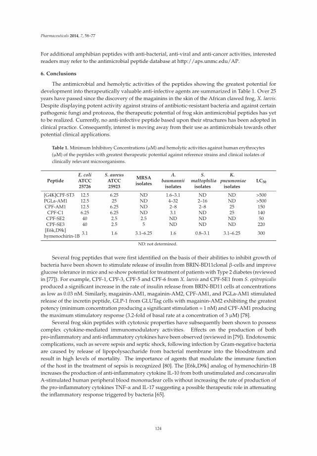
Pharmaceuticals 2014, 7, 58–77
For additional amphibian peptides with anti-bacterial, anti-viral and anti-cancer activities, interestedreaders may refer to the antimicrobial peptide database at http://aps.unmc.edu/AP.
6. Conclusions
The antimicrobial and hemolytic activities of the peptides showing the greatest potential fordevelopment into therapeutically valuable anti-infective agents are summarized in Table 1. Over 25years have passed since the discovery of the magainins in the skin of the African clawed frog, X. laevis.Despite displaying potent activity against strains of antibiotic-resistant bacteria and against certainpathogenic fungi and protozoa, the therapeutic potential of frog skin antimicrobial peptides has yetto be realized. Currently, no anti-infective peptide based upon their structures has been adopted inclinical practice. Consequently, interest is moving away from their use as antimicrobials towards otherpotential clinical applications.
Table 1. Minimum Inhibitory Concentrations (μM) and hemolytic activities against human erythrocytes(μM) of the peptides with greatest therapeutic potential against reference strains and clinical isolates ofclinically relevant microorganisms.
PeptideE. coliATCC25726
S. aureusATCC25923
MRSAisolates
A.baumannii
isolates
S.maltophilia
isolates
K.pneumoniae
isolatesLC50
[G4K]CPF-ST3 12.5 6.25 ND 1.6–3.1 ND ND >500PGLa-AM1 12.5 25 ND 4–32 2–16 ND >500CPF-AM1 12.5 6.25 ND 2–8 2–8 25 150
CPF-C1 6.25 6.25 ND 3.1 ND 25 140CPF-SE2 40 2.5 2.5 ND ND ND 50CPF-SE3 40 2.5 5 ND ND ND 220
[E6k,D9k]hymenochirin-1B 3.1 1.6 3.1–6.25 1.6 0.8–3.1 3.1–6.25 300
ND: not determined.
Several frog peptides that were first identified on the basis of their abilities to inhibit growth ofbacteria have been shown to stimulate release of insulin from BRIN-BD11clonal β-cells and improveglucose tolerance in mice and so show potential for treatment of patients with Type 2 diabetes (reviewedin [77]). For example, CPF-1, CPF-3, CPF-5 and CPF-6 from X. laevis and CPF-SE1 from S. epitropicalisproduced a significant increase in the rate of insulin release from BRIN-BD11 cells at concentrationsas low as 0.03 nM. Similarly, magainin-AM1, magainin-AM2, CPF-AM1, and PGLa-AM1 stimulatedrelease of the incretin peptide, GLP-1 from GLUTag cells with magainin-AM2 exhibiting the greatestpotency (minimum concentration producing a significant stimulation = 1 nM) and CPF-AM1 producingthe maximum stimulatory response (3.2-fold of basal rate at a concentration of 3 μM) [78].
Several frog skin peptides with cytotoxic properties have subsequently been shown to possesscomplex cytokine-mediated immunomodulatory activities. Effects on the production of bothpro-inflammatory and anti-inflammatory cytokines have been observed (reviewed in [79]). Endotoxemiccomplications, such as severe sepsis and septic shock, following infection by Gram-negative bacteriaare caused by release of lipopolysaccharide from bacterial membrane into the bloodstream andresult in high levels of mortality. The importance of agents that modulate the immune functionof the host in the treatment of sepsis is recognized [80]. The [E6k,D9k] analog of hymenochirin-1Bincreases the production of anti-inflammatory cytokine IL-10 from both unstimulated and concanavalinA-stimulated human peripheral blood mononuclear cells without increasing the rate of production ofthe pro-inflammatory cytokines TNF-α and IL-17 suggesting a possible therapeutic role in attenuatingthe inflammatory response triggered by bacteria [65].
124

Pharmaceuticals 2014, 7, 58–77
Acknowledgments: The work carried out in the authors’ laboratory was supported by grants from U.A.E.University and the Terry Fox Fund for Cancer Research.
Conflicts of Interest: The authors declare no conflict of interest.
References
1. Savard, P.; Perl, T.M. A call for action: Managing the emergence of multidrug-resistant Enterobacteriaceae inthe acute care settings. Curr. Opin. Infect. Dis. 2012, 25, 371–377. [CrossRef]
2. Conlon, J.M. The contribution of skin antimicrobial peptides to the system of innate immunity in anurans.Cell Tissue Res. 2011, 343, 201–212. [CrossRef]
3. Conlon, J.M. Structural diversity and species distribution of host-defense peptides in frog skin secretions.Cell. Mol. Life Sci. 2011, 68, 2303–2315. [CrossRef]
4. Yeung, A.T.Y.; Gellatly, S.L.; Hancock, R.E. Multifunctional cationic host defence peptides and their clinicalapplications. Cell. Mol. Life Sci. 2011, 68, 2161–2176. [CrossRef]
5. Gammill, W.M.; Fites, J.S.; Rollins-Smith, L.A. Norepinephrine depletion of antimicrobial peptides from theskin glands of Xenopus laevis. Dev. Comp. Immunol. 2012, 37, 19–27. [CrossRef]
6. Almeida, P.F.; Pokorny, A. Mechanisms of antimicrobial, cytolytic, and cell-penetrating peptides: Fromkinetics to thermodynamics. Biochemistry 2009, 48, 8083–8093. [CrossRef]
7. Huang, Y.; Huang, J.; Chen, Y. Alpha-helical cationic antimicrobial peptides: Relationships of structure andfunction. Protein Cell 2010, 1, 143–152. [CrossRef]
8. Tennessen, J.A.; Woodhams, D.C.; Chaurand, P.; Reinert, L.K.; Billheimer, D.; Shyr, Y.; Caprioli, R.M.;Blouin, M.S.; Rollins-Smith, L.A. Variations in the expressed antimicrobial peptide repertoire of northernleopard frog (Rana pipiens) populations suggest intraspecies differences in resistance to pathogens. Dev. Comp.Immunol. 2009, 33, 1247–1257. [CrossRef]
9. Conlon, J.M.; Al-Ghaferi, N.; Abraham, B.; Leprince, J. Strategies for transformation of naturally-occurringamphibian antimicrobial peptides into therapeutically valuable anti-infective agents. Methods 2007, 42,349–357. [CrossRef]
10. Jiang, Z.; Vasil, A.I.; Hale, J.D.; Hancock, R.E.; Vasil, M.L.; Hodges, R.S. Effects of net charge and the numberof positively charged residues on the biological activity of amphipathic alpha-helical cationic antimicrobialpeptides. Biopolymers 2008, 90, 369–383. [CrossRef]
11. Matsuzaki, K. Control of cell selectivity of antimicrobial peptides. Biochim. Biophys. Acta 2009, 1788,1687–1692. [CrossRef]
12. Frost, D.R. Amphibian Species of the World: An Online Reference, Version 5.6. 2013, Electronic Database.American Museum of Natural History: New York, USA. Available online: http://research.amnh.org/herpetology/amphibia/index.php (accessed on 13 January 2014).
13. Frost, D.R.; Grant, T.; Faivovich, J.; Bain, R.H.; Haas, A.; Haddad, C.F.B.; de Sá, R.O.; Channing, A.;Wilkinson, M.; Donnellan, S.C.; et al. The amphibian tree of life. Bull. Am. Mus. Nat. Hist. 2006, 297, 1–370.
14. Evans, B.J.; Kelley, D.B.; Tinsley, R.C.; Melnick, D.J.; Cannatella, D.C. A mitochondrial DNA phylogeny ofAfrican clawed frogs: Phylogeography and implications for polyploid evolution. Mol. Phylogenet. Evol. 2004,33, 197–213. [CrossRef]
15. Evans, B.J. Genome evolution and speciation genetics of clawed frogs (Xenopus and Silurana). Front. Biosci.2008, 13, 4687–4706. [CrossRef]
16. Irisarri, I.; Vences, M.; San Mauro, D.; Glaw, F.; Zardoya, R. Reversal to air-driven sound production revealedby a molecular phylogeny of tongueless frogs, family Pipidae. BMC Evol. Biol. 2011, 11. [CrossRef]
17. Bewick, A.J.; Chain, F.J.; Heled, J.; Evans, B.J. The pipid root. Syst. Biol. 2012, 61, 913–926. [CrossRef]18. Roelants, K.; Bossuyt, F. Archaeobatrachian paraphyly and pangaean diversification of crown- group frogs.
Syst. Biol. 2005, 54, 111–126. [CrossRef]19. Roelants, K.; Gower, D.J.; Wilkinson, M.; Loader, S.P.; Biju, S.D.; Guillaume, K.; Moriau, L.; Bossuyt, F. Global
patterns of diversification in the history of modern amphibians. Proc. Natl. Acad. Sci. USA 2007, 104, 887–892.20. Báez, A.M. The Fossil Record of the Pipidae. In The Biology of Xenopus; Tinsley, R.C., Kobel, H.R., Eds.;
Clarendon Press: Oxford, UK, 1996; pp. 329–347.21. Kobel, H.R.; Loumont, C.; Tinsley, R.C. The Extant Species. In The Biology of Xenopus; Tinsley, R.C., Kobel, H.R.,
Eds.; Clarendon Press: Oxford, UK, 1996; pp. 9–33.
125

Pharmaceuticals 2014, 7, 58–77
22. Evans, B.J.; Greenbaum, E.; Kusamba, C.; Carter, T.F.; Tobias, M.L.; Mendel, S.A.; Kelley, D.B. Description ofa new octoploid frog species (Anura: Pipidae: Xenopus) from the Democratic Republic of the Congo, with adiscussion of the biogeography of African clawed frogs in the Albertine Rift. J. Zool. 2011, 283, 276–290.
23. Tymowska, J.; Fischberg, M. A comparison of the karyotype, constitutive heterochromatin, and nucleolarorganizer regions of the new tetraploid species Xenopus epitropicalis Fischberg and Picard with those ofXenopus tropicalis Gray (Anura, Pipidae). Cytogenet. Cell Genet. 1982, 34, 49–157.
24. Zasloff, M. Magainins, a class of antimicrobial peptides from Xenopus skin: Isolation, characterization of twoactive forms and partial cDNA sequence of a precursor. Proc. Natl. Acad. Sci. USA 1987, 84, 5449–5453.
25. Gibson, B.W.; Poulter, L.; Williams, D.H.; Maggio, J.E. Novel peptide fragments originating from PGLa andthe caerulein and xenopsin precursors from Xenopus laevis. J. Biol. Chem. 1986, 261, 5341–5349.
26. Soravia, E.; Martini, G.; Zasloff, M. Antimicrobial properties of peptides from Xenopus granular glandsecretions. FEBS Lett. 1988, 228, 337–340. [CrossRef]
27. Hunt, L.T.; Barker, W.C. Relationship of promagainin to three other prohormones from the skin of Xenopuslaevis: A different perspective. FEBS Lett. 1988, 233, 282–288. [CrossRef]
28. Conlon, J.M.; Al-Ghaferi, N.; Ahmed, E.; Meetani, M.A.; Leprince, J.; Nielsen, P.F. Orthologs of magainin,PGLa, procaerulein-derived, and proxenopsin-derived peptides from skin secretions of the octoploid frogXenopus amieti (Pipidae). Peptides 2010, 31, 989–994. [CrossRef]
29. Mechkarska, M.; Ahmed, E.; Coquet, L.; Leprince, J.; Jouenne, T.; Vaudry, H.; King, J.D.; Takada, K.;Conlon, J.M. Genome duplications within the Xenopodinae do not increase the multiplicity of antimicrobialpeptides in Silurana paratropicalis and Xenopus andrei skin secretions. Comp. Biochem. Physiol. D GenomicsProteomics 2011, 6, 206–212. [CrossRef]
30. Mechkarska, M.; Ahmed, E.; Coquet, L.; Leprince, J.; Jouenne, T.; Vaudry, H.; King, J.D.; Conlon, J.M.Antimicrobial peptides with therapeutic potential from skin secretions of the Marsabit clawed frog Xenopusborealis (Pipidae). Comp. Biochem. Physiol. C Toxicol. Pharmacol. 2010, 152, 467–472. [CrossRef]
31. Conlon, J.M.; Mechkarska, M.; Ahmed, E.; Leprince, J.; Vaudry, H.; King, J.D.; Takada, K. Purification andproperties of antimicrobial peptides from skin secretions of the Eritrea clawed frog Xenopus clivii (Pipidae).Comp. Biochem. Physiol. C Toxicol. Pharmacol. 2011, 153, 350–354. [CrossRef]
32. King, J.D.; Mechkarska, M.; Coquet, L.; Leprince, J.; Jouenne, T.; Vaudry, H.; Takada, K.; Conlon, J.M.Host-defense peptides from skin secretions of the tetraploid frogs Xenopus petersii and Xenopus pygmaeus,and the octoploid frog Xenopus lenduensis (Pipidae). Peptides 2012, 33, 35–43. [CrossRef]
33. Mechkarska, M.; Ahmed, E.; Coquet, L.; Leprince, J.; Jouenne, T.; Vaudry, H.; King, J.D.; Conlon, J.M.Peptidomic analysis of skin secretions demonstrates that the allopatric populations of Xenopus muelleri(Pipidae) are not conspecific. Peptides 2011, 32, 1502–1508. [CrossRef]
34. King, J.D.; Mechkarska, M.; Meetani, M.A.; Conlon, J.M. Peptidomic analysis of skin secretions providesinsight into the taxonomic status of the African clawed frogs Xenopus victorianus and Xenopus laevis sudanensis(Pipidae). Comp. Biochem. Physiol. D Genomics Proteomics 2013, 8, 250–254. [CrossRef]
35. Mechkarska, M.; Meetani, M.; Michalak, P.; Vaksman, Z.; Takada, K.; Conlon, J.M. Hybridization betweenthe tetraploid African clawed frogs Xenopus laevis and Xenopus muelleri (Pipidae) increases the multiplicityof antimicrobial peptides in the skin secretions of female offspring. Comp. Biochem. Physiol. D GenomicsProteomics 2012, 7, 285–291. [CrossRef]
36. Mechkarska, M.; Prajeep, M.; Leprince, J.; Vaudry, H.; Meetani, M.A.; Evans, B.J.; Conlon, J.M. A comparisonof host-defense peptides in skin secretions of female Xenopus laevis × Xenopus borealis and X. borealis × X.laevis F1 hybrids. Peptides 2013, 45, 1–8. [CrossRef]
37. Ali, M.F.; Soto, A.; Knoop, F.C.; Conlon, J.M. Antimicrobial peptides isolated from skin secretions of thediploid frog, Xenopus tropicalis (Pipidae). Biochim. Biophys. Acta 2001, 1550, 81–89.
38. Roelants, K.; Fry, B.G.; Ye, L.; Stijlemans, B.; Brys, L.; Kok, P.; Clynen, E.; Schoofs, L.; Cornelis, P.; Bossuyt, F.Origin and functional diversification of an amphibian defense peptide arsenal. PLoS Genet. 2013, 9, e1003662.
39. Conlon, J.M.; Mechkarska, M.; Prajeep, M.; Sonnevend, A.; Coquet, L.; Leprince, J.; Jouenne, T.; Vaudry, H.;King, J.D. Host-defense peptides in skin secretions of the tetraploid frog Silurana epitropicalis with potentactivity against methicillin-resistant Staphylococcus aureus (MRSA). Peptides 2012, 37, 113–119. [CrossRef]
40. Mechkarska, M.; Prajeep, M.; Coquet, L.; Leprince, J.; Jouenne, T.; Vaudry, H.; King, J.D.; Conlon, J.M. Thehymenochirins: A family of antimicrobial peptides from the Congo dwarf clawed frog Hymenochirus boettgeri(Pipidae). Peptides 2012, 35, 269–275. [CrossRef]
126

Pharmaceuticals 2014, 7, 58–77
41. Conlon, J.M.; Prajeep, M.; Mechkarska, M.; Coquet, L.; Leprince, J.; Jouenne, T.; Vaudry, H.; King, J.D.Characterization of the host-defense peptides from skin secretions of Merlin’s clawed frog Pseudhymenochirusmerlini: Insights into phylogenetic relationships among the Pipidae. Comp. Biochem. Physiol. D GenomicsProteomics 2013, 8, 352–357. [CrossRef]
42. Helmerhorst, E.J.; Reijnders, M.; van’t Hof, W.; Veerman, C.; Nieuw-Amerongen, A.V. A critical comparisonof the hemolytic and fungicidal activities of cationic antimicrobial peptides. FEBS Lett. 1999, 449, 105–110.
43. Imura, Y.; Choda, N.; Matsuzaki, K. Dagainin 2 in action: Distinct modes of membrane permeabilization inliving bacterial and mammalian cells. Biophys. J. 2008, 95, 5757–5765. [CrossRef]
44. Tamba, Y.; Ariyama, H.; Levadny, V.; Yamazaki, M. Kinetic pathway of antimicrobial peptide magainin2-induced pore formation in lipid membranes. J. Phys. Chem. B 2010, 114, 12018–12026. [CrossRef]
45. Epand, R.F.; Maloy, W.L.; Ramamoorthy, A.; Epand, R.M. Probing the “charge cluster mechanism” inamphipathic helical cationic antimicrobial peptides. Biochemistry 2010, 49, 4076–4084. [CrossRef]
46. Zasloff, M.; Martin, B.; Chen, H.C. Antimicrobial activity of synthetic magainin peptides and severalanalogues. Proc. Natl. Acad. Sci. USA 1988, 85, 910–913. [CrossRef]
47. Cuervo, J.H.; Rodriguez, B.; Houghten, R.A. The magainins: Sequence factors relevant to increasedantimicrobial activity and decreased hemolytic activity. Pept. Res. 1988, 1, 81–86.
48. Shin, S.Y.; Kang, J.H.; Lee, M.K.; Kim, S.Y.; Kim, Y.; Hahm, K.S. Cecropin A—Magainin 2 hybrid peptideshaving potent antimicrobial activity with low hemolytic effect. Biochem. Mol. Biol. Int. 1998, 44, 1119–1126.
49. Fuchs, P.C.; Barry, A.L.; Brown, S.D. In vitro antimicrobial activity of MSI-78, a magainin analog. Antimicrob.Agents Chemother. 1998, 42, 1213–1216.
50. Ge, Y.; MacDonald, D.L.; Holroyd, K.J.; Thornsberry, C.; Wexler, H.; Zasloff, M. In vitro antibacterial propertiesof pexiganan, an analog of magainin. Antimicrob. Agents Chemother. 1999, 43, 782–788.
51. Ge, Y.; MacDonald, D.; Henry, M.M.; Hait, H.I.; Nelson, K.A.; Lipsky, B.A.; Zasloff, M.A.; Holroyd, K.J.In vitro susceptibility to pexiganan of bacteria isolated from infected diabetic foot ulcers. Diagn. Microbiol.Infect. Dis. 1999, 35, 45–53. [CrossRef]
52. Lipsky, B.A.; Holroyd, K.J.; Zasloff, M. Topical versus systemic antimicrobial therapy for treating mildlyinfected diabetic foot ulcers: A randomized, controlled, double-blinded, multicenter trial of pexiganan cream.Clin. Infect. Dis. 2008, 47, 1537–1545. [CrossRef]
53. Iwahori, A.; Hirota, Y.; Sampe, R.; Miyano, S.; Takahashi, N.; Sasatsu, M.; Kondo, I.; Numao, N. On theantibacterial activity of normal and reversed magainin 2 analogs against Helicobacter pylori. Biol. Pharm. Bull.1997, 20, 805–808. [CrossRef]
54. Macias, E.A.; Rana, F.; Blazyk, J.; Modrzakowski, M.C. Bactericidal activity of magainin 2: Use oflipopolysaccharide mutants. Can. J. Microbiol. 1990, 36, 582–584. [CrossRef]
55. Genco, C.A.; Maloy, W.L.; Kari, U.P.; Motley, M. Antimicrobial activity of magainin analogues againstanaerobic oral pathogens. Int. J. Antimicrob. Agents 2003, 21, 75–78. [CrossRef]
56. Schuster, F.L.; Jacob, L.S. Effects of magainins on ameba and cyst stages of Acanthamoeba polyphaga. Antimicrob.Agents Chemother. 1992, 36, 1263–1271. [CrossRef]
57. Helmerhorst, E.J.; Reijnders, I.M.; van’t Hof, W.; Simoons-Smit, I.; Veerman, E.C.; Amerongen, A.V.Amphotericin B- and fluconazole-resistant Candida spp., Aspergillus fumigatus, and other newly emergingpathogenic fungi are susceptible to basic antifungal peptides. Antimicrob. Agents Chemother. 1999, 43, 702–704.
58. Westerhoff, H.V.; Zasloff, M.; Rosner, J.L.; Hendler, R.W.; de Waal, A.; Vaz Gomes, A.; Jongsma, P.M.;Riethorst, A.; Juretic, D. Functional synergism of the magainins PGLa and magainin-2 in Escherichia coli,tumor cells and liposomes. Eur. J. Biochem. 1995, 228, 257–264. [CrossRef]
59. Lohner, K.; Prossnigg, F. Biological activity and structural aspects of PGLa interaction with membranemimetic systems. Biochim. Biophys. Acta 2009, 1788, 1656–1666. [CrossRef]
60. Strandberg, E.; Zerweck, J.; Wadhwani, P.; Ulrich, A.S. Synergistic insertion of antimicrobial magainin-familypeptides in membranes depends on the lipid spontaneous curvature. Biophys. J. 2013, 104, L9–L11. [CrossRef]
61. Conlon, J.M.; Sonnevend, A.; Pál, T.; Vila-Farrés, X. Efficacy of six frog skin-derived antimicrobial peptidesagainst colistin-resistant strains of the Acinetobacter baumannii group. Int. J. Antimicrob. Agents 2012, 39,317–320. [CrossRef]
62. Conlon, J.M.; Galadari, S.; Raza, H.; Condamine, E. Design of potent, non-toxic antimicrobial agents basedupon the naturally occurring frog skin peptides, ascaphin-8 and peptide XT-7. Chem. Biol. Drug Des. 2008,72, 58–64. [CrossRef]
127

Pharmaceuticals 2014, 7, 58–77
63. Subasinghage, A.P.; Conlon, J.M.; Hewage, C.M. Development of potent anti-infective agents from Siluranatropicalis: Conformational analysis of the amphipathic, alpha-helical antimicrobial peptide XT-7 and itsnon-haemolytic analogue [G4K]XT-7. Biochim. Biophys. Acta 2010, 1804, 1020–1028. [CrossRef]
64. Moore, K.S.; Bevins, C.L.; Brasseur, M.M.; Tomassini, N.; Turner, K.; Eck, H.; Zasloff, M. Antimicrobialpeptides in the stomach of Xenopus laevis. J. Biol. Chem. 1991, 266, 19851–19857.
65. Mechkarska, M.; Prajeep, M.; Radosavljevic, G.D.; Jovanovic, I.P.; Al Baloushi, A.; Sonnevend, A.; Lukic, M.L.;Conlon, J.M. An analog of the host-defense peptide hymenochirin-1B with potent broad-spectrum activityagainst multidrug-resistant bacteria and immunomodulatory properties. Peptides 2013, 50, 153–159. [CrossRef]
66. Mader, J.S.; Hoskin, D.W. Cationic antimicrobial peptides as novel cytotoxic agents for cancer treatment.Expert Opin. Investig. Drugs 2006, 15, 933–946. [CrossRef]
67. Ohsaki, Y.; Gazdar, A.F.; Chen, H.C.; Johnson, B.E. Antitumor activity of magainin analogues against humanlung cancer cell lines. Cancer Res. 1992, 52, 3534–3538.
68. Lehmann, J.; Retz, M.; Sidhu, S.S.; Suttmann, H.; Sell, M.; Paulsen, F.; Harder, J; Unteregger, G.; Stöckle, M.Antitumor activity of the antimicrobial peptide magainin II against bladder cancer cell lines. Eur. Urol. 2006,50, 141–147. [CrossRef]
69. Cruciani, R.A.; Barker, J.L.; Zasloff, M.; Chen, H.C.; Colamonici, O. Antibiotic magainins exert cytolytic activityagainst transformed cell lines through channel formation. Proc. Natl. Acad. Sci. USA 1991, 88, 3792–3796.
70. Koszałka, P.; Kamysz, E.; Wejda, M.; Kamysz, W.; Bigda, J. Antitumor activity of antimicrobial peptidesagainst U937 histiocytic cell line. Acta. Biochim. Pol. 2011, 58, 111–117.
71. Baker, M.A.; Maloy, W.L.; Zasloff, M.; Jacob, L.S. Anticancer efficacy of magainin 2 and analogue peptides.Anticancer efficacy of magainin 2 and analogue peptides. Cancer Res. 1993, 53, 3052–3057.
72. Miyazaki, Y.; Aoki, M.; Yano, Y.; Matsuzaki, K. Interaction of antimicrobial peptide magainin 2 withgangliosides as a target for human cell binding. Biochemistry 2012, 51, 10229–10235. [CrossRef]
73. Attoub, S.; Arafat, H.; Mechkarska, M.; Conlon, J.M. Anti-tumor activities of the host-defense peptidehymenochirin-1B. Regul. Pept. 2013, 115, 141–149.
74. Albiol Matanic, V.C.; Castilla, V. Antiviral activity of antimicrobial cationic peptides against Junin virus andherpes simplex virus. Int. J. Antimicrob. Agents 2004, 23, 382–389. [CrossRef]
75. Chinchar, V.G.; Bryan, L.; Silphadaung, U.; Noga, E.; Wade, D.; Rollins-Smith, L. Inactivation of viruses infectingectothermic animals by amphibian and piscine antimicrobial peptides. Virology 2004, 323, 268–275. [CrossRef]
76. Dean, R.E.; O’Brien, L.M.; Thwaite, J.E.; Fox, M.A.; Atkins, H.; Ulaeto, D.O. A carpet-based mechanism fordirect antimicrobial peptide activity against vaccinia virus membranes. Peptides 2010, 31, 1966–1972. [CrossRef]
77. Srinivasan, D.; Mechkarska, M.; Abdel-Wahab, Y.H.; Flatt, P.R.; Conlon, J.M. Caerulein precursor fragment(CPF) peptides from the skin secretions of Xenopus laevis and Silurana epitropicalis are potent insulin-releasingagents. Biochimie 2013, 95, 429–435. [CrossRef]
78. Ojo, O.O.; Conlon, J.M.; Flatt, P.R.; Abdel-Wahab, Y.H. Frog skin peptides (tigerinin-1R, magainin-AM1,-AM2, CPF-AM1, and PGla-AM1) stimulate secretion of glucagon-like peptide 1 (GLP-1) by GLUTag cells.Biochem. Biophys. Res. Commun. 2013, 431, 14–18. [CrossRef]
79. Pantic, J.M.; Mechkarska, M.; Lukic, M.L.; Conlon, J.M. Effects of tigerinin peptides on cytokine productionby mouse peritoneal macrophages and spleen cells and by human peripheral blood mononuclear cells.Biochimie 2014, in press.
80. Kotsaki, A.; Giamarellos-Bourboulis, E.J. Emerging drugs for the treatment of sepsis. Expert Opin. Emerg.Drugs 2012, 17, 379–391. [CrossRef]
© 2014 by the authors. Licensee MDPI, Basel, Switzerland. This article is an open accessarticle distributed under the terms and conditions of the Creative Commons Attribution(CC BY) license (http://creativecommons.org/licenses/by/4.0/).
128

pharmaceuticals
Review
Chapter 7:
Antimicrobial Peptides in Reptiles
Monique L. van Hoek
National Center for Biodefense and Infectious Diseases, and School of Systems Biology,George Mason University, MS1H8, 10910 University Blvd, Manassas, VA 20110, USA;[email protected]; Tel.: +1-703-993-4273; Fax: +1-703-993-7019
Received: 6 March 2014; in revised form: 9 May 2014; Accepted: 12 May 2014; Published: 10 June 2014
Abstract: Reptiles are among the oldest known amniotes and are highly diverse in their morphologyand ecological niches. These animals have an evolutionarily ancient innate-immune system that isof great interest to scientists trying to identify new and useful antimicrobial peptides. Significantwork in the last decade in the fields of biochemistry, proteomics and genomics has begun to revealthe complexity of reptilian antimicrobial peptides. Here, the current knowledge about antimicrobialpeptides in reptiles is reviewed, with specific examples in each of the four orders: Testudines (turtlesand tortosises), Sphenodontia (tuataras), Squamata (snakes and lizards), and Crocodilia (crocodilans).Examples are presented of the major classes of antimicrobial peptides expressed by reptiles includingdefensins, cathelicidins, liver-expressed peptides (hepcidin and LEAP-2), lysozyme, crotamine, andothers. Some of these peptides have been identified and tested for their antibacterial or antiviralactivity; others are only predicted as possible genes from genomic sequencing. Bioinformatic analysisof the reptile genomes is presented, revealing many predicted candidate antimicrobial peptides genesacross this diverse class. The study of how these ancient creatures use antimicrobial peptides withintheir innate immune systems may reveal new understandings of our mammalian innate immunesystem and may also provide new and powerful antimicrobial peptides as scaffolds for potentialtherapeutic development.
Keywords: antimicrobial peptides; antibacterial; reptile; biofilm; broad-spectrum; Gram-positive;Gram-negative
1. Introduction
Reptiles are among the oldest amniotes. They are cold-blooded (ectothermic) vertebrates withdry and scaly skin that usually lay soft-shelled eggs with amniotic membranes. They thrive in diverseenvironments, ranging as far north as Hudson’s Bay in Canada, and as far south as Cape Horn, Chile.They range from very small geckos to enormous crocodiles and have survived millennia of evolution.They are highly adapted to their environment, including their innate immune systems, allowing themto be such successful animals. Antimicrobial peptides are part of the innate immune system, and maycontribute to the survival of these animals in microbe-filled, challenging environment. This paperwill review the known and predicted antimicrobial peptides of reptiles. A set of known host defenseantimicrobial peptides is collected in the Antimicrobial Peptide Database [1,2] and the examples fromreptiles have been annotated (Table 1). Recently, many reptilian genomes have been sequenced. Fromthese genomes, potential antimicrobial peptide genes have been predicted here by bioinformaticsanalysis (Tables 2–8), which should be synthesized and tested for activity in future work. Overall,the Reptile class thrives in diverse and challenging environments, partly due to their robust innate
Pharmaceuticals 2014, 7, 723–753 129 www.mdpi.com/journal/pharmaceuticals

Pharmaceuticals 2014, 7, 723–753
immune system, and our hypothesis is that antimicrobial peptides contribute to the evolutionarysuccess of reptiles as they do in mammals and other species.
Table 1. Known reptile antimicrobial peptides identified in the Antimicrobial Peptide Database(APD2) [2].
Peptide name SequenceAPD
IdentifiedSource
OrganismCommentReference
Activity (*)
Cathelicidin
OH-CATH KRFKKFFKKLKNSVKKRAKKFFKKPRVIGVSIPF AP00895 O. Hannah
(Snake)
Derivatives:OH-CATH30;OH-CM6 [3,4]
G+, G−
DerivativeOH-CATH30
KFFKKLKNSVKKRAKKFFKKPRVIGVSIPF [5,6] G+, G−
DerivativeOH-CM6 KFFKKLKKAVKKGFKKFAKV [3,4] G+, G−
BF-CATH KRFKKFFKKLKKSVKKRAKKFFKKPRVIGVSIPF AP00896
Bungarusfasciatus(Snake)
[7] G+, G−, F,Cancer cells
Derivative BF-30 KFFRKLKKSVKKRAKEFFKKPRVIGVSIPF AP01239 B. fasciatus [8–10] G+, G−
Derivative BF-15 KFFRKLKKSVVKRFK B. fasciatus [8] G+, G−NA-CATH KRFKKFFKKLKNSVKKRA
KKFFKKPKVIGVTFPF AP00897 N. atra (Snake) [3] G+, G−
Waprin
OmwaprinKDRPKKPGLCPPRPQKPCVK
ECKNDDSCPGQQKCCNYGCKDECRDPIFVG
AP01589Oxyuranus
microlepidotus(Snake)
4S = S[11,12] G+
Proline Rich
Lethal peptideI/Waglerin
GGKPDLRPCHPPCHYIPRPKPR AP00238
Trimeresuruswagleri,
Wagler’s pitviper (Snake)
P24335,[13] Tx
β-Defensin or defensin-like
TBD-1 (Turtleβ-defensin 1)
YDLSKNCRLRGGICYIGKCPRRFFRSGSCSRGNVCCLRFG AP01380 Turtle 3S = S
[14] G+, G−, F
Pelovaterin DDTPSSRCGSGGWGPCLPIVDLLCIVHVTVGCSGGFGCCRIG AP01381 Turtle
2JR3BBBh2o[15,16]
G−
TEWP (turtleegg-white protein)
EKKCPGRCTLKCGKHERPTLPYNCGKYICCVPVKVK AP01382 Turtle
2B5BXXQ
[17–19]G-, V
Crotamine defensin-like toxin
Crotamine YKQCHKKGGHCFPKEKICLPPSSDFGKMDCRWRWKCCKKGSG AP01650 Snake Venom
1Z99,3S = SZZP
[20,21]
G+, G−, F, P,Mammalianand Cancer
cells
(*) Activity abbreviations: G+, G−, inhibiting both G+ and G− bacteria; G+, Gram-positive bacteria only, G−,Gram-negative bacteria only; V, antiviral; F, antifungal; P: antiparasitic, Tx: Toxin activity to mammals.
2. Four Orders of Reptiles
The structural and sequence diversity of antimicrobial peptides is impressive (see below), but itis useful to first review the phylogenetic and physiological diversity of the Reptilia class in order toappreciate all the ecosystems and environments that they live in and the prey that they eat.
All living reptiles fall within the kingdom Animalia, the phylum Chordata, the class Reptilia,and the clade Sauropsida [22]. There are four orders within the class Reptilia: turtles and tortoises(Testudines), tuataras (Sphenodontia), snakes and lizards (Squamata), and crocodilans (Crocodilia).Each order will be briefly introduced below. Many reptile species are endangered, including the paintedturtle, and the Siamese crocodile. Although considered by phylogenetic analysis to be “monophyletic”with avians (Sauropsida; See Cladogram, Figure 1), reptiles are still generally considered to be separate
130

Pharmaceuticals 2014, 7, 723–753
from birds. Some researchers use the term avian reptile and non-avian reptile to distinguish the twogroups [23]. Within the non-avian reptilians, the crocodilians are considered to be the most closelyrelated to the avian branch. Interestingly, this evolutionary connection is reflected in the antimicrobialprofile in that neither avians nor reptilians encode α-defensin antimicrobial peptides, for example,which are a critical part of mammalian innate immunity. In contrast, avians do not appear to expresshepcidin peptides, while reptiles do, highlighting a potential difference.
Figure 1. Cladogram showing the relationships of extant members of the Sauria (Sauropsida) whichincludes birds and reptiles. Branch lengths are not representative of divergence time. 1. Tuataras;2. Lizards; 3. Snakes; 4. Crocodiles; 5. Birds. Cladogram by Benchill, licensed under the CreativeCommons Attribution 3.0 Unported license [24].
2.1. Testudines (Turtles and Tortises)
The testudine order includes the turtles and tortosises. The western painted turtle (Chrysemyspicta bellii) is a small turtle of North America, commonly found in ponds and other slow-moving freshwater [25].
(a) (b)
Figure 2. Western Painted Turtle Chrysemys picta bellii. (a) Western painted turtle. Photo by Gary M.Stolz, U.S. Fish and Wildlife Service in the Public domain [26]. (b) Underside of a Western PaintedTurtle. Photo by Matt Young [27].
This intensely colorful turtle (Figure 2) has existed for approximately 15 million years, accordingto the fossil record [25]. The genomes of three turtles have recently been sequenced, including the greensea turtle (Chelonia mydas) and the Chinese softshell turtle Pelodiscus sinensis [28], as well as the westernpainted turtle, Chrysemys picta bellii [29,30]. Interestingly, these papers place the turtles as a sister groupto crocodiles and birds, which is different than their prior morphological classification [31]. The precise
131
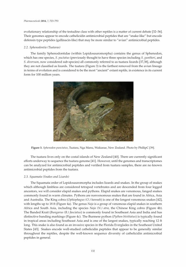
Pharmaceuticals 2014, 7, 723–753
evolutionary relationship of the testudine class with other reptiles is a matter of current debate [32–36].Their genomes appear to encode cathelicidin antimicrobial peptides that are “snake-like” but encodedefensin type peptides (gallinacin-like) that may be more similar to “avian” antimicrobial peptides.
2.2. Sphenodontia (Tuataras)
The family Sphenodontidae (within Lepidosauromorpha) contains the genus of Sphenodon,which has one species, S. puctatus (previously thought to have three species including S. guntheri, andS. diversum, now considered sub-species) all commonly referred to as tuatara lizards [37,38], althoughthey are not classified as lizards. The tuatara (Figure 3) is the farthest removed from the avian lineagein terms of evolution and is considered to be the most “ancient” extant reptile, in existence in its currentform for 100 million years.
Figure 3. Sphenodon punctatus, Tuatara, Nga Manu, Waikanae, New Zealand. Photo by PhillipC [39].
The tuatara lives only on the costal islands of New Zealand [40]. There are currently significantefforts underway to sequence the tuatara genome [41]. However, until the genomes and transcriptomescan be analyzed for antimicrobial peptides and verified from tuatara samples, there are no knownantimicrobial peptides from the tuatara.
2.3. Squamata (Snakes and Lizards)
The Squamata order of Lepidosauromorpha includes lizards and snakes. In the group of snakeswhich although limbless are considered tetrapod vertebrates and are descended from four leggedancestors, we will consider elapid snakes and pythons. Elapid snakes are venomous, fanged snakescommonly found in warm climates. Pythons are nonvenomous snakes that are found in Africa, Asiaand Australia. The King cobra (Ophiophagus (O.) hannah) is one of the longest venomous snakes [42],with lengths up to 18 ft (Figure 4a). The genus Naja is a group of venomous elapid snakes in southernAfrica and South Asia, including the species Naja (N.) atra, the Chinese King cobra (Figure 4b).The Banded Krait (Bungarus (B.) fasciatus) is commonly found in Southeast Asia and India and hasdistinctive banding markings (Figure 4c). The Burmese python (Python bivittatus) is typically foundin tropical areas including Southeast Asia and is one of the largest snakes, typically reaching 12 ftlong. This snake is also found as an invasive species in the Florida Everglades in the Southeast UnitedStates [43]. Snakes encode well-studied cathelicidin peptides that appear to be generally similarthroughout the reptiles, despite the well-known sequence diversity of cathelicidin antimicrobialpeptides in general.
132
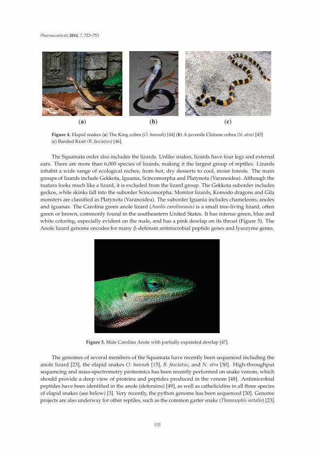
Pharmaceuticals 2014, 7, 723–753
(a) (b) (c)
Figure 4. Elapid snakes (a) The King cobra (O. hannah) [44] (b) A juvenile Chinese cobra (N. atra) [45](c) Banded Krait (B. fasciatus) [46].
The Squamata order also includes the lizards. Unlike snakes, lizards have four legs and externalears. There are more than 6,000 species of lizards, making it the largest group of reptiles. Lizardsinhabit a wide range of ecological niches, from hot, dry desserts to cool, moist forests. The maingroups of lizards include Gekkota, Iguania, Scincomorpha and Platynota (Varanoidea). Although thetuatara looks much like a lizard, it is excluded from the lizard group. The Gekkota suborder includesgeckos, while skinks fall into the suborder Scincomorpha. Monitor lizards, Komodo dragons and Gilamonsters are classified as Platynota (Varanoidea). The suborder Iguania includes chameleons, anolesand iguanas. The Carolina green anole lizard (Anolis carolinensis) is a small tree-living lizard, oftengreen or brown, commonly found in the southeastern United States. It has intense green, blue andwhite coloring, especially evident on the male, and has a pink dewlap on its throat (Figure 5). TheAnole lizard genome encodes for many β-defensin antimicrobial peptide genes and lysozyme genes.
Figure 5. Male Carolina Anole with partially expanded dewlap [47].
The genomes of several members of the Squamata have recently been sequenced including theanole lizard [23], the elapid snakes O. hannah [15], B. fasciatus, and N. atra [30]. High-throughputsequencing and mass-spectrometry proteomics has been recently performed on snake venom, whichshould provide a deep view of proteins and peptides produced in the venom [48]. Antimicrobialpeptides have been identified in the anole (defensins) [49], as well as cathelicidins in all three speciesof elapid snakes (see below) [3]. Very recently, the python genome has been sequenced [30]. Genomeprojects are also underway for other reptiles, such as the common garter snake (Thamnophis sirtalis) [23].
133

Pharmaceuticals 2014, 7, 723–753
2.4. Crocodilia (Crocodilians)
The order of crocodilians (Crocodilia) within the group Archosauromorpha includes alligatorsand caimans (family Alligatoridae), crocodiles (family Crocodylidae), and gharials (gavials, familyGavialidae) (Figure 6). Most species live in fresh-water, but a few have also adapted to salt-waterconditions. These animals are evolutionarily ancient, and along with birds, are considered the onlysurviving relatives of dinosaurs (Archosauria). Within their semi-aquatic ecosystems, these largeanimals are the apex predators, using their powerful jaws to capture large and small prey [50]. They arecold-blooded and egg-laying. They can be found around the world, in the Americas, in Asia, SouthAmerica, and Africa [51]. The ancestors of alligators and crocodiles first emerged in the Late Cretaceousera, and the modern alligators and crocodiles are estimated to be as much as 83 million years old.
(a) (b)
Figure 6. Crocodilians. (a) The American alligator, Alligator mississippiensis [52]. (b) The Siamesecrocodile, Crocodylus siamesnsis [53].
Today, the American alligator is commonly found all around the Gulf of Mexico and SoutheasternUnited States, having made a comeback from near extinction due to overhunting. The meat and skinsof farmed alligators are now harvested for consumption and use. This large animal is typically 8–11ft long and 200–500 lbs depending on gender and age [54]. The alligator has a broad snout and thetop teeth come down over the bottom lips, unlike the crocodile. Alligators inhabit swamps such asthe Great Dismal Swamp, rivers and streams, ponds and lakes, with a preference for fresh water overbrackish water, and intolerance for salt water. Alligators can be infected by Mycoplasma alligatoris [55]as well as other bacterial pathogens. In terms of temperature preference, alligators are more coldtolerant than most other crocodilian species, accounting for their location as far north as South Carolinain the United States [54].
The Siamese crocodile (Crocodylus siamensis) [56,57] is a critically endangered freshwatercrocodilian found in Southeast Asia (Cambodia, Vietnam, Thailand, Indonesia and Burma for example).Like most crocodiles, it is a tropical animal with low tolerance for the cold [3]. This animal is smallerthan the alligator, typically about 7 ft long and 80–150 lbs when fully grown, although larger specimenshave been recorded [4]. These animals are being intensively studied as part of their conservation, andare bred in captivity, and thus are accessible to researchers for DNA or blood samples [58–60].
Crocodilians such as alligators can carry a high burden of fecal coliforms from their aquaticenvironment [61], yet despite their potentially near-constant exposure to potential pathogens, alligatorsand other crocodilians do not seem to be susceptible to infection by these organisms, either systemicallyor on their skin, via wounds or lesions. This has led to the idea that these animals may have highly potentantimicrobial components to their immune systems [57,62–67]. Several crocodilian genomes have beenpublished [68] or are underway [69]. The peptides that have been discovered in crocodilians are outlinedin the sections below and include lysozyme, defensin, hepcidin, hemocidin and other antimicrobial
134

Pharmaceuticals 2014, 7, 723–753
peptides. Several important crocodilian genomes have been sequenced, including the Chinese alligator(Alligator sinensis), the American alligator (Alligator mississippiensis), the saltwater crocodile (Crocodylusporosus) and the Indian gharial (Gavialis gangeticus) [69,70]. It will be very interesting to comparefreshwater species to saltwater species in terms of their innate immune response profiles, as theenvironmental organisms will be significantly different between the two types of water.
3. Antimicrobial Peptides of Reptiles
Reptiles are highly adapted to their environments, which often include many bacteria, allowingthem to be such successful animals. Antimicrobial peptides are part of the innate immune system, andmay contribute to the survival of reptiles. However, to date there has been no direct data in reptiles suchas gene knock-out experiments to demonstrate their importance to overall survival, development andresistance to infection, as has been done in mice [71]. There has been some suggestion that β-defensinpeptides are associated with fur color in dogs [72,73], but again no studies of this kind have yet beendone on reptiles. This area of research presents many opportunities for scientists to explore the role ofantimicrobial peptides in these animals. This paper will review the known (Table 1) and predictedantimicrobial peptides of reptiles. Recently, new reptilian genomes have been sequenced. From thesegenomes, additional antimicrobial peptide genes have been predicted here by bioinformatics analysis(Tables 2–8), which should be synthesized and tested for activity.
Antimicrobial peptides are well-characterized peptides, and exhibit significant structure-functionspecificity. Following the identification of magainin peptides in amphibians in 1987 [74], scientists havebeen exploring the diversity of peptides expressed in different animals, looking for new structures andnew functions of antimicrobial peptides. The structure and function of each of the major classes ofantimicrobial peptides is described below, in addition to the data pertaining to its known or predictedexpression in reptiles.
3.1. Defensin Peptides in Reptiles
Defensins are one of the major classes of antimicrobial peptides in higher vertebrates. These 3–4kDa cationic peptides are characterized by having six cysteines arranged in three disulfide bonds, withthe characteristic pairing of the bonds highly characteristic for each type of defensin. Defensins havepredominantly β-sheet characteristic with some α-helices. Defensins are encoded in the genome andare processed from a pro-defensin molecule by proteases [75].
3.1.1. Three Sub-Classes of Defensins
Defensins are known to be critical components of innate immunity in many animals [76]. The threemain sub-classes of defensins are α, β- and θ-defensins. In humans, α-defensins are commonly foundin neutrophils and other leukocytes (for example, Human Neutrophil peptide HNP-1 = α-defensin 1),and are important in the ability of white blood cells to deal with pathogens [77]. The β-defensins aredefined as having a Cys1-Cys5, Cys2-Cys4, and Cys3-Cys6 bonding pattern of cysteines. In humans,β-defensins are commonly expressed in epithelial cells, and are widely expressed in the body [78].The expression of these cationic antimicrobial peptides are often induced following bacterial or viralinfection as part of the innate immune response, except for hBD1 which appears to be constitutivelyexpressed in humans. The third class of defensins is the θ-defensins. θ-Defensins are not knownoutside of primates, and are not expressed in humans [21] but are very active antiviral peptides [76].
Within the reptiles, there are no known genes for α-defensins, and only the β-defensins appear tobe expressed, similar to what is found in avians. Within some species of reptiles, β-defensin genesand peptides have been identified, for example in the red-eared slider turtle [79]. Lizards are knownto be highly resistant to infection, and recently genes encoding up to 32 different β-defensin-likepeptides were identified in the Anole carolinensis genome [49]. Indeed, using an antibody that reactsto AcBD15 (one of those β-defensin-like peptides in anole), staining was observed in some (but notall) of the granules of heterophilic and basophilic granulocytes in lizards, a snake, a turtle and the
135

Pharmaceuticals 2014, 7, 723–753
tuatara, but not alligator nor chicken granulocytes. Cells from other tissues such as epidermis werenegative for staining. Alibardi et al. describe that not all the granules within a granulocyte stainedwith the antibody, suggesting that there may be different types of granules as is seen in mammalianneutrophils [22].
Similarly, turtle leukocytes were found to contain TBD-1, the first β-defensin identified in reptileleukocytes [14]. In the turtle system, TBD-1 was also identified in other tissues, such as the skin [18,80,81]. These fascinating and unique results suggest that further study of the antimicrobial peptidome ofreptilian granulocytes should be performed.
3.1.2. Inducible Expression of β-Defensins in Wounded Lizards
The first extensive report of an in vivo role for β-defensin peptide expression was in the anolelizard (Table 2). It has long been known that lizards can lose their tails as a method of predator escape,and that these tail then regenerate from the wound site. In this process, a wound is formed, which doesnot typically get infected. β-Defensin peptides are found to be expressed both within the azurophilicgranulocytes in the wound-bed as well as in the associated epithelium [82,83], and are observed inphagosomes containing degraded bacteria. While there is a distinct lack of inflammation in the wound,which is associated with regeneration, there is a high level of expression of AcBD15 and AcBD27(two of the most highly expressed β-defensins in that tissue) [84,85]. Overall, there appears to be afascinating role of AcBD15 and AcBD27 in the wound healing and regeneration in the anole lizard.
Table 2. Predicted defensin-like protein genes in multiple reptilian species.
Organism Peptide annotation aa Locus- Accession #Alligator mississippiensis Gallinacin-14-like 58 XP_006270781.1
Alligator sinensis Gallinacin-14-like 58 XP_006033878.1
Anolis carolinensis β-Defensin-like protein 5 62 CBY85058.1Anolis carolinensis β-Defensin-like protein 8 65 CBY85059.1Anolis carolinensis β-Defensin-like protein 9 66 CBY85060.1Anolis carolinensis β-Defensin-like protein 10 67 CBY85061.1Anolis carolinensis β-Defensin-like protein 15 63 CCA62931.1Anolis carolinensis β-Defensin-like protein 21 89 CBY85062.1Anolis carolinensis β-Defensin-like protein 22 95 CBY85063.1Anolis carolinensis β-Defensin-like protein 27 81 CBY85064.1Anolis carolinensis Gallinacin-10-like 68 XP_003225602.1Anolis carolinensis Gallinacin-13-like 60 XP_003225598.1Anolis carolinensis Hypothetical protein LOC100555370 XP_003227809.1Anolis carolinensis Hypothetical protein LOC100555565 XP_003227810.1Anolis carolinensis Hypothetical protein LOC100555756 XP_003227811.1Anolis carolinensis Hypothetical protein LOC100562305 XP_003225604.1Anolis carolinensis Hypothetical protein LOC100562502 65 XP_003225605.1Anolis carolinensis Hypothetical protein LOC100562898 XP_003225607.1Anolis carolinensis Hypothetical protein LOC100563098 XP_003225608.1
Chrysemys picta bellii β-Defensin 1-like 80 XP_005308390.1Chrysemys picta bellii Gallinacin-5-like XP_005290738.1Chrysemys picta bellii Gallinacin-5-like, partial XP_005314963.1Chrysemys picta bellii Gallinacin-14-like 58 XP_005308403.1
Pelodiscus sinensis Gallinacin-1 α-like XP_006137072.1Pelodiscus sinensis Lingual antimicrobial peptide-like isoform X2 XP_006127561.1
Bothrops neuwiedi β-Defensin-like protein AGF25392.1Bothrops jararacussu β-Defensin-like protein AGF25388.1
Bothrops leucurus β-Defensin-like protein AGF25389.1Bothrops matogrossensis β-Defensin-like protein AGF25391.1, AGF25390.1
Bothrops diporus β-Defensin-like protein AGF25384.1Bothrops pauloensis β-Defensin-like protein AGF25393.1Bothrops jararaca β-Defensin-like protein AGF25386.1, AGF25387.1
Bothrops atrox β-Defensin-like protein AGF25383.1Bothrops erythromelas β-defensin-like protein AGF25385.1
136

Pharmaceuticals 2014, 7, 723–753
3.1.3. Expression of β-Defensins in Reptile Eggs
Reptile eggs are a good biological sample for the purification of peptides and proteins, sincethere is so much material within each egg. Recently, it was found that while the β-defensin-likepeptide pelovaterin (Table 1) identified in the eggshell of the Chinese soft-shelled turtle does indeedhave antimicrobial activity, these peptides may also play an additional role in the formation of theeggshell, through aggregation [15]. This is similar to the role of the gallin defensin-like peptide inavian eggs [86–90].
3.2. Cathelicidin Peptides in Reptiles
Cathelicidins are a second major class of antimicrobial peptides in higher eukaryotes. They arecharacterized as being antimicrobial peptides derived by proteolytic cleavage from a pre-propeptidethat includes the cathelin domain in mammals and less-well conserved cathelin domain in othereukaryotes [91,92]. In humans, cathelicidins are stored in the azurophilic granules of neutrophils as theinactive prepropeptide, and are processed by enzymes (neutrophil elastase [93] or a serine protease [19])to the mature active peptide [94]. In humans and higher vertebrates, the active cathelicidin peptide isalways encoded on Exon 4 of the cathelicidin encoding gene [91,95,96]. Four cathelicidin-like peptideshave been identified in the chicken (Gallus gallus domesticus) [97], including fowlicidin-1, -2 and -3(also known as chCATH-1, chCATH-2/CMAP27, chCATH-3) [98], and chCATH-B1/chCATH-4 [99].In humans, the hCAP18 cathelicidin is processed by proteinase 3 inside the granule [19] or neutrophilelastase in the extracellular space [4] to its active form. In the case of chicken cathelicidin, thepre-pro-cathelin domain peptide is cleaved by a serine protease to release the mature peptide followingstimulation of heterophils with LPS [100].
3.3. Cathelicidin Peptides in Snakes
Cathelicidin peptides have been identified and characterized in the snake family (Table 1) [1,2],although there may be similar genes in other reptiles by genomic analysis (see below). Highly relatedcathelicidins were identified in B. fasciatus, O. hannah and N. atra [3] (Table 3). We identified additionalgenes for antimicrobial peptides by BLAST searching the genomes of the pit vipers (Bothrops atrox,Trimeresurus wagler and Crotalus durissus) [13] and the Eastern brown snake (Pseudonaja textilis) (Table 3).The genes of these cathelicidins have the general structure of the cathelicidin genes, including a poorlyconserved cathelin domain and the c-terminal active peptide. The snake cathelicidins have been wellstudied. The functional cathelicidin peptide of the snake family is highly divergent from the functionalcathelicidin peptide of humans, for example, confirming the “general rule” of cathelicidins, which isthat the active peptides are highly divergent, but the pre-pro-regions in the gene and the peptide aremore highly conserved [101].
3.3.1. King Cobra (Ophiophagus (O.) Hannah)
Zhao et al. determined the hemolytic and antimicrobial activity of the predicted O. hannahcathelicidin, OH-CATH [3] (Table 1). The OH-CATH peptide proved to be an excellent inhibitorof bacterial growth and demonstrated broad-spectrum activity against bacterial isolates includingmulti-drug resistant strains. The OH-CATH peptide displayed greater potency than the humancathelicidin LL-37 against a variety of known human bacterial pathogens and the antimicrobial potency ofOH-CATH was not significantly impacted by the concentration of salt in the media. They demonstratedthat OH-CATH showed no hemolytic activity against erythrocytes, even at a concentration of 200 μg/mL,suggesting low cytotoxicity of the peptide to eukaryotic cells [3]. Additional studies with smallerfragments of OH-CATH (Table 1) have also been tested and found to be active both in vitro and in vivo,even against antibiotic resistant bacteria [4,6]. Their strong antimicrobial activity and lack of hemolyticactivity make the reptile cathelicidins strong candidates for development into new therapeutics.
137
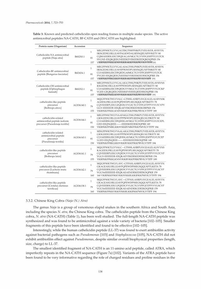
Pharmaceuticals 2014, 7, 723–753
Table 3. Known and predicted cathelicidin open reading frames in multiple snake species. The activeantimicrobial peptides NA-CATH, BF-CATH and OH-CATH are highlighted.
Protein name [Organism] Accession Sequence
Cathelicidin-NA antimicrobialpeptide [Naja atra] B6S2X0.1
1MEGFFWKTLLVVGALTISGTSSFPHKPLTYEEAVDLAVSVYNSKSGEDSLYRLLEAVPALKWDALSESNQELNFSVKETV 80
81CQMAEERSLEECDFQEAGAVMGCTGYYFFGESPPVLVLTCKSVGNE-EEQKQEEGNEEEKEVEKEEKEEDQKDQPKR 156
157 VVKRFKKFFKKLKNSVKKRAKKFFKKPKVIGVTFPF191
Cathelicidin-BF antimicrobialpeptide [Bungarus fasciatus] B6D434.1
1MEGFFWKTLLVVGALAIAGTSSLPHKPLIYEEAVDLAVSIYNSKSGEDSLYRLLEAVSPPKWDPLSESNQELNFTMKETV 80
81CLVAEERSLEECDFQEDGAIMGCTGYYFFGESPPVLVLTCKPVGEE-EEQKQEEGNEEEKEVEKEEKEEDEKDQPRR 156
157 VKRFKKFFRKLKKSVKKRAKEFFKKPRVIGVSIPF 191
Cathelicidin-OH antimicrobialpeptide [Ophiophagus
hannah]B6S2X2.1
1MEGFFWKTLLVVGALAIGGTSSLPHKPLTYEEAVDLAVSIYNSKSGEDSLYRLLEAVPPPEWDPLSESNQELNFTIKETV 80
81CLVAEERSLEECDFQEDGVVMGCTGYYFFGESPPVVVLTCKPVGEE-GEQKQEEGNEEEKEVEEEEQEEDEKDQPRR 156
157 VKRFKKFFKKLKNSVKKRAKKFFKKPRVIGVSIPF 191
cathelicidin-like peptideprecursor
[Bothrops atrox]AGS36140.1
1MQGFFWKTWLVVALC–GTSSSLAHRPLSYGEALELALSIYNSKAGEESLFRLLEAVPQPEWDPLSEGSQQLNFTIKETV 78
79CQVEEERPLEECGFQEDGVVLECTGYYFFGETPPVVVLTCVPVGGV-EEEEEDE-EEQKAEVEKDEKEDEEKDRPKR 154
155 VKRFKKFFKKLKNSVKKRVKKFFRKPRVIGVTFPF 189
cathelicidin-relatedantimicrobial peptide isoformprecursor [Pseudonaja textilis]
AGS36144.11
MEGFFWKTWLVVAAFAIGGTSSLPHKPLTYEEAVDLAVSTYNGKSGEESLYRLLEAVPPPKWDPLSESNQELNLTIKETV 80
81CLVAEERSLEECDFQDDGAVMGCTGYFFFGESPPVLVLTCEPLGED-EEQNQEEE——-EEEEKEEDEKDQPRR 149
150 VKRFKKFFMKLKKSVKKRVMKFFKKPMVIGVTFPF 184
cathelicidin-relatedantimicrobial peptide
precursor[Pseudonaja textilis]
AGS36143.1
1MDGFFWKTWLVVAALAIGGTSSLPHKPLTYEEAVDLAVSTYNGKSGEESLYRLLEAVPPPKWDPLSESNQELNLTIKETV 80
81CLVAEERSLEECDFQDDGAVMGCTGYFFFGESPPVLVLTCEPLGED-EEQNQEEE——-EEEEKEEDEKDQPRR 149
150 VKRFKKFFRKLKKSVKKRVKKFFKKPRVIGVTIPF 184
cathelicidin-like peptideprecursor
[Bothrops lutzi]AGS36141.1
1MQGFFWKTLLVVALC—GTSSSLAHRPLSYGEALELALSVYNSKAGEESLFRLLEAVPQPEWDPLSEGSQQLNFTIKETV 78
79CQVEEERPLEECGFQEDGVVLECTGYYFFGETPPVVVLTCVPVGGV-EEEEEDE-EEQKAEVEKDEKEDEEKDRPKR 154
155 VKRFKKFFKKLKNNVKKRVKKFFRKPRVIGVTIPF 189
cathelicidin-like peptideprecursor [Lachesis muta
rhombeata]AGS36142.1
1MQGFFWKTWLVLAVC–GTPASLAHRPLSYGEALELAVSVYNGKAGEASLYRLLEAVPQPEWDPSSEGSQQLNFTLKETA 78
79CQVEEERSLEECGFQEDGVVLECTGYYFFGETPPVVVLSCVPVGGVeEEEEEEE-EEQKAEAENDEKEDEEKDQPKR 159
160 160 VKRFKKFFKKVKKSVKKRLKKIFKKPMVIGVTFPF 194
cathelicidin-like peptideprecursor [Crotalus durissus
terrificus]AGS36138.1
1MQGFFWKTWLVLAVC—GTPASLAHRPLSYGEALELAVSVYNGKAGEASLYRLLEAVPQPEWDPSSEGSQQLNFTLKETA 78
79CQVEEERSLEECGFQEDGVVLECTGYYFFGETPPVVVLSCVPVGGVeEEEEEEE-EEQKAEAENDEKGDEEKDQPKR 159
160 VKRFKKFFKKVKKSVKKRLKKIFKKPMVIGVTIPF 194
3.3.2. Chinese King Cobra (Naja (N.) Atra)
The genus Naja is a group of venomous elapid snakes in the southern Africa and South Asia,including the species N. atra, the Chinese King cobra. The cathelicidin peptide from the Chinese Kingcobra, N. atra (NA-CATH) (Table 1), has been well studied. The full-length NA-CATH peptide wassynthesized and was found to be antimicrobial against a wide variety of bacteria [102–105]. Smallerfragments of this peptide have been identified and found to be effective [102–105].
Interestingly, while the human cathelicidin peptide (LL-37) was found to exert antibiofilm activityagainst bacterial pathogens such as Pseudomonas [103] and Staphylococcus [105], NA-CATH did notexhibit antibiofilm effect against Pseudomonas, despite similar overall biophysical properties (length,size, charge) to LL-37.
The smallest identified fragment of NA-CATH is an 11-amino acid peptide, called ATRA, whichimperfectly repeats in the NA-CATH sequence (Figure 7a) [102]. Variants of the ATRA peptide havebeen found to be very informative regarding the role of charged residues and proline residues in the
138

Pharmaceuticals 2014, 7, 723–753
activity of small antimicrobial peptides [102]. The peptide ATRA-1 is almost identical to the ATRA-2peptide, with the exception of the 3d (A/F) and 10th (L/P) residues. Variants of these peptides weremade to switch out the 3rd and 10th position amino acids, such that ATRA-1A contains alanine atposition 3 rather than phenylalanine (F). Another variant was ATRA-1P, which contains proline atposition 10 rather than leucine (L). All the peptides were the same length, similar molecular weight,and the same net charge. It was found for multiple gram-negative bacteria that ATRA-1P was almost asineffective as ATRA-2, while ATRA-1A was as effective as ATRA-1 [102]. These results suggested thatthe introduction of a proline in ATRA-1P may have affected the charge distribution on that peptide,which then may disrupt the antibacterial activity of the peptide (Figure 7b,c).
Further studies have examined the D-amino acid enantiomer of the ATRA peptides [106], andfound that D-ATRA-1A is as effective as ATRA-1A, and as it is protease resistant, potentially has a longerhalf-life. The ATRA peptides have also been demonstrated to be highly antimicrobial both in vitroand in vivo against both gram-negative bacteria such as Escherichia coli K12 strain and Aggregatibacteractinomycetemcomitans-Y4 [102], Pseudomonas aeruginosa [103] and Francisella novicida [104]. ATRApeptides are also antimicrobial against the gram-positive bacteria Staphylococcus aureus [105].
3.3.3. Banded Krait (B. fasciatus)
The cathelicidin peptide from B. fasciatus (BF-CATH, Table 1, Table 3) is also very well studied. Thefull-length BF-CATH peptide has been shown to have antimicrobial activity against a variety of bacteriaincluding antibiotic resistant bacteria [7,107]. Smaller fragments of BF-CATH have also been expressedand studied [10,108] (Table 1). For example, BF-30 showed significant activity against drug-resistantE. coli and S. aureus both in vitro and in vivo [109]. BF-15 is a shorter version of BF-CATH with lowerhemolytic activity, and broad-spectrum antimicrobial activity even against antibiotic-resistant bacteria [8].
Peptide Description Peptide Sequence NA-CATH Active Peptide KRFKKFFKKLKNSVKKRAKKFFKKPKVIGVTFPF
ATRA-1 KRFKKFFKKLK ATRA-2 ------------------------------- KRAKKFFKKPK
ATRA-1A KAFKKFFKKLK ATRA-1P KRFKKFFKKPK
(a) Sequence of the Chinese King Cobra Naja atra cathelicidin and ATRA derivatives.
(b) (c) (d)
Figure 7. Naja atra cathelicidin peptide analysis. (a) Sequences of the NA-CATH active peptide andderivatives [102–105]. (b) Helical wheel projection of NA-CATH. (c) Analysis of the active ATRA-1Apeptide (d) Analysis of the inactive ATRA-1P peptide. From Rzlab.ucr.edu/scripts/wheel/wheel.cgi:“The hydrophilic residues are presented as circles, hydrophobic residues as diamonds, potentiallynegatively charged as triangles, and potentially positively charged as pentagons. Hydrophobicity iscolor coded as well: the most hydrophobic residue is green, and the amount of green is decreasingproportionally to the hydrophobicity, with zero hydrophobicitycoded as yellow. Hydrophilic residuesare coded red with pure red being the most hydrophilic (uncharged) residue, and the amount of reddecreasing proportionally to the hydrophilicity. The potentially charged residues are light blue.”
139
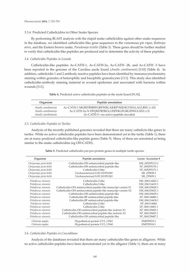
Pharmaceuticals 2014, 7, 723–753
3.3.4. Predicted Cathelicidins in Other Snake Species
By performing BLAST analysis with the elapid snake cathelicidins against other snake sequencesin the database, we identified cathelicidin-like gene sequences in the venomous pit viper, Bothropsatrox, and the Eastern brown snake, Pseudonaja textilis (Table 3). These genes should be further studiedto verify that cathelicidin-like peptides are produced and to determine the activity of these peptides.
3.4. Cathelicidin Peptides in Lizards
Cathelicidin-like peptides Ac-CATH-1, Ac-CATH-2a, Ac-CATH -2b, and Ac-CATH -3 havebeen reported in the genome of the Carolina anole lizard (Anolis carolinensis) [110] (Table 4). Inaddition, cathelicidin 1 and 2 antibody reactive peptides have been identified by immunocytochemistrystaining within granules of heterophilic and basophilic granulocytes [111]. This study also identifiedcathelicidin-antibody staining material in wound epidermis and associated with bacteria withinwounds [111].
Table 4. Predicted active cathelicidin peptides in the anole lizard [91,92].
Organism Peptide annotation
Anolis carolinensis Ac-CATH-1 MGRITRSRWGRFWRGAKRFVKKHGVSIALAGLRFG (+10)Anolis carolinensis Ac-CATH-2a/b DPQMTRFRGLGHFFKGFGRGFIWGLNH (+3)Anolis carolinensis Ac-CATH-3—no active peptide encoded.
3.5. Cathelicidin Peptides in Turtles
Analysis of the recently published genomes revealed that there are many cathelicin-like genes inturtles. While no active cathelicidin peptides have been demonstrated yet in the turtle (Table 1), thereare at many predicted cathelicidin-like peptide genes (Table 5). Many of these are annotated as beingsimilar to the snake cathelicidins (eg OH-CATH).
Table 5. Predicted cathelicidin pre-pro-protein genes in multiple turtle species.
Organism Peptide annotation Locus- Accession #
Chrysemys picta bellii Cathelicidin-OH antimicrobial peptide-like XM_005295113.1Chrysemys picta bellii Cathelicidin-OH antimicrobial peptide-like XP_005295170.1Chrysemys picta bellii Cathelicidin-2-like XP_005295171.1Chrysemys picta bellii Uncharacterized LOC101951069 XR_255838.1Chrysemys picta bellii Uncharacterized LOC101951243 XR_255839.1
Pelodiscus sinensis Cathelicidin-2-like XM_006114422.1Pelodiscus sinensis Cathelicidin-2-like XM_006114419.1Pelodiscus sinensis Cathelicidin-OH antimicrobial peptide-like transcript variant X1 XM_006129620.1Pelodiscus sinensis Cathelicidin-OH antimicrobial peptide-like transcript variant X2 XM_006129621.1Pelodiscus sinensis Cathelicidin-OH antimicrobial peptide-like XM_006129625.1Pelodiscus sinensis Cathelicidin-BF antimicrobial peptide-like XP_006114480.1Pelodiscus sinensis Cathelicidin-BF antimicrobial peptide-like XM_006114418.1Pelodiscus sinensi Cathelicidin-2-like XP_006114484.Pelodiscus sinensis Cathelicidin-2-like XP_006114481.1Pelodiscus sinensis Cathelicidin-OH antimicrobial peptide-like isoform X1 XP_006129682.1Pelodiscus sinensis Cathelicidin-OH antimicrobial peptide-like isoform X2 XP_006129683.1Pelodiscus sinensis Cathelicidin-OH antimicrobial peptide-like XP_006129687.1
Chelonia mydas Hypothetical protein UY3_13361 EMP29519.1Chelonia mydas Hypothetical protein UY3_13360 EMP29518.1
3.6. Cathelicidin Peptides in Crocodilians
Analysis of the databases revealed that there are many cathelicidin-like genes in alligators. Whileno active cathelicidin peptides have been demonstrated yet in the alligator (Table 1), there are at many
140

Pharmaceuticals 2014, 7, 723–753
predicted cathelicidin-like peptide genes (Table 6). Many of these genes are annotated as being similarto the snake cathelicidins (e.g., OH-CATH).
Table 6. Predicted cathelicidin pre-pro-protein genes in Alligator species.
Organism Peptide annotation Locus- Accession #
Alligator mississippiensis Cathelicidin-2-like XM_006262429.1Alligator mississippiensis Cathelicidin-2-like XP_006262491.1Alligator mississippiensis Cathelicidin-OH antimicrobial peptide-like XM_006262431.1Alligator mississippiensis Cathelicidin-OH antimicrobial peptide-like XM_006262430.1Alligator mississippiensis Cathelicidin-OH antimicrobial peptide-like XP_006262492.1Alligator mississippiensis Cathelicidin-OH antimicrobial peptide-like XP_006262493.1
Alligator sinensis Cathelicidin-2-like XM_006026412.1Alligator sinensis Cathelicidin-2-like XP_006026474.1Alligator sinensis Cathelicidin-2-like XP_006026475.1Alligator sinensis Cathelicidin-3-like XM_006026409.1Alligator sinensis Cathelicidin-3-like XP_006026471.1Alligator sinensis Cathelicidin-OH antimicrobial peptide-like XM_006037224.1Alligator sinensis Cathelicidin-OH antimicrobial peptide-like XM_006026410.1Alligator sinensis Cathelicidin-OH antimicrobial peptide-like XM_006026411.1Alligator sinensis Cathelicidin-OH antimicrobial peptide-like XM_006037211.1Alligator sinensis Cathelicidin-OH antimicrobial peptide-like XP_006037286.1Alligator sinensis Cathelicidin-OH antimicrobial peptide-like XP_006026472.1Alligator sinensis Cathelicidin-OH antimicrobial peptide-like XP_006037273.1Alligator sinensis Cathelicidin-OH antimicrobial peptide-like XP_006026487.1
3.7. Liver-Derived Peptides in Reptiles
3.7.1. Hepcidin (HAMP1)
Hepcidin antimicrobial peptides are liver-expressed peptides containing eight cysteines (fourdisulfide bonds) that are broadly antimicrobial, binds iron and is involved in ferroportin binding [112,113]. A gene encoding a hepcidin-like peptide (E8ZAD0_CROSI) was recently proposed to be encodedby the Siamese crocodile (Crocodylus siamensis). A 26 aa peptide (Cshepc), representing the predictedactive peptide (FNSHFPICSYCCNCCRNKGCGLCCRT), was expressed in yeast, and the unpurifiedproduct was found to have antimicrobial activity against both Gram-positive bacteria such as S. aureusand Bacillus subtilis, as well as Gram-negative bacteria Escherichia coli and Aeromonas sobria [114].Hepcidin-like sequences were also identified in the anole lizard, although interestingly hepcidinsappear to be missing in most avians [115].
3.7.2. LEAP-2, Liver Expressed Antimicrobial Peptide-2
Another liver-expressed peptide, LEAP-2, is expressed in many different organisms, hasbroad-spectrum antibacterial and antifungal activity, and can be induced following bacterialchallenge [116–118]. This 40 aa, 4-cysteine, cationic peptide differs significantly from hepcidin insequence and predicted structure [119], but is also predominantly expressed in the liver.
The Leap-2 gene is annotated in many of the sequenced reptiles (Table 7), including Pelodiscussinensis, Chrysemys picta bellii, Alligator sinensis, Alligator mississippiensis, as identified by a BLASTanalysis we performed with the LEAP-2 from Anolis carolinensis. The antimicrobial activity or biologicalrole of this peptide in reptiles has not been studied.
141

Pharmaceuticals 2014, 7, 723–753
Table 7. Predicted LEAP-2 pre-propeptide amino-acid sequences identified in reptiles.
Species Predicted LEAP-2 full sequence Accession Number
Anolis carolinensis MTPLKITAVILICSALLFQTQGASLYPPNSQLVRQRRMTPFWRGISRPIGASCRDNSECSTRLCRSKHCSLRTSQE XP_003217432.1
Alligator sinensis MHWLKVIAVMLLFALHLFQIHCASLHQPNSQPKRQRRMTPFWRGVSSLRPIGASCRDDIECVTMLCRKSHCSLRTSRE XP_006023615.1
Alligator mississippiensis MHWLKVIAVMLLFALHLFQIHCASLHQPNSQPKRQRRMTPFWRGVSSLRPIGASCKDDGECITMRCRKSHCSLRTSRE XP_006263463.1
Pelodiscus sinensis MQCLKVIALLLFCAALLTQTHCASLHHSSSQLTRQRRMTPFWRGISLRPIGALCRHDNECISMLCRKNRCSLRISCE XP_006128591.1
Chrysemys picta belli MQYLKVIAVLLLCAALLSQIHSASLHRPSSHLTRQRRMTPFWRGISLRPIGAICRDDSECVSRLCRKNHCSIRISRA XP_005302895.1
3.8. Lysozyme in Reptiles
Lysozyme is a very important part of innate immunity in most animals. In humans, it is packagedinto neutrophil granules, released in tears and other secretions, and is very effective against a broadspectrum of bacteria. Not surprisingly, reptiles have also been found to use lysozyme as part of theirinnate immune response. In the crocodilians, a lysozyme-like enzyme was identified with broad-spectrumantibacterial activity [67]. Lysozyme proteins very similar to chicken lysozymes (~130 aa) have beenidentified from some species of turtles, including the soft shelled turtle (Trionyx sinensis), the Asiatic softshelled turtle (Amyda cartilagenea) and the green sea turtle (Chelonia mydas) [17,19,120–122], although theantimicrobial activity of these molecules has not yet been proven. Lysozyme peptide has also beenidentified from Crocodylus siamensis [67]. Lysozyme-like genes were identified by analysis of reptilegenomes (Table 8).
3.9. Crotamine Peptides in Reptiles
Defensins are one example of cysteine-stabilized polypeptides with antimicrobial function.A similar family of peptides is the crotamine toxin family isolated from rattlesnakes with asimilar gamma-core motif to defensins [123,124], which is considered a cell-penetrating peptide Thesequence of crotamine is YKQCHKKGGHCFPKEKICLPPSSDFGKMDCRWRWKCCKKGSG (+8) [2].This cationic peptide contains nine lysines (underlined), three disulfide bonds (6 cysteines, shown inbold) and has a defensin-like fold (Figure 8).
Figure 8. The crotamine chemical structure. Crotamine, a Na+ channel-affecting toxin from Crotalusdurissus terrificus venom (PDB 1H5O) [125].
142

Pharmaceuticals 2014, 7, 723–753
Table 8. Selected predicted lysozyme genes identified in reptiles.
Reptile name Enzyme Name Accession Number
Softshell turtle lysozyme C(SSTL) Lysozyme C (1,4-β-N-acetylmuramidase C) Q7LZQ1.3Asiatic softshell turtle lysozyme C (ASTL) Lysozyme C P85345.1
Pelodiscus sinensis Lysozyme ADR51676.1Pelodiscus sinensis PREDICTED: lysozyme g-like isoform X2 XP_006113603.1Pelodiscus sinensis PREDICTED: lysozyme g-like isoform X1 XP_006113602.1Pelodiscus sinensis PREDICTED: lysozyme g-like XP_006113601.1
Chelonia mydas Lysozyme C EMP38935.1Chelonia mydas Lysozyme G EMP27176.1
Chrysemys picta bellii PREDICTED: lysozyme C-like XP_005314893.1Chrysemys picta bellii PREDICTED: lysozyme C-like XP_005312037.1Chrysemys picta bellii PREDICTED: lysozyme g-like protein 2 XP_005283410.1Chrysemys picta bellii PREDICTED: lysozyme G-like XP_005283294.1
Ophiophagus hannah Lysozyme C, partial ETE58503.1
XP_003225844.1,Anolis carolinensis Lysozyme C, milk isozyme-like XP_003216710.1,
XP_003216704.1Anolis carolinensis Lysozyme C II-like XP_003224512.1Anolis carolinensis Lysozyme g-like XP_003227178.1
Alligator sinensis Lysozyme C-like XP_006027022.1,XP_006027021.1
Alligator sinensis Lysozyme G-like
XP_006026406.1,XP_006026397.1,XP_006026395.1,XP_006026396.1
Some reptiles are known to express crotamine-like peptides, and The X-ray structure of crotamine,from the Brazilian snake Crotalus durissus terrificus, was recently solved (PDB 4GV5_A) [126]. We foundthat some species of reptiles have genes that are annotated as crotamine-like (Table 9).
Table 9. Crotamine-like peptide genes in reptiles.
Reptile AA Name Accession numbers
Uromastyx aegyptia 67 crotamine-Uro-1 AGI97143.1
Crotalus durissus terrificus 65 crotamine
AAF34911.1,AAF34910.1,AAC02995.1,AAC06241.1
Crotalus durissus 34 crotamine
ACA63453.1,ACA63452.1,ACA63451.1,ACA63450.1,ACA63449.1,ACA63448.1,ACA63447.1,ACA63446.1
Crotalus oreganus helleri 65 crotamine 7 AEU60015.1Crotalus oreganus helleri 65 crotamine 6 AEU60014.1Crotalus oreganus helleri 65 crotamine 5 AEU60013.1Crotalus oreganus helleri 70 crotamine 4 AEU60012.1Crotalus oreganus helleri 70 crotamine 3 AEU60011.1Crotalus oreganus helleri 83 crotamine 2 AEU60010.1Crotalus oreganus helleri 70 crotamine 1 AEU60009.1
Pogona barbata 102 CLP-POGL1 AAZ75614.1Pogona barbata 67 CLP-POGL2 AAZ75615.1Pogona barbata 61 CLP-POGU3 AAZ75613.1Pogona barbata 98 CLP-POGU2 AAZ75612.1Pogona barbata 76 CLP-POGU1 AAZ75611.1Varanus tristis 83 crotamine-Var-5 AGI97148.1
Varanus glauerti 83 crotamine-Var-4 AGI97147.1Varanus glauerti 83 crotamine-Var-3 AGI97146.1Varanus glauerti 83 crotamine-Var-2 AGI97145.1Varanus glauerti 83 crotamine-Var-1 AGI97144.1
143

Pharmaceuticals 2014, 7, 723–753
It has been a matter of some debate whether crotamine peptides are antimicrobial or if they justcontain a similar cysteine-stabilized core structure. Recently, it was demonstrated that crotaminetoxin is expressed similarly on epithelial or mucosal surfaces, and displays some antimicrobial activityagainst Bacillus subtilis, and was able to permeabilize Staphylococcus aureus cells, for example [20]. Inaddition, crotamine has been shown to be antifungal [127]. Thus, it seems that crotamine should beconsidered as an antimicrobial peptide of reptiles.
3.10. Other Peptides in Reptiles
3.10.1. Leucrocin
There are a handful of peptides identified in reptiles that were not easily classified in the categoriesabove. One interesting example is the peptide leucrocin, an antibacterial compound from white bloodcells of the Siamese crocodile (Crocodylus siamensis). Unlike Crocosin [65], which does not appear to bepeptide based, leucrocins are very small peptides with antibacterial activity. The amino acid sequenceof Leucrocin I is NGVQPKY, and of Leucrocin II is NAGSLLSGWG [66]. No known genes encodefor these peptides, so their source is unclear. Recently, a synthetic peptide based on the Leucrocinsequence was found to have broad-spectrum antibacterial activity [128].
3.10.2. Omwaprin
Another example of an unusual antimicrobial peptide in reptiles is omwaprin, which is a memberof the waprin family of venom proteins (Sequence shown in Table 1) [11,12], and was isolated fromthe Australian inland taipan (Oxyuranus microlepidotus), considered the most venomous snake (amember of the Elapid family of snakes). This 50 aa peptide has relatively salt-tolerant antibacterialactivity against gram-positive bacteria and was found to be non-toxic to mice upon injection. Itsactivity was found to be highly dependent upon the four disulfide-bonds, similar to the hepcidins anddefensins [11]. Thus, except for its large size, this peptide appears to have many favorable propertiesas a potential therapeutic candidate.
3.10.3. Hemocidin
Fragments of hemoglobin have been found to have some antimicrobial activity, and have beenreferred to as hemocidins [129–131]. In the study of the Siamese crocodile, 13 fragments of crocodilehemoglobin were found to have antimicrobial activity, including peptides similar to hemoglobin β
subunit [59,60]. Recently, the hemoglobin α- and β-chains of crocodilian species (Crocodylus siamensis,Alligator mississippiensis, Crocodylus niloticus and Caiman crocodilus) have been reported [132]. The in vivorelevance of these peptides in the crocodile remains to be determined, but it represents a potentialother class of antimicrobial peptides from reptiles that should be investigated.
3.10.4. Other Peptides
Other peptides have been identified in reptiles that do not fall into the various classes ofantimicrobial peptides described above. These include a short, synthetic tryptophan-rich cationicantimicrobial peptide, pEM-2 (KKWRWWLKALAKK) that was shown to have broad-spectrumbactericidal activities, and was identified as a fragment of myotoxin II, a snake venom Lys49phospholipase A2 [133,134]. Synthetic variants of this peptide were shown to have improvedantimicrobial activity, salt resistance and reduced hemolytic activity [135,136]. These peptides haveled to other advances in rational design of synthetic peptides, including peptides with exclusivelyarginine and tryptophan residues [137].
Gomes et al isolated, but did not sequence, a small 1370 Da peptide from the venom of a pit viper(Borthrops jaracaca), which showed very good activity against different fungi and yeast [138].
144

Pharmaceuticals 2014, 7, 723–753
Another group identified an antimicrobial peptide derived from N. atra venom, the vgf-1 peptide,and found that this peptide had significant antimicrobial activity against drug-resistant clinical strainsof Mycobacterium tuberculosis [139].
4. Conclusions
Reptiles are evolutionarily ancient, found in diverse and microbially challenging environmentsand appear to have robust immune systems. Some reptiles, especially lizards, have unique propertiessuch as tail regeneration. All of these features suggest that reptiles may express many interestingantimicrobial peptides. A few reptilian antimicrobial peptides have been isolated and studied whichdemonstrate broad-spectrum antimicrobial and antifungal activity. These include members of thecathelicidin and defensin and lysozyme class.
Antimicrobial peptides are known in three of the four orders of reptiles: the testudines,crocodilians, and the squamata. No peptides are known from the sphenodontia (tuataras), as thisorganism has just been sequenced [41]. Reptile neutrophils appear to have granules that contain bothcathelicidin-like peptides as well as β-defensin peptides, although unlike mammals, there are nogenes encoding α-defensins. β-defensin-like peptides and lysozyme are also found in reptile eggs.These peptides are expressed in wounds, such as when lizards lose their tails. Detailed study of theChinese cobra Naja atra cathelicidin peptides has revealed smaller peptides that could be useful fortherapeutic applications.
Overall, reptiles reflect the diversity of antimicrobial peptides within higher organisms. Theyappear to express cathelicidins and β-defensins, and express hepcidins unlike avians. Additionalclasses of antimicrobial peptides such as lysozyme, LEAP-2 and crotamine also appear to be highlyexpressed in reptiles. With the advent of high-throughput genomics, new reptilian genomes have beensequenced and their transcriptomes determined. Analysis of these sequences has revealed that thereare additional genes that may encode AMPs in reptiles. Future studies may reveal the in vivo role ofantimicrobial peptides in reptiles. In the face of the emerging challenges due to antibiotic resistantbacteria, new and useful molecules that could be developed into future antibiotics may potentially befound in the reptilian AMPs.
Acknowledgments: MVH was partially supported by HDTRA1-12-C-0039, Translational Peptides forPersonnel Protection.
Conflicts of Interest: The author declares no conflict of interest.
References
1. Wang, G. Antimicrobial Peptide Database. Available online: http://aps.unmc.edu/AP (accessed on 9 May2014).
2. Wang, G.; Li, X.; Wang, Z. APD2: The updated antimicrobial peptide database and its application in peptidedesign. Nucleic Acids Res. 2009, 37, D933–D937. [CrossRef]
3. Zhao, H.; Gan, T.X.; Liu, X.D.; Jin, Y.; Lee, W.H.; Shen, J.H.; Zhang, Y. Identification and characterization ofnovel reptile cathelicidins from elapid snakes. Peptides 2008, 29, 1685–1691. [CrossRef]
4. Li, S.A.; Lee, W.H.; Zhang, Y. Efficacy of OH-CATH30 and its analogs against drug-resistant bacteria in vitroand in mouse models. Antimicrob. Agents Chemother. 2012, 56, 3309–3317. [CrossRef]
5. Zhang, B.Y.; Li, S.M.; Gao, Z.H.; Shen, J.H. Protective effects of snake venom antimicrobial peptide OH-CATHon E. coli induced rabbit urinary tract infection models. Dong Wu Xue Yan Jiu 2013, 34, 27–32.
6. Zhang, Y.; Zhao, H.; Yu, G.Y.; Liu, X.D.; Shen, J.H.; Lee, W.H.; Zhang, Y. Structure-function relationship ofking cobra cathelicidin. Peptides 2010, 31, 1488–1493. [CrossRef]
7. Wang, Y.; Hong, J.; Liu, X.; Yang, H.; Liu, R.; Wu, J.; Wang, A.; Lin, D.; Lai, R. Snake cathelicidin fromBungarus fasciatus is a potent peptide antibiotics. PloS One 2008, 3, e3217.
8. Chen, W.; Yang, B.; Zhou, H.; Sun, L.; Dou, J.; Qian, H.; Huang, W.; Mei, Y.; Han, J. Structure-activityrelationships of a snake cathelicidin-related peptide, BF-15. Peptides 2011, 32, 2497–2503. [CrossRef]
145

Pharmaceuticals 2014, 7, 723–753
9. Wang, H.; Ke, M.; Tian, Y.; Wang, J.; Li, B.; Wang, Y.; Dou, J.; Zhou, C. BF-30 selectively inhibits melanoma cellproliferation via cytoplasmic membrane permeabilization and DNA-binding in vitro and in B16F10-bearingmice. Eur. J. Pharmacol. 2013, 707, 1–10.
10. Zhou, H.; Dou, J.; Wang, J.; Chen, L.; Wang, H.; Zhou, W.; Li, Y.; Zhou, C. The antibacterial activity of BF-30in vitro and in infected burned rats is through interference with cytoplasmic membrane integrity. Peptides2011, 32, 1131–1138. [CrossRef]
11. Nair, D.G.; Fry, B.G.; Alewood, P.; Kumar, P.P.; Kini, R.M. Antimicrobial activity of omwaprin, a new memberof the waprin family of snake venom proteins. Biochem. J. 2007, 402, 93–104. [CrossRef]
12. Banigan, J.R.; Mandal, K.; Sawaya, M.R.; Thammavongsa, V.; Hendrickx, A.P.; Schneewind, O.; Yeates, T.O.;Kent, S.B. Determination of the X-ray structure of the snake venom protein omwaprin by total chemicalsynthesis and racemic protein crystallography. Protein Sci. 2010, 19, 1840–1849. [CrossRef]
13. Schmidt, J.J.; Weinstein, S.A.; Smith, L.A. Molecular properties and structure-function relationships of lethalpeptides from venom of Wagler’s pit viper, Trimeresurus wagleri. Toxicon 1992, 30, 1027–1036. [CrossRef]
14. Stegemann, C.; Kolobov, A., Jr.; Leonova, Y.F.; Knappe, D.; Shamova, O.; Ovchinnikova, T.V.; Kokryakov, V.N.;Hoffmann, R. Isolation, purification and de novo sequencing of TBD-1, the first beta-defensin from leukocytesof reptiles. Proteomics 2009, 9, 1364–1373. [CrossRef]
15. Lakshminarayanan, R.; Vivekanandan, S.; Samy, R.P.; Banerjee, Y.; Chi-Jin, E.O.; Teo, K.W.; Jois, S.D.;Kini, R.M.; Valiyaveettil, S. Structure, self-assembly, and dual role of a beta-defensin-like peptide from theChinese soft-shelled turtle eggshell matrix. J. Am. Chem. Soc. 2008, 130, 4660–4668. [CrossRef]
16. Lakshminarayanan, R.; Chi-Jin, E.O.; Loh, X.J.; Kini, R.M.; Valiyaveettil, S. Purification and characterizationof a vaterite-inducing peptide, pelovaterin, from the eggshells of Pelodiscus sinensis (Chinese soft-shelledturtle). Biomacromolecules 2005, 6, 1429–1437. [CrossRef]
17. Thammasirirak, S.; Ponkham, P.; Preecharram, S.; Khanchanuan, R.; Phonyothee, P.; Daduang, S.;Srisomsap, C.; Araki, T.; Svasti, J. Purification, characterization and comparison of reptile lysozymes.Comp. biochem. Phys. Toxicol. Pharmacol. 2006, 143, 209–217.
18. Chattopadhyay, S.; Sinha, N.K.; Banerjee, S.; Roy, D.; Chattopadhyay, D.; Roy, S. Small cationic protein froma marine turtle has beta-defensin-like fold and antibacterial and antiviral activity. Proteins 2006, 64, 524–531.[CrossRef]
19. Ponkham, P.; Daduang, S.; Kitimasak, W.; Krittanai, C.; Chokchaichamnankit, D.; Srisomsap, C.; Svasti, J.;Kawamura, S.; Araki, T.; Thammasirirak, S. Complete amino acid sequence of three reptile lysozymes. Comp.Biochem. Phys. Toxicol. Pharmacol. 2010, 151, 75–83. [CrossRef]
20. Yount, N.Y.; Kupferwasser, D.; Spisni, A.; Dutz, S.M.; Ramjan, Z.H.; Sharma, S.; Waring, A.J.; Yeaman, M.R.Selective reciprocity in antimicrobial activity versus cytotoxicity of hBD-2 and crotamine. Proc. Natl. Acad.Sci. USA 2009, 106, 14972–14977. [CrossRef]
21. Coronado, M.A.; Georgieva, D.; Buck, F.; Gabdoulkhakov, A.H.; Ullah, A.; Spencer, P.J.; Arni, R.K.; Betzel, C.Purification, crystallization and preliminary X-ray diffraction analysis of crotamine, a myotoxic polypeptidefrom the Brazilian snake Crotalus durissus terrificus. Acta Crystallogr. F 2012, 68, 1052–1054. [CrossRef]
22. Modesto, S.P.; Anderson, J.S. The phylogenetic definition of reptilia. Syst. Biol. 2004, 53, 815–821. [CrossRef]23. Alfoldi, J.; Di Palma, F.; Grabherr, M.; Williams, C.; Kong, L.; Mauceli, E.; Russell, P.; Lowe, C.B.; Glor, R.E.;
Jaffe, J.D.; et al. The genome of the green anole lizard and a comparative analysis with birds and mammals.Nature 2011, 477, 587–591. [CrossRef]
24. Cladogram by Benchill, licensed under the Creative Commons Attribution 3.0 Unported license. Availableonline: http://en.wikipedia.org/wiki/File:Tuatara_cladogram.svg (accessed on 9 May 2014).
25. Ernst, C.H.; Lovich, J.E. Turtles of the United States and Canada, 2nd ed.; Johns Hopkins University Press:Baltimore, MD, USA, 2009; pp. 185–259.
26. Photo from Oregon Department of Fish & Wildlife, licensed under the Creative Commons Attribution-ShareAlike 2.0 Generic license. Photo by Gary M. Stolz/U. S. Fish and Wildlife Service in the Public domain. Availableonline: http://en.wikipedia.org/wiki/FileA4_Western_painted_turtle.jpg (accessed on 9 May 2014).
27. Underside of a Western Painted Turtle. Photo by Matt Young, l.u.t.C.C.A.-S.A.G.l. Available online: http://en.wikipedia.org/wiki/File:B4_Western_painted_turtle_underside.jpg (accessed on 9 May 2014).
28. Wang, Z.; Pascual-Anaya, J.; Zadissa, A.; Li, W.; Niimura, Y.; Huang, Z.; Li, C.; White, S.; Xiong, Z.; Fang, D.;et al. The draft genomes of soft-shell turtle and green sea turtle yield insights into the development andevolution of the turtle-specific body plan. Nat. Genet. 2013, 45, 701–706. [CrossRef]
146

Pharmaceuticals 2014, 7, 723–753
29. Jiang, J.J.; Xia, E.H.; Gao, C.W.; Gao, L.Z. The complete mitochondrial genome of western painted turtle,Chrysemys picta bellii (Chrysemys, Emydidae). Mitochondrial DNA 2014. [CrossRef]
30. Shaffer, H.B.; Minx, P.; Warren, D.E.; Shedlock, A.M.; Thomson, R.C.; Valenzuela, N.; Abramyan, J.;Amemiya, C.T.; Badenhorst, D.; Biggar, K.K.; et al. The western painted turtle genome, a model for theevolution of extreme physiological adaptations in a slowly evolving lineage. Genome Biol. 2013, 14, R28.[CrossRef]
31. Gilbert, S.F.; Corfe, I. Turtle origins: Picking up speed. Dev. Cell 2013, 25, 326–328. [CrossRef]32. Zardoya, R.; Meyer, A. Complete mitochondrial genome suggests diapsid affinities of turtles. Proc. Natl.
Acad. Sci. USA 1998, 95, 14226–14231. [CrossRef]33. Iwabe, N.; Hara, Y.; Kumazawa, Y.; Shibamoto, K.; Saito, Y.; Miyata, T.; Katoh, K. Sister group relationship
of turtles to the bird-crocodilian clade revealed by nuclear DNA-coded proteins. Mol. Biol. Evol. 2005, 22,810–813. [CrossRef]
34. Roos, J.; Aggarwal, R.K.; Janke, A. Extended mitogenomic phylogenetic analyses yield new insight intocrocodylian evolution and their survival of the Cretaceous-Tertiary boundary. Mol. Phylogenet. Evol. 2007, 45,663–673. [CrossRef]
35. Katsu, Y.; Matsubara, K.; Kohno, S.; Matsuda, Y.; Toriba, M.; Oka, K.; Guillette, L.J., Jr.; Ohta, Y.; Iguchi, T.Molecular cloning, characterization, and chromosome mapping of reptilian estrogen receptors. Endocrinology2010, 151, 5710–5720.
36. Lyson, T.R.; Sperling, E.A.; Heimberg, A.M.; Gauthier, J.A.; King, B.L.; Peterson, K.J. MicroRNAs support aturtle + lizard clade. Biol. Lett. 2012, 8, 104–107. [CrossRef]
37. Jones, M.E.; Cree, A. Tuatara. Curr. Biol. 2012, 22, R986–R987. [CrossRef]38. Hay, J.M.; Sarre, S.D.; Lambert, D.M.; Allendorf, F.W.; Daugherty, C.H. Genetic diversity and taxonomy: A
reassessment of species designation in tuatara (Sphenodon: Reptilia). Conserv. Genet. 2010, 11, 1063–1081.39. Photo by PhillipC, licensed under the Creative Commons Attribution 2.0 Generic license. Available online:
http://en.wikipedia.org/wiki/File:Sphenodon_punctatus_in_Waikanae,_New_Zealand.jpg (accessed on 9May 2014).
40. Ramstad, K.M.; Nelson, N.J.; Paine, G.; Beech, D.; Paul, A.; Paul, P.; Allendorf, F.W.; Daugherty, C.H. Speciesand cultural conservation in New Zealand: maori traditional ecological knowledge of tuatara. Conserv. Biol.2007, 21, 455–464. [CrossRef]
41. Miller, H.C.; Biggs, P.J.; Voelckel, C.; Nelson, N.J. De novo sequence assembly and characterisation of apartial transcriptome for an evolutionarily distinct reptile, the tuatara (Sphenodon punctatus). BMC Genomics2012, 13, 439. [CrossRef]
42. Vonk, F.J.; Casewell, N.R.; Henkel, C.V.; Heimberg, A.M.; Jansen, H.J.; McCleary, R.J.; Kerkkamp, H.M.;Vos, R.A.; Guerreiro, I.; Calvete, J.J.; et al. The king cobra genome reveals dynamic gene evolution andadaptation in the snake venom system. Proc. Natl. Acad. Sci. US A 2013, 110, 20651–20656. [CrossRef]
43. Pyron, R.A.; Burbrink, F.T.; Guiher, T.J. Claims of potential expansion throughout the U.S. by invasive pythonspecies are contradicted by ecological niche models. PloS One 2008, 3, e2931. [CrossRef]
44. Photo by Greg Hume, licensed under the Creative Commons Attribution 2.0 Generic license. Availableonline: http://en.wikipedia.org/wiki/File:King_Cobra_25.jpg (accessed on 9 May 2014).
45. Found inside a water catchment on Lantau. 2011, Photo by: Thomas Brown. This file is licensed under theCreative Commons Attribution 2.0 Generic license. Available online: http://en.wikipedia.org/wiki/File:Naja_atra_juvenile.jpg (accessed on 9 May 2014).
46. Photo by AshLin. Photo of Banded Krait captured in Binnaguri, N.B., India on 19 Sep 2006. This fileis licensed under the Creative Commons Attribution-Share Alike 2.5 Generic license. Available online:http://en.wikipedia.org/wiki/File:AB_054_Banded_Krait.JPG (accessed on 9 May 2014).
47. Photo by PiccoloNamek at en.wikipedia, licensed under the Creative Commons Attribution-Share Alike 3.0Unported license. Available online: http://en.wikipedia.org/wiki/File:Anolis_carolinensis.jpg (accessed on9 May 2014).
48. Aird, S.D.; Watanabe, Y.; Villar-Briones, A.; Roy, M.C.; Terada, K.; Mikheyev, A.S. Quantitativehigh-throughput profiling of snake venom gland transcriptomes and proteomes (Ovophis okinavensis andProtobothrops flavoviridis). BMC Genomics 2013, 14, 790. [CrossRef]
147

Pharmaceuticals 2014, 7, 723–753
49. Dalla Valle, L.; Benato, F.; Maistro, S.; Quinzani, S.; Alibardi, L. Bioinformatic and molecular characterizationof beta-defensins-like peptides isolated from the green lizard Anolis carolinensis. Dev. Comp. Immunol. 2012,36, 222–229. [CrossRef]
50. Erickson, G.M.; Gignac, P.M.; Steppan, S.J.; Lappin, A.K.; Vliet, K.A.; Brueggen, J.D.; Inouye, B.D.; Kledzik, D.;Webb, G.J. Insights into the ecology and evolutionary success of crocodilians revealed through bite-force andtooth-pressure experimentation. PloS One 2012, 7, e31781. [CrossRef]
51. Shirley, M.H.; Vliet, K.A.; Carr, A.N.; Austin, J.D. Rigorous approaches to species delimitation have significantimplications for African crocodilian systematics and conservation. Proc. Biol. Sci. 2014, 281, 20132483.
52. An American Alligator (One of two) in captivity at the Columbus Zoo, Powell, Ohio. This file is licensedunder the Creative Commons Attribution-Share Alike 3.0 Unported license. Available online: http://en.wikipedia.org/wiki/File:American_Alligator.jpg (accessed on 9 May 2014).
53. A Siamese Crocodile (Crocodylus siamensis) at the Jerusalem Biblical Zoo. This file is licensed underthe Creative Commons Attribution-Share Alike 3.0 Unported license. Available online: http://commons.wikimedia.org/wiki/File:Siamese_Crocodile-Biblical_Zoo.JPG (accessed on 9 May 2014).
54. Lance, V.A. Alligator physiology and life history: The importance of temperature. Exp. Gerontol. 2003, 38,801–805. [CrossRef]
55. Brown, D.R.; Schumacher, I.M.; Nogueira, M.F.; Richey, L.J.; Zacher, L.A.; Schoeb, T.R.; Vliet, K.A.;Bennett, R.A.; Jacobson, E.R.; Brown, M.B. Detection of antibodies to a pathogenic mycoplasma in Americanalligators (Alligator mississippiensis), broad-nosed Caimans (Caiman latirostris), and Siamese crocodiles(Crocodylus siamensis). J. Clin. Microbiol. 2001, 39, 285–292. [CrossRef]
56. Meganathan, P.R.; Dubey, B.; Batzer, M.A.; Ray, D.A.; Haque, I. Molecular phylogenetic analyses of genusCrocodylus (Eusuchia, Crocodylia, Crocodylidae) and the taxonomic position of Crocodylus porosus. Mol.Phylogenet. Evol. 2010, 57, 393–402. [CrossRef]
57. Kommanee, J.; Preecharram, S.; Daduang, S.; Temsiripong, Y.; Dhiravisit, A.; Yamada, Y.; Thammasirirak, S.Antibacterial activity of plasma from crocodile (Crocodylus siamensis) against pathogenic bacteria. Ann. Clin.Microbiol. Antimicrob. 2012, 11, 22. [CrossRef]
58. Theansungnoen, T.; Yaraksa, N.; Daduang, S.; Dhiravisit, A.; Thammasirirak, S. Purification and Characterizationof Antioxidant Peptides from Leukocyte Extract of Crocodylus siamensis. Protein J. 2014, 33, 24–31.
59. Srihongthong, S.; Pakdeesuwan, A.; Daduang, S.; Araki, T.; Dhiravisit, A.; Thammasirirak, S. Completeamino acid sequence of globin chains and biological activity of fragmented crocodile hemoglobin (Crocodylussiamensis). Protein J. 2012, 31, 466–476.
60. Jandaruang, J.; Siritapetawee, J.; Thumanu, K.; Songsiriritthigul, C.; Krittanai, C.; Daduang, S.; Dhiravisit, A.;Thammasirirak, S. The effects of temperature and pH on secondary structure and antioxidant activity ofCrocodylus siamensis hemoglobin. Protein J. 2012, 31, 43–50.
61. Johnston, M.A.; Porter, D.E.; Scott, G.I.; Rhodes, W.E.; Webster, L.F. Isolation of faecal coliform bacteria fromthe American alligator (Alligator mississippiensis). J. Appl. Microbiol. 2010, 108, 965–973. [CrossRef]
62. Merchant, M.E.; Mills, K.; Leger, N.; Jerkins, E.; Vliet, K.A.; McDaniel, N. Comparisons of innate immuneactivity of all known living crocodylian species. Com. Biochem. Physiol. Part B 2006, 143, 133–137.
63. Merchant, M.E.; Leger, N.; Jerkins, E.; Mills, K.; Pallansch, M.B.; Paulman, R.L.; Ptak, R.G. Broad spectrumantimicrobial activity of leukocyte extracts from the American alligator (Alligator mississippiensis). Vet.Immunol. Immunopathol. 2006, 110, 221–228. [CrossRef]
64. Merchant, M.E.; Roche, C.; Elsey, R.M.; Prudhomme, J. Antibacterial properties of serum from the Americanalligator (Alligator mississippiensis). Comp. biochem. Physiol. Part B 2003, 136, 505–513. [CrossRef]
65. Preecharram, S.; Jearranaiprepame, P.; Daduang, S.; Temsiripong, Y.; Somdee, T.; Fukamizo, T.; Svasti, J.;Araki, T.; Thammasirirak, S. Isolation and characterisation of crocosin, an antibacterial compound fromcrocodile (Crocodylus siamensis) plasma. Nihon chikusan Gakkaiho 2010, 81, 393–401.
66. Pata, S.; Yaraksa, N.; Daduang, S.; Temsiripong, Y.; Svasti, J.; Araki, T.; Thammasirirak, S. Characterization ofthe novel antibacterial peptide Leucrocin from crocodile (Crocodylus siamensis) white blood cell extracts. Dev.Comp. Immunol. 2011, 35, 545–553.
67. Pata, S.; Daduang, S.; Svasti, J.; Thammasirirak, S. Isolation of Lysozyme like protein from crocodile leukocyteextract (Crocodylus siamensis). KMITL Sci. Technol. J. 2007, 7, 70–85.
68. Castoe, T.A.; Pollock, D.D. Chinese alligator genome illustrates molecular adaptations. Cell res. 2013, 23,1254–1255. [CrossRef]
148

Pharmaceuticals 2014, 7, 723–753
69. St John, J.A.; Braun, E.L.; Isberg, S.R.; Miles, L.G.; Chong, A.Y.; Gongora, J.; Dalzell, P.; Moran, C.; Bed'hom, B.;Abzhanov, A.; et al. Sequencing three crocodilian genomes to illuminate the evolution of archosaurs andamniotes. Genome Biol. 2012, 13, 415. [CrossRef]
70. Wan, Q.H.; Pan, S.K.; Hu, L.; Zhu, Y.; Xu, P.W.; Xia, J.Q.; Chen, H.; He, G.Y.; He, J.; Ni, X.W.; et al. Genomeanalysis and signature discovery for diving and sensory properties of the endangered Chinese alligator. CellRes. 2013, 23, 1091–1105. [CrossRef]
71. Chromek, M.; Arvidsson, I.; Karpman, D. The antimicrobial peptide cathelicidin protects mice fromEscherichia coli O157:H7-mediated disease. PloS One 2012, 7, e46476. [CrossRef]
72. Oguro-Okano, M.; Honda, M.; Yamazaki, K.; Okano, K. Mutations in the melanocortin 1 receptor,beta-defensin103 and agouti signaling protein genes, and their association with coat color phenotypesin Akita-inu dogs. J. Vet. Med. Sci. 2011, 73, 853–858. [CrossRef]
73. Schmutz, S.M.; Berryere, T.G. Genes affecting coat colour and pattern in domestic dogs: A review. Anim.Genet. 2007, 38, 539–549. [CrossRef]
74. Zasloff, M. Magainins, a class of antimicrobial peptides from Xenopus skin: isolation, characterization oftwo active forms, and partial cDNA sequence of a precursor. Proc. Natl. Acad. Sci. USA 1987, 84, 5449–5453.[CrossRef]
75. Wilson, C.L.; Schmidt, A.P.; Pirila, E.; Valore, E.V.; Ferri, N.; Sorsa, T.; Ganz, T.; Parks, W.C. DifferentialProcessing of {alpha}- and {beta}-Defensin Precursors by Matrix Metalloproteinase-7 (MMP-7). J. Biol. Chem.2009, 284, 8301–8311.
76. Zhao, L.; Lu, W. Defensins in innate immunity. Curr. Opin. Hematol. 2014, 21, 37–42. [CrossRef]77. Lehrer, R.I.; Lu, W. alpha-Defensins in human innate immunity. Immunol. Rev. 2012, 245, 84–112. [CrossRef]78. Garcia, J.R.; Jaumann, F.; Schulz, S.; Krause, A.; Rodriguez-Jimenez, J.; Forssmann, U.; Adermann, K.;
Kluver, E.; Vogelmeier, C.; Becker, D.; et al. Identification of a novel, multifunctional beta-defensin (humanbeta-defensin 3) with specific antimicrobial activity. Its interaction with plasma membranes of Xenopusoocytes and the induction of macrophage chemoattraction. Cell Tissue Res. 2001, 306, 257–264. [CrossRef]
79. Kaplinsky, N.J.; Gilbert, S.F.; Cebra-Thomas, J.; Lillevali, K.; Saare, M.; Chang, E.Y.; Edelman, H.E.; Frick, M.A.;Guan, Y.; Hammond, R.M.; et al. The Embryonic Transcriptome of the Red-Eared Slider Turtle. PloS one 2013,8, e66357.
80. Lorenzo, A. Immunolocalization of a beta-defensin (Tu-BD-1) in the skin and subdermal granulocytes ofturtles indicate the presence of an antimicrobial skin barrier. Ann. Anat. 2013, 195, 554–561. [CrossRef]
81. Benato, F.; Dalla Valle, L.; Skobo, T.; Alibardi, L. Biomolecular identification of beta-defensin-like peptidesfrom the skin of the soft-shelled turtle Apalone spinifera. J. Exp. Zool. Part B 2013, 320, 210–217. [CrossRef]
82. Alibardi, L. Ultrastructural immunolocalization of beta-defensin-27 in granulocytes of the dermis and woundepidermis of lizard suggests they contribute to the anti-microbial skin barrier. Anat. Cell Biol. 2013, 46,246–253. [CrossRef]
83. Alibardi, L. Histochemical, Biochemical and Cell Biological aspects of tail regeneration in lizard, an amniotemodel for studies on tissue regeneration. Prog. Histochem. Cytochem. 2014, 48, 143–244. [CrossRef]
84. Alibardi, L. Granulocytes of reptilian sauropsids contain beta-defensin-like peptides: a comparativeultrastructural survey. J. Morphol. 2013, 274, 877–886. [CrossRef]
85. Alibardi, L.; Celeghin, A.; Dalla Valle, L. Wounding in lizards results in the release of beta-defensins at thewound site and formation of an antimicrobial barrier. Dev. Comp. Immunol. 2012, 36, 557–565. [CrossRef]
86. Abdel Mageed, A.M.; Isobe, N.; Yoshimura, Y. Immunolocalization of avian beta-defensins in the hen oviductand their changes in the uterus during eggshell formation. Reproduction 2009, 138, 971–978. [CrossRef]
87. Gong, D.; Wilson, P.W.; Bain, M.M.; McDade, K.; Kalina, J.; Herve-Grepinet, V.; Nys, Y.; Dunn, I.C. Gallin; anantimicrobial peptide member of a new avian defensin family, the ovodefensins, has been subject to recentgene duplication. BMC Immunol. 2010, 11, 12.
88. Herve, V.; Meudal, H.; Labas, V.; Rehault Godbert, S.; Gautron, J.; Berges, M.; Guyot, N.; Delmas, A.F.;Nys, Y.; Landon, C. 3D NMR structure of hen egg gallin (chicken ovo-defensin) reveals a new variation ofthe beta-defensin fold. J. Biol. Chem. 2014.
89. Herve-Grepinet, V.; Rehault-Godbert, S.; Labas, V.; Magallon, T.; Derache, C.; Lavergne, M.; Gautron, J.;Lalmanach, A.C.; Nys, Y. Purification and characterization of avian beta-defensin 11, an antimicrobialpeptide of the hen egg. Antimicrob. Agents Chemother. 2010, 54, 4401–4409. [CrossRef]
149

Pharmaceuticals 2014, 7, 723–753
90. Mine, Y.; Oberle, C.; Kassaify, Z. Eggshell matrix proteins as defense mechanism of avian eggs. J. Agric. FoodChem. 2003, 51, 249–253. [CrossRef]
91. Kosciuczuk, E.M.; Lisowski, P.; Jarczak, J.; Strzalkowska, N.; Jozwik, A.; Horbanczuk, J.; Krzyzewski, J.;Zwierzchowski, L.; Bagnicka, E. Cathelicidins: family of antimicrobial peptides. A review. Mol. Biol. Reports2012, 39, 10957–10970. [CrossRef]
92. Lehrer, R.I.; Ganz, T. Cathelicidins: A family of endogenous antimicrobial peptides. Curr. Opin. Hematol.2002, 9, 18–22. [CrossRef]
93. Cole, A.M.; Shi, J.; Ceccarelli, A.; Kim, Y.H.; Park, A.; Ganz, T. Inhibition of neutrophil elastase preventscathelicidin activation and impairs clearance of bacteria from wounds. Blood 2001, 97, 297–304. [CrossRef]
94. Tongaonkar, P.; Golji, A.E.; Tran, P.; Ouellette, A.J.; Selsted, M.E. High fidelity processing and activation ofthe human alpha-defensin HNP1 precursor by neutrophil elastase and proteinase 3. PloS One 2012, 7, e32469.
95. Nizet, V.; Gallo, R.L. Cathelicidins and innate defense against invasive bacterial infection. Scand. J. Infect.Dis. 2003, 35, 670–676. [CrossRef]
96. Zanetti, M.; Gennaro, R.; Scocchi, M.; Skerlavaj, B. Structure and biology of cathelicidins. Adv. Exp. Med. Biol.2000, 479, 203–218.
97. van Dijk, A.; Molhoek, E.M.; Bikker, F.J.; Yu, P.L.; Veldhuizen, E.J.; Haagsman, H.P. Avian cathelicidins:Paradigms for the development of anti-infectives. Vet. Microbiol. 2011, 153, 27–36. [CrossRef]
98. Van Dijk, A.; Veldhuizen, E.J.; van Asten, A.J.; Haagsman, H.P. CMAP27, a novel chicken cathelicidin-likeantimicrobial protein. Vet. Immunol. Immunopathol. 2005, 106, 321–327. [CrossRef]
99. Xiao, Y.; Cai, Y.; Bommineni, Y.R.; Fernando, S.C.; Prakash, O.; Gilliland, S.E.; Zhang, G. Identification andfunctional characterization of three chicken cathelicidins with potent antimicrobial activity. J. Biol. Chem.2006, 281, 2858–2867.
100. Van Dijk, A.; Tersteeg-Zijderveld, M.H.; Tjeerdsma-van Bokhoven, J.L.; Jansman, A.J.; Veldhuizen, E.J.;Haagsman, H.P. Chicken heterophils are recruited to the site of Salmonella infection and release antibacterialmature Cathelicidin-2 upon stimulation with LPS. Mol. Immunol. 2009, 46, 1517–1526. [CrossRef]
101. Sorensen, O.E.; Borregaard, N. Cathelicidins–nature's attempt at combinatorial chemistry. Comb. Chem. HighThroughput Screen. 2005, 8, 273–280. [CrossRef]
102. De Latour, F.A.; Amer, L.S.; Papanstasiou, E.A.; Bishop, B.M.; van Hoek, M.L. Antimicrobial activity ofthe Naja atra cathelicidin and related small peptides. Biochem. Biophys. Res. Commun. 2010, 396, 825–830.[CrossRef]
103. Dean, S.N.; Bishop, B.M.; van Hoek, M.L. Susceptibility of Pseudomonas aeruginosa Biofilm to Alpha-HelicalPeptides: D-enantiomer of LL-37. Front. Microbiol. 2011, 2, 128.
104. Amer, L.S.; Bishop, B.M.; van Hoek, M.L. Antimicrobial and antibiofilm activity of cathelicidins and short,synthetic peptides against Francisella. Biochem. Biophys. Res. Commun. 2010, 396, 246–251. [CrossRef]
105. Dean, S.N.; Bishop, B.M.; van Hoek, M.L. Natural and synthetic cathelicidin peptides with anti-microbialand anti-biofilm activity against Staphylococcus aureus. BMC Microbiol. 2011, 11, 114. [CrossRef]
106. Juba, M.; Porter, D.; Dean, S.; Gillmor, S.; Bishop, B. Characterization and performance of short cationicantimicrobial peptide isomers. Biopolymers 2013, 100, 387–401. [CrossRef]
107. Wang, Y.; Zhang, Z.; Chen, L.; Guang, H.; Li, Z.; Yang, H.; Li, J.; You, D.; Yu, H.; Lai, R. Cathelicidin-BF, asnake cathelicidin-derived antimicrobial peptide, could be an excellent therapeutic agent for acne vulgaris.PloS One 2011, 6, e22120.
108. Hao, Q.; Wang, H.; Wang, J.; Dou, J.; Zhang, M.; Zhou, W.; Zhou, C. Effective antimicrobial activity ofCbf-K16 and Cbf-A7 A13 against NDM-1-carrying Escherichia coli by DNA binding after penetrating thecytoplasmic membrane in vitro. J. Pept. Sci. 2013, 19, 173–180. [CrossRef]
109. Wang, J.; Li, B.; Li, Y.; Dou, J.; Hao, Q.; Tian, Y.; Wang, H.; Zhou, C. BF-30 effectively inhibitsciprofloxacin-resistant bacteria in vitro and in a rat model of vaginosis. Arch. Pharm. Res. 2013. [CrossRef]
110. Dalla Valle, L.; Benato, F.; Paccanaro, M.C.; Alibardi, L. Bioinformatic and molecular characterizationof cathelicidin-like peptides isolated from the green lizard Anolis carolinensis (Reptilia: Lepidosauria:Iguanidae). Ital. J. Zool. 2013, 80, 177–186. [CrossRef]
111. Alibardi, L. Ultrastructural immunolocalization of chatelicidin-like peptides in granulocytes of normal andregenerating lizard tissues. Acta Histochem. 2013.
112. Park, C.H.; Valore, E.V.; Waring, A.J.; Ganz, T. Hepcidin, a urinary antimicrobial peptide synthesized in theliver. J. Biol. Chem. 2001, 276, 7806–7810.
150
Pharmaceuticals 2014, 7, 723–753
113. Rodriguez, R.; Jung, C.L.; Gabayan, V.; Deng, J.C.; Ganz, T.; Nemeth, E.; Bulut, Y. Hepcidin induction bypathogens and pathogen-derived molecules is strongly dependent on interleukin-6. Infect. Immun. 2014, 82,745–752. [CrossRef]
114. Hao, J.; Li, Y.W.; Xie, M.Q.; Li, A.X. Molecular cloning, recombinant expression and antibacterial activityanalysis of hepcidin from Simensis crocodile (Crocodylus siamensis). Comp. Biochem. Phys. Part B 2012, 163,309–315. [CrossRef]
115. Hilton, K.B.; Lambert, L.A. Molecular evolution and characterization of hepcidin gene products in vertebrates.Gene 2008, 415, 40–48.
116. Sang, Y.; Ramanathan, B.; Minton, J.E.; Ross, C.R.; Blecha, F. Porcine liver-expressed antimicrobial peptides,hepcidin and LEAP-2: cloning and induction by bacterial infection. Dev. Comp. Immunol. 2006, 30, 357–366.[CrossRef]
117. Hocquellet, A.; Odaert, B.; Cabanne, C.; Noubhani, A.; Dieryck, W.; Joucla, G.; le Senechal, C.; Milenkov, M.;Chaignepain, S.; Schmitter, J.M.; et al. Structure-activity relationship of human liver-expressed antimicrobialpeptide 2. Peptides 2010, 31, 58–66. [CrossRef]
118. Henriques, S.T.; Tan, C.C.; Craik, D.J.; Clark, R.J. Structural and functional analysis of human liver-expressedantimicrobial peptide 2. Chembiochem 2010, 11, 2148–2157. [CrossRef]
119. Krause, A.; Sillard, R.; Kleemeier, B.; Kluver, E.; Maronde, E.; Conejo-Garcia, J.R.; Forssmann, W.G.;Schulz-Knappe, P.; Nehls, M.C.; Wattler, F.; et al. Isolation and biochemical characterization of LEAP-2, anovel blood peptide expressed in the liver. Protein Sci. 2003, 12, 143–152. [CrossRef]
120. Araki, T.; Yamamoto, T.; Torikata, T. Reptile lysozyme: the complete amino acid sequence of soft-shelledturtle lysozyme and its activity. Biosci. Biotechnol. Biochem. 1998, 62, 316–324. [CrossRef]
121. Chijiiwa, Y.; Kawamura, S.; Torikata, T.; Araki, T. Amino acid sequence and activity of green turtle (Cheloniamydas) lysozyme. Protein J. 2006, 25, 336–344. [CrossRef]
122. Prajanban, B.O.; Shawsuan, L.; Daduang, S.; Kommanee, J.; Roytrakul, S.; Dhiravisit, A.; Thammasirirak, S.Identification of five reptile egg whites protein using MALDI-TOF mass spectrometry and LC/MS-MSanalysis. J. Proteomics 2012, 75, 1940–1959. [CrossRef]
123. Radis-Baptista, G.; Kerkis, I. Crotamine, a small basic polypeptide myotoxin from rattlesnake venom withcell-penetrating properties. Curr. Pharm. Design 2011, 17, 4351–4361. [CrossRef]
124. Kerkis, I.; Silva Fde, S.; Pereira, A.; Kerkis, A.; Radis-Baptista, G. Biological versatility of crotamine—Acationic peptide from the venom of a South American rattlesnake. Expert Opin. Investig. Drugs 2010, 19,1515–1525. [CrossRef]
125. Structure of crotamine, a Na+ channel affecting toxin from Crotalus durissus terrificus venom (PDB 1H5O).This image is licensed under the Creative Commons Attribution-Share Alike 3.0 Unported license. Availableonline: http://en.wikipedia.org/wiki/File:Crotamin_1H5O.png (accessed on 9 May 2014).
126. Radis-Baptista, G.; Kubo, T.; Oguiura, N.; Prieto da Silva, A.R.; Hayashi, M.A.; Oliveira, E.B.; Yamane, T.Identification of crotasin, a crotamine-related gene of Crotalus durissus terrificus. Toxicon 2004, 43, 751–759.[CrossRef]
127. Yamane, E.S.; Bizerra, F.C.; Oliveira, E.B.; Moreira, J.T.; Rajabi, M.; Nunes, G.L.; de Souza, A.O.; da Silva, I.D.;Yamane, T.; Karpel, R.L.; et al. Unraveling the antifungal activity of a South American rattlesnake toxincrotamine. Biochimie 2013, 95, 231–240.
128. Yaraksa, N.; Anunthawan, T.; Theansungnoen, T.; Daduang, S.; Araki, T.; Dhiravisit, A.; Thammasirirak, S.Design and synthesis of cationic antibacterial peptide based on Leucrocin I sequence, antibacterial peptidefrom crocodile (Crocodylus siamensis) white blood cell extracts. J. Antibiot. 2013, 67, 205–212.
129. Mak, P. Hemocidins in a functional and structural context of human antimicrobial peptides. Front. Biosci.2008, 13, 6859–6871.
130. Sheshadri, P.; Abraham, J. Antimicrobial properties of hemoglobin. Immunopharm. Immunotoxicol. 2012, 34,896–900. [CrossRef]
131. Parish, C.A.; Jiang, H.; Tokiwa, Y.; Berova, N.; Nakanishi, K.; McCabe, D.; Zuckerman, W.; Xia, M.M.;Gabay, J.E. Broad-spectrum antimicrobial activity of hemoglobin. Bioorg. Med. Chem. 2001, 9, 377–382.
132. Anwised, P.; Kabbua, T.; Temsiripong, T.; Dhiravisit, A.; Jitrapakdee, S.; Araki, T.; Yoneda, K.;Thammasirirak, S. Molecular cloning and expression of alpha-globin and beta-globin genes from crocodile(Crocodylus siamensis). Protein J. 2013, 32, 172–182.
151
Pharmaceuticals 2014, 7, 723–753
133. Santamaria, C.; Larios, S.; Quiros, S.; Pizarro-Cerda, J.; Gorvel, J.P.; Lomonte, B.; Moreno, E. Bactericidal andantiendotoxic properties of short cationic peptides derived from a snake venom Lys49 phospholipase A2.Antimicrob. Agents Chemother. 2005, 49, 1340–1345. [CrossRef]
134. Santamaria, C.; Larios, S.; Angulo, Y.; Pizarro-Cerda, J.; Gorvel, J.P.; Moreno, E.; Lomonte, B. Antimicrobialactivity of myotoxic phospholipases A2 from crotalid snake venoms and synthetic peptide variants derivedfrom their C-terminal region. Toxicon 2005, 45, 807–815. [CrossRef]
135. Yu, H.Y.; Huang, K.C.; Yip, B.S.; Tu, C.H.; Chen, H.L.; Cheng, H.T.; Cheng, J.W. Rational design oftryptophan-rich antimicrobial peptides with enhanced antimicrobial activities and specificities. Chembiochem2010, 11, 2273–2282. [CrossRef]
136. Chu, H.L.; Yu, H.Y.; Yip, B.S.; Chih, Y.H.; Liang, C.W.; Cheng, H.T.; Cheng, J.W. Boosting salt resistance ofshort antimicrobial peptides. Antimicrob. Agents Chemother. 2013, 57, 4050–4052. [CrossRef]
137. Deslouches, B.; Steckbeck, J.D.; Craigo, J.K.; Doi, Y.; Mietzner, T.A.; Montelaro, R.C. Rational design ofengineered cationic antimicrobial peptides consisting exclusively of arginine and tryptophan, and theiractivity against multidrug-resistant pathogens. Antimicrob. Agents Chemother. 2013, 57, 2511–2521. [CrossRef]
138. Gomes, V.M.; Carvalho, A.O.; Da Cunha, M.; Keller, M.N.; Bloch, C., Jr.; Deolindo, P.; Alves, E.W. Purificationand characterization of a novel peptide with antifungal activity from Bothrops jararaca venom. Toxicon 2005,45, 817–827. [CrossRef]
139. Xie, J.P.; Yue, J.; Xiong, Y.L.; Wang, W.Y.; Yu, S.Q.; Wang, H.H. In vitro activities of small peptides from snakevenom against clinical isolates of drug-resistant Mycobacterium tuberculosis. Int. J. Antimicrob. Agents 2003,22, 172–174. [CrossRef]
© 2014 by the author. Licensee MDPI, Basel, Switzerland. This article is an open accessarticle distributed under the terms and conditions of the Creative Commons Attribution(CC BY) license (http://creativecommons.org/licenses/by/4.0/).
152

pharmaceuticals
Review
Chapter 8:
Avian Antimicrobial Host Defense Peptides:From Biology to Therapeutic Applications
Guolong Zhang 1,2,3,* and Lakshmi T. Sunkara 1
1 Department of Animal Science, Oklahoma State University, Stillwater, OK 74078, USA2 Department of Biochemistry and Molecular Biology, Oklahoma State University, Stillwater, OK 74078, USA3 Department of Physiological Sciences, Oklahoma State University, Stillwater, OK 74078, USA* Author to whom correspondence should be addressed; [email protected];
Tel.: +1-405-744-6619; Fax: +1-405-744-7390.
Received: 6 February 2014; in revised form: 18 February 2014; Accepted: 19 February 2014; Published: 27February 2014
Abstract: Host defense peptides (HDPs) are an important first line of defense with antimicrobial andimmunomoduatory properties. Because they act on the microbial membranes or host immune cells,HDPs pose a low risk of triggering microbial resistance and therefore, are being actively investigated as anovel class of antimicrobials and vaccine adjuvants. Cathelicidins and β-defensins are two major familiesof HDPs in avian species. More than a dozen HDPs exist in birds, with the genes in each HDP familyclustered in a single chromosomal segment, apparently as a result of gene duplication and diversification.In contrast to their mammalian counterparts that adopt various spatial conformations, mature aviancathelicidins are mostly α-helical. Avian β-defensins, on the other hand, adopt triple-stranded β-sheetstructures similar to their mammalian relatives. Besides classical β-defensins, a group of avian-specificβ-defensin-related peptides, namely ovodefensins, exist with a different six-cysteine motif. Like theirmammalian counterparts, avian cathelicidins and defensins are derived from either myeloid or epithelialorigin expressed in a majority of tissues with broad-spectrum antibacterial and immune regulatoryactivities. Structure-function relationship studies with several avian HDPs have led to identification ofthe peptide analogs with potential for use as antimicrobials and vaccine adjuvants. Dietary modulationof endogenous HDP synthesis has also emerged as a promising alternative approach to diseasecontrol and prevention in chickens.
Keywords: host defense peptides; antimicrobial resistance; antibiotic alternatives; chicken
1. Introduction
Host defense peptides (HDPs), also known as antimicrobial peptides, constitute a large groupof small peptides that have been discovered in virtually all forms of life [1–3]. Natural HDPs aregenerally positively charged and comprised of less than 100 amino acid residues with amphipathicproperties. HDPs represent an important first-line of defense particularly in those species whoseadaptive immune system is lacking or primitive. Most HDPs are encoded by distinct genes, and alarge number of structurally different HDPs normally exist in a single species. A majority of HDPs arestrategically synthesized in the host phagocytic and mucosal epithelial cells that regularly encounterthe microorganisms from the environment. Synthesized initially as precursors, HDPs are generallyprocessed by host proteases to release mature peptides upon infection and inflammation [1,2].
Mature HDPs are broadly active against Gram-negative and Gram-positive bacteria, mycobacteria,fungi, viruses, and even cancerous cells [1,2]. Relying primarily on the physical membrane-lytic
Pharmaceuticals 2014, 7, 220–247 153 www.mdpi.com/journal/pharmaceuticals

Pharmaceuticals 2014, 7, 220–247
mechanisms, HDPs kill bacteria with a low risk of triggering resistance [4]. Additionally, HDPs wererecently found to interact specifically with several membrane-bound or intracellular receptors witha profound ability to modulate the host response to inflammation and infection [5–7]. Because ofantimicrobial and immunomodulatory activities, HDPs are being actively explored for antimicrobialtherapy particularly against drug-resistant microbes [8]. A number of HDPs have been found withpreferential expression in the male reproductive tract and several are linked to sperm maturation andmight have potential for infertility treatment [9,10].
2. Classification of HDPs
HDPs are structurally diversified. Based on the secondary structure, HDPs are classified intoα-helical, β-sheet, αβ, and non-αβ families [1–3,11]. In general, α-helical peptides are amphipathic,often with a bend around the central region disrupting an otherwise fairly perfect cylindrical structure.The β-sheet HDPs are usually formed by the presence of disulfide bonds with amphipathic patchesscattered on the surface. The αβ-peptides consist of both α-helical and β-sheet structures, whereasnon-αβ HDPs are free of α-helical and β-sheet structures, often unstructured, and rich in proline,arginine or histidine residues.
2.1. Cathelicidins
Among an ever increasing number of HDPs that have been reported, two major families,namely cathelicidins and defensins, exist in vertebrate animals [12–14] (Figure 1). Cathelicidinshave been reported in a variety of vertebrate species including fish, amphibians, reptiles, birds, andmammals [12,15]. Multiple cathelicidins normally exist in each species, except for euarchontogliers(e.g., primates, rabbits, and rodents) and carnivorans (e.g., cats and dogs). Cathelicidins are named forthe presence of a cathelin-like domain in the N-terminal region of the peptide precursor (Figure 1).Although the signal peptide and cathelin-like domain of cathelicidins are extremely conserved acrossspecies, mature peptide sequences at the C-terminal region are highly diversified even within aspecies. Neutrophilic granule proteins (NGPs) are a group of cathelicidin-related HDPs that have beenreported in rabbits, rodents, and many other mammalian species [16–18]. Although they apparentlyhave evolved from cathelicidins, NGPs are conserved throughout the entire sequence, including theC-terminal region. Given rabbit NGP, also known as p15, is biologically active without proteolyticcleavage [19], it is possible that other NGPs may not be processed to become biologically active.
Figure 1. Cont.
154

Pharmaceuticals 2014, 7, 220–247
Figure 1. Schematic drawing of vertebrate cathelicidin and defensin precursor peptides. (A) Cathelicidinsand cathelicidin-related peptides known as NGPs are highly conserved in the cathelin-like domain thatcontains two disulfide bridges. Unlike cathelicidins whose C-terminal segments are highly variableacross species and proteolytically cleaved from the cathelicidin-like domain to become biologically active,NGPs are conserved throughout the entire sequence and functionally active without being processed.(B) The defensin family includes classical α-, β-, and θ-defensins with indicated disulfide bonds as well asfour subfamilies of defensin-related peptides with unknown disulfide bonding patterns. Avian-specificovodefensins contain six cysteines but with a different spacing pattern from that of classical defensins.Rodent-specific CRS1C, CRS4C, and rattusin also exist with 11, 9, and 5 cysteine residues, respectively,that presumably form intermolecular disulfide bonds. Positively and negatively charged amino acidsare indicated in red and green, respectively.
2.2. Defensins
Vertebrate defensins are further classified into three subfamilies including α-, β-, and θ-defensins thatare characterized by the presence of six cysteines with different spacing and bonding patterns (Figure 1) [9,13,14]. The α-defensins are mammalian-specific with a C1-C6, C2-C4, and C3-C5 cysteine-bridging pattern,whereas β-defensins are universal in vertebrates with a C1-C5, C2-C4, and C3-C6 bridging pattern. Theθ-defensins, on the other hand, have only been discovered in primates, with a pseudogene presentin the human genome [20]. The six cysteines of θ-defensins form a cyclic structure by a head-to-tailligation of two truncated α-defensins [20]. Three additional subfamilies of α-defensin-related peptideshave also been found in rodents [21,22]. Two groups of cryptdin-related sequence (CRS) peptides,namely CRS1C and CRS4C, appear to exist only in mice with 11 and nine cysteines, respectively [21].A unique rat-specific rattusin was recently reported to consist of five cysteines [22]. Although rodentdefensin-related sequences are located within theα-defensin gene cluster and highly similar toα-defensinsin the signal and pro-sequences, the disulfide bridging patterns of the C-terminal mature peptides aretotally different among them (Figure 1). Albeit with no reported tertiary structures, these defensin-relatedpeptides are likely to form homo- or hetero-dimers or oligomers because of the presence of an odd numberof cysteines. Another group of β-defensin-related molecules, recently classified as ovodefensins, appearto be specific in birds [23]. Albeit with six cysteines in the C-terminal mature region forming a disulfidebonding pattern identical to β-defensins [24], avian ovodefensins consist of a different cysteine spacingpattern (Figure 1).
155

Pharmaceuticals 2014, 7, 220–247
3. Discovery of Avian HDPs
Both the cathelicidin and defensin families of HDPs exist in birds [25,26]. However, NGPs appearto be specific to mammals and no NGP-like cathelicidins have been reported in any avian species. Onlyβ-defensins have been discovered in birds, and no α- or θ-defensins exist. Rodent-specific rattusin orCRS peptides are also absent in birds. However, ovodefensins are uniquely present in several avianspecies [23]. Excellent reviews are available on the general knowledge of avian HDPs [27,28], and thisreview will focus on their biology and therapeutic applications, with more emphasis on the similaritiesand differences between avian and mammalian HDPs.
3.1. Avian Cathelicidins
Four distinct cathelicidin genes have been reported in birds. The first two avian cathelicidins (CATH1and CATH2) were reported in chickens in 2004 and 2005, respectively [29,30]. The same two peptides,together with a third chicken cathelicidin (CATH3), were also independently reported as fowlicidin1–3 [18]. The fourth chicken cathelicidin, known as CATH-B1, was discovered to be preferentiallyexpressed in the bursa of Fabricius [31], a specialized organ for hematopoiesis and B cell developmentin birds. All four chicken cathelicidins were found to cluster densely together within a 7.5-kb distancetoward one end of chromosome 2 [18,31]. All four chicken cathelicidin genes adopt a 4-exon, 3-intronstructure, typical for a mammalian cathelicidin. The first three exons encode the 5′-untranslated region,signal peptide, and a majority of the cathelin-like domain, while the last exon encodes primarily themature peptides, in addition to the 3′-untranslated region [18,31]. Chicken CATH1–3 share similar exonsizes and a typical cathelin-like domain with mammalian cathelicidins, whereas CATH-B1 consists of analternatively spliced and unusually large first exon and an uncharacteristic cathelin-like domain [31].
Alignment of four chicken cathelicidin peptide sequences revealed that they are similar to eachother and also homologous to mammalian cathelicidins in the signal peptide and cathelin-like domain(Figure 2). Among them, CATH1 and CATH3 are most closely related with >90% identity throughoutthe entire sequence, while CATH-B1 is a distant member, sharing only 40% with CATH1 (Figure 2).The orthologs of chicken CATH1–3 were also recently reported in several other avian species such asthe common quail and common pheasant [32,33]. With recent completion of genome sequencing forthe turkey, mallard duck, rock dove (Columba livia), ground tit (Pseudopodoces humilis), saker falcon(Falco cherrug), Peregrine Falcon (Falco peregrinus), and budgerigar (Melopsittacus undulatus), a numberof avian cathelicidin sequences have been predicted and become available in GenBank.
Figure 2. Amino acid sequence alignment of four chicken cathelicidins. Conserved sequences areshaded and identical residues are in red. Dashes are created to maximize the alignment. Eachcathelicidin precursor consists of a conserved signal peptide sequence, a cathelin-like domain, and avariable C-terminal mature peptide sequence. Four cysteines in the cathelin-like domain are highlightedin yellow. Note that an N-terminal, 117-amino acid segment of CATH-B1 was omitted for clarity.
156
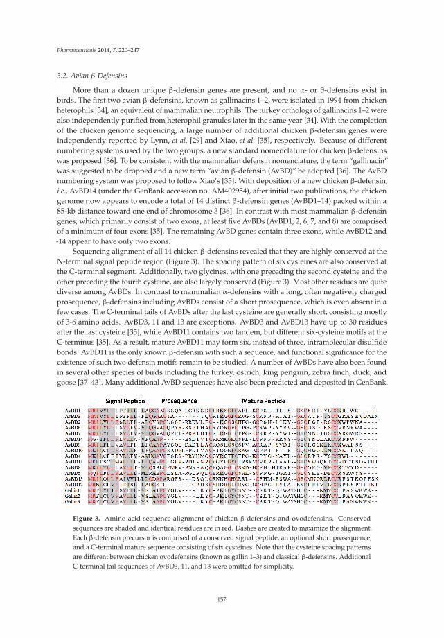
Pharmaceuticals 2014, 7, 220–247
3.2. Avian β-Defensins
More than a dozen unique β-defensin genes are present, and no α- or θ-defensins exist inbirds. The first two avian β-defensins, known as gallinacins 1–2, were isolated in 1994 from chickenheterophils [34], an equivalent of mammalian neutrophils. The turkey orthologs of gallinacins 1–2 werealso independently purified from heterophil granules later in the same year [34]. With the completionof the chicken genome sequencing, a large number of additional chicken β-defensin genes wereindependently reported by Lynn, et al. [29] and Xiao, et al. [35], respectively. Because of differentnumbering systems used by the two groups, a new standard nomenclature for chicken β-defensinswas proposed [36]. To be consistent with the mammalian defensin nomenclature, the term “gallinacin”was suggested to be dropped and a new term “avian β-defensin (AvBD)” be adopted [36]. The AvBDnumbering system was proposed to follow Xiao’s [35]. With deposition of a new chicken β-defensin,i.e., AvBD14 (under the GenBank accession no. AM402954), after initial two publications, the chickengenome now appears to encode a total of 14 distinct β-defensin genes (AvBD1–14) packed within a85-kb distance toward one end of chromosome 3 [36]. In contrast with most mammalian β-defensingenes, which primarily consist of two exons, at least five AvBDs (AvBD1, 2, 6, 7, and 8) are comprisedof a minimum of four exons [35]. The remaining AvBD genes contain three exons, while AvBD12 and-14 appear to have only two exons.
Sequencing alignment of all 14 chicken β-defensins revealed that they are highly conserved at theN-terminal signal peptide region (Figure 3). The spacing pattern of six cysteines are also conserved atthe C-terminal segment. Additionally, two glycines, with one preceding the second cysteine and theother preceding the fourth cysteine, are also largely conserved (Figure 3). Most other residues are quitediverse among AvBDs. In contrast to mammalian α-defensins with a long, often negatively chargedprosequence, β-defensins including AvBDs consist of a short prosequence, which is even absent in afew cases. The C-terminal tails of AvBDs after the last cysteine are generally short, consisting mostlyof 3-6 amino acids. AvBD3, 11 and 13 are exceptions. AvBD3 and AvBD13 have up to 30 residuesafter the last cysteine [35], while AvBD11 contains two tandem, but different six-cysteine motifs at theC-terminus [35]. As a result, mature AvBD11 may form six, instead of three, intramolecular disulfidebonds. AvBD11 is the only known β-defensin with such a sequence, and functional significance for theexistence of such two defensin motifs remain to be studied. A number of AvBDs have also been foundin several other species of birds including the turkey, ostrich, king penguin, zebra finch, duck, andgoose [37–43]. Many additional AvBD sequences have also been predicted and deposited in GenBank.
Figure 3. Amino acid sequence alignment of chicken β-defensins and ovodefensins. Conservedsequences are shaded and identical residues are in red. Dashes are created to maximize the alignment.Each β-defensin precursor is comprised of a conserved signal peptide, an optional short prosequence,and a C-terminal mature sequence consisting of six cysteines. Note that the cysteine spacing patternsare different between chicken ovodefensins (known as gallin 1–3) and classical β-defensins. AdditionalC-terminal tail sequences of AvBD3, 11, and 13 were omitted for simplicity.
157

Pharmaceuticals 2014, 7, 220–247
Besides classical β-defensins, chickens have also been found to express three closely relatedβ-defensin-related peptides, namely gallin 1–3 [23,44,45]. In fact, multiple peptides with a similarcysteine-spacing pattern have been reported earlier in the mallard duck, turkey, and black swan [46–48]. This group of peptides were in turn classified as ovodefensins because of their preferentialexpression in the oviduct with abundant presence in egg white [23]. Ovodefensins appear to beavian-specific, containing a six-cysteine motif of C-X3-5-C-X3-C-X11-C-X3-4-CC, as opposed to that ofC-X4-8-C-X3-5-C-X9-13-C-X4-7-CC in classical avian β-defensins (Figure 3).
4. Evolution of Avian HDPs
4.1. Avian Cathelicidins
Phylogenetic analysis of all publically available avian cathelicidins revealed that they are clusteredinto three distinct clades, namely CATH1/3, CATH2, and CATH-B1 (Figure 4), suggesting that thesethree clades of cathelicidin genes have evolved before divergence of these bird species from each other,unlike mammalian cathelicidin genes, a majority of which were duplicated after species separation [18].Surprisingly, an initial analysis of the zebra finch (Taeniopygia guttata) genome failed to identify anycathelicidin sequences [39]. If that is the case, it will be interesting to know why the cathelicidin geneswere lost in zebra finch, although it is likely that the genome segments containing the cathelicidingenes failed to be sequenced, because the entire genome were only sequenced to 5.5 × coverage toencompass 94% of the genome [49]. Consistent with our assumption, scores of gaps are present inthe syntenic cathelicidin region in the current zebra finch genome assembly (WUGSC 3.2.4/taeGut1)released in July 2008.
Figure 4. Phylogenetic analysis of avian cathelicidins. The phylogenetic tree was constructed with thefull-length amino acid sequences using the neighbor-joining method, and the reliability of each branchwas assessed by using 1,000 bootstrap replications. Numbers on the branches indicate the percentageof 1,000 bootstrap samples supporting the branch. The species and GenBank accession number of eachsequence are indicated.
158
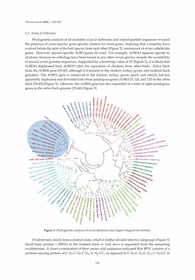
Pharmaceuticals 2014, 7, 220–247
4.2. Avian β-Defensins
Phylogenetic analysis of all available avian β-defensins and related peptide sequences revealedthe presence of cross-species, gene-specific clusters for most genes, implying that a majority haveevolved before the split of the bird species from each other (Figure 5), reminiscent of avian cathelicidingenes. However, species-specific AvBD genes do exist. For example, AvBD14 appears specific tochickens, because no orthologs have been found in any other avian species, despite the availabilityof several avian genome sequences. Supported by a bootstrap value of 52 (Figure 5), it is likely thatAvBD14 duplicated from AvBD13 after the separation of chickens from other birds. Zebra finchlacks the AvBD6 gene [39,40], although it is present in the chicken, turkey, goose, and mallard duckgenomes. The AvBD1 gene is conserved in the chicken, turkey, goose, quail, and ostrich, but hasapparently duplicated and diversified into three paralogous genes (AvBD123, 124, and 125) in the zebrafinch [39,40] (Figure 5). Likewise, the AvBD3 gene has also expanded to a total of eight paralogousgenes in the zebra finch genome [39,40] (Figure 5).
Figure 5. Phylogenetic analysis of avian defensins (see Figure 4 legend for details).
Ovodefensins clearly form a distinct clade, which is further divided into two subgroups (Figure 5).Small basic protein 1 (BPS1) in the mallard duck or rock dove is separated from the remainingovodefensins. A closer examination of their amino acid sequences indicated that BPS1 consists of acysteine-spacing pattern of C-X3-C-X3-C-X11-C-X4-CC, as opposed to C-X5-C-X3-C-X11-C-X3-CC in
159
Pharmaceuticals 2014, 7, 220–247
other ovodefensins. It remains to see whether these two subgroups of ovodefensins are present onlyin certain species of birds. It is also important to know how they are functionally different from eachother and from classical defensins.
Importantly, ovodefensins are clustered with AvBD12 as supported by a bootstrap value of 32(Figure 5), suggesting that ovodefensins might have duplicated and diversified from AvBD12 as aresult of gene duplication after separations of birds from other animal species. Consistent with thenotion that ovodefensins are derived from classical AvBDs, three chicken ovodefensin/gallin genesare located in tandem on chromosome 3, approximately 260 kb centromeric to the AvBD gene clusteron the current chicken genome assembly. To further support a possible origination of gallins fromAvBD12, the gallin genes consist of two exons separated by an intron, which is identical to the genomicstructure of the AvBD12 and AvBD14 genes, whereas all other AvBD genes are comprised of at leastthree exons [35].
5. Expression and Regulation of Avian HDPs
5.1. Tissue Expression Pattern
Like mammalian counterparts, avian cathelicidins and β-defensins are derived from the bonemarrow and/or epithelial cells, with the majority expressing in a wide variety of tissues. CATH1–3 areprimarily of myeloid origin, while CATH-B1, a distant member of avian cathelicidins, is derived fromepithelial cells. Chicken CATH1–3 mRNAs are predominantly expressed in the bone marrow, but alsothroughout the mucosal tissues of the digestive, respiratory, and urogenital tracts [29,30,50]. On theother hand, chicken CATH-B1 mRNA shows a more restricted expression pattern, with preferentialexpression in the secretory epithelial cells of the bursa of Fabricius [31,50]. Consist with the roleof cathelicidins in the first line of host defense, abundant CATH1–3 proteins can be detected in thegranules of heterophils, as in the case of chicken CATH2 [51], whereas mature CATH-B1 protein issecreted from the epithelial cells and concentrated on the basolateral surfaces of the M cells in thebursal lymphoid follicles [31].
Myeloid avian β-defensins include AvBD1, 2, and 4–7, whereas the remaining AvBD8–14 aremainly of epithelial origin, although both myeloid and epithelial AvBDs are also expressed in a majorityof other tissues [29,35,52]. In agreement with their myeloid origin, AvBD1 and AvBD2 mRNAs havebeen found abundantly in the bone marrow and their proteins in heterophil granules in the chickens,turkey, and ostrich [34,42,53]. It will be interesting to know the tissue expression pattern of thosespecies-specific AvBDs such as AvBD115–125 in the zebra finch [40]. However, because they areorthologous to AvBD1 and AvBD3 (Figure 5), they are expected to share a similar expression pattern toAvBD1 and AvBD3.
Human cathelicidin LL-37 has been found in seminal plasma associated with sperm andprostasomes [54]. A majority of β-defensins in rats (and likely in other mammalian species aswell) are expressed preferentially in the male reproductive system and the epididymis and testisin particular [55]. Like their mammalian counterparts, many avian cathelicidins and β-defensins areexpressed adequately in the male and female reproductive organs, particularly in the testis, epididymis,ovary, and oviduct [29,35,50,56], suggesting a possible role in reproduction. Ovodefensins have beenfound to be among the major components of egg white in the chicken, turkey, and duck [44–48].Consistently, chicken ovodefensin gallin 1–3 mRNA and proteins are the most abundantly expressedin tubular gland cells in the magnum of the chicken oviduct [23], a segment that secretes egg white.
5.2. Developmental Regulation
The expression of chicken cathelicidins and β-defensins has been studied during the pre- andpost-hatch periods and found to be developmentally regulated. At the embryonic stage, most chickencathelicidin and AvBD mRNAs were detected as early as embryonic day 3 (E3), except for CATH-B1and AvBD11, which did not appear until day E9 [57]. All four cathelicidin mRNA expression was
160

Pharmaceuticals 2014, 7, 220–247
generally increased as embryos develops, whereas the 14 β-defensins were differentially expressed [57].AvBD3, 4, 5, 10, 11, 12, and 14 were largely enhanced during the embryonic development, whereas theremaining chicken β-defensins showed a biphasic expression pattern. In the case of AvBD2, 6, and 7,their expression was increased on day E6 relative to that in day E3, decreased on day E9, and thenincreased gradually with the age of embryos [57].
After hatch, chicken cathelicidins and β-defensins are also developmentally regulated in bothgene- and tissue-specific patterns. During the first 28 days, CATH1–3 showed an age-dependentincrease both in the cecal tonsil and lung, whereas all four cathelicidins were peaked in the bursa onday 4 after hatching, with a gradual decline by day 28 [50]. On the other hand, CATH1–3 showed apeak expression in the cecum on day 28, while the highest expression of CATH-B1 was seen in boththe lung and cecal tonsil on day 14 [50]. AvBD1 and AvBD2 mRNA gradually reduced in differentsegments of the intestinal tract in the first week post-hatch, but restored and increased gradually inthe following week [58]. In the reproductive tract, more than a half of AvBDs increased during thesexual maturation in the vagina, ovary, and epididymis of chickens, whereas others showed little noexpression [56,59,60].
5.3. Regulation by Infection and Inflammation
HDPs are critically involved in the first line of host defense. Dysregulation of the HDP synthesisoften leads to immune deficiency or autoimmunity such as Crohn’s disease and psoriasis in humans [61,62]. Many, but not all, HDPs are induced upon infection and inflammation in humans and mice. On theother hand, certain pathogens suppress HDP synthesis as a strategy to evade the immune system [63–65]. A number of studies have been conducted on the transcriptional response of cathelicidins andβ-defensins in chickens and several other avian species. Like their mammalian relatives, multipleAvBDs are inducible in response to microbial products (e.g., lipopolysaccharides and CpG DNA), livebacteria, viruses or parasites in the intestinal, reproductive, and respiratory tracts [38,56,59,60,66,67].However, it appears that many avian HDP genes are regulated differently in different tissues inresponse to different stimuli. For example, chicken CATH1 was induced in the cecal tonsil [66], but notthe jejunum of chickens in response to Salmonella infections [51]. On the other hand, chicken CATH1was down-regulated by Campylobacter jejuni or Eimeria praecox [68–70], perhaps as a mechanism ofimmune evasion.
5.4. Regulation by Dietary Compounds
Because suppressing HDP expression is a microbial strategy for immune subversion, inducing thesynthesis of endogenous HDPs will conversely augment the host capacity to fight off infections [71–73].Unlike infection or injury that triggers HDP expression with an unwanted and often exaggeratedinflammatory response, butyrate and vitamin D3 have been found to be highly potent in augmentingHDP synthesis without provoking inflammation in humans [74–78]. Several other compounds likedietary histone deacetylase inhibitors, retinoic acid, forskolin, and sugars are also capable of inducingHDP expression in humans [79–83].
In chickens, butyrate has been revealed as a strong inducer of HDP expression in vitro andin vivo. Among all 14 AvBDs and 4 cathelicidins, half of them were induced with others largelyunchanged in chicken cells in response to butyrate [84]. Butyrate was further shown to enhance theantibacterial activity of chicken monocytes, and supplementation of butyrate in the feed enhancedclearance of Salmonella enteritidis in the cecum of chickens following an experimental infection [84].Among all saturated free fatty acids with 1–18 carbons, butyrate was the most potent in stimulatingHDP expression in chicken cells [85]. Furthermore, butyrate synergizes with the agonists of the cyclicadenosine monophosphate (cAMP) signaling pathway in inducing HDP expression [86]. Feeding withbutyrate and a plant extract containing forskolin, which is an adenylyl cyclase agonist, showed a strongsynergy in augmenting HDP expression in the crop and jejunum of chickens [86]. Mitogen-activatedprotein kinase signaling pathways were revealed to be critically involved in the HDP-inducing synergy
161

Pharmaceuticals 2014, 7, 220–247
between butyrate and forskolin [86]. The results indicated the potential for use of these dietarycompounds in promoting HDP synthesis, host immunity, and disease resistance.
6. Biological Activities of Avian HDPs
6.1. Antimicrobial Activities
The antibacterial efficacy of all four chicken cathelicidins and many defensins have been evaluated.Like their mammalian counterparts, most chicken HDPs are capable of killing a broad spectrum ofGram-positive and Gram-negative bacteria, and fungi including antibiotic-resistant strains generallyin the low micromolar range. For example, chicken CATH1–3 are broadly active with the minimuminhibitory concentration (MIC) values mostly between 0.5 and 2 μM against a range of bacteria [18,87], and CATH-B1 also has the MIC values between 0.5 and 2.5 μM against E. coli, S. aureus, andP. aeruginosa [31]. However, many HDPs showed varying efficiencies against different pathogens.Chicken AvBD1 and AvBD2 kill 90% S. enteriditis, C. jejuni, and Candida albicans at <4 μM, but showeda much reduced efficiency against Pasteurella multocida [41]. Similarly, AvBD9 is active against mostGram-positive and Gram-negative bacteria tested with the MIC values in the range of 2–4 μM, butwith a minimum activity against S. typhimurium (>30 μM) [88]. On the other hand, AvBD13 was foundto be minimally active against a range of bacteria examined, with the MIC values in the range of 50–100μM [89]. The antibacterial activity of chicken CATH1–3 is not affected by the presence of physiologicalconcentrations of salt [18]; however, that of chicken defensins is greatly reduced by salt [26], reminiscentof mammalian defensins [13]. In the case of CATH1–3, AvBD2, and presumably most other avianHDPs, non-specific membrane disruption and lysis is a major bactericidal mechanism [87,90,91].
Ovodefensins are unique in that they generally lack an obvious antibacterial activity as seenwith turkey meleagrin as well as duck BPS1 and BPS2 [46,48]. Chicken gallin is the only ovodefensinwith known antibacterial activities, but appears to have a narrow range. Among several commonGram-negative and Gram-positive bacteria tested, chicken gallin1/2 showed an activity only against E.coli, but not Salmonella, S. aureus or Listeria monocytogenes [24]. It is hence very unlikely that the majorbiological function of ovodefensins is antibacterial.
6.2. Immunomodulatory Activities
Besides having direct microbicidal activities, HDPs have increasingly been appreciated to playa profound role in regulating host immune responses to infections. Many peptides are shown tobe actively involved in chemotaxis and activation of immune cells, regulation of dendritic celldifferentiation, induction of angiogenesis and re-epithelialization, modulation of cytokine andchemokine gene expression, and potentiation of antigen-specific adaptive immune response [5,92,93].Importantly, many HDPs directly bind to and neutralize bacterial membrane components suchas lipopolysaccharides (LPS), lipotechoic acid, and peptidoglycan and suppress the production ofproinflammatory cytokines induced by bacteria and membrane components [94,95].
In chickens, CATH1 was shown to possess excellent immunomodulatory properties with a strongcapacity to specifically chemoattract neutrophils without affecting the migration of monocytes orlymphocytes [96]. Furthermore, CATH1 and CATH2 activates macrophages or peripheral bloodmononuclear cells by inducing synthesis of an array of cytokines and chemokines at moderate levels,which is distinct from that induced by LPS [96,97], CATH1–3 were shown to bind to LPS directly,with 50% binding occurring at approximately 10 μM [18,87]. Moreover, three peptides at 10–20 μMsubstantially abrogated LPS-induced production of proinflammatory cytokines in macrophages andperipheral blood mononuclear cells [18,87,97]. CATH1 was further found to augment adaptive immuneresponse when administered into mice together with chicken ovalbumin, a model antigen [96]. Inthe case of human cathelicidin LL-37, glyceraldehyde 3-phosphate dehydrogenase (GAPDH) andsequestosome-1/p62 were recently identified as intracellular receptors to mediate cytokine/chemokine
162

Pharmaceuticals 2014, 7, 220–247
production in monocytes [98,99]. It will be interesting to examine whether chicken cathelicidins alsoutilize the same receptors to modulate the macrophage response.
7. Structures-Activity Relationships of Avian HDPs
7.1. Structural Features
To date, the tertiary structures of three chicken cathelicidins (CATH1–3), two β-defensins (chickenAvBD2 and penguin AvBD103a/spheniscin-2), and a chicken ovodefensin (gallin1/2) have beendetermined by nuclear magnetic resonance (NMR) in solutions. Unlike mammalian cathelicidinsthat adopt various conformations, all three chicken cathelicidins are largely α-helical with a mildkink or rather extensive bend around the center in aqueous solutions [87,90,100] (Figure 6). A mildkink in CATH1 and CATH3 is induced by the presence of a glycine residue, whereas an extensivebend in CATH2 is caused by proline (Figure 6A). Additionally, all three chicken cathelicidins consistof a flexible unstructured segment at the N-terminal region [87,90,100]. Unlike typical amphipathicα-helical HDPs, no obvious segregation of hydrophobic residues from hydrophilic residues is seenwith either CATH1 or CATH2; instead, the positively charged residues are mostly concentrated at bothends (Figure 6B). On the other hand, CATH2 are rather amphipathic throughout the entire α-helix.Three truncated analogs of chicken CATH1 consisting of amino acid residues 1–16, 8–26, and 5–26were also revealed to adopt similar confirmations to the full-length peptide in the presence of LPS orzwitterionic dodecylphosphocholine micelles [101,102].
Figure 6. Structures of avian cathelicidins and β-defensins. (A) Secondary structural features ofcathelicidins and β-defensins. (B) Tertiary ribbon structures of cathelicidins and β-defensins. Polarresidues are indicated in blue and nonpolar residues in red. Disulfide bonds of β-defensins are shownin yellow. Protein Data Bank identification number for each molecule is indicated in parenthesis.
163

Pharmaceuticals 2014, 7, 220–247
Both penguin AvBD103a and chicken AvBD2 adopt a triple-stranded, antiparallel β-sheet structurestabilized by three pairs of intramolecular disulfide bonds [91,103] (Figure 6B), typical of mammalianβ-defensins. Penguin AvBD103a also consists of an α-helical segment at the N-terminal region witha hydrophobic patch on the surface [103]. However, AvBD2 lacks either the α-helical or an obviousamphipathic feature [91]. A β-bulge that is formed by the G-X-C motif around the fourth cysteineand highly conserved in both mammalian α- and β-defensins is also present in both AvBD2 andAvBD103a (Figure 6B). Albeit with a different cysteine-spacing pattern, chicken gallin1/2 also consistsof a characteristic β-defensin fold with three antiparallel β-sheets [24]. However, unlike classicalβ-defensins, gallin1/2 is comprised of two additional β-sheets formed separately by Val2-Leu3 at theN-terminal end and Thr24-Ser25 preceding the fourth cysteine [24] (Figure 6).
7.2. Structure-Activity Relationships
Structure-activity relationship (SAR) studies with α-helical HDPs indicated that the antimicrobialpotency and target specificity are strongly influenced by structural and physicochemical parameters,such as cationicity (net charge), helicity, amphipathicity, and hydrophobicity [11,104,105]. However, ingeneral there is no simple correlation between any of these physicochemical properties and peptidefunctions. A delicate balance of these parameters often dictates the antimicrobial potency and targetselectivity [11,104,105]. Within a certain range, an improvement in these parameters is often positivelycorrelated with the antimicrobial activity of peptides, but sometimes is accompanied by unwantedenhancement in cytotoxicity as well [104–107]. In several cases, the antimicrobial domain of thepeptides is located separately from the domain responsible for cytotoxicity [108,109], meaning that thepeptide derivatives devoid of the lytic domain could be identified with improved therapeutic potential.
In the case of β-sheet HDPs and defensins in particular, antimicrobial and immunomodulatoryactivities are strongly influenced by structural integrity, cationicity, and hydrophobicity [11,110,111].The presence of three intramolecular disulfide bonds in many cases is dispensable for the antibacterialactivity, but essential for other activities such as chemotactic activity [112] and the ability to resistproteolysis [113]. On the contrary, the antibacterial activity of human β-defensin-1 drastically increaseswhen the peptide is reduced [114]. In fact, some defensins may be naturally reduced in the intestinaltract by thioredoxin, a redox enzyme [114]. Cationicity of defensins is believed to dictate the killing ofGram-negative bacteria, whereas hydrophobicity appears to confer the activity against Gram-positivebacteria [111].
In avian species, a series of SAR studies with chicken CATH1, CATH2, and AvBD2 haveyielded some very interesting observations. Investigations of chicken CATH1 analogs with eitherN- or C-terminal deletions revealed that the cationic residues at both N- and C-terminal regions aredispensable for the antibacterial, LPS-binding, and cytotoxic activities, whereas the C-terminal helix(Arg21-Lys25) is essential for all three activities [100]. Tryptophan at position 6 (Trp6) is critical in bothLPS binding and cytotoxicity [100,115], but is dispensable for neutrophil chemotaxis [96]. Furthermore,an omission of Trp6 in CATH1 also resulted in an obvious reduction in its ability to kill bacteria [115]and induce chemokine synthesis in macrophages [96]. Replacing the kink-causing glycine (Gly16)with a helix-stabilizing residue, leucine, resulted in no obvious difference in either antibacterial,LPS-binding or cytotoxic activity [100], indicating that enhancing the helicity of α-helical HDPs maynot necessarily result in an improvement in the antibacterial potency. Simultaneous substitutions ofmultiple amino acid residues to make CATH1 nearly perfectly amphipathic surprisingly caused aloss of the antibacterial potency against certain bacteria and undesirably, an increase in hemolyticactivities [100]. Collectively, these findings suggested that a fine tuning of various structural andphysicochemical parameters including cationicity, helicity, hydrophobicity, and amphipathicity, ratherthan a simple alteration of one, will result in an enhancement in the therapeutic potential of α-helicalHDPs, which is in agreement with earlier findings [11,104,105].
Studies with a series of CATH2 analogs with deletions of either N- or C-terminal residues showedthat neither α-helical segment per se is sufficient to bind LPS, kill bacteria or lyse mammalian cells [90].
164

Pharmaceuticals 2014, 7, 220–247
Inclusion of four additional amino acids in the central bending region (Arg15-Arg18) beyond either N-or C-terminal α-helical segment to the α-helical segment was associated with a significant enhancementin both antibacterial and LPS-neutralizing activities [90]. To directly evaluate the functional significanceof the central bending segment, a substitution of leucine for proline greatly reduced the antibacterialand hemolytic activities. The abilities to neutralize LPS-induced cytokine production and to stimulatechemokine synthesis in peripheral blood mononuclear cells were also significantly impaired by such aproline-to-leucine substitution [97], implying that the bending is critically important in the peptideinteractions with membranes as well as the cell activation receptors. Interestingly, a gradual increasein cationicity, helicity and amphipathicity among all peptide analogs led to a gradual enhancement inantibacterial potency and LPS neutralization [90]. Furthermore, substitution of multiple tryptophansfor phenylalanines in an N-terminal, 15-amino acid fragment of CATH2 led to an improvement inantibacterial and LPS-neutralizing activities [116]. Head-to-tail cyclization of this CATH2 variantfurther increased its serum stability with a reduced cytotoxicity [117]. The same study also revealedthat D-amino acid substitutions rendered the peptide completely resistant to trypsin proteolysis [117].
In the case of chicken AvBD2, D-enantiomerization resulted in little difference in the activityagainst both Gram-positive and Gram-negative bacteria, suggesting that membrane is the primarytarget [91]. However, unlike many mammalian defensins whose structural integrity has a minimumimpact on the antibacterial activity, reducing AvBD2 led to a drastic loss in the activity againstGram-positive bacteria, but was less prominent against Gram-negative bacteria [91]. Substitution ofalanine for a conserved lysine following the sixth cysteine resulted in an obvious N-terminal structuralmodification and a marked decrease in the antibacterial activity against both Gram-positive andGram-negative bacteria [91]. Surprisingly, a reduction of disulfide bonds rendered the lysine-alaninesubstituted AvBD2 nearly completely inactive in killing bacteria [91]. Therefore, conformationalchanges (and subsequent changes in hydrophobicity and/or amphipathicity) are likely the underlyingmechanism behind many of the alterations in the antibacterial activity of AvBD2.
8. Potential Therapeutic Applications
HDPs including avian HDPs can be potentially used in a variety of applications such asantimicrobial therapy (Figure 7). Additionally, HDPs hold promise in augmenting the efficacy ofvaccines as adjuvants. Although a number of avian HDPs are expressed in both male and femalereproductive tracts and believed to promote sperm maturation and fertility like their mammaliancounterparts [10,118], experimental evidence is yet to prove the link. Therefore, the potential roleand application of avian HDPs in infertility treatment will not be discussed here. To reduce the costof delivering synthetic peptides and minimize peptide degradation, endogenous HDPs can also beinduced by certain cost-effective dietary compounds to help the host better fight off infections (Figure 7).
Figure 7. Potential therapeutic applications of host defense peptides (HDPs). Synthetic HDPscan be directly administered exogenously as antimicrobials, vaccine adjuvants or infertility drugs.Alternatively, endogenous HDPs can modulated by dietary compounds for antimicrobial therapies.
165

Pharmaceuticals 2014, 7, 220–247
8.1. Antimicrobial Therapies
HDPs are active against a broad range of bacteria, mycobacteria, fungi, and parasites [1,8]. Ratherthan relying on a single or a limited number of intracellular targets like most currently availableantibiotics, HDPs kills microbes primarily through physical electrostatic interactions and membranedisruption. Therefore, it is difficult for microbes to gain resistance to HDPs [1,8]. At the same time,most HDPs have the capacity to recruit and activate immune cells and facilitate the resolution ofinflammation [6,8]. In fact, the antibacterial and immunomodulatory properties of HDPs can beharnessed separately for antimicrobial therapy, particularly against antibiotic-resistant strains [6,8]. Afew HDPs have been evaluated clinically for their antibacterial efficacy, and several more are currentlyat different stages of human trials. Because of a relatively low efficacy as compared with many of theconventional antibiotics, all clinical trials with HDPs have met a limited success [119]. As a result, noHDPs have been approved by the FDA to date. More efforts are being shifted toward exploring theimmune regulatory activities of HDPs. Excitedly, several small HDPs with no or weak antibacterialactivities have been proved to be highly efficient in protecting animals from infections by recruitingand activating neutrophils and/or monocytes [95,120]. Because they act on the host but not on thepathogens, these immunomodulatory peptides have the potential to control a broad spectrum ofpathogens without triggering resistance.
In avian species, only chicken CATH1 has been evaluated for its in vivo antibacterial efficacy.A single intraperitoneal administration of a C-terminal, 21-amino acid CATH1 peptide analog,known as fowlicidin-1 (6–26), led to a 50% increase in the survival of mice from a lethal dose ofmethicillin-resistant S. aureus (MRSA), concomitant with a reduction in the bacterial titer in bothperitoneal fluids and spleens of mice [115]. Additionally, fowlicidin-1(6–26) is more potent in inducingneutrophil chemotaxis and macrophage activation than human cathelicidin LL-37 and a de novosynthesized peptide, IDR-1 [96]. Because of its ability to induce neutrophil chemotaxis and macrophageactivation, fowlicidin-1(6–26) protected 50% mice if given 4 days prior to an otherwise lethal MRSAinfection, and 100% mice survived if the peptide was received 1 or 2 days before infection [96]. This isthe first HDP that has been shown to protect animals from bacterial infections beyond a 48-h window.Therefore, fowlicidin-1(6–26) represents an attractive candidate for further exploration as a novelantimicrobial for both therapeutic and prophylactic applications. Rational design and functionalscreening of additional CATH1-related peptides may lead to identification of new peptide analogswith improved safety and therapeutic potential, particularly against antibiotic-resistant pathogens.
It is worth noting that many HDPs and defensins in particular have obvious antiviral effects byacting as lectins or by modulating host cell responses. Enveloped and non-enveloped viruses suchas human immunodeficiency virus (HIV-1), influenza A virus (IAV), cytomegalovirus (CMV), herpessimplex virus (HSV-1 and HSV-2), vesicular stomatitis virus (VSV) , adenovirus, and papillomavirus(HPV) have been shown to be sensitive to human α-, β- and/or θ-defensins [121,122]. In somecases, defensins can directly inactivate viruses by disrupting envelop lipid bilayers, aggregating viralglycoproteins or blocking the binding of viruses to host cell receptors [122]. In other cases, the antiviraleffects of defensins are indirectly mediated by modifying host cell responses such as inhibition ofprotein kinase C (PKC) activation or down-regulation of host cell receptor expression [122]. Limitedwork exists on the antiviral activities of avian HDPs. Only a few duck β-defensins were recently shownto inhibit the replication of duck hepatitis virus (DHV) [37,67]. However, the antiviral mechanisms orthe susceptibility of other viruses to avian HDPs remains unknown, but warrant further investigations.
8.2. Vaccine Adjuvants
HDPs have been shown to profoundly impact the development of adaptive immune response byregulating the migration, maturation, and activation of different immune cell types including dendriticcells and T and B lymphocytes [5,8,123]. Several HDPs are capable of enhancing antigen-specificadaptive immune response when co-administered with vaccines [123]. HDPs have been found tosynergize with other adjuvants like CpG DNA and polyphosphazene in potentiating adaptive immune
166

Pharmaceuticals 2014, 7, 220–247
response [124–126]. The adjunvanticity of chicken AvBD1, duck AvBD2, and chicken CATH1 havebeen experimentally verified. When fused with the infectious bursal disease virus (IBDV) VP2 genein a DNA vaccine, chicken AvBD1 increased the VP2-specific antibody titers, CD4+ and CD8+ Tcell populations, and conferred better protection against an infectious bursal disease virus (IBDV)challenge in chickens [127]. Duck AvBD2 was shown to be chemotactic to T- and B-lymphocytesin vitro, with the ability to suppress the mRNA expression of an inhibitory receptor, namely dendriticcell immunoreceptor (DCIR), in duck splenocytes [128]. Chicken CATH1, when co-administered withchicken ovalbumin (OVA), was found to enhance both IgG1 and IgG2a titers to OVA in mice [96].Because CATH 1 was more potent than LL-37 or IDR-1 in inducing surface expression of CD86, aco-stimulatory molecule, on macrophages [96], CATH1 may be more efficient in promoting antigenpresentation and adaptive immunity and therefore, represent an excellent candidate as an adjuvant ora component of an adjuvant complex.
8.3. Direct Administration vs. Indirect Modulation
The production cost and stability are two major obstacles for in vivo applications of manypeptide-based drugs. Purification from natural sources or chemical synthesis are inefficient andcost-prohibitive for large-scale production of HDPs. Although recombinant expression of HDPs hasbeen achieved in bacteria and yeasts, an inclusion of a fusion protein is often needed to reducethe peptide toxicity to the host and aid in the peptide solubility [129]. In order to achieve themaximum activity, an extra proteolytic cleavage step and additional production costs are oftenunavoidable. Given a short half-life of most natural peptides in vivo, it is desirable to retard the peptidedegradation by chemical modifications that often involves the use of D-amino acids, cyclization orpeptidomimetics [130]. However, it remains unknown how those modifications would impact theimmunomodulatory functions of HDPs, which appear to be receptor-dependent.
To overcome the high manufacturing cost and minimize degradation of HDPs for in vivoapplications, it is advantageous to develop convenient and cost-effective strategies to specificallyinduce the synthesis of endogenous HDPs. Several dietary compounds including short-chain fattyacids and vitamin D3 have shown promise in stimulating HDP synthesis in humans without triggeringinflammatory response [131,132]. Dietary supplementation of HDP-inducing compounds has emergedas a novel antibiotic-alternative approach to antimicrobial therapy [131,132]. In chickens, butyrate,structural analogs of butyrate, and cAMP signaling agonists have been shown to be potent inducersof HDPs [84–86]. Desirably, butyrate and cAMP agonists are synergistic in augmenting HDP geneexpression and bacterial clearance in chickens [86], suggesting their potential as alternatives to antibioticsfor disease control and prevention. However, dietary regulation of HDPs often exhibit gene-, cell-, andspecies-specific patterns. HDP genes are differentially regulated in response to a dietary compound,with some being induced and others unaltered. In chickens, approximately a half number of chickenHDPs are induced by butyrate [84]. Some HDPs are regulated in a cell-specific pattern. For example,chicken AvBD9 gene increased by more than 5000-fold in HD11 macrophages, but with only a lessthan 10-fold induction in cecal intestinal cells after a 24-h exposure to 4 mM butyrate [84]. The samecompound that show a strong HDP-inducing activity in one animal species, may completely lose itsability to induce HDPs in another species. A case in point is vitamin D3, which strongly augmentscathelicidin gene expression in human but not mouse cells [75,76]. Therefore, it is important to evaluatethe HDP-inducing efficacy of individual compounds in different species.
9. Conclusions
Birds harbor approximately 20 unique cathelicidins and β-defensin genes in each species.It appears that most have evolved before divergence of birds from each other. Unlike mammaliancathelicidins that adopt different tertiary structures, avian cathelicidins are mostly α-helical, witha hinge around the central region and a flexible N-terminal segment. Besides classical β-defensins,a group of avian-specific, β-defensin-related peptides, namely ovodefensins, exist with a different
167

Pharmaceuticals 2014, 7, 220–247
cysteine-spacing pattern. However, the overall three-dimensional structure of ovodenfensins resemblethat of β-defensins. Coupled with their close chromosomal proximity with the β-defensin genecluster, ovodefensins were clearly diversified from a β-defensin ancestral gene, possibly AvBD12, afterseparation of the birds from other vertebrate species. Several avian HDPs have been shown to possesspotent, broad-spectrum antibacterial activities with a strong ability to modulate the host response toinfection and inflammation. Structure-activity relationship studies have led to the identification of afew avian HDPs and their analogs as promising candidates as antimicrobials or vaccine adjuvants.Because avian HDPs can be induced by dietary compounds such as short-chain fatty acids and cAMPsignaling agonists, dietary modulation of endogenous HDP synthesis may have potential to be furtherexplored as a novel, cost-effective antimicrobial strategy.
Acknowledgments: Invaluable contributions from the past and current members of the Zhang Laboratory aregreatly appreciated. The research on host defense peptides in the Zhang Laboratory is supported by the U.S.Department of Agriculture grant (2008-35204-04544), Oklahoma Center for the Advancement of Science andTechnology grants (AR12.2-077 and HR12-051), and Oklahoma Agricultural Experiment Station project (H-2811).
Conflicts of Interest: The authors declare no conflict of interest.
References
1. Zasloff, M. Antimicrobial peptides of multicellular organisms. Nature 2002, 415, 389–395. [CrossRef]2. Pasupuleti, M.; Schmidtchen, A.; Malmsten, M. Antimicrobial peptides: Key components of the innate
immune system. Crit. Rev. Biotechnol. 2012, 32, 143–171. [CrossRef]3. Wang, G. Database-Guided Discovery of Potent Peptides to Combat HIV-1 or Superbugs. Pharmaceuticals
(Basel) 2013, 6, 728–758. [CrossRef]4. Brogden, K.A. Antimicrobial peptides: Pore formers or metabolic inhibitors in bacteria? Nat. Rev. Microbiol.
2005, 3, 238–250. [CrossRef]5. Yang, D.; Biragyn, A.; Hoover, D.M.; Lubkowski, J.; Oppenheim, J.J. Multiple roles of antimicrobial defensins,
cathelicidins, and eosinophil-derived neurotoxin in host defense. Annu. Rev. Immunol. 2004, 22, 181–215.[CrossRef]
6. Choi, K.Y.; Chow, L.N.; Mookherjee, N. Cationic host defence peptides: Multifaceted role in immunemodulation and inflammation. J. Innate Immun. 2012, 4, 361–370.
7. Hilchie, A.L.; Wuerth, K.; Hancock, R.E. Immune modulation by multifaceted cationic host defense(antimicrobial) peptides. Nat. Chem. Biol. 2013, 9, 761–768. [CrossRef]
8. Hancock, R.E.; Nijnik, A.; Philpott, D.J. Modulating immunity as a therapy for bacterial infections. Nat. Rev.Microbiol. 2012, 10, 243–254. [CrossRef]
9. Semple, F.; Dorin, J.R. β-Defensins: Multifunctional modulators of infection, inflammation and more?J. Innate Immun. 2012, 4, 337–348. [CrossRef]
10. Tollner, T.L.; Bevins, C.L.; Cherr, G.N. Multifunctional glycoprotein DEFB126—A curious story ofdefensin-clad spermatozoa. Nat. Rev. Urol. 2012, 9, 365–375. [CrossRef]
11. Takahashi, D.; Shukla, S.K.; Prakash, O.; Zhang, G. Structural determinants of host defense peptides forantimicrobial activity and target cell selectivity. Biochimie 2010, 92, 1236–1241. [CrossRef]
12. Zanetti, M. Cathelicidins, multifunctional peptides of the innate immunity. J. Leukoc. Biol. 2004, 75, 39–48.[CrossRef]
13. Selsted, M.E.; Ouellette, A.J. Mammalian defensins in the antimicrobial immune response. Nat. Immunol.2005, 6, 551–557. [CrossRef]
14. Ganz, T. Defensins: Antimicrobial peptides of innate immunity. Nat. Rev. Immunol. 2003, 3, 710–720.[CrossRef]
15. Kosciuczuk, E.M.; Lisowski, P.; Jarczak, J.; Strzalkowska, N.; Jozwik, A.; Horbanczuk, J.; Krzyzewski, J.;Zwierzchowski, L.; Bagnicka, E. Cathelicidins: Family of antimicrobial peptides. A review. Mol. Biol Rep.2012, 39, 10957–10970. [CrossRef]
16. Levy, O.; Ooi, C.E.; Elsbach, P.; Doerfler, M.E.; Lehrer, R.I.; Weiss, J. Antibacterial proteins of granulocytesdiffer in interaction with endotoxin. Comparison of bactericidal/permeability-increasing protein, p15s, anddefensins. J. Immunol. 1995, 1995, 5403–5410.
168

Pharmaceuticals 2014, 7, 220–247
17. Moscinski, L.C.; Hill, B. Molecular cloning of a novel myeloid granule protein. J. Cell. Biochem. 1995, 59,431–442. [CrossRef]
18. Xiao, Y.; Cai, Y.; Bommineni, Y.R.; Fernando, S.C.; Prakash, O.; Gilliland, S.E.; Zhang, G. Identification andfunctional characterization of three chicken cathelicidins with potent antimicrobial activity. J. Biol Chem.2006, 281, 2858–2867.
19. Zarember, K.A.; Katz, S.S.; Tack, B.F.; Doukhan, L.; Weiss, J.; Elsbach, P. Host defense functions ofproteolytically processed and parent (unprocessed) cathelicidins of rabbit granulocytes. Infect. Immun.2002, 70, 569–576. [CrossRef]
20. Lehrer, R.I. Primate defensins. Nat. Rev. Microbiol. 2004, 2, 727–738. [CrossRef]21. Andersson, M.L.; Karlsson-Sjoberg, J.M.; Putsep, K.L. CRS-peptides: Unique defense peptides of mouse
Paneth cells. Mucosal Immunol. 2012, 5, 367–376. [CrossRef]22. Patil, A.A.; Ouellette, A.J.; Lu, W.; Zhang, G. Rattusin, an intestinal alpha-defensin-related peptide in rats
with a unique cysteine spacing pattern and salt-insensitive antibacterial activities. Antimicrob. AgentsChemother. 2013, 57, 1823–1831. [CrossRef]
23. Gong, D.; Wilson, P.W.; Bain, M.M.; McDade, K.; Kalina, J.; Herve-Grepinet, V.; Nys, Y.; Dunn, I.C. Gallin; anantimicrobial peptide member of a new avian defensin family, the ovodefensins, has been subject to recentgene duplication. BMC Immunol. 2010, 11, 12. [CrossRef]
24. Herve, V.; Meudal, H.; Labas, V.; Rehault Godbert, S.; Gautron, J.; Berges, M.; Guyot, N.; Delmas, A.F.;Nys, Y.; Landon, C. 3D NMR structure of hen egg gallin (chicken ovo-defensin) reveals a new variation ofthe beta-defensin fold. J. Biol. Chem. 2014, in press.
25. Van Dijk, A.; Molhoek, E.M.; Bikker, F.J.; Yu, P.L.; Veldhuizen, E.J.; Haagsman, H.P. Avian cathelicidins:Paradigms for the development of anti-infectives. Vet. Microbiol. 2011, 153, 27–36. [CrossRef]
26. Van Dijk, A.; Veldhuizen, E.J.; Haagsman, H.P. Avian defensins. Vet. Immunol Immunopathol. 2008, 124, 1–18.[CrossRef]
27. Sugiarto, H.; Yu, P.L. Avian antimicrobial peptides: The defense role of beta-defensins. Biochem. Biophys. Res.Commun. 2004, 323, 721–727. [CrossRef]
28. Cuperus, T.; Coorens, M.; van Dijk, A.; Haagsman, H.P. Avian host defense peptides. Dev. Comp. Immunol.2013, 41, 352–369. [CrossRef]
29. Lynn, D.J.; Higgs, R.; Gaines, S.; Tierney, J.; James, T.; Lloyd, A.T.; Fares, M.A.; Mulcahy, G.; O’Farrelly, C.Bioinformatic discovery and initial characterisation of nine novel antimicrobial peptide genes in the chicken.Immunogenetics 2004, 56, 170–177. [CrossRef]
30. Van Dijk, A.; Veldhuizen, E.J.; van Asten, A.J.; Haagsman, H.P. CMAP27, a novel chicken cathelicidin-likeantimicrobial protein. Vet. Immunol. Immunopathol. 2005, 106, 321–327. [CrossRef]
31. Goitsuka, R.; Chen, C.L.; Benyon, L.; Asano, Y.; Kitamura, D.; Cooper, M.D. Chicken cathelicidin-B1, anantimicrobial guardian at the mucosal M cell gateway. Proc. Natl. Acad. Sci. USA 2007, 104, 15063–15068.[CrossRef]
32. Feng, F.; Chen, C.; Zhu, W.; He, W.; Guang, H.; Li, Z.; Wang, D.; Liu, J.; Chen, M.; Wang, Y.; Yu, H. Genecloning, expression and characterization of avian cathelicidin orthologs, Cc-CATHs, from Coturnix coturnix.FEBS J. 2011, 278, 1573–1584.
33. Wang, Y.; Lu, Z.; Feng, F.; Zhu, W.; Guang, H.; Liu, J.; He, W.; Chi, L.; Li, Z.; Yu, H. Molecular cloning andcharacterization of novel cathelicidin-derived myeloid antimicrobial peptide from Phasianus colchicus. Dev.Comp. Immunol. 2011, 35, 314–322. [CrossRef]
34. Evans, E.W.; Beach, G.G.; Wunderlich, J.; Harmon, B.G. Isolation of antimicrobial peptides from avianheterophils. J. Leukoc. Biol. 1994, 56, 661–665.
35. Xiao, Y.; Hughes, A.L.; Ando, J.; Matsuda, Y.; Cheng, J.F.; Skinner-Noble, D.; Zhang, G. A genome-widescreen identifies a single beta-defensin gene cluster in the chicken: Implications for the origin and evolutionof mammalian defensins. BMC Genomics 2004, 5, 56. [CrossRef]
36. Lynn, D.J.; Higgs, R.; Lloyd, A.T.; O'Farrelly, C.; Herve-Grepinet, V.; Nys, Y.; Brinkman, F.S.; Yu, P.L.;Soulier, A.; Kaiser, P.; et al. Avian beta-defensin nomenclature: A community proposed update. Immunol.Lett. 2007, 110, 86–89. [CrossRef]
37. Ma, D.; Zhang, K.; Zhang, M.; Xin, S.; Liu, X.; Han, Z.; Shao, Y.; Liu, S. Identification, expression and activityanalyses of five novel duck beta-defensins. PLoS One 2012, 7, e47743.
169

Pharmaceuticals 2014, 7, 220–247
38. Ma, D.; Zhang, M.; Zhang, K.; Liu, X.; Han, Z.; Shao, Y.; Liu, S. Identification of three novel avianbeta-defensins from goose and their significance in the pathogenesis of Salmonella. Mol. Immunol. 2013, 56,521–529. [CrossRef]
39. Cormican, P.; Lloyd, A.T.; Downing, T.; Connell, S.J.; Bradley, D.; O’Farrelly, C. The avian Toll-Like receptorpathway—Subtle differences amidst general conformity. Dev. Comp. Immunol. 2009, 33, 967–973. [CrossRef]
40. Hellgren, O.; Ekblom, R. Evolution of a cluster of innate immune genes (beta-defensins) along the ancestrallines of chicken and zebra finch. Immunome Res. 2010, 6, 3. [CrossRef]
41. Evans, E.W.; Beach, F.G.; Moore, K.M.; Jackwood, M.W.; Glisson, J.R.; Harmon, B.G. Antimicrobial activity ofchicken and turkey heterophil peptides CHP1, CHP2, THP1, and THP3. Vet. Microbiol. 1995, 47, 295–303.[CrossRef]
42. Sugiarto, H.; Yu, P.L. Identification of three novel ostricacins: An update on the phylogenetic perspective ofbeta-defensins. Int. J. Antimicrob. Agents 2006, 27, 229–235. [CrossRef]
43. Thouzeau, C.; Le Maho, Y.; Froget, G.; Sabatier, L.; Le Bohec, C.; Hoffmann, J.A.; Bulet, P. Spheniscins, avianbeta-defensins in preserved stomach contents of the king penguin, Aptenodytes patagonicus. J. Biol Chem.2003, 278, 51053–51058.
44. Mann, K. The chicken egg white proteome. Proteomics 2007, 7, 3558–3568. [CrossRef]45. Mann, K.; Mann, M. In-depth analysis of the chicken egg white proteome using an LTQ Orbitrap Velos.
Proteome Sci 2011, 9, 7. [CrossRef]46. Odani, S.; Koide, T.; Ono, T.; Takahashi, Y.; Suzuki, J. Covalent structure of a low-molecular-mass protein,
meleagrin, present in a turkey (Meleagris gallopavo) ovomucoid preparation. J. Biochem. 1989, 105, 660–663.47. Simpson, R.J.; Morgan, F.J. Isolation and complete amino acid sequence of a basic low molecular weight
protein from black swan egg white. Int. J. Pept. Protein Res. 1983, 22, 476–481.48. Naknukool, S.; Hayakawa, S.; Ogawa, M. Multiple biological functions of novel basic proteins isolated from
duck egg white: Duck basic protein small 1 (dBPS1) and 2 (dBPS2). J. Agric. Food Chem. 2011, 59, 5081–5086.[CrossRef]
49. Warren, W.C.; Clayton, D.F.; Ellegren, H.; Arnold, A.P.; Hillier, L.W.; Kunstner, A.; Searle, S.; White, S.;Vilella, A.J.; Fairley, S.; et al. The genome of a songbird. Nature 2010, 464, 757–762. [CrossRef]
50. Achanta, M.; Sunkara, L.T.; Dai, G.; Bommineni, Y.R.; Jiang, W.; Zhang, G. Tissue expression anddevelopmental regulation of chicken cathelicidin antimicrobial peptides. J. Anim. Sci. Biotechnol. 2012, 3, 15.[CrossRef]
51. Van Dijk, A.; Tersteeg-Zijderveld, M.H.; Tjeerdsma-van Bokhoven, J.L.; Jansman, A.J.; Veldhuizen, E.J.;Haagsman, H.P. Chicken heterophils are recruited to the site of Salmonella infection and release antibacterialmature Cathelicidin-2 upon stimulation with LPS. Mol. Immunol. 2009, 46, 1517–1526. [CrossRef]
52. Zhao, C.; Nguyen, T.; Liu, L.; Sacco, R.E.; Brogden, K.A.; Lehrer, R.I. Gallinacin-3, an inducible epithelialbeta-defensin in the chicken. Infect. Immun. 2001, 69, 2684–2691. [CrossRef]
53. Harwig, S.S.; Swiderek, K.M.; Kokryakov, V.N.; Tan, L.; Lee, T.D.; Panyutich, E.A.; Aleshina, G.M.;Shamova, O.V.; Lehrer, R.I. Gallinacins: Cysteine-rich antimicrobial peptides of chicken leukocytes. FEBSLett. 1994, 342, 281–285. [CrossRef]
54. Andersson, E.; Sorensen, O.E.; Frohm, B.; Borregaard, N.; Egesten, A.; Malm, J. Isolation of human cationicantimicrobial protein-18 from seminal plasma and its association with prostasomes. Hum. Reprod. 2002, 17,2529–2534. [CrossRef]
55. Patil, A.A.; Cai, Y.; Sang, Y.; Blecha, F.; Zhang, G. Cross-species analysis of the mammalian beta-defensingene family: Presence of syntenic gene clusters and preferential expression in the male reproductive tract.Physiol. Genomics 2005, 23, 5–17. [CrossRef]
56. Anastasiadou, M.; Avdi, M.; Michailidis, G. Expression of avian beta-defensins and Toll-like receptor genesin the rooster epididymis during growth and Salmonella infection. Anim. Reprod. Sci. 2013, 140, 224–231.[CrossRef]
57. Meade, K.G.; Higgs, R.; Lloyd, A.T.; Giles, S.; O’Farrelly, C. Differential antimicrobial peptide gene expressionpatterns during early chicken embryological development. Dev. Comp. Immunol. 2009, 33, 516–524.[CrossRef]
58. Bar-Shira, E.; Friedman, A. Development and adaptations of innate immunity in the gastrointestinal tract ofthe newly hatched chick. Dev. Comp. Immunol. 2006, 30, 930–941. [CrossRef]
170
Pharmaceuticals 2014, 7, 220–247
59. Anastasiadou, M.; Avdi, M.; Theodoridis, A.; Michailidis, G. Temporal changes in the expression of avianbeta-defensins in the chicken vagina during sexual maturation and Salmonella infection. Vet. Res. Commun.2013, 37, 115–122. [CrossRef]
60. Michailidis, G.; Avdi, M.; Argiriou, A. Transcriptional profiling of antimicrobial peptides avian beta-defensinsin the chicken ovary during sexual maturation and in response to Salmonella enteritidis infection. Res. Vet.Sci. 2012, 92, 60–65. [CrossRef]
61. Gersemann, M.; Wehkamp, J.; Stange, E.F. Innate immune dysfunction in inflammatory bowel disease.J. Intern. Med. 2012, 271, 421–428. [CrossRef]
62. Gilliet, M.; Lande, R. Antimicrobial peptides and self-DNA in autoimmune skin inflammation. Curr. Opin.Immunol. 2008, 20, 401–407. [CrossRef]
63. Islam, D.; Bandholtz, L.; Nilsson, J.; Wigzell, H.; Christensson, B.; Agerberth, B.; Gudmundsson, G.Downregulation of bactericidal peptides in enteric infections: A novel immune escape mechanism withbacterial DNA as a potential regulator. Nat. Med. 2001, 7, 180–185. [CrossRef]
64. Bergman, P.; Johansson, L.; Asp, V.; Plant, L.; Gudmundsson, G.H.; Jonsson, A.B.; Agerberth, B. Neisseriagonorrhoeae downregulates expression of the human antimicrobial peptide LL-37. Cell. Microbiol. 2005, 7,1009–1017. [CrossRef]
65. Chakraborty, K.; Ghosh, S.; Koley, H.; Mukhopadhyay, A.K.; Ramamurthy, T.; Saha, D.R.; Mukhopadhyay, D.;Roychowdhury, S.; Hamabata, T.; Takeda, Y.; et al. Bacterial exotoxins downregulate cathelicidin (hCAP-18/LL-37) and human beta-defensin 1 (HBD-1) expression in the intestinal epithelial cells. Cell. Microbiol. 2008,10, 2520–2537. [CrossRef]
66. Akbari, M.R.; Haghighi, H.R.; Chambers, J.R.; Brisbin, J.; Read, L.R.; Sharif, S. Expression of antimicrobialpeptides in cecal tonsils of chickens treated with probiotics and infected with Salmonella enterica serovartyphimurium. Clin. Vaccine Immunol. 2008, 15, 1689–1693. [CrossRef]
67. Ma, D.; Lin, L.; Zhang, K.; Han, Z.; Shao, Y.; Liu, X.; Liu, S. Three novel Anas platyrhynchos avianbeta-defensins, upregulated by duck hepatitis virus, with antibacterial and antiviral activities. Mol. Immunol.2011, 49, 84–96. [CrossRef]
68. Meade, K.G.; Narciandi, F.; Cahalane, S.; Reiman, C.; Allan, B.; O’Farrelly, C. Comparative in vivo infectionmodels yield insights on early host immune response to Campylobacter in chickens. Immunogenetics 2009,61, 101–110. [CrossRef]
69. Van Dijk, A.; Herrebout, M.; Tersteeg-Zijderveld, M.H.; Tjeerdsma-van Bokhoven, J.L.; Bleumink-Pluym, N.;Jansman, A.J.; Veldhuizen, E.J.; Haagsman, H.P. Campylobacter jejuni is highly susceptible to killing bychicken host defense peptide cathelicidin-2 and suppresses intestinal cathelicidin-2 expression in youngbroilers. Vet. Microbiol. 2012, 160, 347–354. [CrossRef]
70. Sumners, L.H.; Miska, K.B.; Jenkins, M.C.; Fetterer, R.H.; Cox, C.M.; Kim, S.; Dalloul, R.A. Expression ofToll-like receptors and antimicrobial peptides during Eimeria praecox infection in chickens. Exp. Parasitol.2011, 127, 714–718. [CrossRef]
71. Raqib, R.; Sarker, P.; Bergman, P.; Ara, G.; Lindh, M.; Sack, D.A.; Nasirul Islam, K.M.; Gudmundsson, G.H.;Andersson, J.; Agerberth, B. Improved outcome in shigellosis associated with butyrate induction of anendogenous peptide antibiotic. Proc. Natl. Acad. Sci. USA 2006, 103, 9178–9183. [CrossRef]
72. Liu, P.T.; Stenger, S.; Tang, D.H.; Modlin, R.L. Cutting edge: Vitamin D-mediated human antimicrobialactivity against Mycobacterium tuberculosis is dependent on the induction of cathelicidin. J. Immunol. 2007,179, 2060–2063.
73. Sarker, P.; Ahmed, S.; Tiash, S.; Rekha, R.S.; Stromberg, R.; Andersson, J.; Bergman, P.; Gudmundsson, G.H.;Agerberth, B.; Raqib, R. Phenylbutyrate counteracts Shigella mediated downregulation of cathelicidin inrabbit lung and intestinal epithelia: A potential therapeutic strategy. PLoS One 2011, 6, e20637.
74. Schauber, J.; Svanholm, C.; Termen, S.; Iffland, K.; Menzel, T.; Scheppach, W.; Melcher, R.; Agerberth, B.;Luhrs, H.; Gudmundsson, G.H. Expression of the cathelicidin LL-37 is modulated by short chain fatty acidsin colonocytes: Relevance of signalling pathways. Gut 2003, 52, 735–741. [CrossRef]
75. Wang, T.T.; Nestel, F.P.; Bourdeau, V.; Nagai, Y.; Wang, Q.; Liao, J.; Tavera-Mendoza, L.; Lin, R.;Hanrahan, J.W.; Mader, S.; et al. Cutting edge: 1,25-dihydroxyvitamin D3 is a direct inducer of antimicrobialpeptide gene expression. J. Immunol. 2004, 173, 2909–2912.
171

Pharmaceuticals 2014, 7, 220–247
76. Gombart, A.F.; Borregaard, N.; Koeffler, H.P. Human cathelicidin antimicrobial peptide (CAMP) gene is adirect target of the vitamin D receptor and is strongly up-regulated in myeloid cells by 1,25-dihydroxyvitaminD3. FASEB J. 2005, 19, 1067–1077. [CrossRef]
77. Schauber, J.; Dorschner, R.A.; Yamasaki, K.; Brouha, B.; Gallo, R.L. Control of the innate epithelialantimicrobial response is cell-type specific and dependent on relevant microenvironmental stimuli.Immunology 2006, 118, 509–519.
78. Steinmann, J.; Halldorsson, S.; Agerberth, B.; Gudmundsson, G.H. Phenylbutyrate induces antimicrobialpeptide expression. Antimicrob. Agents Chemother. 2009, 53, 5127–5133. [CrossRef]
79. Schauber, J.; Iffland, K.; Frisch, S.; Kudlich, T.; Schmausser, B.; Eck, M.; Menzel, T.; Gostner, A.; Luhrs, H.;Scheppach, W. Histone-deacetylase inhibitors induce the cathelicidin LL-37 in gastrointestinal cells. Mol.Immunol. 2004, 41, 847–854.
80. Elloumi, H.Z.; Holland, S.M. Complex regulation of human cathelicidin gene expression: Novel splicevariants and 5'UTR negative regulatory element. Mol. Immunol. 2008, 45, 204–217. [CrossRef]
81. Schwab, M.; Reynders, V.; Loitsch, S.; Steinhilber, D.; Schroder, O.; Stein, J. The dietary histone deacetylaseinhibitor sulforaphane induces human beta-defensin-2 in intestinal epithelial cells. Immunology 2008, 125,241–251.
82. Chakraborty, K.; Maity, P.C.; Sil, A.K.; Takeda, Y.; Das, S. cAMP stringently regulates humancathelicidin antimicrobial peptide expression in the mucosal epithelial cells by activating cAMP-responseelement-binding protein, AP-1, and inducible cAMP early repressor. J. Biol. Chem. 2009, 284, 21810–21827.[CrossRef]
83. Cederlund, A.; Kai-Larsen, Y.; Printz, G.; Yoshio, H.; Alvelius, G.; Lagercrantz, H.; Stromberg, R.; Jornvall, H.;Gudmundsson, G.H.; Agerberth, B. Lactose in human breast milk an inducer of innate immunity withimplications for a role in intestinal homeostasis. PLoS One 2013, 8, e53876.
84. Sunkara, L.T.; Achanta, M.; Schreiber, N.B.; Bommineni, Y.R.; Dai, G.; Jiang, W.; Lamont, S.; Lillehoj, H.S.;Beker, A.; Teeter, R.G.; et al. Butyrate enhances disease resistance of chickens by inducing antimicrobial hostdefense peptide gene expression. PLoS One 2011, 6, e27225.
85. Sunkara, L.T.; Jiang, W.; Zhang, G. Modulation of antimicrobial host defense peptide gene expression by freefatty acids. PLoS One 2012, 7, e49558.
86. Sunkara, L.T.; Zeng, X.; Curtis, A.R.; Zhang, G. Cyclic AMP synergizes with butyrate in promotingbeta-defensin 9 expression in chickens. Mol. Immunol. 2014, 57, 171–180.
87. Bommineni, Y.R.; Dai, H.; Gong, Y.X.; Soulages, J.L.; Fernando, S.C.; Desilva, U.; Prakash, O.;Zhang, G. Fowlicidin-3 is an alpha-helical cationic host defense peptide with potent antibacterial andlipopolysaccharide-neutralizing activities. FEBS J. 2007, 274, 418–428. [CrossRef]
88. Van Dijk, A.; Veldhuizen, E.J.; Kalkhove, S.I.; Tjeerdsma-van Bokhoven, J.L.; Romijn, R.A.; Haagsman, H.P.The beta-defensin gallinacin-6 is expressed in the chicken digestive tract and has antimicrobial activityagainst food-borne pathogens. Antimicrob. Agents Chemother. 2007, 51, 912–922. [CrossRef]
89. Higgs, R.; Lynn, D.J.; Gaines, S.; McMahon, J.; Tierney, J.; James, T.; Lloyd, A.T.; Mulcahy, G.; O’Farrelly, C.The synthetic form of a novel chicken beta-defensin identified in silico is predominantly active againstintestinal pathogens. Immunogenetics 2005, 57, 90–98.
90. Xiao, Y.; Herrera, A.I.; Bommineni, Y.R.; Soulages, J.L.; Prakash, O.; Zhang, G. The central kink region offowlicidin-2, an alpha-helical host defense peptide, is critically involved in bacterial killing and endotoxinneutralization. J. Innate Immun. 2009, 1, 268–280.
91. Derache, C.; Meudal, H.; Aucagne, V.; Mark, K.J.; Cadene, M.; Delmas, A.F.; Lalmanach, A.C.; Landon, C.Initial insights into structure-activity relationships of avian defensins. J. Biol. Chem. 2012, 287, 7746–7755.[CrossRef]
92. McPhee, J.B.; Hancock, R.E. Function and therapeutic potential of host defence peptides. J. Pept. Sci. 2005,11, 677–687. [CrossRef]
93. Bowdish, D.M.; Davidson, D.J.; Hancock, R.E. A re-evaluation of the role of host defence peptides inmammalian immunity. Curr. Protein Pept. Sci. 2005, 6, 35–51.
94. Bowdish, D.M.; Hancock, R.E. Anti-endotoxin properties of cationic host defence peptides and proteins.J. Endotoxin Res. 2005, 11, 230–236. [CrossRef]
172

Pharmaceuticals 2014, 7, 220–247
95. Scott, M.G.; Dullaghan, E.; Mookherjee, N.; Glavas, N.; Waldbrook, M.; Thompson, A.; Wang, A.; Lee, K.;Doria, S.; Hamill, P.; et al. An anti-infective peptide that selectively modulates the innate immune response.Nat. Biotechnol. 2007, 25, 465–472.
96. Bommineni, Y.R.; Pham, G.H.; Sunkara, L.T.; Achanta, M.; Zhang, G. Immune regulatory activities offowlicidin-1, a cathelicidin host defense peptide. Mol. Immunol. 2014, 59, 55–63.
97. Van Dijk, A.; Molhoek, E.M.; Veldhuizen, E.J.; Bokhoven, J.L.; Wagendorp, E.; Bikker, F.; Haagsman, H.P.Identification of chicken cathelicidin-2 core elements involved in antibacterial and immunomodulatoryactivities. Mol. Immunol. 2009, 46, 2465–2473.
98. Yu, H.B.; Kielczewska, A.; Rozek, A.; Takenaka, S.; Li, Y.; Thorson, L.; Hancock, R.E.; Guarna, M.M.;North, J.R.; Foster, L.J.; et al. Sequestosome-1/p62 is the key intracellular target of innate defense regulatorpeptide. J. Biol. Chem. 2009, 284, 36007–36011. [CrossRef]
99. Mookherjee, N.; Lippert, D.N.; Hamill, P.; Falsafi, R.; Nijnik, A.; Kindrachuk, J.; Pistolic, J.; Gardy, J.; Miri, P.;Naseer, M.; et al. Intracellular receptor for human host defense peptide LL-37 in monocytes. J. Immunol. 2009,183, 2688–2696.
100. Xiao, Y.; Dai, H.; Bommineni, Y.R.; Soulages, J.L.; Gong, Y.X.; Prakash, O.; Zhang, G. Structure-activityrelationships of fowlicidin-1, a cathelicidin antimicrobial peptide in chicken. FEBS J. 2006, 273, 2581–2593.
101. Bhunia, A.; Mohanram, H.; Bhattacharjya, S. Lipopolysaccharide bound structures of the active fragments offowlicidin-1, a cathelicidin family of antimicrobial and antiendotoxic peptide from chicken, determined bytransferred nuclear Overhauser effect spectroscopy. Biopolymers 2009, 92, 9–22. [CrossRef]
102. Saravanan, R.; Bhattacharjya, S. Oligomeric structure of a cathelicidin antimicrobial peptide indodecylphosphocholine micelle determined by NMR spectroscopy. Biochim. Biophys. Acta 2011, 1808, 369–381.
103. Landon, C.; Thouzeau, C.; Labbe, H.; Bulet, P.; Vovelle, F. Solution structure of spheniscin, a beta-defensinfrom the penguin stomach. J. Biol. Chem. 2004, 279, 30433–30439.
104. Tossi, A.; Sandri, L.; Giangaspero, A. Amphipathic, alpha-helical antimicrobial peptides. Biopolymers 2000,55, 4–30. [CrossRef]
105. Dathe, M.; Wieprecht, T. Structural features of helical antimicrobial peptides: Their potential to modulateactivity on model membranes and biological cells. Biochim. Biophys. Acta 1999, 1462, 71–87.
106. Nagaoka, I.; Kuwahara-Arai, K.; Tamura, H.; Hiramatsu, K.; Hirata, M. Augmentation of the bactericidalactivities of human cathelicidin CAP18/LL-37-derived antimicrobial peptides by amino acid substitutions.Inflamm. Res. 2005, 54, 66–73. [CrossRef]
107. Chen, Y.; Mant, C.T.; Farmer, S.W.; Hancock, R.E.; Vasil, M.L.; Hodges, R.S. Rational design of alpha-helicalantimicrobial peptides with enhanced activities and specificity/therapeutic index. J. Biol. Chem. 2005, 280,12316–12329. [CrossRef]
108. Skerlavaj, B.; Gennaro, R.; Bagella, L.; Merluzzi, L.; Risso, A.; Zanetti, M. Biological characterization of twonovel cathelicidin-derived peptides and identification of structural requirements for their antimicrobial andcell lytic activities. J. Biol. Chem. 1996, 271, 28375–28381.
109. Shin, S.Y.; Park, E.J.; Yang, S.T.; Jung, H.J.; Eom, S.H.; Song, W.K.; Kim, Y.; Hahm, K.S.; Kim, J.I.Structure-activity analysis of SMAP-29, a sheep leukocytes-derived antimicrobial peptide. Biochem. Biophys.Res. Commun. 2001, 285, 1046–1051. [CrossRef]
110. Taylor, K.; Barran, P.E.; Dorin, J.R. Structure-activity relationships in beta-defensin peptides. Biopolymers2008, 90, 1–7. [CrossRef]
111. Lehrer, R.I.; Lu, W. alpha-Defensins in human innate immunity. Immunol. Rev. 2012, 245, 84–112.112. Wu, Z.; Hoover, D.M.; Yang, D.; Boulegue, C.; Santamaria, F.; Oppenheim, J.J.; Lubkowski, J.; Lu, W.
Engineering disulfide bridges to dissect antimicrobial and chemotactic activities of human beta-defensin 3.Proc. Natl. Acad. Sci. USA 2003, 100, 8880–8885.
113. Maemoto, A.; Qu, X.; Rosengren, K.J.; Tanabe, H.; Henschen-Edman, A.; Craik, D.J.; Ouellette, A.J. Functionalanalysis of the alpha-defensin disulfide array in mouse cryptdin-4. J. Biol. Chem. 2004, 279, 44188–44196.
114. Schroeder, B.O.; Wu, Z.; Nuding, S.; Groscurth, S.; Marcinowski, M.; Beisner, J.; Buchner, J.; Schaller, M.;Stange, E.F.; Wehkamp, J. Reduction of disulphide bonds unmasks potent antimicrobial activity of humanbeta-defensin 1. Nature 2011, 469, 419–423. [CrossRef]
115. Bommineni, Y.R.; Achanta, M.; Alexander, J.; Sunkara, L.T.; Ritchey, J.W.; Zhang, G. A fowlicidin-1 analogprotects mice from lethal infections induced by methicillin-resistant Staphylococcus aureus. Peptides 2010,31, 1225–1230.
173

Pharmaceuticals 2014, 7, 220–247
116. Molhoek, E.M.; van Dijk, A.; Veldhuizen, E.J.; Dijk-Knijnenburg, H.; Mars-Groenendijk, R.H.; Boele, L.C.;Kaman-van Zanten, W.E.; Haagsman, H.P.; Bikker, F.J. Chicken cathelicidin-2-derived peptides withenhanced immunomodulatory and antibacterial activities against biological warfare agents. Int. J. Antimicrob.Agents 2010, 36, 271–274. [CrossRef]
117. Molhoek, E.M.; van Dijk, A.; Veldhuizen, E.J.; Haagsman, H.P.; Bikker, F.J. Improved proteolytic stabilityof chicken cathelicidin-2 derived peptides by D-amino acid substitutions and cyclization. Peptides 2011, 32,875–880.
118. Zhou, Y.S.; Webb, S.; Lettice, L.; Tardif, S.; Kilanowski, F.; Tyrrell, C.; Macpherson, H.; Semple, F.; Tennant, P.;Baker, T.; et al. Partial deletion of chromosome 8 beta-defensin cluster confers sperm dysfunction andinfertility in male mice. PLoS Genet. 2013, 9, e1003826. [CrossRef]
119. Hancock, R.E.; Sahl, H.G. Antimicrobial and host-defense peptides as new anti-infective therapeuticstrategies. Nat. Biotechnol. 2006, 24, 1551–1557. [CrossRef]
120. Nijnik, A.; Madera, L.; Ma, S.; Waldbrook, M.; Elliott, M.R.; Easton, D.M.; Mayer, M.L.; Mullaly, S.C.;Kindrachuk, J.; Jenssen, H.; et al. Synthetic cationic peptide IDR-1002 provides protection against bacterialinfections through chemokine induction and enhanced leukocyte recruitment. J. Immunol. 2010, 184,2539–2550. [CrossRef]
121. Klotman, M.E.; Chang, T.L. Defensins in innate antiviral immunity. Nat. Rev. Immunol. 2006, 6, 447–456.122. Wilson, S.S.; Wiens, M.E.; Smith, J.G. Antiviral mechanisms of human defensins. J. Mol. Biol. 2013, 425,
4965–4980. [CrossRef]123. Nicholls, E.F.; Madera, L.; Hancock, R.E. Immunomodulators as adjuvants for vaccines and antimicrobial
therapy. Ann. N. Y. Acad. Sci. 2010, 1213, 46–61.124. Yang, J.; Mao, M.; Zhang, S.; Li, H.; Jiang, Z.; Cao, G.; Cao, D.; Wang, X.; Zhang, L. Innate defense regulator
peptide synergizes with CpG ODN for enhanced innate intestinal immune responses in neonate piglets. Int.Immunopharmacol. 2012, 12, 415–424. [CrossRef]
125. Kovacs-Nolan, J.; Mapletoft, J.W.; Latimer, L.; Babiuk, L.A.; Hurk, S. CpG oligonucleotide, host defensepeptide and polyphosphazene act synergistically, inducing long-lasting, balanced immune responses incattle. Vaccine 2009, 27, 2048–2054. [CrossRef]
126. Kindrachuk, J.; Jenssen, H.; Elliott, M.; Townsend, R.; Nijnik, A.; Lee, S.F.; Gerdts, V.; Babiuk, L.A.;Halperin, S.A.; Hancock, R.E. A novel vaccine adjuvant comprised of a synthetic innate defence regulatorpeptide and CpG oligonucleotide links innate and adaptive immunity. Vaccine 2009, 27, 4662–4671.
127. Zhang, H.H.; Yang, X.M.; Xie, Q.M.; Ma, J.Y.; Luo, Y.N.; Cao, Y.C.; Chen, F.; Bi, Y.Z. The potent adjuvanteffects of chicken beta-defensin-1 when genetically fused with infectious bursal disease virus VP2 gene. Vet.Immunol. Immunopathol. 2010, 136, 92–97. [CrossRef]
128. Soman, S.S.; Nair, S.; Issac, A.; Arathy, D.S.; Niyas, K.P.; Anoop, M.; Sreekumar, E. Immunomodulation byduck defensin, Apl_AvBD2: In vitro dendritic cell immunoreceptor (DCIR) mRNA suppression, and B- andT-lymphocyte chemotaxis. Mol. Immunol. 2009, 46, 3070–3075. [CrossRef]
129. Li, Y. Recombinant production of antimicrobial peptides in Escherichia coli: A review. Protein Expr Purif.2011, 80, 260–267. [CrossRef]
130. Rotem, S.; Mor, A. Antimicrobial peptide mimics for improved therapeutic properties. Biochim. Biophys. Acta2009, 1788, 1582–1592.
131. Campbell, Y.; Fantacone, M.L.; Gombart, A.F. Regulation of antimicrobial peptide gene expression bynutrients and by-products of microbial metabolism. Eur. J. Nutr. 2012, 51, 899–907. [CrossRef]
132. Van der Does, A.M.; Bergman, P.; Agerberth, B.; Lindbom, L. Induction of the human cathelicidin LL-37 as anovel treatment against bacterial infections. J. Leukoc. Biol. 2012, 92, 735–742. [CrossRef]
© 2014 by the authors. Licensee MDPI, Basel, Switzerland. This article is an open accessarticle distributed under the terms and conditions of the Creative Commons Attribution(CC BY) license (http://creativecommons.org/licenses/by/4.0/).
174

pharmaceuticals
Review
Chapter 9:
Human Antimicrobial Peptides and Proteins
Guangshun Wang
Department of Pathology and Microbiology, College of Medicine, University of Nebraska Medical Center,986495 Nebraska Medical Center, Omaha, NE 68198-6495, USA; [email protected]; Tel.: +1-402-559-4176;Fax: +1-402-559-4077
Received: 17 January 2014; in revised form: 15 April 2014; Accepted: 29 April 2014; Published: 13 May 2014
Abstract: As the key components of innate immunity, human host defense antimicrobial peptides andproteins (AMPs) play a critical role in warding off invading microbial pathogens. In addition, AMPscan possess other biological functions such as apoptosis, wound healing, and immune modulation.This article provides an overview on the identification, activity, 3D structure, and mechanism of actionof human AMPs selected from the antimicrobial peptide database. Over 100 such peptides have beenidentified from a variety of tissues and epithelial surfaces, including skin, eyes, ears, mouths, gut,immune, nervous and urinary systems. These peptides vary from 10 to 150 amino acids with a netcharge between −3 and +20 and a hydrophobic content below 60%. The sequence diversity enableshuman AMPs to adopt various 3D structures and to attack pathogens by different mechanisms.While α-defensin HD-6 can self-assemble on the bacterial surface into nanonets to entangle bacteria,both HNP-1 and β-defensin hBD-3 are able to block cell wall biosynthesis by binding to lipid II.Lysozyme is well-characterized to cleave bacterial cell wall polysaccharides but can also kill bacteriaby a non-catalytic mechanism. The two hydrophobic domains in the long amphipathic α-helix ofhuman cathelicidin LL-37 lays the basis for binding and disrupting the curved anionic bacterialmembrane surfaces by forming pores or via the carpet model. Furthermore, dermcidin may serveas ion channel by forming a long helix-bundle structure. In addition, the C-type lectin RegIIIα caninitially recognize bacterial peptidoglycans followed by pore formation in the membrane. Finally,histatin 5 and GAPDH(2-32) can enter microbial cells to exert their effects. It appears that granulysinenters cells and kills intracellular pathogens with the aid of pore-forming perforin. This arsenalof human defense proteins not only keeps us healthy but also inspires the development of a newgeneration of personalized medicine to combat drug-resistant superbugs, fungi, viruses, parasites,or cancer. Alternatively, multiple factors (e.g., albumin, arginine, butyrate, calcium, cyclic AMP,isoleucine, short-chain fatty acids, UV B light, vitamin D, and zinc) are able to induce the expressionof antimicrobial peptides, opening new avenues to the development of anti-infectious drugs.
Keywords: antimicrobial chemokines; antimicrobial neuropeptides; antimicrobial proteins; cathelicidinLL-37; defensins; dermcidin; hepcidins; histatins; RNases
1. Introduction
Host defense antimicrobial peptides (AMPs) are key components of the innate immune systemshared by both invertebrates and vertebrates. Invertebrates such as insects and crustaceans do nothave adaptive immune systems and innate defense systems serve as the only protective mechanism.It is now appreciated that innate immune systems also play an indispensable role in vertebrates bydirectly killing invading microbes in the early stage. Later, vertebrate AMPs can also help augment the
Pharmaceuticals 2014, 7, 545–594 175 www.mdpi.com/journal/pharmaceuticals

Pharmaceuticals 2014, 7, 545–594
adaptive immune system to further handle infections. Thus, host defense peptides may have dual role:rapid microbial killing and subsequent immune modulation [1–6].
The universality of AMPs is evidenced by the identification of these molecules in a variety oforganisms. Over 2,300 such peptides have been isolated and characterized according to the onlineupdated Antimicrobial Peptide Database (APD) [7,8]. As of April 2014, there are 233 AMPs frombacteria (i.e., bacteriocins), six from protozoa, 12 from fungi, 306 from plants, and 1,801 host defensepeptides from animals. AMPs are usually gene-coded and can be constitutively expressed or inducedto fend off invading pathogens. In addition, some bacteria use a second mechanism to assemblepeptide antibiotics by using a multiple-enzyme system, leading to distinct chemical modifications notobserved in gene-coded cases. Most of the AMPs are cationic and short with less than 50 amino acids.Such features are ideal to target the negatively charged surface of bacteria [1–6].
AMPs also protect humans from microbial infection. They have been identified in a varietyof exposed tissues or surfaces such as skin, eyes, ears, mouth, airways, lung, intestines, and theurinary tract. While human cathelicidin LL-37 is detected in the skin of new born infants [9], humanbeta-defensin 2 (hBD-2) is frequently expressed in older individuals. Human S100 proteins, hBD-2,human beta-defensin 3 (hBD-3), and cathelicidin are significantly higher in fetal keratinocytes thanin postnatal skin cells [10]. In addition, psoriasin (S100A7), RNase 7, and hBD-3 are differentiallyexpressed in healthy human skin [11]. When the skin barrier is broken, psoriasin is up-regulated [12].Lysozyme and lactoferrin have been found in human tears [13]. In addition, β-defensins are expressedin human middle ear epithelial cells [14]. Drosomycin-like defensin (DLD) [15] is produced in humanoral epithelial cells as part of host defense against fungal infection. Defensins, cathelicidins, andhistatins are important in preventing oral cavity [16]. Apart from antimicrobial activity, human AMPssuch as cathelicidins and defensins possess other functions such as immune modulation, apoptosis,and wound healing [1–6]. It is now recognized that certain defensins also play a critical role in spermfertilization [17–19]. By the time this manuscript was completed, 103 human AMPs were found in theAPD [7,8]. A selected set of these peptides is provided in Table 1. The lengths of these human peptidesrange from 10 (neurokinin A) to 149 amino acids (RegIIIα). Their net charges vary from −3 (β-amyloidpeptide) to +20 (antimicrobial chemokine CXCL9). On average, human AMPs have 55 amino acids witha net charge of +5.6. Thus, both the average length and net charge for the current list of human AMPsare higher than those averages from all the AMPs (32.4 residues and net charge +3.2). An importantreason for this is the inclusion of human antimicrobial proteins (> 100 amino acids) with high netcharges (on average +10). The sequence diversity of human AMPs directly determines their structuraland functional diversity. This review article highlights the discovery, activity, structure, mechanism ofaction, and therapeutic strategies of human host defense peptides selected from the APD.
Table 1. Discovery timeline of select human antimicrobial peptides and proteins 1.
Year Name Sequence Source Activity 2 Ref.
1922 Lysozyme
KVFERCELARTLKRLGMDGYRGISLANWMCLAKWESGYNTRATNYNAGDRSTDYGIFQINSRYWCNDGKTPGAVNACHLSCSALLQDNIADAVACAKRVVRDPQGIRAWVAWRNRCQNRDVRQYVQGCGV
saliva, tears,intestine G, F
[20]
1985 α-DefensinHNP-1
ACYCRIPACIAGERRYGTCIYQGRLWAFCC
Neutrophils,bone marrow G, V, F, P, C [21]
1985 α-DefensinHNP-2
CYCRIPACIAGERRYGTCIYQGRLWAFCC
Neutrophils,bone marrow G, V, F, C [21]
1985 α-DefensinHNP-3
DCYCRIPACIAGERRYGTCIYQGRLWAFCC
Neutrophils,bone marrow G, V, F, C [21]
1988 Histatin 1 DSHEKRHHGYRRKFHEKHHSHREFPFYGDYGSNYLYDN saliva F [22]
176

Pharmaceuticals 2014, 7, 545–594
Table 1. Cont.
Year Name Sequence Source Activity 2 Ref.
1988 Histatin 3 DSHAKRHHGYKRKFHEKHHSHRGYRSNYLYDN saliva G, F [22]
1989 α-Defensin HNP-4 VCSCRLVFCRRTELRVGNCLIGGVSFTYCCTRV neutrophils G, V, F [23]
1990 RNase 2
KPPQFTWAQWFETQHINMTSQQCTNAMQVINNYQRRCKNQNTFLLTTFANVVNVCGNPNMTCPSNKTRKNCHHSGSQVPLIHCNLTTPSPQNISNCRYAQTPANMFYIVACDNRDQRRDPPQYPVVPVHLDRII
eosinophils V, P [24]
1990 RNase 3 (Eosinophil cationicprotein, ECP)
RPPQFTRAQWFAIQHISLNPPRCTIAMRAINNYRWRCKNQNTFLRTTFANVVNVCGNQSIRCPHNRTLNNCHRSRFRVPLLHCDLINPGAQNISNCTYADRPGRRFYVVACDNRDPRDSPRY PVVPVHLDTTI
neutrophils G, V, P [24]
1992 α-Defensin HD-5
ATCYCRTGRCATRESLSGVCEISGRLYRLCCR
Panethcells/intestine,femalereproductivesystem
G, V, F [25]
1993 α-DefensinHD-6
AFTCHCRRSCYSTEYSYGTCTVMGINHRFCCL
Panethcells/intestine V, F [26]
1995β-DefensinhBD-1
DHYNCVSSGGQCLYSACPIFTKIQGTCYRGKAKCCK
Kidney, Skin,salivaryglands
G, F, C [27]
1995 CathelicidinLL-37
LLGDFFRKSKEKIGKEFKRIVQRIKDFLRNLVPRTES
neutrophils;skin G, V, F, P, C [28–30]
1997 β-Defensin hBD-2
GIGDPVTCLKSGAICHPVFCPRRYKQIGTCGLPGTKCCKKP
skin, lung,epithelia,uterus,salivaryglands
G, V, F [31]
1998 GranulysinGRDYRTCLTIVQKLKKMVDKPTQRSVSNAATRVCRTGRSRWRDVCRNFMRRYQSRVTQGLVAGETA QQICEDLR
cytolytic Tand NK cells G, F, P, C [32]
1999 UbiquicidinKVHGSLARAGKVRGQTPKVAKQEKKKKKTGRAKRRMQYNRRFVNVVPTFGKKKGPNANS
macrophages G [33]
2000 Thrombocidin-1 (TC-1)AELRCMCIKTTSGIHPKNIQSLEVIGKGTHCNQVEVIATLKDGRKICLDPDAPRIKKIVQKKLAGDES
human bloodplatelets G, F [34]
2000 Hepcidin 25 (LEAP-1) DTHFPICIFCCGCCHRSKCGMCCKT plasma,Urine/Liver G, F [35]
2000 Neuropeptide α-MSH SYSMEHFRWGKPV brain G+, V, F [36]
2001 β-Defensin hBD-3 GIINTLQKYYCRVRGGRCAVLSCLPKEEQIGKCSTRGRKCCRRKK
Skin, salivaryglands G, V, F [37]
2001 β-Defensin hBD-4FELDRICGYGTARCRKKCRSQEYRIGRCPNTYACCLRKWDESLLNRTKP
testis, lung,kidney,neutrophils
G [38]
2001 Dermcidin SSLLEKGLDGAKKAVGGLGKLGKDAVEDLESVGKGAVHDVKDVLDSV
eccrinesweat/skin G, F [39]
2002 RNase 7
KPKGMTSSQWFKIQHMQPSPQACNSAMKNINKHTKRCKDLNTFLHEPFSSVAATCQTPKIACKNGDKNCHQSHGAVSLTMCKLTSGKYPNCRYKEKRQNKSYVVACKPPQKKDSQQFHLVPVHLDRVL
urinary tract;respiratorytract; skin G, F [40]
2003 RNase 5 (angiogenin)
QDNSRYTHFLTQHYDAKPQGRDDRYCESIMRRRGPTSPCKDINTFIHGNKRSIKAICENKNGNPHRENLRISKSSFQVTTCKLHGGSPWPPCQYRATAGFRNVVVACENGLPVHLDQSIFRRPRP
Liver, skin,intestine
G+, F [41]
177

Pharmaceuticals 2014, 7, 545–594
Table 1. Cont.
Year Name Sequence Source Activity 2 Ref.
2003 ChemokineCCL20
SNFDCCLGYTDRILHPKFIVGFTRQLANEGCDINAIIFHTKKKLSVCANPKQTWVKYIVRLLSKKVKNM
skin G, F, P [42]
2003 ChemokineCXCL9
TPVVRKGRCSCISTNQGTIHLQSLKDLKQFAPSPSCEKIEIIATLKNGVQTCLNPDSADVKELIKKWEKQVSQKKKQKNGKKHQKKKVLKVRKSQRSRQKKTT
blood G, P [42]
2005 Psoriasin (S100A7)
MSNTQAERSIIGMIDMFHKYTRRDDKIDKPSLLTMMKENFPNFLSACDKKGTNYLADVFEKKDKNEDKKIDFSEFLSLLGDIATDYHKQSHGAAPCSGGSQ
Skin, salivaryglands, breast G- [43]
2006 RegIIIα
EEPQRELPSARIRCPKGSKAYGSHCYALFLSPKSWTDADLACQKRPSGNLVSVLSGAEGSFVSSLVKSIGNSYSYVWIGLHDPTQGTEPNGEGWEWSSSDVMNYFAWERNPSTISSPGHCASLSRSTAFLRWKDYNCNVRLPYVCKFTD
intestine G+ [44]
2008 Substance P RPKPQQFFGLM the nervoussystem G, F [45]
2008 Drosomycin-like defensin(DLD)
CLAGRLDKQCTCRRSQPSRRSGHEVGRPSPHCGPSRQCGCHMD
oral epithelialcells, skin F [46]
2009 ElafinAQEPVKGPVSTKPGSCPIILIRCAMLNPPNRCLKDTDCPGIKKCCEGSCGMACFVPQ
γδ T cells G, F, V [47]
2010 β-amyloid peptide 1-42 DAEFRHDSGYEVHHQKLVFFAEDVGSNKGAIIGLMVGGVVI brain G, F [48]
2011 Chemerin
ELTEAQRRGLQVALEEFHKHPPVQWAFQETSVESAVDTPFPAGIFVRLEFKLQQTSCRKRDWKKPECKVRPNGRKRKCLACIKLGSEDKVLGRLVHCPIETQVLREAEEHQETQCLRVQRAGEDPHSFYFPGQFAFS
skin G, F [49]
2012 Amylin KCNTATCATQRLANFLVHSSNNFGAILSSTNVGSNTY
pancreaticβ-cells G [50]
2012 KDAMP RAIGGGLSSVGGGSSTIKY eyes G- [51]
2013 DEFB114 DRCTKRYGRCKRDCLESEKQIDICSLPRKICCTEKLYEEDDMF epididymis G, F [19]
1 Data from the APD [7,8]. For a complete list of human AMPs, please visit the APD website (http://aps.unmc.edu/AP)and search in the name field using “human”. 2 In the APD, antimicrobial activities against different types ofmicrobes are annotated as below: G, bacteria; G+, Gram-positive bacteria only; G-, Gram-negative bacteria only; F,fungi; V, viruses; P, parasites; C, cancer cells.
2. Identification of Human Antimicrobial Peptides
Antimicrobial substances might have been noticed long time ago [1–6]. Human lysozyme (130amino acids), discovered in saliva by Alexander Fleming in 1922 [20], is recognized as the firstantimicrobial protein [6]. However, the isolation and characterization of many more AMPs withdefined amino acid sequences did not start until the 1980s (please refer to the annual AMP discoveryplot in ref. [52]). Two major methods were utilized for AMP identification. Initially, chromatographicapproaches were used to isolate and characterize new peptides. With the recognition of peptidesequence motifs, bioinformatic approaches were later developed to identify AMPs at the genomiclevel. In the following, we describe the discovery of the major families of human antibacterial peptidesidentified during 1985 and 2013.
178

Pharmaceuticals 2014, 7, 545–594
2.1. Human Defensins
Host defense peptides are usually expressed as precursor proteins and the mature form is releasedby protease processing. In 1985, the Lehrer group isolated a family of the mature form of α-defensinsfrom human blood [21]. Based on the source, property and size, these peptides were named as humanneutrophil peptides (HNP-1, HNP-2, and HNP-3). The three defensins have nearly identical aminoacid sequences (Table 1). Compared to HNP-2, both HNP-1 and HNP-3 contain only one additionalamino acid residue at the N-terminus: alanine for HNP-1 and aspartate for HNP-3. This additionalacidic residue in HNP-3 may make HNP-3 less active than HNP-1 or HNP-2 in killing Staphylococcusaureus, Pseudomonas aeruginosa, and Escherichia coli. A fourth human neutrophil defensin, HNP-4, wasreported in 1989 [23]. It was also purified to homogeneity by chromatographic methods. HNP-4has a distinct peptide sequence with 33 amino acids. In vitro, purified HNP-4 was shown to kill E.coli, Streptococcus faecalis, and Candida albicans. Lehrer and colleagues found that HNP-1, HNP-2, andHNP-3 are abundant in bone marrow, and can be detected in peripheral blood leukocytes, spleen andthymus by RT-PCR [53].
In 1989, Ouellette and colleagues identified α-defensin genes from mouse Paneth cells [54].Bevins hypothsized that such orthologs also exist in human epithelial cells as part of the host defensemechanism. A genetic approach was developed to map additional defensin genes based on the highconservation of the nucleotide sequences in the signal coding region as well as the untranslated 5′
region. Using the probes from the conserved regions, they cloned the gene of HD-5 from humanPaneth cells [25]. Likewise, HD-6 was identified [26]. These peptides are tissue-specific as they are onlyexpressed in the Paneth cells of human intestines. The six human α-defensins (Table 1) share the samedisulfide bond pattern. If we number the six cysteines in Roman numbers: I, II, III, to VI, the threedisulfide bonds in α-defensins are CI–CVI, CII–CIV, and CIII–CV. Interestingly, alpha defensins havebeen found in the neutrophils of rabbits, rats, hamsters, and guinea pigs, but not in mice or pigs [55].
Members from the human β-defensin family were discovered in the 1990s. Different fromα-defensins, the three disulfide bonds in β-defensins are CI–CV, CII–CIV, and CIII–CVI. Also,β-defensins have a slightly longer sequence to allow for an additional helical region. The first humanβ defensin (hBD-1) was reported by Bensch et al. from human plasma in 1995 [27]. QuantitativemRNA analysis revealed kidney as the major source for hBD-1 [56]. In addition, different truncatedforms of hBD-1 were also isolated and found to be active against E. coli. The activity of hBD-1might have been compromised in cystic fibrosis (CF) lung due to its salt-sensitive activity against P.aeruginosa [57]. Subsequently, hBD-2 was identified from lesional psoriatic skin using the whole E. coliaffinity column [32]. This material was selected for AMP isolation based on the fact that patients withlesional psoriatic skin have fewer skin infections than expected. This peptide is effective in killingGram-negative bacteria E. coli, P. aeruginosa, and yeast C. albicans, but is only bacteriostatic againstGram-positive S. aureus. Like hBD-1, the activity of HBD-2 is also salt-sensitive [58]. In 2001, bothhBD-3 and hBD-4 were documented [37,38]. Based on this disulfide bond linkage pattern, hBD-3 wasalso identified by using a bioinformatic approach [59,60]. In contrast to hBD-1 and hBD-2, hBD-3remained active against S. aureus and vancomycin-resistant Enterococcus faecium at physiological saltconcentrations [37]. Bioinformatic studies led to the identification of 28 additional human and 43mouse β-defensin genes in the respective genome [61]. Several of these β-defensins (hBD-6, hBD-26,hBD-27, hBD-28, and DEFB114) are indeed antimicrobial in vitro [62–64]. This sequence motif-basedpeptide prediction may be applied to any other species with a completed genome.
In insects, the activation of TOLL directly leads to the expression of drosomycin against fungalinfection [65]. In 2008, human drosomycin-like defensin was detected in oral mucosa [46,66], indicatingthat this ancient innate defense mechanism is conserved. Sequence alignment in the APD revealed40% similarity to insect drosomycin, which comprises one α-helical and three β-strands. Therefore,such a combined structure resembles human β-defensins to some extent. This peptide appears tobe specifically effective against filamentous fungi (e.g., Aspergillus spp) as it did not kill tested yeast,Gram-positive or Gram-negative bacteria. Although the connection pattern of the six cysteines has
179
Pharmaceuticals 2014, 7, 545–594
not yet elucidated, the resulting three disulfide bonds are critical for the antifungal activity of humandrosomycin-like defensing [46].
It is interesting to mention that a different type of defensins (called θ-defensins) has been identifiedfrom non-human primates [66,67]. These 18-residue defensins are circular due to the formation of apeptide bond between the N- and C-terminal ends. Like α- and β-defensins, they are also stabilizedby three sets of disulfide bonds (CI–CVI, CII–CV, and CIII–CIV). These defensins are generated byliganding two truncated α-defensins. These genes are not expressed in humans due to the existence ofa premature stop codon. Like monkey θ-defensins, synthetic peptides corresponding to these humancounterpart pseudogenes are HIV-1 inhibitory [68–70]. It is uncertain whether the loss of θ-defensinsmade humans generally more susceptible to HIV-1 infection.
2.2. Human Histatins: Two Genes Multiple Peptides
Histatins are a family of AMPs rich in histidines. In 1988, histatins 1, 3, and 5 were isolatedfrom human saliva by size-exclusion chromatography followed by HPLC separation [22]. All threehistatins exhibit the ability to kill the pathogenic yeast, C. albicans. Subsequently, other histatins werealso isolated [71]. However, only histatins 1 and 3 are gene encoded [22,72], since others are thecleaved products of these two peptides. The histatin genes are located on chromosome 4, band q13and exclusively expressed in human salivary secretions [73].
2.3. Human Cathelicidins: One Gene Multiple Peptides
The significance of cathelicidins in protecting humans from infection is established by data fromanimal models [74–76]. Cathelicidin peptides were first isolated in 1989 [77]. The precursor proteins ofcathelicidins share a highly conserved N-terminal “cathelin” domain, but have drastically differentantimicrobial sequences, ranging from Pro- and Arg-rich peptides, helical peptides, to disulfide-linkedsequences [78]. The word “cathelicidin” was originally used to refer to the entire precursor protein.However, it is now accepted as the family name for mature AMPs from the C-terminal region. Anotherterm hCAP-18 is abbreviated from human cationic protein of 18 kDa, representing the precursors priorto the release of cathelicidin peptides. However, these terms are interchangeably used in the literature.
Based on the conserved sequences in the cathelicidin precursors, Agerberth and colleagues clonedthe only human cathelicidin gene and predicted the antimicrobial peptide as FALL-39 in analogy toPR-39 discovered in cattle [28]. Using the probes designed based on rabbit CAP-18, Larrick et al. alsocloned the C-terminal antimicrobial peptide [29]. In the same year, Cowland et al. isolated a 19 kDaprecursor protein hCAP-18 from human neutrophils [30]. Subsequently, the European group isolatedthe natural form of the mature human cathelicidin peptide from neutrophils [79]. Since the isolatedpeptide contains 37 amino acids and starts with a pair of leucines, it was named LL-37, which is tworesidues shorter than the initially predicted peptide FALL-39 [28]. Interestingly, ALL-38, another formof human cathelicidin peptides, was also characterized in 2003 [80]. This alterative form containsone more alanine at the N-terminus than LL-37. ALL-38 was generated in female vagina due to theaction of gastricsin on sperm hCAP-18. Antimicrobial assays revealed a similar activity spectrum forLL-37 and ALL-38 against a panel of bacteria, including E. coli, S. aureus, P. aeruginosa, and Bacillusmegaterium. Thus, ALL-38, as well as other active peptides such as SgI-29 [81], plays a defense rolein the human reproductive system. In addition, LL-37 fragments were isolated from human skin bychromatographic approaches [82]. These fragments have varying activities compared to intact LL-37.Hence, a single human cathelicidin gene has been processed into different forms of active peptides [83].This phenomenon, however, is not unique to humans. There is precedence that multiple AMPs areprogrammed in a single plant gene [84]. One possibility for this is that different cathelicidin fragmentsprovide a means to expanding the functional space of the single human cathelicidin gene. A differentmodel is utilized by sheep, horses, and cattle, which produce multiple cathelicidins with varyingfunctions [85–88].
180

Pharmaceuticals 2014, 7, 545–594
2.4. Human Dermcidin
Besides human cathelicidin LL-37 [89], dermcidin, an anionic defense peptide, was found inhuman sweat [39]. Unlike human defensins and cathelicidins that are induced under inflammatoryand injured conditions, dermcidin is constitutively expressed in human sweat [90]. Furthermore,dermcidin variants as well as fragments were also detected. It appears that the level of dermcidin didnot vary between healthy people and infected patients [91]. In addition, dermcidin may be related toother human diseases such as cancer and atherosclerosis [92,93].
2.5. Human Hepcidins
Human liver expressed antimicrobial peptide-1 (LEAP-1) was discovered from human bloodultrafiltrate in 2000 [35]. The same peptide was also found by Ganz et al. from human urine andnamed as hepcidin 25 [94]. This liver-synthesized peptide is especially rich in cysteines (32%), leadingto four disulfide bonds in a 25-residue peptide. In 2009, the connection pattern of the four disulfidebonds was revised to C7–C22, C10–C13, C11–C19, and C14–C22 [95]. This antimicrobial peptide alsoplays an important role in ferrous use [96] and single-residue mutations in this molecule are associatedwith severe juvenile hemochromatosis, a genetic disease of severe iron overload [97]. Unlike LEAP-1,antimicrobial peptide LEAP-2, however, is not involved in the regulation of iron use [98].
2.6. Human AMPs Derived from Known Proteins
Some AMPs are derived from known proteins. Park et al. isolated buforin I from amphibiansin 1996 [99]. Sequence comparison revealed that buforin I is a cleaved fragment of histone H2A.Of interest, this mRNA was also detected in humans, suggesting a possible role of this peptide inantimicrobial defense [100].
The human airways are essential for the exchange of molecules with the environment. It isnecessary to guard this channel to prevent the infection of microbes in the air. In 2001, Ganz andcolleagues isolated calcitermin primarily targeting Gram-negative bacteria. This 15-residue peptide isderived from the C-terminus of calgranulin C (a S100 protein). It contains three histidines (His9, His11,and His13) at the N-terminus and has the potential to adopt a helical conformation in membranes.These histidines may explain its enhanced activity in acidic buffers (pH 5.4) and in the presence ofmicromolar concentrations of ZnCl2 [101].
Another example for protein-derived AMPs is KDAMP, keratin-derived AMPs, which wereidentified from bactericidal lysate fractions of human corneal epithelial cells. These molecules arerich in glycines [51]. The glycines appear to be important for killing P. aeruginosa as substitution of astring of glycines with alanines reduced peptide potency. A search of the APD reveals that glycine-rich(>25%) AMPs have also been identified in bacteria, plants, insects, spiders, nematodes, crustaceans,fish, and amphibians. Thus, glycine-rich peptides constitute a common molecular design for hostdefense [102–110].
2.7. Antimicrobial Chemokines and AMPs from Human Immune Cells
The fact that some AMPs possess chemotactic effects inspired the evaluation of antimicrobialactivity of chemokines, which are known for chemotaxis. In 2000, Krijgsveld et al. found antimicrobialthrombocidins, peptides derived from CXC chemokines in human blood platelets [34]. In 2003,Oppenheim and colleagues identified 20 antimicrobial chemokines [42] and additional two members(CCL27 and CCL28) were also reported by Hieshima and colleagues [111]. In 2011, antimicrobialactivity of chemotactic chemerin was also reported [49]. Further studies are required to establish thein vivo relevance of the in vitro activity of these chemokines. The common nature of chemokines andAMPs, however, bridges the innate and adaptive immune systems.
181

Pharmaceuticals 2014, 7, 545–594
Antimicrobial peptides have also been found in other immune cells. In 1999, Hiemstra et al.identified ubiquicidin from ribosomal protein S30 in various tissues. This peptide is active againstListeria monocytogenes, S. aureus, Salmonella typhimurium, and E. coli. Considering the fact that L.monocytogenes can live within cells, the expression of ubiquicidin in macrophages would limit thereplication of this bacterium. Interestingly, ribosomal protein S30 is identical in rats, mice andhumans [33]. In 1998, granulysin (74 amino acids) was detected in human cytotoxic T cells and naturalkiller (NK) cells [32]. Similar proteins called NK-lysins are found in other animals [112]. Granulysin isactive against Gram-positive and Gram-negative bacteria, and fungi, including mycobacteria. Thus,the human adaptive immune system has incorporated an innate defense molecule for direct disruptionof tumor cells or invading microbes. In addition, human γδ T cells produce antimicrobial peptideelafin [47]. Like human secretory leucoprotease inhibitor (SLPI), elafin was initially identified as aprotease inhibitor. Elafin shows an inhibitory effect on bacteria, fungi, and viruses [113].
2.8. Antimicrobial Neuropeptides
Antibacterial peptides were also isolated from the neuroendocrine system from cattle. Secretolytincorresponds to the C-terminal fragment (residues 614–626) of bovine chromogranin B [114]. Thispeptide displayed activity against M. luteus and reduced the growth of B. megaterium. Vasostatin-1is a 76-residue N-terminal fragment of bovine chromogranin A. This peptide is active against bothGram-positive bacteria and fungi [115]. It is conserved in humans, pigs, horses, mice, rats, andfrogs. Catestatin is a 21-residue AMP derived from human chromogranin A [116]. Subsequently,cattle enkelytin, the C-terminal fragment corresponding to residues 209–237 of proenkephalin-A wasalso found to be antibacterial [117]. A similar proenkephalin system exists in humans although theprocessing machinery can differ [118]. Thus, these neuropeptides also play a communication rolebetween neuroendocrine and the immune system [119].
In 1998, human neuropeptide Y (36 residues) was demonstrated to have antimicrobialactivity [120]. Interestingly, the antifungal activity of this peptide against C. albicans increased severalfolds by truncating N-terminal 12 residues, implying the importance of the helical region 14–32. Amore recent study, however, only observed activity against E. coli, but not C. albicans [121]. Futurestudies will clarify whether the peptide has direct antimicrobial effects. In 1999, adrenomedullin,usually expressed on surface epithelial cells, was reported to have antibacterial activity against allthe tested bacteria, but not C. albicans [122]. Alpha-MSH was added to the human AMP list in 2000as well [36]. These findings extended host defense peptides to the nervous system [123]. Since then,more antimicrobial neuropeptides have been documented [45,124]. Some of these neuropeptides maybecome leads for developing new antibiotics. For example, alarin is a human brain neuropeptide withactivity against only Gram-negative bacteria such as E. coli, but not Gram-positive bacteria such asS. aureus [124]. In particular, α-MSH showed in vitro antifungal activity against C. albicans at fM topM [125], much lower than nM for some bacterial lantibiotics and μM for many cationic peptides.
2.9. Beta-Amyloid Peptides
Beta-amyloid peptides have long been thought to be the culprit of Alzheimer's disease. It isbelieved that the β-sheet form, not the helical form of the peptide, is toxic. A recent demonstrationof antimicrobial activity for β-amyloid peptides adds a new research dimension to this worrisomedisease [48]. Further studies along this line could be useful to provide novel insight into the mechanismof this human disease. In 2012, amylin (human islet amyloid polypeptide, hIAPP) was found bactericidal,too [50]. Transformation of amylin into toxic fibrils can disrupt cell membranes and lead to β-cell death,perhaps one of the causative factors of type 2 diabetes mellitus. The aggregated form of amylin is likelyto exert its cytotoxicity by damaging cell membranes [126]. It is noticeable that, when in excess, bacteriamicrocin E492 can form fibrils as a storage form and lose antimicrobial activity [127]. Is there anythingin common here from bacteria to humans? Perhaps, this is one mechanism that nature attempts toremove the toxic effects of an over expressed protein, including antimicrobial peptide.
182

Pharmaceuticals 2014, 7, 545–594
2.10. Human Antimicrobial Proteins
There are 14 antimicrobial proteins (>100 amino acids) in the APD database as of March 2014 [8].This includes the first antimicrobial protein lysozyme [20]. Several eosinophil proteins were purifiedin 1990. Both eosinophil-derived neurotoxin (EDN, or RNase 2) and eosinophil cationic protein (ECP,or RNase 3) possess anti-parasitic activity against Brugia pahangi and Brugia malayi [24]. These tworibonucleases are also active against respiratory syncytial virus (RSV) with a single-stranded RNA [128].In addition, RNase 3 has a unique bacterial agglutinating activity [129]. In 2002, RNase 7 was identifiedfrom human skin [40]. Remarkably, it is active against bacteria such as Mycobacterium vaccae and yeast,even at 4 ◦C [130]. Recombinant RNase 7 exhibited antimicrobial activity against uropathogens (E. coli,P. aeruginosa, Klebsiella pneumonia, Proteus mirabilis, E. faecalis, and Staphylococcus saprophyticus) [131,132].In fact, RNase 7 is the most abundant innate defense peptide in the human urinary tract [132], althoughthe contributions of other human AMPs cannot be ignored [56,133–135]. Human RNase 5, (also knownas angiogenin or ANG) with weak RNase activity, is initially implicated in angiogenesis. It appearsthat the nucleus location is necessary for angiogenesis [136]. In 2003, RNase 5 was found to have atoxic effect on Gram-positive bacteria and fungi [41]. However, another study found little activityusing a commercial material [137]. In 2006, RNase 8 was found to inhibit M. vaccae [138,139]. Therefore,of the eight human ribonucleases, RNases 2, 3, 5, 7, and 8 appear to play a role in host defense.
Psoriasin is another human protein with multiple functions. It is identical to S100A7, a proteinmember of the S100 family. Psoriasin was initially characterized as a Ca2+ binding protein withchemotactic property from human skin psoriatic lesions [140,141]. Subsequently, psoriasin was foundin cancer lesions, making it a potential cancer biomarker [142]. However, the antimicrobial activity ofpsoriasin against E. coli was not demonstrated until 2005 [43].
Three RegIII (regenerating gene family protein III) proteins were initially identified in mice [143].RegIIIα, a RegIIIγ homolog protein, was found in humans. Different from RegIIIβ from mice, humanRegIIIα only inhibited the growth of Gram-positive bacteria such as L. monocytogenes, Listeria innocua,and E faecalis [44].
3. Antimicrobial and Anticancer Activities of Human Antimicrobial Peptides
3.1. Antibacterial Activities
Antibacterial activities of human AMPs have been mentioned in the preceding section. This sectionprovides an overview of the activity spectrum of these peptides. Based on the APD [8], the majority ofhuman AMPs (90 out of 103) can inhibit the growth of bacteria. They display a broad-spectrum activityagainst a variety of Gram-positive and Gram-negative bacteria. However, three human AMPs killprimarily Gram-positive bacteria. RNase 5 has an effect on S. pneumonia [41], while α-MSH is inhibitoryto S. aureus [36]. A third AMP, RegIIIα, is active against L. monocytogenes, L. innocua, and E. faecalis [44].In addition, ten human AMPs are inhibitory mainly to Gram-negative bacteria. These include hBD-26,hBD-27, human calcitermin, psoriasin/S100A7, CCL8, CCL13, CCL19, alarin, HMGN2, and KDAMPpeptides [42,43,51,63,101]. They may control different Gram-negative pathogens. For example, bothcalcitermin and HMGN2 were active against E. coli and P. aeruginosa [101,144]. Alarin is active againstE. coli, while the KDAMP peptides are primarily active against P. aeruginosa. Thus, these peptides mayform the basis for developing new antimicrobials with a desired antibacterial activity spectrum.
3.2. Antiviral Activity
Of the 103 human AMPs, 16 are virucidal. They include the six well-characterized humanα-defensins (HNP-1, HNP-2, HMP-3, HNP-4, HD-5, and HD-6), three β-defensins (hBD-1, hBD-2,and hBD-3), cathelicidin LL-37, histatin 5, α-MSH, elafin, SLPI, CXCL12, RNase 2, and RNase 3. Elafinis the major antiviral protein in cervicovaginal lavage fluid [113]. Both RNase 2 and RNase 3 inhibitrespiratory syncytial viruses (RSV) [145,146]. SLPI levels in saliva and semen, but not breast milk,approximate the level required for HIV-1 inhibition in vitro [147]. Although HNP-1 is a lectin that binds
183

Pharmaceuticals 2014, 7, 545–594
to gp120 and CD4, it shows an inhibitory effect after HIV-1 entry [148]. It also inhibits non-envelopedBK virus infection by aggregating virions and blocks binding to host cells [149]. Of the four humanβ-defensins, hBD-2 and hBD-3 can be induced by viral infection and block HIV replication by directlyneutralizing the virions and through modulation of the CXCR4 coreceptor [150]. Human cathelicidinLL-37 is also inhibitory to HIV-1 [151,152]. Wang et al. further dissected the active region of LL-37.While the central fragment GI-20 showed an optimal therapeutic index, the LL-37 core peptide FK-13contains the minimal anti-HIV sequence [152]. In the histatin family, only a histatin-5-derived peptidehas an effect on HIV-1 [153]. For a systematic review of AMPs with known anti-HIV activity, interestedreaders may refer to a recent review article [154].
3.3. Antifungal Activity
In the APD, 58 human AMPs are fungicidal [8]. Typical examples are human α-defensins,cathelicidin LL-37, hepcidins, and histatins. In the case of LL-37, protease processing into fragmentssuch as KS-30 and RK-31 is essential in inhibiting C. albicans [155]. In addition, the antifungal activity ofLL-37 depends on both media and pH. Both LL-25 and RK-31 can rapidily enter the cell cytoplasm [156].It is likely that these LL-37 fragments target intracellular molecules. It appears that such cathelicidinfragments in human sweat play a role in human skin innate defense against fungal infection [155].
3.4. Antiparasitic Activity
Some human AMPs also have antiparasitic properties. These include HNP-1, LL-37, granulysin,CCL2, CCl20, CCL28, CXCL4 (hPF4), CXCL6, CXCL9, CXCL10, RNase 2, and RNase 3. RNases 2 and3 might be the earliest AMP examples from humans that were demonstrated to have antiparasiticactivity [24]. Other more recent examples are HNP-1 against the promastigotes and amastigotes formsof Leishmania major [157], chemokine CCL28 against Leishmania mexicana [158], LL-37 against Entamoebahistol ytica [159], and Platelet factor 4 (hPF4) against malarial parasite Plasmodium falciparum [160].These examples verify that human defense peptides also play a role in controlling parasite infections.
3.5. Anticancer Activity
There is a growing interest in developing AMPs into anticancer peptides. Magainins, cecropins,and defensins were all shown to have anticancer effects [161–163]. Many other AMPs possess thisactivity as well and some were discussed in previous review articles [164–166]. A more completeand updated list of anticancer AMPs can be searched in the APD database [8]. The 166 anticancerpeptides cover multiple sources, including animals (105 peptides), plants (48 AMPs), bacteria (sevenbacteriocins), fungi (one peptide), and laboratory synthesis (five peptides).
The anticancer activities of human AMPs have not been widely evaluated since only six membersare annotated as anticancer in the APD [8]. They are HNP-1, HNP-2, HNP-3, hBD-1, LL-37, andgranulysin. Indeed, reduced expression of granulysin in patients is correlated with the progressionof cancer [167]. However, the level of granulysin increased substantially in cancer cells [168]. Similarobservations have been made with human defensins and LL-37. While HNP-1 inhibits the growth ofhuman lung adenocarcinoma xenograft in nude mice [169], the same molecule can be overexpressedin tumors [170,171]. HBD-1 can suppress urological and prostate cancers [172,173]. In oral squamouscell carcinoma, hBD-1 also suppressed tumor proliferation, whereas hBD-2 and hBD-3 showed anopposite effect [174]. Likewise, human cathelicidin LL-37 is overexpressed in breast, ovarian, and lungcancers, but it suppresses tumorigenesis in gastric cancer [175]. Such a complex involvement of AMPsin various cancers deserves additional studies. In particular, it is important to elucidate the factors thattrigger the overexpression of AMPs in certain cancer cells.
Our current knowledge, however, may be utilized to our advantage. For instance, the over-expressionof human cathelicidin LL-37 or defensins may serve as useful biomarkers for cancer diagnosis [176–178].In addition, the fragments of LL-37 with demonstrated anticancer effects in vitro [179] and in vivo [180]might constitute useful templates for designing new anti-tumor drugs, especially those resistant to
184

Pharmaceuticals 2014, 7, 545–594
existing therapeutics. Anticancer AMPs can work by different mechanisms. The effects may result froma direct bacterial killing when bacteria could be the culprit of cancer (e.g., gastric cancer) [175]. AMPsmay selectively kill cancer cells in part due to exposed anionic phosphatidylserines (PS) [181]. It is alsopossible that some AMPs kill cancer cells indirectly by inducing apoptosis (see below).
3.6. Cytotoxic Effects of Human AMPs
It is accepted that many AMPs target bacterial membranes. While bacteria are abundant in anioniclipids such as phosphatidylglycerols (PG) and cardiolipin (CL) [83], human cells comprise zwitterionicphosphocholines (PC) and cholesterol. The differences in membranes of bacterial and human cells to alarge extent determine cell selectivity of cationic AMPs. Indeed, among the 103 human AMPs, onlythree peptides are annotated to have cytotoxic effects on mammalian cells. In the case of LL-37, wefound it possible to reduce the peptide cytotoxicity by decreasing peptide hydrophobicity [179,182].The relatively low cytotoxicity of human AMPs makes them attractive templates for engineeringnew antimicrobials.
In addition to membrane differences, human cells could use other mechanisms to reduce orremove the potential toxic effect of AMPs on themselves. By expressing a peptide called p33 on the cellsurface, the toxic effects of human LL-37 is masked [183]. Instread of directly secreting AMPs, humansalso release exosomes (nanovesicles enriched in host defense peptides) into the urinary tract to keep itsterile [184]. It may be speculated that such exosomes, similar to artificial AMP-containing liposomes,could be an effective way to reduce cytotoxicity to human cells.
3.7. Other Biological Functions of Human AMPs
In addition to antimicrobial and anticancer activities, many human AMPs possess other functionssuch as chemotaxis, apoptosis, and wound healing:
Chemotactic activity. Currently, 35 human AMPs have chemotactic properties. Examples aredefensins and cathelicidin LL-37. Antimicrobial chemokines were originally identified for chemotacticactivity. A clear difference between chemokines and AMPs is the concentration needed for action.While the chemotactic effect needs peptides in the nM-pM range, antimicrobial action usually requiresμM peptides [185]. Another difference between chemotactic and antimicrobial activities lies in themolecular target. While AMPs usually target membranes of invading bacteria, the chemotactic effectsrequire the association of peptides to host cell receptors. As one example, human LL-37 achieves itschemotactic ability to monocytes, macrophages, neutrophils, and T cells by binding formyl peptidereceptor-like 1 (FPRL-1) [186].
Apoptosis. Apoptosis is the process of programmed cell death. Different factors can trigger cellapoptosis. AMPs are one of these factors. Interestingly, apoptosis may be induced or suppressed bythe same peptide depending on the biological context. Human LL-37 induces apoptosis in vascularsmooth muscle cells, primary airway epithelial cells, oral squamous cell carcinoma SAS-H1 cells, intactrat aorta rings and cultured rat aorta smooth muscle cells, Jurkat T-cells and A549 cells [187–191], whileit suppresses this process in keratinocytes and neutrophils [192,193]. As a consequence, elucidation ofthe mechanism to control apoptosis may offer new therapeutic strategies. A recent interesting findingis that inhibition of human LL-37-induced apoptosis by administrating urothelial glycosaminoglycan(GAG) analogs can prevent the development of interstitial cystitis (IC) in a mouse model [194].LL-37 could suppress the lipopolysaccharides (LPS)-induced apoptosis of endothelial cells, therebyattenuating lethal sepsis/endotoxin shock [195]. In the case of colon cancer, however, activation ofapoptosis suppresses tumorigenesis [196]. It seems that apoptosis requires the major antimicrobialregion FK-16 (i.e., corresponding to residues 17-32) of human cathelicidin LL-37 [179,180].
Wound healing. Human host defense peptides can also promote wound healing, a process of injuryrepairs. Salivary histatin 2 can enhance fibroblast cell migration, whereas human LL-37 at 1 μM caninduce cell migration and promote proliferation [197]. It is proposed that these peptides play a role infast wound coverage.
185

Pharmaceuticals 2014, 7, 545–594
In summary, antimicrobial activity is a common property of human AMPs, although the in vivorelevance has not firmly established for each polypeptide. Furthermore, human AMPs can performother functions depending on the biological context, peptide concentration, proteases, and themetabolic state. Under diseased conditions, AMPs may have an opposite effect (e.g., cancer suppressionvs. progression). Understanding the elegant balance of AMPs in these processes in the healthy stateas well as the factors that could tilt the balance to a diseased state may yield useful means forcancer treatment.
4. Three-Dimensional Structures of Human Antimicrobial Peptides
Three-dimensional structure of human host defense AMPs are helpful to understand the functionof AMPs described above. Many short and linear antimicrobial peptides do not have a folded structurefree in solution. However, they may become structured upon interactions with host cells by binding toa specific receptor to trigger the biological responses to microbial invasion. In addition, such AMPsmay also adopt a defined structure upon association with bacterial targets such as membranes. Thebound structure in either category is not trivial to determine. Membrane-mimetic models are normallyutilized to determine the membrane-bound structures of receptors or AMPs. These models includeorganic solvents, detergent/lipid micelles, lipid bicelles, nanodiscs, and lipid bilayers [198,199]. Theknown 3D structures of these short peptides are primarily determined by multi-dimensional solutionnuclear magnetic resonance (NMR) spectroscopy [199]. Some AMPs possess a folded structure inaqueous solutions primarily due to the structural stabilization of disulfide bonds. The structuresof these small proteins can be determined by NMR or X-ray crystallographic methods. When bothmethods are applied, similar structures are usually found. In addition, NMR measurements can gaininsight into protein dynamics (i.e., motions). Among the 103 human AMPs annotated in the APDdatabase, 42 have a known 3D structure (27 determined by NMR and 15 by X-ray) [8].
Although there are different classification schemes [1–6,200], the structures of natural AMPsfall into four large families (α, β, αβ, and non-αβ) [201]. Peptides in the α family contain α-helicalstructure as the major secondary structure. Typical examples are human cathelicidin LL-37, histatins,dermcidin, and granulysin. The β family is characterized by at least a pair of two β-strands in thestructure. Human α-defensins, hepcidins, and SLPI use this type of structure. The αβ family containsboth α and β secondary structures, whereas the non-αβ family has neither α nor β structure (alsocalled extended structures). While there are multiple structural examples for the αβ family (Table 2),no structural example has been found for the non-αβ family of human AMPs. In the following, wehighlight atomic structures of human AMPs from the α, β, and αβ families. These structures areannotated in the APD database [8] and structural coordinates can be obtained from the Protein DataBank (PDB) [202] via the APD links.
Table 2. Properties of selected human antimicrobial peptides with known 3D structure 1.
APD ID Peptide name Length Net charge Pho% Boman index Structure class
2257 Lysozyme 130 +8 40 2.28 α
505 Histatin 5 24 +5 8 4.81 α
780 Lactoferricin 49 +10 36 3.14 α
310 LL-37 37 +6 35 2.99 α
433 Dermcidin 47 −2 38 1.11 α
1161 Granulysin 74 +11 33 3.5 α
2072 Psoriasin/S100A7 101 −1 32 2.3 α
1676 β-Amyloid peptide 1-42 42 −3 45 0.77 α
176 HNP-1 30 +3 53 1.07 β
177 HNP-2 29 +3 51 1.17 β
178 HNP-3 30 +2 50 1.42 β
179 HNP-4 33 +4 51 1.4 β
186

Pharmaceuticals 2014, 7, 545–594
Table 2. Cont.
APD ID Peptide name Length Net charge Pho% Boman index Structure class
180 HD-5 32 +4 40 2.6 β
181 HD-6 32 +2 40 1.71 β
192 Hepcidin 20 20 +3 60 0.46 β
193 Hepcidin 25 (LEAP-1) 25 +2 52 0.89 β
2095 SLPI 107 +12 34 1.87 β
451 hBD-1 36 +4 36 1.3 αβ
524 hBD-2 41 +7 36 0.9 αβ
283 hBD-3 45 +11 33 2.87 αβ
811 LEAP-2 40 +4 40 2.94 αβ
2067 RNase 5 125 +11 28 2.99 αβ
2073 RNase 7 128 +16 32 2.16 αβ
2071 RegIIIα 149 +1 33 1.77 αβ
2085 CCL1 73 +10 41 2.25 αβ
2086 CCL8 75 +6 37 2.27 αβ
2088 CCL13 75 +11 36 1.89 αβ
2075 CCL20 69 +8 43 1.34 αβ
2187 CCL27 56 +1 41 1.57 αβ
2076 CXCL1 73 +6 38 1.51 αβ
2080 CXCL10 77 +11 36 2.25 αβ
1 Obtained from the Antimicrobial Peptide Database (http://aps.unmc.edu/AP) [8]. Peptide hydrophobic amino acidcontent (percent) is represented by pho% in the table. Protein-binding potential [1] was re-named as Boman indexin the APD database in 2003.
4.1. The α-Helical Family: Histatins, Cathelicidins, Dermcidin, and Granulysin
Histatins. Histatins 1, 3, and 5 are active against C. albicans and their candidacidal activities arein the following order: histatin 5 > histatin 3 > histatin 1 [203]. Rai et al. investigated the relationshipbetween sequence length and activity using histatin 5 as the template. They found that the C-terminalsequence is important [204]. P-113 with 12 residues corresponding to residues 4–15 of histatin 5retained anti-candida activity [205]. Using histatin 3 as a model, Zuo et al. found increased activitywhen the active sequence was expressed in tandem (repeated once) [206]. However, duplication of thefunctional domain of histatin 5 did not enhance candidacidal activity [207]. These results suggest thatactive domain duplication is not necessary a universal strategy for activity enhancement. Histatin 5can adopt a helical conformation in the presence of membrane-mimetic agents [204]. However, thishelical structure did not appear to be essential for candidacidal activity as peptides with a less helicalstructure (achieved by proline insertion) can be equally active [208]. Histatin 5 contains a consensussequence, HEXXH, which is known to bind Zinc. Zinc binding can lead to vesicle fusion and helicalconformation as well [209]. Zinc binding actually potentiates peptide activity against gram-positivebacteria E. faecalis [210]. An analog of histatin 5 was found to bind DNA and have nuclease activitydue to the synergistic oxidative and hydrolytic activities of the metal-peptide complex [211].
Human cathelicidin LL-37. To understand the structural basis of antimicrobial activity, 3Dtriple-resonance NMR spectroscopy was used to solve a high-quality structure for human LL-37in the presence of sodium dodecyl sulfate (SDS) micelles [182]. LL-37 has a long helix covering residues2–31, whereas the C-terminal tail is disordered and does not superimpose well to each other (Figure 1A).This structure is fully consistent with the backbone dynamics measured by an independent NMRexperiment. This amphipathic helical region determined in SDS micelles is responsible for bindingto bacterial outer and inner membranes. Interestingly, the LL-37 tail appeared to be involved inpeptide aggregation [212], which influences LL-37 activity [213]. The N-terminal region of LL-37 isless important for antibacterial activity since LL-23, a natural fragment of LL-37, is only active againstsusceptible E. coli or S. aureus strains. The weak activity of LL-23 is attributed to a hydrophilic residueSer9 that splits the hydrophobic face into two clusters [214]. This Ser9 residue also segregates thehydrophobic surface of LL-37 into two hydrophobic domains. It is established that the central helix
187

Pharmaceuticals 2014, 7, 545–594
(Figure 1B) of human LL-37 is critical for antibacterial, anti-biofilm, and antiviral activity (reviewed inref [83]).
Dermcidin. Unlike cationic cathelicidin LL-37 (net charge +6), dermcidin (DCD-1) has a netcharge of −2. This peptide also prefers anionic membranes, however. DCD-1L, a variant with oneadditional leucine at the C-terminus, showed an enhanced affinity for membranes than DCD-1. In themembrane bound state, DCD-1L has a helical conformation. It is located on the membrane surfaceand can aggregate in the presence of zinc [215]. This oligomeric structure of dermcidin has recentlybeen determined by X-ray crystallography [216]. A hexameric helix-bundle structure (Figure 1C) isproposed to insert into bacterial membrane as an ion channel. In the crystal, the Zn2+ coordinateswith a group of acidic amino acids. It should be pointed out that the 3D structure of DCD-1L was alsodetermined previously by 3D NMR spectroscopy using a 15N-labeled peptide in a 50% trifluoenthanol(TFE) solution. Four helical regions were identified (α1: 5–7; α2: 10–12; α3: 26–33; and α4: 36–45) [217].In this case, TFE might have disrupted the oligomeric structure of dermcidin. There are precedentsfor such an effect of TFE. For example, the oligomeric structure of a K+ channel did not survive inTFE but retained in membrane-mimetic micelles such as SDS (reviewed in ref. [198]). These examplesemphasize the importance of determining the 3D structure of AMPs in a proper environment.
Figure 1. Three-dimensional structures of human antimicrobial peptides from the α-helical family:(A) and (B) human cathelicidin LL-37 determined by NMR spectroscopy (PDB ID: 2K6O); (C) dermcidindetermined by X-ray crystallography (PDB ID, 2YMK); and (D) granulysin determined by X-raydiffraction (PDB ID: 1L9L). In the case of LL-37, an ensemble of five structures is shown to better viewthe disordered C-terminal tail (A), whereas a space-filling model is given to show the segregation of thehydrophobic surface (gold) into two domains (B) [182]. The longer one corresponds to the central helixwhich is important for antimicrobial, anti-biofilm and antiviral activities [83]. Images were generatedby using the software MOLMOL [218]. Further details can be found in the text.
Granulysin. Incorporation of AMPs into cytotoxic T cells might have conferred the ability to lysecells. The crystal structure of granulysin is shown in Figure 1D. There are five helical regions (α1: 2–17;α2: 23–35); α3: 39–61; α4: 66–69; α5: 71–73) [219]. The sequences of human granulysin (74 aminoacids) and other NK-lysins share a low degree of similarity (~35%), but homologous modeling revealsantimicrobial features (active helices and basic residue positions) are conserved [112]. Although
188

Pharmaceuticals 2014, 7, 545–594
granulysin only contains two disulfide bonds, it belongs to the saposin-like protein family [220].Saposin-like proteins comprise several helices usually stabilized by three disulfide bonds [221]. TwelveAMPs in the APD [8] from amoebozoa, nematodes, and large animals such as pigs are annotated toshare the saposin-like protein fold [112]. To be antimicrobial, however, neither the entire sequencenor the protein fold is needed. For example, synthetic peptides and analogs derived from helices 3and 4 are active against Vibrio cholera [222]. In addition, the peptide based on the helix-bend-helixmotif (residues 31–50) displayed similar antimicrobial activity against Propionibacterium acnes (akey therapeutic target in acne) when synthesized entirely using D-amino acids [223]. Because shortpeptides can be readily synthesized, they provide useful alternatives for topical treatment of suchbacterial infections.
4.2. The β Family: α-Defensins
Alpha-defensins. Structurally, α-defensins consist of three β-strands that form a β-sheet. In thecrystal, a dimeric structure is found for human HNP-1, where two copies of the molecule pack together.HNP-2 and HNP-3 have a similar structure. Thus, it is primarily due to the single amino acid differencein these defensins (Table 1) that influences peptide activity. With a more hydrophobic sequence, HNP-4is more potent against E. coli and C. albicans than other human α-defensins [23]. Using a kinetic96-well turbidimetric procedure, the relative potencies of six human α-defensins were compared. Inthe case of Gram-positive S. aureus, the activity is in the following order: HNP-2 > HNP-1 > HNP-3 >HNP-4. In contrast, their relative potencies against Gram-negative E. coli is HNP-4 > HNP-2 > HNP-1= HNP-3 [224]. Thus, the antibacterial activities of these defensins are also bacteria dependent. Thislikely reflects the distinct differences in membranes of these organisms. The poor antibacterial activityof HNP-3 is not surprising considering the presence of an acidic aspartate at the N-terminus of thepeptide, making it unfavorable to target the negatively charged surface of bacteria. HD-5 displayeda rather potent activity, which is comparable to HNP-2 against S. aureus and HNP-4 against E. coli.The higher activities of HNP-4 and HD-5 against E. coli are correlated with their higher net chargeof +4 (Table 2). HD-6 has a poor antibacterial activity. In the crystal, it forms a tetrameic structure(Figure 2B) [123].
Figure 2. Select 3D structures of human antimicrobial peptides from the β and αβ families: (A) HNP-1(dimeric crystal structure, PDB ID: 3GNY); (B) HD-6 (tetrameric crystal structure, PDB ID: 1ZMQ); (C)hBD-3 (NMR structure, PDB ID: 1KJ6) and RegIIIα (crystal structure, PDB ID: 4MTH). See the text forfurther details.
189

Pharmaceuticals 2014, 7, 545–594
4.3. The αβ Family: β-Defensins, Antimicrobial Chemokines, RNases, and RegIIIα
Beta-defensins. Human β-defensins comprise both α and β structures in the same 3D fold.Figure 2C shows the NMR structure of human β-defensin 3 (hBD-3), which starts with a helicalstructure followed by three beta strands [225]. NMR translational diffusion studies revealed a dimerfor hBD-3, but a monomer for both hBD-1 and hBD-2 in solution. The stronger antibacterial activity ofhBD-3 than either hBD-1 or hBD-2 was attributed to the dimeric structure as well as higher chargedensity on the protein surface [225]. Interestingly, the disulfide-linked form of hBD-1 is poorly activeand became highly potent against bacteria and fungus C. albicans under reduced conditions wherethe disulfide-linked structure was disrupted [226]. It seems that the folded hBD-1 is the stored form,which can be transformed into an active form when needed.
Antimicrobial chemokines. Chemokines interact with receptors to realize chemotactic functions.They share a similar fold consisting of a three-stranded sheet followed by one α-helix at theC-terminus [227–229]. The N-terminal region is frequently disordered (Figure 3A–C).
Figure 3. 3D structures of human chemokines with antimicrobial activity. Shown are (A) CCL1 (NMRstructure, PDB ID: 1EL0); (B) CCL8 (crystal structure, PDB ID: 1ESR); (C) CCL11 (NMR structure, PDBID: 2EOT); (D) CCL21 (NMR structure, PDB ID: 2L4N); (E) CCL27 (NMR structure, PDB ID: 2KUM);(F) CXCL12 (NMR structure, PDB ID: 2KOL); (G) CCL20 (crystal structure, PDB ID: 1M8A); (H) CCL13(crystal structure, PDB ID: 2RA4); (I) CXCL1 (NMR structure, PDB ID: 1MSH); (J) CXCL10 (crystalstructure, PDB ID: 1O80).
190

Pharmaceuticals 2014, 7, 545–594
This can be best seen using a superimposed structural ensemble for CCL20 or CCL27 [230,231]determined by NMR (Figure 3D,E). The β-sheet appears to separate the N-terminal domain thatinteracts with cell receptors and the C-terminal domain that contains the antimicrobial helix fortargeting bacterial membranes (Figure 3F) [232]. Some chemokines can also form oligomers. Thedimeric forms of CCL20, CCL13, CXCL1, and CXCL10 [233–236] are shown in panels G to J of Figure 3.The dimer interface is normally composed of the C-terminal helix and strand 3. In the case of CCL13,however, it is the N-terminal region that occupies the interface (Figure 3H). Yung et al. found a directbinding of antimicrobial chemokines CXCL9 (net charge of +20) and CXCL10 (net charge +11) to thecell wall of S. aureus likely via the positively charged patches on these protein surfaces [237].
Under certain situations, the antimicrobial peptide is generated by further processing of aprecursor protein. For example, antimicrobial thrombocidin-1 (TC-1) is produced by truncatingtwo residues from the C-terminus of the parent protein NAP-2 (i.e., neutrophil-activating peptide-2),which is poorly active. NMR analysis revealed that the C-terminus of TC-1 is mobile. In contrast,the C-terminus of NAP-2 is less mobile. It was proposed that the additional two residues locked theC-terminus via electrostatic interactions [238]. The additional Asp residue could have masked thepositively-charged surface of the C-terminal helix that targets bacterial membranes. Likewise, insertionof an acidic Glu to the N-terminal region of GF-17, a peptide corresponding to the major antimicrobialregion of human cathelicidin LL-37 [179], substantially reduced the peptide activity [239].
RNases. The structures of RNase 3 (Figure 4, panels A and B) and RNase 5 (panels C and D)were solved by both X-ray diffraction and multi-dimensional NMR spectroscopy. Although RNase3 is dimeric in the crystal, the protein fold determined by the two techniques is similar. In addition,NMR studies revealed two conformations for His114 of RNase 5 in solution [240]. The structures ofRNase 2 and RNase 7 are given in Figure 4 (panels E and F). Unlike chemokines discussed above, theantimicrobial region has been mapped to the N-terminus of RNase 7 [130,241]. In particular, a clusterof lysines were identified as key elements for antibacterial activity (bold in Table 1). It is proposedrecently that the N-terminal antimicrobial function is conserved in the ribonuclease family [242]. Onemay wonder why a protein is created for bacterial defense if only part of the chain is required to killpathogens. One possibility is the stability gain as part of the protein. Another possibility is that afolded protein structure allows for the incorporation of a variety of active sites on the protein surface.In certain cases, such functional sites may be overlapping [243]. In the case of RNase 7, the adjacentactive site and antimicrobial residues allows us to propose a yet-to-be-proved “peel-and-kill” model.In other words, binding to bacteria by the cationic amino acids is followed by digestion of pathogenicnucleic acids. The multiple active sites also enable functional regulation. For example, an endogenousmolecule can bind to RNase 7 and regulates its antimicrobial activity [244]. This could be one of theunique features of antimicrobial proteins distinct from small antimicrobial peptides.
Antimicrobial lectin RegIIIα. RegIIIα (or HIP/PAP) is a C-type lectin that binds peptidoglycancarbohydrates of Gram-positive bacterial cell walls. The structural basis of this binding has beenelucidated (Figure 2D) [245]. Different from other C-type calcium-dependent lectins, the binding ofRegIIIα to peptidoglycans is calcium independent (i.e., lacking calcium-binding motif). The binding,however, requires the “EPN” motif and depends on sugar chain length. However, it seems that thispeptidoglycan binding serves as an early recognition step for the peptide action as it can create apore on bacterial membranes. The structure of the oligomeric form of the protein has recently beendetermined by combining X-ray structure and electron microscopy data, providing insight into thelethal step of bacterial killing by this intestine lectin [246].
191

Pharmaceuticals 2014, 7, 545–594
Figure 4. 3D structures of human ribonucleases with antimicrobial activity. Shown are (A) RNase 3(dimeric crystal structure, PDB ID: 4A2O); (B) RNase 3 (NMR structure, PDB ID: 2KB5); (C) RNase 5(crystal structure, PDB ID: 1B1I); (D) RNase 5 (NMR structure, PDB ID: 1AWZ); (E) RNase 2 (crystalstructure, PDB ID: 2BZZ); and (F) RNase 7 (NMR structure, PDB ID: 2HKY).
5. Mechanism of Action of Human Antimicrobial Peptides
It is generally believed that cationic AMPs target anionic bacterial membranes. In the pastyears, significant advances have been made in elucidating the molecular targets of human AMPs. Asdescribed below, human AMPs can interact with a variety of molecular targets either on the cell surface(including membranes) or within the cells.
5.1. Targeting Bacterial Cell Wall
The molecular targets of AMPs are not limited to bacterial membranes. The assembly of HD-6on bacterial surface entangles bacteria, providing a new defense mechanism for human innateimmunity [123]. HNP-1 targets lipid II to block the biosynthesis of bacterial cell walls [247]. Böhlinget al. found a close correlation of the hBD-3 activity with cell wall components [248]. A subsequentstudy corroborated the binding of hBD-3 to lipid II as well [249]. In the APD [7,8], there are 18 AMPsthat use this mechanism to combat bacteria. Examples are nisins, mersacidin from bacteria, plectasinfrom fungi, Cg-Def, an oyster defensin [250–252]. Hence, blocking bacterial cell wall synthesis is awidely deployed innate defense mechanism. It is possible to identify small molecule mimetics that
192

Pharmaceuticals 2014, 7, 545–594
bind bacterial cell walls [253,254]. Although not discussed here, other defensins can directly recognizespecific lipids in fungal membranes [255–257].
RegIII proteins are a family of lectins that can specifically recognize the carbohydrate portion ofbacteria. While RegIIIα targets the peptidoglycan carbohydrate backbone for Gram-positive bacterialkilling [245], mouse RegIIIβ associates with the lipid A portion of Gram-negative bacterial LPS [258].In the case of murine RegIIIβ, amino acid residues in two structural motifs termed “loop 1” and “loop2” are important for peptidoglycan and lipid A binding (Arg-135, Asp-142) and for the bactericidalactivity (Glu-134, Asn-136, and Asp-142).
It is well known that human lysozyme not only binds a single peptidoglycan chain but alsocuts the sugar repeats, thereby inhibiting bacterial cell wall synthesis [259]. In addition, some AMPscan associate with cell surface proteins to interfere with the docking and entry of viruses such ashuman immunodeficiency virus type 1 (HIV-1). For example, the association of SLPI with humanannexin II can avoid the attachment of viral lipid phosphatidylserine (PS) to the same protein in humanmacrophages [260].
5.2. Targeting Bacterial Inner Membranes
Human cathelicidin LL-37 is a representative member in the helical family. It is proposed thatLL-37 disrupts bacterial membranes. The membrane disruption of LL-37 involves at least threesteps. First, the cationic peptide can recognize and coat the anionic surface of bacteria. With a classicamphipathic helical structure, this cationic peptide prefers to target anionic bacterial membranes.Second, LL-37 binds to the outer membranes and cross the outer membrane. Third, the peptide reachesthe inner membrane. It initially binds to the inner membrane parallel to the surface, which is the basisfor the carpet model [261]. At elevated concentrations, the peptide may disrupt the membranes bymicellization. Alternatively, the peptide might take a vertical position to form a pore [262].
The ability of hepcidin 25 (hep-25) and its isoform hepcidin 20 (hep-20) to perturb bacterialmembranes is markedly pH-dependent. The membrane disruption is more evident at acidic pH than atneutral pH. At acidic conditions, histidines become positively charged and more effective in membranedisruption [263].
While there is no agreement in the case of human LL-37 regarding the carpet or pore formation,recent structural determination of dermcidin provides evidence for possible pore formation in bacteriamembranes. In the crystal structure [216], the peptide forms a hexamer, where two trimers areconnected by zinc (Figure 1C). It is proposed that this structure might be directly inserted into bacterialmembranes, serving as an ion channel.
In addition to α-defensins (HD-5 and HD-6), RegIII peptides are expressed to control themicrobiota and keep the bacteria away from the epithelial surface of intestine. In particular, specificbacteria (e.g., Bifidobacterium breve NCC2950) can effectively induce the expression of RegIIIγ (anortholog of human RegIIIα) in the intestine of mice via the MyD88-Ticam1 pathway [264]. RegIIIαis a lectin that binds to peptidoglycans of Gram-positive bacteria. However, it adopts a hexameticmembrane-permeating pore structure to kill bacteria [246]. Such a pore is reminiscent of other porestructures solved for toxins [265–267]. The structure also provides a basis for selective killing ofGram-positive bacteria such as L. monocytogenes, L. innocua, and E. faecalis but not Gram-negativebacteria. This is because LPS, the major component of the outer membranes of Gram-negative bacteria,inhibits the pore-forming activity of RegIIIα [246].
5.3. Cell-Penetrating Peptides and Intracellular Targets
There are other AMPs that may work primarily by binding DNA. Buforin is such an example [83].This is not surprising because this AMP was derived from DNA-binding histone 2A. In addition, SLPI,a small protein that inhibits elastase and cathepsin G, displayed antibacterial activity against E. coli bybinding nucleic acids [268]. It is also likely that AMPs kill bacteria by more than one mechanism. For
193

Pharmaceuticals 2014, 7, 545–594
example, human LL-37 may first damage bacterial membranes followed by DNA binding, leading tothe shutdown of bacterial machinery [83].
Unlike human LL-37, histatin 5 caused only small membrane damaging effects [269]. To interpretthe killing effect of this peptide, two models have been proposed. In the first model, treatment of C.albicans with histatin 5 induces the efflux of ATP and increases cell permeability to small molecules,leading to ion imbalance. Thus, C. albicans cells respond by activating the osmotic stress respondingpathways to minimize ion loss. This model is supported by the fact that knocking out the TRK1 genethat encodes a major K+ uptake system made histatin 5 ineffective. A second model was also proposedbased on the observation that the candida killing ability of histatin 5 is lost using a mitochondrialrespiration mutant or after treatment with sodium azide that inhibits cellular metabolism [270]. Inaddition to the requirement of the mitochondrial respiration machinery [271], cellular internalizationof histatins is also facilitated by peptide binding to heat shock protein Ssa2p on the surface of C.albicans [272]. This interference with mitochondrial respiration chain may be responsible for theformation of reactive oxygen species (ROS), leading to cell death [273]. Vylkova et al. performed aDNA microarray study of the effect of histatin 5 and found that these two models can be unified. Thisis because the oxidative stress could be produced as a secondary effect of osmotic stress. This proposalis in line with the observation that the killing of histatin 5 is facilitated in the presence of an osmoticagent sorbital but not an oxidant agent H2O2 [274]. It should be mentioned that human GAPDH(2–32),an antifungal peptide derived from the highly conserved protein GAPDH, can also enter C. albicans toinduce apoptosis [275].
As a different mechanism to combat bacterial infection, some AMPs are reported to penetrateimmune cells and activate them to boost immune response. For example, chromagranin A-derivedpeptides can penetrate neutrophils, bind to cytoplasmic calmodulin, and induce Ca2+ influx, leadingto neutrophil activation and immune system augmentation [276].
Granulysin is an effector molecule in the cytotoxic granules of cytotoxic T lymphocytes and naturalkiller (NK) cells. It can kill intracellular pathogens in infected cells in the presence of perforin and toinduce a cytotoxic effect against tumor cells. Although perforin and granulysin can colocalize [277], itis unclear how they work together in bacterial killing. A recent study reveals that perforin can formpores that preferentially allow the entry of cationic molecules [278]. Thus, granulysin might haveentered the cell via the perforin pores, thereby providing yet another mechanism for intracellularbacterial killing by forming a molecular pair.
6. Concluding Remarks and Potential Therapeutic Strategies
Human antimicrobial peptides and proteins occupy an important niche in the current research onhuman host defense and innate immunity [1–6,279]. Except for antimicrobial protein lysozyme, whichwas found in 1922, most of short cationic peptides were discovered after 1980 (Table 1). By the timethis article was written, over 100 human AMPs have been identified and characterized. They wereeither isolated from human tissues or predicted from the human genome by bioinformatics. Althoughgenomic prediction constitutes an invaluable method, isolation from natural sources remains importantin determining the exact mature form of AMPs. The discovery story of LL-37 nicely illustrates thispoint (Section 2.3). These peptides have diverse amino acid sequences (Table 1) and physical properties(Table 2), leading to a panel of defense molecules with varying activities (Table 1). While psoriasinand KDAMP primarily inhibit the growth of Gram-negative bacteria, RegIIIα is mainly active againstGram-positive bacteria. In addition, histatins and drosomycin-like defensin are primarily fungicidal.Many human AMPs such as LL-37 and defensins are broad-spectrum peptides against pathogens.
Remarkably, human AMPs are able to hinder bacterial growth by interactions with differenttargets, ranging from surface molecules (e.g., cell walls), inner membranes, to intracellular molecules(Table 3). Some AMPs can interact with two or more molecules. For example, the binding of RegIIIα topeptidoglycans constitutes only the initial recognition step and subsequent pore formation in bacterialmembranes could be the lethal step [246].
194

Pharmaceuticals 2014, 7, 545–594
Table 3. Select human antimicrobial peptides and their proposed targets.
APD ID AMP Structure Molecular target
181 HD-6 β Aggregate on bacterial surface283 hBD-3 αβ Bacterial cell wall (lipid II)176 HNP-1 β Bacterial cell wall (lipid II)
2257 Lysozyme α Cell wall carbohydrate2071 RegIIIα αβ Membrane pores310 LL-37 α Bacterial membranes and/or DNA433 Dermcidin α Membranes ion channel
2017 hGAPDH(2-32) Unknown Intracellular targets of fungi505 Histatin 5 α Intracellular mitochondria
2352 Chromagranin A-derived peptides Unknown Cytoplasmic calmodulin of neutrophils
1161 Granulysin α
Perforin generates a pore to allowgranulysin to enter the cell and kill
intracellular bacteria
For interactions with different molecules, human AMPs are capable of adopting a variety of 3Dstructures (Figures 1–4). It is clearly important to determine the structure to high quality so that themolecular basis of these interactions can be uncovered accurately (reviewed in ref. [52]). It is alsoimportant to correlate the structure with the active state of the peptide. In the case of dermcidin, whicholigomerizes on bacterial surface, the helix-bundle structure determined by X-ray crystallography [216]should be more relevant. Likewise, the disulfide-bonded structure of HBD-1 does not explain peptideactivity under reduced conditions [226]. Therefore, human AMPs are diverse in terms of amino acidsequence, 3D structure, activity, and mechanism of action.
Many human AMPs are currently under close examination for their functional roles as wellas potential applications in detection and diagnosis of human diseases. The type and expressionlevel of human AMPs, if accurately mapped, may have clinical relevance. A clear variation in theexpression level of AMPs can serve as biomarkers for human diseases, such as eczema severityand cancer [280–283]. In addition, this remarkable array of molecules may be used for detection,imaging, and diagnosis of bacterial infection. An example of this application is based on the preferentialassociation of Technetium-99m labeled ubiquicidin with bacteria, enabling the physician to differentiateinfection from aseptic loosening of hip prostheses in 30 min with high accuracy [284–286].
The collection of human AMPs discussed herein also inspires us in developing noveltherapeutics [287]. First, new antimicrobials may be developed using human AMPs as templates.The rationale is that AMPs have remained potent for millions of years and are thus less prone tomicrobial resistance [1–6]. In particular, peptides with different structural scaffolds may kill thesame bacterium by different mechanisms (Table 3). Furthermore, the same peptide sequence can betailored into various peptides that selectively target pathogens such as Gram-positive, Gram-negativebacteria, or viruses [83]. This is highly desirable for selective bacterial elimination without destroyingthe probiotic microbial flora. The success of this line depends on whether a selected template willachieve the desired potency in vivo against a target pathogen, low cytotoxicity to humans, stability toproteases, and cost-effective production [287]. One can also consider alternative peptide forms. Forinstance, a pro-drug can be used to reduce the cytotoxicity of AMPs if a mechanism can be found torelease it when needed [288]. While portions of antimicrobial proteins are preferred to design novelantimicrobials [222,223,242,289], a whole protein may also be considered. Unlike short peptides, thefolded structure of proteins confers stability to the action of proteases. However, the production of sucha long polypeptide chain may require recombinant expression in bacteria or cell-free systems [199].
Second, new strategies are actively sought to bring the invading pathogens under control. It isappreciated that different receptors and signal pathways are activated in response to the invasionof different microbes [65]. Of outstanding interest is that non-pathogen factors can also induceAMP expression (Table 4). One of the earliest examples might be the light therapy invented by NielFinsen [290]. The establishment of a link between light therapy, vitamin D and human cathelicidinLL-37 expression provides a completely different way for infection treatment. Instead of treating
195

Pharmaceuticals 2014, 7, 545–594
patients with traditional antibiotics, doctors may be able to use light or vitamin D [291,292]. Indeedusing narrow-band UV B light, the level of vitamin D was increased in psoriasis patients (psoriasis isa common autoimmune disease on skin) [293]. In addition, other small molecules such as butyratecan induce LL-37 expression [294]. Components from Traditional Chinese Medicine may regulate theAMP expression as well [295]. These factors may induce the expression of a single peptide or multipleAMPs [296]. It is also possible that certain factors can work together to induce AMP expression.While cyclic AMP and butyrate synergistically stimulate the expression of chicken β-defensin 9 [297],4-phenylbutyrate (PBA) and 1,25-dihydroxyvitamin D3 (or lactose) can induce AMP gene expressionsynergistically [294,298]. It appears that stimulation of LL-37 expression by histone deacetylase (HDAC)inhibitors is cell dependent. Trichostatin and sodium butyrate increased the peptide expression inhuman NCI-H292 airway epithelial cells but not in the primary cultures of normal nasal epithelialcells [299]. However, the induction of the human LL-37 expression may not be a general approach forbacterial clearance. During Salmonella enterica infection of human monocyte-derived macrophages,LL-37 is neither induced nor required for bacterial clearance [300].
Table 4. Some known factors that induce antimicrobial peptide expression
Factor AMP induced Cells Ref
Bacteria/LPS LL-37, HBD-2 keratinocytes [296]
TNF-α LL-37, HBD-2 keratinocytes [286]
UV Light LL-37, HBD-2, chemerin keratinocytes [286,301]
Vitamin D3 LL-37 neutrophil progenitors andEBV-transformed B cells [302,303]
Lactose LL-37 colonic epithelial cells T84, THP-1monocytes and macrophages [304]
Short-chain fatty acids LL-37; pBD-2, pBD-3,pEP2C, and protegrins
human HT-29 colonic epithelial cellsand U-937 monocytic cells; [305,306]
Isoleucine hBD-1; epithelialdefensins
human colon cells, HCT-116; bovinekidney epithelial cells [307–309]
Arginine hBD-1 human colon cells, HCT-116 [307]
Ca2+ hBD-2, hBD-3 human keratinocyte monolayers [310]
Zn2+ LL-37; pBD-1,pBD-2, pBD-3 Caco-2 cell; Intestinal epithelial cells [311]
Butyrate LL-37 colon, gastric and hepatocellular cells [312]
Albumin hBD-1 human colon cells, HCT-116 [307]
Cyclic AMP/Butyrate Chicken β-defensin 9 macrophages and primaryjejunal explants [297]
Phenylbutyrate/1,25-dihydroxyvitamin D3 cathelicidins immortalized human bronchial
epithelial cell line VA10 [298]
Finally, immune modulation peptides may find therapeutic use because they do not act on microbesdirectly and thereby are less likely to induce antimicrobial resistance [4,5]. Immune modulation isactivated via peptide binding to host cell receptors that initiate various signal transduction pathways.Recently, a natural peptide was found to have immune modulating activity but no antimicrobialactivity [313]. Besides engineering peptides with distinct properties, there has been growing interest inelucidating the bacterial mechanisms in generating resistance to AMPs or by subverting host immunesystems [314,315]. It can be anticipated that new therapeutic approaches will continue to emerge fromour understanding of the host-pathogen interactions. All these strategies will facilitate the developmentof AMPs into novel antimicrobials to meet the challenge of antibiotics-resistant superbugs, RNA viralinfections and difficult-to-treat cancers [287].
196

Pharmaceuticals 2014, 7, 545–594
Acknowledgments: The author is grateful for the support of the fundings from the NIH (R56AI081975) and thestate of Nebraska during this study.
Conflicts of Interest: The author does not declare a conflict of interest.
References
1. Boman, H.G. Antibacterial peptides: Basic facts and emerging concepts. J. Inter. Med. 2003, 254, 197–215.[CrossRef]
2. Ganz, T.; Lehrer, R.I. Defensins. Curr. Opin. Immunol. 1994, 6, 584–589. [CrossRef]3. Zasloff, M. Antimicrobial peptides of multicellullar organisms. Nature 2002, 415, 359–365. [CrossRef]4. Hancock, R.E.W.; Sahl, H.G. Antimicrobial and host-defense peptides as new anti-infective therapeutic
strategies. Nat. Biotechnol. 2006, 24, 1551–1557. [CrossRef]5. Lai, Y.; Gallo, R.L. AMPed up immunity: How antimicrobial peptides have multiple roles in immune defense.
Trends Immunol. 2009, 30, 131–141. [CrossRef]6. Yount, N.Y.; Yeaman, M.R. Emerging themes and therapeutic prospects for anti-infective peptides. Annu.
Rev. Pharmacol. Toxicol. 2012, 52, 337–360. [CrossRef]7. Wang, Z.; Wang, G. APD: The antimicrobial peptide database. Nucleic Acids Res. 2004, 32, D590–D592.
[CrossRef]8. Wang, G.; Li, X.; Wang, Z. The updated antimicrobial peptide database and its application in peptide design.
Nucleic Acids Res. 2009, 37, D933–D937. [CrossRef]9. Marchini, G.; Lindow, S.; Brismar, H.; Ståbi, B.; Berggren, V.; Ulfgren, A.K.; Lonne-Rahm, S.; Agerberth, B.;
Gudmundsson, G.H. The newborn infant is protected by an innate antimicrobial barrier: Peptide antibioticsare present in the skin and vernix caseosa. Br. J. Dermatol. 2002, 147, 1127–1134. [CrossRef]
10. Gschwandtner, M.; Zhong, S.; Tschachler, A.; Mlitz, V.; Karner, S.; Elbe-Bürger, A.; Mildner, M. Fetal HumanKeratinocytes Produce Large Amounts of Antimicrobial Peptides: Involvement of Histone-MethylationProcesses. J. Invest. Dermatol. 2014. [CrossRef]
11. Wittersheim, M.; Cordes, J.; Meyer-Hoffert, U.; Harder, J.; Hedderich, J.; Gläser, R. Differential expressionand in vivo secretion of the antimicrobial peptides psoriasin (S100A7), RNase 7, human beta-defensin-2 and-3 in healthy human skin. Exp. Dermatol. 2013, 22, 364–366. [CrossRef]
12. Gläser, R.; Meyer-Hoffert, U.; Harder, J.; Cordes, J.; Wittersheim, M.; Kobliakova, J.; Fölster-Holst, R.;Proksch, E.; Schröder, J.M.; Schwarz, T. The antimicrobial protein psoriasin (S100A7) is upregulated in atopicdermatitis and after experimental skin barrier disruption. J. Invest. Dermatol. 2009, 129, 641–649. [CrossRef]
13. McDermott, A.M. Antimicrobial compounds in tears. Exp. Eye Res. 2013, 117, 53–61. [CrossRef]14. Underwood, M.; Bakaletz, L. Innate immunity and the role of defensins in otitis media. Curr. Allergy Asthma
Rep. 2011, 11, 499–507. [CrossRef]15. Sato, J.; Nishimura, M.; Yamazaki, M.; Yoshida, K.; Kurashige, Y.; Saitoh, M.; Abiko, Y. Expression profile of
drosomycin-like defensin in oral epithelium and oral carcinoma cell lines. Arch. Oral Biol. 2013, 58, 279–285.[CrossRef]
16. Da Silva, B.R.; de Freitas, V.A.; Nascimento-Neto, L.G.; Carneiro, V.A.; Arruda, F.V.; de Aguiar, A.S.;Cavada, B.S.; Teixeira, E.H. Antimicrobial peptide control of pathogenic microorganisms of the oral cavity:A review of the literature. Peptides 2012, 36, 315–321. [CrossRef]
17. Tollner, T.L.; Bevins, C.L.; Cherr, G.N. Multifunctional glycoprotein DEFB126—A curious story ofdefensin-clad spermatozoa. Nat. Rev. Urol. 2012, 9, 365–375. [CrossRef]
18. Yu, H.; Dong, J.; Gu, Y.; Liu, H.; Xin, A.; Shi, H.; Sun, F.; Zhang, Y.; Lin, D.; Diao, H. The novel humanβ-defensin 114 regulates lipopolysaccharide (LPS)-mediated inflammation and protects sperm from motilityloss. J. Biol. Chem. 2013, 288, 12270–12282.
19. Tollner, T.L.; Yudin, A.I.; Tarantal, A.F.; Treece, C.A.; Overstreet, J.W.; Cherr, G.N. Beta-defensin 126 onthe surface of macaque sperm mediates attachment of sperm to oviductal epithelia. Biol. Reprod. 2008, 78,400–412. [CrossRef]
20. Fleming, A. On a remarkable bacteriolytic element found in tissues and secretions. Proc. R. Soc. B 1922, 93,306–317. [CrossRef]
21. Selsted, M.E.; Harwig, S.S.; Ganz, T.; Schilling, J.W.; Lehrer, R.I. Primary structures of three human neutrophildefensins. J. Clin. Invest. 1985, 76, 1436–1439. [CrossRef]
197

Pharmaceuticals 2014, 7, 545–594
22. Oppenheim, F.G.; Xu, T.; McMillian, F.M.; Levitz, S.M.; Diamond, R.D.; Offner, G.D.; Troxler, R.F. Histatins,a novel family of histidine-rich proteins in human parotid secretion. Isolation, characterization, primarystructure, and fungistatic effects on Candida albicans. J. Biol. Chem. 1988, 263, 7472–7477.
23. Wilde, C.G.; Griffith, J.E.; Marra, M.N.; Snable, J.L.; Scott, R.W. Purification and characterization of humanneutrophil peptide 4, a novel member of the defensin family. J. Biol. Chem. 1989, 264, 11200–11203.
24. Hamann, K.J.; Gleich, G.J.; Checkel, J.L.; Loegering, D.A.; McCall, J.W.; Barker, R.L. In vitro killing ofmicrofilariae of Brugia pahangi and Brugia malayi by eosinophil granule proteins. J. Immunol. 1990, 144,3166–3173.
25. Jones, D.E.; Bevins, C.L. Paneth cells of the human small intestine express an antimicrobial peptide gene.J. Biol. Chem. 1992, 267, 23216–23225.
26. Jones, D.E.; Bevins, C.L. Defensin-6 mRNA in human Paneth cells: Implications for antimicrobial peptides inhost defense of the human bowel. FEBS Lett. 1993, 315, 187–192. [CrossRef]
27. Bensch, K.W.; Raida, M.; Mägert, H.J.; Schulz-Knappe, P.; Forssmann, W.G. hBD-1: A novel beta-defensinfrom human plasma. FEBS Lett. 1995, 368, 331–335. [CrossRef]
28. Agerberth, B.; Gunne, H.; Odeberg, J.; Kogner, P.; Boman, H.G.; Gudmundsson, G.H. FALL-39, a putativehuman peptide antibiotic, is cysteine-free and expressed in bone marrow and testis. Proc. Natl. Acad.Sci. USA 1995, 92, 195–199. [CrossRef]
29. Larrick, J.W.; Hirata, M.; Balint, R.F.; Lee, J.; Zhong, J.; Wright, S.C. Human CAP18: A novel antimicrobiallipopolysaccharide-binding protein. Infect. Immun. 1995, 63, 1291–1297.
30. Cowland, J.B.; Johnsen, A.H.; Borregaard, N. hCAP-18, a cathelin/pro-bactenecin-like protein of humanneutrophil specific granules. FEBS Lett. 1995, 368, 173–176. [CrossRef]
31. Harder, J.; Bartels, J.; Christophers, E.; Schröder, J.M. A peptide antibiotic from human skin. Nature 1997, 387,861. [CrossRef]
32. Stenger, S.; Hanson, D.A.; Teitelbaum, R.; Dewan, P.; Niazi, K.R.; Froelich, C.J.; Ganz, T.; Thoma-Uszynski, S.;Melián, A.; Bogdan, C.; et al. An antimicrobial activity of cytolytic T cells mediated by granulysin. Science1998, 282, 121–125. [CrossRef]
33. Hieshima, K.; Ohtani, H.; Shibano, M.; Izawa, D.; Nakayama, T.; Kawasaki, Y.; Shiba, F.; Shiota, M.; Katou, F.;Saito, T.; et al. CCL28 has dual roles in mucosal immunity as a chemokine with broad-spectrum antimicrobialactivity. J. Immunol. 2003, 170, 1452–1461. [CrossRef]
34. Krijgsveld, J.; Zaat, S.A.; Meeldijk, J.; van Veelen, P.A.; Fang, G.; Poolman, B.; Brandt, E.; Ehlert, J.E.;Kuijpers, A.J.; Engbers, G.H.; et al. Thrombocidins, microbicidal proteins from human blood platelets, areC-terminal deletion products of CXC chemokines. J. Biol. Chem. 2000, 275, 20374–20381. [CrossRef]
35. Krause, A.; Neitz, S.; Mägert, H.J.; Schulz, A.; Forssmann, W.G.; Schulz-Knappe, P.; Adermann, K. LEAP-1, anovel highly disulfide-bonded human peptide, exhibits antimicrobial activity. FEBS Lett. 2000, 480, 147–150.[CrossRef]
36. Cutuli, M.; Cristiani, S.; Lipton, J.M.; Catania, A. Antimicrobial effects of alpha-MSH peptides. J. Leukoc. Biol.2000, 67, 233–239.
37. Harder, J.; Bartels, J.; Christophers, E.; Schroeder, J.M. Isolation and characterization of humandeta-defensin-3, a novel human inducible peptide antibiotic. J. Biol. Chem. 2001, 276, 5707–5713. [CrossRef]
38. García, J.R.; Krause, A.; Schulz, S.; Rodríguez-Jiménez, F.J.; Klüver, E.; Adermann, K.; Forssmann, U.;Frimpong-Boateng, A.; Bals, R.; Forssmann, W.G. Human beta-defensin 4: A novel inducible peptide with aspecific salt-sensitive spectrum of antimicrobial activity. FASEB J. 2001, 15, 1819–1821.
39. Schittek, B.; Hipfel, R.; Sauer, B.; Bauer, J.; Kalbacher, H.; Stevanovic, S.; Schirle, M.; Schroeder, K.; Blin, N.;Meier, F.; et al. Dermcidin: A novel human antibiotic peptide secreted by sweat glands. Nat. Immunol. 2001,2, 1133–1137. [CrossRef]
40. Harder, J.; Schroder, J.M. RNase 7, a novel innate immune defense antimicrobial protein of healthy humanskin. J. Biol. Chem. 2002, 277, 46779–46784. [CrossRef]
41. Hooper, L.V.; Stappenbeck, T.S.; Hong, C.V.; Gordon, J.I. Angiogenins: A new class of microbicidal proteinsinvolved in innate immunity. Nat. Immunol. 2003, 4, 269–273. [CrossRef]
42. Yang, D.; Chen, Q.; Hoover, D.M.; Staley, P.; Tucker, K.D.; Lubkowski, J.; Oppenheim, J.J. Many chemokinesincluding CCL20/MIP-3alpha display antimicrobial activity. J. Leukoc. Biol. 2003, 74, 448–455. [CrossRef]
43. Gläser, R.; Harder, J.; Lange, H.; Bartels, J.; Christophers, E.; Schröder, J.M. Antimicrobial psoriasin (S100A7)protects human skin from Escherichia coli infection. Nat. Immunol. 2005, 6, 57–64. [CrossRef]
198

Pharmaceuticals 2014, 7, 545–594
44. Cash, H.L.; Whitham, C.V.; Behrendt, C.L.; Hooper, L.V. Symbiotic bacteria direct expression of an intestinalbactericidal lectin. Science 2006, 313, 1126–1130. [CrossRef]
45. El Karim, I.A.; Linden, G.J.; Orr, D.F.; Lundy, F.T. Antimicrobial activity of neuropeptides against a rangeof micro-organisms from skin, oral, respiratory and gastrointestinal tract sites. J. Neuroimmunol. 2008, 200,11–16. [CrossRef]
46. Simon, A.; Kullberg, B.J.; Tripet, B.; Boerman, O.C.; Zeeuwen, P.; van der Ven-Jongekrijg, J.; Verweij, P.;Schalkwijk, J.; Hodges, R.; van der Meer, J.W.; et al. Drosomycin-like defensin, a human homologue ofDrosophila melanogaster drosomycin with antifungal activity. Antimicrob. Agents Chemother. 2008, 52,1407–1412. [CrossRef]
47. Marischen, L.; Wesch, D.; Schröder, J.M.; Wiedow, O.; Kabelitz, D. Human γδ T cells produce the proteaseinhibitor and antimicrobial peptide elafin. Scand. J. Immunol. 2009, 70, 547–552. [CrossRef]
48. Soscia, S.J.; Kirby, J.E.; Washicosky, K.J.; Tucker, S.M.; Ingelsson, M.; Hyman, B.; Burton, M.A.; Goldstein, L.E.;Duong, S.; Tanzi, R.E.; et al. The Alzheimer’s disease-associated amyloid beta-protein is an antimicrobialpeptide. PLoS One 2010, 5, e9505. [CrossRef]
49. Kulig, P.; Kantyka, T.; Zabel, B.A.; Banas, M.; Chyra, A.; Stefanska, A.; Tu, H.; Allen, S.J.; Handel, T.M.;Kozik, A.; et al. Regulation of chemerin chemoattractant and antibacterial activity by human cysteinecathepsins. J. Immunol. 2011, 187, 1403–1410. [CrossRef]
50. Wang, L.; Liu, Q.; Chen, J.C.; Cui, Y.X.; Zhou, B.; Chen, Y.X.; Zhao, Y.F.; Li, Y.M. Antimicrobial activityof human islet amyloid polypeptides: An insight into amyloid peptides’ connection with antimicrobialpeptides. Biol. Chem. 2012, 393, 641–646.
51. Tam, C.; Mun, J.J.; Evans, D.J.; Fleiszig, S.M. Cytokeratins mediate epithelial innate defense through theirantimicrobial properties. J. Clin. Invest. 2012, 122, 3665–3677. [CrossRef]
52. Wang, G. Database-guided discovery of potent peptides to combat HIV-1 or superbugs. Pharmaceuticals 2013,6, 728–758. [CrossRef]
53. Zhao, C.; Wang, I.; Lehrer, R.I. Widespread expression of beta-defensin hBD-1 in human secretory glandsand epithelial cells. FEBS Lett. 1996, 396, 319–322. [CrossRef]
54. Ouellette, A.J.; Greco, R.M.; James, M.; Frederick, D.; Naftilan, J.; Fallon, J.T. Developmental regulation ofcryptdin, a corticostatin/defensin precursor mRNA in mouse small intestinal crypt epithelium. J. Cell Biol.1989, 108, 1687–1695. [CrossRef]
55. Ganz, T. Defensins in the urinary tract and other tissues. J. Infect. Dis. 2001, 183 Suppl 1, S41–S42. [CrossRef]56. Valore, E.V.; Park, C.H.; Quayle, A.J.; Wiles, K.R.; McCray, P.B., Jr.; Ganz, T. Human beta-defensin-1: An
antimicrobial peptide of urogenital tissues. J. Clin. Invest. 1998, 101, 1633–1642. [CrossRef]57. Goldman, M.J.; Anderson, G.M.; Stolzenberg, E.D.; Kari, U.P.; Zasloff, M.; Wilson, J.M. Human
beta-defensin-1 is a salt-sensitive antibiotic in lung that is inactivated in cystic fibrosis. Cell 1997, 88,553–560. [CrossRef]
58. Bals, R.; Wang, X.; Wu, Z.; Freeman, T.; Bafna, V.; Zasloff, M.; Wilson, J.M. Human beta-defensin 2 is asalt-sensitive peptide antibiotic expressed in human lung. J. Clin. Invest. 1998, 102, 874–880. [CrossRef]
59. Jia, H.P.; Schutte, B.C.; Schudy, A.; Linzmeier, R.; Guthmiller, J.M.; Johnson, G.K.; Tack, B.F.; Mitros, J.P.;Rosenthal, A.; Ganz, T.; et al. Discovery of new human beta-defensins using a genomics-based approach.Gene 2001, 263, 211–218. [CrossRef]
60. García, J.R.; Jaumann, F.; Schulz, S.; Krause, A.; Rodríguez-Jiménez, J.; Forssmann, U.; Adermann, K.;Klüver, E.; Vogelmeier, C.; Becker, D.; et al. Identification of a novel, multifunctional beta-defensin (humanbeta-defensin 3) with specific antimicrobial activity. Its interaction with plasma membranes of Xenopusoocytes and the induction of macrophage chemoattraction. Cell Tissue Res. 2001, 306, 257–264. [CrossRef]
61. Scheetz, T.; Bartlett, J.A.; Walters, J.D.; Schutte, B.C.; Casavant, T.L.; McCray, P.B., Jr. Genomics-basedapproaches to gene discovery in innate immunity. Immunol. Rev. 2002, 190, 137–145. [CrossRef]
62. Huang, L.; Leong, S.S.; Jiang, R. Soluble fusion expression and characterization of bioactive humanbeta-defensin 26 and 27. Appl. Microbiol. Biotechnol. 2009, 84, 301–308. [CrossRef]
63. Schulz, A.; Klüver, E.; Schulz-Maronde, S.; Adermann, K. Engineering disulfide bonds of the novel humanbeta-defensins hBD-27 and hBD-28: Differences in disulfide formation and biological activity among humanbeta-defensins. Biopolymers 2005, 80, 34–49. [CrossRef]
64. Xin, A.; Zhao, Y.; Yu, H.; Shi, H.; Liu, H.; Diao, H.; Zhang, Y. Soluble fusion expression, characterization andlocalization of human β-defensin 6. Mol. Med. Rep. 2014, 9, 149–155.
199

Pharmaceuticals 2014, 7, 545–594
65. Lemaitre, B.; Reichhart, J.M.; Hoffmann, J.A. Drosophila host defense: Differential induction of antimicrobialpeptide genes after infection by various classes of microorganisms. Proc. Natl. Acad. Sci. USA 1997, 94,14614–14619. [CrossRef]
66. Selsted, M.E. Theta-defensins: cyclic antimicrobial peptides produced by binary ligation of truncatedalpha-defensins. Curr Protein Pept Sci. 2004, 5, 365–371. [CrossRef]
67. Tang, Y.Q.; Yuan, J.; Osapay, G.; Osapay, K.; Tran, D.; Miller, C.J.; Ouellette, A.J.; Selsted, M.E. A cyclicantimicrobial peptide produced in primate leukocytes by the ligation of two truncated alpha-defensins.Science 1999, 286, 498–502. [CrossRef]
68. Cole, A.M.; Wang, W.; Waring, A.J.; Lehrer, R.I. Retrocyclins: Using past as prologue. Curr. Protein Pept. Sci.2004, 5, 373–381. [CrossRef]
69. Yang, C.; Boone, L.; Nguyen, T.X.; Rudolph, D.; Limpakarnjanarat, K.; Mastro, T.D.; Tappero, J.; Cole, A.M.;Lal, R.B. Theta-Defensin pseudogenes in HIV-1-exposed, persistently seronegative female sex-workers fromThailand. Infect Genet Evol. 2005, 5, 11–15. [CrossRef]
70. Lehrer, R.I.; Cole, A.M.; Selsted, M.E. θ-Defensins: Cyclic peptides with endless potential. J. Biol. Chem. 2012,287, 27014–27019. [CrossRef]
71. Troxler, R.F.; Offner, G.D.; Xu, T.; Vanderspek, J.C.; Oppenheim, F.G. Structural relationship between humansalivary histatins. J. Dent. Res. 1990, 69, 2–6. [CrossRef]
72. Sabatini, L.M.; Azen, E.A. Histatins, a family of salivary histidine-rich proteins, are encoded by at least twoloci (HIS1 and HIS2). Biochem. Biophys. Res. Commun. 1989, 160, 495–502. [CrossRef]
73. vanderSpek, J.C.; Wyandt, H.E.; Skare, J.C.; Milunsky, A.; Oppenheim, F.G.; Troxler, R.F. Localization of thegenes for histatins to human chromosome 4q13 and tissue distribution of the mRNAs. Am. J. Hum. Genet.1989, 45, 381–387.
74. Nizet, V.; Ohtake, T.; Lauth, X.; Trowbridge, J.; Rudisill, J.; Dorschner, R.A.; Pestonjamasp, V.; Piraino, J.;Huttner, K.; Gallo, R.L. Innate antimicrobial peptide protects the skin from invasive bacterial infection.Nature 2001, 414, 454–457. [CrossRef]
75. Braff, M.H.; Zaiou, M.; Fierer, J.; Nizet, V.; Gallo, R.L. Keratinocyte production of cathelicidin provides directactivity against bacterial skin pathogens. Infect. Immun. 2005, 73, 6771–6781. [CrossRef]
76. Lee, P.H.; Ohtake, T.; Zaiou, M.; Murakami, M.; Rudisill, J.A.; Lin, K.H.; Gallo, R.L. Expression of anadditional cathelicidin antimicrobial peptide protects against bacterial skin infection. Proc. Natl. Acad. Sci.USA 2005, 102, 3750–3755.
77. Romeo, D.; Skerlavaj, B.; Bolognesi, M.; Gennaro, R. Structure and bactericidal activity of an antibioticdodecapeptide purified from bovine neutrophils. J. Biol. Chem. 1988, 263, 9573–9575.
78. Zanetti, M. Cathelicidins, multifunctional peptides of the innate immunity. J. Leukoc. Biol. 2004, 75, 39–48.[CrossRef]
79. Gudmundsson, G.H.; Agerberth, B.; Odeberg, J.; Bergman, T.; Olsson, B.; Salcedo, R. The human geneFALL-39 and processing of the cathelin precursor to the antibacterial peptide LL-37 in granulocytes. Eur. J.Biochem. 1996, 238, 325–332.
80. Sørensen, O.E.; Gram, L.; Johnsen, A.H.; Andersson, E.; Bangsbøll, S.; Tjabringa, G.S.; Hiemstra, P.S.; Malm, J.;Egesten, A.; Borregaard, N. Processing of seminal plasma hCAP-18 to ALL-38 by gastricsin: A novelmechanism of generating antimicrobial peptides in vagina. J. Biol. Chem. 2003, 278, 28540–28546. [CrossRef]
81. Zhao, H.; Lee, W.H.; Shen, J.H.; Li, H.; Zhang, Y. Identification of novel semenogelin I-derived antimicrobialpeptide from liquefied human seminal plasma. Peptides 2008, 29, 505–511. [CrossRef]
82. Yamasaki, K.; Schauber, J.; Coda, A.; Lin, H.; Dorschner, R.A.; Schechter, N.M.; Bonnart, C.; Descargues, P.;Hovnanian, A.; Gallo, R.L. Kallikrein-mediated proteolysis regulates the antimicrobial effects of cathelicidinsin skin. FASEB J. 2006, 20, 2068–2080. [CrossRef]
83. Wang, G.; Mishra, B.; Epand, R.F.; Epand, R.M. High-quality 3D structures shine light on antibacterial,anti-biofilm and antiviral activities of human cathelicidin LL-37 and its fragments. Biochim. Biophys. Acta2014. [CrossRef]
84. Tailor, R.H.; Acland, D.P.; Attenborough, S.; Cammue, B.P.; Evans, I.J.; Osborn, R.W.; Ray, J.A.; Rees, S.B.;Broekaert, W.F. A novel family of small cysteine-rich antimicrobial peptides from seed of Impatiens balsaminais derived from a single precursor protein. J. Biol. Chem. 1997, 272, 24480–24487. [CrossRef]
85. Scocchi, M.; Bontempo, D.; Boscolo, S.; Tomasinsig, L.; Giulotto, E.; Zanetti, M. Novel cathelicidins in horseleukocytes. FEBS Lett. 1999, 457, 459–464. [CrossRef]
200

Pharmaceuticals 2014, 7, 545–594
86. Anderson, R.C.; Yu, P.L. Isolation and characterisation of proline/arginine-rich cathelicidin peptides fromovine neutrophils. Biochem. Biophys. Res. Commun. 2003, 312, 1139–1146. [CrossRef]
87. Skerlavaj, B.; Benincasa, M.; Risso, A.; Zanetti, M.; Gennaro, R. SMAP-29: A potent antibacterial andantifungal peptide from sheep leukocytes. FEBS Lett. 1999, 463, 58–62. [CrossRef]
88. Castiglioni, B.; Scocchi, M.; Zanetti, M.; Ferretti, L. Six antimicrobial peptide genes of the cathelicidin familymap to bovine chromosome 22q24 by fluorescence in situ hybridization. Cytogenet. Cell Genet. 1996, 75,240–242. [CrossRef]
89. Murakami, M.; Ohtake, T.; Dorschner, R.A.; Schittek, B.; Garbe, C.; Gallo, R.L. Cathelicidin anti-microbialpeptide expression in sweat, an innate defense system for the skin. J. Invest. Dermatol. 2002, 119, 1090–1095.[CrossRef]
90. Rieg, S.; Garbe, C.; Sauer, B.; Kalbacher, H.; Schittek, B. Dermcidinis constitutively produced by eccrinesweat glands and is not induced in epidermal cells under inflammatory skin conditions. Br. J. Dermatol.2004, 151, 534–539. [CrossRef]
91. Rieg, S.; Saborowski, V.; Kern, W.V.; Jonas, D.; Bruckner-Tuderman, L.; Hofmann, S.C. Expression of thesweat-derived innate defence antimicrobial peptide dermcidin is not impaired in Staphylococcus aureuscolonization or recurrent skin infections. Clin. Exp. Dermatol. 2013. [CrossRef]
92. Schittek, B. The multiple facets of dermcidin in cell survival and host defense. J. Innate Immun. 2012, 4, 349–360.93. Ghosh, R.; Maji, U.K.; Bhattacharya, R.; Sinha, A.K. The role of dermcidin isoform 2: A two-faceted
atherosclerotic risk factor for coronary artery disease and the effect of acetyl salicylic acid on it. Thrombosis2012, 2012, 987932.
94. Park, C.H.; Valore, E.V.; Waring, A.J.; Ganz, T. Hepcidin, a urinary antimicrobial peptide synthesized in theliver. J. Biol. Chem. 2001, 276, 7806–7801. [CrossRef]
95. Jordan, J.B.; Poppe, L.; Haniu, M.; Arvedson, T.; Syed, R.; Li, V.; Kohno, H.; Kim, H.; Schnier, P.D.; Harvey, T.S.;et al. Hepcidin revisited, disulfide connectivity, dynamics, and structure. J. Biol. Chem. 2009, 284, 24155–24167.[CrossRef]
96. Pigeon, C.; Ilyin, G.; Courselaud, B.; Leroyer, P.; Turlin, B.; Brissot, P.; Loréal, O. A new mouse liver-specificgene, encoding a protein homologous to human antimicrobial peptide hepcidin, is overexpressed duringiron overload. J. Biol. Chem. 2001, 276, 7811–7819.
97. Roetto, A.; Papanikolaou, G.; Politou, M.; Alberti, F.; Girelli, D.; Christakis, J.; Loukopoulos, D.; Camaschella, C.Mutant antimicrobial peptide hepcidin is associated with severe juvenile hemochromatosis. Nat. Genet. 2003,33, 21–22.
98. Krause, A.; Sillard, R.; Kleemeier, B.; Klüver, E.; Maronde, E.; Conejo-García, J.R.; Forssmann, W.G.;Schulz-Knappe, P.; Nehls, M.C.; Wattler, F.; et al. Isolation and biochemical characterization of LEAP-2, anovel blood peptide expressed in the liver. Protein Sci. 2003, 12, 143–152.
99. Park, C.B.; Kim, M.S.; Kim, S.C. A novel antimicrobial peptide from Bufo bufo gargarizans. Biochem. Biophys.Res. Commun. 1996, 218, 408–413. [CrossRef]
100. Minn, I.; Kim, H.S.; Kim, S.C. Antimicrobial peptides derived from pepsinogens in the stomach of thebullfrog, Rana catesbeiana. Biochim. Biophys. Acta 1998, 1407, 31–39. [CrossRef]
101. Cole, A.M.; Kim, Y.H.; Tahk, S.; Hong, T.; Weis, P.; Waring, A.J.; Ganz, T. Calcitermin, a novel antimicrobialpeptide isolated from human airway secretions. FEBS Lett. 2001, 504, 5–10. [CrossRef]
102. Park, C.J.; Park, C.B.; Hong, S.S.; Lee, H.S.; Lee, S.Y.; Kim, S.C. Characterization and cDNA cloning of twoglycine- and histidine-rich antimicrobial peptides from the roots of shepherd’s purse, Capsella. bursa-pastoris.Plant. Mol. Biol. 2000, 44, 187–197. [CrossRef]
103. Chang, C.I.; Pleguezuelos, O.; Zhang, Y.A.; Zou, J.; Secombes, C.J. Identification of a novel cathelicidin genein the rainbow trout, Oncorhynchus. mykiss. Infect. Immun. 2005, 73, 5053–5064. [CrossRef]
104. Bayer, A.; Freund, S.; Jung, G. Post-translational heterocyclic backbone modifications in the 43-peptideantibiotic microcin B17. Structure elucidation and NMR study of a 13C,15N-labelled gyrase inhibitor. Eur. J.Biochem. 1995, 234, 414–426.
105. Sousa1, J.C.; Berto1, R.F.; Gois, E.A.; Fontenele-Cardi, N.C.; Honório-Júnior, J.E.; Konno, K.; Richardson, M.;Rocha, M.F.; Camargo, A.A.; Pimenta, D.C.; et al. Leptoglycin: A new Glycine/Leucine-rich antimicrobialpeptide isolated from the skin secretion of the South American frog Leptodactylus. pentadactylus(Leptodactylidae). Toxicon 2009, 54, 23–32. [CrossRef]
201

Pharmaceuticals 2014, 7, 545–594
106. Couillault, C.; Pujol, N.; Reboul, J.; Sabatier, L.; Guichou, J.F.; Kohara, Y.; Ewbank, J.J. TLR-independentcontrol of innate immunity in Caenorhabditis elegans by the TIR domain adaptor protein TIR-1, an orthologof human SARM. Nat. Immunol. 2004, 5, 488–494. [CrossRef]
107. Hao, X.; Yang, H.; Wei, L.; Yang, S.; Zhu, W.; Ma, D.; Yu, H.; Lai, R. Amphibian cathelicidin fills theevolutionary gap of cathelicidin in vertebrate. Amino Acids 2012, 43, 677–685. [CrossRef]
108. Lorenzini, D.M.; da Silva, P.I., Jr.; Fogaça, A.C.; Bulet, P.; Daffre, S. Acanthoscurrin: A novel glycine-richantimicrobial peptide constitutively expressed in the hemocytes of the spider Acanthoscurria. gomesiana. Dev.Comp. Immunol. 2003, 27, 781–791. [CrossRef]
109. Zeng, Y. Procambarin: A glycine-rich peptide found in the haemocytes of red swamp crayfish Procambarusclarkii and its response to white spot syndrome virus challenge. Fish. Shellfish Immunol. 2013, 35, 407–412.[CrossRef]
110. Lee, S.Y.; Moon, H.J.; Kurata, S.; Natori, S.; Lee, B.L. Purification and cDNA cloning of an antifungal proteinfrom the hemolymph of Holotrichia. diomphalia larvae. Biol. Pharm. Bull. 1995, 18, 1049–1052. [CrossRef]
111. Hiemstra, P.S.; van den Barselaar, M.T.; Roest, M.; Nibbering, P.H.; van Furth, R. Ubiquicidin, a novel murinemicrobicidal protein present in the cytosolic fraction of macrophages. J. Leukoc. Biol. 1999, 66, 423–428.
112. Linde, C.M.; Grundström, S.; Nordling, E.; Refai, E.; Brennan, P.J.; Andersson, M. Conserved structure andfunction in the granulysin and NK-lysin peptide family. Infect. Immun. 2005, 73, 6332–6339. [CrossRef]
113. Drannik, A.G.; Nag, K.; Sallenave, J.M.; Rosenthal, K.L. Antiviral activity of trappin-2 and elafin in vitro andin vivo against genital herpes. J. Virol. 2013, 87, 7526–7538. [CrossRef]
114. Strub, J.M.; Garcia-Sablone, P.; Lonning, K.; Taupenot, L.; Hubert, P.; van Dorsselaer, A.; Aunis, D.;Metz-Boutigue, M.H. Processing of chromogranin B in bovine adrenal medulla. Identification of secretolytin,the endogenous C-terminal fragment of residues 614–626 with antibacterial activity. Eur. J. Biochem. 1995,229, 356–368. [CrossRef]
115. Lugardon, K.; Raffner, R.; Goumon, Y.; Corti, A.; Delmas, A.; Bulet, P.; Aunis, D.; Metz-Boutigue, M.H.Antibacterial and antifungal activities of vasostatin-1, the N-terminal fragment of chromogranin A. J. Biol.Chem. 2000, 275, 10745–10753.
116. Briolat, J.; Wu, S.D.; Mahata, S.K.; Gonthier, B.; Bagnard, D.; Chasserot-Golaz, S.; Helle, K.B.; Aunis, D.;Metz-Boutigue, M.H. New antimicrobial activity for the catecholamine release-inhibitory peptide fromchromogranin A. Cell. Mol. Life Sci. 2005, 62, 377–385. [CrossRef]
117. Goumon, Y.; Strub, J.M.; Moniatte, M.; Nullans, G.; Poteur, L.; Hubert, P.; van Dorsselaer, A.; Aunis, D.;Metz-Boutigue, M.H. The C-terminal bisphosphorylated proenkephalin-A-(209-237)-peptide from adrenalmedullary chromaffin granules possesses antibacterial activity. Eur. J. Biochem. 1996, 235, 516–525.
118. Vindrola, O.; Padrós, M.R.; Sterin-Prync, A.; Ase, A.; Finkielman, S.; Nahmod, V. Proenkephalin systemin human polymorphonuclear cells. Production and release of a novel 1.0-kD peptide derived fromsynenkephalin. J. Clin. Invest. 1990, 86, 531–537. [CrossRef]
119. Metz-Boutigue, M.H.; Goumon, Y.; Strub, J.M.; Lugardon, K.; Aunis, D. Antimicrobial chromogranins andproenkephalin-A-derived peptides. Ann. N. Y. Acad. Sci. 2003, 992, 168–178.
120. Shimizu, M.; Shigeri, Y.; Tatsu, Y.; Yoshikawa, S.; Yumoto, N. Enhancement of antimicrobial activity ofneuropeptide Y by N-terminal truncation. Antimicrob. Agents Chemother. 1998, 42, 2745–2746.
121. Hansen, C.J.; Burnell, K.K.; Brogden, K.A. Antimicrobial activity of Substance P and Neuropeptide Y againstlaboratory strains of bacteria and oral microorganisms. J. Neuroimmunol. 2006, 177, 215–218. [CrossRef]
122. Allaker, R.P.; Zihni, C.; Kapas, S. An investigation into the antimicrobial effects of adrenomedullin onmembers of the skin, oral, respiratory tract and gut microflora. FEMS Immunol. Med. Microbiol. 1999, 23,289–293. [CrossRef]
123. Chu, H.; Pazgier, M.; Jung, G.; Nuccio, S.P.; Castillo, P.A.; de Jong, M.F.; Winter, M.G.; Winter, S.E.;Wehkamp, J.; Shen, B.; et al. Human α-defensin 6 promotes mucosal innate immunity through self-assembledpeptide nanonets. Science 2012, 337, 477–481. [CrossRef]
124. Wada, A.; Wong, P.F.; Hojo, H.; Hasegawa, M.; Ichinose, A.; Llanes, R.; Kubo, Y.; Senba, M.; Ichinose, Y.Alarin but not its alternative-splicing form, GALP (Galanin-like peptide) has antimicrobial activity. Biochem.Biophys. Res. Commun. 2013, 434, 223–227. [CrossRef]
125. Brogden, K.A.; Guthmiller, J.M.; Salzet, M.; Zasloff, M. The nervous system and innate immunity: Theneuropeptide connection. Nat. Immunol. 2005, 6, 558–564.
202

Pharmaceuticals 2014, 7, 545–594
126. Khemtémourian, L.; Killian, J.A.; Höppener, J.W.; Engel, M.F. Recent insights in islet amyloidpolypeptide-induced membrane disruption and its role in beta-cell death in type 2 diabetes mellitus. Exp.Diabetes Res. 2008, 2008, 421287.
127. Shahnawaz, M.; Soto, C. Microcin amyloid fibrils A are reservoir of toxic oligomeric species. J. Biol. Chem.2012, 287, 11665–11676. [CrossRef]
128. Domachowske, J.B.; Bonville, C.A.; Dyer, K.D.; Rosenberg, H.F. Evolution of antiviral activity in theribonuclease A gene superfamily: Evidence for a specific interaction between eosinophil-derived neurotoxin(EDN/RNase 2) and respiratory syncytial virus. Nucleic Acids Res. 1998, 26, 5327–5332. [CrossRef]
129. Pulido, D.; Torrent, M.; Andreu, D.; Nogués, M.V.; Boix, E. Two human host defense ribonucleases againstmycobacteria, the eosinophil cationic protein (RNase 3) and RNase 7. Antimicrob. Agents Chemother. 2013, 57,3797–3805. [CrossRef]
130. Huang, Y.C.; Lin, Y.M.; Chang, T.W.; Wu, S.J.; Lee, Y.S.; Chang, M.D.; Chen, C.; Wu, S.H.; Liao, Y.D. Theflexible and clustered lysine residues of human ribonuclease 7 are critical for membrane permeability andantimicrobial activity. J. Biol. Chem. 2007, 282, 4626–4633. [CrossRef]
131. Spencer, J.D.; Schwaderer, A.L.; Dirosario, J.D.; McHugh, K.M.; McGillivary, G.; Justice, S.S.; Carpenter, A.R.;Baker, P.B.; Harder, J.; Hains, D.S. Ribonuclease 7 is a potent antimicrobial peptide within the human urinarytract. Kidney Int. 2011, 80, 174–180. [CrossRef]
132. Spencer, J.D.; Schwaderer, A.L.; Wang, H.; Bartz, J.; Kline, J.; Eichler, T.; DeSouza, K.R.; Sims-Lucas, S.;Baker, P.; Hains, D.S. Ribonuclease 7, an antimicrobial peptide upregulated during infection, contributes tomicrobial defense of the human urinary tract. Kidney Int. 2013, 83, 615–625.
133. Nielsen, K.L.; Dynesen, P.; Larsen, P.; Jakobsen, L.; Andersen, P.S.; Frimodt-Møller, N. Role of urinarycathelicidin LL-37 and human β-defensin 1 in uncomplicated Escherichia coli urinary tractinfections. InfectImmun. 2014, 82, 1572–1578. [CrossRef]
134. Chromek, M.; Slamová, Z.; Bergman, P.; Kovács, L.; Podracká, L.; Ehrén, I.; Hökfelt, T.; Gudmundsson, G.H.;Gallo, R.L.; Agerberth, B.; et al. The antimicrobial peptide cathelicidin protects the urinary tract againstinvasive bacterial infection. Nat. Med. 2006, 12, 636–641. [CrossRef]
135. Becknell, B.; Spencer, J.D.; Carpenter, A.R.; Chen, X.; Singh, A.; Ploeger, S.; Kline, J.; Ellsworth, P.; Li, B.;Proksch, E.; et al. Expression and antimicrobial function of beta-defensin 1 in the lower urinary tract. PLoSOne. 2013, 8, e77714.
136. Wiedłocha, A. Following angiogenin during angiogenesis: A journey from the cell surface to the nucleolus.Arch. Immunol. Ther. Exp. (Warsz) 1999, 47, 299–305.
137. Avdeeva, S.V.; Chernukha, M.U.; Shaginyan, I.A.; Tarantul, V.Z.; Naroditsky, B.S. Human angiogenin lacksspecific antimicrobial activity. Curr. Microbiol. 2006, 53, 477–478. [CrossRef]
138. Rudolph, B.; Podschun, R.; Sahly, H.; Schubert, S.; Schröder, J.M.; Harder, J. Identification of RNase 8 as anovel human antimicrobial protein. Antimicrob. Agents Chemother. 2006, 50, 3194–3196. [CrossRef]
139. Boix, E.; Torrent, M.; Sánchez, D.; Nogués, M.V. The antipathogen activities of eosinophil cationic protein.Curr. Pharm. Biotechnol. 2008, 9, 141–152. [CrossRef]
140. Hoffmann, H.J.; Olsen, E.; Etzerodt, M.; Madsen, P.; Thøgersen, H.C.; Kruse, T.; Celis, J.E. Psoriasin bindscalcium and is upregulated by calcium to levels that resemble those observed in normal skin. J. Invest.Dermatol. 1994, 103, 370–375.
141. Porre, S.; Heinonen, S.; Mäntyjärvi, R.; Rytkönen-Nissinen, M.; Perola, O.; Rautiainen, J.; Virtanen, T.Psoriasin, a calcium-binding protein with chemotactic properties is present in the third trimester amnioticfluid. Mol. Hum. Reprod. 2005, 11, 87–92. [CrossRef]
142. Ostergaard, M.; Rasmussen, H.H.; Nielsen, H.V.; Vorum, H.; Orntoft, T.F.; Wolf, H.; Celis, J.E. Proteomeprofiling of bladder squamous cell carcinomas: Identification of markers that define their degree ofdifferentiation. Cancer Res. 1997, 57, 4111–4117.
143. Narushima, Y.; Unno, M.; Nakagawara, K.; Mori, M.; Miyashita, H.; Suzuki, Y.; Noguchi, N.; Takasawa, S.;Kumagai, T.; Yonekura, H.; et al. Structure, chromosomal localization and expression of mouse genesencoding type III Reg, RegIII α, RegIII β, RegIII γ. Gene 1997, 185, 159–168. [CrossRef]
144. Feng, Y.; Huang, N.; Wu, Q.; Wang, B. HMGN2: A novel antimicrobial effector molecule of humanmononuclear leukocytes? J. Leukoc. Biol. 2005, 78, 1136–1141. [CrossRef]
203

Pharmaceuticals 2014, 7, 545–594
145. Domachowske, J.B.; Dyer, K.D.; Bonville, C.A.; Rosenberg, H.F. Recombinant human eosinophil-derivedneurotoxin/RNase 2 functions as an effective antiviral agent against respiratory syncytial virus. J. Infect. Dis.1998, 177, 1458–64. [CrossRef]
146. Domachowske, J.B.; Dyer, K.D.; Adams, A.G.; Leto, T.L.; Rosenberg, H.F. Eosinophil cationic protein/RNase3 is another RNase A-family ribonuclease with direct antiviral activity. Nucleic Acids Res. 1998, 26, 3358–63.[CrossRef]
147. Shugars, D.C. Endogenous mucosal antiviral factors of the oral cavity. J. Infect. Dis. 1999, 179, S431–S435.[CrossRef]
148. Wang, W.; Owen, S.M.; Rudolph, D.L.; Cole, A.M.; Hong, T.; Waring, A.J.; Lal, R.B.; Lehrer, R.I. Activity ofalpha- and theta-defensins against primary isolates of HIV-1. J. Immunol. 2004, 173, 515–520. [CrossRef]
149. Dugan, A.S.; Maginnis, M.S.; Jordan, J.A.; Gasparovic, M.L.; Manley, K.; Page, R.; Williams, G.; Porter, E.;O'Hara, B.A.; Atwood, W.J. Human alpha-defensins inhibit BK virus infection by aggregating virions andblocking binding to host cells. J. Biol. Chem. 2008, 283, 31125–31132. [CrossRef]
150. Quiñones-Mateu, M.E.; Lederman, M.M.; Feng, Z.; Chakraborty, B.; Weber, J.; Rangel, H.R.; Marotta, M.L.;Mirza, M.; Jiang, B.; Kiser, P.; et al. Human epithelial beta-defensins 2 and 3 inhibit HIV-1 replication. AIDS2003, 17, F39–F48.
151. Bergman, P.; Walter-Jallow, L.; Broliden, K.; Agerberth, B.; Söderlund, J. The antimicrobial peptide LL-37inhibits HIV-1 replication. Curr. HIV Res. 2007, 5, 410–415. [CrossRef]
152. Wang, G.; Watson, K.M.; Buckheit, R.W., Jr. Anti-human immunodeficiency virus type 1 activities ofantimicrobial peptides derived from human and bovine cathelicidins. Antimicrob. Agents Chemother. 2008, 52,3438–3440. [CrossRef]
153. Groot, F.; Sanders, R.W.; ter Brake, O.; Nazmi, K.; Veerman, E.C.; Bolscher, J.G.; Berkhout, B. Histatin5-derived peptide with improved fungicidal properties enhances human immunodeficiency virus type 1replication by promoting viral entry. J. Virol. 2006, 80, 9236–9243. [CrossRef]
154. Wang, G. Natural antimicrobial peptides as promising anti-HIV candidates. Curr. Topics Peptide Protein Res.2012, 13, 93–110.
155. López-García, B.; Lee, P.H.; Yamasaki, K.; Gallo, R.L. Anti-fungal activity of cathelicidins and their potentialrole in Candida albicans skin infection. J. Invest. Dermatol. 2005, 125, 108–115. [CrossRef]
156. Den Hertog, A.L.; van Marle, J.; Veerman, E.C.; Valentijn-Benz, M.; Nazmi, K.; Kalay, H.; Grün, C.H.; Van’tHof, W.; Bolscher, J.G.; Nieuw Amerongen, A.V. The human cathelicidin peptide LL-37 and truncatedvariants induce segregation of lipids and proteins in the plasma membrane of Candida albicans. Biol. Chem.2006, 387, 1495–1502.
157. Dabirian, S.; Taslimi, Y.; Zahedifard, F.; Gholami, E.; Doustdari, F.; Motamedirad, M.; Khatami, S.;Azadmanesh, K.; Nylen, S.; Rafati, S. Human neutrophil peptide-1 (HNP-1): A new anti-leishmanialdrug candidate. PLoS Negl. Trop. Dis. 2013, 7, e2491. [CrossRef]
158. Söbirk, S.K.; Mörgelin, M.; Egesten, A.; Bates, P.; Shannon, O.; Collin, M. Human chemokines as antimicrobialpeptides with direct parasiticidal effect on Leishmania mexicana in vitro. PLoS One 2013, 8, e58129.
159. Rico-Mata, R.; De Leon-Rodriguez, L.M.; Avila, E.E. Effect of antimicrobial peptides derived from humancathelicidin LL-37 on Entamoeba histolytica trophozoites. Exp. Parasitol. 2013, 133, 300–306. [CrossRef]
160. Love, M.S.; Millholland, M.G.; Mishra, S.; Kulkarni, S.; Freeman, K.B.; Pan, W.; Kavash, R.W.; Costanzo, M.J.;Jo, H.; Daly, T.M.; et al. Platelet factor 4 activity against P. falciparum and its translation to nonpeptidicmimics as antimalarials. Cell Host Microbe. 2012, 12, 815–823. [CrossRef]
161. Baker, M.A.; Maloy, W.L.; Zasloff, M.; Jacob, L.S. Anticancer efficacy of Magainin2 and analogue peptides.Cancer Res. 1993, 53, 3052–3057.
162. Winder, D.; Günzburg, W.H.; Erfle, V.; Salmons, B. Expression of antimicrobial peptides has an antitumoureffect in human cells. Biochem. Biophys. Res. Commun. 1998, 242, 608–612. [CrossRef]
163. Lichtenstein, A.; Ganz, T.; Selsted, M.E.; Lehrer, R.I. In vitro tumor cell cytolysis mediated by peptidedefensins of human and rabbit granulocytes. Blood 1986, 68, 1407–1410.
164. Gaspar, D.; Veiga, A.S.; Castanho, M.A. From antimicrobial to anticancer peptides. A review. Front. Microbiol.2013, 4, 294.
165. Riedl, S.; Zweytick, D.; Lohner, K. Membrane-active host defense peptides—Challenges and perspectives forthe development of novel anticancer drugs. Chem. Phys. Lipids 2011, 164, 766–781. [CrossRef]
204

Pharmaceuticals 2014, 7, 545–594
166. Hoskin, D.W.; Ramamoorthy, A. Studies on anticancer activities of antimicrobial peptides. Biochim. Biophys.Acta 2008, 1778, 357–375.
167. Kishi, A.; Takamori, Y.; Ogawa, K.; Takano, S.; Tomita, S.; Tanigawa, M.; Niman, M.; Kishida, T.; Fujita, S.Differential expression of granulysin and perforin by NK cells in cancer patients and correlation of impairedgranulysin expression with progression of cancer. Cancer Immunol. Immunother. 2002, 50, 604–614. [CrossRef]
168. Sekiguchi, N.; Asano, N.; Ito, T.; Momose, K.; Momose, M.; Ishida, F. Elevated serum granulysin and itsclinical relevance in mature NK-cell neoplasms. Int. J. Hematol. 2012, 96, 461–468. [CrossRef]
169. Xu, N.; Wang, Y.S.; Pan, W.B.; Xiao, B.; Wen, Y.J.; Chen, X.C.; Chen, L.J.; Deng, H.X.; You, J.; Kan, B.; et al.Human alpha-defensin-1 inhibits growth of human lung adenocarcinoma xenograft in nude mice. Mol.Cancer Ther. 2008, 7, 1588–1597. [CrossRef]
170. Mizukawa, N.; Sugiyama, K.; Fukunaga, J.; Ueno, T.; Mishima, K.; Takagi, S.; Sugahara, T. Defensin-1, apeptide detected in the saliva of oral squamous cell carcinoma patients. Anticancer Res. 1998, 18, 4645–4649.
171. Müller, C.A.; Markovic-Lipkovski, J.; Klatt, T.; Gamper, J.; Schwarz, G.; Beck, H.; Deeg, M.; Kalbacher, H.;Widmann, S.; Wessels, J.T.; et al. Human alpha-defensins HNPs-1, -2, and -3 in renal cell carcinoma: Influenceson tumor cell proliferation. Am. J. Pathol. 2002, 160, 1311–1324. [CrossRef]
172. Sun, C.Q.; Arnold, R.; Fernandez-Golarz, C.; Parrish, A.B.; Almekinder, T.; He, J.; Ho, S.M.; Svoboda, P.;Pohl, J.; Marshall, F.F.; et al. Human beta-defensin-1, a potential chromosome 8p tumor suppressor: Controlof transcription and induction of apoptosis in renal cell carcinoma. Cancer Res. 2006, 66, 8542–8549.
173. Bullard, R.S.; Gibson, W.; Bose, S.K.; Belgrave, J.K.; Eaddy, A.C.; Wright, C.J.; Hazen-Martin, D.J.; Lage, J.M.;Keane, T.E.; Ganz, T.A.; et al. Functional analysis of the host defense peptide Human Beta Defensin-1: Newinsight into its potential role in cancer. Mol. Immunol. 2008, 45, 839–848. [CrossRef]
174. Winter, J.; Pantelis, A.; Reich, R.; Martini, M.; Kraus, D.; Jepsen, S.; Allam, J.P.; Novak, N.; Wenghoefer, M.Human beta-defensin-1, -2, and -3 exhibit opposite effects on oral squamous cell carcinoma cell proliferation.Cancer Invest. 2011, 29, 196–201.
175. Wu, W.K.; Wang, G.; Coffelt, S.B.; Betancourt, A.M.; Lee, C.W.; Fan, D.; Wu, K.; Yu, J.; Sung, J.J.; Cho, C.H.Emerging roles of the host defense peptide LL-37 in human cancer and its potential therapeutic applications.Int. J. Cancer. 2010, 127, 1741–1747. [CrossRef]
176. van den Broek, I.; Sparidans, R.W.; Engwegen, J.Y.; Cats, A.; Depla, A.C.; Schellens, J.H.; Beijnen, J.H.Evaluation of human neutrophil peptide-1, -2 and -3 as serum markers for colorectal cancer. Cancer Biomark.2010, 7, 109–115.
177. Albrethsen, J.; Bøgebo, R.; Gammeltoft, S.; Olsen, J.; Winther, B.; Raskov, H. Upregulated expression ofhuman neutrophil peptides 1, 2 and 3 (HNP 1–3) in colon cancer serum and tumours: A biomarker study.BMC Cancer 2005, 5, 8. [CrossRef]
178. Albrethsen, J.; Møller, C.H.; Olsen, J.; Raskov, H.; Gammeltoft, S. Human neutrophil peptides 1, 2 and 3 arebiochemical markers for metastatic colorectal cancer. Eur. J. Cancer 2006, 42, 3057–3064. [CrossRef]
179. Li, X.; Li, Y.; Han, H.; Miller, D.W.; Wang, G. Solution structures of human LL-37 fragments and NMR-basedidentification of a minimal membrane-targeting antimicrobial and anticancer region. J. Am. Chem. Soc. 2006,128, 5776–5785. [CrossRef]
180. Ren, S.X.; Shen, J.; Cheng, A.S.; Lu, L.; Chan, R.L.; Li, Z.J.; Wang, X.J.; Wong, C.C.; Zhang, L.; Ng, S.S.; et al.FK-16 derived from the anticancer peptide LL-37 induces caspase-independent apoptosis and autophagiccell death in colon cancer cells. PLoS One 2013, 8, e63641. [CrossRef]
181. Zhou, J.; Shi, J.; Hou, J.; Cao, F.; Zhang, Y.; Rasmussen, J.T.; Heegaard, C.W.; Gilbert, G.E. Phosphatidylserineexposure and procoagulant activity in acute promyelocytic leukemia. J. Thromb. Haemost. 2010, 8, 773–782.[CrossRef]
182. Wang, G. Structures of human host defense cathelicidin LL-37 and its smallest antimicrobial peptide KR-12in lipid micelles. J. Biol. Chem. 2008, 283, 32637–32643.
183. Svensson, D.; Westman, J.; Wickström, C.; Jönsson, D.; Herwald, H.; Nilsson, B.O. Human endogenouspeptide p33 inhibits detrimental effects of LL-37 on osteoblast viability. J. Periodontal Res. 2014. [CrossRef]
184. Hiemstra, T.F.; Charles, P.D.; Gracia, T.; Hester, S.S.; Gatto, L.; Al-Lamki, R.; Floto, R.A.; Su, Y.; Skepper, J.N.;Lilley, K.S.; et al. Human Urinary Exosomes as Innate Immune Effectors. J. Am. Soc. Nephrol. 2014, in press.
185. Yung, S.C.; Murphy, P.M. Antimicrobial chemokines. Front Immunol. 2012, 3, 276.
205
Pharmaceuticals 2014, 7, 545–594
186. Yang, D.; Chen, Q.; Schmidt, A.P.; Anderson, G.M.; Wang, J.M.; Wooters, J.; Oppenheim, J.J.; Chertov, O.LL-37, the neutrophil granule- and epithelial cell-derived cathelicidin, utilizes formyl peptide receptor-like 1(FPRL1) as a receptor to chemoattract human peripheral blood neutrophils, monocytes, and T cells. J. Exp.Med. 2000, 192, 1069–1074. [CrossRef]
187. Ciornei, C.D.; Tapper, H.; Bjartell, A.; Sternby, N.H.; Bodelsson, M. Human antimicrobial peptide LL-37 ispresent in atherosclerotic plaques and induces death of vascular smooth muscle cells: A laboratory study.BMC Cardiovasc. Disord. 2006, 6, 49. [CrossRef]
188. Barlow, P.G.; Li, Y.; Wilkinson, T.S.; Bowdish, D.M.; Lau, Y.E.; Cosseau, C.; Haslett, C.; Simpson, A.J.;Hancock, R.E.; Davidson, D.J. The human cationic host defense peptide LL-37 mediates contrasting effectson apoptotic pathways in different primary cells of the innate immune system. J. Leukoc. Biol. 2006, 80,509–520. [CrossRef]
189. Aarbiou, J.; Tjabringa, G.S.; Verhoosel, R.M.; Ninaber, D.K.; White, S.R.; Peltenburg, L.T.; Rabe, K.F.;Hiemstra, P.S. Mechanisms of cell death induced by the neutrophil antimicrobial peptides alpha-defensinsand LL-37. Inflamm. Res. 2006, 55, 119–127. [CrossRef]
190. Okumura, K.; Itoh, A.; Isogai, E.; Hirose, K.; Hosokawa, Y.; Abiko, Y.; Shibata, T.; Hirata, M.; Isogai, H.C-terminal domain of human CAP18 antimicrobial peptide induces apoptosis in oral squamous cellcarcinoma SAS-H1 cells. Cancer Lett. 2004, 212, 185–194. [CrossRef]
191. Ciornei, C.D.; Egesten, A.; Bodelsson, M. Effects of human cathelicidin antimicrobial peptide LL-37 onlipopolysaccharide-induced nitric oxide release from rat aorta in vitro. Acta Anaesthesiol. Scand. 2003, 47,213–220. [CrossRef]
192. Chamorro, C.I.; Weber, G.; Grönberg, A.; Pivarcsi, A.; Ståhle, M. The human antimicrobial peptide LL-37suppresses apoptosis in keratinocytes. J. Invest. Dermatol. 2009, 129, 937–944. [CrossRef]
193. Nagaoka, I.; Tamura, H.; Hirata, M. An antimicrobial cathelicidin peptide, human CAP18/LL-37, suppressesneutrophil apoptosis via the activation of formyl-peptide receptor-like 1 and P2X7. J. Immunol. 2006, 176,3044–3052. [CrossRef]
194. Lee, W.Y.; Savage, J.R.; Zhang, J.; Jia, W.; Oottamasathien, S.; Prestwich, G.D. Prevention of anti-microbialpeptide LL-37-induced apoptosis and ATP release in the urinary bladder by a modified glycosaminoglycan.PLoS One 2013, 8, e77854.
195. Suzuki, K.; Murakami, T.; Kuwahara-Arai, K.; Tamura, H.; Hiramatsu, K.; Nagaoka, I. Human anti-microbialcathelicidin peptide LL-37 suppresses the LPS-induced apoptosis of endothelial cells. Int. Immunol. 2011, 23,185–193. [CrossRef]
196. Ren, S.X.; Cheng, A.S.; To, K.F.; Tong, J.H.; Li, M.S.; Shen, J.; Wong, C.C.; Zhang, L.; Chan, R.L.; Wang, X.J.;et al. Host immune defense peptide LL-37 activates caspase-independent apoptosis and suppresses coloncancer. Cancer Res. 2012, 72, 6512–6523. [CrossRef]
197. Oudhoff, M.J.; Blaauboer, M.E.; Nazmi, K.; Scheres, N.; Bolscher, J.G.; Veerman, E.C. The role of salivaryhistatin and the human cathelicidin LL-37 in wound healing and innate immunity. Biol. Chem. 2010, 391,541–548.
198. Wang, G. NMR of membrane-associated peptides and proteins. Curr. Protein Pept. Sci. 2008, 9, 50–69.[CrossRef]
199. Wang, G. NMR of membrane proteins. In “Advances in Protein and Peptide Sciences” (edited by Dunn BM).Bentham Sci. 2013, 1, 128–188.
200. Nguyen, L.T.; Haney, E.F.; Vogel, H.J. The expanding scope of antimicrobial peptide structures and theirmodes of action. Trends Biotechnol. 2011, 29, 464–472. [CrossRef]
201. Wang, G.; Li, X.; Zasloff, M. A Database View of Natural Antimicrobial Peptides: Nomenclature,Classification and Amino acid Sequence Analysis. In Antimicrobial Peptides: Discovery, Design and NovelTherapeutic Strategies; Wang, G., Ed.; CABI: Wallingford, UK, 2010; pp. 1–21.
202. Rose, P.W.; Bi, C.; Bluhm, W.F.; Christie, C.H.; Dimitropoulos, D.; Dutta, S.; Green, R.K.; Goodsell, D.S.;Prlic, A.; Quesada, M.; et al. The RCSB Protein Data Bank: New resources for research and education. NucleicAcids Res. 2013, 41, D475–D482. [CrossRef]
203. Xu, T.; Levitz, S.M.; Diamond, R.D.; Oppenheim, F.G. Anticandidal activity of major human salivary histatins.Infect. Immun. 1991, 59, 2549–2554.
204. Raj, P.A.; Edgerton, M.; Levine, M.J. Salivary histatin 5: Dependence of sequence, chain length, and helicalconformation for candidacidal activity. J. Biol. Chem. 1990, 265, 3898–3905.
206

Pharmaceuticals 2014, 7, 545–594
205. Rothstein, D.M.; Spacciapoli, P.; Tran, L.T.; Xu, T.; Roberts, F.D.; Dalla Serra, M.; Buxton, D.K.;Oppenheim, F.G.; Friden, P. Anticandida activity is retained in P-113, a 12-amino-acid fragment of histatin 5.Antimicrob. Agents Chemother. 2001, 45, 1367–1373. [CrossRef]
206. Zuo, Y.; Xu, T.; Troxler, R.F.; Li, J.; Driscoll, J.; Oppenheim, F.G. Recombinant histatins: Functional domainduplication enhances candidacidal activity. Gene 1995, 161, 87–91. [CrossRef]
207. Situ, H.; Tsai, H.; Bobek, L.A. Construction and characterization of human salivary histatin-5 multimers. J.Dent. Res. 1999, 78, 690–698. [CrossRef]
208. Situ, H.; Balasubramanian, S.V.; Bobek, L.A. Role of alpha-helical conformation of histatin-5 in candidacidalactivity examined by proline variants. Biochim. Biophys. Acta 2000, 1475, 377–382. [CrossRef]
209. Melino, S.; Rufini, S.; Sette, M.; Morero, R.; Grottesi, A.; Paci, M.; Petruzzelli, R. Zn2+ ions selectivelyinduce antimicrobial salivary peptide histatin-5 to fuse negatively charged vesicles. Identification andcharacterization of a zinc-binding motif present in the functional domain. Biochemistry 1999, 38, 9626–9633.[CrossRef]
210. Rydengård, V.; Andersson Nordahl, E.; Schmidtchen, A. Zinc potentiates the antibacterial effects ofhistidine-rich peptides against Enterococcus faecalis. FEBS J. 2006, 273, 2399–2406. [CrossRef]
211. Melino, S.; Gallo, M.; Trotta, E.; Mondello, F.; Paci, M.; Petruzzelli, R. Metal-binding and nuclease activity ofan antimicrobial peptide analogue of the salivary histatin 5. Biochemistry 2006, 45, 15373–15383. [CrossRef]
212. Wang, G. Structural studies of antimicrobial peptides provide insight into their mechanisms of action. InAntimicrobial Peptides: Discovery, Design and Novel Therapeutic Strategies; Wang, G., Ed.; CABI: Wallingford,UK, 2010; pp. 141–168.
213. Xhindoli, D.; Pacor, S.; Guida, F.; Antcheva, N.; Tossi, A. Native oligomerization determines the mode ofaction and biological activities of human cathelicidin LL-37. Biochem. J. 2014, 457, 263–275. [CrossRef]
214. Wang, G.; Elliott, M.; Cogen, A.L.; Ezell, E.L.; Gallo, R.L.; Hancock, R.E.W. Structure, dynamics, antimicrobialand immune modulatory activities of human LL-23 and its single residue variants mutated based onhomologous primate cathelicidins. Biochemistry 2012, 51, 653–664. [CrossRef]
215. Paulmann, M.; Arnold, T.; Linke, D.; Özdirekcan, S.; Kopp, A.; Gutsmann, T.; Kalbacher, H.; Wanke, I.;Schuenemann, V.J.; Habeck, M.; et al. Structure-activity analysis of the dermcidin-derived peptide DCD-1L,an anionic antimicrobial peptide present in human sweat. J. Biol. Chem. 2012, 287, 8434–8443. [CrossRef]
216. Song, C.; Weichbrodt, C.; Salnikov, E.S.; Dynowski, M.; Forsberg, B.O.; Bechinger, B.; Steinem, C.; deGroot, B.L.; Zachariae, U.; Zeth, K. Crystal structure and functional mechanism of a human antimicrobialmembrane channel. Proc. Natl. Acad. Sci. USA 2013, 110, 4586–4591. [CrossRef]
217. Jung, H.H.; Yang, S.T.; Sim, J.Y.; Lee, S.; Lee, J.Y.; Kim, H.H.; Shin, S.Y.; Kim, J.I. Analysis of the solutionstructure of the human antibiotic peptide dermcidin and its interaction with phospholipid vesicles. BMBRep. 2010, 43, 362–368. [CrossRef]
218. Koradi, R.; Billeter, M.; Wüthrich, K. MOLMOL: A program for display and analysis of macromolecularstructures. J. Mol. Graph. 1996, 14, 51–55. [CrossRef]
219. Anderson, D.H.; Sawaya, M.R.; Cascio, D.; Ernst, W.; Modlin, R.; Krensky, A.; Eisenberg, D. Granulysincrystal structure and a structure-derived lytic mechanism. J. Mol. Biol. 2003, 325, 355–365.
220. Peña, S.V.; Krensky, A.M. Granulysin, a new human cytolytic granule-associated protein with possibleinvolvement in cell-mediated cytotoxicity. Semin. Immunol. 1997, 9, 117–125. [CrossRef]
221. Bruhn, H. A short guided tour through functional and structural features of saposin-like proteins. Biochem. J.2005, 389, (Pt 2). 249–257.
222. da Silva, A.P.; Unks, D.; Lyu, S.C.; Ma, J.; Zbozien-Pacamaj, R.; Chen, X.; Krensky, A.M.; Clayberger, C.In vitro and in vivo antimicrobial activity of granulysin-derived peptides against Vibrio cholerae. J. AntimicrobChemother. 2008, 61, 1103–1109. [CrossRef]
223. McInturff, J.E.; Wang, S.J.; Machleidt, T.; Lin, T.R.; Oren, A.; Hertz, C.J.; Krutzik, S.R.; Hart, S.; Zeh, K.;Anderson, D.H.; et al. Granulysin-derived peptides demonstrate antimicrobial and anti-inflammatory effectsagainst Propionibacterium. acnes. J. Invest. Dermatol. 2005, 125, 256–263.
224. Ericksen, B.; Wu, Z.; Lu, W.; Lehrer, R.I. Antibacterial activity and specificity of the six human α-defensins.Antimicrob. Agents Chemother. 2005, 49, 269–275. [CrossRef]
225. Schibli, D.J.; Hunter, H.N.; Aseyev, V.; Starner, T.D.; Wiencek, J.M.; McCray PB, Jr.; Tack, B.F.; Vogel, H.J. Thesolution structures of the human beta-defensins lead to a better understanding of the potent bactericidalactivity of HBD3 against Staphylococcus aureus. J. Biol. Chem. 2002, 277, 8279–8289. [CrossRef]
207

Pharmaceuticals 2014, 7, 545–594
226. Schroeder, B.O.; Wu, Z.; Nuding, S.; Groscurth, S.; Marcinowski, M.; Beisner, J.; Buchner, J.; Schaller, M.;Stange, E.F.; Wehkamp, J. Reduction of disulphide bonds unmasks potent antimicrobial activity of humanβ-defensin 1. Nature 2011, 469, 419–423. [CrossRef]
227. Keizer, D.W.; Crump, M.P.; Lee, T.W.; Slupsky, C.M.; Clark-Lewis, I.; Sykes, B.D. Human CC chemokineI-309, structural consequences of the additional disulfide bond. Biochemistry 2000, 39, 6053–6059. [CrossRef]
228. Blaszczyk, J.; Coillie, E.V.; Proost, P.; Damme, J.V.; Opdenakker, G.; Bujacz, G.D.; Wang, J.M.; Ji, X. Completecrystal structure of monocyte chemotactic protein-2, a CC chemokine that interacts with multiple receptors.Biochemistry 2000, 39, 14075–14081.
229. Crump, M.P.; Rajarathnam, K.; Kim, K.S.; Clark-Lewis, I.; Sykes, B.D. Solution structure of eotaxin, achemokine that selectively recruits eosinophils in allergic inflammation. J. Biol. Chem. 1998, 273, 22471–22479.
230. Love, M.; Sandberg, J.L.; Ziarek, J.J.; Gerarden, K.P.; Rode, R.R.; Jensen, D.R.; McCaslin, D.R.; Peterson, F.C.;Veldkamp, C.T. Solution structure of CCL21 and identification of a putative CCR7 binding site. Biochemistry2012, 51, 733–735.
231. Jansma, A.L.; Kirkpatrick, J.P.; Hsu, A.R.; Handel, T.M.; Nietlispach, D. NMR analysis of the structure,dynamics, and unique oligomerization properties of the chemokine CCL27. J. Biol. Chem. 2010, 285,14424–14437.
232. Veldkamp, C.T.; Ziarek, J.J.; Su, J.; Basnet, H.; Lennertz, R.; Weiner, J.J.; Peterson, F.C.; Baker, J.E.; Volkman, B.F.Monomeric structure of the cardioprotective chemokine SDF-1/CXCL12. Protein Sci. 2009, 18, 1359–1369.
233. Hoover, D.M.; Boulegue, C.; Yang, D.; Oppenheim, J.J.; Tucker, K.; Lu, W.; Lubkowski, J. The structureof human macrophage inflammatory protein-3alpha/CCL20. Linking antimicrobial and CC chemokinereceptor-6-binding activities with human beta-defensins. J. Biol. Chem. 2002, 277, 37647–37654.
234. Barinka, C.; Prahl, A.; Lubkowski, J. Structure of human monocyte chemoattractant protein 4(MCP-4/CCL13). Acta. Crystallogr. D Biol. Crystallogr. 2008, 64, (Pt 3). 273–278. [CrossRef]
235. Kim, K.S.; Clark-Lewis, I.; Sykes, B.D. Solution structure of GRO/melanoma growth stimulatory activitydetermined by 1H-NMR spectroscopy. J. Biol. Chem. 1994, 269, 32909–32915.
236. Swaminathan, G.J.; Holloway, D.E.; Colvin, R.A.; Campanella, G.K.; Papageorgiou, A.C.; Luster, A.D.;Acharya, K.R. Crystal structures of oligomeric forms of the IP-10/CXCL-10 chemokine. Structure 2003, 11,521–532. [CrossRef]
237. Yung, S.C.; Parenti, D.; Murphy, P.M. Host chemokines bind to Staphylococcus aureus and stimulate protein Arelease. J. Biol. Chem. 2011, 286, 5069–5077. [CrossRef]
238. Nguyen, L.T.; Kwakman, P.H.; Chan, D.I.; Liu, Z.; de Boer, L.; Zaat, S.A.; Vogel, H.J. Exploring plateletchemokine antimicrobial activity: Nuclear magnetic resonance backbone dynamics of NAP-2 and TC-1.Antimicrob. Agents Chemother. 2011, 55, 2074–2083. [CrossRef]
239. Wang, G.; Epand, R.F.; Mishra, B.; Lushnikova, T.; Thomas, V.C.; Bayles, K.W.; Epand, R.M. Decodingthe functional roles of cationic side chains of the major antimicrobial region of human cathelicidin LL-37.Antimicrob. Agents Chemother. 2012, 56, 845–856. [CrossRef]
240. Lequin, O.; Thüring, H.; Robin, M.; Lallemand, J.Y. Three-dimensional solution structure of humanangiogenin determined by 1H,15N-NMR spectroscopy–characterization of histidine protonation statesand pKa values. Eur. J. Biochem. 1997, 250, 712–726.
241. Wang, H.; Schwaderer, A.L.; Kline, J.; Spencer, J.D.; Kline, D.; Hains, D.S. Contribution of structural domainsto the activity of ribonuclease 7 against uropathogenic bacteria. Antimicrob. Agents Chemother. 2013, 57,766–774. [CrossRef]
242. Torrent, M.; Pulido, D.; Valle, J.; Nogués, M.V.; Andreu, D.; Boix, E. Ribonucleases as a host-defence family:Evidence of evolutionarily conserved antimicrobial activity at the N-terminus. Biochem. J. 2013, 456, 99–108.[CrossRef]
243. Boix, E.; Salazar, V.A.; Torrent, M.; Pulido, D.; Nogués, M.V.; Moussaoui, M. Structural determinants of theeosinophil cationic protein antimicrobial activity. Biol. Chem. 2012, 393, 801–815.
244. Spencer, J.D.; Schwaderer, A.L.; Eichler, T.; Wang, H.; Kline, J.; Justice, S.S.; Cohen, D.M.; Hains, D.S. Anendogenous ribonuclease inhibitor regulates the antimicrobial activity of ribonuclease 7 in the human urinarytract. Kidney Int. 2013, 85, 1179–1191.
245. Lehotzky, R.E.; Partch, C.L.; Mukherjee, S.; Cash, H.L.; Goldman, W.E.; Gardner, K.H.; Hooper, L.V. Molecularbasis for peptidoglycan recognition by a bactericidal lectin. Proc. Natl. Acad. Sci. USA 2010, 107, 7722–7727.
208

Pharmaceuticals 2014, 7, 545–594
246. Mukherjee, S.; Zheng, H.; Derebe, M.G.; Callenberg, K.M.; Partch, C.L.; Rollins, D.; Propheter, D.C.; Rizo, J.;Grabe, M.; Jiang, Q.X.; et al. Antibacterial membrane attack by a pore-forming intestinal C-type lectin. Nature2013, 505, 103–107. [CrossRef]
247. De Leeuw, E.; Li, C.; Zeng, P.; Li, C.; Diepeveen-de Buin, M.; Lu, W.Y.; Breukink, E.; Lu, W. Functionalinteraction of human neutrophil peptide-1 with the cell wall precursor lipid II. FEBS Lett. 2010, 584,1543–1548. [CrossRef]
248. Böhling, A.; Hagge, S.O.; Roes, S.; Podschun, R.; Sahly, H.; Harder, J.; Schröder, J.M.; Grötzinger, J.; Seydel, U.;Gutsmann, T. Lipid-specific membrane activity of human beta-defensin-3. Biochemistry 2006, 45, 5663–5670.[CrossRef]
249. Sass, V.; Schneider, T.; Wilmes, M.; Körner, C.; Tossi, A.; Novikova, N.; Shamova, O.; Sahl, H.G. Humanbeta-defensin 3 inhibits cell wall biosynthesis in Staphylococci. Infect. Immun. 2010, 78, 2793–2800. [CrossRef]
250. Schmitt, P.; Wilmes, M.; Pugnière, M.; Aumelas, A.; Bachère, E.; Sahl, H.G.; Schneider, T.;Destoumieux-Garzón, D. Insight into invertebrate defensin mechanism of action: Oyster defensins inhibitpeptidoglycan biosynthesis by binding to lipid II. Biol. Chem. 2010, 285, 29208–29216. [CrossRef]
251. Bierbaum, G.; Sahl, H.G. Lantibiotics: Mode of action, biosynthesis and bioengineering. Curr. Pharm.Biotechnol. 2009, 10, 2–18. [CrossRef]
252. Schneider, T.; Kruse, T.; Wimmer, R.; Wiedemann, I.; Sass, V.; Pag, U.; Jansen, A.; Nielsen, A.K.; Mygind, P.H.;Raventós, D.S.; et al. Plectasin, a fungal defensin, targets the bacterial cell wall precursor Lipid II. Science2010, 328, 1168–1172. [CrossRef]
253. Derouaux, A.; Turk, S.; Olrichs, N.K.; Gobec, S.; Breukink, E.; Amoroso, A.; Offant, J.; Bostock, J.; Mariner, K.;Chopra, I.; et al. Small molecule inhibitors of peptidoglycan synthesis targeting the lipid II precursor. Biochem.Pharmacol. 2011, 81, 1098–1105. [CrossRef]
254. Varney, K.M.; Bonvin, A.M.; Pazgier, M.; Malin, J.; Yu, W.; Ateh, E.; Oashi, T.; Lu, W.; Huang, J.; Diepeveen-deBuin, M.; et al. Turning Defense into Offense: Defensin Mimetics as Novel Antibiotics Targeting Lipid II.PLoS Pathog. 2013, 9, e1003732. [CrossRef]
255. Poon, I.K.h.; Baxter, A.A.; Lay, F.T.; Mills, G.D.; Adda, C.G.; Payne, J.A.; Phan, T.K.; Ryan, G.F.; White, J.A.;Veneer, P.K.; et al. Phosphoinositide-mediated oligomerization of a defensin induces cell lysis. Elife 2014, 3,e01808. [CrossRef]
256. Silva, P.M.; Gonçalves, S.; Santos, N.C. Defensins: Antifungal lessons from eukaryotes. Front. Microbiol. 2014,5, 97.
257. Sagaram, U.S.; El-Mounadi, K.; Buchko, G.W.; Berg, H.R.; Kaur, J.; Pandurangi, R.S.; Smith, T.J.; Shah, D.M.Structural and functional studies of a phosphatidic acid-binding antifungal plant defensin MtDef4:identification of an RGFRRR motif governing fungal cell entry. PLoS One. 2013, 8, e82485. [CrossRef]
258. Miki, T.; Holst, O.; Hardt, W.D. The bactericidal activity of the C-type lectin RegIIIβ against Gram-negativebacteria involves binding to lipid A. J. Biol. Chem. 2012, 287, 34844–34855. [CrossRef]
259. Formanek, H. A three dimensional model of the digestion of peptidoglycan by lysozyme. Biophys. Struct.Mech. 1977, 4, 1–14. [CrossRef]
260. Ma, G.; Greenwell-Wild, T.; Lei, K.; Jin, W.; Swisher, J.; Hardegen, N.; Wild, C.T.; Wahl, S.M. Secretoryleukocyte protease inhibitor binds to annexin II, a cofactor for macrophage HIV-1 infection. J. Exp. Med.2004, 200, 1337–1346. [CrossRef]
261. Oren, Z.; Lerman, J.C.; Gudmundsson, G.H.; Agerberth, B.; Shai, Y. Structure and organization of thehuman antimicrobial peptide LL-37 in phospholipid membranes: Relevance to the molecular basis for itsnon-cell-selective activity. Biochem. J. 1999, 341, (Pt. 3). 501–513.
262. Lee, C.C.; Sun, Y.; Qian, S.; Huang, H.W. Transmembrane pores formed by human antimicrobial peptideLL-37. Biophys. J. 2011, 100, 1688–1696. [CrossRef]
263. Maisetta, G.; Vitali, A.; Scorciapino, M.A.; Rinaldi, A.C.; Petruzzelli, R.; Brancatisano, F.L.; Esin, S.;Stringaro, A.; Colone, M.; Luzi, C.; et al. pH-dependent disruption of Escherichia coli ATCC 25922 andmodel membranes by the human antimicrobial peptides hepcidin 20 and 25. FEBS J. 2013, 280, 2842–2854.[CrossRef]
264. Natividad, J.M.; Hayes, C.L.; Motta, J.P.; Jury, J.; Galipeau, H.J.; Philip, V.; Garcia-Rodenas, C.L.; Kiyama, H.;Bercik, P.; Verdu, E.F. Differential Induction of Antimicrobial REGIII by the Intestinal Microbiota andBifidobacterium breve NCC2950. Appl. Environ. Microbiol. 2013, 79, 7745–7754. [CrossRef]
209

Pharmaceuticals 2014, 7, 545–594
265. Parker, M.W.; Feil, S.C. Pore-forming protein toxins: From structure to function. Prog. Biophys. Mol. Biol.2005, 88, 91–142. [CrossRef]
266. Birck, C.; Damian, L.; Marty-Detraves, C.; Lougarre, A.; Schulze-Briese, C.; Koehl, P.; Fournier, D.;Paquereau, L.; Samama, J.P. A new lectin family with structure similarity to actinoporins revealed bythe crystal structure of Xerocomus chrysenteron lectin XCL. J. Mol. Biol. 2004, 344, 1409–1420. [CrossRef]
267. Mechaly, A.E.; Bellomio, A.; Gil-Cartón, D.; Morante, K.; Valle, M.; González-Mañas, J.M.; Guérin, D.M.Structural insights into the oligomerization and architecture of eukaryotic membrane pore-forming toxins.Structure 2011, 19, 181–191. [CrossRef]
268. Miller, K.W.; Evans, R.J.; Eisenberg, S.P.; Thompson, R.C. Secretory leukocyte protease inhibitor binding tomRNA and DNA as a possible cause of toxicity to Escherichia coli. J. Bacteriol. 1989, 171, 2166–2172.
269. den Hertog, A.L.; van Marle, J.; van Veen, H.A.; Van't Hof, W.; Bolscher, J.G.; Veerman, E.C.; NieuwAmerongen, A.V. Candidacidal effects of two antimicrobial peptides: Histatin 5 causes small membranedefects, but LL-37 causes massive disruption of the cell membrane. Biochem. J. 2005, 388, 689–695. [CrossRef]
270. Gyurko, C.; Lendenmann, U.; Troxler, R.F.; Oppenheim, F.G. Candida albicans mutants deficient in respirationare resistant to the small cationic salivary antimicrobial peptide histatin 5. Antimicrob. Agents Chemother.2000, 44, 348–354. [CrossRef]
271. Gyurko, C.; Lendenmann, U.; Helmerhorst, E.J.; Troxler, R.F.; Oppenheim, F.G. Killing of Candida albicans byhistatin 5: Cellular uptake and energy requirement. Antonie. Van Leeuwenhoek. 2001, 79, 297–309. [CrossRef]
272. Li, X.S.; Sun, J.N.; Okamoto-Shibayama, K.; Edgerton, M. Candida albicans cell wall ssa proteins bind andfacilitate import of salivary histatin 5 required for toxicity. J. Biol. Chem. 2006, 281, 22453–22463.
273. Helmerhorst, E.J.; Breeuwer, P.; Van’t Hof, W.; Walgreen-Weterings, E.; Amerongen, A.V.; Abee, T. Thecellular target of histatin 5 on Candida albicans is the energized mitochondrion. J. Biol. Chem. 1999, 274,7286–7291.
274. Vylkova, S.; Jang, W.S.; Li, W.; Nayyar, N.; Edgerton, M. Histatin 5 initiates osmotic stress response inCandida albicans via activation of the Hog1 mitogen-activated protein kinase pathway. Eukaryot. Cell 2007, 6,1876–1888. [CrossRef]
275. Wagener, J.; Schneider, J.J.; Baxmann, S.; Kalbacher, H.; Borelli, C.; Nuding, S.; Küchler, R.; Wehkamp, J.;Kaeser, M.D.; Mailänder-Sanchez, D.; et al. A peptide derived from the highly conserved proteinGlyceraldehyde-3-phosphate dehydrogenase (GAPDH) is involved in tissue protection by differentantifungal strategies and epithelial immunomodulation. J. Invest. Dermatol. 2013, 133, 144–153. [CrossRef]
276. Aslam, R.; Atindehou, M.; Lavaux, T.; Haïkel, Y.; Schneider, F.; Metz-Boutigue, M.H. ChromograninA-derived peptides are involved in innate immunity. Curr. Med. Chem. 2012, 19, 4115–4123. [CrossRef]
277. Ochoa, M.T.; Stenger, S.; Sieling, P.A.; Thoma-Uszynski, S.; Sabet, S.; Cho, S.; Krensky, A.M.; Rollinghoff, M.;Nunes Sarno, E.; Burdick, A.E.; et al. T-cell release of granulysin contributes to host defense in leprosy. Nat.Med. 2001, 7, 174–179. [CrossRef]
278. Stewart, S.E.; Kondos, S.C.; Matthews, A.Y.; D'Angelo, M.E.; Dunstone, M.A.; Whisstock, J.C.; Trapani, J.A.;Bird, P.I. The Perforin Pore Facilitates the Delivery of Cationic Cargos. J. Biol. Chem. 2014, 289, 9172–9181.[CrossRef]
279. Diamond, G.; Beckloff, N.; Weinberg, A.; Kisich, K.O. The roles of antimicrobial peptides in innate hostdefense. Curr. Pharm Des. 2009, 15, 2377–2392. [CrossRef]
280. Leung, T.F.; Ching, K.W.; Kong, A.P.; Wong, G.W.; Chan, J.C.; Hon, K.L. Circulating LL-37 is a biomarker foreczema severity in children. J. Eur. Acad. Dermatol. Venereol. 2012, 26, 518–522. [CrossRef]
281. Weber, G.; Chamorro, C.I.; Granath, F.; Liljegren, A.; Zreika, S.; Saidak, Z.; Sandstedt, B.; Rotstein, S.;Mentaverri, R.; Sánchez, F.; et al. Human antimicrobial protein hCAP18/LL-37 promotes a metastaticphenotype in breast cancer. Breast Cancer Res. 2009, 11, R6. [CrossRef]
282. Cheng, W.L.; Wang, C.S.; Huang, Y.H.; Liang, Y.; Lin, P.Y.; Hsueh, C.; Wu, Y.C.; Chen, W.J.; Yu, C.J.; Lin, S.R.;et al. Overexpression of a secretory leukocyte protease inhibitor in human gastric cancer. Int. J. Cancer 2008,123, 1787–1796. [CrossRef]
283. Emson, C.L.; Fitzmaurice, S.; Lindwall, G.; Li, K.W.; Hellerstein, M.K.; Maibach, H.I.; Liao, W.; Turner, S.M. Apilot study demonstrating a non-invasive method for the measurement of protein turnover in skin disorders:Application to psoriasis. Clin. Transl. Med. 2013, 2, 12. [CrossRef]
210

Pharmaceuticals 2014, 7, 545–594
284. Welling, M.M.; Paulusma-Annema, A.; Balter, H.S.; Pauwels, E.K.; Nibbering, P.H. Technetium-99m labeledantimicrobial peptides discriminate between bacterial infections and sterile inflammations. Eur. J. Nucl. Med.2000, 27, 292–301. [CrossRef]
285. Saeed, S.; Zafar, J.; Khan, B.; Akhtar, A.; Qurieshi, S.; Fatima, S.; Ahmad, N.; Irfanullah, J. Utility of99mTc-labelled antimicrobial peptide ubiquicidin (29–41) in the diagnosis of diabetic foot infection. Eur. J.Nucl. Med. Mol. Imaging 2013, 40, 737–743. [CrossRef]
286. Aryana, K.; Hootkani, A.; Sadeghi, R.; Davoudi, Y.; Naderinasab, M.; Erfani, M.; Ayati, N. (99m)Tc-labeledubiquicidin scintigraphy: A promising method in hip prosthesis infection diagnosis. Nuklearmedizin 2012, 51,133–139. [CrossRef]
287. Wang, G. Antimicrobial Peptides: Discovery, Design and Novel Therapeutic Strategies; CABI: Wallingford, UK,2010.
288. Forde, E.; Humphreys, H.; Greene, C.M.; Fitzgerald-Hughes, D.; Devocelle, M. The potential of host defencepeptide prodrugs as neutrophil elastase-dependent anti-infective agents for cystic fibrosis. Antimicrob.Agents. Chemother. 2013, 58, 978–985.
289. Reynolds, N.L.; de Cecco, M.; Taylor, K.; Stanton, C.; Kilanowski, F.; Kalapothakis, J.; Seo, E.; Uhrin, D.;Campopiano, D.; Govan, J.; et al. Peptide fragments of a beta-defensin derivative with potent bactericidalactivity. Antimicrob. Agents Chemother. 2010, 54, 1922–1929. [CrossRef]
290. Roelandts, R. The history of phototherapy: Something new under the sun? J. Am. Acad. Dermatol. 2002, 46,926–930. [CrossRef]
291. Wang, T.T.; Nestel, F.P.; Bourdeau, V.; Nagai, Y.; Wang, Q.; Liao, J.; Tavera-Mendoza, L.; Lin, R.;Hanrahan, J.W.; Mader, S.; et al. Cutting edge: 1,25-dihydroxyvitamin D3 is a direct inducer of antimicrobialpeptide gene expression. J. Immunol. 2004, 173, 2909–2912. [CrossRef]
292. Gombart, A.F.; Borregaard, N.; Koeffler, H.P. Human cathelicidin antimicrobial peptide (CAMP) gene is adirect target of the vitamin D receptor and is strongly up-regulated in myeloid cells by 1,25-dihydroxyvitaminD3. FASEB J. 2005, 19, 1067–1077. [CrossRef]
293. Ala-Houhala, M.J.; Karppinen, T.; Vähävihu, K.; Kautiainen, H.; Dombrowski, Y.; Snellman, E.; Schauber, J.;Reunala, T. Narrow-band Ultraviolet B Treatment Boosts Serum 25-hydroxyvitamin D in Patients withPsoriasis on Oral Vitamin D Supplementation. Acta Derm. Venereol. 2014, 94, 146–151.
294. Steinmann, J.; Halldórsson, S.; Agerberth, B.; Gudmundsson, G.H. Phenylbutyrate (PBA) inducesantimicrobial peptide expression. Antimicrob. Agents Chemother. 2009, 53, 5127–5133. [CrossRef]
295. Gan, Y.; Cui, X.; Ma, T.; Liu, Y.; Li, A.; Huang, M. Paeoniflorin Upregulates β-Defensin-2 Expression inHuman Bronchial Epithelial Cell Through the p38 MAPK, ERK, and NF-κB Signaling Pathways. Inflammation.2014, in press.
296. Kim, B.J.; Rho, Y.K.; Lee, H.I.; Jeong, M.S.; Li, K.; Seo, S.J.; Kim, M.N.; Hong, C.K. The effect of calcipotriol onthe expression of human beta defensin-2 and LL-37 in cultured human keratinocytes. Clin. Dev. Immunol.2009, 2009, 645898.
297. Sunkara, L.T.; Zeng, X.; Curtis, A.R.; Zhang, G. Cyclic AMP synergizes with butyrate in promotingβ-defensin9 expression in chickens. Mol. Immunol. 2014, 57, 171–180. [CrossRef]
298. Cederlund, A.; Nylén, F.; Miraglia, E.; Bergman, P.; Gudmundsson, G.H.; Agerberth, B. Label-FreeQuantitative Mass Spectrometry Reveals Novel Pathways Involved in LL-37 Expression. J. Innate Immun.2014, 6, 365–376.
299. Liu, Q.; Liu, J.; Roschmann, K.I.; van Egmond, D.; Golebski, K.; Fokkens, W.J.; Wang, D.; van Drunen, C.M.Histone deacetylase inhibitors up-regulate LL-37 expression independent of toll-like receptor mediatedsignalling in airway epithelial cells. J. Inflamm. (Lond) 2013, 10, 15. [CrossRef]
300. Strandberg, K.L.; Richards, S.M.; Gunn, J.S. Cathelicidin antimicrobial peptide expression is not induced orrequired for bacterial clearance during salmonella enterica infection of human monocyte-derived macrophages.Infect. Immun. 2012, 80, 3930–3938. [CrossRef]
301. Yin, Q.; Xu, X.; Lin, Y.; Lv, J.; Zhao, L.; He, R. Ultraviolet B irradiation induces skin accumulation ofplasmacytoid dendritic cells: A possible role for chemerin. Autoimmunity. 2013, 47, 185–192.
302. Karlsson, J.; Carlsson, G.; Larne, O.; Andersson, M.; Pütsep, K. Vitamin D3 induces pro-LL-37 expression inmyeloid precursors from patients with severe congenital neutropenia. J. Leukoc. Biol. 2008, 84, 1279–1286.[CrossRef]
211
Pharmaceuticals 2014, 7, 545–594
303. Martineau, A.R.; Wilkinson, K.A.; Newton, S.M.; Floto, R.A.; Norman, A.W.; Skolimowska, K.;Davidson, R.N.; Sørensen, O.E.; Kampmann, B.; Griffiths, C.J.; et al. IFN-gamma- and TNF-independentvitamin D-inducible human suppression of mycobacteria: The role of cathelicidin LL-37. J. Immunol. 2007,178, 7190–7198. [CrossRef]
304. Cederlund, A.; Kai-Larsen, Y.; Printz, G.; Yoshio, H.; Alvelius, G.; Lagercrantz, H.; Strömberg, R.; Jörnvall, H.;Gudmundsson, G.H.; Agerberth, B. Lactose in human breast milk an inducer of innate immunity withimplications for a role in intestinal homeostasis. PLoS One 2013, 8, e53876.
305. Jiang, W.; Sunkara, L.T.; Zeng, X.; Deng, Z.; Myers, S.M.; Zhang, G. Differential regulation of humancathelicidin LL-37 by free fatty acids and their analogs. Peptides 2013, 50C, 129–138.
306. Zeng, X.; Sunkara, L.T.; Jiang, W.; Bible, M.; Carter, S.; Ma, X.; Qiao, S.; Zhang, G. Inductionof porcine hostdefense peptidegene expression by short-chain fatty acids and their analogs. PLoS One 2013, 8, e72922.
307. Sherman, H.; Chapnik, N.; Froy, O. Albumin and amino acids upregulate the expression of humanbeta-defensin 1. Mol. Immunol. 2006, 43, 1617–1623. [CrossRef]
308. Fehlbaum, P.; Rao, M.; Zasloff, M.; Anderson, G.M. An essential amino acid induces epithelial beta-defensinexpression. Proc. Natl. Acad. Sci. USA 2000, 97, 12723–12728. [CrossRef]
309. Rivas-Santiago, C.E.; Rivas-Santiago, B.; León, D.A.; Castañeda-Delgado, J.; Hernández Pando, R. Inductionof β-defensins by l-isoleucine as novel immunotherapy in experimental murine tuberculosis. Clin. Exp.Immunol. 2011, 164, 80–89. [CrossRef]
310. Pernet, I.; Reymermier, C.; Guezennec, A.; Branka, J.E.; Guesnet, J.; Perrier, E.; Dezutter-Dambuyant, C.;Schmitt, D.; Viac, J. Calcium triggers beta-defensin (hBD-2 and hBD-3) and chemokine macrophageinflammatory protein-3 alpha (MIP-3alpha/CCL20) expression in monolayers of activated humankeratinocytes. Exp. Dermatol. 2003, 12, 755–760. [CrossRef]
311. Talukder, P.; Satho, T.; Irie, K.; Sharmin, T.; Hamady, D.; Nakashima, Y.; Kashige, N.; Miake, F. Trace metalzinc stimulates secretion of antimicrobial peptide LL-37 from Caco-2 cells through ERK and p38 MAP kinase.Int. Immunopharmacol. 2011, 11, 141–144. [CrossRef]
312. Schauber, J.; Iffland, K.; Frisch, S.; Kudlich, T.; Schmausser, B.; Eck, M.; Menzel, T.; Gostner, A.; Lührs, H.;Scheppach, W. Histone-deacetylase inhibitors induce the cathelicidin LL-37 in gastrointestinal cells. Mol.Immunol. 2004, 41, 847–854. [CrossRef]
313. Thivierge, K.; Cotton, S.; Schaefer, D.A.; Riggs, M.W.; To, J.; Lund, M.E.; Robinson, M.W.; Dalton, J.P.;Donnelly, S.M. Cathelicidin-like helminth defence molecules (HDMs): Absence of cytotoxic, anti-microbialand anti-protozoan activities imply a specific adaptation to immune modulation. PLoS Negl. Trop. Dis. 2013,7, e2307.
314. Yeaman, M.R.; Yount, N.Y. Mechanisms of antimicrobial peptide action and resistance. Pharmacol. Rev. 2003,55, 27–55. [CrossRef]
315. Peschel, A. How do bacteria resist human antimicrobial peptides? Trends Microbiol. 2002, 10, 179–186.[CrossRef]
© 2014 by the author. Licensee MDPI, Basel, Switzerland. This article is an open accessarticle distributed under the terms and conditions of the Creative Commons Attribution(CC BY) license (http://creativecommons.org/licenses/by/4.0/).
212
Part III: Therapeutic Potential of Antimicrobial Peptides


pharmaceuticals
Article
Chapter 10:
Therapeutic Potential of Gramicidin S inthe Treatment of Root Canal Infections
Marina Berditsch 1, Hannah Lux 1, Oleg Babii 1, Sergii Afonin 2 and Anne S. Ulrich 1,2,*
1 Institute of Organic Chemistry, Karlsruhe Institute of Technology (KIT), Fritz-Haber-Weg 6, Karlsruhe 76131,Germany; [email protected] (M.B.); [email protected] (H.L.); [email protected] (O.B.)
2 Institute of Biological Interfaces (IBG-2), KIT, P. O. Box 3640, Karlsruhe 76021, Germany;[email protected]
* Correspondence: [email protected]; Tel.: +49-721-608-43912
Academic Editor: Guangshun WangReceived: 27 May 2016; Accepted: 23 August 2016; Published: 7 September 2016
Abstract: An intrinsic clindamycin-resistant Enterococcus faecalis, the most common single speciespresent in teeth after failed root canal therapy, often possesses acquired tetracycline resistance. In thesecases, root canal infections are commonly treated with Ledermix® paste, which contains demeclocycline,or the new alternative endodontic paste Odontopaste, which contains clindamycin; however, thesetreatments are often ineffective. We studied the killing activity of the cyclic antimicrobial peptidegramicidin S (GS) against planktonic and biofilm cells of tetracycline-resistant clinical isolates ofE. faecalis. The high therapeutic potential of GS for the topical treatment of problematic teeth isbased on the rapid bactericidal effect toward the biofilm-forming, tetracycline-resistant E. faecalis. GSreduces the cell number of planktonic cells within 20–40 min at a concentration of 40–80 μg/mL.It kills the cells of pre-grown biofilms at concentrations of 100–200 μg/mL, such that no re-growth ispossible. The translocation of the peptide into the cell interior and its complexation with intracellularnucleotides, including the alarmon ppGpp, can explain its anti-biofilm effect. The successful treatmentof persistently infected root canals of two volunteers confirms the high effectiveness of GS. The broadGS activity towards resistant, biofilm-forming E. faecalis suggests its applications for approval in rootcanal medication.
Keywords: gramicidin S; Enterococcus faecalis; tetracycline resistance; biofilms; root canal infections;alarmone ppGpp; polymyxin B
1. Introduction
Teeth with necrotic pulps and periapical radiolucencies indicate the presence of bacteria ina root canal system. They can cause apical periodontitis, an inflammatory process around the apexof a tooth root accompanied by bone resorption [1,2]. Root canal infections are characterized bya wide variety of combinations of a few oral anaerobic bacteria, such as Actinomyces species andpeptostreptococci. The non-oral but environmental bacterium Pseudomonas aeruginosa is frequentlyisolated from treatment-resistant cases because of its intrinsic multidrug resistance, including resistanceto tetracycline [3]. However, more and more evidence has been provided on the prevalence ofEnterococcus faecalis in teeth associated with a failed endodontic treatment of root canal infections [2,4–7].The success rate for the re-treatment of teeth with E. faecalis is lower (66%) than the overall success rateof re-treatment (75%) [8]. Previous studies reported about up to 77% prevalence of E. faecalis in teeth
Pharmaceuticals 2016, 9, 56 215 www.mdpi.com/journal/pharmaceuticals

Pharmaceuticals 2016, 9, 56
with failed endodontic treatment [7]. E. faecalis can best adapt to and tolerate the conditions in the filledroot canal [6]. This facultative anaerobe is able to grow at 6.5% NaCl and temperatures up to 45 ◦C andis even able to colonize dentine under alkaline pH and glucose starvation [9]. It has a remarkable abilityto avoid leukocyte-dependent antibacterial mechanisms and to utilize collagen within dentin, using thisproperty for adhesion within the root canal [2]. A number of other virulence factors in E. faecalis, such asa plasmid-encoded surface localized adhesion protein, gelatinase (metalloendopeptidase), a serineprotease, hemolysin, which is classified as a type A lantibiotic, and extracellular superoxide, permitadherence and facilitate invasion [6]. The surface adherence of biofilms is significant in persistentendodontic infections because it increases the difficulty of eliminating biofilms by conventionaltreatment measures [10]. Although the conventional endodontic irrigant sodium hypochlorite (1%–6%)exhibits more efficient elimination of E. faecalis biofilms in comparison to chlorhexidine, calciumhydroxide, povidone iodine and ethylendiamintetraacetat [11,12], this intracanal medication wasfound to have a limited action against bacterial biofilms [13].
Currently, there are no clinically available compounds that disassemble biofilms of E. faecalis,although several antimicrobial peptides are in development, such as siamycin I, which affects quorumsensing, as well as protegrin, and oritavancin that can damage the bacterial membrane [14]. Recently,the use of the commercially available Ledermix® paste, which contains demeclocycline, or the newalternative endodontic paste Odontopaste, which contains clindamycin, was reported to be ineffectivein the treatment of E. faecalis infections due to the natural clindamycin and acquired tetracyclineresistance of these bacteria [6,15]. Their dominance in nosocomial infections is based on their abilityto develop multidrug resistance via the acquisition of antibiotic resistance genes on plasmids ortransposons from other bacteria. Notably, surface aggregation facilitates the contact between thecells, which leads to the exchange of plasmids that carry resistance. Approximately 70% of E. faecalisisolates from primary endodontic infections showed a resistance to tetracycline [16]. In genomicanalysis, the tetracycline resistance genes tetM and tetL, as well as gelatinase, aggregation substanceand enterococcal surface protein genes, were detected in these E. faecalis isolates.
In this paper, we report the antimicrobial and anti-biofilm activity of the cyclic decapeptidegramicidin S (GS) against tetracycline-resistant E. faecalis illustrated by two cases of successful treatmentof Ledermix®-resistant root canal infections using this peptide.
2. Results
2.1. Susceptibility of Tetracycline-Resistant E. faecalis to GS
According to the data of the Clinical and Laboratory Standards Institute [17], enterococciare defined to be resistant to tetracycline if the minimum inhibitory concentration (MIC) values are≥16 μg/mL and they are called susceptible at MIC ≤ 4 μg/mL. Table 1 shows the resistance ofthe E. faecalis DSM 2570, which was isolated from urine and represents a quality control strain forantimicrobial susceptibility testing [18], the WW4 cheese isolate and four tetracycline-resistant clinicalisolates, designated TRE1–TRE5. For comparison, the clinical root canal isolate WW6, which is susceptibleto tetracycline (MIC < 1 μg/mL) but forms robust biofilms (Figure 1A), was also studied (Table 1).Tetracycline inhibits protein synthesis, and its action is therefore bacteriostatic. GS is a bactericidalantibiotic peptide. As shown in Table 1, its MIC, minimum bactericidal concentration (MBC), andminimum biofilm inhibitory concentration (MBIC90) are mostly in the range of 8–16 μg/mL for allstudied E. faecalis strains. This small difference between inhibitory and bactericidal concentrationsas well as the concentrations required for the inhibition of biofilm formation, explains the greatbactericidal activity of GS, which gives bacterial cells no chance to remain alive or to respond to thestress caused by GS treatment.
216

Pharmaceuticals 2016, 9, 56
0.00
0.50
1.00
1.50
TH-Medium MH-Medium MM-Medium
DSM2750
WW4
WW6
TRE1
TRE2
TRE3
TRE4
TRE5
A C
V ab
sorp
tion
at 5
95 n
m
Todd Hewitt broth Mueller Hinton broth Minimal medium
B1 D1 C1
B2 D2 C2
Figure 1. Anti-biofilm activity of GS. (A) When eight E. faecalis strains were compared using a crystalviolet staining assay, the tetracycline-resistant clinical isolate TRE2 exhibited the strongest biofilmformation in all three nutrient media; (B1) SEM image of a TRE2 biofilm formed on a hydroxyapatitedisk, which served as a control and as a starting point for antibiotic treatment; (B2) when B1 wasincubated in TH broth, the biofilm is found to re-grow in a suspended form; (C1) SEM image of B1 aftertreatment with 400 μg/mL demeclocycline, and (C2) re-growth of a biofilm in suspension; (D1) B1 aftertreatment with 400 μg/mL GS; and (D2) no biofilm re-growth was observed after treatment, even whenonly 200 μg/mL of GS were used in D1. Reproducible results were obtained in two independentexperiments. The scale on the SEM images is 1 μm.
Table 1. Minimum inhibitory concentration (MIC), minimum bactericidal concentration (MBC) andminimum biofilm inhibitory concentration (MBIC) of GS against E. faecalis.
E. faecalis Strain
Resistance/Susceptibility (μg/mL) toAntimicrobial Activity of GS (μg/mL)
Tetracycline Demeclocycline
MIC a MIC MIC MBC MBIC90
DSM 2570 16 8 8 8 4–8TRE1 16 16 8 16 -TRE2 16 32 16 16 8–16TRE4 16 8 8 8 -TRE5 16 16 8 16 -WW4 64 16 8 16 -WW6 <1 <1 8 16 8–16
a MICs, MBCs and MBICs were determined in at least in two independent experiments, each performed intriplicate. MBIC was determined only for the three best biofilm-formers (see Figure 1A).
217

Pharmaceuticals 2016, 9, 56
2.2. Time-Dependent Killing Effect of GS
The bactericidal activity of GS against E. faecalis strains over time was studied in killing assays.For this experiment, we used stationary cultures, which contained metabolically inactive, slow- ornon-dividing dormant cells that tolerate conventional antibiotics and cause persistent infections [19].At the time points of 20 min and 40 min after the addition of GS, the undiluted 100-μL bacterial aliquotsrevealed no colonies on agar plates. This indicates a prompt and complete reduction of the cell numberin planktonic suspension, which contained 108 CFU/mL cells (Figure 2). Notably, the metabolicallyinactive dormant cells were also killed at the studied concentrations, which corresponded to the5 × MIC and 10 × MIC values (Table 1). The increase from 5 to 10 × MICs led to a decrease in killingtime from 40 to 20 min for the E. faecalis DSM 2570 and WW6. The same killing curve was observed at5 × MIC values of GS for the best biofilm former, the tetracycline-resistant clinical isolate E. faecalisTRE2. However, its effective concentration was 80 μg/mL because its MIC value was 16 μg/mL.
Time (min)
Log 1
0, C
FU/m
L Lo
g 10,
CFU
/mL
A C B
0
2
4
6
8
10
0 20 40 60
Time (min)
0
2
4
6
8
10
0 20 40 60 0
2
4
6
8
10
0 20 40 60
0
2
4
6
8
10
0 20 40 60
D
0
2
4
6
8
10
0 20 40 60
E
0
2
4
6
8
10
0 20 40 60
F
Figure 2. Reduction of the number of E. faecalis bacteria during exposure to GS: (A) control strainDSM 2570; (B) cheese isolate WW4; (C) root canal isolate WW6; and (D–F) clinical tetracycline-resistantisolates TRE1, TRE2, and TRE4, respectively. The bacterial number remained unchanged in the controls(dotted lines), but decreased drastically during exposure to 5 × MIC of GS (solid lines). The dashedlines show the faster bactericidal effect of 10 × MIC for DSM 2570 and WW6 strains; 10 CFU/mL wastaken as the detection limit although no colonies were grown from undiluted sample. The standarddeviations were calculated from at least two independent experiments.
2.3. Biofilm-Killing Effect of GS
For these experiments, it was necessary to determine the biofilm forming capacity of all E. faecalisisolates to choose the strongest biofilm formers and the most suitable medium. A standard crystalviolet staining assay was applied to evaluate the mass of the biofilms that were pre-grown for 24 h in96-well microtiter plates (Figure 1A).
The strongest biofilm growth was found to occur in the Todd Hewitt (TH) broth, which containsglucose. The Mueller Hinton (MH) broth and minimal medium were less suitable for biofilm formation.The biofilm-forming capacity of the control strain DSM 2570 was comparable with that of thetetracycline-resistant WW4 (cheese isolate) and the tetracycline-susceptible WW6 (root canal isolate)(Table 1). The tetracycline-resistant clinical isolate E. faecalis TRE2 was the best biofilm former. Scanningelectron microscopy (SEM) showed that the cells in this biofilm completely covered the surface of thehydroxyapatite disc (HAD) and were attached to the HAD nanoparticles (Figure 1B1). After exposure
218

Pharmaceuticals 2016, 9, 56
to 400 μg/mL of GS for 18 h, the cell morphology of E. faecalis TRE2 was drastically altered (Figure 1D1)in comparison to the control or to the treatment with 400 μg/mL of demeclocycline (Figure 1C1).The GS-treated cells decreased in size from 1 to 0.6–0.8 μm, the cell surface got wrinkled, and thecells detached from the HAD particles (Figure 1D1). Treatment of the pre-grown biofilms with 400μg/mL of GS led to a disappearance of the matrix layer and to a partial elimination of biofilm cells,so that the nanoparticles of the HAD were clearly exposed underneath the cells. No re-growth of therobust TRE2 biofilm in the fresh TH broth was observed, even when only 200 μg/mL GS has beenapplied (Figure 1D2). This concentration represents the minimal biofilm bactericidal concentration(MBBC) of GS against E. faecalis TRE2, because subsequent plating of this non re-grown culture onnutrient agar did not reveal any viable cells. The control biofilm (Figure 1B2) as well as the biofilm afterdemeclocycline treatment (Figure 1C2) were clearly found to re-grow in the fresh TH nutrient brothafter cultivation at 37 ◦C for 24 h. The second strongest biofilm former, the tetracycline-susceptible rootcanal isolate E. faecalis WW6 (Figure 1A), did not exhibit re-growth after treatment with 400 μg/mLdemeclocycline, as expected due to its high susceptibility (see Table 1). We note, however, that theMBBC of GS against this strain was even lower (100 μg/mL) (data not shown).
2.4. GS Penetration into the Bacterial Cells and Binding Affinity to Nucleotides
Microbiological assays helped to elucidate the complex action of GS at the cellular level.To understand the anti-biofilm activity at the molecular level, we first studied the membrane-penetratingability of GS, in order to find out whether it has any access to intracellular targets. For comparativefluorescent microscopy, we used the dye 5(6)-carboxy-fluorescein-N-hydroxysuccinimide ester (CFSE)as a control, which cannot penetrate the cell membrane. To detect GS by fluorecscence microscopy,the photo-switchable GS analog GS-sw(FP) was used, which possesses an intrinsic green fluorescenceupon irradiation with visible light [20]. The fluorescence image of E. faecalis TRE2 stained with CFSEshowed strong fluorescence of only the cell envelope (Figure 3A). On the other hand, bacteria that wereincubated with GS peptide for 30 min (Figure 3B) showed an even fluorescence internally. The sameresult was obtained by staining the other E. faecalis strains (data not shown).
The access of cationic GS to the cytoplasm suggests that it may engage in electrostatic interactionswith intracellular anionic targets. A key target could be the alarmon ppGpp, which regulates biofilmformation [21]. Indeed, the addition of ppGpp to GS at different molar ratios led to their co-precipitationas a result of mutual binding. At ppGpp:GS ratios of 2:1, 1:1 and 1:2 (Figure 3C), floating white flakescould be seen, suggesting the presence of large aggregates. A further excess of GS led to the formationof a stable milky opalescent suspension, which contained smaller nano-rods (Figure 3D). 31P-NMRanalysis of the intrinsic ppGpp phosphate signals confirmed the binding and aggregation, as sharpsignals were observed only in the presence of low concentrations of GS (Figure 3E). The broadeningand disappearance of the 31P-NMR signals with excess peptide indicated the precipitation of thealarmone/GS complex. Electron microscopy of the milky suspension at a molar ratio of ppGpp:GS 1:8(Figure 3D) revealed the assembly of GS with ppGpp into nano-rods with 50 nm width and 200 nmlength. The ability of GS to bind to ppGpp and thus to deplete its free cytoplasmic pool may explainthe strong inhibition of biofilm formation by GS described above (see MBIC90 values in Table 1).
Notably, ppGpp is not the only possible intracellular target of GS. In the literature, complexformation of GS with nucleic acids and adenosine phosphates has already been described [22,23].Because the triphosphates GTP and ATP are utilized in the biosynthesis of ppGpp, their binding toGS may further enhance the anti-biofilm effect of the peptide. Using the same 31P-NMR approach asdescribed above, we also compared the ability of several other phosphorus-containing nucleotidesto bind GS. Judged by the molar ratio at which the 31P-NMR signal disappeared (see Figures inSupplementary Materials), the studied nucleotides exhibited GS binding affinities in the followingorder: ADP < ppGpp < ATP = GTP < GDP. In particular, this means that the ppGpp precursors representeven stronger complex partners for GS, and their binding to GS should additionally contribute to theoverall inhibitory and bactericidal effects in bacterial biofilms.
219

Pharmaceuticals 2016, 9, 56
31P (ppm) -12 -10 -8 -6 -4
2:1
1:1
1:2
1:4
1:8
1:16
1:0
ppGpp:GS
A E
1:0 2:1 1:1 1:2 1:4 1:8 1:16 ppGpp:GS
C
D
200 nm
B
Figure 3. Translocation of GS into the cells and its binding affinity to the bacterial alarmon ppGpp.(A) Green fluorescence of the E. faecalis cell envelope upon staining with the dye CFSE, which cannotpenetrate the cellular membrane. (B) Fluorescence throughout the interior of the cells is seen uponstaining with a fluorescent GS-sw(FP) analog, confirming that the peptide can translocate across thecell membrane into the cytoplasm. (C) Co-precipitation of GS and ppGpp is seen to occur at roughlyequimolar ratios, and a stable opalescent suspension is formed when GS is in large (i.e., electrostatic)excess. (D) SEM showed that the aggregation ppGpp by GS led to the formation of short nano-rods ofabout 50 nm width and 200 nm length. (E) When GS was added to ppGpp at different molar ratios,the 31P-NMR signals of the latter disappeared successively. The pure ppGpp sample (1:0) is shown inred. The scale on the fluorescent images is 5 μm.
2.5. Medication Reports
Case I of root canal infection: Molar 38 of a 59-year-old female patient (M.B.) was percussion-sensitive and responded negative to cold testing with CO2 snow. The initial root canal treatment wascarried out in a standard way using irrigation with sodium hypochlorite. An intracanal Ledermix®
Paste dressing was administered. For the next six weeks, the tooth remained percussion sensitive andpainful, so the root canals were again treated in the same way for the next four weeks, but the secondtreatment with Ledermix® Paste was also unsuccessful. The pain was stronger and radiated into theear and head. At the patient’s request, the tooth was medicated using a mixture of the antimicrobialpeptides GS and polymyxin B (PMB) at a molar ratio of 2:1. The powders were co-dissolved in a dropof 50% ethanol and applied as an intracanal dressing with a temporary Cavit™ restoration. Alleviationof pain was experienced after one hour, and a positive healing effect was noted. After a few weekswithout symptoms the tooth was sealed, and in the following five years no recurrence was observed.
220

Pharmaceuticals 2016, 9, 56
Case II of root canal infection: Decay in molar 36 of a 25-year-old male patient (M.B.’s son)was observed by X-ray as a radiolucent spot in the pulp region (Figure 4A). Without treatment,an irreversible pulpitis developed within six months. The root canals were treated in a standard way,but a painful root canal infection appeared after two months. After the direct application of ~2 mg GS,which was suspended in sterile isotonic saline, the pain receded within an hour, and the root canalswere sealed (Figure 4B). After treatment, no reoccurrence of root canal infection was observed in thefollowing five years.
Figure 4. Molar 36 of a 25-year-old male patient before and after treatment. (A) The radiolucent area inthe X-ray image indicates decay. (B) The sealed root canals after treatment with GS. The red arrowheadsmark the decayed area and the sealed root canals, respectively.
3. Discussion
The cyclic decapeptide gramicidin S (S = ”Soviet”) was isolated by Gause and Brazhnikovafrom soil bacilli and described in Nature and Lancet in 1944 [24,25]. The symmetric structureof GS is a double-repeated sequence of five amino acids, which are arranged in an antiparallelcyclic manner (cyclo[phe-Pro-Val-Orn-Leu]2) [26]. GS has an amphiphilic structure, consisting of thecationic non-canonical amino acid ornithine and the hydrophobic Dphenylalanine, valine and leucine.As pharmaceuticals, antimicrobial peptides generally exhibit some unfavorable properties, such asinstability, salt sensitivity, high cost of production, and a rather non-specific spectrum of activity,including hemolytic effects [27]. GS, on the other hand, is a very stable peptide. Its cyclic structure andthe presence of the unusual amino acids ornithine and Dphenylalanine avoid the typical proteolyticdegradation by common proteases. Overall, GS has a high hemolytic activity, which limits its useto topical applications, though the addition of polyethylene glycol has been shown to prevent itshemolytic effects [28]. The production of GS via the fermentation of producing Aneurinibacillus migulanusphenotypes [29] is less costly than the usual chemical synthesis of antimicrobial peptides of non-bacterialorigin. GS is approved as the bioactive agent in Grammidin®Neo, a lozenge used against sore throatand mouth ulcers, produced by Russian JSC Valenta Pharmaceuticals. Notably, despite a long treatmenthistory, no clinical cases of bacterial resistance against GS have been reported [30].
The promising observation that GS does not lead to bacterial resistance can be attributed to itsability to attack multiple targets in bacterial cells at the same time. The first target of GS is the prokaryoticplasma membrane; therefore, the peptide possesses enhanced activity against Gram-positive bacterialike Staphylococcus spp., Streptococcus spp., and Enterococcus spp. [31]. In addition to depolarization,presumably via the formation of short-lived pores at sub-MICs [32], it also inhibits the respiratoryenzymes NADH dehydrogenase and cytochrome bd terminal oxidase in the bacterial membrane [33],and it detaches several vital peripheral membrane proteins, namely the cell-division regulator MinD, theLipid II biosynthesis protein MurG, and cytochrome c [34]. Here, we have shown that GS does not onlyinteract with the bacterial membrane, which causes structural and functional damage as described above.
221

Pharmaceuticals 2016, 9, 56
In fact, it can also penetrate into the cytoplasm, where it can bind to ppGpp, the intracellular regulator ofbiofilm growth [35], and to its precursors ATP and GTP, as well as the energy metabolites ADP and GDP(Figures S1–S4 in the Supplementary Materials). These new modes of activity broaden the spectrum ofeffects known so far for GS. It is especially important to realize that this multifaceted activity profileprovides GS with unique bactericidal properties toward Gram-positive bacteria irrespective of theirphysiological state, virulence, resistance, tolerance and phenotypic variation. Generally, the stationaryphase of a liquid bacterial culture contains a population of metabolically inactive non- or slow-growingreversible phenotypes, which are known as persister cells. They are tolerant to conventional antibiotics,even at 100 × MIC [36], because these antibiotics target only growing cells. The ability of GS to disruptmembrane function and to form complexes with intracellular targets after its rapid penetration intothe cell interior leads to a complete and rapid killing. This was demonstrated in our killing assays ofstationary cells (Figure 2), and in the biofilm re-growth experiments with each of the selected threestrongest biofilm forming strains of E. faecalis (Table 1 and Figure 1).
The origin of E. faecalis in the root canal is unclear, as enterococci do not belong to the normaloral microbial flora [7]. However, 77% of British-produced cheeses [37], 60% of French soft cheeses,and 20% of mozzarella, feta and Swiss Tilsiter cheese [38], contain enterococci. A genetic analysis ofsaliva, previously treated root canal samples, and of cheeses, using repetitive extragenic palindromic(REP)-PCR, revealed nine E. faecalis genotypes in all specimens studied. The transitional colonizationof the oral cavity after the consumption of cheese may thus be a possible reason for the prevalence ofE. faecalis in root canal infections [37,38]. Tetracycline-resistant enterococcal or multispecies infectionsmay provoke treatment failure, requiring tooth extraction, if the therapy using an intracanal Ledermix®
paste dressing is applied [16]. Even if several bacteria cause the infection, a GS-containing dressingwould be an unsurpassable choice for successful treatment due to the broad-spectrum activity ofGS [30]. Although microbiological analysis was not performed in two above described medicationcases, a cure using GS alone and in combination with PMB has been successfully achieved. Recently,we also reported that these two peptides exert a synergistic effect against multidrug-resistant strainsand against biofilms of P. aeruginosa [39]. In the treatment of root canal infections caused by thispathogen, which is frequently isolated from treatment-resistant cases, we expect that the combinationof GS/PMB could play a very promising role. Remarkably, the pain-alleviating effect, which wedescribe here for both medication cases, had been previously observed in Russian clinical trials asa characteristic property only of GS but not of the related tyrothricin complex [40]—a peptide mixtureof the lineal gramicidin A and cyclic tyrocidines [41]. This unique property of GS should thus be highlyadvantageous in the treatment of painful root canal infections.
Biofilm formation is a key virulence factor in the pathogenicity of persistent E. faecalis infections [42].Nutrients such as hemin, vitamin K and glucose enhance biofilm formation. The tetracycline-resistantE. faecalis strain TRE2 was the best biofilm former amongst all clinical isolates. The exposure of itspre-grown biofilm to an aqueous solution of GS showed a bactericidal effect (no re-growth of biofilms wasobserved, and no viable cells were found in this culture upon plating), and some drastic morphologicalalterations of the biofilm remnants were observed (Figure 3). Our results thus highlight the promisingpotential of GS in the successful treatment of root canal infections caused by tetracycline-resistant biofilmsof E. faecalis. In persistent infections, these bacteria can penetrate the cellular and tissue barrier and therebypresent an imminent risk to human health, causing further non-oral, life-threatening infections, such asbacteremia or endocarditis [6,43]. Therefore, the successful treatment of root canal infections and theirprevention are vital tasks. GS alone or in combination with PMB offers an opportunity to help patientswith untreatable root canal infections, to save their natural teeth and to avoid high prosthetic costs.
222
Pharmaceuticals 2016, 9, 56
4. Materials and Methods
4.1. E. faecalis Strains, Antibiotics and the Determination of the Minimum Inhibitory Concentrations
The control strain Enterococcus faecalis DSM 2570 (ATCC 29212) was purchased from the GermanCollection of Microorganisms and Cell Cultures (DSMZ). The WW4 isolate from cheese and the rootcanal isolate WW6 were a kind gift from William G. Wade (Department of Microbiology, King’s CollegeLondon, London, UK). The tetracycline-resistant clinical isolates TRE1-TRE5 were obtained from themedical laboratory of Staber & Kollegen, Heilbronn, Germany.
GS was produced by fermentation, extracted from producer cells and HPLC purified as describedearlier [28]. Other antibiotics (tetracycline and demeclocycline hydrochlorides) were purchased formSigma-Aldrich Chemie GmbH (Taufkirchen, Germany).
The bacterial strains were maintained at −80 ◦C using a Cryobank™System (Mast Diagnostica,Reinfeld, Germany). To refresh the bacterial cells, single beads were recovered in 10 mL BHI broth(Becton, Dickinson and Co., Sparks, MD, USA) in an overnight incubation at 37 ◦C and 200 rpm, in the50 mL culture flasks. Single colonies were obtained by streaking these cultures on BHI agar plates,which were then stored at 4 ◦C. Subsequently, the colonies were used to inoculate 10 mL MH broth(Becton, Dickinson and Co., Sparks, MD, USA) to an optical density (OD) of 0.02 at 550 nm, and thecultures were grown overnight. The test cultures were prepared by inoculating 10 mL MH broth toan OD550 of 0.2 with the overnight cultures and allowing them to grow until the bacteria reachedOD550 of 1–2 at 37 ◦C and 200 rpm. For inoculation, the test cultures were diluted immediately prior tothe experiment in MH broth to obtain final inoculation doses of 5 × 10⁵ CFU/mL according to theCLSI recommendations [17].
For the determination of the MIC, the standard broth microdilution procedure [18] was modifiedto obtain the uniform medium concentration after the addition of antibiotic stock solutions dissolvedin sterile deionized water (tetracycline and demeclocycline) or in 50% ethanol (GS). Briefly, 50 μL ofthe double-strength MH broth was added to the upper row of the 96-well microtiter plates (Nunclon™,Nunc GmbH & Co., Wiesbaden, Germany). Next, 50 μL of the standard MH broth was added to theremaining wells, including the columns for the ethanol control and the positive (without peptides)and negative (sterility) controls. Addition of the antibiotic stock solutions (50 μL) to the upper wellsprovided a standard medium concentration and the antibiotic concentration to establish a concentrationgradient. Once the two-fold dilution series of the antibiotic concentration was prepared accordingto [44], the plates were inoculated with 50 μL of bacterial suspensions from the exponentially growingculture to reach 5 × 10⁵ CFU/mL. The plates were incubated for 22 h at 37 ◦C and 5% CO2 withoutagitation. To examine bacterial growth, 20 μL of an aqueous 80 μM solution of the redox indicatorresazurin was added to each well. The plates were incubated for another 2 h at 37 ◦C. The respirationactivity was calculated for each well as the difference in the absorbance of resorufin at 570 nmand resazurin at 600 nm using the microtiter plate reader FlashScan550 (Analytic Jena GmbH, Jena,Germany) and the WinFlash program. Positive values indicated bacterial growth and allowed thedetermination of the lowest peptide concentrations that inhibit bacterial growth. All results wereobtained from several independent experiments, and each was performed in triplicate.
4.2. Determination of the Minimum Bactericidal Concentration (MBC)
Determination of MBC was carried out directly after the MIC assay. The 10-μL samples of all8 dilution rows of microtiter plates were spotted on the square agar plates and incubated overnightat 37 ◦C and 5% CO2. The MBC was determined as the lowest concentration at which no bacterialgrowth was observed on the two parallel spotted plates in several independent experiments.
4.3. Determination of the Minimum Biofilm Inhibitory Concentration (MBIC)
The MBIC leading to a 90% decrease in biofilm growth (MBIC90) was obtained using themicrodilution procedure in the same way as for the determination of MIC, but the inoculation of the
223

Pharmaceuticals 2016, 9, 56
wells was performed with bacterial cells from stationary cultures to reach 5 × 10⁷ CFU/mL. In contrastto the determination of MBIC for the peptide IDR-1018 in [21], we used TH broth as a medium,which facilitated maximum biofilm growth (Figure 1A). After incubation at 37 ◦C and 5% CO2 withoutagitation for 24 h, the planktonic cells were washed and the crystal violet staining method was appliedto evaluate the biofilm growth [45] under the different peptide concentrations. The results wereobtained from two independent experiments, and each was performed in triplicate.
4.4. Determination of the Biofilm-Forming Capacity of the E. faecalis Strains
Three nutrition media—TH broth, MH broth and minimal medium [21], containing 62 mMpotassium phosphate (pH 7.0), 7 mM ammonium sulfate, 2 mM MgSO4, 10 μM FeSO4, 0.4% glucose and0.5% casamino acids—were used to determine the best biofilm formers and the medium that supportedthe best biofilm growth. The central 6 × 3 wells separated by the empty columns in the 96-wellmicrotiter plates were filled with overnight bacterial suspensions of OD550 = 0.2 in three differentmedia and incubated at 37 ◦C and 5% CO2 without agitation for 24 h. The adherent biofilms werewashed, dried, fixed in methanol and dried again. The staining was carried out according to previouslydescribed methods [45] in 100 μL of the 0.1% crystal violet solution for 20 min. The excess dye wasremoved with water, and the wells were thoroughly dried. The dye absorbed by biofilms was dissolvedin absolute ethanol, and its absorption was evaluated in a microtiter plate reader at 595 nm.
4.5. Determination of GS Killing Activity
The killing activity of GS was monitored during the 60 min after exposure to GS concentrations of5 × MIC and 10 × MIC if needed. The stationary cultures were diluted up to OD550 = 0.2 calculatedfor 1.5 mL culture in MH broth, including the addition of GS stock solution. The 200-μL aliquots wereremoved from the cultures, which were incubated in 4 mL culture tubes at 37 ◦C with agitation ofabout 220 rpm, at 0 min, 20 min, 40 min and 60 min. For each time point, the culture samples werediluted 1:10 in 900 μL MH broth and 100 μL of undiluted culture, and each dilution was spotted asten 10 μL on the agar plates according to the drop plate method [46]. After the incubation of the plates,the bacterial growth was evaluated by the enumeration of colonies. The decrease in bacterial numberindicated the killing activity for the each time point.
4.6. Biofilms on HAD: Scanning Electron Microscopy and Re-Growth
The three best biofilm formers were grown on HAD (3D Biotek, NJ, USA), which served asa tooth-like material. The discs were placed into the 24-well microtiter plates (Nunc GmbH & Co.,Wiesbaden, Germany) and inoculated with a cell suspension from the overnight stationary cultures,which were grown in TH broth and diluted to OD550 = 0.2. After 30 h growth at 37 ◦C withoutagitation, biofilms on HAD were placed into solutions containing 50 μg/mL, 100 μg/mL, 200 μg/mLand 400 μg/mL of GS, 400 μg/mL of demeclocycline, or 150 mM sodium phosphate buffer (SPB, pH 7.2)for 18 h. After that, the biofilms on the HAD were washed in SPB and placed into 1 mL of fresh THbroth to examine their re-growth. Cultures were incubated at 37 ◦C with agitation at 200 rpm for 24 h.To determine the number of viable cells after exposure to GS, the 100 μL of TH culture after re-growthexperiment were spotted as described in 4.5. The second series of biofilms on HAD after the treatmentwere washed in SPB, fixed for 1 h in 2% glutaraldehyde dissolved in SPB, washed twice in distilledwater and dried in the sample box. The samples were sputtered to obtain a 1-nm platinum layerusing the high vacuum coating system Leica EM MED020 (Leica Microsystems, Wetzlar, Germany).The biofilm images were obtained with a Supra 55 VP scanning electron microscope (Carl Zeiss,Ostfildern, Germany).
4.7. Fluorescence Microscopy
The fluorescence of the photo-switchable analog GS-sw(FP), described previously [20], was usedto study GS translocation into the bacterial cytoplasm. Approximately 1 mL of the E. faecalis cell
224

Pharmaceuticals 2016, 9, 56
suspension adjusted to OD550 = 1.0 was co-incubated with 100 μg/mL of GS-sw(FP) in less activeclosed-ring form and incubated at 37 ◦C for 30 min without agitation. To observe the fluorescence thisform was converted to the fluorescent open-ring form upon the irradiation with visible light directlyunder the microscope for 5 min. The CFSE dye (Sigma-Aldrich, St. Louis, MO, USA), which cannotpenetrate the cell membrane, was applied for comparison. The bacterial suspension for this stainingwas resuspended in the fresh 150 mM NaHCO3 buffer (pH 8.3). This was necessary because at this pH,the amino groups of membrane proteins remain deprotonated and can better bind to CFSE. The dyeconcentration used for the cell staining was 1 μL/mL (stock solution 10 mg/mL in DMSO). The stainingprocedure was carried out at the same conditions (37 ◦C, 30 min, without agitation). The fluorescencewas observed using a Axioskop 40 light microscope (Carl Zeiss Light Microscopy, Göttingen, Germany)equipped with an “A-Plan” objective (100x/1.25 Ph3), a fluorescence filter (type 09, λex 450–490,λem 515) and a digital camera (PowerShot G5, Canon, Tokyo, Japan). The sensitivity setting ISO 50 wasused in experiments with the highly fluorescent CFSE dye. Due to the low fluorescence quantum yieldof GS-sw(FP), the sensitivity setting was increased to the ISO 400.
4.8. 31P-NMR Spectroscopy of GS with Nucleotides in An Aqueous Environment
The ppGpp solution was purchased from TriLink BioTechnologies (San Diego, CA, USA), and thenucleotides ATP, ADP, GTP, and GDP were purchased from Sigma-Aldrich (Munich, Germany). 600 μLof 1 mM or 0.5 mM ppGpp solution in D2O (pH 7.2) were added to lyophilized aliquots of GS, yielding0.5 mM, 1.0 mM, 2.0 mM, 4.0 mM and 8.0 mM for the end volume of 600 μL and thoroughly vortexed.Each NMR sample was then allowed to remain without any perturbation at ambient temperaturefor at least 2 h to allow precipitate formation. Proton-decoupled 31P-NMR spectra were acquiredon a Bruker AVANCE 400 MHz spectrometer (Bruker-Biospin, Rheinstetten, Germany), operatingat a 31P frequency of 161.974 MHz. The experiments were performed without temperature control(room temperature) using a Bruker 5 mm BB-PABBO probe. A single pulse (30 degrees, 15 μs) usinga standard pulse sequence zgpg30 (Bruker library) was used. For 1H decoupling, a waltz16 decouplingsequence (Bruker library) was used. A total of 3500 scans were accumulated for each spectrum withan interpulse delay of 2 s. Spectra were processed with TopSpin 3.1 software (Bruker) using linebroadening of 5 Hz.
4.9. Medication of the Root Canal Infections
In Case I, lyophilized powders of 9.0 mg GS (1141.4 g/mol) and 5.5 mg PMB-sulfate (1385.6 g/mol)were placed onto a glass slide, mechanically mixed with a spatula, and suspended by addition a 50 μLdroplet of 50% ethanol. In Case II, about 4 mg of GS powder were suspended by addition a 50 μLdroplet of sterile isotonic saline. A fraction of the resulting slurry was applied to the open rootcanal as an intracanal dressing, employing standard filling instruments. The tooth was temporarilyrestored by Cavit™. When a few weeks without pain had passed, both teeth were permanently sealed.Both individual cases studies (of the first author and a family member) were carried out privately andwith full responsibility and awareness of the risks, as the only alternative would have been to have thedecayed tooth extracted. Both the participants in medication signed a “Note of patient consent”.
5. Conclusions
In this study, we have demonstrated the considerable therapeutic potential of GS, which canbe used for the treatment of root canal infections caused by tetracycline-resistant E. faecalis andby any of its tenacious biofilm-forming strains. The rapid killing by GS is based not only on itsmembrane-perturbing activity but also on further intracellular effects. The latter seem to involve theability of the peptide to cross the lipid bilayer and bind to anionic intracellular targets (e.g., the alarmoneppGpp, and energetic nucleotides), as demonstrated here. The persistently infected root canals oftwo patients have been successfully treated, exhibiting rapid pain-alleviating effects. The broad activityspectrum of GS should thus be especially beneficial if the taxonomic status of the bacterial burden in
225

Pharmaceuticals 2016, 9, 56
root canals cannot be rapidly identified. For multispecies infections containing multidrug-resistantP. aeruginosa, the combination with PMB can significantly promote the curing effect further. Therefore,we recommend the approval of GS alone and/or as a complex with PMB for the treatment of persistenttetracycline-resistant root canal infections.
Supplementary Materials: The following are available online at http://www.mdpi.com/1424-8247/9/3/56/s1,Figure S1. Binding of ATP by GS; Figure S2. Binding of ADP by GS; Figure S3. Binding of GTP by GS; Figure S4.Binding of GDP by GS.
Acknowledgments: We thank Thomas Wittgens, Kristina Schkolin (Labor Staber & Kollegen, Heilbronn,Germany) and William G. Wade (Department of Microbiology, King’s College London, London, UK) for providingthe clinical E. faecalis isolates. We are grateful to Christian Engel (Zahnaerzte Engel, Karlsruhe, Germany) forthe root canal therapy, and Stefan Kuhn (Laboratory of Electron Microscopy, Karlsruhe Institute of Technology,Germany) for the technical assistance with SEM.
Author Contributions: M.B. conceived the study, designed the experiments, performed biofilm studies onhydroxyapatite discs, prepared 31P-NMR experiments, was the first patient, and wrote the paper; H.L. determinedthe biofilm-forming capacity and performed the time-killing assay, fluorescence microscopy, and MIC, MBCand MBIC experiments; O.B. performed the chemical synthesis of the fluorescent GS-Sw(FP) derivate and itsapplication for bacterial staining; S.A. performed and analyzed the 31P-NMR spectra and wrote the paper;and A.S.U. discussed the results, supervised the entire study and wrote the paper.
Conflicts of Interest: The authors declare no conflict of interest.
Abbreviations
The following abbreviations are used in this manuscript:
GS gramicidin SPMB polymyxin BMIC minimum inhibitory concentrationMBC minimum bactericidal concentrationMBIC90 minimum biofilm inhibitory concentration, 90% inhibitionMBBC minimal biofilm bactericidal concentrationTRE tetracycline resistant enterococciCFU/mL colony forming units per mLOD optical densitySEM scanning electron microscopyCFSE 5(6)-carboxy-fluorescein-N-hydroxysuccinimide esterHAD hydroxyapatite discNMR nuclear magnet resonance analysisMH Mueller Hinton brothTH Todd Hewitt brothBHI brain heart infusion brothMM minimal mediumSPB sodium phosphate buffer
References
1. Tronstad, L. Recent development in endodontic research. Eur. J. Oral Sci. 1992, 100, 52–59. [CrossRef]2. Figdor, D.; Sundqvist, G. A big role for the very small—understanding the endodontic microbial flora.
Aust. Dent. J. 2007, 52, S38–S51. [CrossRef] [PubMed]3. Li, X.Z.; Livermore, D.M.; Nikaido, H. Role of efflux pump(s) in intrinsic resistance of Pseudomonas aeruginosa:
Resistance to tetracycline, chloramphenicol, and norfloxacin. Antimicrob. Agents Chemother. 1994, 38,1732–1741. [CrossRef] [PubMed]
4. Wang, Q.Q.; Zhang, C.F.; Chu, C.H.; Zhu, X.F. Prevalence of Enterococcus faecalis in saliva and filled rootcanals of teeth associated with apical periodontitis. Int. J. Oral Sci. 2012, 4, 19–23. [CrossRef] [PubMed]
5. Stuart, C.H.; Schwartz, S.A.; Beeson, T.J.; Owatz, C.B. Enterococcus faecalis: Its role in root canal treatmentfailure and current concepts in retreatment. J. Endod. 2006, 32, 93–98. [CrossRef] [PubMed]
226

Pharmaceuticals 2016, 9, 56
6. Portenier, I.; Waltimo, T.M.T.; Haapasalo, M. Enterococcus faecalis—The root canal survivor and “star” inpost-treatment disease. Endod. Topics 2003, 6, 135–159. [CrossRef]
7. Vidana, R.; Sullivan, A.; Billstrom, H.; Ahlquist, M.; Lund, B. Enterococcus faecalis infection in rootcanals—Host-derived or exogenous source? Lett. Appl. Microbiol. 2011, 52, 109–115. [CrossRef] [PubMed]
8. Molander, A.; Warfvinge, J.; Reit, C.; Kvist, T. Clinical and radiographic evaluation of one- and two-visitendodontic treatment of asymptomatic necrotic teeth with apical periodontitis: a randomized clinical trial.J. Endod. 2007, 33, 1145–1148. [CrossRef] [PubMed]
9. Ran, S.; Wang, J.; Jiang, W.; Zhu, C.; Liang, J. Assessment of dentinal tubule invasion capacity ofEnterococcus faecalis under stress conditions ex vivo. Int. Endontic. J. 2015, 48, 362–372. [CrossRef] [PubMed]
10. De Paz, L.C. Redefining the persistent infection in root canals: Possible role of biofilm communities. J. Endod.2007, 33, 652–662. [CrossRef] [PubMed]
11. Abdullah, M.; Ng, Y.L.; Gulabivala, K.; Moles, D.R.; Spratt, D.A. Susceptibilties of two Enterococcus faecalisphenotypes to root canal medications. J. Endod. 2005, 31, 30–36. [CrossRef] [PubMed]
12. Dunavant, T.R.; Regan, J.D.; Glickman, G.N.; Solomon, E.S.; Honeyman, A.L. Comparative evaluation ofendodontic irrigants against Enterococcus faecalis biofilms. J. Endod. 2006, 32, 527–531. [CrossRef] [PubMed]
13. Estrela, C.; Sydney, G.B.; Figueiredo, J.A.; Estrela, C.R. Antibacterial efficacy of intracanal medicaments onbacterial biofilm: A critical review. J. Appl. Oral Sci. 2009, 17, 1–7. [CrossRef] [PubMed]
14. Paganelli, F.L.; Willems, R.J.; Leavis, H.L. Optimizing future treatment of enterococcal infections: Attackingthe biofilm? Trends Microbiol. 2012, 20, 40–49. [CrossRef] [PubMed]
15. Hollenbeck, B.L.; Rice, L.B. Intrinsic and acquired resistance mechanisms in enterococcus. Virulence 2012, 3,421–433. [CrossRef] [PubMed]
16. Lins, R.X.; de Oliveira Andrade, A.; Junior, R.H.; Lewis, M.A.O.; Wilson, M.J.; Fidel, R.A.S. Antimicrobialresistance and virulence traits of Enterococcus faecalis from primary endodontic infections. J. Dent. 2013, 41,779–786. [CrossRef] [PubMed]
17. Clinical and Laboratory Standards Institute. Performance Standards for Antimicrobial Susceptibility Testing.Twenty-Fourth Informational Supplement; Wayne, PA, USA, 2014; PP. 76–79.
18. Wiegand, I.; Hilpert, K.; Hancock, R.E. Agar and broth dilution methods to determine the minimal inhibitoryconcentration (MIC) of antimicrobial substances. Nat. Protoc. 2008, 3, 163–175. [CrossRef] [PubMed]
19. Maisonneuve, E.; Gerdes, K. Molecular mechanisms underlying bacterial persisters. Cell 2014, 157, 539–548.[CrossRef] [PubMed]
20. Babii, O.; Afonin, S.; Berditsch, M.; Reibetaer, S.; Mykhailiuk, P.K.; Kubyshkin, V.S.; Steinbrecher, T.;Ulrich, A.S.; Komarov, I.V. Controlling biological activity with light: Diarylethene-containing cyclicpeptidomimetics. Angew. Chem. Int. Ed. Engl. 2014, 53, 3392–3395. [CrossRef] [PubMed]
21. de la Fuente-Nunez, C.; Reffuveille, F.; Haney, E.F.; Straus, S.K.; Hancock, R.E. Broad-spectrum anti-biofilmpeptide that targets a cellular stress response. PLoS Pathog. 2014, 10, e1004152. [CrossRef] [PubMed]
22. Krauss, E.M.; Chan, S.I. Complexation and phase transfer of nucleotides by gramicidin S. Biochemistry 1983,22, 4280–4291. [CrossRef] [PubMed]
23. Krauss, E.M.; Chan, S.I. Complexation and phase transfer of nucleic acids by gramicidin S. Biochemistry 1984,23, 73–77. [CrossRef] [PubMed]
24. Gause, G.F.; Brazhnikova, M.G. Gramicidin S and its use in the treatment of infected wounds. Nature 1944,154, 703. [CrossRef]
25. Gause, G.F.; Brazhnikova, M.G. Gramicidin S Origin and mode of action. Lancet 1944, 244, 715–716.26. Hoyer, K.M.; Mahlert, C.; Marahiel, M.A. The iterative gramicidin s thioesterase catalyzes peptide ligation
and cyclization. Chem. Biol. 2007, 14, 13–22. [CrossRef] [PubMed]27. Aoki, W.; Ueda, M. Characterization of Antimicrobial Peptides toward the Development of Novel Antibiotics.
Pharmaceuticals 2013, 6, 1055–1081. [CrossRef] [PubMed]28. Katsu, T.; Ninomiya, C.; Kuroko, M.; Kobayashi, H.; Hirota, T.; Fujita, Y. Action mechanism of amphipathic
peptides gramicidin S and melittin on erythrocyte membrane. Biochim. Biophys. Acta 1988, 939, 57–63.[CrossRef]
29. Berditsch, M.; Afonin, S.; Ulrich, A.S. The ability of Aneurinibacillus migulanus (Bacillus brevis) to produce theantibiotic gramicidin S is correlated with phenotype variation. Appl. Environ. Microbiol. 2007, 73, 6620–6628.[CrossRef] [PubMed]
227

Pharmaceuticals 2016, 9, 56
30. Polin, A.N.; Egorov, N.S. Structural and functional characteristics of gramicidin S in connection with itsantibiotic activity. Antibiot. Khimioter. 2003, 48, 29–32. [PubMed]
31. Salgado, J.; Grage, S.L.; Kondejewski, L.H.; Hodges, R.S.; McElhaney, R.N.; Ulrich, A.S. Membrane-boundstructure and alignment of the antimicrobial β-sheet peptide gramicidin S derived from angular and distanceconstraints by solid state 19F-NMR. J. Biomol. NMR 2001, 21, 191–208. [CrossRef] [PubMed]
32. Zhang, L.; Rozek, A.; Hancock, R.E. Interaction of cationic antimicrobial peptides with model membranes.J. Biol. Chem. 2001, 276, 35714–35722. [CrossRef] [PubMed]
33. Mogi, T.; Kita, K. Gramicidin S and polymyxins: the revival of cationic cyclic peptide antibiotics. Cell. Mol.Life Sci. 2009, 66, 3821–3826. [CrossRef] [PubMed]
34. Wenzel, M.; Chiriac, A.I.; Otto, A.; Zweytick, D.; May, C.; Schumacher, C.; Gust, R.; Albada, H.B.; Penkova, M.;Krämer, U.; et al. Small cationic antimicrobial peptides delocalize peripheral membrane proteins. Proc. Natl.Acad. Sci. USA 2014, 111, E1409–E1418. [CrossRef] [PubMed]
35. de la Fuente-Nunez, C.; Korolik, V.; Bains, M.; Nguyen, U.; Breidenstein, E.B.; Horsman, S.; Lewenza, S.;Burrows, L.; Hancock, R.E. Inhibition of bacterial biofilm formation and swarming motility by a smallsynthetic cationic peptide. Antimicrob. Agents Chemother. 2012, 56, 2696–2704. [CrossRef] [PubMed]
36. Lechner, S; Lewis, K.; Bertram, R. Staphylococcus aureus persisters tolerant to bactericidal antibiotics. J. Mol.Microbiol. Biotechnol. 2012, 22, 235–244. [CrossRef] [PubMed]
37. Williams, S.C.; Wade, W.G. A comparison of enterococcal genotypes from the human mouth and cheese.Int. Endontic. J. 2010, 43, 352–353. [CrossRef]
38. Razavi, A.; Gmur, R.; Imfeld, T.; Zehnder, M. Recovery of Enterococcus faecalis from cheese in the oral cavityof healthy subjects. Oral Microbiol. Immunol. 2007, 22, 248–251. [CrossRef] [PubMed]
39. Berditsch, M.; Jager, T.; Strempel, N.; Schwartz, T.; Overhage, J.; Ulrich, A.S. Synergistic effect ofmembrane-active peptides polymyxin B and gramicidin S on multidrug-resistant strains and biofilmsof Pseudomonas aeruginosa. Antimicrob. Agents Chemother. 2015, 59, 5288–5296. [CrossRef] [PubMed]
40. Sergiev, P.G. Clinical use of Gramicidin S. Lancet 1944, 244, 717–718. [CrossRef]41. Lapage, G. Gramicidin-S. Nature 1945, 155, 246–246. [CrossRef]42. Seneviratne, C.J.; Yip, J.W.; Chang, J.W.; Zhang, C.F.; Samaranayake, L.P. Effect of culture media and nutrients
on biofilm growth kinetics of laboratory and clinical strains of Enterococcus faecalis. Arch. Oral Biol. 2013, 58,1327–1334. [CrossRef] [PubMed]
43. Murray, C.A.; Saunders, W.P. Root canal treatment and general health: A review of the literature.Int. Endontic. J. 2000, 33, 1–18. [CrossRef]
44. Amsterdam, D. Susceptibility testing of antimicrobials in liquid media. Antibiot. Laboratory Med. 1996, 4,61–143.
45. Stepanovic, S.; Vukovic, D.; Dakic, I.; Savic, B.; Svabic-Vlahovic, M. A modified microtiter-plate test forquantification of staphylococcal biofilm formation. J. Microbiol. Methods 2000, 40, 175–179. [CrossRef]
46. Herigstad, B.; Hamilton, M.; Heersink, J. How to optimize the drop plate method for enumerating bacteria.J. Microbiol. Methods 2001, 44, 121–129. [CrossRef]
© 2016 by the authors. Licensee MDPI, Basel, Switzerland. This article is an open accessarticle distributed under the terms and conditions of the Creative Commons Attribution(CC BY) license (http://creativecommons.org/licenses/by/4.0/).
228

pharmaceuticals
Review
Chapter 10:
The Role of Antimicrobial Peptides in InfluenzaVirus Infection and Their Potential as Antiviraland Immunomodulatory Therapy
I-Ni Hsieh and Kevan L. Hartshorn *
Department of Medicine, Boston University School of Medicine, Boston, MA 02118, USA; [email protected]* Correspondence: [email protected]; Tel.: +1-617-638-5638; Fax: +1-617-638-7530
Academic Editor: Guangshun WangReceived: 28 June 2016; Accepted: 31 August 2016; Published: 6 September 2016
Abstract: Influenza A virus (IAV) remains a major threat that can cause severe morbidity andmortality due to rapid genomic variation. Resistance of IAVs to current anti-IAV drugs has beenemerging, and antimicrobial peptides (AMPs) have been considered to be potential candidates fornovel treatment against IAV infection. AMPs are endogenous proteins playing important roles inhost defense through direct antimicrobial and antiviral activities and through immunomodulatoryeffects. In this review, we will discuss the anti-IAV and immunomodulatory effects of classicalAMPs (defensins and cathelicidins), and proteins more recently discovered to have AMP-like activity(histones and Alzheimer’s associated β-amyloid). We will discuss the interactions between AMPs andother host defense proteins. Major emphasis will be placed on novel synthetic AMPs derived frommodification of natural proteins, and on potential methods of increasing expression of endogenousAMPs, since these approaches may lead to novel antiviral therapeutics.
Keywords: cathelicidin; defensin; LL-37; histone; amyloid
1. Introduction
IAV presents an ongoing major threat to human health and there is much yet to be learned aboutthe role of innate immunity during IAV infection [1]. Although IAV elicits strong adaptive immuneresponses, it is prone to rapid genomic variation either through small incremental mutations or majorchanges resulting from exchange of genome segments with those of animal strains (reassortment).These genomic changes allow IAV to escape immune responses generated against prior strains.Generally, the small incremental changes lead to seasonal epidemics, whereas reassortment leads topandemics. The presence of animal reservoirs allows introduction of avian or pig strains (or genesfrom these strains) into humans resulting in pandemics, as in 2009 [2]. Seasonal epidemics of influenzavirus still contribute tremendous morbidity and mortality including annual mortality in the USA of~40,000 [3]. Certain groups of individuals are more susceptible to severe outcomes of seasonal IAV:those at extremes of age, smokers, individuals with COPD, cystic fibrosis or asthma, diabetes mellitus,cardiovascular disease, or immune compromise. Some otherwise healthy young people die duringseasonal epidemics, sometimes due to bacterial super-infection (e.g., note recent association of IAVwith MRSA pneumonia) [4]. Pandemics cause more indiscriminate mortality in young healthy adultsthan seasonal IAV [5]. There is a period of 5–7 days prior to arrival of CD8+ T cells in the lung afterexposure to a new IAV strain and innate defense is critical at this time.
Pharmaceuticals 2016, 9, 53 229 www.mdpi.com/journal/pharmaceuticals

Pharmaceuticals 2016, 9, 53
There is clearly a need for more therapies for IAV infection. Currently there are only two classes ofantiviral drugs active against IAV: inhibitors of the viral proton channel (M protein) and neuraminidaseinhibitors. High level of resistance to amantadines and emerging resistance to neuraminidase inhibitorshave been reported. In this review, we evaluate the potential of antimicrobial peptides (AMPs) astherapies for IAV through summarizing in vitro and in vivo antiviral and immunomodulatory activityof natural and modified forms these peptides.
2. Antiviral Activity of Various AMPs in Vitro and in Vivo vs. IAV
IAV is a respiratory tract infection that rarely causes viremia or direct infection of organsoutside the lung. Despite this it can induce severe systemic illness largely through the productionof pro-inflammatory cytokines. Mortality is most often linked to respiratory failure due to acutelung injury and/or bacterial super-infection. In addition, some deaths occur due to cardiovascularevents likely triggered by the profound inflammatory state resulting from IAV infection in somevulnerable subjects. There has been extensive interest in development of antivirals for IAV, but alsoin designing therapies to dampen inflammatory injury induced by the virus. AMPs are attractiveas potential therapies for IAV since they have antiviral and antibacterial activity and also exertimmunomodulatory effects.
There are two major classes of amphipathic AMPs present in human respiratory lining fluids:defensins and cathelicidins. There is evidence that both of these classes of AMPs play a role during IAVinfection. We will review the antiviral and immune modulatory activities of defensins, cathelicidins,and also other peptides that have other important functions but also act as AMPs (e.g., histonesand Alzheimer’s associated amyloid beta). We will then discuss novel modified versions of AMPssynthesized with the aim of increasing antiviral activity. Finally, we will review potential means ofinducing increased production of endogenous AMPs as an approach to antiviral treatment.
2.1. Defensins and Influenza
There are two major classes of defensins: α- and β-defensins. One group of α-defensins arepackaged in neutrophil granules and these are termed human neutrophil peptides (HNPs) 1–4.The HNPs are very likely to interact with IAV in vivo since neutrophils predominate in the earlyinfiltrate in the IAV infected airway and play a pivotal role in initiation of the immune responseto the virus. HNPs are also displayed on neutrophil extracellular traps (NETs), which are formedin response to IAV infection in vitro [6] and in vivo [7]. Another group of α-defensins is expressedby epithelial cells, predominantly in the gut and genitourinary tracts. These are termed humandefensins (HDs) 5 and 6. There is less reason to believe that HDs play a role in vivo during IAVinfection, although they are of interest for their strong antiviral activity [8]. β-defensins are expressedpredominantly by epithelial cells and are relevant in particular to IAV since they are expressed by therespiratory epithelium.
2.1.1. HNPs and IAV
These have strong neutralizing activity for many IAV strains [8–10]. The mechanisms of antiviralactivity of HNPs have not been fully elucidated. We have found that HNPs induce viral aggregationand inhibit infectivity mainly through direct interactions with the virus [8,10–12]. In our studies,incubating epithelial cells with defensins pre- or post-infection had minimal inhibitory activity anddirect incubation of HNPs with the virus was needed. In contrast, Salvatore et al. have found thatHNPs also inhibit IAV through binding to epithelial cells and inhibition of protein kinase C [9].In addition, the viral binding activity of defensins is potentiated by formation of multimolecularassemblies of defensins [8,13,14]. This may help to account for viral aggregating activity of defensins.Unlike the collectins and other proteins that bind the viral hemagglutinin (HA), HNPs do not inhibitHA activity of IAV [10]. Mice do not have neutrophil α-defensins but have other antimicrobial peptides
230

Pharmaceuticals 2016, 9, 53
that may play a similar role; hence, it has not been possible to use mouse models to test the role ofneutrophil α-defensins.
2.1.2. β-Defensins and IAV
Human β-defensins (HBDs), are produced by respiratory epithelial cells either constitutively orin response to inflammatory stimuli and also inhibit IAV [8,15,16]. The HBDs are less potent as directinhibitors of IAV than the HNPs; however, they may have important immunomodulatory roles duringIAV infection as well. Ryan et al. have demonstrated that mice lacking mouse β defensin 1 (mBD1)have more severe lung inflammation when infected with IAV although viral titers were not differentcompared to control mice [16]. This paper also demonstrated that IAV infection increases productionof mBD1 by plasmacytoid dendritic cells. Further studies of the in vivo contributions of this and otherβ-defensins during IAV infection will be of great interest.
2.1.3. Retrocyclins
Of interest, there is a third class of defensins called θ-defensins or retrocylins (because oftheir cyclic nature) that are expressed in primates but not humans and have very strong anti-IAVactivity [8,17]. The retrocyclins, like HNPs, can induce aggregation of IAV and they appear to havestronger intrinsic antiviral activity than HNPs [8]. As with HNPs, it was necessary to directly incubatethe virus with the retrocyclins to achieve optimal inhibition of infectivity in our studies. There isevidence that retrocyclins and other defensins can act as lectins binding to viral carbohydrates [18,19],and this may be an important contributor to their activity against viruses.
2.1.4. Potential for Paradoxical Activity of Defensins
HD5 and HD6 have been found to promote infection by human immunodeficiency virus (HIV)in vitro by increasing viral entry [20]. These defensins also counteract anti-HIV effects of polyanionmicrobicides [21]. HNP1 was also reported to increase HIV infectivity by facilitating transfer of thevirus across epithelial barriers [22]. Note that other studies show an ability of these defensins to inhibitHIV [23–25]. No studies have indicated promotion of infection of IAV by defensins.
2.2. LL-37 and Influenza
A distinct group of antimicrobial peptides is called the cathelicidins and the one representative ofthis class in humans is LL-37. Recent reviews have discussed the extraordinary range of activities ofLL-37, which include direct antimicrobial and antiviral activities, chemotactic activities for variousimmune cells, modulation of macrophage responses to inflammatory stimuli, and modulation ofdendritic cell responses [26,27]. It is very likely that LL-37 participates in the host response to IAV.LL-37 resembles HNPs in that it is packaged in neutrophil granules and released upon cellularactivation. LL-37 is also displayed on NETs where it can interact with IAV, which binds to NETs [6].Like HBDs, LL-37 is produced by respiratory (and other) epithelial cells in response to variousstimuli [26]. Leukotriene B4 (LTB4) has been shown to promote defense against IAV probably throughits ability to stimulate release of LL-37 and β-defensins from respiratory epithelial cells [28].
As with other AMPs there is considerably more literature about the anti-bacterial activity thanthere is about the antiviral activity of LL-37. Recently, however, LL-37 has been found to inhibitseveral viruses including IAV, adenovirus, respiratory syncytial virus and HIV [29–32]. Several studieshave recently established a role of LL-37 during IAV infection. Barlow et al. first demonstrated thatLL-37 has direct antiviral activity against IAV and contributes to host defense against the virus in vivoboth by limiting viral replication and virus-induced inflammation [33]. There is also a single murinecathelicidin, called CRAMP, which also inhibits IAV in vitro and in vivo [33,34]. Our laboratory thenreported on the mechanism of antiviral activity of LL-37 that is distinct from that of collectins ordefensins [34]. As with the defensins, direct incubation of the virus with LL-37 was needed to causeoptimal inhibition of viral replication. Adding LL-37 to cells before or after infection was less effective.
231

Pharmaceuticals 2016, 9, 53
In contrast to the HNPs, LL-37 did not induce viral aggregation. In addition, LL-37 did not alter viraluptake by epithelial cells, although it markedly limited subsequent viral replication in the cells. Usingelectron microscopy, there was evidence of viral membrane degradation that could alter infectivityafter viral internalization. This is consistent with the known membrane perturbing activities of LL-37.A similar finding was obtained by Currie et al. with respect to neutralization of RSV by LL-37 [35].
2.3. Other Peptides with Antiviral Activity for IAV
Fragments of other host defense molecules or peptides not commonly known to serve host defensefunctions have been showed to have AMP like activity with regard to bacteria and viruses.
2.3.1. Histones
Histones are cationic peptides that resemble AMPs in some regards. There is increasing evidencethat histones serve a host defense role vs. various organisms [36]. Histones are mainly known fortheir ability to bind to and regulate expression of DNA. However, histones are also found in cytosoland extracellular fluids where they appear to serve other functions, including antimicrobial activity.There are several reasons to believe histones may participate in the immune response during IAVinfection. Like defensins and LL-37, histones are a major component of NETs [37]. In addition, histonesare released into respiratory lining fluids during a variety of lung inflammatory states [38].
The bulk of studies of the antimicrobial activity of histones have focused on antibacterial activity.The lysine rich histones, H2A and H2B, have generally been found to have stronger anti-bacterialactivity than the arginine rich histones H3 and H4 [39]. Several studies have demonstrated antiviralactivity for histones as well [36,40]. We have recently reported that histones are able to neutralizeinfluenza A viruses, with H3 and H4 having greater antiviral activity than H1, H2A and H2B [40].The antiviral effect of the H4 was mediated by direct interaction with the virus rather than the hostcells. In addition, H4 did not cause cell injury in a wide range of concentrations that reduced viralinfectivity. H4 was able to strongly aggregate viral particles and this was associated with reduceduptake of the virus by target cells. Further studies of antiviral mechanisms of histones are warranted.
2.3.2. Amyloid Beta (Aβ) Peptides
Aβ peptides are mainly known for their key role in the development of Alzheimer’s disease.However, Aβ peptides resemble some anti-microbial peptides or AMPs in their structure [41,42].Aβ peptides are similar to the porcine AMP, protegrin, in ability to form channels in membranestructures, which is believed to be one of the anti-bacterial and anti-fungal mechanisms of AMPs.Soscia et al. demonstrated antibacterial and antifungal activity for Aβ peptides [43]. In addition,this study showed that Aβ isolated from the brain of AD patients had antimicrobial activity and thatincubation of these brain derived samples with antibodies to Aβ ablated the antimicrobial activity.We recently reported that Aβ peptides also have antiviral activity against IAV [44]. A study byBourgade et al. also showed inhibition of herpes simplex virus by Aβ peptides [45]. In our study andthat of Soscia et al., Aβ1-42 was found to have greater antimicrobial or antiviral activity than Aβ1-40.We demonstrated that Aβ1-42, but not Aβ1-40, caused viral aggregation which appears to contributeto its antiviral effects. This implies a possible connection between the ability of Aβ1-42 to assembleinto oligomers and its antiviral activity, since this peptide has a greater propensity to form oligomersand fibrils than Aβ1-40. An important recent study showed that Aβ peptides exert protective effectsin mouse and worm models in vivo [46]. This study demonstrated that the antimicrobial mechanismof the peptides involved direct binding and then agglutination of the bacteria and fungi.
3. Immunomodulatory Effects of AMPs
In addition to their direct antimicrobial and antiviral effects, AMPs have important modulatoryeffects on responses of many immune cells (see Table 1).
232
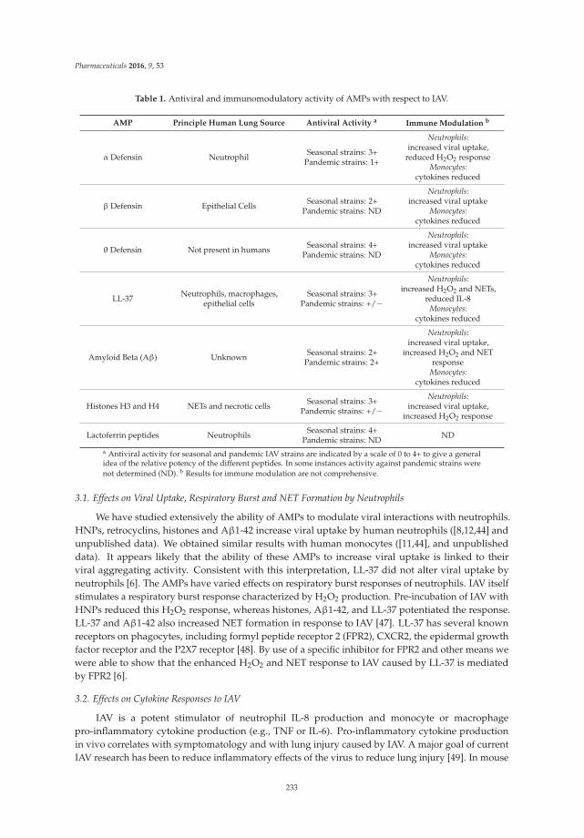
Pharmaceuticals 2016, 9, 53
Table 1. Antiviral and immunomodulatory activity of AMPs with respect to IAV.
AMP Principle Human Lung Source Antiviral Activity a Immune Modulation b
α Defensin Neutrophil Seasonal strains: 3+Pandemic strains: 1+
Neutrophils:increased viral uptake,
reduced H2O2 responseMonocytes:
cytokines reduced
β Defensin Epithelial Cells Seasonal strains: 2+Pandemic strains: ND
Neutrophils:increased viral uptake
Monocytes:cytokines reduced
θ Defensin Not present in humans Seasonal strains: 4+Pandemic strains: ND
Neutrophils:increased viral uptake
Monocytes:cytokines reduced
LL-37 Neutrophils, macrophages,epithelial cells
Seasonal strains: 3+Pandemic strains: +/−
Neutrophils:increased H2O2 and NETs,
reduced IL-8Monocytes:
cytokines reduced
Amyloid Beta (Aβ) Unknown Seasonal strains: 2+Pandemic strains: 2+
Neutrophils:increased viral uptake,
increased H2O2 and NETresponse
Monocytes:cytokines reduced
Histones H3 and H4 NETs and necrotic cells Seasonal strains: 3+Pandemic strains: +/−
Neutrophils:increased viral uptake,
increased H2O2 response
Lactoferrin peptides Neutrophils Seasonal strains: 4+Pandemic strains: ND ND
a Antiviral activity for seasonal and pandemic IAV strains are indicated by a scale of 0 to 4+ to give a generalidea of the relative potency of the different peptides. In some instances activity against pandemic strains werenot determined (ND). b Results for immune modulation are not comprehensive.
3.1. Effects on Viral Uptake, Respiratory Burst and NET Formation by Neutrophils
We have studied extensively the ability of AMPs to modulate viral interactions with neutrophils.HNPs, retrocyclins, histones and Aβ1-42 increase viral uptake by human neutrophils ([8,12,44] andunpublished data). We obtained similar results with human monocytes ([11,44], and unpublisheddata). It appears likely that the ability of these AMPs to increase viral uptake is linked to theirviral aggregating activity. Consistent with this interpretation, LL-37 did not alter viral uptake byneutrophils [6]. The AMPs have varied effects on respiratory burst responses of neutrophils. IAV itselfstimulates a respiratory burst response characterized by H2O2 production. Pre-incubation of IAV withHNPs reduced this H2O2 response, whereas histones, Aβ1-42, and LL-37 potentiated the response.LL-37 and Aβ1-42 also increased NET formation in response to IAV [47]. LL-37 has several knownreceptors on phagocytes, including formyl peptide receptor 2 (FPR2), CXCR2, the epidermal growthfactor receptor and the P2X7 receptor [48]. By use of a specific inhibitor for FPR2 and other means wewere able to show that the enhanced H2O2 and NET response to IAV caused by LL-37 is mediatedby FPR2 [6].
3.2. Effects on Cytokine Responses to IAV
IAV is a potent stimulator of neutrophil IL-8 production and monocyte or macrophagepro-inflammatory cytokine production (e.g., TNF or IL-6). Pro-inflammatory cytokine productionin vivo correlates with symptomatology and with lung injury caused by IAV. A major goal of currentIAV research has been to reduce inflammatory effects of the virus to reduce lung injury [49]. In mouse
233

Pharmaceuticals 2016, 9, 53
models, lung injury can be reduced even when viral loads are unaffected in some cases [49]. LL-37 hasbeen reported to reduce cytokine responses to various stimuli [50–54], and, indeed we found that itreduced neutrophil IL-8 responses to IAV and LPS [6]. We are in the process of studying the effects ofthe various AMPs on monocyte and macrophage cytokine responses to IAV. As noted, LL-37 treatmentof mice infected with IAV reduces cytokine responses [33]. This effect appeared be in part independentof reduction of viral load by LL-37 in vivo.
HNPs released from dying neutrophils have been shown to mediate anti-inflammatory effects [55].In addition, HNPs can be taken up by macrophages during bacterial infection where they inhibitmacrophage inflammatory cytokine production by restricting protein translation [56]. The relevanceof these findings to IAV infection has not been evaluated thus far. As noted also, deletion of HBD-1resulted in greater inflammatory reaction to IAV, despite no change in viral titers. HBD 3 has beenshown to have strong anti-inflammatory effects as well in an LPS model [57,58]. It will be of interest totest this peptide in the context of IAV infection. Even though Aβ1-42 has been extensively studiedas a pro-inflammatory stimulus [59], we found that it reduced inflammatory cytokine productiontriggered by IAV in monocytes in vitro [44].
Overall, these results indicate that maximizing anti-inflammatory effects as well as antiviralactivity should be considered when designing novel AMPs for treatment of IAV. A contrastingperspective has arisen from several studies in which increasing inflammation prior to IAV or SARS-CoVinfection was protective [60–62]. In the study by Wohlford-Lenane et al. intranasal instillation ofretrocyclin 1 alone induced lung inflammation and this was associated with protection vs. SARS-CoV.In another study, direct instillation of HNPs into the airway of mice had pro-inflammatory effectsindependent of infection [63]. Note that in other models listed above HNPs had anti-inflammatoryeffects. Hence, further study is needed to determine how to best administer AMPs in vivo and whetherpossible pro-inflammatory effects would be harmful or beneficial with respect to infection.
Although histones have antimicrobial and antiviral activities, they have been considered toinstigate several systemic inflammatory diseases and tissue injuries, including sepsis, peritonitis,pancreatitis, stroke, acute lung injury, liver injury and kidney injury [36]. The injection of histonesinto mice can stimulate pro-inflammatory cytokine/chemokine release (e.g., IL-6, IL-8 and TNF) andleukocyte infiltration, resulting in sepsis-like pathology [64–67]. In these cases histones function asdamage-associated molecular pattern molecules, and possible mechanisms for them to induce cytokinestorm may be through the interactions with toll-like receptors (TLR), especially TLR2, -4 and -9,and the NLRP3 inflammasome [68]. Knowing the possible detrimental pro-inflammatory effects ofhistones, further study is needed to investigate the roles of histones during severe IAV and IAV-relatedlung injury.
3.3. Effects of AMPs on Adaptive Immune Responses
AMPs have been termed “alarmins”, in that they are able to trigger recruitment and activationof immune cells. AMPs modulate adaptive immune responses in various ways. This topic is beyondthe scope of this paper; however, we refer the reader to other reviews [48,69]. As examples, HBDsand other defensins bind to receptors on lymphocytes and act as chemoattractants [70,71] and LL-37facilitates presentation of antigens to by DCs to T cells [48,72]. α-defensins have been shown topotentiate neutralizing antibody responses to enteric viral infection [73]. The contribution of theseactivities to IAV infection have not been studied but certainly are valuable areas for future research.
3.4. Effects on Bacterial Superinfection
Bacterial superinfections are a major contributor to mortality during pandemics and seasonalepidemics of IAV [4]. These bacterial superinfections are difficult to treat with antibiotics. As a resultthere is great interest in identifying the cause of bacterial superinfection and how to interveneto prevent or treat these infections. The role of AMPs in bacterial superinfection, or for possibletreatment of bacterial superinfection, has not been studied. However, given the combined antiviral
234
Pharmaceuticals 2016, 9, 53
and antibacterial activity of many AMPs they are attractive candidates for treatment of combined IAVand bacterial infection.
3.5. Interactions of AMPs with Other Host Defense Proteins
It is likely that the activity of AMPs in vivo reflects complex interactions among various AMPsand other host defense molecules [74,75]. We found that HNPs 1-3 bind to the lung host defense andsurfactant regulatory protein, surfactant protein D (SP-D) [10,74]. The importance of this bindingin vivo is not clear; however, HNPs inhibited antiviral activity of SP-D and caused precipitation of SP-Dout of human BAL fluid. Since SP-D has anti-inflammatory properties in the uninfected and infectedlung, this property of HNPs could account for inflammatory responses seen upon instillation of HNPsin the lung [63]. On the other hand, SP-D might aid in clearance of HNPs after neutrophil infiltration.Of note, we found that LL-37 and HBDs do not bind to SP-D [8], which may be advantageous whenconsidering use of these peptides for therapy in the lung. Retrocyclins and LL-37 had additive antiviraleffects when combined with SP-D [8,34].
Histones have been considered to augment inflammatory responses and promote thrombosis andthus can be responsible for IAV-related acute lung injury as noted above. There is evidence that otherhost defense proteins can bind histones and modulate their potential adverse effects. Little is knownabout the relationship between histones and SP-D. Evidence suggests that SP-D can simultaneouslybind to both pathogens and NETs [64], and we recently found that SP-D can bind to histones andinhibit histone-induced respiratory burst in neutrophils (unpublished data). C-reactive protein (CRP),an acute phase reactant, is another host defense protein that is usually elevated during infectionsor inflammation, and CRP treatment in mice has been found to alleviate histone-induced toxicity,including endothelial damage, thrombosis and lung edema [76]. Other proteins like thrombomodulinhave similar effects [77]. These findings suggest some possible modulators for histones, but furtherstudies are needed [76].
4. Production of Modified Synthetic AMPs with Increased Antiviral Activity
4.1. Novel Cyclic or Alpha Defensins
There has been extensive research into ways to modify or mimic the structure of defensins in orderto improve anti-bacterial or antiviral activity. Results of some of these studies are summarized in Table 2.Even single amino acid substitutions can potentiate antiviral activity. For instance, replacement ofa single arginine with a lysine in retrocyclin 1 resulted in increased binding to, and inhibition of, HIV [14].This modified retrocyclin also had increased IAV neutralizing activity [27]. Similarly, replacement of asingle amino acid in HD5 resulted in increased activity vs. HSV [78]. The strong activity of retrocyclinsalso inspired construction of a panel of cyclic peptides by Ruchala and Lehrer [11]. The intent was toretain or improve on antiviral activities of naturally occurring retrocyclins with cyclic peptides that areeasier to synthesize. These peptides were termed hapivirins and diprovirins and they incorporatedsystematic substitutions of key cationic or hydrophobic residues and also incorporated several syntheticamino acids. Several of the hapivirins and diprovirins had markedly increased neutralizing activityfor IAV [11]. In addition, the peptides retained the properties of viral aggregation and increasing viraluptake by neutrophils. Like the retrocyclins, they did not interfere with the viral neutralizing activityof SP-D. In the case of the hapivirins, substitutions that increased hydrophobicity caused clear increasein antiviral activity, indicating that hydrophobicity is as important a feature as cationic charge forantiviral activity. The most active hapivirins and diprovirins also had strong ability to suppress TNFgeneration by human monocytes.
235

Pharmaceuticals 2016, 9, 53
Table 2. Therapeutic Directions: Creation of Novel AMPs.
Prototype Peptide Antiviral Activity Immune modulation
β Defensin Novel p9 peptide has increased antiviralactivity against human and avian IAV strains Mediate anti-inflammatory effects
Cyclic defensins Hapivirins and Diprovirins have increasedantiviral activity against seasonal IAV
Cause increased neutrophil uptake compared withHNPs and suppress TNF generation by monocytes
LL-37 GI-20 gains activity against pandemic strain Have similar immunomodulatory effects to LL-37
LactoferrinShorter peptide fragments show increasedanti-IAV activity for seasonal and mouse
adapted strainsNot tested
BPI 27 amino acid N-terminal fragment of humanBPI inhibits infectivity of various IAV strains
Inhibit monocyte cytokine productionin response to IAV
4.2. Novel β-Defensins
A recent paper by Zhao et al. took the approach of preparing a series of fragments of mousebeta defensin 4 and testing their activity against a panel of clinically important respiratory viruses,including human and avian IAV strains, SARS-CoV and MERS-CoV [79]. A 30 amino acid fragmentof mBD4 (termed P9 by the authors) was found to have activity exceeding that of the full 40 aminoacid protein both in vitro and in vivo in mice. In fact, 100% of mice survived lethal challenge witha mouse adapted H1N1 viral strain when they were pretreated with 50 μg/mouse of P9 intranasally.The P9 peptide also reduced lung inflammation and viral loads in the mice. P9 also increased survivalof mice infected with a lethal dose of avian H5N1 or H7N9 and had in vitro antiviral activity againstSARS-CoV and MERS-CoV. All of these respiratory viral strains are of great concern due to pandemicpotential and severe outcomes of infection in humans. The mechanism of action of P9 was foundto include binding to H1N1 IAV, entering the cell along with the virus and impairing endosomalacidification and hence viral escape from endosomes. This study provides and interesting example ofa shortened fragment of a naturally occurring defensin having greater activity than the parent peptide.
4.3. LL-37 Derived Peptides
In a similar vein, shortened or subtly modified version of LL-37 have been tested for antiviraland antibacterial activity. A 20 amino acid fragment, termed GI-20, was found to have retained theantiviral activity of the full molecule against seasonal IAV [80] and against HIV [81]. Another fragmentcalled K-13 had no activity for IAV, despite the fact that it inhibits HIV and has anti-bacterial activityas well [82,83]. Two N-terminal fragments (LL-23 and LL-23V9) that have anti-bacterial activity [84]failed to inhibit IAV [85]. The GI-20 fragment incorporates the central helix of LL-37 and differs by oneamino acid from the LL-37 sequence (isoleucine 13 is changed to glycine). Of note, pandemic H1N1 of2009 was inhibited in vitro by GI-20, but not by LL-37 or the other peptides [85]. This provides anotherexample of a shortened version of a natural AMP with increased antiviral activity compared to theparent peptide. GI-20 also retained the ability of LL-37 to increase neutrophil respiratory burst andNET responses to IAV, while also suppressing neutrophil IL-8 response to the virus [80]. The abilityof GI-20 to increase virus induced respiratory burst and NET responses was also mediated throughbinding to FPR2. Using molecular modeling GI-20 was found to incorporate the domain of the proteinpredicted to bind to the FPR2 receptor.
4.4. Lactoferrin and Bacterial/Permeability Increasing Protein (BPI) Based Peptides
Lactoferrin is an evolutionarily conserved innate immune protein present in breast milk and othermucosal secretions that is cationic and has activity against bacteria and several viruses, includingIAV [86]. Lactoferrin has two lobes (the N and C lobes) both of which contain an iron bindingsite. Although the N and C lobes have similar structures they have distinct sequences withinthem. The two lobes can be separated by proteolytic cleavage and shown to have distinct functionalactivities [87]. A recent study by Ammendolia et al. showed that all of the anti-influenza activity of
236
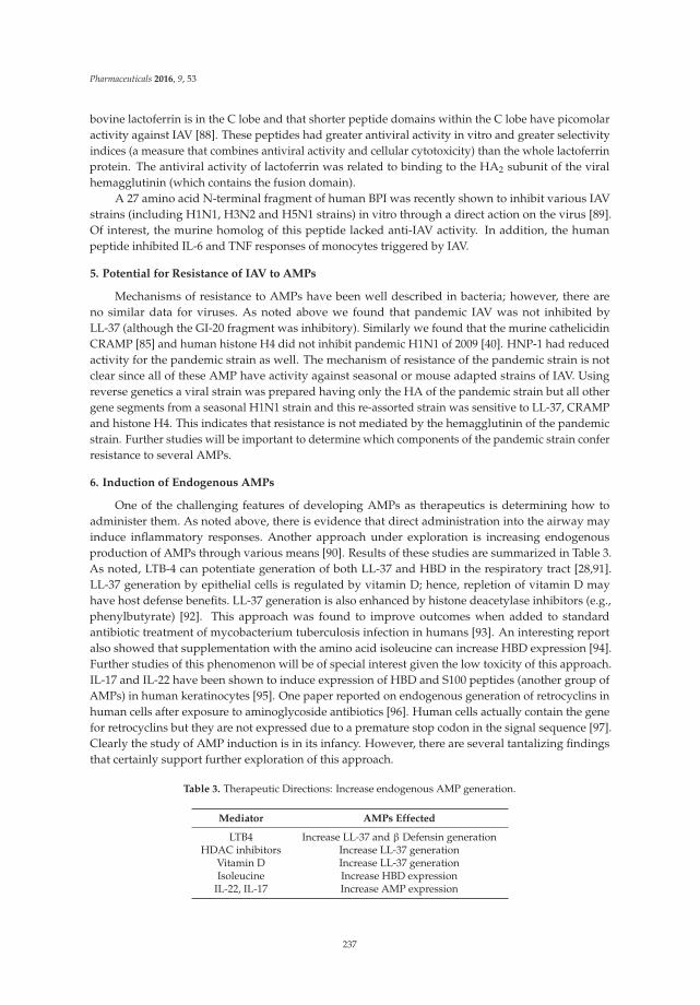
Pharmaceuticals 2016, 9, 53
bovine lactoferrin is in the C lobe and that shorter peptide domains within the C lobe have picomolaractivity against IAV [88]. These peptides had greater antiviral activity in vitro and greater selectivityindices (a measure that combines antiviral activity and cellular cytotoxicity) than the whole lactoferrinprotein. The antiviral activity of lactoferrin was related to binding to the HA2 subunit of the viralhemagglutinin (which contains the fusion domain).
A 27 amino acid N-terminal fragment of human BPI was recently shown to inhibit various IAVstrains (including H1N1, H3N2 and H5N1 strains) in vitro through a direct action on the virus [89].Of interest, the murine homolog of this peptide lacked anti-IAV activity. In addition, the humanpeptide inhibited IL-6 and TNF responses of monocytes triggered by IAV.
5. Potential for Resistance of IAV to AMPs
Mechanisms of resistance to AMPs have been well described in bacteria; however, there areno similar data for viruses. As noted above we found that pandemic IAV was not inhibited byLL-37 (although the GI-20 fragment was inhibitory). Similarly we found that the murine cathelicidinCRAMP [85] and human histone H4 did not inhibit pandemic H1N1 of 2009 [40]. HNP-1 had reducedactivity for the pandemic strain as well. The mechanism of resistance of the pandemic strain is notclear since all of these AMP have activity against seasonal or mouse adapted strains of IAV. Usingreverse genetics a viral strain was prepared having only the HA of the pandemic strain but all othergene segments from a seasonal H1N1 strain and this re-assorted strain was sensitive to LL-37, CRAMPand histone H4. This indicates that resistance is not mediated by the hemagglutinin of the pandemicstrain. Further studies will be important to determine which components of the pandemic strain conferresistance to several AMPs.
6. Induction of Endogenous AMPs
One of the challenging features of developing AMPs as therapeutics is determining how toadminister them. As noted above, there is evidence that direct administration into the airway mayinduce inflammatory responses. Another approach under exploration is increasing endogenousproduction of AMPs through various means [90]. Results of these studies are summarized in Table 3.As noted, LTB-4 can potentiate generation of both LL-37 and HBD in the respiratory tract [28,91].LL-37 generation by epithelial cells is regulated by vitamin D; hence, repletion of vitamin D mayhave host defense benefits. LL-37 generation is also enhanced by histone deacetylase inhibitors (e.g.,phenylbutyrate) [92]. This approach was found to improve outcomes when added to standardantibiotic treatment of mycobacterium tuberculosis infection in humans [93]. An interesting reportalso showed that supplementation with the amino acid isoleucine can increase HBD expression [94].Further studies of this phenomenon will be of special interest given the low toxicity of this approach.IL-17 and IL-22 have been shown to induce expression of HBD and S100 peptides (another group ofAMPs) in human keratinocytes [95]. One paper reported on endogenous generation of retrocyclins inhuman cells after exposure to aminoglycoside antibiotics [96]. Human cells actually contain the genefor retrocyclins but they are not expressed due to a premature stop codon in the signal sequence [97].Clearly the study of AMP induction is in its infancy. However, there are several tantalizing findingsthat certainly support further exploration of this approach.
Table 3. Therapeutic Directions: Increase endogenous AMP generation.
Mediator AMPs Effected
LTB4 Increase LL-37 and β Defensin generationHDAC inhibitors Increase LL-37 generation
Vitamin D Increase LL-37 generationIsoleucine Increase HBD expression
IL-22, IL-17 Increase AMP expression
237

Pharmaceuticals 2016, 9, 53
7. Conclusions
IAV causes many deaths or severe illness every year in seasonal outbreaks and has the potentialto cause massive morbidity and mortality during pandemics. Resistance to two classes of drugsthat are currently used for IAV treatment has been emerging, so there has been extensive interest indeveloping new antiviral treatment for this virus. Deaths caused by IAV infection mostly resulted fromacute lung injury, systemic inflammation or bacterial superinfection, suggesting that new treatmentswith anti-viral, anti-bacterial and anti-inflammation effects would be ideal. AMPs are antimicrobialpeptides that not only play important roles as host defense against pathogens but also modulateinflammatory responses, and thus they are potential candidates for IAV treatment. We discussed theinteractions of two classes of classical AMPs (defensins and cathelicidins) and two non-classical AMPs(histones and Aβ) with IAVs in this paper. For the most part, these AMPs possess anti-IAV activityby direct interacting with the virus, although in some instances direct interactions with mammaliancells may contribute to antiviral effects. All of AMPs we discussed also have immunomodulatoryeffects, with some up-regulating and others down-regulating inflammation. We provide someexamples (e.g., hapivirins and diprovirins or GI-20) in which novel synthetic AMPs or AMP fragmentshave improved anti-IAV or immunomodulatory activities, suggesting that modification of AMPs isan attractive strategy. Further studies are required to determine the interactions between AMPs andother host defense proteins in the lung, the best methods to administer or induce generation of AMPs,and to explain instances of resistance of pandemic IAV strains to AMPs.
Acknowledgments: This work was supported by NIH R01 HL069031 (KH).
Conflicts of Interest: The authors declare no conflict of interest.
References
1. Tripathi, S.; White, M.R.; Hartshorn, K.L. The amazing innate immune response to influenza a virus infection.Innate Immun. 2013, 96, 931–938. [CrossRef] [PubMed]
2. Dawood, F.S.; Jain, S.; Finelli, L.; Shaw, M.W.; Lindstrom, S.; Garten, R.J.; Gubareva, L.V.; Xu, X.; Bridges, C.B.;Uyeki, T.M. Emergence of a novel swine-origin influenza a (H1N1) virus in humans. N. Engl. J. Med. 2009,360, 2605–2615. [PubMed]
3. Morens, D. Influenza-related mortality: Considerations for practice and public health. J. Am. Med. Assoc.2003, 289, 227–229. [CrossRef]
4. Hartshorn, K.L. New look at an old problem: Bacterial superinfection after influenza. Am. J. Pathol. 2010,176, 536–539. [CrossRef] [PubMed]
5. Hartshorn, K.L. Why does pandemic influenza virus kill? Am. J. Pathol. 2013, 183, 1125–1127. [CrossRef][PubMed]
6. Tripathi, S.; Verma, A.; Kim, E.J.; White, M.R.; Hartshorn, K.L. LL-37 modulates human neutrophil responsesto influenza a virus. J. Leukoc. Biol. 2014. [CrossRef] [PubMed]
7. Narasaraju, T.; Yang, E.; Samy, R.P.; Ng, H.H.; Poh, W.P.; Liew, A.A.; Phoon, M.C.; Van Rooijen, N.;Chow, V.T. Excessive neutrophils and neutrophil extracellular traps contribute to acute lung injury ofinfluenza pneumonitis. Am. J. Pathol. 2011, 179, 199–210. [CrossRef] [PubMed]
8. Doss, M.; White, M.R.; Tecle, T.; Gantz, D.; Crouch, E.C.; Jung, G.; Ruchala, P.; Waring, A.J.; Lehrer, R.I.;Hartshorn, K.L. Interactions of alpha-, beta-, and theta-defensins with influenza a virus and surfactantprotein D. J. Immunol. 2009, 182, 7878–7887. [CrossRef] [PubMed]
9. Salvatore, M.; Garcia-Sastre, A.; Ruchala, P.; Lehrer, R.I.; Chang, T.; Klotman, M.E. β-defensin inhibitsinfluenza virus replication by cell-mediated mechanism(s). J. Infect. Dis. 2007, 196, 835–843. [CrossRef][PubMed]
10. Hartshorn, K.L.; White, M.R.; Tecle, T.; Holmskov, U.; Crouch, E.C. Innate defense against influenza a virus:Activity of human neutrophil defensins and interactions of defensins with surfactant protein d. J. Immunol.2006, 176, 6962–6972. [CrossRef] [PubMed]
238

Pharmaceuticals 2016, 9, 53
11. Doss, M.; Ruchala, P.; Tecle, T.; Gantz, D.; Verma, A.; Hartshorn, A.; Crouch, E.C.; Luong, H.; Micewicz, E.D.;Lehrer, R.I. Hapivirins and diprovirins: Novel theta-defensin analogs with potent activity against influenzaa virus. J. Immunol. 2012, 188, 2759–2768. [CrossRef] [PubMed]
12. Tecle, T.; White, M.R.; Gantz, D.; Crouch, E.C.; Hartshorn, K.L. Human neutrophil defensins increaseneutrophil uptake of influenza a virus and bacteria and modify virus-induced respiratory burst responses.J. Immunol. 2007, 178, 8046–8052. [CrossRef] [PubMed]
13. Hoover, D.M.; Rajashankar, K.R.; Blumenthal, R.; Puri, A.; Oppenheim, J.J.; Chertov, O.; Lubkowski, J.The structure of human β-defensin-2 shows evidence of higher order oligomerization. J. Biol. Chem.2000, 275, 32911–32918. [CrossRef] [PubMed]
14. Owen, S.M.; Rudolph, D.L.; Wang, W.; Cole, A.M.; Waring, A.J.; Lal, R.B.; Lehrer, R.I. RC-101, a retrocyclin-1analogue with enhanced activity against primary HIV type 1 isolates. AIDS Res. Hum. Retrovir. 2004, 20,1157–1165. [CrossRef] [PubMed]
15. Jiang, Y.; Wang, Y.; Kuang, Y.; Wang, B.; Li, W.; Gong, T.; Jiang, Z.; Yang, D.; Li, M. Expression of mousebeta-defensin-3 in mdck cells and its anti-influenza-virus activity. Arch. Virol. 2009, 154, 639–647. [CrossRef][PubMed]
16. Ryan, L.K.; Dai, J.; Yin, Z.; Megjugorac, N.; Uhlhorn, V.; Yim, S.; Schwartz, K.D.; Abrahams, J.M.; Diamond, G.;Fitzgerald-Bocarsly, P. Modulation of human beta-defensin-1 (hBD-1) in plasmacytoid dendritic cells (PDC),monocytes, and epithelial cells by influenza virus, herpes simplex virus, and sendai virus and its possiblerole in innate immunity. J. Leukoc. Biol. 2011, 90, 343–356. [CrossRef] [PubMed]
17. Liang, Q.L.; Zhou, K.; He, H.X. Retrocyclin 2: A new therapy against avian influenza H5N1 virus in vivoand vitro. Biotechnol. Lett. 2009, 32, 387–392. [CrossRef] [PubMed]
18. Leikina, E.; Delanoe-Ayari, H.; Melikov, K.; Cho, M.S.; Chen, A.; Waring, A.J.; Wang, W.; Xie, Y.; Loo, J.A.;Lehrer, R.I. Carbohydrate-binding molecules inhibit viral fusion and entry by crosslinking membraneglycoproteins. Nat. Immunol. 2005, 6, 995–1001. [CrossRef] [PubMed]
19. Wang, W.; Cole, A.M.; Hong, T.; Waring, A.J.; Liang, Q.L.; Zhou, K.; He, H.X. Retrocyclin 2: A new therapyagainst avian influenza h5n1 virus in vivo and vitro. Biotechnol. Lett. 2009, 32, 387–392. [CrossRef] [PubMed]
20. Klotman, M.E.; Rapista, A.; Teleshova, N.; Micsenyi, A.; Jarvis, G.A.; Lu, W.; Porter, E.; Chang, T.L. Neisseriagonorrhoeae-induced human defensins 5 and 6 increase HIV infectivity: Role in enhanced transmission.J. Immunol. 2008, 180, 6176–6185. [CrossRef] [PubMed]
21. Ding, J.; Rapista, A.; Teleshova, N.; Lu, W.; Klotman, M.E.; Chang, T.L. Mucosal human defensins 5 and 6antagonize the anti-HIV activity of candidate polyanion microbicides. J. Innate. Immun. 2011, 3, 208–212.[CrossRef] [PubMed]
22. Valere, K.; Rapista, A.; Eugenin, E.; Lu, W.; Chang, T.L. Human alpha-defensin hnp1 increases hiv traversalof the epithelial barrier: A potential role in sti-mediated enhancement of HIV transmission. Viral Immunol.2015, 28, 609–615. [CrossRef] [PubMed]
23. Furci, L.; Tolazzi, M.; Sironi, F.; Vassena, L.; Lusso, P. Inhibition of HIV-1 infection by human α-defensin-5,a natural antimicrobial peptide expressed in the genital and intestinal mucosae. PLoS ONE 2012, 7, e45208.[CrossRef] [PubMed]
24. Wang, W.; Owen, S.M.; Rudolph, D.L.; Cole, A.M.; Hong, T.; Waring, A.J.; Lal, R.B.; Lehrer, R.I. Activity ofalpha- and theta-defensins against primary isolates of HIV-1. J. Immunol. 2004, 173, 515–520. [CrossRef][PubMed]
25. Wilson, S.S.; Wiens, M.E.; Holly, M.K.; Smith, J.G. Defensins at the mucosal surface: Latest insights intodefensin-virus interactions. J. Virol. 2016, 90, 5216–5218. [CrossRef] [PubMed]
26. Tecle, T.; Tripathi, S.; Hartshorn, K.L. Review: Defensins and cathelicidins in lung immunity. Innate. Immun.2010, 16, 151–159. [CrossRef] [PubMed]
27. Doss, M.; White, M.R.; Tecle, T.; Hartshorn, K.L. Human defensins and LL-37 in mucosal immunity.J. Leukoc. Biol. 2010, 87, 79–92. [CrossRef] [PubMed]
28. Gaudreault, E.; Gosselin, J. Leukotriene B4 induces release of antimicrobial peptides in lungs of virallyinfected mice. J. Immunol. 2008, 180, 6211–6221. [CrossRef] [PubMed]
29. Currie, S.M.; Findlay, E.G.; McHugh, B.J.; Mackellar, A.; Man, T.; Macmillan, D.; Wang, H.; Fitch, P.M.;Schwarze, J.; Davidson, D.J. The human cathelicidin LL-37 has antiviral activity against respiratory syncytialvirus. PLoS ONE 2013, 8, e73659. [CrossRef] [PubMed]
239

Pharmaceuticals 2016, 9, 53
30. Uchio, E.; Inoue, H.; Kadonosono, K. Anti-adenoviral effects of human cationic antimicrobialprotein-18/LL-37, an antimicrobial peptide, by quantitative polymerase chain reaction. Korean J. Ophthalmol.2013, 27, 199–203. [CrossRef] [PubMed]
31. Wang, G.; Watson, K.M.; Buckheit, R.W., Jr. Anti-human immunodeficiency virus type 1 activities ofantimicrobial peptides derived from human and bovine cathelicidins. Antimicrob. Agents Chemother. 2008, 52,3438–3440. [CrossRef] [PubMed]
32. Bergman, P.; Walter-Jallow, L.; Broliden, K.; Agerberth, B.; Soderlund, J. The antimicrobial peptide LL-37inhibits HIV-1 replication. Curr. HIV Res. 2007, 5, 410–415. [CrossRef] [PubMed]
33. Barlow, P.G.; Svoboda, P.; Mackellar, A.; Nash, A.A.; York, I.A.; Pohl, J.; Davidson, D.J.; Donis, R.O. Antiviralactivity and increased host defense against influenza infection elicited by the human cathelicidin LL-37.PLoS ONE 2011, 6, e25333. [CrossRef] [PubMed]
34. Tripathi, S.; Tecle, T.; Verma, A.; Crouch, E.; White, M.; Hartshorn, K.L. The human cathelicidin LL-37 inhibitsinfluenza a viruses through a mechanism distinct from that of surfactant protein D or defensins. J. Gen. Virol.2013, 94, 40–49. [CrossRef] [PubMed]
35. Currie, S.M.; Gwyer Findlay, E.; McFarlane, A.J.; Fitch, P.M.; Bottcher, B.; Colegrave, N.; Paras, A.; Jozwik, A.;Chiu, C.; Schwarze, J. Cathelicidins have direct antiviral activity against respiratory syncytial virus in vitroand protective function in vivo in mice and humans. J. Immunol. 2016, 196, 2699–2710. [CrossRef] [PubMed]
36. Hoeksema, M.; Van Eijk, M.; Haagsman, H.P.; Hartshorn, K.L. Histones as mediators of host defense,inflammation and thrombosis. Future Microbiol. 2016, 11, 441–453. [CrossRef] [PubMed]
37. Lewis, H.D.; Liddle, J.; Coote, J.E.; Atkinson, S.J.; Barker, M.D.; Bax, B.D.; Bicker, K.L.; Bingham, R.P.;Campbell, M.; Chen, Y.H. Inhibition of PAD4 activity is sufficient to disrupt mouse and human net formation.Nat. Chem. Biol. 2015, 11, 189–191. [CrossRef] [PubMed]
38. Cheng, O.Z.; Palaniyar, N. Net balancing: A problem in inflammatory lung diseases. In NETosis: At theIntersection of Cell Biology, Microbiology, and Immunology; Frontiers Media SA: Lausanne, Switzerland, 2013;Volume 4, p. 1.
39. Morita, S.; Tagai, C.; Shiraishi, T.; Miyaji, K.; Iwamuro, S. Differential mode of antimicrobial actions ofarginine-rich and lysine-rich histones against gram-positive staphylococcus aureus. Peptides 2013, 48, 75–82.[CrossRef] [PubMed]
40. Hoeksema, M.; Tripathi, S.; White, M.; Qi, L.; Taubenberger, J.; van Eijk, M.; Haagsman, H.; Hartshorn, K.L.Arginine-rich histones have strong antiviral activity for influenza a viruses. Innate Immun. 2015. [CrossRef][PubMed]
41. Kagan, B.L.; Jang, H.; Capone, R.; Teran Arce, F.; Ramachandran, S.; Lal, R.; Nussinov, R. Antimicrobialproperties of amyloid peptides. Mol. Pharm. 2012, 9, 708–717. [CrossRef] [PubMed]
42. Jang, H.; Ma, B.; Lal, R.; Nussinov, R. Models of toxic beta-sheet channels of protegrin-1 suggest a commonsubunit organization motif shared with toxic alzheimer β-amyloid ion channels. Biophys. J. 2008, 95,4631–4642. [CrossRef] [PubMed]
43. Soscia, S.J.; Kirby, J.E.; Washicosky, K.J.; Tucker, S.M.; Ingelsson, M.; Hyman, B.; Burton, M.A.; Goldstein, L.E.;Duong, S.; Tanzi, R.E. The alzheimer’s disease-associated amyloid beta-protein is an antimicrobial peptide.PLoS ONE 2010, 5, e9505. [CrossRef] [PubMed]
44. White, M.R.; Kandel, R.; Tripathi, S.; Condon, D.; Qi, L.; Taubenberger, J.; Hartshorn, K.L. Alzheimer’sassociated beta-amyloid protein inhibits influenza a virus and modulates viral interactions with phagocytes.PLoS ONE 2014, 9, e101364. [CrossRef] [PubMed]
45. Bourgade, K.; Garneau, H.; Giroux, G.; Le Page, A.Y.; Bocti, C.; Dupuis, G.; Frost, E.H.; Fulop, T., Jr.Beta-amyloid peptides display protective activity against the human alzheimer’s disease-associated herpessimplex virus-1. Biogerontology 2015, 16, 85–98. [CrossRef] [PubMed]
46. Kumar, D.; Choi, S.; Washicosky, K.; Eimer, W.; Tucker, S.; Ghofrani, J.; Lefkowitz, A.; McColl, G.; Goldstein, L.;Tanzi, R. Amyloid-β peptide protects against microbial infection in mouse and worm models of alzheimer’sdisease. Sci. Transl. Med. 2016, 8, 340ra372. [CrossRef] [PubMed]
47. Hartshorn, K.L.; Collamer, M.; White, M.R.; Schwartz, J.H.; Tauber, A.I. Characterization of influenza a virusactivation of the human neutrophil. Blood 1990, 75, 218–226. [PubMed]
48. Kahlenberg, J.M.; Kaplan, M.J. Little peptide, big effects: The role of LL-37 in inflammation and autoimmunedisease. J. Immunol. 2013, 191, 4895–4901. [CrossRef] [PubMed]
240
Pharmaceuticals 2016, 9, 53
49. Tripathi, S.; White, M.R.; Hartshorn, K.L. The amazing innate immune response to influenza a virus infection.Innate Immun. 2015, 21, 73–98. [CrossRef] [PubMed]
50. Hu, Z.; Murakami, T.; Suzuki, K.; Tamura, H.; Kuwahara-Arai, K.; Iba, T.; Nagaoka, I. Antimicrobialcathelicidin peptide LL-37 inhibits the LSP/ATP-induced pyroptosis of macrophages by dual mechanism.PLoS ONE 2014, 9, e85765. [CrossRef] [PubMed]
51. Chen, X.; Takai, T.; Xie, Y.; Niyonsaba, F.; Okumura, K.; Ogawa, H. Human antimicrobial peptide LL-37modulates proinflammatory responses induced by cytokine milieus and double-stranded rna in humankeratinocytes. Biochem. Biophys. Res. Commun. 2013, 433, 532–537. [CrossRef] [PubMed]
52. Ruan, Y.; Shen, T.; Wang, Y.; Hou, M.; Li, J.; Sun, T. Antimicrobial peptide LL-37 attenuates lta inducedinflammatory effect in macrophages. Int. Immunopharmacol. 2013, 15, 575–580. [CrossRef] [PubMed]
53. Brown, K.L.; Poon, G.F.; Birkenhead, D.; Pena, O.M.; Falsafi, R.; Dahlgren, C.; Karlsson, A.; Bylund, J.;Hancock, R.E.; Johnson, P. Host defense peptide LL-37 selectively reduces proinflammatory macrophageresponses. J. Immunol. 2011, 186, 5497–5505. [CrossRef] [PubMed]
54. Braff, M.H.; Hawkins, M.A.; Di Nardo, A.; Lopez-Garcia, B.; Howell, M.D.; Wong, C.; Lin, K.; Streib, J.E.;Dorschner, R.; Leung, D.Y. Structure-function relationships among human cathelicidin peptides: Dissociationof antimicrobial properties from host immunostimulatory activities. J. Immunol. 2005, 174, 4271–4278.[CrossRef] [PubMed]
55. Miles, K.; Clarke, D.J.; Lu, W.; Sibinska, Z.; Beaumont, P.E.; Davidson, D.J.; Barr, T.A.; Campopiano, D.J.;Gray, M. Dying and necrotic neutrophils are anti-inflammatory secondary to the release of alpha-defensins.J. Immunol. 2009, 183, 2122–2132. [CrossRef] [PubMed]
56. Brook, M.; Tomlinson, G.H.; Miles, K.; Smith, R.W.; Rossi, A.G.; Hiemstra, P.S.; van’t Wout, E.F.; Dean, J.L.;Gray, N.K.; Lu, W. Neutrophil-derived alpha defensins control inflammation by inhibiting macrophage mrnatranslation. Proc. Natl. Acad. Sci. USA 2016, 113, 4350–4355. [CrossRef] [PubMed]
57. Semple, F.; MacPherson, H.; Webb, S.; Cox, S.L.; Mallin, L.J.; Tyrrell, C.; Grimes, G.R.; Semple, C.A.; Nix, M.A.;Millhauser, G.L. Human beta-defensin 3 affects the activity of pro-inflammatory pathways associated withMyD88 and TRIF. Eur. J. Immunol. 2011, 41, 3291–3300. [CrossRef] [PubMed]
58. Semple, F.; Webb, S.; Li, H.N.; Patel, H.B.; Perretti, M.; Jackson, I.J.; Gray, M.; Davidson, D.J.; Dorin, J.R.Human β-defensin 3 has immunosuppressive activity in vitro and in vivo. Eur. J. Immunol. 2010, 40,1073–1078. [CrossRef] [PubMed]
59. Gold, M.; El Khoury, J. Beta-amyloid, microglia, and the inflammasome in alzheimer’s disease.Semin. Immunopathol. 2015, 37, 607–611. [CrossRef] [PubMed]
60. Tuvim, M.J.; Gilbert, B.E.; Dickey, B.F.; Evans, S.E. Synergistic TLR2/6 and TLR9 activation protects miceagainst lethal influenza pneumonia. PLoS ONE 2012, 7, e30596. [CrossRef] [PubMed]
61. Tuvim, M.J.; Evans, S.E.; Clement, C.G.; Dickey, B.F.; Gilbert, B.E. Augmented lung inflammation protectsagainst influenza a pneumonia. PLoS ONE 2009, 4, e4176. [CrossRef] [PubMed]
62. Wohlford-Lenane, C.L.; Meyerholz, D.K.; Perlman, S.; Zhou, H.; Tran, D.; Selsted, M.E.; McCray, P.B., Jr.Rhesus theta-defensin prevents death in a mouse model of sars coronavirus pulmonary disease. J. Virol.2009, 83. [CrossRef] [PubMed]
63. Zhang, H.; Porro, G.; Orzech, N.; Mullen, B.; Liu, M.; Slutsky, A.S. Neutrophil defensins mediate acuteinflammatory response and lung dysfunction in dose-related fashion. Am. J. Physiol. Lung Cell. Mol. Physiol.2001, 280, 947–954.
64. Xu, J.; Zhang, X.; Monestier, M.; Esmon, N.L.; Esmon, C.T. Extracellular histones are mediators of deaththrough TLR2 and TLR4 in mouse fatal liver injury. J. Immunol. 2011, 187, 2626–2631. [CrossRef] [PubMed]
65. Xu, J.; Zhang, X.; Pelayo, R.; Monestier, M.; Ammollo, C.T.; Semeraro, F.; Taylor, F.B.; Esmon, N.L.; Lupu, F.;Esmon, C.T. Extracellular histones are major mediators of death in sepsis. Nat. Med. 2009, 15, 1318–1321.[CrossRef] [PubMed]
66. Chen, R.; Kang, R.; Fan, X.G.; Tang, D. Release and activity of histone in diseases. Cell Death Dis. 2014, 5,e1370. [CrossRef] [PubMed]
67. Westman, J.; Papareddy, P.; Dahlgren, M.W.; Chakrakodi, B.; Norrby-Teglund, A.; Smeds, E.; Linder, A.;Morgelin, M.; Johansson-Lindbom, B.; Egesten, A. Extracellular histones induce chemokine productionin whole blood ex vivo and leukocyte recruitment in vivo. PLoS Pathog. 2015, 11, e1005319. [CrossRef][PubMed]
241

Pharmaceuticals 2016, 9, 53
68. Huang, H.; Chen, H.W.; Evankovich, J.; Yan, W.; Rosborough, B.R.; Nace, G.W.; Ding, Q.; Loughran, P.;Beer-Stolz, D.; Billiar, T.R. Histones activate the NLRP3 inflammasome in kupffer cells during sterileinflammatory liver injury. J. Immunol. 2013, 191, 2665–2679. [CrossRef] [PubMed]
69. Oppenheim, J.J.; Yang, D. Alarmins: Chemotactic activators of immune responses. Curr. Opin. Immunol.2005, 17, 359–365. [CrossRef] [PubMed]
70. Grigat, J.; Soruri, A.; Forssmann, U.; Riggert, J.; Zwirner, J. Chemoattraction of macrophages, T lymphocytes,and mast cells is evolutionarily conserved within the human alpha-defensin family. J. Immunol. 2007, 179,3958–3965. [CrossRef] [PubMed]
71. Chertov, O.; Michiel, D.F.; Xu, L.; Wang, J.M.; Tani, K.; Murphy, W.J.; Longo, D.L.; Taub, D.D.; Oppenheim, J.J.Identification of defensin-1, defensin-2, and CAP37/azurocidin as T-cell chemoattractant proteins releasedfrom interleukin-8-stimulated neutrophils. J. Biol. Chem. 1996, 271, 2935–2940. [CrossRef] [PubMed]
72. Davidson, D.J.; Currie, A.J.; Reid, G.S.; Bowdish, D.M.; MacDonald, K.L.; Ma, R.C.; Hancock, R.E.; Speert, D.P.The cationic antimicrobial peptide LL-37 modulates dendritic cell differentiation and dendritic cell-inducedT cell polarization. J. Immunol. 2004, 172, 1146–1156. [CrossRef] [PubMed]
73. Gounder, A.P.; Myers, N.D.; Treuting, P.M.; Bromme, B.A.; Wilson, S.S.; Wiens, M.E.; Lu, W.; Ouellette, A.J.;Spindler, K.R.; Parks, W.C. Defensins potentiate a neutralizing antibody response to enteric viral infection.PLoS Pathog. 2016, 12, e1005474. [CrossRef] [PubMed]
74. White, M.R.; Tecle, T.; Crouch, E.C.; Hartshorn, K.L. Impact of neutrophils on antiviral activity of humanbronchoalveolar lavage fluid. Am. J. Physiol. Lung Cell. Mol. Physiol. 2007, 293, 1293–1299. [CrossRef][PubMed]
75. Bals, R.; Hiemstra, P.S. Innate immunity in the lung: How epithelial cells fight against respiratory pathogens.Eur. Respir. J. 2004, 23, 327–333. [CrossRef] [PubMed]
76. Abrams, S.T.; Zhang, N.; Dart, C.; Wang, S.S.; Thachil, J.; Guan, Y.; Wang, G.; Toh, C.H. Human CRP defendsagainst the toxicity of circulating histones. J. Immunol. 2013, 191, 2495–2502. [CrossRef] [PubMed]
77. Nakahara, M.; Ito, T.; Kawahara, K.; Yamamoto, M.; Nagasato, T.; Shrestha, B.; Yamada, S.; Miyauchi, T.;Higuchi, K.; Takenaka, T. Recombinant thrombomodulin protects mice against histone-induced lethalthromboembolism. PLoS ONE 2013, 8, e75961. [CrossRef] [PubMed]
78. Wang, A.; Chen, F.; Wang, Y.; Shen, M.; Xu, Y.; Hu, J.; Wang, S.; Geng, F.; Wang, C.; Ran, X. Enhancementof antiviral activity of human alpha-defensin 5 against herpes simplex virus 2 by arginine mutagenesis atadaptive evolution sites. J. Virol. 2013, 87, 2835–2845. [CrossRef] [PubMed]
79. Zhao, H.; Zhou, J.; Zhang, K.; Chu, H.; Liu, D.; Poon, V.K.; Chan, C.C.; Leung, H.C.; Fai, N.; Lin, Y.P. A novelpeptide with potent and broad-spectrum antiviral activities against multiple respiratory viruses. Sci. Rep.2016, 6, 22008. [CrossRef] [PubMed]
80. Tripathi, S.; Wang, G.; White, M.; Rynkiewicz, M.; Seaton, B.; Hartshorn, K. Identifying the critical domainof LL-37 involved in mediating neutrophil activation in the presence of influenza virus: Functional andstructural analysis. PLoS ONE 2015, 10, e0133454. [CrossRef] [PubMed]
81. Wang, G. Database-guided discovery of potent peptides to combat HIV-1 or superbugs. Pharmaceuticals 2013,6, 728–758. [CrossRef] [PubMed]
82. Wang, G.; Mishra, B.; Epand, R.F.; Epand, R.M. High-quality 3D structures shine light on antibacterial,anti-biofilm and antiviral activities of human cathelicidin LL-37 and its fragments. Biochim. Biophys. Acta2014, 1838, 2160–2172. [CrossRef] [PubMed]
83. Jacob, B.; Park, I.S.; Bang, J.K.; Shin, S.Y. Short KR-12 analogs designed from human cathelicidin LL-37possessing both antimicrobial and antiendotoxic activities without mammalian cell toxicity. J. Pept. Sci. 2013,19, 700–707. [CrossRef] [PubMed]
84. Wang, G.; Elliott, M.; Cogen, A.L.; Ezell, E.L.; Gallo, R.L.; Hancock, R.E. Structure, dynamics, andantimicrobial and immune modulatory activities of human LL-23 and its single-residue variants mutated onthe basis of homologous primate cathelicidins. Biochemistry 2012, 51, 653–664. [CrossRef] [PubMed]
85. Tripathi, S.; Wang, G.; White, M.; Qi, L.; Taubenberger, J.; Hartshorn, K.L. Antiviral activity of the humancathelicidin, LL-37, and derived peptides on seasonal and pandemic influenza a viruses. PLoS ONE2015, 10, e0124706. [CrossRef] [PubMed]
86. Wakabayashi, H.; Oda, H.; Yamauchi, K.; Abe, F. Lactoferrin for prevention of common viral infections.J. Infect. Chemother. 2014, 20, 666–671. [CrossRef] [PubMed]
242

Pharmaceuticals 2016, 9, 53
87. Sharma, S.; Sinha, M.; Kaushik, S.; Kaur, P.; Singh, T.P. C-lobe of lactoferrin: The whole story of thehalf-molecule. Biochem. Res. Int. 2013, 2013, 271641. [CrossRef] [PubMed]
88. Ammendolia, M.G.; Agamennone, M.; Pietrantoni, A.; Lannutti, F.; Siciliano, R.A.; De Giulio, B.; Amici, C.;Superti, F. Bovine lactoferrin-derived peptides as novel broad-spectrum inhibitors of influenza virus.Pathog. Glob. Health 2012, 106, 12–19. [CrossRef] [PubMed]
89. Pinkenburg, O.; Meyer, T.; Bannert, N.; Norley, S.; Bolte, K.; Czudai-Matwich, V.; Herold, S.; Gessner, A.;Schnare, M. The human antimicrobial protein bactericidal/permeability-increasing protein (BPI) inhibits theinfectivity of influenza a virus. PLoS ONE 2016, 11, e0156929. [CrossRef] [PubMed]
90. Zasloff, M. Inducing endogenous antimicrobial peptides to battle infections. Proc. Natl. Acad. Sci. USA 2006,103, 8913–8914. [CrossRef] [PubMed]
91. Flamand, L.; Tremblay, M.J.; Borgeat, P. Leukotriene B4 triggers the in vitro and in vivo release of potentantimicrobial agents. J. Immunol. 2007, 178, 8036–8045. [CrossRef] [PubMed]
92. Mily, A.; Rekha, R.S.; Kamal, S.M.; Akhtar, E.; Sarker, P.; Rahim, Z.; Gudmundsson, G.H.; Agerberth, B.;Raqib, R. Oral intake of phenylbutyrate with or without vitamin D3 upregulates the cathelicidin LL-37 inhuman macrophages: A dose finding study for treatment of tuberculosis. BMC Pulm. Med. 2013, 13, 23.
93. Mily, A.; Rekha, R.S.; Kamal, S.M.; Arifuzzaman, A.S.; Rahim, Z.; Khan, L.; Haq, M.A.; Zaman, K.;Bergman, P.; Brighenti, S. Significant effects of oral phenylbutyrate and vitamin D3 adjunctive therapyin pulmonary tuberculosis: A randomized controlled trial. PLoS ONE 2015, 10, e0138340.
94. Fehlbaum, P.; Rao, M.; Zasloff, M.; Anderson, G.M. An essential amino acid induces epithelial beta-defensinexpression. Proc. Natl. Acad. Sci. USA 2000, 97, 12723–12728. [CrossRef] [PubMed]
95. Liang, S.C.; Tan, X.Y.; Luxenberg, D.P.; Karim, R.; Dunussi-Joannopoulos, K.; Collins, M.; Fouser, L.A.Interleukin (IL)-22 and IL17 are coexpressed by TH17 cells and cooperatively enhance expression ofantimicrobial peptides. J. Exp. Med. 2006, 203, 2271–2279. [CrossRef] [PubMed]
96. Venkataraman, N.; Cole, A.L.; Ruchala, P.; Waring, A.J.; Lehrer, R.I.; Stuchlik, O.; Pohl, J.; Cole, A.M.Reawakening retrocyclins: Ancestral human defensins active against HIV-1. PLoS Biol. 2009, 7, e95.
97. Lehrer, R.I.; Cole, A.M.; Selsted, M.E. Theta-defensins: Cyclic peptides with endless potential. J. Biol. Chem.2012, 287, 27014–27019. [CrossRef] [PubMed]
© 2016 by the authors. Licensee MDPI, Basel, Switzerland. This article is an open accessarticle distributed under the terms and conditions of the Creative Commons Attribution(CC BY) license (http://creativecommons.org/licenses/by/4.0/).
243

pharmaceuticals
Review
Chapter 12:
Potential Use of Antimicrobial Peptides as VaginalSpermicides/Microbicides
Nongnuj Tanphaichitr 1,2,3,*, Nopparat Srakaew 1,4, Rhea Alonzi 1,3,†, Wongsakorn Kiattiburut 1,†,
Kessiri Kongmanas 1,5, Ruina Zhi 1,6, Weihua Li 6, Mark Baker 7, Guanshun Wang 8 and
Duane Hickling 1,9
1 Chronic Disease Program, Ottawa Hospital Research Institute, Ottawa, ON K1H 8L6, Canada;[email protected] (N.S.); [email protected] (R.A.); [email protected] (W.K.);[email protected] (K.K.); [email protected] (R.Z.); [email protected] (D.H.)
2 Department of Obstetrics and Gynecology, Faculty of Medicine, University of Ottawa, Ottawa, ON K1H 8L6,Canada
3 Department of Biochemistry, Microbiology, Immunology, Faculty of Medicine, University of Ottawa, Ottawa,ON K1H 8M5, Canada
4 Department of Zoology, Faculty of Science, Kasetsart University, Bangkok 10900, Thailand5 Division of Dengue Hemorrhagic Fever Research Unit, Office of Research and Development,
Faculty of Medicine Siriraj Hospital, Mahidol University, Bangkok 10700, Thailand6 Key Laboratory of Reproduction Regulation of NPFPC, Shanghai Institute of Planned
Parenthood Research, and School of Public Health, Fudan University, Shanghai 200032, China;[email protected]
7 Reproductive Proteomics, Department of Science and Information technology, University of Newcastle,Callaghan Drive, Newcastle, NSW 2308 Australia; [email protected]
8 Department of Pathology and Microbiology, College of Medicine, University of Nebraska Medical Center,986495 Nebraska Medical Center, Omaha, NE 68198-6495, USA; [email protected]
9 Division of Urology, Department of Surgery, Faculty of Medicine, University of Ottawa, Ottawa,ON K1Y 4E9 , Canada
* Correspondence: [email protected]; Tel.: +1-737-8899 (ext. 72793)† These authors contributed equally to this work.
Academic Editor: Jean Jacques Vanden EyndeReceived: 25 January 2016; Accepted: 3 March 2016; Published: 11 March 2016
Abstract: The concurrent increases in global population and sexually transmitted infection (STI)demand a search for agents with dual spermicidal and microbicidal properties for topical vaginalapplication. Previous attempts to develop the surfactant spermicide, nonoxynol-9 (N-9), into a vaginalmicrobicide were unsuccessful largely due to its inefficiency to kill microbes. Furthermore, N-9 causesdamage to the vaginal epithelium, thus accelerating microbes to enter the women’s body. For thisreason, antimicrobial peptides (AMPs), naturally secreted by all forms of life as part of innateimmunity, deserve evaluation for their potential spermicidal effects. To date, twelve spermicidalAMPs have been described including LL-37, magainin 2 and nisin A. Human cathelicidin LL-37 is themost promising spermicidal AMP to be further developed for vaginal use for the following reasons.First, it is a human AMP naturally produced in the vagina after intercourse. Second, LL-37 exertsmicrobicidal effects to numerous microbes including those that cause STI. Third, its cytotoxicity isselective to sperm and not to the female reproductive tract. Furthermore, the spermicidal effectsof LL-37 have been demonstrated in vivo in mice. Therefore, the availability of LL-37 as a vaginalspermicide/microbicide will empower women for self-protection against unwanted pregnanciesand STI.
Pharmaceuticals 2016, 9, 13 244 www.mdpi.com/journal/pharmaceuticals

Pharmaceuticals 2016, 9, 13
Keywords: antimicrobial peptide; spermicide; spermicidal antimicrobial peptide; vaginal microbicide;vaginal contraceptive; sexually transmitted infection; vaginitis; LL-37; cathelicidin; hCAP-18
1. The Need for New Types of Contraceptives
As the world population continues to grow at an exponential rate, so does the need fornovel contraceptives. Oral contraceptive pills (OC), formulated with a balanced combination ofestrogen and progestin, are praised for their highest contraceptive efficacy and reversibility. The high,non-physiological dosages of estrogen and progestin in OCs induce an imbalance of reproductivehormones in the hypothalamus-pituitary-ovary axis, and subsequently a failure in ovulation. Progestinalso thickens the cervical mucus, thus impeding sperm from swimming into the uterus and oviduct.These features empower women to protect themselves from unwanted pregnancies and therefore havebecome the most widely used form of contraception. However, these sex steroid based pills are notwithout risks, as estrogen and progestin are also involved in other physiological processes outside ofovulation and pregnancy. Excess circulating amounts of either hormone can lead to undesirable sideeffects. The most common minor side effects include nausea, weight gain and acne formation, whereasmajor side effects can include thromboembolism, hypertension, hyperlipidemia, cardiovascular disease,and breast and cervical malignancies. Long-term exposure to these hormones has also been shownto cause vaginal atrophy, which can lead to local symptoms and predisposition to infection. Womenwith a personal or family history of the aforementioned medical abnormalities or those who engagein high-risk behaviours such as smoking are medically advised against the use of OCs [1]. Estrogenand/or progestin can also be systemically administered via transdermal patch, subdermal implant,intradermal injection, vaginal ring, or intrauterine device (IUD). Regardless of the administrationroute, the side effects associated with estrogen/progestin are similar. Recent studies further suggestthat the intradermally injected progestin, depo-medroxyprogesterone acetate (DMPA), may induce thethinning of the vaginal epithelium, and therefore increase the infection risk of microbes [2,3]. What ismore, there is now growing concern about bioaccumulation of these hormonally based contraceptivesand their associated negative impact on ecologic systems of all scales [4,5]. Therefore, development ofreversible, locally acting and non-hormonal contraceptives should be considered.
As the global population increases, so does the rate of sexually transmitted infection (STI).HIV/AIDS continues to be a prevalent condition, affecting approximately 37 million people worldwide(http://www.who.int/hiv/data/epi_core_july2015.png?ua=1). The vaginal mucosa is the portal bywhich HIV from semen of seropositive men infects women following intercourse [6]. Many other viruses,bacteria, yeasts and protozoa also cause sexually transmitted infection (Table 1) via this route. Physicalbarriers such as male and female condoms prevent transmission of potential pathogens between bothsexes. STI-induced microorganisms, as well as sperm, cannot penetrate through the condom layer,made from polyurethane or polyisoprene [1]. Therefore, the use of condoms is considered part of asafe sex practice for both men and women, as they are protected against unwanted pregnancies andSTIs. However, the main drawback of condom use is decreased sensation and sensual pleasure duringintercourse. Also, improper use and breakage during intercourse are concerns of the contraceptiveefficacy of condoms [1]. Development for chemical spermicides and microbicides for vaginal use istherefore much needed.
245

Pharmaceuticals 2016, 9, 13
Table 1. Common microorganisms that cause sexually transmitted infection and genitourinarytract infection.
Sexually Transmitted Infection Vaginitis Urinary Tract Infection
Viral Infection
‚ Human immunodeficiency virus (HIV)‚ Herpes simplex virus 1 & 2 (HSV-1 & HSV-2)‚ Human papillomavirus (HPV)‚ Hepatitis B and C
Protozoal Infection
‚ Trichomonas vaginalis
Bacterial infection
‚ Chlamydia trachomatis‚ Neisseria gonorrhoeae‚ Treponema pallidum
Yeast Infection
‚ Candida albicans
Protozoal Infection
‚ Trichomonas vaginalis
Bacterial Vaginosis
‚ Gardnerella vaginalis‚ Prevotella spp.‚ Porphyromonas spp.‚ Bacteroides spp.‚ Peptostreptococcus spp.‚ Mycoplasma hominis‚ Ureaplasma urealyticum‚ Mobiluncus spp.
Yeast Infection
‚ Candida albicans
Bacterial infection
‚ Escherichia coli‚ Klebsiella pneumoniae‚ Enterococcus spp.‚ Streptococcus agalactiae‚ Proteus mirabilis‚ Staphylococcus saprophyticus‚ Viridans streptococci‚ Klebsiella oxytoca‚ Staphylococcus aureus
2. Vaginally Administered Compounds with Dual Actions as Spermicides and Microbicides
Clearly, compounds with dual spermicide/microbicide action need to be developed. Variousapproaches to this can be considered, including microbicide screening against compounds with knownspermicidal activity. Indeed, this was the approach taken for the spermicide, nonoxynol-9 (N-9).However, the final development of N-9 into a vaginal microbicide was not successful due to itsdeleterious effects on the vaginal epithelium as well as its microbicide inefficacy. Apparently, thedevelopment of N-9 into a spermicide over 50 years ago did not take into account the physiology ofsperm function, the knowledge of which has been slowly unfolding over the past decades. Details ofthe current knowledge on sperm physiology/biochemistry are therefore given herein for considerationof the development of the next generation of vaginal contraceptive/microbicide compounds.
2.1. Mechanisms on How Mammalian Sperm Gain Fertilizing Ability
The fertilizing potential of sperm in the ejaculate correlates with a number of basic parametersincluding motility, concentration, total sperm number, and sperm morphology [7]. However, spermcan fertilize eggs only after they undergo the capacitation process. This occurs naturally in thefemale reproductive tract, and can be mimicked in vitro by incubating isolated sperm relativelyfree of seminal plasma in a medium containing calcium, bicarbonate and albumin. “Capacitation”defines the overall biochemical and physiological changes that allow sperm to bind to the egg andthen enter into its cytoplasm. During capacitation significant changes occur on the sperm plasmamembrane [8]. This is partly attributed to a cholesterol efflux [8,9], which subsequently increasesoverall sperm plasma membrane fluidity, preparing sperm for the two membrane fusion eventsessential for completing the fertilization process. The first event is part of the onset of the acrosomereaction. The acrosome is a membrane enveloped cap-like structure that is underneath the plasmamembrane of the sperm head anterior. Upon exposure to stimulators such as zona pellucida (ZP)glycoproteins and progesterone, calcium is rapidly transported into sperm, and the acrosome reactionis initiated with the multi-site fusion between the sperm anterior head plasma membrane and theouter acrosomal membrane. This membrane fusion results in the pore formation in the sperm headanterior and finally exocytosis of the acrosomal content, mainly composed of hydrolytic enzymes,into the surrounding. These hydrolytic enzymes digest the egg vestments (networks of cumuluscell layers composed of proteo-glycosaminoglycans and the egg extracellular matrix-the ZP), thusfacilitating sperm to swim towards the egg plasma membrane [8]. Without the completion of theacrosome reaction, fertilization cannot take place [8,10]. On the other hand, if the acrosome reaction iscompleted prematurely, sperm will have problems penetrating the egg vestments and also binding tothe egg ZP.
246

Pharmaceuticals 2016, 9, 13
Once acrosome reacted sperm penetrate through the ZP, they reach and bind to the egg plasmamembrane. At this time the second membrane fusion event occurs between the plasma membraneof the head (post-acrosomal) region of an acrosome reacted sperm and the egg plasma membrane.This fusion is immediately followed by incorporation of the whole sperm into the egg proper and thissignifies that fertilization has occurred.
The increase of the membrane fluidity due to cholesterol efflux also leads to a change in the spermmovement patterns. Sperm swim with a progressive forward pattern before capacitation. Capacitatedsperm, however, swim with “hyperactivated motility” patterns, which are whiplash like with a highamplitude of lateral head (ALH) displacement. These swimming patterns endow sperm with a highthrusting force, facilitating them to penetrate through the egg vestments [8,11]. CatSper calcium cationchannels play an integral role in sperm acquisition of hyperactivated motility patterns [12]. Male micegenetically deleted of CatSperδ are infertile; despite normal sperm production, sperm of these knockoutmice cannot move with hyperactivated motility patterns [13]. Changes in the sperm plasma membranecomposition during capacitation also lead to the exposure of sperm head surface molecules that areresponsible for binding to the egg ZP in a species specific manner [8]. These ZP binding molecules arelocalized to the sperm anterior head plasma membrane overlying the acrosome. To date, more than15 proteins, as well as a male germ cell specific sulfoglycolipid, sulfogalactosylglycerolipid (SGG, akaseminolipid) [14–16], have been shown for their affinity for the egg ZP. Results from knockout mousestudies indicate that most of these proteins and SGG are not essential for sperm fertilizing ability, sincethe knockout male mice remain fertile [17]. These results can be interpreted by the possibility thatthese proteins/SGG have backups for one another, as the fertilization process is of utmost importancefor the maintenance of life in the next generation within a species [18]. Supporting this concept isthe fact that sperm head surface proteins with ZP affinity exist together as high molecular weightcomplexes, which have direct ZP binding ability [19,20].
With sperm physiological events described above, it is logical to search for compounds thatdisable sperm fertilizing ability through the following processes: forward motility in non-capacitatedsperm, and hyperactivated motility, acrosome reaction and sperm-ZP binding in capacitated sperm.However, targeting each individual event may be a complicated task, especially at the step of sperm-ZPbinding in which numerous sperm surface molecules are engaged. Hypothetically, inhibiting spermcapacitation would result in inability of sperm to fertilize eggs. However, there are several challengestowards this approach. First, compounds used to inhibit sperm capacitation should be specific toonly this sperm event, so as to minimize unrelated side effects. Second, sperm swim out from theseminal plasma in the vagina through the cervix and into the uterine cavity to undergo capacitation atdifferent rates. Therefore, it is important that the tested compounds administered in the vagina cantravel through the cervix into the upper part of the uterine cavity.
2.2. Unsuccessful Attempts to Develop the Spermicide, Nonoxynol-9, as a Microbicide
Since the search for spermicides started over 50 years ago before the significant unfolding of themolecular mechanisms on how sperm gain fertilizing ability, a simple Sander-Cramer assay based onsperm immotility was implemented at the time for spermicide screening. Compounds are consideredas spermicides if they can completely inhibit motility of sperm in the diluted semen suspension within20 s. However, these criteria are unlikely to represent the physiological events occurring duringconception. First, motile sperm swim out from the liquefied semen in the vagina to enter the cervixand then the uterine cavity, while most of the seminal plasma is left in the vagina along with spermthat have much less progressive motility. Given this scenario, a spermicide needs to target thosemotile sperm with only residual amounts of the seminal plasma. In contrast, in the Sander-Cramerassay, semen is diluted three to four-fold with medium, leaving 20%–25% of seminal plasma in thesperm suspension. The seminal plasma contains high amounts of amyloid peptide complexes, whichsurround sperm in the ejaculate [21]. In general, this makes it hard for compounds added exogenouslyto reach the surface of sperm in the seminal plasma. As such, only compounds that have the strengthto immediately disaggregate the amyloid complexes, such as harsh surfactants, can accomplish the
247
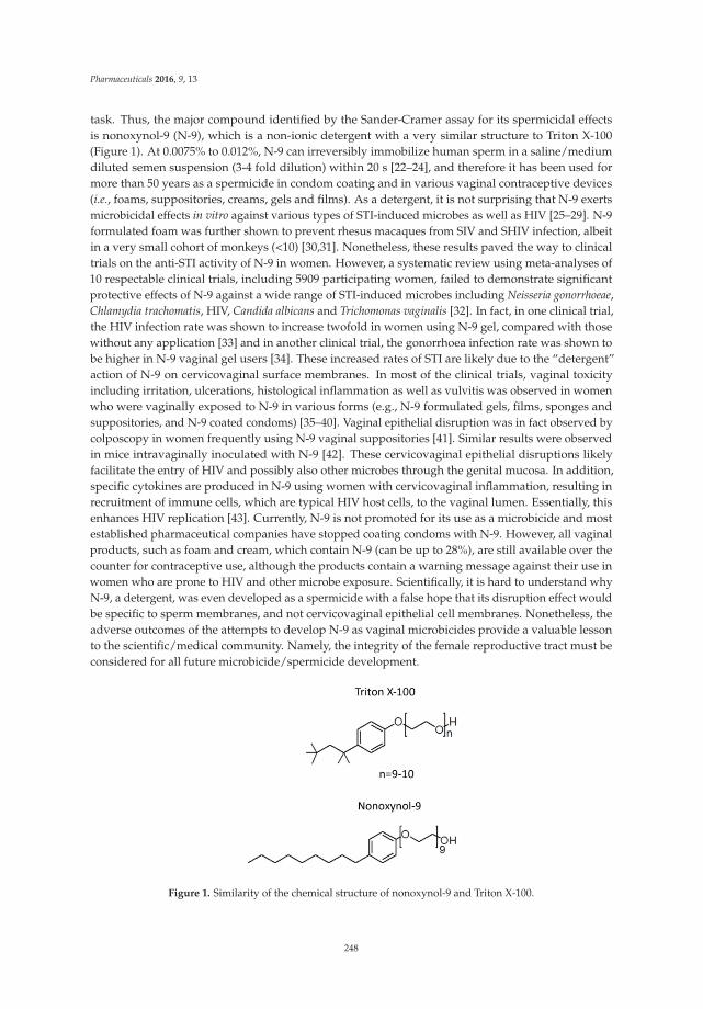
Pharmaceuticals 2016, 9, 13
task. Thus, the major compound identified by the Sander-Cramer assay for its spermicidal effectsis nonoxynol-9 (N-9), which is a non-ionic detergent with a very similar structure to Triton X-100(Figure 1). At 0.0075% to 0.012%, N-9 can irreversibly immobilize human sperm in a saline/mediumdiluted semen suspension (3-4 fold dilution) within 20 s [22–24], and therefore it has been used formore than 50 years as a spermicide in condom coating and in various vaginal contraceptive devices(i.e., foams, suppositories, creams, gels and films). As a detergent, it is not surprising that N-9 exertsmicrobicidal effects in vitro against various types of STI-induced microbes as well as HIV [25–29]. N-9formulated foam was further shown to prevent rhesus macaques from SIV and SHIV infection, albeitin a very small cohort of monkeys (<10) [30,31]. Nonetheless, these results paved the way to clinicaltrials on the anti-STI activity of N-9 in women. However, a systematic review using meta-analyses of10 respectable clinical trials, including 5909 participating women, failed to demonstrate significantprotective effects of N-9 against a wide range of STI-induced microbes including Neisseria gonorrhoeae,Chlamydia trachomatis, HIV, Candida albicans and Trichomonas vaginalis [32]. In fact, in one clinical trial,the HIV infection rate was shown to increase twofold in women using N-9 gel, compared with thosewithout any application [33] and in another clinical trial, the gonorrhoea infection rate was shown tobe higher in N-9 vaginal gel users [34]. These increased rates of STI are likely due to the “detergent”action of N-9 on cervicovaginal surface membranes. In most of the clinical trials, vaginal toxicityincluding irritation, ulcerations, histological inflammation as well as vulvitis was observed in womenwho were vaginally exposed to N-9 in various forms (e.g., N-9 formulated gels, films, sponges andsuppositories, and N-9 coated condoms) [35–40]. Vaginal epithelial disruption was in fact observed bycolposcopy in women frequently using N-9 vaginal suppositories [41]. Similar results were observedin mice intravaginally inoculated with N-9 [42]. These cervicovaginal epithelial disruptions likelyfacilitate the entry of HIV and possibly also other microbes through the genital mucosa. In addition,specific cytokines are produced in N-9 using women with cervicovaginal inflammation, resulting inrecruitment of immune cells, which are typical HIV host cells, to the vaginal lumen. Essentially, thisenhances HIV replication [43]. Currently, N-9 is not promoted for its use as a microbicide and mostestablished pharmaceutical companies have stopped coating condoms with N-9. However, all vaginalproducts, such as foam and cream, which contain N-9 (can be up to 28%), are still available over thecounter for contraceptive use, although the products contain a warning message against their use inwomen who are prone to HIV and other microbe exposure. Scientifically, it is hard to understand whyN-9, a detergent, was even developed as a spermicide with a false hope that its disruption effect wouldbe specific to sperm membranes, and not cervicovaginal epithelial cell membranes. Nonetheless, theadverse outcomes of the attempts to develop N-9 as vaginal microbicides provide a valuable lessonto the scientific/medical community. Namely, the integrity of the female reproductive tract must beconsidered for all future microbicide/spermicide development.
Figure 1. Similarity of the chemical structure of nonoxynol-9 and Triton X-100.
248

Pharmaceuticals 2016, 9, 13
3. Antimicrobial Peptides as Spermicides
An alternative approach to search for compounds with both spermicidal and microbicidalactions can be taken by screening for spermicidal properties of known microbicidal agents. However,conventional antibiotics must be excluded in this pursuit due to the growing antibiotic resistance [44,45].In this regard, antimicrobial peptides (AMPs) have been appropriately considered, as they aresmall peptides (<10 kDa) produced by all domains of life as part of innate immunity. In humansand eutherian mammals, AMPs are produced by neutrophils and other immune cells as well asepithelial cells of various tissues especially those that are connected or exposed to an externalenvironment (e.g., genitourinary tract, lung, skin) [46,47]. As natural compounds, resistance against themicrobicidal activities of AMPs is less anticipated and AMPs are considered to be the next generationof anti-infectives [45]. The broad spectrum activity against Gram positive and Gram negative bacteria,fungi, viruses and certain protozoa is another attractive property of AMPs [46–48]. In addition to thedirect microbicidal properties, AMPs can abrogate the action of lipopolysaccharides (LPS), a pathogenassociated molecular pattern (PAMP) from Gram negative bacteria [47], through their affinity forLPS [49–51]. Therefore, the binding of LPS to the host cell surface Toll-like receptor 4 and subsequentcell signalling events that lead to inflammatory responses cannot occur [52]. AMPs can also act asimmune modulators with various positive consequences to the target cells (reviews [53–55]). For thisreason, AMPs are also referred to as host defence peptides [46,55]. Immunomodulatory effects ofAMPs, which are relevant to the health of the lower female genital tract tissues include wound repair,angiogenesis and cell proliferation [46,48,52,56–59], as these processes would be beneficial to thevaginal mucosa after intercourse that often induces abrasion to the mucosal surface.
Most of AMPs are cationic and amphipathic peptides. To date, over 2500 natural AMPswith diverse sequence, structure and property have been archived in the APD3 database(http://aps.unmc.edu/AP/ [60]). Based on the types of secondary structures, AMPs are categorizedinto four classes (α, β, αβ, and non-αβ) [61]. Because less than 12% of the AMPs in the APD3 databasehave a 3D structure, covalent bonding patterns of polypeptide chains are also used to categorize AMPsinto four classes: linear (UCLL, with no covalent bonds between different amino acids in the polypeptidechain, e.g., human cathelicidin LL-37 and frog magainins), side-chain linked (UCSS, covalent bondsbetween different amino acid side chains, e.g., disulfide bonded defensins and lanthionine ether bondedlantibiotics), sidechain-backbone linked (UCSB, such as those present in bacterial lasso peptides), andbackbone-backbone connected circular peptides (UCBB, such as those present in plant cyclotides) [62].In addition, specific properties have been used to define AMP families. For example, cathelicidinsare AMPs, where their precursors contain a conserved cathelin domain in the N-terminal region [46].Examples include LL-37 in humans, CRAMP in mice, indolicidin in cows. Another family of AMPscontaining disulfide bonds is called “defensins”, which are sub-classified to the α, β and θ groups. Theα- and β-defensins are differentiated from each other based on the positions of the disulfide bonds (αhaving CI–CVI, CII–CIV and CIII–CV, whereas β having CI–CV, CII–CIV and CIII–CVI). So are θ-defensins,which possess three different disulfide bonds (CI–CVI, CII–CV and CIII–CIV). Interestingly, most ofdefensins with known 3-D structure (α: HNP-1, HNP-2, HNP-3, HNP-4, HD-5 and HD-6; β: hBD-3; θ:RTD-1) have β-sheet secondary structure, while hBD-1 (human β-defensin-1) contains both α-helixand β-sheet structures. In addition, “bacteriocins” is used to define AMPs produced by bacteria.Notably, a number of bacteriocins contain intramolecular lanthionine or methyl lanthionine ethers(formed through thioether linkages between serine/threonine and cysteine); such bacteriocins arereferred to as lantibiotics and lantipeptides depending on whether they have antibacterial activity [63].In addition, some bacteriocins such as gramicidin A contain D-amino acids [64]. Both lanthioninesand D-amino acids contribute to the 3-D structure of bacteriocins [65]. It is notable that a number ofbacteriocins differ substantially from cationic cathelicidins and defensins. For example, gramicidin Ahas a net charge of zero and consists of essentially all hydrophobic amino acids, allowing it to insertinto bacterial membranes as an ion channel. In addition to structural categorization, APD3 classifiesAMPs according to their functions (e.g., antimicrobial (as expected), anti-HIV, antimalarial, wound
249

Pharmaceuticals 2016, 9, 13
healing). Relevant to this review is the spermicidal activity of certain AMPs, at least in vitro. TheseAMPs are referred to as spermicidal AMPs.
Being positively charged, AMPs first bind to anionic molecular components of the cell wall ofGram positive bacteria (i.e., lipoteichoic acid) and outer membrane of Gram negative bacteria (i.e.,LPS) [66]. This initial binding results in destabilization of these outer surfaces, thus allowing AMPs toreach and bind to anionic molecules of the bacterial cell membrane (i.e., phosphatidylglycerol (PG)and cardiolipin (CL)). Initially, AMPs may lie horizontally through their affinity for PG and CL on thebacterial cell membrane. Since AMPs are amphiphathic, clusters of their hydrophobic amino acidswould then interact with the hydrocarbon chains of lipids in the membrane bilayers. This may resultin the vertical insertion of AMP peptides into the lipid bilayer. AMPs adjacent to each other also tendto polymerize through the interaction of their hydrophobic domains. Alternatively, these peptides mayintersperse in the membranes by preferentially interacting with anionic lipids. All of these could leadto pore formation in the bacterial cell membrane and the eventual loss of cellular homeostasis, andfinally death [46,48,67]. In addition, some AMPs can inhibit bacterial cell wall synthesis [67], whileothers can enter the bacteria to bind to their ribosomes, resulting in inhibition of protein translation [60].The fungicidal, virucidal and protozoacidal mechanisms, however, are less understood [48].
The desire to use AMPs as spermicides has been in place for decades, although in the early phaseof work, the selective action of AMPs on sperm and not on epithelial cells of the vagina (the mostlogical site for their administration) may not have been well considered. Mammalian sperm are knownto have a negatively charged surface [68,69] in the head region. This is due to the specific presence ofSGG in the outer leaflet of the plasma membrane. The level of SGG is at 10 mole% of total sperm lipidswith the main molecular species having C16:0 in both its sn-1 alkyl chain and sn-2 acyl chain [70,71](Figure 2). SGG is a structural analog of sulfogalactosylceramide (SGC, aka sulfatide) (Figure 2), whichis known to be an integral component of membrane lipid rafts. Likewise, SGG is a major lipid in spermlipid rafts; it exists together with its lipid binding partner, cholesterol [72,73]. Our lab has shownthat lipid rafts in capacitated sperm, housing a number of ZP binding proteins [19,20,74], are surfaceplatforms on sperm for the ZP binding [72]. SGG itself also has a direct affinity for the ZP, contributingto the ZP binding ability of sperm lipid rafts [15,16]. It is expected that positively charged molecules,including AMPs, would interact electrostatically with SGG on the sperm head surface and thus reducethe sperm-ZP binding process. In addition to SGG, polysialyated glycoproteins found in the spermplasma membrane endow negative charges to the sperm surface [75,76].
Figure 2. Chemical structure of sulfogalactosylglycerolipid (SGG) and sulfogalactosylceramide (SGC).
Table 2 lists all spermicidal AMPs described in the literature and the antimicrobial peptidedatabase (http://aps.unmc.edu/AP/ [60]), with their sequences and relevant biochemical propertiesdescribed in Table 3.
250

Pharmaceuticals 2016, 9, 13
Ta
ble
2.
Sper
mic
idal
anti
mic
robi
alpe
ptid
es:m
icro
bici
dal,
stru
ctur
alan
dsp
erm
icid
alpr
oper
ties
.
Pe
pti
de
Na
me
AP
D1
IDU
niP
rot
IDP
DB
IDS
ou
rce
Mic
rob
icid
al/
Bio
log
ica
l/B
ioch
em
ica
lP
rop
ert
ies
Re
fere
nce
sfo
rS
pe
rmic
ida
lE
ffe
cts
Re
ma
rks
LL-3
7A
PD:A
P003
10U
niPr
ot:P
4991
3PD
B:2K
6O
Neu
trop
hils
,mon
ocyt
es,
lym
phoc
ytes
,ker
atin
ocyt
es,
epit
helia
lcel
lsof
the
lung
,nas
alca
vity
,gen
itou
rina
rytr
act,
gast
roin
test
inal
trac
t,oc
ular
surf
ace,
and
ging
iva
ofH
omo
sapi
ens,
Expr
essi
onof
LL-3
7in
Pan
trog
lody
tes
(chi
mpa
nzee
),M
acac
am
ulat
ta(r
hesu
sm
acaq
ue)
have
also
been
desc
ribe
d.
LL-3
7ex
erts
mic
robi
cida
leff
ects
onG
ram
posi
tive
and
Gra
mne
gati
veba
cter
ia,y
east
s,C
andi
daal
bica
ns,a
ndH
IVan
dot
her
viru
ses,
incl
udin
gth
ose
that
caus
eST
Is(s
eeTa
ble
4).
Inan
aque
ous
solu
tion
,the
stru
ctur
eof
LL-3
7is
diso
rder
ed.H
owev
er,w
hen
itin
tera
cts
wit
hlip
idm
embr
anes
such
asSD
San
ddo
decy
lpho
spho
chol
ine
mic
elle
s,LL
-37
adop
tsan
alph
ahe
lical
amph
ipat
hic
stru
ctur
e,as
reve
aled
byN
MR
anal
ysis
wit
hon
esi
deof
the
helix
enri
ched
inhy
drop
hilic
amin
oac
ids
and
the
othe
rsi
dehy
drop
hobi
cre
sidu
es[7
7,78
].
[79]
LL-3
7co
mpl
etel
yin
hibi
tshu
man
and
mou
sesp
erm
mot
ility
wit
hin
5m
inat
10.8
μM
and
3.7μ
M,r
espe
ctiv
ely.
Thi
sin
hibi
tion
islik
ely
due
toth
esp
ecifi
cdi
srup
tive
effe
cts
ofLL
-37
onsp
erm
surf
ace
mem
bran
es,a
ssh
own
byel
ectr
onm
icro
scop
yan
dSy
tox
Gre
en(a
mem
bran
eim
perm
eabl
eD
NA
dye)
stai
ning
.In
addi
tion
,LL-
37tr
eate
dsp
erm
beco
me
prem
atur
ely
acro
som
ere
acte
d,th
ushi
nder
ing
them
toef
fect
ivel
ybi
ndto
the
egg.
Thes
esp
ecifi
cad
vers
eef
fect
sof
LL-3
7on
sper
mar
elik
ely
due
toit
saf
finit
yfo
rth
ene
gati
vely
char
ged
sulf
ogal
acto
sylg
lyce
rolip
id(S
GG
)pre
sent
sele
ctiv
ely
onth
esp
erm
surf
ace.
The
cont
race
ptiv
eef
fect
ofLL
-37
isfu
rthe
rde
mon
stra
ted
infe
mal
em
ice.
Fem
ales
,nat
ural
lycy
clin
gto
the
estr
ous
phas
ean
dtr
ansc
ervi
cally
inje
cted
with
sper
m+
LL-3
7,fa
ilto
beco
me
preg
nant
,whe
reas
preg
nanc
yoc
curs
in92
%of
fem
ales
inje
cted
wit
hsp
erm
alon
e.T
here
prod
ucti
vetr
actt
issu
esof
the
fem
ales
adm
inis
tere
dw
ith
LL-3
7ap
pear
sto
beno
rmal
like
that
obse
rved
infe
mal
esun
expo
sed
toLL
-37
[79]
.
Max
imin
1A
PD:A
P000
58U
niPr
ot:P
8308
0PD
B:N
one
and
Max
imin
3A
PD:A
P000
60U
niPr
ot:P
8308
2PD
B:N
one
Skin
:Chi
nese
red
belly
toad
Bom
bina
max
ima
Laie
tal.
[80]
have
show
nth
atm
axim
in1
and
max
imin
3ha
vem
icro
bici
dala
ctiv
itie
sag
ains
tbot
hG
ram
posi
tive
and
Gra
mne
gati
veba
cter
iaan
dye
asts
,Can
dida
albi
cans
.Bo
thal
soha
vean
ti-c
ance
ran
dan
ti-H
IVac
tivi
ties
.A
ltho
ugh
both
max
imin
1an
dm
axim
in4
have
noPD
BID
,the
yar
ecl
ose
hom
olog
sof
max
imin
4(P
DB:
2MH
W).
The
3Dst
ruct
ure
ofm
axim
in4,
asre
veal
edby
NM
Ran
alys
es,i
sa
linea
rca
tion
icam
phip
athi
cpe
ptid
ein
solu
tion
,but
form
sa
kink
edal
pha
helix
wit
hlip
idm
icel
les
[81]
.
[80]
The
sper
mic
idal
wor
kis
base
don
lyon
sper
mim
mot
ility
.At1
00μ
g/m
L(3
7μ
M)o
fmax
imin
1or
max
imin
3,~8
0%of
hum
ansp
erm
beco
me
imm
otile
.
Mag
aini
n2
APD
:AP0
0144
Uni
Prot
:P11
006
for
mag
aini
ns;m
agai
nin
2is
one
ofth
efiv
ecl
eave
dpr
oduc
tsof
mag
aini
ns.
PDB:
2MA
G
Skin
and
stom
ach:
Afr
ican
claw
edfr
og,X
enop
usla
evis
Mag
aini
n2
has
mic
robi
cida
leff
ects
onG
ram
posi
tive
and
Gra
mne
gati
veba
cter
ia,y
east
san
dvi
ruse
s.It
also
acts
agai
nstc
erta
inpr
otoz
oain
clud
ing
mal
aria
caus
ing
Plas
mod
ium
falc
ipar
um.H
owev
er,m
agai
nin
2do
esno
tha
vean
anti
-HIV
acti
vity
[82]
.N
MR
anal
ysis
indi
cate
sth
atm
agai
nin
2ad
opts
anal
pha
helic
alst
ruct
ure
inth
epr
esen
ceof
SDS
and
othe
rlip
idm
icel
les
[83]
.
[84–
87]
Mag
aini
nA
,asy
nthe
tic
deri
vati
ves
ofm
agai
nin
2[8
8]po
sses
ssp
erm
imm
obili
zing
acti
vity
,as
show
nin
rat,
rabb
it,m
onke
yan
dhu
man
sper
m.A
t~5
0μ
g/m
L(2
0μ
M),
mag
aini
nA
com
plet
ely
inhi
bith
uman
/mon
key
sper
mm
otili
tyw
ithin
7–10
min
oftr
eatm
ent,
alth
ough
itta
kes
~300
μg/
mL
(120
μM
)of
the
pept
ide
for
the
imm
edia
teim
mob
iliza
tion
ofhu
man
sper
m.L
ower
dose
sar
ere
quir
edfo
rra
tand
rabb
itsp
erm
for
the
sam
ere
sult
s[8
2,85
,86]
.Th
ese
sper
mis
tatic
effe
cts
are
likel
ydu
eto
the
abili
tyof
mag
aini
nA
todi
srup
tth
esp
erm
surf
ace
mem
bran
es[8
5].M
agai
nin
2-am
ide
(250
μg/
mL
(101
μM
))al
soex
erts
sper
mis
tati
cef
fect
son
hum
ansp
erm
,alt
houg
hit
isat
50%
effic
acy.
Onl
yw
hen
sper
mar
etr
eate
dw
ith
both
cycl
odex
trin
and
mag
aini
n2-
amid
e,sp
erm
imm
otili
tyis
enha
nced
to80
%.
Red
dy’s
grou
pha
vede
mon
stra
ted
vagi
nalc
ontr
acep
tive
effe
cts
ofm
agai
nin
Ain
rabb
itsan
dm
onke
ys.F
emal
esea
chad
min
iste
red
with
1m
gm
agai
nin
Adi
dno
tbec
ome
preg
nant
upon
natu
ralm
atin
g.Si
deef
fect
sof
mag
aini
nA
onth
efe
mal
ere
prod
ucti
vetr
acta
ppea
red
tobe
min
imum
[82,
84].
Der
mas
epti
nS1
APD
:AP0
0157
Uni
Prot
:802
77PD
B:N
one
Skin
:Sau
vage
’sle
affr
og,
Phyl
lom
edus
asa
uvag
ii
Der
mas
eptin
S1ha
sm
icro
bici
dala
ctiv
ities
agai
nstG
ram
posi
tive
and
Gra
mne
gativ
eba
cter
iaan
dhe
rpes
sim
plex
viru
s.It
also
kills
the
prot
ozoa
,Lei
shm
ania
.D
erm
asep
tin
S1ha
san
alph
ahe
lixst
ruct
ure
asre
veal
edby
circ
ular
dich
rois
man
alys
es[8
9].
[90]
The
sper
mic
idal
wor
kis
base
don
lyon
sper
mim
mot
ility
.Hum
ansp
erm
beco
me
com
plet
ely
imm
otile
imm
edia
tely
afte
rtr
eatm
entw
ith20
0μ
g/m
L(5
8μ
M)o
fder
mas
epti
nS1
.
251
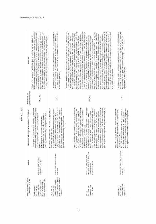
Pharmaceuticals 2016, 9, 13
Ta
ble
2.
Con
t.
Pe
pti
de
Na
me
AP
D1
IDU
niP
rot
IDP
DB
IDS
ou
rce
Mic
rob
icid
al/
Bio
log
ica
l/B
ioch
em
ica
lP
rop
ert
ies
Re
fere
nce
sfo
rS
pe
rmic
ida
lE
ffe
cts
Re
ma
rks
Der
mas
epti
nS4
APD
:AP0
0160
Uni
Prot
:P80
280
PDB:
2DD
6fo
ra
clos
ean
alog
oftr
unca
ted
derm
asep
tin-
S4(a
a1-1
3).
Skin
:Sau
vage
1 sle
affr
og,
Phyl
lom
edus
asa
uvag
ii
Der
mas
eptin
S4ha
sm
icro
bici
dala
ctiv
ities
agai
nstG
ram
posi
tive
and
Gra
mne
gativ
eba
cter
iaan
dvi
ruse
s(h
erpe
ssi
mpl
exvi
rus
and
HIV
).It
also
kills
Pfa
lcip
arum
prot
ozoa
.N
MR
anal
yses
ofa
clos
ean
alog
oftr
unca
ted
derm
asep
tin
S4(a
a1-1
3)in
dica
tes
its
alph
ahe
lixst
ruct
ure
whe
nin
tera
ctin
gw
ith
lipid
mic
elle
s[9
1].
[90,
92,9
3]
Des
pite
asi
mila
rst
ruct
ure
tode
rmas
epti
nS1
,onl
y10
0μ
g/m
L(3
6μ
M)o
fde
rmas
eptin
S4is
requ
ired
toin
duce
com
plet
ehu
man
sper
mm
otili
ty,i
ndic
atin
ga
twic
esp
erm
ista
ticpo
tenc
yof
derm
asep
tinS4
[90]
.Ahi
gher
sper
mis
tatic
effe
ctis
furt
her
obse
rved
ina
derm
asep
tin
S4de
riva
tive
wit
hon
eam
ino
acid
repl
acem
entw
ithly
sine
toin
crea
seth
epo
sitiv
ech
arge
.Onl
y20
μg/
mL
(7.2μ
M)
ofth
isde
riva
tive
isre
quir
edto
indu
ceco
mpl
ete
sper
mim
mot
ility
effe
cts.
The
nati
vede
rmas
epti
nS4
(100
μg/
mL
(36μ
M))
and
its
deri
vati
ve(2
0μ
g/m
L(7
.2μ
M))
caus
e10
0%an
d50
%cy
toto
xici
tyto
HeL
ace
lls,r
espe
ctiv
ely
[92]
.
Sarc
otox
inPd
APD
:AP0
2212
Uni
Prot
:Non
efo
rth
issa
rcot
oxin
buta
vaila
ble
for
othe
rsa
rcot
oxin
isof
orm
s.PD
B:N
one
Inse
cts:
rove
beet
les,
Paed
erus
derm
atiti
s
Sarc
otox
inPd
has
mic
robi
cida
leff
ects
onG
ram
posi
tive
and
Gra
mne
gati
veba
cter
ia.
Alt
houg
hth
ere
isno
PDB
info
rmat
ion
onsa
rcot
oxin
Pd,
inth
epu
blic
atio
nof
Zar
e-Z
ardi
niet
al.[
94],
a3D
stru
ctur
eof
sarc
otox
inPd
was
show
nto
cons
isto
ftw
oal
pha
helic
es.T
his
stru
ctur
alin
form
atio
nw
asob
tain
edfr
omco
mpu
tatio
nalm
odel
ing,
alth
ough
node
tails
wer
egi
ven
onho
wth
ism
odel
ing
was
perf
orm
ed.
[94]
The
sper
mic
idal
wor
kis
base
don
lyon
sper
mim
mot
ility
.The
conc
entr
atio
nof
sarc
otox
inPd
toim
mob
ilize
hum
ansp
erm
is80
μg/
mL
(22μ
M).
Cyt
otox
icit
yM
TTas
say
was
done
onH
eLa
cells
;at8
0μ
g/m
Lof
sarc
otox
inPd
,clo
seto
100%
ofce
llssh
owcy
toto
xici
ty.
Nis
inA
APD
:AP0
0205
Uni
Prot
:P13
068
PDB:
1WC
O
Bact
erio
cin
from
lact
icac
idba
cter
ia(L
AB)
,Lac
toco
ccus
lact
is(f
orm
erly
calle
dSt
rept
ococ
cus
lact
is)
The
mic
robi
cida
leff
ects
ofni
sin
Aar
em
ore
pref
eren
tial
toG
ram
posi
tive
bact
eria
;thi
sm
aybe
attr
ibut
edto
its
abili
tyto
bind
tolip
idII
,ast
ruct
ural
com
pone
ntof
Gra
mpo
siti
veba
cter
ialp
epti
dogl
ycan
s.Th
ein
tera
ctio
nbe
twee
nni
sin
Aan
dlip
idII
lead
sto
inhi
biti
onof
the
bact
eria
lcel
lwal
lsyn
thes
is[9
5].
Itis
aca
tion
icam
phip
athi
cla
ntip
epti
de(i
.e.,
cont
aini
nga
lant
hion
ine
ethe
rlin
kage
betw
een
S3-C
7an
d4
met
hylla
nthi
onin
es:T
8-C
11,T
13-C
19,T
23-C
26an
dT2
5-C
28)(
see
Tabl
e3)
.The
seth
ioet
her
linka
ges
crea
tea
cons
trai
ntpo
lycy
clic
feat
ure
toth
epe
ptid
e.H
owev
er,
NM
Ran
alys
isre
veal
edth
atni
sin
Ais
still
flexi
ble
enou
ghto
inte
ract
wit
hSD
Sm
icel
les,
first
via
ioni
cin
tera
ctio
nan
dth
enth
roug
hhy
drop
hobi
cin
tera
ctio
n.It
was
ther
efor
epr
opos
edth
atni
sin
Aca
ncr
eate
pore
sin
lipid
bila
yers
follo
win
git
str
an-b
ilaye
rin
sert
ion
[96,
97].
[98–
103]
The
sper
mim
mob
ilizi
ngef
fect
sof
nisi
nA
have
been
show
nin
vari
ous
spec
ies,
i.e.,
rats
,rab
bits
,bul
ls,h
orse
s/po
nies
,boa
rs,m
onke
ysan
dhu
man
s[9
8,99
,101
].Th
ein
stan
tane
ous
sper
mis
tati
cco
ncen
trat
ion
ofni
sin
Ais
300–
400μ
g/m
L(8
6–11
4μ
M)f
orhu
man
/mon
key
sper
m,2
00μ
g/m
L(5
7μ
M)f
orra
bbit
sper
man
d50
μg/
mL
(14μ
M)f
orra
tspe
rm.S
cann
ing
elec
tron
mic
rosc
opy
reve
aled
obvi
ous
disr
upti
onof
the
hum
ansp
erm
plas
ma
mem
bran
efo
llow
ing
trea
tmen
tw
ith
360μ
g/m
L(1
03μ
M)o
fnis
inA
.Thi
sdi
srup
tion
was
sim
ilar
tow
hat
obse
rved
onth
esu
rfac
eof
Stap
hylo
cocc
usau
reus
bact
eria
,whi
chw
ere
trea
ted
with
asi
mila
rni
sin
Aco
ncen
trat
ion.
Inco
ntra
st,h
uman
red
bloo
dce
llsw
ere
not
affe
cted
bytr
eatm
entw
itheq
uiva
lent
conc
entr
atio
nsof
nisi
nA
.The
pref
eren
tial
effe
cts
ofni
sin
Aon
sper
mpl
asm
am
embr
ane
perm
eabi
lizat
ion
was
also
confi
rmed
bypr
opid
ium
iodi
denu
clea
rst
aini
ngof
the
trea
ted
sper
m[1
02]
Red
dyet
al.h
ave
furt
her
dem
onst
rate
dth
atni
sin
Ais
anef
fect
ive
vagi
nal
cont
race
ptiv
ein
rats
and
rabb
its.
Fem
ale
rats
natu
rally
cycl
ing
inth
epr
oest
rous
/est
rous
phas
ean
dva
gina
llyad
min
iste
red
with
nisi
nA
(200
μg
each
)di
dno
tbec
ome
preg
nant
follo
win
gna
tura
lmat
ing
[98]
.Suc
cess
fulc
ontr
acep
tive
resu
lts
wer
elik
ewis
eob
tain
edin
fem
ale
rabb
its
intr
avag
inal
lyin
ject
edw
ith
nisi
nA
(1m
gea
ch),
prov
ided
that
mat
ing
took
plac
ew
ithi
n30
min
ofth
epe
ptid
ead
min
istr
atio
n[9
9].I
nbo
than
imal
spec
ies,
the
auth
ors
clai
med
that
ther
ew
ere
noch
ange
sto
the
anat
omy
ofth
efe
mal
ere
prod
uctiv
etr
actt
issu
esor
cyto
kine
prod
uctio
npr
ofile
inth
efe
mal
esva
gina
llyad
min
iste
red
with
nisi
nA
atth
eco
ntra
cept
ive
dose
orev
enhi
gher
and
repe
ated
dose
sof
nisi
nA
[98–
100]
.By
MTT
assa
y,H
eLa
cells
appe
ared
tobe
less
susc
eptib
leto
cyto
toxi
cef
fect
sof
nisi
nA
,as
com
pare
dw
ith
sper
m.
Pedi
ocin
CP2
APD
:AP0
0634
Uni
Prot
:Q8R
L96
PDB:
Non
e
Bact
erio
cin
from
LAB,
Pedi
oocc
usac
idila
ctic
i
Pedi
ocin
CP2
exer
tsm
icro
bici
dale
ffec
tson
both
Gra
mpo
siti
vean
dG
ram
nega
tive
bact
eria
asw
ella
sye
asts
,C
andi
daal
bica
ns[1
04]
No
PDB
IDis
avai
labl
e.H
owev
er,t
he3-
Dst
ruct
ure
ofit
sho
mol
og,s
akac
inP
(PD
B:1O
G7,
68%
iden
tica
lpe
ptid
ese
quen
ceto
pedi
ocin
CP)
,rev
eals
the
pres
ence
ofan
alph
ahe
lixin
the
mid
dle
regi
onof
the
pept
ide.
[104
]
The
sper
mic
idal
wor
kis
base
don
lyon
sper
mim
mot
ility
.The
conc
entr
atio
nof
pedi
ocin
CP2
toim
mob
ilize
hum
ansp
erm
is>2
50μ
g/m
L(5
4μ
M).
Ther
eis
nore
port
edw
ork
onth
ecy
toxi
city
conc
entr
atio
nof
pedi
ocin
CP2
onfe
mal
ere
prod
ucti
vetr
actt
issu
es/c
ells
.
252

Pharmaceuticals 2016, 9, 13
Ta
ble
2.
Con
t.
Pep
tid
eN
am
eA
PD
1ID
Un
iPro
tID
PD
BID
So
urc
eM
icro
bic
idal/
Bio
log
ical/
Bio
chem
ical
Pro
pert
ies
Refe
ren
ces
for
Sp
erm
icid
al
Eff
ect
sR
em
ark
s
Gra
mic
idin
AA
PD:A
P004
99U
niPr
ot:N
one
PDB:
1MA
G
Bact
erio
cin
from
soil
bact
eriu
m,
Baci
llus
brev
is
Gra
mic
idin
Aex
erts
mic
robi
cida
leff
ects
onbo
thG
ram
posi
tive
and
Gra
mne
gati
veba
cter
iaas
wel
las
viru
ses
(inc
ludi
ngH
IV)[
105]
.G
ram
icid
inA
has
asp
ecia
lβ-h
elix
stru
ctur
ebe
caus
eof
its
poss
essi
onof
D-a
min
oac
ids.
Gra
mic
idin
Adi
mer
izes
wit
hth
eN
-ter
min
usof
each
pept
ide
bein
gad
jace
ntto
each
othe
r.A
sa
resu
lt,t
hedi
mer
form
sa
cati
onch
anne
lin
lipid
bila
yers
wit
hth
etw
oC
-ter
min
iexp
osed
[64]
.
[105
–107
]
Gra
mic
idin
has
been
used
for
alo
ngtim
ein
Rus
sia
asa
sper
mic
ide
asre
ferr
edto
inBo
urin
baia
ret
al.[
105]
,alt
houg
hde
taile
dda
taof
its
effic
acy
isno
tava
ilabl
e.Ex
peri
men
tally
,the
sper
mic
idal
effe
cts
ofgr
amic
idin
Aar
eba
sed
onhu
man
sper
mim
mot
ility
and
cons
eque
ntly
the
inab
ility
ofsp
erm
tope
netr
ate
lam
bce
rvic
alm
ucus
[106
].O
nly
5μ
g/m
L(2
.8μ
M)o
fgr
amic
idin
Aco
uld
com
plet
ely
imm
obili
zesp
erm
.Gra
mic
idin
D(m
ixtu
reof
gram
icid
inA
,Ban
dC
)has
sim
ilar
sper
mim
mob
ilizi
ngef
fect
sbu
tata
high
erco
ncen
trat
ion
than
gram
icid
inA
.
Subt
ilosi
nA
APD
:AP0
0928
Uni
Prot
:007
623
PDB:
1PX
Q
Bact
erio
cin
from
Baci
llus
subt
ilis,
Baci
llus
amyl
oliq
uefa
cien
s,Ba
cillu
sat
roph
aeus
Subt
ilosi
nA
has
mic
robi
cida
leff
ects
onbo
thG
ram
posi
tive
and
Gra
mne
gati
veba
cter
iaas
wel
las
herp
essi
mpl
exvi
ruse
s.It
isa
lant
ipep
tide
,con
tain
ing
ahi
ghpe
rcen
tage
ofhy
drop
hobi
cre
sidu
es(6
0%)w
ith
anov
eral
lne
gati
vech
arge
of-2
.Lan
thio
nine
sbe
twee
nC
13an
dF2
2,C
7an
dT28
,and
C4
and
F31
mak
eth
eN
-ter
min
usan
dC
-ter
min
usfo
ldto
war
dsea
chot
her,
givi
ngan
over
allb
oatc
onfo
rmat
ion,
asre
veal
edby
NM
Ran
alys
is.T
here
isan
alph
ahe
lixin
the
C-t
erm
inus
star
ting
from
G29
toW
34[1
08].
[101
,109
]
The
sper
mic
idal
test
isba
sed
only
onsp
erm
imm
otili
ty.A
tasi
ngle
conc
entr
atio
nte
sted
,800
μg/
mL
(233
μM
),su
btilo
sin
Ain
stan
tane
ousl
yim
mob
ilize
dbu
llan
dho
rse/
pony
sper
m.T
hesa
me
sper
mic
idal
effe
cts
wer
eob
serv
edin
boar
and
rats
perm
at20
0μ
g/m
L(5
8μ
M)o
fsub
tilo
sin
A[1
01].
Hum
ansp
erm
trea
ted
wit
hsu
btilo
sin
also
beca
me
imm
otile
ina
dose
-dep
ende
ntm
anne
rw
ith
com
plet
eim
mob
iliza
tion
at11
0μ
g/m
L(3
2μ
M).
Not
ably
,sub
tilo
sin
atth
isco
ncen
trat
ion
does
notr
educ
ece
llvi
abili
tyin
the
hum
anEp
iVag
inal
ecto
cerv
ical
tiss
uem
odel
[109
].
Lact
icin
3147
APD
:AP0
1194
Uni
Prot
:087
236
PDB:
Non
e
Bact
erio
cin
from
LAB
Lact
ococ
cus
lact
isD
PC31
47
Lact
icin
3147
isco
mpo
sed
oftw
ola
ntip
epti
deco
mpo
nent
s,Lt
nA1
and
LtnA
2.M
icro
bici
dal
acti
vity
ofla
ctic
in31
47is
pref
eren
tial
onG
ram
posi
tive
bact
eria
and
isst
rong
erw
ith
LtnA
1an
dLt
nA2
com
bine
d,co
mpa
red
wit
hea
chla
ctic
in31
47ch
ain
alon
e.Th
ere
are
four
lant
hion
ines
inLt
nA1
and
two
inLt
nA2,
whi
chge
nera
tea
poly
cycl
icst
ruct
ure
tobo
thpe
ptid
es.N
MR
anal
yses
also
reve
ala
helic
alst
ruct
ure
inLt
nA2
[110
].N
ote
that
ther
ear
ea
few
D-A
lare
sidu
esin
both
lant
ipep
tide
chai
ns,a
ndth
eir
exis
tenc
eis
impo
rtan
tfor
the
mic
robi
cida
lact
ivit
yof
lact
icin
[110
].
[101
]
The
sper
mic
idal
test
isba
sed
only
onsp
erm
imm
otili
ty.L
tnA
1ch
ain
had
muc
hle
sssp
erm
icid
alef
fect
sth
anLt
nA2.
The
com
bina
tion
ofLt
nA1
+Lt
nA2
at20
0μ
g/m
L(3
1μ
M)c
ould
effe
ctiv
ely
imm
obili
zera
t,bu
ll,an
dho
rse/
pony
sper
m.B
uton
ly50
μg/
mL
(7.8μ
M)o
fLt
nA1
+Lt
nA2
was
requ
ired
toin
duce
imm
otili
tyof
bull
sper
m[1
01].
1Th
elis
ting
ofsp
erm
icid
alA
MPs
ises
sent
ially
from
the
anti
mic
robi
alpe
ptid
eda
taba
se(A
PD)(
http
://a
ps.u
nmc.
edu/
AP/
[60]
).
253

Pharmaceuticals 2016, 9, 13
Ta
ble
3.
Sequ
ence
san
dbi
oche
mic
alpr
oper
ites
ofsp
erm
icid
alan
tim
icro
bial
.
1A
min
oac
ids
are
colo
rco
ded
acco
rdin
gto
thei
rch
arge
s,hy
drop
hilic
ity
and
hydr
opho
bici
ty.P
osit
ivel
ych
arge
dam
ino
acid
s(K
,Ran
dH
)are
inre
d,w
here
asne
gati
vely
char
ged
resi
dues
(D,E
)are
ingr
een.
Unc
harg
edhy
drop
hilic
amin
oac
ids
(S,T
,Y,N
,Q,C
)are
inpu
rple
.Hyd
roph
obic
amin
oac
ids
are
indi
ffer
ents
hade
sof
blue
,with
stro
ngbl
uein
dica
ting
the
high
esth
ydro
phob
icity
leve
l(L,
F,I,
V,A
),fo
llow
edby
light
blue
(G),
and
very
light
blue
(W,M
,P)w
ithth
ele
asth
ydro
phob
icity
.2A
lthou
ghm
axim
in4
has
notb
een
show
nto
besp
erm
icid
al,i
tsse
quen
ceis
show
nhe
rebe
caus
eof
itshi
ghsi
mila
rity
tom
axim
in1
and
max
imin
3an
dits
3Dst
ruct
ure
issh
own
inFi
gure
3.3
Brac
kets
deno
tela
nthi
onin
eet
hers
.4
Ital
icle
tter
sde
note
D-a
min
oac
ids.
254

Pharmaceuticals 2016, 9, 13
In addition to these 12 AMPs, cecropin D2A21 was mentioned but without any data on itsspermicidal effects in the publication describing its chlamydiacidal property [111]. Seven of theseAMPs are produced in humans and animals, i.e., LL-37-humans; maximin 1, maximin 3, magainin 2,dermaseptin S1 and dermaseptin S4-lower vertebrate animals (frogs and toads); and sarcotoxinPd-invertebrates (insects). The other five AMPs (nisin A, pediocin CP2, gramicidin A, subtilosin A andlacticin 3147) are bacteriocins produced by various bacteria species. Interestingly, gramicidin A hasbeen used for decades in humans in Russia [105], although the scientific and medical details of thisuse are not available in the literature. Except for our work on LL-37, the screening for the spermicidalaction of AMPs listed in Table 1 was based primarily on their ability to immobilize sperm in thesuspension that still contained 20%–25% of seminal plasma within 20 s of treatment (Sander-Cramertest). As discussed above, this approach is likely an “overkill”, since motile sperm swim out ofthe seminal plasma and carry only residual amounts of seminal plasma into the cervix and uterinecavity. Rather “swim-up” sperm and “Percoll-gradient centrifuged”, prepared in a laboratory, wouldbe more representative of motile sperm that swim through the cervix into the uterine cavity [23].Swim-up sperm are simply prepared by overlaying semen placed in a tube with appropriate medium.Swim-up sperm are motile sperm that have swum up into the medium layer that still carry a smallamount of seminal plasma [112]. Percoll-gradient centrifuged sperm are prepared by loading semenonto a discontinuous gradient of Percoll solutions (usually 45% and 90%) followed by centrifugationat ~600 g. Motile sperm have “clean” morphology (devoid of membranous vesicles and havingcompact chromatin) with a higher specific density than immotile/morphologically abnormal sperm,and therefore they sediment as a pellet, which can then be resuspended in a medium for experimentaluse. Percoll-gradient centrifuged sperm have the highest fertilizing ability [113] and therefore theyshould be used for the screening of a candidate spermicide. However, an assay for human spermmotility that is more physiologically representative is to observe the rate of sperm to swim throughcervical mucus collected from women in the middle of their menstrual cycle (ovulation time) andplaced in a capillary tube [7,114]. The cervical mucus fluid is viscoelastic due to the abundanceof proteo-glycosaminoglycans. Sperm must have sufficient motility force as well as biochemicalcomponents to digest the proteo-glycosaminoglycan networks in order to swim through the cervicalmucus column, a situation that does not exist in sperm resuspended in medium [11,115]. Thissperm-cervical mucus assay was performed with gramicidin A treated human sperm. This assay,however, is impractical to be used for screening of spermicide compounds due to the restriction inobtaining sufficient quantities of mid-menstrual cycle cervical mucus from women.
Among all spermicidal AMPs listed in Table 2, LL-37, magainin 2 and nisin A are the only AMPswith confirmed in vivo contraceptive effects in experimental animals. The outcome of zero pregnanciesis considered the gold standard indicator of contraceptive effects of these AMPs. Our group performedthis in vivo work on LL-37 in mice [79], whereas Reddy & Aranha’s group examined the effects ofnisin A in rats and rabbits [98,99], and magainin 2 in rabbits and monkeys [82,84]. Experimentationin animal models is essential in the screening of vaginal spermicides and/or microbicides, since itis a means not only to confirm the spermicidal/microbicidal effects in vivo but also to determinewhether there are any adverse effects of the compounds on the female reproductive tract. Selectionof appropriate animals for these in vivo studies is also critical for the next clinical trials in humans.Monkeys are animals with reproductive physiology closest to the human system. However, onemay only use a restricted number of monkeys for each trial. In addition, it is important to preservethe female monkey lives after the experiment. Therefore, the examination of any adverse effects onthe reproductive system in female monkeys can only be done in an indirect manner (e.g., assessingtypes and quality of cells from vaginal smears). Such is the case for the study on in vivo spermicidaleffects of magainin 2 in monkeys [82]. In contrast, histology of the female reproductive tract can beevaluated directly in smaller animals including rabbits, rats and mice following their sacrifice andtissue removal for fixing and processing. Histology images should be explicitly shown such as thosedescribed in the studies for LL-37 in mice [79] and nisin A in rabbits [100]. However, interpretation
255

Pharmaceuticals 2016, 9, 13
of results from spermicidal and microbicidal studies in female rabbits, rats and mice must be madewith caution that the reproductive systems in these animals differ from that of humans in a numberof aspects. While insemination in rabbits occurs in the vagina like the situation in humans, rabbitfemales do not have a reproductive cycle (estrus or menstrual cycle) with peaked estrogen, whichenriches the vaginal/cervical secretion and microflora [116]. Therefore, rabbit vaginal microfloraare rather simple and in fact contain only residual amounts of lactobacilli [116], which are foundabundantly in the human vagina and responsible for acidifying the vaginal luminal milieu due tolactate production [117]. The pH in the rabbit vagina is in the neutral range [118] in contrast to thepH 4 in the human vagina [119,120]. With no defined reproductive cycle, ovulation in rabbits isnot hormonally regulated but rather induced by coitus [121], a process so markedly different fromhuman ovulation. The reproductive processes in rats and mice, as well, differ from those in humans ina number of aspects despite the fact that rodents do have a reproductive estrus cycle. Although rodentsemen is first deposited in the vagina, sperm together with seminal plasma are swept into the uteruswithin minutes [11]. In addition, the mouse vaginal pH is at ~6.5, more basic than that of the humanvagina. This is due to lower numbers of lactobacilli colonies in the rodent vagina [122]. Regardless,rodents are less expensive to purchase and maintain and the in vitro fertilization procedures in rodentsare well described, allowing the assessment of candidate spermicides for their direct inhibitory effectson fertilization in vitro. While it is logical that various animal models should be used to validatespermicidal and microbicidal effects of the candidate compounds, human vaginal and cervical celllines as well as reconstructed human vagina models should also be employed in the spermicide andmicrobicide ex vivo studies. The former include immortalized cell lines derived from human vaginalepithelia (Vk2/E6E7), human ectocervical epithelia (Ect1/E6E7) and human endocervical epithelia(End1/E6E7), all established by Deborah Anderson, Brighams and Women’s Hospital, Inc. [123],and available from ATCC. For the latter, MatTek Inc. has produced various types of organotypicvaginal-ectocervical tissue models through reconstruction of human vagina and cervix tissues [124].Vk2/E6E7, Ect1/E6E7 and End1/E6E7 cell lines have been widely used for the studies of candidatemicrobicides [125–127]. Likewise, MatTek human vaginal-ectocervical tissue models have recentlybeen used for studies on the properties of potential vaginal anti-HIV agents [128–130]. To date,there are no publications on the use of immortalized Vk2/E6E7, Ect1/E6E7 and End1/E6E7 cellsfor studies of spermicidal AMPs. MatTek organotypic vaginal-ectocervical tissue models were usedonly in subtilosin A study (Table 2). On the other hand, HeLa cell lines (derived from cervical cancercells obtained from Henrietta Black over 60 years ago [131]) were used for evaluating cytotoxicity ofdermaseptin S4, sarcotoxin Pd and nisin A (see Table 2). However, HeLa cells are highly transformedand may not have the expected properties of human cervical epithelial cells [131].
As discussed above, the acidic pH in the healthy human vagina is attributed to the presence ofcommensal lactobacilli. The vaginal acidity is essential in inhibition of unwanted proliferation aswell as infection of pathogenic microbes such as Gardnerella vaginalis, Prevotella bivia and HIV [132].Depletion of lactobacilli in the vagina leads to bacterial vaginosis. In this regard, caution must betaken in the selection of AMPs with no microbicidal effects on lactobacilli for further developmentinto vaginal microbicides. This is the case for nisin A, which has been used as a food preservativefor human consumption and shown to be an effective contraceptive in rabbits and monkeys ([98,99],Table 2). However, nisin A is not a good candidate to be developed into a vaginal microbicide, since itexerts microbicidal activity on vaginal Lactobacillus spp [132,133].
While the microbicidal effects of AMPs in general are of a broad spectrum, it is most desirableto search for spermicidal AMPs that also have anti-HIV properties. Among spermicidal AMPs listedin Table 2, LL-37 [134–136], maximin 1 [80], maximin 3 [80], dermaseptin S1, dermaseptin S4 [137]and gramicidin A [105] have been shown to possess anti-HIV properties. Therefore, they should beconsidered for further research studies and development into vaginal contraceptives/microbicides.Most of these HIV infection studies were performed using HIV lab strains in CD4+ T cells, T-cellleukemia cell lines or T4 lymphoblastoid cell lines, and in some cases monocyte derived dendritic cells.
256

Pharmaceuticals 2016, 9, 13
In order to validate the physiological significance of the anti-HIV properties of these AMPs, workshould be comprehensively repeated using dual tropic clinical isolates of HIV at sufficiently high titers.In addition, the inhibitory effects of the candidate AMPs on HIV infection in the human cervicovaginalepithelium should be directly assessed using MatTek human vaginal-ectocervical tissue models. On theother hand, since gramicidin A has been used as a spermicide in Russia [105], a population studyshould be carried out to determine whether women who have been using gramicidin A vaginally andare more prone to exposure to HIV have a lower rate of HIV infection.
The 3D structures of a number of spermicidal AMPs or their close homologs have been determinedby NMR spectroscopy. These include human LL-37 [77], frog maximin 4 (homolog of maximin 1) [81],magainin 2 [83], dermaseptin S4 [91], bacterial nisin A [138], gramicidin A [64], subtilosin A [108] andlacticin 3147 (chain A1 and A2) [110].
Figure 3A shows the 3D structures of these AMPs. Interestingly, LL-37, maximin 4, magainin 2,dermaseptin S4 and lacticin 3147 chain A2 contain an alpha helical structure in a significant length oftheir sequences. A short helix also exists in subtilosin A and nisin A (Figure 3A). However, lacticin3147 chain A1 (which has a minimal spermicidal activity) does not have an alpha helical structure [110]and is therefore not displayed in Figure 3A. On the other hand, gramicidin A has a β-helical structure(Figure 3A). The helical wheel projections of LL-37 (both the whole peptide and the sequence fromamino acids 10-37), maximin 1, maximin 3, maximin 4, magainin 2, dermaseptin S1 and dermaseptinS4 (all from humans and animals) reveal an amphipathic feature of the helix, with hydrophobic aminoacids enriched in approximately one half of the top view helical circle and hydrophilic amino acids inthe other half (Figure 3B). Such an amphipathic feature can also be seen in Figure 3A, especially forLL-37, magainin 2, and dermaseptin S4 peptide analog. It is remarkable that both LL-37 and magainin2 possess 3-4 aromatic phenylalanines on the hydrophobic surfaces. NMR studies have demonstratedthat the four phenylalanines all interact with acyl chains of anionic PGs [77]. This amphipathic helicalstructure of AMPs may be required for spermicidal activity. If this is proven, this requirement wouldbe analogous to that needed for AMPs for full exertion of microbicidal activity [135,139,140].
Bacteriocins utilize specific features to maintain their 3D structure. Lacticin chain A1 does nothave any helical structure but its 3D confined structure is attributed to the existence of both D-aminoacids and lanthionine ethers in its sequence [110]. These two structural components also exist inlacticin 3147 chain A2 with an apparent alpha helical structure [110]. Nisin A and subtilosin A alsocontain lanthionine ethers [46,141], which contribute to their 3D structure (Table 3). On the otherhand, gramicidin A does not contain any lanthionines but it possesses five D-amino acids in its15-mer sequence (Table 3). This unique feature endows gramicidin A a β-helical structure, which isfurther stabilized by dimerization, and this allows gramicidin A dimer to form a cation channel inlipid bilayers [64]. The channel and pore formation in the membranes by gramicidin A and otherspermicidal bacteriocins like subtilosin A and lacticin 3147 chain A1 is unlikely to be initiated withthe electrostatic binding between these AMPs and the negatively charged surface molecules of themicrobes, since these AMPs have a net charge of zero or even minus values (Table 3). A close lookat the helical regions in these bacteriocins (Figure 3E, G, and H) reveals a lack of amphipathic natureobserved with human LL-37, magainin 2 and dermaseptin S4 (Figure 3A,B).
257

Pharmaceuticals 2016, 9, 13
Figure 3. Structures of selected antimicrobial peptides with spermicidal activity annotated in theAPD3 [142]. (A) 3D structures shown as ribbon diagrams of (a) human LL-37 (PDB ID: 2K6O);(b) frog maximin 4 (PDB ID: 2MHW); (c) magainin 2 (PDB ID: 1MAG); (d) a close analog of truncateddermaseptin S4 (amino acids 1-13; PDB ID: 2DD6); (e) bacterial nisin A complexed with lipid II (PDBID: 1WCO); (f) gramicidin A (PDB ID: 1MAG); (g) subtilosin A (PDB ID: 1PXQ); and (h) lacticin 3147(structural co-ordinates were provided by Dr. John Vedaras, University of Alberta, according to hispublished work [110]). Except for gramicidin A, the N-terminus of the peptide is positioned at the top.In the case of gramicidin A (f), its dimer is shown. The two N-termini of each dimer are positionednext to each other in the middle, whereas the C-termini are exposed and their four tryptophansapproximate the lipid head group regions of the lipid bilayers for membrane positioning and ionchannel conductance (f). Note that the C and N-termini of subtilosin A approximate in the structure (g).The side chains of human LL-37, magainin 2, and the dermaseptin S4 analog are selectively labeledto illustrate the amphipathic nature of these AMPs. Images were generated using MOLMOL [143].(B) Helical wheel projections of selected spermicidal AMPs expressed in eukaryotes: LL-37, maximin1, maximin 3, maximin 4, magainin 2, truncated dermaseptin S1 and dermaseptin S4. All of theseAMPs show an amphipathic structure, with hydrophobic amino acids (blue circles) organized inapproximately one half of the wheel and the hydrophilic residues in the other half. For LL-37, the wheelprojections are shown for both the whole LL-37 sequence (amino acids 1–37) and the sequence fromamino acids 10–37. This is because the helical structure of the whole LL-37 sequence has a kink at Ser9.The LL-37 peptide (amino acids 10-37) actually shows a better distribution of hydrophobic amino acidsin one half of the helical wheel. Although maximin 4 has not been shown for the direct microbicidaleffects, as demonstrated for maximin 1 and maximin 3, its helical wheel projection is shown hereinto corroborate its 3D structure shown in (A) and also for a comparison with the wheel projections ofmaximin 1 and maximin 3. The wheel projections of truncated dermaseptin S1 and dermaseptin S4 areboth for their truncated sequence (amino acids 1-13). Again, this is to corroborate the 3D structure ofthe close analog of the truncated dermaseptin S4 peptide shown in (A).
258

Pharmaceuticals 2016, 9, 13
4. LL-37, the Most Promising Spermicidal AMP
Cationic antimicrobial peptide LL-37 is coded by the only human cathelicidin gene [46,47,144].LL-37 or its very close homologs are also present in non-human primates. Chimpanzees possessLL-37 identical to the human sequence, whereas in Gorillas and orangutans, two and three spotsin the human LL-37 sequence are replaced by other amino acids [145]. The name “LL-37” denotesa peptide that contains 37 amino acids with the Leu-Leu sequence at the N-terminus. LL-37 isproduced as a propeptide, hCAP-18, by neutrophils [50,146], other immune cells [147,148], normal andinflammatory skin cells [149–151], and epithelial cells of various tissues, especially those that connectwith the external (male reproductive tract [152,153], urinary tract [154,155], gastrointestinal tract [156],lung [157,158], gingiva [159], eye [160], nasal cavity [161,162]) (for reviews see [149,163,164]). Ofsignificance to this review is the expression of hCAP-18 by the human epididymal epithelial cellsfollowed by its secretion into the epididymal lumen. This makes hCAP-18 a component of seminalplasma with a physiological concentration range of 2 to 10 μM [152,153,165]. hCAP-18 (MW:16442)is expressed by neutrophils and released during degranulation together with proteinase 3, whichimmediately processes hCAP-18 at physiological pH into LL-37 (MW:4493) with full microbicidalactivity [166]. In skin, LL-37 is generated from hCAP-18 through the proteolytic activity of kallikrein 5also at the neutral pH. Both kallikrein 5 and kallikrein 7 (also present in the skin surface) then furthercleave LL-37 into smaller fragments (i.e., RK-31, KS-30, KS-22, KR-20, LL-29), which have higherantimicrobial activity than LL-37 [151]. However, the processing of hCAP-18 is more complicated.It does not occur in the male reproductive tract or in the ejaculate. Sorensen et al. [153] identifiedgastricsin, secreted from the prostate gland and thus also a component of seminal plasma, to bethe enzyme responsible for processing hCAP-18 in seminal plasma into ALL-38 (LL-37 + Ala at theN-terminus) at a pH optimum of approximately 4. Therefore, gastricsin cannot function in seminalplasma, which has a high buffering capacity at neutral pH. Following ejaculation in the vagina, seminalplasma changes the normally acidic pH of the vagina (pH 4) into the neutral range. It takes 2-6 hpost-ejaculation for the vaginal lumen to resume its acidic pH and it is only at this time that gastricsinbecomes active to process hCAP-18 into ALL-38, with microbicidal activity [153].
However, immediately after semen liquefaction, typically 30 min post-ejaculation, sperminstantaneously swim out from seminal plasma through the cervix into the uterine cavity. Onlyfeeble sperm are left behind together with seminal plasma in the vagina. Therefore, motile sperm withfertilizing ability are never exposed to ALL-38, which is generated long after their movement fromthe vagina (Figure 4). It is likely that the production of ALL-38 is meant for protection of the vaginalepithelium against microbes introduced during intercourse [167]. As a very close analog of LL-37,ALL-38 should possess all properties described for LL-37. For example, they have identical antibacterialactivity against all the organisms tested [153]. LL-37 has been shown for its direct microbicidal effectsagainst numerous Gram negative and Gram positive bacteria, yeast and viruses (including HIV)(Table 4), but not on lactobacilli [168–170], which are essential for maintaining acidic pH and thushealth of the vagina (see Section 3).
259

Pharmaceuticals 2016, 9, 13
Figure 4. ALL-38 is processed from hCAP-18 post-ejaculation. (A) Processing of hCAP-18 to ALL-38by gastricsin occurs at the optimum pH of 4. Although both hCAP-18 and gastricsin are seminalplasma components, this processing does not occur in the seminal plasma due to its high bufferingcapacity at neutral pH. (B) Immunoblotting showing that processing of hCAP-18 to ALL-38 can occurin acidified seminal plasma. Anti-LL-37 antibody used in immunoblotting was produced against thewhole LL-37 sequence [79] and therefore, it recognized only ALL-38 and hCAP-18, but not cathelin.Before acid treatment (BA), seminal plasma contains hCAP-18 and its oligomers (Oligo; ~60 kDa), aswell as a 14 kDa band (presumably a cleaved product of hCAP-18). Upon acidification of seminalplasma with HCl to pH 4, hCAP-18 is processed to ALL-38; this is due to the activation of gastricsin.AA = after acid extraction. (C) ALL-38 is produced 2–6 h post-ejaculation. Upon ejaculation, seminalplasma neutralizes the vaginal pH, and it takes 2–6 h post-ejaculation for the vagina lumen to resumeits acidity. Only at this time, ALL-38 is produced from hCAP-18 via gastricsin activity. However,immediately after semen liquefaction (30 min post-coitus), motile sperm in the ejaculate swim intothe uterine cavity. Therefore, motile sperm are never exposed to ALL-38. The drawing is based onSørensen et al. [153].
260
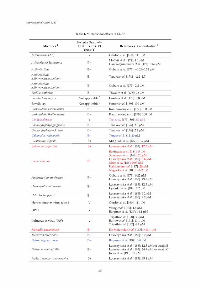
Pharmaceuticals 2016, 9, 13
Table 4. Microbicidal effects of LL-37.
Microbes 1Bacteria Gram +/´
(B+/´) Virus (V)Yeast (Y)
References: Concentration 2
Adenovirus (Ad) V Gordon et al. [160]: 111 μM
Acinetobacter baumannii B´ Moffatt et al. [171]: 1.1 μMGarcia-Quintanilla et al. [172]: 0.67 μM
Actinobacillus B´ Ouhara et al. [173]: ~0.26–0.52 μM
Actinobacillusactinomycetemcomitans B´ Tanaka et al. [174]: ~2.2–2.7
Actinobacillusactinomycetemcomitans B´ Ouhara et al. [173]: 2.2 μM
Bacillus anthracis B´ Thwaite et al. [175]: 22 μM
Borrelia burgdorferi Not applicable 3 Lusitani et al. [176]: 8.8 μM
Borrelia spp Not applicable 3 Sambri et al. [169]: 100 μM
Burkholderia pseudomallei B´ Kanthawong et al. [177]: 100 μM
Burkholderia thailandensis B´ Kanthawong et al. [178]: 100 μM
Candida albicans Y Tsai et al. [179,180]: 8.9 μM
Capnocytophaga gingivalis B´ Tanaka et al. [174]: 2.0 μM
Capnocytophaga ochracea B´ Tanaka et al. [174]: 2.4 μM
Chlamydia trachomatis B´ Tang et al. [181]: 20 μM
Clostridium difficile B+ McQuade et al. [182]: 10.7 μM
Enterococcus faecalis B+ Leszczynska et al. [183]: 12.5 μM
Escherichia coli B´
Benincasa et al. [184]: 5 μMSmeianov et al. [168]: 25 μMLeszczynska et al. [185]: 5.6 μMChen et al. [186]: 0.07 μMKai-Larsen et al. [187]: 20 μMNagaoka et al. [188]: ~1-2 μM
Fusobacterium nucleatum B´ Ouhara et al. [173]: 0.22 μMLeszczynska et al. [183]: 49.8 μM
Haemophilus influenzae B´ Leszczynska et al. [183]: 12.5 μMLysenko et al. [189]: 2.2 μM
Helicobacter pylori B´ Leszczynska et al. [183]: 6.2 μMLeszczynska et al. [185]: 2.2 μM
Herpes simplex virus type 1 V Gordon et al. [160]: 111 μM
HIV-1 VWang et al. [135]: 1.6 μMBergman et al. [134]: 11.1 μM
Influenza A virus (IAV) VTripathi et al. [190]: 13 μMBarlow et al. [191]: 11.1 μMTripathi et al. [192]: 6.7 μM
Klebsiella pneumoniae B´ De Majumdar et al. [193]: >11.1 μM
Moraxella catarrhalis B´ Leszczynska et al. [183]: 6.2 μM
Neisseria gonorrhoeae B´ Bergman et al. [194]: 0.8 μM
Neisseria meningitidis B´Leszczynska et al. [183]: 12.5 μM for strain BLeszczynska et al. [183]: 24.9 μM for strain CJones et al. [195]: 10 μM
Peptostreptococcus anaerobius B+ Leszczynska et al. [183]: 49.8 μM
261

Pharmaceuticals 2016, 9, 13
Table 4. Cont.
Microbes 1Bacteria Gram +/´
(B+/´) Virus (V)Yeast (Y)
References: Concentration 2
Porphyromonas gingivalis B´ Leszczynska et al. [183]: 49.8 μMOuhara et al. [173]: 11.1 μM
Prevotella intermedia B´ Ouhara et al. [173]: 1.1 μM
Pseudomonas aeruginosa B´
Bergsson et al. [196]: 5.6 μMDean et al. [197]: 0.22 μMDosler and Karaaslan [198]: ~14.2–28.4 μMGordon et al. [160]: ~11.1–22.2 μMLeszczynska et al. [183]: 99.7 μM
Respiratory syncytial virus V Currie et al. [199]: 5.6 μM
Staphylococcus aureus B+
Leszczynska et al. [183]: 6.2 μMNoore et al. [200]: 2 μMChen et al. [186]: 0.67 μMSenyurek et al. [201]: 11.1 μMNagaoka et al. [188]: 1 μMGordon et al. [160]: ~11.1–22.2 μM
Staphylococcus epidermidis B+Leszczynska et al. [183]: 12.5 μMGordon et al. [160]: ~11.1–22.2 μM
Streptococcus mitis B+ Ouhara et al. [173]: 2.2 μM
Streptococcus mutans B+Ouhara et al. [173]: 0.22 μMLeszczynska et al. [183]: 6.2 μM
Streptococcus pneumoniae B+Nagaoka et al. [188]: 1 μMLeszczynska et al. [183]: 3.1 μM
Streptococcus pyogenes B+ Leszczynska et al. [183]: 3.1 μM
Streptococcus salivarius B+Ouhara et al. [173]: 1.1 μMLeszczynska et al. [183]: 6.2 μM
Streptococcus sanguis B+Ouhara et al. [173]: 0.22 μMLeszczynska et al. [183]: 6.2 μM
Streptococcus sobrinus B+ Ouhara et al. [173]: 1.1 μM
Tannerella forsythensis B+ Leszczynska et al. [183]: 49.8 μM
Treponema pallidum B- Sambri et al. [169]: 100.1 μM
Ureaplasma parvum NA 3 Xiao et al. [202]: 22.2 μM
Ureaplasma urealyticum NA 3 Xiao et al. [202]: 22.2 μM
Vaccinia virus V Howell et al. [203]: 20 μM
Varicella zoster virus (VZV) V Crack et al. [204]: 0.1 μM1 Highlighted microbes: blue causing STI; green causing vaginitis; Red causing UTI; Pink causing vaginitis andUTI. 2 Concentrations of LL-37 given are those that exert microbicidal effects on ě90% of the microbes. In somecases where this information is not clearly described in the publication, estimated concentrations are given.3 NA = not applicable; Borrelia spp. and Ureaplasma spp. do not react well with the Gram stain.
Besides the direct microbicidal activity, LL-37 possesses other properties including anti-endotoxinactivity [49–51], immunomodulation (reviews [53–55]), angiogenesis [59] and wound healing [56–58].If the anti-endotoxin and immunomodulatory properties are confirmed in the female reproductive tractsystem, it will strengthen the possibility that LL-37/ALL-38 can be used as vaginal microbicides thatcan clear infection and minimize infection-associated inflammation. Angiogenesis and wound healingproperties would also aid in the repair of minor vaginal tissue damages occurring during intercourse.Further studies on LL-37’s direct microbicidal effects must also be carried out in all microbes thatare causes of STIs and vaginitis, as well as urinary tract infection (UTI). The vagina and its normal
262

Pharmaceuticals 2016, 9, 13
microbiota represent an important barrier against uropathogenic bacteria (Table 1). Perturbations of thenormal vaginal microbiota, such as depletion of lactobacilli, can promote colonization of uropathogens,such as uropathogenic E. coli (UPEC), within the vagina [205]. The vagina can then become anextra-urinary uropathogen reservoir and in turn increase the risk of UTI [206].LL-37 exerts microbicidaleffects on most of the STI-inducing microorganisms including HIV (causing life threatening AIDSwith no cure), HSV-1 (causing genital herpes with no cure), Neisseria gonorrhoeae (causing gonorrhoea,curable), Treponema pallidum (causing syphilis, curable) and Chlamydia trachomatis (causing cervicitis,salpingitis and endometriosis, curable) (Table 4). Although gonorrhoea, syphilis and chlamydiainfection are curable, a number of complications are associated with these three STIs. They all increasethe risk of infertility. A higher susceptibility to HIV transmission is also associated with gonorrhoeaand syphilis. Salpingitis and oviductal tubal blockade caused by chlamydia infection can also leadto ovarian cancer. For gonorrhoea, resistance to antibiotics used for the treatment has increasinglybecome a problem. As listed in Table 1, LL-37 needs to be tested for its microbicidal activity againsta number of additional microorganisms that cause STI (viruses:HPV, hepatitis A and C; protozoon,Trichomonas vaginalis) and bacterial vaginosis (Gardnerella vaginalis, Bacteroides spp., Mycoplasma hominisand Mobiluncus spp.), prior to its development as a vaginal microbicide. Further testing againsturopathogenic microorganisms (Table 1) will also allow LL-37 to be developed for therapeutic andprophylactic uses for urinary tract infection in this system. LL-37 formulated gel administered intothe vagina would exert microbicidal action on uropathogenic microbes that opportunistically forma reservoir in the vagina. The vaginal secretion, which can travel upwards into the urinary tract, wouldalso likely contain LL-37 released from the gel, which then can fight against microbes in this tract (seethe list in Table 4).
The majority of sperm co-existing with seminal plasma, which contains 2-10 μM of hCAP-18 [153],remain motile in ejaculated semen of fertile donors. Interestingly, despite the negatively chargedsurface of sperm and the overall positive charge of hCAP-18 (+6, pI = 9.25), hCAP-18 is present ata residual amount on human sperm [79]. However, since motile sperm with fertilizing ability are neverexposed to ALL-38 produced from seminal plasma (Figure 4), it raises a possibility that ALL-38/LL-37may have a deleterious effect on sperm, possibly after the deposition of ALL-38/LL-37 onto the spermsurface. Although hCAP-18/LL-37 is also produced by cervicovaginal epithelial cells, its amountis 1000x less than ALL-38 originated from seminal plasma (i.e., 1.3 nM [136] versus 2–10 μM [153],respectively). Therefore, we asked the question of whether LL-37 when added exogenously to a spermsuspension could bind to sperm with a deleterious consequence on their fertilizing ability. Also, ifsperm-LL-37 binding did occur, was it dependent on the interaction of LL-37 with a negatively–chargedSGG existing specifically on the mammalian sperm head surface? The latter question is relevant interms of possible development of LL-37 into a spermicide with specificity to sperm and not to othersomatic cells such as cervicovaginal epithelial cells. Our results revealed that LL-37 bound to SGGand its anionic lipid analog, sulfogalactosylceramide (SGC), as well as phosphatidylserine (alsonegatively charged), all immobilized separately in a well of a microtiter plate, in a specific manner.Kd values of the binding of LL-37 to these three anionic lipids were 456, 157 and 24 nM, respectively.In contrast, LL-37 did not bind to neutral lipids, phosphatidylcholine and galactosylglycerolipid(GG—the desulfated form of SGG) [79]. Direct binding of LL-37 to Percoll-gradient centrifuged mouseand human sperm resuspended in medium was further demonstrated. For the reason describedin Section 3, Percoll-gradient centrifuged sperm were first used in all studies, and in the case ofhuman sperm replicate experiments were performed with swim-up sperm. The partial dependence ofLL-37-sperm interaction on SGG on the sperm surface was then demonstrated by a decrease in LL-37binding to sperm that were pre-incubated with anti-SGG antibody [79]. Pretreatment of capacitatedmouse sperm with 3.6 μM LL-37 resulted in a complete loss of sperm ability to fertilize eggs in vitro.This 3.6 μM of LL-37 is equivalent to the concentration of SGG in the mouse sperm suspension usedfor the treatment [71,79], further corroborating the concept that SGG was involved in LL-37 binding.Notably, 100% of mouse sperm treated with 3.6 μM LL-37 became immotile within 5 min of treatment,
263

Pharmaceuticals 2016, 9, 13
and this result would be one of explanations for the inability of these LL-37 treated sperm to fertilizeeggs in vitro. Likewise, human sperm lost their motility when treated with LL-37, although 10.8 μM ofLL-37 was needed for the majority of sperm to become immotile. Furthermore, when non-capacitatedsperm and partially capacitated sperm, both human and mouse, were treated with the same LL-37concentrations as used for fully capacitated sperm, the inhibition of their motility was similarlyobserved [79]. Non-capacitated sperm, partially capacitated sperm and fully capacitated sperm wereprepared in the lab by resuspending sperm in the medium without bicarbonate, calcium and albumin,with bicarbonate and calcium but no albumin, and with all of these three components, respectively. Theprepared non-capacitated sperm represent sperm that have just swum out from the seminal plasmain vivo, whereas partially capacitated sperm resemble those that are swimming through the cervix.Finally, the prepared capacitated sperm are equivalent to sperm that have swum into the uterine cavityand their cholesterol is induced to release by albumin and HDL present in the uterus [8,207].
The loss of sperm motility upon LL-37 treatment can be from the direct interaction of LL-37 withthe axoneme, the motility apparatus, in the sperm tail [8]. However, this possibility is discounted bythe lack of binding of LL-37, added exogenously to the sperm suspension, to the sperm tail [79]. It isalso unlikely that the mechanism of the effects of LL-37 on sperm motility is through its direct effect onCatSper, the cation channel responsible for sperm hyperactivated motility [12], since LL-37 also inducesimmotility in non-capacitated sperm, which normally do not have hyperactivated motility patterns.Rather, immotility of LL-37 treated sperm may come from the loss of intracellular homeostasis due tothe LL-37 induced damage on the sperm surface, a situation that is parallel to that observed on themicrobial membrane [46,67]. This postulation was confirmed by the observation that LL-37 treatedsperm became positively stained with Sytox Green (a membrane impermeable DNA fluorescent dye),an indication that the surface membranes of these treated sperm were compromised. Pharmacokineticstudies further indicated that the sperm plasma membrane damage induced by LL-37 occurred priorto the loss of sperm motility. Finally, LL-37 treated sperm became acrosome-reacted, thus markedlylowering their ability to bind to the egg. Transmission electron microscopy confirmed that the damageson the sperm plasma membrane and the outer acrosomal membrane as well as the loss of the acrosomeand in some cases the inner acrosomal membrane were the adverse effects of LL-37 on sperm [79].In contrast, our unpublished results reveal that LL-37 at the spermicidal concentration range did nothave any adverse effects to mouse eggs and embryos. Mouse eggs treated with 3.6 μM LL-37 werenormally fertilized by sperm collected from a fertile animal, and mouse two-cell embryos treated withthe same LL-37 concentration developed into blastocysts at the same rate as untreated two-cell embryos.
We have further demonstrated the loss of sperm fertilizing ability in vivo. Mouse sperm treatedwith LL-37 were transcervically injected into female mice naturally cycling to the estrous phase inthe reproductive cycle. None of these female mice (n = 26) became pregnant, whereas pregnancywas observed in 92% of female mice (n = 26) injected with untreated sperm. Significantly, the femalereproductive system of mice injected with LL-37 + sperm did not show any apparent changes, ascompared with that of control females (injected with medium + sperm) (Figure 5A). In females thatwere exposed to LL-37, the histology of their vagina and uterus as well as the dimension, shapeand color of their reproductive tissues did not differ from those of control females (unexposed toLL-37) (Figure 5A). There were no signs of immune cell recruitment (implicating inflammation) to thevaginal/uterine tissues of these LL-37 exposed females (Figure 5B). This was in contrast to female micetranscervically injected with 2% N-9, whereas numerous neutrophils were apparent in the vaginalepithelium (Figure 5B). In fact, female mice transcervically injected with sperm + LL-37 for three estruscycles with no pregnancy outcomes could resume their fecundity as shown by pups delivered bythese females following their natural mating with fertile males two weeks after the last transcervicalinjection with LL-37.
264

Pharmaceuticals 2016, 9, 13
Figure 5. (A) Left panel: Normal histology of the vagina and uterus of female mice transcervicallyinjected with LL-37 + sperm. The female reproductive tract tissues were collected for fixing and paraffinembedding one day after the transcervical injection of LL-37 + sperm, or sperm alone (control). Sectionsof the vagina and uterus revealed that the vaginal stratified epithelial cell layers and the uterine singleepithelial cell layer (denoted as the area between the two arrows) as well as the corresponding laminapropria of the LL-37 treated and control animals did not differ from each other. Bar = 20 μm. This imagewas taken from our published article [79]. Right panel: The anatomy (shape, dimension and color)of the vagina/cervix and uterus, dissected from females injected with LL-37 (3.7or 5.4 μM) + sperm(n = 3 each) or sperm alone (control) (n = 3) one day after the injection, was the same among the threegroups of the animals. (B) Absence of immune cell recruitment to the vaginal epithelium of micetranscervically injected with LL-37 + sperm. Sections of the vagina were prepared as in (A). The vaginalepithelium and lamina propria of both the control and LL-37 treated mice similarly show minimalnumbers of immune cells, indicating no recruitment of these cells to the vagina as a consequence ofLL-37 injection. In contrast, when females were transcervically injected with 2% nonoxynol-9 (N-9),numerous neutrophils were recruited into the vaginal epithelium. The polymorphonuclear structure ofneutrophils is apparent in the close-up image (denoted by asterisks). Triangles point to the N-9 inducedrupture of the vaginal epithelial surface. Bar = 20 μm.
We have further confirmed that LL-37 had minimal adverse effects on the immortalized humancervicovaginal epithelial cell lines, i.e., Vk2/E6E7 (vaginal), Ect1/E6E7 (ectocervical) and End1/E6E7(endocervical) cells. Cytotoxicity MTT (3-(4,5-dimethylthiazol-2-yl)-2,5-diphenyltetrazolium bromide)assays showed no significant differences of percent viable cells of these three cell lines upon treatmentwith LL-37 at concentrations up to 3.6 μM (Figure 6A). Sytox Green incorporation assays for themembrane intactness of these cell lines revealed similar results. In all of these three cell lines, less than10% of cells incorporated Sytox Green into their nuclei upon treatment with 3.6 μM. However, thesepercentages increased to 20, 25 and 40% in Ect1/E6E7, End1/E6E7 and Vk2/E6E7 cells, respectively,following treatment with 10.8 μM of LL-37 (Figure 6B).
265

Pharmaceuticals 2016, 9, 13
Figure 6. Low adverse effects of LL-37 on human vaginal and cervical cell lines. Vk2/E6E7 (vaginal),Ect1/E6E7 (ectocervical) and End1/E6E7 (endocervical) cells cultured as previously described [123]were treated with various concentrations of LL-37 (24 h, 37 ˝C, 5% CO2) and subjected to a cytotoxicityMTT assay (A) or a membrane intactness assay using Sytox (B). (A) Treatment of all three cell lines withLL-37 up to 3.7 μM did not change percentages of viable cells. In contrast, the percentages of viable cellswere only 50% or less upon treatment with 2% NaN3. (B) Sytox Green staining was observed in lessthan 20% of all three cell lines upon treatment with LL-37 up to 3.7 μM. When the LL-37 concentrationwas increased to 10.8 μM, the numbers of Sytox Green stained cells became higher, although for bothEnd1/E6E7 and Ect1/E6E7 cells, the percentages were still <20%. For Vk2/E6E7 cells, this percentagewas 40%.
266

Pharmaceuticals 2016, 9, 13
While the higher susceptibility to LL-37 in the vaginal epithelial cell line should be a matter ofconcern, the situation in vivo may not be as unfavourable as that observed in vitro. Cells of the upperlayer of the vaginal epithelium continue to slough off into the lumen and it is possible that thosecells with compromised membrane intactness due to LL-37 exposure may undergo this sloughing ata higher rate than normal cells.
In summary, we have shown that LL-37 possesses spermicidal effects in both human and mousesperm with minimal adverse effects on the female reproductive tract epithelia. This spermicidalproperty with molecular mechanisms described above appears to be unique to LL-37. A numberof defensin AMPs are secreted by epididymal epithelial cells (examples-human: HE2β1 (akaSPAG11D) [208], DEFB126 [209–211], DEFB118 [212,213], DEFB114 [214], DEFB1 [215]; -rodents: Bin1b(aka SPAG11E) [216,217], β -defensin 22 [218]) and some of them deposit onto the surface of transitsperm to endow fertilizing ability (e.g., HE2 β 1, DEFB126, DEFB118 and HBD1 in humans, and Bin1bin rodents). We have shown that both HE2 β 1 and Bin1b could bind to SGG/SGC in vitro, but spermincubated with excess amounts (18 μM) of these two defensins were still motile with intact acrosomeand in the case of mouse sperm that were treated with these β-defensins, they could still fertilize eggsat 50% control values (our unpublished results).
The physiological significance of LL-37 as a microbicide and an immunomodulatory and woundhealing peptide is well documented. In particular, LL-37 exerts microbicidal effects on a number ofmicrobes responsible for STI, vaginitis and UTI (Table 4). With these microbicidal and spermicidalproperties, LL-37 warrants further attempts to be developed as a vaginal contraceptive/microbicide.As a natural peptide, its repeated and long-term use will likely create minimal side effects andmicrobicidal resistance. Further studies, however, are needed in a number of aspects in potentialapplication of LL-37 in the vagina. First, an effective method to deliver LL-37 into the vaginal lumenhas to be established. Careful studies on any adverse effects of LL-37, following its repeated/long-termadministration, must also be performed at both topical and systemic levels. Regardless, the cost ofchemical synthesis of LL-37 for this use is a challenge, and the shortest truncated LL-37 peptide or itsmimetic, which still bears spermicidal and microbicidal activity, has to be discovered. In a separateavenue, ultrashort cationic lipopeptides with high antibiotic activity [219–221] may be screened forspermicidal effects. However, the selectivity of the deleterious effects of these lipopeptides to spermand not to the female reproductive tract epithelial cells must be considered. Nonetheless, success infinding the most cost-effective form of LL-37 as a vaginal contraceptive/microbicide will undoubtedlyprovide women with empowerment in having a healthy and safe sex practice—the ability to protectthemselves from unwanted pregnancies and microbial infections.
Acknowledgments: Our work on LL-37 described in this review is supported by a grant from Canadian Institutesof Health Research and previously by Bill & Melinda Gates Foundation (Grand Challenges Exploration Project)both awarded to NT. GW is supported by the NIH grant R01AI105147 during this study. We thank Ryan Jarratt,RPh, Westboro Pharmasave, Ottawa, ON, Canada for the insightful information on the nonoxynol-9 formulationused in vaginal contraceptives. Help in manuscript preparation from Terri Van Gulik is highly appreciated.
Author Contributions: N.T. conceived and designed the scheme of this review and wrote about 90% of it. N.S.significantly contributed to the results indicating the spermicidal effects of LL-37. As well, he gathered informationfrom databases for the content of Table 4, helped with Table 3 preparation, and drew Figure 4A,C. R.A. contributedto the data shown in Figure 4B and helped in the preparation of Table 4. W.K. helped in all aspects of the writing,including reference citation and checking, and table/figure preparation. K.K. prepared Figure 3B and the first draftof Figure 3A. R.Z. and W.L. contributed to the information on contraceptives. M.B. contributed to informationrelated to sperm physiology and the biochemistry of hCAP-18/LL-37. G.W. prepared Figure 3A and contributedsignificantly to the up-to-date knowledge of antimicrobial peptides. D.H. prepared Table 1 and contributed toinformation on the clinical relevance of antimicrobial peptides in the genitourinary system. All authors proofreadand edited the manuscript.
Conflicts of Interest: The authors declare no conflicts of interest.
267

Pharmaceuticals 2016, 9, 13
References
1. Shoupe, D.; Mishell, D.R. Contraception. Women and Health, 2nd ed.; Goldman, M.B., Troisi, R., Rexrode, K.M.,Eds.; Academic Press: London, UK, 2013.
2. Wadman, M. Contraceptive risk of HIV long suspected. Nature News 2011. [CrossRef]3. Spevack, E. The long-term health implications of depo-provera. Int. Med. 2013, 12, 27–34.4. Filby, A.L.; Neuparth, T.; Thorpe, K.L.; Owen, R.; Galloway, T.S.; Tyler, C.R. Health impacts of estrogens
in the environment, considering complex mixture effects. Environ. Health Perspect. 2007, 115, 1704–1710.[CrossRef] [PubMed]
5. Wise, A.; O’Brien, K.; Woodruff, T. Are oral contraceptives a significant contributor to the estrogenicity ofdrinking water? Environ. Sci. Technol. 2011, 45, 51–60. [CrossRef] [PubMed]
6. Haase, A.T. Early events in sexual transmission of HIV and SIV and opportunities for interventions.Annu. Rev. Med. 2011, 62, 127–139. [CrossRef] [PubMed]
7. WHO. Annual technical report. Department of Reproductive Health and Research 2013.8. Florman, H.; Fissore, R. Fertilization in mammals. In Knobil and Neill’s Physiology of Reproduction, 4th ed.;
Plant, T.M., Zeleznik, A., Eds.; Elsevier Inc.: New York, NY, USA, 2015; pp. 149–195.9. Travis, A.J.; Kopf, G.S. The role of cholesterol efflux in regulating the fertilization potential of mammalian
spermatozoa. J. Clin. Investig. 2002, 110, 731–736. [CrossRef] [PubMed]10. Okabe, M. The cell biology of mammalian fertilization. Development. 2013, 140, 4471–4479. [CrossRef]
[PubMed]11. Suarez, S.S.; Pacey, A.A. Sperm transport in the female reproductive tract. Hum. Reprod. Update. 2006, 12,
23–37. [CrossRef] [PubMed]12. Lishko, P.V.; Kirichok, Y.; Ren, D.; Navarro, B.; Chung, J.J.; Clapham, D.E. The control of male fertility by
spermatozoan ion channels. Annu. Rev. Physiol. 2012, 74, 453–475. [CrossRef] [PubMed]13. Chung, J.J.; Navarro, B.; Krapivinsky, G.; Krapivinsky, L.; Clapham, D.E. A novel gene required for male
fertility and functional Catsper channel formation in spermatozoa. Nature Commun. 2011, 2, 1–12. [CrossRef][PubMed]
14. Tanphaichitr, N.; Carmona, E.; Bou Khalil, M.; Xu, H.; Berger, T.; Gerton, G.L. New insights into sperm-zonapellucida interaction: Involvement of sperm lipid rafts. Front. Biosci. 2007, 12, 1748–1766. [CrossRef][PubMed]
15. White, D.; Weerachatyanukul, W.; Gadella, B.; Kamolvarin, N.; Attar, M.; Tanphaichitr, N. Role of spermsulfogalactosylglycerolipid in mouse sperm-zona pellucida binding. Biol. Reprod. 2000, 63, 147–155.[CrossRef] [PubMed]
16. Weerachatyanukul, W.; Rattanachaiyanont, M.; Carmona, E.; Furimsky, A.; Mai, A.; Shoushtarian, A.;Sirichotiyakul, S.; Ballakier, H.; Leader, A.; Tanphaichitr, N. Sulfogalactosylglycerolipid is involved in humangamete interaction. Mol. Reprod. Dev. 2001, 60, 569–578. [CrossRef] [PubMed]
17. Ikawa, M.; Inoue, N.; Benham, A.M.; Okabe, M. Fertilization: A sperm’s journey to and interaction with theoocyte. J. Clin. Investig. 2010, 120, 984–994. [CrossRef] [PubMed]
18. Lyng, R.; Shur, B.D. Sperm-egg binding requires a multiplicity of receptor-ligand interactions: New insightsinto the nature of gamete receptors derived from reproductive tract secretions. Soc. Reprod. Fertil. Suppl.2007, 65, 335–351. [PubMed]
19. Kongmanas, K.; Kruevaisayawan, H.; Saewu, A.; Sugeng, C.; Fernandes, J.; Souda, P.; Angel, J.B.; Faull, K.F.;Aitken, R.J.; Whitelegge, J.; et al. Proteomic characterization of pig sperm anterior head plasma membranereveals roles of acrosomal proteins in ZP3 binding. J. Cell Physiol. 2015, 230, 449–463. [CrossRef] [PubMed]
20. Tanphaichitr, N.; Kongmanas, K.; Kruevaisayawan, H.; Saewu, A.; Sugeng, C.; Fernandes, J.; Souda, P.;Angel, J.B.; Faull, K.F.; Aitken, R.J.; et al. Remodeling of the plasma membrane in preparation for sperm-eggrecognition: Roles of acrosomal proteins. Asian J. Androl. 2015, 17, 574–582. [CrossRef] [PubMed]
21. Munch, J.; Rucker, E.; Standker, L.; Adermann, K.; Goffinet, C.; Schindler, M.; Wildum, S.; Chinnadurai, R.;Rajan, D.; Specht, A.; et al. Semen-derived amyloid fibrils drastically enhance HIV infection. Cell 2007, 131,1059–1071. [CrossRef] [PubMed]
22. Chijioke, P.C.; Zaman, S.; Pearson, R.M. Comparison of the potency of d-propanolol, chlorhexidine andnonoxynol-9 in the sander cramer test. Contraception 1986, 34, 207–211. [CrossRef]
268
Pharmaceuticals 2016, 9, 13
23. Dunmire, E.N.; Katz, D.F. Kinematic response of human spermatozoa to nonoxynol-9. Biol. Reprod. 1994, 50,903–911. [CrossRef] [PubMed]
24. Thompson, K.A.; Malamud, D.; Storey, B.T. Assessment of the anti-microbial agent c31g as a spermicide:Comparison with nonoxynol-9. Contraception 1996, 53, 313–318. [CrossRef]
25. Asculai, S.S.; Weis, M.T.; Rancourt, M.W.; Kupferberg, A.B. Inactivation of herpes simplex viruses by nonionicsurfactants. Antimicrob. Agents Chemother. 1978, 13, 686–690. [CrossRef] [PubMed]
26. Moench, T.R.; Whaley, K.J.; Mandrell, T.D.; Bishop, B.D.; Witt, C.J.; Cone, R.A. The cat/felineimmunodeficiency virus model for transmucosal transmission of aids: Nonoxynol-9 contraceptive jellyblocks transmission by an infected cell inoculum. AIDS 1993, 7, 797–802. [CrossRef] [PubMed]
27. Benes, S.; McCormack, W.M. Inhibition of growth of Chlamydia trachomatis by nonoxynol-9 in vivo.Antimicrob. Agents Chemother. 1985, 27, 724–726. [CrossRef] [PubMed]
28. Malkovsky, M.; Newell, A.; Dalgleish, A.G. Inactivation of HIV by nonoxynol-9. Lancet 1988, 1, 645.[CrossRef]
29. Doncel, G.F. Exploiting common targets in human fertilization and HIV infection: Development of novelcontraceptive microbicides. Hum. Reprod. Update 2006, 12, 103–117. [CrossRef] [PubMed]
30. Miller, C.J.; Alexander, N.J.; Gettie, A.; Hendrickx, A.G.; Marx, P.A. The effect of contraceptives containingnonoxynol-9 on the genital transmission of simian immunodeficiency virus in Rhesus macaques. Fertil. Steril.1992, 57, 1126–1128. [PubMed]
31. Weber, J.; Nunn, A.; O’Connor, T.; Jeffries, D.; Kitchen, V.; McCormack, S.; Stott, J.; Almond, N.; Stone, A.;Darbyshire, J. Chemical condoms’ for the prevention of HIV infection: Evaluation of novel agents againstSHIV(89.6PD) in vitro and in vivo. AIDS 2001, 15, 1563–1568. [CrossRef] [PubMed]
32. Wilkinson, D.; Tholandi, M.; Ramjee, G.; Rutherford, G.W. Nonoxynol-9 spermicide for prevention ofvaginally acquired HIV and other sexually transmitted infections: Systematic review and meta-analysis ofrandomised controlled trials including more than 5000 women. Lancet Infect. Dis. 2002, 2, 613–617. [CrossRef]
33. Van Damme, L.; Ramjee, G.; Alary, M.; Vuylsteke, B.; Chandeying, V.; Rees, H.; Sirivongrangson, P.;Mukenge-Tshibaka, L.; Ettiegne-Traore, V.; Uaheowitchai, C.; et al. Effectiveness of col-1492, a nonoxynol-9vaginal gel, on HIV-1 transmission in female sex workers: A randomised controlled trial. Lancet 2002, 360,971–977. [CrossRef]
34. Richardson, B.A.; Lavreys, L.; Martin, H.L., Jr.; Stevens, C.E.; Ngugi, E.; Mandaliya, K.; Bwayo, J.;Ndinya-Achola, J.; Kreiss, J.K. Evaluation of a low-dose nonoxynol-9 gel for the prevention of sexuallytransmitted diseases: A randomized clinical trial. Sex. Transm. Dis. 2001, 28, 394–400. [CrossRef] [PubMed]
35. Stafford, M.K.; Ward, H.; Flanagan, A.; Rosenstein, I.J.; Taylor-Robinson, D.; Smith, J.R.; Weber, J.; Kitchen, V.S.Safety study of nonoxynol-9 as a vaginal microbicide: Evidence of adverse effects. J. Acquir. ImmuneDefic. Syndr. 1998, 17, 327–331. [CrossRef]
36. Schreiber, C.A.; Meyn, L.A.; Creinin, M.D.; Barnhart, K.T.; Hillier, S.L. Effects of long-term use of nonoxynol-9on vaginal flora. Obstet. Gynecol. 2006, 107, 136–143. [CrossRef] [PubMed]
37. Klebanoff, S.J. Effects of the spermicidal agent nonoxynol-9 on vaginal microbial flora. J. Infect. Dis. 1992, 165,19–25. [CrossRef] [PubMed]
38. Martin, H.L., Jr.; Stevens, C.E.; Richardson, B.A.; Rugamba, D.; Nyange, P.M.; Mandaliya, K.;Ndinya-Achola, J.; Kreiss, J.K. Safety of a nonoxynol-9 vaginal gel in Kenyan prostitutes. A randomizedclinical trial. Sex. Transm. Dis. 1997, 24, 279–283. [CrossRef] [PubMed]
39. Kreiss, J.; Ngugi, E.; Holmes, K.; Ndinya-Achola, J.; Waiyaki, P.; Roberts, P.L.; Ruminjo, I.; Sajabi, R.; Kimata, J.;Fleming, T.R.; et al. Efficacy of nonoxynol 9 contraceptive sponge use in preventing heterosexual acquisitionof HIV in Nairobi prostitutes. JAMA 1992, 268, 477–482. [CrossRef] [PubMed]
40. Weber, J.; Desai, K.; Darbyshire, J.; Microbicides Development Program. The development of vaginalmicrobicides for the prevention of HIV transmission. PLoS Med 2005, 2, e142. [CrossRef] [PubMed]
41. Niruthisard, S.; Roddy, R.E.; Chutivongse, S. The effects of frequent nonoxynol-9 use on the vaginal andcervical mucosa. Sex. Transm. Dis. 1991, 18, 176–179. [CrossRef] [PubMed]
42. Lozenski, K.; Ownbey, R.; Wigdahl, B.; Kish-Catalone, T.; Krebs, F.C. Decreased cervical epithelial sensitivityto nonoxynol-9 (N-9) after four daily applications in a murine model of topical vaginal microbicide safety.BMC Pharmacol. Toxicol. 2012, 13, 1–11. [CrossRef] [PubMed]
269
Pharmaceuticals 2016, 9, 13
43. Fichorova, R.N.; Tucker, L.D.; Anderson, D.J. The molecular basis of nonoxynol-9-induced vaginalinflammation and its possible relevance to human immunodeficiency virus type 1 transmission. J. Infect. Dis.2001, 184, 418–428. [CrossRef] [PubMed]
44. Ventola, C.L. The antibiotic resistance crisis: Part 1: Causes and threats. Pharm. Ther. 2015, 40, 277–283.45. Reardon, S. Bacterial arms race revs up. Nature 2015, 521, 402–403. [CrossRef] [PubMed]46. Kindrachuk, J.; Nijnik, A.; Hancock, R.E.W. Host defense peptides: Bridging antimicrobial and
immunomodulatory activities. In Comprehensive Natural Products II: Chemistry and Biology; Mander, L.,Lui, H.W., Eds.; Elsevier Science: Oxford, UK, 2010; Volume 5, pp. 175–216.
47. Wang, G. Human antimicrobial peptides and proteins. Pharmaceuticals 2014, 7, 545–594. [CrossRef] [PubMed]48. Yarbrough, V.L.; Winkle, S.; Herbst-Kralovetz, M.M. Antimicrobial peptides in the female reproductive
tract: A critical component of the mucosal immune barrier with physiological and clinical implications.Hum. Reprod. Update 2015, 21, 353–377. [CrossRef] [PubMed]
49. Lee, S.H.; Jun, H.K.; Lee, H.R.; Chung, C.P.; Choi, B.K. Antibacterial and lipopolysaccharide(LPS)-neutralising activity of human cationic antimicrobial peptides against periodontopathogens. Int. J.Antimicrob Agents 2010, 35, 138–145. [CrossRef] [PubMed]
50. Larrick, J.W.; Hirata, M.; Balint, R.F.; Lee, J.; Zhong, J.; Wright, S.C. Human CAP18: A novel antimicrobiallipopolysaccharide-binding protein. Infect. Immun. 1995, 63, 1291–1297. [PubMed]
51. Molhoek, E.M.; den Hertog, A.L.; de Vries, A.M.; Nazmi, K.; Veerman, E.C.; Hartgers, F.C.; Yazdanbakhsh, M.;Bikker, F.J.; van der Kleij, D. Structure-function relationship of the human antimicrobial peptide LL-37 andLL-37 fragments in the modulation of TLR responses. Biol. Chem. 2009, 390, 295–303. [CrossRef] [PubMed]
52. Bowdish, D.M.; Davidson, D.J.; Lau, Y.E.; Lee, K.; Scott, M.G.; Hancock, R.E. Impact of LL-37 on anti-infectiveimmunity. J. Leukoc. Biol. 2005, 77, 451–459. [CrossRef] [PubMed]
53. Choi, K.Y.; Mookherjee, N. Multiple immune-modulatory functions of cathelicidin host defense peptides.Front. Immunol. 2012, 3. [CrossRef] [PubMed]
54. Steinstraesser, L.; Kraneburg, U.; Jacobsen, F.; Al Benna, S. Host defense peptides and theirantimicrobial-immunomodulatory duality. Immunobiology 2011, 216, 322–333. [CrossRef] [PubMed]
55. Hilchie, A.L.; Wuerth, K.; Hancock, R.E. Immune modulation by multifaceted cationic host defense(antimicrobial) peptides. Nat. Chem Biol. 2013, 9, 761–768. [CrossRef] [PubMed]
56. Carretero, M.; Escamez, M.J.; Garcia, M.; Duarte, B.; Holguin, A.; Retamosa, L.; Jorcano, J.L.;Rio, M.D.; Larcher, F. In vitro and in vivo wound healing-promoting activities of human cathelicidin LL-37.J. Investig. Dermatol. 2008, 128, 223–236. [CrossRef] [PubMed]
57. Tokumaru, S.; Sayama, K.; Shirakata, Y.; Komatsuzawa, H.; Ouhara, K.; Hanakawa, Y.; Yahata, Y.; Dai, X.;Tohyama, M.; Nagai, H.; et al. Induction of keratinocyte migration via transactivation of the epidermalgrowth factor receptor by the antimicrobial peptide LL-37. J. Immunol. 2005, 175, 4662–4668. [CrossRef][PubMed]
58. Steinstraesser, L.; Hirsch, T.; Schulte, M.; Kueckelhaus, M.; Jacobsen, F.; Mersch, E.A.; Stricker, I.; Afacan, N.;Jenssen, H.; Hancock, R.E.; et al. Innate defense regulator peptide 1018 in wound healing and woundinfection. PLoS ONE 2012, 7, e39373. [CrossRef] [PubMed]
59. Koczulla, R.; von Degenfeld, G.; Kupatt, C.; Krotz, F.; Zahler, S.; Gloe, T.; Issbrucker, K.; Unterberger, P.;Zaiou, M.; Lebherz, C.; et al. An angiogenic role for the human peptide antibiotic LL-37/hCAP-18.J. Clin. Investig. 2003, 111, 1665–1672. [CrossRef] [PubMed]
60. Wang, G.; Mishra, B.; Lau, K.; Lushnikova, T.; Golla, R.; Wang, X. Antimicrobial peptides in 2014.Pharmaceuticals 2015, 8, 123–150. [CrossRef] [PubMed]
61. Wang, G.; Li, X.; Zasloff, M. Part 1: Natural antimicrobial peptides: Nomenclature, classification andinteresting templates for peptide engineering. In Antimicrobial peptides, Discovery, Design and Novel TherapeuticStrategies, 1st ed.; Wang, G., Ed.; CABI: Oxfordshire, UK, 2010; pp. 1–21.
62. Wang, G. Improved methods for classification, prediction, and design of antimicrobial peptides.Methods Mol. Biol. 2015, 1268, 43–66. [PubMed]
63. Chatterjee, C.; Paul, M.; Xie, L.; van der Donk, W.A. Biosynthesis and mode of action of lantibiotics. Chem. Rev.2005, 105, 633–684. [CrossRef] [PubMed]
64. Ketchem, R.R.; Lee, K.C.; Huo, S.; Cross, T.A. Macromolecular structural elucidation with solid-stateNMR-derived orientational constraints. J. Biomol. NMR 1996, 8, 1–14. [CrossRef] [PubMed]
270

Pharmaceuticals 2016, 9, 13
65. Lohans, C.T.; Li, J.L.; Vederas, J.C. Structure and biosynthesis of carnolysin, a homologue of enterococcalcytolysin with d-amino acids. J. Am. Chem. Soc. 2014, 136, 13150–13153. [CrossRef] [PubMed]
66. Wang, G.; Mishra, B.; Epand, R.F.; Epand, R.M. High-quality 3D structures shine light on antibacterial,anti-biofilm and antiviral activities of human cathelicidin LL-37 and its fragments. Biochim. Biophys. Acta2014, 1838, 2160–2172. [CrossRef] [PubMed]
67. Brogden, K.A. Antimicrobial peptides: Pore formers or metabolic inhibitors in bacteria? Nat. Rev. Microbiol.2005, 3, 238–250. [CrossRef] [PubMed]
68. Yanagimachi, R.; Noda, Y.D.; Fujimoto, M.; Nicolson, G.L. The distribution of negative surface charges onmammalian spermatozoa. Am. J. Anat. 1972, 135, 497–519. [CrossRef] [PubMed]
69. Ainsworth, C.; Nixon, B.; Aitken, R.J. Development of a novel electrophoretic system for the isolation ofhuman spermatozoa. Hum. Reprod. 2005, 20, 2261–2270. [CrossRef] [PubMed]
70. Tanphaichitr, N.; Bou Khalil, M.; Weerachatyanukul, W.; Kates, M.; Xu, H.; Carmona, E.; Attar, M.; Carrier, D.Physiological and biophysical properties of male germ cell sulfogalactosylglycerolipid. In Lipid Metabolismand Male Fertility; De Vriese, S., Ed.; AOCS Press: Champaign, IL, USA, 2003; Volume 11, pp. 125–148.
71. Kongmanas, K.; Xu, H.; Yaghoubian, A.; Franchini, L.; Panza, L.; Ronchetti, F.; Faull, K.; Tanphaichitr, N.Quantification of seminolipid by LC-ESI-MS/MS-multiple reaction monitoring: Compensatory levels inCgt(+/´) mice. J. Lipid Res. 2010, 51, 3548–3558. [CrossRef] [PubMed]
72. Bou Khalil, M.; Chakrabandhu, K.; Xu, H.; Weerachatyanukul, W.; Buhr, M.; Berger, T.; Carmona, E.;Vuong, N.; Kumarathasan, P.; Wong, P.T.; et al. Sperm capacitation induces an increase in lipid rafts havingzona pellucida binding ability and containing sulfogalactosylglycerolipid. Dev. Biol. 2006, 290, 220–235.[CrossRef] [PubMed]
73. Attar, M.; Kates, M.; Bou Khalil, M.; Carrier, D.; Wong, P.T.T.; Tanphaichitr, N. A fourier-transforminfrared study of the interaction between germ-cell specific sulfogalactosylglycerolipid anddimyristoylglycerophosphocholine. Chem. Phys. Lipids 2000, 106, 101–114. [CrossRef]
74. Tanphaichitr, N.; Faull, K.F.; Yaghoubian, A.; Xu, H. Lipid rafts and sulfogalactosylglycerolipid (SGG) insperm functions: Consensus and controversy. Trends Glycosci. Glycotech. 2007, 19, 67–83. [CrossRef]
75. Simon, P.; Baumner, S.; Busch, O.; Rohrich, R.; Kaese, M.; Richterich, P.; Wehrend, A.; Muller, K.;Gerardy-Schahn, R.; Muhlenhoff, M.; et al. Polysialic acid is present in mammalian semen as a post-translationalmodification of the neural cell adhesion molecule NCAM and the polysialyltransferase ST8Siall. J. Biol. Chem.2013, 288, 18825–18833. [CrossRef] [PubMed]
76. Kirchhoff, C.; Schroter, S. New insights into the origin, structure and role of CD52: A major component ofthe mammalian sperm glycocalyx. Cells Tiss. Organs 2001, 168, 93–104. [CrossRef]
77. Wang, G. Structures of human host defense cathelicidin LL-37 and its smallest antimicrobial peptide KR-12in lipid micelles. J. Biol. Chem. 2008, 283, 32637–32643. [CrossRef] [PubMed]
78. Porcelli, F.; Verardi, R.; Shi, L.; Henzler-Wildman, K.A.; Ramamoorthy, A.; Veglia, G. NMR structure of thecathelicidin-derived human antimicrobial peptide LL-37 in dodecylphosphocholine micelles. Biochemistry2008, 47, 5565–5572. [CrossRef] [PubMed]
79. Srakaew, N.; Young, C.D.; Sae-wu, A.; Xu, H.; Quesnel, K.L.; di Brisco, R.; Kongmanas, K.; Fongmoon, D.;Hommalai, G.; Weerachatyanukul, W.; et al. Antimicrobial host defence peptide, LL-37, as a potential vaginalcontraceptive. Hum. Reprod. 2014, 29, 683–696. [CrossRef] [PubMed]
80. Lai, R.; Zheng, Y.T.; Shen, J.H.; Liu, G.J.; Liu, H.; Lee, W.H.; Tang, S.Z.; Zhang, Y. Antimicrobial peptidesfrom skin secretions of chinese red belly toad Bombina maxima. Peptides 2002, 23, 427–435. [CrossRef]
81. Toke, O.; Banoczi, Z.; Kiraly, P.; Heinzmann, R.; Burck, J.; Ulrich, A.S.; Hudecz, F. A kinked antimicrobialpeptide from Bombina maxima. I. Three-dimensional structure determined by NMR in membrane-mimickingenvironments. Eur. Biophys. J. 2011, 40, 447–462. [CrossRef] [PubMed]
82. Clara, A.; Manjramkar, D.D.; Reddy, V.K. Preclinical evaluation of magainin-a as a contraceptive antimicrobialagent. Fertil. Steril. 2004, 81, 1357–1365. [CrossRef] [PubMed]
83. Gesell, J.; Zasloff, M.; Opella, S.J. Two-dimensional 1h nmr experiments show that the 23-residue magaininantibiotic peptide is an alpha-helix in dodecylphosphocholine micelles, sodium dodecylsulfate micelles, andtrifluoroethanol/water solution. J. Biomol. NMR 1997, 9, 127–135. [CrossRef] [PubMed]
84. Reddy, V.R.; Manjramkar, D.D. Evaluation of the antifertility effect of magainin-a in rabbits: In vitro andin vivo studies. Fertil. Steril. 2000, 73, 353–358. [CrossRef]
271

Pharmaceuticals 2016, 9, 13
85. Edelstein, M.C.; Gretz, J.E.; Bauer, T.J.; Fulgham, D.L.; Alexander, N.J.; Archer, D.F. Studies on the in vivospermicidal activity of synthetic magainins. Fertil. Steril. 1991, 55, 647–649. [PubMed]
86. Reddy, K.V.; Shahani, S.K.; Meherji, P.K. Spermicidal activity of magainins: In vitro and in vivo studies.Contraception 1996, 53, 205–210. [CrossRef]
87. Wojcik, C.; Sawicki, W.; Marianowski, P.; Benchaib, M.; Czyba, J.C.; Guerin, J.F. Cyclodextrin enhancesspermicidal effects of magainin-2-amide. Contraception 2000, 62, 99–103. [CrossRef]
88. Chen, H.C.; Brown, J.H.; Morell, J.L.; Huang, C.M. Synthetic magainin analogues with improvedantimicrobial activity. FEBS Lett. 1988, 236, 462–466. [CrossRef]
89. Mor, A.; Nicolas, P. The NH2-terminal alpha-helical domain 1-18 of dermaseptin is responsible forantimicrobial activity. J. Biol. Chem. 1994, 269, 1934–1939. [PubMed]
90. Zairi, A.; Belaid, A.; Gahbiche, A.; Hani, K. Spermicidal activity of dermaseptins. Contraception 2005, 72,447–453. [CrossRef] [PubMed]
91. Shalev, D.E.; Rotem, S.; Fish, A.; Mor, A. Consequences of N-acylation on structure and membrane bindingproperties of dermaseptin derivative K4-S4-(1-13). J. Biol Chem. 2006, 281, 9432–9438. [CrossRef] [PubMed]
92. Zairi, A.; Serres, C.; Tangy, F.; Jouannet, P.; Hani, K. In vivo spermicidal activity of peptides from amphibianskin: Dermaseptin S4 and derivatives. Bioorg. Med. Chem. 2008, 16, 266–275. [CrossRef] [PubMed]
93. Zairi, A.; Tangy, F.; Bouassida, K.; Hani, K. Dermaseptins and magainins: Antimicrobial peptides fromfrogs’ skin-new sources for a promising spermicides microbicides-a mini review. J. Biomed. Biotechnol. 2009.[CrossRef] [PubMed]
94. Zare-Zardini, H.; Fesahat, F.; Anbari, F.; Halvaei, I.; Ebrahimi, L. Assessment of spermicidal activity of theantimicrobial peptide sarcotoxin Pd: A potent contraceptive agent. Eur. J. Contracept. Reprod. Health Care2015, 21, 15–21. [CrossRef] [PubMed]
95. Hasper, H.E.; Kramer, N.E.; Smith, J.L.; Hillman, J.D.; Zachariah, C.; Kuipers, O.P.; de Kruijff, B.; Breukink, E.An alternative bactericidal mechanism of action for lantibiotic peptides that target lipid II. Science 2006, 313,1636–1637. [CrossRef] [PubMed]
96. Van Den Hooven, H.W.; Doeland, C.C.; Van De Kamp, M.; Konings, R.N.; Hilbers, C.W.; Van De Ven, F.J.Three-dimensional structure of the lantibiotic nisin in the presence of membrane-mimetic micelles ofdodecylphosphocholine and of sodium dodecylsulphate. Eur. J. Biochem. 1996, 235, 382–393. [CrossRef][PubMed]
97. Van Den Hooven, H.W.; Spronk, C.A.; Van De Kamp, M.; Konings, R.N.; Hilbers, C.W.; Van De Van, F.J.Surface location and orientation of the lantibiotic nisin bound to membrane-mimicking micelles ofdodecylphosphocholine and of sodium dodecylsulphate. Eur. J. Biochem. 1996, 235, 394–403. [CrossRef][PubMed]
98. Aranha, C.; Gupta, S.; Reddy, K.V. Contraceptive efficacy of antimicrobial peptide Nisin: In vitro and in vivostudies. Contraception 2004, 69, 333–338. [CrossRef] [PubMed]
99. Reddy, K.V.; Aranha, C.; Gupta, S.M.; Yedery, R.D. Evaluation of antimicrobial peptide nisin as a safe vaginalcontraceptive agent in rabbits: In vitro and in vivo studies. Reproduction 2004, 128, 117–126. [CrossRef][PubMed]
100. Aranha, C.C.; Gupta, S.M.; Reddy, K.V. Assessment of cervicovaginal cytokine levels following exposure tomicrobicide nisin gel in rabbits. Cytokine 2008, 43, 63–70. [CrossRef] [PubMed]
101. Silkin, L.; Hamza, S.; Kaufman, S.; Cobb, S.L.; Vederas, J.C. Spermicidal bacteriocins: Lacticin 3147 andsubtilosin a. Bioorg. Med. Chem. Lett. 2008, 18, 3103–3106. [CrossRef] [PubMed]
102. Gupta, S.M.; Aranha, C.C.; Bellare, J.R.; Reddy, K.V. Interaction of contraceptive antimicrobial peptide nisinwith target cell membranes: Implications for use as vaginal microbicide. Contraception 2009, 80, 299–307.[CrossRef] [PubMed]
103. Reddy, K.V.; Gupta, S.M.; Aranha, C.C. Effect of antimicrobial peptide, nisin, on the reproductive functionsof rats. ISRN Vet. Sci. 2011, 2011. [CrossRef] [PubMed]
104. Kumar, B.; Balgir, P.P.; Kaur, B.; Mittu, B.; Garg, N. Antimicrobial and spermicidal activity of native andrecombinant pediocin CP2: A camparative evaluation. Arch. Clin. Microbiol. 2012, 3, 1–12.
105. Bourinbaiar, A.S.; Krasinski, K.; Borkowsky, W. Anti-HIV effect of gramicidin in vivo: Potential for spermicideuse. Life Sci. 1994, 54, 5–9. [CrossRef]
272

Pharmaceuticals 2016, 9, 13
106. Lee, C.H.; Bagdon, R.; Chien, Y.W. Comparative in vivo spermicidal activity of chelating agents andsynergistic effect with nonoxynol-9 on human sperm functionality. J. Pharm. Sci. 1996, 85, 91–95. [CrossRef][PubMed]
107. Centola, G.M. Dose-response effects of gramicidin-D, EDTA, and nonoxynol-9 on sperm motion parametersand acrosome status. Contraception 1998, 58, 35–38. [CrossRef]
108. Kawulka, K.E.; Sprules, T.; Diaper, C.M.; Whittal, R.M.; McKay, R.T.; Mercier, P.; Zuber, P.; Vederas, J.C.Structure of subtilosin A, a cyclic antimicrobial peptide from Bacillus subtilis with unusual sulfur toalpha-carbon cross-links: Formation and reduction of alpha-thio-alpha-amino acid derivatives. Biochemistry2004, 43, 3385–3395. [CrossRef] [PubMed]
109. Sutyak, K.E.; Anderson, R.A.; Dover, S.E.; Feathergill, K.A.; Aroutcheva, A.A.; Faro, S.; Chikindas, M.L.Spermicidal activity of the safe natural antimicrobial peptide subtilosin. Infect. Dis. Obstet. Gynecol.2008, 2008. [CrossRef] [PubMed]
110. Martin, N.I.; Sprules, T.; Carpenter, M.R.; Cotter, P.D.; Hill, C.; Ross, R.P.; Vederas, J.C. Structuralcharacterization of lacticin 3147, a two-peptide lantibiotic with synergistic activity. Biochemistry 2004, 43,3049–3056. [CrossRef] [PubMed]
111. Ballweber, L.M.; Jaynes, J.E.; Stamm, W.E.; Lampe, M.F. In vivo microbicidal activities of cecropin peptidesD2A21 and D4E1 and gel formulations containing 0.1 to 2% D2A21 against Chlamydia trachomatis.Antimicrob. Agents Chemother. 2002, 46, 34–41. [CrossRef] [PubMed]
112. Tanphaichitr, N.; Randall, M.; Fitzgerald, L.; Lee, G.; Seibel, M.; Taymor, M. An increase in in vivo fertilizationability of low-density human sperm capacitated by multiple-tube swim up. Fert. Steril. 1987, 48, 821–827.
113. Tanphaichitr, N.; Millette, C.F.; Agulnick, A.; Fitzgerald, L.M. Egg-penetration ability and structuralproperties of human sperm prepared by percoll-gradient centrifugation. Gamete Res. 1988, 20, 67–81.[CrossRef] [PubMed]
114. Katz, D.F.; Drobnis, E.Z.; Overstreet, J.W. Factors regulating mammalian sperm migration through the femalereproductive tract and oocyte vestments. Gamete Res. 1989, 22, 443–469. [CrossRef] [PubMed]
115. Suarez, S.S. Gamete and zygote transport. In Knobil and Neill’s Physiology of Reproduction, 4th ed.; Plant, T.M.,Zeleznik, A., Eds.; Elsevier Inc.: New York, NY, USA, 2015; pp. 113–145.
116. Noguchi, K.; Tsukumi, K.; Urano, T. Qualitative and quantitative differences in normal vaginal flora ofconventionally reared mice, rats, hamsters, rabbits, and dogs. Comp. Med. 2003, 53, 404–412. [PubMed]
117. Ronnqvist, P.D.; Forsgren-Brusk, U.B.; Grahn-Hakansson, E.E. Lactobacilli in the female genital tract inrelation to other genital microbes and vaginal pH. Acta Obstet. Gynecol. Scand. 2006, 85, 726–735. [CrossRef][PubMed]
118. Jacques, M.; Olson, M.E.; Crichlow, A.M.; Osborne, A.D.; Costerton, J.W. The normal microflora of the femalerabbit’s genital tract. Can. J. Vet. Res. 1986, 50, 272–274. [PubMed]
119. Cohen, L. Influence of pH on vaginal discharges. Br. J. Vener. Dis. 1969, 45, 241–247. [CrossRef] [PubMed]120. Tevi-Benissan, C.; Belec, L.; Levy, M.; Schneider-Fauveau, V.; Si Mohamed, A.; Hallouin, M.C.; Matta, M.;
Gresenguet, G. In vivo semen-associated pH neutralization of cervicovaginal secretions. Clin. Diagn.Lab. Immunol. 1997, 4, 367–374. [PubMed]
121. Castle, P.E.; Hoen, T.E.; Whaley, K.J.; Cone, R.A. Contraceptive testing of vaginal agents in rabbits.Contraception 1998, 58, 51–60. [CrossRef]
122. Meysick, K.C.; Garber, G.E. Interactions between Trichomonas vaginalis and vaginal flora in a mouse model.J. Parasitol. 1992, 78, 157–160. [CrossRef] [PubMed]
123. Fichorova, R.N.; Rheinwald, J.G.; Anderson, D.J. Generation of papillomavirus-immortalized cell lines fromnormal human ectocervical, endocervical, and vaginal epithelium that maintain expression of tissue-specificdifferentiation proteins. Biol. Reprod. 1997, 57, 847–855. [CrossRef] [PubMed]
124. Ayehunie, S.; Cannon, C.; Lamore, S.; Kubilus, J.; Anderson, D.J.; Pudney, J.; Klausner, M. Organotypic humanvaginal-ectocervical tissue model for irritation studies of spermicides, microbicides, and feminine-careproducts. Toxicol. In Vitro 2006, 20, 689–698. [CrossRef] [PubMed]
125. Zalenskaya, I.A.; Joseph, T.; Bavarva, J.; Yousefieh, N.; Jackson, S.S.; Fashemi, T.; Yamamoto, H.S.; Settlage, R.;Fichorova, R.N.; Doncel, G.F. Gene expression profiling of human vaginal cells in vivo discriminatescompounds with pro-inflammatory and mucosa-altering properties: Novel biomarkers for preclinicaltesting of HIV microbicide candidates. PLoS ONE 2015, 10, e0128557. [CrossRef] [PubMed]
273

Pharmaceuticals 2016, 9, 13
126. Krebs, F.C.; Miller, S.R.; Catalone, B.J.; Fichorova, R.; Anderson, D.; Malamud, D.; Howett, M.K.;Wigdahl, B. Comparative in vivo sensitivities of human immune cell lines, vaginal and cervical epithelialcell lines, and primary cells to candidate microbicides nonoxynol 9, C31G, and sodium dodecyl sulfate.Antimicrob. Agents Chemother. 2002, 46, 2292–2298. [CrossRef] [PubMed]
127. Catalone, B.J.; Kish-Catalone, T.M.; Budgeon, L.R.; Neely, E.B.; Ferguson, M.; Krebs, F.C.; Howett, M.K.;Labib, M.; Rando, R.; Wigdahl, B. Mouse model of cervicovaginal toxicity and inflammation for preclinicalevaluation of topical vaginal microbicides. Antimicrob. Agents Chemother. 2004, 48, 1837–1847. [CrossRef][PubMed]
128. Bon, I.; Lembo, D.; Rusnati, M.; Clo, A.; Morini, S.; Miserocchi, A.; Bugatti, A.; Grigolon, S.; Musumeci, G.;Landolfo, S.; et al. Peptide-derivatized SB105-A10 dendrimer inhibits the infectivity of R5 and X4 HIV-1strains in primary pbmcs and cervicovaginal histocultures. PLoS ONE 2013, 8, e76482. [CrossRef] [PubMed]
129. Lagenaur, L.A.; Sanders-Beer, B.E.; Brichacek, B.; Pal, R.; Liu, X.; Liu, Y.; Yu, R.; Venzon, D.; Lee, P.P.;Hamer, D.H. Prevention of vaginal SHIV transmission in macaques by a live recombinant lactobacillus.Mucosal Immunol. 2011, 4, 648–657. [CrossRef] [PubMed]
130. Mahalingam, A.; Simmons, A.P.; Ugaonkar, S.R.; Watson, K.M.; Dezzutti, C.S.; Rohan, L.C.; Buckheit, R.W., Jr.;Kiser, P.F. Vaginal microbicide gel for delivery of IQP-0528, a pyrimidinedione analog with a dual mechanismof action against HIV-1. Antimicrob. Agents Chemother. 2011, 55, 1650–1660. [CrossRef] [PubMed]
131. Masters, J.R. Hela cells 50 years on: The good, the bad and the ugly. Nat. Rev. Cancer 2002, 2, 315–319.[CrossRef] [PubMed]
132. Dover, S.E.; Aroutcheva, A.A.; Faro, S.; Chikindas, M.L. Natural antimicrobials and their role in vaginalhealth: A short review. Int. J. Probiotics Prebiotics 2008, 3, 219–230. [PubMed]
133. Aroutcheva, A.; Gariti, D.; Simon, M.; Shott, S.; Faro, J.; Simoes, J.A.; Gurguis, A.; Faro, S. Defense factors ofvaginal lactobacilli. Am. J. Obstet. Gynecol. 2001, 185, 375–379. [CrossRef] [PubMed]
134. Bergman, P.; Walter-Jallow, L.; Broliden, K.; Agerberth, B.; Soderlund, J. The antimicrobial peptide LL-37inhibits HIV-1 replication. Curr. HIV Res. 2007, 5, 410–415. [CrossRef] [PubMed]
135. Wang, G.; Watson, K.M.; Buckheit, R.W., Jr. Anti-human immunodeficiency virus type 1 activities ofantimicrobial peptides derived from human and bovine cathelicidins. Antimicrob. Agents Chemother. 2008, 52,3438–3440. [CrossRef] [PubMed]
136. Levinson, P.; Choi, R.Y.; Cole, A.L.; Hirbod, T.; Rhedin, S.; Payne, B.; Guthrie, B.L.; Bosire, R.; Cole, A.M.;Farquhar, C.; et al. HIV-neutralizing activity of cationic polypeptides in cervicovaginal secretions of womenin HIV-serodiscordant relationships. PLoS ONE 2012, 7, e31996. [CrossRef] [PubMed]
137. Lorin, C.; Saidi, H.; Belaid, A.; Zairi, A.; Baleux, F.; Hocini, H.; Belec, L.; Hani, K.; Tangy, F. The antimicrobialpeptide dermaseptin S4 inhibits HIV-1 infectivity in vivo. Virology 2005, 334, 264–275. [CrossRef] [PubMed]
138. Hsu, S.T.; Breukink, E.; Tischenko, E.; Lutters, M.A.; de Kruijff, B.; Kaptein, R.; Bonvin, A.M.;van Nuland, N.A. The nisin-lipid II complex reveals a pyrophosphate cage that provides a blueprintfor novel antibiotics. Nat. Struct. Mol. Biol. 2004, 11, 963–967. [CrossRef] [PubMed]
139. Li, X.; Li, Y.; Han, H.; Miller, D.W.; Wang, G. Solution structures of human LL-37 fragments and nmr-basedidentification of a minimal membrane-targeting antimicrobial and anticancer region. J. Am. Chem. Soc.2006, 128, 5776–5785. [CrossRef] [PubMed]
140. Epand, R.F.; Wang, G.; Berno, B.; Epand, R.M. Lipid segregation explains selective toxicity of a series offragments derived from the human cathelicidin LL-37. Antimicrob. Agents Chemother. 2009, 53, 3705–3714.[CrossRef] [PubMed]
141. Paiva, A.; Breukink, E. Antimicrobial peptides produced by microorganisms. In Antimicrobial Peptides andInnate Immunity; Hiemstra, P.S., Zaat, S.A.J., Eds.; Springer: Basel, Switzerland, 2013; pp. 53–95.
142. Wang, G.; Li, X.; Wang, Z. Apd3: The antimicrobial peptide database as a tool for research and education.Nucleic Acids Res. 2016, 44, D1087–D1093. [CrossRef] [PubMed]
143. Koradi, R.; Billeter, M.; Wuthrich, K. Molmol: A program for display and analysis of macromolecularstructures. J. Mol. Graph. 1996, 14, 51–55. [CrossRef]
144. Durr, U.H.; Sudheendra, U.S.; Ramamoorthy, A. LL-37, the only human member of the cathelicidin family ofantimicrobial peptides. Biochim. Biophys. Acta 2006, 1758, 1408–1425. [CrossRef] [PubMed]
145. Zelezetsky, I.; Pontillo, A.; Puzzi, L.; Antcheva, N.; Segat, L.; Pacor, S.; Crovella, S.; Tossi, A. Evolution ofthe primate cathelicidin. Correlation between structural variations and antimicrobial activity. J. Biol. Chem.2006, 281, 19861–19871. [CrossRef] [PubMed]
274
Pharmaceuticals 2016, 9, 13
146. Cowland, J.B.; Johnsen, A.H.; Borregaard, N. hCAP-18, a cathelin/pro-bactenecin-like protein of humanneutrophil specific granules. FEBS Lett. 1995, 368, 173–176. [CrossRef]
147. Agerberth, B.; Charo, J.; Werr, J.; Olsson, B.; Idali, F.; Lindbom, L.; Kiessling, R.; Jornvall, H.; Wigzell, H.;Gudmundsson, G.H. The human antimicrobial and chemotactic peptides LL-37 and alpha-defensins areexpressed by specific lymphocyte and monocyte populations. Blood 2000, 96, 3086–3093. [PubMed]
148. Di Nardo, A.; Vitiello, A.; Gallo, R.L. Cutting edge: Mast cell antimicrobial activity is mediated by expressionof cathelicidin antimicrobial peptide. J. Immunol. 2003, 170, 2274–2278. [CrossRef] [PubMed]
149. Frohm Nilsson, M.; Sandstedt, B.; Sorensen, O.; Weber, G.; Borregaard, N.; Stahle-Backdahl, M. The humancationic antimicrobial protein (hCAP-18), a peptide antibiotic, is widely expressed in human squamousepithelia and colocalizes with interleukin-6. Infect. Immun. 1999, 67, 2561–2566. [PubMed]
150. Reinholz, M.; Ruzicka, T.; Schauber, J. Cathelicidin LL-37: An antimicrobial peptide with a role ininflammatory skin disease. Ann. Dermatol. 2012, 24, 126–135. [CrossRef] [PubMed]
151. Yamasaki, K.; Schauber, J.; Coda, A.; Lin, H.; Dorschner, R.A.; Schechter, N.M.; Bonnart, C.; Descargues, P.;Hovnanian, A.; Gallo, R.L. Kallikrein-mediated proteolysis regulates the antimicrobial effects of cathelicidinsin skin. FASEB J. 2006, 20, 2068–2080. [CrossRef] [PubMed]
152. Malm, J.; Sorensen, O.; Persson, T.; Frohm-Nilsson, M.; Johansson, B.; Bjartell, A.; Lilja, H.;Stahle-Backdahl, M.; Borregaard, N.; Egesten, A. The human cationic antimicrobial protein (hCAP-18)is expressed in the epithelium of human epididymis, is present in seminal plasma at high concentrations,and is attached to spermatozoa. Infect. Immun. 2000, 68, 4297–4302. [CrossRef] [PubMed]
153. Sorensen, O.E.; Gram, L.; Johnsen, A.H.; Andersson, E.; Bangsboll, S.; Tjabringa, G.S.; Hiemstra, P.S.;Malm, J.; Egesten, A.; Borregaard, N. Processing of seminal plasma hCAP-18 to ALL-38 by gastricsin:A novel mechanism of generating antimicrobial peptides in vagina. J. Biol. Chem. 2003, 278, 28540–28546.[CrossRef] [PubMed]
154. Chromek, M.; Slamova, Z.; Bergman, P.; Kovacs, L.; Podracka, L.; Ehren, I.; Hokfelt, T.; Gudmundsson, G.H.;Gallo, R.L.; Agerberth, B.; et al. The antimicrobial peptide cathelicidin protects the urinary tract againstinvasive bacterial infection. Nat. Med. 2006, 12, 636–641. [CrossRef] [PubMed]
155. Nielsen, K.L.; Dynesen, P.; Larsen, P.; Jakobsen, L.; Andersen, P.S.; Frimodt-Moller, N. Role of urinarycathelicidin LL-37 and human β-defensin 1 in uncomplicated Escherichia coli urinary tract infections.Infect. Immun. 2014, 82, 1572–1578. [CrossRef] [PubMed]
156. Hase, K.; Eckmann, L.; Leopard, J.D.; Varki, N.; Kagnoff, M.F. Cell differentiation is a key determinantof cathelicidin LL-37/human cationic antimicrobial protein 18 expression by human colon epithelium.Infect. Immun. 2002, 70, 953–963. [CrossRef] [PubMed]
157. Bals, R.; Wang, X.; Zasloff, M.; Wilson, J.M. The peptide antibiotic LL-37/hCAP-18 is expressed in epitheliaof the human lung where it has broad antimicrobial activity at the airway surface. Proc. Natl. Acad. Sci. USA1998, 95, 9541–9546. [CrossRef] [PubMed]
158. Tjabringa, G.S.; Rabe, K.F.; Hiemstra, P.S. The human cathelicidin LL-37: A multifunctional peptide involvedin infection and inflammation in the lung. Pulm. Pharmacol. Ther. 2005, 18, 321–327. [CrossRef] [PubMed]
159. Greer, A.; Zenobia, C.; Darveau, R.P. Defensins and LL-37: A review of function in the gingival epithelium.Periodontology 2000 2013, 63, 67–79. [CrossRef] [PubMed]
160. Gordon, Y.J.; Huang, L.C.; Romanowski, E.G.; Yates, K.A.; Proske, R.J.; McDermott, A.M. Human cathelicidin(LL-37), a multifunctional peptide, is expressed by ocular surface epithelia and has potent antibacterial andantiviral activity. Curr. Eye Res. 2005, 30, 385–394. [CrossRef] [PubMed]
161. Cederlund, A.; Olliver, M.; Rekha, R.S.; Lindh, M.; Lindbom, L.; Normark, S.; Henriques-Normark, B.;Andersson, J.; Agerberth, B.; Bergman, P. Impaired release of antimicrobial peptides into nasal fluid ofhyper-IgE and CVID patients. PLoS ONE 2011, 6, e29316. [CrossRef] [PubMed]
162. Nell, M.J.; Tjabringa, G.S.; Vonk, M.J.; Hiemstra, P.S.; Grote, J.J. Bacterial products increase expression of thehuman cathelicidin hCap-18/LL-37 in cultured human sinus epithelial cells. FEMS Immunol Med. Microbiol.2004, 42, 225–231. [CrossRef] [PubMed]
163. Doss, M.; White, M.R.; Tecle, T.; Hartshorn, K.L. Human defensins and LL-37 in mucosal immunity.J. Leukoc. Biol. 2010, 87, 79–92. [CrossRef] [PubMed]
164. Yang, H.; Reinherz, E.L. Dynamic recruitment of human CD2 into lipid rafts. Linkage to T cell signaltransduction. J. Biol. Chem. 2001, 276, 18775–18785. [CrossRef] [PubMed]
275

Pharmaceuticals 2016, 9, 13
165. Andersson, E.; Sorensen, O.E.; Frohm, B.; Borregaard, N.; Egesten, A.; Malm, J. Isolation of human cationicantimicrobial protein-18 from seminal plasma and its association with prostasomes. Hum Reprod. 2002, 17,2529–2534. [CrossRef] [PubMed]
166. Sorensen, O.E.; Follin, P.; Johnsen, A.H.; Calafat, J.; Tjabringa, G.S.; Hiemstra, P.S.; Borregaard, N. Humancathelicidin, hCAP-18, is processed to the antimicrobial peptide LL-37 by extracellular cleavage withproteinase 3. Blood 2001, 97, 3951–3959. [CrossRef] [PubMed]
167. Cottell, E.; Harrison, R.F.; McCaffrey, M.; Walsh, T.; Mallon, E.; Barry-Kinsella, C. Are seminal fluidmicroorganisms of significance or merely contaminants? Fertil. Steril. 2000, 74, 465–470. [CrossRef]
168. Smeianov, V.; Scott, K.; Reid, G. Activity of cecropin p1 and FA-LL-37 against urogenital microflora.Microbes Infect. 2000, 2, 773–777. [CrossRef]
169. Sambri, V.; Marangoni, A.; Giacani, L.; Gennaro, R.; Murgia, R.; Cevenini, R.; Cinco, M. Comparative in vivoactivity of five cathelicidin-derived synthetic peptides against leptospira, borrelia and Treponema pallidum.J. Antimicrob. Chemother. 2002, 50, 895–902. [CrossRef] [PubMed]
170. Bai, L.; Takagi, S.; Guo, Y.; Kuroda, K.; Ando, T.; Yoneyama, H.; Ito, K.; Isogai, E. Inhibition of Streptococcusmutans biofilm by LL-37. Int. J. Med. Sci. Biotechnol. 2013, 1, 56–64.
171. Moffatt, J.H.; Harper, M.; Mansell, A.; Crane, B.; Fitzsimons, T.C.; Nation, R.L.; Li, J.; Adler, B.; Boyce, J.D.Lipopolysaccharide-deficient Acinetobacter baumannii shows altered signaling through host toll-like receptorsand increased susceptibility to the host antimicrobial peptide LL-37. Infect. Immun. 2013, 81, 684–689.
172. Garcia-Quintanilla, M.; Pulido, M.R.; Moreno-Martinez, P.; Martin-Pena, R.; Lopez-Rojas, R.; Pachon, J.;McConnell, M.J. Activity of host antimicrobials against multidrug-resistant Acinetobacter baumannii acquiringcolistin resistance through loss of lipopolysaccharide. Antimicrob. Agents Chemother. 2014, 58, 2972–2975.
173. Ouhara, K.; Komatsuzawa, H.; Yamada, S.; Shiba, H.; Fujiwara, T.; Ohara, M.; Sayama, K.; Hashimoto, K.;Kurihara, H.; Sugai, M. Susceptibilities of periodontopathogenic and cariogenic bacteria to antibacterialpeptides, beta-defensins and LL-37, produced by human epithelial cells. J. Antimicrob. Chemother. 2005, 55,888–896. [CrossRef] [PubMed]
174. Tanaka, D.; Miyasaki, K.T.; Lehrer, R.I. Sensitivity of actinobacillus actinomycetemcomitans andCapnocytophaga spp. To the bactericidal action of LL-37: A cathelicidin found in human leukocytes andepithelium. Oral Microbiol. Immunol. 2000, 15, 226–231. [CrossRef] [PubMed]
175. Thwaite, J.E.; Hibbs, S.; Titball, R.W.; Atkins, T.P. Proteolytic degradation of human antimicrobial peptideLL-37 by Bacillus anthracis may contribute to virulence. Antimicrob. Agents Chemother. 2006, 50, 2316–2322.
176. Lusitani, D.; Malawista, S.E.; Montgomery, R.R. Borrelia burgdorferi are susceptible to killing by a variety ofhuman polymorphonuclear leukocyte components. J. Infect. Dis. 2002, 185, 797–804. [CrossRef] [PubMed]
177. Kanthawong, S.; Nazmi, K.; Wongratanacheewin, S.; Bolscher, J.G.; Wuthiekanun, V.; Taweechaisupapong, S.In vitro susceptibility of Burkholderia pseudomallei to antimicrobial peptides. Int. J. Antimicrob. Agents 2009, 34,309–314. [CrossRef] [PubMed]
178. Kanthawong, S.; Bolscher, J.G.; Veerman, E.C.; van Marle, J.; Nazmi, K.; Wongratanacheewin, S.;Taweechaisupapong, S. Antimicrobial activities of LL-37 and its truncated variants against Burkholderiathailandensis. Int. J. Antimicrob. Agents 2010, 36, 447–452. [CrossRef] [PubMed]
179. Tsai, P.W.; Yang, C.Y.; Chang, H.T.; Lan, C.Y. Human antimicrobial peptide LL-37 inhibits adhesion ofCandida albicans by interacting with yeast cell-wall carbohydrates. PLoS ONE 2011, 6, e17755. [CrossRef] [PubMed]
180. Tsai, P.W.; Cheng, Y.L.; Hsieh, W.P.; Lan, C.Y. Responses of Candida albicans to the human antimicrobialpeptide LL-37. J. Microbiol. 2014, 52, 581–589. [CrossRef]
181. Tang, L.; Chen, J.; Zhou, Z.; Yu, P.; Yang, Z.; Zhong, G. Chlamydia-secreted protease CPAF degrades hostantimicrobial peptides. Microbes Infect. 2015, 17, 402–408. [CrossRef] [PubMed]
182. McQuade, R.; Roxas, B.; Viswanathan, V.K.; Vedantam, G. Clostridium difficile clinical isolates exhibit variablesusceptibility and proteome alterations upon exposure to mammalian cationic antimicrobial peptides.Anaerobe 2012, 18, 614–620. [CrossRef] [PubMed]
183. Leszczynska, K.; Namiot, D.; Byfield, F.J.; Cruz, K.; Zendzian-Piotrowska, M.; Fein, D.E.; Savage, P.B.;Diamond, S.; McCulloch, C.A.; Janmey, P.A.; et al. Antibacterial activity of the human host defence peptideLL-37 and selected synthetic cationic lipids against bacteria associated with oral and upper respiratory tractinfections. J. Antimicrob. Chemother. 2013, 68, 610–618. [CrossRef] [PubMed]
184. Benincasa, M.; Mattiuzzo, M.; Herasimenka, Y.; Cescutti, P.; Rizzo, R.; Gennaro, R. Activity of antimicrobialpeptides in the presence of polysaccharides produced by pulmonary pathogens. J. Peptide Sci. 2009, 15,595–600. [CrossRef] [PubMed]
276

Pharmaceuticals 2016, 9, 13
185. Leszczynska, K.; Namiot, A.; Fein, D.E.; Wen, Q.; Namiot, Z.; Savage, P.B.; Diamond, S.; Janmey, P.A.;Bucki, R. Bactericidal activities of the cationic steroid CSA-13 and the cathelicidin peptide LL-37 againstHelicobacter pylori in simulated gastric juice. BMC Microbiol. 2009, 9. [CrossRef] [PubMed]
186. Chen, X.; Niyonsaba, F.; Ushio, H.; Okuda, D.; Nagaoka, I.; Ikeda, S.; Okumura, K.; Ogawa, H.Synergistic effect of antibacterial agents human beta-defensins, cathelicidin LL-37 and lysozyme againstStaphylococcus aureus and Escherichia coli. J. Dermatol. Sci. 2005, 40, 123–132. [CrossRef] [PubMed]
187. Kai-Larsen, Y.; Luthje, P.; Chromek, M.; Peters, V.; Wang, X.; Holm, A.; Kadas, L.; Hedlund, K.O.; Johansson, J.;Chapman, M.R.; et al. Uropathogenic Escherichia coli modulates immune responses and its curli fimbriaeinteract with the antimicrobial peptide LL-37. PLoS Pathog. 2010, 6, e1001010. [CrossRef] [PubMed]
188. Nagaoka, I.; Kuwahara-Arai, K.; Tamura, H.; Hiramatsu, K.; Hirata, M. Augmentation of the bactericidalactivities of human cathelicidin CAP18/LL-37-derived antimicrobial peptides by amino acid substitutions.Inflamm. Res. 2005, 54, 66–73. [CrossRef] [PubMed]
189. Lysenko, E.S.; Gould, J.; Bals, R.; Wilson, J.M.; Weiser, J.N. Bacterial phosphorylcholine decreasessusceptibility to the antimicrobial peptide LL-37/hCAP18 expressed in the upper respiratory tract.Infect. Immun. 2000, 68, 1664–1671. [CrossRef] [PubMed]
190. Tripathi, S.; Wang, G.; White, M.; Qi, L.; Taubenberger, J.; Hartshorn, K.L. Antiviral activity of the humancathelicidin, LL-37, and derived peptides on seasonal and pandemic influenza a viruses. PLoS One 2015, 10,e0124706. [CrossRef] [PubMed]
191. Barlow, P.G.; Svoboda, P.; Mackellar, A.; Nash, A.A.; York, I.A.; Pohl, J.; Davidson, D.J.; Donis, R.O. Antiviralactivity and increased host defense against influenza infection elicited by the human cathelicidin LL-37.PLoS ONE 2011, 6, e25333. [CrossRef] [PubMed]
192. Tripathi, S.; Tecle, T.; Verma, A.; Crouch, E.; White, M.; Hartshorn, K.L. The human cathelicidin LL-37 inhibitsinfluenza A viruses through a mechanism distinct from that of surfactant protein D or defensins. J. Gen. Virol.2013, 94, 40–49. [CrossRef] [PubMed]
193. De Majumdar, S.; Yu, J.; Fookes, M.; McAteer, S.P.; Llobet, E.; Finn, S.; Spence, S.; Monahan, A.;Kissenpfennig, A.; Ingram, R.J.; et al. Elucidation of the rama regulon in Klebsiella pneumoniae revealsa role in LPS regulation. PLoS Pathog. 2015, 11, e1004627. [CrossRef] [PubMed]
194. Bergman, P.; Johansson, L.; Asp, V.; Plant, L.; Gudmundsson, G.H.; Jonsson, A.B.; Agerberth, B.Neisseria gonorrhoeae downregulates expression of the human antimicrobial peptide LL-37. Cell. Microbiol.2005, 7, 1009–1017. [CrossRef] [PubMed]
195. Jones, A.; Georg, M.; Maudsdotter, L.; Jonsson, A.B. Endotoxin, capsule, and bacterial ttachment contribute toNeisseria meningitidis resistance to the human antimicrobial peptide LL-37. J. Bacteriol. 2009, 191, 3861–3868.
196. Bergsson, G.; Reeves, E.P.; McNally, P.; Chotirmall, S.H.; Greene, C.M.; Greally, P.; Murphy, P.; O’Neill, S.J.;McElvaney, N.G. LL-37 complexation with glycosaminoglycans in cystic fibrosis lungs inhibits antimicrobialactivity, which can be restored by hypertonic saline. J. Immunol. 2009, 183, 543–551. [CrossRef] [PubMed]
197. Dean, S.N.; Bishop, B.M.; van Hoek, M.L. Susceptibility of Pseudomonas aeruginosa biofilm to alpha-helicalpeptides: D-enantiomer of LL-37. Front. Microbiol. 2011, 2. [CrossRef] [PubMed]
198. Dosler, S.; Karaaslan, E. Inhibition and destruction of Pseudomonas aeruginosa biofilms by antibiotics andantimicrobial peptides. Peptides 2014, 62, 32–37. [CrossRef] [PubMed]
199. Currie, S.M.; Findlay, E.G.; McHugh, B.J.; Mackellar, A.; Man, T.; Macmillan, D.; Wang, H.; Fitch, P.M.;Schwarze, J.; Davidson, D.J. The human cathelicidin LL-37 has antiviral activity against respiratory syncytialvirus. PLoS ONE 2013, 8, e73659. [CrossRef] [PubMed]
200. Noore, J.; Noore, A.; Li, B. Cationic antimicrobial peptide LL-37 is effective against both extra- andintracellular Staphylococcus aureus. Antimicrob. Agents Chemother. 2013, 57, 1283–1290. [CrossRef] [PubMed]
201. Senyurek, I.; Paulmann, M.; Sinnberg, T.; Kalbacher, H.; Deeg, M.; Gutsmann, T.; Hermes, M.; Kohler, T.;Gotz, F.; Wolz, C.; et al. Dermcidin-derived peptides show a different mode of action than the cathelicidinLL-37 against Staphylococcus aureus. Antimicrob. Agents Chemother. 2009, 53, 2499–2509. [CrossRef] [PubMed]
202. Xiao, L.; Crabb, D.M.; Dai, Y.; Chen, Y.; Waites, K.B.; Atkinson, T.P. Suppression of antimicrobial peptideexpression by ureaplasma species. Infect. Immun. 2014, 82, 1657–1665. [CrossRef] [PubMed]
203. Howell, M.D.; Jones, J.F.; Kisich, K.O.; Streib, J.E.; Gallo, R.L.; Leung, D.Y. Selective killing of vaccinia virusby LL-37: Implications for eczema vaccinatum. J. Immunol. 2004, 172, 1763–1767. [CrossRef] [PubMed]
277

Pharmaceuticals 2016, 9, 13
204. Crack, L.R.; Jones, L.; Malavige, G.N.; Patel, V.; Ogg, G.S. Human antimicrobial peptides LL-37 andhuman beta-defensin-2 reduce viral replication in keratinocytes infected with varicella zoster virus.Clin. Exp. Dermatol. 2012, 37, 534–543. [CrossRef] [PubMed]
205. Laupland, K.B.; Ross, T.; Pitout, J.D.; Church, D.L.; Gregson, D.B. Community-onset urinary tract infections:A population-based assessment. Infection 2007, 35, 150–153. [CrossRef] [PubMed]
206. Kunin, C.M.; Polyak, F.; Postel, E. Periurethral bacterial flora in women. Prolonged intermittent colonizationwith Escherichia coli. JAMA 1980, 243, 134–139. [CrossRef] [PubMed]
207. Langlais, J.; Kan, F.W.; Granger, L.; Raymond, L.; Bleau, G.; Roberts, K.D. Identification of sterol acceptorsthat stimulate cholesterol efflux from human spermatozoa during in vitro capacitation. Gamete Res. 1988, 20,185–201. [CrossRef] [PubMed]
208. Hamil, K.G.; Sivashanmugam, P.; Richardson, R.T.; Grossman, G.; Ruben, S.M.; Mohler, J.L.; Petrusz, P.;O’Rand, M.G.; French, F.S.; Hall, S.H. HE2beta and HE2gamma, new members of an epididymis-specificfamily of androgen-regulated proteins in the human. Endocrinology 2000, 141, 1245–1253. [PubMed]
209. Tollner, T.L.; Venners, S.A.; Hollox, E.J.; Yudin, A.I.; Liu, X.; Tang, G.; Xing, H.; Kays, R.J.; Lau, T.;Overstreet, J.W.; et al. A common mutation in the defensin DEFB126 causes impaired sperm functionand subfertility. Sci. Transl. Med. 2011, 3. [CrossRef] [PubMed]
210. Tollner, T.L.; Yudin, A.I.; Treece, C.A.; Overstreet, J.W.; Cherr, G.N. Macaque sperm coating protein defb126facilitates sperm penetration of cervical mucus. Hum. Reprod. 2008, 23, 2523–2534. [CrossRef] [PubMed]
211. Yudin, A.I.; Tollner, T.L.; Li, M.W.; Treece, C.A.; Overstreet, J.W.; Cherr, G.N. ESP13.2, a member of thebeta-defensin family, is a macaque sperm surface-coating protein involved in the capacitation process.Biol. Reprod. 2003, 69, 1118–1128. [CrossRef] [PubMed]
212. Liu, Q.; Hamil, K.G.; Sivashanmugam, P.; Grossman, G.; Soundararajan, R.; Rao, A.J.; Richardson, R.T.;Zhang, Y.L.; O’Rand, M.G.; Petrusz, P.; et al. Primate epididymis-specific proteins: Characterization of ESC42,a novel protein containing a trefoil-like motif in monkey and human. Endocrinology 2001, 142, 4529–4539.
213. Yenugu, S.; Hamil, K.G.; Radhakrishnan, Y.; French, F.S.; Hall, S.H. The androgen-regulated epididymalsperm-binding protein, human beta-defensin 118 (defb118) (formerly esc42), is an antimicrobial beta-defensin.Endocrinology 2004, 145, 3165–3173. [CrossRef] [PubMed]
214. Yu, H.; Dong, J.; Gu, Y.; Liu, H.; Xin, A.; Shi, H.; Sun, F.; Zhang, Y.; Lin, D.; Diao, H. The novel humanbeta-defensin 114 regulates lipopolysaccharide (LPS)-mediated inflammation and protects sperm frommotility loss. J. Biol. Chem. 2013, 288, 12270–12282. [CrossRef] [PubMed]
215. Diao, R.; Fok, K.L.; Chen, H.; Yu, M.K.; Duan, Y.; Chung, C.M.; Li, Z.; Wu, H.; Li, Z.; Zhang, H.; et al. Deficienthuman beta-defensin 1 underlies male infertility associated with poor sperm motility and genital tractinfection. Sci. Transl. Med. 2014, 6. [CrossRef] [PubMed]
216. Li, P.; Chan, H.C.; He, B.; So, S.C.; Chung, Y.W.; Shang, Q.; Zhang, Y.D.; Zhang, Y.L. An antimicrobial peptidegene found in the male reproductive system of rats. Science 2001, 291, 1783–1785. [CrossRef] [PubMed]
217. Zhou, C.X.; Zhang, Y.L.; Xiao, L.; Zheng, M.; Leung, K.M.; Chan, M.Y.; Lo, P.S.; Tsang, L.L.; Wong, H.Y.;Ho, L.S.; et al. An epididymis-specific beta-defensin is important for the initiation of sperm maturation.Nat. Cell Biol. 2004, 6, 458–464. [CrossRef] [PubMed]
218. Diao, H.; Yu, H.G.; Sun, F.; Zhang, Y.L.; Tanphaichitr, N. Rat recombinant beta-defensin 22 isa heparin-binding protein with antimicrobial activity. Asian J. Androl. 2011, 13, 305–311. [CrossRef] [PubMed]
219. Mishra, B.; Lushnikova, T.; Wang, G. Small lipopeptides possess anti-biofilm capability comparable todaptomycin and vancomycin. RSC Adv. 2015, 5, 59758–59769. [CrossRef] [PubMed]
220. Mangoni, M.L.; Shai, Y. Short native antimicrobial peptides and engineered ultrashort lipopeptides:Similarities and differences in cell specificities and modes of action. Cell. Mol. Life Sci. 2011, 68, 2267–2280.
221. Makovitzki, A.; Avrahami, D.; Shai, Y. Ultrashort antibacterial and antifungal lipopeptides. Proc. Natl. Acad.Sci. USA 2006, 103, 15997–16002. [CrossRef] [PubMed]
© 2016 by the authors. Licensee MDPI, Basel, Switzerland. This article is an open accessarticle distributed under the terms and conditions of the Creative Commons Attribution(CC BY) license (http://creativecommons.org/licenses/by/4.0/).
278

MDPI AG
St. Alban-Anlage 66
4052 Basel, Switzerland
Tel. +41 61 683 77 34
Fax +41 61 302 89 18
http://www.mdpi.com
Pharmaceuticals Editorial Office
E-mail: [email protected]
http://www.mdpi.com/journal/pharmaceuticals


MDPI AG St. Alban-Anlage 66 4052 Basel Switzerland
Tel: +41 61 683 77 34 Fax: +41 61 302 89 18
www.mdpi.com ISBN 978-3-03842-463-5